gfap+ astrocytes in culture
TRANSCRIPT
ACNR • VOLUME 4 NUMBER 4 SEPTEMBER/OCTOBER 2004 1
MultipleThe Year in
SclerosisResearch
2010
The production of this supplement hasbeen sponsored by a grant from Merck Serono
ACNRADVANCES IN CLINICAL NEUROSCIENCE & REHABILITATION
SUPPLEMENT TO ACNR VOLUME 10 ISSUE 6 JANUARY/FEBRUARY 2011
ISSN 1473-9348

Foreword
Welcome to the secondACNR annual review of MS research
This is not a systematic collection.A few colleagues and friendshave kindly agreed to write up their favourite piece of multiplesclerosis research from 2010. (And some of my PhD students
were told they had to).There can be little doubt that the three most important papers this
year, at least for the short term,were the publication of the phase 3 trialsof cladribine and fingolimod in the NEJM.On the back of these,fingolimod has been licensed in the US and will shortly be accepted inthe UK, I imagine. I suspect cladribine will follow,but it is hard to saywhen as there have been some regulatory delays. So, these three papersget the coveted ACNR Prize for The Most Important MS ResearchPublished In 2010.
And the ACNR Prize for Best Bit Of Advocacy For Patients With MS In2010 goes to the UK MS Society who,alone amongst patient groups,refused to bow to mob pressure and spend its research budget onscanning the veins of people with multiple sclerosis.We are very grateful to Merck Serono for funding this supplement.
But the company had nothing to do with the choices of articles or theviews expressed.If my flippant tone annoys you,or if you have some comments on our
reviews,or feel we have missed a really important piece of MS researchin 2010,please do not hesitate to write in to ACNR.
Alasdair Coles, Cambridge, UK.
> Once your patients have reached EDSS 3.0 (which will be around the age of 40 yearsold), their subsequent disease course is pretty fixed > The genetic causes of multiplesclerosis will amount eventually to hundreds of genes with modest effects and possiblythousands with very small effects > Babies whose first trimester occurred during thewinter have a 1.3 times increased risk of multiple sclerosis > The MRI activity ofpatients with multiple sclerosis is increased during winter months > Vitamin D candirectly act on genes > A single T cell can have two sets of receptors, one recognising aforeign bug and one targeting the brain: activating the first can lead to brain inflammationvia the second > Interferon-beta only works in that subgroup of patients whosemultiple sclerosis is driven by “Th1” cytokines. For those inclined towards “Th17”, andpeople with neuromyelitis optica, interferon-beta exacerbates the disease >Neuromyelitis optica can present with vomiting (or hiccups) alone > Fingolimod, moreefficacious and more problematic than interferon-beta, will be the first oral treatment ofmultiple sclerosis ever licensed in the UK. There will, I predict, be big arguments overits price > Cladribine, similarish to fingolimod in efficacy and with a similarish level ofconcern over adverse effects, has been delayed by having to go through a second submissionto the regulators > Myelin peptide skin patches may be a treatment of the future >The first two trials of mesenchymal stem cell therapy (given iv in one and intrathecal in theother) were published this year. So far, it appears safe > Agonists at the retinoid Xreceptor promote remyelination in laboratory animals. Human trials are awaited >The published data on venous drainage of multiple sclerosis patients shows no differencefrom controls, except in Dr Zamboni’s hands.
2 SUPPLEMENT TO ACNR • VOLUME 10 NUMBER 6 JANUARY/FEBRUARY 2011
Simple Summary for the Stressed

Contents
Foreword ................................................................................................................................................................02
Epidemiology ................................................................................................................................................04
• A disease of two halves, Alasdair Coles ..................................................................................................................04
Genetical Things ......................................................................................................................................04
• Awesome study of identical twins, Maria Ban........................................................................................................04• GenomeWide Association Screening and Donald Rumsfeld, Steve Sawcer......................................................05
Vitamin D and Sunshine..............................................................................................................06
• Vitamin D meddling in our genes, Sreeram Ramagopalan and Gavin Giovannoni ..........................................06• It’s a seasonal thing, Neil Robertson........................................................................................................................07
Bugs and Viruses ......................................................................................................................................08• EBV and the brain: are we there yet? Ute Meier and Gavin Giovannoni ............................................................08• Viruses and multiple personality T cells, Orla Tuohy ............................................................................................08
Immune Bits and Bobs......................................................................................................................09• Yet another regulatory cell, Allison Curry ..............................................................................................................09• Why interferon-beta works for some but not others, Bruno Gran ........................................................................10• Dendritic cells: an unhealthy imbalance, Allison Curry........................................................................................11
Devic’s Disease ..........................................................................................................................................12
• A cause of vomiting and hiccups, Isabel Leite and Joanna Kitley ......................................................................12• Worrying epidemiology of neuromyelitis optica, Isabel Leite and Joanna Kitley ............................................12• Interferon-beta may exacerbate neuromyelitis optica, Isabel Leite and Joanna Kitley ....................................13
Astrocytes:Who They? ....................................................................................................................13• Do astrocytes regulate CNS inflammation? PatrickWaters....................................................................................13• Transporters: cassettes and media, Mike Zandi ......................................................................................................14• Th17 cells seek and destroy neurons, Denise Fitzgerald ......................................................................................14• Are histone mimics the future? Mike Zandi ..........................................................................................................15
Tablets and Trials......................................................................................................................................15• Disease-activity free status, Gavin Giovannoni ......................................................................................................15• Fingolimod has arrived! Martin Lee ........................................................................................................................16• Cladribine: we’ll have to wait a while, Martin Lee ................................................................................................17• Ocrelizumab and Ofatutumab; the new kids on the block, Ruth Dobson and Gavin Giovannoni..................17• Daclizumab: an improbable treatment, Orla Tuohy ..............................................................................................18• A skin patch to treat multiple sclerosis, Alasdair Coles ........................................................................................19• Update on natalizumab-associated PML, David Hunt and Gavin Giovannoni ..................................................19
Stem Cell Corner ....................................................................................................................................20• At last some mesenchymal stem cells into humans, Paolo Muraro. ....................................................................20
Repair..........................................................................................................................................................................21• Retinoids, rodents and remyelination, Suzanne Mosely ........................................................................................21• BDNF, in and outside the CNS,and repair? Suzanne Mosely ................................................................................22• Brain repair through neuroprotective autoimmunity in humans? Tom Button..................................................22
Last and Least ................................................................................................................................................23• Dem veins,dem veins,dem dry veins, Alasdair Coles ..........................................................................................23
SUPPLEMENT TO ACNR • VOLUME 10 NUMBER 6 JANUARY/FEBRUARY 2011 3

Epidemiology
A disease of two halves
It has become commonplace for people to
talk about multiple sclerosis being a disease
of two parts: first an inflammatory relapsing-
remitting phase and secondly a
neurodegenerative progressive phase.
Intuitively, the more relapses one has, the
more severe the subsequent progression
should be,but big studies in recent years
from London,Ontario, and Lyons have
suggested this is not the case.But when
does one phase end and the other begin?
This is important because the subtext is that
the neurodegenerative phase is untreatable.
Christian Confavreux has suggested in the
past that perhaps EDSS 4.0 (when patients
first have limitation of their walking,but are
still working full time) is the turning point:
once the disease causes this much disability
it becomes“amnestic”of its past history and
marches inexorably towards progressive
disability at a stereotyped rate.
Gilles Edan at Rennes is perhaps best
known for his promotion of mitoxantrone as
a treatment of multiple sclerosis.But he and
his team have been quietly registering cases
of multiple sclerosis since 1976,amassing
2054 patients, accounting for 26,273 patient-
years (1609 relapsing/445 progressive
onset).They hypothesise that the tipping
point is EDSS 3.0, significantly earlier than
the Lyons view.And it seems they might be
correct. For there is a wide variety of times
taken by people with multiple sclerosis to
get from disease onset to EDSS 3.0 (where
people are not disabled in any conventional
sense,but have some minor deficits).But
the duration of the phase from 3.0-6.0 is
nearly identical, taking from 6-9 years.They
also confirmed Christian Confavreux’s
finding that people tend to hit disability
milestones at the same age (EDSS 3.0 at 42
years old and EDSS 6.0 at 52 years old),
regardless of what their disease has been
like up until then.
At first glance, the 10th paper in the
iconic London (Ontario) series (which
started in 1989), involving 28,000 patient-
years,would appear to disagree.Ebers and
colleagues – going back on earlier
statements – suggest that the relapse
frequency in the first two years of the
disease can predict the extent of long-term
disability. For instance, those patients who
have more than three relapses in this time
will reach EDSS 6.0 13 years more quickly
than those who just had one.However,most
(but not all) of this variance is explained by
the differing time it takes patients to reach
EDSS 3.0; beyond that point,patients
disability follows a similar trajectory.
The big worry from these results, and
those of Christian Confavreux before, is the
possibility that immunotherapies may not
influence the long-term accumulation of
disability once patients reach EDSS 3.0.This
is by no means proven: observational
studies such as these can never do that.But
our treatment trials should be constructed
so that we can see if baseline EDSS above
or below 3.0 determines the long-term
disability outcome from treatment.
Leray E, Yaouanq J, Le Page E, Coustans M,
Laplaud D, Oger J, Edan G.
Evidence for a two-stage disability progression in
multiple sclerosis.
Brain. 2010 Jul;133(Pt 7):1900-13.
Scalfari A, Neuhaus A, Degenhardt A, Rice GP,
Muraro PA, Daumer M, Ebers GC.
The natural history of multiple sclerosis: a
geographically based study 10: relapses and
long-term disability.
Brain. 2010 Jul;133(Pt 7):1914-29.
Genetical ThingsEd: There is no doubt: all the money and
effort into genetics research on multiple
sclerosis is beginning to bear fruit. It has
taken decades, literally. But in 2009, we
saw an outpouring of new information on
genes that have something to do with the
risk of getting the disease. 2010 has been
a little quieter in terms of new genes. But
our reviewers have picked out two papers
which make important nuances to the
genetics story so far. And Steve Sawcer
hints at big news for 2011.
Awesome study ofidentical twins
Maria Ban, a Cambridge geneticist,
reviews the only paper on multiple
sclerosis to appear in that hallowed
journal, Nature, in 2010. Steve Hauser and
Stephen Kingsmore, at San Francisco and
Sante Fe, and a couple of football teams-
worth of authors studied three identical
twin pairs, discordant for multiple
sclerosis. The technological achievement
of the study is extraordinary; it shows just
how much can be done to investigate the
intricacies of gene translation and
transcription in the individual. Sadly,
though, nothing positively new was learnt
about multiple sclerosis.
Significant progress has been made in
identifying the genetic factors that lead to
susceptibility to MS,although much remains
to be understood of the complex aetiology
of this disease.The 30% concordance rate of
MS in monozygotic twins clearly indicates
that factors other than shared inherited
genetics contribute to the development of
MS. In a pioneering effort, the study by
Baranzini et al provides the first systematic
attempt in an autoimmune disease to
comprehensively screen the entire genome
of monozygotic twins to identify genetic
factors that may account for some of the
discordance in disease.
Given their role in the pathogenesis of
MS,CD4+ lymphocytes were isolated from
three monozygotic twin pairs discordant for
MS.Utilising next generation sequencing
technologies, they examined for differences
in genetic variants such as somatic
mutations, insertion/deletions and copy
number variations and also investigated
Journals
4 SUPPLEMENT TO ACNR • VOLUME 10 NUMBER 6 JANUARY/FEBRUARY 2011
2010 Prizes for MSResearch
– The John DystelPrize was awarded
by the AAN toDavid Hafler
– The KJ Zülch Prizefor basic neurological
research went toAlastair Compston and
Hans Lassmann– The Sobek Research
Prize was given toCatherine Lubetzki and
Rudolf Martini(The Charcot award was given
last year toJohn Prineas and is
awarded only every two years)

epigenetic changes, such as methylation,
which can alter a phenotype including gene
expression without an underlying change in
the DNA sequence.No outstanding
difference in genetic variants or methylation
patterns was found to account for the
discordance amongst the twin pairs.
The most interesting results came from
the study of the transcriptome [Ed: the
fancy word to describe the expression of
all the various RNA molecules].While no
differences in the expression of 19,000
genes was found in the CD4+ cells, as the
entire mRNA of each individual was re-
sequenced, this resolution allowed allele
specific expression to be measured.This
analysis provides a useful measure for the
‘real’ effects of an allele on gene expression
as it reduces the effects of other trans-acting
factors and environmental sources that can
impact on gene expression.Baranzini et al's
study identified that 43% of the tested
coding SNPs show a different direction or
degree of allelic imbalance in gene
expression between twin siblings.The
genetic or external factors that contribute to
this difference though remain to be
investigated.
Baranzini SE, Mudge J, van Velkinburgh JC,
Khankhanian P, Khrebtukova I, Miller NA, Zhang L,
Farmer AD, Bell CJ, Kim RW, May GD, Woodward
JE, Caillier SJ, McElroy JP, Gomez R, Pando MJ,
Clendenen LE, Ganusova EE, Schilkey FD,
Ramaraj T, Khan OA, Huntley JJ, Luo S, Kwok PY,
Wu TD, Schroth GP, Oksenberg JR, Hauser SL,
Kingsmore SF.
Genome, epigenome and RNA sequences of
monozygotic twins discordant for multiple
sclerosis.
Nature. 2010 Apr 29;464(7293):1351-6.
Genome WideAssociation Screeningand Donald Rumsfeld
Steve Sawcer, from Cambridge, explains
that we now know how much we do not
know about the genetics of multiple
sclerosis. And he should know. He
produced one of the first GWAS, back in
the day when you had do your PCRs by
hand. He ends optimistically, promising
that there will be news in 2011 of more
genes associated with multiple sclerosis.
So then what? We will all have to get our
biological thinking caps on...
Epidemiological studies have repeatedly
and consistently shown that genetic factors
have a marked influence on susceptibility;
however these studies lack the power to say
much about the genetic architecture
underlying the variation in individual risk.
Crude estimates of the relationship between
familial recurrence risk and the degree of
relatedness can be made and suggests that
risk is primarily determined by common
variants exerting modest effects, rather than
multiple rare variants of larger effect,but
such segregation analysis is unable to
provide answers to any of the more detailed
questions.How many genes are involved?
Do the relevant alleles act in a dominant or
recessive manner? Do these alleles interact
with each other?
Genome-Wide Association Studies (GWAS)
have begun to uncover the relevant genes but
to date the identified alleles only account for
a fraction of the observed heritability
(perhaps one quarter). In an effort to gain
some further insight into“the shape of things
to come”in the genetic analysis of multiple
sclerosis,we at the International Multiple
Sclerosis Genetics Consortium (IMSGC)
compared the data from two independent
GWAS and reported their finding in the
American Journal of Human Genetics.
Because genes influencing susceptibility
might lie in any part of the genome
screening for we them need to cover as
much of the genome as possible and
therefore necessarily has to involve many
thousands of Single Nucleotide
Polymorphisms (SNPs, variations in the DNA
sequence). In a typical GWAS it is usual to
test in the order of 250,000 to 1,000,000
SNPs.One problem with performing such a
large number of tests is that some will
inevitably show apparently significant
difference in allele frequency between
cases and controls just by chance,even if
there are no genes influencing
susceptibility.By definition the number of
such false positive results is of course just
related to the significance threshold
considered (the p-value). For example in a
study involving 500,000 SNPs we would
expect 5,000 false positives with a p-value of
< 10–2 (5,000 is 1% of 500,000), 500 with a p-
value of < 10–3 and 50 with a p-value of <
10–4 etc.One way of separating the wheat
from this chaff is to set a very high
significance threshold (typically 5x10⁻⁸).Any
SNP showing association with a p-value
exceeding this threshold is very likely to be
genuinely associated because it would be
highly unlikely to see such an extreme
difference by chance,even if you have
tested 500,000 SNPs.This logic is robust but
reality is even more intriguing. It turns out
that even when you remove all the SNPs
known to be associated with multiple
sclerosis there is a systematic tendency to
see more markers above any given
significance threshold than you would
expect by chance.This so called“genomic
inflation”occurs in almost all GWAS and has
been a focus of attention in the complex
genetics community.Unsurprisingly most of
this inflation results from subtle biases
which differentiate cases and controls but
are unrelated to the cause of the disease; in
particular differences in ancestry (so called
population stratification) and in genotyping
efficiency. In considering the implications of
genomic inflation one should remember
that such inflation is expected to occur in
all biomedical research employing the case
controls approach, from studies of
immunology through to clinical trials.The
only difference is that GWAS involve so
many tests that the phenomenon can be
observed and even measured. In the other
situations we have no way of knowing the
extent to which results have been
influenced by these inevitable and
confounding effects.While experimental
biases undoubtedly account for most of the
observed genomic inflation it is also
possible that undiscovered risk alleles could
Journals
SUPPLEMENT TO ACNR • VOLUME 10 NUMBER 6 JANUARY/FEBRUARY 2011 5
In September 2010,Roslyn Ekes died,aged 62. She both
had the disease andstudied it. She was
the patientcoordinator, and an
author, forDr Larry Jacobs’
pioneering studieson intrathecal
interferontreatment of
multiple sclerosis.

Journals
produce at least some of this inflation.
Following logic suggested by the
International Schizophrenia Consortium
(ISC) the IMSGC reasoned that if some of the
genomic inflation observed in their first
GWAS (reported in 2007) was due to as yet
undiscovered risk alleles that were exerting
individual effects on risk that were too weak
to be detected with extreme significance but
were acting together to produce some part
of the genomic inflation, then one would
expect the alleles over represented in the
cases from one GWAS to predict disease
status in a second independent GWAS.
Random ascertainment effects, such as
population stratification,and experimental
effects, such as differences in genotyping
efficiency,would not be expected to be
consistent and replicate between GWAS.
Testing this idea by comparing their original
GWAS data with that from an independent
study the IMSGC found highly significant
evidence (p= 10–18) for such an“en masse”
effect,which accounted for approximately
3% of the observed genomic inflation.
Moreover,we showed that this effect was
even apparent amongst those markers that
did not reach nominally significant
association individually. In other words when
taken together even those markers with p
values such as 0.2 in the original GWAS were
to a modest,but significant,extent able to
predict disease status in a second
independent GWAS. Inevitably the predictive
value was greatest amongst those markers
that had nominally significant association
(p<0.05) but fell short of genome wide
significance (5x10⁻⁸) but the fact that
prediction improved as less and less strongly
associated markers were included in the
analysis confirms that many risk alleles
underlie susceptibility.As we concluded
“These results statistically demonstrate a
polygenic component to MS susceptibility
and suggest that the risk alleles identified to
date represent just the tip of an iceberg of
risk variants likely to include hundreds of
modest effects and possibly thousands of
very small effects.”
Much remains to be discovered about the
genetic architecture underlying
susceptibility but these results suggest that
many common variants of modest effect
remain to be discovered as GWAS are
expanded and combined.We can expect a
new crop of susceptibility alleles in 2011.
International Multiple Sclerosis Genetics
Consortium (IMSGC), Bush WS, Sawcer SJ, de
Jager PL, Oksenberg JR, McCauley JL, Pericak-
Vance MA, Haines JL.
Evidence for polygenic susceptibility to multiple
sclerosis--the shape of things to come.
Am J Hum Genet. 2010 Apr 9;86(4):621-5
Vitamin D andSunshineEd: This year’s American Association of
Neurology meeting will be forever
remembered for the ash cloud. I was
trapped, with many other hundreds of
European neurologists, and have distinct
memories of enforced laziness conflicting
with scenes reminiscent (sort of) of that
last helicopter leaving the American
embassy in Saigon.... The meeting itself
was not the AAN at its best. I remain
appalled by the keynote Presidential
address, which the President devoted to
solemnly articulating that the main aim of
the organisation was to protect the
remuneration of US neurologists.
An uncharacteristic moment of self-
doubt came over me in one of the MS
teaching sessions, which was otherwise
great fun because I could play with an
interactive remote controller. We were
asked our views on prescribing vitamin D.
We were presented with options, such as
whether we measure vitamin D levels
before prescribing or not, and various
different dosing regimes. But the option I
needed, that I do not prescribe vitamin D,
was not available. It seems that there is
already a North American vitamin D
“feeding frenzy”, to quote Neil Robertson
below. I often wonder if there is some
special Harry Potter journal (“Neurology
and ¾”) to which I do not have access;
perhaps it is there that the phase 3 trials
demonstrating that vitamin D alters
multiple sclerosis disease activity have
been published? In fact, we are still at the
stage when a trial of 15 people with
vitamin D makes it into a journal like
PLoS One [Smolders 2010]. The patients
took, and mostly tolerated, 20 000 IU/d
vitamin D3 for 12 weeks. Very few
immunological effects were seen and none
on FOxP3 CD25hi Tregs. But, the
proportion of Tcells expressing IL-10
increased, and those producing IFN-
gamma decreased which is “good”. But
don’t we really want to know just whether
vitamin D has any impact on disability or
relapses? A stack of trials investigating
this come to an end in 2011, so perhaps
we will know soon.
It has become standard teaching that
multiple sclerosis incidence falls off as you
approach the Equator, amongst peoples of
similar genetic stock. This story took a hit
when Thiebault Moreau produced a new
analysis of the variation of multiple
sclerosis prevalence in France: no North-
South gradient was seen. It turns out
multiple sclerosis is more common in North
Eastern France and less common around
Paris, with South France having a medium
prevalence [Fromont, Brain 2010].
Vitamin D meddling inour genes
Sreeram Ramagopalan and George Ebers
have led the way in describing how
vitamin D might interact with the genetics
of multiple sclerosis. In their most recent
study, they identified 229 sites at which
vitamin D may bind and influence gene
expression. They found enrichment for
VDR binding amongst GWAS sites
6 SUPPLEMENT TO ACNR • VOLUME 10 NUMBER 6 JANUARY/FEBRUARY 2011
“The quicker and lighter you are the better”: on baking scones,by Richard Whittington, vitriolic cookery writer, who died this year,
having had multiple sclerosis for twenty years
Journals
associated with MS, type 1 diabetes,
Crohn’s disease, systemic lupus
erythematosus, rheumatoid arthritis,
chronic lymphocytic leukaemia, colorectal
cancer, hair colour, tanning, and height!
Sreeram Ramagopalan and Gavin
Giovannoni take up the story...
It has been estimated that over one billion
people worldwide lack vitamin D to some
degree due to dietary deficiency or
inadequate sun exposure. Initially thought
to play a restricted role in calcium
homeostasis, the pleiotropic actions of
vitamin D in biology and their clinical
significance are only now becoming
apparent.Vitamin D deficiency has been
associated with increased risk for several
diseases, including multiple sclerosis (MS).
Despite many research efforts, it is still
incompletely understood how vitamin D
acts at the molecular level to influence
disease susceptibility.
Vitamin D signalling occurs through
binding of activated vitamin D (calcitriol) to
the vitamin D receptor (VDR) which then
binds to specific genomic sequences
(vitamin D response elements,or VDREs) .
Binding of theVDR toVDREs influences
gene transcription.Using chromatin
immunoprecipitation followed by ‘next-
generation’DNA sequencing (ChIP-seq),we
generated a comprehensive genomic map
of VDR-DNA binding in lymphoblastoid cell
lines.Thousands of hitherto-unknown sites
of VDR binding across the genome were
identified,highlighting the widespread
actions of vitamin D. Intriguingly,VDR
binding sites were significantly enriched
near genes associated to MS identified from
genome-wide association (GWA) studies.
Notable genes influenced by vitamin D
included IRF8,CD226,CLEC16A,CD40 and
CTLA4.Several functional observations have
demonstrated that vitamin D influences the
innate and adaptive immune systems,and
the influence of vitamin D on the genes
identified in this study likely provides the
mechanism of these effects.
This study strongly supports the notion of
gene-vitamin D interactions in determining
MS susceptibility, in line with previous
epidemiological data.The support for a role
for vitamin D in MS is growing and raises
the idea of preventing MS.Based on the
data from the only longitudinal study of
serum vitamin D levels and MS,a large
proportion of MS cases in the U.S. and
Europe could be prevented by increasing
serum vitamin D levels to concentrations
commonly found in individuals in sunny
regions such as Africa. Further studies of
vitamin D in MS are therefore strongly
warranted.
Ramagopalan SV, Heger A, Berlanga AJ, Maugeri
NJ, Lincoln MR, Burrell A, Handunnetthi L,
Handel AE, Disanto G, Orton SM, Watson CT,
Morahan JM, Giovannoni G, Ponting CP, Ebers
GC, Knight JC.
A ChIP-seq defined genome-wide map of vitaminD
receptor binding: associations with disease and
evolution.
Genome Res. 2010 Oct;20(10):1352-60.
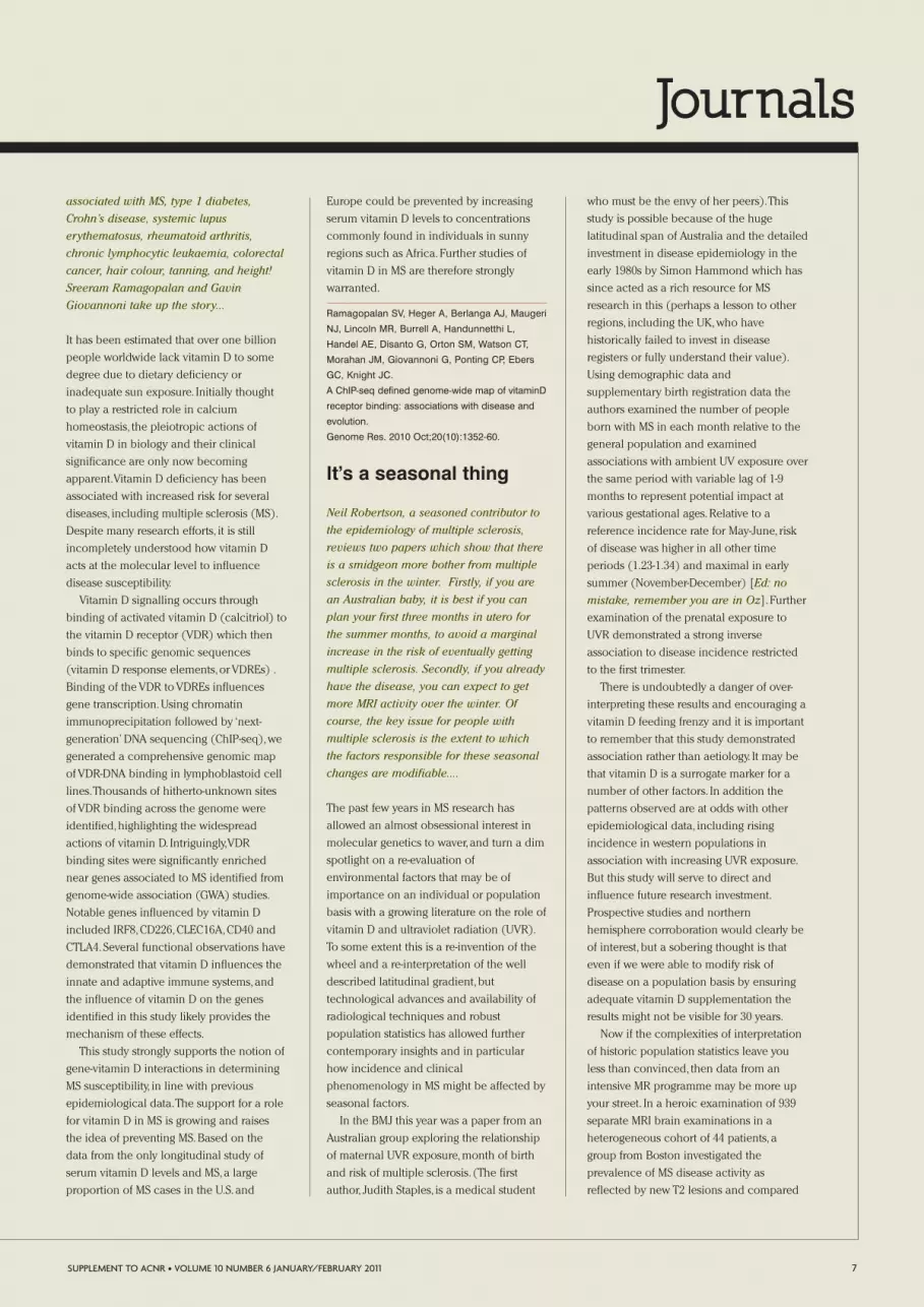
It’s a seasonal thing
Neil Robertson, a seasoned contributor to
the epidemiology of multiple sclerosis,
reviews two papers which show that there
is a smidgeon more bother from multiple
sclerosis in the winter. Firstly, if you are
an Australian baby, it is best if you can
plan your first three months in utero for
the summer months, to avoid a marginal
increase in the risk of eventually getting
multiple sclerosis. Secondly, if you already
have the disease, you can expect to get
more MRI activity over the winter. Of
course, the key issue for people with
multiple sclerosis is the extent to which
the factors responsible for these seasonal
changes are modifiable....
The past few years in MS research has
allowed an almost obsessional interest in
molecular genetics to waver, and turn a dim
spotlight on a re-evaluation of
environmental factors that may be of
importance on an individual or population
basis with a growing literature on the role of
vitamin D and ultraviolet radiation (UVR).
To some extent this is a re-invention of the
wheel and a re-interpretation of the well
described latitudinal gradient,but
technological advances and availability of
radiological techniques and robust
population statistics has allowed further
contemporary insights and in particular
how incidence and clinical
phenomenology in MS might be affected by
seasonal factors.
In the BMJ this year was a paper from an
Australian group exploring the relationship
of maternal UVR exposure,month of birth
and risk of multiple sclerosis. (The first
author, Judith Staples, is a medical student
who must be the envy of her peers).This
study is possible because of the huge
latitudinal span of Australia and the detailed
investment in disease epidemiology in the
early 1980s by Simon Hammond which has
since acted as a rich resource for MS
research in this (perhaps a lesson to other
regions, including the UK,who have
historically failed to invest in disease
registers or fully understand their value).
Using demographic data and
supplementary birth registration data the
authors examined the number of people
born with MS in each month relative to the
general population and examined
associations with ambient UV exposure over
the same period with variable lag of 1-9
months to represent potential impact at
various gestational ages.Relative to a
reference incidence rate for May-June, risk
of disease was higher in all other time
periods (1.23-1.34) and maximal in early
summer (November-December) [Ed: no
mistake, remember you are in Oz]. Further
examination of the prenatal exposure to
UVR demonstrated a strong inverse
association to disease incidence restricted
to the first trimester.
There is undoubtedly a danger of over-
interpreting these results and encouraging a
vitamin D feeding frenzy and it is important
to remember that this study demonstrated
association rather than aetiology. It may be
that vitamin D is a surrogate marker for a
number of other factors. In addition the
patterns observed are at odds with other
epidemiological data, including rising
incidence in western populations in
association with increasing UVR exposure.
But this study will serve to direct and
influence future research investment.
Prospective studies and northern
hemisphere corroboration would clearly be
of interest,but a sobering thought is that
even if we were able to modify risk of
disease on a population basis by ensuring
adequate vitamin D supplementation the
results might not be visible for 30 years.
Now if the complexities of interpretation
of historic population statistics leave you
less than convinced, then data from an
intensive MR programme may be more up
your street. In a heroic examination of 939
separate MRI brain examinations in a
heterogeneous cohort of 44 patients, a
group from Boston investigated the
prevalence of MS disease activity as
reflected by new T2 lesions and compared
SUPPLEMENT TO ACNR • VOLUME 10 NUMBER 6 JANUARY/FEBRUARY 2011 7

Journals
these to time of year and other climate data.
The results are beautifully illustrated and
the authors must be congratulated on the
extra investment in colour printing.
Although the patient numbers are small, the
biases which provide significant difficulty in
seasonal epidemiological studies are
effectively sidestepped by the objective MR
data which demonstrates the likelihood of
new T2 activity is 2-3 times higher in March-
August than during the rest of the year and
is most striking in patients with relapsing
disease.Although there have been one or
two earlier seasonal MR studies producing
variable results this is by far the most
convincing and the intensity of the
scanning remarkable and the patients
should be applauded for their commitment.
The importance of both these studies is
to underline that there are potentially
modifiable environmental factors that could
make significant impacts on not only
disease frequency but also clinical
expression. In addition the findings have
implications for trial design and service
delivery. So,MS really is a seasonal thing.
Staples J, Ponsonby AL, Lim L.
Low maternal exposure to ultraviolet radiation in
pregnancy, month of birth, and risk of multiple
sclerosis in offspring: longitudinal analysis.
British Medical Journal. 2010 Apr 29;340.
Meier DS, Balashov KE, Healy B, Weiner HL,
Guttmann CR.
Seasonal prevalence of MS disease activity.
Neurology. 2010 Aug 31;75(9):799-806.
Bugs andVirusesEd: The idea that multiple sclerosis may
be “triggered by a virus” has been around
for long time. As far as I know the earliest
proponent of an infective cause of the
disease was Pierre Marie, one of Charcot’s
residents, of CMT fame. My favourite MS-
virus story is how Cook and Dowling came
to suggest that canine distemper virus
might be responsible for multiple sclerosis
in 1977: they noted that three New York
sisters who lived together developed
multiple sclerosis after their dog had an
encephalopathy. The current candidate is
Epstein-Barr virus, for which there is lots
of uncontested evidence. But, in the last
few years, there has been some argy
bargey about whether or not EBV is in the
brains of people with multiple sclerosis.
The 2010 zeitgeist is that most people do
not find it. Ute Meier and Gavin
Giovannoni, from the Blizard Institute at
the Barts and the London, review one of
the key “no” papers, and bring to our
attention one of their own (unpublished)
studies, which was presented at the
International Society for
Neuroimmunology meeting.
EBV and the brain: arewe there yet?
No, the journey continues.Yet another US
publication by Jeffrey Bennett and
collaborators using real-time PCR failed to
detect active EBV infection in the brain of
patients with MS. In active MS plaques, a
small-non-coding EBV RNA (EBER-1) was
the only and rarely detected transcript.The
Denver group concluded that the MS brain
did not show any evidence of active EBV
infection.There are now two independent
PCR-based studies (Denver and Boston),
which conclude that active EBV infection is
not a characteristic feature of the MS brain.
However,how could latent infection play a
role?We and our collaborators in Oxford and
Abu Dhabi used in situ hybridisation to
detect EBER+ cells in active MS lesions.
However, this finding was not exclusive to the
MS brain as EBER+ cells were also found in
cases of stroke.We propose a more indirect
mechanism by which latent EBV infection
could contribute to neuroinflammation: that
these small RNAs bind to the toll-like
receptor 3 and thus stimulate IFN-alpha
production in active MS lesions.Could innate
activation, triggered by latent EBV infection,
be part of the game? Perhaps we have to
think differently - EBV might be more subtle
than we anticipated. It is a persistent virus
after all with the aim to co-exist with the host
rather than eradicate it.
The question remains – why do we get
discrepant results with different techniques?
Hans Lassmann invited all European and US
MS teams and several EBV researchers to a
July workshop inVienna.United under one
roof on neutral grounds they discussed the
different findings and techniques,exchanged
ideas,opinions and expertise.The Treaty of
Vienna has not been signed,but hopefully
productive EBV alliances were formed to
solve the conundrum of EBV and the brain.
Sargsyan SA, Shearer AJ, Ritchie AM, Burgoon
MP, Anderson S, Hemmer B, Stadelmann C,
Gattenlöhner S, Owens GP, Gilden D, Bennett JL.
Absence of Epstein-Barr virus in the brain and
CSF of patients with multiple sclerosis.
Neurology. 2010 Apr 6;74(14):1127-35.
Epub 2010 Mar 10.
Tzartos j, Khan G Vossenkamper A, Meager A,
Sefia E, Middledorp J, Giovannoni G, Meier UC.
Activation of innate immunity is a hallmark of the
active lesion in multiple sclerosis. J
Neuroimmunology. 228(1/2):163-4.
[Meeting Abstract].
Viruses and multiplepersonality T cells
Ed: Classical immunology has it that
viruses can trigger autoimmune disease by
mimicking a self-antigen, so that the
activated T cells get confused and attack
self, thinking it is the virus (“molecular
mimicry”) or that viruses cause such
widespread alarm in the immune system
that self-reactive T cells are non-
specifically wound up (“bystander
upregulation”). There is another way. Dan
Altmann had shown that a single T cell
can express two types of T cell receptor in
1995 at the Hammersmith; he argued that
8 SUPPLEMENT TO ACNR • VOLUME 10 NUMBER 6 JANUARY/FEBRUARY 2011
In January 2010, Fampridine (4-aminpyridine)was licensed by the FDA to improve walking
speed in people with multiple sclerosis;a UK decision is not expected until 2011.
It costs roughly $12,000 a year wholesale.

Journals
these aberrant cells had slipped through
the normal negative selection process.
Now, Joan Goverman and colleagues from
the University of Washington have done
some very clever experiments to show
that there can be a single CD8+ T cell
clone which responds to a virus with one
TCR and causes autoimmune
demyelination with the other. Orla Tuohy,
a clinical PhD student in Cambridge,
reports:
Ji et al in a paper published in Nature
Immunology describe a mechanism in
which viral infection can induce CNS
autoimmunity. The potential for T cells to
express more than one antigen-specific T
cell receptor (TCR) and trigger
autoimmunity has been previously
described. In this paper the authors use a
transgenic mouse model, that is MHC class 1
restricted and therefore amenable to the
study of CD8+ T cell-mediated interactions.
Although the CD4+ subsets have attracted
more interest and study in autoimmune
demyelination, the authors point out that
the increased presence of CD8+ myelin
antigen specific cells in MS warrants
consideration of this lymphocyte population
as a key player in disease pathogenesis. In
addition the high rate of serological
evidence of past EBV exposure in MS may
implicate viral involvement in disease
pathogenesis.
The transgenic mouse model they use
(“8.8”) is a MHC class I–restricted TCR-
transgenic model that generates CD8+ T
cells specific for myelin basic protein. The
authors found the 8.8 mouse was resistant
to traditional techniques to induce EAE
induction (which are generally MHC class 2
restricted,CD4+ cell-mediated).On the other
hand,EAE could be induced by vaccinating
with a recombinant MBP-expressing
vaccinia virus.
The key observations are that:
• The 8.8 mice also developed EAE when
they were infected with wild-type (non-
MBP expressing) vaccinia virus.Vaccinia
does not cross-react with MBP, so it
implies that the T cells in these animals
had newly generated TCRs to respond
to vaccinia,which then went on to
cause EAE.
• However, the 8.8 mice did not get EAE
with vaccinia, if they were made unable
to generate endogenous TCR chains by
knocking out Rag.
This led to the hypothesis that the disease
was caused by T cells expressing receptors
for vaccinia virus antigens and a second
TCR specific for myelin (and these TCRs
require Rag to be made),which could be
activated during viral infection and lead to
an autoimmune attack.
To support this hypothesis, evidence was
needed that the MBP-specific T cells
activated after wild-type vaccinia virus
infection,co-expressed endogenous TCRs
that recognised viral antigens. This could
account for the occurrence of disease in
Rag-competent mice versus Rag knockout
mice as Rag allows the generation of
endogenous TCR chains via incomplete
allelic exclusion in the transgenic MHC-
class 1 restricted mice models. The 8.8 mice
that still expressed Rag could potentially
express endogenous TCR chains that were
viral specific, in addition to their MBP-TCR.
Following wild-type vaccinia virus some
of the activated CD8+ T cells from 8.8 Rag
+/+ mice were seen to express an
endogenous TCR beta chain (beta 6). 8.8 T
cells expressing the TCR beta 6 chain
expressed more Interferon-gamma after
exposure to vaccinia-virus infection cells in-
vitro, than after MBP stimulation suggesting
the beta 6 chain was viral antigen reactive.
Along the way the authors excluded
molecular mimicry and bystander
activation as mechanisms for viral
inoculation to trigger disease in the 8.8
mouse model.
Through a series of carefully engineered
experiments, Ji et al have illustrated a
mechanism through which viral infection
could interact with genetic susceptibility to
trigger autoimmunity in a mouse model
with a genetically modified immune cell
repertoire. Whether this mechanism is
involved in autoimmune disease initiation
in the presence of a naturally occurring
immune system has yet to be proved.
Ji Q, Perchellet A, Goverman JM.
Viral infection triggers central nervous system
autoimmunity via activation of CD8+ T cells
expressing dual TCRs.
Nature Immunology. 2010 Jul;11(7):628-34.
Immune Bitsand Bobs
Yet another regulatorycell
Ed: Allison Curry, a post doc in my lab,
reviews a study from that prolific MS
immunologist, Jorge Correale, from the
Raúl Carrea Institute for Neurological
Research in Argentina. He makes the case
for the importance of the rather obscure
CD8 regulatory cell in multiple sclerosis.
You will remember that the CD4 regulatory
cell does not work properly in people with
multiple sclerosis. In fact, the failure of the
CD4 T reg to suppress aggressive T cells is
one of the very few consistent
immunological abnormalities in this
disease. My own take on this detailed
paper is that there is one important
element missing. I would like to know if the
CD8 regulatory cells in multiple sclerosis
are also intrinsically defective, compared to
controls, like their CD4 cousins.
The inflammatory process is a complex
network involving many cell types, their
related cytokines and signalling pathways.
We know that self reactive T cells exist in
the periphery,presumably having escaped
thymic clonal deletion,and that these must
be kept under tight surveillance in order to
prevent dys-regulation,potential
autoimmunity and disease.
SUPPLEMENT TO ACNR • VOLUME 10 NUMBER 6 JANUARY/FEBRUARY 2011 9
This year Jimmie Heuga died, founder ofThe Heuga Center for Multiple Sclerosis,
and 1964 bronze medallist in slalom at theWinter Olympics in Innsbruck, Austria.

Journals
Our understanding of T cell function and
regulation has expanded from initial CD4/8
phenotypic distinction through the
characteristic Th1/Th2 cytokine profiles to
the current focus on T regulatory cells (T
regs). Although CD8+ regulatory cells in MS
have previously received attention, recent
research has focused on existing and
emerging CD4 T reg populations.The role of
the CD8+ CD25+ Fox P3+ regulatory cell
(CD8 T reg) has been revisited by these
Argentinean MDs.
The authors focus on CD8 Treg in the
PBMC and CSF from 35 patients with RRMS
(15 patients in remission),15 controls and 10
patients suffering from other inflammatory
neurological diseases (OIND).Much of the
CD4 T reg data published in MS to date has
relied on precise isolation of CD4+ CD25 high
cells and their ability to suppress polyclonal
stimulation. Using limiting dilution
techniques,plus stimulation with Myelin Basic
Protein (MBP)/Myelin oligodendrocyte
(MOG) peptides,CD4 and CD8 T cells were
cloned from both the PBMC and CSF.CD8 T
regs were identified and shown to reduce the
responses of CD4 cells to their antigens.This
suppression was impaired in transwell
experiments which showed that CD8 cells
suppress by cell contact.The CD8 T regs seem
to do this via dendritic cells,using STAT3
signalling,rather than directly on CD4 T cells.
Finally FACS analysis of CSF and PBMC
showed reduced representation of CD8+ T
regs in patients during acute exacerbation
suggesting a reduction in CD8+ regulation at
potential sites of inflammation.
How CD8 T regs interact with CD4 T regs,
CD4 cells and the APC interact is a
fascinating puzzle.One can only
contemplate how future identification of
further target antigens may yet contribute to
solving the complicated,unfolding maze of
immune regulation.
Correale J, and Villa A.
The role of CD8+ CD25+ Foxp3+ regulatory T
cells in multiple Sclerosis.
Annals of Neurology 2010;67:625-38.
Why interferon-betaworks for some but notothers
Ed: This paper has certainly struck a
chord. Four of our reviewers wanted to
review it for the ACNR. In the end, I asked
Bruno Gran, a neuroimmunologist from
Nottingham, to comment. He gives it a
very fair write-up. It is certainly attractive,
because it uses the latest understanding of
multiple sclerosis immunology (around the
difference between Th1 and Th17 cells) to
explain why some people respond to the
standard licensed drug for multiple
sclerosis, interferon-beta. Personally, I am
more sanguine about this study, mainly
because each stage in the argument is
based on results from rather few animals
or patients, and it all looks a bit tidy. But
perhaps I am just jealous!
In the last 15 years,progress in MS
management has been remarkable.The
introduction of immunotherapies that
reduce the frequency of disease
exacerbations and,at least in the short-
medium term, the tendency to accrual of
disability,has been welcomed by patients
and,perhaps with less enthusiasm,by
clinical neurologists,whose ambitions are
often higher than patients’.The first of such
treatments was,of course, interferon-beta
(IFNb),a“type I interferon”whose main
physiological job may be to signal the
presence of viral infections. In addition,
IFNb is a useful immunomodulator,which
can reduce disease activity in perhaps two
thirds of the patients for whom it is
appropriately prescribed.
We are currently in a rather exciting phase
of MS treatment history as promising oral
treatment are being developed and
introduced into clinical practice.However,
reasonable concerns by regulatory agencies –
most recently raised for cladribine in Europe
– suggest that we may well be using injected
interferons,as well as Copaxone, for longer
than we have thought in the last couple of
years. It is then even more important to try
and understand why at least a third,but
possibly up to a half,of people with relapsing-
remitting MS do not respond to IFNb.If we
take a step back and consider how IFNb is
thought to work in MS,some humility is
called for.As in so many areas of medicine,
we have to admit that while some
mechanistic facts are reasonably clear,many
are not.Most will agree that the induction of
interleukin-10 (IL-10),a (usually) anti-
inflammatory immune molecule,and a
reduction of permeability of the inflamed
blood-brain barrier are involved.But what
about the effects on immune cells that are
thought to initiate autoimmune reactivity
against myelin? T helper 1 (Th1) and Th17
cells are considered most relevant in this
respect,due to their ability to induce CNS
inflammation in animal models of MS
(collectively called“experimental
autoimmune encephalomyelitis”,EAE),and to
their increased levels in the brain,spinal fluid,
and peripheral blood of people with MS.
In a study published in“Nature Medicine”
earlier this year, the Stanford team
addressed the role of the Th1 and Th17
“cytokine pathways” in how mice and
humans with autoimmune inflammatory
demyelination respond to IFNb. In a
nutshell,what they found is that when Th1
cells (producers of Interferon gamma, IFNg
and historically the first T helper subset to
be indicted with a role in MS pathogenesis)
are the key players in the induction of EAE,
IFNb helps. It reduces disease severity both
clinically and pathologically.However,when
Th17 cells (identified in 2005 as the most
likely“real culprit” in autoimmune
neuroinflammation) are the inducers of
EAE,not only does IFNb not help,but
disease is actually made worse.Technically,
the key experiments in the study involved
the use of“adoptive transfer EAE”, in which
mice are immunised with myelin peptides
and,at the time when a potent myelin-
reactive response develops, autoreactive T
10 SUPPLEMENT TO ACNR • VOLUME 10 NUMBER 6 JANUARY/FEBRUARY 2011
In 2010, the UK MS Society funded 12 newresearch projects costing more than £1.6
million. The US National MS Society funded34 new projects, costing $14.6 million.
Journals
cells are harvested from these mice and
cultured in vitro before transferring them to
recipient,“naive”mice.During in vitro
culture, these cells can be potently skewed
to a Th1 or a Th17 phenotype by adding
either IL-12 or IL-23 to the cultures.
Recipient mice will develop an MS-like
disease (with paralysis and CNS
inflammation) and can be treated with IFNb
to assess its effect on clinical,pathological
and immunological disease parameters. A
summary of the rather complex
immunology experiments reported is as
follows: in agreement with preliminary in
vitro studies in mouse T cells, in vivo studies
showed that suppression of experimental
disease by IFNb correlated nicely with the
ability of IFNb to induce IL-10 in the treated
mice. In fact, IL-10 production increased
after IFNb treatment of“Th1 EAE”and was
not affected by treatment of“Th17 EAE”. In
addition,when disease was induced with
Th1 cells, IFNb treatment reduced the
numbers of all types of inflammatory T
helper cells in the spinal cord, i.e., both Th1
and Th17.By contrast, IFNb given to mice
with Th17-induced disease was associated
with an increase in both cell types.Another
clear result was that the beneficial effects of
IFNb on Th1 EAE required the presence of
IFNg signalling, as mice deficient in IFNg
receptor were not protected.As most
cytokines, IFNg will have different or even
opposite effects in different biological
contexts, and these findings confirm that in
mouse EAE, IFNg is predominantly“good”.
Unfortunately, this is a well known example
of weak or absent“translational link” from
mice to men,as IFNg was found to
exacerbate MS in patients in the late 1980s
and has since been considered“bad”for MS.
But back to IL-17 and,perhaps as
importantly, to IL-10.Was there a link to
anything of relevance to clinical practice?
This is what the final part of the study starts
to look at,without going as far as in the
mouse studies,but pointing to an interesting
direction. In human immune cell cultures,
the authors showed that IFNb had little
effect on the differentiation of Th1 cells but,
by contrast, it did inhibit the development
of Th17 cells.More importantly, the
induction of IL-10 by IFNb in these cultures
was clear and potent only in the Th1 cells.
This suggests that if there is such a thing as
“Th1 MS”, it might be more susceptible to
IFNb treatment,because it is Th1 cells that
respond to IFNb by making IL-10.The same
concept would not apply to“Th17 MS”.
The study concludes by testing another
important hypothesis: could MS patients
who fail to respond to IFNb have a stronger
propensity to Th17 responses? To start
addressing this question, the authors
measured“pre-treatment cytokine profiles”
in the sera of individuals with MS before
starting treatment with IFNb and classified
them as either“responders”(12 subjects) or
“non responders”(14 subjects) based on
clinical parameters such as relapse
frequency,degree of disability, and the need
for steroid courses before and after at least
12 months of IFNb treatment.Cluster
analysis of the 28 cytokines studied showed
that a subgroup of non responders (6
subjects) had significantly elevated serum
concentrations of both IL-17F (one of the
two main isoforms of IL-17) and IFNb
(which is itself produced by the immune
system).Furthermore, there was a strong
correlation between serum levels of IL-17F
and IFNb not just in this subgroup,but also
in all non responders, responders, and
healthy controls.
In summary, the take-home message at
this stage is that if these findings are
confirmed in a considerably larger
population of patients, it might one day be
possible to use serum concentrations of IL-
17F and IFNb (or possibly other
“biomarkers”) to predict the therapeutic
success of IFNb therapy.With the caveat that
biomarker studies in MS have often been
promising,but eventually disappointing, this
line of investigation appears worth
pursuing.
Axtell RC, de Jong BA, Boniface K, van der Voort
LF, Bhat R, De Sarno P, Naves R, Han M, Zhong F,
Castellanos JG, Mair R, Christakos A, Kolkowitz I,
Katz L, Killestein J, Polman CH, de Waal Malefyt
R, Steinman L, Raman
T helper type 1 and 17 cells determine efficacy of
interferon-β in multiple sclerosis and experimental
encephalomyelitis.
Nature Medicine 2010;16(4):406-12.
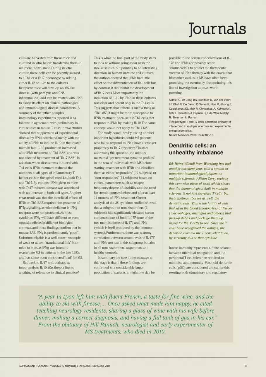
Dendritic cells: anunhealthy imbalance
Ed: Heinz Wiendl from Wurzburg has had
another excellent year, with a stream of
important immunological papers on
multiple sclerosis. Allison Curry reviews
this very nice piece of work which shows
that the immunological fault in multiple
sclerosis is not just wayward T cells, but
their upstream bosses as well: the
dendritic cells. This is the family of cells
that sit in the blood (monocytes) or tissues
(macrophages, microglia and others) that
pick up debris and package them up
nicely for the T cells to see. Once the T
cells have recognised the antigen, the
dendritic cells tell the T cells what to do,
by secreting this or that cytokine.
Innate immunity represents a finite balance
between microbial recognition and the
peripheral T cell tolerance required to
minimise autoimmunity. Plasmoid dendritic
cells (pDC) are considered critical for this,
exerting both stimulatory and regulatory
SUPPLEMENT TO ACNR • VOLUME 10 NUMBER 6 JANUARY/FEBRUARY 2011 11
“A year in Lyon left him with fluent French, a taste for fine wine, and theability to ski with finesse ... Once asked what made him happy, he cited
teaching neurology residents, sharing a glass of wine with his wife beforedinner, making a correct diagnosis, and having a full tank of gas in his car.”
From the obituary of Hill Panitch, neurologist and early experimenter ofMS treatments, who died in 2010.

Journals
effects on T cells.Their contribution to
antigen presentation in vivo however,and
their contribution to autoimmune T cell
activation and localised inflammatory
reactions remains unclear. Not found in the
CNS under normal conditions,elevated pDC
have been reported under
neuroinflammatory conditions with impaired
maturation and altered regulatory functions
implicated in the pathogenesis of Multiple
Sclerosis (MS). Here,Schwab et al
demonstrate that pDC can be further
distinguished into two separate populations
(“pDC1 and pDC2”) based on their surface
markers,cytokine production,activation
criteria and ability to prime naïve allogenic T
cells.pDC 1(CD123 high CD86- MHC II low)
resemble immature DC whereas pDC2 exhibit
a more mature phenotype.The role of pDC1
and pDC2 in MS autoimmunity is contrasted
with autoimmune Myasthenia Gravis (MG).
The innate immune system relies on
conserved pattern recognition receptors to
distinguish pathogen associated molecular
patterns (PAMPs). One such PAMP is that of
the CpG oligonucleotides present in
microbial DNA often used to mimic DC
activation. In response to CpG activation,
pDC1 showed a 3.8 fold increase in
regulatory (TR1) cells whereas pDC2
caused a 4.8 fold elevated autoaggressive
(Th17) T cells.
To determine any contribution of these
two subsets to MS,pDC from the PBMC of 16
MS patients were investigated.The
pDC1:pDC2 ratio was low in MS patients,
compared to healthy controls although
overall numbers were comparable.The
impact of inverted pDC ratios in MS patients
on immune response priming was
investigated using HC and MS derived pDC
in allogenic T cell co-culture assays.MS pDC
not only stimulated higher levels of Th17
cells compared to HC but also the
production of IL-17+IFN+ double positive
cells absent in HC.These populations have
previously been identified as highly pro-
inflammatory and, in the proper antigenic
setting, an encephalitolytic subpopulation of
CD4+ cells. Interestingly,pDC1 numbers rise
in MS following 12 months of IFN beta
treatment.Changes were observed by two
months suggesting that the subset
imbalance observed during MS is not a
fixed abnormality and is amenable to
therapeutic modification.
Although IFN beta may act at a variety of
levels,of which the pDC may be just one,
this research highlights how immunotherapy
can act at a cellular level in modulating the
immunoregulatory dysfunctions of patients
with MS and gives an insight into
mechanisms both involved and required for
therapeutic intervention.
Schwab N, Zozulya A, Kieseier K, and Wiendl H.
An imbalance of two functionally and
phenotypically different subsets of plasmoid
Dendritic cells characterises the functional
immune regulation in multiple sclerosis.
Journal of Immunology 2010;184:5368-74.
Devic’s DiseaseEd: In 2010, 174 papers were published on
neuromyelitis optica; in 2000, the number
was only 15. This sudden interest has
largely been down to Vanda Lennon’s
discovery in 2004 of an antibody
associated with the illness, which once
and for all resolved the question of
whether neuromyelitis optica was a form
of multiple sclerosis or a distinct disease.
It has also allowed people to explore the
boundaries of the phenotype of
neuromyelitis optica, much in the same
way that anti-GQ1b antibodies have done
for Miller Fisher syndrome. Isabel Leite
and Joanna Kitley, from the
neuroimmunology group at Oxford, have
picked out three of this year’s crop of
papers on Devic’s disease, my favourite
being the largest series on the disease yet
published, from a survey of 25 French
centres including that of Christian
Confavreux, whose wife is the
granddaughter of Eugene Devic. This
confirms the clinical impression that
neuromyelitis optica is a nasty disabling
disease: for instance 30% of the patients
had unilateral visual loss (<6/60) and 13%
had bilateral severe visual loss. Patients
with a high early relapse rate do worse
and we are encouraged to treat them
early... but not with interferon-beta as both
the Japanese and Oxford experience is
that this may exacerbate the condition.
A cause of vomiting andhiccups
In 2008,Takahashi et al showed that
intractable hiccup and nausea could be a
clinical marker of aquaporin-4 (AQP4)
antibody mediated disease,which could
precede the neurological symptoms at first
presentation or exacerbation of
neuromyelitis optica.Those symptoms were
thought to be related to the autoimmune
attack on the AQP4 molecules of astrocytes
in the pericanal region in the medulla
oblongata,which includes the area postrema.
Recently,Apiwattanakul and colleagues
described 12 aquaporin-4 antibody positive
patients who had an initial presentation of
intractable vomiting that remained
unexplained despite extensive investigations,
until the appearance of typical clinical
manifestations of neuromyelitis optica.
Although some of the patients did have mild
neurological symptoms it was shown that
vomiting constitutes a well defined
presenting feature in neuromyelitis optica in
at least 12% of patients.There was a variable
interval between vomiting onset and optic
neuritis (optic neuritis) or longitudinally
extensive transverse myelitis (LETM).The
authors hypothesised that the structural and
molecular characteristics of the area
postrema,such as the presence of fenestrated
capillaries,may contribute to the entry of
circulating IgG/AQP4 antibodies into the
CNS.They also emphasised the unique
nature of lesions in the area postrema,by
illustrating the focal loss of AQP4 and the
presence of inflammatory cells,but unusually
no demyelination or necrosis,which may
explain the reversibility of clinical symptoms
and imaging abnormalities in this area.As
the initial site of antibody binding in some
patients with neuromyelitis optica, these
presenting symptoms may allow a ‘window-
of-opportunity’ for antibody testing and
treatment in patients that are found to be
aquaporin-4 antibody positive. As 75% of
these patients first presented to a
gastroenterologist it is important to make
these clinicians aware of the importance of
intractable nausea as an initial symptom of
neuromyelitis optica.
Apiwattanakul M, Popescu BF, Matiello M,
Weinshenker BG, Lucchinetti CF, Lennon VA,
McKeon A, Carpenter AF, Miller GM, Pittock SJ.
Intractable vomiting as the initial presentation of
neuromyelitis optica.
Annals of Neurology. 2010 Nov;68(5):757-61.
Worrying epidemiologyof neuromyelitis optica
This is the largest observational,
retrospective,multicentre study of
neuromyelitis optica (NMO) involving 125
patients.By applying the 2006 NMO
diagnostic criteria, excluding the criteria of
12 SUPPLEMENT TO ACNR • VOLUME 10 NUMBER 6 JANUARY/FEBRUARY 2011

Journals
NMO-IgG, the authors were able to make the
diagnosis of neuromyelitis optica in 90% of
cases,with the NMO-IgG indirect
immunofluorescence assay (IIF) mandatory
for the diagnosis of neuromyelitis optica in
only 10% of patients, confirming the power
of the clinical and imaging features in the
diagnosis and treatment of neuromyelitis
optica.The demographic and basic clinical
characteristics of the neuromyelitis optica
patients are in agreement with those of
previous studies,but it is important to
highlight that in more recent reports, the
range of onset age is even wider than found
in this paper; neuromyelitis optica patients
aged above 70 years at onset have been
diagnosed.Analyses of the course of
neuromyelitis optica and disability
underlined the high degree of disability
following a first attack of myelitis or optic
neuritis (optic neuritis).Nearly 50% of those
neuromyelitis optica patients were left with
a severe deficit after their first attack of
optic neuritis. Importantly, a shorter time to
residual visual acuity of ≤ 6/60 was found to
be associated with a high number of MRI
brain lesions,which was the only predictive
factor of poor evolution that this study
revealed. Interestingly, in our (Ed; that is the
Oxford] cohort of patients with aquaporin-4
(AQP4) antibody-positive disease,we have
also observed that brain disease is
associated with more severe optic neuritis
involvement,particularly in young and non-
Caucasian patients with neuromyelitis
optica (Leite,Palace unpublished data).
Finally, the sensitivity of their assay is lower
than other published methods.When
comparing clinical and imaging features of
antibody negative and antibody positive
patients the use of the most sensitive cell-
based assays may highlight further
differences and should aid the search for
new target antigens in neuromyelitis optica.
Collongues N, Marignier R, Zéphir H, Papeix C,
Blanc F, Ritleng C, Tchikviladzé M, Outteryck O,
Vukusic S, Fleury M, Fontaine B, Brassat D, Clanet
M, Milh M, Pelletier J, Audoin B, Ruet A, Lebrun-
Frenay C, Thouvenot E, Camu W, Debouverie M,
Créange A, Moreau T, Labauge P, Castelnovo G,
Edan G, Le Page E, Defer G, Barroso B, Heinzlef
O, Gout O, Rodriguez D, Wiertlewski S, Laplaud D,
Borgel F, Tourniaire P, Grimaud J, Brochet B,
Vermersch P, Confavreux C, de Seze J.
Neuromyelitis optica in France: a multicenter
study of 125 patients.
Neurology. 2010 Mar 2;74(9):736-42.
Interferon-beta mayexacerbateneuromyelitis optica
Whilst there is undoubtedly clinical overlap,
neuromyelitis optica (NMO) is now
generally considered to be an antibody
mediated autoimmune disease unlike
multiple sclerosis.A report by Shimuzu and
colleagues suggests that
immunomodulatory treatment may even
exacerbate neuromyelitis optica. In this
retrospective study of 56 patients diagnosed
with relapsing remitting MS (RRMS) and
treated with interferon beta (IFNβ), 7
patients experienced severe neurological
exacerbation shortly after initiating therapy.
Retrospectively, the diagnosis in these 56
patients was reclassified as neuromyelitis
optica in 14,high risk of neuromyelitis
optica in 6 and conventional RRMS in 36.
All 7 patients who deteriorated on IFNβ
were patients reclassified as neuromyelitis
optica, and all tested positive for the
aquaporin-4 antibody.The patients
experiencing exacerbations all developed
transverse myelitis with longitudinally
extensive signal change on spinal MRI.Two
patients also had optic neuritis, and two
developed new tumefactive brain lesions.
Exacerbations were severe,with EDSS
ranging from 7.5 to 9.5, and occurred within
90 days of starting IFNβ. We have recently
described a neuromyelitis optica patient
misdiagnosed with RRMS who experienced
an increase in relapses and also a dramatic
increase in the AQP4 antibody levels on
treatment with IFNβ,which reduced quickly
following conventional immunosuppressive
treatment (Palace Arch Neurol 2010).
Shimuzu and colleagues’ paper highlights
the need to test for aquaporin-4 antibodies
in all RRMS patients with atypical clinical
features such as poor spontaneous recovery
from relapses,dramatic response to steroid
therapy with exacerbation following steroid
withdrawal,unusually severe optic nerve or
spinal cord relapses and in those whose
brain imaging or CSF characteristics are not
classical for MS. Immunosuppressive
treatments that appear effective for
neuromyelitis optica [Ed: such as rituximab
and azathioprine] are also effective
treatments of multiple sclerosis.Thus, it is
sensible to treat those with an optico-spinal
MS phenotype,or borderline cases, as
though they had neuromyelitis optica to
avoid the potentially catastrophic
deterioration if treated with IFNβ.
Shimizu J, Hatanaka Y, Hasegawa M, Iwata A,
Sugimoto I, Date H, Goto J, Shimizu T, Takatsu M,
Sakurai Y, Nakase H, Uesaka Y, Hashida H,
Hashimoto K, Komiya T, Tsuji S.
IFNβ-1b may severely exacerbate Japanese optic-
spinal MS in neuromyelitis optica spectrum.
Neurology 2010;75:1423-7.
Astrocytes:Who They?
Do astrocytes regulateCNS inflammation?
Ed: I always thought that astrocytes were
pretty harmless. They don’t seem to do an
awful lot of anything really, just plodding
their foot processes around and looking
SUPPLEMENT TO ACNR • VOLUME 10 NUMBER 6 JANUARY/FEBRUARY 2011 13
Immunofluorescence binding of NMOpatient serum IgG to aquaporin-4(AQP4) expressed on the surface ofHEK cells. Live cells were identified bythe blue nuclear stain dapi and thepatient AQP4 specific antibodies weredetected by red anti-human IgGantibody.
This picture illustrates the diagnosticassay used to detect antibodies toAQP4, which is free of charge for allthe NHS patients from England andScotland. This is part of the Diagnosticand Advisory Service for NMO fundedby the National Commissioning Group,NHS, UK.
For further information regarding theservice, please, visit:http://www.oxfordradcliffe.nhs.uk/forclinicians/referrals/neurosciences/nmoclinic/nmoclinic.aspxor call 01865 234889.
Image courtesy of Patrick Waters,M Isabel Leite and Angela Vincent.

Journals
spidery. But it turns out that they can be
really quite aggressive, as this EAE study
from the Cleveland Clinic shows. Patrick
Waters, a neuroimmunologist in Oxford,
walks us through the data.
Neuromyelitis optica (NMO) is a severe
destructive antibody mediated
demyelinating disease of the CNS.A key
feature of NMO lesions is the early loss of
astrocytes, commonly demonstrated as loss
of GFAP stain by immunohistochemistry.
Other antibody mediated diseases of the
CNS whose antibody targets are on neurons
rather than astrocytes (e.g.NMDAR
encephalitis) are not destructive, suggesting
that the target cell may play a key role in
modifying CNS inflammation.
Kang et al use both a MOG 33-55 peptide
active immunisation and a Th17 T-cells
adoptive transfer EAE mouse model to
demonstrate that astrocytes, via the IL-17R,
play a key role in stimulating the effector
phase of Th17 induced CNS inflammation.
Previously they demonstrated that knocking
out the IL-17R signalling molecule Act1-/-
greatly reduced the severity and delayed the
onset of Th17 induced EAE.They have now
followed this using the Cre-lox transgenic
system to delete Act1 in different cell
lineages.They demonstrate that deletion of
Act1 in endothelial or myeloid cells does
not affect disease severity in their EAE
mouse model.However,using the NesCre
transgene they deleted Act1 from
neuroectodermal cells of the CNS
(astrocytes,oligodendrocytes and neurons)
and confirmed that these cells were the key
cell to the reduction in inflammation seen
in their previous knock-out animal.They
further demonstrated that the only cell type
in which the IL-17 induced chemokine
profile was impaired, in Act1-/-,was the
astrocytes, implicating them in recruitment
of leukocytes into the brain, inducing the
second wave of inflammation.
Although the destruction of astrocytes is
only one aspect of NMO, this paper
highlights their importance in Th17 induced
autoimmune disease in an animal model
which may shed some light on the
mechanism of pathology in this human
disease.
Kang Z, Altuntas CZ, Gulen MF, Liu C, Giltiay N,
Qin H, Liu L, Qian W, Ransohoff RM, Bergmann C,
Stohlman S, Tuohy VK, Li X.
Astrocyte-restricted ablation of interleukin-17-
induced Act1-mediated signaling ameliorates
autoimmune encephalomyelitis.
Immunity. 2010 Mar 26;32(3):414-25.
Transporters: cassettesand media
Ed: there is an ancient family of proteins
that act as sherpas across membranes,
both within and on the outside of cells.
They require energy in the form of ATP and
so have been named: the “ATP-binding
cassette transporter family” (great
creativity). Many are responsible for
bacterial multi-drug resistance. And a
defect in one of the members leads to X-
linked adrenoleukodystrophy. Elga de
Vries’ blood-brain barrier group in
Amsterdam now ask whether these
proteins do anything in multiple sclerosis.
Mike Zandi takes us through their Brain
paper. Once again, astrocytes are involved,
although it is hard to say whether they are
goodies or baddies in this set-up.
This study examined the expression and
function of members of the ATP-binding
cassette transporter family in multiple
sclerosis (MS) using post mortem tissue.
The study showed enhanced multidrug
resistance associated protein-1 (MRP-1) and
P-glycoprotein (P-gp) on reactive astrocytes,
in active and chronic MS lesions compared
to MS normal appearing white matter
(NAWM) and controls.The authors then
tried to address the issue of causality by
using two functional in vitro chemotaxis
assays.Conditioned media from astrocytes,
harvested from MS lesions, increased
monocyte migration across a human
endothelial cell model of the blood brain
barrier.Blocking MRP-1 and P-glycoprotein
(P-gp) activity reduced the migration in
both models,probably by reducing the
secretion of a chemokine,CCL2.
The usual problems of post mortem
studies stack the odds against the findings
being primary and relevant to early disease -
most of the 10 patients had longstanding
disease (range 7-39 years, and 2 with an
unknown disease duration) and died from
some major systemic insult (pneumonia) or
local disease (stroke).The lack of difference
between NAWM and healthy controls is
perhaps surprising.The specificity of
function of these diverse molecules remains
unclear – in PloS ONE last year (8;4:e8212),
the same group published a study
demonstrating reduced severity of MOG-
induced experimental autoimmune
encephalomyelitis through reduced
secretion of pro-inflammatory cytokines in a
P-gp knockout mouse.The authors conclude
reasonably that the generation of astrocyte-
specific P-gp knockout mice will be the
most fruitful step forward for further studies.
Kooij G, Mizee MR, van Horssen J, Reijerkerk A,
Witte ME, Drexhage JA, van der Pol SM, van Het
Hof B, Scheffer G, Scheper R, Dijkstra CD, van der
Valk P, de Vries HE.
Adenosine triphosphate-binding cassette
transporters mediate chemokine (C-C motif)
ligand 2 secretion from reactive astrocytes:
relevance to multiple sclerosis pathogenesis.
Brain epub 22 Dec 2010.
Th17 cells seek anddestroy neurons
Ed: Th17 cells are the new black. We
reviewed their history in last year’s
supplement. Basically we had all been
made to look very silly, because in the
past we had been measuring IL-12 with an
antibody which also detects IL-23, but we
did not realise it. So what we thought was
IL12 making “Th1” cells was in fact a
mixture of IL12 and IL23... and IL23 makes
a distinctive type of T cell called the
“Th17” cell. This year, everyone and his
dog has had a crack at working out what
exactly they do in multiple sclerosis. If
Frauke Zipp from Mainz is correct, the
news is bad. It seems Th17 cells have a
14 SUPPLEMENT TO ACNR • VOLUME 10 NUMBER 6 JANUARY/FEBRUARY 2011
Harry Potter author, JK Rowling, donated £10million to set up the Anne Rowling
Regenerative Neurology Clinic in Edinburgh.
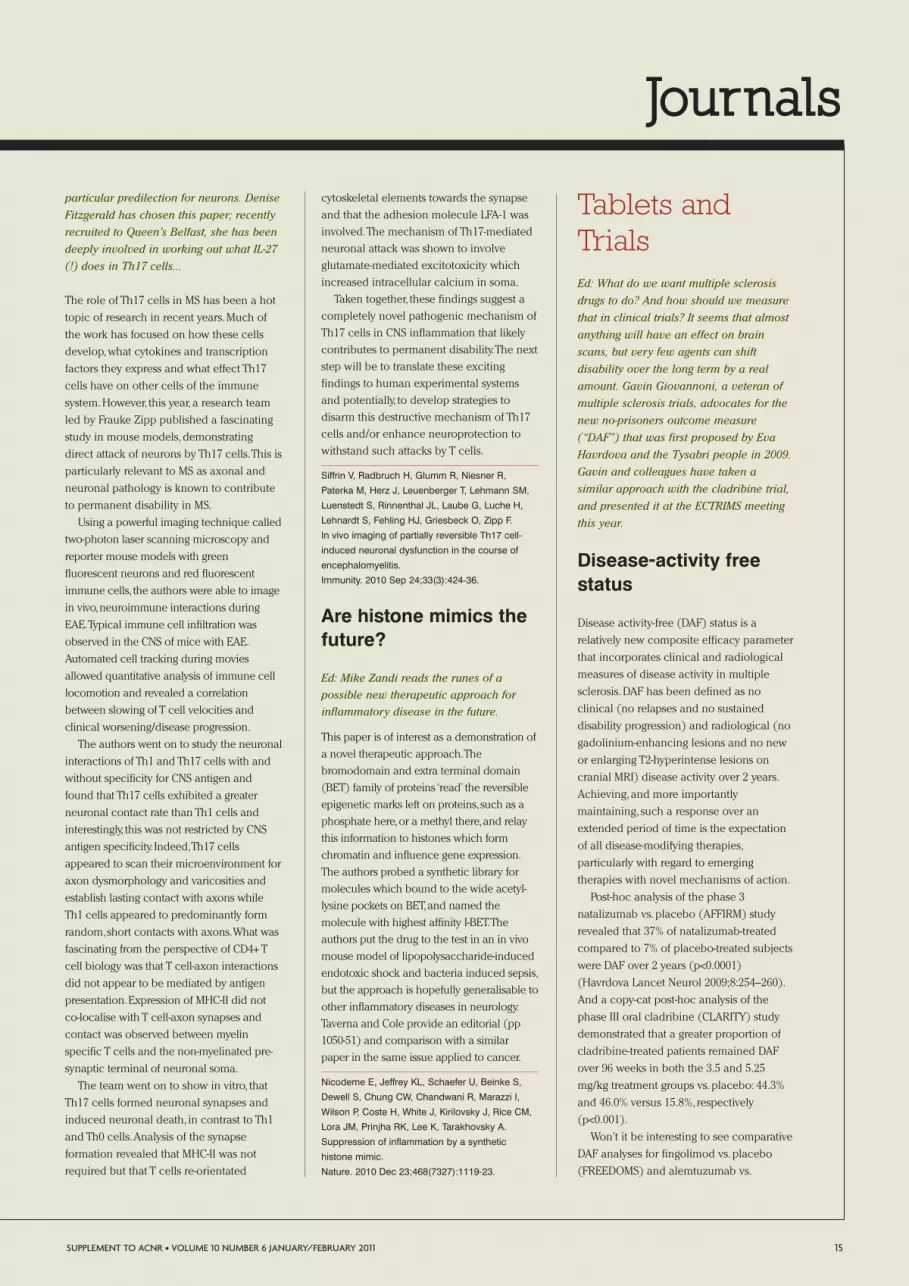
Journals
particular predilection for neurons. Denise
Fitzgerald has chosen this paper; recently
recruited to Queen’s Belfast, she has been
deeply involved in working out what IL-27
(!) does in Th17 cells...
The role of Th17 cells in MS has been a hot
topic of research in recent years.Much of
the work has focused on how these cells
develop,what cytokines and transcription
factors they express and what effect Th17
cells have on other cells of the immune
system.However, this year, a research team
led by Frauke Zipp published a fascinating
study in mouse models,demonstrating
direct attack of neurons by Th17 cells.This is
particularly relevant to MS as axonal and
neuronal pathology is known to contribute
to permanent disability in MS.
Using a powerful imaging technique called
two-photon laser scanning microscopy and
reporter mouse models with green
fluorescent neurons and red fluorescent
immune cells, the authors were able to image
in vivo,neuroimmune interactions during
EAE.Typical immune cell infiltration was
observed in the CNS of mice with EAE.
Automated cell tracking during movies
allowed quantitative analysis of immune cell
locomotion and revealed a correlation
between slowing of T cell velocities and
clinical worsening/disease progression.
The authors went on to study the neuronal
interactions of Th1 and Th17 cells with and
without specificity for CNS antigen and
found that Th17 cells exhibited a greater
neuronal contact rate than Th1 cells and
interestingly, this was not restricted by CNS
antigen specificity. Indeed,Th17 cells
appeared to scan their microenvironment for
axon dysmorphology and varicosities and
establish lasting contact with axons while
Th1 cells appeared to predominantly form
random,short contacts with axons.What was
fascinating from the perspective of CD4+ T
cell biology was that T cell-axon interactions
did not appear to be mediated by antigen
presentation.Expression of MHC-II did not
co-localise with T cell-axon synapses and
contact was observed between myelin
specific T cells and the non-myelinated pre-
synaptic terminal of neuronal soma.
The team went on to show in vitro, that
Th17 cells formed neuronal synapses and
induced neuronal death, in contrast to Th1
and Th0 cells.Analysis of the synapse
formation revealed that MHC-II was not
required but that T cells re-orientated
cytoskeletal elements towards the synapse
and that the adhesion molecule LFA-1 was
involved.The mechanism of Th17-mediated
neuronal attack was shown to involve
glutamate-mediated excitotoxicity which
increased intracellular calcium in soma.
Taken together, these findings suggest a
completely novel pathogenic mechanism of
Th17 cells in CNS inflammation that likely
contributes to permanent disability.The next
step will be to translate these exciting
findings to human experimental systems
and potentially, to develop strategies to
disarm this destructive mechanism of Th17
cells and/or enhance neuroprotection to
withstand such attacks by T cells.
Siffrin V, Radbruch H, Glumm R, Niesner R,
Paterka M, Herz J, Leuenberger T, Lehmann SM,
Luenstedt S, Rinnenthal JL, Laube G, Luche H,
Lehnardt S, Fehling HJ, Griesbeck O, Zipp F.
In vivo imaging of partially reversible Th17 cell-
induced neuronal dysfunction in the course of
encephalomyelitis.
Immunity. 2010 Sep 24;33(3):424-36.
Are histone mimics thefuture?
Ed: Mike Zandi reads the runes of a
possible new therapeutic approach for
inflammatory disease in the future.
This paper is of interest as a demonstration of
a novel therapeutic approach.The
bromodomain and extra terminal domain
(BET) family of proteins ‘read’ the reversible
epigenetic marks left on proteins,such as a
phosphate here,or a methyl there,and relay
this information to histones which form
chromatin and influence gene expression.
The authors probed a synthetic library for
molecules which bound to the wide acetyl-
lysine pockets on BET,and named the
molecule with highest affinity I-BET.The
authors put the drug to the test in an in vivo
mouse model of lipopolysaccharide-induced
endotoxic shock and bacteria induced sepsis,
but the approach is hopefully generalisable to
other inflammatory diseases in neurology.
Taverna and Cole provide an editorial (pp
1050-51) and comparison with a similar
paper in the same issue applied to cancer.
Nicodeme E, Jeffrey KL, Schaefer U, Beinke S,
Dewell S, Chung CW, Chandwani R, Marazzi I,
Wilson P, Coste H, White J, Kirilovsky J, Rice CM,
Lora JM, Prinjha RK, Lee K, Tarakhovsky A.
Suppression of inflammation by a synthetic
histone mimic.
Nature. 2010 Dec 23;468(7327):1119-23.
Tablets andTrialsEd: What do we want multiple sclerosis
drugs to do? And how should we measure
that in clinical trials? It seems that almost
anything will have an effect on brain
scans, but very few agents can shift
disability over the long term by a real
amount. Gavin Giovannoni, a veteran of
multiple sclerosis trials, advocates for the
new no-prisoners outcome measure
(“DAF”) that was first proposed by Eva
Havrdova and the Tysabri people in 2009.
Gavin and colleagues have taken a
similar approach with the cladribine trial,
and presented it at the ECTRIMS meeting
this year.
Disease-activity freestatus
Disease activity-free (DAF) status is a
relatively new composite efficacy parameter
that incorporates clinical and radiological
measures of disease activity in multiple
sclerosis.DAF has been defined as no
clinical (no relapses and no sustained
disability progression) and radiological (no
gadolinium-enhancing lesions and no new
or enlarging T2-hyperintense lesions on
cranial MRI) disease activity over 2 years.
Achieving,and more importantly
maintaining, such a response over an
extended period of time is the expectation
of all disease-modifying therapies,
particularly with regard to emerging
therapies with novel mechanisms of action.
Post-hoc analysis of the phase 3
natalizumab vs.placebo (AFFIRM) study
revealed that 37% of natalizumab-treated
compared to 7% of placebo-treated subjects
were DAF over 2 years (p<0.0001)
(Havrdova Lancet Neurol 2009;8:254–260).
And a copy-cat post-hoc analysis of the
phase III oral cladribine (CLARITY) study
demonstrated that a greater proportion of
cladribine-treated patients remained DAF
over 96 weeks in both the 3.5 and 5.25
mg/kg treatment groups vs.placebo: 44.3%
and 46.0% versus 15.8%, respectively
(p<0.001).
Won’t it be interesting to see comparative
DAF analyses for fingolimod vs.placebo
(FREEDOMS) and alemtuzumab vs.
SUPPLEMENT TO ACNR • VOLUME 10 NUMBER 6 JANUARY/FEBRUARY 2011 15

Journals
interferon-beta-1a (CAMMS) studies? DAF is
an appealing concept and I predict that it
will become the primary outcome measure
of choice in the next generation of phase 3
studies in an era where placebo-controlled
trials will be unethical and impossible to
do.Will we eventually define DAF status of
>5, 10 or 15 years as a cure?
Giovannoni G, Comi G, Cook S, et al.
Analysis of sustained disease activity-free status in
patients with relapsing–remitting multiple sclerosis
treated with cladribine tablets, in the double-blind,
96-week CLARITY study.
Mult Scler. 2010;16(S7):S288(P825).
Fingolimod has arrived!
Ed: One of the most distinguished English
neurologists of the 19th century, Sir
Frederick Bateman, plied his trade at the
Norfolk & Norwich Hospital. His natural
successor, Martin Lee, summarises the
data from the three phase 3 studies that
emblazoned the NEJM in the early weeks
of 2010, and led to the licensing of one
drug (fingolimod)... but not of the other
(cladribine). 2011 will be remembered as
the year that people with multiple
sclerosis could first take a pill to control
their disease. As Martin emphasises, both
of these drugs show useful efficacy and
the key determinant of their future use
will be long-term safety, particularly with
respect to malignancy and viral
infections. These are low frequency
events, Martin makes the point that the
two deaths from disseminated herpes
infections with fingolimod occurred
around the time of corticosteroid use;
personally, I find it hard to think that is
really relevant (or if it is, it is of little
comfort, because plenty of multiple
sclerosis patients will need steroids after
fingolimod).
There has been surprisingly little fanfare
following the FDA’s approval of 0.5mg
Fingolimod (GilenyaTM) in September 2010
as the first oral therapy for patients with
relapsing MS.However with European
Licensing likely to follow soon and patient
demand expected to be high,Fingolimod is
unlikely to pass you by.
Fingolimod is a synthetic analogue of
myriocin (derived from Isaria sinclairii, an
entomopathogenic fungi) and has a
relatively novel mode of action as an
immunomodulator. It acts primarily by
preventing lymphocyte egress from lymph
nodes by the down-regulation of
lymphocyte sphingosine-1-phosphate (S1P)
receptors. In MS this is presumed to reduce
the circulation of auto-aggressive
lymphocytes.Pre-clinical studies have also
suggested there may be directly beneficial
effects within the CNS via S1P receptors
expressed at gap junctions and neural cells.
Recent licensing was based in the main
on two pivotal phase III trials reported side
by side in the New England Journal of
Medicine in February 2010.The first
‘FREEDOMS’ study involved 1272 relapsing
MS patients over 2 years comparing two
doses (0.5mgs and 1.25mgs) of Fingolimod
with placebo.Annualised relapse rates were
reduced by 54% and 60% respectively with a
significant reduction in the rate of disability
progression confirmed at 3 and 6 months.
All MRI surrogates were positively impacted,
including those linked with disability
including T1-black holes and brain atrophy.
The second ‘TRANSFORMS’ study was
similarly sized and compared the two doses
of Fingolimod with once weekly Avonex
over a year.Annualised relapse rates were
again significantly reduced (by 52% and
38%) compared with Avonex,with
significant but perhaps less marked MRI
parameter differences.Overall the results
suggest the efficacy of Fingolimod probably
lies somewhere between the standard DMT
therapies and Tysabri.This may be where
several of the new oral agents will sit in
terms of benefit and it is therefore their side
effect profile which may determine
licensing and treatment decisions.
Particular to Fingolimod is an association
with transient first dose bradycardia which
is usually asymptomatic and unless there is
a pre-existing cardiac history probably
unlikely to be troublesome in practice.
Macular oedema is also associated though
occurred in less than 1% and usually at the
higher dose which has not been licensed.
Concern regarding an increased
susceptibility to herpesvirus infections is
common to most of the drugs affecting
lymphocyte function/surveillance.A
reduction in peripheral lymphocyte counts
was universal with Fingolimod but
lymphocytopenia very rare.Overall
incidence of herpesvirus infection was not
greater in the 0.5mg group than controls but
two herpetic deaths did occur in the 1.25mg
study group in the TRANSFORMS study.The
first death was due to systemic varicella
zoster, in a patient with negative serology at
study entry who received high dose
intravenous methylprednisolone for a
relapse and was unlucky enough to be
exposed to a child with chicken pox.The
second death occurred from HSV
ACNR Prize for theBest MS Research in 2010
16 SUPPLEMENT TO ACNR • VOLUME 10 NUMBER 6 JANUARY/FEBRUARY 2011
The US Food and Drug Administration has approved Gilenya capsules(fingolimod) to reduce relapses and delay disability progression in patients withrelapsing forms of multiple sclerosis (MS). “Gilenya is the first oral drug that can
slow the progression of disability and reduce the frequency and severity ofsymptoms in MS, offering patients an alternative to currently available injectabletherapies,” said Russell Katz, MD, director of the Division of Neurology Products
in the FDA’s Center for Drug Evaluation and Research.FDA Press release, September 22nd 2010.
(The $48,000 per anum will cause our poor NHS to squeal a bit!)

Journals
encephalitis, also possibly complicated by
IV methylprednisolone use.
The other issue concerns the potential
risk of malignancy common amongst all
long-term immunomodulators.Overall
incidence of malignancy in these trials was
low and roughly equal across the three
treatment arms suggesting no increased risk
but long-term reporting will be required to
confirm this. It is difficult to know whether
three cases of melanoma picked up in the
0.5mg Fingolimod arm after dermatological
review was added to the study
retrospectively are statistically relevant.
In summary therefore it would probably
be fair to say that Fingolimod works, is likely
to be coming to a clinic near you soon and
will have high patient interest.An unknown
long-term safety profile, and funding issues
in the UK may initially temper the
excitement.
Kappos L et al. for the FREEDOMS study group.
A Placebo-controlled Trial of Oral Fingolimod in
Relapsing Multiple Sclerosis.
New Eng. J. Med. 2010 Feb 4;362(5):387-401.
Cohen JA et al. for the TRANSFORMS study
group.
Oral Fingolimod or Intramuscular Interferon for
Relapsing Multiple Sclerosis.
New Eng. J. Med. 2010 Feb 4;362(5):402-15.
Cladribine: we’ll have towait a while
You have to feel a little sorry for oral
Cladribine.Better known than Fingolimod,
with a favourable dosing regimen and
positive licensing decisions in Australia and
Russia, the recent thumbs down from the
European Medicines Agency was a big blow
and is unlikely to bode well for the pending
US licensing decision.European concerns
over long-term safety and a lack of
comparison with standard DMT therapy
appear to have thrown a spanner in the
works,at least while we wait for the results of
the resubmission to the regulatory agencies.
Cladribine is a deoxyadenosine analogue
that impairs DNA synthesis and repair
resulting in a relatively selective apoptosis of
CD4+ and CD8+ lymphocytes.Previous
studies of the intravenous preparation
showed predictably disappointing results in
fairly advanced MS but the results of the
phase III ‘CLARITY’ oral therapy trial
published in the same issue of the New
England Journal of Medicine as the
Fingolimod trials were anticipated to be
better. In this study, 1326 relapsing MS
patients with mild to moderate disability
were randomised to receive low or high
dose (3.5mg or 5.25mg/kg bodyweight)
cladribine given over 4 or 5 days once a
month for 4 months in year one with a
further 2 courses in year two or placebo.For
this study patients were excluded if they
had previously failed 2 or more DMT
therapies.
Results showed a reduction in annualised
relapse rate of 58% and 55% for low and high
dose groups respectively.Cladribine was also
associated with a 5% absolute reduction in
patients experiencing confirmed disability
progression at 3 months although the more
robust measure of 6 month confirmed
progression was not assessed.Surrogate MRI
parameters were reduced by around 75% in
the treatment arms.Overall the efficacy
results available look remarkably similar to
those of Fingolimod although disability
outcome measures were perhaps less
extensively assessed.
Lymphopenia occurred in 20-30% of
patients though counts below 0.2 x10⁹/L
were unusual. Severe neutropenia occurred
in three patients which is of some concern.
Herpes zoster infections occurred in 20
cladribine patients and were all
dermatomal.No maligancies occurred in
the placebo group versus 10 in the
treatment group including 5 leiomyomas,a
melanoma and a pancreatic and ovarian
carcinoma.Chriocarcinoma also occurred
in one patient 9 months after study exit.
There were two possible deaths related to
cladribine in the study,one from metastatic
pancreatic carcinoma and one from
pancytopenia and reactivation of latent TB.
We can say therefore that cladribine
appears effective in MS but the balance
between benefit and risk remains
uncertain, at least in the eyes of the
European regulators. Lingering anxiety
regarding long-term cancer risk coupled
possibly with the potential risks of chronic
lymphopenia mean that for the near future
this is unlikely to be an option for patients
with MS in the UK.
Giovannoni G et al. for the CLARITY study group.
A Placebo-controlled trial of Oral Cladribine for
Relapsing Multiple Sclerosis.
New Eng. J. Med. 2010 feb 4; 362(5):416-26.
Ocrelizumab andOfatutumab; the newkids on the block
Ed: By now, most people interested in
multiple sclerosis will have heard that
rituximab is a pretty effective treatment of
the disease, at least as far as one phase 2
study shows. This might have come as a
surprise because this monoclonal antibody
depletes B cells, originally regarded as
“also-runs”, and leaves the evil T cells
alone. But, it turns out that (a) B cells can
be far more unpleasant than originally
thought and (b) T cells need B cells
around to be really nasty. So, it would
make sense to develop rituximab as a
treatment for multiple sclerosis.
Unfortunately its patent life is short, so the
pharmaceutical companies have invested
in newer, brighter versions of the
antibody.... iPhone 4 supersedes 3 sort of
thing. Ruth Dobson and Gavin Giovannoni
review the early (unpublished) news from
ECTRIMS on the results of these novel B
cell depleters.
Historically, research has focused on the
role of T-cells in multiple sclerosis (MS),
although recently attention has shifted to
the role of B-cells.Whilst previously, the
presence of B-cells within MS plaques was
thought to be secondary to T cell
dysregulation, it is now becoming clear that
these B-cells play an independent role in
disease, including antigen presentation,
cytokine production and the establishment
of follicle-like structures in the CNS
compartment.The role of B-cells in MS has
been further cemented by the success of
Rituximab,and more recently ocrelizumab
and ofatumumab,monoclonal antibodies
directed against the B cell surface marker
CD20 have undergone phase 2 clinical
trials.Both ocrelizumab and ofatumumab
are humanised monoclonal antibodies,
which in theory reduce side effects and
improve efficacy when compared to
rituximab,which is a chimeric antibody.
These exciting new advances in MS therapy
were discussed at the ECTRIMS meeting in
2010,and are briefly summarised.
The phase 2 trials of ocrelizumab
(Genentech and Biogen Idec) and
ofatutumab (Genmab) in RRMS evaluated
both efficacy,as demonstrated by MRI, and
safety.
ACNR Prize for theBest MS Research in 2010
SUPPLEMENT TO ACNR • VOLUME 10 NUMBER 6 JANUARY/FEBRUARY 2011 17
Journals
Ocrelizumab reduced the mean number
of gad-enhancing lesions at week 24 (the
primary endpoint) by 89% and 96% (600mg
and 2000mg total dose respectively)
compared to placebo (p<0.0001).The
percentage of patients free of new
gadolinium-enhancing lesions was lower in
the ocrelizumab groups compared to
placebo (76.5% and 69.2% vs 35.2%,
p<0.0001). There was no significant
difference in the number of new T2 lesions.
The clinical endpoint was also met: the
ocrelizumab group showed 80% and 73%
reduction in the annualised relapse rate
compared to placebo (600mg,p=0.0005,
2000mg,p=0.0014).There were no clear dose
differences.Ocrelizumab was generally well
tolerated,and there was no evidence of
opportunistic infections in the treatment
group.Worryingly one patient,who received
ocrelizumab,died of a systemic
inflammatory syndrome.
Three doses of ofatutumab were studied
– 100mg,300mg and 700mg.Ofatutumab
reduced the mean number of new
gadolinium-enhancing lesions (weeks 8-24)
to 0.04 compared to 9.69 in the placebo
group (relative risk reduction 99.8%,
p<0.001).There were no significant
differences between the treatment groups.
Similar reductions occurred for the
cumulative number of total Gd-enhancing
lesions and new and/or enlarging T2
lesions.No significant safety signals
emerged from this small trial.
It remains an exciting time for MS
therapeutics –“the new kids on the block”–
the new anti-B-cell therapies are a
therapeutic breakthrough and have put the
B cell centre stage.
Kappos L, Calabresi P, O’Connor P, Bar-Or A, Li D,
Barkhof F, Yin M, Glanzman R, Tinbergen J,
Hauser S.
Efficacy and safety of ocrelizumab in patients with
relapsing–remitting multiple sclerosis: results of a
phase II randomised placebo-controlled
multicentre trial.
Multiple Sclerosis 2010; 16: S7–S39, abstract 114.
Soelberg Sorensen P, Drulovic J, Havrdova E,
Lisby S, Graff O, Shackelford S. Magnetic
resonance imaging (MRI) efficacy of ofatumumab
in relapsing-remitting multiple sclerosis (RRMS) –
24-week results of a phase II study.
Multiple Sclerosis 2010; 16: S7–S39, abstract 136.
Daclizumab: animprobable treatment
Ed: the use of daclizumab as a treatment
of multiple sclerosis is a happy historical
accident. It binds to the CD25 molecule
which, as everyone knows, is expressed at
highest density on regulatory T cells.
Neutralising regulatory T cells is hardly
the way forward to treat multiple
sclerosis... But daclizumab has been
around for a lot longer than we have
known about Tregs. So it was first used,
mainly in transplantation, when people
thought that CD25 was just a marker of
activated T cells. Success in this setting led
to its use in multiple sclerosis, first by Bibi
Bielekova and colleagues at the NIH in the
early 2000s. She worked out that
daclizumab works not by suppressing
activated T cells or modifying Tregs, but –
bizarrely – by upregulating a curious
subgroup of natural killer cells defined by
high CD56 expression. Quite why this is
good news for multiple sclerosis is beyond
me. Orla Tuohy, a clinical PhD student
working with me, summarises the most
recent clinical trial.
The CHOICE study reported in Lancet
Neurology investigated the efficacy and
safety of daclizumab as add-on therapy in
patients with disease activity on beta-
interferon. Daclizumab,a monoclonal
antibody against CD25 (part of the IL2
receptor structure), is licensed in the US for
prevention of renal transplant rejection,and
has been used experimentally in
autoimmune uveitis and rheumatoid
arthritis. As CD25 is upregulated on
activated lymphocytes the initial assumption
was that blocking this component of the IL-2
receptor would block the function and/or
survival of the activated auto-reactive T cells
involved in MS pathogenesis. Further study
on the mechanism of action of daclizumab
however,has identified an upregulated
subpopulation of Natural Killer cells (CD56
bright cells), that may mediate the anti-
autoreactive T cell effect of daclizumab.
In multiple sclerosis daclizumab has
been used in open-label studies and in a
small phase 2 trial reported in Neurology in
2007. In the 2007 trial daclizumab reduced
radiological inflammatory lesions (the
primary endpoint),but the effect of
withdrawing concomitant beta-interferon
after six months of daclizumab led to
relapse in three patients.
The CHOICE study was designed as a
larger,multi-centre,phase 2 study in which
230 patients were randomised to one of
three groups; beta-interferon + placebo
(n=77),beta-interferon + high-dose
daclizumab (n=75),beta-interferon + low
dose daclizumab(n=78). The treatment
phase lasted 24 weeks,with an additional 48
weeks of follow-up during the wash-out
period.Patients included had either clinical
or radiological evidence of MS activity in
the year prior to enrolment despite
treatment with beta-interferon. Of the 230
patients included,92% had relapsing-
remitting MS,while the remaining 8% had
secondary progressive disease.Mean
interval since MS diagnosis was
approximately 6.5 years,with a mean EDSS
of 3.0 across the treatment groups. At
baseline the mean number of Gd-enhancing
lesions was higher in the low-dose
daclizumab group (2.75) than the Interferon
+ placebo (1.1) or Interferon + high dose
daclizumab group (0.8).The primary
endpoint was the total number of new or
enlarged contrast-enhancing lesions on MRI
brain scans done at four weekly intervals
between weeks 8 and 24.Clinical outcomes
(annual relapse rate,EDSS scores,MSFC
scores), and other radiological parameters
(change in T1-weighted lesions,T2-weighted
lesion changes), encompassing
inflammatory and neurodegenerative
markers, were included as secondary
18 SUPPLEMENT TO ACNR • VOLUME 10 NUMBER 6 JANUARY/FEBRUARY 2011
The top Google news hit for “multiple sclerosis”in 2010 was the assisted suicide at Dignitas of
Andrew Colgan, aged 42, a former marinebiologist who had the disease.
Journals
endpoints.On the immunology side, this
paper reproduced the finding of an
expansion of the CD56 bright population of
NK cells in daclizumab-treated patients (up
to 8-fold relative to interferon-treated
patients).
The only endpoint which reached
statistical significance was the reduction in
new and enlarged MRI contrast-enhancing
lesions between the high-dose daclizumab +
beta-interferon,and the beta-interferon +
placebo groups. There was no significant
difference in annual relapse rate between
groups,while mean EDSS scores remained
unchanged at the end of follow-up for all
groups.
From a safety perspective, serious adverse
events were more frequent in daclizumab
treated patients (13%) versus those on
interferon and placebo (5%) with infections
being the most common adverse events.
More concerning was the occurrence of
malignancy in two daclizumab-treated
patients,which were one case of breast
ductal carcinoma-in-situ, and a recurrence
of pseudomyxoma peritoneii.
Overall, high-dose daclizumab in
combination with interferon reduced MRI
activity markers relative to interferon alone,
an effect which seemed to wane after the
end of the 24 week daclizumab treatment
phase. The brief duration of the study may
have been insufficient to detect any
differences in clinical parameters. In the
current highly competitive field of MS
therapeutics,daclizumab needs to prove
that its effect extends beyond a reduction in
radiological markers of inflammation.
Wynn D, Kaufman M, Montalban X, Vollmer T,
Simon J, Elkins J, O'Neill G, Neyer L, Sheridan J,
Wang C, Fong A, Rose JW; CHOICE investigators.
Daclizumab in active relapsing multiple sclerosis
(CHOICE study): a phase 2, randomised,
double-blind, placebo-controlled, add-on trial with
interferon beta.
Lancet Neurology. 2010 April;9(4):381-90.
A skin patch to treatmultiple sclerosis
The editor writes:This is fantastic.Krys
Selmaj at Lodz in Poland has come up with
a really nifty idea.He picks up on the work
of DavidWraith and others,who have shown
that antigen-specific IL-10 producing
regulatory cells can be induced by injection
of peptides into the skin or by sniffing them
up the nose! He goes for the much more
elegant approach of applying myelin
peptides in skin patches.30 patients with
multiple sclerosis were treated with a
mixture of three peptides or control over
one year.And it worked! At least, it worked at
the immunological level: IL10 producing
regulatory T cells were induced. It is quite a
big step from here to actually controlling the
disease,but nonetheless,an exciting start.
Jurynczyk M, Walczak A, Jurewicz A,
Jesionek-Kupnicka D, Szczepanik M, Selmaj K.
Immune regulation of multiple sclerosis by
transdermally applied myelin peptides.
Ann Neurol. 2010 Nov;68(5):593-60.
Update on natalizumab-associated PML
Ed: When the history of multiple sclerosis
therapeutics is written, it will be divided
into two phases. First, before natalizumab-
PML, was an age of innocence, when any
disease-modifying agent was a miracle
cure offering unblemished promise. The
second, after PML was reported in
association with natalizumab, is now:
where we recognise the serious risks of
some of our treatments and – to put that in
context – we and our patients have to face
the seriousness of leaving multiple
sclerosis untreated. I think that is why
fingolimod is being received cautiously, so
differently from the fanfare which greeted
the arrival of the less efficacious
interferons. What we need is clarity about
the risks of this or that treatment... and to
be able to individualise risk as much as
possible. David Hunt, from Edinburgh,
commentates with Gavin Giovannoni on
the 2010 literature on PML risk after
natalizumab. In addition to their helpful
insights, it is worth saying that the risk of
PML after natalizumab does seem to relate
to duration of exposure: in the Lancet
Neurology report of 28 cases (of whom 8
died), the risk rises steadily towards
1:1000 at 36 months. Thereafter, it seems
superficially that the risk falls off, but the
confidence intervals are wide and there
were many fewer patients who have
received natalizumab this long. And, while
we are talking about the anti-adhesion
molecule antibody, Natalizumab, can I just
mention a very nice study by Heinz
Wiendl in PLoS One (Doerck 2010)
showing that blocking adhesion molecules
may not always be a good idea.
Regulatory T cells use LFA-1/ICAM-I (but
not VLA-4/VCAM-I, the Tysabri molecules)
to get into the CNS and downregulate
inflammation. If you stop them from
entering the brain, the disease lasts longer.
2010 saw the clinical experience with
natalizumab in multiple sclerosis pass
100,000 person-years and the tally of
associated PML cases rise to over 70.1 The
bottom line is that the risk of PML has not
changed much from the 1:1000 figure
originally suggested after the problem first
emerged in the phase 3 trials.However
analysis of these new cases has been
instructive on a number of fronts.2
Firstly, in contrast to PML associated with
other monoclonal antibodies such as
rituximab, survival is reasonably favourable
at approximately 80%,with some survivors
being able to return to work.2 It is likely that
this is due to a combination of early
detection and a reversal of the
pharmacodynamic effects of natalizumab
by plasma exchange,enabling a rapid
recovery of CNS immune surveillance.3
Much of the neurological damage to
survivors is mediated by a vigorous immune
reconstitution syndrome (PML-IRIS) akin to
that seen in HIV-associated PML after
HAART and optimisation of the
management of PML-IRIS will be important
to further improve outcome.
Secondly, a number of risk factors for the
development of PML are emerging.Prior
immunosuppressant use is associated with a
fourfold increase in risk.1 JC virus
seropositivity at baseline,as assessed with a
SUPPLEMENT TO ACNR • VOLUME 10 NUMBER 6 JANUARY/FEBRUARY 2011 19
The 5 year impactfactor of journals
recorded in 2009 (thelast information
available) was 9.98for Brain, 9.60 for
Annals of Neurology,7.20 for Neurology,4.7 for the JNNP.

Journals
novel 2-stage ELISA developed by Biogen,
has so far been positive in 17/17 patients
with natalizumab-associated PML who had
baseline serology available for assessment.4
One potential drawback is the 2.5% false-
negative rate of the ELISA plus evidence of
seroconversion on serial testing.However
these results suggest that JCV antibody
testing may be of potential clinical utility as
we learn more about how to risk-stratify
patients. In particular, a negative JCV
serology result may confer a much lower
risk of developing PML. In contrast, PCR-
based methods for virus detection in blood
and urine,when tested in a large
population,are a less promising prospect for
risk stratification.5
These results highlight the need for
ongoing vigilance for PML at both the
individual and population level to maximise
the benefits and minimise the risks
associated with this drug.
1. Bozic C, Cristiano LM, Hyde R. Utilization and
Safety of Natalizumab in Patients with Relapsing
Multiple Sclerosis. Presented at ECTRIMS;
October 13-16, 2010; Gothenburg, Sweden.P.893
2. Clifford DB, De Luca A, Simpson DM, Arendt G,
Giovannoni G, Nath A. Natalizumab-associated
progressive multifocal leukoencephalopathy in
patients with multiple sclerosis: lessons from 28
cases. Lancet Neurology, 2010;9:438-46.
3. Khatri BO, Man S, Giovannoni G, Koo AP, Lee
JC, Tucky B, Lynn F, Jurgensen S, Woodworth J,
Goelz S, Duda PW, Panzara MA, Ransohoff RM,
Fox RJ. Effect of plasma exchange in accelerating
natalizumab clearance and restoring leukocyte
function. Neurology, 2010;72:402-9.
4. Gorelik L, Lerner M, Bixler S, Crossman M,
Schlain B, Simon K, Pace A, Cheung A, Chen LL,
Berman M, Zein F, Wilson E, Yednock T, Sandrock
A, Goelz SE, Subramanyam M. Anti-JC virus
antibodies: Implications for PML Risk Stratification.
Annals of Neurology 2010;68:295-303.
5. Rudick RA, O'Connor PW, Polman CH,
Goodman AD, Ray SS, Griffith NM, Jurgensen SA,
Gorelik L, Forrestal F, Sandrock AW, E Goelz S.
Assessment of JC virus DNA in blood and urine
from natalizumab-treated patients. Annals of
Neurology 2010 Sep;68(3):304-10.
Stem CellCorner
At last somemesenchymal stemcells into humans
Ed: Perhaps the second most useful thing
that has happened in stem cell therapies
in 2010 is that Dr Troussel was struck off
the register. He is the charlatan who was
recruiting patients in this country to
travel to his “clinic” in Rotterdam to
receive supposed stem cell injections in
exchange for thousands of pounds. We
are now in the surreal situation that
“stem cell therapy” could mean anything
from dodgy deals in the lavatory of
ferries crossing to Ireland, or pointless
trips to unheard-of institutes in China or
Russia, to the most sophisticated of
modern medicine. This is compounded by
the fact that there are many different
types of stem cell treatments. Many
hundreds of patients with multiple
sclerosis have already had
haematopoietic stem cells to rescue them
from ablative chemotherapy. This is the
ultimate version of “rebooting” the
immune system. Paolo Muraro, at
Imperial, is a leading light in this high-
risk world and he reviews two papers for
us below. Another type of stem cell is the
mesenchymal cell derived from bone
marrow. There has been much talk in
recent years about whether these cells,
given intrathecally or perhaps
intravenously, can crawl into the brain
and repair damage. In 2010 it is looking
less likely, from the experimental
literature, that they do this; but they are
certainly immunosuppressive. Despite the
talk, there had been no published trials of
mesenchymal stem cell treatments in
multiple sclerosis...
The most useful thing to have happened
in 2010 is that two groups have done the
hard graft of giving mesenchymal stem
cells to people with multiple sclerosis in
controlled and regulated environments.
First out of the blocks was Neil Scolding’s
team in Bristol [Clin Pharmacol Ther.
2010 Jun;87(6):679-85] who showed that
intravenous injection of autologous bone
marrow cells was safe in 6 people with
progressive multiple sclerosis. Paolo
points us to another trial, in which
intrathecal therapy was tested. Finally,
there is much chatter about the potential
of neural precursor cells as a therapy of
multiple sclerosis; but, to my knowledge,
no clinical data yet.
Paolo Muraro writes: Stem cell therapies
attract great interest from the general public
and of course from patient communities.
Curiously, it often happens that advances in
basic aspects of stem cell biology are
reverberated by the mass media in a way that
misrepresent the results as immediately
applicable in the clinic.The consequences of
this perverted process are well known to
clinicians as well as to patient associations:
stem-cell related news are always followed by
a wave of patient enquiries about the
availability of clinical trials,only
20 SUPPLEMENT TO ACNR • VOLUME 10 NUMBER 6 JANUARY/FEBRUARY 2011
“Your conduct has unquestionably done lasting harm, if not physically, thenmentally and financially, to these patients and also to their families and supporters.
It is, therefore, undeniable that you have abused the position of trust afforded toyou. You continue to advocate untested and unproved treatments, using your statusas a registered doctor to reinforce your personal beliefs,” Professor Gomes da Costa
told Dr Toussel at his GMC hearing.BMJ 2010; 341:c5410 doi: 10.1136/bmj.c5410 (Published 30 September 2010)

Journals
outnumbered by questions on CCSVI
(though perhaps not for too much longer).
Quite a frustrating exercise for everyone
involved.So what about published clinical
trials of stem cells during 2010? I wish I could
tell you that the second decade of the
millennium started with milestone
publications;well, I’m afraid, I can’t.There
were no breakthroughs in 2010; however,
thankfully, it was a year of small, stepwise
progress.Why so? As things stand, trials of
stem cell therapies are being developed at
few academic departments, struggling to find
support and to navigate the complexities of
regulatory and ethical approval in an
evolving scenario of MS drugs and
biologicals.The products of this research are,
at best, small scale uncontrolled clinical
studies,which do not change current clinical
practice.Yet in my opinion these efforts
command respect and attention,albeit a
critical one.Thus, I have selected two articles
illustrating this principle.
Yamout B, Hourani R, Salti H, Barada W, El-Hajj T,
Al-Kutoubi A, Herlopian A, Baz EK, Mahfouz R,
Khalil-Hamdan R, Kreidieh NM, El-Sabban M,
Bazarbachi A.
Bone marrow mesenchymal stem cell
transplantation in patients with multiple sclerosis:
a pilot study.
J Neuroimmunol. 2010 Oct 8;227(1-2):185-9.
Several preclinical studies have described
highly desirable immunological and
neurotrophic effects of mesenchymal stem
cells (MSCs) on cultured cells in vitro and
in animal models of CNS autoimmunity
(experimental autoimmune
encephalomyelitis, EAE), yet evidence
describing their safety, tolerability and
efficacy as therapy for MS is lacking. In this
article,Yamout and colleagues report a
small open-label,uncontrolled clinical trial
of intrathecal injection of bone marrow-
derived mesenchymal stem cells in patients
with MS,which was primarily aimed at
establishing safety and tolerability.Ten
patients were treated in the study.One
patient developed a transient
encephalopathy with seizures,which
however resolved without sequelae.The
authors concluded that intrathecal
administration of autologous MSC was
feasible and relatively safe.Although the
lack of blinding and of a control arm
preclude drawing any conclusions about
efficacy, this is one of the first few original
reports of a clinical protocol utilising MSCs
for treatment of MS,and utilising
quantitative clinical and MRI outcome
measures.The authors must be applauded
for their courage and effort to develop a
novel therapy.Much stronger evidence (i.e.
larger controlled studies with longer follow-
up) remains needed to establish a treatment
effect of MSCs and to support an intrathecal
rather than peripheral (intravenous)
delivery of MSCs in MS.
Krasulová E, Trneny M, Kozák T, Vacková B,
Pohlreich D, Kemlink D, Kobylka P, Kovárová I,
Lhotáková P, Havrdová E.
High-dose immunoablation with autologous
haematopoietic stem cell transplantation in
aggressive multiple sclerosis: a single centre 10-
year experience.
Mult Scler. 2010 Jun;16(6):685-93.
This article describes long-term outcomes in
26 patients with severe, treatment refractory
MS who received autologous hematopoietic
transplantation in a clinical protocol jointly
carried out by the Neurology (Dr Eva
Havdrova’ and colleagues) and the
Haematology (Dr Thomas Kozak)
departments at the University Hospital in
Prague,Czech Republic.Although some
centres such as Thessaloniki have slightly
larger number of cases and much longer
follow-up (Professor Athanasios Fassas,
personal communication),as of the end of
2010 this article reports the longest post-
therapy follow-up evaluation (median of 5.5
years) carried out at a single-centre.
Reassuringly there was no mortality for a
treatment that has overall high risk (2-3%
according to the recent estimate by
Mancardi and Saccardi [Lancet Neurology
2008]).
The Prague case series by Krasulova et al.
makes a couple of additional interesting
points: first,a clear trend to a more
favourable outcome in relapsing-remitting
than in secondary progressive patients,
consistent with previous observations both in
alemtuzumab (Coles et al J Neurol 2006)
and in HSCT utilising a reduced intensity
conditioning (Burt et al Lancet Neurology
2009).Secondly,an association of better
responses was found with short disease
duration (<5years from diagnosis) and
younger age (<35), the latter being consistent
with a previous analysis of a European bone
marrow transplantation registry data.
Unfortunately the study falls short of
providing evidence-based information on
the treatment’s efficacy because of the lack
of a control group (undoubtedly a hard nut
to crack in trials of HSCT) and in view of
the much shorter duration of follow-up in
RR than in SPMS patients (again justifiable
from the historical perspective of the
evolving understanding of MS
pathophysiology and the development of
most immunotherapies).
Nonetheless, the Prague experience gives
us a first look into the long-term outcome in
patients who received autologous HSCT as
treatment aggressive, conventional
immunotherapy-refractory MS.As such it is a
valuable experience that can help design a
(much needed) controlled phase 3 trial of
HSCT in MS,and I congratulate the authors
for their tenacity.
Repair
Retinoids, rodents andremyelination
Ed: Robin Franklin, a colleague of mine in
Cambridge, gets this year’s ACNR Award
For The Most Impressive Performance By
An MS Scientist On The Today Radio 4
Programme. The occasion was the
SUPPLEMENT TO ACNR • VOLUME 10 NUMBER 6 JANUARY/FEBRUARY 2011 21
Tony Kennan, Chairman of the MS Society, was awarded a CBE in the Queen's New YearsHonours list for his services to healthcare. Rosemary Tocock, chairman of the MS Society’sReading and Wokingham branch and the Thames Valley Region, was made an MBE. She
said she was ‘gobsmacked’ when she received the letter.

Journals
publication of this Nature Neuroscience
article on a way to promote remyelination.
The science is formidable. And this is
important work: as we increasingly gain
control over the inflammatory phase of
multiple sclerosis, we will need to patch up
our patients with remyelinating therapies.
The media reaction was hopelessly ill-
informed. One of my patients showed me a
newspaper article which forgot to mention
that the “patients” in the study were furry
rodents! Suzanne Mosely, who is doing a
PhD with Robin, tells the story.
The recent development of more effective
immunomodulatory treatments in MS has
been welcomed with enthusiasm.However
none have,as of yet,been proven to enable
repair of demyelinated lesions.The Franklin
group has focused on the possibility of
therapeutically encouraging naturally-
occurring mechanisms of remyelination.By
studying changes in gene expression after
experimental demyelination in rodents, they
have identified a key signalling pathway
upregulated during remyelination: the
retinoid X receptor (RXR) pathway. In
particular,RXRγ was found to beupregulated in rodent oligodendrocyte
lineage cells in response to demyelination as
well as in active MS lesions in post-mortem
tissue.The functional role of this signalling
pathway was investigated by blocking RXR
expression using small interfering RNA
(siRNA),RXR antagonists and rxrg-/- mice.
Loss of this pathway caused impaired
oligodendrocyte precursor cell (OPC)
differentiation both in vitro and in vivo.
In order to see if this pathway could also
be used to enhance OPC differentiation and
hence remyelination, the RXR agonist 9-cis-
retinoic acid (9cRA) was added to OPC
cultures.This led to an increase in OPC
differentiation and, in cerebellar slices,was
found to boost remyelination. In vivo the
administration of 9cRA also yielded
promising results with an increase in
numbers of oligodendrocytes and
remyelinated axons.
Therapeutically enhancing the RXR
pathway could therefore offer the attractive
possibility of promoting remyelination in MS.
Huang JK, Jarjour AA, Oumesmar BN, Kerninon
C, Williams A, Krezel W, Kagechika H, Bauer J,
Zhao C, Evercooren AB, Chambon P, Ffrench-
Constant C, Franklin RJ.
Retinoid X receptor gamma signaling accelerates
CNS remyelination.
Nat Neurosci. 2011 Jan;14(1):45-53.
BDNF, in and outsidethe CNS, and repair?
Ed: Since the early 1990s, Michal Schwartzat the Weizman Institute has beenpromoting the idea that the peripheralimmune system can help protect thecentral nervous system from secondarydamage induced by trauma. She hasshown in animals that, in the recoveryfrom spinal cord injury for instance, theimmune system will generate anti-CNS Tcells which deliver goodies likeneurotrophins to the damaged area andpromote repair. Hence the name“neuroprotective autoimmunity”. Otherexperimental studies however suggest theimmune response to CNS trauma isharmful. In humans, there is very littleevidence either way. But the concept hasbeen picked up by those studying CNSinflammation. Could part of the CNS-specific immune response beneuroprotective? In 2010, two papers werepublished back to back on this topic inBrain. Suzanne Mosely reviews the first, amouth-wateringly elegant study from RalfGold’s team at Bochum, cleverlyemploying multiple knock-outs.
Could neurotrophins be beneficial in
multiple sclerosis? Brain-derived
neurotrophic factor (BDNF) in particular
is known to be produced by cells in the
CNS as well as infiltrating immune cells.
However its effect in vivo is still unclear,
with some studies pointing towards a
detrimental role (Javeri Clin Immunol
2010) and others supporting a more
beneficial effect (Makar J Neurosci 2008).
In order to clarify its role, Linker et al have
looked at an animal model of multiple
sclerosis in two conditional BDNF
knockout mice.They compare the effects
of a widespread loss of BDNF production
by astrocytes and some neurons in the
CNS versus a loss of BDNF limited to
infiltrating immune cells (T cells and
myeloid cells).
Firstly, widespread BDNF loss is shown
to lead to more severe disease and
increased axon damage.This would
suggest that endogenous CNS-derived
BDNF has a protective effect. Secondly,
although BDNF loss restricted to either T
cells or myeloid cells has no effect on
disease severity, an important protective
effect of immune cell-derived BDNF is
revealed by overcoming the redundancy
between both types of immune cell when
BDNF is knocked out of both T cells and
myeloid cells: these mice initially show
milder disease (due to weakened
inflammatory assault) but then the severity
of their disease continues to increase
whilst wild type mice show improved
symptoms.Thirdly, this study also supports
a role for BDNF in the mechanism of
action of the immunomodulatory
treatment glatiramer acetate.The
beneficial effects of this treatment are
suppressed in the immune cell double
knockout mouse despite CNS immune
infiltration being unchanged.To further
prove the neuroprotective ability of BDNF
independently of changes in the immune
response,mice were injected with BDNF-
secreting autoimmune cells after the initial
immune activation.This caused milder
disease and protection of naked axons.
These experiments strongly support the
concept of autoimmune infiltrating cells
exerting neuroprotective effects and show
their functional relevance in an animal
model of MS.
Linker RA, Lee DH, Demir S, Wiese S, Kruse N,
Siglienti I, Gerhardt E, Neumann H, Sendtner M,
Lühder F, Gold R
Functional role of brain-derived neurotrophic
factor in neuroprotective autoimmunity:
therapeutic implications in a model of multiple
sclerosis
Brain. 2010 Aug;133(Pt 8):2248-63.
Brain repair throughneuroprotectiveautoimmunity inhumans?
Ed: I am afraid we could not resist
plugging one our papers. Our group
studies Campath-1H, which we have just
about learnt to call “alemtuzumab” when
the owners, Genzyme, announced it was
to be called “Lemtrada” from now on.
They explain that this name is based on
the Spanish word for entrance, which
makes a lot of sense doesn’t it? Anyhow,
we have been investigating the possibility
that Michal Schwartz’s hypothesis
explains the improvement in disability we
see in our patients after Campath-1H
(oops sorry Lemtrada) treatment. Tom
Button, who is doing a PhD with me,
describes the paper.
22 SUPPLEMENT TO ACNR • VOLUME 10 NUMBER 6 JANUARY/FEBRUARY 2011

Journals
The monoclonal antibody alemtuzumab,
which binding to CD52 induces profound
and long lasting modulation of the
lymphocyte repertoire, is currently in phase
III trials for the treatment of multiple
sclerosis (http://care-ms.com). In 2008,
phase II data showed an impressive
reduction in relapse rate (74% relative to
interferon-β1a). This is in the context of a30% rate of thyroid autoimmunity and a 3%
risk of immune thrombocytopaenic
purpura. Importantly and uniquely,
disability in those treated with
alemtuzumab was reduced at 6 months, an
improvement sustained at 5 years (EDSS 1.9
reduced to 1.51) [presented at ECTRIMS].
These findings led to the hypothesis that
alemtuzumab might possess
neuroprotective properties.
This paper establishes a potential
mechanism by which these effects might be
exerted. The production of neurotrophic
factors by the immune cells of 15 patients
treated with alemtuzumab were studied in
detail. 12 months after treatment with
alemtuzumab,peripheral blood monocytes
in vitro secreted increased BDNF,CNTF,PDGF
and FGF (brain-derived neurotrophic factor,
ciliary neurotrophic factor,platelet derived
growth factor, fibroblast growth factor). This
response was maximal after stimulation with
MBP (myelin basic protein)(selected on the
basis of previous suggestion that
neuroprotective autoimmunity requires
reactivity to central nervous system
antigens),being less marked in unstimulated
or polyclonally stimulated cultures. [2]
To support the relevance of immune cell
derived factors, rat neurons were cultured in
media conditioned by MBP-stimulated
immune cells. Media from immune cells
derived from alemtuzumab treated patients
improved neuronal survival and axon length
at least 3-fold relative to healthy controls and
untreated patients. This effect was reversed
most strongly by blocking antibodies to BDNF.
The work is detailed and the proposition
that immunotherapies might have brain
repair activity is exciting, not least if it
proves to be a mechanism which might be
enhanced.
The limitations are acknowledged within
the paper. The sustained improvement in
disability from 0-6 months correlates with
altered neurotrophin production at 12
months shown to have efficacy in vitro on
murine neurons. It is not shown directly to
result from tissue repair. However, Jones et
al comment on the potential to investigate
this in a treatment model, a mouse
transgenic for human CD52.
Jones JL, Anderson JM, Phuah CL, Fox EJ,
Selmaj K, Margolin D, Lake SL, Palmer J,
Thompson SJ, Wilkins A, Webber DJ, Compston
DA, Coles AJ.
Improvement in disability after alemtuzumab
treatment of multiple sclerosis is associated with
neuroprotective autoimmunity.
Brain. 2010 Aug;133(Pt 8):2232-4.
Last and Least
Dem veins, dem veins,dem dry veins
The editor writes: It is hard for me to
summon up the energy to review this year’s
outpourings in response to Dr Zamboni’s
2009 claims that multiple sclerosis is due to
blockages of the cerebral veins and that it
may be cured by venous stenting.Partly, I am
fatigued by going over it all yet again (too
many clinics recently).Partly, I despair that so
much money is being spent refuting a daft
idea; so far the US and Canadian MS society
has spend $2.4 million funding independent
studies on“CCVSI”(chronic something
venous thingy...Zamboni’s term).Mainly, I am
cross that once again our vulnerable patients
are parting with serious money for unproven
dodgy treatments.For instance,people are
paying £7,990 pounds for the complete
package of scan and stenting at the“Essential
Health Clinic”in Glasgow
(http://www.essentialhealthclinic.com/webs
ite/index.php/clinic/ccsvi/treatment-
packages.html).
Anyhow,as far as I can see, there have
only been a few, small-scale, studies on
CCVSI published in 2010:
(Let me remind you that Zamboni
reported in the JNNP that 33/35 multiple
sclerosis cases, and no controls, had jugular
vein insufficiency).
• Frederik Barkhof in Amsterdam studied
20 patients and 20 controls with MR
venography and found anomalies of the
venous system in 10 patients and 8
controls
• Stephan Schreiber at the Charite in
Berlin found that 10/56 patients and
4/20 controls fulfilled at least one
“CCSVI criteria”.
• Jan Malm in Umea, Sweden, studied
cerebral blood and CSF flow,by MRI, in
21 multiple sclerosis patients and 20
controls.No difference was found.
• Zidavinov and Zamboni reported in
“International Angiology” that 16/16
patients fulfilled the diagnosis of CCSVI,
and none of 8 controls.This was
associated with higher iron
concentrations in the thalamus and
hippocampus, as measured by MRI in
the patients.
• In the same issue of the journal,
representatives of a private healthcare
facility in Poland report that ultrasound
abnormalities of cerebral veins were
found in 91% of 70 multiple sclerosis
patients (no controls).
I know what I think.....
Wattjes MP, van Oosten BW, de Graaf WL,
Seewann A, Bot JC, van den Berg R, Uitdehaag
BM, Polman CH, Barkhof F.
No association of abnormal cranial venous
drainage with multiple sclerosis: a magnetic
resonance venography and flow-quantification
study.
J Neurol Neurosurg Psychiatry. 2010 Oct 27.
Sundström P, Wåhlin A, Ambarki K, Birgander R,
Eklund A, Malm J.
Venous and cerebrospinal fluid flow in multiple
sclerosis: a case-control study.
Ann Neurol. 2010 Aug;68(2):255-9.
Doepp F, Paul F, Valdueza JM, Schmierer K,
Schreiber SJ.
No cerebrocervical venous congestion in patients
with multiple sclerosis.
Ann Neurol. 2010 Aug;68(2):173-83.
Zivadinov R, Schirda C, Dwyer MG, Haacke ME,
Weinstock-Guttman B, Menegatti E, Heininen-
Brown M, Magnano C, Malagoni AM, Wack DS,
Hojnacki D, Kennedy C, Carl E, Bergsland N,
Hussein S, Poloni G, Bartolomei I, Salvi F,
Zamboni P.
Chronic cerebrospinal venous insufficiency and
iron deposition on susceptibility-weighted imaging
in patients with multiple sclerosis: a pilot case-
control study.
Int Angiol. 2010 Apr;29(2):158-75.
Simka M, Kostecki J, Zaniewski M, Majewski E,
Hartel M.
Extracranial Doppler sonographic criteria of
chronic cerebrospinal venous insufficiency in the
patients with multiple sclerosis.
Int Angiol. 2010 Apr;29(2):109-14.
SUPPLEMENT TO ACNR • VOLUME 10 NUMBER 6 JANUARY/FEBRUARY 2011 23
“Zamboni forNobel Prize”
– from a MS blog

Alasdair Coles is co-editor of ACNR. He is a University Lecturer inNeuroimmunology at Cambridge University. He works on experimentalimmunological therapies in MS.
Mike Zandi is co-editor of ACNR. He is an Honorary Specialist Registrar inNeurology at Addenbrooke's Hospital, Cambridge and a Research Fellow atCambridge University. His research interests are in neuroimmunology,biomarkers and therapeutics in particular.
Cover image courtesy of David Menassa andAngela Vincent showing GFAP stain of culturedastrocytes, the cell targeted by anti-aquaporin-4antibodies in patients with neuromyelitis optica.
Paolo Muraro is a Clinical Reader in Neuroimmunology and HonoraryConsultant Neurologist at Imperial College London and Imperial CollegeHealthcare NHS Trust. He has an interest in the mechanisms of action ofimmune modifying treatments with a focus on haematopoietic stem celltransplantation (HSCT) in MS.
Ute C Meier studied at the Universities of Heidelberg and Oxford.Shemoved to London in 2007 to pursue her longstanding interest inneuroimmunology and leads the human immunology programme related totranslational research within the Neuroimmunology Group at the BlizardInstitute.
Sreeram Ramagopalan is a Junior Research Fellow at Somerville College,University of Oxford and a Post-Doctoral Research Fellow at the Barts and theLondon School of Medicine. For his research he studies the genetics andepidemiology of MS and is very interested in how gene-environmentinteractions influence the risk of developing MS.
Patrick Waters is a postdoctoral research scientist in the NeurosciencesGroup at the Nuffield Department of Neurosciences (Neurology), Universityof Oxford. His research interests include neuroimmunology, antigen discoveryand innate immunity.
Maria Ban is a Post-Doctoral Research Associate in the Department ofClinical Neurosciences, Cambridge University. Her research interest is inidentifying the genetic factors involved in susceptibility to MS.
David Hunt is based in Edinburgh and his research is focused onstem/precursor cells derived from non-neural tissues, such as skin and bonemarrow, and their relevance to neurological disease.
.
Joanna Kitley is a Specialist Registrar in Neurology and has recently beenappointed as Neuromyelitis Optica Clinical Fellow in the Department ofClinical Neurology, John Radcliffe Hospital, Oxford.
Martin Lee s a Consultant Neurologist and Hon Senior Lecturer at theNorfolk & Norwich University Hospital. His interests include imaging andclinical trials in MS.
Maria Isabel Leite is an Honorary Post-Doctoral Clinician Scientist inNeurology at John Radcliffe Hospital, Oxford. She trained in neurology inPorto, Portugal, where she worked as Consultant from 1992 to 2001, beforemoving to Oxford. Her clinical and research interests are in neuroimmunology,particularly in antibody mediated diseases. She works in the Oxford NMOteam as part of the activities of the National Commissioning GroupDiagnostic and Advisory Service for Neuromyelitis Optica.
Suzanne Mosely is a second year PhD student working with Dr AlasdairColes at the University of Cambridge. Her research project focuses on the roleof neurotrophins in beneficial effects of alemtuzumab treatment for MS.
Neil Robertson is Professor of Clinical Neurology at Cardiff University andthe University Hospital of Wales. He heads a clinical service for MS in southWales and has research interests in neuroepidemiology and biomarkers ofdisease.
Orla Tuohy is a neurology trainee, doing research towards a PhD in theDepartment of Clinical Neurosciences, Cambridge.
Ruth Dobson is a Clinical Research Fellow in the Neuroimmunology groupat the Blizard Institute of Cell and Molecular Science and an HonorarySpecialist Registrar at the Royal London Hospital. She is currently funded byan ABN/MS Society clinical fellowship. Her research focuses on factorsassociated with an increased risk of MS, and how they eventually lead toclinical disease.
Gavin Giovannoni was appointed to the Chair of Neurology, Blizard Institute ofCell and Molecular Science, Barts and The London School of Medicine and Dentistryand the Department of Neurology, Barts and The London NHS Trust in November2006. His clinical interests are MS and other inflammatory disorders of the centralnervous system. He is particularly interested in clinical issues related to optimisingMS disease modifying therapies. Other interests are immune-mediated movementdisorders.
Bruno Gran is Associate Professor of Neurology at Nottingham University.After postdoctoral training at the NIH and the University of Pennsylvania, hewas Assistant Professor of Neurology at Thomas Jefferson University inPhiladelphia. His current research focuses on the role of Toll-like receptors inMS and experimental autoimmune encephalomyelitis.
Tom Button is a neurology registrar who trained as an SHO in Yorkshire.He is currently undertaking research work in collaboration betweenAddenbrooke's Hospital, Cambridge, and the Institute of Neurology, London;studying patients treated with Alemtuzumab from the imaging of brain repairto its immunology.
Denise Fitzgerald is a Lecturer in Immunology at Queens University Belfastin Northern Ireland where she has recently started an MS-focused researchgroup. Her group works on CNS remyelination and the role of T cells in thepathogenesis of MS.
Allison Curry is a Senior Research Associate in the research team ofDr Alasdair Coles, the Department of Clinical Neurosciences at the Universityof Cambridge. Allison is a cellular immunologist and her research is focusedon the role of Alemtuzumab in MS.
Contributors