astrocytes prevent abnormal neuronal development in the fragile x mouse
TRANSCRIPT
Neurobiology of Disease
Astrocytes Prevent Abnormal Neuronal Development in theFragile X Mouse
Shelley Jacobs and Laurie C. DoeringDepartment of Pathology and Molecular Medicine, McMaster University, Hamilton, Ontario L8N 3Z5, Canada
Astrocytes are now distinguished as major regulators of neuronal growth and synaptic development. Recently, they have been identifiedas key players in the progression of a number of developmental disorders; however, in fragile X syndrome (FXS), the role of astrocytes isnot known. Using a coculture design, we found that hippocampal neurons exhibited abnormal dendritic morphology and a decreasednumber of presynaptic and postsynaptic protein aggregates when they were grown on astrocytes from a fragile X mouse. Moreover, wefound that normal astrocytes could prevent the development of abnormal dendrite morphology and preclude the reduction of presyn-aptic and postsynaptic protein clusters in neurons from a fragile X mouse. These experiments are the first to establish a role for astrocytesin the altered neurobiology of FXS. Our results support the notion that astrocytes contribute to abnormal dendrite morphology and thedysregulated synapse development in FXS.
IntroductionFragile X syndrome (FXS) is a neurodevelopmental disorder, causedby a mutation in the fragile X mental retardation 1 (FMR1) gene(Kremer et al., 1991). Affecting �1 of 2500 individuals (both maleand female with the full mutation) (Hagerman, 2008), FXS is themost common cause of inherited mental retardation. Children withFXS exhibit a wide spectrum of cognitive and behavioral featuresincluding the following: mild to severe cognitive impairment, atten-tion deficit, anxiety, social withdrawal, susceptibility to seizures,motor disorders, and autistic behaviors (Beckel-Mitchener andGreenough, 2004). To date, the mutation in the FMR1 gene is theleading known inherited cause of autism (Reddy, 2005).
FMR1 codes for the fragile X mental retardation protein(FMRP) (Devys et al., 1993), a regulator of mRNA translation(Brown et al., 2001; Miyashiro et al., 2003; Liao et al., 2008),which plays an important role modulating the translation of anumber of mRNAs that are important for dendritic growth(Brown et al., 2001; Lee et al., 2003) and the development ofsynapses (Greenough et al., 2001; Miyashiro et al., 2003; Liao etal., 2008). In FXS, there is an abnormal number (�200) of CGGrepeats in the 5� noncoding region of the FMR1 gene (Kremer etal., 1991), which results in hypermethylation, transcriptional si-lencing, and a lack of FMRP. Without FMRP, there is a distur-bance of translational regulation and a disruption in thecomposition of the normal protein milieu.
The Fmr1 knock-out (Fmr1 KO) mouse is a well establishedmodel of FXS. As in the human, the Fmr1 KO mouse displays a
lack of FMRP (Bakker et al., 1994). This model recapitulatesmany characteristics of FXS, both behaviorally and at the cellularlevel, including the following: learning impairments (Bakker etal., 1994), hyperactivity (Qin et al., 2002), susceptibility to sei-zures (Chen and Toth, 2001), characteristics associated with at-tention deficits (Nielsen et al., 2002), predictors of autisticbehaviors (Frankland et al., 2004), and abnormal neuronal den-dritic spine development (Comery et al., 1997; Nimchinsky et al.,2001). Although preclinical and clinical studies have suggested aneuronal pathology in FXS, there is new evidence that glial–neu-ronal interactions may be important in FXS. In the past 10 years,FMRP has also been found in oligodendrocytes and their precur-sors (Wang et al., 2004; Castren et al., 2005). We recently re-ported that FMRP is also expressed in developing mouseastrocytes (Pacey and Doering, 2007). In fact, it appears that theexpression of FMRP is developmentally regulated. Pacey andDoering (2007) found that FMRP was expressed in early devel-opment in cells of the glial lineage both in vitro and in vivo, butnot in adult astrocytes. Wang et al. (2004) also demonstrated adevelopmental expression of FMRP in glial cells. In their ex-periments, they found that FMRP was expressed in oligoden-drocyte progenitors but not in mature oligodendrocytes(Wang et al., 2004). However, the effect of a lack of FMRP inthe glial lineage on neuronal development has not been deter-mined experimentally.
Materials and MethodsAnimals. All animal experiments were performed in accordance with theguidelines set out by the Canadian Council on Animal Care and wereapproved by the McMaster University Animal Research Ethics Board.The FMRP mouse colony was established from breeding pairs ofFVB.129P2(B6)-Fmr1tm1Cgr mice originally obtained from Dr. CarlDobkin at the New York State Institute for Basic Research in Develop-mental Disabilities (Staten Island, NY). The Fmr1 targeted insertion(KO) mutation was originally engineered in embryonic day 14 (E14)embryonic stem cells derived from the 129/Ola strain (Bakker et al.,1994). FVB/N-129 hybrid mice carrying this mutation were repeatedly
Received Oct. 9, 2009; revised Feb. 8, 2010; accepted March 1, 2010.This work was supported by the Natural Sciences and Engineering Research Council of Canada and the Fragile X
Research Foundation of Canada. We thank Momal Mazhar for her help in the quantification of the neurons andastrocytes.
Correspondence should be addressed to Laurie C. Doering, Department of Pathology and Molecular Medicine,McMaster University, HSC 1R1, 1200 Main Street West, Hamilton, Ontario L8N 3Z5, Canada. E-mail:[email protected].
DOI:10.1523/JNEUROSCI.5027-09.2010Copyright © 2010 the authors 0270-6474/10/304508-07$15.00/0
4508 • The Journal of Neuroscience, March 24, 2010 • 30(12):4508 – 4514
backcrossed (n � 11 FVB/N backcross generations). The congenic Fmr1KO mice used in these experiments were also regularly genotyped. Thewild-type (WT) and KO mice were maintained as individual strains,and both male and female mice were used in the experiments. TheFMRP knock-out mice [FVB.129P2(B6)-Fmr1tm1Cgr] were housed andbred at the McMaster University Central Animal Facility.
Cell culture. Primary hippocampal neurons with astrocytes weregrown in coculture conditions as detailed previously by our laboratory(Jacobs and Doering, 2009). Briefly, astrocytes were isolated from eitherWT or Fmr1 KO postnatal day 0 (P0) to P1 pups and grown on poly-L-lysine (1 mg/ml)- and laminin (0.1 mg/ml)-coated coverslips in MEM(Invitrogen) supplemented with 6% glucose and 10% horse serum (In-vitrogen) for 1 week. Primary hippocampal neurons were then isolatedfrom embryonic day 17 (day of sperm plug counted as E1) animals andseeded on the astrocytes and maintained in MEM supplemented with N2(Invitrogen), sodium pyruvate (Invitrogen), and 6% glucose, for theduration of the experiments. For each experiment, separate litters of micewere used to generate both the neurons and astrocytes.
Immunocytochemistry. After 7 d in vitro (DIV), the cells were fixed withice-cold (�20°C) methanol and processed for immunocytochemistry.After the appropriate serum block, the cells were incubated with primaryantibodies overnight at 4°C. Secondary antibodies were applied for 3 h atroom temperature. The following antibodies, diluted in 1% BSA, wereused: chicken microtubule-associated protein 2 (MAP2) (1:20,000; Neu-romics), mouse monoclonal synaptophysin (clone SVP-38; 1:250;Sigma-Aldrich), mouse monoclonal postsynaptic density 95 (PSD-95)(clone 6G6-1C9; 1:200; Millipore Bioscience Research Reagents), anti-mouse Alexa Fluor 594 (1:1500; Invitrogen), and anti-chicken FITC (1:100; Jackson ImmunoResearch Laboratories). Coverslips were mountedwith Vectashield fluorescent mounting medium with 4�,6-diamidino-2-phenylindole (DAPI).
Image acquisition and quantification. Images were captured using aZeiss Axioskop 2 epifluorescence and Axiovision image acquisition soft-ware. The quantification of dendritic arbor morphology and punctaanalysis were performed using plug-ins for ImageJ (http://rsbweb.nih.gov/ij/). Morphological measurements of isolated neurons were ob-tained using Neuronmetrics (http://www.ibridgenetwork.org/arizona/UA07-56-Neuronmetrics) (Narro et al., 2007). Puncta analysis was
performed on isolated cells using a custom-written plug-in (Christopherson et al., 2005, Ero-glu et al., 2009).
For each experiment, the same culture con-ditions were established in multiple wells. Foranalysis, two or three coverslips per experi-ment were used to obtain the sample of isolatedneurons. For the morphometric analyses, �100cells were selected at random from three cov-erslips, from each of three independent exper-iments (with the exception of the KO/KOcondition in which only two experiments wereperformed). For the synaptic protein analysis,�50 isolated neurons were selected at randomper experiment, over two coverslips for each ofthree independent experiments (with the ex-ception of the KO/KO condition as above).
Statistical analyses. For all analyses, the ob-server was blind to the identity of the sample.Statistical analyses were conducted usingSPSS17, with an � level set to 0.05. Data arepresented as mean � SEM.
N represents the number of independentculture preparations per condition, and n rep-resents the total number of cells analyzed fromthose combined experiments. For multiplecomparisons, a one-way ANOVA with post hocTukey’s tests were performed. Where data hadsignificantly different variances (as determinedby Levene’s test for equal variances) and devi-ated significantly from a normal distribution(as determined by the Shapiro–Wilk test for
normality), Mann–Whitney U (two-tailed) tests were used. Effect size isindicated by Cohen’s f2, or Cohen’s d using a pooled SD, where appropriate.
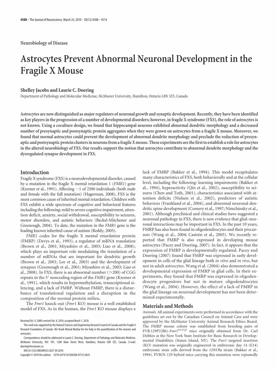
ResultsIn these experiments, we investigated the role of astrocytes in thedevelopment of the abnormal dendrite morphology and synapticaberrations seen in FXS. To examine whether astrocytes from anFMRP-deficient animal could affect normal neuron growth, weused a coculture procedure in which neurons are grown at lowdensities (400 cells/�l) in serum-free media (Jacobs and Doering,2009). In these cultures, the neurons are dependent on a feederlayer of astrocytes for survival (Banker, 1980). Consistent withpreviously reported findings, hippocampal neurons from bothWT (normal) and Fmr1 KO (fragile X) mice grown in the absenceof an astrocyte feeder layer did not survive. Both WT and Fmr1KO neurons grown on WT astrocytes exhibited normal neuronalgrowth at 7 DIV (Fig. 1a, top row). In stark contrast, when Fmr1KO neurons were grown on Fmr1 KO astrocytes, the neuronsexhibited a distinct abnormal morphology (Fig. 1a, bottom row).The dendritic arbors were more complex and individual pro-cesses showed random meandering on the astrocyte surface.
The survival of Fmr1 KO neurons (grown on either WT orFmr1 KO astrocytes) was decreased compared with WT neurons( p � 0.004; f 2 � 0.12) (Fig. 1b). However, survival was not sig-nificantly affected by growth on Fmr1 KO astrocytes comparedwith WT, for either WT ( p � 0.977) or Fmr1 KO neurons ( p �0.382) (Fig. 1b). Therefore, the results that we present are notattributable to a density effect or selective neuron survival, andthe morphological differences seen are a result of some quality ofthe different astrocyte populations. This difference could not beattributed to a significant variance in the purity of the astrocytepopulations because the astrocytes isolated from both WT andFmr1 KO mice were 98.7 � 0.5% GFAP positive. Therefore, theobserved alterations in the neuronal phenotype can be concluded
0
5
10
15
20
WT Neurons
Fmr1 KONeurons
p=0.004
p=0.006
WT AstrocytesFmr1 KO Astrocytes
Per
cent
age
of C
ells
Sur
vivi
ng
a b
WT Astrocytes Fmr1 KO Astrocytes
WT
Neu
rons
Fmr1
KO
Neu
rons
Figure 1. Effects of astrocytes on the growth of hippocampal neurons in coculture at 7 DIV. E17 primary hippocampal neuronswere cocultured with P0 –P1 primary cortical astrocytes for 7 DIV in each of four coculture conditions. a, Immunofluorescentimages of neurons in each of the four culture combinations. Neurons are stained with an antibody directed against the neuronaldendritic marker, MAP2. Scale bar, 100 �m. b, Quantification of percentage of surviving neurons at 7 DIV in each of the four cultureconditions. Data shown are mean values � SEM from two or three independent experiments (10 –15 regions of 1.5 mm 2 from 2coverslips per experiment). Significant differences revealed by post hoc Tukey’s tests are indicated ( p � 0.001).
Jacobs and Doering • Astrocytes in Fragile X J. Neurosci., March 24, 2010 • 30(12):4508 – 4514 • 4509
to result from the characteristics of the astrocytes imparted by thegenotype of their origin animal.
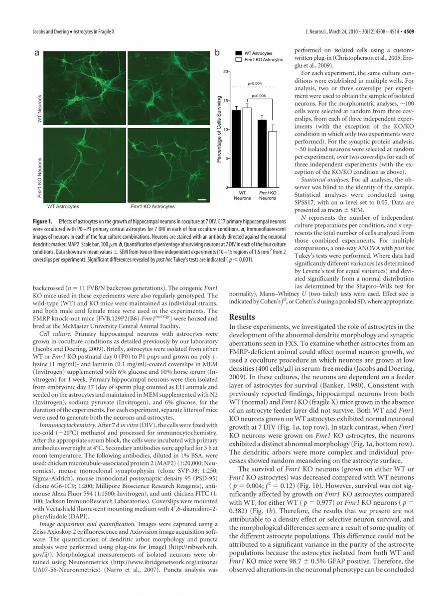
Fmr1 KO astrocytes alter the morphology of WT neuronsTo evaluate more precisely how the Fmr1 KO astrocytes affectedthe growth of the neurons, we performed a detailed morpholog-ical analysis of the dendritic arbors. After staining with an anti-body targeting the dendrite marker, MAP2, isolated neuronswere selected at random from a minimum of two coverslips perexperiment, and the morphology of their dendritic arbors wasanalyzed. WT neurons exhibited altered morphology whengrown on astrocytes isolated from an Fmr1 KO mouse (Fig. 2a,b).Comparison of the dendritic arbor morphology of WT neuronsgrown on Fmr1 KO astrocytes with those grown on WT astro-cytes showed the following: the length of the longest primarydendrite and the extent of the area covered by the dendritic arborwere decreased by 15.5% ( p � 0.001; d � 0.32) and 31.8% ( p �0.001; d � 0.47), respectively (Fig. 2c,d); the area covered permicrometer of dendrite was decreased by 17.3% ( p � 0.001; d �0.38) (Fig. 2g); the number of branches per cell was increased by13.0% ( p � 0.046; d � 0.22) (Fig. 2e); and the branch density(number of dendritic branches per square micrometer) was in-creased by 57.4% ( p � 0.001; d � 0.54) (Fig. 2h). These obser-vations indicate that growth on Fmr1 KO astrocytes appeared toalter the normal dendritic arborization of WT neurons.
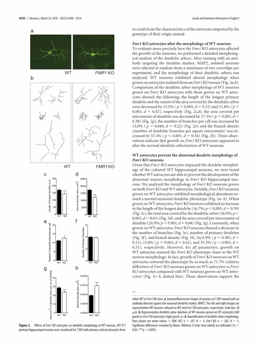
WT astrocytes prevent the abnormal dendrite morphology ofFmr1 KO neuronsGiven that Fmr1 KO astrocytes impacted the dendrite morphol-ogy of the cultured WT hippocampal neurons, we next testedwhether WT astrocytes are able to prevent the development of theabnormal neuron morphology in Fmr1 KO hippocampal neu-rons. We analyzed the morphology of Fmr1 KO neurons grownon both Fmr1 KO and WT astrocytes. Notably, Fmr1 KO neuronsgrown on WT astrocytes exhibited morphological alterations to-ward a normal neuronal dendritic phenotype (Fig. 3a– h). Whengrown on WT astrocytes, Fmr1 KO neurons exhibited an increasein the length of the longest dendrite (16.7%; p � 0.001; d � 0.39)(Fig. 3c), the total area covered by the dendritic arbor (36.9%; p �0.001; d � 0.61) (Fig. 3d), and the area covered per micrometer ofdendrite (24.9%; p � 0.001; d � 0.68) (Fig. 3g). Conversely, whengrown on WT astrocytes, Fmr1 KO neurons showed a decrease inthe number of branches (Fig. 3e), number of primary dendrites(Fig. 3f), and branch density (Fig. 3h), by 6.9% ( p � 0.381; d �0.13), 13.8% ( p � 0.001; d � 0.42), and 33.3% ( p � 0.001; d �0.51), respectively. However, for all parameters, growth onWT astrocytes restored the Fmr1 KO phenotype closer to the WTneuron morphology. In fact, growth of Fmr1 KO neurons on WTastrocytes restored the phenotype by as much as 71.7% (relativedifference of Fmr1 KO neurons grown on WT astrocytes vs Fmr1KO astrocytes compared with WT neurons grown on WT astro-cytes) (Fig. 3c– h, dotted line). These observations support the
0
50
100
150
200
250
WT KO
***
Leng
th o
f lon
gest
den
drite
(µm
)
0
10
20
30
40
***
WT KO
Area
(µm
2 ) of D
endr
itic A
rbor
per
µm
of D
endr
ite
0
5
10
15
20
*
WT KO
Num
ber o
f den
dritic
bra
nche
s pe
r cel
l
0
5000
10000
15000
20000
25000
WT KO
***
Area
of d
endr
itic a
rbor
(µm
2 )
0.0000
0.0005
0.0010
0.0015
0.0020
***
WT KO
Bran
ch D
ensi
ty (p
er µ
m2 )
0
2
4
6
WT KO
Num
ber o
f Prim
ary
Den
drite
s
WT FMR1 KO
WT FMR1 KO
a
b
c d
e f
g h
Figure 2. Effects of Fmr1 KO astrocytes on dendritic morphology of WT neurons. WT E17primary hippocampal neurons were cocultured for 7 DIV with primary cortical astrocytes from
4
either WT or Fmr1 KO mice. a, Immunofluorescent images of neurons at 7 DIV stained with anantibody directed against the neuronal dendritic marker, MAP2. The left and right images arerepresentative WT neurons cultured on WT and Fmr1 KO astrocytes, respectively. Scale bar, 50�m. b, Representative dendritic arbor skeletons of WT neurons grown on WT astrocytes (leftpanel) or Fmr1 KO astrocytes (right panel). c– h, Quantification of dendritic arbor morphology.Data shown are mean values � SEM. WT: n � 297, N � 3; Fmr1 KO: n � 282, N � 3.Significant differences revealed by Mann–Whitney U tests (two-tailed) are indicated (*p �0.05; ***p � 0.001).
4510 • J. Neurosci., March 24, 2010 • 30(12):4508 – 4514 Jacobs and Doering • Astrocytes in Fragile X
notion that astrocytes from a WT mouse prevent the develop-ment of abnormal dendritic morphology of Fmr1 KO neurons.
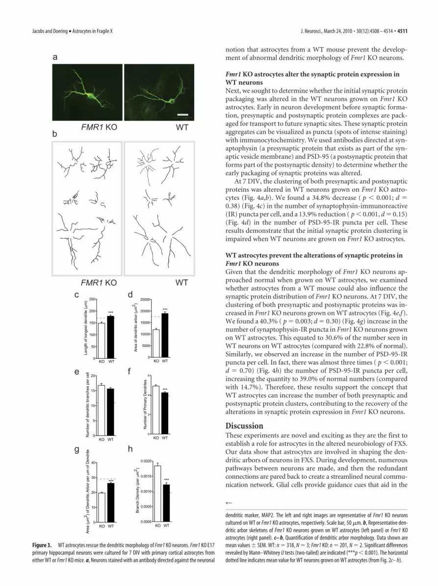
Fmr1 KO astrocytes alter the synaptic protein expression inWT neuronsNext, we sought to determine whether the initial synaptic proteinpackaging was altered in the WT neurons grown on Fmr1 KOastrocytes. Early in neuron development before synaptic forma-tion, presynaptic and postsynaptic protein complexes are pack-aged for transport to future synaptic sites. These synaptic proteinaggregates can be visualized as puncta (spots of intense staining)with immunocytochemistry. We used antibodies directed at syn-aptophysin (a presynaptic protein that exists as part of the syn-aptic vesicle membrane) and PSD-95 (a postsynaptic protein thatforms part of the postsynaptic density) to determine whether theearly packaging of synaptic proteins was altered.
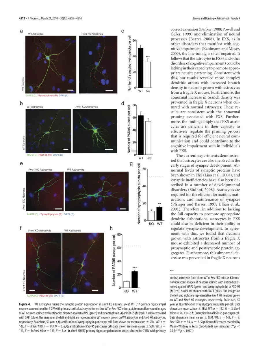
At 7 DIV, the clustering of both presynaptic and postsynapticproteins was altered in WT neurons grown on Fmr1 KO astro-cytes (Fig. 4a,b). We found a 34.8% decrease ( p � 0.001; d �0.38) (Fig. 4c) in the number of synaptophysin-immunoreactive(IR) puncta per cell, and a 13.9% reduction ( p � 0.001, d � 0.15)(Fig. 4d) in the number of PSD-95-IR puncta per cell. Theseresults demonstrate that the initial synaptic protein clustering isimpaired when WT neurons are grown on Fmr1 KO astrocytes.
WT astrocytes prevent the alterations of synaptic proteins inFmr1 KO neuronsGiven that the dendritic morphology of Fmr1 KO neurons ap-proached normal when grown on WT astrocytes, we examinedwhether astrocytes from a WT mouse could also influence thesynaptic protein distribution of Fmr1 KO neurons. At 7 DIV, theclustering of both presynaptic and postsynaptic proteins was in-creased in Fmr1 KO neurons grown on WT astrocytes (Fig. 4e,f).We found a 40.3% ( p � 0.003; d � 0.30) (Fig. 4g) increase in thenumber of synaptophysin-IR puncta in Fmr1 KO neurons grownon WT astrocytes. This equated to 30.6% of the number seen inWT neurons on WT astrocytes (compared with 22.8% of normal).Similarly, we observed an increase in the number of PSD-95-IRpuncta per cell. In fact, there was almost three times ( p � 0.001;d � 0.70) (Fig. 4h) the number of PSD-95-IR puncta per cell,increasing the quantity to 39.0% of normal numbers (comparedwith 14.7%). Therefore, these results support the concept thatWT astrocytes can increase the number of both presynaptic andpostsynaptic protein clusters, contributing to the recovery of thealterations in synaptic protein expression in Fmr1 KO neurons.
DiscussionThese experiments are novel and exciting as they are the first toestablish a role for astrocytes in the altered neurobiology of FXS.Our data show that astrocytes are involved in shaping the den-dritic arbors of neurons in FXS. During development, numerouspathways between neurons are made, and then the redundantconnections are pared back to create a streamlined neural commu-nication network. Glial cells provide guidance cues that aid in the
0
5000
10000
15000
20000
25000
KO WT
***
Area
of d
endr
itic
arbo
r (µm
2 )
0
10
20
30
40
***
KO WT
Area
(µm
2 ) of D
endr
itic
Arbo
r per
µm
of D
endr
ite
0.0000
0.0005
0.0010
0.0015
0.0020
KO WT
***
Bran
ch D
ensi
ty (p
er µ
m2 )
0
50
100
150
200
250
WTKO
***
Leng
th o
f lon
gest
den
drite
(µm
)
0
5
10
15
20
KO WT
Num
ber o
f den
dritic
bra
nche
s pe
r cel
l
0
2
4
6
***
KO WT
Num
ber o
f Prim
ary
Den
drite
s
WT
WT
FMR1 KO
FMR1 KO
a
c d
e f
g h
b
Figure 3. WT astrocytes rescue the dendritic morphology of Fmr1 KO neurons. Fmr1 KO E17primary hippocampal neurons were cultured for 7 DIV with primary cortical astrocytes fromeither WT or Fmr1 KO mice. a, Neurons stained with an antibody directed against the neuronal
4
dendritic marker, MAP2. The left and right images are representative of Fmr1 KO neuronscultured on WT or Fmr1 KO astrocytes, respectively. Scale bar, 50 �m. b, Representative den-dritic arbor skeletons of Fmr1 KO neurons grown on WT astrocytes (left panel) or Fmr1 KOastrocytes (right panel). c– h, Quantification of dendritic arbor morphology. Data shown aremean values � SEM. WT: n � 318, N � 3; Fmr1 KO: n � 201, N � 2. Significant differencesrevealed by Mann–Whitney U tests (two-tailed) are indicated (***p � 0.001). The horizontaldotted line indicates mean value for WT neurons grown on WT astrocytes (from Fig. 2c– h).
Jacobs and Doering • Astrocytes in Fragile X J. Neurosci., March 24, 2010 • 30(12):4508 – 4514 • 4511
correct extension (Banker, 1980; Powell andGeller, 1999) and elimination of neuralprocesses (Barres, 2008). In FXS, as inother disorders that manifest with cog-nitive impairment (Kaufmann and Moser,2000), the fine-tuning is often impaired. Itfollows that the astrocytes in FXS (and otherdisorders of cognitive impairment) could belacking in their capacity to promote appro-priate neurite patterning. Consistent withthis, our results revealed more complexdendritic arbors with increased branchdensity in neurons grown with astrocytesfrom a fragile X mouse. Furthermore, theabnormal increase in branch density wasprevented in fragile X neurons when cul-tured with normal astrocytes. These re-sults are consistent with the abnormalpruning associated with FXS. Further-more, the findings imply that FXS astro-cytes are deficient in their capacity toeffectively regulate the pruning processthat is required for efficient neural com-munication and could contribute to thecognitive impairment seen in individualswith FXS.
The current experiments demonstra-ted that astrocytes are also involved in theearly stages of synapse development. Ab-normal levels of synaptic proteins havebeen shown in FXS (Liao et al., 2008), andsynaptic inefficiencies have also been de-scribed in a number of developmentaldisorders (Sudhof, 2008). Astrocytes arerequired for the efficient formation, mat-uration, and maintenance of synapses(Pfrieger and Barres, 1997; Ullian et al.,2001). Therefore, in addition to lackingthe full capacity to promote appropriatedendrite elaborations, astrocytes in FXScould also be deficient in their ability toregulate synapse development. In agree-ment with this, we found that neuronsgrown with astrocytes from a fragile Xmouse exhibited a decreased number ofpresynaptic and postsynaptic protein ag-gregates. Furthermore, this abnormal de-crease was prevented in fragile X neurons
WT Astrocytes Fmr1 KO Astrocytes
WT
Neu
rons
MAP2(G) PSD-95 (R) DAPI (B)
MAP2(G) Synaptophysin (R) DAPI (B)
Fmr1
KO
Neu
rons
WT AstrocytesFmr1 KO Astrocytes
0
20
40
60
80
KO WT
***
Num
ber o
f PSD
95 p
unct
a pe
r cel
l
Fmr1 KO Astrocytes WT Astrocytes
Fmr1
KO
Neu
rons
MAP2(G) PSD-95 (R) DAPI (B)
0
20
40
60
KO WT
**
Num
ber o
f syn
apto
phys
in p
unct
a pe
r cel
l
0
20
40
60
80
WT KO
Num
ber o
f PSD
95 p
unct
a pe
r cel
l
0
20
40
60
WT KO
***
Num
ber o
f syn
apto
phys
in p
unct
a pe
r cel
l
a
b
e
f
c
d
g
h
WT
Neu
rons
WT Astrocytes Fmr1 KO Astrocytes
MAP2(G) Synaptophysin (R) DAPI (B)
Figure 4. WT astrocytes rescue the synaptic protein aggregation in Fmr1 KO neurons. a– d, WT E17 primary hippocampalneurons were cultured for 7 DIV with primary cortical astrocytes from either WT or Fmr1 KO mice. a, b, Immunofluorescent imagesof WT neurons stained with antibodies directed against MAP2 (green) and synaptophysin (a) or PSD-95 (b) (red). Nuclei are stainedwith DAPI (blue). The images on the left and right are representative WT neurons grown on WT astrocytes and Fmr1 KO astrocytes,respectively. Scale bars, 50 �m. c, Quantification of synaptophysin puncta per cell. Data shown are mean values � SEM. WT: n �147, N � 3; Fmr1 KO: n � 143, N � 3. d, Quantification of PSD-95 puncta per cell. Data shown are mean values � SEM. WT: n �111, N � 3; Fmr1 KO: n � 119, N � 3. e– h, Fmr1 KO E17 primary hippocampal neurons were cultured for 7 DIV with primary
4
cortical astrocytes from either WT or Fmr1 KO mice. e, f, Immu-nofluorescent images of neurons stained with antibodies di-rected against MAP2 (green) and synaptophysin (e) or PSD-95(f) (red). Nuclei are stained with DAPI (blue). The images onthe left and right are representative Fmr1 KO neurons grownon WT and Fmr1 KO astrocytes, respectively. Scale bars, 50�m. g, Quantification of synaptophysin puncta per cell. Datashown are mean values � SEM. WT: n � 112, N � 3; Fmr1KO: n � 94, N � 2. h, Quantification of PSD-95 puncta per cell.Data shown are mean values � SEM. WT: n � 143, N � 3;Fmr1 KO: n � 94, N � 2. Significant differences revealed byMann–Whitney U tests (two-tailed) are indicated (**p �0.05; ***p � 0.001).
4512 • J. Neurosci., March 24, 2010 • 30(12):4508 – 4514 Jacobs and Doering • Astrocytes in Fragile X
when they were cultured with normal astrocytes. These findingsare in concert with the current understanding of synaptic proteindysregulation in FXS. In addition, these experiments suggestthat astrocytes contribute to the erroneous synapse develop-ment in FXS and could therefore be a fundamental factor in thedevelopment of the behavioral maladaptations seen in individu-als with FXS.
Previous research has shown that glial cells contribute to thedevelopment of several neurological disorders (for review, seeBarres, 2008), and our results support the concept of astrocytesguiding appropriate neurite and synapse development (Pfriegerand Barres, 1997; Mauch et al., 2001; Ullian et al., 2001; Murai etal., 2003; Christopherson et al., 2005; Nishida and Okabe, 2007;Guizzetti et al., 2008). Most recently, Ballas et al. (2009) foundthat astrocytes lacking the protein responsible for Rett syndrome,MeCP (methyl-CpG-binding protein 2), could not support nor-mal neuron growth. Similar to Rett syndrome, FXS was previ-ously thought to be a disease resulting from a loss of functioncaused by a lack of a specific protein in neurons only. However, itis possible that the abnormal phenotype in FXS, as in Rett syn-drome, results from additional non-neuronal deficiencies. Forexample, the astrocytes in individuals with FXS may also be dys-functional, and this deficit could be caused by an indirect or adirect loss of FMRP. The loss of FMRP in neurons could result,indirectly, in aberrant astrocyte-mediated support functions as aresult of impaired neuron-to-astrocyte signaling caused by theloss of neuronal FMRP. Alternatively, the astrocytes in an indi-vidual with FXS may also be deficient in FMRP and suffer abnor-malities in normal functioning directly attributable to the loss ofFMRP. Recently, our laboratory documented that FMRP is ex-pressed in cells of the glial lineage and that FMRP was absent fromputative astrocytes in the Fmr1 KO mouse (Pacey and Doering,2007). Therefore, it is possible that astrocytes lack FMRP, specif-ically at a time during development when astrocyte support ofneuron growth and synapse formation are vital, and this lack ofFMRP could contribute to the abnormal neuron phenotype seenin FXS. Given that FMRP is a key regulator of translation of anumber of mRNA targets in neurons (Brown et al., 2001; Miyashiroet al., 2003; Liao et al., 2008) and that the list is not yet complete,it is plausible that FMRP also regulates a subset of mRNAs inastrocytes. In this scenario, the loss of FMRP seen in FXS wouldresult in aberrant protein translation in astrocytes in addition tothat already documented in neurons. In turn, this could lead toaltered astrocyte–neuron signaling and interfere with astrocyte-mediated neuronal growth and synaptic development. Althoughthe present study provides strong evidence for a role of astrocytesin the development of the neurobiological abnormalities seen inFXS, the experiments do not identify the molecular basis of thealterations and cannot confirm whether they are a direct or indi-rect result of a lack of FMRP.
Future studies designed to investigate these possibilitieswould provide valuable information on the neurobiological pro-cesses that are altered in FXS. For example, in vitro studies eval-uating the effect of FMRP transfection in FMR1 KO astrocytes,and the short interfering RNA-mediated specific downregulationof FMR1 in WT astrocytes, on WT neurons could provide moredirect evidence for a role of astrocyte FMRP in the neurobiologyof FXS. Such studies could yield insight into whether there areany astrocyte-secreted factors that are altered consequent to adeficiency of FMRP and would therefore provide novel avenuesfor therapeutic intervention. Additionally, in vivo studies target-ing FMRP expression in astrocytes using the FMR1 conditionalknock-out and FMR1 conditional expression mice would pro-
vide an invaluable in vivo correlate and offer insight into thefunctional consequences of alterations in astrocyte FMRP.
ReferencesBakker C, Verheij C, Willemsen R, van der Helm R, Oerlemans F, Vermey M,
Bygrave A, Hoogeveen A, Oostra B, Reyniers E, De Boulle K, D’Hooge R,Cras P, Van Velzen D, Nagels G, Martin J, De Deyn PP, Darby JK, WillemsPJ (1994) Fmr1 knockout mice: a model to study fragile X mental retar-dation. The Dutch-Belgian Fragile X Consortium. Cell 78:23–33.
Ballas N, Lioy DT, Grunseich C, Mandel G (2009) Non-cell autonomousinfluence of MeCP2-deficient glia on neuronal dendritic morphology.Nat Neurosci 12:311–317.
Banker GA (1980) Trophic interactions between astroglial cells and hip-pocampal neurons in culture. Science 209:809 – 810.
Barres BA (2008) The mystery and magic of glia: a perspective on their rolesin health and disease. Neuron 60:430 – 440.
Beckel-Mitchener A, Greenough WT (2004) Correlates across the struc-tural, functional, and molecular phenotypes of fragile X syndrome. MentRetard Dev Disabil Res Rev 10:53–59.
Brown V, Jin P, Ceman S, Darnell JC, O’Donnell WT, Tenenbaum SA, Jin X,Feng Y, Wilkinson KD, Keene JD, Darnell RB, Warren ST (2001) Mi-croarray identification of FMRP-associated brain mRNAs and alteredmRNA translational profiles in fragile X syndrome. Cell 107:477– 487.
Castren M, Tervonen T, Karkkainen V, Heinonen S, Castren E, Larsson K,Bakker CE, Oostra BA, Akerman K (2005) Altered differentiation ofneural stem cells in fragile X syndrome. Proc Natl Acad Sci U S A102:17834 –17839.
Chen L, Toth M (2001) Fragile X mice develop sensory hyperreactivity toauditory stimuli. Neuroscience 103:1043–1050.
Christopherson KS, Ullian EM, Stokes CC, Mullowney CE, Hell JW, Agah A,Lawler J, Mosher DF, Bornstein P, Barres BA (2005) Thrombospondinsare astrocyte-secreted proteins that promote CNS synaptogenesis. Cell120:421– 433.
Comery TA, Harris JB, Willems PJ, Oostra BA, Irwin SA, Weiler IJ,Greenough WT (1997) Abnormal dendritic spines in fragile X knockoutmice: maturation and pruning deficits. Proc Natl Acad Sci U S A94:5401–5404.
Devys D, Lutz Y, Rouyer N, Bellocq JP, Mandel JL (1993) The FMR-1 pro-tein is cytoplasmic, most abundant in neurons and appears normal incarriers of a fragile X premutation. Nat Genet 4:335–340.
Eroglu C, Allen NJ, Susman MW, O’Rourke NA, Park CY, Ozkan E,Chakraborty C, Mulinyawe SB, Annis DS, Huberman AD, Green EM,Lawler J, Dolmetsch R, Garcia KC, Smith SJ, Luo ZD, Rosenthal A,Mosher DF, Barres BA (2009) Gabapentin receptor alpha2delta-1 is aneuronal thrombospondin receptor responsible for excitatory CNS syn-aptogenesis. Cell 139:380 –392.
Frankland PW, Wang Y, Rosner B, Shimizu T, Balleine BW, Dykens EM,Ornitz EM, Silva AJ (2004) Sensorimotor gating abnormalities in youngmales with fragile X syndrome and Fmr1-knockout mice. Mol Psychiatry9:417– 425.
Greenough WT, Klintsova AY, Irwin SA, Galvez R, Bates KE, Weiler IJ (2001)Synaptic regulation of protein synthesis and the fragile X protein. ProcNatl Acad Sci U S A 98:7101–7106.
Guizzetti M, Moore NH, Giordano G, Costa LG (2008) Modulation of neu-ritogenesis by astrocyte muscarinic receptors. J Biol Chem283:31884 –31897.
Hagerman PJ (2008) The fragile X prevalence paradox. J Med Genet45:498 – 499.
Jacob S, Doering LC (2009) Primary dissociated astrocyte and neuronal co-culture. In: Protocols for neural cell culture, Ed 4 (Doering LC, ed), pp.269 –284. New York: Humana.
Kaufmann WE, Moser HW (2000) Dendritic anomalies in disorders associ-ated with mental retardation. Cereb Cortex 10:981–991.
Kremer EJ, Pritchard M, Lynch M, Yu S, Holman K, Baker E, Warren ST,Schlessinger D, Sutherland GR, Richards RI (1991) Mapping of DNAinstability at the fragile X to a trinucleotide repeat sequence p(CCG)n.Science 252:1711–1714.
Lee A, Li W, Xu K, Bogert BA, Su K, Gao FB (2003) Control of dendriticdevelopment by the Drosophila fragile X-related gene involves the smallGTPase Rac1. Development 130:5543–5552.
Liao L, Park SK, Xu T, Vanderklish P, Yates JR 3rd (2008) Quantitativeproteomic analysis of primary neurons reveals diverse changes in synaptic
Jacobs and Doering • Astrocytes in Fragile X J. Neurosci., March 24, 2010 • 30(12):4508 – 4514 • 4513
protein content in fmr1 knockout mice. Proc Natl Acad Sci U S A105:15281–15286.
Mauch DH, Nagler K, Schumacher S, Goritz C, Muller EC, Otto A, PfriegerFW (2001) CNS synaptogenesis promoted by glia-derived cholesterol.Science 294:1354 –1357.
Miyashiro KY, Beckel-Mitchener A, Purk TP, Becker KG, Barret T, Liu L,Carbonetto S, Weiler IJ, Greenough WT, Eberwine J (2003) RNA car-goes associating with FMRP reveal deficits in cellular functioning in Fmr1null mice. Neuron 37:417– 431.
Murai KK, Nguyen LN, Irie F, Yamaguchi Y, Pasquale EB (2003) Control ofhippocampal dendritic spine morphology through ephrin-A3/EphA4 sig-naling. Nat Neurosci 6:153–160.
Narro ML, Yang F, Kraft R, Wenk C, Efrat A, Restifo LL (2007) NeuronMet-rics: software for semi-automated processing of cultured neuron images.Brain Res 1138:57–75.
Nielsen DM, Derber WJ, McClellan DA, Crnic LS (2002) Alterations in theauditory startle response in Fmr1 targeted mutant mouse models of frag-ile X syndrome. Brain Res 927:8 –17.
Nimchinsky EA, Oberlander AM, Svoboda K (2001) Abnormal developmentof dendritic spines in FMR1 knock-out mice. J Neurosci 21:5139–5146.
Nishida H, Okabe S (2007) Direct astrocytic contacts regulate local matura-tion of dendritic spines. J Neurosci 27:331–340.
Pacey LK, Doering LC (2007) Developmental expression of FMRP in theastrocyte lineage: implications for fragile X syndrome. Glia 55:1601–1609.
Pfrieger FW, Barres BA (1997) Synaptic efficacy enhanced by glial cells invitro. Science 277:1684 –1687.
Powell EM, Geller HM (1999) Dissection of astrocyte-mediated cues inneuronal guidance and process extension. Glia 26:73– 83.
Qin M, Kang J, Smith CB (2002) Increased rates of cerebral glucose metab-olism in a mouse model of fragile X mental retardation. Proc Natl AcadSci U S A 99:15758 –15763.
Reddy KS (2005) Cytogenetic abnormalities and fragile-X syndrome in au-tism spectrum disorder. BMC Med Genet 6:3.
Sudhof TC (2008) Neuroligins and neurexins link synaptic function to cog-nitive disease. Nature 455:903–911.
Ullian EM, Sapperstein SK, Christopherson KS, Barres BA (2001) Controlof synapse number by glia. Science 291:657– 661.
Wang H, Ku L, Osterhout DJ, Li W, Ahmadian A, Liang Z, Feng Y (2004)Developmentally-programmed FMRP expression in oligodendrocytes: apotential role of FMRP in regulating translation in oligodendroglia pro-genitors. Hum Mol Genet 13:79 – 89.
4514 • J. Neurosci., March 24, 2010 • 30(12):4508 – 4514 Jacobs and Doering • Astrocytes in Fragile X