neurorepair promoting crosstalk between astrocytes
TRANSCRIPT
ABSTRACT
Title of Dissertation: NEUROREPAIR PROMOTING
CROSSTALK BETWEEN ASTROCYTES & OLFACTORY ENSHEATHING CELLS
Aybike Sağlam, Doctor of Philosophy, 2020
Dissertation directed by: Dr. Susan Wray, Chief, Cellular and Developmental Neurobiology Section NINDS, NIH
Astrocytes within the neurogenic zones of the adult central nervous system (CNS) support
the formation and maturation of neurons from progenitor cells throughout life. In contrast,
astrocytes outside of neurogenic zones dedifferentiate and contribute to scar tissue formation
after injury, creating a physical barrier to regenerating neurons. Moreover, reactive astrocytes
can create a chemical barrier and be toxic for neurons after injury. Therefore, understanding
the signaling pathways that switch astrocytes from neurogenesis-inhibitory to neurogenesis-
supportive is a promising approach to reverse the progression of neurodegenerative diseases
and traumatic CNS injuries.
The mammalian olfactory system shows robust neurogenesis throughout life, with
neurosensory cells capable of renewal and differentiation. There is growing evidence that a
distinct type of glia, olfactory ensheathing cells (OECs), regulate the astrocytic stress response
in the olfactory bulb (OB) and is critical for the neuroregenerative properties of the olfactory
system. Therefore, OECs can be leveraged as a tool to identify signals pertinent for
maintaining neurorepair-promoting characteristics in astrocytes.
Exosomes are extracellular nanovesicles that serve intercellular communication. Our results
show that an exosome secreted protein, Alpha-crystallin B chain (CryAB), plays an important
role in astrocyte-OEC crosstalk. CryAB was shown to have protective roles for cells against
stress conditions. In accordance, our results indicate that OEC-secreted, CryAB positive
exosomes are taken up by astrocytes and this intercellular vesicle trafficking plays an anti-
inflammatory role in astrocytes by moderating activity of pro-inflammatory factors.
OECs also support astrocyte differentiation via sustained fibroblast growth factor (FGF)
signaling. FGFs are crucial factors in CNS development and injury response, and are targets
for neuroregenerative strategies. Heparan sulfate proteoglycans (HSPGs) are cell-specific
proteins that can be shed from the membrane and regulate FGF signaling in the donor cell.
We show OEC-HSPGs differentially activate FGF receptor-1 (FGFR1) signaling in astrocytes
and suppress reactivity. Moreover, our results suggest a mechanistic role for OEC-HSPGs in
intracellular FGFR1 trafficking and its association with the transcriptional machinery in
astrocytes. Together this work shows that OB OECs are integral components of one of the
few neurogenic zones in the CNS. Mimicking OEC-astrocyte crosstalk in vivo may provide
new approaches to ameliorating CNS injuries by targeting astrocytes.

NEUROREPAIR PROMOTING CROSSTALK BETWEEN ASTROCYTES
& OLFACTORY ENSHEATHING CELLS
by:
Aybike Sağlam
Dissertation submitted to the Faculty of the Graduate School of the University of Maryland,
College Park, in partial fulfillment of the requirements for the degree of
Doctor of Philosophy
2020
Advisory Committee:
Dr. Susan Wray Dr. Ricardo Araneda Dr. Elizabeth Quinlan Dr. Leonardo Belluscio Dr. Jens Herberholz

ii
Dedication:
This work would not have been possible without numerous compassionate, brilliant,
resilient, and uplifting people who have helped me persevere in my life and academic
work. From labmates to committee members, advisors, family, and friends, I'm
humbled by the many ways, large and small, you have supported me. I strongly
believe that our world is changed for the better by people like you.
I owe a special acknowledgment to my loving family, in particular my mom whose
empowering voice is always with me. Even though we are physically far apart, I carry
you all with me wherever I go. Your kindness and positivity inspire me. Because of
you, I am an optimist who sees obstacles as only temporary and I know that even a
small number of people can make a big difference.

iii
Table of Contents
Dedication: ............................................................................................................................................ ii
Table of Contents ................................................................................................................................ iii
List of Figures ....................................................................................................................................... v
Chapter 1: Background ........................................................................................................................ 1
Introduction ...................................................................................................................................... 1
CNS injury & factors limiting recovery ........................................................................................ 2
Astrocytes before & after the injury or during a degenerative disease .................................... 5
Neurogenic potential & heterogeneity of astrocytes .................................................................. 7
Neurorepair supportive crosstalk between olfactory bulb glia: astrocytes & OECs ............. 9
Significance/ Aims ........................................................................................................................ 12
Aim 1: Identify OEC-secreted factors which suppress neurotoxic astrocyte reactivity ...... 13
Aim 2: Investigate the role of FGFR1 in OEC-astrocyte crosstalk ....................................... 14
Chapter 2: A Novel Factor in Olfactory Ensheathing Cell-Astrocyte Crosstalk: Anti-Inflammatory Protein α-Crystallin B ............................................................................................... 15
2.1 Abstract ..................................................................................................................................... 15
2.2 Introduction ............................................................................................................................. 16
2.3 Materials and Methods ........................................................................................................... 18
2.4 Results ....................................................................................................................................... 27
2.5 Discussion ................................................................................................................................ 39
Chapter 3: OEC-HSPGs moderate astrocyte reactivity, proliferation, and differentiation via FGFR1 signaling................................................................................................................................. 44
3.1 Abstract ..................................................................................................................................... 44
3.2 Introduction ............................................................................................................................. 45
3.3 Materials and Methods ........................................................................................................... 49
3.4 Results ....................................................................................................................................... 58
3.5 Discussion ................................................................................................................................ 71
Chapter 4: Implication of Results & Future Directions for Research ........................................ 79
4.1 Discussion of Results .............................................................................................................. 79 4.1.1 Novel observations regarding the role of OECs in CNS regeneration ................... 79 4.1.2 Assessing the role of OECs for maintaining astrocytes of the OB in a neurogenesis supportive state .................................................................................................. 80
4.2 Limitations of Experimental Approach ............................................................................... 82
4.3 Recommendations for Future Research .............................................................................. 84
iv
APPENDIX A: .................................................................................................................................. 87
OEC depletion increases the number of C3 positive astrocytes along the RMS...................... 87
APPENDIX B: ................................................................................................................................... 90
Experimental autoimmune encephalomyelitis increases the number of progenitors reaching to the OB from RMS ......................................................................................................................... 90
APPENDIX C: ................................................................................................................................... 91
Bibliography: ....................................................................................................................................... 92

v
List of Figures
Figure 1: Anatomy of the Olfactory System..................................................................................... 4
Figure 2: Glia limitans of the OB..................................................................................................... 10
Figure 3: OEC-CM is sufficient to suppress astrocyte reactivity ................................................ 28
Figure 4: An anti-inflammatory protein: CryAB ............................................................................ 31
Figure 5:CryAB, in OEC-exosomes, is important for OEC-astrocyte crosstalk ...................... 33
Figure 6: CryAB secretion is cell type and context dependent .................................................... 35
Figure 7: CryAB in OEC-exosomes is internalized by astrocytes ............................................... 36
Figure 8: OEC-CM suppresses neurotoxic-astrocyte transcripts ................................................ 38
Figure 9: OEC-HSPGs supress astrocyte reactivity via FGFR1 ................................................. 61
Figure 10: OEC-CM induced activation of FGFR1 at the astrocyte membrane ...................... 64
Figure 11: OEC-CM increases pY766 FGFR1 signaling in astrocytes....................................... 66
Figure 12: OEC-HSPG regulates FGFR1 trafficking in astrocytes ............................................ 70
Figure 13: OEC depletion increases C3-positive glia in vivo ...................................................... 87
Figure 14: No difference was observed for GFAP, Iba1 or cell proliferation markers
following OEC depletion .................................................................................................................. 88
Figure 15: OEC depletion increases astrocyte neurotoxicity in vivo .......................................... 89
Figure 16: EAE increases number of progenitors in the OB ...................................................... 90
Figure 17: Co-stimulation of astrocytes with Anosmin1 & LPS increases astrocyte
proliferation ......................................................................................................................................... 91

1
Chapter 1: Background
Introduction
Astrocytes, traditionally identified by intermediate filament protein glial fibrillary acidic
protein (GFAP) immunostaining, are the most numerous cell type in the CNS and perform
crucial functions for neuronal development and synapse formation (reviewed in Stevens &
Muthukumar, 2016). The adult mammalian subventricular zone (SVZ) contains GFAP
positive neural stem cells (NSCs) which are also are classified as astrocytes. These cells give
rise to neuroblasts and migrate to the OB where they commit to neuronal (and to a lesser
degree to glial) lineage (Doetsch et al., 1999; Picard-Riera et al., 2002). Remarkably, local
astrocytes can also enter neurogenic program after injury (Magnuson et al., 2014, Nato et al.,
2015). Indeed, adult neural stem cells and reactive astrocytes share a number of features
(Magnusson & Frisén, 2016), yet instead of repairing and remodeling damaged tissue, reactive
astrocytes typically limit recovery. The main motivation behind this thesis work was to better
understand the microenvironment maintaining neurorepair promoting astrocytes of the
olfactory bulb (OB), with the prospect of applying these cues to astrocytes outside of the
neurogenic zones and prevent their conversion to neurotoxic astrocytes after injury.
Astrocyte reactivity in response to injury was noted as early as 1970’s shortly after the
discovery of GFAP, an astrocyte cytoskeleton protein characterized by its upregulation under
pathological conditions (Bignami & Dahl, 1976). Since then, GFAP had been used as a
standard marker of astrocyte reactivity which was considered to be detrimental. However,
recent studies show that astrocyte reactivity is not a uniform response but rather a
heterogenous mixture of activation states (reviewed in Khakh & Deneen., 2019). This context

2
dependent response is controlled by both intrinsic and environmental signals such as neural
activity (Horner & Palmer, 2003).
Significantly higher GFAP expression was observed in the olfactory system, one of the few
neurogenic zones in the adult mammalian brain, in particular where regenerating olfactory
sensory neurons (OSNs) enter the OB (Mackay-Sim & Kittel, 1991; Nazareth et al., 2018).
Notably, this high astrocyte reactivity is correlated with the plastic nature of the OB which is
influenced by both peripheral (Carmen Martinez Garcia et al., 1991) and CNS input (Göktaş
et al., 2010; Tanık et al. 2015; Yaldızlı et al., 2016; reviewed in Huart et al., 2019). These
observations suggest that astrocyte reactivity must be tightly regulated in the OB, where
neurogenesis continues throughout life.
Regenerating neurons use aligned glial scaffolds for elongation and migration. Olfactory
ensheathing cells (OECs) are a specialized type of glia that form continuous open scaffolds
into the OB, enabling entry of regenerating axons into the brain. OECs perform numerous
functions important for neuroregeneration and migration, and differentiation (Doucette &
Words., 1990; Santos-Silva et al., 2007). Moreover, OECs can migrate through scar tissue,
moderate astrocyte reactivity, and improve regeneration in vivo (reviewed in Roet et al., 2014).
Therefore, we hypothesized that studying the crosstalk between the OECs and astrocytes
would reveal mechanisms that control astrocyte reactivity and fate decisions.
CNS injury & factors limiting recovery
In the healthy CNS, astrocytes form functional barriers to restrict and regulate the entry of
immune cells. Once the integrity of the blood brain barrier (BBB) between the endothelial
cells and astrocytes is compromised in response to an insult, the activity of ion pumps,

3
neuronal membrane potentials and the synthesis of signaling molecules are disrupted
(Gulbenkian et al., 2001; Girouard & Iadecola, 2006). This initial damage is followed by a
spectrum of astrocytic stress responses. In severe cases, astrocyte reactivity results in glial scar
(astrogliosis); characterized by astrocyte misalignment and size increase, in an attempt to
maintain BBB integrity and limit the spread of neurotoxic inflammation. Reactive astrocytes
forming the glial scar act as a physical barrier to regenerating axons by expressing intermediate
filament proteins (Vimentin, GFAP, etc.), and secrete nerve growth-inhibitory chondroitin
sulfate proteoglycans (neurocan, phosphacan, versican, etc). While reactive astroglia express
some growth-promoting extracellular matrix (ECM) molecules, the growth-inhibitory
molecule secretion dominates with increased severity of injury. Thus, astrocytes have the
potential to carry both anti-inflammatory and pro-inflammatory functions and are targets for
pharmacologic manipulations to reverse CNS damage (Hamby & Sofroniew, 2010).
The nervous system must be able to continuously update functional connections between
neurons, indicating it must remain plastic throughout life (Raisman & Li., 2007). Hence, it has
been suggested that if severed neurons were guided through the damaged site, they could make
new functional connections. This idea drew attention to glial cells due to their regenerative
abilities, such as secreting growth-promoting factors and providing topographic guidance. In
the peripheral nervous system (PNS), Schwann cells (SCs) support and guide transected
neurons through damaged tissue and promote nerve regeneration. Several groups transplanted
SCs into the damaged CNS, in an attempt to repair the glial pathway and promote
neuroregeneration across the injury site. However, SCs fail to communicate with CNS-specific
glia (primarily astrocytes) and cannot build a glial pathway through the scar tissue (Grimpe et
al., 2005).

4
These findings draw attention to OSNs (Figure 1), which are vulnerable to damage and
microbial infections (Dando et al., 2016) due to their exposed location in the nasal cavity (PNS)
and have a remarkable capacity for regeneration (Calof et al., 1996, Forni et al., 2013).
Ensheathing glia of the olfactory system, OECs, wrap and guide olfactory sensory neuron
axons into the OB in the CNS, where they establish new connections (Figure 1). Moreover,
Figure 1: Anatomy of the Olfactory System
Figure 1: Anatomy of the Olfactory System. (A) OSNs (turquase) send their axons to the OB with the
support of OECs (red). OE= olfactory epithelium, ONL= nerve layer, RMS= rostral migratory stream. (B)
OECs (red) mingle with astrocytes (blue) in the OB where astrocytes form a defense barrier glia limitans. In the
OB, mitral cells (ML), external plexiform layer (EPL) glomerular layer, and internal (iNFL) and outer nerve layer
(oNFL) are shown (adopted from Nazareth et al., 2018).
5
OECs metabolize toxic macromolecules, undergo structural remodeling (Roet et al., 2014), and
secrete trophic factors including brain-derived neurotrophic factor, nerve growth factor, glial
cell line-derived neurotrophic factor, neuregulins, ciliary neurotrophic factor, integrins, cell
adhesion molecules, cadherins and laminin (Franssen et al., 2008, reviewed in Hale, 2011).
These observations suggested that OECs are important contributors in directing axons to their
correct target and also for maintaining regenerating neurons (Doucette, 1990). Different
groups transplanted OECs into CNS injury sites for neuroregenerative purposes and observed
therapeutic benefits (Li et al., 1997, Yang et al., 2015), including improved axon sprouting and
regrowth (Guntinas-Lichius et al., 2001; Deumens et al., 2006), decreased tissue and neuronal
damage (Ruitenberg et al., 2003), formation of myelin sheaths around axons (Barnett et al.,
2000), angiogenesis (Richter et al., 2003) and enhanced recovery (Lu et al., 2002; Li et al., 2003;
Johansson et al., 2005; reviewed in Chuah et al., 2011).
Notably, OECs directly interact with astrocytes at the entry point into the CNS (Figure 1),
enabling regenerating olfactory axons to make new connections (Raisman & Li., 2007). Both
transplanted OECs and OECs co-cultured with astrocytes in vitro were reported to intermingle
with astrocytes and reduce astrocyte activation (Lakatos et al., 2000; Lakatos et al., 2003; Hale
et al., 2011). These results suggest a moderating effect of OECs on harmful astrocyte reactivity
and is being investigated by several groups. Yet, crosstalk mechanisms between OECs and the
surrounding neural-niche cells, including astrocytes, remain to be discovered.
Astrocytes before & after the injury or during a degenerative disease
Glial scar formation is a hallmark of severe brain injuries, where the lesion site is enclosed
by a network of astrocytic processes. Although the type of injury varies, in most cases, reactive
6
astrocytes account for cell death and limited neuroregeneration across the damaged tissue via
disruption of metabolic supply and creation of an inhibitory environment, respectively. Insight
from transgenic mouse models shows that tumor necrosis factor-alpha (TNF-α)
overexpression by astrocytes (Probert et al., 1997) or secretion by microglia (Liddelow et al.,
2017) is sufficient to trigger CNS inflammation and neurotoxic astrocyte reactivity. TNF-α is
a key pathogenic mediator in inflammatory CNS disorders and induces pro-inflammatory
transcriptional factor NFB production. NFB is expressed in most mammalian cells, and has
5 subunits, p65, RelA, RelB, p50 and p52. Under basal conditions, NFB binds to the B
inhibitor IB in the cytoplasm. After inflammation, IB phosphorylation causes NFB to
separate from IB and free NFB subunits move to the nucleus. This nuclear translocation
can directly be detected by immunocytochemistry, providing a readout of this early key event
in neurotoxic astrocyte reactivity.
Bacterial endotoxin lipopolysaccharide (LPS) is widely used to mimic pro-inflammatory
astrocytes and NFB translocation in astrocytes in vitro. However, a recent study reported that
rodent astrocytes in culture do not respond to LPS if the cultures are completely free from
microglia (Liddelow et al., 2017). Instead, the study shows astrocytic activation to be induced
by the microglial cytokines, primarily interleukin 1α (Il-1α), TNF-α, and complement
component 1, subcomponent q (C1q). The results – although vital to understand the
progression of CNS inflammation and the role of mitochondria– did not end the use of LPS
to mimic astrocyte reactivity in vitro, since numerous negative immunopanning steps (selection
of cells using an antibody immobilized to the culture dish) are required in order to remove
microglia from astrocyte cultures entirely (Foo et al, 2011). Moreover, this technique is not
efficient to isolate large number of cells and may end up isolating a single astrocyte

7
subpopulation. An alternative and more cost-effective technique that provides high number
of non-reactive astrocytes is magnetic sorting, as previously described (Holt et al., 2019).
Certainly, the astrocyte isolation method should be chosen after careful consideration that is
most appropriate to distinguish the astrocyte function under investigation.
Upregulation of GFAP (and some other intermediate filaments) is a pan-marker for
reactive astrocytes and can also be induced by other CNS injuries such as ischemia. However,
in contrast to inflammatory astrocytes, ischemic astrocytes are shown to promote neuronal
survival and CNS repair (Bush et al., 1999; Faulkner et al., 2004; Herrmann et al., 2008). Studies
strongly suggest that this neurorepair-promoting astrocyte reactivity is mediated by STAT3.
This pathway can be activated by tyrosine kinase growth factor receptors and regulates various,
functional outcomes such as proliferation, differentiation, expression of intermediate
filaments, and multiple other aspects of astrocyte reactivity, via both transcription-dependent
and independent mechanisms (reviewed in Ceyzériat et al., 2016). Much remains to be done to
understand how these pathways can be targeted to mediate astrocyte reactivity.
Neurogenic potential & heterogeneity of astrocytes
During early stages of development, NSCs go through symmetrical self-renewal divisions,
followed by the neurogenic phase which slowly turns into the gliogenic phase. While
gliogenesis is still supported in the adult mammalian brain, neurogenesis is restricted to two
zones: the SVZ of the lateral ventricle and the dentate gyrus subgranular zone (SGZ) of the
hippocampus. NSCs from the SVZ in the adult mammalian brain give rise to neuroblasts and
migrate a long distance anteriorly into the OB, where they commit to a lineage (Doetsch et al.,
1999). This route is called the rostral migratory stream (RMS, Figure 1) and, notably, cells that

8
share structural and molecular characteristics of astrocytes along this route can function as
stem cells or support the maintenance and differentiation of neuroblasts (Horner & Palmer,
2003). Studies show the crucial role of microenvironment in stem cell self-renewal and
progenitor differentiation: while allogenic transplantation of NSCs to SVZ of the host animal
generates neurons in the OB, transplantation into nonneurogenic CNS regions results in
limited neurogenesis (Alvarez-Buylla & Lim, 2004). In addition, while astrocytes of the
neurogenic zones are fundamental players in neurogenesis, astrocytes outside of the
neurogenic zones inhibit neurogenesis but support gliogenesis (Horner & Palmer, 2003).
Although the studies described here do not directly focus on the stem cell potential of
astrocytes, it should be noted that reactive astrocytes and adult NSCs share many
characteristics (Magnusson & Frisén, 2016). Faiz and colleagues (2015) found that NSCs can
migrate to injury site, and 4 days after, give rise to neuroblasts which differentiate into neurons,
astrocytes, and oligodendrocytes in vivo. However, within about 2 months, the majority of these
cells turn into reactive astrocytes and contribute to glial scar formation. Unexpectedly, other
studies showed that local astrocytes also enter the neurogenic program after injury (Buffo et
al., 2008; Magnusson et al., 2014; Nato et al., 2015). Cumulatively, these results indicate that,
NSCs that migrate to the injury site cannot functionally integrate into the lesion and instead
turn into reactive astrocytes, due to the inhibitory environment (Addington et al., 2015,
reviewed in Boccazzi & Ceruti., 2016). Local astrocytic subpopulations also continue to show
high plasticity in adulthood – particularly under pathological conditions – and their identity is
regulated by cell-cell and cell-ECM interactions (Alvarez-Buylla & Lim, 2004). Numerous
studies have focused on using these cells as a pool for new neural cells in the injured brain, yet

9
more work needs to be done to understand the mechanisms regulating astrocyte reactivity and
fate decisions.
Astrocytes exhibit morphological, molecular and functional heterogeneity depending on
the CNS region (Doyle et al., 2008, Bachoo et al., 2004, Perego et al., 2000, McKhann, et al.,
1997, Poopalasundaram et al., 2000, reviewed in Stevens & Muthukumar, 2016). Similarly,
astrocyte reactivity shows great diversity in a location- or injury-specific manner even within
the same disorder (Verkhratsky et al., 2012). Exposure to LPS shifts astrocytes towards pro-
inflammatory and neurotoxic profiles (Hamby et al., 2012, John et al., 2005), whereas in
response to experimental ischaemia, astrocytes upregulate neuroprotective mechanisms
(Zamanian et al., 2012). Undoubtedly, the classification of reactive astrocytes into these two;
‘helpful versus harmful’ groups is a generalization, drawn from a single parameter: survival of
neurons. Considering the complex and crucial roles of astrocytes in CNS, this classification—
although much needed— is perhaps just the first step towards defining the heterogeneity of
astrocyte reactivity.
Neurorepair supportive crosstalk between olfactory bulb glia: astrocytes & OECs
It is well appreciated that astrocytes are key players after injury, that have indispensable
roles supporting neuroregeneration (Horner & Palmer, 2003). Nevertheless, adult astrocytes
outside of the neurogenic zones of the mammalian brain, support gliogenesis rather than
neurogenesis. A striking exception to this general rule of neuroregeneration in the mammalian
brain is the olfactory system, which exhibits continuous neurogenesis throughout adulthood.
In this study we focused on the communication between the glial cells of the olfactory system,
OECs and astrocytes, to identify factors that support this unique microenvironment. In the

10
PNS, OECs create glial pathways for regenerating OSNs to enter and synapse in the olfactory
bulb. In the CNS, OECs and astrocytes are in close proximity and form the glia limitans,
contributing to the formation of BBB of the OB. The exact nature of the BBB in the OB, and
the cellular organization of OECs and astrocytes in this region has been debated. Nazareth
and colleagues (2018) have suggested that the CNS starts at the inner olfactory nerve layer
(ONL), and the glia limitans consists of astrocytes alone, in support of earlier work suggesting
astrocytes are absent in outer layer of the ONL (Au et al., 2002; Doucette, 1990). These
investigators proposed that the BBB start at the inner ONL of the OB and OECs reside in
the PNS only.
Figure 2: Glia limitans of the OB
Figure 2: Blood brain barrier in the mouse olfactory nerve layer. OECs (red), AC= astrocytes (blue), ONL=
olfactory nerve layer, G= glomeruli, GL= glomerular layer, L= lumen of capillary. (adopted from Beiersdorfer
et al., 2019).

11
However, PNS glia do not make direct contact with blood constituents (Kanda, 2013),
hence in the PNS, this barrier is termed blood nerve barrier (BNB). In contrast, in the olfactory
system, olfactory axon bundles traversing the cribriform plate are intermingled with arteries
and OECs. Indeed, a more recent study presents evidence for the association of OECs and
astrocytes with blood vessels in the ONL (Beiersdorfer et al., 2019). Via visualization of blood
vessels by lanthanum and electron microscopy, the study shows that in the outer ONL, mainly
OECs, while in the inner ONL mainly astrocytes are in contact with blood vessels (Figure 2).
Moreover, clonal fate analysis of OB astrocytes using lineage-tracing method confirms the
presence of astrocytes throughout the OB, including the outer ONL (García-Marqués &
López-Mascaraque, 2017). These results suggest that OECs and astrocytes coexist in the CNS,
contributing to the formation of BBB. Independent of whether they are primarily PNS glia or
not, it is clear that OECs share properties of both PNS and CNS glia, providing topographic
guidance to the regenerating olfactory axons and supporting astrocytes to be neurorepair-
promoting, qualities that are suppressed in other parts of the CNS (Smith et al., 2012).

12
Significance/ Aims
Neurodegenerative diseases and traumatic CNS injuries affect hundreds of millions of
people worldwide. Disability-adjusted life-years (DALY) due to these disorders increased 41%
(from 182 million to 258 million; where one DALY equals to one lost year of ‘healthy’ life in
the population) between 1990 and 2010 and is expected to grow exponentially. An estimated
9 million people die annually as a result of neurological disorders, and combined with the
effects on disability result in an outsized effect of disease burden worldwide (WHO, 2017).
Furthermore, the medical costs associated with acute traumatic injuries and chronic
degenerative diseases pose a significant financial burden to patients. This burden is six times
higher in low- and middle-income countries. Finding a therapeutic strategy for CNS repair
would improve the quality of life for these patients dramatically and also ease the economic
burden associated with chronic care.
Research over the past century has significantly advanced our understanding of the causes
and the prognosis of common neurological disorders such as epilepsy, multiple sclerosis,
neuro infections, traumatic brain injuries, Parkinson’s, and Alzheimer’s disease. These
disorders characteristically show different outcomes but convergence in an aspect of their
pathology: astrocyte reactivity. Because olfactory ensheathing cells (OECs) regulate the
astrocytic stress response in the olfactory bulb (OB) and are critical for the neuroregenerative
properties of the olfactory sensory neurons in the olfactory epithelium, we hypothesize that
characterizing OEC-astrocyte communication would enable the identification of the
molecular mechanisms targeting astrocyte pathology, with the potential to be translated into
many distinct CNS injuries and disorders.

13
Aim 1: Identify OEC-secreted factors which suppress neurotoxic astrocyte reactivity
OEC-secreted factors have been shown to be sufficient to suppress astrocyte reactivity,
measured by reduced nuclear translocation of pro-inflammatory factor NFB (Hale et al.,
2011). Our strategy to identify such factors involved assessing the anti-inflammatory capacity
of six immortalized OEC lines (OEinfmyc790 lines) that were gifted to our lab by Dr. Anne
Calof (Calof & Jose-Guevara., 1993). Conditioned medium (CM) from only two of these lines
was successful at mimicking primary OECs, allowing us to select one line as positive control
and one line as a negative control for our studies. Mass spectrometry analysis revealed that
CryAB, a molecular chaperone protein, was expressed at similar levels by primary OECs and
positive control line OEmyc790-C7, and was three times higher compared to the negative
control line OEmyc790-C4. This small heat shock protein can bind to misfolded proteins and
has been shown to carry protective roles for various cells against stress conditions. Hence, we
asked whether OECs from CryAB null (CryAB−/−) mice can block astrocyte reactivity. CryAB
has been shown to be released via exosomes in several types of glia, possibly coordinating an
inter-cellular immune response (Gupta & Pulliam, 2014). Thus, the presence of CryAB was
quantified in OEC exosomes compared to astrocyte and oligodendrocyte exosomes. In
addition, we examined whether OEC exosomes were sufficient to reduce astrocyte reactivity,
measured by nuclear NFκB. Finally, using OEC-CM, CryAB−/−OEC-CM or
immunoprecipitated CryAB to treat reactive astrocytes, transcripts associated with neurotoxic
reactivity were analyzed to determine whether CryAB suppressed astrocyte reactivity. This
work identified a novel mechanism for OEC-astrocyte intercellular communication and is
presented in Chapter 2.

14
Aim 2: Investigate the role of FGFR1 in OEC-astrocyte crosstalk
Considering the parallels between developmental and reactive astrocytes, and the
importance of FGF signaling for astrocytes in both of these states, we asked whether OECs
regulate the FGF pathway in reactive astrocytes to control their stress-response. Our results
showed that in the presence of FGFR1 inhibitors, OECs fail to moderate astrocyte stress-
response. Consequently, we investigated downstream targets of the FGF/FGFR1 signaling
pathway that may play a role in this crosstalk.
Both FGF ligands and receptors, as well as the co-receptor heparan sulfate proteoglycans
(HSPGs) can move into the nucleus and carry out signaling functions independent of receptor
dimerization on the membrane (Leadbeater et al., 2006). Considering the weak expression of
HSPGs in healthy adult CNS, the upregulation of these ‘non-traditional nuclear proteins’ in
reactive astrocytes suggests important roles for HSPGs in the regulation injury response. Using
Ext1 (HSPG-synthesizing enzyme) knockout OECs and immunoblotting against NFB, we
investigated whether OEC-secreted HSPGs are a key factor in OEC-CM that blocks astrocyte
reactivity. Notably, a regulatory effect for FGFR1 over several transcriptional factors has been
reported (Ornitz & Itoh., 2015). Considering that the majority of the nuclear FGFR1 is non-
glycosylated (Dunham- Ems et al., 2006; Stachowiak et al., 2015), we asked whether OEC-
HSPGs are involved in intracellular FGFR1 trafficking and subsequent association with the
transcriptional machinery in astrocytes. This work identified novel mechanisms for astrocyte
intracellular changes in the presence of OEC secreted cues and is presented in Chapter 3.
Experiments detailed in this dissertation shows that OEC-astrocyte crosstalk enables
astrocytes to be neurorepair-supportive. In vivo studies will be needed to evaluate the overall
contribution of OECs for the neurogenic microenvironment in the olfactory system. We will
discuss some of our preliminary results related to these experiments in Chapter 4.
15
Chapter 2: A Novel Factor in Olfactory Ensheathing Cell-Astrocyte Crosstalk: Anti-
Inflammatory Protein α-Crystallin B
2.1 Abstract
Astrocytes are key players in CNS neuroinflammation and neuroregeneration that may help
or hinder recovery, depending on the context of the injury. Although pro-inflammatory
factors that promote astrocyte-mediated neurotoxicity have been shown to be secreted by
reactive microglia, anti-inflammatory factors that suppress astrocyte activation are not well-
characterized. Olfactory ensheathing cells (OECs), glial cells that wrap axons of olfactory
sensory neurons, have been shown to moderate astrocyte reactivity, creating an environment
conducive to regeneration. Similarly, astrocytes cultured in medium conditioned by cultured
OECs (OEC-CM) show reduced nuclear translocation of Nuclear Factor kappa-B (NFκB),
a pro-inflammatory protein that induces neurotoxic reactivity in astrocytes. In this study, we
screened primary and immortalized OEC lines to identify these factors and discovered that
Alpha B-crystallin (CryAB), an anti-inflammatory protein, is secreted by OECs via exosomes,
coordinating an intercellular immune response. Our results showed: 1) OEC exosomes block
nuclear NFκB translocation in astrocytes while exosomes from CryAB-null OECs could not;
2) OEC exosomes could be taken up by astrocytes and 3) CryAB treatment suppressed
multiple neurotoxicity-associated astrocyte transcripts. Our results indicate that OEC-
secreted factors are potential agents that can ameliorate, or even reverse, the growth-
inhibitory environment created by neurotoxic reactive astrocytes following CNS injuries.

16
2.2 Introduction
Damage to the central nervous system (CNS) provokes morphological and molecular
changes in astrocytes, causing them to become ‘reactive astrocytes’ (Liddelow & Barres,
2017). These reactive cells play positive roles during CNS injury, such as confining
inflammation by surrounding the damaged tissue and creating a barrier between it and
uninjured tissues (Silver, et al., 2015). Reactive astrocytes have been traditionally characterized
by increased expression of intermediate filament proteins such as GFAP (Glial Fibrillary
Acidic Protein), vimentin, and nestin (summarized in Liddelow & Barres, 2017). Excessive
or sustained astrocyte reactivity is characterized by activation of pro-inflammatory pathways
such as the NFκB pathway (Liddelow & Barres., 2017; Wheeler 2020). This activity can be
deleterious to functional recovery, since it can lead to chronic inflammation and neurotoxicity
(Sofroniew, et al., 2010). A better understanding of the molecular mechanisms that govern
astrocyte reactivity would therefore be helpful to create environments conducive to
regeneration following CNS injury.
The mammalian olfactory system shows robust neurogenesis throughout life. Data suggest
that both neural niche signals and the surrounding glia, including olfactory ensheathing cells
(OECs), give the olfactory mucosa this unique capability (Li, et al., 2005; Roet & Verhaagen,
2014). Previous investigators have transplanted OECs into CNS injury sites, and observed
improved axonal regeneration (Li et al., 1997; Imaizumi et al., 2000), functional recovery
(Johansson et al., 2005), reduced astrocytic scar tissue (Ramer et al., 2004), and an attenuated
hostile astrocyte response (Lakatos et al., 2003; summarized in Roet & Verhaagen, 2014).
Moreover, factors secreted by OECs have been shown to moderate astrocyte reactivity, at
least insofar as their presence results in reduction of GFAP expression and nuclear
translocation of NFκB (Chuah et al., 2011; O'Toole et al., 2007). Identification of molecules

17
secreted by OECs, which specifically affect astrocyte reactivity, should lead not only to a
better understanding of the crosstalk between astrocytes and OECs; it may also reveal
mechanisms that can block the metamorphosis of astrocytes into neurotoxic cells.
To identify such molecules, this study used lipopolysaccharide (LPS)-treated astrocytes, a
model for neurotoxic reactive astrocytes, and assessed conditioned medium (CM) from
immortalized clonal mouse OEC cell lines (Calof & Guevara, 1993). Nuclear NFκB
translocation in astrocytes was measured to determine if CM from these cell lines could
mimic primary OEC-CM, which blocked the LPS pro-inflammatory response in astrocytes.
Two immortalized cell lines were chosen for further study: one whose CM mimicked the
effect of primary OECs (positive control); and a second, whose CM did not block the LPS
response in astrocytes (negative control). These two cell lines and primary OECs were
challenged with LPS, and the conditioned media screened by mass spectrometry. Using this
strategy, the heat-shock protein CryAB (D’Agostino et al., 2013) was identified. Subsequent
experiments showed that: 1) CryAB is secreted by OECs via exosomes; 2) exogenous CryAB
suppressed LPS-induced astrocyte reactivity; 3) exosomes containing CryAB are taken up by
astrocytes; and 4) unlike wildtype (WT) OEC-exosomes, CryAB-null (CryAB−/−) OEC
exosomes fail to suppress LPS-induced astrocyte reactivity measured by nuclear NFκB
translocation. Finally, examination of transcripts that are associated with neurotoxic-reactive
astrocytes (Liddelow et al., 2017) revealed that either exogenous CryAB or OEC-CM can
suppress expression of several of these transcripts. Taken together, the data indicate that
CryAB secreted by OECs, via exosomes, is an important factor for OEC-astrocyte crosstalk
that can block astrocytes from becoming neurotoxic cells. Ultimately, mimicking appropriate
astrocyte-OEC crosstalk in vivo may contribute to an environment conducive to
regeneration following a broad range of CNS injuries.

18
2.3 Materials and Methods
2.3.1 Mice
All mice were maintained, and all animal handling procedures were performed according to
protocols approved by the National Institutes of Health NINDS Institutional Animal Care
and Use Committee. CryAB-null (CryABDel(9Hspb2-Cryab)1Wawr, henceforth referred to as CryAB−/−)
mice (Brady et al., 2001) were obtained as homozygous sperm, revived by IVF using eggs from
C57bl6/N mice (Jackson Laboratory), and resulting heterozygotes intercrossed to obtain
obtain CryAB−/− and CryAB+/+ (wildtype, WT) lines, which were used as the source for OEC
primary cultures (see below). Mice were genotyped using a three-primer PCR protocol: 5’-
TAGCTTAATAATCTGGGCCA-3’, 5’-GGAGTTCCACAGGAAGTACC-3’, and 5’-
TGGAAGGATTGGAGCTACGG-3’ primers were used in 4:1:1 molar ratio. Amplification
was performed for 40 cycles at 940C for 15 sec, 620C for 30 sec and 720C at 1 min. PCR
produced a 310-bp product for the WT allele and a 600-bp product for the null allele.
2.3.2 Cell culture and reagents
Primary cultures of OECs were generated as described previously (Dairaghi et al., 2018).
Briefly, olfactory bulbs of postnatal (PN) day 0-7 mice were collected and placed in an
enzyme mix: 30μg/ml hyaluronidase (Sigma, Cat# H3631, St. Louis, MO), 30U/ml dispase
I (Sigma, Cat# D4818), 1.2 mg/ml collagenase type 4 (Worthington, Cat# 43E14231,
Lakewood, NJ), 10U/ml DNAse I (Worthington, Cat# 54E7315); for 35 min at 370C with
constant agitation (Au & Roskams, 2003). Cells were run through a 40µm cell strainer to
remove non-dissociated tissue pieces and then washed with DMEM-F12 medium.
Subsequently, cells were purified by the differential cell adhesion method (Nash et al., 2001),
which consists of three steps: 1) Cells were seeded into uncoated T75 flasks (4x10⁶ viable

19
cells/flask, VWR, Cat# 734-2788, Radnor, PA) for 18 hrs to remove fibroblasts; 2) the
supernatant of the first step was seeded into another uncoated flask for up to 36 hrs to
remove astrocytes; and finally 3) the supernatant of the second flask was seeded onto poly-
L-lysine (Sigma, Cat# P4707)-coated flasks to grow primary OECs. Cells were cultured for
up to 2 weeks and medium was changed every 2–3 days. OECs constituted more than 90% of
the cells in the culture based on p75 and S100ß immunostaining consistent with earlier
reports (Au & Roskams, 2003). For OECs to be co-cultured with primary astrocytes, the
medium was gradually changed to serum-free medium (Klenke & Taylor-Burds, 2012)
supplemented with 5ng/ml HB-EGF (PeproTech, Cat# 100-47, Rocky Hill, NJ), and B27
(Thermo Fisher Scientific, Cat# A3582901) to provide a medium compatible with astrocyte
culture, since serum has been shown to induce astrocyte reactivity (Foo et al., 2011).
Primary astrocyte cultures were obtained by magnetic sorting as previously described (Holt
et al., 2019), with some modifications. Briefly, 10-20 cortices of PN day 2-4 pups were
dissociated using the MACS Neural Tissue Dissociation Kit-T (Miltenyi Biotec, Cat# 130-
093-231, Auburn, CA) at 370C (5% CO2, 30 min). Non-dissociated tissue was removed using
a 40μm cell strainer (Fisher Scientific, Cat# 22-363-547), and the remaining cell solution was
centrifuged (300g, 5 min). Next, a discontinuous density gradient, prepared using 1:1
albumin-ovomucoid solution (10mg/ml of each) (Worthington, Cat# OI; GeminiBio, Cat#
700-102P, West Sacramento, CA), was used to remove cell debris and inhibit enzyme activity.
The cell pellet was resuspended in 80μl Hank’s Balanced Salt Solution (HBSS) (Gibco, Cat#
14025-092) plus 20μl anti-GLAST (ACSA-1) MicroBeads (Miltenyi Biotec, Cat# 130-095-
825, Auburn, CA) for up to 107 cells, and incubated for 10 min (40C). Cells were washed and
incubated in 90μl HBSS plus 10μl anti-Biotin MicroBeads for another 15 min (40C) before
20
running through MACS column for positive selection of astrocytes. Cells were cultured for
one week and then the same procedure was followed with anti-Prominin-1 MicroBeads
(Miltenyi Biotec, Cat# 130-092-564) for the negative selection of radial glia, followed by
another positive selection with anti-GLAST antibody the same day, to increase the purity of
astrocyte cultures. Sorted cells were cultured in T25 flasks coated with poly-L-lysine, in 5ml
serum-free astrocyte culture medium (ACM, described above). In our hands, astrocytes
isolated by this method and cultured in ACM were not reactive when stained with NFκB (not
shown). The same method was adjusted to obtain oligodendrocyte cultures using anti-O4-
MicroBeads (Miltenyi Biotec, Cat# 130-096-670). The immortalized mouse astrocyte line
C8D30 (ATCC, VA, USA) was cultured in DMEM-F12 (Gibco, Cat# 10313-02, 11765-054,
Long Island, NY) containing 10% Fetal Bovine Serum (FBS) (Gibco, Cat# 10438-026), plus
0.5% antibiotic-antimycotic (Gibco, Cat# 15240-062) at 370C in 5%CO2.
2.3.3 Immortalized OEmyc790 Cell Lines
Six immortalized OEC lines (OEmyc790-C7s.D, D6s.AB8, C6s.BG9, D10 and D4), derived
from retrovirus-mediated transformation of primary embryonic mouse olfactory epithelium
cultures derived from E15 mouse embryos (Calof & Guevara, 1993), were analyzed; two lines,
OEmyc790-C7s.D (C7) and OEmyc790-C4 (C4), were used for the studies described below.
Cells were plated on cell culture plates (Fisher Scientific, Cat# 130190, Waltham, MA) and
cultured in DMEM-F12 as described above. Medium was changed every 3-5 days. When 60%
confluent, a 1:4 dilution of trypsin was used (Gibco, Cat# 15400054) to split the cells into
thirds.

21
2.3.4 Primary antibodies and recombinant proteins
The following antibodies were used: CryAB rabbit polyclonal antibody (Millipore, Cat#
ABN185, Darmstadt, Germany, 1:1K for WB, 1:4K for immunofluorescence (IF); Histone
mouse monoclonal antibody (Fisher Scientific, Cat# AHO1432, Waltham, MA, 1:200 for
WB); NFκB rabbit polyclonal antibody (C-20, Santa Cruz, Cat# sc-372, Santa Cruz, CA,
1:650 for WB, 1:750 for IF); Sox10 goat polyclonal antibody (N-20, Santa Cruz, Cat# sc-
17342, 1:300 for IF); Alix mouse monoclonal antibody (3A9, Cell Signaling, Cat# 2171T,
1:1K for WB); GFAP chicken polyclonal antibody (Aves, 1:4K for IF); Flotillin-1 rabbit
polyclonal antibody (D2V7J, Cell Signaling, Cat# 18634, 1:1K for WB); -actin mouse
monoclonal antibody (AC-74, Millipore, Cat# A2228, 1:1K for WB); Tomm20 rabbit
polyclonal antibody (FL-145, Santa Cruz, Cat# sc-11415, 1:1K for WB); CD63 biotinylated
antibody (Miltenyi Biotec, Cat# 130-108-922, Auburn, CA, 1:15 for IF); BLBP mouse
monoclonal antibody (Abcam, Cat# ab131137, 1:2K for IF) and p75-NGFR rabbit
polyclonal antibody (Millipore, Cat# AB1554, 1:5K). Recombinant chicken Anosmin1
(MyBioSource, Cat# MBS963562-COA, San Diego, CA) was used at 5nM while recombinant
CryAB protein (MyBioSource, Cat# MBS964495) and recombinant myoglobin
(MyBioSource, Cat# MBS142891) were used at 50ng/ml unless stated otherwise.
2.3.5 Mass spectrometry
Primary OECs, and immortalized C7 and C4 OEC cell lines, were established by seeding them
in T75 flasks at a concentration of 8x10⁵ cells/flask in regular growth medium. To concentrate
secreted proteins, the cells in each flask were rinsed and media replaced with 10ml/flask of
Earle's Balanced Salt Solution (EBSS) with 5.5mM D-Glucose (Gibco, Cat# 14155-063);

22
conditioned medium (CM) was collected after 48 hrs total of incubation. For the last 2 hrs of
the 48-hr collection period, either 1μl/ml LPS (Sigma, Cat# L6529) or 5nM recombinant
chicken Anosmin1 was added. CM was then collected, centrifuged to remove debris, and
frozen at -800C. Frozen samples were freeze-dried using a lyophilizer (Novalyphe-NL150,
Savant Instruments, Holbrook, NY). The pellets were reconstituted in water and bicinchoninic
acid (BCA) protein assay was performed. 200μg/60ul protein per group was submitted for
mass spectrometry analysis (NINDS Protein Facility, NIH). Each sample was digested with
trypsin. Tandem Mass Tag (TMT) labeled samples were mixed together (TMT 126-131). The
mixture was separated using hydrophilic interaction liquid chromatography (HILIC) high
performance liquid chromatography (HPLC) system. Five HILIC fractions were collected
from the mixed sample. One liquid chromatography-tandem mass spectrometry (LC/MS/MS)
experiment was performed for each HILIC fraction, using an Orbitrap Fusion Lumos Mass
Spectrometer (Thermo Fisher Scientific, Waltham, MA) coupled to a 3000 Ultimate high-
pressure liquid chromatography instrument (Thermo Fisher Scientific). Proteome Discoverer
2.2 software used for database search and TMT quantification, and data were mapped against
the Sprot mouse database. “Primary OEC+LPS-CM” was used as reference to calculate the
ratio for LPS treated samples; “Primary OEC+5nMA1-CM” was used as reference to calculate
the ratio for 5nMAnosmin1-treated samples. No normalization was performed. See
Supplemental Data 1 and 2 for obtained values.
2.3.6 Immunoblot analysis
As a readout of reactivity, quantitative immunoblot analysis was performed on nuclear
fractions of immortalized C8D30 astrocytes treated with 1μl/ml LPS or vehicle control for 2
hrs. For co-culture groups, OECs seeded on porous inserts (0.4µm Millicell Cell Culture Insert,
23
Millipore, Cat# PICM0RG50) were placed on top of astrocytes for 24 hrs and were discarded
at the end of the incubation period, so that only astrocytes were collected for subsequent
protein analysis. For the CM treated groups, CM from each line was collected (after 24 hr
incubation) and then added to astrocytes for 22 hrs, followed by a 2-hour LPS treatment.
Astrocytes were then collected by scraping and the CNMCS Compartmental Protein
Extraction Kit (BioChain Cat# K3013010 Hayward, CA) with protease/phosphatase
inhibitors (PI, Cell Signaling, Cat# 5872S, Danvers, MA) was used and the nuclear fractions
were isolated for each treatment condition. The fractions were run on BioRad Mini-Protean
TGX Stain-Free Gels (Cat#4568084), transferred to PVDF stain-free blot (Trans-Blot Turbo
Transfer Pack, Cat#1704156) via the Trans-Blot Turbo transfer system (BioRad), and blocked
with 5% dry milk (BioRad, Cat #170-6404 ) prior to staining with NFκB antibody. Membranes
were exposed to Clarity enhanced chemiluminescence (ECL) reagent (Cat. # 170–5061, Bio-
Rad) for 5 min and the signal was detected using ChemiDoc MP (Cat. # 170–8280, Bio-Rad).
Quantification of band intensities was calculated using Image Lab 5.0 software (Bio-Rad) and
normalized by the loading control immunostained for Histone on the same sample and the
same blot. Three biological replicates were used for statistical analysis.
2.3.7 Quantitative immunofluorescence
Fluorescent immunostaining for nuclear NFκB and cytoplasmic NFκB was quantified in
immortalized C8D30 astrocytes following 2-hr treatment with 1μg/ml LPS or a cocktail of 3
cytokines: Il-1α (3ng/ml, Sigma, Cat# I3901), TNFα (30ng/ml, Cell Signaling, Cat# 8902SF)
and C1q (400ng/ml, MyBioSource, Cat# MBS143105, San Diego, CA), as follows: After
immunofluorescence staining for NFκB, confocal images were taken on a Zeiss LSM 800
Confocal Microscope (Carl Zeiss, Thornwood, NY). A defined area was measured in both

24
nuclear and cytoplasmic compartments for each astrocyte, and the fluorescence intensity was
quantified for each area in each cell using Imaris software. The ratio of nuclear to cytoplasmic
fluorescence intensity was used as a quantitative readout of astrocyte reactivity. Median
values were calculated for each biological replicate (N=3) obtained from multiple images (2-
3/well) containing a total of ~100 cells/treatment (Figure 1C) or ~50 cells/treatment (Figure
2B). Values greater than one indicate that the NFB value was higher in the nucleus
compared to cytoplasm, and cells were reactive. Statistics (ANOVA) were performed and the
average median value ±standard deviation (SD) per treatment plotted.
2.3.8 Isolation of exosomes and exosome uptake experiments
For isolation of exosomes, the protocol of Adolf and colleagues (2018) was used with slight
modifications. Briefly, 24 hr prior to exosome collection, cells were washed and medium
changed to exosome depleted medium (EDM) containing 10% exosome-depleted FBS
(Gibco, Cat# A27208-03). CM was collected, (protease inhibitor (PI) was added immediately
for immunoblotting) and samples kept at 40C until exosome isolation. Exosomes were isolated
through three centrifugation steps: 1) CM was spun for 10 min at 2,000g to remove debris; 2)
the resulting supernatant was centrifuged for 30 min at 10,000g to pellet microvesicles; and 3)
this second supernatant was centrifuged for 4 hrs at 100,000g (Optima MAX-XP
ultracentrifuge, TLA-100.3 rotor, Beckman Coulter). Following ultracentrifugation, pelleted
exosomes were re-suspended in buffer (for ELISA and immunoblotting) or cell culture
medium, as required. For astrocyte uptake experiments, isolated OEC-exosomes were
resuspended by pipetting and added directly to the culture medium of CryAB−/− astrocytes for
4 hrs. Cultures were then fixed with 4% paraformaldehyde and stained for markers of interest.
25
Images were taken on a stimulated emission depletion (STED) confocal microscope (Leica,
Wetzlar, Germany) for the visualization of internalized exosomes in astrocytes.
2.3.9 CryAB Immunoprecipitation
CryAB was immunoprecipitated (IP-CryAB) from isolated OEC-exosome fractions that were
lysed in RIPA buffer. Briefly, 200μl Protein A Dynabeads (30mg/ml, Invitrogen, Cat#
10001D, Carlsbad, CA) were washed 3 times in PBS+ 0.1% Tween (PBST) using a magnetic
stand (Millipore, PureProteome Cat# LSKMAGS08), CryAB antibody (400μl, 1:50 (10
µg/mL) in PBST) was added to the beads, and the mixture was incubated (30 min, RT) with
constant agitation. The antibody solution was removed, beads washed (3x), exosome fractions
resuspended in PBS were added, and the mixture was incubated overnight (40C). Beads were
then washed (4x) and CryAB protein eluted by addition of 60μl of 0.2M Glycine (pH 2.5); the
pH of the eluate was neutralized by addition of 5μl of 1M Tris (pH 8.5). Cell culture,
immunoblotting or ELISA was performed.
2.3.10 ELISA
CryAB concentration was measured in exosome fractions using a competitive ELISA kit
(MyBioSource, Cat# MBS7239470, San Diego, CA) according to manufacturer's instructions.
Isolated exosomes were sonicated and lysed in Buffer M (containing NP40) plus PI from
Protein Extraction Kit (BioChain). Equivalent quantities of total exosomal protein or
supernatant CM protein, determined by BCA protein assay, were brought to equivalent
volumes in EDM. 100µl of samples were added to each well and measured with a microplate
reader (FlexStation 3; Molecular Devices, Sunnyvale, CA). Results were analyzed with SoftMax
Pro Software (Molecular Devices).

26
2.3.11 Quantitative RT-PCR (q-RT-PCR)
cDNA synthesis was performed using SuperscriptTM III reverse transcriptase (Invitrogen), and
PCR carried out using the ViiA7 Real-Time PCR System (Applied Biosystems, Waltham, MA)
in 20μl final volume, containing 10μl of SsoAdvanced Universal SYBR Green Supermix
(BioRad Cat#1725271), 2μl of a primer mix with a concentration of 1μM of each primer and
1μl of cDNA and 7μl water. Samples were run in triplicate. The expression levels of genes of
interest were normalized using the primers (forward; reverse)
(AGTGCCAGCCTCGTCCCGTA; TGAGCCCTTCCACAATGCCA), for expression of
GAPDH. All other primer sequences are detailed in (Liddelow et al., 2017; Clarke et al., 2018).
Data were analyzed by one-way ANOVA followed by Dunnett’s multiple post hoc test.
2.3.12 Statistical analysis and cell counting
All statistical analyses were done using GraphPad Prism 8.00 software. The results are shown
as mean ± SD. Statistical analysis was performed using one-way or two-way ANOVA, unless
otherwise stated. Probability values of 0.05 (p<0.05) were considered to indicate statistical
significance. N=biological replicates, n=technical replicates.

27
2.4 Results
2.4 Results
2.4.1 OECs secrete anti-inflammatory factor(s) that reduce astrocyte reactivity
Nuclear translocation of the pro-inflammatory protein NFκB was used as a readout of
astrocyte reactivity evoked by bacterial endotoxin LPS (Rothhammer et al., 2016), as measured
by immunoblot analysis of the nuclear fraction of astrocyte lysates (Figure 3A, inset). As
expected, NFκB increased in the nuclear fraction of immortalized C8D30 astrocytes treated
with LPS (Figure 3A, gray bars, p<0.05). Co-culture of astrocytes with OECs blocked nuclear
translocation of NFκB in response to LPS, as previously reported (Hale et al., 2011; Figure 3A,
purple bars). Adding CM from untreated OEC monocultures, (OEC-CM), also decreased
NFκB translocation into nuclei of astrocytes exposed to LPS (Figure 3A, red bars,), indicating
that anti-inflammatory factor(s) are secreted by OECs even in the absence of a stress signal.
To facilitate identification of OEC factors of interest, the anti-inflammatory capacity of six
immortalized OEC lines (Calof & Guevara, 1993) were screened. Immortalized astrocytes
(C8D30) were treated with CM from the different OEC lines, treated with LPS,
immunostained for NFκB (Figure 3B), and the nuclear/cytoplasmic ratio of NFκB
immunostaining was determined. As shown in Figure 1C, CM from two of the immortalized
OEC lines, C7 and D6, significantly reduced nuclear NFκB translocation in C8D30 astrocytes
compared to LPS treatment alone (Figure 3C, red bars, p<0.05), while D4 and C4 CM were
similar to LPS alone. Original characterization of these immortalized OEC lines had been
based on morphology and immunostainining with markers expressed by primary OECs (Calof
& Guevara, 1993). Characterization of the re-grown lines was consistent with earlier reports,

28
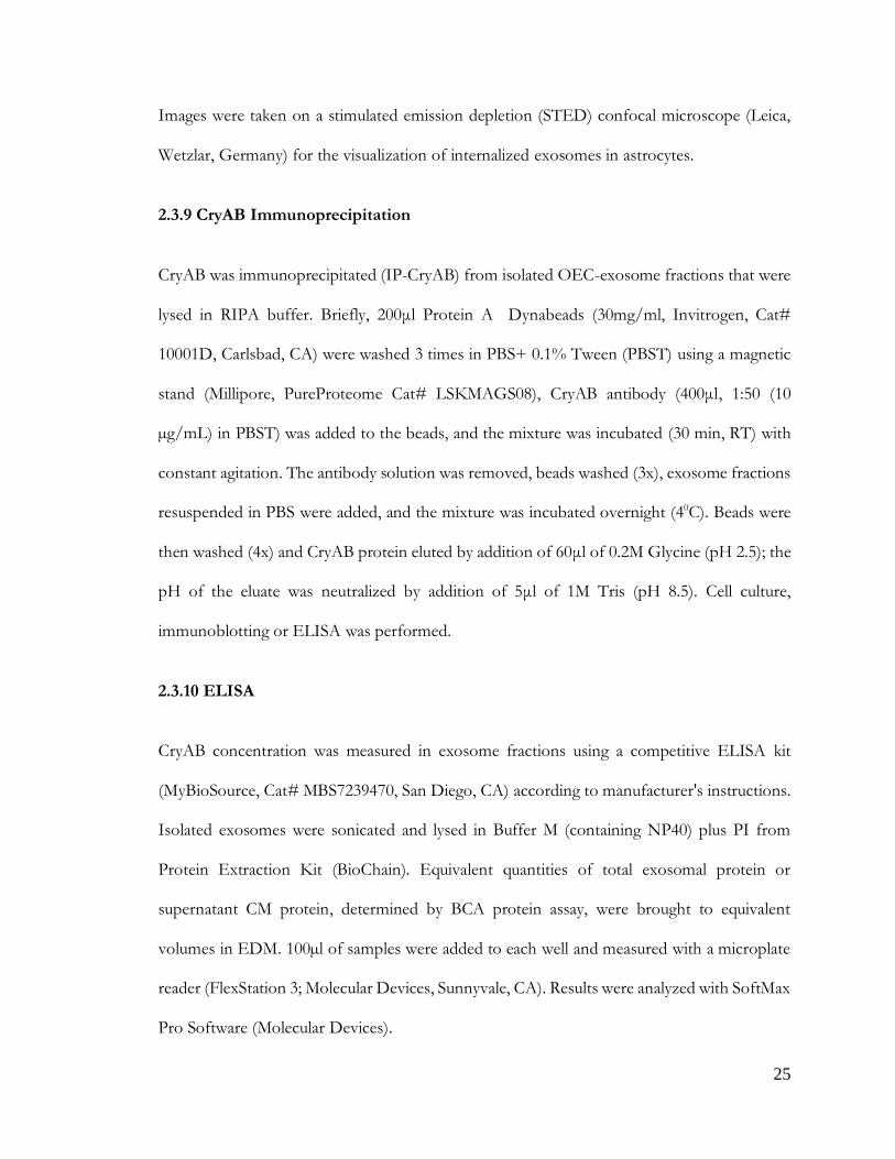
Figure 3: OEC-CM is sufficient to suppress astrocyte reactivity
with C7 cells, for example, showing heterogeneous morphologies depending on culture
conditions and density (Figure 3D): these included Schwann Cell spindle-like (majority; Figure
3Da), astrocyte-like type1 (Figure 3Db), and astrocyte-like type2 (Figure 3Dc) morphologies
(Huang et al., 2008). Cell lines were re-examined by immunofluorescence for expression of
OEC-specific markers, such as p75, Sox10, and brain lipid-binding protein (BLBP). Both C7
and C4 cell lines were positive for these OEC markers (Figure 3E). Since C7-CM significantly
suppressed nuclear NFκB translocation in astrocytes, whereas C4-CM did not (Figure3B, C),
and both lines expressed the OEC markers tested, C7-CM was used as a positive control, and
C4-CM as a negative control in further experiments.
Figure 3 (figure continued on next page)
29
Figure 3: OEC-CM alone is sufficient to suppress LPS-induced astrocyte reactivity, an effect mimicked by a subset of immortalized OEC lines. (A) Quantitative immunoblotting for NFκB was performed on the nuclear fraction of C8D30 astrocyte lysates (inset). All groups were compared using a one-way ANOVA (N=3). Treatment with LPS for 2 hrs significantly increased NFκB activity (* indicates p<0.05, gray bars). The presence of OECs blocked the effect of LPS (purple bars). OEC conditioned medium (OEC-CM) alone also blocked the increase in nuclear NFκB (red bars). (B and C) CM from six immortalized OEC lines were investigated for their ability to block the effect of LPS on nuclear NFκB translocation in C8D30 astrocytes, as measured by the ratio of fluorescence intensity of nuclear to cytoplasmic NFκB. (B) Photomicrograph of images of C8D30 astrocytes cocultured with CM of immortalized cell lines. (C) Fluorescent NFκB nuclear and cytoplasmic intensities were measured and ratios plotted. Pink dashed line depicts value of astrocytes treated with primary OEC-CM +LPS and red dashed line depicts value of astrocytes +LPS. A median value was calculated from ~100 cells per field, and then a mean/group was calculated. C7-CM and D6-CM decreased NFκB nuclear translocation (N = 3; * p ≤ 0.05; one-way ANOVA), while C4-CM treated groups was not significantly different than astrocytes +LPS alone (N = 3; * p<0.05; one-way ANOVA). (D) Photomicrograph from line C7. Multiple morphologies were found in all the OEC cell lines: (a) Schwann Cell-like, (b) astrocyte-like type1, and (c) astrocyte-like type2. Schwann cell-like spindle cells (a) predominated in C7. (E) C7 and C4 olfactory cell lines share multiple markers with OECs including BLBP (magenta), Sox10 (green) and P75 (red). Scale bars represent 25 and 20μm.

30
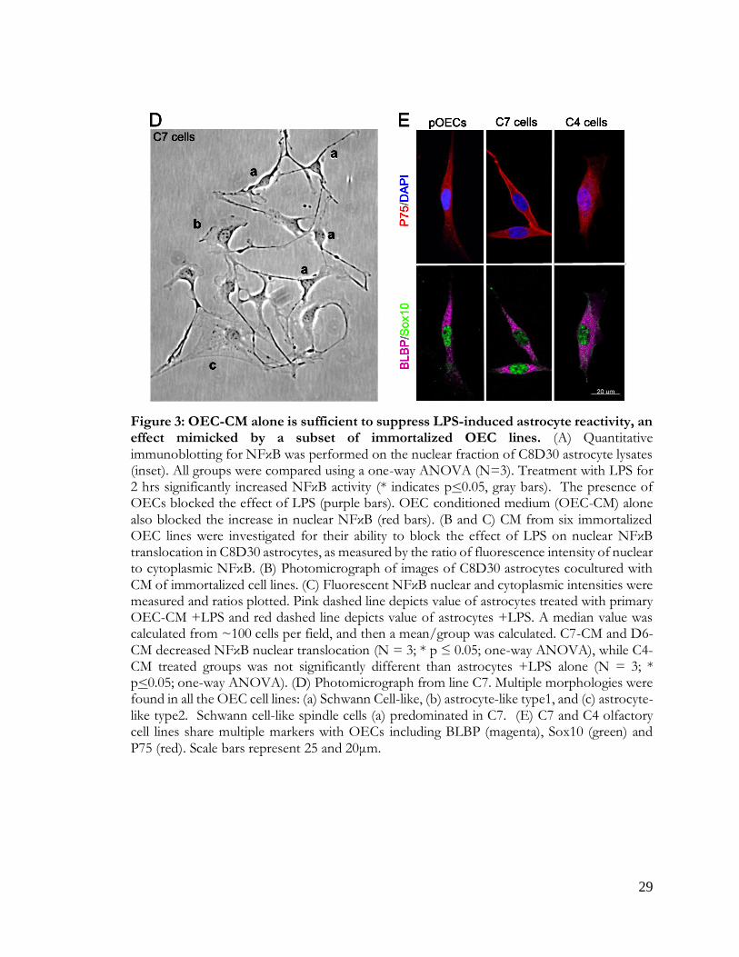
2.4.2 OEC-secreted CryAB suppresses LPS-induced astrocyte reactivity
To identify OEC-derived molecules potentially involved in crosstalk between OECs and
astrocytes, secreted proteins from primary OEC-CM, C7-CM and C4-CM were compared by
mass spectrometry. Before collection of CM, LPS was added to cultures as a stress signal.
Secreted proteins from LPS-treated cells were ranked based on 1) their abundance in C7-CM
compared to C4-CM; 2) their abundance in C7-CM compared to primary OEC-CM; and 3)
absence from C4-CM (Figure 4A). Proteins that were secreted at similar levels by C7 cells and
primary OECs (Figure 4A, horizontal dashed line), but are not likely to be present in C4-CM
(Figure 4A, X axis) were determined. Based on these criteria, we identified two proteins of
particular interest: the heat shock protein alpha crystallin B chain (CryAB), and the cell surface
glycoprotein MUC18 (MCAM). To identify OEC secreted molecules in response to an
endogenous signal from astrocytes, similar experiments were performed after treatment with
Anosmin1, an extracellular binding protein secreted by mature astrocytes (Gianola et al., 2009)
and shown to act on OECs (Hu et al., 2019). Even though the ortholog is yet to be identified
for this protein in mice, we observed a robust migration of primary mouse OECs towards
recombinant Anosmin-1 (personal observation). Notably, both CryAB and MCAM were
identified as major secreted proteins in this screen as well (Figure 4B). CryAB was selected for
further study because of its known role as an anti-inflammatory protein involved in stress
responses by CNS glia (e.g., Ousman et al., 2007; Kuipers et al., 2017), and because it was the
most abundant protein fitting our criteria in screens of both LPS (Fig. 4A) and Anosmin-1
(Fig. 4B) treated samples. Recombinant CryAB protein mimicked the effect of OEC-CM or
C7-CM on astrocyte reactivity, as measured by suppressed nuclear translocation of NFκB,
following either LPS- or cytokine-induced inflammation (Figure 4C).
31
Figure 4: An anti-inflammatory protein: CryAB
Figure 4: OEC-secreted anti-inflammatory protein CryAB and recombinant CryAB is sufficient to suppress astrocyte reactivity measured by NFκB. (A, B) Comparison of factors secreted from C7 line, C4 line and primary OECs (pOECs), analyzed by mass spectrometry. Proteins detected in CM following LPS treatment (A) or Anosmin1 treatment (B) were ranked by their relative abundance indicated by color code (heat map, inset). Relative abundance of detected proteins in C7-CM compared to pOEC-CM is graphed on the Y axes, and relative absence of the same proteins from C4-CM (1-P(C4-CM) = probability of not being found in C4-CM) is graphed on the X axes. Proteins of similar abundance in CMs from C7 cells and pOECs (horizontal red-dashed lines), and not likely to be present in C4-CM (Y axes) were identified. Alpha crystallin B chain (CryAB) had the highest C7/C4 expression ratio and was equally abundant in CM from C7 cells and pOECs. (C) Recombinant CryAB alone

32
suppressed the inflammatory response, quantified as the ratio of nuclear to cytoplasmic NFκB (Y axis) in astrocytes exposed to either LPS or a cocktail of the cytokines Il-1α, TNFα and C1q for 2 hrs. Group values were obtained from triplicate wells in which a median value was calculated from ~50 cells per field. N = 3; (*p ≤ 0.05; **p ≤ 0.01; ****p ≤ 0.0001;) two-way ANOVA.
2.4.3 Exosomes secreted by OECs contain CryAB, which moderates intercellular
immune response
Since it has been shown that CryAB secretion can occur via exosomes (Sreekumar et al., 2010;
Kore et al., 2014; Guo et al., 2019), exosomes were isolated from OECs to determine whether
they were positive for CryAB and whether the CryAB secreted via OEC-exosomes had the
ability to attenuate astrocyte reactivity. For these experiments, exosome fractions were isolated
from culture supernatants of OECs generated from both CryAB−/− mice and WT (CryAB+/+)
controls. To ensure the quality of fractions used, exosomes and whole cell lysates (CL) from
WT OECs were analyzed by immunoblotting for the following proteins: the structural protein,
-actin; a mitochondrial protein, Tomm20; a nuclear protein, histone H3; and the
extravesicular protein Flotilin-1. The exosome fraction was devoid of -actin, Tomm20 and
histone H3, but was positive for Flotilin-1 (Figure 5A), consistent with published information
for exosome fractions (Jeppesen et al., 2019).

33
Figure 5:CryAB, in OEC-exosomes, is important for OEC-astrocyte crosstalk
Figure 5: CryAB, secreted by primary OECs into exosomes, suppresses inflammatory response in an astrocyte cell line. (A) Immunoblot of exosome (Exo, left) and whole cell
lysate (CL, right) fractions from WT primary OECs were screened for -actin, Tomm20,

34
histone H3, and Flotilin-1. The exosome fraction from WT exosomes was devoid of cellular
-actin, Tomm20 and histone H3, but contained the extravesicular protein Flotilin-1. (B) Immunoblots for CryAB and the exosome marker Alix were performed on exosome fractions made from CryAB−/− and WT (CryAB+/+) OEC cultures. CryAB was absent in exosome fractions from CryAB−/− OEC culture medium, whereas the exosome marker Alix was present. (C) C8D30 astrocytes were treated for 24 hours with exosomes isolated from WT or CryAB−/− OECs. Astrocytes were exposed to 1μg/ml LPS for the last 2 hrs of exosome treatment. Nuclear fractions of astrocytes were analyzed via quantitative immunoblotting for NFκB and Histone H3. Inset shows a representative immunoblot and graph shows mean ± SD of NFκB/histone ratio. All conditions were compared to astrocyte alone group (Control, N = 3; p ≤ 0.05; one-way ANOVA). Treatment of astrocytes with WT OEC-exosomes (exo) + LPS, blocked nuclear NFκB translocation. In contrast, CryAB−/−OEC-exosomes failed to suppress nuclear NFκB translocation. Recombinant CryAB (50ng/ml) added to CryAB−/−OEC-exosomes was sufficient to attenuate NFκB translocation induced by LPS, with levels comparable to WT OEC-exo +LPS. (D) C8D30 astrocytes treated with LPS for 2 hrs (top right: “+ LPS”) showed stronger immunostaining for NFκB in the nucleus (magenta) compared to untreated controls (top left). Astrocytes co-cultured with CryAB−/− OECs (Sox 10-positive cells with blue nuclei) had increased levels of NFκB immunostaining in the nucleus (bottom right: “+CryAB−/−OECs +LPS”) compared to astrocytes co-cultured with WT OECs (bottom left: “+OECs +LPS”). Scale bar represents 40μm.
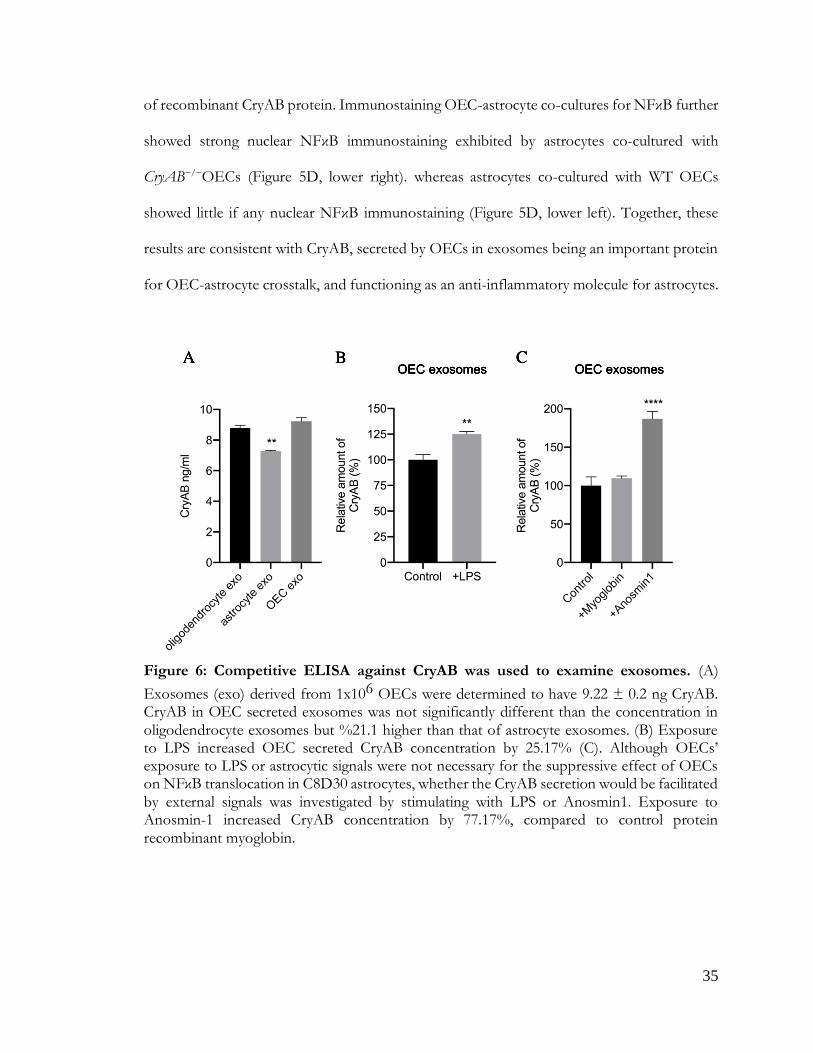
Competitive ELISA against CryAB confirmed the presence of CryAB in OEC exosomes.
Exosomes derived from 1x10⁶ OECs contained 9.22 ± 0.2 ng CryAB, whereas CryAB protein
was undetectable in CM from which exosomes were depleted (Figure 6A). Next, exosomes
from both genotypes were immunoblotted for CryAB and the endocytosis protein, Alix, which
is concentrated in exosomes (Figure 5B; Jeppesen et al., 2019). CryAB was present in WT
OECexo fractions but was absent in CryAB−/−OECexo fractions; while the exosome marker
Alix was present in exosome fractions from OECs of both genotypes. Finally, to assay whether
CryAB present in OEC exosomes could suppress astrocyte reactivity, the exosomes were
added to immortalized C8D30 astrocytes for 24 hrs, and treated with LPS for the last 2 hours
of this incubation. As shown in Figure 3C, quantitative immunoblotting demonstrated that: a)
exosomes from WT OECs were able to suppress astrocyte reactivity, as measured by reduced
nuclear translocation of NFκB; b) astrocytes treated with CryAB−/−OECexo remained reactive;
and c) the reactivity of astrocytes treated with CryAB−/−OECexo was reduced by the presence
35
Figure 6: CryAB secretion is cell type and context dependent
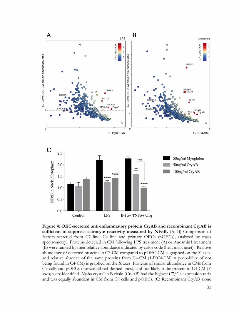
of recombinant CryAB protein. Immunostaining OEC-astrocyte co-cultures for NFκB further
showed strong nuclear NFκB immunostaining exhibited by astrocytes co-cultured with
CryAB−/−OECs (Figure 5D, lower right). whereas astrocytes co-cultured with WT OECs
showed little if any nuclear NFκB immunostaining (Figure 5D, lower left). Together, these
results are consistent with CryAB, secreted by OECs in exosomes being an important protein
for OEC-astrocyte crosstalk, and functioning as an anti-inflammatory molecule for astrocytes.
Figure 6: Competitive ELISA against CryAB was used to examine exosomes. (A)
Exosomes (exo) derived from 1x10⁶ OECs were determined to have 9.22 ± 0.2 ng CryAB. CryAB in OEC secreted exosomes was not significantly different than the concentration in oligodendrocyte exosomes but %21.1 higher than that of astrocyte exosomes. (B) Exposure to LPS increased OEC secreted CryAB concentration by 25.17% (C). Although OECs’ exposure to LPS or astrocytic signals were not necessary for the suppressive effect of OECs on NFκB translocation in C8D30 astrocytes, whether the CryAB secretion would be facilitated by external signals was investigated by stimulating with LPS or Anosmin1. Exposure to Anosmin-1 increased CryAB concentration by 77.17%, compared to control protein recombinant myoglobin.

36
Figure 7: CryAB in OEC-exosomes is internalized by astrocytes
2.4.4 Astrocytes internalize CryAB-containing OEC exosomes
To determine whether astrocytes take up CryAB-containing exosomes secreted by OECs,
CryAB−/− astrocytes were cultured with exosome fractions from OEC cultures generated from
WT mice. Uptake was visualized by immunostaining of GFAP-positive astrocytes (Figure 7,
magenta); colabeled with antibodies to endosome/exosome marker CD63 (red) and CryAB
(green). CryAB and CD63 colocalized in CryAB−/− astrocytes treated with exosomes for 4 hrs
(Figure 7, insets). Neither untreated CryAB−/− astrocytes (Figure 7B) nor WT astrocytes
(Figure 7C) showed such specific colocalization, consistent with the uptake of CryAB-
containing OEC exosomes by astrocytes.
Figure 7: CryAB in OEC exosomes is internalized by astrocytes. (A) OEC exosomes from WT mice were co-cultured with primary astrocytes from CryAB KO mice. (B) Untreated astrocytes from CryAB KO. (C) Untreated astrocytes from WT mice. All groups were stained for CD63 (endosomes; red), CryAB (green), GFAP (magenta) and Dapi (blue). Uptake of OEC secreted CryAB (green) is detected in CryAB−/− astrocytes and is often associated with endosomes (red) (A, arrows, top arrow area shown in inset, arrowhead points to CryAB

37
positive endosome). No CryAB staining (green) is detected in untreated astrocytes from CryAB KO (B, inset). CryAB (green) is present in untreated astrocytes from WT mice but rarely associated with endosomes (C, inset, arrowhead). Scale bar represents 5μm in low mag and 1μm in insets.
2.4.5 OEC secreted factors, including CryAB, reduce astrocytes’ expression of genes
associated with neurotoxic reactivity
To evaluate the effects of OEC-secreted CryAB on expression of “neurotoxic” genes,
astrocytes were exposed to LPS alone; or WT OEC-CM, CryAB−/−OEC-CM, or CryAB
immunoprecipitated from isolated OEC-exosome fractions (IP-CryAB) together with LPS.
mRNA from treated astrocytes was then analyzed for 12 transcripts known to be associated
with neurotoxic astrocyte reactivity (Liddelow et al., 2017). Q-RT-PCR analysis (Figure 8)
showed that all tested transcripts were reduced in expression in the presence of WT OEC-
CM, and this effect was significant for 9 of the 12 (Figure 8, second row, white arrows, p<0.05
A vs B). In contrast, 4 of the transcripts showed increased expression when treated with
CryAB−/−OEC-CM (Figure 8C, black arrows). The analysis also suggests that suppression of
expression of Ggta1, Serping1, ligp1, Gbp2 and Amigo2 was CryAB-dependent, for the following
reasons: a) suppression of expression failed to occur with CryAB−/−OEC-CM treatment, while
still taking place with IP-CryAB treatment (Figure 8, C vs D); or b) expression was upregulated
in the CryAB−/−OEC-CM group (Figure 8, C vs A). Suppression of expression of 4 genes
(H2-T23, Srgn, H2D1and C3) appeared to be independent of CryAB, since it still occurred in
astrocytes treated with CryAB−/−OEC-CM (Figure 8, C vs A). In contrast to either OEC-CM
treatments (CryAB−/−or WT), a significant increase in Fbln5 was detected in astrocytes treated
with IP-CryAB (Figure 8, D vs A). These results are consistent with the finding that CryAB,

38
Figure 8: OEC-CM suppresses neurotoxic-astrocyte transcripts
secreted by OECs, functions as an anti-inflammatory agent for astrocytes. In addition,
comparison of OEC-CM treatment to IP-CryAB for transcripts Ugt1a1, C3 and Fbln5 suggest
that there are factors in OEC-CM, in addition to CryAB, that suppress neurotoxic astrocyte
reactivity.
Figure 8: OEC-CM suppresses multiple transcripts associated with neurotoxic astrocyte reactivity in LPS-treated astrocytes. Heat map illustrating results of Q-RT-PCR to detect neurotoxic astrocyte transcripts in primary astrocytes treated with LPS (A); LPS plus WT OEC-CM (B); LPS plus CryAB−/−OEC-CM (C); or IP-CryAB for 24 hrs (D). Compared to LPS, OEC-CM (B) significantly suppressed 9 transcripts, whereas CryAB−/−OEC-CM (C) significantly suppressed 4. (D) IP-CryAB suppressed 6 of the 9 transcripts suppressed by OEC-CM. In addition, CryAB−/−OEC-CM+LPS caused a significant increase in 4 transcripts, while IP-CryAB caused a significant increase in one. Finally, one transcript, C3, was significantly suppressed by both OEC-CM treatments (CryAB−/− or WT) but was elevated by IP-CryAB treatment. Q-RT-PCR experiments were analyzed by one-way ANOVA followed by Dunnett’s multiple post hoc test (n= 3; arrows p ≤ 0.05).
39
2.5 Discussion
2.5.1 OEC-secreted factors that moderate astrocyte reactivity
Astrocyte reactivity is a pathological response that occurs in a wide range of CNS injuries,
inflammation and diseases. In vivo studies show that some reactive astrocytes induced by
ischemia can promote neural recovery and repair (reviewed in Rossi et al., 2007); in contrast,
reactive astrocytes induced by bacterial endotoxins such as LPS are neurotoxic (Zamanian et
al., 2012). This harmful, neurotoxic astrocyte reactivity appears to be driven by pro-
inflammatory cytokines secreted by activated microglia (Liddelow et al., 2017). However, anti-
inflammatory factors that suppress neurotoxic astrocyte reactivity are largely unknown.
Olfactory system is one of the few niches in the mammalian CNS that supports neuronal
regeneration (Forni et al., 2013). Olfactory sensory neurons are vulnerable to damage due to
their exposed location in the nasal cavity and have a remarkable capacity for regeneration
(Calof et al., 1996, Forni et al., 2013), suggesting the presence of robust anti-inflammatory
factors in the olfactory system. OECs wrap and guide axons of the olfactory sensory neurons
en route to the OB, where they establish new connections. Moreover, OECs directly interact
with astrocytes at the entry point into the CNS, enabling regenerating olfactory axons to make
new connections (Williams et al., 2004; Li et al., 2005; Raisman & Li, 2007). Notably, both
transplanted OECs (Lakatos et al., 2003; reviewed in Roet & Verhaagen, 2014) and co-cultured
OECs (Hale et al., 2011) have been shown to intermingle with astrocytes and to moderate
astrocyte activation.
The studies in this report investigate anti-inflammatory factors secreted by OECs
participating in OEC-astrocyte crosstalk. Mass spectrometry was used to analyze proteins in
CM from primary OECs and compared to the CM of immortalized OEC lines with different

40
anti-inflammatory capacities. Two proteins were identified as potential factors that could
suppress neurotoxic astrocyte reactivity: MCAM and CryAB. MCAM (also called CD146 or
MUC18) is a signaling receptor that can be cleaved from the cell membrane, generating a
soluble form that is associated with increased cell migration and invasion (Seftalioglu &
Karakoc, 2000), and primarily has been studied in endothelial cell angiogenesis and cancer
metastasis (reviewed in Dye et al., 2013). Studies on Multiple Sclerosis (MS) patients showed
no association between MCAM expression and disease activity (Petersen et al., 2019). In
contrast, CryAB treatment of MS patients was associated with a therapeutic outcome,
downregulation of T cell proliferation and pro-inflammatory cytokine production (Quach et
al., 2013; van Noort et al., 2015). In addition, CryAB has been shown to have neuroprotective
and regenerative effects in neuroinflammatory animal model systems (Arac et al., 2011;
Ousman et al., 2007; van Noort et al., 2015). Moreover, there is a correlation between glial
activation and increased CryAB levels in Alexander, Alzheimer’s and Parkinson’s diseases, as
well as traumatic brain injury and stroke (reviewed in Dulle & Fort, 2016). Thus, we
investigated the role of OEC-secreted CryAB in OEC- astrocyte crosstalk.
2.5.2 Exosomal release of CryAB
Our results show that OECs secrete CryAB via exosomes, and that these exosomes
produce an intercellular anti-inflammatory effect following their uptake by astrocytes.
Exosomes are small vesicles packaged inside multivesicular endosomes (MVE) and released
into the extracellular matrix when MVE fuse with the plasma membrane (Jeppesen et al., 2019).
Subsequent uptake by a neighboring cell initiates intercellular communication. Notably,
released exosomes are functional components of the extracellular matrix that can be induced
by stress signals but are associated with cell-cell communication rather than apoptosis

41
(Jeppesen et al., 2019; Gupta & Pulliam, 2014). Consistent with our findings, recent studies
suggest that CryAB can be secreted by glia in an autocrine manner (Kore et al., 2014; Guo et
al., 2019) and play a protective role (Ousman et al., 2007). Although the downstream effects
of CryAB in astrocytes that we demonstrate have yet to be fully explored, one important role
of CryAB may be its interaction with transcription factors, including NFkB, to suppress
inflammation by inhibition of their nuclear translocation (Shao et al., 2013; Zhang et al., 2015;
Qiu et al., 2016). However, exosome-mediated regulation of the astrocytic immune response
by OECs may also be a unique interaction, as membrane composition and protein content of
exosomes is cell type (Kalra et al., 2012; Keerthikumar et al., 2015; Kim et al., 2015) and context
specific (György et al., 2011; Müller et al., 2012).
The robust anti-inflammatory response induced by OEC-secreted CryAB, shown in the
present report, may also be a function of concentration, as our results indicate the
concentration of CryAB in OEC exosomes to be higher than that found in astrocyte
exosomes. We found that CryAB in OEC secreted exosomes was ~21% higher than that of
astrocyte exosomes ( Figure 6A) and exposure to LPS increased OEC secreted CryAB
concentration by ~25% ( 6B), indicating OECs actively respond to stress by either secreting
more exosomes or increasing CryAB concentration in exosomes, or both. In addition,
exposure to Anosmin1 increased CryAB concentration by ~77% (Figure 6C), compared to
control protein recombinant myoglobin, suggesting OECs can actively respond to extracellular
astrocyte signals by increasing CryAB concentrations.

42
2.5.3 CryAB, as well as other factors secreted by OECs, can suppress neurotoxic astrocyte reactivity
Stress causes denaturation of correctly folded proteins that can result in their aggregation
and binding to CryAB (Muranova et al., 2018) and subsequent changes in gene expression
(Singh et al., 2019). Our results show that OEC-secreted CryAB suppressed expression of a
number of genes associated with neurotoxic astrocyte reactivity and suggest there are
additional factor(s) in OEC-CM that further suppress this harmful reactivity. In fact,
complement component-3 (C3) expression in IP-CryAB +LPS-treated astrocytes, although
not significantly different than that observed in astrocytes treated with LPS alone, was
significantly greater than expression in astrocytes treated with either OEC-CM groups (Figure
5). C3 is an important marker for neurotoxic astrocytes, evident by knockout mice showing
reduced activity in microglia and astrocytes, as well as neuron loss (Shi et al., 2017). Moreover,
C3 is found colocalized with astrocyte markers in regions of neurodegeneration in human
post-mortem tissue (Liddelow et al., 2017). Therefore, more experiments are required to
identify OEC-secreted factor(s) that can suppress C3. In this regard, other proteins identified
in our mass spectrometric screen, showing smaller changes, may be worthwhile to evaluate.
Certainly, crosstalk mechanisms between OECs and surrounding niche cells, including
astrocytes, are highly complicated (Chuah et al., 2011). In addition to unidentified anti-
inflammatory factors, absence or suppression of pro-inflammatory factors might also play a
role in OEC-CM’s effect on astrocyte reactivity. For example, our mass spectrometry
screening showed higher concentration of S100A4 and S100A6 proteins in C4-CM compared
to pOEC-CM or C7-CM (Figure 4, Supplemental Data-1 & 2). S100 proteins are known to
modulate neuroinflammation (Donato, 2001), and as such are also candidate molecules that
43
may contribute to the observed difference in the inflammatory reactivity of astrocytes treated
with C4-CM versus pOEC-CM or C7-CM.
Recent studies have transformed our perception of astrocytes from a passive structural
support network for neurons, to active effectors in the regulation of synaptic transmission,
neural excitability, plasticity and recovery. In this paper, we identify OEC secreted CryAB as
an anti-inflammatory factor that can moderate astrocyte reactivity, suppressing both
transcription of neurotoxic classified genes and nuclear translocation of pro-inflammatory
factor NFκB. Improving our understanding of the crosstalk between astrocytes and OECs
may inform strategies to identify other endogenous repair mechanisms that facilitate CNS
repair, and consequently impact function, in the injured nervous system.

44
Chapter 3: OEC-HSPGs moderate astrocyte reactivity, proliferation, and
differentiation via FGFR1 signaling
3.1 Abstract
FGFR1 signaling pathway is important in astrocyte fate determination and reactivity.
However, the contribution of specific downstream signaling components has been difficult to
identify due to compensation/feedback mechanisms between FGFRs and other pathways. In
this study, we show that olfactory ensheathing cell (OEC)-secreted factors moderate astrocyte
reactivity by suppressing pro-inflammatory transcription factor (TF) NFκB. In addition,
(OEC)-secreted factors induce the expression of differentiation and neuro-repair promoting
TF STAT3 in astrocytes. FGF signaling is one of the few known pathways that can block
harmful astrocyte reactivity in vitro. In accordance, when FGFR1 RTK activity was chemically
inhibited in cocultures, OECs failed to suppress stress-induced nuclear translocation of pro-
inflammatory transcription factors in astrocytes. OEC-secreted heparan sulfate proteoglycans
(HSPGs) are strong candidates for factors that play a role in this crosstalk, since they are cell-
specific ECM proteins known to support and regulate FGF signaling. Moreover, a regulatory
role in intracellular FGFR trafficking has also been shown for HSPGs. Our results indicate
that differential FGF/FGFR1 signaling can directly and rapidly impact the binary choice of
astrocytes to proliferate or differentiate. These changes are mediated by differential membrane
activity in the presence of OEC-HSPGs and may also reflect nuclear FGFR-mediated
regulation of transcription. OEC-HSPGs, or synthetic heparan sulfate oligosaccharides that
mimic OEC-HSPGs, may guide new therapies for traumatic CNS injury and other
neurological disorders by targeting astrocytes.
45
3.2 Introduction
Astrocytes are one of the most abundant and heterogeneous cell types in the central
nervous system (CNS), yet, in contrast to many other CNS cell types, changes in gene-
expression profiles of astrocytes during development and pathological states remain poorly
defined. Studies in the developing spinal cord, and in vitro human astrocytes, identify two gene-
expression patterns: an early, developmental phase characterized by cell cycle, cytoskeletal and
axon guidance genes and a late, differentiated phase characterized by channels, neuronal
signaling and metabolic genes (Chaboub et al., 2016; Molofsky et al., 2013). Differentiated
astrocytes exhibit regionally distinct and unique properties that are maintained by local signals
(Morel et al., 2017). Under pathological conditions, differentiated astrocytes become reactive
and demonstrate key features of developmental phase astrocytes, such as upregulation of
structural proteins (glial fibrillary acidic protein (GFAP), Nestin and Vimentin), rounded
morphologies and in some cases increased proliferation (Sirko et al., 2013; Lepore et al., 2008;
Anderson et al., 2016, reviewed in Liddelow & Barres, 2017). The return to a less mature
genotype/phenotype is referred to as ‘‘dedifferentiation’’ (Buffo et al., 2008; Sirko et al., 2013).
Indeed, astrocytes can re-acquire stem cell properties both in vitro, upon FGF2 stimulation
(Kleiderman et al., 2016), and in vivo after injury (Faiz et al., 2015). All together, these results
suggest that under pathological conditions, reactive astrocytes turn on developmentally
encoded genes and dedifferentiate due to unsuitability, or absence of, extracellular signals in
the microenvironment.
Dedifferentiation includes the upregulation of intermediate filaments, which enables
reactive astrocytes to surround and isolate inflamed tissue, creating the ‘glial scar’. Glial scar
can prevent the tissue from secondary damage but also limits access for regenerating neural
46
cells to repair the damaged tissue after injury (Faiz et al., 2015). The mechanism(s) responsible
for dedifferentiation of reactive astrocytes and the contribution of transcriptional regulation
to the harmfulness of astrocyte reactivity remain enigmatic. Our study focusses on aspects of
astrocyte differentiation that are regulated by extracellular signals to test the hypothesis that
activation of these signaling pathways could be used to support astrocytes for a therapeutic
outcome following CNS injury.
Astrocytes display plasticity in protein expression, gliotransmitter release, calcium activity,
changes of morphology or gap junction (Doetsch et al., 1999; Alvarez-Buylla & Lim., 2004;
Farmer et al., 2016, reviewed in Pirttimaki & Parri, 2013). Astrocyte plasticity can have
profound effects on neuronal network activity and also can be induced by local activity, in
parallel with their reactive nature to pathological changes (Pirttimaki & Parri, 2013, Khakh &
Deneen, 2019). Support for this observation comes from a recent study in the olfactory bulb
(OB) where layers of morphologically distinct astrocytes are shaped by local interactions rather
than clonal relationships (García-Marqués & López-Mascaraque, 2017). Astrocytes in the
outermost layer of the OB, olfactory nerve layer (ONL), commingle with a distinct type of glia
that is unique to the olfactory system, olfactory ensheathing cells (OECs). Astrocytes and
OECs form the glia limitans of the OB and protect CNS against inflammation (Beiersdorfer
et al., 2019). Notably, astrocytes of the ONL show the most differentiated morphology in the
OB (García-Marqués & López-Mascaraque, 2017).
The olfactory system is one of the few regions in the adult mammalian CNS that supports
the differentiation of new neurons and glia throughout life. Both neural niche signals and the
surrounding glia, including OECs, give the olfactory mucosa this capability (Li et al., 2005,
Roet, 2014). OECs transplanted into CNS injury sites for neuroregenerative purposes show
47
therapeutic benefits (Lakatos, 2003; reviewed in Roet, 2014). Similarly, astrocytes cultured in
medium conditioned by cultured OECs (OEC-CM) show reduced nuclear translocation of
Nuclear Factor kappa-B (NFκB), a pro-inflammatory protein that is a hallmark of harmful
astrocyte reactivity (Hale et al., 2011, Liddelow & Barres, 2017, see Chapter 2, Figure 5).
Moreover, our own work shows that OEC-secreted factors suppress multiple neurotoxicity-
associated transcripts in astrocytes (see Chapter 2, Figure 8).
FGFs are a family of critically important growth factors for CNS development, homeostasis
and after injury; regulating neurogenesis, gliogenesis and also regeneration. FGF1 and FGF2
are significantly upregulated following CNS injuries (Woodbury & Ikezu, 2014; Ornitz & Itoh,
2015). FGF2 enhances neural stem cell proliferation (Newman et al., 2000) and is upregulated
in astrocytes in neurodegenerative disorders and following traumatic brain injury (Kirby et al.,
2013). While FGF2 signaling via FGFR1 is primarily known for its mitogenic effect and role
in dedifferentiation, FGF2/FGFR1 has also been shown to be crucial for cell differentiation.
These conflicting observations regarding the role of FGFR1 in cell-fate control of neural stem
cells (NSCs) and reactive astrocytes has been contentious (Reuss et al., 1998; Fahmy & Moftah,
2010). This controversy was settled by a loss- and gain- of function study showing that FGF
signaling inhibits astrocyte reactivity in both normal and injured CNS (Kang et al., 2014).
Indeed, in vitro, FGF signaling significantly decreases expression of transcripts associated with
neurotoxic astrocyte reactivity (Liddelow et al., 2017). Curiously, both the gain, and loss of
FGFR function decreases scar tissue formation in vivo (Kang et al., 2014). Although
compensation between FGFR members cannot be excluded (Oh et al., 2003; Pringle et al.,
2003; Kang et al., 2014), these findings suggest developmentally contrasting roles of pathways
downstream of FGFR activation, as well as positive and negative feedback mechanisms
48
regulating FGFR signaling. However, the downstream effectors of FGF/FGFR signaling that
enable astrocyte differentiation and their potential role in prevention of astrocyte
metamorphosis into harmful reactive astrocytes remain to be identified.
FGFR1 is one of the most crucial signaling pathways that govern nervous system
development (Stachowiak et al., 2016) and FGF signaling is one of the few known pathways
that can block harmful astrocyte reactivity in vitro (Liddelow et al., 2017). An interesting partner
of regulation for differential FGFR1 activity is heparan sulfate proteoglycan (HSPG). HSPGs
are cell-specific extracellular matrix (ECM) proteins known to support and regulate FGF
signaling. Barnett and colleagues suggest a role for OEC-secreted HSPGs in suppressing
astrocyte reactivity, measured by GFAP expression (Santos-Silva et al., 2007) and astrocyte-
OEC mingling (Higginson et al., 2012), with chemical blockage of FGF receptors promoting
cell mingling. In agreement, Leadbeater and colleagues (2006) suggest that HSPGs regulate
cell-specific injury responses through FGF2 after CNS injury.
Here we used OEC-astrocyte cultures to characterize FGFR1 downstream targets that can
block harmful astrocyte reactivity. Experimental results showed that OEC-secreted HSPGs
block astrocyte reactivity and suggest that this effect is mediated via increased FGFR1
phosphorylation initiated on the astrocyte membrane. On the other hand, FGF2 potentiated
proliferation in astrocytes was neutralized in the presence of OEC-secreted factors. These
results are consistent with a role for OEC-HSPGs in intracellular FGFR1 trafficking via
association with the transcriptional machinery in astrocytes. Novel treatment strategies
designed to mimic this crosstalk, targeting astrocytes, may enable improved recovery following
CNS injuries.

49
3.3 Materials and Methods
3.3.1 Mice
All mice were maintained, and all animal handling procedures were performed according to
protocols approved by the National Institutes of Health NINDS Institutional Animal Care
and Use Committee. Ext1floxed/floxed mice were kindly gifted by Dr. Yu Yamaguchi. Mice were
genotyped using primers 5’-GGAGTGTGGATGAGTTGAAG-3’ and 5’-
CAACACTTTCAGCTCCAGTC-3’, using the PCR protocol previously described (Chen et
al., 2008). Male mice were obtained as heterozygous both for the Ext1floxed allele and also the
BLBP-Cre allele (Hegedus et al., 2007). Female mice were obtained as homozygous for the
Ext1floxed allele. All mice were transgenic for the reporter S100ß-DsRed (Windus et al., 2007).
Ext1-CKO (Ext1BLBPKO S100ß-DsRed) mice had developmental defects including
diminished or absent olfactory bulbs and severe arrest of limb development, in particular in
the hindlimbs, and did not survive postnatally. Absence of olfactory bulbs was previously
reported for Nestin conditioned Ext1-KO mice which also does not survive postnatally,
however these embryos are reported to grossly look normal (Inatani et al., 2003). Even though
BLBP expression is restricted in the nervous system in adults, BLBPCre-RosaTom mice showed
reporter expression in the early limb buds. Thus, the most likely explanation for the severely
impaired limb development phenotype we observed is due to the transient expression of BLBP
during limb development. This skeletal defect allowed us to distinguish mutant mice from
heterozygous or WT littermates without PCR genotyping. Embryos were collected from time
mated females between E16- 21 for future experiments.

50
3.3.2 Cell culture and reagents
Olfactory Epithelium (OE) OECs were obtained via fluorescence activated cell sorting
(FACS). Unless otherwise stated, OB derived OECs were used for the experiments as
previously described (Dairaghi et al., 2018). Briefly, olfactory bulbs of postnatal (PN) day 0-
7 mice were collected and enzymatically digested at 370C for 35 min using 30μg/ml
hyaluronidase (Sigma, Cat# H3631, St. Louis, MO), 30U/ml dispase I (Sigma, Cat# D4818),
1.2 mg/ml collagenase type 4 (Worthington, Cat# 43E14231, Lakewood, NJ), 10U/ml
DNAse I (Worthington, Cat# 54E7315) (Au & Roskams, 2003; Richter et al., 2008). Enzymes
were deactivated by adding fresh medium (DMEM:F12) and the cell suspension was
centrifuged for 7 minutes at 1000rpm. The cell pellet was resuspended in fresh medium and
the cells were run through a 40-micron cell strainer (Falcon) before the three steps of
differential cell adhesion method (Nash et al., 2001). Initially cells were seeded into uncoated
T75 flasks (4x10⁶ viable cells/flask, VWR, Cat# 734-2788, Radnor, PA) for 18 hrs. This initial
seeding helps to remove fibroblasts. Unattached cells were collected and seeded into another
uncoated flask for up to 36 hrs to remove astrocytes, followed by a final step where
unattached cells were seeded onto poly-L-lysine (Sigma, Cat# P4707)-coated flasks to grow
primary OECs. The first medium was changed at day 4 and every 2-3 days after the initial
medium change. Cells were cultured for up to 2 weeks. More than 90% of the cells in the
culture were OECs based on p75 and S100ß immunostaining consistent with earlier reports
(Au & Roskams, 2003). For OECs to be co-cultured with primary astrocytes, the medium
was gradually changed to serum-free medium (Klenke & Taylor-Burds, 2012) supplemented
with 5ng/ml HB-EGF (PeproTech, Cat# 100-47, Rocky Hill, NJ), and B27 (Thermo Fisher
Scientific, Cat# A3582901) to provide a medium compatible with astrocyte cultures, since
serum has been shown to induce astrocyte reactivity (Foo et al., 2011).
51
Primary astrocytes were obtained by magnetic sorting as previously described (Holt et al.,
2019), with some modifications. Briefly, 10-20 cortices of PN day 2-4 pups were dissociated
using the MACS Neural Tissue Dissociation Kit-T (Miltenyi Biotec, Cat# 130-093-231,
Auburn, CA) at 370C (5% CO2, 30 min). Non-dissociated tissue was removed using a 40μm
cell strainer (Fisher Scientific, Cat# 22-363-547), and the remaining cell solution was
centrifuged (300g, 5 min). Next, a discontinuous density gradient, prepared using 1:1
albumin-ovomucoid solution (10mg/ml of each) (Worthington, Cat# OI; GeminiBio, Cat#
700-102P, West Sacramento, CA), was used to remove cell debris and inhibit enzyme activity.
The cell pellet was resuspended in 80μl Hank’s Balanced Salt Solution (HBSS) (Gibco, Cat#
14025-092) plus 20μl anti-GLAST (ACSA-1) MicroBeads (Miltenyi Biotec, Cat# 130-095-
825, Auburn, CA) for up to 107 cells, and incubated for 10 min (40C). Cells were washed and
incubated in 90μl HBSS plus 10μl anti-Biotin MicroBeads for another 15 min (40C) before
running through MACS column for positive selection of astrocytes. Cells were cultured for
one week and then the same procedure was followed with anti-Prominin-1 MicroBeads
(Miltenyi Biotec, Cat# 130-092-564) for the negative selection of radial glia, followed by
another positive selection with anti-GLAST antibody the same day, to increase the purity of
astrocyte cultures. Sorted cells were cultured in T25 flasks coated with poly-L-lysine, in 5ml
serum-free astrocyte culture medium (ACM, described above). In our hands, astrocytes
isolated by this method and cultured in ACM were not reactive when stained with NFκB (not
shown). The immortalized mouse astrocyte line C8D30 (ATCC, VA, USA) was cultured in
DMEM-F12 (Gibco, Cat# 10313-02, 11765-054, Long Island, NY) containing 10% Fetal
Bovine Serum (FBS) (Gibco, Cat# 10438-026), plus 0.5% antibiotic-antimycotic (Gibco,
Cat# 15240-062) at 370C in 5%CO2. FGFR1 inhibitor SU5402 (Tocris, Cat# 3300) was
52
used at 30nM and PD173074 (Tocris, Cat# 3044) was used at 20nM. Inhibitors were added
1 hr before the 2 hr LPS treatment.
3.3.3 Collection and heparinase treatment of OEC-CM
Once primary OECs reached 70% confluency, medium was replaced with fresh medium and
collected 24 hours after. This OEC-conditioned medium (OEC-CM) was spun down (30 min)
to remove cell debris and then directly used on astrocyte cultures unless otherwise stated. For
the heparinase treatment, primary OECs were gradually adapted to serum free medium (ACM)
prior to medium collection. Heparin containing proteins in OEC-CM were digested as
previously described (Higginson et al., 2012) with slight modifications. Briefly, the OEC-CM
received 3 consecutive treatments of a cocktail of Heparinase I and III (Sigma, Cat# H3917)
and Heparinase II (Biolabs, Cat# SP0736S), containing 3mU/ml of each heparinase enzyme.
The first treatment was for 10 hrs (37°C). The second treatment was for 24 hrs (37°C) and the
final treatment was for 5 hrs (37°C). The conditioned medium was then mixed 1:1 with serum
containing medium (DMEM-F12 + 10% FBS) to inactivate the enzymes and added on to
immortalized C8D30 astrocytes for 24 hrs. The last 2 hours of this incubation, cells were
stimulated with LPS as described earlier. Nuclear compartments of astrocytes were isolated,
and immunoblotting was performed (described below).
3.3.4 Primary antibodies
The following antibodies were used: NFκB rabbit polyclonal antibody (C-20, Santa Cruz,
Cat# sc-372, Santa Cruz, CA, 1:650 for WB, 1:750 for IF); Histone mouse monoclonal
antibody (Fisher Scientific, Cat# AHO1432, Waltham, MA, 1:200 for WB); Sox10 goat
polyclonal antibody (N-20, Santa Cruz, Cat# sc-17342, 1:300 for IF); FGFR1 mouse
monoclonal antibody (Santa Cruz, Cat# sc-57132, 1:1K for IF); FGFR1 rabbit polyclonal

53
antibody (Abcam, Cat# ab10646, 1:400 for WB); FGFR1 (phospho-Tyr653/654) rabbit
polyclonal antibody (Cell Signaling, Cat# 3471, 1:1K for IF); FGFR1 (phospho-Tyr766)
rabbit polyclonal, biotinylated antibody (Biorbyt, Cat# orb501512, 1:50 for IF); O4 mouse
monoclonal antibody conjugated to APC (Miltenyi Biotec, Cat# 130-119-155, 1:50 for
FACS); F4/80 mouse monoclonal antibody conjugated to PE-Cyanine7 (Invitrogen, Cat#
25-4801-82, 1:50 for FACS); CD45.2 mouse monoclonal antibody conjugated to FITC
(BioLegend, Cat# 109805, 1:100 for FACS); pERK mouse monoclonal antibody (Millipore,
Cat# M8159, Darmstadt, Germany) was used at 1:5K for WB; GFAP chicken polyclonal
antibody (Aves, 1:4K for IF); p75-NGFR rabbit polyclonal antibody (Millipore, Cat#
AB1554, 1:5K for IF) and p75-NGFR goat polyclonal antibody (R&D Systems, Cat#
AF1157, 1:200 for FACS).
3.3.5 FACS
Dissociated OE tissue obtained from WT (Ext1floxed/floxed S100ß-DsRed) and Ext1-CKO
(Ext1BLBPKO S100ß-DsRed) E16- 21 mouse embryos was subjected to FACS for single cell
sorting of S100ß-DsRed expressing cells. Cells were dissociated using trypsin according to
manufacturer’s protocol (Miltenyibiotech, Cat#130-093-231) and were filtered through 35mm
cell strainers (Falcon, Cat#352235) to remove cell clumps before sorting. The single cell
suspensions were then incubated with 1mg/ml DAPI (1:500, Thermo Scientific, Cat#62248)
for a few minutes to label dead cells before loading samples to FACS (MoFlo Astrios EQ high
speed cell sorter; Beckman Coulter). Flow data setting of sorting gates on DsRed-expressing
cells that are DAPI-negative were carried out using Summit software V6.3.016900 (Beckman
Coulter). Determination of the optimum time point for sorting after dissociation was
established using antibodies against OECs (p75-NGFR or O4-APC) and tissue resident

54
macrophages (F480 and CD45.2). For OE cell dissociates that were sorted on the day of
dissociation (acute sorting), trypsin was preferred instead of papain in order to preserve the
F4/80 epitope. However, trypsin dropped the cell viability (from ~70% to 40%). Moreover,
acute sorting revealed that a considerable percentage of S100ß-DsRed cells were positive for
tissue resident macrophage markers (also called monocytic cells, Smithson & Kawaja., 2010)
(Chart 1). However, these cells were eliminated when the cell suspension was plated and
maintained in serum containing culture medium (Bohlen et al., 2017) for 25-35 days (DIV). In
addition, these established cells were more resilient against trypsin. As can be seen in Chart 1,
both cell viability and the percentage of S100ß-DsRed positive OE cells that were OECs
dramatically increased when the cells were sorted after culturing. The purity of OECs (S100ß-
DsRed+ cells) obtained by FACS after culturing was similar to our primary OEC cultures
obtained via differential-attachment method described above. Therefore, for future
experiments, primary OECs were obtained via FACS from embryonic OE cultures (25-35
days DIV) by relying solely on the S100ß-DsRed signal. Following this protocol, typically ≥1K
cells were obtained from 4 mouse embryos. After sorting, cells were grown for ~1 week on
poly-L-lysine (Sigma, Cat# P4707)-coated flasks before conducting experiments.
Chart 1
Live cells
(% of All)
S100ß + cells
% of Hist
S100ß – cells
% of Hist
S100ß + cells
that are
OECs % of
Hist
S100ß + cells
tissue resident
macrophages
% of Hist
E18 OE
Acute sorting
36.39% 8.11% 88.61% 7.79% 65.82%
E18 OE Sorted
at 31 DIV
78.02% 25.28% 75.21% 85.48% 0.1%
55
3.3.6 Immunoblot analysis
Nuclear fractions of immortalized C8D30 astrocytes were used for quantitative immunoblot
analysis. To be able to collect astrocytes alone for the analysis, OECs were cultured on porous
inserts (0.4µm Millicell Cell Culture Insert, Millipore, Cat# PICM0RG50), that were discarded
at the end of the experiment. For the CM treated groups, OEC-CM was collected (24 hr-old
medium). Treatment was 24 hours for all groups and the last 2 hours of this incubation, all
groups were stimulated with 1μl/ml LPS (Sigma, Cat# L6529). Astrocytes were then scraped
and the CNMCS Compartmental Protein Extraction Kit (BioChain Cat# K3013010 Hayward,
CA) plus protease/phosphatase inhibitors (PI, Cell Signaling, Cat# 5872S, Danvers, MA) was
used for the nuclear fractionation of each treatment condition. The fractions were run on
BioRad Mini-Protean TGX Stain-Free Gels (Cat#4568084), transferred to PVDF stain-free
blot (Trans-Blot Turbo Transfer Pack, Cat#1704156) via the Trans-Blot Turbo transfer
system (BioRad), and blocked with 5% dry milk (BioRad, Cat #170-6404 ) prior to staining
with NFκB, FGFR1 or pErk1/2 antibodies. Membranes were exposed to Clarity enhanced
chemiluminescence (ECL) reagent (Cat. # 170–5061, Bio-Rad) for 5 min and the signal was
detected using ChemiDoc MP (Cat. # 170–8280, Bio-Rad). Quantification of band intensities
was calculated using Image Lab 5.0 software (Bio-Rad) and normalized by the loading control
Histone.
3.3.7 Quantitative immunofluorescence
Following immunofluorescence staining for antibodies of interest, confocal images were
taken on Zeiss LSM 800 Confocal Microscope (Carl Zeiss, Thornwood, NY). Imaging was
done on a single plane and the same parameters (gain, pin size, etc.) were used between the
groups while imaging. On the images, intensity median for each channel that overlaps with
56
FGFR1 staining (magenta, threshold=30) was calculated using Imaris software. Statistics
(two-way ANOVA) were performed and the average median value ±standard deviation (SD)
per treatment plotted. To calculate the covariance of two fluorescent signals independently
of fluorescence intensity, pixels colocalized or Pearson’s colocalization coefficient was used.
For quantifications, the thresholds were determined automatically using Imaris 9.1.0; based
on the maximum threshold of intensity for each channel that shows any statistical correlation
(Costes et al., 2004). Fiji was used for the quantification of factorial Markov random field
(FMRF) signal on a single plane and the FGFR1 signal was scaled up x3 to enable selection
of individual spots of correlation.
3.3.8 Cell proliferation
Cell proliferation was analyzed continually every day over a period of up to 3 days using the
Alamar blue assay (Nikolaychik et al. 1996) and 50ng/ml FGF2 was used to stimulate the
cells. Briefly, at the day of measurement, the cells were rinsed with PBS and then incubated
for 2 hr with their respective growth medium supplemented with 5% Alamar blue reagent
(Thermo Fisher Scientific, Cat# DAL1025). At the end of incubation time, supernatant was
collected and Alamar blue absorbance was measured (NanoDrop, Thermo Fisher Scientific),
using the appropriate filter set (570 nm and 600 nm). The data, expressed as means ± SD,
are presented in arbitrary fluorescence units.
3.3.9 Quantitative RT-PCR (q-RT-PCR)
cDNA synthesis was performed using SuperscriptTM III reverse transcriptase (Invitrogen), and
PCR carried out using the ViiA7 Real-Time PCR System (Applied Biosystems, Waltham, MA)
in 20μl final volume, containing 10μl of SsoAdvanced Universal SYBR Green Supermix
(BioRad Cat#1725271), 2μl of primer mix (1μM of each primer) 1μl of cDNA and 7μl water.

57
For samples that were mixed with FGFR1 primers, a different template was used containing
3μl of cDNA and 5μl water. Samples were run in triplicate. The expression levels of FGFR
mRNA were determined using primers (forward; reverse)
TCTGGAAGCCCTGGAAGAGAGA; TCTTAGAGGCAAGATACTCCAT for FGFR1
(Verheyden et al., 2005), CAAAGGCAACTACACCTGCC; CAGCCATGACTACTT-
GCCCG for FGFR2 (Yu et al., 2003), GCTGTAGGCTTAACACTTCC;
GCTGACAAACCATGTGCTAGG FGFR3, GGACTAGCTGCAAAACTTATGC;
CCATGTCTTCTGTCGTTCC for FGFR4 and were normalized using the primers (forward;
reverse) (AGTGCCAGCCTCGTCCCGTA; TGAGCCCTTCCACAATGCCA), for
expression of GAPDH. Data were analyzed by a one-way ANOVA followed by Dunnett’s
multiple post hoc test.
3.3.10 Statistical analysis
All statistical analyses were done using GraphPad Prism 8.00 software. The results are shown
as mean ± SD. Statistical analysis was performed using one-way or two-way ANOVA, unless
otherwise stated. Probability values of 0.05 (p<0.05) were considered to indicate statistical
significance. N=biological replicates, n=technical replicates.

58
3.4 Results
3.4.1 OEC-HSPGs suppress harmful astrocyte reactivity and promote astrocyte
differentiation.
Previous studies, as well as our own work (see Chapter 2) shows that OECs secrete anti-
inflammatory factors that reduce neurotoxic-astrocyte reactivity. Expression of neurotoxicity
associated genes in astrocytes are understood to be mediated by the pro-inflammatory factor
NFκB (Lian et al., 2015; Liddelow et al., 2017). In contrast, recovery-promoting astrocytes are
mediated by activation of STAT3, a transcription factor (TF) shown to moderate reactivity
and support differentiation of astrocytes (Anderson et al., 2016; Hong & Song, 2014). We
analyzed mRNA expression of immortalized C8D30 astrocytes for NFκB and STAT3 2 hr
after LPS exposure, with or without OEC-cultured inserts. As expected, NFκB expression was
blocked by factors secreted from OEC-cultured inserts. Moreover, OEC-secreted factors
increased STAT3 expression (Figure 1A). These results indicate that OEC-astrocyte crosstalk
suppresses early transcriptional changes associated with neurotoxic astrocytes and upregulates
changes associated with neurorepair-promoting astrocytes. Here, this crosstalk was used to
characterize FGFR1 downstream targets that play a role in astrocyte reactivity. First, mRNA
expression of different FGFR subtypes at this time point was examined (Figure 1B). Studies
suggest that FGFR3 and FGFR2 upregulation correlates with glial lineage commitment while
FGFR1 is crucial for both self-renewal and differentiation (Maric et al., 2007; Zhang et al.,
2015;). Our results show that under LPS-induced stress conditions, astrocytes cultured with
OECs express significantly higher FGFR expression for FGFR1-3, but not 4. The absence of
increased FGFR4 expression is not surprising given it is expressed mostly during development
(Lichtenstein et al., 2012) (Figure 1A, 1B). Notably FGFR1 expression increased over 100-fold
59
in the presence of OECs (Figure 1B). In accordance to the observed increase in STAT3;
FGFR2 and FGFR3 expression were also significantly upregulated, pointing out increased
astrocyte differentiation (Hong & Song, 2014; cite FGFRs).
To assess whether the change in FGFR1 activity plays a role in the suppression of harmful
TFs in astrocytes, two different inhibitors to block receptor tyrosine kinase (RTK) activity,
30nM SU5402 or 20nM PD173074, were used. Stimulation with LPS treatment for 2 hrs
increased nuclear translocation of NFκB in astrocyte monocultures (Figure 9C, left panel).
Addition of neither FGFR1 inhibitor impacted nuclear NFκB translocation, but significantly
slowed the proliferation of C8D30 cells measured by a viability test consistent with earlier
reports (Reilly et al., 2004). Presence of OECs (distinguished by Sox10 staining, Figure 9C
middle panel) blocked the inflammatory reactivity in C8D30 cells stimulated with LPS
(arrowheads, middle panel, notice weak nuclear NFκB staining). However, this effect
disappeared when FGFR1 activity was chemically inhibited using SU5402 (arrowheads, right
panel) or PD173074 (data not shown). These results are consistent with our hypothesis that
astrocyte FGFR1 signaling, activated via OEC secreted factors, suppresses harmful astrocyte
reactivity and support a more differentiated phenotype in astrocytes.
Candidates for the OEC-secreted factors, enabling observed changes in astrocyte reactivity
via FGFR1 1) should be secreted into the extracellular space, and 2) act as FGFR co-receptors
and moderate downstream signaling. Heparan sulfate proteoglycans (HSPGs) are strong
candidates for such OEC-secreted factors since they are cell-specific ECM proteins known to
support and regulate FGF signaling; and have been suggested to play a role in suppressing
astrocyte reactivity (Higginson et al., 2012). To examine the contribution of OEC-HSPGs, we
enzymatically degraded secreted HSPGs in the OEC conditioned medium with heparinase

60
(OEC-CM). Heparinase-treated (Hase) OEC-CM (see material & methods) failed to block
nuclear NFκB translocation in astrocytes, as measured by quantitative immunoblotting in
nuclear fractions of C8D30 cells (Figure 9D). To confirm these results, transgenic animals
with conditional ablation of the HSPG-synthesizing enzyme Ext1 (Irie et al., 2012; Yamaguchi
et al., 2010) in OECs (Ext1BLBPKO) were used. Ext1BLBPKO-OECs (± WT OEC-CM) and
WT-OECs were grown on hanging inserts and moved to multi-well plates containing C8D30
astrocytes 24 hrs before the experiment. Cells were treated with LPS for the last 2 hours of
this incubation. Inserts were discarded at the end of experiment and nuclear fractions of
C8D30 cells were collected for immunoblotting. Factors secreted from WT OECs on inserts
brought nuclear NFκB levels down, comparable to the control group (Figure 9E). Similar to
HaseOEC-CM, Ext1BLBPKO-OECs failed to block nuclear NFκB translocation in astrocytes.
Notably, addition of WT OEC-CM to astrocytes co-cultured with Ext1BLBPKO-OECs also
showed NFκB levels comparable to the control group, ruling out the role of negative factors
that may have been upregulated by conditional ablation of HSPG in OECs (Figure 9E). These
results are consistent with our hypothesis that observed changes in astrocytes are mediated by
OEC-secreted HSPGs.

61
Figure 9: OEC-HSPGs supress astrocyte reactivity via FGFR1
Figure 9: Role of HSPGs in OEC-dependent suppression of astrocyte reactivity via FGFR1. (A) Representative images show mRNA expression of 6 transcript of interest in immortalized C8D30 astrocytes 2 hr after LPS exposure, with or without OECs. Presence of OECs blocked expression of pro-inflammatory TF NFκB and increased expression of STAT3.

62
(B) In the presence of OECs, quantitative PCR revealed increased expression for all FGFR subtypes, but developmental subtype FGFR4, in cultured C8D30 astrocytes 2 hr after LPS exposure (n=3, one-way ANOVA (F (2, 6) = 297732, P<0.0001 for FGFR1; F (2, 6) = 35.86, p=0.0005 for FGFR2; F (2, 6) = 16.35, p=0.0037 for FGFR3; F (2, 6) = 6.153, p=0.0352 for FGFR4 * p≤ 0.05, *** p≤ 0.001, **** p ≤ 0.0001; followed by Dunnett's multiple comparisons test). (C) Immunofluorescence staining of C8D30 astrocytes treated with LPS for 2 hr (left bottom) shows nuclear NFκB translocation compared to untreated astrocytes (left top image). In contrast, LPS treated OEC-astrocyte co-cultures block NFκB translocation (middle). This effect is lost when co-cultures treated with FGFR1 inhibitors (right). Bottom middle and right panels images show OEC nuclei stained with neural crest marker Sox10. (D) Heparinase-treated (Hase) OEC-CM+LPS impaired OEC-astrocyte crosstalk measured by nuclear NFκB; relative to control OEC-CM+LPS (N=2). C8D30 astrocytes were treated with OEC-CM for 24 hr. Cells were exposed to 1μg/ml LPS for the last 2 hours of this treatment. Nuclear fractions of astrocytes were analyzed via quantitative immunoblotting for NFκB. (E) Ext1KO-
OECs fail to prevent nuclear accumulation of NFκB (N≤2). Scale bar represents 30μm.
3.4.2 FGFR-1 signaling differentially activates primary astrocytes cultured in OEC-CM
FGF/FGFR signaling pathways have been proposed to differentially regulate
proliferation and differentiation of cells through interactions with downstream targets (Figure
10A). Our results suggest that OEC-secreted factors support astrocyte differentiation via
FGFR1 in a HSPG dependent mechanism. To investigate the regulation of FGFR1
phosphorylation sites as a possible target of astrocyte-fate regulation we isolated primary
astrocytes and challenged in the presence of OEC-secreted factors.
Tyrosine autophosphorylation of FGFR1 induced by extracellular cues can occur on seven
phosphorylation sites: Y463, Y583, Y585, Y653, Y654, Y730, and Y766 (Figure 10A).
Structural and biochemical studies have shown that, FGFR1 phosphorylation sequence follow
a precise order independent of the proximity of these tyrosines to each other (Mohamadi et
al., Bae et al., 2009; Furdui et al., 2006). The identity and the ratio of tyrosines phosphorylated
along this activation determines which proteins are recruited and pathways are stimulated
(Furdui et al., 2006).

63
Activation by FGFR-1 leads to the phosphorylation of fibroblast growth factor receptor
substrate 2 (Frs2a) on several sites that results in activation of extracellular signal regulated
kinase 1/2 (Erk1/2), which is necessary both for self-renewal (Ma et al., 2009) and for
differentiation (Lanner & Rossant., 2010). Briefly, FRS2a, Src homology region 2 domain
containing phosphatase 2 (Shp2; also known as Ptpn11) and growth factor receptor-bound
protein 2 (GRB2) complex initiates son of sevenless homology (SOS)-RAS activation (Figure
10A). RAS protein activation regulates multiple downstream pathways of which one is
included in the schematic model: MEK-Erk1/2 pathway. This pathway can be activated via
FGFR1 kinase function on the activation loop Y653/Y654. Both phospholipase-C γ (PLCγ)
and SH2 domain-containing adapter protein-B (Shb) bind to phosphorylated Y766 (serves as
a docking site) following FGF/FGFR1 signaling and result in the activation of PKC. Shb can
also regulate FRS2 phosphorylation via Shp-2. Other structural motifs may also activate the
FRS2/Ras pathway (Cross et al., 2002). These pathways can directly or indirectly regulate
numerous genes via activation of transcriptional factors. The balance between their
phosphorylation levels induce the expression of cell proliferation, survival and differentiation
related genes; in a cell type and stimulus dependent manner (Zarubin & Han., 2005).
The Erk1/2 is pathway is most well-known for its mitogenicity. In contrast, PLCγ is not
responsible for cell proliferation (Mohammadi et al., 1992; Peters et al., 1992) but maintains
differentiation (Ma et al., 2009). Thus, antibodies recognizing specific phosphorylation sites
for Erk1/2 (Y653/654) and PLCγ (Y766) were used to evaluate differential FGFR1 activation
in the presence of OEC-secreted factors. GFAP positive primary astrocytes (Figure 10 B-D
magenta), ±LPS ±OEC-CM, were stained with anti-pY653/654 antibody (red) and anti-
pY766 antibody (green) (Figure 10B-D). In the control group (Figure 10B), pY653/654 signal

64
Figure 10: OEC-CM induced activation of FGFR1 at the astrocyte membrane
was detected throughout the cell, including the perinuclear area (Figure 10B, red). In contrast
the signal was less robust and appeared diffuse and distant from the nucleus following LPS
treatment (Figure 10C, red). The signal appeared more clustered in astrocytes cultured with
OEC-CM +LPS (Figure 10D, red). Notably, astrocytes cultured with OEC-CM showed
enhanced phosphorylation on Y766 (Figure 10D, green) compared to control and also LPS-
stimulated astrocytes Figure 10B and C).
Figure 10: Differential FGFR-1 signaling in GFAP positive primary astrocytes. (A) Schema summarizing the functional outcomes of differential FGFR-1 signaling in astrocytes demonstrating how the presence of OEC-HSPGs can change astrocyte fate decision (shown in green; HSPG-pY766-PLCγ/PKC-differentiation). The schema was adapted from Cross et al., 2002; and created using biorender. (B) Differential FGFR1 signaling in GFAP positive(magenta) primary astrocytes (B) compared to (C) primary astrocytes+ LPS or (D) LPS+OEC-CM visualized with phosphorylated FGFR1 Y653/654 (red) or Y766 (green) antibodies. Primary astrocytes cultured with OEC-CM show enhanced phosphorylation at Y766 (D, open arrowheads)(N=2).

65
3.4.3 OEC-CM stabilizes FGFR1 signaling at pY766.
Next, C8D30 astrocytes were used to quantify differential FGFR1 signaling in response to
LPS ±OEC-secreted factors (Figure 11). Cells were stained with antibodies against
phosphorylated FGFR1 Y653/654 (red), Y766 (green), FGFR1 (magenta) and DAPI. C8D30
astrocytes showed a similar pattern to primary astrocytes (Figure 11A). Quantifications for
each antibody (normalized to control value for each channel) confirmed that 1) Y653/654
phosphorylation in +LPS +OEC-CM group (Figure 11B, red rhombus) was lower, compared
to either +LPS (Figure 11B, red triangle), or control groups (Figure 11B, red circles) and 2)
Y766 was significantly increased in +LPS +OEC-CM group (Figure 11B, green rhombus)
compared to control (Figure 11B, green circles). In accordance, quantitative
immunoblotting performed on the nuclear fraction of C8D30 astrocyte lysates showed that
pErk1/2 was significantly suppressed by the presence of OECs cocultured on hanging inserts,
confirming the observed suppression on pY653/654 reflects Erk1/2 phosphorylation (Figure
11B, inset).
Erk1/2 phosphorylation is linked to different outcomes of cellular fate. While some studies
indicate that Erk1/2 signaling suppresses self-renewal, (Li et al., 2006, Chan et al., 2013,
reviewed in Lanner & Rossant, 2010); other studies show that it is involved in regulating
proliferation (Ma et al., 2009). Hence, the Alamar blue test was performed on C8D30 astrocytes
±FGF2 ±OECs (Figure 11C). This widely used viability test is often interpreted as a readout
of cell proliferation. Higher proliferation rates in astrocyte monocultures that were treated
with 50ng/ml FGF2 were observed (Figure 11C, grey triangles), consistent with previous
studies (Gomez- Pinilla et al., 1995). The presence of OECs neutralized this effect (Figure 11C,
green rhombus).

66
Figure 11: OEC-CM increases pY766 FGFR1 signaling in astrocytes
Figure 11: OEC-CM preferentially stabilizes pY766 FGFR1 signaling. (A) Representative images of differential FGFR1 signaling in immortalized astrocyte line C8D30 in response to LPS or LPS+OEC-secreted factors, stained with phosphorylated FGFR1 Y653/654 (red), Y766 (green), FGFR1 (magenta) and DAPI. (B) Quantification of differential FGFR1 signaling in C8D30 line shows lower FGFR1 phosphorylation at Y653/654 and significantly higher phosphorylation at Y766 compared to control. Notably LPS-induced stress signal significantly increased intensity median for nuclear signal colocalized with FGFR1 and
67
presence of OEC-CM suppressed this increase (N=3, * p ≤ 0.05; two-way ANOVA). C8D30 astrocytes with or without OECs for 24 hours were exposed to 1μg/ml LPS for the last 2 hours of this treatment. Nuclear fractions of astrocytes were analyzed via immunoblotting for NFκB, FGFR1 and pErk1/2. While LPS treatment upregulated NFκB translocation in astrocyte monocultures, the presence of OECs blocked this effect. (Figure 9B inset) (C) Viability test results on C8D30 line treated with FGF2 showed significantly increased proliferation for +FGF2 group (gray line, triangle), while the presence of OECs neutralized this effect, consistent with the suppression of FGFR1 pY653/654 signaling (green line, rhombus) (N=3, **** p ≤ 0.0001; two-way ANOVA).
3.4.4 Role of OEC-HSPGs for astrocyte intracellular FGFR1 trafficking.
To further quantify differential FGFR1 signaling in Figure 11B (magenta), intensity median
for DAPI that overlaps with FGFR1 was calculated (Figure 12A, left). Intensity of the DAPI
signal (normalized to control group) showed a significant increase following LPS treatment,
and this increase was suppressed by the presence of OEC-CM. Next, we asked whether the
same pattern applies when the colocalization is quantified for FGFR1 and DAPI by pixels
colocalized (Figure 12A, middle) or by the Pearson’s colocalization coefficient (Figure 12A,
right). These two quantifications showed no significant difference between treatments,
suggesting that the observed increase in intensity median for DAPI, colocalized with FGFR1,
may be due to its colocalization on brighter DAPI-stained regions within the astrocyte nuclei.
Histones block the access of DAPI to DNA, therefore, heterochromatin loci are known to
show weaker DAPI staining, while in the euchromatin loci, DNA is open and ready for
transcription, revealing DAPI binding sites in the DNA (De Cecco et al., 2011). Thus, the
observed difference in the quantifications related to nuclear FGFR1 & DAPI may be a
consequence of differential association of FGFR1 with DNA under different treatments. A
second analysis was made by setting the threshold to select euchromatin loci (using DAPI
signal) which had a similar distribution, where LPS treatment augmented FGFR1 in the

68
euchromatin, although not significant (p= 0.34). We also observed a non-significant increase
in overall DAPI signal in LPS stimulated cells. Thus, whether the increased DAPI intensity
(colocalized with FGFR1) in the presence of LPS-induced stress is due to a change in
chromatin organization or FGFR1 trafficking to the euchromatin loci remains to be
determined.
Next, the nuclear fractions of astrocytes were analyzed by quantitative immunoblotting
against FGFR1. A significant increase in nuclear FGFR1 in the LPS treated group was
detected (Figure 12B). In accordance with previous observations (Irschick et al., 2013.,
Stachowiak et al., 2015; Stachowiak et al., 2016), neither stimulation of membrane FGFR
signaling by FGF2 nor the inhibition by FGFR inhibitor SU5402 cause nuclear accumulation
of the receptor. Moreover, SU5402 did not prevent the observed nuclear FGFR1
accumulation in the presence of LPS, consistent with earlier reports (Reilly et al., 2004),
providing further evidence that the LPS-induced stress signal itself is the primary cause for the
nuclear FGFR1 accumulation. LPS-induced increase in nuclear FGFR1 was suppressed in the
presence of OECs but not Ext1BLBPKO-OECs. Notably addition of WT OEC-CM was
sufficient to suppress the increase observed in the astrocytes cocultured with Ext1BLBPKO-
OECs (Figure 12B). Glycosylation of proteins causes a size change with varying degrees,
preventing the total amount to be detected via immunoblotting. In contrast, the total protein
can still be detected via immunostaining. Considering that nuclear colocalization of FGFR1
and DAPI (either by pixels colocalized or by the Pearson’s colocalization coefficient) was
similar between different treatment groups, we hypothesize that the differences in quantitative
immunoblots reflect non-glycosylated forms of FGFR1. This may also explain how OEC

69
secreted HSPGs may play a role in intracellular FGFR1 trafficking in astrocytes, and their
association with the DNA.
To further assess our hypothesis that injury signal increasing nuclear FGFR1 association
with DNA in the euchromatin, the colocalization of FGFR1 as a function of DAPI intensity
was quantified (Figure 12C). In the control group, FGFR1 was predominantly located in
heterochromatin loci (arrowheads) where DAPI signal is weaker (left image). FGFR1 was
observed to colocalize with bright euchromatin regions following LPS treatment, indicating
that regions of DNA that are available for active transcription are associated with FGFR1
(middle image, arrowheads). In the presence of OEC-CM; FGFR1 showed a similar pattern
to controls, colocalized with regions of low DAPI signal, indicative of condensed chromatin
that is not available for transcription (right image, arrowheads). To quantify this, a plane was
selected based on strongest distribution of the FMRF signal per image (Figure 12C, dashed
lines). Quantification of the fluorescence intensity for DAPI (blue) and FGFR1 (yellow) on
these planes showed FGFR1 signaling was correlated with DAPI intensity in LPS treated
group (overlap of blue/yellow lines in bottom middle panel). These findings suggest the
association of FGFR1 with DNA may explain how astrocytes dedifferentiate in the presence
of LPS-induced stress signal; and are consistent with the role of FGFR1 as a master regulator
of transcription. Whether such association can be responsible for the harmful astrocyte
reactivity is an interesting hypothesis for future investigations.

70
Figure 12: OEC-HSPG regulates FGFR1 trafficking in astrocytes
Figure 12: OEC-HSPGs limit intracellular FGFR1 trafficking to the euchromatin in astrocytes. (A) Intensity of the DAPI signal that overlaps with FGFR1 showed a significant increase following LPS treatment, and this increase was suppressed by the presence of OEC-CM (left panel). In contrast, when the colocalization of FGFR1 and DAPI was quantified by pixels colocalized (middle) or Pearson’s colocalization coefficient (right), no difference between treatments was observed. Astrocytes were treated with OECs or OEC-CM for 24 hours. Cells were exposed to 1μg/ml LPS for the last 2 hours of this treatment. Nuclear fractions of astrocytes were analyzed via quantitative immunoblotting for FGFR1. (B) Astrocytes cocultured with WT OECs, but not Ext1KO-OECs, attenuated nuclear accumulation of FGFR1. Addition of WT OEC-CM to HSPG-devoid OECs was sufficient

71
to prevent nFGFR1 accumulation, proposing a role for OEC-HSPGs in intracellular FGFR1 trafficking in astrocytes (N≤2). (C) Immunofluorescence staining for FGFR1 localized in nuclear region of astrocyte was assigned to yellow signal via Imaris. In the control group, FGFR1 signal was mostly located in heterochromatin loci (arrowheads) where DAPI signal is weaker (left image). Signal was observed to colocalize with bright euchromatin regions following LPS treatment, indicating active transcription associated with FGFR1 and self-renewal (middle image). Whereas in the presence of OEC-CM; signal showed a similar pattern to control group where FGFR1 did not correlate with euchromatin loci completely, suggesting limited active transcription and a more differentiated state (right image). (D) Quantification of the fluorescence intensity for DAPI (blue) and FGFR1 (orange) on the planes shown with dashed lines on C, show overlap in FGFR1 signal and DAPI rich euchromatin loci.
3.5 Discussion
FGF signaling is important for mediating astrocyte reactivity, but the respective
contribution of downstream signaling components is unclear due to compensation/feedback
mechanisms between FGFRs and other signaling pathways. In this study, we used OEC-
astrocyte crosstalk as a reliable in vitro system that can be manipulated to determine FGFR1
downstream targets that take part in astrocyte reactivity. Our results suggest that OECs-secrete
factors not only suppress astrocyte reactivity but also induce the expression of differentiation
and neuro-repair promoting transcripts in astrocytes, both via FGFR1 in a HSPG-dependent
mechanism.
FGFRs have cell–specific functions and show diverse distribution patterns in the nervous
system. FGFR1 and FGFR2 knockout mice don’t survive past embryonic day (E)7.5-9.5, and
E10-11, respectively (Ornitz & Itoh, 2015), and loss-of-function mutations in either gene lead
to a reduction in the number of neural progenitors and neurons (Ohkubo et al., 2004). While
FGFR3 null animals are viable, these animals show an increased GFAP expression in
astrocytes and deficiencies in oligodendrocyte differentiation (Ornitz & Itoh, 2015). In fact,
astrocyte specific deletion of FGFR1, FGFR2, and FGFR3 results in embryonic lethality due
to severe reduction of cortical progenitors (Kang et al., 2009). In contrast, FGFR4 knockout
72
mice are viable (Ornitz & Itoh, 2015) and FGFR4 expression in the brain is detectable only in
early stages of development (Yazaki et al., 1994, Fuhrmann et al., 1999).
Upregulation of FGFR2 (Maric et al., 2007), and FGFR3 (Zhang et al., 2015; Cahoy et al.,
2008) correlates with glial and astrocyte lineage commitment, respectively. The role of FGFR3
is the most characterized FGFR subtype in astrocytes. Astrocytes overexpressing FGFR3
adopt reactive astrocyte characteristics such as increased size, number of branches, and GFAP
expression, whereas suppression of FGFR3 activity reverses this phenotype (Kang et al., 2009).
Notably, FGFR3 null mice also exhibit reactive astrocyte phenotype with high GFAP
expression, indicating an optimal FGFR3 activity or ratio between FGFR subtypes is required
for normal development of astrocytes (Oh et al., 2003; Pringle et al., 2003). Hence, astrocyte
differentiation requires a balanced signaling via different FGFR subunits.
FGFRs are equipped with different functional domains and binding specificities that can
be activated by distinct ligands, along with HSPG coreceptors. FGF signaling, via FGFR1,
displays four distinct transduction pathways: 1. the Janus kinase/signal transducer and
activator of transcription (JAK/STAT), 2. phospholipase C (PLC)/calcium, 3.
phosphatidylinositol 3-kinase (PI3K)/AKT, and 4. mitogen-activated protein kinase/
extracellular signal-regulated kinase (MAPK/ERK) pathways (Dailey et al., 2005). Activation
of MAP kinases is a common response to all FGFRs, which turns on a number of TF families.
Expectedly, abnormal MAPK signaling is detected in a wide range of cancers and
inflammatory diseases. RAS protein activation regulates two downstream pathways: ERK1/2
and JNK & p38. These pathways can directly or indirectly regulate numerous genes via
activation of TFs. The balance between MAPK phosphorylation levels induces the gene
expression for cell proliferation, survival, and differentiation, in a cell type- and stimulus-
73
dependent manner (Zarubin & Han., 2005). In primary astrocyte cultures, the
proinflammatory activity of transcription factor NFκB, which turns on genes associated with
neurotoxic reactive astrocytes, is mediated by p38 (Roy Choudhury et al., 2014). However,
stress-activated signal transduction together with nuclear translocation of NFκB is a
coordinated event and, in addition to p38, is regulated/balanced by other MAP kinases. In
most instances, NFκB translocation inhibits cellular differentiation and also apoptosis,
increasing the number of undifferentiated cells (Joyce et al., 2001). In contrast, STAT3 pathway
is essential for the maturation of astrocytes during early brain development (He et al., 2005;
Kanski et al., 2014; Hong et al., 2014). STAT3 pathway possibly mediate the activation of
neurorepair-promoting reactive astrocytes (Liddelow & Barres, 2017).
Here we show that astrocytes cocultured with OECs upregulate STAT3 and downregulate
NFκB expression. Under stress, the presence of OECs upregulated all FGFR subunits in
astrocytes, except FGFR4, suggesting that OECs secrete factors that not only suppress
astrocyte reactivity but also induce the expression of differentiation and neuro-repair
promoting transcripts in astrocytes. Our experiments targeting FGFR1 RTK activity indicate
membrane signaling to have a role in astrocyte-OEC crosstalk. HSPGs play a regulatory role
on FGFR pathway and also on cell-cell communication as secreted ECM proteins. Our
experiments investigating the role of FGFR/HSPG pathway in OEC-astrocyte crosstalk show
that astrocytes cultured with HSPG-deficient (Ext1BLBPKO) OECs remain reactive, measured
by nuclear NFκB. Similarly, while OEC-CM was sufficient to block astrocyte reactivity, OEC-
CM treated with heparinase failed to do so. These results suggest that OEC-HSPGs, or
synthetic heparan sulfate oligosaccharides that mimic OEC-HSPGs, have a therapeutic
potential after CNS injury by targeting neurotoxic reactive astrocytes.

74
HSPGs can be cleaved from the cell membrane and regulate a wide range of intercellular
functions by binding to ECM proteins, ligands and also forming complexes with cell surface
receptors (Coles et al., 2011), impacting the duration and the strength of the RTK activity
(Ornitz & Itoh, 2015). Astrocytes are highly susceptible to local signals coming from other
neural niche cells, and accordingly go through morphological, physiological and functional
changes; a concept referred to as astrocyte plasticity (reviewed in Prittimaki & Parri, 2013;
Khakh & Deneen, 2019). Currently, the criteria that validate astrocyte differentiation are
poorly defined and mostly rely on increased complexity in astrocyte morphology (Molofsky et
al., 2015). Structural analyses of OB astrocytes show an increasingly complex morphology
towards the ONL layer (Chiu & Greer, 1996; Larriva-Sahd 2014; García-Marqués & López-
Mascaraque., 2017); where astrocytes mingle with OECs (Beiersdorfer et al., 2019). Our
findings showing differential FGFR1 phosphorylation activated on the astrocyte membrane
in the presence of OEC-CM demonstrates that, secreted OEC-HSPGs are important
moderators of astrocyte reactivity. FGFR1 receptor activation, via FGF, induces
phosphorylation of ERK1/2 kinases, which is required for proliferation and also lineage
specification in a cell type specific manner (Lanner & Rossant, 2010, Li et al., 2006; see Chan
et al., 2013 for a summary table of findings). FGFR1 pY766 doesn’t affect proliferation
(Mohammadi et al., 1992; Peters et al., 1992) but differentiation via activation of PLCγ (Cross
et al., 2002; Spivak-Kroizman et al., 1994; Furdui et al., 2006). Immunofluorescent staining of
the activated receptor using antibodies against PLCγ site (pY766-FGFR1) revealed enhanced
overall signal in both primary and immortalized astrocytes after treatment with OEC-CM. In
contrast, antibodies against pY653/Y654, (associated with kinase activity and ERK1/2
phosphorylation) showed decreased overall signal after treatment with OEC-CM in astrocytes,
in particular around the perinuclear area, yet the astrocyte membrane was still positive for
75
pY653/Y654 signal. Endocytosis in response to ligand binding, as well as subsequent
trafficking and degradation of FGFR, are mechanisms for regulating receptor tyrosine kinase
(RTK) signaling (von Zastrow & Sorkin, 2007; Le Roy & Wrana, 2005, Auciello et al., 2013).
Perinuclear staining of FGFR1 is associated with receptor recycling (Irschick et al., 2013)
therefore the observed differences in phospho-FGFR1 staining suggest differential activation
of receptor degradation mechanisms. Indeed, FGFR1 pY766 has been shown to be important
for the internalization and degradation of the receptor (Sorokin et al. 1994, Auciello et al., 2013;
Reilly & Maher, 2001).
Notably, previous studies show ERK1/2 phosphorylation to be persistent following FGF
stimulation (up to 24 hours), while PLCγ tyrosine phosphorylation is transient (30 minutes)
(Ma et al., 2009); suggesting that OEC-secreted factors preferentially stabilize pY766.
Furthermore, PLCγ activation may inhibit ERK phosphorylation (Ma et al., 2009). Indeed, we
observed a decrease in pY653/Y654 in +LPS +OECs group coincident with significantly
lower ERK phosphorylation in astrocyte nuclei. Additionally, our results show that OECs
normalize the mitogenic effect of FGF2 on astrocytes, in accordance with decreased
pY653/654 and pERK activity.
Mature astrocytes are quiescent and do not proliferate in the intact brain (Buffo et al., 2008;
Bush et al., 1999; Horner et al., 2000). FGF1 subfamily (FGF1 and FGF2) are secreted FGFs
are important regulators of injury response and recovery (Ornitz and Itoh, 2015, Woodbury
& Ikezu, 2014). FGF2 was observed in the cytoplasm of quiescent astrocytes, and the nucleus
of proliferating astrocytes (Joy et al., 1997; Planque, 2006). FGF2 upregulation by astrocytes
after injury (Clarke et al., 2001; Kirby et al., 2013) induce cell cycle reentry (Kleiderman et al.,
2016). These results imply that in astrocyte monocultures, Y653/654 is phosphorylated and

76
able to transduce signals in response to FGF2 (or FGF1) secreted by astrocytes in an autocrine
manner. In response to LPS-induced stress signal, astrocytes suppress pY653/654 and
upregulate pY766. However, for a significant increase in Y766 phosphorylation, astrocytes
need to be stimulated with both OEC-HSPG and FGF2.
Curiously, a nuclear function has also been reported for FGF2/FGFR1 complexes
(Stachowiak et al., 1997; Carpenter et al., 2003). Moreover, nuclear accumulation of FGFRs in
glial cells increases after injury (Leadbeater et al., 2006) and is associated with cell proliferation
(Stachowiak et al., 1997, Reilly & Maher, 2001). However, we found that neither stimulation
of membrane FGFR signaling by FGF2 nor the inhibition by FGFR inhibitor SU5402 induce
nuclear accumulation, in consensus with previous studies (Irschick et al., 2013, Auciello et al.,
2013; reviewed in Stachowiak et al., 2016). Stachowiak and colleagues proposed a 2-pathway
model in which FGFR1 is synthesized on ER-attached polyribosomes and then follows either
the ‘membrane pathway’ or ‘nuclear pathway’. While the membrane pathway involves
extensive glycosylation of the receptor in Golgi for the generation of plasma membrane bound
receptors, non-glycosylated FGFR1 may exit pre-Golgi vesicles into the cytosol and
translocate into the nucleus (Stachowiak et al., 2015). The mechanisms for relaying FGFR1
from the membrane pathway into nucleus have not been clarified in detail yet. It has been
postulated that the presence of the FGFR-bound HSPGs regulate receptor trafficking to
subcellular compartments (Stewart & Sanderson, 2014; Leadbeater et al., 2006). Remarkably,
HSPGs are also upregulated and can be translocated to the nuclear region together with FGF2
and FGFR1 post-injury (Leadbeater et al., 2006). Studies show that the majority of the nuclear
FGFR1 is non-glycosylated (Dunham-Ems et al., 2006; reviewed in Tuzon et al., 2019) and this
form of FGFR1 directly interacts with CREB binding protein (CBP) (Fang et al., 2005;

77
reviewed in Stachowiak et al., 2015). FGFR1 is in a dynamic equilibrium with CBP and pp90
Ribosomal S6 kinase-I (RSK1) (Stachowiak et al., 2015). Nuclear accumulation of non-
glycosylated FGFR1 disrupts the inactive RSK1-CBP complex, activating CBP transactivation
of genes as well as protein complexes with histone modifying enzymes (Cheung et al., 2000).
Notably, PKC can also dissociate RSK1-CBP complex (Peng et al., 2002; reviewed in
Stachowiak et al., 2015). Thus, the CBP related transcriptional changes may be moderated by
several mechanisms following FGFR1 membrane activity.
These results indicate that context dependent, differential FGF/FGFR1 signaling can
directly and rapidly impact the binary choice of cells to proliferate or differentiate. These
changes are mediated by membrane activity and may also reflect nuclear FGFR-mediated
regulation of transcription. Interesting candidates for genes that may be induced by nuclear
FGFR1 include the recent list of genes turned on by neuroprotective versus neurotoxic
astrocytes (Liddelow et al., 2017).
Our studies suggest that non-glycosylated FGFR1 accumulates in astrocyte nuclei upon
stress and OEC-secreted factors can diminish this accumulation. We investigated whether
OEC-secreted HSPGs may be responsible for the observed change in FGFR1 trafficking and
found that Ext1BLBPKO-OECs failed to diminish nuclear FGFR1 accumulation. Our
examination of the consequences of nuclear FGFR1 trafficking and the function of OEC-
HSPGs in nucleus suggest that stress signal increases the association between euchromatin
and nuclear FGFR1. In contrast, in the presence of OEC-CM nuclear FGFR1 is found in
heterochromatin loci, similar to unstimulated control astrocytes. Heparan sulfation is shown
to have a role in self renewal (Jung et al., 2016). In contrast, OEC HSPGs are known to be less
sulfated compared to HSPGs secreted by other glia (Higginson et al., 2012). Interestingly,

78
OECs are unusual in that they rarely form tumors (only 11 cases reported to date) (Murtaza et
al., 2019). Whether these observations on differential HSPG sulfation may be a mechanism
for OEC-induced astrocyte differentiation is an open question.
79
Chapter 4: Implication of Results & Future Directions for Research
4.1 Discussion of Results
4.1.1 Novel observations regarding the role of OECs in CNS regeneration
In the adult mammalian nervous system, neurogenesis levels are the highest in the olfactory
system (Hellström et al., 2009). OB astrocytes contribute to the neurogenesis by promoting a
microenvironment in the OB that allows regenerating OSNs to pass through the BBB and
integrate into the CNS (Li et al., 2005; 2012), a process that is not permitted in other parts of
the brain (Smith et al., 2012). OB astrocytes stand out from other CNS astrocytes exhibiting
weaker S100ß and stronger GFAP expression (see Allen Mouse Brain Atlas). This observation
is of importance since high GFAP-expression is associated with multipotency (Magnusson &
Frisén, 2016) while S100ß is neuroprotective only when expressed at low levels (Villarreal et
al., 2014), being up-regulated later in mature astrocytes (Raponi et al., 2007). Moreover, OB
astrocytes show structural variation in OB cortex layers, with a gradient of increased
complexity towards the ONL (García-Marqués & López-Mascaraque, 2017), where OECs
meet astrocytes (Beiersdorfer et al., 2019). These observations suggest that the OB must exhibit
a delicate balance of factors to maintain these neurorepair-promoting astrocytes and our work
shows that some of these factors are secreted by OECs. We determined that an anti-
inflammatory protein, CryAB, secreted via OEC-exosomes, moderates astrocyte reactivity.
This secretion is an active response that can be stimulated by stress signals or potential factors
secreted by astrocytes. Moreover, we showed that CryAB can suppress activation of multiple
genes that were previously associated with neurotoxic astrocyte reactivity (Figure 8). Notably,
our results indicated that there are other OEC-secreted factors, in addition to CryAB, that can
suppress this harmful reactivity.
80
Previous studies pointed out that both FGF (Fahmy & Moftah, 2010; Goldshmit et al.,
2014), FGFR (Kang et al., 2014) and also the FGF coreceptor HSPGs (Higginson et al., 2012)
can moderate astrocyte reactivity. HSPGs can be cleaved from the cell membrane of the donor
cell, initiating an intercellular signaling pathway in the host cell (Steward & Sanderson, 2014).
FGF signaling pathways take crucial and contrasting roles during neurodevelopment and
recovery (Lanner & Rossant, 2010), hence it is not surprising that their regulation is highly
complicated. We used OEC-astrocyte cultures to characterize FGFR1 downstream targets that
can block neurotoxic astrocyte reactivity. Our findings indicate that OEC-HSPGs may
account for CryAB independent factors secreted by OECs, that can suppress neurotoxic
astrocyte reactivity. Moreover, we identified a possible regulatory role for OEC-HSPGs in
intracellular FGFR1 trafficking and astrocyte differentiation. As a consequence of differential
FGFR1 trafficking to the astrocyte nuclei, its association with the transcriptional machinery
may also be regulated. Hence, we suggest that future studies should determine whether OEC
secreted HSPGs support expression of neurorepair-promoting genes in astrocytes.
4.1.2 Assessing the role of OECs for maintaining astrocytes of the OB in a neurogenesis supportive state
The work presented thus far, examining OEC-astrocyte crosstalk, exploited in vitro models.
Thus, future work will determine whether OECs can maintain neurogenesis supportive
astrocytes in an in vivo model. Astrocytes and OECs express many of the same glial markers
such as GFAP, S100ß and BLBP. Yet, previous work from our lab revealed OECs are neural
crest derivatives (Forni et al., 2011). Therefore, we plan to use a transgenic mouse line floxed
for the neural crest marker Sox10 to target OECs in the OB. We propose to deplete OECs
using Sox10flox/flox mice (Finzsch et al., 2010) via local injections of AAV-Cre into the OB of
81
adult mice. We hypothesize that the altered communication between astrocytes and OECs will
shift astrocytes of the OB from neurorepair-promoting to neurotoxic.
While GFAP is the most widely used marker for pan-reactive astrocytes, recent
transcriptomic studies revealed that complement component-3 (C3) is specifically upregulated
in neurogenesis-inhibitory astrocytes (Liddelow et al., 2017; Liddelow & Barres, 2017). Thus,
in addition to glial fibrillary acidic protein (GFAP), we will analyze whether OEC depletion in
the OB causes a change in C3 expression in astrocytes. Our preliminary studies using AAV-
Cre mediated local Sox10 knockdown in the ONL suggest OEC depletion increases the
number of C3 positive glia but not the number of Sox2 positive cells in the RMS (Figure 13).
Similar to the Sox2 data, no difference was observed for Ki67 or PCNA positive cells across
the RMS (Figure 14). Curiously, no change was observed for GFAP or Iba1 (a microglia
marker) expression across the RMS (Figure 14), although there was a slight increase for both
markers at the injury site (data not shown). No difference was observed for DCX positive
NSCs (Figure 13), which represent a subpopulation of Sox2 positive cells. NSCs were also C3
positive (Figure 13) for either treatment. Following Sox10 knockdown, Sox2 positive, DCX
negative cells that were positive for C3 increased 278% as measured by immunofluorescence.
These cells represent oligodendrocyte and astrocyte populations, as evident by S100ß signal
(Figure 13 B’’ and B’’’, insets, orange and blue). Notably, despite unchanged GFAP signal, the
S100ß signal increased in glia following Sox10 knockdown (Figure 13), suggesting increased
reactivity (Villareal et al., 2014).
Characterization of C3 positive cells adjacent to the RMS following local Sox10 knockdown
in the OB showed that C3 staining overlapped with GFAP positive astrocytes (Figure 15, open
arrowheads), Olig2 positive oligodendrocytes (Figure 15, arrowheads) and Iba1 positive

82
microglia (Figure 15, asterisk). Both oligodendrocytes and astrocytes were also Sox2 positive
(yellow), while microglia were not, consistent with previously shown data (Zhang et al., 2014).
The majority of C3 positive cells were found to be astrocytes. In particular, out of C3 positive
cells, less than 10% overlapped with the microglia marker Iba1 (purple), around 20%
overlapped with the oligodendrocyte marker Olig2 (red), while more than 45% overlapped
with the astrocyte marker GFAP (magenta). Very few cells were positive for the neuronal
marker NeuN (data not shown). Some C3 cells did not overlap with any of these markers, but
were Sox2 positive (yellow), possibly indicating the radial glia (Emsley et al., 2012).
4.2 Limitations of Experimental Approach
Previous studies report that zinc sulfate lesion (ZL) of the OE results in cell death in the
OB (Kim et al, 2006). We observed no differences in GFAP or C3 expression in OB astrocytes
10 days after ZL, despite a significant increase in the number of OECs (data not shown),
serving as a control for our studies. A minimum of one week is predicted for viral transduction
to be efficient, and our preliminary studies indicates a difference in astrocyte reactivity 14 days
after OEC depletion. Therefore, an optimum timeline needs to be established to observe the
changes predicted in astrocytes following AAV-Cre injections with appropriate controls. In
addition, it is important to remember that the OB is a highly regenerative system which is
expected to be maintained by numerous neurogenic and gliogenic signals. Therefore, in
addition to knockdown of OECs, it may be necessary to induce CNS injury to trigger the
expected changes in astrocytes. For this purpose, experimental autoimmune encephalomyelitis
(EAE) model could be used. Studies show improved recovery following OEC transplantation
into the CNS in multiple mouse models of CNS injury, including EAE (Li et al., 2015).
However, the changes in astrocyte recovery in these models are often overlooked. EAE is the

83
primary mouse model for MS and in addition to the oligodendrocyte cell loss, it is
characterized by activation of astrocytes, and loss of their end-feet around small blood vessels
even at early stages (Brosnan & Raine., 2013). This standardized animal model for injury is
advantageous because it provides multiple readouts without the need for any CNS-invasive
techniques. Moreover, EAE increases neural progenitor and stem cell migration from the SVZ
to the OB, while the proliferating GFAP positive cells at the cortical layers of the OB remained
constant (Picard-Riera et al., 2002). In collaboration with Dr. Vanja Lazarevic at NIH/NCI,
we repeated these experiments on EAE mouse brains and confirmed the increased migration
of several cell types 30 days p.i. (Figure 16). In EAE mice, the percentage of DCX positive
cells and the percentage of GFAP positive cells increased significantly where the RMS entered
the OB (Figure 16 C & D). The percentage of proliferating oligodendrocytes, determined by
co-staining for the proliferative marker Ki67 and the oligodendrocyte marker GPR17,
increased in the corpus callosum (Figure 16, E), although the difference did not reach
significance in this first study. Thus, using this EAE model ± Sox10 ablation and examining
the olfactory bulb, may reveal the dramatic astrocytic changes predicted from our earlier work,
with OECs modulating astrocytic reactivity. Specifically, following OEC depletion, inducing
the EAE model should attenuate the factors that maintain astrocytes in the OB and thereby
enable us to observe a significant difference in astrocytes in the absence/scarcity of OEC
secreted factors. Additionally, we expect that OE OECs would eventually proliferate and
replace the dead OB OECs. This state could be used as an internal control via changing the
timeline after the AAV-Cre injection.
84
4.3 Recommendations for Future Research
A significant gap in our knowledge lies in regulation of FGFR signaling by the immune
system which determines, at least in part, the proliferative states of astrocytes that contribute
to the formation of the glial scar and inhibit regeneration. Over the last years, it has become
increasingly clear that the FGFR pathway has a regulatory role in astrocyte differentiation and
response to injury (Fahmy & Moftah, 2010; Goldshmit et al., 2014; Kang et al., 2014; Liddelow
et al., 2017). Our results imply that in astrocyte monocultures, Y653/654 is phosphorylated
and transduces signals in response to FGF2 (or FGF1) secreted by astrocytes in an autocrine
manner. In response to LPS-induced stress signal, astrocytes suppress pY653/654 and
upregulate pY766. However, for a significant increase in Y766 phosphorylation, astrocytes
need to be also stimulated with OEC-HSPGs. One of the important questions that still
remains is how FGFR1 nuclear trafficking is initiated by astrocytes in reaction to the LPS-
induced stress signal. Secretory FGFs (FGF1 and FGF2) are known to be upregulated
following injury (Goldshmit et al., 2014; Woodbury & Ikezu, 2014; Ornitz & Itoh, 2015).
Moreover, both FGF1 and 2 and FGFR1 can accumulate in nuclei after injury (Wiȩdłocha et
al., 2005). Although FGFR1 protein doesn’t have a nuclear localization signal, it has been
suggested that it is co-transported with FGF2 (Stachowiak et al., 2011; 2015; 2016). However,
we found that FGF2 stimulation by itself doesn’t induce nuclear accumulation. Hence there
must be additional astrocyte regulated factors that induce FGFR1 activation and intracellular
trafficking. One such candidate protein is Anosmin1, a heparan sulfate dependent extracellular
matrix protein that binds to FGF/FGFR1 complex and regulates FGF signaling (Choy &
Kim, 2010).

85
Anosmin1 is coded by ANOS1 (previously called KAL1) gene and its expression is required
for neural outgrowth and targeting (Rugarli et al., 1993). Yamada and colleagues studied
Anosmin1 expression in chicken embryos and showed that Anosmin1 is essential for neural
crest formation and that it regulates the activities of several growth factors, including FGF
(Endo et al., 2012). ANOS1 has been identified in human, chicken, zebrafish, and musk shrew
(reviewed in Dellovade et al., 2003). Although the whole mouse genome is annotated, an
ANOS1 orthologue has not been identified suggesting that the gene has been lost (possibly
due to redundancy of function by another molecule) or shows such significant sequence
divergence that it cannot be detected (Hu & Bouloux, 2011; Rugarli et al., 1993). Although the
gene has also not been found in rat, immunocytochemistry for Anosmin1 revealed robust
staining in the OB, and Anosmin1 has been shown to promote olfactory neuronal
development during rat embryogenesis (Soussi-Yanicostas et al., 2002; Clemente et al., 2008).
These results indicate existence of an orthologue gene in rat and mouse genome, possibly on
an autosome (Hu & Bouloux, 2011). During development, the identity of the cells expressing
Anosmin1 in nasal regions is still unclear – however in the cerebellum the majority of
Anosmin1 positive cells were also S100ß positive, pointing to a glial source (Gianola et al.,
2009). In agreement, human brain RNAseq data shows the highest ANOS1 expression to be
in mature astrocytes (Zhang et al., 2016).
Anosmin1 is an extracellular regulator of FGFR1 and Anosmin1 binding to either
FGF/FGFR/HSPG complex or to FGFR-alone can have opposing effects, such as
proliferation versus differentiation (Hu & Bouloux, 2011; Hu et al., 2009). Notably, the glucidic
composition and structure of HSPGs varies with species, cell type, and age which could
control the binding and function of the different Anosmin1 domains (reviewed in Hu &

86
Bouloux 2011). Our results indicate a significantly increased proliferation in immortalized
astrocytes stimulated with Anosmin1 and LPS, exceeding that observed with either stimulation
alone (Figure 17). Indeed, earlier studies indicate that Anosmin1 (Garcia-Gonzalez et al., 2010)
or inflammatory signals (Liddelow & Barres, 2017) alone has no/or little effect on cell
proliferation in vivo.
In summary, Anosmin1 can regulate FGFR1 signaling by differentially binding to ligands
as well as HSPG and/or FGFR1. We hypothesize that insufficient levels of the right HSPG
in the ECM together with excessive Anosmin1 may: 1) block FGFR signaling and 2) cause
nuclear FGFR1 accumulation and increased astrocyte proliferation. Thus, it would be
interesting to investigate the role of Anosmin1 for OEC-astrocyte crosstalk both in vitro and
in vivo.
Clearly, when working in vivo, multiple parameters can change including changes in other
neural niche cells. The OB provides a unique neural niche that supports neuroregeneration,
with OECs being an integral component of OB plasticity. Future work will utilize this system
to extend our in vitro results to identify factors that support astrocyte health, and apply these
cues to astrocytes outside of the neurogenic zones of the CNS to improve recovery following
injuries.
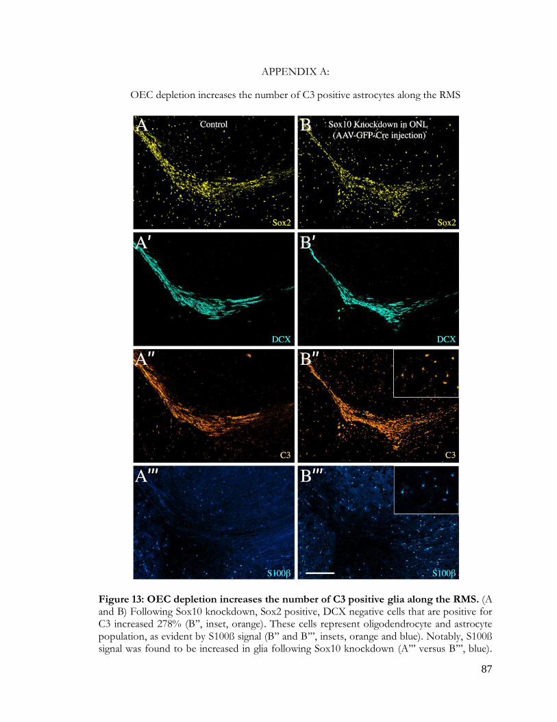
87
Figure 13: OEC depletion increases C3-positive glia in vivo
APPENDIX A:
OEC depletion increases the number of C3 positive astrocytes along the RMS
Figure 13: OEC depletion increases the number of C3 positive glia along the RMS. (A and B) Following Sox10 knockdown, Sox2 positive, DCX negative cells that are positive for C3 increased 278% (B’’, inset, orange). These cells represent oligodendrocyte and astrocyte population, as evident by S100ß signal (B’’ and B’’’, insets, orange and blue). Notably, S100ß signal was found to be increased in glia following Sox10 knockdown (A’’’ versus B’’’, blue).
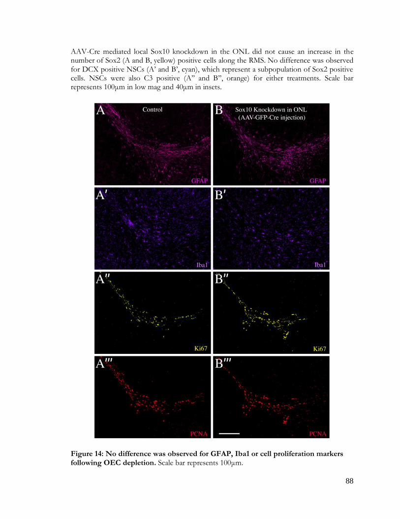
88
Figure 14: No difference was observed for GFAP, Iba1 or cell proliferation markers following
OEC depletion
AAV-Cre mediated local Sox10 knockdown in the ONL did not cause an increase in the number of Sox2 (A and B, yellow) positive cells along the RMS. No difference was observed for DCX positive NSCs (A’ and B’, cyan), which represent a subpopulation of Sox2 positive cells. NSCs were also C3 positive (A’’ and B’’, orange) for either treatments. Scale bar represents 100μm in low mag and 40μm in insets.
Figure 14: No difference was observed for GFAP, Iba1 or cell proliferation markers following OEC depletion. Scale bar represents 100μm.

89
Figure 15: OEC depletion increases astrocyte neurotoxicity in vivo
Figure 15: The majority of C3 positive cells are GFAP positive astrocytes. Characterization of C3 positive cells at the intersection of RMS and the OB. Following local Sox10 knockdown in the OB, C3 staining overlaps with GFAP positive astrocytes (magenta, open arrowheads), Olig2 positive oligodendrocytes (red, arrowheads) and Iba1 positive microglia (blue, asterisk). Oligodendrocytes and astrocytes, but not microglia are Sox2 positive (yellow). Less than 10% C3 positive cells overlapped with microglia marker Iba1 (purple), ~20% overlapped with oligodendrocyte marker Olig2 (red), > 45% overlapped with astrocyte marker GFAP (magenta). Some C3 cells did not overlap with any of these markers but were Sox2 positive (yellow). Scale bar represents 20μm.
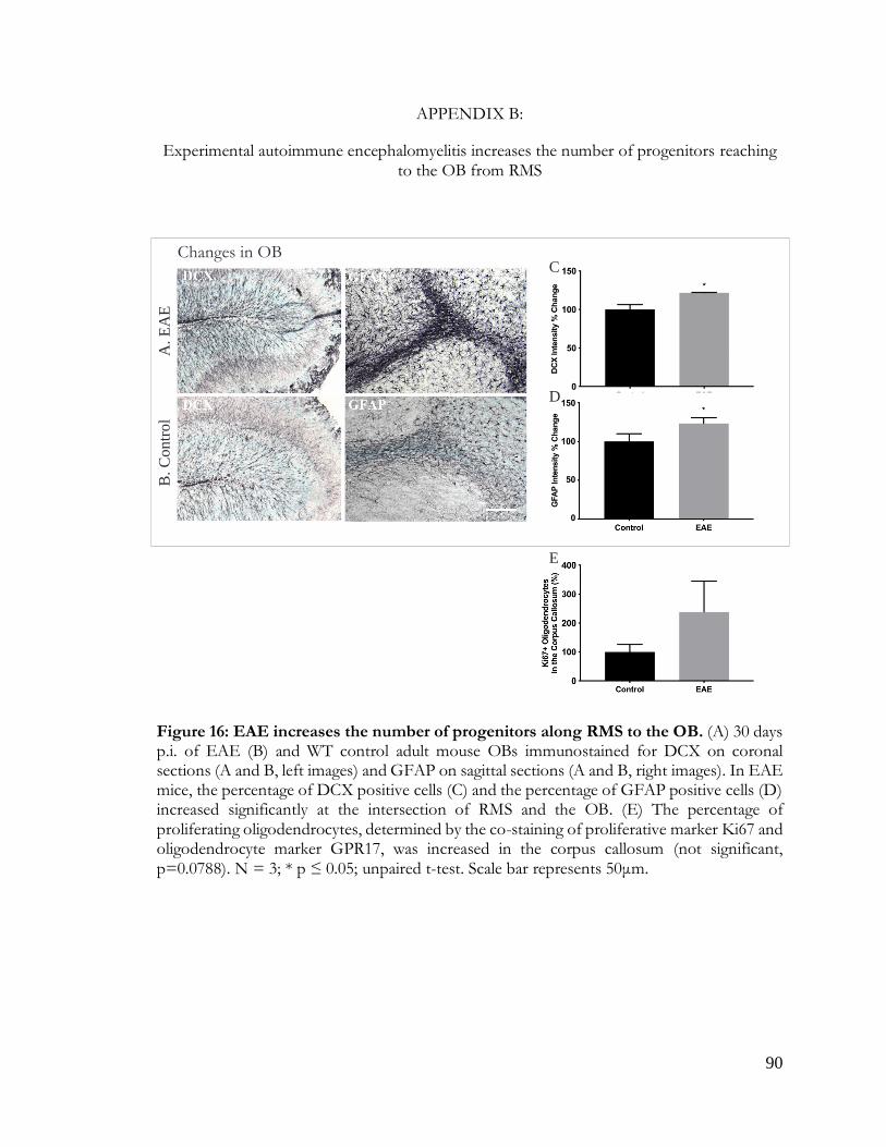
90
Figure 16: EAE increases number of progenitors in the OB
APPENDIX B:
Experimental autoimmune encephalomyelitis increases the number of progenitors reaching to the OB from RMS
Figure 16: EAE increases the number of progenitors along RMS to the OB. (A) 30 days p.i. of EAE (B) and WT control adult mouse OBs immunostained for DCX on coronal sections (A and B, left images) and GFAP on sagittal sections (A and B, right images). In EAE mice, the percentage of DCX positive cells (C) and the percentage of GFAP positive cells (D) increased significantly at the intersection of RMS and the OB. (E) The percentage of proliferating oligodendrocytes, determined by the co-staining of proliferative marker Ki67 and oligodendrocyte marker GPR17, was increased in the corpus callosum (not significant, p=0.0788). N = 3; * p ≤ 0.05; unpaired t-test. Scale bar represents 50μm.
43
Changes in OBC
D
E
A. E
AE
B. C
on
trol

91
APPENDIX C:
Co-stimulation of astrocytes with Anosmin1 and LPS increases astrocyte proliferation
Figure 17: LPS and Anosmin1 have a combinational effect on astrocyte proliferation. Viability test on immortalized astrocyte line C8D30 shows significantly increased proliferation following 24hr 1μl LPS treatment (black dashed lines, rhombus) compared to untreated, control astrocytes (red lines, circle) (top image, 24hr, and bottom image, day1). 5ng/ml Anosmin1 significantly increased astrocyte proliferation on days 3 and 5, while no difference for LPS treated group was observed for these time points (bottom image). Compared to control group, combinational stimulation of LPS and Anosmin1 resulted in increased astrocyte proliferation on day5 (turquoise line, triangle) which was also significantly higher compared to either stimulation alone (bottom image, blue line, triangle, or black dashed line, rhombus) (N=3, * p ≤ 0.05, **p ≤ 0.01, *** p ≤ 0.001, **** p ≤ 0.0001; two-way ANOVA).
Figure 17: Co-stimulation of astrocytes with Anosmin1 & LPS increases
astrocyte proliferation
2hr 6hr 10hr 24hr-0.1
0.0
0.1
0.2
0.3
0.4
Ala
mar
Blu
e A
bsorb
ance
*
*
+2µl LPS
+1µl LPS
Control
*

92
Bibliography:
Addington, C. P., Roussas, A., Dutta, D., & Stabenfeldt, S. E. (2015). Endogenous repair signaling after brain injury and complementary bioengineering approaches to enhance neural regeneration. Biomarker Insights, 10(Suppl 1), 43–60. https://doi.org/10.4137/BMI.S20062
Adolf, A., Rohrbeck, A., Münster-Wandowski, A., Johansson, M., Kuhn, H. G., Kopp, M. A., … Höltje, M. (2019). Release of astroglial vimentin by extracellular vesicles: Modulation of binding and internalization of C3 transferase in astrocytes and neurons. Glia, 67(4), 703–717. https://doi.org/10.1002/glia.23566
Alvarez-Buylla, A., & Lim, D. A. (2004). For the long run: Maintaining germinal niches in the adult brain. Neuron. https://doi.org/10.1016/S0896-6273(04)00111-4
Arac, A., Brownell, S. E., Rothbard, J. B., Chen, C., Ko, R. M., Pereira, M. P., … Steinberg, G. K. (2011). Systemic augmentation of B-crystallin provides therapeutic benefit twelve hours post-stroke onset via immune modulation. Proceedings of the National Academy of Sciences. https://doi.org/10.1073/pnas.1107368108
Au, E., & Roskams, A. J. (2003). Olfactory ensheathing cells of the lamina propria in vivo and in vitro. GLIA, 41(3), 224–236. https://doi.org/10.1002/glia.10160
Au, W. W., Treloar, H. B., & Greer, C. A. (2002). Sublaminar organization of the mouse olfactory bulb nerve layer. Journal of Comparative Neurology. https://doi.org/10.1002/cne.10182
Auciello, G., Cunningham, D. L., Tatar, T., Heath, J. K., & Rappoport, J. Z. (2013). Regulation of fibroblast growth factor receptor signalling and trafficking by Src and Eps8. Journal of Cell Science, 126(Pt 2), 613–624. https://doi.org/10.1242/jcs.116228
Bachoo, R. M., Kim, R. S., Ligon, K. L., Maher, E. A., Brennan, C., Billings, N., … DePinho, R. A. (2004). Molecular diversity of astrocytes with implications for neurological disorders. Proceedings of the National Academy of Sciences of the United States of America. https://doi.org/10.1073/pnas.0402140101
Barnett, S. C., Alexander, C. L., Iwashita, Y., Gilson, J. M., Crowther, J., Clark, L., … Franklin, R. J. (2000). Identification of a human olfactory ensheathing cell that can effect transplant-mediated remyelination of
demyelinated CNS axons. Brain: A Journal of Neurology, 123 (Pt 8, 1581–1588. https://doi.org/10.1093/brain/123.8.1581
Beiersdorfer, A., Wolburg, H., Grawe, J., Scheller, A., Kirchhoff, F., & Lohr, C. (2020). Sublamina-specific organization of the blood brain barrier in the mouse olfactory nerve layer. GLIA. https://doi.org/10.1002/glia.23744
Bignami, A., & Dahl, D. (1976). The Astroglial Response to Stabbing. Immunofluorescence Studies with
Antibodies to Astrocyte‐Specific Protein (GFAP) In Mammalian and Submammalian Vertebrates. Neuropathology and Applied Neurobiology. https://doi.org/10.1111/j.1365-2990.1976.tb00488.x
Boccazzi, M., & Ceruti, S. (2016). Where do you come from and what are you going to become, reactive astrocyte? Stem Cell Investig 2016; 3:15. https://doi.org/10.21037/sci.2016.05.02
Bohlen, C. J., Bennett, F. C., Tucker, A. F., Collins, H. Y., Mulinyawe, S. B., & Barres, B. A. (2017). Diverse Requirements for Microglial Survival, Specification, and Function Revealed by Defined-Medium Cultures. Neuron. https://doi.org/10.1016/j.neuron.2017.04.043
Brady, J. P., Garland, D. L., Green, D. E., Tamm, E. R., Giblin, F. J., & Wawrousek, E. F. (2001). B-Crystallin in Lens Development and Muscle Integrity: A Gene Knockout Approach. Investigative Ophthalmology & Visual Science (Vol. 42).
Brosnan, C. F., & Raine, C. S. (2013). The astrocyte in multiple sclerosis revisited. GLIA. https://doi.org/10.1002/glia.22443
Buffo, A., Rite, I., Tripathi, P., Lepier, A., Colak, D., Horn, A.-P., … Götz, M. (2008). Origin and progeny of reactive gliosis: A source of multipotent cells in the injured brain. Proceedings of the National Academy of Sciences of the United States of America, 105(9), 3581–3586. https://doi.org/10.1073/pnas.0709002105
Bush, T. G., Puvanachandra, N., Horner, C. H., Polito, A., Ostenfeld, T., Svendsen, C. N., … Sofroniew, M. V. (1999). Leukocyte infiltration, neuronal degeneration, and neurite outgrowth after ablation of scar-forming, reactive astrocytes in adult transgenic mice. Neuron. https://doi.org/10.1016/S0896-6273(00)80781-3
Cahoy, J. D., Emery, B., Kaushal, A., Foo, L. C., Zamanian, J. L., Christopherson, K. S., … Barres, B. A. (2008). A transcriptome database for astrocytes, neurons, and oligodendrocytes: A new resource for understanding brain development and function. Journal of Neuroscience. https://doi.org/10.1523/JNEUROSCI.4178-07.2008

93
Calof, A. L., & Guevara, J. L. (1993). Cell lines derived from retrovirus-mediated oncogene transduction into olfactory epithelium cultures. Methods in Neurosciences, 3, 222–231. Retrieved from https://doi.org/10.1006/ncmn.1993.1057
Calof, A. L., Hagiwara, N., Holcomb, J. D., Mumm, J. S., & Shou, J. (1996). Neurogenesis and cell death in olfactory epithelium. Journal of Neurobiology, 30(1), 67–81. https://doi.org/10.1002/(SICI)1097-4695(199605)30:1<67::AID-NEU7>3.0.CO;2-E
Carmen Martinez Garcia, M., Cuervas-Mons Finat, M., Latorre Macarron, E., Bullon Sopelana, M., & Gayoso Rodriguez, M. J. (1991). Responses of the astroglia in sensory deprived olfactory bulb of developing rats. Histology and Histopathology.
Carpenter, G. (2003). Nuclear localization and possible functions of receptor tyrosine kinases. Current Opinion in Cell Biology. https://doi.org/10.1016/S0955-0674(03)00015-2
Ceyzériat, K., Abjean, L., Carrillo-de Sauvage, M. A., Ben Haim, L., & Escartin, C. (2016). The complex STATes of astrocyte reactivity: How are they controlled by the JAK-STAT3 pathway? Neuroscience. https://doi.org/10.1016/j.neuroscience.2016.05.043
Chan, W. S., Sideris, A., Sutachan, J. J., Montoya G, J. V., Blanck, T. J. J., & Recio-Pinto, E. (2013). Differential regulation of proliferation and neuronal differentiation in adult rat spinal cord neural stem/progenitors by ERK1/2, Akt, and PLCγ. Frontiers in Molecular Neuroscience. https://doi.org/10.3389/fnmol.2013.00023
Chen, S., Wassenhove-McCarthy, D. J., Yamaguchi, Y., Holzman, L. B., Van Kuppevelt, T. H., Jenniskens, G. J., … McCarthy, K. J. (2008). Loss of heparan sulfate glycosaminoglycan assembly in podocytes does not lead to proteinuria. Kidney International. https://doi.org/10.1038/ki.2008.159
Cheung, P., Allis, C. D., & Sassone-Corsi, P. (2000). Signaling to chromatin through historic modifications. Cell. https://doi.org/10.1016/S0092-8674(00)00118-5
Choy, C., & Kim, S. H. (2010). Biological actions and interactions of Anosmin-1. In Kallmann Syndrome and Hypogonadotropic Hypogonadism (Vol. 39, pp. 78–93). https://doi.org/10.1159/000312695
Chuah, M. I., Hale, D. M., & West, A. K. (2011). Interaction of olfactory ensheathing cells with other cell types in vitro and after transplantation: Glial scars and inflammation. Experimental Neurology. https://doi.org/10.1016/j.expneurol.2010.08.012
Clarke, L. E., Liddelow, S. A., Chakraborty, C., Münch, A. E., Heiman, M., & Barres, B. A. (2018). Normal aging induces A1-like astrocyte reactivity. Proceedings of the National Academy of Sciences of the United States of America, 115(8), E1896–E1905. https://doi.org/10.1073/pnas.1800165115
Clarke, W. E., Berry, M., Smith, C., Kent, A., & Logan, A. (2001). Coordination of fibroblast growth factor receptor 1 (FGFR1) and fibroblast growth factor-2 (FGF-2) trafficking to nuclei of reactive astrocytes around cerebral lesions in adult rats. Molecular and Cellular Neuroscience. https://doi.org/10.1006/mcne.2000.0920
Clemente, D., Esteban, P. F., Del Valle, I., Bribián, A., Soussi-Yanicostas, N., Silva, A., & De Castro, F. (2008). Expression pattern of Anosmin-1 during pre- and postnatal rat brain development. Developmental
Dynamics: An Official Publication of the American Association of Anatomists, 237(9), 2518–2528. https://doi.org/10.1002/dvdy.21659
Cross, M. J., Lu, L., Magnusson, P., Nyqvist, D., Holmqvist, K., Welsh, M., & Claesson-Welsh, L. (2002). The Shb adaptor protein binds to tyrosine 766 in the FGFR-1 and regulates the Ras/MEK/MAPK pathway via FRS2 phosphorylation in endothelial cells. Molecular Biology of the Cell. https://doi.org/10.1091/mbc.E02-02-0103
D’Agostino, M., Lemma, V., Chesi, G., Stornaiuolo, M., Serio, M. C., D’Ambrosio, C., … Bonatti, S. (2013). The cytosolic chaperone α-crystallin B rescues folding and compartmentalization of misfolded multispan transmembrane proteins. Journal of Cell Science, 126(18), 4160–4172. https://doi.org/10.1242/jcs.125443
Dailey, L., Ambrosetti, D., Mansukhani, A., & Basilico, C. (2005). Mechanisms underlying differential responses to FGF signaling. Cytokine & Growth Factor Reviews, 16(2), 233–247. https://doi.org/10.1016/j.cytogfr.2005.01.007
Dairaghi, L., Flannery, E., Giacobini, P., Saglam, A., Saadi, H., Constantin, S., … Wray, S. (2018). Reelin can modulate migration of olfactory ensheathing cells and gonadotropin releasing hormone neurons via the canonical pathway. Frontiers in Cellular Neuroscience. https://doi.org/10.3389/fncel.2018.00228
Dando, S. J., Ipe, D. S., Batzloff, M., Sullivan, M. J., Crossman, D. K., Crowley, M., … Ulett, G. C. (2016). Burkholderia pseudomallei capsule exacerbates respiratory melioidosis but does not afford protection against antimicrobial signaling or bacterial killing in human olfactory ensheathing cells. Infection and Immunity. https://doi.org/10.1128/IAI.01546-15

94
de Castro, F., Seal, R., & Maggi, R. (2017). ANOS1: A unified nomenclature for Kallmann syndrome 1 gene (KAL1) and Anosmin-1. Briefings in Functional Genomics, 16(4), 205–210. https://doi.org/10.1093/bfgp/elw037
De Cecco, M., Jeyapalan, J., Zhao, X., Tamamori-Adachi, M., & Sedivy, J. M. (2011). Nuclear protein accumulation in cellular senescence and organismal aging revealed with a novel single-cell resolution fluorescence microscopy assay. Aging. https://doi.org/10.18632/aging.100372
Dellovade, T. L., Hardelin, J. P., Soussi-Yanicostas, N., Pfaff, D. W., Schwanzel-Fukuda, M., & Petit, C. (2003). Anosmin-1 immunoreactivity during embryogenesis in a primitive eutherian mammal. Developmental Brain Research. https://doi.org/10.1016/S0165-3806(02)00544-8
Deumens, R., Koopmans, G. C., Honig, W. M. M., Hamers, F. P. T., Maquet, V., … Joosten, E. A. J. (2006). Olfactory ensheathing cells, olfactory nerve fibroblasts and biomatrices to promote long-distance axon regrowth and functional recovery in the dorsally hemisected adult rat spinal cord. Experimental Neurology, 200(1), 89–103. https://doi.org/10.1016/j.expneurol.2006.01.030
DM Hale. (2011). Olfactory ensheathing cells moderate astrocyte inflammatory activation. Doetsch, F., Caille, I., Lim, D. A., Garcia-Verdugo, J. M., & Alvarez-Buylla, A. (1999). Subventricular zone
astrocytes are neural stem cells in the adult mammalian brain. Cell. https://doi.org/10.1016/S0092-8674(00)80783-7
Donato, R. (2001). S100: a multigenic family of calcium-modulated proteins of the EF-hand type with intracellular and extracellular functional roles. The International Journal of Biochemistry & Cell Biology (Vol. 33). Retrieved from www.elsevier.com/locate/ijbcb*
Doucette, R., & Words, K. E. Y. (1990). Glial influences on axonal growth in the primary olfactory system. Glia, 3(6), 433–449. https://doi.org/10.1002/glia.440030602
Doyle, J. P., Dougherty, J. D., Heiman, M., Schmidt, E. F., Stevens, T. R., Ma, G., … Heintz, N. (2008). Application of a Translational Profiling Approach for the Comparative Analysis of CNS Cell Types. Cell. https://doi.org/10.1016/j.cell.2008.10.029
Dulle, J. E., & Fort, P. E. (2016). Crystallins and neuroinflammation: The glial side of the story. Biochimica et Biophysica Acta - General Subjects, 1860(1), 278–286. https://doi.org/10.1016/j.bbagen.2015.05.023
Dunham-Ems, S. M., Pudavar, H. E., Myers, J. M., Maher, P. A., Prasad, P. N., & Stachowiak, M. K. (2006). Factors Controlling Fibroblast Growth Factor Receptor-1’s Cytoplasmic Trafficking and Its Regulation as Revealed by FRAP Analysis. Molecular Biology of the Cell. https://doi.org/10.1091/mbc.E05-08-0749
Dye, D. E., Medic, S., Ziman, M., & Coombe, D. R. (2013). Melanoma biomolecules: Independently identified but functionally intertwined. Frontiers in Oncology. https://doi.org/10.3389/fonc.2013.00252
Emsley, J. G., Menezes, J. R. L., Madeiro Da Costa, R. F., Martinez, A. M. B., & Macklis, J. D. (2012). Identification of radial glia-like cells in the adult mouse olfactory bulb. Experimental Neurology. https://doi.org/10.1016/j.expneurol.2012.05.012
Endo, Y., Ishiwata-Endo, H., & Yamada, K. M. (2012). Extracellular Matrix Protein Anosmin Promotes Neural Crest Formation and Regulates FGF, BMP, and WNT Activities. Developmental Cell, 23(2), 305–316. https://doi.org/10.1016/j.devcel.2012.07.006
Fahmy, G. H., & Moftah, M. Z. (2010). FGF-2 in astroglial cells during vertebrate spinal cord recovery. Frontiers in Cellular Neuroscience. https://doi.org/10.3389/fncel.2010.00129
Faiz, M., Sachewsky, N., Gascón, S., Bang, K. W. A., Morshead, C. M., & Nagy, A. (2015). Adult Neural Stem Cells from the Subventricular Zone Give Rise to Reactive Astrocytes in the Cortex after Stroke. Cell Stem Cell, 17(5), 624–634. https://doi.org/10.1016/j.stem.2015.08.002
Fang, X., Stachowiak, E. K., Dunham-Ems, S. M., Klejbor, I., & Stachowiak, M. K. (2005). Control of CREB-binding protein signaling by nuclear fibroblast growth factor receptor-1: A novel mechanism of gene regulation. Journal of Biological Chemistry. https://doi.org/10.1074/jbc.M504400200
Faulkner, J. R., Herrmann, J. E., Woo, M. J., Tansey, K. E., Doan, N. B., & Sofroniew, M. V. (2004). Reactive Astrocytes Protect Tissue and Preserve Function after Spinal Cord Injury. Journal of Neuroscience. https://doi.org/10.1523/JNEUROSCI.3547-03.2004
Finzsch, M., Schreiner, S., Kichko, T., Reeh, P., Tamm, E. R., Bösl, M. R., … Wegner, M. (2010). Sox10 is required for Schwann cell identity and progression beyond the immature Schwann cell stage. Journal of Cell Biology. https://doi.org/10.1083/jcb.200912142
Foo, L. C., Allen, N. J., Bushong, E. A., Ventura, P. B., Chung, W. S., Zhou, L., … Barres, B. A. (2011). Development of a method for the purification and culture of rodent astrocytes. Neuron. https://doi.org/10.1016/j.neuron.2011.07.022

95
Forni, P. E., Bharti, K., Flannery, E. M., Shimogori, T., & Wray, S. (2013). The indirect role of fibroblast growth factor-8 in defining neurogenic niches of the olfactory/GnRH systems. Journal of Neuroscience, 33(50), 19620–19634. https://doi.org/10.1523/JNEUROSCI.3238-13.2013
Franssen, E. H. P., De Bree, F. M., Essing, A. H. W., Ramon-Cueto, A., & Verhaagen, J. (2008). Comparative gene expression profiling of olfactory ensheathing glia and Schwann cells indicates distinct tissue repair characteristics of olfactory ensheathing glia. GLIA, 56(12), 1285–1298. https://doi.org/10.1002/glia.20697
Fuhrmann, V., Kinkl, N., Leveillard, T., Sahel, J., & Hicks, D. (1999). Fibroblast growth factor receptor 4 (FGFR4) is expressed in adult rat and human retinal photoreceptors and neurons. Journal of Molecular Neuroscience. https://doi.org/10.1385/jmn:13:1-2:187
Furdui, C. M., Lew, E. D., Schlessinger, J., & Anderson, K. S. (2006). Autophosphorylation of FGFR1 kinase is mediated by a sequential and precisely ordered reaction. Molecular Cell. https://doi.org/10.1016/j.molcel.2006.01.022
García-González, D., Clemente, D., Coelho, M., Esteban, P. F., Soussi-Yanicostas, N., & de Castro, F. (2010). Dynamic roles of FGF-2 and Anosmin-1 in the migration of neuronal precursors from the subventricular zone during pre- and postnatal development. Experimental Neurology. https://doi.org/10.1016/j.expneurol.2010.01.006
García-Marqués, J., & López-Mascaraque, L. (2017). Clonal Mapping of Astrocytes in the Olfactory Bulb and
Rostral Migratory Stream. Cerebral Cortex (New York, N.Y. : 1991). https://doi.org/10.1093/cercor/bhw071
Gianola, S., de Castro, F., & Rossi, F. (2009). Anosmin-1 stimulates outgrowth and branching of developing Purkinje axons. Neuroscience, 158(2), 570–584. https://doi.org/10.1016/j.neuroscience.2008.10.022
Girouard, H., & Iadecola, C. (2006). Neurovascular coupling in the normal brain and in hypertension, stroke,
and Alzheimer disease. Journal of Applied Physiology (Bethesda, Md. : 1985), 100(1), 328–335. https://doi.org/10.1152/japplphysiol.00966.2005
Göktaş, Ö., Cao Van, H., Fleiner, F., Lacroix, J. S., & Landis, B. N. (2010). Chemosensory function in Wegener’s granulomatosis: A preliminary report. European Archives of Oto-Rhino-Laryngology. https://doi.org/10.1007/s00405-009-1184-4
Goldshmit, Y., Frisca, F., Pinto, A. R., Pébay, A., Tang, J. K. K. Y., Siegel, A. L., … Currie, P. D. (2014). Fgf2 improves functional recovery-decreasing gliosis and increasing radial glia and neural progenitor cells after spinal cord injury. Brain and Behavior. https://doi.org/10.1002/brb3.172
Gómez-Pinilla, F., Vu, L., & Cotman, C. W. (1995). Regulation of astrocyte proliferation by FGF-2 and heparan
sulfate in vivo. The Journal of Neuroscience : The Official Journal of the Society for Neuroscience, 15(March), 2021–2029.
Gómez, R. M., Sánchez, M. Y., Portela-Lomba, M., Ghotme, K., Barreto, G. E., Sierra, J., & Moreno-Flores, M. T. (2018). Cell therapy for spinal cord injury with olfactory ensheathing glia cells (OECs). Glia, 66(7), 1267–1301. https://doi.org/10.1002/glia.23282
Grimpe, B., Pressman, Y., Bunge, M. B., & Silver, J. (2005). The role of proteoglycans in Schwann cell/astrocyte interactions and in regeneration failure at PNS/CNS interfaces. Molecular and Cellular Neuroscience. https://doi.org/10.1016/j.mcn.2004.06.010
Gulbenkian, S., Uddman, R., & Edvinsson, L. (2001). Neuronal messengers in the human cerebral circulation. Peptides, 22(6), 995–1007. https://doi.org/10.1016/S0196-9781(01)00408-9
Guntinas-Lichius, O., Angelov, D. N., Tomov, T. L., Dramiga, J., Neiss, W. F., & Wewetzer, K. (2001). Transplantation of olfactory ensheathing cells stimulates the collateral sprouting from axotomized adult rat facial motoneurons. Experimental Neurology, 172(1), 70–80. https://doi.org/10.1006/exnr.2001.7774\rS0014-4886(01)97774-X [pii]
Guo, Y. S., Liang, P. Z., Lu, S. Z., Chen, R., Yin, Y. Q., & Zhou, J. W. (2019). Extracellular αB-crystallin modulates the inflammatory responses. Biochemical and Biophysical Research Communications, 508(1), 282–288. https://doi.org/10.1016/j.bbrc.2018.11.024
Gupta, A., & Pulliam, L. (2014). Exosomes as mediators of neuroinflammation. Journal of Neuroinflammation, 11, 1–10. https://doi.org/10.1186/1742-2094-11-68
György, B., Szabó, T. G., Pásztói, M., Pál, Z., Misják, P., Aradi, B., … Buzás, E. I. (2011). Membrane vesicles, current state-of-the-art: Emerging role of extracellular vesicles. Cellular and Molecular Life Sciences. https://doi.org/10.1007/s00018-011-0689-3
Hale, D. M., Ray, S., Leung, J. Y., Holloway, A. F., Chung, R. S., West, A. K., & Chuah, M. I. (2011). Olfactory ensheathing cells moderate nuclear factor kappaB translocation in astrocytes. Molecular and Cellular Neuroscience. https://doi.org/10.1016/j.mcn.2010.09.004

96
Hamby, M. E., Coppola, G., Ao, Y., Geschwind, D. H., Khakh, B. S., & Sofroniew, M. V. (2012). Inflammatory Mediators Alter the Astrocyte Transcriptome and Calcium Signaling Elicited by Multiple G-Protein-Coupled Receptors. Journal of Neuroscience. https://doi.org/10.1523/JNEUROSCI.1256-12.2012
He, F., Ge, W., Martinowich, K., Becker-Catania, S., Coskun, V., Zhu, W., … Sun, Y. E. (2005). A positive autoregulatory loop of Jak-STAT signaling controls the onset of astrogliogenesis. Nature Neuroscience. https://doi.org/10.1038/nn1440
Hegedus, B., Dasgupta, B., Shin, J. E., Emnett, R. J., Hart-Mahon, E. K., Elghazi, L., … Gutmann, D. H. (2007). Neurofibromatosis-1 Regulates Neuronal and Glial Cell Differentiation from Neuroglial Progenitors In Vivo by Both cAMP- and Ras-Dependent Mechanisms. Cell Stem Cell. https://doi.org/10.1016/j.stem.2007.07.008
Hellström, N. A. K., Björk-Eriksson, T., Blomgren, K., & Kuhn, H. G. (2009). Differential Recovery of Neural Stem Cells in the Subventricular Zone and Dentate Gyrus After Ionizing Radiation. Stem Cells. https://doi.org/10.1634/stemcells.2008-0732
Herrmann, J. E., Imura, T., Song, B., Qi, J., Ao, Y., Nguyen, T. K., … Sofroniew, M. V. (2008). STAT3 is a critical regulator of astrogliosis and scar formation after spinal cord injury. Journal of Neuroscience. https://doi.org/10.1523/JNEUROSCI.1709-08.2008
Higginson, J. R., Thompson, S. M., Santos-Silva, A., Guimond, S. E., Turnbull, J. E., & Barnett, S. C. (2012). Differential Sulfation Remodelling of Heparan Sulfate by Extracellular 6-O-Sulfatases Regulates Fibroblast Growth Factor-Induced Boundary Formation by Glial Cells: Implications for Glial Cell Transplantation. Journal of Neuroscience. https://doi.org/10.1523/JNEUROSCI.6340-11.2012
Holt, L. M., Stoyanof, S. T., & Olsen, M. L. (2019). Magnetic Cell Sorting for In Vivo and In Vitro Astrocyte, Neuron, and Microglia Analysis. Current Protocols in Neuroscience. https://doi.org/10.1002/cpns.71
Hong, S., & Song, M. R. (2014). STAT3 but not STAT1 is required for astrocyte differentiation. PLoS ONE, 9(1), 1–9. https://doi.org/10.1371/journal.pone.0086851
Horner, P. J., & Palmer, T. D. (2003). New roles for astrocytes: The nightlife of an “astrocyte”. La vida loca! Trends in Neurosciences. https://doi.org/10.1016/j.tins.2003.09.010
Horner, P. J., Power, A. E., Kempermann, G., Kuhn, H. G., Palmer, T. D., Winkler, J., … Gage, F. H. (2000). Proliferation and differentiation of progenitor cells throughout the intact adult rat spinal cord. Journal of Neuroscience. https://doi.org/10.1523/jneurosci.20-06-02218.2000
Hu, Y., & Bouloux, P. M. (2011). X-linked GnRH deficiency: Role of KAL-1 mutations in GnRH deficiency. Molecular and Cellular Endocrinology. https://doi.org/10.1016/j.mce.2011.04.001
Hu, Y., Butts, T., Poopalasundaram, S., Graham, A., & Bouloux, P. M. (2019). Extracellular matrix protein anosmin-1 modulates olfactory ensheathing cell maturation in chick olfactory bulb development. European Journal of Neuroscience, 50(9), 3472–3486. https://doi.org/10.1111/ejn.14483
Hu, Y., Guimond, S. E., Travers, P., Cadman, S., Hohenester, E., Turnbull, J. E., … Bouloux, P.-M. (2009). Novel mechanisms of fibroblast growth factor receptor 1 regulation by extracellular matrix protein anosmin-1. The Journal of Biological Chemistry, 284(43), 29905–29920. https://doi.org/10.1074/jbc.M109.049155
Huang, Z. H., Wang, Y., Cao, L., Su, Z. Da, Zhu, Y. L., Chen, Y. Z., … He, C. (2008). Migratory properties of cultured olfactory ensheathing cells by single-cell migration assay. Cell Research. https://doi.org/10.1038/cr.2008.38
Huart, C., Rombaux, P., & Hummel, T. (2019). Neural plasticity in developing and adult olfactory pathways – focus on the human olfactory bulb. Journal of Bioenergetics and Biomembranes. https://doi.org/10.1007/s10863-018-9780-x
Imaizumi, T., Lankford, K. L., Burton, W. V., Fodor, W. L., & Kocsis, J. D. (2000). Xenotransplantation of transgenic pig olfactory ensheathing cells promotes axonal regeneration in rat spinal cord. Nature Biotechnology. https://doi.org/10.1038/79432
Inatani, M., Irie, F., Plump, A. S., Tessier-Lavigne, M., & Yamaguchi, Y. (2003). Mammalian Brain Morphogenesis and Midline Axon Guidance Require Heparan Sulfate. Science. https://doi.org/10.1126/science.1090497
Irie, F., Badie-Mahdavi, H., & Yamaguchi, Y. (2012). Autism-like socio-communicative deficits and stereotypies in mice lacking heparan sulfate. Proceedings of the National Academy of Sciences of the United States of America, 109(13), 5052–5056. https://doi.org/10.1073/pnas.1117881109
Irschick, R., Trost, T., Karp, G., Hausott, B., Auer, M., Claus, P., & Klimaschewski, L. (2013). Sorting of the FGF receptor 1 in a human glioma cell line. Histochemistry and Cell Biology. https://doi.org/10.1007/s00418-012-1009-1

97
Jeppesen, D. K., Fenix, A. M., Franklin, J. L., Higginbotham, J. N., Zhang, Q., Zimmerman, L. J., … Coffey, R. J. (2019). Reassessment of Exosome Composition. Cell, 177(2), 428-445.e18. https://doi.org/10.1016/j.cell.2019.02.029
Johansson, S., Lee, I. H., Olson, L., & Spenger, C. (2005). Olfactory ensheathing glial co-grafts improve functional recovery in rats with 6-OHDA lesions. Brain, 128(12), 2961–2976. https://doi.org/10.1093/brain/awh644
John, G. R., Lee, S. C., Song, X., Rivieccio, M., & Brosnan, C. F. (2005). IL-1-regulated responses in astrocytes: Relevance to injury and recovery. GLIA. https://doi.org/10.1002/glia.20109
Joy, A., Moffett, J., Neary, K., Mordechai, E., Stachowiak, E. K., Coons, S., … Stachowiak, M. K. (1997). Nuclear accumulation of FGF-2 is associated with proliferation of human astrocytes and glioma cells. Oncogene. https://doi.org/10.1038/sj.onc.1200823
Joyce, D., Albanese, C., Steer, J., Fu, M., Bouzahzah, B., & Pestell, R. G. (2001). NF-kB and cell-cycle regulation: The cyclin connection. Cytokine and Growth Factor Reviews. https://doi.org/10.1016/S1359-6101(00)00018-6
Jung, S. H., Lee, H. C., Yu, D. M., Kim, B. C., Park, S. M., Lee, Y. S., … Lee, J. S. (2016). Heparan sulfation is essential for the prevention of cellular senescence. Cell Death and Differentiation. https://doi.org/10.1038/cdd.2015.107
Kalra, H., Simpson, R. J., Ji, H., Aikawa, E., Altevogt, P., Askenase, P., … Mathivanan, S. (2012). Vesiclepedia: A Compendium for Extracellular Vesicles with Continuous Community Annotation. PLoS Biology. https://doi.org/10.1371/journal.pbio.1001450
Kanda, T. (2013). Biology of the blood-nerve barrier and its alteration in immune mediated neuropathies. Journal of Neurology, Neurosurgery and Psychiatry. https://doi.org/10.1136/jnnp-2012-302312
Kang, W., Balordi, F., Su, N., Chen, L., Fishell, G., & Hebert, J. M. (2014). Astrocyte activation is suppressed in both normal and injured brain by FGF signaling. Proceedings of the National Academy of Sciences. https://doi.org/10.1073/pnas.1320401111
Kang, W., Wong, L. C., Shi, S.-H., & Hébert, J. M. (2009). The transition from radial glial to intermediate
progenitor cell is inhibited by FGF signaling during corticogenesis. The Journal of Neuroscience: The Official Journal of the Society for Neuroscience, 29(46), 14571–14580. https://doi.org/10.1523/JNEUROSCI.3844-09.2009
Kanski, R., Van Strien, M. E., Van Tijn, P., & Hol, E. M. (2014). A star is born: New insights into the mechanism of astrogenesis. Cellular and Molecular Life Sciences. https://doi.org/10.1007/s00018-013-1435-9
Keerthikumar, S., Chisanga, D., Ariyaratne, D., Al Saffar, H., Anand, S., Zhao, K., … Mathivanan, S. (2016). ExoCarta: A Web-Based Compendium of Exosomal Cargo. Journal of Molecular Biology. https://doi.org/10.1016/j.jmb.2015.09.019
Khakh, B. S., & Deneen, B. (2019). The Emerging Nature of Astrocyte Diversity. Annual Review of Neuroscience. https://doi.org/10.1146/annurev-neuro-070918-050443
Kim, D. K., Lee, J., Kim, S. R., Choi, D. S., Yoon, Y. J., Kim, J. H., … Gho, Y. S. (2015). EVpedia: A community web portal for extracellular vesicles research. Bioinformatics. https://doi.org/10.1093/bioinformatics/btu741
Kim, H. H., Puche, A. C., & Margolis, F. L. (2006). Odorant deprivation reversibly modulates transsynaptic changes in the NR2B-mediated CREB pathway in mouse piriform cortex. Journal of Neuroscience. https://doi.org/10.1523/JNEUROSCI.1727-06.2006
Kirby, E. D., Muroy, S. E., Sun, W. G., Covarrubias, D., Leong, M. J., Barchas, L. A., & Kaufer, D. (2013). Acute stress enhances adult rat hippocampal neurogenesis and activation of newborn neurons via secreted astrocytic FGF2. ELife. https://doi.org/10.7554/eLife.00362
Klenke, U., & Taylor-Burds, C. (2012). Culturing embryonic nasal explants for developmental and physiological study. Current Protocols in Neuroscience. https://doi.org/10.1002/0471142301.ns0325s59
Klopstein, A., Santos-Nogueira, E., Francos-Quijorna, I., Redensek, A., David, S., Navarro, X., & López-Vales, R. (2012). Beneficial effects of αB-crystallin in spinal cord contusion injury. Journal of Neuroscience, 32(42), 14478–14488. https://doi.org/10.1523/JNEUROSCI.0923-12.2012
Kore, R. A., & Abraham, E. C. (2014). Inflammatory cytokines, interleukin-1 beta and tumor necrosis factor-alpha, upregulated in glioblastoma multiforme, raise the levels of CRYAB in exosomes secreted by U373 glioma cells. Biochemical and Biophysical Research Communications. https://doi.org/10.1016/j.bbrc.2014.09.068
Kore, R. A., & Abraham, E. C. (2016). Phosphorylation negatively regulates exosome mediated secretion of CryAB in glioma cells. Biochimica et Biophysica Acta - Molecular Cell Research, 1863(2), 368–377. https://doi.org/10.1016/j.bbamcr.2015.11.027

98
Kuipers, H. F., Yoon, J., Van Horssen, J., Han, M. H., Bollyky, P. L., Palmer, T. D., & Steinman, L. (2017). Phosphorylation of αB-crystallin supports reactive astrogliosis in demyelination. Proceedings of the National Academy of Sciences of the United States of America, 114(9), E1745–E1754. https://doi.org/10.1073/pnas.1621314114
Lakatos, A., Barnett, S. C., & Franklin, R. J. M. (2003). Olfactory ensheathing cells induce less host astrocyte response and chondroitin sulphate proteoglycan expression than Schwann cells following transplantation into adult CNS white matter. Experimental Neurology. https://doi.org/10.1016/S0014-4886(03)00270-X
Lakatos, A., Franklin, R. J. M., & Barnett, S. C. (2000). Olfactory ensheathing cells and Schwann cells differ in their in vitro interactions with astrocytes. GLIA. https://doi.org/10.1002/1098-1136(200012)32:3<214::AID-GLIA20>3.0.CO;2-7
Lanner, F., & Rossant, J. (2010). The role of FGF/Erk signaling in pluripotent cells. Development. https://doi.org/10.1242/dev.050146
Larriva-Sahd, J. (2014). Structural variation and interactions among astrocytes of the rostral migratory stream and olfactory bulb: II. Golgi and electron microscopic study of the adult rat. Neuroscience Research. https://doi.org/10.1016/j.neures.2014.08.011
Le Roy, C., & Wrana, J. L. (2005). Clathrin- and non-clathrin-mediated endocytic regulation of cell signalling. Nature Reviews Molecular Cell Biology. https://doi.org/10.1038/nrm1571
Leadbeater, W. E., Gonzalez, A. M., Logaras, N., Berry, M., Turnbull, J. E., & Logan, A. (2006). Intracellular trafficking in neurones and glia of fibroblast growth factor-2, fibroblast growth factor receptor 1 and heparan sulphate proteoglycans in the injured adult rat cerebral cortex. Journal of Neurochemistry, 96(4), 1189–1200. https://doi.org/10.1111/j.1471-4159.2005.03632.x
Li, J., Chen, W., Li, Y., Chen, Y., Ding, Z., Yang, D., & Zhang, X. (2015). Transplantation of olfactory ensheathing cells promotes partial recovery in rats with experimental autoimmune encephalomyelitis. International Journal of Clinical and Experimental Pathology.
Li, Y., Decherchi, P., & Raisman, G. (2003). Transplantation of olfactory ensheathing cells into spinal cord lesions
restores breathing and climbing. The Journal of Neuroscience: The Official Journal of the Society for Neuroscience, 23(3), 727–731. https://doi.org/23/3/727 [pii]
Li, Y., Field, P. M., & Raisman, G. (2005). Olfactory ensheathing cells and olfactory nerve fibroblasts maintain continuous open channels for regrowth of olfactory nerve fibres. GLIA, 52(3), 245–251. https://doi.org/10.1002/glia.20241
Li, Y., Field, P. M., & Raisman, G. (1997). Repair of adult rat corticospinal tract by transplants of olfactory ensheathing cells. Science, 277(5334), 2000–2002. https://doi.org/10.1126/science.277.5334.2000
Li, Y., Li, D., Ibrahim, A., & Raisman, G. (2012). Repair involves all three surfaces of the glial cell. In Progress in Brain Research. https://doi.org/10.1016/B978-0-444-59544-7.00010-X
Li, Z., Theus, M. H., & Wei, L. (2006). Role of ERK 1/2 signaling in neuronal differentiation of cultured embryonic stem cells. Development Growth and Differentiation. https://doi.org/10.1111/j.1440-169X.2006.00889.x
Li, Z., Theus, M. H., & Wei, L. (2006). Role of ERK 1/2 signaling in neuronal differentiation of cultured embryonic stem cells. Development Growth and Differentiation. https://doi.org/10.1111/j.1440-169X.2006.00889.x
Lian, H., Yang, L., Cole, A., Sun, L., Chiang, A. C. A., Fowler, S. W., … Zheng, H. (2015). NFκB-Activated Astroglial Release of Complement C3 Compromises Neuronal Morphology and Function Associated with Alzheimer’s Disease. Neuron. https://doi.org/10.1016/j.neuron.2014.11.018
Liddelow, S. A., & Barres, B. A. (2017). Reactive Astrocytes: Production, Function, and Therapeutic Potential. Immunity, 46(6), 957–967. https://doi.org/10.1016/j.immuni.2017.06.006
Liddelow, S. A., Guttenplan, K. A., Clarke, L. E., Bennett, F. C., Bohlen, C. J., Schirmer, L., … Barres, B. A. (2017). Neurotoxic reactive astrocytes are induced by activated microglia. Nature, 541(7638), 481–487. https://doi.org/10.1038/nature21029
Lu, J., Féron, F., Mackay‐Sim, A., & Waite, P. M. E. (2002). Olfactory ensheathing cells promote locomotor recovery after delayed transplantation into transected spinal cord. Brain, 125(1), 14–21. https://doi.org/10.1093/brain/awf014
Ma, D. K., Ponnusamy, K., Song, M. R., Ming, G. L., & Song, H. (2009). Molecular genetic analysis of FGFR1 signalling reveals distinct roles of MAPK and PLC1 activation for self-renewal of adult neural stem cells. Molecular Brain, 2(1), 1–14. https://doi.org/10.1186/1756-6606-2-16
Mackay-Sim, A., & Kittel, P. (1991). Cell dynamics in the adult mouse olfactory epithelium: A quantitative autoradiographic study. Journal of Neuroscience. https://doi.org/10.1523/jneurosci.11-04-00979.1991

99
Magnusson, J. P., & Frisen, J. (2016). Stars from the darkest night: unlocking the neurogenic potential of astrocytes in different brain regions. Development. https://doi.org/10.1242/dev.133975
Magnusson, J. P., Göritz, C., Tatarishvili, J., Dias, D. O., Smith, E. M. K., Lindvall, O., … Frisén, J. (2014). A latent neurogenic program in astrocytes regulated by Notch signaling in the mouse. Science. https://doi.org/10.1126/science.346.6206.237
Maric, D., Fiorio Pla, A., Chang, Y. H., & Barker, J. L. (2007). Self-renewing and differentiating properties of cortical neural stem cells are selectively regulated by basic fibroblast growth factor (FGF) signaling via
specific FGF receptors. The Journal of Neuroscience: The Official Journal of the Society for Neuroscience, 27(8), 1836–1852. https://doi.org/10.1523/JNEUROSCI.5141-06.2007
McKhann, G. M., D’Ambrosio, R., & Janigro, D. (1997). Heterogeneity of astrocyte resting membrane potentials and intercellular coupling revealed by whole-cell and gramicidin-perforated patch recordings from cultured
neocortical and hippocampal slice astrocytes. The Journal of Neuroscience: The Official Journal of the Society for Neuroscience, 17(18), 6850–6863. https://doi.org/MO1
Mohammadi, M., Dionne, C. A., Li, W., Li, N., Spivak, T., Honegger, A. M., … Schlessinger, J. (1992). Point mutation in FGF receptor eliminates phosphatidylinositol hydrolysis without affecting mitogenesis. Nature. https://doi.org/10.1038/358681a0
Molofsky, A.V., Glasgow, S.M., Chaboub, L.S., Tsai, H.H., Murnen, A.T., Kelley, K.W., Fancy, S.P.J., Yuen, T.J., Madireddy, L., Baranzini, S., et al. (n.d.). Expression profiling of Aldh1l1-precursors in the developing spinal cord reveals glial lineage-specific genes and direct Sox9-Nfe2l1 interactions. Glia 61, 1518–1532.
Molofsky, A. V., & Deneen, B. (2015). Astrocyte development: A Guide for the Perplexed. Glia, 63(8), 1320–1329. https://doi.org/10.1002/glia.22836
Müller, G. (2012). Microvesicles/exosomes as potential novel biomarkers of metabolic diseases. Diabetes, Metabolic Syndrome and Obesity: Targets and Therapy. https://doi.org/10.2147/dmso.s32923
Muranova, L. K., Sudnitsyna, M. V., & Gusev, N. B. (2018). αB-Crystallin Phosphorylation: Advances and Problems. Biochemistry (Moscow). https://doi.org/10.1134/S000629791810005X
Murtaza, M., Chacko, A., Delbaz, A., Reshamwala, R., Rayfield, A., McMonagle, B., … Ekberg, J. A. K. (2019). Why are olfactory ensheathing cell tumors so rare? Cancer Cell International. https://doi.org/10.1186/s12935-019-0989-5
Nash, H. H., Borke, R. C., & Anders, J. J. (2001). New Method of Purification for Establishing Primary Cultures of Ensheathing Cells from the Adult Olfactory Bulb. GLIA (Vol. 34).
Nato, G., Caramello, a., Trova, S., Avataneo, V., Rolando, C., Taylor, V., … Luzzati, F. (2015). Striatal astrocytes produce neuroblasts in an excitotoxic model of Huntington’s disease. Development, 142(5), 840–845. https://doi.org/10.1242/dev.116657
Nazareth, L., Chen, M., Shelper, T., Shah, M., Tello Velasquez, J., Walkden, H., … Ekberg, J. A. K. (2019). Novel insights into the glia limitans of the olfactory nervous system. Journal of Comparative Neurology, 527(7), 1228–1244. https://doi.org/10.1002/cne.24618
Nikolaychik, V. V., Samet, M. M., & Lelkes, P. I. (1996). A new method for continual quantitation of viable cells on endothelialized polyurethanes. Journal of Biomaterials Science, Polymer Edition. https://doi.org/10.1163/156856296X00057
O’Toole, D. A., West, A. K., & Chuah, M. I. (2007). Effect of olfactory ensheathing cells on reactive astrocytes in vitro. Cellular and Molecular Life Sciences. https://doi.org/10.1007/s00018-007-7106-y
Ohkubo, Y., Uchida, A. O., Shin, D., Partanen, J., & Vaccarino, F. M. (2004). Fibroblast growth factor receptor 1 is required for the proliferation of hippocampal progenitor cells and for hippocampal growth in mouse.
The Journal of Neuroscience: The Official Journal of the Society for Neuroscience, 24(27), 6057–6069. https://doi.org/10.1523/JNEUROSCI.1140-04.2004
Ornitz, D. M., & Itoh, N. (2015). Advanced Review: The Fibroblast Growth Factor Signaling Pathway. WIREs Dev Biol, 4, 215–266. https://doi.org/WIREs Dev Biol2015, 4:215–266. doi: 10.1002/wdev.176
Ousman, S. S., Tomooka, B. H., Van Noort, J. M., Wawrousek, E. F., O’Conner, K., Hafler, D. A., … Steinman, L. (2007). Protective and therapeutic role for αB-crystallin in autoimmune demyelination. Nature. https://doi.org/10.1038/nature05935
Peng, H., Myers, J., Fang, X., Stachowiak, E. K., Maher, P. A., Martins, G. G., … Stachowiak, M. K. (2002). Integrative nuclear FGFR1 signaling (INFS) pathway mediates activation of the tyrosine hydroxylase gene by angiotensin II, depolarization and protein kinase C. Journal of Neurochemistry. https://doi.org/10.1046/j.1471-4159.2002.00833.x
Perego, C., Vanoni, C., Bossi, M., Massari, S., Basudev, H., Longhi, R., & Pietrini, G. (2000). The GLT-1 and GLAST glutamate transporters are expressed on morphologically distinct astrocytes and regulated by
100
neuronal activity in primary hippocampal cocultures. Journal of Neurochemistry. https://doi.org/10.1046/j.1471-4159.2000.0751076.x
Peters, K. G., Marie, J., Wilson, E., Ives, H. E., Escobedo, J., Rosario, M. Del, … Williams, L. T. (1992). Point mutation of an FGF receptor abolishes phosphatidylinositol turnover and Ca2+ flux but not mitogenesis. Nature. https://doi.org/10.1038/358678a0
Petersen, E. R., Ammitzbøll, C., Søndergaard, H. B., Oturai, A. B., Sørensen, P. S., Nilsson, A. C., … Sellebjerg, F. (2019). Expression of melanoma cell adhesion molecule-1 (MCAM-1) in natalizumab-treated multiple sclerosis. Journal of Neuroimmunology, 337. https://doi.org/10.1016/j.jneuroim.2019.577085
Picard-Riera, N., Decker, L., Delarasse, C., Goude, K., Nait-Oumesmar, B., Liblau, R., … Evercooren, A. B.-V. (2002). Experimental autoimmune encephalomyelitis mobilizes neural progenitors from the subventricular zone to undergo oligodendrogenesis in adult mice. Proceedings of the National Academy of Sciences. https://doi.org/10.1073/pnas.192314199
Pirttimaki, T. M., & Parri, H. R. (2013). Astrocyte plasticity: Implications for synaptic and neuronal activity. Neuroscientist. https://doi.org/10.1177/1073858413504999
Planque, N. (2006). Nuclear trafficking of secreted factors and cell-surface receptors: New pathways to regulate cell proliferation and differentiation, and involvement in cancers. Cell Communication and Signaling. https://doi.org/10.1186/1478-811X-4-7
Poopalasundaram, S., Knott, C., Shamotienko, O. G., Foran, P. G., Dolly, J. O., Ghiani, C. A., … Wilkin, G. P. (2000). Glial heterogeneity in expression of the inwardly rectifying K+ channel, Kir4.1, in adult rat CNS. GLIA. https://doi.org/10.1002/(SICI)1098-1136(200006)30:4<362: AID-GLIA50>3.0.CO;2-4
Pringle, N. P., Yu, W.-P., Howell, M., Colvin, J. S., Ornitz, D. M., & Richardson, W. D. (2003). Fgfr3 expression by astrocytes and their precursors: evidence that astrocytes and oligodendrocytes originate in distinct neuroepithelial domains. Development (Cambridge, England), 130(1), 93–102. https://doi.org/10.1242/dev.00184
Probert, L., Akassoglou, K., Kassiotis, G., Pasparakis, M., Alexopoulou, L., & Kollias, G. (1997). TNF-α transgenic and knockout models of CNS inflammation and degeneration. In Journal of Neuroimmunology (Vol. 72, pp. 137–141). https://doi.org/10.1016/S0165-5728(96)00184-1
Qiu, J., Yan, Z., Tao, K., Li, Y., Li, Y., Li, J., … Chen, H. (2016). Sinomenine activates astrocytic dopamine D2 receptors and alleviates neuroinflammatory injury via the CRYAB/STAT3 pathway after ischemic stroke in mice. Journal of Neuroinflammation, 13(1). https://doi.org/10.1186/s12974-016-0739-8
Quach, Q. L., Metz, L. M., Thomas, J. C., Rothbard, J. B., Steinman, L., & Ousman, S. S. (2013). CRYAB modulates the activation of CD4+ T cells from relapsing-remitting multiple sclerosis patients. Multiple Sclerosis Journal, 19(14), 1867–1877. https://doi.org/10.1177/1352458513489853
Raisman, G., & Li, Y. (2007). Repair of neural pathways by olfactory ensheathing cells. Nature Reviews. Neuroscience, 8(4), 312–319. https://doi.org/10.1038/nrn2099
Ramer, L. M., Au, E., Richter, M. W., Liu, J., Tetzlaff, W., & Roskams, A. J. (2004). Peripheral Olfactory Ensheathing Cells Reduce Scar and Cavity Formation and Promote Regeneration after Spinal Cord Injury. Journal of Comparative Neurology. https://doi.org/10.1002/cne.20049
Raponi, E., Agenes, F., Delphin, C., Assard, N., Baudier, J., Legraverend, C., & Deloulme, J. C. (2007). S100ß expression defines a state in which GFAP-expressing cells lose their neural stem cell potential and acquire a more mature developmental stage. GLIA. https://doi.org/10.1002/glia.20445
Reddy, V. S., Madala, S. K., Trinath, J., & Reddy, G. B. (2018, May 1). Extracellular small heat shock proteins: exosomal biogenesis and function. Cell Stress and Chaperones. Cell Stress and Chaperones. https://doi.org/10.1007/s12192-017-0856-z
Reilly, J. F., & Maher, P. A. (2001). Importin β-mediated nuclear import of fibroblast growth factor receptor: Role in cell proliferation. Journal of Cell Biology. https://doi.org/10.1083/jcb.152.6.1307
Reilly, J. F., Mizukoshi, E., & Maher, P. A. (2004). Ligand dependent and independent internalization and nuclear translocation of fibroblast growth factor (FGF) receptor 1. DNA and Cell Biology. https://doi.org/10.1089/dna.2004.23.538
Richter M, Au E, Liu J, Kwon B, Tetzlaff W, R. A. (2003). Neoangiogenesis in an ensheathing cell matrix: a trinity of mechanisms to promote spinal cord regeneration. Washington, DC: Society for Neuroscience: Abstract Viewer/Itinerary Planner.
Roet, K. C. D., & Verhaagen, J. (2014). Understanding the neural repair-promoting properties of olfactory ensheathing cells. Experimental Neurology. Academic Press Inc. https://doi.org/10.1016/j.expneurol.2014.05.007
Rossi, D. J., Brady, J. D., & Mohr, C. (2007, November). Astrocyte metabolism and signaling during brain ischemia. Nature Neuroscience. https://doi.org/10.1038/nn2004
101
Rothhammer, V., Mascanfroni, I. D., Bunse, L., Takenaka, M. C., Kenison, J. E., Mayo, L., … Quintana, F. J. (2016). Type I interferons and microbial metabolites of tryptophan modulate astrocyte activity and central nervous system inflammation via the aryl hydrocarbon receptor. Nature Medicine, 22(6), 586–597. https://doi.org/10.1038/nm.4106
Roy Choudhury, G., Ryou, M. G., Poteet, E., Wen, Y., He, R., Sun, F., … Yang, S. H. (2014). Involvement of p38 MAPK in reactive astrogliosis induced by ischemic stroke. Brain Research, 1551, 45–58. https://doi.org/10.1016/j.brainres.2014.01.013
Rugarli EI, Lutz B, Kuratani SC, Wawersik S, Borsani G, Ballabio A, E. G. (1993). Expression pattern of the Kallmann syndrome gene in the olfactory system suggests a role in neuronal targeting. Nat Genet.
Ruitenberg MJ, Plant GW, Hamers FP, Wortel J, Blits B, Dijkhuizen PA, Gispen WH, Boer GJ, V. J. (2003). Ex vivo adenoviral vector-mediated neurotrophin gene transfer to olfactory ensheathing glia: effects on rubrospinal tract regeneration, lesion size, and functional recovery after implantation in the injured rat spinal cord. J. Neurosci.
Santos-Silva. (2007). FGF/Heparin Differentially Regulates Schwann Cell and Olfactory Ensheathing Cell Interactions with Astrocytes: A Role in Astrocytosis.
Seftalioglu, A., & Karakoc, L. (2000). Expression of CD146 adhesion molecules (MUC18 or MCAM) in the thymic microenvironment. acta histochem (Vol. 102). Urban & Fischer Verlag. Retrieved from http://www.urbanfischer.de/journals/actahist
Shao, W., Zhang, S. Z., Tang, M., Zhang, X. H., Zhou, Z., Yin, Y. Q., … Zhou, J. W. (2013). Suppression of neuroinflammation by astrocytic dopamine D2 receptors via αb-crystallin. Nature, 494(7435), 90–94. https://doi.org/10.1038/nature11748
Shi, Q., Chowdhury, S., Ma, R., Le, K. X., Hong, S., Caldarone, B. J., … Lemere, C. A. (2017). Complement C3 deficiency protects against neurodegeneration in aged plaque-rich APP/PS1 mice. Science Translational Medicine. https://doi.org/10.1126/scitranslmed.aaf6295
Silver, J., Schwab, M. E., & Popovich, P. G. (2015). Central nervous system regenerative failure: Role of oligodendrocytes, astrocytes, and microglia. Cold Spring Harbor Perspectives in Biology, 7(3). https://doi.org/10.1101/cshperspect.a020602
Singh, J. P., Kumar, R. R., Goswami, S., Rai, G. K., Sakhare, A., Kumar, S., … Praveen, S. (2019). A putative heat-responsive transcription factor (TaHD97) and its targets in wheat (Triticum aestivum) providing thermotolerance. Indian Journal of Biotechnology.
Smith, G. M., Falone, A. E., & Frank, E. (2012). Sensory axon regeneration: Rebuilding functional connections in the spinal cord. Trends in Neurosciences. https://doi.org/10.1016/j.tins.2011.10.006
Smithson, L. J., & Kawaja, M. D. (2010). Microglial/macrophage cells in mammalian olfactory nerve fascicles. Journal of Neuroscience Research. https://doi.org/10.1002/jnr.22254
Sofroniew, M. V., & Vinters, H. V. (2010, January). Astrocytes: Biology and pathology. Acta Neuropathologica. https://doi.org/10.1007/s00401-009-0619-8
Sorokin, A., Mohammadi, M., Huang, J., & Schlessinger, J. (1994). Internalization of fibroblast growth factor receptor is inhibited by a point mutation at tyrosine 766. Journal of Biological Chemistry.
Soussi-Yanicostas, N., De Castro, F., Julliard, A. K., Perfettini, I., Chédotal, A., & Petit, C. (2002). Anosmin-1, defective in the X-linked form of Kallmann syndrome, promotes axonal branch formation from olfactory bulb output neurons. Cell (Vol. 109). https://doi.org/10.1016/S0092-8674(02)00713-4
Spivak-Kroizman, T., Mohammadi, M., Hu, P., Jaye, M., Schlessinger, J., & Lax, I. (1994). Point mutation in the fibroblast growth factor receptor eliminates phosphatidylinositol hydrolysis without affecting neuronal differentiation of PC12 cells. Journal of Biological Chemistry.
Sreekumar, P. G., Kannan, R., Kitamura, M., Spee, C., Barron, E., Ryan, S. J., & Hinton, D. R. (2010). αB crystallin is apically secreted within exosomes by polarized human retinal pigment epithelium and provides neuroprotection to adjacent cells. PLoS ONE, 5(10). https://doi.org/10.1371/journal.pone.0012578
Stachowiak, E. K., Maher, P. A., Tucholski, J., Mordechai, E., Joy, A., Moffett, J., … Stachowiak, M. K. (1997). Nuclear accumulation of fibroblast growth factor receptors in human glial cells - Association with cell proliferation. Oncogene. https://doi.org/10.1038/sj.onc.1201057
Stachowiak, M. K., Birkaya, B., Aletta, J. M., Narla, S. T., Benson, C. A., Decker, B., & Stachowiak, E. K. (2015). “Nuclear FGF Receptor-1 and CREB binding protein: An integrative signaling module.” Journal of Cellular Physiology. https://doi.org/10.1002/jcp.24879
Stachowiak, M. K., & Stachowiak, E. K. (2016). Evidence-Based Theory for Integrated Genome Regulation of Ontogeny-An Unprecedented Role of Nuclear FGFR1 Signaling. Journal of Cellular Physiology, 231(6), 1199–1218. https://doi.org/10.1002/jcp.25298
102
Stachowiak, M. K., Stachowiak, E. K., Aletta, J. M., & Tzanakakis, E. S. (2011). A common integrative nuclear signaling module for stem cell development. In Stem Cells: From Mechanisms to Technologies. https://doi.org/10.1142/9789814317931_0004
Stevens, B., & Muthukumar, A. K. (2016). Cellular neuroscience. Differences among astrocytes. Science (New York, N.Y.). https://doi.org/10.1126/science.aaf2849
Stewart, M. D., & Sanderson, R. D. (2014). Heparan sulfate in the nucleus and its control of cellular functions. Matrix Biology. https://doi.org/10.1016/j.matbio.2013.10.009
Tanık, N., Serin, H. I., Celikbilek, A., Inan, L. E., & Gündoğdu, F. (2015). Olfactory bulb and olfactory sulcus depths are associated with disease duration and attack frequency in multiple sclerosis patients. Journal of the Neurological Sciences. https://doi.org/10.1016/j.jns.2015.09.016
Tuzon, C. T., Rigueur, D., & Merrill, A. E. (2019). Nuclear Fibroblast Growth Factor Receptor Signaling in Skeletal Development and Disease. Current Osteoporosis Reports. https://doi.org/10.1007/s11914-019-00512-2
van Noort, J. M., Bsibsi, M., Nacken, P. J., Verbeek, R., & Venneker, E. H. G. (2015). Therapeutic intervention in multiple sclerosis with alpha B-crystallin: A randomized controlled phase IIa trial. PLoS ONE, 10(11). https://doi.org/10.1371/journal.pone.0143366
Verkhratsky, A., Sofroniew, M. V., Messing, A., deLanerollei, N. C., Rempe, D., Rodríguez, J. J., & Nedergaard, M. (2012). Neurological diseases as primary gliopathies: A reassessment of neurocentrism. ASN Neuro. https://doi.org/10.1042/AN20120010
Villarreal, A., Seoane, R., Torres, A. G., Rosciszewski, G., Angelo, M. F., Rossi, A., … Ramos, A. J. (2014). S100ß protein activates a RAGE-dependent autocrine loop in astrocytes: Implications for its role in the propagation of reactive gliosis. Journal of Neurochemistry. https://doi.org/10.1111/jnc.12790
von Zastrow, M., & Sorkin, A. (2007). Signaling on the endocytic pathway. Current Opinion in Cell Biology. https://doi.org/10.1016/j.ceb.2007.04.021
Wheeler, M. A., Clark, I. C., Tjon, E. C., Li, Z., Zandee, S. E. J., Couturier, C. P., … Quintana, F. J. (2020). MAFG-driven astrocytes promote CNS inflammation. Nature, 578(7796), 593–599. https://doi.org/10.1038/s41586-020-1999-0
Wiȩdłocha, A., Nilsen, T., Wesche, J., Sørensen, V., Małecki, J., Marcinkowska, E., & Olsnes, S. (2005). Phosphorylation-regulated nucleocytoplasmic trafficking of internalized fibroblast growth factor-1. Molecular Biology of the Cell. https://doi.org/10.1091/mbc.E04-05-0389
Williams, S. K., Franklin, R. J. M., & Barnett, S. C. (2004). Response of Olfactory Ensheathing Cells to the Degeneration and Regeneration of the Peripheral Olfactory System and the Involvement of the Neuregulins. Journal of Comparative Neurology, 470(1), 50–62. https://doi.org/10.1002/cne.11045
Woodbury, M. E., & Ikezu, T. (2014). Fibroblast growth factor-2 signaling in neurogenesis and neurodegeneration. Journal of Neuroimmune Pharmacology. https://doi.org/10.1007/s11481-013-9501-5
Yaldızlı, Penner, I. K., Yonekawa, T., Naegelin, Y., Kuhle, J., Pardini, M., … Sprenger, T. (2016). The association between olfactory bulb volume, cognitive dysfunction, physical disability and depression in multiple sclerosis. European Journal of Neurology. https://doi.org/10.1111/ene.12891
Yamaguchi, Y., Inatani, M., Matsumoto, Y., Ogawa, J., & Irie, F. (2010). Roles of heparan sulfate in mammalian brain development: Current views based on the findings from ext1 conditional knockout studies. Progress in Molecular Biology and Translational Science, 93(C), 133–152. https://doi.org/10.1016/S1877-1173(10)93007-X
Yazaki, N., Hosoi, Y., Kawabata, K., Miyake, A., Minami, M., Satoh, M., … Itoh, N. (1994). Differential
expression patterns of mRNAs for members of the fibroblast growth factor receptor family, FGFR‐1–
FGFR‐4, in rat brain. Journal of Neuroscience Research. https://doi.org/10.1002/jnr.490370403 Zamanian, J. L., Xu, L., Foo, L. C., Nouri, N., Zhou, L., Giffard, R. G., & Barres, B. A. (2012). Genomic Analysis
of Reactive Astrogliosis. Journal of Neuroscience. https://doi.org/10.1523/JNEUROSCI.6221-11.2012 Zarubin, T., & Han, J. (2005). Activation and signaling of the p38 MAP kinase pathway. Cell Research, 15(1),
11–18. https://doi.org/10.1038/sj.cr.7290257 Zhang, Y., Chen, Y., Wu, J., Manaenko, A., Yang, P., Tang, J., … Zhang, J. H. (2015). Activation of Dopamine
D2 Receptor Suppresses Neuroinflammation Through αb-Crystalline by Inhibition of NF-κB Nuclear Translocation in Experimental ICH Mice Model. Stroke, 46(9), 2637–2646. https://doi.org/10.1161/STROKEAHA.115.009792
Zhang, Y., Sloan, S. A., Clarke, L. E., Caneda, C., Plaza, C. A., Blumenthal, P. D., … Barres, B. A. (2016). Purification and Characterization of Progenitor and Mature Human Astrocytes Reveals Transcriptional and Functional Differences with Mouse. Neuron. https://doi.org/10.1016/j.neuron.2015.11.013