the role of astrocytes in the ... - core
TRANSCRIPT
THE ROLE OF ASTROCYTES IN THE NEUROPATHOGENESIS OF AFRICANTRYPANOSOMIASIS
by
Charity Wangui Gichuki
BVM (Nairobi)
A thesis submitted for the degree of Doctor of Philosophy
in the Faculty of Veterinary Medicine
University of Glasgow
Department of Veterinary Medicine and University Department of Neurology,
Southern General Hospital
© Charity Wangui Gichuki 1994
ProQuest Number: 13834030
All rights reserved
INFORMATION TO ALL USERS The quality of this reproduction is dependent upon the quality of the copy submitted.
In the unlikely event that the author did not send a com p le te manuscript and there are missing pages, these will be noted. Also, if material had to be removed,
a note will indicate the deletion.
uestProQuest 13834030
Published by ProQuest LLC(2019). Copyright of the Dissertation is held by the Author.
All rights reserved.This work is protected against unauthorized copying under Title 17, United States C ode
Microform Edition © ProQuest LLC.
ProQuest LLC.789 East Eisenhower Parkway
P.O. Box 1346 Ann Arbor, Ml 48106- 1346

IGLASGOW U NIVERSITY LIBRARY
DECLARATION
I hereby declare that the work presented in this thesis is original and was
conducted solely by the author, except where collaboration with others is
acknowledged.
ACKNOWLEDGEMENTS
I am very grateful to the British Council for financial support throughout
the study period, and to the Director, Kenya Trypanosomiasis Research Institute
for granting me study leave.
I would like to express my sincere gratitude to Professors Max Murray
and Peter Kennedy, my supervisors, for their patience, guidance, helpful
criticism, ready assistance, advice and encouragement. I am also immeasurably
grateful to Drs. Joanne Burke, Ilse Sommer, Frank Jennings and Chris Hunter,
whose continued assistance helped to shape the nature of these studies and the
interpretation of the results obtained. I am thankful to Drs. Jorge Atougia,
Monica Fannaraga and Peter Julu, who found time to offer constructive
criticism during the writing up of this thesis.
Special thanks go to Barbara Bradley and Colin Chapman, of the
Department of Veterinary Parasitology for performing the animal infections,
monitoring the infections, separation of trypanosomes and brain perfusions;
Alan May for all the photographic illustrations; Jean Rodgers for the helpful
hints in staining techniques; the animal house attendants at the Southern General
Hospital for maintaining the breeding mice; Dr Eileen Devaney and Miss Julie
of Veterinary Parasitology, Dr. Craig Roberts and Miss Joan Humphreys of
Strathclyde University for teaching and availing me the materials for cytokine
ELISA techniques; and the staff of the Department of Veterinary Pathology for
processing and cutting the pathological sections.
At the Southern General Hospital, I wish to thank all the staff of
Neuroimmunology Department, for giving me a friendly atmosphere that made
working away from home a lot easier, and for bearing with me in times of
academic depression; and all the staff of Clinical Physics Department for the use
of computers and especially, to Colin Bums and Jim Mullin, for the answers to
those many questions regarding word processing. Many thanks to the stQff of
Veterinary Medicine Department, whose friendship I have enjoyed through the
years of my study at Glasgow.
I would also like to acknowledge appreciation to the librarians at the
Veterinary School and the Southern General Hospital for assistance with
literature.
Finally, for their patience, moral support and prayers, I sincerely thank
my family and all my friends.
DEDICATION
TO MY PARENTS
AND
MY SON
Thank you for the love, prayers, encouragement and sacrifice that
enabled me to go through this study.
LIST OF CONTENTS Page number
Title page i
Declaration ii
Acknowledgements iii
Dedication v
List of contents vi
List of tables xv
List of figures xvii
Abbreviations xxiv
Summary xxviii
CHAPTER 1
A GENERAL OVERVIEW OF HUMAN AFRICAN TRYPANOSOMIASIS
1.1 HISTORICAL PERSPECTIVE AND IMPORTANCE 2
1.2 EPIDEMIOLOGY 5
1.2.1 Aetiology 5
1.2.2 Transmission 7
1.2.3 Distribution 8
1.3 LIFECYCLE 10
1.3.1 Inman 10
1.3.2 In the tsetse fly 11
1.4 THE DISEASE IN MAN 13
1.4.1 Clinical findings 13
1.4.1.1 Early-stage 13
1.4.1.2 Late-stage 17
1.4.2 Pathology 18
1.4.2.1 Systemic circulation and tissues other than the
central nervous system 19
1.4.2.2 Central nervous system 25
1.5 CONTROL OF HUMAN AFRICAN TRYPANOSOMIASIS 28
1.5.1 Diagnosis 28
1.5.1.1 Parasitological diagnosis 29
1.5.1.2 Immunological diagnosis 30
1.5.1.3 Molecular diagnosis 31
1.5.2 Chemotherapy 32
1.5.2.1 Early-stage drugs 33
1.5.2.1.1 Suramin 33
1.5.2.1.2 Pentamidine 34
1.5.2.1.3 Berenil 35
1.5.2.2 Late-stage drugs 36
1.5.2.2.1 Melarsoprol 36
1.5.2.2.2 Eflomithine 38
1.5.2.2.3 Nifurtimox 40
1.5.3 Supportive therapy: immunomodulation 42
1.5.3.1 Corticosteroids 42
1.5.3.2 Azathioprine 43
1.6 POSSIBLE PATHOGENIC MECHANISMS 44
1.6.1 Toxin production 44
1.6.2 Induction of harmful metabolic changes 46
1.6.3 Im munopathogenesis 47
1.6.3.1 Changes in the lymphoid organs 47
1.6.3.2 Immune complex mediated pathology 49
1.6.3.3 Autoimmunity 52
1.6.3.4 Immunosuppression 52
vi i
1.6.3.5 Immunopathogenesis of the central nervous
system lesions 56
1.6.4 The role of cytokines in disease 58
1.6.4.1 Interleukin 1 59
1.6.4.2 Tumor necrosis factor 61
1.6.4.3 Granulocyte-macrophage and granulocyte
colony stimulating factor 63
1.6.4.4 Interleukin 6 64
1.6.4.5 Macrophage inflammatory protein 1 66
1.6.4.6 Interferon gamma 66
1.7 ASTROCYTES AND THEIR POSSIBLE ROLE IN THE
PATHOGENESIS OF CENTRAL NERVOUS SYSTEM
DISEASE 67
1.7.1 Functions of astrocytes 68
1.7.2 Astrocytes in central nervous system disease 71
1.8 THE MOUSE MODEL 73
1.8.1 The clinical syndromes 74
1.8.1.1 Early-stage 74
1.8.1.2 Late-stage 74
1.8.1.3 Advanced-stage 80
1.9 JUSTIFICATION AND AIMS OF THE STUDY 81
CHAPTER 2
GENERAL MATERIALS AND METHODS
2.1 ANIMALS 87
2.1.1 Adult mice 87
2.1.2 Neonate mice 87
2.2 TRYPANOSOMES 87
2.2.1 Eflomithine-sensitive trypanosomes 87
vi i i
2.2.2 Eflomithine-resistant trypanosomes 89
2.3 INFECTION AND TREATMENT REGIMENS 91
2.3.1 Infections without treatment 91
2.3.2 Induction of post-treatment reactive
encephalitis 91
2.3.3 Evaluation of curative trypanocidal therapy
on astrocyte activation 92
2.3.4 Evaluation of the effect of eflomithine and
azathioprine on the induction of post-treatment
reactive encephalitis 92
2.3.4.1 Eflomithine 92
2.3.4.2 Azathioprine 94
2.3.5 Evaluation of the effect of eflomithine
and azathioprine on astrocyte activation
during an established post-treatment reactive
encephalitis 94
2.3.5.1 Eflomithine 94
2.3.5.2 Eflomithine and putrescine 96
2.3.5.3 Azathioprine 96
2.4 EVALUATION OF PATHOLOGICAL CHANGES IN THE
BRAIN 98
2.4.1 Perfusion of brain to remove peripheral
blood 98
2.4.2 Fixation of the brain for histopathology and
immunocytochemistry 98
2.4.3 Immunostaining for evaluation of astrocyte
activation 99
2.5 PREPARATION OF IN VITRO CULTURES OF
IX
ASTROCYTES 101
2.5.1 Dissections and dissociation of cells 101
2.5.2 Purification of the cell cultures 102
2.5.3 Evaluation of the purity c" ‘he cultures 105
2.6 PREPARATION OF ASTROCYTE STIMULI 107
2.6.1 Separation of trypanosomes from host blood
cells 107
2.6.2 Preparation of whole trypanosome lysate 109
2.6.3 Quantification of the protein content in the
trypanosome lysate 109
2.6.4 Purification of the membrane-bound form of the
trypanosomal variable surface glycoprotein 109
2.6.5 Lipopoly saccharide 111
2.6.6 Drugs 111
2.6.7 Exposing astrocytes to the stimuli 111
2.7 EVALUATION OF CYTOKINE GENE EXPRESSION 112
2.7.1 Extraction of RNA from the cell cultures 112
2.7.2 Quantification of the RNA 113
2.7.3 Generation of complementary DNA and
amplification by polymerase chain reaction 114
2.7.4 Electrophoresis of the polymerase chain
reaction-products 117
2.8 EVALUATION OF THE PRESENCE OF CYTOKINE
PROTEIN 118
2.8.1 Detection of secreted cytokines in the astrocyte
supernatants 118
2.8.2 Detection of cytokine proteins in cultured
astrocytes 119
2.8.2.1 Preparation of astrocytes cultures for
x
immunocytochemistry 119
2.8.2.2 Immunostaining for cytokine protein detection in
cultured astrocytes 120
2.8.2.3 Colour development for visualisation 120
CHAPTER 3
ASTROCYTE ACTIVATION DURING TRYPANOSOMA BRUCEIBRUCEI
INFECTION IN MICE
3.1 INTRODUCTION 123
3.2 MATERIALS AND METHODS 124
3.2.1 Infection 124
3.2.2 Infection without treatment 124
3.2.3 Induction of post-treatment encephalitis by
treatment with diminazene aceturate 124
3.2.4 Curative treatment after induction of
post-treatment encephalitis 125
3.2.5 Monitoring infections 127
3.2.6 Perfusion of the brain to remove
peripheral blood 127
3.2.7 Immunostaining for astrocyte activation 127
3.2.8 Assessment of astrocyte activation 127
3.3 RESULTS 127
3.3.1 Astrocyte activation in infected untreated mice 127
3.3.2 Astrocyte activation in post-treatment reactive
encephalitis 134
3.3.3 Astrocyte activation after curative trypanocidal therapy 137
3.4 DISCUSSION 143
CHAPTER 4
THE EFFECT OF TRYPANOSOMES, WHOLE TRYPANOSOME
LYSATE AND MEMBRANE-BOUND FORM OF VARIABLE SURFACE
GLYCOPROTEIN ON ASTROCYTES CULTURED IN VITRO.
4.1 INTRODUCTION 154
4.2 MATERIALS AND METHODS 155
4.2.1 Preparation of monolayers of purified type-1
astrocytes 155
4.2.2 Trypanosomes 156
4.2.3 Trypanosome lysate 156
4.2.4 Purification of membrane-bound form of
variable surface glycoprotein 157
4.2.5 Lipopoly saccharide 158
4.2.6 Stimulation of astrocytes 158
4.2.7 Harvesting cells for RNA extraction 160
4.2.8 Preparation of RNA 160
4.2.9 Preparation of cDNA and amplification by
polymerase chain reaction 161
4.2.10 Detection of cytokine protein in cultured
astrocytes 162
4.2.11 Detection of secreted cytokine protein in
astrocyte supernatants 163
4.3 RESULTS 164
4.3.1 Controls 164
4.3.2 Stimulation with live trypanosomes 167
4.3.3 Stimulation with whole trypanosome lysate 171
4.3.4 Stimulation with the membrane-bound form of
variable surface glycoprotein 176
4.3.5 Detection of secreted cytokines in the
astrocyte supernatants
4.4 DISCUSSION
180
180
CHAPTER 5
THE EFFECT OF EFLORNITHINE ON ASTROCYTE ACTIVATION IN VIVOAND IN VITRO
5.1 INTRODUCTION 193
5.2 MATERIALS AND METHODS 195
5.2.1 The effect of eflomithine on the induction of
post-treatment reactive encephalitis 195
5.2.2 The effect of eflomithine on an existing
post-treatment reactive encephalitis 196
5.2.3 The effect of simultaneous eflomithine and
putrescine treatment on an existing
post-treatment encephalitis 199
5.2.4 Preparation of brains for histopathology 201
5.2.5 In vitro astrocyte preparation and exposure
to eflomithine 201
5.3 RESULTS 202
5.3.1 The effect of eflomithine on the induction of
post-treatment encephalitis 202
5.3.2 The effect of eflomithine on an existing post-treatment
reactive encephalitis 204
5.3.3 The effect of simultaneous eflomithine and putrescine
treatment on an existing post-treatment encephalitis 209
5.3.4 In vitro effects of eflomithine on astrocyte
stimulation with lipopolysaccharide 209
5.4 DISCUSSION 219
CHAPTER 6
THE EFFECT OF AZATHIOPRINE ON ASTROCYTE ACTIVATION IN VIVOAND IN VITRO
6.1 INTRODUCTION 228
6.2 MATERIALS AND METHODS 229
6.2.1 The effect of azathioprine on induction of
post-treatment reactive encephalitis 229
6.2.2 The effect of azathioprine on an existing
post-treatment reactive encephalitis 231
6.2.3 Preparation of brains for histopathology 231
6.2.4 In vitro astrocyte preparation and exposure
to azathioprine 233
6.3 RESULTS 233
6.3.1 The effect of azathioprine on
induction of post-treatment reactive encephalitis 233
6.3.2 The effect of azathioprine on an existing
post-treatment reactive encephalitis 234
6.3.3 In vitro effects of azathioprine on astrocyte
activation 237
6.4 DISCUSSION 240
CHAPTER 7
GENERAL DISCUSSION AND CONCLUSIONS 247
REFERENCES 252
x i v
LIST OF TABLES
Thble 1.1 The classification of trypanosomes pathogenic to man 6
Thble 2.1 The history of the eflomithine-sensitive T.b.brucei
stabilate GVR 35/C1.5 88
Thble 2.2 The history of the eflomithine-resistant T.b. brucei
GVR 35/C 1.3 DFMO 5 90
Thble 2.3 Treatment schedules for evaluation of the effect of
eflomithine and azathioprine on the induction of
post-treatment encephalitis 93
Thble 2.4 Treatment schedules for evaluation of the effect of
eflomithine and putrescine on an established
post-treatment reactive encephalitis 95
Thble 2.5 Treatment schedule for evaluation of the effect of
azathioprine therapy on an established post-treatment
reactive encephalitis 97
Thble 2.6 Nucleotide sequences 5' to 3' for the murine
oligonucleotides used in this study 115
Thble 3.1 Treatment schedules for induction of post-treatment
reactive encephalitis and for curative treatment 126
Thble 4.1 The grouping of astrocyte cultures for stimulation 159
Thble 4.2 Cytokine transcripts expressed by astrocytes following
stimulation with lipopolysaccharide 165
Thble 4.3 Cytokine transcripts expressed by astrocytes after
stimulation with live trypanosomes 168
Thble 4.4 Cytokine transcripts expressed by astrocytes following
stimulation with whole trypanosome lysate 172
Thble 4.5 Cytokine transcripts expressed by astrocytes after
stimulation with the membrane-bound form of variable
XV
Tkble 5.1
Tkble 5.2
Thble 5.3
Thble 5.4
Thble 5.5
Thble 5.6
Thble 6.1
Thble 6.2
surface glycoprotein 179
Treatment schedules with eflomithine during the
induction of post-treatment reactive encephalitis 197
Treatment schedules with eflomithine during post-
treatment reactive encephalitis 198
Treatment schedule for evaluation of the effect
of putrescine and eflomithine during post-treatment
reactive encephalitis 200
Cytokine transcripts expressed by astrocytes following
stimulation with lipopolysaccharide 211
The effect of eflomithine on the expression of cytokine
transcripts by astrocytes in vitro 213
The effect of eflomithine on the expression of
cytokine transcripts by lipopolysaccharide-stimulated
astrocytes in vitro 215
Treatment schedule for evaluation of the effect of
azathioprine on the induction of post-treatment
reactive encephalitis 230
Treatment schedules for evaluating the effect of
azathioprine on an existing post-treatment reactive
encephalitis 232
x v i
LIST OF FIGURES
Figure 1.1 The cerebral cortex (a) and the cerebellar cortex (b) of
a normal mouse 76
Figure 1.2 Cerebral sections from mice killed in the late (a) and the
advanced (b) stages of infection with T. b. brucei GVR 35,
showing infiltration of the meninges and the perivascular
spaces by inflammatory cells 77
Figure 1.3 The hippocampal area of a mouse killed in the advanced
stage of T.b. brucei GVR 35 infection 78
Figure 1.4 Cerebellar cortices of mice killed in late (a) and advanced
(b) stages of infection with T.b. brucei GVR 35, showing
infiltration of the meninges, along the cerebellar lobes,
by inflammatory cells and perivascular cuffs 79
Figure 1.5 Hippocampal parenchyma of a mouse in advanced
stage of infection with T.b. brucei GVR 35, showing
trypanosomes scattered in the parenchyma, not
associated with blood vessels 82
Figure 3.1 Sections of the cerebrum (a) and the cerebellum (b) from
a normal mouse showing the parts of brain most commonly
referred to during the description of astrocyte activation,
in this study 128
Figure 3.2 Brain sections from uninfected untreated (a, c and e) mice,
compared to sections from T.b. brucei-infected untreated
mice, group IC, sacrificed 28 days post infection
(b,d and f) 130
Figure 3.3 Brain sections from mice killed after 52 days of infection
with T.b. brucei (Group IC) 132
Figure 3.4 Cerebellar sections from mice infected with T.b. brucei
XVII
Figure 3.5
Figure 3.6
Figure 3.7
Figure 3.8
Figure 3.9
(group IC) and sacrificed on day 28 (a) and day 52 (b)
after infection 133
Brain sections of a mouse from group PI, that was
infected with T.b. brucei, t ^ te d with Berenil 21 days
later and sacrificed on day 28 after infection, i.e., 7 days
after Berenil injection 135
Diffuse distribution of activated astrocytes in the cerebral
(a) and cerebellar (b) cortices of a mouse treated with
Berenil on day 21 and sacrificed on day 43 after infection
(group PI) 136
Comparison of the degree of astrocyte activation and
inflammation in the brain of a mouse from group PI, that
received a single Berenil treatment on day 21 and killed
on day 52 after infection (a and c), to the brain of a mouse
from group P2, that received Berenil on day 21 after
infection and again after relapse of parasitaemia (day 45
after infection), then sacrificed 7 days (day 52 after
infection) after the second Berenil (b and d) 138
The cerebrum of a mouse from group P2, treated with
Berenil on day 28 after infection and again after relapse
and killed 14 days after the second Berenil treatment 139
Comparison of astrocyte activation in the cerebral grey
matter of a mouse from group P2, treated with the double-
Berenil regimen and killed 12 days later (a), with that of
mice, from group CT, that were treated with the curative
regimen of 15mg/kg MK 436 and 5mg/kg Mel Cy for 2
consecutive days, starting on the 7th day after the second
Berenil treatment, and sacrificed 19 (b) and 26 (c) days
later 141
XVIII
Figure 3.10
Figure 3.11
Figure 4.1
Figure 4.2
Figure 4.3
Figure 4.4
Comparison of astrocyte activation in the hippocampus
of a mouse from group P2, treated with the double-Berenil
regimen and killed 12 days later (a), with that of mice,
from group CT, that were treated with the curative
regimen of 15mg/kg MK 436 and 5mg/kg Mel Cy for 2
consecutive days, starting on the 7th day after the second
Berenil treatment, and sacrificed 19 (b) and 26
(c) days later 142
Comparison of astrocyte activation in the cerebellum of a
mouse from group P2, treated with the double-Berenil
regimen and killed 12 days later (a), with that in mice,
from group CT, that were treated with the curative
regimen of 15mg/kg MK 436 and 5mg/kg Mel Cy for 2
consecutive days, starting on the 7th day after the second
Berenil treatment, and sacrificed 19 (b) and 26 (c) days
later 144
Gel electrophoresis showing cytokine transcripts expressed
by astrocytes exposed to LPS (group E): a) IL -la, b)
MIP-1, c) GM-CSF and d) IL-1B 166
Gel electrophoresis showing transcripts for a) IL -la and
TNFa, b) MIP-1 and IL-6, extracted from astrocytes in
group T l, stimulated with 104/ml (right), and group T2
stimulated with 107/ml (left) live trypanosomes 169
Gel electrophoresis showing amplified transcripts for
a) IL-lft and b) GM-CSF expressed by astrocytes in
group T2, exposed to 107/ml trypanosomes 170
Transcripts for IL-la and TNFa expressed by astrocytes
in group LI, which were exposed to trypanosome lysate at
25/xg/ml (left) and from group L2, exposed to 50/xg/ml
XIX
Figure 4.5
Figure 4.6
Figure 4.7
Figure 4.8
Figure 5.1
Figure 5.2
Figure 5.3
(right) 173
Transcripts for IL-6 and GM-CSF expressed by astrocytes
in group LI, exposed to trypanosome lysate at 25/xg/ml
(right) and astrocytes from group L2, exposed to
50/xg/ml (left) 174
Cytokine transcripts expressed by astrocytes after
exposure to 25/xg/ml, group LI, and 50/xg/ml, group L2,
trypanosome lysate. Panels a and b show the transcripts for
IL-115, while panel c shows transcripts for MIP-1 175
Micrographs showing the presence of cytokine protein in
astrocytes from group L2, which were stimulated with
50/xg/ml of trypanosome lysate 177
Astrocytes show the perinuclear position of the cytokine
protein 178
Comparison of pathology in cerebral cortices and the
cerebellum of a mouse in group PI sacrificed 7 days after
induction of PTRE (a and c), compared to that in a mouse
from group E l, which were treated with Berenil on day 21
and with eflomithine on days 21-28 after infection, and
sacrificed at the end of eflomithine course (b and d) 203
Cerebral cortices comparing astrocyte activation in a
mouse, in group E l , which received eflomithine from the
day of PTRE induction and was sacrificed 14 days after
discontinuation of eflomithine therapy (a) to that in a
mouse, in the same group, which was sacrificed at the
end of a 14-day eflomithine course (b) 205
Comparison of astrocyte activation in a mouse from
group PI, which was sacrificed 21 days after induction of
PTRE (a and c), to that in a mouse, from group E5, which
XX
Figure 5.4
Figure 5.5
Figure 5.6
Figure 5.7
Figure 5.8
received eflomithine for 14 days, starting from 7 days
after induction of PTRE and sacrificed at the end of
eflomithine course (b and d). Eflomithine treatment
completely reversed the inflammatory cell infiltration and
resulted in a marked reduction in astrocyte activation 207
A comparison of pathology in the CNS of a mouse, from
group P2, infected with the eflomithine-resistant stabilate
of T.b.brucei GVR 35/C 1.3 DFMO 5, treated with
Berenil on day 21 after infection and again after relapse
of parasitaemia, to that in a mouse, from group E6, that
received eflomithine for 14 days, from day 7 after the
second Berenil treatment 208
Brain sections comparing the pathology from a mouse in
group E6 (a and c), that received eflomithine, to that in a
mouse in group EP (b and d), that received a combination
of eflomithine and putrescine, for 14 days starting 7 days
after the second Berenil treatment 210
Gel electrophoresis showing the expression of mRNA
transcripts for IL-la (top) and TNFa (bottom) in cultured
astrocytes stimulated with 10/xg/ml LPS (L), 0.5mM
eflomithine (E) and a combination of LPS and
eflomithine (E+L) 214
Gel electrophoresis showing transcripts for the cytokines,
IL -la and TNFa, from cultured astrocytes exposed to
10/xg/ml LPS (L) and 5.0mM eflomithine (E) and a
combination of 10/xg/ml LPS and 5.0mM
eflomithine (E+L) 216
Gel electrophoresis showing transcripts for the cytokines,
IL-lfl and GM-CSF, from cultured astrocytes exposed to
XXI
Figure 5.9
Figure 6.1
Figure 6.2
Figure 6.3
Figure 6.4
10/xg/ml LPS (L) and 5.0mM eflomithine (E) and a
combination of 10/xg/ml LPS and 5.0mM
eflomithine (E+L) 217
Gel electrophoresis showin^ transcripts for the cytokines,
IL-6 and MIP-1, from cultured astrocytes exposed to
10/xg/ml LPS (L) and 5.0mM eflomithine (E) and a
combination of 10/xg/ml LPS and 5.0mM
eflomithine (E+L) 218
Comparison of the CNS pathology and astrocyte
activation in a mouse, from group BC1, infected with
T.b.brucei GVR 35/C 1.5 and treated with Berenil 26
days later and sacrificed 7 days after the Berenil therapy
(a, c and e), to that in a mouse, from group Al, infected
the same way, treated with Berenil on day 26 after
infection and with azathioprine from the 24th to 32nd day of
infection, and sacrificed at the end of azathioprine therapy
(b, d and f) 235
Brain sections from a mouse in group A2, that was treated
with Berenil on day 26 after infection with the eflomithine-
sensitive trypanosome stabilate, followed by a 14-day
azathioprine course 236
The effect of azathioprine on the CNS pathology in mice
treated with Berenil on day 21 after infection with
eflomithine-sensitive T.b.brucei and again after relapse of
parasitaemia 238
Gel electrophoresis showing transcripts for the cytokines:
a) IL-la b) MIP-1 and IL-6 and c)IL-18 and GM-CSF
expressed by cultured astrocytes after exposure to 10/xg/ml
LPS (L), or 10/xg/ml azathioprine (A), or a combination
of LPS and azathioprine (A+L)
x x i i i
ABBREVIATIONS
ALT Alanine transferase
AMP Adenosine monophosphate
cAMP Cyclic adenosine monophosphate
AP Alkaline phosphatase
ATP Adenosine triphosphate
BAL British anti-Lewisite
BBB Blood-brain barrier
BSA Bovine serum albumin
°C Degrees celcius
Ca+ + Calcium ions
CNS Central nervous system
CSF Cerebrospinal fluid
CSFs Colony stimulating factors
CSF-1 Colony stimulating factor 1
DAB Diaminobenzidine
DEAE Diethylaminoethyl
DMEM Dulbeccos modified Eagles medium
DMF Dimethylformamide
DNA Deoxyribonucleic acid
cDNA Complementary deoxyribonucleic acid
DNase Deoxyribonuclease
DFMO Difluoromethylomithine
ECs Endothelial cells
EDTA Ethylene diamine tetraacetic acid
ELISA Enzyme-linked immunosorbent assay
ELAM-1 Endothelial leukocyte adhesion molecule 1
FCS Fetal calf serum
x x i v
g Gram
g Relative centrifugal force
GFAP Glial fibrillary acidic protein
G-CSF Granulocyte colony stimulating factor
GM-CSF Granulocyte-macrophage colony stimulating factor
GPF Glial promoting factor
GSF Glial cell stimulating factor
HBSS Hanks balanced salt solution
HC1 Hydrochloric acid
ICAM-1 Intercellular adhesion molecule 1
IFN Interferon
Ig Immunoglobulin
IgG Immunoglobulin G
IgM Immunoglobulin M
IL-1 to 6 Interleukin 1 to 6
IMP Inosine monophosphate
I.U. International units
K+ Potassium ions
Kg Kilogram
1 Litre
L15 Leibovitch medium
LDL Low density lipid
LFA Leukocyte function activating factor
LPS Lipopolysaccharide
M Molar concentration
mA Milliampere
mg Milligram
ml Millilitre
mM Millimolar
Mg+ + Magnesium ions
MHC Major histocompatibility complex
MIP-1 Macrophage inflammatory protein 1
MNTI Methylnitroimidazole
6-MP 6-mercaptopurine
N Normal concentrations
Na+ Sodium ions
n h 4+ Ammonium ions
NGS Normal goat serum
dNTPs Deoxynucleoside triphosophates
OD Optical density
PBS Phosphate buffered saline
PCR Polymerase chain reaction
PDGF Platelet derived growth factor
PG Prostaglandin
PGD Prostaglandin D
PGE Prostaglandin E
PLL Poly-L-lysine
PSG Phosphate buffered saline with glucose
PTRE Post-treatment reactive encephalitis
RNA Ribonucleic acid
mRNA Messenger ribonucleic acid
SBTI Soya bean trypsin inhibitor
SH Sulphydryl
TBE Tris boric EDTA buffer
TBS Tris buffered saline
T 1H Type 1 T-helper cells
x x v i
Th2 Type 2 T-helper cells
TMB Tetramethyl benzidine
TNF Tumor necrosis factor
Tris Tris(hydroxymethyl)methylamine
VCAM-1 Vascular cell adhesion molecule 1
VLDL Very low density lipids
WHO World Health Organisation
XMP Xanthine monophosphate
a Alpha
15 Beta
7 Gamma
fx g Micrograms
pm Micrometers
H1 Microlitres
x x v i i
SUMMARY
This thesis concerns the role of astrocytes in the neuropathogenesis of
trypanosomiasis caused by Trypanosoma brucei brucei in mice, a model of the
human disease caused by T.b. rhodesiense and T.b. gambiense.
Chapter 1 of this thesis includes a literature review of the published work
on human African trypanosomiasis (HAT) with a bias on the pathology and
possible pathogenic mechanisms, particularly as they relate to the central
nervous system (CNS). It also reviews the role of astrocytes in the normal and
diseased CNS, and their possible role in the genesis of CNS pathological
changes during trypanosome infections. The biological activities of cytokines
reported to be produced in the CNS of trypanosome-infected animal models are
considered. The chronic trypanosomiasis mouse model, used in this study, is
described highlighting the similarities of the disease syndromes in this model to
HAT.
Chapter 2 describes the experimental techniques used in this study.
Chapter 3 demonstrates the occurrence of astrocyte activation, as judged
by an increase in the intensity of glial fibrillary acidic protein (GFAP) staining
and morphological changes, within the CNS of mice chronically infected with
T.b. brucei. It was shown that astrocyte activation first occurred 21 days after
infection around the ventricles and the choroid plexus, along the choroid fissure,
including, the hippocampus and the base of the cerebellum. It was also
demonstrated that astrocyte activation preceded the infiltration of the choroid
fissure, the perivascular spaces and the meninges, with inflammatory cells. It
would appear that this activation involves production of mediators of
inflammation which initiate extravasation of inflammatory cells, and possibly
induce further astrocyte activation. From these initial sites, astrocyte activation
spreads first to the white matter, the corpus callosum and the cerebellar lobes,
and then to the grey matter, of both the cerebrum and the cerebellum. The
infiltration of inflammatory cells seemed to enhance the spread and the degree
of astrocyte activation. It is known that these areas where initial astrocyte
activation occurred, have an incomplete blood-brain barrier and, they have been
reported as the first sites of trypanosome invasion, at around day 14 of
infection. This suggested that the astrocyte activation observed in these areas,
was the first CNS response to invasion by trypanosomes. Subcurative therapy
with Berenil, seemed to facilitate inflammatory cell infiltration and astrocyte
activation and to hasten the progress of the CNS pathological changes.
Treatment of relapsed infections with a second subcurative dose of Berenil
increased the inflammatory cell infiltration and the astrocyte activation, resulting
in a post-treatment reactive encephalitis (PTRE), similar to that observed in
human reactive arsenical encephalopathy (RAE). Curative trypanocidal therapy
rapidly removed the infiltrating cells followed by a slow regression of astrocyte
activation. It was concluded that the CNS pathological changes are the host's
response to invasion by the parasite and that astrocytes played an important role
in initiating these changes, possibly by producing inflammatory mediators such
as cytokines.
Chapter 4 demonstrates the response of cultured astrocytes, as judged by
cytokine transcript expression, to in vitro stimulation with trypanosomes, whole
trypanosome lysate and variable surface glycoprotein (VSG). Using reverse
transcription, cDNA amplification by polymerase chain reaction, and gel
electrophoresis, it was found that astrocytes responded by expressing transcripts
for the cytokines IL-la and -B, IL-6, TNFa, MIP-1 and GM-CSF.
Trypanosomes and whole trypanosome lysate were more potent stimuli than
VSG in inducing expression of cytokine transcripts. To investigate translation of
the gene transcripts into cytokine protein, immunocytochemistry using anti
mouse cytokine antibodies, was performed on astrocytes stimulated with
trypanosome lysate in vitro. Cytokine protein for IL-la, TNFa and IL-6, was
detectable by 2 hours after exposure to trypanosome lysate. The cytokines
xxix
shown to be expressed, in this study, are known to act in tandem and synergy
to perform important inflammatory functions, including, leukocyte
extravasation, migration, proliferation, adhesion-molecule and MHC-antigen
expression, and cytokine production by inflammatory cells, microglia and
endothelial cells. In addition, the same cytokines activate astrocytes in an
autocrine manner, causing proliferation, MHC-antigen and adhesion-molecule
expression. It is proposed that astrocytes respond to trypanosome invasion of the
CNS by producing cytokines which recruit inflammatory cells into the
subarachnoid and perivascular spaces, and the choroid fissure. The recruited
inflammatory cells respond to these cytokines by proliferating, differentiating to
antibody-producing plasma cells or to actively phagocytic cells, in the case of
macrophages, as well as by producing cytokines. Autocrine and paracrine
stimulation of the intrinsic brain cells and the infiltrating inflammatory cells,
progressively aggravate the CNS lesions, leading to severe meningoencephalitis
and later neuronal degeneration, characteristic features of both advanced human
and experimental trypanosomiasis.
With the identification of astrocytes as one source of key inflammatory
cytokines within the CNS of T.b.brucei-infected mice, this study explores how
drugs known to prevent and attenuate the Berenil-induced PTRE, affects
astrocyte activation. Eflomithine and azathioprine are such drugs.
Chapter 5 investigates the effect of eflomithine, a reversible inhibitor of
ornithine decarboxylase (ODC), on astrocyte activation in mice infected with the
eflomithine-resistant trypanosomes (T.b. brucei GVR 35/Cl.3 DFMO 5), and
on cytokine production by cultured astrocytes stimulated with lipopolysaccharide
(LPS) in vitro. Eflomithine was administered as a 2% solution in drinking water
ad libitum for 7 or 14 days. When administered from day 21 of infection, it
delayed the onset of astrocyte activation and the inflammatory cell infiltration
into the CNS. To investigate the effect of eflomithine on the induction of PTRE,
Berenil treatment was administered in such away that that it was given at the
XXX
beginning or the end of an eflomithine course, or from 7 days after the start of
treatment with eflomithine. It was found that eflomithine prevented the
astrocyte activation and infiltration of inflammatory cells, for as long as it was
administered. Inflammatory cell infiltration was detectable 15 days after
discontinuation of eflomithine therapy. To investigate the effect of eflomithine
on an established PTRE, eflomithine therapy was started 7 days after the first or
the second Berenil treatment. In each case, it was found that eflomithine
ameliorated both the inflammatory-cell infiltration and astrocyte activation. To
examine the effect on cultured astrocytes, eflomithine was introduced to
cultured astrocytes simultaneously with 10/xg/ml LPS. Eflomithine was used at
two concentrations: 0.5mM, reflecting concentrations reaching the CNS of HAT
patients during a therapeutic eflomithine regimen, and 5mM, the usual
concentration applied to in vitro experiments. At therapeutic doses, eflomithine
inhibited the LPS-induced expression of IL-la. At higher (experimental) dose, it
inhibited the expression of IL-la, TNFa and IL-6, reduced expression of IL-1B
to baseline levels, shortened the duration of GM-CSF expression from 8 to 4
hours, but did not affect the expression of MIP-1. It is proposed that, by
inhibiting ODC, eflomithine prevents the proliferation of microglia and
inflammatory cells, and by inhibiting IL-1 production, it prevents the cascade of
events that leads to the expression of adhesion molecules on endothelial cells,
leukocyte extravasation and proliferation.
Chapter 6 examined the effect of azathioprine, a non-steroidal anti
inflammatory drug, on astrocyte activation in vivo and in vitro. To investigate
the effect on the induction of PTRE, azathioprine was administered at lOOmg/kg
intraperitoneally (i.p.) for 7 or 14 days, starting from day 24, followed by
Berenil treatment at 40mg/kg i.p. on day 26 after infection of mice with
T.b.brucei GVR 35/C 1.5. It was found that azathioprine completely
prevented the inflammatory cell infiltration but did not prevent astrocyte
activation. To investigate the effect of azathioprine on an established PTRE,
xxxi
azathioprine was administered 7 days after the first Berenil treatment, or at the
time of the second Berenil treatment. It was found that when given 7 days
after Berenil therapy, azathioprine did not reverse the astrocyte activation or the
inflammatory cell infiltration, and that when given at the time of the second
Berenil treatment it stopped the inflammatory cell infiltration but had no effect
on astrocyte activation. It was concluded that azathioprine therapy at the time of
trypanocidal therapy might be beneficial since administration at the time of
Berenil treatment (even after relapse of parasitaemia) seemed to slow down the
progress of the PTRE. It is possible that azathioprine therapy might arrest the
development of clinical reactive arsenical encephalopathy (RAE) especially
when used in combination with the first-time trypanocidal treatment.
The results from chapter 5 and 6 indicate that both eflomithine and
azathioprine, when used in conjunction with trypanocidal therapy might reduce
the incidence of RAE. The results show that eflomithine is superior to
azathioprine in this respect. As an ODC inhibitor, it is possible that eflomithine
inhibits proliferation of both the infiltrating inflammatory cells and the activated
astrocytes, and by blocking both the ODC and the cytokine production by
astrocytes and, possibly, by the infiltrating inflammatory cells, the cascade of
events that leads to meningeal inflammation. It would appear that using a drug
aimed at a specific mediator would be more effective in preventing
inflammatory reactions in the brain than using a cell-type specific inhibitor.
xxxii
CHAPTER 1
A GENERAL OVERVIEW OF HUMAN AFRICAN TRYPANOSOMIASIS
1
1.1 HISTORICAL PERSPECTIVE AND IMPORTANCE
Foci of human sleeping sickness have existed for a long time in Africa to
the extent that the disease and its transmission has passed into folklore
(Service, 1978). As the Arabs and the ~^rtuguese explorers developed trade
between Africa and the outside world, well-documented accounts on the clinical
signs of both human and animal trypanosomiasis were reported and in the latter
case, its association with the tsetse fly was documented (reviewed by
Lambretch, 1964). Much later in the nineteenth century settlers and hunters in
Southern Africa and explorers in Central and Eastern Africa observed the
association between the tsetse fly and a disease that killed their horses and
cattle. Tsetse in the Setswana language of Botswana means" a fly destructive
of cattle”. Fuller (1924) attributed many of the hardships that the early
European settlers encountered in Africa as being due to animal trypanosomiasis.
As European settlers and hunters moved Northwards across the Vaal River into
the Transvaal of South Africa, they began to encounter a disease of cattle and
horses referred to as 'nagana' in the Zulu language.
Shortly after the British annexed Zululand in 1887, game laws were
enforced that protected wild game from the hunt by indigenous people. The
result was an increase in the number of game animals followed by a
corresponding increase in the cases of animal trypanosomiasis. In 1894, David
Bruce was assigned to investigate the cause of numerous deaths in cattle and
horses. In 1895, he and his wife, Mary, discovered trypanosomes in the blood
of infected animals and concluded that trypanosomes were the causative agents
of the disease 'nagana' (Bruce, 1895). He investigated and showed that tsetse
flies transmitted trypanosomes and that wild animals could serve as disease
reservoir hosts for the infection (Bruce, 1897). In 1909, Klein showed that
trypanosomes underwent cyclical development in tsetse flies, thus proving they
were the biological vectors of the disease.
In 1886, Bruce sent a trypanosome-infected dog to England for further
2
investigation. Plimmer and Bradford (1899) isolated the causative parasite from
the blood of that dog and named it Trypanosoma brucei in honour of Dr Bruce.
Shortly thereafter, some of the most important trypanosomes affecting domestic
animals in Africa such as, T.congolense (Broden, 1904), T.suis (Ochman, 1905)
and T.simiae (Bruce, Hamerton, Bateman, Mackie and Bruce, 1911) were
identified and their life cycles determined.
The causative organism of human sleeping sickness was isolated from a
patient from Gambia by Forde in 1901, and was first recognised as a
trypanosome by Dutton (1902) who named it T. gambiense (reviewed by
Davies, 1962). In 1902, Castellani had independently isolated the same
causative agent of sleeping sickness from the cerebrospinal fluid of a patient in
Uganda and named it T.ugandense (Castellani, 1903; Davies, 1962). The name
T.rhodesiense was given by Stephens and Fantham (1910) to a strain isolated
from a sleeping sickness patient from Rhodesia, which was distinguished from
T. gambiense by the presence of posteronuclear forms and by its greater
virulence for laboratory rodents.
Two forms of human African trypanosomiasis are now recognised. The
chronic form caused by T.b. gambiense and the acute form caused by T.b.
rhodesiense. Both forms of the disease are invariably fatal in the absence of
chemotherapy. Recent estimates suggest that 50 million people are at risk of
infection in Africa (Kuzoe, 1989) and the reported annual incidence of 20,000 to
25,000 new cases may be a gross underestimate owing to the rural location of
this disease. Indeed, the World Health Organisation believes that these figures
are more likely to be between 200,000 and 300,000 new cases annually (Kuzoe,
1993a). The present Ugandan epidemic caused by the acute rhodesiense form of
the disease and the outbreaks in the Southern Sudan, since the 1970s, of the
chronic gambiense form illustrate the profound effects of the breakdown of
diagnostic and surveillance facilities brought about through civil unrest
3
(Goodwin, 1985).
The diagnosis of trypanosomiasis is based on detection of parasites in the
blood, the cerebrospinal fluid (CSF) and the lymph node aspirates from patients
(WHO, 1986). Parasites, especially in the gambiense form of the disease, are
not always detectable in patients due to the intermittent nature of parasitaemia
and the tissue invasive nature of the parasites. Immunological methods of
diagnosis, based on the detection of antibodies to the parasites and the detection
of parasite antigen in the sera and CSF of patients are also used (Luckins, Gray
and Rae, 1978; Nantulya, 1989). However, they show long persistence after
curative regimens. This makes it difficult to diagnose ongoing infections for
treatment. More accurate methods, using molecular techniques for the detection
of parasite DNA, have been developed but are very expensive and impractical in
the very rural areas of Africa where trypanosomiasis occurs (WHO, 1986).
There is no satisfactory treatment for trypanosomiasis largely because
the available medicaments can be toxic and are uncertain in action. Moreover,
there are reports of resistance in some trypanosome strains to Pentamidine and
Melarsoprol, the drugs commonly used for treatment of human trypanosomiasis,
(Williamson, 1970; Kayembe and Wery, 1972; Oganda, 1974; Bacchi, Nathan,
Livingston, Valladares, Saric, Sayer, Njogu and Clarkson, 1990) and cross
resistance to these drugs was reported as early as 1951 (Rollo and Williamson,
1951). Nor has there been much enthusiasm in the pharmaceutical industry to
develop new drugs. The research is difficult and unpredictable; modem
requirements for toxicology before clinical trials are lengthy and very
expensive; new discoveries are open to piracy by competitors; and the people
who need the drugs are mostly too poor to pay for them (Kuzoe, 1993b).
No vaccine is available because of the formidable problem of antigenic
variation. Individual clones of trypanosomes have been reported to develop at
least 100 different variable antigenic types (Capbem, Giroud, Baltz and
Mattem, 1977), and genetic analysis suggests that between 300 and 1000%
4
different serotypes could be produced (Van der Ploeg, Valerio, De Lange,
Bernards, Borst and Grosveld, 1982). Immunocompetent hosts produce lytic and
opsonising antibodies directed against the surface antigens of the trypanosomes.
These antibodies can clear parasitaemia, but cannot eliminate the trypanosome
infection because of the continuous appearance of new variants (Turner, 1985).
Although serious efforts have been made to reduce transmission of the
disease, complete eradication of trypanosome vectors has not proved possible
due to: political and civil unrest, worsening economies of endemic countries and
competing health priorities. As a result, the disease has not been given due
attention by the national health authorities, leading to recrudesence of old foci
and geographical spread (Kuzoe, 1993b). Furthermore, sleeping sickness foci
and epidemics can extend across country borders with difficult relations between
neighbouring countries hindering intercountry cooperation (Kuzoe, 1993b).
Consequently, trypanosomiasis has remained a major concern in endemic
countries .
1.2 EPIDEMIOLOGY
1.2.1 Aetiology
The causative organisms of trypanosomiasis in man and animals are
various species of Trypanosoma, a genus of parasitic protozoa found in the
blood and tissues of their hosts. The Trypanosoma species that undergo a cycle
of development in the tsetse fly are classified as members of the Section
Salivaria, of the Family Trypanomastidae, Order Kinetoplastida (Thble 1.1).
Four subgenera of the salivarian trypanosomes are recognised: Dutonella,
Nannomonas, Trypanozoon, and Pycnomonas. Human African trypanosomiasis
is caused by Trypanosoma brucei gambiense and T.b. rhodesiense both of which
belong to the subgenus, Trypanozoon. A third species in this subgenus is T.b.
brucei which is non-infective in man but causes an acute disease in dogs and
5
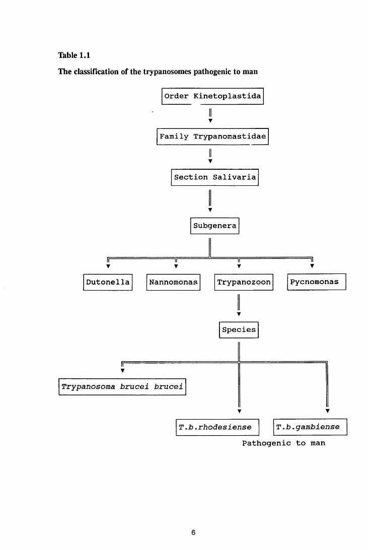
Table 1.1
The classification of the trypanosomes pathogenic to man
O r d e r K i n e t o p l a s t i d a
▼
F a m i l y T r y p a n o m a s t i d a e
▼
S e c t i o n S a l i v a r i a
▼
S u b g e n e r a
D u t o n e l l a N a n n o m o n a s T r y p a n o z o o n P y c n o m o n a s
▼
S p e c i e s
T r y p a n o s o m a b r u c e i b r u c e i
▼
T . b . r h o d e s i e n s e
P a t h o g e n i c t o m a n
▼
T . b , g a m b i e n s e
6
horses, and a chronic disease in cattle and pigs, and in experimental animals
such as mice (Ormerod, 1970; Hoare, 1972; Molyneux and Ashford, 1983;
Jennings, Whitelaw and Urquhart, 1977). The three species of Trypanozoon are
morphologically indistinguishable (Ormerod, 1970; Hoare, 1972; Molyneux and
Ashford, 1983).
1.2.2 Transmission
Trypanosoma brucei gambiense and T.b. rhodesiense, are cyclically
transmitted by dipteric flies of the Glossina species also called tsetse flies, many
of which are capable of transmitting the human infective trypanosomes
(Glasgow, 1970).
The gambiense form of the disease is mainly found in West and Central
Africa. This disease has been reported to be essentially an anthroponosis,
transmitted from man to man (reviewed by Jordan, 1986). However, this has
been contradicted by the finding that domestic pigs and certain species of
wildlife, can harbour T.b. gambiense for over 70 days, in a tsetse-infective form
(Van Hoof, 1947; Watson, 1962). This suggests that this form of the disease
could be an anthropozoonosis.
The rhodesiense type, found mainly in East and South Africa, is an
anthropozoonosis frequently transmitted from wild or domestic animals to man.
Amongst the trypanosome-susceptible wild animals, the bushbuck, Tragelaphus
scriptus (Heisch, McMahon and Manson-Bahr, 1958) and the hartebeest,
Acelaphus busephalus (Geigy, Mwambu and Kaufmann, 1971; Geigy, Mwambu
and Onyango, 1972) have been reported to serve as reservoir hosts for T.b.
rhodesiense.
The association of the trypanosomes with a particular species of tsetse
fly is determined by epidemiological characteristics of the disease they
cause.Thus, Trypanosoma brucei gambiense is transmitted by riverine flies of
the palpalis group, represented by Glossina palpalis and G.tachinoides in West
7
Africa, and by G. fuscipes in East Africa. The main vectors of T.b. rhodesiense
belong to the morsitans group represented by G. morsitans, G. swynnertoni and
G.pallidipes; these flies inhabit the savanna-like woodlands of East and South
Africa, which are abundant in game animals (Hoare, 1970).
However, the suitability of a given species of Glossina as a vector
depends upon the degree of contact between man and the flies that exist locally.
For instance, in North-Eastern Uganda, the vector of T.b. rhodesiense is a
member of the palpalis group (G.fuscipes) which usually transmits the
gambiense form of the disease; at the same time, T.b. gambiense has been
reported to be transmissible cyclically through G.morsitans (Lester, 1933;
Corson, 1936), a major vector of the rhodesiense form of the disease.
1.2.3 Distribution
The tsetse fly infests some 11 million km2 of Africa (reviewed by
Jordan, 1986). The Northern limit is about 14°N and 4°N corresponding closely
to the Southern edges of the Sahara and Somalia deserts, respectively. In the
South-West it varies from 10°S and 20°S corresponding to the Northern edges of
Kalahari and Namibian deserts, whereas in the South-East, it is generally at
about 20°S but extends as far as 29°S along the East African littoral (Jordan,
1986).
Glossina palpalis and G.fuscipes, the chief vectors of the gambiense
form of sleeping sickness, occur throughout the lowland rain forests and extend
well into the drier savanna zones. In the drier parts of their ranges, these
Glossina species are often associated with surface water and with riparian and
lacustrine vegetation (Challier, Gouteux and Coosemans, 1983). The other main
vector of gambiense sleeping sickness, G.tachnoides, occurs along rivers and
streams in the savannas of West Africa, although isolated pockets exist in
similar vegetation as far East as Ethiopia (Challier et al, 1983).
8
The main vectors of rhodesiense sleeping sickness belong to the
morsitans group and include G.morsitans, G.swynnertoni, and G.pallidipes.
Glossina morsitans occurs from Mozambique and Zimbabwe in the South to
Tanzania in the North (Hoare, 1970). In these areas, this species infests the
extensive ’miombo1 woodlands of East Africa and the mopane woodlands in the
Zambia Valley. Glossina swynnertoni is restricted to Acacia-Commiphora
vegetation in North Tanzania, extending into Kenya. The third species,
G.pallidipes, occupies a wide range of thicket and forest edge vegetation in
East Africa extending from Ethiopia in the North to Mozambique in the South
(reviewed by Jordan, 1986).
These limits of distribution are determined by climate, often through its
effect on vegetation. The 500mm isohyet is the limit near the deserts but flies
are found in drier areas in the vegetation lining the watercourses (Nash, 1937).
Where rainfall is high, the presence of tsetse flies is limited by seasonal low
temperatures. The adult flies are inactive at or below 16°C and the puparia do
not complete maturation to adult stages below this temperature (Bursell, 1960).
On the other hand, puparia of tsetse flies, which develop in the soil, succumb to
temperatures above 40-41°C (Bursell, 1960).
Within these general limits, infestations of Glossina are not continuous.
Extensive areas devoid of trees, naturally or man made, and high grounds are
generally tsetse free (Bursell, 1960). For instance, near the equator, the tsetse
do not occur above 1,800m above sea level due to the low temperatures of these
regions. This altitude decreases with the distance from the equator and is about
1,300m in Zimbabwe. The tree cover would provide shade, reducing the soil
temperatures and increasing the survival chances for the puparia (Bursell,
1960).
9
1.3 LIFECYCLE
1.3.1 In man
The biting tsetse fly can inoculate metacyclic trypanosomes, during a
bloodmeal, into the dermal connective tissue, where they start dividing
(Fairbaim and Godfrey, 1957; 1958). A local inflammatory reaction, called a
chancre, develops at the site of this initial division. From this site, the
trypanosomes enter the draining lymphatics and then the bloodstream. Once in
the bloodstream, trypanosomes can traverse the walls of blood and lymph
capillaries into the connective tissue and, at a later stage, into the brain and
cerebrospinal fluid (CSF) (Fairbaim and Godfrey, 1957; 1958). In all these
sites, trypanosomes multiply by binary fission as long slender trypomastigotes,
with a mean doubling time of 6 hours (Seed, 1978).
The parasitaemia fluctuates owing to evasion of the host's immune
response by antigenic variation. When the host mounts an IgM response to the
homotype variable antigenic type (VAT), the parasitaemia goes into remission
as trypanosomes of that homotype are killed off (Seed, 1977). Non-dividing,
stumpy trypomastigotes replace the slender forms as parasitaemia declines. The
stumpy forms can only continue their life cycle in the vector and are killed by
the mammalian host's immune mechanisms. Heterotype VATs continue to
multiply during the remission of parasitaemia. One of these heterotype VATs
overgrows the others to give rise to a recrudescent parasitaemia, in which it
becomes the homotype (Van Meirvenne, Janssens and Magnus, 1975). The
process continues and gives rise to a chronic infection that is characteristic of
African trypanosomiasis.
A particular population of trypanosomes contains a major homotype
VAT and several minor heterotype VATs. Each VAT is characterised by a
variant-specific glycoprotein (VSG) coat, found only in the mammalian stages
and not in the vector stages (Vickerman, 1969; Cross, 1975). The coat protects
10
the trypanosome from the hosts specific and non-specific defences. Coatless
forms activate the complement alternative pathway (Mosser and Robert, 1982;
Ferrante and Allison, 1983a) and are readily engulfed and destroyed by
macrophages (Mosser and Robert, 1982). In contrast, macrophages do not take
up coated trypanosomes except in the presence of VAT-specific antibody
(Mosser and Robert, 1982). Many mammals contain natural agglutinins (which
also opsonize the parasites for macrophage uptake) to coatless trypanosomes in
their serum (Ferrante and Allison, 1983b). Antigenic variation involves the
replacement of one VSG coat with another brought about by transcriptional
activation of a new VSG gene (Borst and Cross, 1982; Steinert and Pays, 1985).
Non-dividing forms, such as the stumpy forms, do not undergo antigenic
variation.
Heterotypes arise spontaneously in cloned populations of trypanosomes.
Antigenic variation has been reported to take place in vitro (Doyle, Hirumi,
Hirumi, Lupton and Cross, 1980; Gardiner, Thtthi, Gathuo, Nelson and Moloo,
1986), although the pace of variation is slower than in the vertebrate host. Thus,
the host antibody is not the inductive signal for antigenic variation, but can serve
to clear specific populations from the blood which are then replaced by the
growth of newly arising variants.
1.3.2 In the tsetse fly
The tsetse fly ingests blood into the crop and then the lumen of the
midgut where the stumpy trypanosomes transform into the procyclic stage. The
slender forms die or change into stumpy forms in the anterior midgut
(Vickerman, 1985). Morphological changes of the trypanosomes in the tsetse-
anterior midgut include elongation of the post-kinetoplast portion of the body as
the simple mitochondrion enlarges and becomes branched. Concomitantly,
glycosomes change from spherical to bacilliform structures (Vickerman, 1985).
The variable antigenic coat is progressively lost and endocytosis ceases. These
11
changes occur over a period of 48-72 hours and are accompanied by active
division of the flagellated trypanosomes (Steiger, 1973). As the trypanosomes
change, there is a switch of energy source from utilisation of glucose, which is
scarce in the vector, to proline the source of energy for tsetse flight (Hatson,
1975), and a change from aerobic to anaerobic respiration, as the oxygen
tension in the bloodmeal within the tsetse midgut decreases (Bowman and Flynn,
1976).
Four days after ingestion, the procyclics penetrate the peritrophic
membrane into the ectoperitrophic space, where they elongate, move forwards
to the proventriculus and cease to divide (Hecker, 1980; Vickerman, 1985).
They then re-invade the endotrophic space and migrate via the oesophagus to the
salivary glands, where they develop into epimastigotes. The epimastigotes
attach, with the flagellum, to the microvilli of the epithelial cells lining the
salivary glandular lumen. Here, the epimastigotes transform into uncoated
trypomastigotes which, like their predecessors, attach to the glandular microvilli
and divide. Trypomastigotes then transform into the final stage, the mature
metacyclics; these have a variable antigenic coat, lie free in the lumen of the
gland, do not divide in the vector and are infective to the mammalian host
(Vickerman, 1985).
The mature metacyclic population of a given serodeme of Trypanozoon
is heterogeneous with respect to VAT (Gray and Luckins, 1976; Steinert and
Pays, 1985). A serodeme is a stable, immunologically distinct strain of
trypanosomes (i.e has a distinct VAT repertoire), that does not exhibit cross
immunity with metacyclic VATs of other serodemes (Van Meirvenne, Magnus
and Vervoort, 1977).
12
1.4 THE DISEASE IN MAN
1.4.1 Clinical findings
The clinical features of the disease largely depend on the species of the
infecting trypanosome. Trypanosoma brucei rhodesiense causes an acute to sub
acute illness, with signs of cardiac involvement being most prominent (Manson-
Bahr and Charters, 1963; Manuelidis, Robertson, Amberson, Pola and
Haymaker, 1965; Francis, 1972; Jones, Lowenthal and Buyst, 1975; Harries
and Wirima, 1988). The pre-patent period before the appearance of clinical
signs may be as short as 2 weeks (Harries and Wirima, 1988) and the total
duration of the disease, from infection to death, may be only 6 weeks
(Manuelidis et al, 1965). In contrast, Trypanosoma brucei gambiense causes a
subacute to chronic infection, and although signs of heart damage are
encountered (Bertrand, Lobiere, Barabe and Ette, 1971; Adams, Haller, Boa,
Doua, Dago and Konian, 1986), the disease is mainly associated with central
nervous system involvement (Molyneux, de Raadt and Seed, 1984; Boa, Traore,
Doua, Kouassi-Traore, Kouassi and Giordano, 1988).
1.4.1.1 Early stage
Within 5 to 15 days following an infective bite from a tsetse fly, a round
inflamed area, of several centimetres in diameter, forms at the site of the bite.
This area of inflammation is called a chancre and is characterised by, a red spot
surrounded by a waxen zone which appears as a mass of hot and painful minute
vesicles (Molyneux et al, 1984). It then subsides over a period of 2 weeks to
leave an area of scaly disquamation. The chancre is more commonly seen in
T.b. rhodesiense than in T.b. gambiense infections (Molyneux et al, 1984).
In gambiense sleeping sickness, there are intermittent bouts of fever
interrupted, for 2 to 3 days, by periods of lassitude that may last for long
durations (years), during which time the patient is unaware of illness. These
bouts of fever correspond to the peaks of parasitaemia and trypanolytic crisis
13
(Onyango, Van Hoeve and de Raadt, 1966; Apted, 1970). Other initial clinical
signs, include, tachycardia, progressive headache, malaise, weakness, an
intermittent morbilliform skin rash, and occasional hyperaesthesia (Duggan and
Hutchingson, 1966).
In contrast, the rhodesiense form may not show these periods of
lassitude. Instead, within 10 or so days after the infective bite, there is fever
accompanied by headache, weakness, tiredness, occasional rigours and
vomiting; generalised oedema with subcutaneous tipping of legs and back. The
oedema of the face gives patients a puffy moonface appearance (Manson-Bahr
and Charters, 1963; Apted, 1970). Cases of severe diarrhoea have also been
reported (Basson, Page and Myburgh, 1977).
The involvement of the CNS occurs faster in the rhodesiense form than
in the gambiense form with CSF showing pathological signs (increased white
cell counts and protein) in only 4 or so weeks after infection (Apted, 1970).
Severe congestive heart failure can occur in gambiense infections but is
more common in rhodesiense infections, giving rise to signs such as intermittent
tachycardia (Manuelidis et al, 1965), dyspnoea even at rest, coughing and
occasional haemoptysis (Manson-Bahr and Charters, 1963; Koten and de Raadt,
1969; Francis, 1972), marked venous congestion and hepatosplenomegaly
(Manson-Bahr and Charters, 1963). Transient local oedema of the face, and
other parts of the body may be seen (Apted, 1970; Wellde, Chumo, Reardon,
Mwangi, Asenti, Mbwambi, Abinya, Wanyama and Smith, 1989). Radiography
demonstrates cardiomegaly, widening of the superior vena cava and enlargement
of hilar vessels, hydrothorax and ascites (Manson-Bahr and Charters, 1963;
Francis, 1972; Mbala, Blackett, Mbonifor, Leke and Etoundi, 1988).
Abnormal electrocardiographic (ECG) tracings mainly with flattening or
inversion of T-waves have been reported in T.b. rhodesiense-mfoctions
(Manson-Bahr and Charters, 1963; Manuelidis et al, 1965; Jones et al, 1975)
14
and in T.b.gambiense (Bertrand, Sentilhes, Ducasse, Vacher and Boudin, 1965;
Francis, 1972; Mbala et al, 1988). In a few patients, there is evidence of
conduction defects and of cardiac ischaemia (Bertrand et al, 1965; Mbala et al,
1988).
Patients develop severe anaemia and lose weight rapidly (Koten and de
Raadt, 1969). The anaemia is mostly normocytic and normochromic, sometimes
associated with red cell abnormalities, including anisocytosis, polychromasia
and hypochromasia (Manson-Bahr and Charters, 1963). The anaemia also
involves an increase in the erythrocyte sedimentation rate, consistent with
coating of RBCs with antibodies or immune complexes (Molyneux et al, 1984).
The pathogenesis of anaemia is thought to be multifactorial.
Thrombocytopaenia and decreased serum fibrinogen levels are observed
in both T.b.rhodesiense and T.b. gambiense infections accompanied by
increased serum and urine levels of fibrinogen degradation products, and
decreased prothrombin activity (Barret-Connor, Ugoretz and Braude, 1973;
Davis, Robbins, Weller and Braude, 1974; Robins-Browne, Schneider and
Metz, 1975; Basson et al, 1977; Molyneux et al, 1984).
Leukocytes increase reaching maximum levels a few days after peak
parasitaemia, mainly due to an increase in mononuclear leukocytes (Ormerord,
1970). Instances of leukocytopaenia with a relative lymphocytosis have been
observed in T.b.gambiense infections (Basson et al, 1977). A dominant event is
the proliferation of B lymphoid series, either due to deficient T cell control
over B cells, or due to the presence of a mitogenic factor for the B cells
(Greenwood and Whittle, 1980).
Elevated IgM levels have been found consistently in serum and CSF of
human sleeping sickness patients (Mattem, Masseyeff, Michel and Peretti,
1961; Greenwood and Whittle, 1980). IgM levels are raised in laboratory
animals infected with the Brucei group of trypanosomes (Seed, Comille, Risby
and Gam, 1969; Hudson, Byner, Freeman and Terry, 1976) and in experimental
15
and natural infections of cattle with these trypanosomes, where the increase over
normal may be upto 9-fold (Luckins, 1976). Temporary drops in IgM levels
occur and are associated with parasitaemia remission; these might be attributable
to parasites mopping up specific IgM (^bayashi and Tizard, 1976). Careful
absorption with a wide range of trypanosome VATs can remove most of this
IgM in man (Herbet, Paratti, Van Meirvenne and Lennox, 1980) and in cattle
(Musoke, Nantulya, Barbet, Kirondeand McGuire, 1981), indicating that IgM
production is a specific response to the trypanosomes. In HAT, IgG and IgA
levels are in the normal range but IgE levels are raised Herbert et al, 1980).
The presence of auto-antibodies has been reported (Blackett and Ngu, 1976;
Mbala et al, 1988).
There is also an increase in serum levels of immunocongluttinins
(Blackett and Ngu, 1976; Basson et al, 1977; Lambert, Berney and Kazyumba,
1981; Mbala et al, 1988) accompanied by a decrease in the total hemolytic
complement, serum C3 and C4, with indications of high C3 catabolism (Blackett
and Ngu, 1976; Mbala et al, 1988). Severe hypoproteinaemia and
hypoalbuminaemia have also been described (Jenkins and Robertson, 1959;
Koten and de Raadt, 1969).
Hepatomegaly and hepatic dysfunction reflected by increased serum
bilirubin, alkaline phosphatase (AP), alanine transferase (ALT) and slight to
severe jaundice, have been reported (Robertson and Jenkins, 1959; Apted,
1970; Basson et al, 1977). Partial renal dysfunction characterised by low urine
volume, high urinary sodium content, mild proteinuria, haematuria and a-
ketoaciduria have also been observed (Hawking and Greenfield, 1941; Poltera,
Owor and Cox, 1977; Basson et al, 1977). Blood urea nitrogen and creatinine
vary from normal to increased concentrations (Barret-Connor et al, 1973).
Enzyme alterations related to muscular involvement have been reported (Barret-
Connor et al, 1973; Basson et al, 1977)
16
Endocrine dysfunction manifests as amenorrhoea, abortion caused by
uterine hypoplasia, premature births and perinatal deaths in women; and as
impotence and, in later stages of the disease, as gyneacomastia in men (Poltera,
1985). Orchitis has been observed in rhodesiense infections of man (Losos and
Ikede, 1972; Wellde et al, 1989) and in experimental T.b. brucei infections of
dog (Morrison, Murray, Sayer and Preston, 1981a).
The eyes may show iridocyclitis with variable degrees of keratitis,
circumcomeal infection, conjunctivitis and photophobia (Apted, 1970).
1.4.1.2 Late-stage
The main clinical signs that occur during the late-stage are related to the
CNS involvement and include, fasciculation of muscles of the limbs, face, lips
and tongue, oscillatory movements of the arms, head, neck, or trunk especially
in children, and an increase in tonicity or muscular rigidity. There is usually a
considerable element of cerebellar ataxia. Focal lesions may cause transient or
more commonly permanent paralysis of certain groups of muscles (Gallais,
Collomb, Miletto, Dutertre and Berardbadier, 1956). Epileptiform convulsions
may occur depending on the area of the CNS affected.
The commonest and most characteristic sign is daytime somnolence
which starts as an occasional nod and progresses to continuous sleep. However,
a small portion of patients suffer insomnia (Wellde et al, 1989). At this time,
speech is slow and indistinct, there is hypothermia, pruritis intensifies and
severe emaciation occurs. Sleep progresses into coma, sphincter control is lost
and death follows often from secondary intercurrent diseases such as pneumonia
(Wellde et al, 1989). Personality alterations are manifested as mental dullness
that progresses to apathy, lethargy and indifference to surroundings. Emotional
disturbances are also common and include, laughter, crying or outbursts of
rage, episodes of manic behaviour sometimes with hallucinations and delirium
(Tooth, 1950; Lambo, 1966). These changes occur mainly in gambiense
17
infections.
These late-stage features of CNS involvement are rarely seen in
rhodesiense sleeping sickness because patients die before such signs develop.
However, they can occur following unsi:.; *ssful therapy with early-stage drugs
such as pentamidine (Apted, 1970).
It is difficult to make a sharp demarcation between the clinical signs of
early- and late-stage sleeping sickness and there is little documented clinical
information on the early stage disease since most patients are diagnosed in the
late stages. A recent study in Uganda highlighted body weakness, somnolence,
inability to stand and walk unaided, disturbance of speech and incontinence as
the most constant presenting symptoms of late-stage rhodesiense sleeping
sickness; while hand and tongue tremors, ataxic gait, loss of Babinsky reflex
and positive cheiro-oral reflex are the most commonly occuring symptoms in
late-stage rhodesiense sleeping sickness (Mbulamberi, 1989). By contrast,
headache, fever, joint pains, insomnia, inability to work normally and loss of
appetite were significantly more common complaints among early-stage sleeping
sickness patients (Mbulamberi, 1989).
1.4.2 Pathology
The pathology of human African trypanosomiasis exhibits a wide range of
variation from the fleeting parasitaemia caused by a strain derived from animals
(Van Hoof, 1947), through the heavy parasitaemia of the virulent T.b.
rhodesiense disease, the insidious T.b. gambiense infection which can
exterminate a whole population, to the unusual balanced situation noted in
Nigeria (Duggan, 1962) and Sierra Leone (Harding and Hutchingson, 1948)
where high rates of infection are associated with mild symptoms and a death rate
similar to that observed in trypanosome free areas.
18
1.4.2.1 Systemic circulation and tissues other than the central nervous
system
The chancre is a hard, painful, red nodule of several centimeters in
diameter, which is similar to the cellulitis seen around a staphylococcal boil but
lacks pus, and represents the first major response of the host to the
trypanosome. The histology of a chancre reveals edema, trypanosomes and
perivascular infiltration by lymphocytes (Fairbaim and Godfrey, 1957; 1958). A
similar and more detailed description of the gross and histopathological changes
of the chancre has been reported in cattle experimentally infected with T. b.
brucei, T.vivax and T. congolense ( Murray and Morrison, 1980; Akol and
Murray, 1982), and in goats infected with T. brucei and T. congolense (Emery,
Akol, Murray, Morrison and Moloo, 1980). In their description, trypanosomes
are readily identified in the dermis accompanied by intense inflammatory
reaction, followed by accumulation of large lymphoid cells and plasmablasts.
These changes result in the disruption of the dermal collagen, causing
fragmentation. As the lesion declines in size, increased numbers of mature
plasma cells, macrophages, eosinophils and mast cells are found (Murray and
Morrison, 1980; Emery et al, 1980; Akol and Murray, 1982).
Following the enlargement of the lymph nodes draining the chancre,
generalised lymph node enlargement occurs. Histology of lymph nodes taken
from patients dying during the acute phase of the disease, show that the
enlargement is partly due to a proliferative lymphoid cell response. The
lymphoid germinal centers undergo changes in composition from clusters of
small lymphocytes to cells of the lymphocyte-plasma cell series, accompanied
by an infiltration by macrophages (Ormerod, 1970). The presence of
trypanosomes may be microscopically evident since aspirations of swollen
lymph nodes is one of the most reliable methods of isolating trypanosomes from
suspected cases. Similar changes have been described in experimental infections
19
of cattle with T.b. brucei, T.congolense and T. vivax (Murray and Morrison,
1980; Akol and Murray, 1982), of goats infected with T.b. brucei and T.
congolense (Emery et al, 1980), and of dogs infected with T.b. brucei
(Morrison, Murray, Sayer and Preston, 1981c). In addition to these changes,
the lymph nodes of dogs experimentally infected with T.b. brucei, show an
expansion of both the cortical and medullary regions, petechial, and sometimes
ecchymotic haemorrhage (Morrison et al, 1981c); numerous IgM-containing
cells are found mainly around the follicles, but a few of these may be found
within the follicles; the lymph node sinuses are extremely oedematous and
distended with, in chronological order of appearance, small lymphocytes, large
lymphocytes, macrophages, trypanosomes and fibrin deposits (Morrison et al,
1981c).
As the disease progresses in man, fibrosis of the lymph vessels and
nodes occur making lymph nodes small and sometimes hard. During this
transition they may contain morular or Mott cells, also called Russell-body
containing cells (Ormerod, 1970), which are modified plasma cells that produce,
but are unable to secrete immunoglobulins. The immunoglobulin-filled vacuoles
distend the cytoplasm giving it a characteristic bunch-of-grapes appearance
(Low and Mott, 1904). There are numerous vacuolated macrophages and Mott
cells throughout the cortex; the germinal centers show a loose cellular
arrangement and a decreased lymphocytic content (Ormerod, 1970; Poltera,
1985). The sinuses are distended with macrophages, and bi- and multinucleate
giant cells (Ormerod, 1970). Similar changes have been shown to occur in T.b.
brucei-infected dogs (Morrison et al, 1981c; Ndung'u, 1990) cattle ( Morrison,
Murray, Whitelaw and Sayer, 1983), mice and rats (Murray, Jennings, Murray
and Urquhart, 1974b).
In the early stages of sleeping sickness, there is a gradual fall of both red
and white blood cells with differential white cell count showing a relative fall in
20
granulocytes and a rise in mononuclear cells (Ormerod, 1970). Proliferation of B
lymphocytes is a dominant pathological event that first takes place in the
parasitised lymph nodes and is associated with the appearance of large amounts
of IgM and autoantibodies in the serum (Greenwood and Whittle, 1980). It has
been suggested that the exhaustion of the B lymhoid system by the sustained
trypanosomal stimulus contributes to immunodepression in man (Greenwood and
Whittle, 1980). In experimental infections, parasite specific immunosuppression
has been found to be transient in early stages and complete in the advanced
disease (Dempsey and Mansfield, 1983). Polymorphonuclear cells seldom occur
in the reaction of man to trypanosome infection and when they occur, are
usually associated with secondary bacterial infections (Ormerod, 1970;
Greenwood and Whittle, 1980). However, in experimental infections
polymorphonuclear cells are consistently found in the tissue lesions in dogs
(Morrison et al, 1981b; Ndung'u, 1990) and occasionally in mice and cattle
(Morrison et al, 1983; Jennings, McNeil, Ndung'u and Murray, 1989).
There is an increase in immunoglobulin production as shown by the rise
in erythrocyte sedimentation rates (Gall, Hutchinson and Yates, 1957). The
continuous accummulation of IgM in the blood and the insignificant secondary
IgG increase, is unusual compared to other infections (Mattem, 1962). IgM is a
large molecule that is unable to cross membranes efficiently, which may explain
the presence and persistence of trypanosomes in areas with blood-tissue barriers
such as the testes (Clapier, 1921), and the brain (Jennings and Gray, 1983). As
the disease progresses, the level of immunoglobulins in blood remains high but
there may be immunological failure characterised by the absence of
complement, the occurrence of pseudo-positive Wassermann reactions and the
presence of heterophile antibodies (Henderson-Begg, 1946; Greenwood and
Whittle, 1980; Poltera, 1985).
The spleen increases in size in proportion to anaemia, becoming firm and
cellular, with fibrosis occuring only at the very late stages of the disease. The
21
sinusoids contain activated macrophages in the early stages (Gall, Hutchingson
and Yates, 1957; Greenwood and Whittle, 1980). There is an increase in the
number of malphigian corpuscles, accompanied by a change in their cJlular
composition, from clusters of small lyn:r l.ocytes to cells of the lymphocyte-
plasma cell series (Ormerod, 1970). The changes that occur in the spleen during
trypanosome infections have been closely studied in experimental animals.
Thus, experimental infections with T.b. brucei in dogs (Morrison et al, 1981c),
and rats (Murray, Jennings, Murray and Urquhart, 1974a; 1974b) show that the
periarteriolar regions of the spleen contain numerous lymphoblasts, some
plasma cells and relatively few small lymphocytes: large active germinal centers
containing heavy granular deposits of IgG and IgM are found in majority of
follicles with numerous macrophages and a prominent layer of small
lymphocytes. Disruption of the lymphoid cellular arrangement occurs due to
accumulation of red blood cells, deposition of fibrin, and infiltration by
polymorphonuclear leukocytes, often accompanied by localized necrosis of the
lymphoid cells. There is marked increase in the number of highly mitotic plasma
cells and large lymphocytes in the red pulp, the cords of Billroth and a less
marked increase in the sinuses. As the infection progresses there is an
increasing number of macrophages in the sinuses some of which contain
phagocytic granules. Megakaryocytes are reported to occur occasionally
accompanied by a small number of erythropoietic cells, a sign of extramedullary
erythropoiesis (Morrison et al, 1981c).
In the late stages of sleeping sickness, the periarteriolar regions of the
spleen become less active and show a higher content of small lymphocytes. The
follicular areas become disorganized with few discrete active germinal centers.
Most of the follicular areas are depleted of lymphocytes and contain
macrophages in what looks like the remains of previously active germinal
centers. In the red pulp the plasma cell content is reduced and occasional Mott
22
cells can be found. The sinuses and cords contain numerous active macrophages,
many of which are involved in erythrophagocytosis. Focal fibrin deposits and
thrombi are common in the sinusoids and, occasionally, in the branches of the
splenic vein (Ormerod, 1970; Poltera, 1985). In dogs, the late-stage disease is
characterised by a widespread presence of megakaryocytes and foci of
erythropoietic cells in the red pulp, the sinuses and the cords of the spleen
(Morrison et al, 1981c; Ndung'u, 1990).
The heart is considered to be the organ most at risk in the early stages of
T.b. rhodesiense infection, e.g., the major cause of mortality in the Ugandan
T.b. rhodesiense-Qpidemic of the 1940s was concluded to be myocarditis. Severe
myocarditis has been reported in both rhodesiense (Hawking and Greenfield,
1941) and gambiense sleeping sickness (Mott, 1906). Histologically, a
pancarditis involving all cardiac layers including valves and conducting system
has been reported (Lavier and Leroux, 1939; Poltera, Cox and Owor, 1976).
The pancarditis consists of a focal or diffuse mononuclear cell infiltration of the
interstitium and the perivascular spaces (Poltera, Cox and Owor, 1976),
myocytolysis, and focal endomyocardial fibrosis (Hawking and Greenfield,
1941; Cohen, 1973; Poltera et al, 1977). This is the basis for the clinically
reported cardiac insufficiency and electrocardiographic changes (Lavier and
Leroux, 1939; Poltera et al, 1976). In human infections, the involvement of the
autonomic nervous system of the heart has also been observed (Poltera et al,
1977).
In experimental infections in various experimental animals, myocarditis
has been reported to be composed of focal haemorrhage, edema and separation
of muscle fibres, extravascular trypanosomes in association with local
inflammatory infiltrates composed of, mainly, lymphocytes, plasma cells and
occasional foci of macrophages. This is the typical lesion reported in T.b.brucei-
infected dogs (Morrison et al, 1981b; 1983; Ndung'u 1990), rats (Murray et al,
1974a; 1974b), mice (Murray et al, 1974a; 1974b; Poltera, 1980), and in
23
T.b.rhodesiense-infected vervet monkeys (Peruzzi, 1928; Poltera and Sayer,
1983). In addition to these changes, polymorphonuclear leukocytes are a
consistent finding in the lesions of the dog (Morrison et al, 1981a; Morrison et
al, 1983; Ndung'u 1990). In the dog, atria are affected to a greater extent
than the ventricles and the infiltrations are more marked in the subepicardial and
perivascular locations (Ndung'u, 1990).
Cardiac aneurysms from trypanosome endemic areas of Africa have been
described (Poltera et al, 1976). Cardiomyopathies and endomyocardial fibrosis
have been found to correlate with autoantibody titres in T.b. gambiense and
T.b. rhodesiense infections in man ( Blakett and Ngu, 1976; Poltera and Cox,
1977, respectively). In experimental T.b. rhodesiense-infections in vervet
monkeys, apical aneurysms have also been reported, which are thought to be a
consequence of the partial obstruction of the cardiac lymphatic drainage (Poltera
and Sayer, 1983). In terminal stages of experimental T.b. brucei infection in
dogs (Morrison et al, 1981b) and rats (Murray, Murray, Fisher and Urquhart,
1974), large numbers of trypanosomes are found scattered diffusely throughout
the myocardium and numerous foci of myocardial cell degeneration and necrosis
occur. These changes are associated with marked infiltration with lymphocytes,
plasma cells and many macrophages and, in the case of dogs,
polymorphonuclear leukocytes.
Hepatic dysfunction has been shown to occur during the course of
disease (Jenkins and Robertson, 1959) and jaundice has been reported (Gelfand
and Friedlander, 1963). Cloudy swelling and centrilobular necrosis are common
findings in the liver. Whether these are due to trypanosome infection or are a
result of secondary infections and terminal state is not clear (Ormerod, 1970).
In T.b. brucei-infected dogs (Morrison et al, 1981b), the hepatic sinusoids are
markedly distended and contain large numbers of plasma cells, lymphocytes,
variable numbers of polymorphonuclear leukocytes and trypanosomes. Kupffer
24
cells increase in number and occasionally contain phagocytosed erythrocytes.
Foci of fatty degeneration occur in close proximity to the hepatic veins.
Swelling and disorganisation of hepatocytes with occasional cell death and
mitosis may occur.
In summary, the most consistent findings are lymphoid hyperplasia,
infiltration of tissues (including, the heart and the brain), by trypanosomes and
cells of lymphocyte/plasma series and macrophages, hypergammaglobulinaemia,
progressive anaemia, leucopoenia and immunosuppression, and great wasting of
the limb, abdominal and thoracic muscles.
1.4.2.2 Central nervous system
It is not clear when the meningeal reaction starts. However, the
appearance of cells, immunoglobulins and protein in the CSF from cisternal
puncture (Neujean, 1950), as well as the involvement of the choroid plexus
suggest that cerebral involvement occurs early and progressively (Poltera,
1985). Le Port (1935) and Calwell (1937) produced the first evidence that
mental disturbances and headaches were accompanied by swelling of the choroid
plexus, which blocked the lateral apertures of the fourth ventricle and of the
interventricular foramen; and led to congestion of cerebral blood vessels.
Meningocortical infiltration by lymphocytes and plasma cells correlates with
early stages of CNS involvement, whereas, cerebral white matter and basal
ganglia lesions are involved in later stages (Manuelidis et al, 1965; Poltera et al,
1976). Perivascular cuffing does not appear to occur until at least 3 months after
infection, and is accompanied by the presence of small round cells (probably
infiltrating lymphocytes or proliferating microglia) in the brain parenchyma
(Mott, 1906; Calwell, 1937; Van Bogaert, 1958; Manuelidis et al, 1965;
Ormerod, 1970; Poltera et al, 1976).
Several gross anatomical descriptions of the CNS lesions in sleeping
sickness were published in the nineteenth century. The most complete work was
25
produced by Clark (1840) who described the inflammation of the brain associated
with swelling and adhesion of the dura to the arachnoid mater.
Meningoencephalitis was first observed by Mott in 1899. He (Mott, 1906) and
much later, Van Bogeart (1958) publish^ detailed descriptions of the CNS
lesions in gambinese sleeping sickness, While Calwell (1937) published a
detailed account of rhodesiense sleeping sickness.
The inflammation affects mainly the pia-arachnoid mater (Ormerod,
1970) and the choroid plexus (Calwell, 1937). The typical histological lesion is
a vasculitis with perivascular infiltration of the leptomeninges and Virchow-
Robin spaces with mononuclear inflammatory cells, including, lymphocytes,
plasma cells and macrophages with occasional Russell body-containing plasma
cells. These cells extend into the adjacent parenchyma in severe cases of the
disease (Mott, 1906; Calwell, 1937; Van Bogaert, 1958; Manuelidis et al, 1965;
Poltera et al, 1976; Poltera, 1985; Adams et al, 1986). The pathology slowly
spreads to the remote parts of the subarachnoid space. Thus, only in advanced
cases are Russell body-containing plasma cells obtained at lumbar puncture
(Mott, 1906). The most characteristic lesion resulting from this spread is the
formation of perivascular cuffs whereby the Virchow Robin spaces are
infiltrated by the mononuclear cells. This may consist of a single layer of cells
or in advanced cases, a 20-30 cell-deep sheath. Russell body-containing plasma
cells are present in all the inflammatory foci in the CNS, which led to their
presence in the perivascular cuffs being erronously considered to be
pathognomonic of sleeping sickness (Mott 1906).
Microglia increase in number, in the brain parenchyma, preceeding the
formation of perivascular cuffing, and infiltrating the cuffs as they form (Mott,
1906; Calwell, 1937; Van Bogaert, 1958; Manuelidis et al, 1965; Ormerod,
1970; Poltera et al, 1976). Poltera et al (1976) and Manuelidis et al (1965)
emphasized the prominence of swollen astrocytes and the absence of neuronal
26
damage. In the very advanced perivascular cuffing, chromatolysis of neurones
around the cuffed vessels, occasionally leading to Wallerian-like degeneration of
nerve tracts, occurs (Mott, 1906; Calwell, 1937; Van Bogaert, 1958;
Manuelidis et al, 1965; Ormerod, 1970; Poltera et al, 1976). Swollen masses of
macroglial cells may be seen where neuronal degeneration occurs.
Topographically, the lesion is most evident in the white matter of the
cerebral hemispheres, the basal nuclei, the white matter of the cerebellum and
the brain stem (Calwell, 1937; Poltera et al, 1977; Adams et al, 1986; Haller,
Adams, Merouze and Dago, 1986). Occasionally the lesion extends to the
deeper layers of the cerebral cortex. In more florid cases, there are collections
of lymphocytes, plasma cells and microglia in the brain not directly related to
blood vessels. Diffuse microglial hyperplasia in the grey matter and in a few
cases large reactive astrocytes in the white matter have also been observed
(Adams et al, 1986; Haller et al, 1986). Russell body-containing plasma cells
occur within perivascular spaces and in association with groups of cells of
inflammatory type in the white matter. Isolated Russell body-containing plasma
cells have been reported to occur in areas that in all other respects appear
normal (Adams et al, 1986).
The involvement of the choroid plexuses and the cranial nerves has been
reported (Calwell, 1937; Poltera et al, 1977). However, Adams et al (1986)
observed that inflammatory processes in the ventricles or in the choroid plexus
occured only occasionally and that encephalitis was more common than
meningitis.
In summary, the consistent pathological features of CNS trypanosomiasis
are lymphocytic-plasmacytic meningoencephalitis, especially in a perivascular
distribution, glial proliferation and sparing of the neuronal elements. In some
fatal cases, chromatolysis of the neurones and degeneration of the nervous
tracts, and choroid plexus involvement occur. Occasionally a haemorrhagic
encephalopathy occurs especially after arsenical therapy.
27
1.5 CONTROL OF HUMAN AFRICAN TRYPANOSOMIASIS
The control of sleeping sickness relies on an integrated strategy of
continuous surveillance, involving, diagnosis and treatment of the population at
risk, and vector control, where applicable (de Raadt, 1986).
Vector populations can be controlled for a time by the application, from
air or ground, of suitably formulated insecticides, a very costly and potentially
ecologically harmful venture. Nowadays, more attention is being given to
cheaper and more ecologically friendly methods, applicable at community level.
These include, modification of the habitat by bush clearing around human
habitation and rivers, and the use of odour-baited, insecticide impregnated traps
and targets (WHO, 1986). Indeed, the control of tsetse flies by means of simple
traps with community participation has been shown to be effective in the control
of the sleeping sickness epidemic in Uganda (Lancien, 1991).
A number of new tools, particulary for diagnosis and vector control have
been developed (Cattand and de Raadt, 1991; Nantulya, Doua and Molisho,
1992; Komba, Odiit, Mbulamberi, Chimfwembe and Nantulya, 1992). Despite
the fact that chemotherapy is the major means of disease control, development
of new anti-trypanosomal drugs has been more or less static over the last three
decades, due to lack of interest by the pharmaceutical industry to invest into
research and development of trypanocides (Gutteridge, 1985). The methods of
diagnosis and the drugs available for treatment of HAT are considered below.
1.5.1 Diagnosis
The standard method for diagnosis of trypanosomiasis is to demonstrate and
identify trypanosomes in body fluids, including, blood, chancre fluid, lymph
node aspirates, and CSF. Owing to the tissue invasiveness of the causative
agents of HAT, T.b. rhodesiense and T.b. gambiense, and the intermittent
28
nature of the infections they cause, it is not always possible to demonstrate
parasites in body fluids. Moreover, in the insidious gambiense infection, several
months may elapse before the clinical manifestations develop; and even when
they do, the manifestations are not pathognomonic (Losos and Ikede, 1972). The
techniques employed for diagnosis of HAT can be divided into three major
groups: parasitological, immunological and molecular.
1.5.1.1 Parasitological diagnosis
The easiest technique for detection of trypansomes in peripheral blood is
by direct microscopy of blood, either the wet film method to detect motile
trypanosomes or, as stained thick and thin smears when parasites are identified
on the basis of their morphology by light microscopy (WHO, 1986).
Examination of wet blood films is quick and suitable for screening large
numbers of samples but is too insensitive as half of the infected patients may not
show parasites (Barnet, 1947; Baker, 1970; WHO, 1986). Trypanosomes can
sometimes be demonstrated by direct microscopy of aspirates from the enlarged
cervical lymph nodes, chancre or cerebrospinal fluids (WHO, 1986). In
suspected cases where the presence of parasites cannot be demonstrated in
blood, lymph fluid or CSF, examination of stained bone-marrow aspirates has
proved to be a sensitive technique (WHO, 1986).
The haematocrit centrifugation technique (HCT) improves the sensitivity
of direct microscopic examination, by concentrating the parasites (by
centrifugation) in unclotted blood at the buffy coat (Woo, 1969; Murray,
Murray and McIntyre, 1977; Kelley and Shillinger, 1983). A modification of
this method to increase its sensitivity by including a plastic float, has been
described (Levine, Wardlaw and Patton, 1989). The HCT is particularly useful
in that the status of anaemia in the test subject can be assessed at the same time.
This is a simple and rapid method that is applicable far afield with the advent of
battery operated microfuges.
29
The miniature anion-exchange centrifugation technique (mAECT) which
removes the red cells from a blood sample by trapping them in a
diethylaminoethyl (DEAE) cellulose column followed by concentratiun by
centrifugation, is widely used to diagr T.b.gambiense infections in man
(Lumsden, Kimber and Strange, 1977; Lumsden, Kimber, Evans and Doig,
1979). In another method, fractionation is achieved by centrifugation of blood
through silicone layers. Other methods of removing red blood cells by lysis
using hypotonic shock or detergent, followed by centrifugation, may gain wider
application due to their simplicity (WHO, 1986). Single and double
centrifugation, has been shown to substantially increase the possibility of finding
trypanosomes in the CSF (Cattand, Miezan and de Raadt, 1988).
The most sensitive method of parasitological diagnosis is sub-inoculation
of test blood or CSF samples into susceptible laboratory animals, followed by
examination of blood for development of parasitaemia (reviewed by Paris,
Murray and McOdimba, 1982). This method has an added advantage in that
trypanosome isolates can be collected for further studies. The delay in diagnosis
and the cost of maintaining laboratory animals makes it impractical for routine
diagnosis. Furthermore, many T.b. gambiense isolates do not infect laboratory
animals. Isolation of parasites from aparasitaemic patients by an in vitro
technique has also been described (Aerts, True, Penchemier, Claes and Le Ray,
1992). Further evaluation of this technique is currently going on.
1.5.1.2 Immunological diagnosis
The tests that detect antibodies in the sera and CSFs of trypanosomiasis
patients include: capillary tube agglutination test - CAT (Ross, 1971),
immunoprecipitin tests (Aiyedun, Amodu, Bidwell, Bone, Buck, Coulm, Frezil,
Kent, Mattem, Njogu, Voller and Wery, 1976; Thylor and Smith, 1983),
indirect immunofluorescence antibody test, IFAT (Wery, Van Wetter, Wery-
30
Paskoff, Van Meirvenne and Mesatewa, 1970; Katende, Nantulya and Musoke,
1987), indirect haemagglutination test, IHAT (Bone and Charlier, 1975),
enzyme-linked immunosorbent assays, ELISA (Voller, 1977; Luckins, Gray and
Rae, 1978, Luckins, Boid, Rae, Mahmoud, El Malik and Gray, 1979) and card
agglutination test for trypanosomiasis, CATT (Magnus, Vervoort and Van
Meirvenne, 1978). These tests detect antibodies and, therefore, cannot
distinguish between active and cured infections. The CATT (Magnus et al,
1978) is the most reliable of these methods and utilizes variant surface antigens
of selected trypanosome antigenic variants which are stabilised on the whole
trypanosome. The other tests utilise antigens of undefined specificity and purity,
making them difficult to standardise (WHO, 1986).
Following the observation that the trypanosome procyclics' surface
antigens are species or subgenus specific, various antigen detection
immunoassays have been developed (Nantulya, Musoke, Rurangirwa, Minja and
Saigar, 1985; Parish, Morrison and Pearson, 1985; Richardson, Jenni, Beecroft
and Pearson, 1986; Liu and Pearson, 1987; Nantulya, Musoke, Rurangirwa,
Saigar, and Minja, 1987; Nantulya, 1989). These tests detect specific,
trypanosome surface antigens, in sera and CSF of infected patients and should
differentiate between current and past infections (WHO, 1986).
1.5.1.3 Molecular diagnosis
This method is based on the detection of parasite DNA from the blood,
CSF, lymph node aspirate, bone marrow or tissues. Within the parasite
genome, there are repeated DNA sequences, known as 'satellite' DNAs, which
have no known cellular function and typically contain a simple sequence that is
repeated thousands and often millions of times. In T.brucei and T.evansi, these
elements constitute 12% and 9% of the genome, respectively (Borst, Face-
Fowler, Frasch, Hoeijmakers and Weijers, 1980; Castro, Craig and Castaneda,
1981; Sloof, Bos, Konings, Borst, Gutteridge and Leon, 1983a; Sloof, Menke,
31
Caspers and Borst, 1983b; Koehavong and Thilly, 1989). The satellite DNA
contains the most convenient parasite species-specific differences, which can be
used for diagnosis.
DNA hybridization can also be ^ed for diagnosis. A single-stranded
DNA fragment, containing specific DNA sequences, such as parasite species-
specific sequences, is identified, purified and labelled with a tracer. The labelled
fragment is then used to probe whole parasite DNA or the whole organism. The
test parasite DNA is split into single strands using denaturing reagents so that
when the probe is applied to the specimen, these strands anneal to
complementary DNA sequences of the parasite. The bound label can then be
revealed by dye development or autoradiography depending on the label used
(WHO 1986).
Recently, amplification of the satellite DNA by the polymerase chain
reaction was introduced (Moser, Cook, Ochs, Barley, McKane and Donelson,
1989) and has been reported to be very sensitive, detecting upto one tenth of a
parasite.
1.5.2 Chemotherapy
Many drugs mainly arsenicals and antimonials, have been used in the
treatment of sleeping sickness. Atoxyl, one of the first to show promise, was
introduced in 1905 but unfortunately, its use was sometimes followed by optic
nerve atrophy (reviewed by Hawking, 1963). Thrtar emetic appeared a few
years later and was effective but relapses after use were frequent. Tryparsamide
became available in 1919 and suramin (Bayer 205), developed some years
earlier, was first used in sleeping sickness in 1922. Other drugs in great
variety, including the diamidines introduced in the 1930s, have been used, but
of the early drugs only suramin and tryparsamide have survived, although tartar
emetic may occasionally be of value if other drugs are not available. Melaminyl
32
(arsenical) compounds introduced in 1939 tended to supersede tryparsamide
except in areas with special problems where the latter is still of use (Apted 1970).
The drugs can be divided into two major groups: those that cannot cross the
blood-brain barrier and can therefore treat only the early stages of the disease,
including, suramin, pentamidine and Berenil, and those that cross the blood-
brain barrier and are required to treat the patients with late-stage central nervous
system involvement, such as melarsoprol and eflomithine.
1.5.2.1 Early-stage drugs
1.5.2.1.1 Suramin
Suramin is the symmetrical 3 urea sodium salt of 8-(3-benzamido-4-
methylbenzamido)naphthalene-l,3,5,-trisulphonic acid. It is administered
intravenously (i.v.) at 20mg/kg single dose (adult dose, maximum adult dose is
lg). The dose is repeated five times at 5-7-day intervals. Suramin is
trypanocidal and parasites disappear from blood and lymph nodes of patients
within a few hours of its administration (Apted, 1970). It is equally effective
against both gambiense and rhodesiense infections. Suramin remains unique
amongst trypanocides as it is negatively charged at physiological pH and as such
readily binds to serum proteins. In a few patients, suramin fails to reach
therapeutic concentrations in blood (Hawking, 1940). Suramin inhibits a
multitude of enzymes in cell free systems at millimolar concentrations amongst
which L-alpha glycerolphosphate oxidase, an enzyme concerned with oxidation
of reduced nicotinamide diphosphate (NADPH) in African trypanosomes, is
markedly sensitive (Fairlamb and Bowman, 1980). Although it permeates the
blood-brain barrier poorly, some effect on early nervous system infections have
been reported (Keevil, 1934).
A febrile reaction to the first or the second injecton is almost invariable.
In a few patients, injection is followed by vomiting, shock and collapse (Apted,
1970). Other early signs of toxicity, include, pruritis, urticaria, a papular
33

eruption, conjunctivitis, photophobia, palpebral oedema and stomatitis.
Cutaneous hyperaesthesia may be noted and particularly pain in the soles of the
feet which may be so severe as to cause difficulties in walking. The commonest
toxic effect is some degree of renal dv....age resulting in mild albuminuria.
However, suramin is one of the most widely used and one of the safest
trypanocides. It forms complexes with plasma proteins and is excreted very
slowly so that a single injection of lg can maintain prophylactic concentrations
in blood for about three months.
1.5.2.1.2 Pentamidine
This aromatic diamidine is present in two forms: pentamidine isethionate
and the related methanesulphonate. It is administered at 3-4mg/kg everyday for
7 days. The single maximum dose is 300-400mg. Pentamidine is trypanocidal
and is very effective in the early stages of gambiense sleeping sickness. It does
not cross the blood-brain barrier and therefore has no effect on the CNS
infections. Its effects are less certain in the rhodesiense form of the disease
where relapses and severe deterioration of the patients have been reported in
East and West Africa (Apted, 1970; Hutchinson ar»u Watson, 1962).
Histamine is released following intramuscular administration of
pentamidine with subsquent hypotension, dizziness and occasional collapse.
The drug also causes hypoglycaemia (Williamson, 1970; Gutteridge, 1985).
Pentamidine is a valuable prophylactic and a single dose can protect against
gambiense infection for at least six months, but its prophylactic value in
rhodesiense form is less certain (Williamson, 1970).
The mechanism of action of aromatic diamidines is still incompletely
resolved. They are positively charged and bind to DNA exerting a marked effect
on biosynthesis of nucleic acids (Gutteridge, 1969; Newton, 1974). The
synthesis of kinetoplast DNA is particularly sensitve to disruption by diamidines
34
(Brack, Delan, Riou and Feisty, 1972). They also inhibit S-adenosylmethionine
(SAM)-decarboxylase (Bitonti, McCann and Sjoerdsma, 1986), an enzyme
involved in the formation of decarboxylated SAM (dSAM) from SAM, which in
turn makes spermidine from putrescine during the synthesis of glutathione, in
mammals, and of trypanothione in the trypanosome (Henderson and Fairlamb,
1987).
1.5.2.1.3 Berenil
Berenil (diminazene aceturate), like pentamidine, is an aromatic
diamidine that has been shown to have marked activity in a variety of
trypanosome infections (but not Trypanosoma cruzi) and the veterinary
parasitosis, babesiosis (Fussganger and Bauer, 1958; Whiteside, 1962a; 1962b;
Fairclough, 1963a; 1963b; Van Hoeve and Grainge, 1966, MacLennan, 1968;
Fink and Dann, 1974). Although Berenil is registered as a veterinary drug, its
successful use in the early stages of human T.b.gambiense (Hutchinson and
Watson, 1962) and T.b.rhodesiense infections has been reported (Apted, 1962;
de Raadt, Van Hoeve, Onyango and Kimber, 1965a; de Raadt, Van Hoeve,
Bailey and Kinyanjui, 1965b; Bailey, 1967 and 1968; Onyango, Bailey, Okach,
Mwangi and Oganda., 1970; Temu and Mbwabi, 1975; Abaru and Matovu,
1981).
Berenil is administered intramuscularly at 5mg/kg in three doses at one
or more day-intervals over a duration of 7-12 days (Abaru and Matovu, 1981).
Oral administration with successful outcome has been reported (Bailey, 1968).
The drug is generally well tolerated and toxic side effects are rare except
for a slight and persistent albuminuria. Other side effects may include pyrexia,
body aches, burning sensations, nausea and or vomiting, weakness and or
paralysis of limbs, numbness and itching (Abaru and Matovu, 1981). One
isolated case of disorientation and another of coma have been reported (Abaru
and Matovu, 1981).
35
1.5.2.2 Late-stage drugs
The development of arsenical drugs was the greatest advance in the
treatment of sleeping sickness since the introduction of suramin and
tryparsamide in the early 1920s. Friedheim carried out field trials with the first
of these drugs, Melarsen, in French West Africa in 1939 (Friedheim, 1949)
with very promising results. Other drugs in this group include: melarsen oxide,
melarsoprol (Mel B), melarsonyl potassium (Mel W) and antimony derivatives.
Of these, only melarsoprol has become firmly and widely established among the
drugs of real value in the treatment of sleeping sickness. Melarsen and
melarsonyl potassium have proved themselves of benefit in some areas in West
Africa. Melarsan oxide was shown to be active against tryparsamide resistant
T.b. gambiense infections (Van Hoof, 1947; Williamson and Lourie, 1948) at a
time when tryparsamide resistance had reached 80% in Congo (Friedheim,
1949). Trials of melarsoprol in treatment of rhodesiense sleeping sickness in
Tanzania reduced death rates from over 33% to 10% (Apted, 1957).
1.5.2.2.1 Melarsoprol
Melarsoprol was formed from melarsen oxide by the incorporation of
dimercaprol (BAL) after melarsen oxide had proved to be too toxic.
Melarsoprol contains 18.8% arsenic, and cures both the rhodesiense and
gambiense disease at all stages, even the very advanced stages. A course of
injections over 2 to 4 weeks is necessary and even in areas where tryparsamide
is effective, the shorter course of treatment gives melarsoprol a great advantage.
The improvement is usually rapid, even dramatic, and is often marked by a
sense of well-being noticeable within the first few days. In particular patients
who are dull or mentally confused, as they often are in the advanced stage of the
disease, lose this dullness and become alert and cheerful soon after the start of
treatment. Occassionally a patient, particularly one who is in advanced stage of
36
the disease, may fail to respond to melarsoprol (Apted, 1970).
There is no single recommended treatment regime for melarsoprol
despite the fact that it has been in use for over 50 years. Dosage and duration of
therapy differ considerably between centres, e.g., the total dose for an adult in
Cote D'Ivoire is 1.62g over a duration of 32 days while 2.16g over 26 days
was, until recently, routine practice in Zaire (Van Nieuwenhove, 1992).
Progressively increasing doses are administered by some therapists, while
maximum daily doses are given straight away by others (WHO, 1986; Van
Nieuwenhove, 1992). For instance, the current treatment of both early and late
stages of sleeping sickness due to T.b. gambiense infection, is a modified form
of the dosage scheme proposed by Neujean (1950). For an adult patient
weighing 50kg, daily intravenous injections are given of 3.6mg/kg which is
equivalent to a daily dose of 180mg. Patients with fewer than 20 cells/mm3 in
the CSF receive one series of daily injections for 4 days. Patients with 21-100
cells/mm3 receive two series of four injections separated by an interval of 10
days. Those with more than 100 cells/mm3 receive three series of four injections
at 10-day intervals (WHO, 1986).
In many centres, melarsoprol is proceeded by a single or multiple
injections of the early-stage drugs, suramin or pentamidine along with
corticosteroids at lmg/kg/day in some centres, while in others it is given alone
(WHO, 1986; \kn Nieuwenhove, 1992).
Trivalent arsenicals like melarsoprol have a high affinity for sulphydryl
groups which form active sites of many enzymes especially kinases. Melarsoprol
markedly inhibits pyruvate kinase in intact trypanosomes thus disrupting ATP
production and energy generation in African trypanosomes (Flynn and Bowman,
1974). It also combines with trypanothione to form a complex that removes the
latter from the system which results in disruption of the trypanosomal protein
synthesis (Henderson and Fairlamb, 1987).
Sulphydryl groups are also involved in the maintainance of secondary
37
and tertiary structures of proteins, as a result of which side effects tc melarsoprol
are common and sometimes quite severe. Reactions which may occur, include,
ftver, chest pain, gripping abdominal pains and subjective sensations of heat and
disturbance of smell (Gutteridge, 1985). most serious side effect is a 5-10%
incidence of severe reactive arsenical encephalopathy, that results in
neurological damage or death (Hurst, 1959; Gutteridge, 1985; Adams et al,
1986; Arroz, 1987). There are three characteristic syndromes of arsenical
encephalopathy: status epilepticus with acute cerebral edema; rapidly
progressive coma without seizures; and non-fatal cognitive abnormalities
without neurological signs (Haller et al, 1986).
1.5.2.2.2 Eflornithine
Eflomithine is the most recent drug used for the treatment of the
meningoencephalitis, associated with African trypanosomiasis. Eflomithine
irreversibly inhibits ornithine decarboxylase (ODC) (Metcalf, Bay, Danzin,
Jung, Casara and Vervet, 1978), the key enzyme in the synthesis of the
polyamines, including, putrescine, spermidine, and spermine, from ornithine.
By inhibiting synthesis of putrescine, eflomithine inhibits differentiation and
proliferation of trypanosomes (Beyer, 1978; Bacchi, Nathan, Hunter, McCann
and Sjeordsma, 1980; McCann, Bitonti, Bacchi and Clarkson, 1987; Pepin,
Milord, Guem and Schechter, 1987; Doua, Boa, Schecter, Miezan, Diai,
Sanon, de Raadt, Haegele, Sjeordsma and Konian, 1990), as it does a number of
rapidly proliferating cells in vitro (Pegg and McCann, 1982; Luk, Goodwin,
Marton and Baylin, 1981; Seidenfeld, Gray and Marton, 1981) and in animal
tumours in vivo (Sunkara, Prakash and Rosenberger, 1982; Sunkara, Prakash,
Mayer and Sjeordsma, 1983).
One of the earliest effects ascribed to eflomithine in mammalian cells is
an inhibition of protein synthesis (Rudkin, Marmont and Seiler, 1984) which
38
parallels the depletion of spermidine. These changes are readily prevented by
the presence of putrescine or spermidine in addition to eflomithine, i.e., by
passing the enzymic block of ornithine decarboxylase.
Eflomithine was first demonstrated to cure experimental T.b. brucei
infection by Bacchi et al (1980). Subsequent treatment of late-stage
trypanosomiasis by the combined use of eflomithine with other compounds has
been extremely successful and cures in the experimental mouse model have been
reported using eflomithine in combination with bleomycin (Clarkson, Bacchi,
Mellow, Nathan, McCann and Sjeordsma, 1983), suramin (Clarkson, Bienen,
Bacchi, McCann, Hunter and Sjeordsma, 1984), guanylhydrazones (Jennings,
Ulrich and Cerami, 1987), melarsoprol (Jennings, 1988a; 1988b; 1990), the
antimonials (Jennings, 1990), 9-deazainosine (Bacchi, Nathan, Clarkson,
Bienen, Bitonti, McCann and Sjeordsma, 1987) and Berenil (Bacchi and
McCann, 1987; Jennings, 1992).
Successful clinical trials in human T.b. gambiense infections were first
reported by Van Nieuwenhove, Schechter, de Clercq, Bone, Hurke and
Sjeordsma (1985) in the Sudan. Subsequently, eflomithine has been successfully
used in treating arseno-resistant late-stage cases of gambiense infections in man
(Sjoerdsma and Schechter, 1984; Schechter and Sjeordsma, 1986; McCann et
al, 1987; Schechter, Barlow and Sjeordsma, 1987; Pepin et al, 1987; Doua et
al, 1990). It is administered intravenously at a dose rate of 400mg/kg per day
for 6 weeks.
Side effects, include, diarrhoea, abdominal pain, vomiting, anaemia,
thrombocytopaenia, convulsions, loss of hearing acuity, alopecia, skin eruptions
and exacerbations of intercurrent infections. Most of these side effects are
reversible upon discontinuation or decreasing eflomithine doses, and
convulsions do not recur even after resumption of treatment (Van Nieuwenhove
et al, 1985; Doua et al, 1990; Pepin and Milord, 1991; Pepin, Milord,
Meurice, Ethier, Loko and Mpia, 1992).
39
1.5.2.2.3 Nifurtimox
Nifurtimox, a 5-nitrofuran administered orally, has been available for
nearly 20 years for the treatment of Chagas' disease, caused by Trypanosoma
cruzi. There have been a few clinical trials involving nifurtimox in the
treatment of T.b.gambiense; these were prompted by the demonstration of
activity against this parasite in an animal model (Janssens and de Muynck,
1977). These trials have yielded equivocal results. Eighty percent of patients
were cured using 15mg/kg/day in three doses for 60 days in both Northern
Zaire (Moens, de Wilde and Ngato, 1984) and the Sudan (Van Nieuwenhove
and de Clercq, 1989). However using the same dosage for the same duration,
cures were obtained in only 31% of the cases in Central Zaire (Pepin, Milord,
Mpia, Meurice, Ethier, Degroof and Bruneel, 1989b; Pepin et al, 1992). Pepin
et al, 1992 reported that while doubling the dosage and halving the duration of
treatment yielded higher cure rates, the toxic effects outweighed the benefits of
this regimen. Studies conducted in the Sudan on arseno-resistant cases of the
late-stage gambiense sleeping sickness, showed the optimum nifurtimox regimen
to be 5mg/kg per os (p.o.), three times a day in adults and 6.5mg/kg p.o. three
times a day for children for 14 days (maximum 21 days) (Van Nieuwenhove,
1992). The efficacy of this dosage programme was high and there was a
considerable reduction in toxicity and mortality (Van Nieuwenhove, 1992).
Optimum treatment schedules for the early-stage gambiense as well as for
rhodesiense trypanosomiasis have not yet been defined. Rigorous supervision of
drug intake is necessary.
Initial use of Lampit, in man, in combination with trimelarsen gave
variable results. However, using a regimen of two daily doses of 50mg/kg for
five consecutive days, in combination with single daily doses of 2.5mg/kg
trimelarsen produced cures in experimental T.b.brucei infections in mice
(Jennings, 1991b). Other researchers (Raseroka and Ormerod, 1986), reported
40
improved cure rates after using high-dose nifurtimox therapy in combination with
suramin in a mouse model.
The mode of action of this group of drugs is not well understood. It is
thought that 5-nitrofurans interfere with biosynthetic reactions, especially of
nucleic acid and protein synthesis (Sims and Gutteridge, 1978; 1979). They
interact with DNA causing single- and double-strand breakages. Metabolism of
nitrofurans creates superoxide anions such as hydrogen peroxide and since
trypanosomes lack catalase, the superoxide anions accumulate to cytotoxic levels
in the parasites, killing them (Docampo and Stoppani, 1979; Docampo and
Moreno, 1984). Superoxide anions produced by the drug metabolism could
interact with DNA and interfere with nucleic acid synthesis.
Nifurtimox is metabolised not only by the parasite but also by the liver,
testes and brain of the mammalian host. The drug is not well tolerated and
causes side effects. Complications reported by various investigators include:
central nervous system toxicity, such as, vertigo, convulsions (which do not
recur after resumption of treatment), status epilepticus, psychotic reactions,
cerebellar syndrome; peripheral polyneuropathy, anorexia and weight loss;
gastro-intestinal discomfort leading to nausea, vomiting, epigastric pain etc; skin
rashes, and exacerbation of pre-existing symptoms such as headaches,
arthralgia etc (reviewed by Van Nieuwenhove, 1992). The side effects can
become quite unpleasant and frightening for the patient, necessitating continuous
reassurance and rigorous supervision of drug-intake as prerequisites for
successful therapy (Van Nieuwenhove, 1992).
Pepin, et al (1992) compared the effects of high-dose nifurtimox and
eflomithine on arseno-resistant T.b. gambiense patients. They found eflomithine
to be of superior therapeutic value since only 10% (at most) patients relapsed
following intravenous administration of eflomithine compared to a 36% relapse
rate with nifurtimox. Furthermore, the toxicity associated with eflomithine
therapy was more acceptable compared to that of high-dose nifurtimox therapy.
41
They concluded that high-dose nifurtimox should be used only for the rare
patients who had relapse after both melarsoprol and eflornithine, or in arseno-
resistance cases if eflornithine was unavailable.
1.5.3 Supportive therapy: immunomodulation
The CNS lesion found in human African trypanosomiasis is an immune-
cellular reaction in the meninges, Virchow Robin spaces and, in the late stages,
the neuropil (Adams et al, 1986; Haller et al, 1986; de Raadt, 1984). The
reactive encephalopathy that occurs in up to 10% of patients following
melarsoprol therapy is an exacerbation of this cellular reaction with inconsistent
additional features such as hypoxic changes and haemorrhages (Robertson,
1963). Ideally, some method of preventing the cellular infiltrate from
developing or reversing an already initiated cellular infiltration would greatly
enhance management of late stages of human African trypanosomiasis. To this
end various anti-inflammatory drugs have been used in the chronic trypanosome
infections in man and murine models, including, corticosteroids, and
azathioprine (Arroz, 1987; Jennings et al, 1989; Hunter et al, 1992c).
1.5.3.1 Corticosteroids
Several investigators (Foulkes, 1975; Arroz 1987; Pepin, Milord,
Guem, Mpia, Ethier and Mansisa, 1989a) have reported on the use of
corticosteroids in treatment of reactive arsenical encephalitis (RAE) in late-stage
sleeping sickness. While Arroz (1987) reported that corticosteroids had no
beneficial value in the prevention or treatment of RAE, other investigators
reported that prednisolone reduced the incidence of RAE but had no effect on
the fatal outcome once the encephalopathy had developed (Pepin et al, 1989a).
To date, the value of corticosteroids, in this respect, remains uncertain.
42
1.5.3.2 Azathioprine
Azathioprine is a non-steroidal anti-inflammatory drug, that is used in
prevention of heterograph graft rejection. It has been reported to inhibit renal
homograft rejections in dogs (Caine and Murray, 1961; Zukosi, 1965), prolong
renal allograft survival in Rhesus monkeys (Dicke, Marquet, Heyshek and
Balner, 1971), and to inhibit rejection of full thickness corneal xenograft in
rabbits (Leibowitz and Elliott, 1966). It has also been shown to inhibit the
secondary phase of adjuvant arthritis in a mouse model of arthritis (Whittington,
1970; Perper, Alvarez, Colombo and Schroder, 1971), and the development of
experimental allergic encephalitis (EAE) in rats, a model of multiple sclerosis
(Rosenthale, Dakto, Kassarich and Schneider, 1969; Babington and Wedeking,
1971).
Azathioprine is a 6-substituted purine, a structural analogue of adenine
and hypoxanthine. It is readily cleaved into the active 6-mercaptopurine (6-MP)
in blood and tissues (Chalmers, Knight and Atkinson, 1967). The process is
non-enzymatic and takes place mainly in erythrocytes but the liver is necessary
for further metabolism of the cleavage product, 6-MP (de Miranda, Beacham,
Creagh and Elion, 1973). Azathioprine can also be split by glutathione and
sulphydryl groups in proteins into methylnitrothioimidazole (Chalmers et al,
1967; Elion, 1972; de Miranda et al, 1973); and into 6-MP and
amino-l-methyl-4-nitroimidazole (de Miranda, et al, 1973).
6-MP is a competitive inhibitor of hypoxanthine guanine
phosophoribosyl transferase. It competes with hypoxanthine inhibiting purine
biosynthesis. As an analogue of inosine monophosphate (IMP), 6-MP inhibits a
variety of purine interconversions (Elion, 1967), the most sensitive being the
conversion of IMP to succinyl adenine monophosphate (AMP), the conversion
of IMP to xanthine monophosphate (XMP) and the synthesis of
phosphoribosylamine (Salser, Hutchinson and Balis, 1960; Elion 1967).
43
1.6 POSSIBLE PATHOGENIC MECHANISMS
The pathogenesis of African trypanosomiasis, particularly as it relates to
the development of the CNS disease, is poorly understood. Numerous
hypotheses have been proposed but few firm data are available upon which to
base a cohesive explanation of the mechanisms that result in trypanosomal CNS
disease. Many descriptive accounts on the lesions in trypanosome-infected man
have been documented (Calwell, 1937; Manuelidis et al, 1965; Poltera et al,
1976; Adams et al, 1986; Haller et al, 1986; Cegielski and Durack, 1991), but
the amount of information on the mechanisms involved is limited.
The outstanding pathological effects of African trypanosomes are
anaemia, lymphoid cell proliferation and immunodepression, circulatory
disturbances associated with increased vascular permeability and severe tissue
damage (Murray, Morrison, Emery, Akol, Masake and Moloo, 1979;
Greenwood and Whittle, 1980; Murray and Morrison, 1980; Vickerman and
Barry, 1982). There are at least three ways in which trypanosomes could
damage their host: production of toxins, induction of harmful metabolic changes
in the host, or induction of an immunopathological reaction.
1.6.1 Toxin production
Several investigators have reported the existence of biologically active
substances of trypanosome origin, including, haemolysins, inflammatory and
permeability factors, complement activating factors, platelet aggregating factor,
immunosuppressive factors and polyclonal B cell mitogens (Seed, 1969; Davis
et al, 1974; Woo and Kobayashi, 1975; Morrison, Murray and Sayer, 1979;
Tizard, Nielsen, Seed and Hall, 1978; Murray, 1979). In most instances,
however, these factors have been detected in vitro. Seed (1969) isolated a
protein fraction from the homogenates of T.b. gambiense which increased
vascular permeability in rabbits while Murray (1979) identified a haemolytic
44
factor that may be operative, in vivo, during bovine trypanosomiasis. It is
possible that these factors act by inducing an immunopathological reaction.
Esievo and Saror (1983) suggested that leukopaenia could result from
plasma neuraminidase released by trypanosomes (Esievo, 1979), which cleaves
sialic acid from leukocytes, directing them from the lymph nodes and spleen to
the liver for destruction, which occurs in a similar manner to that reported to
occur with the influenza virus neuraminidase (Woodruff and Woodruff, 1976).
Trypanosomes have also been reported to release a peptidase which could cause
red cell damage (Knowles, Black and Whitelaw, 1987).
Phospholipase Aj and lysophospholipase 1, have been isolated from
suspensions of a number of trypanosome species, including, T.b. brucei, T.
congolense, T. lewisi and T. theileri with T.b. brucei suspensions showing the
highest levels (Mellors, 1985). Since phospholipases are frequently associated
with membrane remodelling, the primary role of T.b. brucei phospholipases
could be in the development of pleomorphs such as stumpy forms from the
slender forms of trypanosomes. Lower levels of phospholipase Aj in
stercorarian and hematic trypanosomes suggest a role for the enzyme in crossing
the reticuloendothelial barrier of the mammalian host or the gut wall of the
insect vector (Mellors, 1985). Indeed, high levels of phospholipase Ap have
been reported to occur in tissue fluids during peak parasitaemia in T.b. brucei-
infected rabbits, and to rise and fall with the parasitaemic waves (Sage,
Hambrey, Werchola, Mellors and Tizard, 1981).
High levels of phospholipase leads to generation of membrane-active
products from phospholipids such as free fatty acids, which are
immunosuppressive in vivo and in vitro (Meade and Mertin, 1975), making
hosts susceptible to secondary infections. This might contribute to the
generalised immunosuppression that occurs in trypanosomiasis. Direct attack on
the host's membrane lipids by trypanosomal phospholipases may be
pathologically important. In addition, phospholipases could cause the release of
45
low density lipids and very low density lipids leading to cachexia and
hyperlipidaemia observed in trypanosome infections in man and animals (Dixon,
1967; Diehl and Risby; 1974, Greenwood and Whittle, 1980; Ndung'u, 1990).
In addition, phospholipases have been reported to induce prostaglandin D2 and
cytokine production in the CNS (Pentreath, Rees, Owolabi, Philip and Doua,
1990; Fontana, Kristensson, Dubs, Gemsa and Weber, 1982) leading to slow
wave sleep and CNS inflammation.
1.6.2 Induction of harmful metabolic changes
In infections of experimental animals, trypanosomes may be present in
sufficiently large numbers to produce biochemical changes in the host. Large
amounts of lactic acid and pyruvate are produced (Goodwin, 1974) and it is
possible that these metabolites could damage small blood vessels and that they
could interfere with cardiac and cerebral function. African trypanosomes are
capable of metabolising aromatic amino acids such as tryptophan and
phenylalanine, to a series of potentially toxic compounds, including, indole-
ethanol and phenylpyruvate (Seed and Sechelski, 1977). Indole-ethanol induces
a sleep-like state and alters body temperature when administered at
pharmacological levels in rabbits (Seed and Sechelski, 1977), and is
immunosuppressive (Seed, 1980). Indole-ethanol has also been shown to alter
membrane properties leading to osmotic fragility and lysis (Seed, Hall and
Sechelski, 1982); to cross the blood-brain barrier and to alter the brain
metabolism (Seed, Hall and Price, 1983). Phenylpyruvate has a number of
inhibitory effects, including, inhibition of mitochondrial function (Patel, Booth
and Clark, 1977), gluconeogenesis and proteolytic activity (Lane and Neuhoff,
1980). Elevated levels of these metabolites have been reported to occur in
laboratory animals infected with African trypanosomes, and might account for
some of the pathology, such as anaemia and emaciation. These metabolic
46
pathways might deplete the precursor aromatic amino acids or their biologically
active metabolites, including, hormones, neurotransmitters and protein
precursors (Seed et al, 1983). In addition, in acute infections with very high
parasitaemia, trypanosome numbers in the host may reach levels that would
compete for glucose with the mammalian host causing acidosis. Indeed, lactic
acidosis has been reported to occur in terminal stages of gambiense sleeping
sickness (Greenwood and Whittle, 1980).
1.6.3 Immunopathogenesis
The immune reaction, which at the outset is beneficial to the host, may actually
have adverse effects if it is sustained and intensified. It is highly likely that this
is what happens in trypanosome infections. Indeed, granular deposits of
immunoglobulins and trypanosome antigen were reported to occur in between
muscle fibres, in the heart and skeletal muscle sections, of T.b. brucei-infected
mice (Galvao-Castro, Hochmann and Lambert, 1978). In addition, Galvao-
Castro and colleagues (1978) demonstrated that the severity of
lymphocyte/plasma cell infiltrations into the tissues, was related quantitatively to
the amount of immunoglobulin deposits, and that such infiltrates were reduced
in the muscles of immune deprived mice. Furthermore, the characteristic
inflammatory cell-infiltration into tissues of trypanosomiasis patients indicates
that some form of immunopathological reaction is involved in the pathogenesis
of sleeping sickness (Ormerod, 1970; Greenwood and Whittle, 1980; Poltera,
1985; Cegielski and Durack 1991). The nature of this response is poorly
understood.
1.6.3.1 Changes in the lymphoid organs
The clinical features of the African trypanosomiases can in large
measure be attributed to the gross proliferative response of B lymphocytes, first
in the lymphoid system itself and later in other tissues and organs, including the
47
CNS. Thus, the enlargement of the lymph nodes and spleen largely result from an
increase of the B lymphocytes in the germinal centres and the lymphoid
follicles (Ormerod, 1970; Murray et al, 1974c*; Mansfield and Bagasra, 1978;
Morrison and Murray, 1979; Greenwood and Whittle, 1980; Morrison, Murray
and Bovell, 1981a; Morrison et al, 1981b; Morrison, Murray and Hinson,
1982). The T-cell dependent zones (the paracortical zones of the lymph nodes
and the periarteriolar regions of the spleen) become depleted of small
lymphocytes and heavily infiltrated with plasma cells and macrophages
(Ormerod, 1970; Murray et al, 1974a; 1974b; Morrison and Murray, 1979;
Morrison et al, 1981a; 1981c; 1982). Immunofluorescence studies have shown
that B, T and null cells are stimulated to divide but that the T-cell response is
short lived (Mayor-Withey, Clayton, Roelants and Askonas, 1978; Morrison
and Murray, 1979). It has been suggested that the trypanosome may act as
polyclonal B cell mitogen (Urquhart, Murray, Murray, Jennings and Bate,
1973; Hudson et al, 1976) and it has been shown that some trypanosome
fractions are mitogenic (Assoku, Hazzlet and Tizard, 1979).
The pathogenesis of African trypanosomiasis in man is dominated, as in
other species, by the trypanosome's ability to undergo antigenic variation in the
face of the host's immune response (Onyango, de Raadt, Cunningham and Van
Hoeve, 1965; Murray et al, 1979; Hajduk, England and Smith, 1990). Thus,
the disease is characterised by progressive waves of parasitaemia, each wave
consisting of a different antigenic variant of trypanosomes (Koten and de Raadt,
1969; Hajduk et al, 1990). Waves of immunoglobulins corresponding to each
antigenically new population of trypanosomes, are generated resulting in
massive hypergammaglobulinaemia, particularly IgM (Koten and de Raadt,
1969; Hajduk et al, 1990). Although the levels of IgG are not as elevated as
those of IgM, Nielsen, Shephard, Holmes and Tizard (1978) showed that there
is a higher catabolic rate for IgG than for IgM suggesting that IgG production is
48
elevated too. Evidence from laboratory animal infections indicates that much of
the IgG produced is not directed against the trypanosome (Freeman, Hudson,
Longstaffe and Terry, 1973; Corsini, Clayton, Askonas and Ogilvie, 1977) but
contains heterophile antibodies, rheumatoid factor-like substances and a range of
auto-antibodies (Mackenzie and Boreham, 1974; Murray et al, 1974b).
1.6.3.2 Immune complex mediated pathology
Raised soluble immune complex levels are found in the sera of both
experimentally infected animals and sleeping sickness patients (Fruit, Santoro,
Afchain, Durallet and Capron, 1977; Galvao-Castro, Hochmann and Lambert,
1978; Whittle, Mohamed and Greenwood, 1980; Greenwood and Whittle, 1980;
Poltera, 1985). The higher levels of immune complexes, found in advanced-
stage of infection, are attributable to repeated destruction of the trypanosome
populations by host antibody and the shedding of antigen by the live
trypanosomes into an antibody laden environment.
Immune complex-deposition in tissues invaded by trypanosomes has been
reported to occur in both rodents (Murray, 1974; Murray, Lambert and
Morrison, 1975; Poltera, 1980) and primates experimentally infected with
African trypanosomes (Poltera, 1980). Such immune-complexes are thought to
play an important part in the pathogenicity of such experimental infections.
Deposition of immune complexes at the sites of invasion by trypanosomes might
explain many of the clinical, laboratory and pathological features of sleeping
sickness, such as damage to small vessels and anaemia, the latter resulting from
cytoadherence of the immune complexes and subsequent erythrophagocytosis
(Murray and Morrison, 1980; Morrison et al, 1979). The resultant anaemia
might lead to tissue anoxia and subsequent tissue lesions.
Immune complexes are capable of activating the Hageman factor and
therefore the complement and kallekrein-kinin systems, causing the release of
kinins and other autacoids, such as, histamines, 5-hydroxytrypatamine and
49
catecholamines, leading to increased capillary permeability and resultant tissue
oedema (Goodwin and Richards, 1960; Boreham and Goodwin, 1970; Boreham,
1968; 1970). This oedema, together with the infiltrating cells might uisrupt
tissues especially in the skeletal muscle, ^ myocardium and the brain. In this
way the separation of the blood vessels of the choroid plexus from the
ependymal lining might impair the secretion of CSF, while the separation of the
Purkinje cells in the heart from myocardium interferes with the conducting
system of the heart (Morrison et al, 1979; Vickerman and Barry, 1982).
Immune complexes and their related lesions did not occur in tissues of T cell-
deficient, trypanosome-infected mice (Galvao-Castro et al, 1978; Poltera,
Hochmann, Rudin and Lambert, 1980a; Poltera, Hochmann and Lambert,
1980b). This implies a helper role for T cells in the synthesis of the relevant
antibodies and suggests that direct damage by trypanosomes and their products
do not play a major role in the pathogenesis of trypanosomiasis.
Pruritis and urticaria are occasional features of sleeping sickness and
might result from the immune complex-mediated release of autocoids, such as
histamines (Boreham, 1985), or from type 1 immediate hypersensitivity to
trypanosome products (Greenwood and Whittle, 1980). Eosinophilia, a frequent
accompaniment of immediate hypersensitivity reactions, is not a feature of
untreated sleeping sickness, but eosinophil counts may rise following the start of
trypanocidal therapy (de Raadt, 1974), perhaps as part of the response to dying
or dead trypanosomes.
It has been suggested that the interaction of immunoglobulins with
trypanosomes or trypanosome antigens within the tissues is a more important
mechanism for tissue disruption, than the deposition of immune complexes from
the blood circulation (Murray et al, 1979; Murray and Morrison, 1980). The
antibodies involved may enter the tissues from the circulation or may be
synthesised locally by the infiltrating plasma cells (Lambert et al, 1981). The
50
presence of immune complexes in the kidneys of T.b. brucei-infected rabbits
(Goodwin and Guy, 1973), might explain the renal insufficiency and the
progressive proteinuria found in such rabbits (Itazi and Enyaru, 1973).
Increased vascular permeability might also explain the increased protein levels
in the CSF of sleeping sickness patients (ae Raadt, 1984; Adams et al, 1986;
Mbulamberi, 1989) and of experimentally T.b.rhodesiense-infected monkeys
(Fink, Sayer and Schmidt, 1983).
The role of immune complexes in mediating the CNS inflammatory
process is controversial. Thus, Greenwood and Whittle (1980) were unable to
detect immune complexes in the CSF of gambiense sleeping sickness patients,
suggesting that immune complexes do not play an important part in producing
brain damage. In contrast, Lambert# al (1981) demonstrated intracranial
immunoglobulin synthesis, and the presence of immune complexes in the CSF
of gambiense sleeping sickness patients. The deposition complement factor 3
(C3) and immunoglobulins in the CNS of trypanosome infected animals (Poltera
et al, 1980a; 1980b) suggest immune complex mediated activation of the
complement system.
Arthus-type reactions, featuring polymorphonuclear leukocytes and
necrotising vasculitis resulting from local immune complex formation in tissues
have been reported to occur in dogs, to be scanty in rabbits and are absent in
sleeping sickness patients (Goodwin, 1974; Morrison et al, 1979; Greenwood
and Whittle, 1980). The tissue invasiveness of the Brucei group of trypanosomes
would make it likely that a hypersensitivity mechanism of this type may be
operative in human trypanosomiasis. However, the lack of polymorphonuclear
leukocytes and Arthus-type reaction in tissues, and of serum sickness-type k
reactions in sleeping sickness patients, makes the role of locally-formed immune
complexes in the pathogenesis of HAT unclear.
51
1.6.3.3 Autoimmunity
Autoantibody production is a prominent feature of African
trypanosomiasis and tissue damage might be produced in this way (Poltera,
1980). It has been suggested that autoantl^dies directed at trypanosome antigen
bound to host red blood cells might be responsible for the haemolytic anaemia of
T.b.brucei infections of mice (Woo and Kobayashi, 1975). It has been proposed
that attachment of parasite antigens onto brain cells might prime the host
immune system to attack these cells (Pepin and Milord, 1991). Indeed, Rifkin
and Landsberger (1990) showed that trypanosome VSG is capable of inserting
itself into cell membranes. The presence of autoantibodies to myelin basic
protein in sera of gambiense sleeping sickness patients has been reported
(Asonganyi, Lando and Ngu, 1989). However, de Raadt (1974) failed to detect
anti-neuronal antibodies in patients with rhodesiense sleeping sickness.
Autoantibodies to myelin basic protein, galactocerebrosides, gangliosides and
double stranded DNA have been reported to be elevated during T.b. brucei
infections in mice and to correlate with the severity of CNS pathology (Hunter,
Jennings, Tierney, Murray and Kennedy, 1992c). However, the rapidity and
effectiveness with which health is restored when the parasite antigen is removed
after chemotherapy, suggests that autoimmunity does not play a very important
role in the pathogenesis of trypanosomiasis. It is possible that the presence of
autoantibodies is a result and not a cause of tissue damage.
1.6.3.4 Immunosuppression
Immunosuppression is an important feature of the pathology of African
trypanosomiasis in man (Greenwood, Whittle and Molyneux, 1973), and
laboratory animals (Goodwin, 1970; Goodwin, Green, Guy and Voller, 1972;
Murray et al, 1974b; Mansfield and Bagasra, 1978; Hudson and Terry, 1979).
Thus, the host is rendered more susceptible to secondary infections and gives
52
diminished responses to viral and bacterial vaccines in man (Greenwood et al,
1973) and animals (Scott, Pegram, Holmes, Pay, Knight, Jennings and
Urquhart, 1977). For instance, the cellular response of gambiense sleeping
sickness patients to purified protein derivative of Candida and streptococcal
antigens, and the humoral response to Salmonella typhi vaccine was found to be
depressed (Greenwood et al, 1973).
Evidence of immunosuppression in experimental animals came initially
from observations on impaired antibody response to sheep red blood cells in
T.b.brucei-infected mice and rabbits (Goodwin, 1970; Goodwin et al, 1972;
Longstaffe, Freeman and Hudson, 1973). Suppressed B-lymphocyte responses
to other helper T cell-dependent antigens, such as bovine serum albumin, and T
cell-independent antigens, such as bacterial lipopolysaccharide, have also been
noted (Ackerman and Seed, 1976; Murray et al, 1974b). It has also been
reported that the antibody response to the trypanosome itself was depressed
(Hudson and Terry, 1979). The treatment of trypanosome infection rapidly
restored immunocompetence and enables the host to mount a full secondary
response (Freeman, Hudson, Longstaffe and Terry, 1973; Murray et al, 1974b;
Wellhausen and Mansfield, 1980). This suggested that although trypanosome
infection blocks the expression of a secondary immune response, immunological
memory is preserved. However, this immunological memory may become
exhausted in T.b. brucei-infected mice as is shown by the inability of irradiated
recipient mice to mount a secondary response following transfer of sensitised
spleen cells from infected mice (Askonas, Corsini, Clayton and Ogilvie, 1979).
Cell mediated responses may also be depressed by trypanosome
infections. For example, the T cell responses induced by oxazolone treatment of
the skin of mice was found to be depressed as indicated by measurement of ear
thickness (Urquhart et al, 1973; Ackerman and Seed, 1976), and by cell
incoporation of 125I-labelled iododeoxyuridine in local lymph nodes (Urquhart et
al, 1973).
53
The stimulus to B lymphocytes in patients with sleeping sickness is a
massive one and results in disordered immunoglobulin synthesis with the
appearance of incomplete IgM molecules and free light chains (Greenwood and
Whittle, 1980). It is possible that when this stimulus is sustained, the B
lymphoid system becomes exhausted and that this exhaustion contributes to the
immunosuppression associated with the infection (Greenwood and Whittle,
1980). It is also possible that, immunosuppression may be due to decreased
immunoglobulin synthesis and release, but also due to increased
immunoglobulin catabolism, as has been found in T congolense-infected calves
(Nielsen et al, 1978).
Spleen cells from T.b.brucei-infected mice suppress the ability of normal
mouse spleen cells to respond to mitogenic stimuli in vitro (Eardley and
Jayawardena, 1977; Pearson, Roelants, Pinder, Lundin, and Mayor-Withey,
1979; Wellhausen and Mansfield, 1979). Thus, removal of T cells from a
culture of spleen cells, from infected mice, rescored the response of B cells to
lipopolysaccharide. In addition, macrophages from an early mouse infection also
depress the proliferative response of normal spleen cells (Corsini et al, 1977;
Wellhausen and Mansfield, 1979). These observations indicate the presence of
two populations of suppressor cells in the spleen of infected mice. Eardly and
Jayawardena (1977) suggested that non-specific suppressor T cells are
stimulated directly by trypanosomes to produce suppressive factors that act
through macrophages. Macrophages seem to play a central role in the
development of immunosuppression in murine models. Borowy, Stenberg,
Schreiber, Nonnengasser and Overath (1990) showed that the transfer of
peritoneal macrophages from infected to uninfected mice produced
immunosuppression in recipient mice.
Faulty processing of the trypanosome antigens and their inadequate
presentation with the major histocompatibility complex (MHC) class II products
54
(Ia antigen presentation), and altered secretion of the macrophage-derived
immunoregulatory factors are possible means of immunosuppression (Mansfield
and Bagasra, 1978; Bagasra, Schell and Le Frock, 1981; Borowy et al, 1990).
For instance, prostaglandin E2 (PGE2), superoxide anion (O') and hydrogen
peroxide (H20 2) secretion are increased <u the height of parasitaemia (Askonas,
1985). The secretion of interleukin 1 (IL-1) by macrophages from T.b. brucei-
infected mice is increased and is accompanied by increased prostaglandin
synthesis (Sileghem, Daiji, Hamers and De Beatselier, 1989a). Production of
IL-2, a cytokine responsible for T-cell expansion, is severely depressed by PG-
producing macrophages. In addition, the expression of IL-2 receptors on CD4+
and CD8+ cells is blocked by PG/H20 2-independent suppressive mechanisms,
without influence on IL-2 production during trypanosome infections of mice
(Sileghem, Daiji, Hamers and De Beatselier, 1989b). Depletion of CD8+
cells, which are responsible for the marked increase in interferon gamma (IFN-
7) production during trypanosome infections, has been shown to suppress
parasite growth and increase survival of infected mice (Bakheit, Olsson, Van
der Meide and Kristensson, 1990). IFN-7 is a potent activator of macrophages
but it can also inhibit macrophage-mediated T-cell proliferation. Thus, PGs and
IFN-7 may be soluble factors that trigger macrophages to have
immunosuppressive effects on target cells.
Tumour necrosis factor (TNF), a cytokine produced by endotoxin-
stimulated macrophages, increases liver triglyceride clearance and anabolism
causing both hyperlipidaemia and cachexia, which are common in trypanosomal
infection. This has been reported in T.b. rhodesiense infected rats by Dixon
(1967), T.b. brucei infected dogs (Ndung'u, 1990) and rabbits (Goodwin and
Guy, 1973; Rouzer and Cerami, 1980) and in T.b. gambiense infected rabbits
(Diehl and Risby, 1974). Similar observations were made in human T.b.
gambiense patients by Huet, Lemesre, Grard, Noireau, Boutignon, Dieu, Jannin
and Degand (1990). It has been found that human very low density lipids
55
(VLDL) and low density lipids are highly immunoregulatory in vivo (Curtis,
Deheer and Edgington, 1977) and in vitro (Chisari, 1977; Morse, White and
Goodman, 1977). Increase in serum levels of these lipids during trypanosome
infections may contribute to the immunosuppressed state of infected man and
animals.
The progressive loss of antigen-presenting macrophages from the spleen
and later from the lymph nodes and peritoneal cavity of T.b. rhodesiense
infected mice raises the possibility that antigenic competition is responsible for
immunosuppression (Bagasra et al, 1981). One explanation of such competition
is that it takes place at the surface of antigen-presenting macrophages; if these
macrophages are reduced in number, in a trypanosome-laden host, then
antigenic competition occurs leading to defective response.
1.6.3.5 Immunopathogenesis of the central nervous system lesions
The pathology in the nervous system is further complicated by the
special immune properties of this organ, the blood-brain and blood-
cerebrospinal fluid (CSF) barriers and the lack of trypanosome nutrients in the
CNS parenchyma. Precisely how the parasites enter the brain parenchyma is not
known. Invasion of the CNS by trypanosomes is followed by infiltration of the
choroid plexus, the meninges and the perivascular spaces by mononuclear
inflammatory cells, including, lymphocytes, plasma cells and macrophages, and
the proliferation of astrocytes and microglia in the brain parenchyma (Ormerod,
1970; Poltera, 1985; Haller et al, 1986; Adams et al, 1986). Parasites may
penetrate certain regions of the brain where the blood-brain barrier is
incomplete with the resultant induction of MHC class I and class II antigens
(Schultzberg, Ambatsis, Samuelsson, Kristensson and Van Meirvenne, 1988;
Schultzberg, Olsson, Samuelsson, Maehlen and Kristensson, 1989). MHC
antigen expression may be triggered by IFN-7 produced by infiltrating CD8+
56
cells (Bakheit et al, 1990). The affected areas are involved in the control of
sleep and may explain the signs that give sleeping sickness its name. Several
immune response modifiers are important in the control of normal sleep and its
increase in infectious diseases and fever. IL-1 is somnogenic and is thought to
be linked in the regulatory pathways to produce further somnogenic agents,
notably prostaglandin D2 (PGD2) (Hayaishi, 1989; Krueger, 1990). A
significant increase of PGD2 in the CSF of T.b. gambiense-mfocteA patients has
been reported (Pentreath et al, 1990).
Recent work has focused attention on the role of astrocytes in HAT
(Pentreath, 1989). These cells are potent producers of PGD2, IL-la, IL-lfl and
TNF, when stimulated by various factors, including phospholipases,
lipopolysaccharide and neurotropic viruses (Fontana et al, 1982; Fierz and
Fontana, 1986; Murphy, Pearce, Jeremy and Dandona, 1988). There is a
concentration of PGD2 receptors in the preoptic area, which is a centre for
sleep. Therefore, elevated levels of PGD2 in the CNS may cause some of the
neurological signs of late-stage trypanosomiasis, such as, somnolence and
headache. IL-1 and TNF have the capability of mediating meningeal
inflammatory processes (Ramilo, Saez-Llorens. Mertsola, Jafari, Olsen,
Hansen, Yoshinaga, Ohkawara, Naruchi and McCracken, 1990; Saukkonen,
Sande, Cioffe, Wolpe, Sherry, Cerami and Tuomanen, 1990). Although other
cells, such as the microglia and endothelial cells, are also capable of producing
such cytokines in the CNS, experimental T.b. brucei infections in mice showed
a positive correlation between astrocyte activation and the production of
cytokines, including, IL-1 and TNF (Hunter, Gow, Kennedy, Jennings and
Murray, 1991).
It would appear that the lesions produced in the CNS by the African
trypanosomes and the clinical signs associated with these lesions seem to have
their basis in the infiltrating inflammatory cells, the intrinsic brain immune
auxiliary cells, including, astrocytes and microglia, and the mediators
57
produced by these cells. How the trypanosomes invade the central nervous
system and what attracts B lymphocytes to sites invaded by trypanosomes are
unknown; and how infiltration with B cells and plasma cells might impair the
cerebral function is a matter of speculation. It is possible that the answers to
these questions lie in the cells constituting the CNS lesion and the mediators
produced by them, including cytokines (discussed below). A better
understanding of pathogenesis might therefore pave the way to better methods of
disease management.
1.6.4 The role of cytokines in disease
Cells communicate with each other not only by cell to cell contact but
also via elaboration of soluble mediators or cytokines. Cytokines are critical in
the regulation of immune responses, by providing amplification and
differentiation signals for the generation of the effector phase of immune
responses, and by interacting with each other to down regulate immune
responses (Llorente, Crevon, de France, Banchereau and Galanaud, 1989;
Moore, Viera, Florentino, Trounstine, Khan and Mossman, 1990). In contrast,
the absence of cytokines following antigenic stimulation has been reported to be
critical in the induction of tolerance (Nossal, 1989; Malkovsky and Medawar,
1984).
Haemostasis, inflammation and immunity involve close interaction
between immunocompetent cells and the vascular endothelium. Thus, immune
and endothelial cells are both capable of synthesizing and secreting cytokines
following stimulation with chemical and infectious agents. Cytokines produced
by endothelial cells are mediators of complex bidirectional interactions between
leukocytes and vascular cells, including, the endothelial cells themselves.
Endothelial cells also produce polypeptide mediators that regulate haematopoiesis,
differentiation and proliferation of T and B cells, and extravasation of
58
leukocytes, all of which are critical processes in inflammation (Montovani and
Dejana, 1989).
It is likely that infectious agents that invade the nervous system from the
systemic circulatory system do so by activation of the systemic immune cells
and endothelial cells, and probably the enuothelium of brain blood vessels. The
invading organisms may then utilize the resultant effects to gain entry into the
CNS. For this reason, cytokines known to affect the endothelial cell function as
well as the immune cells are considered below.
1.6.4.1 Interleukin 1
Interleukin 1 (IL-1) designates two proteins, IL-la and IL-lfl, which are
products of two distinct genes, but which recognise the same cell surface
receptors. IL-la and IL-1 ft are structurally related polypeptides that show
approximately 25% homology at the amino acid level (Oppenheim, Kovacs,
Matsushima and Durum, 1986). Both are secreted proteins with the IL-la
precursor showing full biological activity while IL-18 precursor shows only
partial biological activity (Rosenwasser, Webb, Clark, Irie, Chang, Dinarello,
Gehrke, Wolff, Rich and Auron, 1986; Mosley, Urdal, Prickett, Larsen,
Cosman, Conlon, Gillis and Dower, 1987; Jobling, Auron, Gurka, Webb,
McDonald, Rosenwasser and Gehrke 1988; Hazuda, Strickler, Kueppers, Simon
and Young, 1990)
With the exception of skin keratinocytes, some epithelial cells and
certain cells of the CNS, IL-1 is not produced by cells of healthy individuals
(Dinarello, 1992a; 1992b). However, in response to stimuli such as those
produced by inflammatory agents, infections or microbial endotoxin, a dramatic
increase in the production of IL-1 by various cells, occurs (Dinarello, 1991;
1992a; 1992b; Dinarello and Wolff, 1993). IL-1 is primarily a product of
activated monocytes and macrophages but has been shown to be produced by
most nucleated cells and exerts its effects through specific receptors. IL-1
59
profoundly alters the anti-thrombotic properties of endothelial cells, inducing
tissue-type procoagulant activity by augmenting production of an inhibitor of
plasminogen activator and decreasing the tissue type plasminogen activator
(Bevilacqua, Schleef, Gimbrone and T oskutoff, 1986b; Nachman, Hajjar,
Silverstein and Dinarello, 1986; Emeis and Kooistra, 1986; Gramse, Brevario,
Pintucci, Millet, Dejana, Van Damme, Donati and Mussoni, 1986). IL-1 plays a
role in leukocyte extravasation indirectly by inducing the production of
chemotactic cytokines by endothelial cells and the expression of intercellular
adhesion molecule 1 (ICAM-1) and endothelial leukocyte adhesion molecule 1
(ELAM-1) on endothelial cells (Dustin and Springer, 1988; Bevilacqua, Pober,
Wheeler, Cotran and Gimbrone, 1985; Bevilacqua, Pober, Majeau, Fiers,
Cotran and Gimbrone, 1986a; Hertz, McFarlin and Waksman, 1990). In
addition, IL-1 triggers lymphokine release by T cells, co-stimulates
differentiation and proliferation of B-cells; augments natural killer (NK)
mediated cytotoxicity; induces chemotaxis, degranulation and release of
neutrophils from bone marrow; prostaglandin (PG) release, chemotaxis, and
tumor cell lysis by macrophages; acute phase protein secretion by hepatocytes,
and fever. IL-1 is also a polyclonal activator of lymphocytes causing
proliferation of pluripotential progenitors in bone marrow by inducing
production of colony stimulating factors.
These reported biological effects of IL-1 range from inducing specific
cell type responses to targeting entire systems. Although normal production of
IL-1 is obviously critical to mediation of host responses to injury and infection,
inappropriate or prolonged production of IL-1 is implicated in the production of
a variety of pathological conditions such as inflammatory or autoimmune
diseases (Dinarello, 1991; 1992a; 1992b; Dinarello and Wolff,' 1993).
Techniques for controlling the synthesis of IL-1 will obviously have therapeutic
implications for the treatment of a variety of diseases.
60
1.6.4.2 Tumor necrosis factor
Tumor necrosis factor (TNF) occurs in two forms: TNFa also known as
cachectin which is produced primarily by macrophages (Beutler, Greenwald,
Hulmes, Chang, Pan, Mathison, Ulevitch and Cerami, 1985a), and TNFB also
known as lymphotoxin, produced by activated T lymphocytes (Ruddle and
Waksman, 1968). Both forms have identical properties and exert their effects by
binding to the same cell surface receptors. There are two distinct forms of
receptor types, each of which can bind either TNFa or TNFfl with high affinity
(Smith, Davis, Anderson, Solam, Beckmann, Jerzy, Dower, Cosman and
Goodwin, 1990; Loetscher, Pan, Lahm, Gentz, Brockhaus, Thbuchi and
Lesslauer, 1990). Virtually all cells express one or both of these receptors
(Dembic, Loetscher, Gubler, Pan, Lahm, Gentz, Brockhaus and Lesslauer,
1990). There is no evidence that interaction between the two receptor types is
necessary for signal transduction (Smith et al, 1990; Loetscher et al, 1990;
Engelmann, Holtmann, Brackebusch, Avni, Sarov, Nophar, Hadas, Leitner and
Whllach, 1990).
TNFs are capable of producing a wide range of effects attributable to the
ubiquity of their receptors, their ability to activate multiple signal transduction
pathways, and to their ability to induce or suppress the expression of a vast
number of genes; including those for growth factors and cytokines, transcription
factors, receptors, inflammatory mediators and acute phase proteins (Vilcek and
Lee, 1991). For instance, TNF acts on endothelial cells initiating expression of
adhesion structures for leukocytes (Gamble, Harlan, Klebanoff and Vadas,
1985), and production of chemotactic cytokines and colony stimulating factors
(Seelentag, Mermod, Montesano and Vassalli, 1987). It also stimulates
prostacyclin (PG12) production (Kawakami, Ishibashi, Ogawa, Murase, Ihkaku
and Shibata, 1986), procoagulant activity (Bevilacqua et al, 1986a), tissue type
plasminogen activator (Van Hinsberg, Kooistra, Van den Berg, Princen, Fiers
61
and Emeis, 1988), platelet activation factor (Camussi, Bussolino, Salvidio and
Baglioni, 1987) and decreases the thrombomodulin-protein C anticoagulation
pathways (Conway and Rosenberg, 1988; Moore, Emson and Emson, 1989).
TNF induces migration but inhibits proliferation of endothelial cells (Leibovich,
Polverini, Shephard, Wiseman, Shively and Nuseir, 1987; Frater-Schroder,
Risau, Hallman, Gautschi and Bohlen, 1987), is angiogenic in vivo and
augments expression of MHC class I on endothelial cells (Pober, Gimbrone,
Lappierre, Mendrick, Fiers, Rothlein and Springer, 1987a).
TNFs also stimulate expression of MHC class I and II antigens,
leukocyte associated adhesion molecules and polymeric immunoglobulin
receptors on secretory epithelial cells. They activate polymorphonuclear (PMN)
leukocytes, osteoclasts and bone resorption, and have antiviral activity (Beutler
and Cerami, 1986; Paul and Ruddle, 1988). TNFs have many functional
similarities to and are synergistic to IL-1 in inducing procoagulant activity,
neutrophil adhesion molecules and colony stimulating factor-production in a
variety of cells (Pober, Lapierre, Stoplen, Brock, Springer, Fiers, Bevilacqua,
Mendrick and Gimbrone 1987b; Bevilacqua et al, 1986b).
In addition, TNFs play a critical role L normal host resistance to
infection and to the growth of malignant tumors, serving as immunostimulants
and as mediators of inflammatory response (Beutler and Cerami, 1989; Vilcek
and Lee; 1991). Based on their anti-tumor activity, TNF-a and -6 have been
used in cancer therapy (Ruddle, Li, Thng, Gray and McGrath, 1987) but the
toxic side effects in most patients have led to dose limitation and an unsuccessful
outcome (Lenk, Thnnenberger, Muller and Shiga, 1988; Creagan, Kovach,
Moertel, Frytak, and Kvols, 1988). On the other hand, overproduction of TNF
has been implicated as playing a role in a number of pathological conditions,
including, cachexia (Beutler, Milsark and Cerami, 1985b; Oliff, 1988), septic
shock following infection with gram-negative bacteria (Tracey, Wei, Monague,
62
Fong, Hesse, Nguyen, Kuo, Beutler, Cotran, Cerami and Lowry, 1988; Nagai,
Saigusa, Shimada, Inagawa, Oshima and Iriki, 1988), and meningococcal
septicaemia (Waage, Halstein and Espevik, 1987; Tuomanen, 1993). The
development of mechanisms for mediating the synthesis and activities of TNFs
will obviously have important potential therapeutic implications.
1.6.4.3 Granulocyte-macrophage and granulocyte colony stimulating factors
Granulocyte colony stimulating factor (G-CSF) and granulocyte-
macrophage colony stimulating factor (GM-CSF) are produced by monocytes,
fibroblasts and endothelial cells and exert effects through receptors found on all
mononuclear phagocytes (Burgess and Metcalf, 1980). Specific saturable
receptors for G-CSF are found on myelomonocytic cell lines, normal bone
marrow cells and macrophage tumor cells. Human G-CSF receptors are found
only in neutrophilic granulocytic cells (Burgess and Metcalf, 1980).
While G-CSF stimulates formation of granulocyte colonies (Metcalf and
Nicola, 1983; Souza, Boone, Gabrilove, Lai, Zsebo, Murdock, Chazin,
Bruszewski, Lu, Chen, Barendt, Platzer, Moore, Martelsmann and Welle,
1986), GM-CSF stimulates formation of pure macrophage, eosinophilic,
neutrophilic granulocyte and, mixed granulocyte/macrophage colonies
(Tomonaga, Golde and Gasson, 1986). GM-CSF also induces neutrophil
migration inhibition factor activity, potentiates neutrophil responses to
physiological stimuli and enhances antibody-dependent killing by eosinophils
(Gasson, Weisbert, Kaufman, Clark, Hewick, Wong and Golde, 1984;
Naccache, Faucher, Borgeat, Gasson and DiPersio, 1988; Chan, Salmon,
Nimer, Golde and Gasson, 1986; Silberstein and David, 1987).
Both G- and GM-CSF induce migration and proliferation of endothelial
cells and the proliferation of marrow fibroblast precursors, but do not stimulate
the proinflammatory/prothrombotic properties or induce functions associated
with accessory cell activity (Bussolino, Wang, Defilippi, Turrini, Sanavio,
63
Edgell, Aglietta, Arese and Montovani, 1989; Dedhar, Gaboury, Galloway and
Eaves, 1988).
The range and types of biological activities that have been described for
the CSFs suggest diverse clinical utility for these proteins. Extensive clinical
trials have been undertaken with both GM-CSF and G-CSF for the treatment of
various congenital and disease- or chemotherapy-induced neutropaenias and/or
pancytopaenias (Sherr, 1990). GM-CSF has received FDA-approval for use in
autologous bone marrow transplantation, while G-CSF has received approval for
the treatment of chemotherapy-induced neutropaenia.
1.6.4.4 Interleukin 6
Interleukin 6 (IL-6) is a multifunctional protein produced by lymphoid
and non-lymphoid cells as well as by normal and transformed cells. The cells
that produce IL-6, include, T cells, B cells, monocytes/macrophages,
fibroblasts, endothelial cells, hepatocytes, astrocytes, keratinocytes osteocytes,
sertoli cells and various carcinomas and sarcomas including, myelomas,
glioblastomas, melanomas plus bladder and renal cell carcinomas (Van Snick,
1990). The production of IL-6 in these various cells is upregulated or
downregulated by a variety of signals such as mitogenic or antigenic
stimulation, lipopolysaccharide, cytokines and viruses (Van Snick, 1990;
Houssiau and Van Snick, 1992; Kishimoto, Akira and Thga, 1992)
The effects of IL-6 on different cells are numerous and varied. The
effect on B cells is to stimulate differentiation and antibody secretion (Hirano,
Teranishi, Lin and Onoue, 1984; Butler, Falkoff and Fauci, 1984; Hirano,
Yasukawa, Harada, Thga, Watanabe, Matsuda, Nakajima, Koyama, Iwamtsu,
Tsunasawa, Sakiyama, Matsui, Takahara, Thniguchi and Kishimoto, 1986). IL-
6 acts as a T-cell co-stimulant with sub-optimal concentrations of
phytohaemagglutinin (PHA) and concanavalin A (Con A) to stimulate IL-2
64
production and IL-2 receptor expression. IL-6 exhibits growth factor activity for
mature thymic or peripheral T cells and reportedly enhances the differentiation
of cytotoxic T cells in the presence of IL-2 or IFN7 (Lotz, Jiriki, Kabouridis,
Tsoukas, Hirano, Kishimoto and Carson, 1988; Tosato and Pike, 1988;
Uyttenhove, Coulie, and Van Snick, 1988). IL-6 stimulates production of acute
phase proteins by hepatocytes (Bauman, Jahreis, Sauder and Koj, 1984) and has
colony-stimulating activity on haematopoietic stem cells (Wong and Clark,
1988; Leary, Ikebuchi, Hirai, Wong, Yang, Clark and Ogawa, 1988);
stimulates growth of sarcomas and carcinomas (Van Damme, Bjan Beeumen,
Decock, Van Snick, De Ley and Billiau, 1988; Eustace, Han, Gooding,
Rowbottom, Richens and Heyderman, 1993). Additional biological activities of
IL-6 include: induction of neuronal cell differentiation (Satoh, Nakamura, Thga,
Matsuda, Hirano, Kishimoto and Kaziro, 1988; Hama, Miyamoto, Tsukui,
Nishio and Hatanaka, 1989), the stimulation of megakaryocytic maturation and
platelet production (Ishibashi, Shikama, Kimura, Kawaguchi, Uchida,
Yamamoto, Okano, Akiyama, Hirano and Kishimoto, 1993), inhibition of B cell
growth (Aderka, Moar, Novick, Engelmann, Kahn, Levo, Wallach, and Revel,
1993) and the induction of fever in an IL-1-dependent mechanism (Coceani,
Lees, Mancilla, Belizario and Dinarello, 1993).
The various activities of IL-6 described above suggest that this factor
might have a major role in the mediation of the inflammatory and immune
responses initiated by infection or injury. Although the exact functions of IL-6
in vivo are not known, elevated IL-6 levels have been reported to be elevated in
a variety of diseases, including autoimmune diseases, mesangial proliferative
glomerulonephritis, psoriasis and malignancies such as plasmacytoma and
myeloma (Gastl, Abrams, Nanus, Oosterkamp, Silver, Liu, Chen, Albino, and
Bander, 1993).
65
1.6.4.5 Macrophage inflammatory protein 1
Macrophage inflammatory protein 1 (MIP-1) consists of two closely
related proteins, MIP-1 a and MIP-115, secreted by T cells, B cells,
monocyte/macrophages and mast cells (Wolpe and Cerami; 1989). Both these
proteins are chemokines with a combination of shared and divergent functions.
A mixture of the two subsets ot natural but not recombinant MIP-1 attracts
neutrophils. Recombinant MIP-1 a and -15 attract monocytes and show subset
specificity. M IP-la attracts B cells, cytotoxic T cells and CD4+ T cells; MIP-
115 is less potent but more selective attracting primarily CD4+ cells with some
preference for the 'naive' phenotype of T cells. Both subsets of MIP-1 suppress
haematopoietic stem cell proliferation with MIP-115 being less potent. MIP-1 a
chemoattracts and degranulates eosinophils (Rot, Krieger, Brunner, Bischoff,
Schall and Dahinden, 1992), induces histamine release from mast cells and
basophils, and attracts basophils (Alam, Forsythe, Stafford, Lett-Brown and
Grant, 1992), while MIP-115 induces adhesiveness in T cells (Tanaka, Adams,
Hubscher, Hirano, Siebenlist and Shaw, 1993).
1.6.4.6 Interferon gamma
Interferon gamma ( IFN7) is primarily a product of activated T
lymphocytes. It exerts its effects on specific saturable receptors found on
myelomonocytic, lymphoid, mast, endothelial, fibroblastic, neuronal and
melanocytic cells (Trinchieri and Perussia, 1985; Perussia, Mangoni, Engers
and Trinchieri, 1980).
In addition to its antiviral activity, IFN7 inhibits cell growth in presence
of TNF15 (Trinchieri and Perussia, 1985) and induces MHC class I and II
antigen expression in target tissues (Wong, Clark-Lewis, McKimm-Breschkin,
Harris and Schrader, 1983; Skoskiewicz, Calvin, Schneeberger and Russell,
1985; Giacomini, Tecce, Gambari, Sacchi, Fisher and Natali, 1988; Gerrard,
66
Dyer, Zoon, Nedden and Siegel, 1988), primes macrophages for tumor killing
and enhances natural killer (NK) cytotoxicity (Trinchieri and Perussia, 1985;
Pace, Russell, Torres, Johnson and Gray, 1983). IFN7 may play an obligatory
role in de novo induction of cytolytic activity in lymphocyte activated killer
(LAK) and cytotoxic T lymphocyte fc iL ) cells by upregulating the p55
component of IL-2 receptor (Giovarelli, Santoni, Jemma, Mussoc, Gillfrida,
Cavalo, Landolfo and Fomi, 1988).
IFN7 augments the expression of MHC class I antigen, induces class II
antigens and the invariant chain on endothelial cells (Pober, Collins and
Gimbrone, 1983; Collins, Korman, Wake, Boss, Kappes, Fiers, Adult,
Gimbrone, Strominger and Pober, 1984). By stimulating MHC class II antigen
expression, and the production of IL-1 and IL-6, IFN7 induces endothelial cells
to act as antigen-presenting cells (Hirschberg, Braathen and Thorsby, 1982) but
differs from IL-1 and TNF with regard to the effects on endothelial cells in that
it does not induce proinflammatory/prothrombotic or proliferative/migratory
activities.
1.7 ASTROCYTES AND THEIR POSSIBLE ROLE IN THE
PATHOGENESIS OF CENTRAL NERVOUS SYSTEM DISEASE
The late-stage CNS lesion in human African trypanosomiasis (HAT) is
characterised by a progressive infiltration of the meninges and the brain
parenchyma by mononuclear inflammatory cells as well as diffuse astrogliosis
(Adams et al, 1986; Poltera et al, 1985; Haller et al, 1986 ).
During experimental T. b. brucei infections in mice, astrocyte activation,
as judged by intensity of staining of GFAP, has been reported to occur prior to
the development of inflammatory lesions (Hunter et al, 1991). This activation
correlates with induction of messenger RNA (mRNA) for the cytokines: TNF,
IL-1, IL-4 and IL-2 in the brains of infected mice. Cytokines, especially IL-1 a
and TNF-a are important in initiating meningeal inflammatory processes
67
(Ramilo et al, 1990; Saukkonen et a l , 1990). While macrophages are thought to
be the major source of TNF and IL-1, other cells such as activated astrocytes
and microglia are capable of producing these cytokines (Fontana et al, 1982;
Lieberman, Pitha, Shin and Shin, i "°9). Astrocytes can be induced to
synthesize and secrete IL-1, IL-3, IFN and prostaglandin E (PGE) (Fontana et
al, 1982; Frei, Bodmer, Schwerdel and Fontana, 1985). Astrocytes can also
strongly stimulate antigen-reactive T cells and in some situations are capable of
suppressing T-cell activation (Wekerle, Linington, Lassman and Meyerman,
1986). It would appear that astrocytes might have a key role in the
neuropathogenesis of late-stage trypanosomiasis.
1.7.1 Functions of astrocytes
There are two types of astrocytes, the protoplasmic and the fibrillary
astrocytes. The fibrillary astrocytes are widely distributed throughout the CNS
and their numbers increase with age. There is a thick layer of fibrillary
astrocytes beneath the pia and ependyma. Between these two surfaces, fibrillary
astrocytes are intimately associated with neurones and myelinated fibres, and
there is a close association with the microvasculature, the basement membrane
of all capillaries being enveloped in the foot processes of astrocytes (reviewed
by Adams and Graham, 1988).
The protoplasmic astrocytes are largely confined to the grey matter
(cortex and basal ganglia) where they closely invest neurones and their
processes, except at synapses. The processes of the protoplasmic astrocytes are
short, stout and branch more often than the long, fine processes of the fibrillary
astrocytes (reviewed by Adams and Graham, 1988). The Bergmann glia are a
variant of astrocytes, whose cell bodies are found adjacent to the Purkirije cells
in the granular layer and whose processes extend into the molecular layer of the
cerebellum (Sommer, Lagenaur and Schachner, 1981).
68
Astrocytes are involved in a variety of homeostatic, supportive,
phagocytic and trophic functions which are essential for the well-being of
neurones (Pentreath, 1989). They form gap junctions with one another and with
ependymal cells and oligodendrocytes (Tko-Cheng and Brightman, 1988). A
special developmental relationship exists between the perinodal astrocyte-
processes and the nodal axon membrane. Optic nerve axons show intense
sodium ion (Na+) channel immunoreactivity at the node of Ranvier, and
numerous voltage-sensitive Na+ channels in the nearby astrocytic processes
(Black and Waxman, 1988; Black, Friedman, Waxman, Elmer and Angelides,
1989). Only a few such channels are present in astrocyte processes forming the
glia limitans or surrounding the blood vessels, suggesting a role of astrocytes in
neurotransmission. In addition, astrocytes maintain the extracellular
concentrations of potassium ions (K+) and of neurotransmitters such as gamma
aminobutyric acid (GABA), in the immediate vicinity of neurones and at the
node of Ranvier. The regulation of volume and the homeostasis of K+, is
suggested to occur by active uptake and special buffering, in which K+ is
rapidly shunted by current flow through electrically joined astrocytes forming a
glial syncytium (Sontheimer, Minturn, Black, Waxman and Ransom, 1990).
According to the 'Modulated Boyle Conway Hypothesis', neural activity
triggers the local accumulation of K+ and chloride ion (Cl ) together with water
in astrocyte processes, allowing local storage of K+ for later return to the
neurones (Barres, Chun and Corey, 1988).
Astrocytes extend cytoplasmic foot processes onto the basement
membrane of all the brain capillaries. They also provide signals for the
formation of tight junctions by the endothelial cells which make the blood-brain
barrier (Tbo-Cheng and Brightman, 1988). The blood-brain barrier is made
complete by the lack of fenestrations and vesicular channels in brain endothelial
cells. This barrier prevents the passive flux of molecules as small as monovalent
ions (Hertz, McFarlin and Waksman, 1990).
69
Astrocytes are involved in cell to cell interactions and therefore, express
surface receptors for at least 20 neuroligands, including, norepinephrine, 5-
hydroxytryptamine (5HT), carbachol, glutamine, histamine, adenine
triphosphate (ATP), and bradykinin (Lerea and McCarthy, 1991; Salm and
McCarthy, 1992). During inflammation, astrocytes are stimulated by a variety
of additional signals, including, cell contacts involving adhesion molecules such
as the ICAM-1 and the leukocyte function accessory molecule 1 (LFA-1),r
cytokines and antigen-antibody complexes with or without complement (Neary,
Laskey, Van Breemen, Blicharska, Norenberg and Norenberg, 1991). Cyclic
adenosine monophosphate (cAMP) stimulation induces development of
astrocyte membrane assemblies and alters astrocytic shape, cytoskeletal
organisation and substrate phosphorylation (Tao-Cheng and Brightman, 1988;
Goldman, Finkbeiner and Smith, 1991; Tfco-Cheng, Bressler and Brightman,
1992).
Astrocytes also perform many of the functions usually attributed to
accessory cells of the immune system, such as the release of a variety of
cytokines, including, TNF-a, TNF-15, IL-1 a, IL-115, IL-6, IFNy and
granulocyte-macrophage colony stimulating factor (Benveniste, Sparacio,
Norris, Grennet and Fuller, 1990; Chung and Benveniste, 1990; Fontana,
Hangartner, de Tribolet and Weber, 1984; Frei, Malipiero, Leist, Zinkemagel,
Schwab and Fontana, 1989; Lieberman et al, 1989; Malipiero, Frei and
Fontana, 1990). In addition, astrocytes are capable of expressing class I and II
major histocompatibility complex (MHC) molecules (Dhib-Jalbut and McFarlin,
1989; Wong, Bartlett, Clark-Lewis, Battyre and Schrader, 1984) and stimulate
antigen specific T cells in an MHC-restricted manner (Fierz, Endler, Reske,
Wekerle and Fontana, 1985; Dhib-Jalbut, Kufta, Flerlage, Shimijo and
McFarlin, 1990). Astrocytes can be stimulated to produce cytokines with
diverse factors such as cytokines, lipopolysaccharide and neurotropic viruses
70
(Benveniste et al, 1990; Chung and Benveniste, 1990; Fontan et al, 1984; Frei et
aly 1989; Lieberman et al, 1989; Malipiero et al, 1990)
Astrocytes may be the primary facultative antigen-presenting cells
regulating immune reactivity within the CNS parenchyma. Their ubiquitous
distribution and special arrangement at uie blood-brain interface are suitably
designed for collecting and concentrating antigenic determinants around the
blood vessels, therefore protecting the parenchyma by focusing the immune
reactions and allowing rapid clearance of toxic metabolites in the blood. The
capacity of astrocytes to produce immunosuppressive mediators such as PGE,
appears additionally important in potentially limiting reactions away from this
immune privileged site. In late-stage sleeping sickness, activated astrocytes may
interact locally with activated T-cells and produce cytokines, which may in turn
produce concomitant B-cell response and antibody secretion (Pentreath et al,
1990)
1.7.2 Astrocytes in central nervous system disease
Following many types of insults to the CNS, astrocytes increase in
number, become large and extend more processes and significantly increase
their cytoplasmic content of glial fibrillary acidic protein (GFAP), an astrocyte-
specific intermediate filament. These cells are called reactive astrocytes and the
response is referred to as reactive gliosis (Bignami, Eng, Dahl and Uyeda,
1972; Bignami and Dahl, 1976; Latov, Nilayer, Zimmerman, Johnson,
Silverman, Dejandini and Cote, 1979; O'Callaghan, Miller and Reinhard, 1990;
Smith, Gibbs, Fomo and Eng, 1987). Reactive gliosis is initiated within hours
of injury and over time the process results in the formation of a 'gliotic scar',
which may be beneficial when it walls off abscesses or detrimental when it
interferes with nervous remyelination as in Multiple Sclerosis (MS) (Adams,
Graham and Gennarelli, 1983). Signals that induce astrocyte reactivity after
injury remain incompletely defined. If such signals could be identified at least
71
for specified injuries and means were available to inhibit their activity, then the
detrimental end-stage glial scars and reactive encephalopathies might be
avoided.
Specific cytokines, including, IL-1 (Giulian and Lachman, 1985), TNF-
a and IL-6 (Selmaj, Farooq, Norton, Raine and Brosnan, 1990) have been
reported to stimulate neonatal rodent and bovine astrocytes in culture. In
addition, TNF has been shown to promote proliferation of adult human
astrocytes in vitro (Bama, Estes, Jacobs, Hudson and Ransohoff, 1990). Certain
cytokines released by inflammatory mononuclear cells (MNC) can provide the
cellular signals to initiate the process of reactive gliosis in the pathological brain
conditions where the MNC are prominently featured. For instance, the
lymphokine IFN-7 , induces proliferation of human adult astrocytes in vitro and
increases the extent of trauma-induced gliosis in adult mouse brain (Yong,
Moumdjian, Yong, Ruijs, Freedman, Cashman and Antel, 1991).
In addition to astrocytes, MNCs, namely, the lymphocytes and blood-
derived macrophages and the intrinsic microglia cells are prominently featured
in many pathological conditions of the brain, such as, multiple sclerosis,
traumatic injuries (Giulian, Chen, Ingeman, George and Naponnen, 1989),
viral infections and parasitic diseases including African trypanosomiasis (Adams
et al, 1986; Poltera, 1985; Hunter et al, 1991). These cells interact with each
other and other cells, such as the astrocytes, via soluble mediators, including,
adhesion molecules, antibodies and cytokines, to propagate the CNS
inflammatory process. It is highly likely that astrocytes play a major role in the
pathogenensis of the CNS lesions that occur during late-stage trypanosomiasis.
72
1.8 THE MOUSE MODEL
The mechanisms of pathogenicity of the CNS trypanosomiasis and the
adverse reactions resulting from some drug treatments are poorly understood and
remain a considerable challenge to scientists. Early studies (Mott, 1906)
described meningoencephalitis in cases of sleeping sickness. Later the
neuropathology of untreated (Van Bogeart, 1958), and insufficiently treated
patients (Calwell, 1937), as well as myocarditis (Hawking and Greenfield,
1941), were documented. Once trypanosomiasis was brought to an acceptable
level of control, autopsy cases became rare (Cohen, 1973; Poltera et al, 1977)
and this situation still exists. This highlights the need for animal models that
closely mimic human trypanosomiasis to facilitate studies on the pathogenesis
and pathology of human African trypanosomiasis, and the adverse drug
reactions associated with trypanocidal therapy.
Jennings et al (1977) used two diminazene aceturate- (BerenilR, Hoechst)
susceptible stabilates of T.b. brucei to investigate the source of relapsing
parasitaemia in mice. They found that relapses consistently occurred if treatment
was delayed by 21 or more days following infection, and that these relapses
were not dependent on dosages of the drug used or the size of the parasite
inoculum. Similar relapses occurred when other trypanocides (samorin,
ethidium and prothidium), that do not cross the blood-brain barrier, were used
instead of Berenil. It was later demonstrated that the brain was the source of the
relapse infections in such mice (Jennings, Whitelaw, Holmes, Chizyuak and
Urquhart, 1979; Jennings and Gray, 1983) and that these relapses resulted from
insufficient treatment of the late-stage disease since parasites isolated from
relapse infections were not drug resistant. This mouse system reflects all the
main stages of human sleeping sickness and is now being widely utilised as a
model to study the disease and evaluate the efficacy of chemotherapeutic agents
for trypanosomiasis.
73
Although T.b.brucei is not infective to man, it belongs to the same
subgenus (Trypanozoon) as the two human infective parasite species (Hoare,
1970). The similarities include tissue invasiveness, and causing chronic infection
in domestic and laboratory animals with similar characteristics to the human
disease, such as, the involvement of the CNS (Goodwin, 1974; Poltera et al,
1980a; Jennings et al, 1989).
1.8.1 The clinical syndromes
This mouse model presents three clinical syndromes: the early-stage, the
late-stage and the advanced stage.
1.8.1.1 Early-stage
This is the stage in the T. b. brucei-infections of the mouse model that is
sensitive to all conventional trypanocides including, those that do not cross the
blood-brain barrier to any significant extent. For the mouse model used in this
study, this coincides with the duration between day 1 and 21 of infection. This
is the stage of infection in the mouse model that is equivalent to early-stage
human African trypanosomiasis.
This stage is characterised by reduced activity, raised hair coats,
hunched posture, enlarged abdomen due to splenomegaly, and by high
parasitaemia. The parasitaemia is detectable from day 3 or 4 reaching a peak on
day 7 to 9 after infection.
Mice in the early stage of infection show no histopathological changes in
the brain although some parasites can be detected in the choroid plexus from day
14 onwards.
1.8.1.2 Late-stage
Mice in this stage of infection are not cured by treatment with early-
74
stage trypanocides such as Berenil, samorin, ethidium and prothidium (Jennings
et al, 1977; Jennings and Gray, 1983). In the mouse model, this stage consists
of two groups of animals: mice with infections of more than 21 days duration
but which have not been given any trypanocidal therapy, and mice that receive
subcurative Berenil therapy on or after cL, 21 of infection. In mice that have
had no trypanocidal therapy, this stage coincides with the duration from day 21
after infection to the end of the disease course. If mice are treated with Berenil
on or after day 21, then this stage lasts for the duration between this treatment
and the relapse of parasitaemia, which occurs 20 to 22 days later.
The late-stage infection is characterised by waves of parasitaemia, each
wave with a lower peak than the previous wave (Jennings et al, 1977; Anosa,
Jennings and Urquhart, 1977). The mice are dull, progressively lose weight and
show intermittent episodes of raised hair coats and hunched postures,
corresponding to the peaks of parasitaemic waves (personal observation). By
day 43 after infection onwards, mice show slight tremors, and progressively
lose the ability to hang upside down from the roof of the cage. Occasionally,
ataxia and hind limb paralysis occurs (F.W. Jennings personal communication
and personal observations). These signs reflect CNS involvement. This stage is
equivalent to the late-stage human African trypanosomiasis.
The two groups of mice, in the late-stage of infection, show similar
pathological changes but the mice that receive subcurative Berenil treatment
develop the changes faster than the untreated mice. Figure 1.1 was included to
provide a comparison of the diseased state to the normal mouse. In the untreated
mice, mononuclear cellular infiltration into the subarachnoid and perivascular
spaces, and the choroid fissure, occur from day 28 of infections onwards
(Figures 1.2 and 1.3). The infiltrating cells consist of lymphocytes, plasma cells
and macrophages. This infiltration starts as a single layer of infiltrating cells but
as the disease progresses, the cellular infiltrations increase in size, and Russell-
body containing plasma cells can be seen in these infiltrations (Jennings et al,
75
Figure 1.1
The cerebral cortex (a) and the cerebellar cortex (b) of a normal
Haematoxylin and eosin. xlOO
mouse.
76
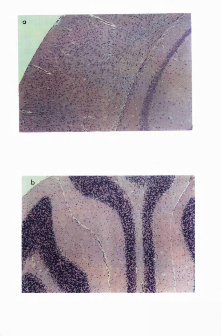
Figure 1.2
Cerebral sections from mice killed in the late (a) and the advanced (b) stages of
infection with T. b. brucei GVR 35, showing infiltration of the meninges and
the perivascular spaces by inflammatory cells. 1.2b shows the increment in the
cellular infiltrates into the meninges and a burst perivascular cuff (arrow) that is
spilling inflammatory cells into the adjacent parenchyma. Haematoxylin and
eosin. xlOO
77
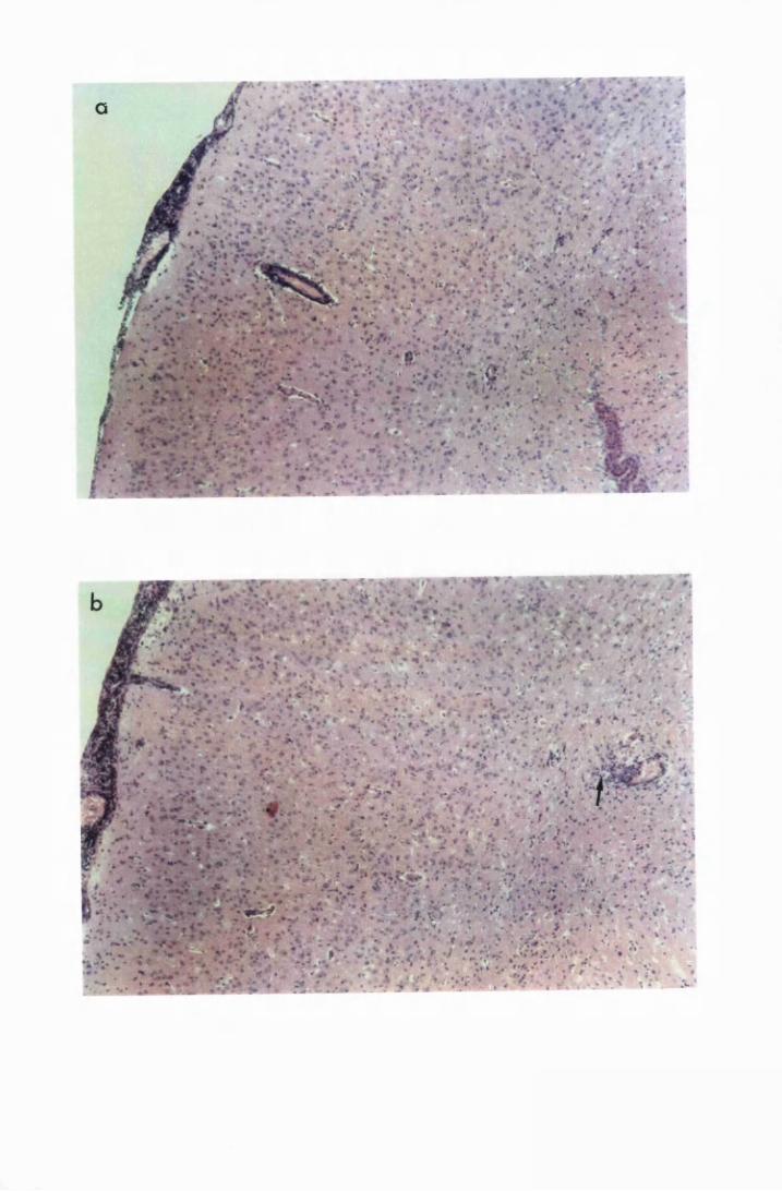
Figure 1.3
The hippocampal area of a mouse killed in the advanced stage of Tb. brucei
GVR 35 infection. It features numerous inflammatory cells infiltrating the
choroid fissure and perivascular cuffs within the hippocampus, some of which
are burst and spilling cells into the adjacent parenchyma (arrows). Haematoxylin
and eosin. xlOO
78
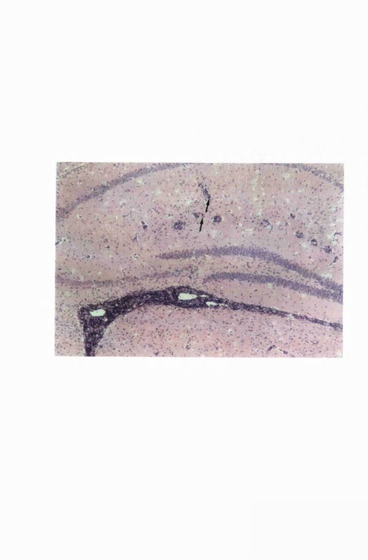
Figure 1.4
Cerebellar cortices of mice killed in late (a) and advanced (b) stages of infection
with T.b. brucei GVR 35, showing infiltration of the meninges, along the
cerebellar lobes, by inflammatory cells and perivascular cuffs. Note that in the
advanced stage (b) inflammatory cells can be se... in the white matter adjacent
to the large perivascular cuff (arrow). The sections were stained with
haematoxylin and eosin. Magnification. xlOO
79
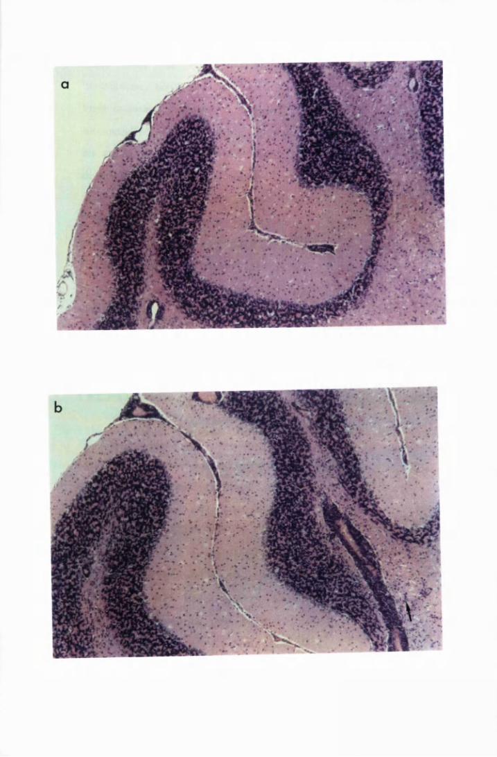
1989; Hunter et al, 1991; personal observations); see Figures 1.2a and 1.4a.
Trypanosomes can be seen mainly in the choroid plexus and occasionally in the
brain parenchyma ( Anosa et al, 1977; personal observations). In mice treated
subcuratively with early-stage drugs, the disease progresses faster. Thus, while
the pathological lesions shown in Figures 1.2a, and 1.4a, took 72 days to
develop in untreated mice, it took only 22 days after the subcurative therapy
(total of 43 days) for similar changes to develop (personal observation).
Following subcurative therapy, trypanosomes are found only in the brain
parenchyma until relapse of parasitaemia when the choroid plexus is re-invaded
(Jennings and Gray, 1983; personal observations). Neuronal degeneration can
be seen in the late-stage of infection, particularly in mice that receive Berenil
therapy (personal observation). The demarcation between the pathology in the
late and advanced stages of infection is not clear cut. Sometimes the changes in
this stage are as severe as those found in the advanced stage of infection.
1.8.1.3 Advanced-stage
Treatment of mice infected with T.b. brucei GVR 35 after 21 days of
infection with early-stage trypanocides, such as Berenil, clears the parasites in
the systemic circulation and in tissues other than the CNS. The parasites that
remain within the brain parenchyma continue to replicate and give rise to
recrudescent parasitaemia (Jennings et al, 1977; Jennings et al, 1979; Jennings
and Gray, 1983). Non-curative treatment of relapsed infections results in
exacerbation of the existing meningoencephalitis creating changes similar to
those seen in the brains of human reactive arsenical encephalopathy victims
(Adams et al, 1986; Haller et al, 1986; Jennings et al, 1989; Hunter et al,
1991). This is the advanced stage of infection in the mouse model and is taken
to represent the human reactive arsenical encephalopathy (RAE). Since the drug
used to simulate RAE, in the studies carried out in the mouse here, is not
arsenical, the reaction created is referred to as post-treatment reactive
80
encephalitis (PTRE). Mice in this stage of infection are dull, ataxic and
emaciated. There is a high incidence of hind limb paralysis (personal
observations). Some mice may still have enlarged spleens reflected as enlarged
abdomens.
Mice that receive a second Berenil treatment after relapse of
parasitaemia, show exacerbations of the CNS histopathological changes
(Jennings et al, 1989; personal observations). The infiltrations in the
subarachnoid and the perivascular spaces enlarge; some of the perivascular cuffs
rupture, releasing inflammatory cells into the adjacent parenchyma (Fig 1.2b,
1.3 and 1.4b). In addition, inflammatory cells and trypanosomes, not associated
with blood vessels, as well as neuronal degeneration, can be seen in the
parenchyma (Figure 1.5). The fore-, mid- and hindbrain are affected equally.
In summary, the pathology in the CNS of the mouse model, is a slowly
progressive infiltration of the meninges, the perivascular spaces and eventually
the brain parenchyma by cells of the lymphocyte/plasma series and
macrophages, and occasional neuronal degeneration. The development of these
changes can be hastened by subcurative therapy with trypanocidal drugs,
including Berenil, on or after 21 days of infection.
1.9 JUSTIFICATION AND AIMS OF THIS STUDY
Despite the fact that HAT has been recognised clinically since the turn of
the century, relatively little is known about the pathogenesis of the disease. The
event that ultimately leads to the development of CNS disease is the invasion of
the brain by trypanosomes (Greenwood and Whittle, 1973; 1980; Schultzberg et
al, 1988). Reports on the clinical signs and the pathological changes in the
brains of HAT patients during trypanosome infections and the adverse reactions
that occur after treatment with melarsoprol are well documented (Greenwood
81
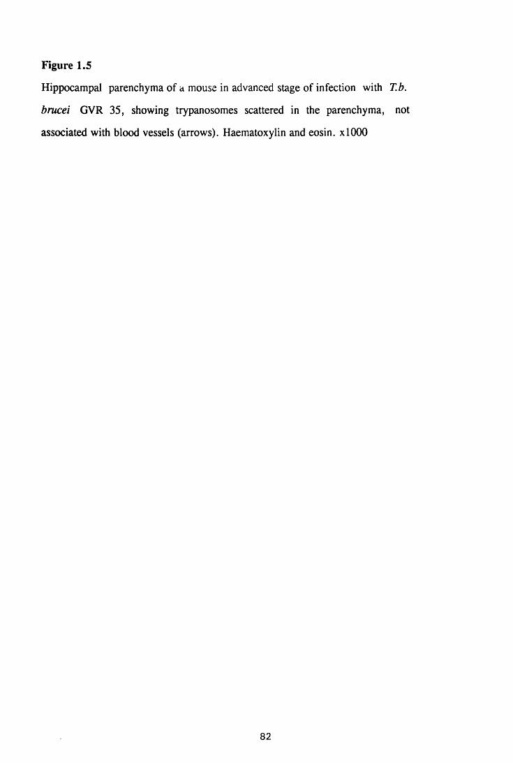
Figure 1.5
Hippocampal parenchyma of a mouse in advanced stage of infection with T.b.
brucei GVR 35, showing trypanosomes scattered in the parenchyma, not
associated with blood vessels (arrows). Haematoxylin and eosin. xlOOO
82
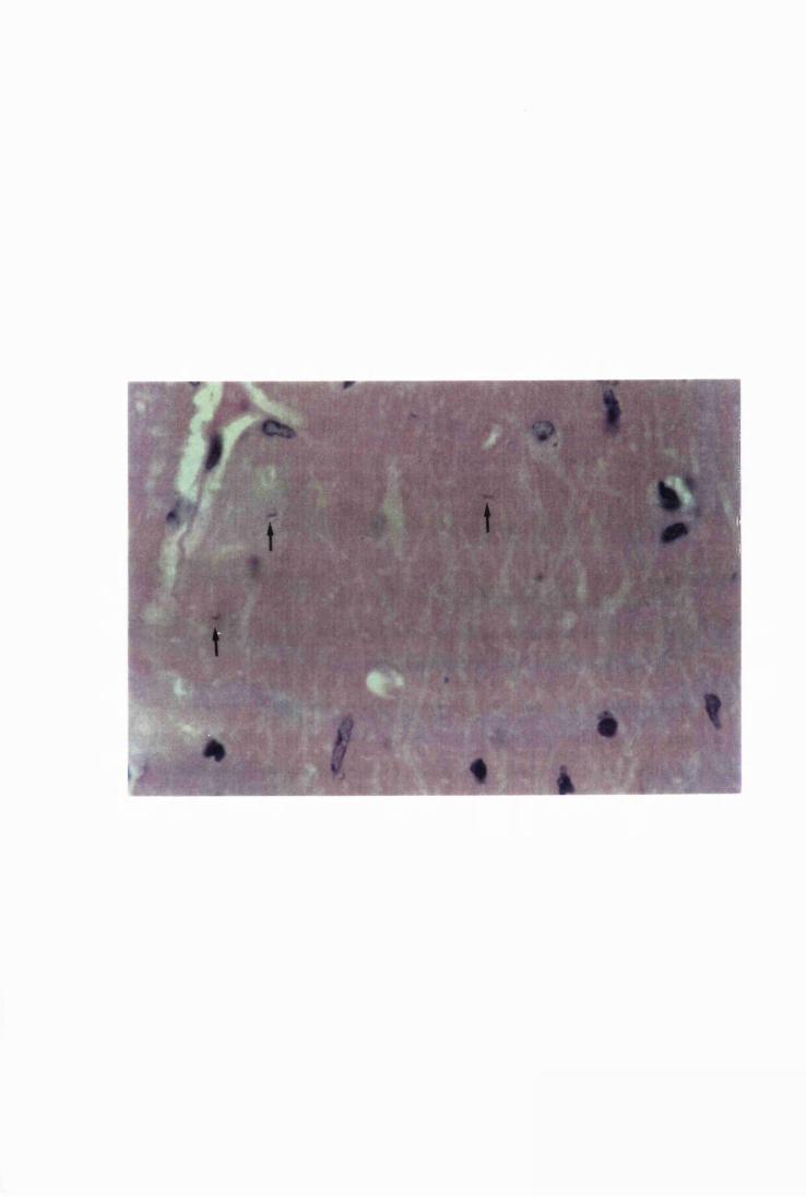
and Whittle, 1980; de Raadt, 1984; Adams et al, 1986; Haller et al, 1986,
Cegielski and Durack, 1991). According to these accounts the major
histopathological changes are infiltration of the subarachnoid and Virchow
Robin spaces by cells of the lymphocytes/plasma cell series and macrophages,
accompanied by diffuse gliosis. Neuronal degeneration is reported to occur only
in the very advanced stages of the disease (Adams et al, 1986).
Various hypotheses regarding the genesis of CNS disease during HAT
and the adverse drug reaction after melarsoprol therapy have been proposed
(reviewed by Hunter and Kennedy, 1992; section 1.6). These include: changes
brought about by trypanosome-produced toxins, such as phospholipases which
lead to production of cytokines and PGD2 production, leading to inflammatory
changes and slow wave sleep (Pentreath et al, 1990); induction of harmful
metabolic changes such as the production of indole-ethanol and phenylpyruvate
from aromatic amino acids, which may lead to depressed levels of
neurotransmitters and inhibition of mitochondrial function, respectively (Seed et
al, 1983; Patel et al, 1977); and the host's immune response to the invading
parasite or parasite fractions (Greenwood and Whittle, 1980; Lambert et al,
1981; Pepin and Milord, 1991). Thus, the CNS pathology could result from:
the tissue disruption that is caused by edema and infiltrating inflammatory cells
and trypanosomes (Adams et al, 1986; Haller et al, 1986); the local production
of immunoglobulins and immune complexes that activate the complement and
Kallekrein-kinin systems (Lambert et al, 1981; Boreham, 1985); the
autoantibody production against brain cells or against trypanosome antigens
adhering on the brain cells (Asonganyi et al, 1979; Rifkin and Landsberger,
1990); and mediators and lytic enzymes produced by the infiltrating cells
(Boreham, 1985; Pentreath et al, 1990; Hunter et al, 1991). Experimental
infections of mice with T.b.brucei, followed by curative and subcurative
treatment of these infections, seem to support the view that the CNS pathology
is a consequence of the host's immune reaction to trypanosomes within the brain
83
parenchyma (Hunter et al, 1992c).
Encephalopathy is a common and catastrophic complication of the
treatment of late stages of HAT with melarsoprol (de Raadt,1984; Poltera,
1985; Veeken, Ebelings and Dolmans, 1989), but its pathogenesis remains
obscure. Jennings et al (1989) suggested that it is caused by ineffective
subcurative therapy that leads to rapid disappearance of parasites from the blood
stream with persistence of trypanosomes in the CNS, concentrating the immune
reaction to the CNS. While melarsoprol, a late-stage drug, induces
encephalopathy (de Raadt, 1984; Adams et al> 1986; Haller et al, 1986; Pepin
et al, 1989), clinical experience with early-stage drugs in late-stage human
patients yielded equivocal results (de Raadt, 1966; Pepin and Milord, 1991).
Arsenical toxicity has been suspected as a cause of encephalopthy but it has been
shown that use of the heavy metal chelator, dimercaprol, does not prevent its
fatal outcome and that patients who recovered from episodes of encephalopathy
could receive further doses of melarsoprol (Pepin and Milord, 1991). Efficacy
of prednisolone in preventing arsenical encephalopathy (Pepin and Milord,
1991) implicates an immune mechanism. Another suggested cause of
encephalopathy is the deposition of immune complexes in the brain but, while
the levels of immune complexes in the blood rise during trypanosomal
infections, only minimal amounts of immune complexes have been found in
cerebrospinal fluid of late-stage patients (Whittle et al, 1980; Lambert et al,
1981) suggesting that immune complexes are not a major cause. Autoimmunity
due to cross-reacting antigens or due to trypanosome antigens attaching onto the
neuronal cell surfaces is not supported by the presence of mononuclear cells in
the lesion (Haller et al, 1986), instead of polymorphonuclear cellular infiltrates
which would normally associated with autoimmune reactions.
Precisely how the trypanosomes invade the CNS, how the inflammatory
cells infiltrate the invaded areas, and how the cellular infiltrations and the
84
diffuse gliosis, that follow trypanosome invasion of the CNS, might affect the
cerebral functions are unanswered questions. Recent studies have focused on the
role of astrocytes in HAT (Pentreath et al, 1989). The facts that astrocytosis
occurs in HAT (Adams et al, 1986; Haller et al, 1986) and in experimental T.b.
brucei infections in mice (Hunter et al, 1991), that astrocytes produce cytokines,
such as IL-1 and TNF, that can mediate meningeal inflammation (Fontana et al,
1982; Lieberman et al, 1989; Hertz et al, 1990) and that astrocyte activation
correlates with production of such cytokines in the brains of Tb. brucei-
infected mice (Hunter et al, 1991), strongly implicate astrocytes as initiators of
the CNS pathology. An understanding of how astrocytes respond to
trypanosomes and how these responses can be prevented or attenuated might
help to provide better methods of management of HAT.
The aims of this study are:
1. To investigate when astrocyte activation occurs in the brain of Tb. brucei-
infected mice, how this activation relates to the inflammatory cell infiltration
and how it is affected by curative and sub-curative trypanocidal therapy
2. To investigate the cytokines produced by cultured astrocytes after in vitro
exposure to trypanosomes and trypanosome fractions including, the whole
trypanosome lysate and purified VSG.
3. To investigate how eflornithine and azathioprine, two drugs reported to
prevent and attenuate inflammatory cell infiltrations in the brains of T.b. brucei-
infected mice, affect astrocyte activation in vivo and in vitro.
85
CHAPTER 2
GENERAL MATERIALS AND METHODS
86
2.1 ANIMALS
2.1.1 Adult mice
Adult female NIH mice were purchased from Charles River Limited,
England. They were allowed to acclim ate to the animal house facilities at the
Veterinary School, Garscube for at least 3 weeks before being infected with
trypanosomes. Feed and water were provided ad libitum throughout their stay in
the animal house facilities.
2.1.2 Neonate mice
Ten NIH breeding pairs of mice were purchased from the Harlan Olac
farm, England, and were used to establish a colony in the animal house facilities
at the Southern General Hospital, Glasgow. Two to 3-day-old litters from these
mice were used for all the in vitro astrocyte cultures used for this study.
2.2 TRYPANOSOMES
All the trypanosome stabilates used in this study were a generous gift
from Dr. F.W. Jennings of the Department of Veterinary Parasitology,
University of Glasgow.
2.2.1 Eflornithine-sensitive trypanosomes
The eflornithine-sensitive cloned stabilate was derived from
trypanosomes originally isolated from a wildebeest in the Serengeti National
Park in 1966 (Serengeti/66/SVRP/10). It was obtained from London School of
Hygiene and Tropical Medicine as the stabilate T brucei LUMP 1001 in 1977.
A clone was grown from this and a reserve stabilate, GVR/35/C1 was
established and maintained in liquid nitrogen (Jennings et al, 1977). As required
new working stabilates were prepared from this reserve and in the current
studies T.b. brucei stabilate GVR/35/C1.5, was used (Tkble 2.1).
87
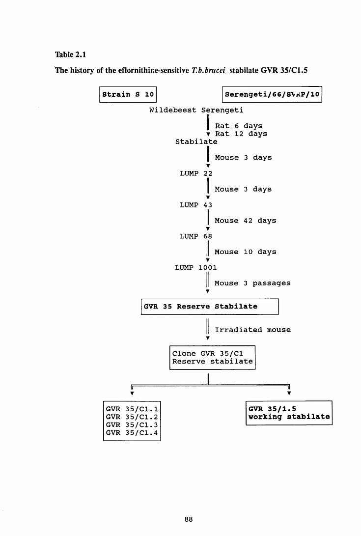
Table 2.1
The history of the eflornithine-sensitive T.b.brucei stabilate GVR 35/Cl.5
Strain S 10 Serengeti/66/S\*P/10W i l d e b e e s t S e r e n g e t i
R a t 6 d a y s v R a t 1 2 d a y s
S t a b i l a t e
M o u s e 3 d a y s▼
L U M P 2 2
M o u s e 3 d a y s▼
L U M P 4 3
M o u s e 4 2 d a y s▼
L U M P 6 8
M o u s e 1 0 d a y sv
L U M P 1 0 0 1
M o u s e 3 p a s s a g e sv
GVR 35 Reserve Stabilate
I r r a d i a t e d m o u s eY
C l o n e G V R 3 5 / C l R e s e r v e s t a b i l a t e
G V R 3 5 / C l . 1G V R 3 5 / C l . 2G V R 3 5 / C l . 3G V R 3 5 / C l . 4
GVR 35/1.5 working stabilate
88
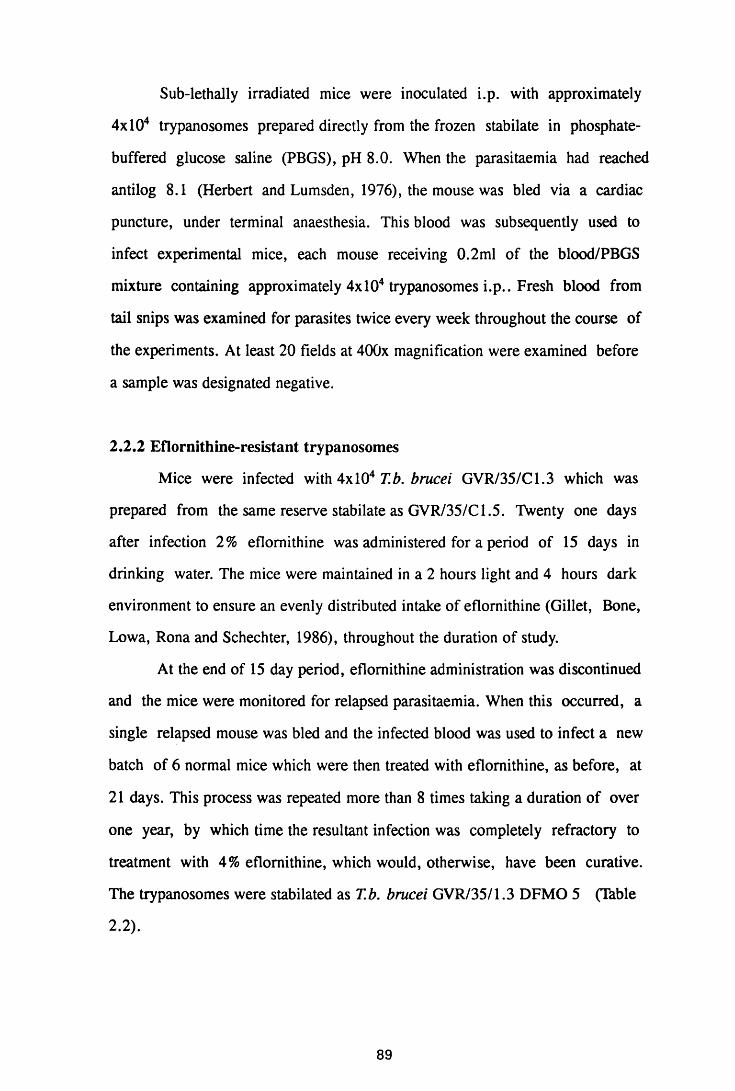
Sub-lethally irradiated mice were inoculated i.p. with approximately
4xl04 trypanosomes prepared directly from the frozen stabilate in phosphate-
buffered glucose saline (PBGS), pH 8.0. When the parasitaemia had reached
antilog 8.1 (Herbert and Lumsden, 1976), the mouse was bled via a cardiac
puncture, under terminal anaesthesia. This blood was subsequently used to
infect experimental mice, each mouse receiving 0.2ml of the blood/PBGS
mixture containing approximately 4xl04 trypanosomes i.p.. Fresh blood from
tail snips was examined for parasites twice every week throughout the course of
the experiments. At least 20 fields at 400x magnification were examined before
a sample was designated negative.
2.2.2 Eflornithine-resistant trypanosomes
Mice were infected with 4x10⁴ T.b. brucei GVR/35/C1.3 which was
prepared from the same reserve stabilate as GVR/35/C 1.5. Twenty one days
after infection 2% eflomithine was administered for a period of 15 days in
drinking water. The mice were maintained in a 2 hours light and 4 hours dark
environment to ensure an evenly distributed intake of eflomithine (Gillet, Bone,
Lowa, Rona and Schechter, 1986), throughout the duration of study.
At the end of 15 day period, eflomithine administration was discontinued
and the mice were monitored for relapsed parasitaemia. When this occurred, a
single relapsed mouse was bled and the infected blood was used to infect a new
batch of 6 normal mice which were then treated with eflomithine, as before, at
21 days. This process was repeated more than 8 times taking a duration of over
one year, by which time the resultant infection was completely refractory to
treatment with 4% eflomithine, which would, otherwise, have been curative.
The trypanosomes were stabilated as T.b. brucei GVR/35/1.3 DFMO 5 (Thble
2 .2).
89
Table 2.2
The history of the eflornithine-resistant T.b.brucei stabilate GVR 35/C1.3 DEMO 5
G V R 3 5 / C l . 3
m i c e 2 % e f l o m i t h i n e R e l a p s e
T
S t a b i l a t e
M i c e 2 % e f l o m i t h i n e > 8 p a s s a g e s
T
S t a b i l a t e
M i c e 4 % e f l o m i t h i n e▼
W o r k i n g s t a b i l a t e
G V R 3 5 / C l . 3 D F M O 1 - 4 GVR 35/Cl.3 DFMO 5w o r k i n g s t a b i l a t e
90
2.3 INFECTION AND TREATMENT REGIMENS
2.3.1 Infection without treatment
A group of adult female NIH mice were infected with 4xl04 parasites of
either the eflomithine sensitive stabilate T.b. brucei GVR 35/C 1.5 or the
eflomithine resistant stabilate GVR 35/Ci.J DFMO 5. At this inoculation dose,
these stabilates have been reported to produce parasitaemia in mice within 3
days of infection. The infection was allowed to proceed without any
trypanocidal therapy to the terminal stages of infection. During the course of the
disease, mice died as a result of infection, with few surviving beyond day 52 of
infection. Five mice were sacrificed every 7 days
2.3.2 Induction of post-treatment reactive encephalitis
Adult female NIH mice were infected with 4x 10⁴ trypanosomes of
either the eflornithine-sensitive stabilate T.b. brucei GVR 35/C 1.5 or the
eflornithine-resistant stabilate T.b. brucei GVR 35/C 1.3 DFMO 5. They were
then divided into two groups. One group, PI, was treated with 40mg/kg
diminazene aceturate (Berenil, Hoechst) on day 21 after infection. Five mice
were killed every 7 days thereafter. At this dosage, Berenil has been reported to
give maximal trypanocidal effect without toxicity to mice (Jennings et al, 1977).
Treatment of T.b.brucei-inioctod mice with drugs that do not cross the blood-
brain barrier such as Berenil, after 21 days of infection, induces a PTRE
(Jennings et al, 1977; Jennings et al, 1989; Hunter, Jennings, Adams, Murray
and Kennedy, 1992a). The second group, P2, received 40mg/kg Berenil on day
21 of infection and again after a relapse of parasitaemia. This relapse occurred
20-22 days after administration of the first Berenil. This second Berenil
treatment has been reported to induce a very severe PTRE (Jennings et al, 1989;
Hunter et al, 1992a). Five mice were sacrificed every 7 days after the second
Berenil treatment.
91
2.3.3 Evaluation of curative trypanocidal therapy on astrocyte activation
A combination of the arsenical drug, Mel Cy (Cymelarsen, Rhone
Meriuex, France) and a nitroimidazole compound, MK436 (Merck Institute for
Therapeutic Research, New Jersey, U.S.A.), has been reported to cure chronic
T.b. brucei infections in the mouse model (Jennings, 1991c). This combination
was, therefore, used in the current study for the evaluation of astrocyte
activation after curative therapy.
Mice were infected with either the eflornithine-sensitive or eflornithine-
resistant T.b. brucei stabilates and treated with 40mg/kg Berenil on day 21 after
infection and again after relapse of parasitaemia. Seven days after the second
Berenil, a group of these mice, CT, were treated with two consecutive daily
doses of 5mg/kg Mel Cy + 15mg/kg Mk436 i.p. Groups of five mice were
serially sacrificed 5 days after the combined Mel Cy/MK436 therapy and every
7 days thereafter.
2.3.4 Evaluation of the effect of eflomithine and azathioprine on the
induction of the post-treatment reactive encephalitis
2.3.4.1 Eflomithine
Adult female NIH mice were infected with the eflomithine resistant T.b.
brucei stabilate, GVR 35/C 1.3 DFMO 5. They were then divided into 4 groups
(Thble 2.3). Two percent eflomithine was administered ad libitum in drinking
water for a duration of 7 or 14 days, in a 2-hour-light-4-hour-dark environment
to spread the drug intake evenly (Gillet et al, 1986). Eflomithine administration
was arranged so that the treatment with Berenil was at the beginning or end of
eflomithine course, or on the 7th day of eflomithine therapy. Thus, one group,
E l, received eflomithine from the time of Berenil treatment on day 21 after
infection. Two other groups, E2 and E3 received eflomithine for 7 and 14 days,
respectively, before the treatment with Berenil on day 28 after infection.
92
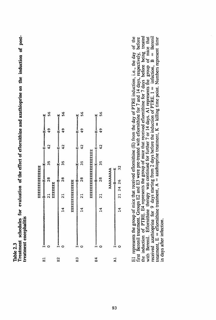
Table
2.
3Tr
eatm
ent
sche
dules
for
ev
alua
tion
of the
ef
fect
of
eflo
mith
ine
and
azat
hiop
rine
on
the
indu
ctio
n of
post
tr
eatm
ent
ence
phal
itis
l£>Ui in
cr> Ui
CMPS
inrn
in PS m
Cl PS
CMPS ^
in PS rn
m o
W
M o
CMu
ID PS in
<n PS <3*
CMPS **
in & rn
in PS in
ClPS ^
PSCMWw w w w wPS PS m
in
u pa CMw pa *£ t*Z mw paw paH paW 00 00 00 pa 00 |CW « CM CQ CM W aa cm pa aa cm < mw H pa pa < CQ CMw w pa paw w pa paw w pa pa CMw pa pa paH rH u rn pa rH pa rH rHw ca cm pa cm pa cm pa CM CMpapapapa<t pa ■d" •d*rn pa rH 1—1 rH
m o
mw
M O
w
M O
5 S "8 ed ’£ ~ .0 P.0 ^ 4 .
• Sh C&D C .I 1)E 11 sj
-§ .8. Oh n
03 C^ ’<u c<L) X) o «j= «
X 3* c~ 55 • O ST ft ^8 3 8 - 3 .2
C S« on <D c J5 O "O -o in II
1 *•O OW 5 ^
e .5D pj ‘“u
s s „ a s gg t - J e£X t "S
l-H . r-« PhO .t3 «+-i 60' t <4"1 C O C1) 5 • r- —
>> C O 5 § rrt
^ *2 w ■§ o i.a•C u. . ' t 'b u o > ^ c *
P C 'S j-*S 2 8 ° J cis a>
r' ^ 1) E^ 03 4) C *3_ ^ -c x: o $t-* I4-1 p U 8 D 3 2 ■*—>.S 85.H'3 ‘S «^ S"C
-C
W
g a o C -° §• o a. o - l 2u £ o . S | 3n o t; P y 8 % 60g£ d
8 Mil8 S « g i <i: c 1 * §<3 03 O trt C■S (N 4J c . W po B o .U r t ' lo m p a) *a «E c w on +-•^ P W o i) O n . e t cg. . g S ’S f i3 *J Q-S .tP -tP . O C C g <L> C C*-< ?T c c g o
o ’co-c<u ip (4—1 p5 S o w O « ^00 ic -e II c ^ — e -tp§ l - 2 c 83c h c d -o c ^& $ ■ § « ! | - 1-1 •"* •--< is
S 8— C « .ti PJ tc: ■£ £
. >» cd*oiC C
93
A fourth group (E4) was treated with eflomithine for 7 days before induction of
PTRE with Berenil on day 28 of infection. In this particular group, eflomithine
therapy was continued for a further 7 or 14 days, after the Berenil treatment.
Mice, in groups El to E4, were sacrificed in groups of 5 at the end of
eflomithine course and every 7 days, thereafter. However, the mice that
received Berenil at the end of the eflomithine course were sacrificed in groups
of five every 7 days after the Berenil treatment.
2.3.4.2 Azathioprine
Female NIH mice were infected with 4x 10⁴ trypanosomes of the
eflornithine-sensitive T.b. brucei stabilate GVR 35/C 1.5. They were treated
with lOOmg/kg azathioprine on day 24 to day 32 after infection and a single
Berenil injection (40mg/kg) on day 26 post infection (Table 2.3). At this dosage,
azathioprine has been reported to prevent and ameliorate inflammatory cell
infiltration into the brains of T.b. brucei-infected mice (Hunter, Jennings
Kennedy and Murray, 1992b). Higher doses of azathioprine were not well
tolerated, with mice dying early in the course of treatment (F.W. Jennings,
personal communication), and lower doses were not effective (personal
observations). Mice were sacrificed at the end of azathioprine course and every
7 days, thereafter.
2.3.5 Evaluation of the effect of eflomithine and azathioprine on astrocyte
activation during an established post-treatment encephalitis
2.3.5.1 Eflomithine
Female NIH mice infected with the eflomithine resistant T.b.brucei
trypanosome stabilate and divided into two groups (T^ble 2.4). The first group
(E5) was treated with 40mg/kg Berenil i.p. on day 21 post infection. Seven days
after the Berenil injection, they received 2% eflomithine ad libitum in drinking
94
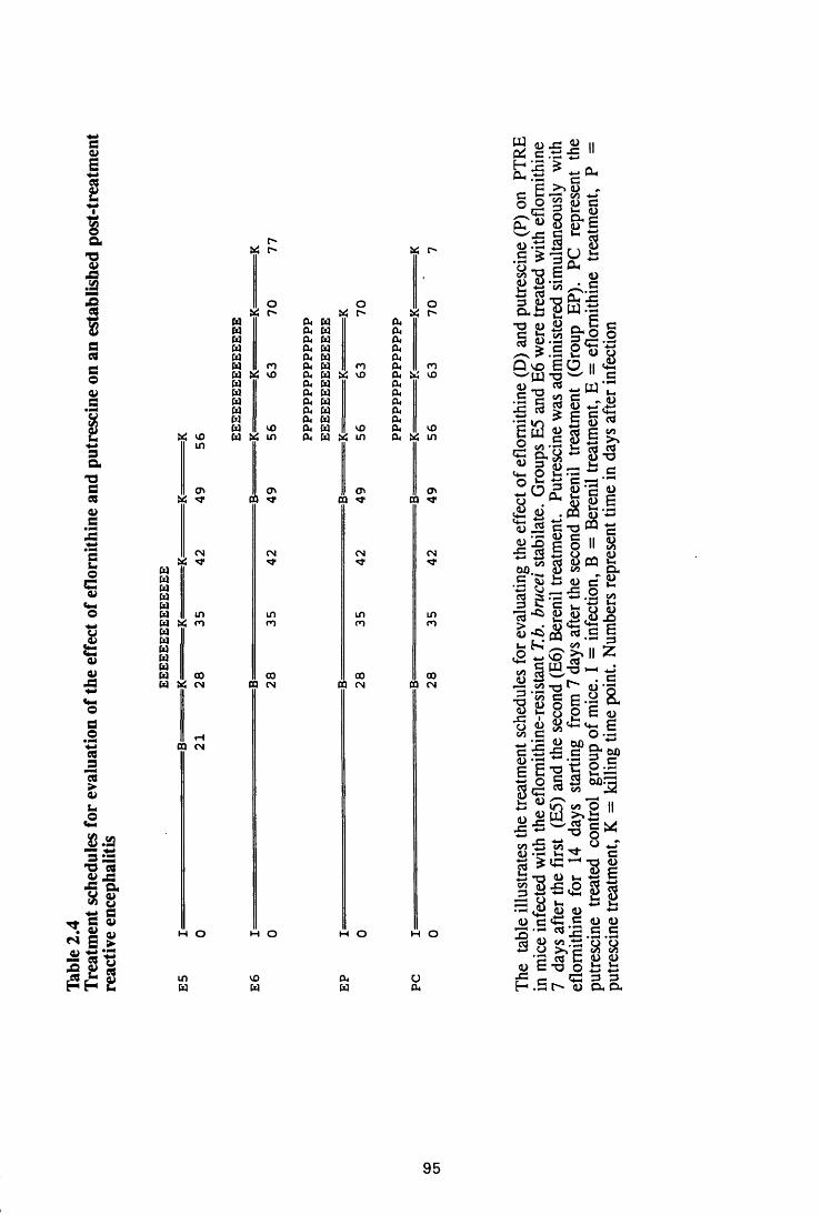
Thble
2.
4Tr
eatm
ent
sche
dules
for
ev
alua
tion
of the
eff
ect
of ef
lom
ithin
e an
d pu
tres
cine
on an
esta
blish
ed
post
-trea
tmen
t re
activ
e en
ceph
aliti
s
X m in
CN
in X rn
ooH X cn
CQ CN
m o
inX
X
O X r-'
(T> CQ
CN
inrn
ooCQ CN
H O
inX
or
tn CQ
CN
inm
ooCQ CN
M O
X r-
XO
X X X XX X X XX X X XX X X XX X X XX m x x n x rnX X 10 X X 5.4 m x x mX X X XX X X XX X X XX X X XX X X XX m x x in x mX X in x X i»4 in x i>4 in
o» cQ
CN
00 CQ CN
M O
11.■§ S ce >>(o ,C 5 TS r/i ■*-*o o <u c
'~G n r u Cu ^ 8 D 6
i n « *<L>
^ ^ . sIsl&f■ o g S a S s§ fe-| g c -S £ u oa vo 6 O II «£wrn -o W II C
_ W -2 £ - fc
i) © "-j
• J I s s* § S *
C/5 >%i « . S 8 I
t/1
• S S -a 1"tfl c
3 ^ ti c<+-. O H .50 t ti c rzjrn 3 <u G a)w n . cl C 25
^ ^D iS C T3 ® r£ ‘I i § 11 8o n o j <*> D -
3 ^ G ■£ .2 cn1 * i s | j s
fe ^ £ n 7; < 2 ~ V D S ' I I z W/o i—i *J3 -a S e § ' |1 8 8 I E O •5 i " * ^ 5 eS <•» G +-• IS 2 rvc £ £ c W) <U 'G G o cS E a S ’3 B B c «g & gg c * c/i^ ' 2i3 W Jo cn O || 2 W >*£2 x: w c s>£ ~ -o § *c/J ■£ to _*. ^ *-T W ■— X ^ Ga ? « - - s a
—1 «£ <D 11) -•-*<U G *+-< C D <u
T ! - C Ccrt 1) cn 'o O-3 O K «•? (« C « u d c-u o i i
-C - G 3 3h .S t '' <u ex cx
95
water for 7 or 14 days.
The second group, E6, was injected with 40mg/kg Berenil i.p., on day
28 after infection and again after a relapse of parasitaemia (20-22 days after the
first Berenil). This was followed, 7 days later, by administration w 2%
eflomithine ad libitum in drinking water for 7 or 14 days. During eflomithine
therapy animals were maintained in a 2-hour light, 4-hour dark environment to
ensure an even intake of eflomithine (Gillet et al, 1986).
Five mice were sacrificed on the last day of eflomithine treatment and 7
days (group E6) or 7 and 14 days (group E5), thereafter.
2.3.5.2 Eflomithine and putrescine
A group of NIH mice were infected with the eflomithine resistant T.b.
brucei stabilate GVR 35/C 1.3 DFMO 5. They were treated with 40mg/kg
Berenil on day 21 after infection and again after relapse. They were then
divided into 2 groups (Thble 2.4). Seven days after the second Berenil injection,
the first group of mice (EP) were treated with a combination of 2% eflomithine
in drinking water and 400mg/kg/day putrescine i.p. for 14 days. The second
group of mice (PC) received putrescine, for 14 days starting from 7 days after
the second Berenil treatment. Mice were then sacrificed on the last day of
eflomithine -I- putrescine administration.
2.3.5.3 Azathioprine
Female NIH mice, infected with 4x 10⁴ trypanosomes of the eflomithine
sensitive T.b. brucei stabilate, were treated with 40mg/kg Berenil on day 26
after infection. They were then divided into two groups (Thble 2.5). One
group, A2, was treated with lOOmg/kg azathioprine for 7 or 14 days starting
from day 32 after infection (6 days after induction of PTRE). Mice were
sacrificed, in groups of 5, on the last day of azathioprine administration.
96
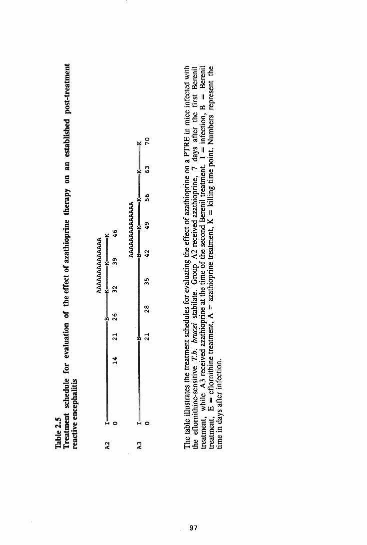
Table
2.
5Tr
eatm
ent
sche
dule
for
evalu
ation
of
the
effec
t of
azat
hiop
rine
ther
apy
on an
estab
lishe
d po
st-tr
eatm
ent
reac
tive
ence
phal
itis
VO
<T> n
CN« rn
VO CO CN
M o
CN<
oUt t-
VOO Z m
ov
CN CO ^
inm
coCN
DQ CN
h o
n
ITS ^ •H g* § a■ <1V -t->c
&£ cx<u
.a
<uo= •« S 2 S - . 2 ??• S a l e g « . S |"I" a
73 5C(N. X ^ o 1 CD
- £ £ g l - s c g «
s - s 3I a ii55 T-i
<D
6D
^ T3 -
• s § §8 S 1In IA £
cn a S < •5 5Vt-H fl\
O nO g§ <u 'C ^ *-1 £ 9* 13 O -a .2 . <L> "5 1)
cO
<UO rt w d
* i.«/i .O fl) II«* ’C <Oh -
2 e .c <uto 6
•8-C
« $ e -* u - L ’ > 0) . c c8 5 - 2
— ^ ^ « c < ® c<D <Dy w o) in rt i O qj£ 2 5 II wV/3 .5 J-3-S S W
u
s<u
<U■SB’S
a)
<L>
eOC/3
E w <_r o C C 3C g g f i d E a
H ■£f\ \
* S S.I
97
The second group of mice, A3, was treated with 40mg/kg Berenil after
relapse of parasitaemia, and with lOOmg/kg azathioprine for 7 or 14 from the
day of the second Bertnil treatment for 7 or 14 days. Five mice were sacrificed
at the end of the azathioprine therapy and every 7 days thereafter.
2.4 EVALUATION OF PATHOLOGICAL CHANGES IN THE BRAIN
2.4.1 Perfusion of the brain to remove peripheral blood
At sacrifice, mice were perfused with normal saline (0.9% NaCl) pH
7.0, to remove peripheral blood from the brain. The mice were terminally
anaesthetised with trichloroethylene (BDH, analytical grade) and while the heart
was still beating, the chest cavity was opened at the level of xiphoid cartilage to
expose the heart. The right atrium was perforated to create an outlet for the
blood plus saline in order to facilitate the perfusion. Normal saline was injected
into the left ventricle with a hypodermic, gauge 20 needle. Approximately
150ml saline was used to achieve sufficient perfusion. The clearance of blood
from the kidneys was used as an indication of sufficient perfusion.
2.4.2 Fixation of the brain for histopathology and immunocytochemistry
The perfused brain was removed and fixed in 10% neutral buffered
formaldehyde for at least 2 hours and then post-fixed in corrosive formol for a
minimum of 24 hours. The embedding of the brains in wax was preceded by a
series of dehydrations with methylated spirit and absolute alcohol. All the stages
were carried out in a programmable machine under vacuum and pressure. Thus
the brains were put through three, 1-hour cycles in methylated spirit, followed
by three, 1-hour cycles in absolute alcohol at 40°C. This was followed by a 1-
hour clearing in xylene/alcohol (1:1) and another, 1-hour cycle in xylene at
40°C. The brains were then put through four cycles of wax processing at 60°C,
the first two of 1 hour each followed by two, 2-hour cycles.
98
Transverse (coronal) sections, 3-4/irn thick, were made through the
forebrain, midbrain and hindbrain and stained with haematoxylin and eosin.
Mercuric chloride-formalin (Corrosive formol or Formal sublimate)
Saturated aqueous mercuric cnloride 900ml
Formalin 100ml
Neutral Buffered formalin (10%)
Formalin
Acid sodium phosphate monohydrate
(NaH2P 04.H20)
Anhydrous disodium phosphate
(Na2HP04)
Make up to 1 litre with tap water
2.4.3 Immunostaining for evaluation of astrocyte activation
Immunostaining for glial fibrillary acidic protein (GFAP) was used to
identify astrocytes and to aid assessment of astrocyte activation in brain
sections. Mercuric chloride reacts with diaminobenzidine (DAB) to produce a
brown colouration, creating unwanted background staining. To remove the
effects of mercuric chloride all sections were washed in Lugols iodine and then
with 1 % sodium thiosulphate for 5 minutes, with a rinse in water after each
wash. The sections were then dewaxed in xylene for 10 minutes and hydrated
through decreasing concentrations of alcohol. Thus, the sections were placed in
100% ethanol through 90%, 70% and 50% ethanol to water for 3 minutes at
each concentration. Peroxidase activity in tissues was quenched by soaking the
sections in 3% H20 2 for 5 minutes and rinsing in PBS, pH 7.4. Non-specific
antibody binding was blocked by incubation with normal swine serum for 30
minutes.
99
100ml
4g
6.5g
A polyclonal rabbit anti-GFAP antibody (DAKO) diluted 1:1,000 in
PBS-0.1 % bovine albumin was placed on the sections and left overnight at 4°C.
After rinsing in PBS, the sections were incubated with the secondary antibody,
swine anti-rabbit immunoglobulins (DAKO) diluted 1:500 in PBS for 30 minutes
at room temperature. After washing in PBS (3x5 minutes), the sections were
placed in the peroxidase substrate diaminobenzidine (DAB) for 5 minutes to
develop colour for visualisation and rinsed in water. The sections were then
counter stained with haematoxylin for 10 seconds, rinsed in tap water, blued in
Scot's tap water substitute and dehydrated before mounting with DPX mountant
(BDH) and coverslips.
Phosphate Buffered Saline
Sodium chloride (KC1) 0.2g
^/-Sodium hydrogen orthophosphate 2.9g
(Na2HP04.12H20)
Potassium dihydrogen orthophosphate 8.0g
(KH2P04)
Potassium chloride (KC1) 0.2g
Make up to 1 litre with distilled water and adjust to pH 7.4
DAB solution
H20 2 300/xl
PBS 500ml
DAB lOOmg
DAB is carcinogenic, so all work with DAB was performed in a safety
cabinet (DAB cabinet), with gloved hands and protective clothes, to prevent
contact and inhalation. Thirty ml of PBS/H20 2 mixture was aspirated into a
syringe and injected into an isopac containing lOOmg of DAB powder and
100
shaken to dissolve it. The mixture was then aspirated out and mixed with the
remaining of PBS/H20 2 mixture and used for colour development.
2.5 PREPARATION OF IN VITRO CUTTURES OF ASTROCYTES
2.5.1 Dissection and dissociation of cells
Astrocyte preparation was performed as described by Wolswijk and
Noble (1989). Two to 3-day-old mice were decapitated and the heads were
transferred to a sterile tissue culture room where they were sprayed with 70%
ethanol. The skull was exposed by removing the skin, cutting from the occiput
and just below the left ear, along the temple to above the left eye and then round
above the right eye, along the temple, below the right ear and back to the
occiput. Using a separate sterile set of instruments, the crown of the skull was
removed along the same cutting lines as the skin to expose the brain.
The cerebral cortices were removed by gently teasing them away from
the cerebellum and the hindbrain along the pineal recess, and gently lifting them
away from the brain stem. They were then placed upside down in a sterile petri
dish containing Hank's balanced salt solution (HBSS). The hippocampus was
removed by gentle dissection along the extension of the lateral ventricles. The
cortices were then turned upside up and the meninges removed, leaving them
free of blood vessels. The "clean" cortices were transferred to another HBSS-
containing sterile dish.
To dissociate the cells, the cortices were removed from the HBSS-
containing-petri dish, placed onto another sterile dry petri dish and chopped into
small pieces using a scaple blade. The pieces were incubated with collagenase
for 45 minutes at 37°C, centrifuged at 89.6g for 10 minutes and the supernatant
discarded. They were then incubated with 0.125% trypsin (Bovine pancreas type
III, Sigma), in ethylene diamine tetra acetic acid (EDTA), for 30 minutes at
37°C. A mixture of soya bean trypsin inhibitor and DNase was used to
inactivate trypsin and digest any free DNA. The cells were centrifuged again at
101
89.6g for 10 minutes and the supernatant was removed. A series of triturations,
first in a 5-ml pipette and then through a 23 gauge-needle were carried out. The
cells were washed in 10ml of HBSS, by spinning at 89.6g, after each trituiation
and then removing the supernatant. The cells were resuspended in culture
medium and counted in an improved Neubauer haemocytometer.
The dissociated brain cells were plated at a density of 5x10⁶ in poly-L-
lysine- (PLL-) coated, 80cm2 tissue culture flasks (Nunclon). They were grown
in Dulbecco's modified Eagles medium containing 1.0g/l glucose and
supplemented with 10% fetal calf serum (DMEM/10%FCS,) (Gibco), 2mM
glutamine (Sigma) and 25/tg/ml gentamycin (Flow Laboratories). Once a
confluent monolayer of flat cells had been formed (about 7-10 days), the layer
of process bearing cells growing on the flat cells were removed by treating with
20fiM cytosine arabinoside (AraC) (Sigma) for 48 hours to eliminate most of
the faster dividing non-astrocytes.
2.5.2. Purification of the cell cultures
The cells were removed from the flask by washing with Ca2+ Mg2+ free
Hanks balanced salt solution (HBSS) (Gibco), followed by an incubation in
0.54mM EDTA (Sigma) in HBSS for 10-15 minutes, and the addition of trypsin
(Bovine pancreas type III) (Sigma) to a final concentration of 300 I.U./ml. After
the cells had detached from the surface of the flask, the trypsinisation was
stopped by adding 1ml per 10ml of EDTA/trypsin solution of SBTI-DNase
[5200 I.U. Soybean trypsin inhibitor (Sigma) and 3.0mg/ml bovine serum
albumin (BSA fraction V, Sigma) in DMEM]. Cells were then slowly triturated
through a lOml-blow out pipette, centrifuged for 10 minutes at 89.6g,
resuspended and treated in suspension with rabbit complement (Buxted Ltd,
diluted 1:10) plus antibody A2B5 (ascites 1:1,000, Eisenbarth, Walsh and
Nirenberg, 1979), monoclonal antibody 04 (concentrated hybridoma supernatant
102
1:100, Sommer and Schachner, 1981) and anti-galactocerebroside (GalC)
monoclonal antibody (hybridoma supernatant 1:10, Ranscht, Clapshaw, Price,
Nobel and Seifert, 1982). Treatment with these antibodies eliminated A2B5+
neurones, A2B5+, 04 , GalC' oligodendrocyte type 2 astrocyte-progenitor
cells, A2B5+ GFAP+, type 2 astrocytes, and GalC" oligodendrocytes (Abney,
Williams and Raff, 1983; Raff, Abney, Cohen, Lindsay and Nobel, 1983a;
Raff, Miller and Nobel, 1983b, Sommer, Lagenaur and Schachner, 1982). The
remaining cells were plated on PLL- coated flasks at a density of 5xl06 cell per
flask and maintained in 50% fresh DMEM 10%FCS + 50% astrocyte
conditioned medium (ACM) (as in Nobel and Murray, 1984). Cultures were
kept in an humidified incubator (Flow Laboratories) at 37°C and 92.5% air and
7.2% C 02. At this stage, over 95% of the cells in these cultures should have the
antigenic phenotype of type-1 astrocytes (GFAP+, A2B5-) as determined by
indirect immunofluorensence (Raff et al, 1983a). In my experiments, using
immunofluorescence, over 3000 cells were counted and 99.8% were type-1
astrocytes.
Dulbeccos modified Eagles medium
Low glucose form of DMEM was purchased from Gibco
DMEM +10% Fetal Calf Serum
FCS (Gibco)
DMEM (Gibco)
Glutamine (200mM)
Store at 4°C.
Astrocyte conditioned medium
Culture supernatant collected from a culture of pure astrocytes
after at least 24 hours on the cells. It contains growth factors
103
10ml
90ml
lml
secreted by astrocytes.
Hanks Balanced Salt Solution
Ca++ and Mg++ free (Gibco)
Leibovitz Medium (LI5)
From Gibco
Collagenase
Collagenase type II from Worthingtons (200U/mg)
A working solution of 2000U/ml was made by dissolving
lOmg/ml Collagenase in L15
Trypsin
Bovine pancreas trypsin from Sigma
A stock solution of 2.5mg/ml was made by dissolving trypsin in
HBSS. It was then filtered through 0.22/>im filter, and stored in
1 and 2ml aliquots.
and stored at -20°C. Before use, the trypsin was diluted 1:1
with EDTA
Soya bean trvpsin-inhibitor-DNAse (SBTI/SD!
Dissolved the following in L15
Bovine serum albumin
fraction V (Sigma, U.K.) 3.0mg/ml
Soybean trypsin inhibitor (Sigma) 0.52mg/ml
Bovine pancreas DNAse (Sigma) 0.04mg/ml
The mixture was placed on a roller mixer and left dissolving for 20 minutes.
104
The solution was then passed through 0.22/*m pore filter and stored in 2ml
aliquotes at -70°C.
Ethylene diamine tetracetic acid £FOTA)
A stock solution was made by dissolving 0.2mg/ml EDTA
(Sigma) in Ca++ Mg++ free HBSS (Gibco). It was then filtered
through 0 .22^ m pore filter and stored in 20ml aliquotes at 4°C.
Glutamine
200mM sterile solution purchased from Sigma, was aliquoted
into 5ml portions and stored at -70°C.
Poly-L-lvsine (PLL)
25mg vials were purchased from Sigma. Stock solutions of
4mg/ml were made, filtered through 0.22pim, aliquoted into
300/xl portions and stored below -20°C. At the time of use, the
300/nl aliquotes were made up to 40ml with double-distilled,
water and used to coat the flasks.
2.5.3 Evaluation of the purity of the cell cultures
A flask containing a confluent monolayer of astrocytes was trypsinised
and cells collected in a centrifuge tube. The trypsin was inactivated with soya
bean trypsin inhibitor, the tubes centrifuged at 89.6g and supernatant discarded.
The cells were then washed twice in HBSS, i.e., the cells were resuspended in
HBSS, centrifuged again and supernatant discarded twice. The cells were
resuspended in DMEM/10%FCS, counted and seeded onto 13mm-diameter,
PLL-coated coverslips in a 24-well tissue culture plate (Nunclon), at a density of
4xl04 cells per coverslip. They were then maintained in 50% DMEM/10%FCS
and 50% astrocyte conditioned medium for 24 hours, in 92.5% air, 7.2% C 02
105
in a humidified incubator, at 37°C.
The cells were stained with fluorescein-labelled rabbit anti-GFAP as
follows: the coverslips were removed from the wells and washed three times in
staining solution. To wash the cells a coverslip was held with tissue forceps and
dipped into three Bijou bottles containing fresh staining solution and gently
moved up and down for 10 seconds in each bottle. The coverslips were then
placed on mounds in a staining tray and kept moist with a film of staining
solution. The cells were incubated with 50^1/coverslip of fluorescein-labelled
rabbit anti-GFAP antibody diluted 1 in 1,000 in staining solution, for 30 minutes
at room temperature. The antibody solution was drained by tipping the edge of
the coverslip onto a tissue paper followed by washing the cells three times, in
staining solution, as before. The cells were chilled at -20°C for 10 seconds,
fixed with 50/d/coverslip prechilled methanol for 10 minutes at -20°C and
washed in distilled water until the methanol streaks disappeared. The coverslips
were then mounted onto slides (with the cells facing the slide) with Citifluor and
the edges sealed with glyceel.
Under a fluorescent microscope, the GFAP+ cells were fluorescent and
the GFAP' cells were non-fluorescent. The cells were counted and the
percentage purity calculated as follows:
number of fluorescent cells
x 100
total cells counted
Over 3000 cells were counted and 99.8% were GFAP+.
Staining solution
lOx HBSS (Gibco)
Donor calf serum
50ml
25 ml
10% sodium azide (in PBS) 5ml
106
HEPES (Sigma
HEPES sodium salt (Sigma)
2.38g
2.60g
Make up to 500mls with distilled water and store at 4°C
2.6 PREPARATION OF ASTROCYTE STIMULI
2.6.1 Separation of trypanosomes from host blood cells
Four to 5 days after infection with T.b.brucei stabilate GVR 35/C 1.5,
mice developed parasitaemia ranging from 107 to 1010trypanosomes per ml of
blood. They were bled by cardiac puncture with 26G x l/2inch needle and a
2ml-syringe, containing a drop of heparin, under terminal anaesthesia. The
blood from 5 such mice, was dispensed into a tube and placed on ice till
trypanosomes could be separated.
Mouse blood cells carry a more negative charge than do trypanosomes
and so can be adsorbed to the anion-exchange diethylaminoethyl (DEAE)-
cellulose column, while trypanosomes pass through, retaining viability and
infectivity (Lanham and Godfrey, 1970). The process is efficient with
salivarian trypanosomes, but less so with stercorarian trypanosomes because of
the smaller difference of surface charge between host cells and the organisms of
this latter group.
Buffers were prepared from analytical grade reagents and included
phosphate-saline glucose buffer (PBGS) pH 8.0. The anion exchanger DEAE-
cellulose (Type DE 52, Whatman, Chromedia) in conjunction with PBGS have
been reported to give the best yields of all trypanosomes (Lanham, 1968) and
was therefore used in this study. The DEAE-cellulose was equilibrated with
batches of PBGS buffer in the ratio of lOOg of preswollen adsorbent (DE 52 as
marketed) to 1.5/. The adsorbent was allowed to settle for about 20 minutes and
the supernatant, containing the fine grains of cellulose, was sucked off. This
was repeated six times when the pH of the supernatant was within 0.05 units of
pH 8.0 (the PBGS pH).
107
The preparation of the column and fractionation of blood were carried
out at room temperature, although the unprocessed blood and the eluate were
kept on ice. Columns ranging from 0.4 to 2.5cm in diameter and 4-8cm in
height are satisfactory for fractionating up to 5ml of blood (Lanham, 1968). For
this reason, the column used for the 3-7ml of blood from 5 infected mice in this
study was packed into a 50ml-syringe, which is 3cm in diameter, to a height of
about 10cm. To pack the column, the plunger was removed from a 50ml-
hypodermic syringe, the lower end of the syringe was packed with glass wool
and a rubber tubing with a clip, to control flow rate, attached to the outlet. The
apparatus was clamped onto a retort stand and receivers for eluate placed in an
ice bath at the bottom. The glass wool was moistened with PBGS and the slurry
of equilibrated DEAE-cellulose poured in. The excess liquid was drained and
the eluating buffer (PBGS) was run through the column to bed down the
adsorbent with a firm horizontal surface, and to remove any air bubbles from
the system. The outlet was closed and cold blood carefully layered on to the
surface. A rate of flow was maintained, using gentle suction when necessary, to
give a sharp descending front of erythrocytes. When all the blood had entered
the adsorbent, buffer was run through the column until microscopic examination
showed that few or no trypanosomes were still being eluted. The trypanosomes
were separated from the eluate by centrifugation at 4°C for 20 minutes at
l,630g, followed by washing with buffer to remove the plasma proteins.
Trypanosomes were either used directly for stimulating astrocytes or pelleted
and stored in liquid nitrogen for future use.
Phosphate-saline glucose buffer
0.2M Na2HP04
0.2M NaH.PO.2 4
0.85% (w/v) NaCl
385ml
300ml
15ml
108
2.5% (w/v) Glucose
Adjust to pH 8.0
400ml
Diethylaminoethvl cellulose
DEAE cellulose ( DE 52 Whatman)
Phosphate-saline glucose buffer
100g
1.5/
2.6.2 Preparation of whole trypanosome lysate
Trypanosome pellet stored in liquid nitrogen was repeatedly freeze-
thawed by plunging into liquid nitrogen, and thawing at room temperature. The
crude lysate was then sonicated at 4°C for 2hrs using a bath sonicator. Protein
concentration was measured using the Biorad protein assay method (Bradford,
2.6.3 Quantification of the protein content in the trypanosome lysate
Bovine serum albumin, supplied by Biorad with the protein assay
reagent, was reconstituted and serially diluted to make concentrations ranging
from 0.2 to 1.4mg/ml. The dye reagent, phosphoric acid and methanol was
diluted 1 in 5 to make a working dilution. The standards or test samples were
mixed with the dye reagent at 1 in 50 dilution. Optical density for each of the
standard dilutions was read at OD595 against the Biorad-reagent blank, and a
standard curve plotted. The optical density for the trypanosome lysate was
determined and the concentration extrapolated from the standard curve.
2.6.4 Purification of the membrane-bound form of the trypanosomal
variable surface glycoprotein
Purification of membrane-bound variable surface glycoprotein (VSGm)
was carried out as described by Jackson, Owen and Voorheis (1985). Dr.
Joanne Burke of the Department of Veterinary Medicine, extracted and
1976).
109
determined the purity of the first VSGm, used in this study. This technique is
based on two facts: 1, that the radiolabelled VSGm, after treatment with
trichloroacetic acid, partitions along with several other proteins into chloroform
/methanol, as long as the molar fraction of water is low enough to give a single
phase system; and 2 , that when the organic extract is separated into two phases,
by addition of aqueous NaCl, the VSG alone re-partitions into the aqueous phase
(Jackson et al, 1985).
A suspension of bloodstream forms of T.b. brucei stabilate GVR
35/Cl.5 (lxlO10 to 5xl010) in 20mls of Krebs-Ringer phosphate buffer
(Voorheis, 1980) was treated with an equal volume of 10% (w/v) trichloroacetic
acid at 0°C and the precipitate centrifuged at 9000g for 10 seconds. It was
critical to obtain a loose pellet at this stage. The pellet was resuspended in
distilled deionised water (4ml/1010 cells) with the aid of a 1ml pipette. The
suspension was extracted with 20 volumes of chloroform/methanol (2:1 v/v) by
vigorous shaking for 5 minutes followed by storage overnight at 5°C. This
storage procedure increases the final yield of purified VSGm. The extract was
then separated into two phases by addition of 0.2 volumes of 0.9% NaCl
solution, and was centrifuged at 12,000g for one hour. The upper, aqueous
phase contained pure VSGm, and was removed by aspiration and dialysed
against a total of 60/ of distilled deionised water at 4°C over a duration of 36
hours, with 15 changes of distilled water. Thus, during the first 8 hours of
dialysis, the distilled water was changed every hour. Thereafter, the water was
changed every 4 hours. The retained material was freeze dried and the resulting
VSG stored dry over P ,0 5 in vacuo until required. Only 50% of VSGm
dissolves in distilled deionised water. To dissolve it completely, 1M NaOII was
added to the VSGm suspension to bring pH to 10.0 followed, immediately, by
neutralisation with 1M Tris/HCl. A separate titration to determine the volume
of Tris required for complete neutralisation of the acid was performed and
110
9
worked out to be 1:400 NaOH:Tris. The final extract was subjected to
SDS/polyacrylamide gel electrophoresis (Studier, 1972), followed by staining
with Coomassie blue. This revealed a major polypeptide of approximately 58kD,
which corresponds to the molecular weight of VSGm.
Krebs-Ringer phosphate buffer
NaHLPO. 2mM2 4
NajHPC^ 20mM
NaCl 98mM
MgCl2 ImM
KC1 2mM
D-glucose lOmM
2.6.5 Lipopolysaccharide
The lipopolysaccharride (LPS) used in our experiment was derived from
Escherichia coli serotype 026:B6 (Sigma, 2% solution).
2.6.6 Drugs
The drugs used in this study were eflomithine (difluoromethyl ornithine,
DFMO) a gift from Dr F.W. Jennings, originally obtained from Merrell Dow
Institute, and azathioprine, purchased from Wellcome Laboratories.
2.6.7 Exposing astrocytes to the stimuli
LPS is known to induce production of various cytokines when applied to
cultures of astrocytes and was therefore used as a positive control in our
astrocyte cultures. The cell supernatants were removed under aseptic conditions
and replaced with fresh medium containing the various stimuli: 10/*g/ml LPS;
25^g/ml and 50/xg/ml whole trypanosome lysate; 25/*g/ml VSG; 104 and 107
111
live trypanosomes/ml. The cells were then incubated at 37°C, 7.2% C 02 and
92.5% air in a humidified incubator. Flasks of cells were harvested at 2, 4, 6
and 8 hours with an additional 24-hour harvest for the group of flasks that were
exposed to live trypanosomes. Fresh culture medium was used in control flasks
to establish baseline values.
To evaluate the effect of eflornithine and azathioprine on LPS-
stimulation of astrocytes, 0.5mM and 5mM eflornithine, or 10/xg/ml azathioprine
were added simultaneously with 10/*g/ml LPS to flasks containing confluent
monolayers of astrocytes. Astrocytes were harvested, as described above, at the
various experimental intervals (2, 4, 6 and 8 hours) after addition of these
stimuli and processed for RNA extraction.
2.7 EVALUATION OF CYTOKINE GENE EXPRESSION
2.7.1 Extraction of RNA from the cell cultures
RNA was prepared as described by Chomczynski and Sacchi (1987),
using acid-guanidinium-thiocyanate-phenol-chloroform extraction. This method
is a single-step extraction that permits recovery of total RNA from small
quantities of tissue or cells making it suitable for gene expression studies for
which only a limited quantity of biological material is available.
At the end of each stimulation, the supernatant was removed and the
astrocytes were washed twice with Ca2+ Mg2+ free HBSS. This was replaced
with 1ml denaturing solution which was spread over the cells using a rubber
policeman. The cells were scraped off the flask and transferred into 10ml
polypropylene tubes and homogenised using a 1 ml syringe and a gauge 25
needle. Sequentially, 0.1ml of 2M sodium acetate, pH 4, 1ml phenol (water
saturated), and 0.2ml chloroform/isoamyl alcohol mixture (49:1) were added to
the homogenate with thorough mixing by vortexing after addition of each
reagent. The final mixing was a 10 second vortex followed by cooling on ice
112
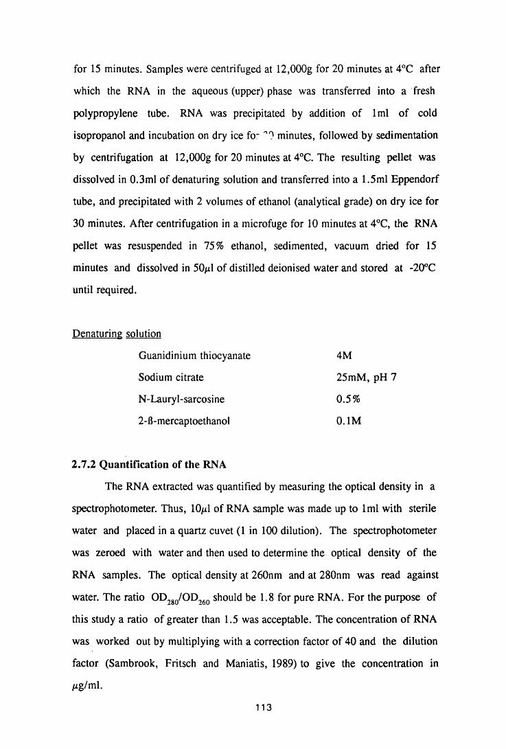
for 15 minutes. Samples were centrifuged at 12,000g for 20 minutes at 4°C after
which the RNA in the aqueous (upper) phase was transferred into a fresh
polypropylene tube. RNA was precipitated by addition of 1ml of cold
isopropanol and incubation on dry ice ftr ^9 minutes, followed by sedimentation
by centrifugation at 12,000g for 20 minutes at 4°C. The resulting pellet was
dissolved in 0.3ml of denaturing solution and transferred into a 1.5ml Eppendorf
tube, and precipitated with 2 volumes of ethanol (analytical grade) on dry ice for
30 minutes. After centrifugation in a microfuge for 10 minutes at 4°C, the RNA
pellet was resuspended in 75% ethanol, sedimented, vacuum dried for 15
minutes and dissolved in 50fx\ of distilled deionised water and stored at -20°C
until required.
Denaturing solution
Guanidinium thiocyanate
Sodium citrate
N-Lauryl-sarcosine
2-B-mercaptoethanol
2.7.2 Quantification of the RNA
The RNA extracted was quantified by measuring the optical density in a
spectrophotometer. Thus, 10/xl of RNA sample was made up to 1ml with sterile
water and placed in a quartz cuvet (1 in 100 dilution). The spectrophotometer
was zeroed with water and then used to determine the optical density of the
RNA samples. The optical density at 260nm and at 280nm was read against
water. The ratio OD.,80/OD 60 should be 1.8 for pure RNA. For the purpose of
this study a ratio of greater than 1.5 was acceptable. The concentration of RNA
was worked out by multiplying with a correction factor of 40 and the dilution
factor (Sambrook, Fritsch and Maniatis, 1989) to give the concentration in
Mg/ml.
113
4M
25mM, pH 7
0.5%
0.1M
2.7.3 Generation of complementary DNA and amplification by polymerase
chain reaction
This involves the generation of complementary DNA (cDNA) from
mRNA by reverse transcription, followed by amplification of the resultant
cDNA by a polymerase catalysed chain reaction (PCR). PCR uses a
thermostable DNA polymerase and specific oligonucleotide primers to conduct
repeated cycles of DNA synthesis, in vitro, on a very small amount of template
DNA (Mullis and Faloona, 1987; Saiki, Gelfand, Stoffel, Scharf, Hiruchi,
Horn, Mullis and Erlich, 1988). Using this procedure, a specific segment of
template DNA can be amplified over a million times making the subsequent
detection of that segment easier. The thermostable DNA polymerase was
purified from the thermophilic bacterium, Thermus aquaticus and named Taq
DNA polymerase (Chien, Edgar and Trela, 1976). This enzyme can survive
extended incubation at 95°C and therefore is not inactivated by the heat-
denaturation step of PCR. In addition, because annealing and extension of
oligonucleotides can be carried out at elevated temperatures, mispriming is
greatly reduced.
This study screened for transcripts for the cytokines: IL-la, IL-lfi,
IL-6, TNFa, MIP-1 and GM-CSF. The sequences for the oligonucleotide
primers for these cytokines are shown in Thble 2.6. Approximately 5/xg of total
RNA was lyophilised and resuspended in 9/d of annealing buffer with 0.5/zg of
the 3'primer, heated at 83°C for 3 minutes, incubated at 69°C for 20 minutes
followed by a 40-minute cooling to 40°C. Fifteen microlitres of
cDNA/deoxyribonucleoside triphosphate (dNTP) buffer were added with 200
units of Moloney murine leukaemia virus reverse transcriptase (Gibco) and
incubated at 43°C for 45 minutes. For amplification of the cDNA generated by
the reverse transcription, 0.5/*g of 5'primer was added with 55/ri of Thq
polymerase buffer, 20/ri 2.5mM dNTPs and 2 units of DNA polymerase (Promega).
114
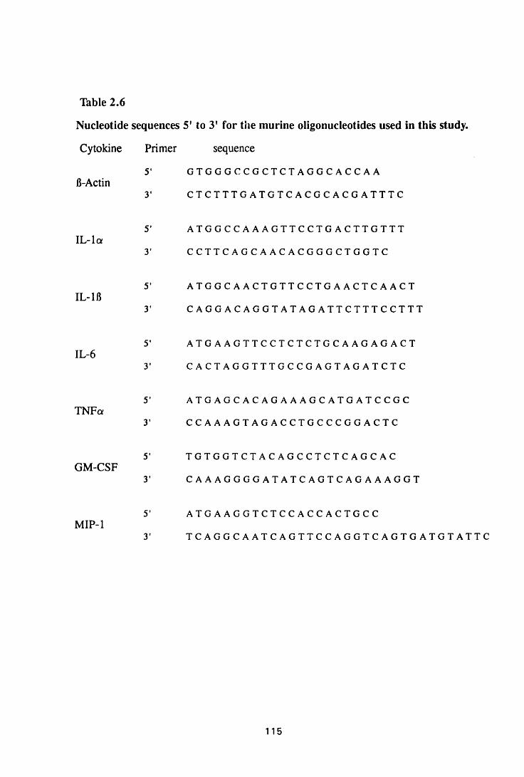
Table 2.6
Nucleotide sequences 5' to 3' for the murine oligonucleotides used in this study.
Cytokine Primer sequence
5' G T G G G C C G C T C T A G G C A C C A A
3' C T C T T T G A T G T C A C G C A C G A T T T CB-Actin
IL-la5' A T G G C C A A A G T T C C T G A C T T G T T T
3' C C T T C A G C A A C A C G G G C T G G T C
IL-1B5' A T G G C A A C T G T T C C T G A A C T C A A C T
3' C A G G A C A G G T A T A G A T T C T T T C C T T T
IL-65' A T G A A G T T C C T C T C T G C A A G A G A C T
3' C A C T A G G T T T G C C G A G T A G A T C T C
TNFcx5' A T G A G C A C A G A A A G C A T G A T C C G C
3 ’ C C A A A G T A G A C C T G C C C G G A C T C
GM-CSF5' T G T G G T C T A C A G C C T C T C A G C A C
3' C A A A G G G G A T A T C A G T C A G A A A G G T
MIP-15* A T G A A G G T C T C C A C C A C T G C C
3' T C A G G C A A T C A G T T C C A G G T C A G T G A T G T A T T C
115
The mixture was then overlain with lOOjxl of mineral oil and subjected to 35
PCR cycles of 94°C denaturation for 1 minute, 60°C annealing for 2 minutes
and 72°C extension for 3 minutes. Thirty five cycles were chosen because, 2kb
in length can be obtained from 30-35 cycles of amplification with only 10"Vg of
starting DNA (Sambrook et al, 1989). In addition, under normal reaction
conditions, the amount of Taq polymerase becomes limiting after an
amplification of level of approximately 106 has been attained (Sambrook et al,
1989).
To visualise the PCR products, 20fi\ of the reaction mixture was
electrophoresed on 2% agarose gel and stained with ethidium bromide.
Annealing buffer
KC1 250mM
Tris HC1, pH 8.3 lOmM
EDTA ImM
cDNA/dNTP buffer
Tris HC1 pH 8.3 24mM
MgCl2 16mM
Dithiothreitol 18mM
dNTPs 0.4mM
Taq polymerase buffer
Tris HC1 pH 8.0 lOOmM
(NH4)2S04 30mM
MgCL, 3mM
2-mercaptoethanol lOmM
116
2.7.4 Electrophoresis of the polymerase chain reaction-products
According to Sambrook et al, (1989), electrophoresis is the standard
method used to separate, identify, and purify DNA fragments. The method is
simple, rapid to perform and capable of resolving fragments of DNA that cannot
be separated adequately by other procedures, such as density gradient
centrifugation. When an electric field is applied across the gel, DNA, which is
negatively charged at neutral pH, migrates toward the anode. The rate of
migration is determined by various factors, including, the molecular size of the
DNA (Fisher and Dingman, 1971; Aaij and Borst, 1972). Larger molecules
migrate more slowly because of greater frictional drag and because they travel
through the gel pores less efficiently.
Two grams of multipurpose agarose (Boehringer Mannheim) was mixed
with 100ml of Tris boric acid/EDTA buffer (TBE), pH 8.3 and heated in a
microwave until all the agarose was melted. A gel plate was put on a level
surface (judged with a spirit level) and fitted with a comb of appropriate size to
make wells. The molten gel, allowed to cool to about 25°C, was poured onto the
gel plate and left to set. The set gel was then placed in a tank containing TBE.
Twenty /d of each PCR product were mixed with 5/d of an inert dye. Orange G
dissolved in TBE/glycerol 1:1 (v/v), and carefully put into the gel wells. At
least one well in each gel was loaded with 4/d of a molecular weight marker, the
123 base pair ladder purchased from Gibco. After loading all the wells with the
various samples, electrophoresis was carried out at about 56mA for 1 hour 30
minutes. After separation, the gel was stained with ethidium bromide and the
separated transcripts identified by their molecular weight. The molecular weight
ladder allowed the sizes of the PCR products to be determined.
117
Tris boric/EDTA buffer
Tris HC1 10.8g
5.5g
0.93g
Boric acid
EDTA
Made up to 1 litre with distilled water. pH 8.3
2.8 EVALUATION OF THE PRESENCE OF CYTOKINE PROTEIN
2.8.1 Detection of secreted cytokines in the astrocyte supernatants
An enzyme-linked immunosorbent assay, for mouse TNF, was used to
test for secreted cytokines in the cell supernatants following exposure to various
stimuli. The protocol followed was a slight modification from the one supplied
by Pharmigen laboratories, an adaptation from the manual of clinical and
laboratory medicine (Rose, Friedman and Fahey, 1986). Briefly, 96-well
microtitre plates (Corning easy wash or Dynatech IV) were coated with
50/ri/well of purified anti-mouse TNF antibodies at a concentration of 2/xg/ml
and incubated overnight at 4°C. The following morning, nonspecific binding
was blocked by incubation with 50/xl/well of 10% FCS in PBS, pH 7.4, for 45
minutes at 37°C. The plates were rinsed three times in PBS containing 0.05%
tween 20, followed by two washes of 3 minutes each.
Recombinant mouse tumor necrosis factor (TNF) purchased from
Pharmigen was used as the standard cytokine. Ten serial double-dilutions in
PBS starting at 80ng/ml were prepared from the stock. The standard- cytokine
dilutions and the neat supernatant samples were added (50/ri/well) in duplicates,
and incubated at 37°C for 2 hours. The plates were then washed in PBS/tween
as before, incubated for 1 hour at room temperature with 50^il/well of
biotinylated mouse anti-TNF antibody (Pharmigen), at 4/xg/ml, and washed
again. Streptavidin peroxidase (Serotec), diluted 1 in 1,000 in 1%
BSA/PBS/tween 20 was added (75/ri/well) and incubated for 1 hour at 37°C and
118
washed. The peroxidase substrate, 3,3', 5, 5' tetramethylbenzidine (TMB) was
added at 100/d/well. Absorbencies were read at OD630, after 15 minutes.
PBS/tween
sterile PBS lOOmls
Tween 20 0.05mls
20ml aliquotes were stored at -20°C
2.8.2 Detection of cytokine proteins in cultured astrocytes
2.8.2.1 Preparation of cultured astrocytes for immunocytochemistry
A flask containing a confluent monolayer of astrocytes was trypsinised
and cells collected in a centrifuge tube. The trypsin was inactivated with soya
bean trypsin inhibitor, the tubes centrifuged at 89.6g and supernatant discarded.
The cells were then washed twice in HBSS, i.e., the cells were resuspended in
HBSS, centrifuged again and supernatant discarded. The cells were resuspended
in DMEM/10%FCS, counted and seeded onto 13mm-diameter, PLL-coated
coverslips in a 24-well tissue culture plate (Nunclon), at a density of 4xl04 cells
per coverslip. They were then maintained in 50% DMEM/10%FCS and 50%
astrocyte conditioned medium 92.5% air, 7.2% C 02 in a humidified incubator,
at 37°C, for 24 hours. The medium was then replaced with DMEM/10%FCS
and cells incubated for a further 24 hours. At this time, confluent monolayers of
astrocytes were established in all the coverslips and cells were ready for use.
The medium was replaced with 50/ig/ml trypanosome lysate in
DMEM/10%FCS (lml/well), and re-incubated. Plates were removed at 2, 4, 6
and 8 hours after addition of stimulant. The medium was removed and the cells
were washed twice with HBSS.
The cells were chilled at -20°C for 10 seconds and fixed with methanol
(lml/well) for 10 minutes at -20°C. The methanol was replaced with 70%
ethanol and cells stored at 4°C until immunocytochemistry could be performed.
119
2.8.2.2 Immunostaining for cytokine protein detection in cultured astrocytes
The coverslips were removed from the ethanol-containing wells, air dried and
fixed onto microscopic slides with glassbond in the presence of ultra violet light
(to activate glassbond). Nonspecific anf binding was blocked by incubating
the cells with 5% BSA and saponin for 10 minutes at room temperature in a
humid chamber. Excess BSA was drained off and the cells incubated, for lhour,
with the primary antibodies diluted 1 in 50 in 2% BSA/Tris buffered saline
containing saponin (2% BSA/TBS/saponin). The primary antibodies consisted
of: rabbit anti-mouse tumor necrosis factor (RbaM TNF), rabbit anti-mouse
interleukin 1 (RbaM IL-1) and rat anti-mouse interleukin 6 (RtoM IL-6). The
cells were washed twice (5 minutes each time) with TBS/saponin and incubated
with the secondary antibodies (biotinylated goat anti-rabbit immunoglobulins for
RBaM TNF and RbaM IL-1 and biotinylated rabbit anti-rat immunoglobulins
for RtaM IL-6 diluted 1 in 200 in 2% BSA/TBS/saponin) fo r i hour at room
temperature. The cells were washed again and incubated with the avidin biotin
complex alkaline phosphatase conjugate for 30 minutes at room temperature in
a humid chamber and washed before the development with naphthol/fast red for
visualisation.
2.8.2.3 Colour development for visualisation
The cells were washed in TBS and stained with naphthol red in veronal acetate
buffer for 10 minutes in the dark. Each wash consisted of two immersions of 5
minutes each into fresh TBS/saponin each time.They were then counterstained
with haematoxylin by placing the slides in haematoxylin for 2 seconds, rinsed in
tap water and blued in Scot's tap water substitute. Coverslips were then
mounted with aquamount.
1 2 0
Tris buffered saline
Tris/HCl
Sodium chloride
0.05M
0.15M pH 7.6
Veronal acetate buffer
0.1MHC1 0.2ml
Veronal acetate solution 5ml
Veronal acetate solution
Sodium (veronal) barbitone 2.9g
Sodium acetate trihydrate 1.9g
Make up to 100ml in distilled water
Tb make up Naphthol red solution
Levamisole 3mg
Fast red 6mg
Naphthol 3 mg
Veronal acetate buffer 12mls
Solution A:
Levamisole and fast red, dissolved in 4ml veronal acetate
buffer
Solution Bi
Naphthol AS-TR phosphate dissolved in two to three drops of
dimethylformamide.
Solution B was added to solution A and the mixture filtered in the dark
at 4°C to give a clear yellow solution, which was used for colour
development.
121
CHAPTER 3
ASTROCYTE ACTIVATION DURING TRYPANOSOMA BRUCEI
BRUCEI INFECTION IN MICE
122
3.1 INTRODUCTION
Astrocyte activation is a prominent characteristic following many types
of insult to the central nervous system (Bignami et al, 1972; Latov et al, 1979;
Nathaniel and Nathaniel; 1981; Smith et al, 1987; O'Callaghan et al, 1990).
When activated, the astrocytes increase in numbers, become larger, extend more
processes, and significantly increase their cytoplasmic content of glial fibrillary
acidic protein (GFAP), an astrocyte specific intermediate filament (Bignami et
al, 1972). This process is referred to as reactive gliosis and can lead to the
formation of dense gliotic scars in the central nervous system (CNS). Infiltration
of inflammatory mononuclear cells (lymphocytes, blood-derived macrophages)
and an increase in intrinsic brain microglial cells are additional features of
many inflammatory conditions of the brain, including, multiple sclerosis
(Traugott, Reinherz and Raine, 1983; Traugott, Scheinberg and Raine, 1985;
Cuzner, Hayes, Newcombe and Woodroofe, 1988) and traumatic injuries of the
brain (Tsuchihashi, Kitamura and Fujita, 1981; Giulian et al, 1989). Reactive
gliosis and mononuclear cell infiltration are major features of late-stage HAT
(Adams et al, 1986; Haller et al\ 1986)
The CNS pathology in mice chronically infected with T. b. brucei is
characterised by an infiltration of the subarachnoid and the Virchow Robin
spaces by the lymphocyte/plasma cell series and macrophages, and by microglial
hyperplasia and large reactive astrocytes in the white matter (Murray et al,
1974a; 1974b; Jennings et al, 1989; Hunter et al, 1991). Subcurative treatment
of T.b.brucei infections of mice after day 21 of infection results in an
exacerbation of this lesion (Jennings and Gray, 1983; Jennings et al, 1989;
Hunter et al, 1992a). These lesions closely simulate the CNS lesions of human
sleeping sickness and of reactive arsenical encephalopathy cases (Ormerod,
1970; de Raadt, 1984; Adams et al, 1986; Haller et al, 1986).
This study was carried out to investigate the progress of astrocyte
123

activation during the course of T. b. brucei infection in mice, and following
curative and subcurative trypanocidal therapy.
3.2 MATERIALS AND METHODS
3.2.1 Infection
Experiments were performed on female NIH mice weighing between 25
and 30g. These animals were infected intraperitoneally with 4xl04 parasites of
either the eflomithine-sensitive or the eflomithine-resistance T.b. brucei,
stabilate GVR 35/C1.5 and GVR 35/Cl.3 DFMO 5, respectively (Chapter 2).
3.2.2 Infection without treatment
Adult female NIH mice were infected with either the eflomithine-
sensitive or the eflornithine-resistant trypanosome stabilates (above). Five mice
from each group were sacrificed every 7 days during the course of infection.
These mice are represented as group IC in Thble 3.1. Deaths, as a consequence
of infection, were common in this group with few animals surviving longer than
52 days. An uninfected, untreated mouse was also killed at each point.
3.2.3 Induction of post-treatment encephalitis by treatment with diminazene
aceturate
Two groups of mice were infected with either the eflomithine-sensitive
or the eflornithine-resistant T b. brucei stabilate in order to compare the CNS
pathology caused by each of the stabilates. On day 21 after infection, all the
mice were treated i.p. with 40mg/kg diminazene aceturate (Berenil, Hoechst,
Germany). Each of these two groups were further divided into two groups, PI
and P2 (Thble 3.1). The first group, PI, was sacrificed serially every 7 days
after the Berenil treatment in groups of five. This treatment, with Berenil on
day 21 after infection, is known to be subcurative (Jennings and Gray, 1983)
124
and to induce a post-treatment reactive encephalitis (PTRE) in T.b. brucei-
infected mice (Jennings et al, 1989; Chapter 1).
The second group, P2, was treated with 40mg/kg Berenil i.p. on day 21
after infection and again after relapse of parasitaemia which occurred 20-22
days after the first Berenil injection. Five mice were killed every 7 days
thereafter. This treatment regimen has been reported to cause a very severe
post-treatment encephalitis (Jennings et al, 1989; Chapter i).
Groups of uninfected mice, which received the same drug treatment as
the infected mice, were sacrificed at the same time as the experimental animals.
Some infected, treated mice were retained to monitor relapse of parasitaemia.
Blood from tail snips was examined every 2 days until relapse of parasitaemia,
when the mice were sacrificed.
3.2.4 Curative treatment after induction of post-treatment encephalitis
Mice in this group, CT (Thble 3.1), were infected, with the two
trypanosome stabilates as in the above groups, and were treated with Berenil on
day 21 after infection and again after relapse of parasitaemia. Seven days after
the second dose of Berenil, mice received two consecutive daily treatments of
15mg/kg MK 436 (Merck Institute for Therapeutic Research) in combination
with 5mg/kg Mel Cy (Cymelarsen) (Rhone Merieux) i.p. At this dosage, this
combination regimen has been shown to be curative for the CNS stages of T.b.
brucei infections in mice (Jennings, 1991c). Mice were sacrificed in groups of
five, 5, 12, 19 and 26 days after this curative therapy.
The pathology in brains of mice from this group was compared to that of
mice treated with the double-Berenil regimen. Uninfected mice treated with
same drug regimen as the infected mice were used as controls. A group of mice
was retained after curative treatment and parasitaemia was monitored, to ensure
the treatment was curative.
125
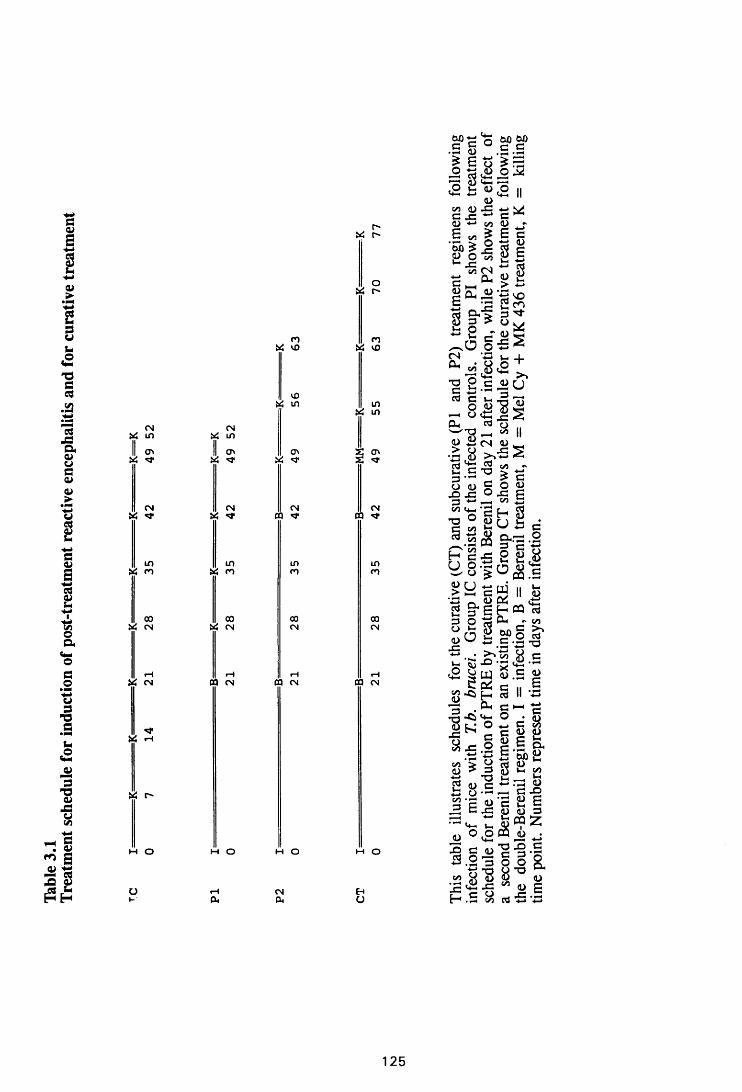
Table
3.
1Tr
eatm
ent
sche
dule
for
indu
ctio
n of
post
-trea
tmen
t re
activ
e en
ceph
aliti
s an
d for
cu
rativ
e tr
eatm
ent
CN Vi i n
II O ' Vi
CNVi
i n Vi r o
ooVi CN
Vi CN
CN Vi i n
II O ' Vi
CN Vi ^
i n Vi n
ooVi CN
0 0 CN
Vi r ~
M O
n Vi vD
VOVi i n
O 'vi
CN 0Q
i nr o
00CN
03 CN
M O
Ou
M O
CNPi
r** Vi f "
o Jxj r *
n Vi vo
i n Vi i n
CN 03
i nco
ooCN
03 CN
w o
g>S >g MM •t3 <u .5 .33lllla<2 is w i2 ii
<L>i/i -C J3 *■* C <1) *-> on <D -e ^ e c
S “ 2 s SC >■ -4-*
p p* M "5 vo5 ‘3 § 2S §* * 3* 8 e «O
C N
u<-" u S <D 0 ’S *
+J2
*2 S .S ^ O
- 8 £3 sPh ^ o<U jy CN <u
■3J1cd ^o m G o fi•° J3 ° ■£ G3 +-* i m | .^ c h g ■§ 2* a §..-3P S 5 g g 8U O.'Sro & *5 £°CQ.£> y c £ 11 £•n - <u 04 _a §*e t - « «3 O - «n
„cB § g“§ S'2 w *- .S *J3 "O5 •- £ \s 8 .s<2 | w u ! s g
g l g S § II -a’33 . CU C h
So x : . 2 g R*4—* \3 C hA 2• 1—< Tv -4—» Of#co > y cj ^(D > 3 o5 1-1 cn t3 T3 1-1 Jrjg g .S “ c | 3 P S'S 2 g, 3 C £ U 1) 3
^ a d S ® *« o .o fflj) j3 c 5 0c o l ’s s
C C O 42 .5C- • .—I c/J 08 •*-» •*-»
O cO . <u *_> C vn O U 1 )
« S &
cQ O g,
125
3.2.5 Monitoring infections
Mice were monitored for parasitaemia by examination of fresh blood
from tail snips. At least twenty fields were examined at x400 magnification
before samples were designated either positive or negative.
3.2.6 Perfusion of the brain to remove peripheral blood
At death, mice were perfused with 150ml normal saline through the left
ventricle to remove peripheral blood. The brains were fixed in neutral buffered
formalin and processed for histopathology (Chapter 2).
3.2.7 Immunostaining for astrocyte activation
Three to 4 im transverse sections were cut at the fore-, mid- and
hindbrain. Some of the sections were stained for glial fibrillary acidic protein
(GFAP) to assess astrocyte activation (Chapter 2), while others were stained
with haematoxylin and eosin to assess pathology (Chapter 2).
3.2.8 Assessment of astrocyte activation
The criteria for identifying astrocyte activation was an increase, above
that found in normal uninfected mice (baseline), in one or more of the following
parameters: the intensity of GFAP staining, the number of GFAP-staining
astrocytes, the number, size, and the degree of branching, of the astrocytic
cytoplasmic processes and the topographical distribution of astrocytes in the brain
parenchyma.
3.3 RESULTS
3.3.1 Astrocyte activation in infected untreated mice
Figure 3.1 was included to illustrate the areas of the cerebrum (3.1a) and
cerebellum (3.1b) most commonly referred to in describing astrocyte activation
127
Figure 3.1
Sections of the cerebrum (a) and the cerebellum (b) from a normai mouse
showing the parts of brain most commonly referred to during the description of
astrocyte activation, in this study: m = meninges; cc = corpus callosum; he =
hippocampus; dg = dentate gyrus; cf = choroid fissure; numbers = cerebellar
lobes (numbering is not in anatomical order); gl = granular layer of the
cerebellum; ml = molecular layer of the cerebellum; b = base of the
cerebellum. Haematoxylin and eosin. x40
128
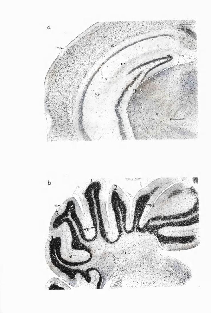
; ' v - W ' f ' & ■. H i h b .
X - - .
;'Si-3;;.i;, ■■’; : ? . * ’ ■- J F - ■• .. *K*?. • - j T . ; • Vk
i v - ' V v . - . v - - . / • ' • • .
«2 B£!
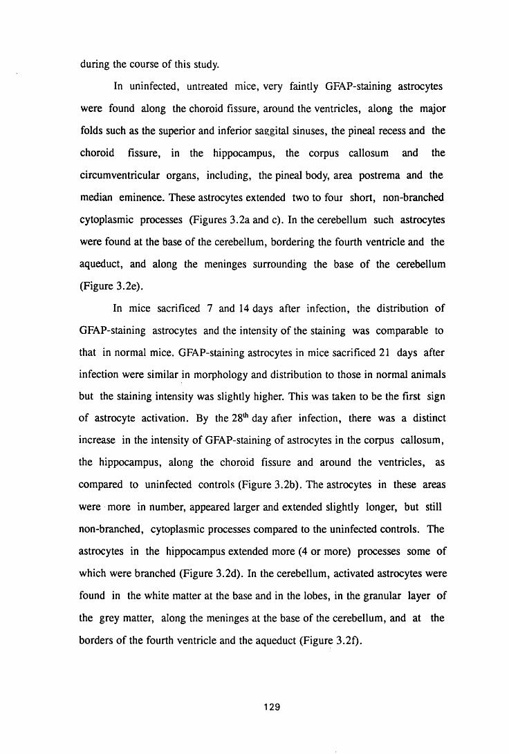
during the course of this study.
In uninfected, untreated mice, very faintly GFAP-staining astrocytes
were found along the choroid fissure, around the ventricles, along the major
folds such as the superior and inferior sasgital sinuses, the pineal recess and the
choroid fissure, in the hippocampus, the corpus callosum and the
circumventricular organs, including, the pineal body, area postrema and the
median eminence. These astrocytes extended two to four short, non-branched
cytoplasmic processes (Figures 3.2a and c). In the cerebellum such astrocytes
were found at the base of the cerebellum, bordering the fourth ventricle and the
aqueduct, and along the meninges surrounding the base of the cerebellum
(Figure 3.2e).
In mice sacrificed 7 and 14 days after infection, the distribution of
GFAP-staining astrocytes and the intensity of the staining was comparable to
that in normal mice. GFAP-staining astrocytes in mice sacrificed 21 days after
infection were similar in morphology and distribution to those in normal animals
but the staining intensity was slightly higher. This was taken to be the first sign
of astrocyte activation. By the 28th day after infection, there was a distinct
increase in the intensity of GFAP-staining of astrocytes in the corpus callosum,
the hippocampus, along the choroid fissure and around the ventricles, as
compared to uninfected controls (Figure 3.2b). The astrocytes in these areas
were more in number, appeared larger and extended slightly longer, but still
non-branched, cytoplasmic processes compared to the uninfected controls. The
astrocytes in the hippocampus extended more (4 or more) processes some of
which were branched (Figure 3.2d). In the cerebellum, activated astrocytes were
found in the white matter at the base and in the lobes, in the granular layer of
the grey matter, along the meninges at the base of the cerebellum, and at the
borders of the fourth ventricle and the aqueduct (Figure 3.2f).
129

Figure 3.2
Brain sections from uninfected untreated (a, c and e) mice, compared to
sections from T.b. brucei-'miectcd untreated mice, group IC, sacrificed 28 days
post infection (b,d and f). The sections were i^munostained with rabbit anti-
GFAP (DAKO), developed with DAB/peroxidase kit (Vectastain) and
counterstained with haematoxylin. There is an increase in the number astrocytes
and their GFAP staining intensity in the corpus callosum (b), and the
hippocampus (d). Faintly GFAP-staining astrocytes can be seen around the
blood vessels and in the granular layer of the cerebellum of mice sacrificed 28
days after infection (f). Meningitis is very mild in the cerebrum (b) and
moderate in the cerebellum (f). x90
130
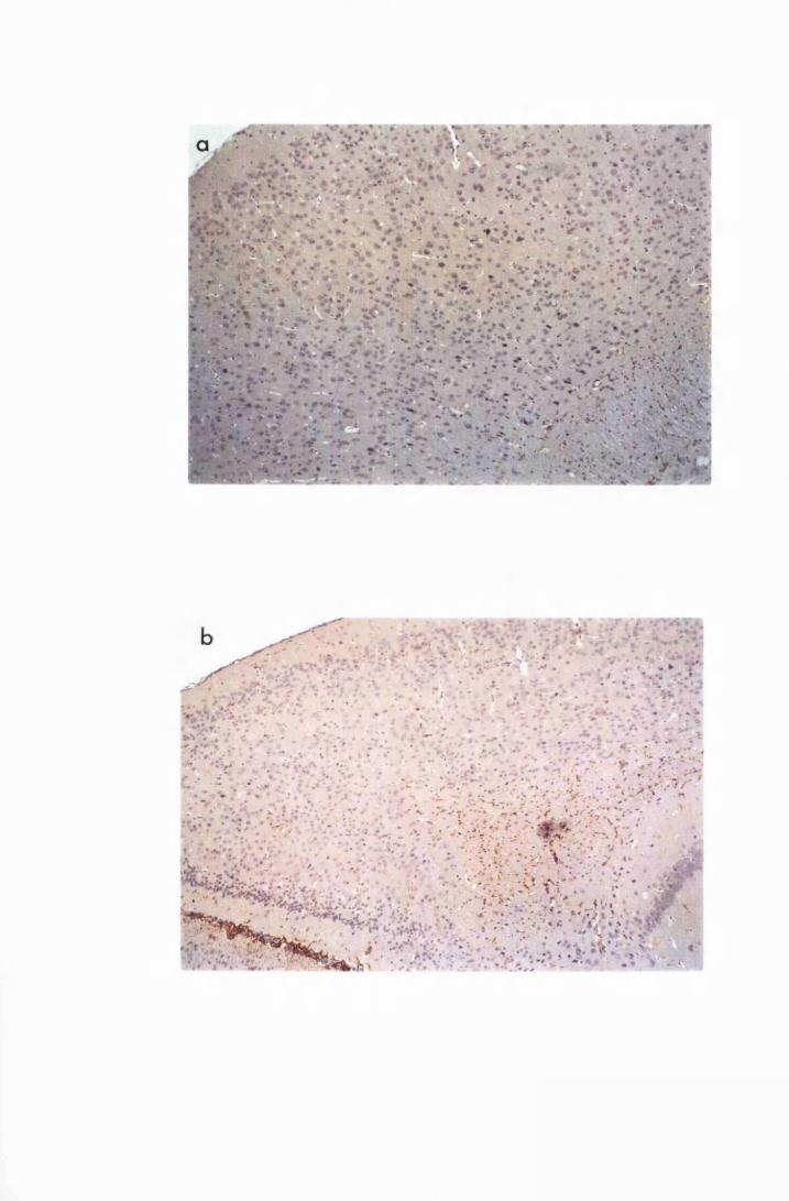
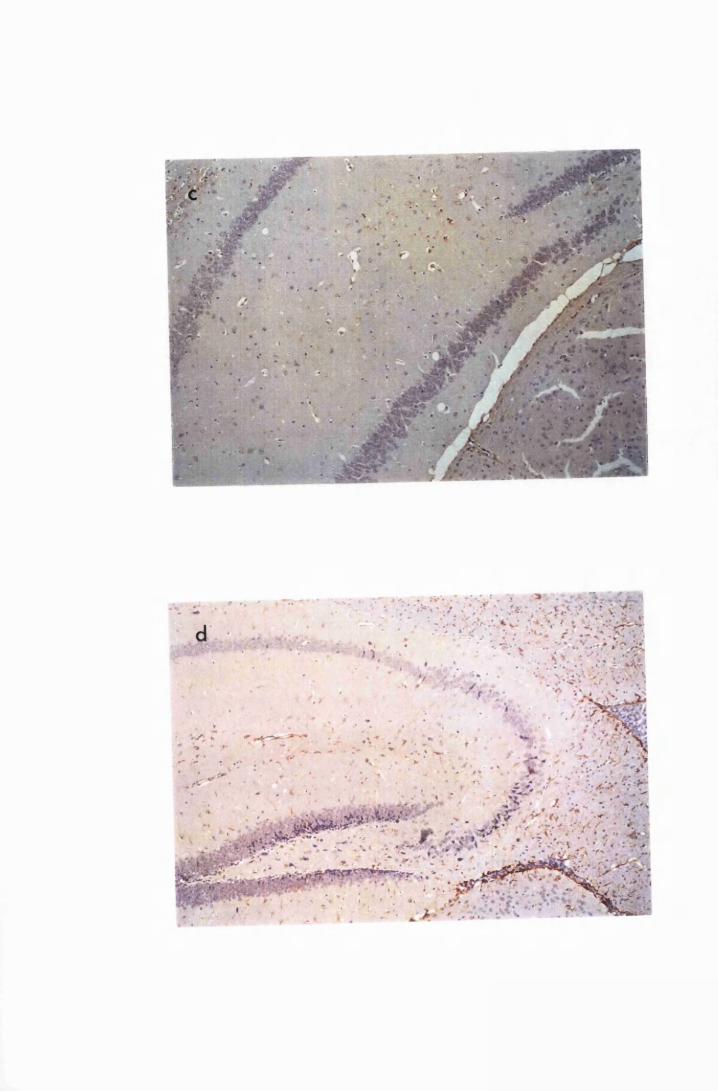
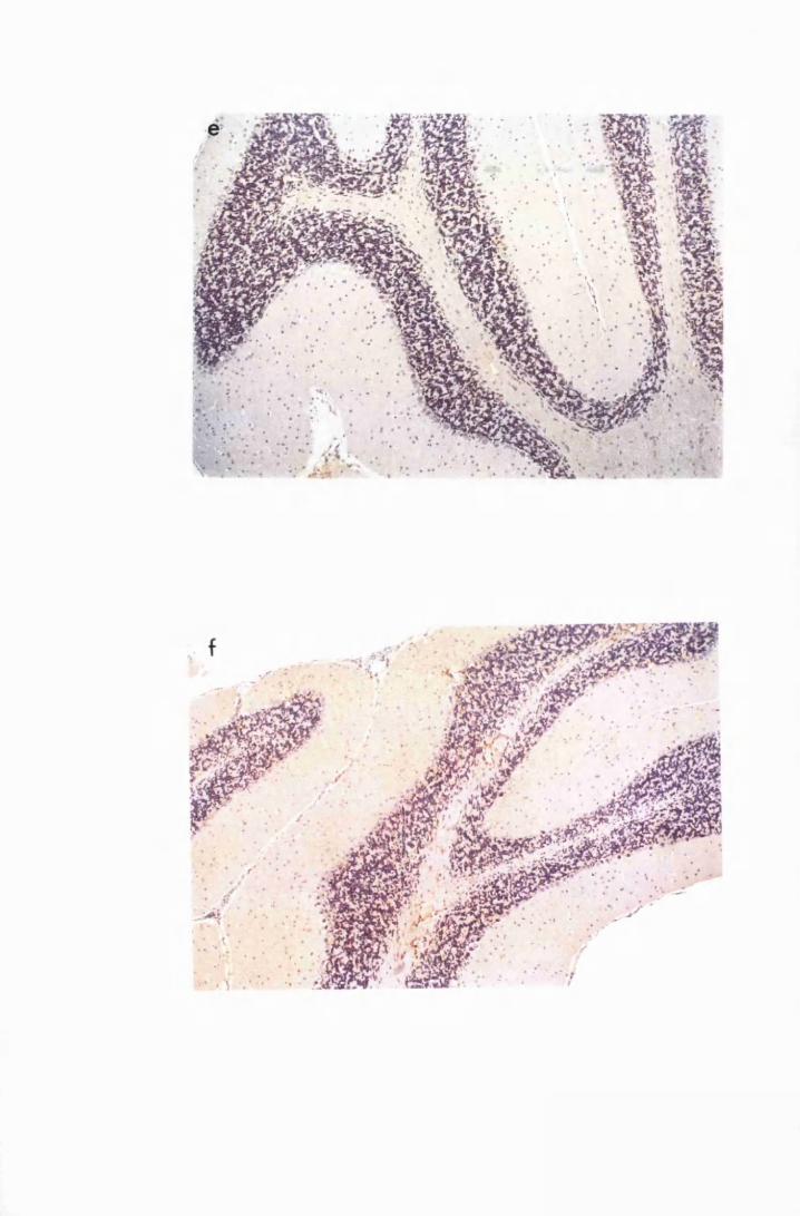
There was a slow progressive spread of astrocyte activation in T.b.
brucei-infected, untreated mice. Thus, astrocyte activation first seen in the
corpus callosum, along the meninges and the choroid fissure, spread outwards
into the brain parenchyma, first the white and then the grey matter. By day 52
after infection, astrocyte activation had spread to form a wide band around the
corpus callosum, involving the adjacent grey matter. Few activated astrocytes
were scattered in the rest of the grey matter, with more of these astrocytes
concentrated around the blood vessels (Figure 3.3a).
Mononuclear inflammatory cells, including lymphocytes, plasma cells
and macrophages, were first observed in the choroid fissure and the meninges in
mice sacrificed 28 days after infection. At this time, infiltration in both the
meninges and the choroid fissure, was focal and only 1 or 2-cells thick.
Astrocyte activation was confined to around the choroid fissure, along the
meninges, in the hippocampus and the corpus callosum (Figures 3.2b and d),
and around the large blood vessels in the cerebellum (Figure 3.2f). As the
disease progressed, the mononuclear cell infiltrations increased to form a
continuous layer in the meninges and the choroid fissure. By day 52 after
infection, there was a continuous layer of inflammatory cells in the entire
circumference of the meninges and in the choroid fissure, 5 or more cells thick.
In addition perivascular cuffs were evident in the hippocampus and the corpus
callosum (Figures 3.3a and b). As the inflammatory cell infiltrate increased so
did the spread of activated astrocytes into the parenchyma.
Similarly, mononuclear inflammatory cells were found infiltrating the
cerebellar meninges, folds and the perivascular spaces at the base and the lobes,
28 days after infection. At this time, the infiltrates were only 1 or 2-cells thick
and astrocyte activation was confined to the white matter and around the blood
vessels in the granular layer (Figure 3.2f; Figure 3.4a). As in the cerebrum, the
infiltrates increased, thickening the meninges and enlarging the perivascular
131
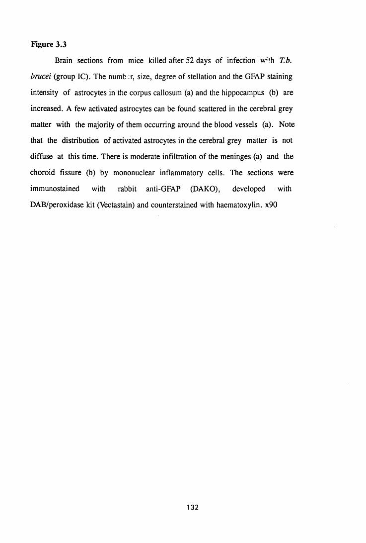
Figure 3.3
Brain sections from mice killed after 52 days of infection w^h T.b.
brucei (group IC). The number, size, degree of stellation and the GFAP staining
intensity of astrocytes in the corpus callosum (a) and the hippocampus (b) are
increased. A few activated astrocytes can be found scattered in the cerebral grey
matter with the majority of them occurring around the blood vessels (a). Note
that the distribution of activated astrocytes in the cerebral grey matter is not
diffuse at this time. There is moderate infiltration of the meninges (a) and the
choroid fissure (b) by mononuclear inflammatory cells. The sections were
immunostained with rabbit anti-GFAP (DAKO), developed with
DAB/peroxidase kit (Vectastain) and counterstained with haematoxylin. x90
132
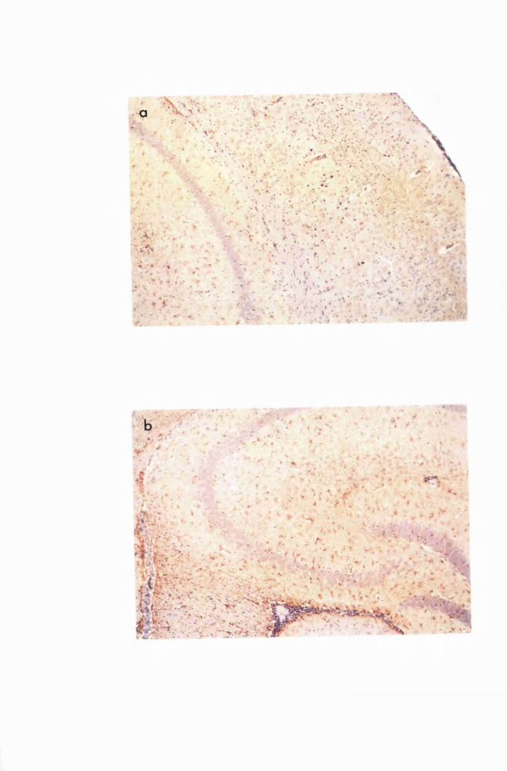
Figure 3.4
Cerebellar sections from mice infected with T.b. brucei (group IC) and
sacrificed on day 28 (a) and day 52 (b) after infection. Note that after 28 days
of infection, activated astrocytes are confined to the base of the cerebellum (a)
and how astrocyte activation spreads to the cerebellar lobes (b). Some activated
astrocytes can be seen in the granular layer of the cerebellum 52 days after
infection (arrows). The sections were stained with rabbit anti-GFAP antibody,
developed with DAB/peroxidase kit and counterstained with haematoxylin. x90
133
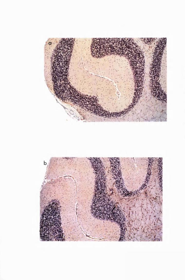
cuffs. By day 52 after infection, there was a 5 to 10-cell layer of mononuclear
inflammatory cells in the meninges and the meningeal folds and prominent
perivascular cuffs in the white matter of the cerebellar lobes (Figure 3.4b).
There was no difference observed between the pathology caused by the
two trypanosome stabilates. Uninfected mice treated with Berenil did not
develop any CNS pathology.
3.3.2 Astrocyte activation in post treatment reactive encephalitis
Brain sections of mice treated with Berenil on day 21 after infection and
sacrificed 7 days later (day 28 after infection), showed active astrocytes
diffusely distributed in the brain parenchyma in both the white and the grey
matter of the cerebral cortex (Figure 3.5a). Astrocytes in the hippocampus were
more in number, stained more intensely for GFAP and were more elaborately
stellate than those in the grey matter (Figure 3.5b). In the cerebellum, activated
astrocytes were found throughout the white matter (Figure 3.5c). In addition,
the meninges, the choroid fissure and the perivascular spaces were infiltrated by
mononuclear inflammatory cells, including, lymphocytes, plasma cells and
macrophages. Perivascular cuffs were also present in the white matter at the
base and in the cerebellar lobes (Figures 3.5c).
The spread of astrocyte activation in this group of mice occurred faster
than in the infected, untreated mice. Activated astrocytes were diffusely
distributed throughout the cerebral and the cerebellar cortices by day 43 after
infection (22 days after induction of PTRE with Berenil) (Figures 3.6a and b).
At this time, there were large perivascular cuffs in the cerebral white and grey
matter, the cerebellar white matter and numerous inflammatory cells in the
meninges and meningeal folds. Astrocytes around the inflammatory-cell foci,
appeared more activated than those in the rest of the parenchyma (Figure 3.6a).
In the cerebellum, activated astrocytes were found in the white matter and in
both the molecular and the granular layers of the grey matter. In addition, the
134
Figure 3.5
Brain sections of a mouse from group PI, that was infected with T.b.
brucei, treated with Berenil 21 days later and sacrificed on day 28 after
infection, i.e., 7 days after Berenil injection. Note that the meningitis in (a) is
greater than in untreated mice sacrificed 52 days after infection (See Figure
3.4). There are activated astrocytes in the cerebral (b) and the cerebellar white
matter (c). Numerous inflammatory cells can be seen infiltrating the choroid
plexus (b) and there are perivascular cuffs in the cerebellar white matter (c).
The sections were immunostained with rabbit anti-GFAP (DAKO), developed
with DAB/peroxidase kit (Vectastain) and counterstained with haematoxylin.
x90
135
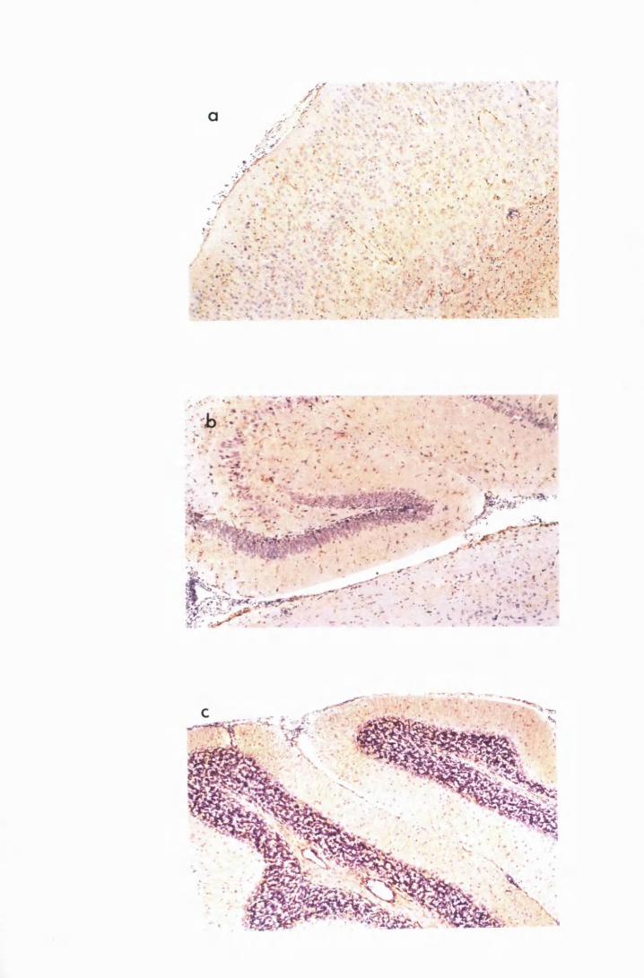
Figure 3.6
Diffuse distribution of activated astrocytes in the cerebral (a) and
cerebellar (b) cortices of a mouse treated with Berenil on day 21 and sacrificed
on day 43 after infection (group PI). Note that astrocytes appear more active
around perivascular cuffs. Bergman glia in the molecular layer, of the
cerebellum, stain for GFAP (arrows). The sections were immunostained with
rabbit anti-GFAP (DAKO), developed with DAB/peroxidase kit (Vectastain)
and counterstained with haematoxylin. x90
136

S; .* i~\c :
Bergman glia in the molecular layer stained for GFAP (Figure 3.6b).
Mice that received a second Berenil treatment after the relapse of
parasitaemia showed an exacerbation of CNS reaction. Thus, 7 days after
the second Berenil treatment, there was an apparent increase in the number and
the degree of astrocyte activation, as judged by morphological appearance and
the intensity of GFAP staining. Activated astrocytes were distributed
throughout the brain parenchyma. There was also an increase in the size of
perivascular cuffs, and the number of mononuclear inflammatory cells
infiltrating the meninges and the choroid fissure (Figures 3.7a to d). At this
time, the astrocytes were enlarged, had many (four or more), long, elaborately
branched cytoplasmic processes and stained very intensely for GFAP (Figure
3.8a). Serial haematoxylin and eosin-stained sections showed the presence of
trypanosomes in the brain parenchyma (Figure 3.8b).
Infected, treated mice showed relapse of parasitaemia 20-22 days after
treatment with Berenil. The pathological changes were identical in mice that
were infected with the eflomithine-sensitive and the eflornithine-resistant
trypanosome stabilates. Uninfected mice treated with Berenil once or twice did
not show any pathological lesions.
3.3.3 Astrocyte activation after curative trypanocidal therapy
Curative trypanocidal therapy after a double-Berenil regimen, was
followed by a progressive resolution of the CNS pathology. The disappearance
of inflammatory cell infiltrations preceded the resolution of astrocyte activation.
In mice killed 5 days after curative treatment, there was a remarkable
decrease in inflammatory-cell infiltration, in the meninges and the perivascular
cuffs. Complete resolution of the meningitis was evident 12 days after curative
treatment. However, encephalitis characterised by a few inflammatory cells in
the perivascular spaces within the hippocampus and in the choroid fissure, was
137
Figure 3.7
Comparison of the degree of astrocyte activation and inflammation in the
brain of a mouse from group PI, that received a single Berenil treatment on day
21 and killed on day 52 after infection (a and c), to the brain of a mouse from
group P2, that received Berenil on day 21 after infection and again after relapse
of parasitaemia (day 45 after infection), then sacrificed 7 days (day 52 after
infection) after the second Berenil (b and d). There is an increase in the number
of astrocytes, the degree of astrocyte stellation and the intensity of GFAP-stain
in mice that received the double-Berenil regimenn (b and d). Note also the
increase in the numbers of mononuclear inflammatory cells infiltrating the
meninges (b), the perivascular cuffs (b and d), and the choroid fissure (d), The
sections were immunostained with rabbit anti-GFAP (DAKO), developed with
DAB/peroxidase kit (Vectastain) and counterstained with haematoxylin.
Magnification. x90
138
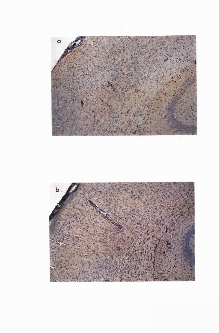
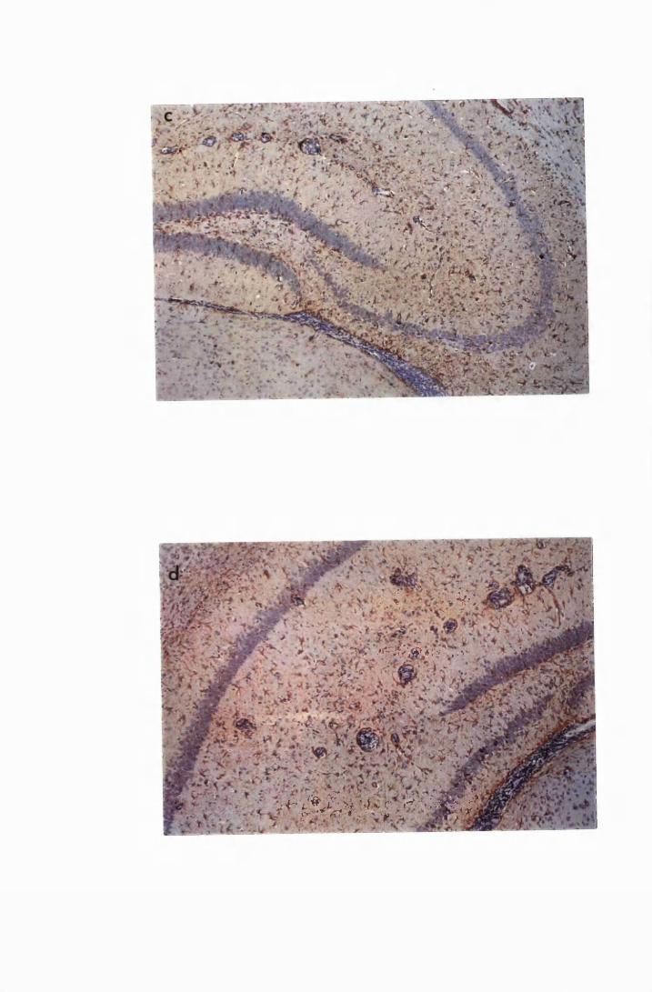
. . r V*':
*%■&&.*'& •■'k‘ L
:-t- & & i ••) v r <*'*• a r a f J' -tv■ * £* i • * ;*
Figure 3.8
The cerebrum of a mouse from group P2, treated with Berenil on day 28
after infection and again after relapse and killed 14 days after the second Berenil
treatment. Figure 3.8a shows the large elaborately stellate astrocytes diffusely
distributed in the brain parenchyma. The cytoplasmic processes are multiple and
branched and stain intensely for GFAP. This section was stained with rabbit
anti-GFAP antibody, developed with DAB/peroxidase kit (Vectastain) and
counterstained with haematoxylin. Figure 3.8b shows a serial section of the
same area, from the same mouse, stained with haematoxylin and eosin. Note the
trypanosomes in the parenchyma (arrows) not associated with perivascular cuffs.
x360
139
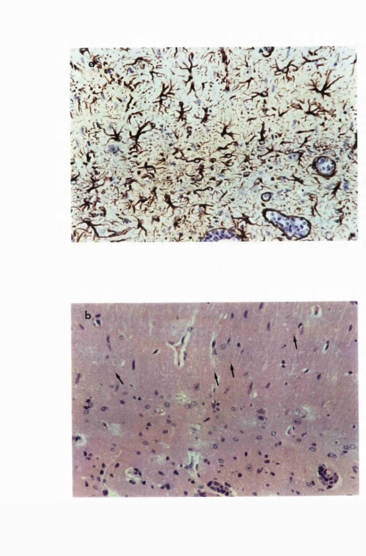
RJKM8I. fj ... i t t
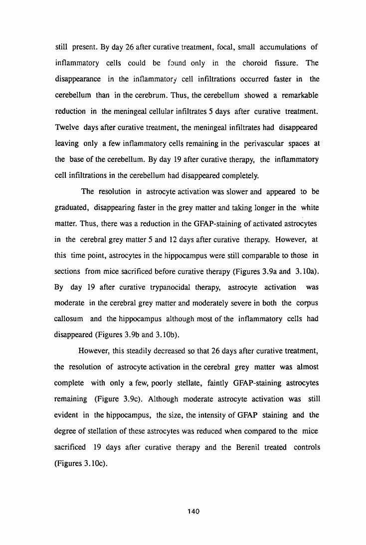
still present. By day 26 after curative treatment, focal, small accumulations of
inflammatory cells could be found only in the choroid fissure. The
disappearance in the inflammatory cell infiltrations occurred faster in the
cerebellum than in the cerebrum. Thus, the cerebellum showed a remarkable
reduction in the meningeal cellular infiltrates 5 days after curative treatment.
Twelve days after curative treatment, the meningeal infiltrates had disappeared
leaving only a few inflammatory cells remaining in the perivascular spaces at
the base of the cerebellum. By day 19 after curative therapy, the inflammatory
cell infiltrations in the cerebellum had disappeared completely.
The resolution in astrocyte activation was slower and appeared to be
graduated, disappearing faster in the grey matter and taking longer in the white
matter. Thus, there was a reduction in the GFAP-staining of activated astrocytes
in the cerebral grey matter 5 and 12 days after curative therapy. However, at
this time point, astrocytes in the hippocampus were still comparable to those in
sections from mice sacrificed before curative therapy (Figures 3.9a and 3.10a).
By day 19 after curative trypanocidal therapy, astrocyte activation was
moderate in the cerebral grey matter and moderately severe in both the corpus
callosum and the hippocampus although most of the inflammatory cells had
disappeared (Figures 3.9b and 3.10b).
However, this steadily decreased so that 26 days after curative treatment,
the resolution of astrocyte activation in the cerebral grey matter was almost
complete with only a few, poorly stellate, faintly GFAP-staining astrocytes
remaining (Figure 3.9c). Although moderate astrocyte activation was still
evident in the hippocampus, the size, the intensity of GFAP staining and the
degree of stellation of these astrocytes was reduced when compared to the mice
sacrificed 19 days after curative therapy and the Berenil treated controls
(Figures 3.10c).
140
Figure 3.9
Comparison of astrocyte activation in the cerebral grey matter of a
mouse from group P2, treated with the double-Berenil regimen and killed 12
days later (a), with that of mice, from group CT, that were treated with the
curative regimen of 15mg/kg MK 436 and 5mg/kg Mel Cy for 2 consecutive
days, starting on the 7th day after the second Berenil treatment, and sacrificed 19
(b) and 26 (c) days later. Note how the activated astrocytes disappear from the
grey matter first (b) and then from the corpus callosum (c). Note also the
reduction in the size, degree of stellation and intensity of GFAP staining in
curatively treated mice (b and c). Astrocyte activation in the grey matter is
almost completely resolved 26 days after curative therapy (c). The sections were
immunostained with rabbit anti-GFAP (DAKO), developed with
DAB/peroxidase kit (Vectastain) and counterstained with haematoxylin. x90
141
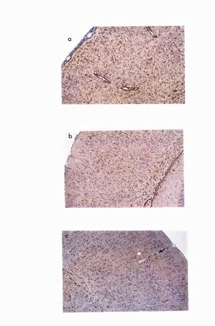
L W i w s P l I1 4$ '*v$^V\ -v :v
*X#IS&S« $bS«23■ . . . . . ^ : ‘-J , ^ ; > " v
•or*/
% v"^ T 'liV>
Figure 3.10
Comparison of astrocyte activation in the hippocampus of a mouse from
group P2, treated with the double-Berenil regimen and killed 12 days later (a),
with that of mice, from group CT, that were treated with the curative regimen
of 15mg/kg MK 436 and 5mg/kg Mel Cy for 2 consecutive days, starting on the
7th day after the second Berenil treatment, and sacrificed 19 (b) and 26 (c) days
later. Note how the astrocytes decrease in size and in the degree of stellation.
Note also how the perivascular cuffs have gradually disappeared (b) and (c). By
26 days after curative therapy, inflammatory cells have disappeared from the
perivascular spaces and the choroid fissure (c). The sections were stained with
rabbit anti-GFAP, developed with DAB/peroxidase kit and counterstained with
haematoxylin. x90
142
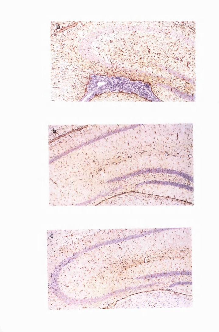
Astrocyte activation 5 and 12 days after curative treatment was
comparable to that seen in the double-Berenil-treated controls (Figure 3.11a).
Nineteen days after curative therapy, there was a decrease in the intensity of
GFAP-stain in the activated astrocytes within the white matter and the Bergman
glia in the molecular layer of the cerebellum (Figure 3.11b). This decreased
further so that by day 26, activated astrocytes were confined to the white matter
at the base of the cerebellum and there was only a faint GFAP-stain on the
Bergman glia (Figure 3.11c).
The response to curative therapy was the same whether the infecting
trypanosome stabilate was eflomithine-sensitive or eflomithine-resistant.
Uninfected mice treated with this combination of drugs did not show any
pathological lesions in the CNS.
3.4 DISCUSSION
These results confirm the observation that astrocyte activation in this
mouse model, is the first pathological sign of neurological involvement after
infection with African trypanosomes and that it occurs before the infill/ation of
the CNS by inflammatory cells (Hunter et al, 1992b). Astrocyte activation was
observed 21 days after infection. Astrocyte activation, in this study, was judged
by the increase in staining of the glial fibrillary acidic protein (GFAP), the
predominant protein in the astrocyte intermediate filaments (Eng, 1985), and by
the morphological changes, i.e., the increase in size and the degree of stellation.
Astrocyte activation occurred first around the ventricles, along the choroid
fissure, along the pineal recess, in the pineal gland, in the hippocampus and in
the corpus callosum. As these are areas where the blood-brain barrier is
incomplete, the activation might be the response of astrocytes to trypanosome
invasion. Indeed, the presence of trypanosomes in the circumventricular organs
(pineal gland, area postrema and the median eminence) and the choroid plexus,
143
Figure 3.11
Comparison of astrocyte activation in the cerebellum of a mouse from
group P2, treated with the double-Berenil regimen and killed 12 days later (a),
with that in mice, from group CT, that were treated with the curative regimen
of 15mg/kg MK 436 and 5mg/kg Mel Cy for 2 consecutive days, starting on the
7th day after the second Berenil treatment, and sacrificed 19 (b) and 26 (c) days
later. The disappearance of inflammatory cells from the perivascular spaces and
the meningeal folds had occurred by day 19 after curative therapy (b) but the
regression of astrocyte activation and the GFAP-stain in the Bergman glia was
not complete after 26 days post curative treatment. However, the intensity of
GFAP staining was decreased (c). The sections were stained with rabbit anti-
GFAP, developed with DAB/peroxidase kit and counterstained with
haematoxylin. x90
144
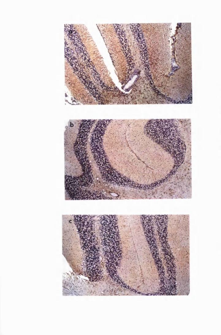
m m m
has been demonstrated to occur in mice as early as 13 days post infection
(Schultzberg et al, 1988).
Astrocytes are one of the most numerous cell types in the CNS and they
give rise to radiating processes which extend across vast distances in the
brain parenchyma to make contact with blood vessels, or with different neurones
or synapses (Hertz et al, 1990). Due to their "strategic" placement at the blood-
brain barrier, astrocytes are likely to be the first of the intrinsic CNS cells to
come into contact with the trypanosome signal emanating from the systemic
blood circulation. This may explain why astrocyte activation appears to occur
before the infiltration of inflammatory cells into the meninges and the brain
parenchyma.
Activated astrocytes are phagocytic, produce cytokines and interact with
antigen-specific T cells to control immune reactivity within the CNS (Pentreath,
1989; Wekerle et al, 1986; Hertz et al, 1990) for instance, by presenting
antigens to MHC-restricted T cells (Vidovic, Sparacio, Elovitz and Benveniste,
1990; Sedgwick, Mobner, Schwender and ter Meulen, 1991). Previous work
has shown that astrocytes can be induced to produce various cytokines and
growth factors notably IL-la, IL-6 and TNFa (Saukkonen et al, 1990;
Lieberman et al, 1989), glial cell stimulating factor (GSF) (Fontana, Otz, de
Week and Grob, 1981) and glial promoting factor (GPF) (Giulian and Baker,
1985). It is possible that astrocytes produce such cytokines during trypanosome
infection and initiate the meningeal inflammation seen in African
trypanosomiasis. Indeed, cytokine transcripts for IL-1, IL-4, TNFa, IFNy, and
MIP-1 have been shown to occur, and to closely correlate with the onset of
astrocyte activation in the CNS of T.b. brucei-infected mice (Hunter et al,
1991).
The cytokines, IL-1 a and TNFa can induce: proliferation, expression of
MHC class II antigens and adhesion molecules, and production of cytokines by
145
various cells. The affected cells include, astrocytes themselves (McCarron,
Spatz, Kempski, Hogan, Muehl and McFarlin, 1986), macrophages (Rouzer and
Cerami, 1980), endothelial cells (Montovani and Dejana, 1989), microglia
(Ramilo et al, 1990; Saukkonnen et al, 1990; Hurwitz, Lyman, Guida,
Calderon and Berman, 1992; Lee, Liu, Roth, Dickson, Berman and Brosnan,
1993b) and lymphocytes (Selmaj et al, 1990). All these cells are found in the
CNS during late-stage trypanosome infections in the mouse (Poltera et al,
1980a; Jennings et al, 1989; Hunter et al, 1991), and man (Ormerod, 1970;
Poltera, 1980; Haller et al, 1986; Adams et al, 1986). Expression of adhesion
molecules on endothelial cells would cause extravasation of leukocytes into the
perivascular spaces (Montovani and Dejana, 1989), while expression of these
molecules on astrocytes and microglia would facilitate the spread of infiltrating
cells into the CNS parenchyma (Hurwitz et al, 1992). Proliferation of the
inflammatory cells would enlarge the perivascular cuffs while proliferation of
the microglia and astrocytes would cause astroglial hyperplasia and activation,
all of which are major features of late stage trypanosomiasis.
It is also possible that the cytokines produced act in an autocrine and
paracrine manner to induce further astrocyte activation and cause leukocyte
extravasation and proliferation, producing the characteristic progressive CNS
lesion seen in African trypanosomiasis. For instance, astrocytes have been
shown to respond to IL-1 and TNFa by producing macrophage chemotactic and
activating factors (Kasahara, Mukaida, Yamashita, Yagisawa, Akahoshi and
Matsushima, 1991), colony stimulating factor 1 (Thery, Hetier, Evrard and
Mallat, 1990), IL-3 and GM-CSF (Fontana et al, 1981; Merrill, 1991); and IL-
1, IL-6 and TNFa (Lee, Liu, Dickson, Berman and Brosnan, 1993a), all of
which act in co-operation to effect leukocyte extravasation and proliferation,
essentially making perivascular cuffs.
The finding in the CNS of normal mice, studied here, that astrocytes
around the ventricles and in the circumventricular organs stain for GFAP, albeit
146
faintly, suggests a pre-activated status of astrocytes in these areas, possibly as a
result of the incomplete blood-brain barrier in these regions. It seems as if these
astrocytes keep "armed guard" against intrusion by invading pathogens. During
trypanosome infections these astrocytes become fully activated (hence the
increase in the intensity of GFAP staining) to deal with the invading
trypanosomes before they infiltrate the CNS parenchyma. Since activated
astrocytes are phagocytic, produce cytokines and present antigens (Pentreath,
1989; Wekerle et al, 1986; Hertz et al, 1990; Vidovic et al, 1990), it is possible
that during trypanosome infections, the activated astrocytes phagocytose
trypanosomes, produce leukocyte-chemotactic cytokines and present
trypanosome antigen to the infiltrating lymphocytes. It would appear that the
initial astrocyte activation could be an attempt by this cell type to prevent
invasion of the brain parenchyma by trypanosomes. This would also focus
inflammatory reactions at the edge of the CNS parenchyma, where immediate
drainage of the resultant inflammatory products into the blood circulation might
occur, avoiding any destructive effects of these products on the brain
parenchyma. Furthermore, the production of CSF by the choroid plexus within
the ventricles might facilitate this drainage. However, constant stimulation by an
unresolved trypanosome infection is likely to lead to increased astrocyte
activation and cytokine production, finally leading to the progressive CNS lesion
found in late-stage trypanosomiasis.
The results in this study also show that in the infected, untreated mice,
inflammatory cell infiltration and astrocyte activation spread slowly: even by
day 52 after infection, astrocyte activation was not completely diffuse, with
mcst of the cerebral grey matter free of activated astrocytes. In addition, the
inflammatory cell infiltration was restricted to the meninges and the choroid
fissure with only a few scattered perivascular cuffs in the white matter. This is
not surprising considering the immune privileged status of the central nervous
147
system. For instance, the normal CNS produces mediators such as the
neurotransmitters, glutamate and norepinephrine, that downregulate the IFN7-
mediated MHC class II antigen expression on astrocytes (Frohman, Frohman,
Dustin, Vayuvegula, Choi, Gupta. Van der Noort and Gupta, 1989; Lee, Liu,
Brosnan and Dickson, 1992). In addition, the interface between the blood and
the central nervous tissue consists of concentric layers of endothelial cells, basal
membranes and astrocyte endfeet, which constitute a physical blood-brain
barrier to molecules even as small as monovalent ions (Hertz et al, 1990).
Astrocytes themselves, consititutively express the gene for colony stimulating
factor 1 (CSF-1), a cytokine that downregulates the IFN7-induced expression of
MHC class II antigens on microglial cells (Lee et al, 1993b). This would reduce
antigen presentation by astrocytes and microglia, curbing inflammatory
reactions within the CNS.
This study further shows that astrocyte activation increases as
inflammatory cells infiltrate the meninges, and becomes diffusely distributed in
the parenchyma as the severity of meningoencephalitis progresses. This suggests
that although the initial astrocyte activation in this infection is no., dependent on
inflammatory cells, the latter may be playing an important part in boosting the
activation and the spread of activated astrocytes that occur in the CNS during
chronic trypanosome infections. The observation in this study that astrocytes
appear more activated around perivascular cuffs supports this suggestion. Other
investigators (Yong et al, 1991) found that IFN7 , a product of activated
lymphocytes, enhances reactive gliosis in adult mice and induces proliferation of
human astrocytes in vitro. Since the infiltrating cells in trypanosome infections
are mainly lymphocytes (Schultzberg et al, 1988), it is possible that IFN7 is
produced and enhances astrocyte activation. Indeed, IFN7 is one of the
cytokines reported to be produced in the CNS of T.b.brucei-infected mice
(Hunter et al, 1991). The cells that infiltrate the perivascular spaces and later
the brain parenchyma are themselves capable of producing cytokines when
148
stimulated with various stimuli including lipopolysaccharide and cytokines
(Yong et al, 1991; Olsson , Bakheit, Edmund, Hojeberg, Van der Meide and
Kristensson, 1991; Merrill, Kono, Clayton, Ando, Hinton and Hofman, 1992);
in addition to being target cells for these cytokines. It would appear that
astrocytes and inflammatory cells work together to aggravate the inflammatory
lesion in the CNS during trypanosome infections.
This study also confirms the findings by other workers (Jennings and
Gray, 1983; Jennings et al, 1989; Hunter et al, 1992c) that treatment with
Berenil, amongst other drugs (Jennings and Gray, 1983), enhances both the
inflammatory cell infiltration and astrocyte activation. It has been suggested that
the rapid disappearance of all the trypanosomes from other parts of the body
allows the immune system, which up to this time was "swamped" by the
massive numbers trypanosomes in the circulation, to concentrate on the
surviving parasites in the CNS, causing meningoencephalitis (Jennings et al,
1989). However, relapse of parasitaemia did not lessen this reaction; instead,
the CNS pathology progressively got worse.
Berenil, like other aromatic diamidines, causes release of histamines
(Gall, 1954; Fussganger and Bauer, 1958; Hawking, 1963), which in turn
increase vascular permeability. If the permeability of brain blood vessels is
increased, there would be an influx of blood borne inflammatory cells and
mediators, such as cytokines, and metabolites into the brain parenchyma.
Indeed, it has been reported that Berenil increases the blood-brain barrier
permeability to albumin when administered during T.b. rhodesiense infections in
vervet monkeys (Waitumbi, Sayer and Gould, 1988; Ndung'u J.M. personal
communication). Since the brain parenchyma does not provide conducive
conditions for replication of trypanosomes (Bafort, Schmidt and Molyneux,
1987), blood-derived metabolites would enhance survival and replication of
trypanosomes within the brain parenchyma. In addition, the blood-borne
149
inflammatory mediators would increase astrocyte activation as well as provide
strong chemotaxis for blood-borne inflammatory cells. Both the activated
astrocytes and the infiltrating inflammatory cells are capable of producing
inflammatory mediators. Stimulation of fhese two cell types by the persisting,
possibly replicating, trypanosomes in the brain parenchyma would lead to the
enhanced reaction within the brain, we now know as PTRE. The observation in
this study that this enhanced reaction occurs in about 7 days after treatment with
Berenil, together with the report by Murray et al (1974b) that
immunocompetence is restored in about a week after curative therapy, and the
fact that Berenil clears trypanosomes in all sites except the brain parenchyma,
seems to support this hypothesis. Thus, the chemofactic signal generated by the
activated astrocytes and possibly the infiltrating inflammatory cells, within the
brain parenchyma, is being directed at a restored immune system leading to
enhanced infiltration into the brain, and enhanced reaction to the remaining
trypanosomes within the brain. The observation in this study that a second
Berenil treatment exacerbated the ongoing CNS pathology, and did not cure the
trypanosome infection, would suggest that the disruption of the blood-brain
barrier caused by Berenil is temporary, and that the continued inflammatory
reaction within the CNS between parasitaemias was locally mediated, and not a
mere influx of inflammatory cells and mediators from the systemic circulation.
A second, possibly temporary, disruption of the blood-brain barrier might
explain the exacerbation of the CNS lesion that followed this second Berenil
treatment.
PTRE has been reported to occur after subcurative treatment with drugs
that cross the blood-brain barrier, such as melarsoprol (Jennings et al, 1977;
Jennings and Gray, 1983), and drugs that partially permeate the blood-brain
barrier, including Berenil and suramin (de Raadt, 1966). It is possible that in
crossing the blood-brain barrier, they cause temporary disruption of the blood-
brain barrier, which introduces the blood-derived stimulatory boost needed for
150
the propagation of the CNS inflammatory process, leading to PTRE. The fact
that reactive arsenical encephalopathy (RAE), in HAT, occurs after therapy with
melarsoprol, as well as some early-stage drugs, seems to support this
hypothesis. Trypanosome DNA has been reported to be present in brains of
RAE victims (Hunter et al, 1992a), suggesting that RAE resulted from
persisting infection. Subcurative trypanocidal therapy with drugs that partially
cross the blood-brain barrier, and low doses of drugs that fully cross the blood-
brain barrier, merely enhances this reaction by affecting the permeability of this
barrier.
Curative trypanocidal treatment during an established PTRE resolves the
astrocyte activation and the inflammatory cell infiltration. This confirms
observations made by Jennings et al (1989) and Hunter et al, (1992a), and
support their conclusions that PTRE results from persistent infection. The
results presented here show that following curative therapy, inflammatory cells
disappear faster than astrocyte activation. It would seem that with the removal
of trypanosomes, astrocytes cease to produce inflammatory cytokines leading to
the disappearance of inflammatory cells. Since astrocyte activation is judged by
an increase in the GFAP content, the size and numbers of the cytoplasmic
processes, it is possible that what is seen as persistent activation are the physical
effects of previous and not of ongoing activation. This is corroborated by the
observation that this apparent activation regresses with time.
The results reported in this chapter lead to the conclusion that the CNS
pathological lesion is a locally mediated immune response to trypanosome
invasion; and that, it is highly likely that astrocytes play a major role in
initiating this reaction, possibly by producing cytokines. However, other cells of
the CNS, including, microglia and neurones are also capable of producing
cytokines, such as IL-1 and TNF, that would activate astrocytes. Therefore, the
possibility that the astrocyte activation observed here is not the primary event
151
but is indirectly induced via other cells, cannot be excluded.
152
CHAPTER 4
THE EFFECT OF TRYPANOSOMES, WHOLE TRYPANOSOME
LYSATE AND MEMBRANE-BOUND FORM OF VARIABLE SURFACE
GLYCOPROTEIN ON ASTROCYTES CULTURED IN VITRO
153
4.1. INTRODUCTION
Astrocyte activation, as judged by intensity of GFAP staining, occurs
prior to the infiltration of inflammatory cells into the brain of T.b. brucei-
infected mice (Chapter 3; Hunter et al, 1991). This activation has been found to
correlate with the induction of messenger RNA (mRNA) transcripts for the
cytokines: tumour necrosis factor (TNF), interleukin 1 (IL-1), interleukin 2 (IL-
2), interleukin 4 (IL-4), macrophage inflammatory protein 1 (MIP-1) and
gamma interferon (IFN gamma) in the brains of infected mice (Hunter et al,
1991).
The cytokines, TNF and IL-1 have been reported to be important in
initiating meningeal inflammatory changes in the brain (Saukkonen et al, 1990;
Ramilo et al, 1990). Although cytokines produced in the CNS were originally
thought to be derived from lymphocytes and macrophages, it has become
obvious that cells not traditionally part of the immune system have the potential
to synthesise these products. For instance, astrocytes can be induced to
synthesise and secrete IL-1, IL-3, IL-6, TNF, interferon (IFN), colony
stimulating factors and prostalglandin E (PGE) when stimulated with appropriate
agents such as lipopolysaccharide, cytokines and neurotropic viruses (Fontana
et al, 1982; Frei et al, 1985; Frei, Bodmer, Schwerdel and Fontana, 1986;
Lieberman et al, 1986; Pentreath, 1989; Chung and Benvensite, 1990;
Malipiero et al, 1990; Thery et al, 1992; Lee et al, 1993).
The early onset of astrocyte activation, and the strong correlation with
the detection of several cytokine transcripts implicates astrocytes as possible
initiators of the CNS inflammatory pathology that occurs in trypanosome
infections. To investigate this possibility further, in vitro cultures of astrocytes,
exposed to live trypanosomes, whole trypanosome lysate and the membrane-
bound form of variable surface glycoprotein (VSGm), were tested for their
ability to produce cytokines. The relationship of the cytokines detected to the
154
events that occur during trypanosome infections in mice are considered.
4.2 MATERIALS AND METHODS
4.2.1. Preparation of monolayers of purified type-1 astrocytes
Astrocyte preparation was performed as described by Wolswijk and
Noble (1989). Dissociated cells from cerebral cortices of 2 to 3-day old NIH
mice were plated at a density of 5xl06 in PLL-coated, 80cm2 tissue culture
flasks (Nunclon). They were grown in Dulbecco's modified Eagles Medium
(DMEM, Gibco) containing 1.0g/l glucose and supplemented with 10% heat-
inactivated fetal calf serum (10%FCS), 2mM glutamine (Sigma) and 25/*g/ml
gentamycin (Flow Laboratories). Once a confluent monolayer of flat cells had
been formed (about 7 to 10 days), the layer of process-bearing cells growing on
the flat monolayer of cells was removed by treating with 20/iM-cytosine
arabinoside (AraC) (Sigma) for 48 hours to eliminate most of the faster dividing
non-astrocytes. Cells were then removed from the flask by washing with Ca2+
and Mg2+ free Hanks balanced salt solution (HBSS,Gibco) followed by an
incubation with 0.54mM EDTA (Sigma) in HBSS for 10 to 15 minutes, and
then addition of trypsin (Bovine Pancreas type III, Sigma) to a final
concentration of 300 I.U./ml. When the cells detached from the surface of the
flask, the trypsinisation was stopped by adding to each flask (containing 10ml of
EDTA/trypsin solution) 1ml of SBTI-DNase [5200 i.u. soybean trypsin
inhibitor (Sigma) and 3.0mg/ml bovine serum albumin (BSA fraction V, Sigma)
in DMEM]. Cells were triturated in a lOml-blow out pipette, centrifuged for 10
minutes at 89.6g, resuspended in HBSS. The cells were then treated in
suspension with rabbit complement (Buxted Ltd, diluted 1:10), anti-A2B5
antibody (supernatant 1:1,000) (Eisenbarth et al 1979), the monoclonal anti-04
antibody (concentrated hybridoma supernatant 1:100) (Sommer and Schachner,
1981), and anti-galactocerebroside (GalC) monoclonal antibody (Hybridoma
155
supernatant 1:7) (Ranscht et al 1982). Treatment with these antibodies eliminated
A2B5+neurones, oligodendrocytes-type 2 astrocyte progenitor cells (A2B5+,
04' /GalC'), type 2 astrocytes (A2B5+, GFAP+), and GalC+ oligodendrocytes
(Abney et al, 1983; Raff et al 1983a; 1983b; Sommer et al 1982). The
remaining cells were plated onto PLL-coated flasks (Nunclon) at a density of
5xl06 cells/flask and maintained in 50% astrocyte conditioned medium (ACM)
and 50% fresh DMEM/10%FCS (Nobel and Murray, 1984). Cultures were
maintained in a humidified incubator (Flow Laboratories) at 37°C and 92.5% air
and 7.2% C02 atmosphere. Purity of the cell cultures was assessed by
immunofluorescence using a polyclonal rabbit anti-GFAP antibody (Raff et al,
1983a); 99.8% of the cells were found to have the antigenic phenotype of type-
1 astrocytes (GFAP+, A2B5 ).
4.2.2. Trypanosomes
Mice were irradiated with 500mCi for 10 minutes and then inoculated
intraperitoneally with 104 trypanosomes prepared by dilution of a frozen
stabilate. The cloned stabilate was T.brucei/GVR 35/1.5 which had been
derived from trypanosomes isolated from a wildebeest in the Serengeti in 1966
(Serengeti/66/SVRP/10), as described in Chapter 2. When parasitaemia had
reached 109 parasites/ml of blood, the mice were exsanguinated by cardiac
puncture under terminal anaesthesia. Trypanosomes were separated from blood
by chromatography through a DEAE cellulose column as described by Lanham
(1968) as outlined in Chapter 2. Trypanosomes were used at a concentration of
104 and 107 parasites/ml of medium.
4.2.3. Trypanosome lysate
Whole trypanosome lysate was prepared by freeze thawing a pellet of
trypanosomes by placing in liquid nitrogen, removing and allowing it to thaw at
room temperature. The pellet was then subjected to 2 hours of sonication in a
156
bath sonicator at 4°C. The concentration of the lysate was estimated by measuring
the protein content using the Biorad method of protein assay (Bradford, 1976).
Trypanosome lysate was used at two different concentrations: 25/xg/ml and
50/xg/ml. These concentrations were chosen because, 25^g/ml VSG has been
reported to stimulate cytokine production in cultured macrophages (Mathias,
Perez and Difley, 1990).
4.2.4. Purification of membrane-bound form of variable surface
glycoprotein
Purification of the membrane-bound form of variable surface
glycoprotein (VSGm) was performed as described by Jackson et al (1985).
Briefly, a suspension of bloodstream forms of T.b.brucei (about lxlO10 cells) in
20ml of Kreb-Ringers phosphate buffer (Voorheis, 1980) was treated at 0°C
with an equal volume of 10% w/v trichloroacetic acid (to inactivate the coat-
releasing enzyme) and the precipitate was sedimented by centrifugation at 9000g
for 10 seconds. It was critical to obtain a loose pellet at this stage. The material
in the pellet was resuspended in distilled deionised water (4ml/1010 cells) with
the aid of a 1ml pipette. The suspension was extracted with 20 volumes of
chloroform/methanol (2:1, v/v) by vigorous shaking for 5 minutes followed by
storage overnight at 4°C. This storage procedure has been found to increase the
final yield of purified VSG (Jackson et al, 1985). The extract was then
separated into 2 phases by the addition of 0.2 volume of 0.9% sodium chloride
solution, and was centrifuged at 12,000g for 1 hour. The upper (aqueous) phase
contained pure VSGm, and was removed by aspiration and dialysed against 60
litres of distilled deionised water at 4°C, with 15 changes of water, over a
duration of 36 hours (Chapter 2). The retained material was freeze-dried and the
resulting VSG stored dry over P20 5 in vacuo until required. This method
recovers over 90% of VSGm in the organic solvent (Jackson et al, 1985).
157
VSGm is partially soluble in water but dissolves completely if sufficient 1M
NaOH is added to bring the suspension to pH 10.0 followed immediately by
neutralisation with 1M Tris HC1 (pH 7.0). A separate titration was performed to
determine amount of Tris required for neutralising 1M NaOH. The freeze dried
VSGm was weighed and dissolved as described above, to make a stock solution.
This stock was diluted appropriately with the culture medium to a final
concentration of 25/zg/ml and 50^g/ml solutions for adding to astrocyte cultures.
4.2.5. Lipopolysaccharide
The lipopolysaccharide (LPS) used in this study was purchased from
Sigma Laboratories and was a chromatographically purified phenol extract
derived from Escherichia coli serotype 026:B6. The concentration used in this
assay was 10jug/ml. At this concentration, LPS has been reported to stimulate
astrocytes, optimally, to produce cytokines (Lieberman et al, 1989) and was
used to compare with the various stimuli tested in this study.
4.2.6. Stimulation of astrocytes
The cells were divided into seven groups namely E, T 1 ,T 2 ,L 1 ,L 2 , VI and
V2 (Thble 4.1). The culture medium was removed and replaced with one that
contained the test reagents. Thus, group E was exposed to LPS at 10/xg/ml;
groups T1 and T2 were stimulated with live trypanosomes at 104 and 107
trypanosomes/ml; groups LI and L2 received 25/zg/ml and 50^tg/ml
trypanosome lysate, respectively, in their media, while VI and V2 received
medium containing 25jUg/ml and 50jug/ml VSGm, respectively. Groups T1 and
T2 consisted of six flasks while each of the other groups had five flasks of
confluent astrocyte monolayer. One flask in each group was not exposed to
stimulant and served as a baseline control. Cells were harvested 2, 4, 6 and 8
hours after stimulation. In groups T1 and T2, there was an additional harvest at
24 hours after exposure.
158
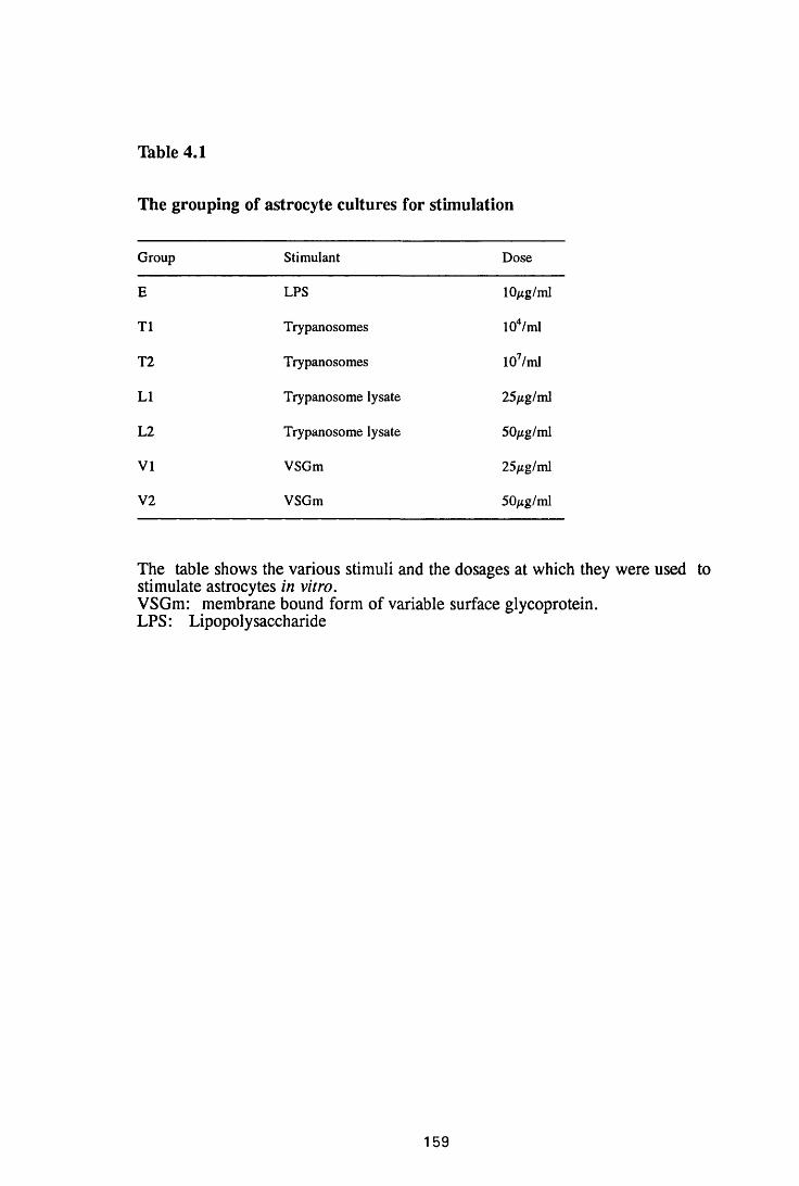
Table 4.1
The grouping of astrocyte cultures for stimulation
Group Stimulant Dose
E LPS 10/xg/ml
T1 Trypanosomes 104/ml
T2 Trypanosomes 107/ml
LI Trypanosome lysate 25/ig/ml
L2 Trypanosome lysate 50/ig/ml
VI VSGm 25/ig/ml
V2 VSGm 50/ig/ml
The table shows the various stimuli and the dosages at which they were used to stimulate astrocytes in vitro.VSGm: membrane bound form of variable surface glycoprotein.LPS: Lipopolysaccharide
159
Trypanosomes, in groups T1 and T2, were alive and actively motile throughout
the duration of the experiment.
4.2.7. Harvesting cells for RNA extraction
At each time point, the supernatant was removed and stored at -70°C
for subsequent analysis of secreted cytokines. The cells were washed twice with
HBSS and then lysed with 2ml of denaturing solution (4mM guanidinium
isothiocyanate, 25mM sodium citrate, pH 7.0, 0.5% sarcosyl, 0.1M 2-6-
mercaptoethanol) and removed with a rubber policeman. The mixture of cells
and denaturing solution was then transferred into glass-Teflon homogenisers and
homogenised at 4°C, transferred into polypropylene tubes (Falcon) and stored at
-70°C until RNA was extracted.
Each flask of cells (used for any one time point for any stimulant)
contained approximately 5xl06 astrocytes and so the amount of RNA from each
flask was expected to be the same. To confirm this, polymerase chain reaction
(PCR) was performed for actin transcripts (actin is a gene expressed by all cells)
on all RNA extracts. In addition, amplification of the actin gene confirmed the
integrity of the extracted RNA as well as verifying that it was amplifiable.
4.2.8. Preparation of RNA
RNA was prepared as described by Chomczynski and Sacchi (1987),
using acid-guanidinium thiocyanate phenol chloroform extraction. The
cell/denaturing solution homogenate was thawed. Sequentially, 0.1ml of 2M
sodium citrate (pH 4.0), 1ml of water-saturated phenol and 0.2ml of
chloroform/isoamyl alcohol (49:1) were added for each ml of denaturing
solution in the homogenate, with thorough mixing after addition of each reagent.
The final suspension was shaken vigorously for 10 seconds and cooled on ice
for 15 minutes. Samples were centrifuged at 10,000g for 20 minutes at 4°C. The
aqueous phase, containing RNA, was transferred to a fresh tube, mixed with
1 6 0
lml of isopropanol, then placed at -20°C for at least 1 hour to precipitate RNA
Sedimentation at 10,000g for 20 minutes at 4°C was performed and the
resulting RNA pellet was dissolved in 300/zl of denaturing solution, transferred
into a 1.5ml eppendorf tube and precipitated with two volumes of ethanol on dry
ice for at least 30 minutes. After centrifugation for 10 minutes at 4°C, the RNA
pellet was washed twice with 70% ethanol, sedimented by centrifugation,
vacuum dried for 15 minutes, dissolved in 60/xl of double distilled water and
stored at -20°C. Five /jl\ aliquots were heated at 72°C to denature RNA
secondary structures and electrophoresed on a 1 % agarose gel to identify the
rRNA by separation of the 28S and 18S bands. This is indicative of the integrity
of the rest of RNA molecules including the mRNA.
4.2.9. Preparation of cDNA and amplification by polymerase chain reaction
Primers for the different cytokines analysed were synthesised by
Genosys (Cambridge, U.K.). The primers are designed to produce bands of
different sizes depending on the cytokine being analysed (Brener, Thm, Nelson,
Engleman, Suzuki, Fry and Larick, 1989); B-actin 540 base pairs (bp), TNFa
692bp, IFN-y 460bp, macrophage inflammatory protein 1 (MIP-1) 267bp, IL-
la 625bp, IL-2 502bp, IL-4 399bp, IL-6 638bp and granulocyte-macrophage
colony stimulating factor (GM-CSF) 368bp. Approximately 5/xg of total RNA
was lyophilised and resuspended in 9/d of annealing buffer (250mM KC1,
lOmM Tris HC1, pH8.3, ImM EDTA) with 0.5 pg of the 3'primer heated at
80°C for 3 minutes, incubated at 69°C for 20 minutes followed by a 20 minute
cooling to 40°C. Fifteen ml of cDNA/dNTP buffer (24mM Tris HC1, pH 8.3,
16mM MgCl2, 18mM dithiothrietol, 0.4mM dNTPs) were added with 200 units
of Moloney murine leukemia virus reverse transcriptase (Gibco) and incubated
at 43°C for 45 minutes. Fbr analysis of the cDNA using PCR, 0.5fxg of the
5 'primer was added with 55/xl of Taq polymerase buffer (lOOmM Tris HC1 pH
161
8.0, 30mM (NH4)2S04,3mM MgCl2, lOmM mercaptoethanol), 20^1 2.5mM
dNTPs and 2 units of DNA polymerase (Promega). The reaction mixture was
subjected to 35 cycles of 94°C (denaturation) for 1 minute, 60°C (annealing) for
2 minutes, 72°C (extension) for 3 minutes. To visualise the PCR products, 20^1
of reaction mixture was electrophoresed through a 2% agarose gel and stained
with ethidium bromide. Brain homogenates from CD1 mice have been reported
to constitutively express mRNA for IL-1 a (Hunter et al, 1992c). For this
reason, water was included as a negative control in all the PCRs performed, to
rule out contamination, and unstimulated cells were taken to provide baseline
levels. Spleen from T.b brucei-infected mice was used as positive control for all
the cytokine transcripts analysed, except for GM-CSF for which a commercial
positive control was purchased from Clontech Laboratories.
4.2.10 Detection of cytokine protein in cultured astrocytes
Astrocytes were removed from a confluent flask by trypsinisation and
seeded onto PLL coated coverslips contained in 24-well tissue culture
plates, at a density of 4xl04 per coverslip. They were maintained in 50%
astrocyte conditioned DMEM/10%FCS for the first 24 hours, after which the
medium was changed to fresh DMEM/10%FCS and cells grown for a further 48
hours, to establish a stable confluent monolayer. The cells were then stimulated
with either 10^g/ml LPS or 50jug/ml lysate or 50jiig/ml VSGm, for 2, 4, 6 and 8
hours. At each time point, the cells were fixed with methanol at -20°C for 10
minutes and placed in PBS, pH 7.0 until the staining procedure could be carried
out.
The coverslips were removed from the PBS containing wells, fixed
onto glass slides with glassbond in ultra violet light (u.v. light activates
glassbond) and incubated for 10 minutes with 5% bovine serum albumin (BSA)
in Tris buffered saline (TBS), pH 7.0, containing 0.01% saponin to block non
specific binding and permeabilise cells for the antibodies. The cells were then
162
incubated with the primary antibodies, (rabbit anti-mouse TNFa and rabbit anti
mouse IL-lce diluted 1 in 50 and 1 in 150 in TBS/1%BSA/0.01% saponin,
respectively, Genzyme) for 1 hour at room temperature in a humid chamber.
After washing with TBS twice (10 minutes each time), the secondary antibodies,
goat anti-rabbit immunoglobulins (Vector Laboratories), diluted 1 in 200 for
TNF and IL-1, were added and incubated for 30 minutes. They were then
washed again in TBS and incubated with avidin-biotin alkaline phosphatase
conjugate, diluted 1 in 50, for 30 minutes and washed again. For colour
visualisation, the cells were incubated with levamisole, fast red and naphthol in
veronal acetate buffer (i.e. naphthol/fast red) for 5 minutes. The cells were post
fixed in neutral buffered formalin, washed in water, counterstained with
haematoxylin and coverslips mounted with aquamount.
4.2.11. Detection of secreted cytokine protein in astrocyte supernatants
An enzyme-linked immunosorbent assay, for mouse TNF, was used
to test for secreted cytokines in the cell supernatants following exposure to
various stimuli. Briefly, 96-well microtitre plates (Corning easy wash or
Dynatech IV) were coated with 50/nl/well of purified anti-mouse TNF antibodies
at a concentration of 2^g/ml and incubated overnight at 4°C. The following
morning, nonspecific binding was blocked by incubation with 50/xl/well 10%
FCS in PBS, pH 7.4, for 45 minutes at 37°C. The plates were rinsed three times
in PBS containing 0.05% tween 20, followed by two washes of 3 minutes each.
Recombinant mouse tumor necrosis factor (TNF) purchased from
Pharmigen was used as the standard cytokine. Ten serial double-dilution in PBS
starting at 80ng/ml were prepared from the stock. The standard-cytokine
dilutions and the neat supernatant samples were added (50^1/well) in duplicates,
and incubated at 37°C for 2 hours. The plates were then washed in PBS/tween
as before, incubated for 1 hour at room temperature with 50/xl/well of
163
biotinylated mouse anti-TNF antibody (Pharmigen), at 4/*g/ml, and washed
again. Streptavidin peroxidase (Serotec), diluted 1 in 1,000 in 1%
BSA/PBS/tween 20 was added (75/d/well) and incubated for 1 hour at 37°C and
washed. The peroxidase substrate, 3,3', 5, 5' tetramethylbenzidine (TMB) was
added at 100/d/well. Absorbencies were read at OD630, after 15 minutes.
4.3. RESULTS
4.3.1. Controls
Messenger RNA transcripts for IL-113 were found in all astrocytes investigated,
including those that were not exposed to any stimulant. This expression
increased after exposure to the various stimuli as judged by the intensity of
complementary DNA (cDNA) bands. Transcripts for the other cytokines were
absent from non-stimulated control samples.
Using the PCR, all the positive controls were positive for mRNA
transcripts for the cytokines studied. On electrophoresis, the actin PCR products
from all the flasks produced bands of approximately same intensity. Therefore,
PCR for cytokine transcripts was used semiquantitatively, i.e., a difference in
band intensity was taken to be indicative of a difference in the amounts of
transcripts expressed.
LPS at a concentration of 10/ig/ml stimulated astrocytes to express
mRNAs for IL-la, IL-6, TNFa, MIP-1 and GM-CSF (Thble 4.2). The
thickness of the bands was used as an indication of the amount of cytokine
transcript and designated either, -, ± , + , + + or + + + depending on the
thickness of the bands. To illustrate this, the cytokine transcripts expressed for
IL-1 a, MIP-1, GM-CSF and IL-113 are shown in Figure 4.1. Messenger RNA
transcripts for all the cytokines studied, were detectable by 2 hours after
exposure to LPS. Thereafter, the expression of transcripts increased, with those
for IL-6 and IL-1B reaching ceiling levels by the 4th and 6th hours, respectively,
164
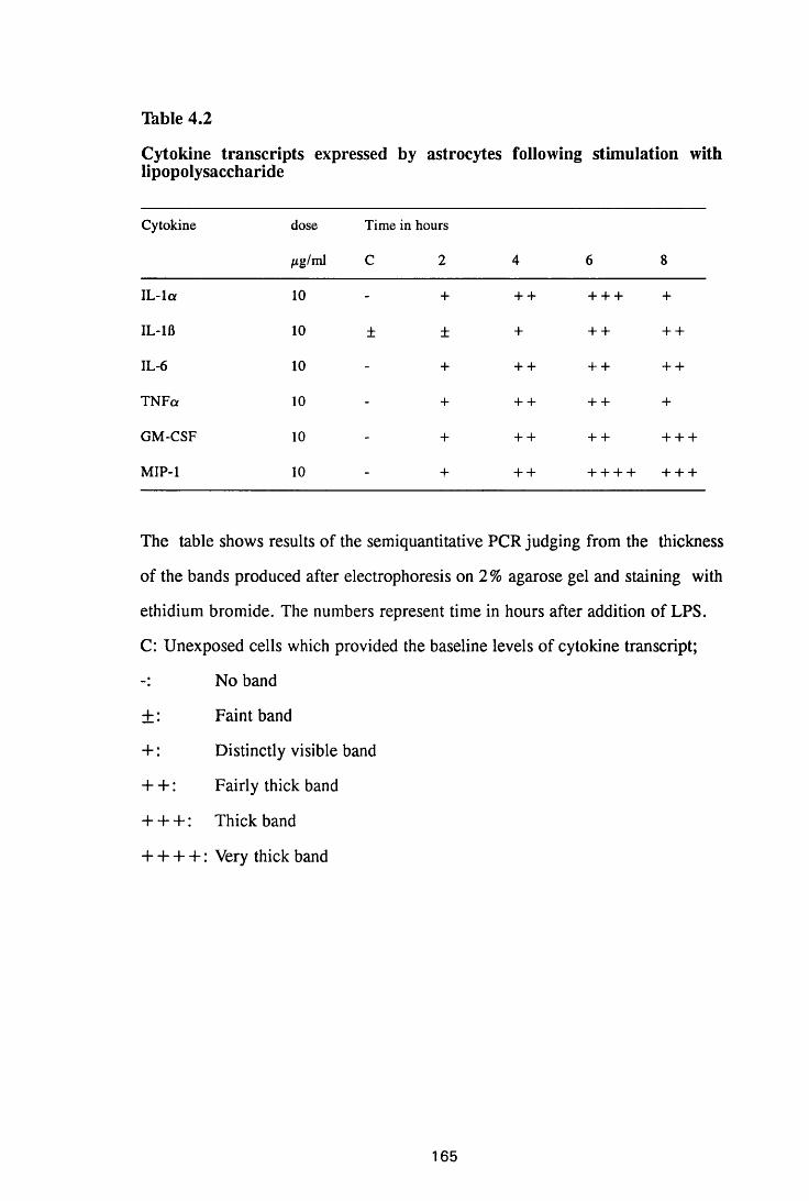
Table 4.2
Cytokine transcripts expressed by astrocytes following stimulation with lipopolysaccharide
Cytokine dose
/ig/ml
Time in hours
C 2 4 6 8
IL -la 10 - + + + + + + +
IL-1B 10 ± ± + + + + +
IL-6 10 - + + + + + + +
TNFa 10 - + + + + + +
GM-CSF 10 - + + + + + + + +
MIP-1 10 - + + + + + + + + + +
The table shows results of the semiquantitative PCR judging from the thickness
of the bands produced after electrophoresis on 2 % agarose gel and staining with
ethidium bromide. The numbers represent time in hours after addition of LPS.
C: Unexposed cells which provided the baseline levels of cytokine transcript;
No band
± : Faint band
+: Distinctly visible band
+ +: Fairly thick band
+ + +: Thick band
+ + + + : Very thick band
165
Figure 4.1
Gel electrophoresis showing cytokine transcripts expressed by astrocytes
exposed to LPS (group E): a) IL-la, b) MIP-1, c) GM-CSF and d) IL-lfl. 2, 4,
6 and 8 is time in hours after introduction of LPS. W = water which was used
as negative control; + = positive control and M = molecular weight marker,
123bp ladder (Gibco).
166
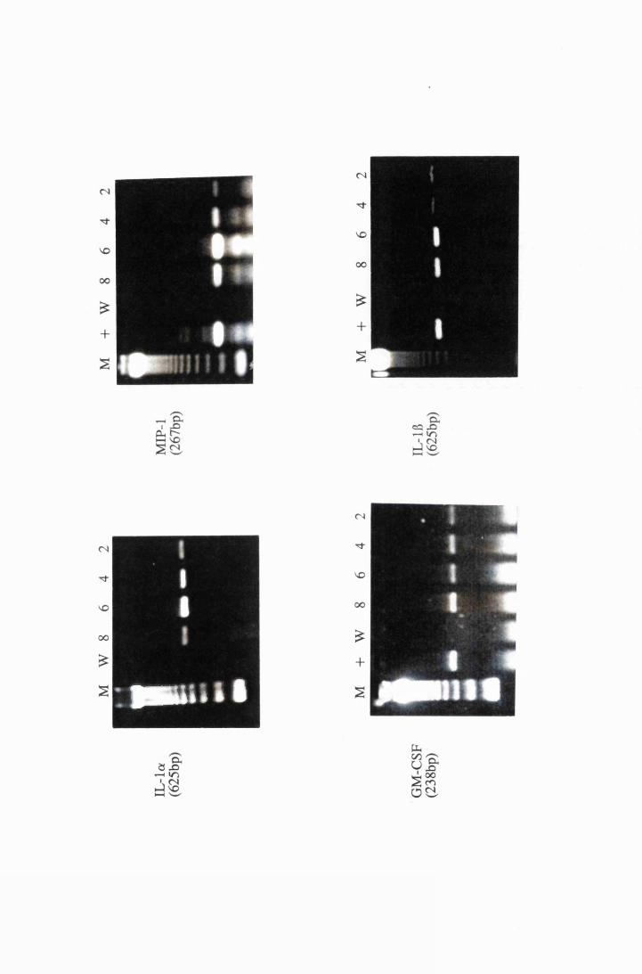
mu* is
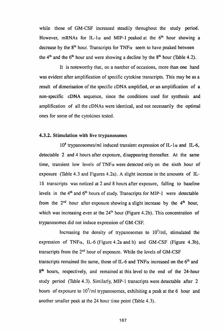
while those of GM-CSF increased steadily throughout the study period.
However, mRNAs for IL-1 ot and MIP-1 peaked at the 6th hour showing a
decrease by the 8th hour. Transcripts for TNFa seem to have peaked between
the 4th and the 6th hour and were showing a decline by the 8lh hour (Thble 4.2).
It is noteworthy that, on a number of occasions, more than one band
was evident after amplification of specific cytokine transcripts. This may be as a
result of dimerisation of the specific cDNA amplified, or an amplification of a
non-specific cDNA sequence, since the conditions used for synthesis and
amplification of all the cDNAs were identical, and not necessarily the optimal
ones for some of the cytokines tested.
4.3.2. Stimulation with live trypanosomes
104 trypanosomes/ml induced transient expression of IL-1 a and IL-6,
detectable 2 and 4 hours after exposure, disappearing thereafter. At the same
time, transient low levels of TNFce were detected only on the sixth hour of
exposure (Thble 4.3 and Figures 4.2a). A slight increase in the amounts of IL-
1B transcripts was noticed at 2 and 8 hours after exposure, falling to baseline
levels in the 4th and 6lh hours of study. Transcripts for MIP-1 were detectable
from the 2nd hour after exposure showing a slight increase by the 4th hour,
which was increasing even at the 24th hour (Figure 4.2b). This concentration of
trypanosomes did not induce expression of GM-CSF.
Increasing the density of trypanosomes to 107/ml, stimulated the
expression of TNFa, IL-6 (Figure 4.2a and b) and GM-CSF (Figure 4.3b),
transcripts from the 2nd hour of exposure. While the levels of GM-CSF
transcripts remained the same, those of IL-6 and TNFa increased on the 6th and
8th hours, respectively, and remained at this level to the end of the 24-hour
study period (Thble 4.3). Similarly, MIP-1 transcripts were detectable after 2
hours of exposure to 107/ml trypanosomes, exhibiting a peak at the 6 hour and
another smaller peak at the 24 hour time point (Table 4.3).
167
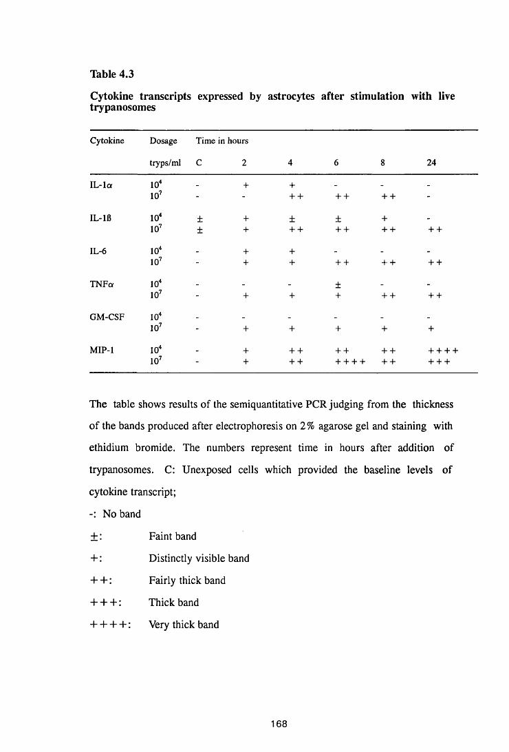
Table 4.3
Cytokine transcripts expressed by astrocytes after stimulation with live trypanosomes
Cytokine Dosage
tryps/ml
Time in hours
C 2 4 6 8 24
IL-1 a 104 - + + - - -
107 - - + + + + + + -
IL-1B 104 ± + ± ± + -
107 ± + + + + + + + + +
IL-6 104 _ + + - _ _
107 - + + + + + + + +
TNFa 104 _ _ _ ± _ -
107 - + + + + + + +
GM-CSF 104 _ _ _ _ _ _
107 - + + + + +
MIP-1 104 + + + + + + + + + + +107 - + + + + + + + + + + + +
The table shows results of the semiquantitative PCR judging from the thickness
of the bands produced after electrophoresis on 2% agarose gel and staining with
ethidium bromide. The numbers represent time in hours after addition of
trypanosomes. C: Unexposed cells which provided the baseline levels of
cytokine transcript;
No band
±: Faint band
+ : Distinctly visible band
+ + : Fairly thick band
+ + + : Thick band
+ + + + : Very thick band
168
Figure 4.2
Gel electrophoresis showing transcripts for a) IL-1 a and TNFa, b)
MIP-1 and IL-6, extracted from astrocytes in group T l, stimulated with 104/ml
(right), and group T2 stimulated with 107/ml (left) live trypanosomes. 2, 4, 6, 8
and 24 represent the time in hours after addition of trypanosomes. C =
transcripts from cells that were not exposed to stimulant; W = water that was
used as negative control; + = positive control; M = molecular weight marker,
123bp ladder (Gibco).
169
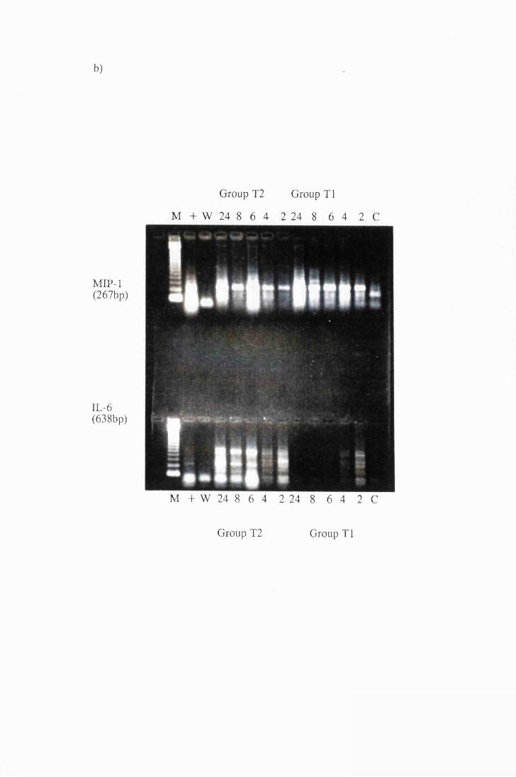
b)
MIP-1(267bp)
IL-6(638bp)
Group T2 Group T1
M + W 24 8 6 4 2 24 8 6 4 2 C
M + W 24 8 6 4 2 24 8 6 4 2 C
Group T2 Group T1
Figure 4.3
Gel electrophoresis showing amplified transcripts for a) IL-lfl and b)
GM-CSF expressed by astrocytes in group T2, exposed to 107/ml trypanosomes.
The numbers represent the time in hours after addition of trypanosomes. C =
transcripts from cells that were not exposed to stimulant; W = water that was
used as negative control; + = positive control and M = molecular weight
marker, 123bp ladder (Gibco).
170
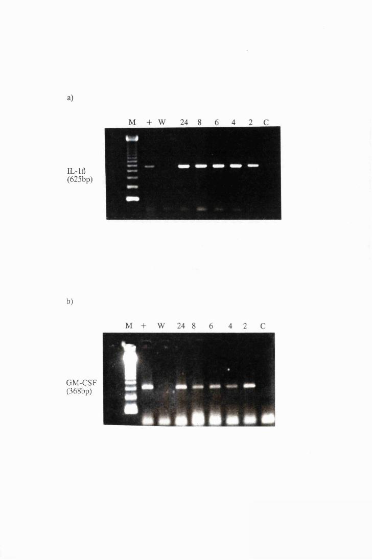
IL-lfl(625bp)
b)
GM-CSF(368bp)
The higher density of trypanosomes upregulated the expression of IL-lfl from
the 2nd hour, showed an increase on the 4th hour after exposure, and remained
high for the rest of the study period (Thble 4.3 and Figure 4.3a). IL-la
transcripts induced by 107 trypanosomes/ml were detectable 4 hours after
exposure, remained at the same level at the 6 and 8 hour sampling but had
disappeared by 24 hours (Table 4.3 and Figure 4.2a).
4.3.3. Stimulation with whole trypanosome lysate
The whole trypanosome lysate also induced expression of mRNAs for IL-la,
IL-6, TNFa, MIP-1 and GM-CSF, and upregulated the expression IL-1B
transcripts (Thble 4.4 and Figures 4.4 to 4.6). Transcripts for IL-6 and MIP-1
were detected 2 hours after exposure to 25^cg/ml trypanosome lysate, while
those of GM-CSF and IL-1 a were detectable at 4 and 6 hours, respectively
(Table 4.4). Thereafter, there was an increase in the levels of IL-6 and MIP-1 at
4 and 8 hours, respectively, with those of IL-6 remaining at this level for the
rest of the study period. In contrast, levels of GM-CSF remained the same while
those of IL-la declined at the 8lh hour of study (Thble 4.4). Expression of IL-115
was upregulated, reaching a peak between 4 and 6 hours after exposure and
showing a decline at the 8th hour of the study. Trypanosome lysate, at this
concentration, did not induce expression of TNFa mRNA transcripts even after
8 hours of exposure (Thble 4.4).
Increasing the concentration of trypanosome lysate to 50^g/ml, hastened the
expression of GM-CSF and IL-la, so that transcripts were detectable at 2 and 4
hours, respectively, with those GM-CSF peaking at the 4th hour while those of
IL-la displayed a drop at the 6lh hour increasing again at the 8th hour. It also
upregulated the expression of IL-115 from the 2nd hour of study, with levels
remaining the same for the rest of the study period. Transcripts for IL-6 and
MIP-1 were detectable at 2 hours and showed an increase at 4 hours. While the
levels of MIP-1 peaked at the 6th hour, those of IL-6 reached a peak between
171
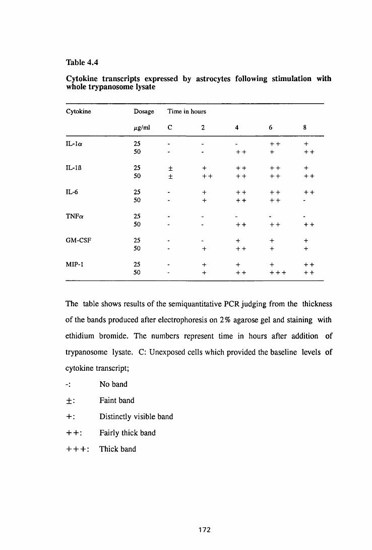
Table 4.4
Cytokine transcripts expressed by astrocytes following stimulation with whole trypanosome lysate
Cytokine Dosage
Mg/ml
Time in hours
C 2 4 6 8
IL -la 25 _ _ + + +50 - - + + + + +
IL-1B 25 ± + + + + + +50 ± + + + + + + + +
IL-6 25 + + + + + + +50 - + + + + + -
TNFa 25 - _ _ - -
50 - - + + + + + +
GM-CSF 25 _ _ + + +50 - + + + + +
MIP-1 25 - + + + + +50 - + + + + + + + +
The table shows results of the semiquantitative PCR judging from the thickness
of the bands produced after electrophoresis on 2% agarose gel and staining with
ethidium bromide. The numbers represent time in hours after addition of
trypanosome lysate. C: Unexposed cells which provided the baseline levels of
cytokine transcript;
No band
±: Faint band
+ : Distinctly visible band
+ + : Fairly thick band
+ + + : Thick band
172
Figure 4.4
Transcripts for IL-la and TNFce expressed by astrocytes in group LI,
which were exposed to trypanosome lysate at 25^g/ml (left) and from group
L2, exposed to 50^g/ml (right). 2, 4, 6 and 8 represent the time in hours after
addition of trypanosome lysate. C = transcripts from cells that were not
exposed to stimulant; W = water that was used as negative control; + =
positive control; M = molecular weight marker, 123bp ladder (Gibco).
173
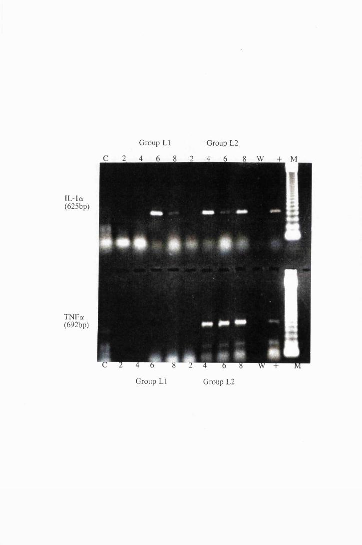
TNFa(692bp)
Group LI Group L2
2 4 6 8 W + M
IL-la(625bp)
Group L2Group LI
Figure 4.5
Transcripts for IL-6 and GM-CSF expressed by astrocytes in group LI,
exposed to trypanosome lysate at 25^g/ml (right) and astrocytes from group L2,
exposed to 50/xg/ml (left). 2, 4, 6 and 8 represent the time in hours after
addition of trypanosomes lysate. C = transcripts from cells that were not
exposed to stimulant; W = water that was used as negative control; + =
positive control; M = molecular weight marker, 123bp ladder (Gibco).
174
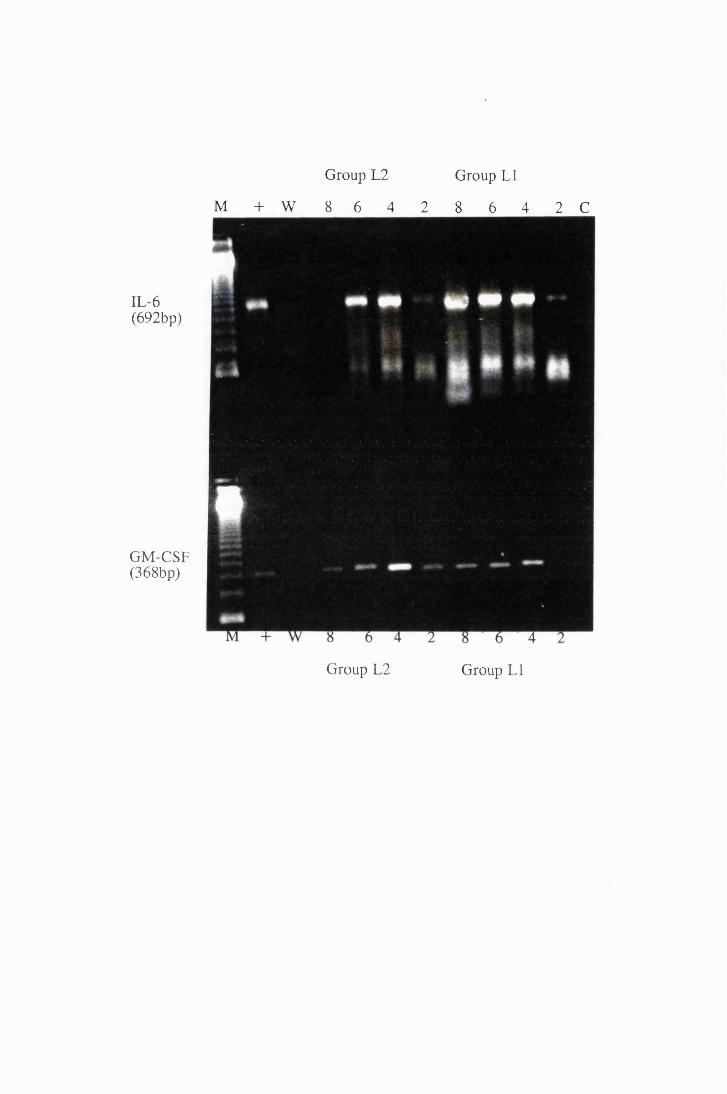
Group L2 Group LI
M + W 8 6 4 2 8 6 4 2 C
IL-6(692bp)
GM-CSF(368bp)
Group L2 Group LI
Figure 4.6
Cytokine transcripts expressed by astrocytes after exposure to 25^g/ml,
group LI, and 50/*g/ml, group L2, trypanosome lysate. Panels a and b show
the transcripts for IL-lfl, while panel c shows transcripts for MIP-1. 2, 4, 6 and
8 represent the time in hours after addition of trypanosome lysate. C =
transcripts from cells that were not exposed to stimulant; W = water that was
used as negative control; + = positive control and M = molecular weight
marker, 123bp ladder (Gibco).
175
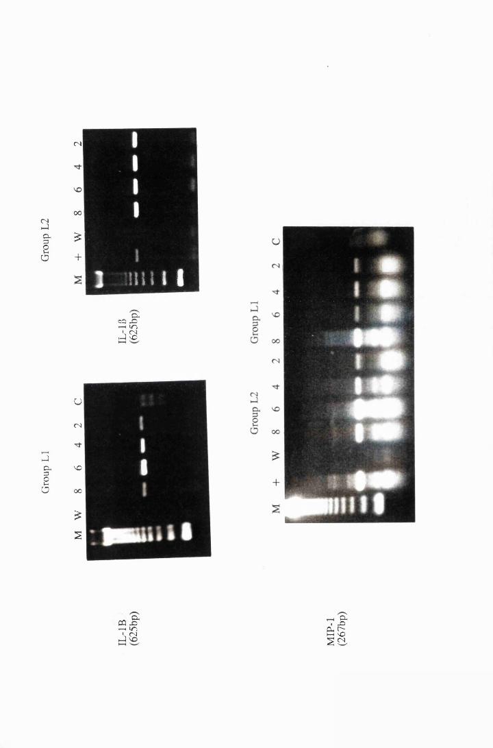
Grou
p LI
Gr
oup
L2
<n
00 I
£
+
U
CN
\D
OO
£
2
mil Ip CCJ X> - h in ' CN >J iO
p CQ XrH lO» (N1-1
P I X)e=!sSO-
*•*
4 and 6 hours after exposure and had disappeared by the 8lh hour of study.
Trypanosome lysate at 50^ig/ml induced expression of TNFa transcripts, which
were detected from the 4th hour and remained at the same level throughout the
study period (Thble 4.4).
Translation of cytokine message into protein, for both IL-la and
TNFa following exposure to 50/zg/ml trypanosome lysate, was detectable by
immunocytochemistry from the 4th hour through to the 8th hour of study, with
cells staining more intensely for IL-la than for TNFa. There was no protein
detectable in the unstimulated cells or in the cells stimulated for 2 hours (Figure
4.7). The protein was diffusely distributed in the cytoplasm of most cells but
was more intense in the perinuclear zone (Figure 4.8). In contrast to IL -la and
TNFa, IL-6 was detectable in the first two hours after stimulation, but this
declined and was only faintly detectable on the 6th and 8th hours of study (data
not shown).
An attempt was made to determine whether TNFa protein was
secreted and to quantify the amounts, by an enzyme-linked immunosorbent
assay (ELISA) (Pharmingen, 1993). Although this technique detected as little as
0.625ng/ml of TNF protein in the standard, the levels of this cytokine in the
astrocyte supernatants from all the stimuli, used in this study, were undetectable
by this method.
4.3.4. Stimulation with the membrane-bound form of variable surface
glycoprotein
Following exposure to 25jug/ml VSGm, induction of transcripts for
IL-la and MIP-1, as well as an increase in the expression of IL-113 were
detected by the 2nd hour, while expression of IL-6, TNFa and GM-CSF
occurred by the 4th hour (Table 4.5). However, this induction seems to have
been transient, with complete disappearance of IL-1J3 by the 4th hour of study,
and of IL-la, GM-CSF and TNFa by the 8th hour of study. There was a
176
Figure 4.7
Micrographs showing the presence of cytokine protein in astrocytes from
group L2, which were stimulated with 50^ig/ml of trypanosome lysate. The pink
colour shows the presence of IL-la after 8 hours of exposure (a). Control
astrocytes that were not exposed to stimulation did not stain for IL-la (b).
Naphthol red is used as chromogen and the nuclei are stained with
haematoxylin. x200
177
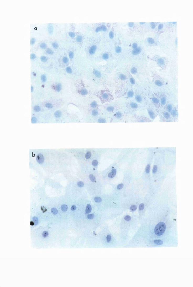
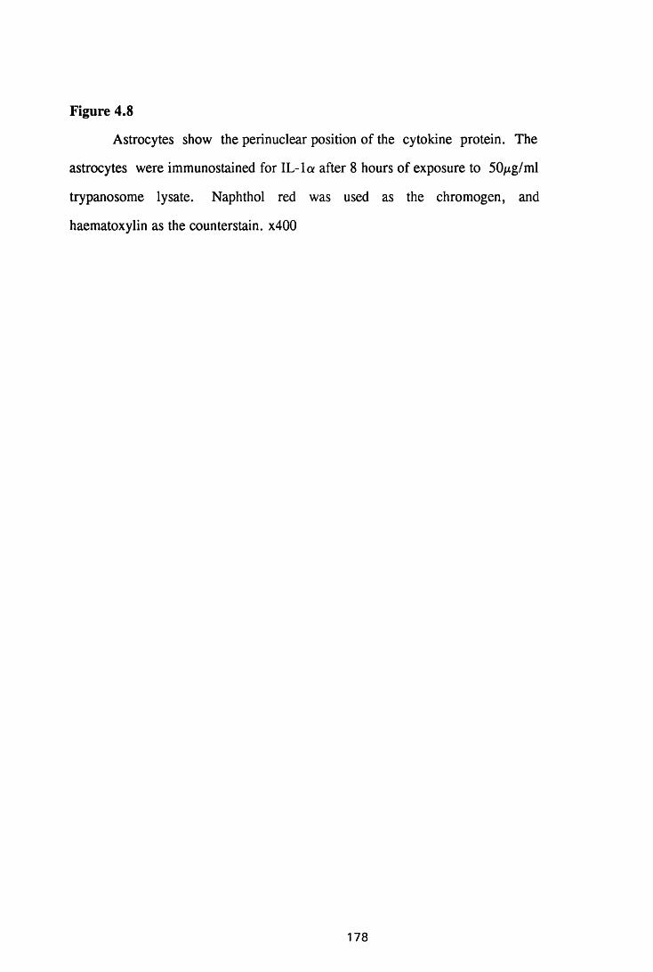
Figure 4.8
Astrocytes show the perinuclear position of the cytokine protein. The
astrocytes were immunostained for IL-la after 8 hours of exposure to 50^cg/ml
trypanosome lysate. Naphthol red was used as the chromogen, and
haematoxylin as the counterstain. x400
178
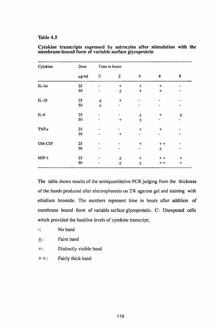
Table 4.5
Cytokine transcripts expressed by astrocytes after stimulation with the membrane-bound form of variable surface glycoprotein
Cytokine Dose Time in hours
/ig/ml C 2 4 6 8
IL -la 25 _ + + + -
50 - ± + + -
IL-1B 25 ± + _ _ _
50 ± - - - -
IL-6 25 _ _ + + ±50 - + ± - -
TNFa 25 _ _ + + _
50 - + - - -
GM-CSF 25 _ _ + + + _
50 - - - ± -
MIP-1 25 . ± + + + +50 - ± ± + + +
The table shows results of the semiquantitative PCR judging from the thickness
of the bands produced after electrophoresis on 2 % agarose gel and staining with
ethidium bromide. The numbers represent time in hours after addition of
membrane bound form of variable surface glycoprotein. C: Unexposed cells
which provided the baseline levels of cytokine transcript;
No band
±: Faint band
+ : Distinctly visible band
+ + : Fairly thick band
179
decrease in the expression of IL-6 and MIP-1 mRNA by the 8th hour of study.
Increasing the concentration of VSG inhibited the expression of IL-lfi
and decreased the duration of transcript expression for IL-6 and TNFa by 1
hour and of GM-CSF by 2 hours, although IL-6 and TNFa transcripts were
detected at an earlier stage. However, it did not affect the duration of IL-la and
MIP-1 expression (Thble 4.5).
4.3.5. Detection of secreted cytokines in the astrocyte supernatants
Astrocyte supernatants, from each of the stimulant used in this study,
were tested for the presence of secreted TNFa protein by ELISA (Chapter 2).
Although all the standard cytokine dilutions, the lowest of which was
1.25ng/ml, gave positive results, no TNFa was detected from any of the
astrocyte supernatants.
4.4. DISCUSSION
For the first time, trypanosomes and trypanosome-derived products
have been shown to induce cultured astrocytes to produce the cytokines IL-la
and -fl, IL-6, TNFa, GM-CSF, and MIP-1. Amongst the cytokines studied
here, the production of IL-la and TNFa mRNA transcripts and protein are
possibly the most significant, considering their key role in the initiation of
meningeal inflammation (Ramilo et al, 1990; Saukkonen et al, 1990).
The results showed that all cultured astrocytes expressed the mRNA
transcripts for IL-113, including, the cells that were not exposed to any stimulant.
While this may result from something in the culture medium selectively
stimulating expression of this cytokine transcript, it is likely that astrocytes from
NIH mice constitutively express the mRNA for IL-113. Other researchers have
reported constitutive expression of colony-stimulating factor 1 (CSF-1) in
cultured astrocytes (Lee et al, 1993b) and IL-la transcripts in the brains of CD
1 mice (Hunter et al, 1992c).
180
Trypanosomes, whole trypanosome lysate and VSG were found to
induce astrocytes, in culture, to express mRNAs for the cytokines IL -la and -6,
TNFa, IL-6, GM-CSF and MIP-1. Live trypanosomes and whole trypanosome
lysate were potent stimuli, inducing, expression of these cytokines for longer
durations than the less potent VSGm. This suggested that other parts of
trypanosomes, or factors produced by these parasites, might be additive to
VSGm in upregulating the expression of cytokine transcripts.
The results of immunocytochemistry showed that translation of the
cytokine message into protein was rapid with the detection of translated protein,
in the three cytokines investigated this way, namely IL-la, IL-6 and TNFa,
coinciding with the time the message was detected. It is possible that there was a
time lapse (of less than two hours) between the mRNA gene expression and
translation of the message into protein but, due to the timing of these
experiments, the lapse was not detectable. The decrease in the IL-6 protein in
the 6th and 8th hours might mean that this cytokine was rapidly secreted from the
cells into the surrounding environment. Unfortunately, the attempt to
demonstrate the presence of secreted cytokine in the cell supernatants, by
ELISA, gave negative results. There are a number of possible reasons why
detection of secreted cytokine by ELISA might fail. 1) The cytokine protein in
supernatants from the cell cultures might have been very dilute since 10ml of the
culture medium was used to feed only 5xl06 astrocytes. Other researchers using
VSG- and LPS- and concanavalin A (Con A)-stimulated macrophages (Mathias
et al, 1990; Dr. E.Devaney, personal communication) used 1ml of medium to
feed 106 cells, and successfully detected secreted cytokines using this technique.
2) The supernatants in this study were stored for long durations and the pH of
the medium had changed to above 8 by the time the ELISA was performed;
these circumstances might have denatured the cytokines. 3) It is possible that
under the in vitro conditions, the cytokine studied, TNF, was not secreted
181
during the period of study.
The cytokines produced by cultured astrocytes in response to whole
trypanosomes, their lysate, or the VSGm in vitro have been shown to induce
important biological changes in various cells, including, blood vascular
endothelial cells (Montovani and Dejana, 1989), lymphocytes (Kasahara et al,
1991; Merrill et al, 1992), macrophages (Broudy, Kaushansky, Harlan and
Adamson, 1987; Sieff, Niemeyer, Mentzer and Faller, 1988; Kasahara et al,
1991), microglia (Hurwitz et al, 1992; Lee, et al, 1993a; 1993b) and astrocytes
themselves (Fontana et al, 1981; Lieberman et al, 1989; Yong et al, 1991;
Merrill et al, 1992). For example, TNFa and IL-la not only induce
proliferation of astrocytes but also expression of MHC class II antigens and
intercellular adhesion molecules (McCarron et al, 1986). These cytokines exert
similar effects on macrophages (Rouzer and Cerami, 1980), endothelial cells
(Montovani and Dejana, 1989), microglia (Ramilo et al, 1990; Saukkonen et al,
1990; Hurwitz et al, 1992; Lee et al, 1993a; 1993b), and lymphocytes (Selmaj
et al, 1990). IL-la and TNF-a have also been found to induce production of
cytokines such as IL-la, TNFa, IL-2 and IL-6 in astrocytes, microglia (Ramilo
et al, 1990; Saukkonen et al, 1990; Chen, Hinton, Apuzzo and Hofman, 1993),
endothelial cells (Montovani and Dejana, 1990), macrophages and lymphocytes
(Broudy et al, 1987; Sieff et al, 1988; Selmaj et al, 1990; Kasahara et al, 1991;
Merrill et al, 1992).
The demonstration, in vitro, that trypanosomes can induce astrocytes
to produce cytokines complements the in vivo studies in Chapter 3, where
astrocyte activation (as judged by the intensity of GFAP staining) occurred early
in the course of infection, prior to the infiltration of inflammatory cells into the
CNS of T.b.brucei-infected mice. Indeed, induction of mRNA transcripts for
some of these cytokines, including, TNF, IL-1, and MIP-1, in addition to IL-4
and IFN7 in the brains of T. &r«ce/-infected mice have been reported to coincide
with astrocyte activation (Hunter et al, 1991). The results of the current in vitro
182
study suggest that astrocytes may produce the cytokines that start the cascade of
events, leading to the development of the CNS inflammation observed in late-
stage trypanosomiasis (Chapter 1).
It would appear likely that the six cytokines found to be produced by
trypanosome-stimulated astrocytes, in this study, might have an important role
in the initiation and propagation of the CNS pathology found in T.b.brucei
infected mice (Jennings et al, 1989) and in sleeping sickness patients (Adams et
al, 1986; Haller et al, 1986). For instance, IL-1 upregulates expression of
intercellular adhesion molecule 1 (ICAM-1) and endothelial leukocyte adhesion
molecule 1 (ELAM-1) which alter the adhesive properties of the vascular
endothelium, and initiate leukocyte extravasation. IL-1 also induces the
endothelial cells to produce chemotactic cytokines such as IL-8, macrophage
chemotactic protein (MCP) and colony-stimulating factors (CSFs) which lead to
leukocyte recruitment and proliferation (Broudy et al, 1987; Sieff et al, 1988).
IL-1 is a polyclonal activator of lymphocytes inducing production of
other cytokines, amongst which, the production of IL-2, and the expression of
IL-2 receptors, are of paramount importance (Durum, Schmidt and Oppenheim,
1985). The action of IL-2 on the induced IL-2 receptors stimulates proliferation
of lymphocytes, differentiation into antibody-producing plasma cells, modulates
expression of IL-2 receptor and downregulates the production of IL-1, thereby
providing a form of negative feedback mechanisms for IL-2 activity and IL-1
production, respectively (Walldmann, 1993). Thus, IL-1 might contribute to the
increase in the number of lymphocytes and the occurrence of plasma cells in the
CNS of late-stage sleeping sickness patients (Adams et al, 1986; Haller et al,
1986) and of T.b.brucei-infected mice (Murray et al, 1974; Jennings et al,
1989). Other important activities of IL-1, include, augmentation of natural killer
(NK)-mediated cytotoxicity, induction of prostaglandin release, chemotaxis and
tumour cell lysis by macrophages; and induction of fever (Durum et al, 1985);
183
these are also properties of activated macrophages. IL-1 production in the brain
may thus account for the presence of activated macrophages in the CNS of late-
stage trypanosomiasis (Poltera, 1980; Schultzberg et al, 1988).
TNFa induces migration of endothelial cells (ECs) but inhibits their
proliferation, stimulates production of chemotactic cytokines and CSFs
(Seelentag et al, 1987), augments the expression of MHC class I expression
(Pober et al, 1987b) and has also been reported to induce migration of
monocytes across the blood-brain barrier (Saukkonen et al, 1990). Treatment of
cultures of human fetal astrocytes with TNFa induces expression of various
adhesion molecules, including, E-selectin and IG9 message and protein (both of
which are monocyte adhesion molecules), vascular cell adhesion molecule 1
(VCAM-1) (Hurwitz et al, 1992) and ICAM-1 (Satoh, Kastrukoff and Kim,
1991; Frohman et al, 1989). This implies that once monocytes and lymphocytes
have crossed the blood-brain barrier, their migration to sites of inflammation
within the CNS parenchyma might be facilitated by the expression of adhesion
molecules on astrocytes (Hurwitz et al, 1992). It is also possible that astrocytes
respond to the TNF they (astrocytes) and other inflammatory cells produce
during trypanosome infections by expressing adhesion molecules which in turn
enhance the spread of inflammatory cells in the CNS.
GM-CSF (and G-CSF) induce migration and proliferation of
endothelial cells but do not stimulate their proinflammatory/prothrombotic
properties or functions associated with immune accessory activity. However,
both these cytokines induce a pattern of responses in ECs that is similar to that
induced by fibroblast growth factors (FGFs). Thus, they are potentially
mitogenic and induce EC chemotaxis and proteinase synthesis which is required
for cell migration and new vessel formation (Montovani and Dejana, 1989). The
migration of ECs, in response to cytokines produced by astrocytes, might create
leaks in the endothelial-brain barrier, allowing passage of cells and
macromolecules, possibly even trypanosomes, into the Virchow Robin spaces to
184
form perivascular cuffs and produce conditions suitable for cellular proliferation
and trypanosome replication. In addition, GM-CSF can cause proliferation of
granulocyte-macrophage progenitor cells in bone marrow cultures, as well as the
initial proliferation of multipotential stem cells in mouse fetal liver and adult
marrow capable of forming mixed or pure erythroid, eosinophil or
megakaryocyte colonies (Metcalf and Nicola, 1983). However, GM-CSF alone
is inadequate in stimulating proliferation of terminal cell divisions in these
pathways (Metcalf and Nicola, 1983), but can stimulate various functional
activities of the mature post-mitotic granulocytic and macrophage cells such as
increased RNA and protein synthesis (Burgess and Metcalf, 1980).
Macrophages are stimulated to exhibit increased phagocytic and cytocidal
activity (Handman and Burgess, 1979), to increase production of prostaglandin
E (PGE) (Kurland, Broxmeyer, Pelus, Bockman and Moore, 1978) and
plasminogen activator (Lin and Gordon, 1979). GM-CSF also induces
tumoricidal activity in human monocytes and macrophages (Grabstein, Urdal,
Tushiski, Mochizuki, Price, Cantrell, Gillis and Conlon, 1986).
G- and GM-CSF seem to regulate both the proliferation of microglial
cells (Giulian and Ingeman, 1988; Raivich, Gehrmann and Kreutzberg, 1991)
and their differentiation into macrophages (Suzumura, Sawada, Yamamoto and
Marunouchi, 1990). GM-CSF induces the differentiation of intrinsic brain
precursor cells into a population of microglia cells, which is intrinsically
activated for antigen-presentation. Such microglia induce antigen-directed
proliferation of TH1 and TH2 lymphocyte series (Fischer, Nitzgen, Germann,
Degitz, Daubener and Hadding, 1993). Production of GM-CSF in the brain in
late-stage trypanosomiasis might account for the proliferation and differentiation
of microglia and antigen-directed proliferation of the infiltrating lymphocytes.
Interleukin 6 is a cytokine with pleiotropic biological functions,
including, release of acute phase proteins, induction of differentiation of
185
activated B cells into immunoglobulin-secreting cells, stimulation of T cell
activation, and cooperation with colony-stimulating factors in the induction of
colony formation by haemopoietic progenitors (Van Snick, 1990). Human fetal
astrocytes in culture are induced to produce IL-6 by IL-111 and to a lesser extent
by TNFa (Yasukawa, Hirano, Watanabe, Muratani, Matsuda, Nakai and
Kishimoto, 1987). The ability of astrocytes to produce IL-6 in response to
cytokines (Frei et al, 1989) may represent an important mechanism by which
resident brain cells, in particular astrocytes and microglia, enhance the local
antibody response in infectious and autoimmune CNS disease, such as viral
meningitis or encephalitis and multiple sclerosis, in which intrathecal synthesis
of immunoglobulins is known to occur (Esiri, 1980; Resnick, Di Marzo-
Veronese, Schupbach, Tourtellotte, Ho, Muller, Shapshak, Vogt, Groopman,
Markham and Gallo, 1985; Vandvik, Skolenberg, Frosgren, Stemstedt,
Jeansson and Norrby, 1985; Houssiau, Bukasa, Sindic, Van Damme and Van
Snick, 1988; Frei, Leist, Meager, Gallo, Leppert, Zinkernagel and Fontana,
1988; Maimone, Gregory, Arnason and Reder, 1991). The finding, in this
study, that astrocytes produce IL-6 in response to trypanosomes and their
fractions (lysate and VSGm), might account for the presence of plasma cells and
Russell-body containing plasma cells (Mott cells) in the CNS of late-stage
sleeping sickness patients.
IL-6 causes proliferation and activation of T-lymphocytes. Therefore,
this cytokine might be involved in enhancing the intracerebral T-lymphocyte
responses against autoantigens and possibly trypanosomes, since lymphocyte
proliferation and activation are characteristic findings in the CNS, during late-
stage trypanosomiasis. The occurrence of autoantigens is evidenced by the
presence of autoantibodies in the serum of T.b. &r«cd-infected mice (Hunter et
al, 1992d). IL-6 also acts as a colony-stimulating factor (Montovani and Dejana,
1989) and might contribute to the migration of endothelial cells, and the
proliferation of microglial and brain macrophages in the CNS during late stages
186
of trypanosomiasis.
Macrophage inflammatory protein 1 (MIP-1) is capable of inducing
migration of monocytes across the blood-brain barrier (Saukkonen et al, 1989)
and has been shown to be related to macrophage chemotactic protein 1 (MCP-1)
which is monocyte specific (Yoshimura, Matsushima, Tanaka, Robinson,
Appela, Oppenheim and Leonard, 1987). Production of MIP-1, by astrocytes, in
the vicinity of endothelial cells in the brain increases expression of ICAM-1 and
ELAM-1 and also the production of chemotactic cytokines (Montovani and
Dejana, 1989), thus causing leukocyte extravasation and creating perivascular
cuffs, which is a characteristic lesion observed in late-stage trypanosomiasis
(Adams et al, 1986; Haller et al, 1982; Pepin and Milord, 1991).
IL-la and TNFa are additive in inducing EC responses, including
migration, expression of adhesion molecules and production of cytokines, as
well as procoagulant activity and neutrophil adhesion (Pober et al, 1987a;
1987b; Bevilacqua et al 1985; Bevilacqua et al, 1986a). In addition, IL-lfi and
TNFa stimulate astrocytes to produce colony-stimulating factor 1 (CSF-1), a
cytokine that is responsible for development and proliferation of mononuclear
phagocytes. CSF-1 induces hypertrophy and hyperplasia of microglia (Lee et al,
1993a; 1993b). However, CSF-1 is capable of down-regulating immune
responses by inducing production of IL-1 inhibitors and down-regulating MHC
class II antigens (Giulian, Woodward, Young, Krebs and Lachman, 1988).
Endothelial cells themselves are important producers of various
mediators that regulate the haematopoietic system, the differentiation and
proliferation of T and B cells, and the recruitment of leukocytes at sites of
inflammation. Stimulation of endothelial cells with agents such as IL-1, TNF
and LPS induces production of IL-1, TNF, CSFs, IL-6, PDGF and chemotactic
factors (Libby, Ordovas, Birinyi, Auger, and Dinarello, 1986a; Libby,
Ordovas, Auger, Robbins, Birinyi and Dinarello, 1986b; Miossec and Ziff, 1986;
187
Locksley, Heinzel, Shephard, Agosti, Eessalu, Aggarrwal and Harlan, 1987;
Broudy et al, 1987; Sieff et al, 1988; Libby, Warner and Friedman, 1988;
Sironi, Brevario, Proserpio, Biondi, Vecchi, Van Damme, Dejana and
Montovani, 1989). This suggests autocrine and/or paracrine circuits involving
ECs and CSFs in the maintenance of the bone marrow microenvironment (Stem,
Bank, Bawroth, Cassimeris, Kisiel, Fenton, Dinarello, Chess and Jaffe, 1985;
Libby et al, 1986; Miossec and Ziff, 1986; Miossec, Cavendeer and Ziff, 1986;
Locksley et al, 1987; Malone, Pierce, Falko and Metcalf, 1988). IL-1 and TNF
produced by astrocytes during trypanosomal infections might stimulate ECs to
produce CSFs, thus causing proliferation and differentiation of inflammatory
cells, and enlarging the perivascular cuffs.
Many theories regarding the route of entry of trypanosomes into the
CNS have been proposed and include free flow into the choroid plexus and
paraventricular apparatus where the blood-brain barrier is incomplete, followed
by passive distribution in the cerebrospinal fluid to all the Virchow Robin
spaces, and migration of the trypanosomes from the perivascular areas to the
rest of the CNS (Poltera, 1980; Schultzberg et al, 1988; Jennings et al, 1989). It
might be possible for infectious agents such as trypanosomes, to activate both
the astrocytes and endothelial cells at the blood-brain barrier and to utilise the
resultant effects to gain entry into the CNS. Indeed, it has been reported that
cytokines produced during bacterial meningitis breach the blood-brain barrier
leading to invasion of the CNS by bacteria (Tuomanen, 1993). The cytokines
shown to be produced by astrocytes in this study are capable of breaching the
blood-brain barrier and may enable trypanosomes to invade the Virchow Robin
spaces and the CNS parenchyma.
Production of CSFs by the astrocytes, microglia, endothelial cells and
probably by the lymphocytes and macrophages in the formed perivascular cuffs
might have autocrine and paracrine effects on all these cells causing further
proliferation and local production of more cytokines, which would lead to
188
diffuse astrocyte activation and encephalitis, major features of the CNS
pathology of African trypanosomiasis. IL-la is a polyclonal activator of
lymphocytes inducing proliferation, production of cytokines and antibodies.
Activated lymphocytes produce IFN-7 which causes activation of astrocytes and
expression of MHC class II antigens on their surfaces (McCarron et al, 1986;
Yong et al, 1991). TNFa, IL-la and MIP-1 have been shown to invoke a unique
set of pathological conditions, underscoring the fact that induction of brain
damage in meningitis is the summation of multiple distinct inflammatory
mechanisms (Saukkonen et al, 1990). Therefore, by producing the cytokines IL-
1, TNFa, GM-CSF, IL-6 (which is colony stimulating), and MIP-1, astrocytes
can cause migration of endothelial cells, expression of adhesion molecules,
chemotaxis and proliferation of inflammatory cells at perivascular sites and
within the CNS, and, at the same time, initiate antigen presentation within the
CNS by inducing expression of MHC class II molecules. Therefore, there is a
strong case that astrocytes might initiate and perpetuate the CNS pathological
lesions observed in late-stage trypanosomiasis, possibly in association with
endothelial, microglial and inflammatory cells.
It is suprising that neither neuronal degeneration nor necrosis or
neutrophil infiltration into the CNS are prominent features of late-stage sleeping
sickness since most of the cytokines shown to be produced by astrocytes, in this
study, enhance natural killer activity, cytotoxicity, proliferation and activation
of phagocytic cells, and neutrophil chemotaxis. It is possible that other cytokines
produced by the astrocytes themselves or by other cells down-regulate the
inflammatory reaction and prevent such degeneration in sleeping sickness. For
instance, IL-1 induces production of IL-2 and expression of IL-2 receptors on
lymphocytes, which in turn enhance cytotoxicity and natural killer activity.
Interaction of IL-2 on the induced receptors down-regulates IL-2 receptor
expression in a negative feedback manner (Walldmann, 1993). In addition, IL-1
189
stimulates prostaglandin production by macrophages which down-regulates the
production of IL-1 (Beutler and Cerami, 1989). Indeed, production of
prostaglandins, in the brain, during late-stage trypanosomiasis has been reported
(Pentreath et al, 1990). Furthermore, cultured astrocytes produce IL-10 in
response to trypanosomes (personal observations), a cytokine that has been
reported to counteract the effects of IFN 7 , suggesting a mechanism for
downregulating inflammation. At the same time, GM-CSF downregulates the
IFN7-induced MHC class II antigen expression on astrocytes (Lee et al,
1993b), thus interfering with the antigen presentation.
It has been observed that macrophages require an optimal level of
Plasmodium falciparum-products for maximal TNF production, and that
increasing the amounts of parasite products above this optimal level leads to
lower TNF production (Picot, Peyron, Vuilez, Barbe, Marsh and Thomas
1990). It is possible that a similar mechanism is operational, in the CNS, during
trypanosome infections, involving, TNF and other cytokines as well. Indeed, the
results presented here show that increasing the concentration of VSGm for
stimulating astrocytes in vitro, decreased gene expression for TNFa, IL-lfi, IL-
6 and GM-CSF, while the levels of IL-la and MIP-1 remained the same.
Together, these events might lead to the lack of neuronal degeneration and the
mild nature of the inflammatory reaction in the untreated, chronic T.b. brucei
infections in mice (Chapter 3). A delicate balance of inflammatory and
modulatory cytokines seems to exist in the CNS of T. b. brucei-'mfected mice,
restricting the inflammatory reaction from developing to destructive proportions,
thus sparing the neurones and brain tissue. It appears as if an active immune
system is required to maintain this balance. If that is the case, then the
generalised immunosuppression, which occurs in advanced late-stage disease,
tips this balance producing the massive reaction and the degeneration of neuropil
observed in terminal stages of the disease.
It must be emphasised that the data presented here probably identify
190
only a few factors in the scenario leading to the CNS inflammation in
trypanosomiasis. Caution is, therefore, necessary in interpreting the cytokine
data. This is due in part to their well-known redundancy and pleiotropism (Paul,
1989; Abbas, Litchman and Pober, 1991) but also to the unavailability of
reagents needed to study the many other potential effector molecules such as
neutral proteases, prostaglandins and other cytokines. In addition, the results
discussed here are from an in vitro study and direct extrapolation to what might
happen in vivo might be misleading, considering the presence of other cells in
vivo that might produce mediators which could inhibit the production of and/or
counteract the effects of the secreted inflammatory cytokines.
In conclusion, this study has shown that cultured astrocytes, exposed
to trypanosomes and trypanosome fractions, produce the key cytokines that
might be involved in the initiation and propagation of the meningeal
inflammatory reactions leading to encephalitis. It is proposed that astrocytes
respond to trypanosome invasion of the CNS by producing cytokines which
recruit inflammatory cells into the perivascular spaces. Subsequently, the
recruited inflammatory cells respond to the astrocyte-derived cytokines by
becoming activated and producing cytokines themselves. The resultant cytokines
then further stimulate both the intrinsic brain cells and the infiltrating
inflammatory cells progressively aggravating leukocyte extravasation and later
causing neuronal degeneration, leading to the severe meningoencephalitis that is
found in advanced human African trypanosomiasis.
191
CHAPTER 5
THE EFFECT OF EFLORNITHINE ON ASTROCYTE ACTIVATION
IN VIVO AND IN VITRO
192
5.1 INTRODUCTION
Management of the late stages of sleeping sickness is hampered by the
lack of safe, inexpensive therapeutic agents, resistance to and toxicity of the
few available trypanocidal drugs, and a fatal post-treatment reaction in patients
treated with melarsoprol, the only curative drug for the late stages of both the
gambiense and the rhodesiense forms of the disease (de Raadt, 1984;
Gutteridge, 1985). Antigenic variation of the causative organism, which makes
it impossible to vaccinate against trypanosomiasis, constantly stimulates the
host's immune system leading to excessive and often detrimental inflammatory
reactions in the invaded tissues (Murray, 1979; Vickerman and Barry, 1982;
Pentreath, 1989). Indeed, the lymphocyte/plasma cell infiltration and the
astrogliosis of the central nervous system are responsible for most of the
symptoms of late-stage sleeping sickness and most probably, its fatal outcome
(Poltera, 1985).
Histological examination of brains from victims of reactive arsenical
encephalopathy reveals a severe infiltration of the Virchow Robin spaces and the
brain parenchyma by lymphocytes, plasma cells and macrophages; accompanied
by diffuse reactive gliosis (Haller et al, 1986; Adams et al, 1986). Occasionally,
an acute haemorrhagic encephalopathy, characterised by fibrinoid necrosis of
the blood-vessel walls, possibly resulting from arsenic-induced vasculitis, has
been reported in gambiense sleeping sickness (Adams et al, 1986). Many
theories regarding the aetiology of this post-treatment encephalopthy (PTRE)
have been proposed (reviewed by Hunter and Kennedy, 1992). The suggestion
that PTRE is immune mediated has been widely supported (Galvao-Castro et al,
1978; Vickerman and Barry, 1982; Milord and Pepin, 1992). Consequently,
corticosteroids have been used in conjunction with melarsoprol therapy in an
attempt to prevent and/or reverse the development of the post-treatment
encephalopathy. However, the results remain equivocal and the value of
193
corticosteroids in this respect is unclear (Foulkes, 1975; Arroz, 1987; Pepin et
al, 1989a).
Eflomithine is a specific enzyme-activated, irreversible inhibitor of
ornithine decarboxylase (ODC), the main regulatory enzyme for the de novo
synthesis of polyamines. ODC synthesizes putrescine, the smallest polyamine,
which is a precursor of spermine and spermidine (Seiler and Heby, 1988).
Eflomithine was primarily used as an anti-cancer drug (Sunkara et al, 1982;
1983), but has also been used in chemotherapy of human African
trypanosomiasis with some success (Van Nieuwenhove et al, 1985; Pepin et al,
1987; Doua et al, 1990). The immediate and dramatic effect of this trypanostatic
drug on comatose sleeping sickness patients has raised the possibility that it may
be acting in a capacity other than trypanostatic. Work performed, in our
laboratory, on the chronic mouse model shows that it prevents the inflammatory
cell infiltration into the CNS following subcurative trypanocidal therapy with
Berenil even when the infecting T.b. brucei is eflomithine-resistant (J.M.
Burke, personal communication). Similar observations have been made in a
monkey model infected with an eflornithine-resistant T.b. rhodesiense stabilate,
KETRI 2537. In this monkey model, the inflammatory cell infiltration, into the
CNS, was observed to take longer to disappear when a curative melarsoprol
regimen was used as compared to a non-curative eflomithine treatment (J.M.
Ndung'u, personal communication). It, therefore, appears that eflomithine
might be a promising supportive drug in the treatment of the late stages of
African trypanosomiasis.
Since eflomithine is trypanostatic, it is possible that its beneficial effect
on the trypanosome-induced CNS lesion might be a result of cure. For this
reason, an eflornithine-resistant trypanosome stabilate was used in order to
separate the trypanostatic effects of this drug, from the other potential effects on
the inflammatory processes in the CNS of T.b. brucei-mitctod mice. In
194
addition, putrescine was used to reverse the eflornithine-induced, ODC-mediated
inhibition of polyamine biosynthesis.
The correlation of astrocyte activation with cytokine production in T.b.
bmcei-miociod mice (Hunter et al, 1991), and the observation during the course
of this thesis (chapter 4) that cultured astrocytes exposed to trypanosomes and
trypanosome-derived materials, produce cytokines implicates cytokines as the
soluble mediators responsible for initiating the reactive gliosis and the
inflammatory reaction observed in late-stage trypanosomiasis. Reactive gliosis
may be beneficial when it walls off abscesses or detrimental when it interferes
with CNS remyelination as in multiple sclerosis (Adams et al, 1983). By
modulating the process of reactive gliosis it is conceivable that one may be able
to enhance the astrocytic responses which promote functional recovery while
blocking the detrimental effects associated with prolonged, extensive gliosis. It
is possible that manipulating the production and the release of the cytokines by
astrocytes may achieve such a modulation.
The aim of the present study is to determine how eflomithine might
affect astrocyte activation, as judged by the intensity of GFAP staining and
morphological changes in vivo, and by the production of cytokines in vitro.
5.2. MATERIALS AND METHODS
5.2.1. The effect of eflomithine on the induction of post treatment reactive
encephalitis
A group of adult female NIH mice were infected with the eflornithine-
resistant stabilate, GVR/35/C1.3 DFMO 5 (Chapter 2). Two percent
eflomithine was administered ad libitum in drinking water for 7 or 14 days in a
2-hour light 4-hour dark environment to distribute the intake of eflomithine
evenly (Gillet et al, 1986). Post-treatment reactive encephalitis (PTRE) was
induced by treating the infected mice with Berenil on or after day 21 post
infection (Chapter 2).
195
The infected mice were divided into six groups (Thble 5.1). To
investigate the optimal time for prevention of PTRE, one group of mice, E l,
was treated with a single i.p. injection of Berenil on the 21st day of infection, at
the same time as the 7 or 14 day oral administration of eflomithine was started.
The second group, E2, was treated with eflomithine for 7 days, from day 21
after infection, prior to the treatment with Berenil, on day 28 after infection.
Eflomithine therapy was continued for a further 7 or 14 days after this Berenil
injection. Mice in groups E3 and E4, received eflomithine for 7 or 14 days,
respectively, and an injection of Berenil at the end of the eflomithine course.
Mice were sacrificed at the end of eflomithine course and every 7 days
thereafter in groups of five.
Control mice consisted of: infected mice, treated with Berenil on either
day 21 and 28 after infection (Group P1A and P1B), or with eflomithine from
day 21 for 7 or 14 days (Group EO). Some uninfected mice were treated with
Berenil or eflomithine or a combination of the two drugs as in the experimental
groups. Mice which received Berenil alone were sacrificed every 7 days after
the Berenil treatment, while the mice which received eflomithine were
sacrificed on the last day of eflomithine therapy, and every 7 days, thereafter.
Some mice from the experimental groups and the infected control groups
were retained and parasitaemia was monitored by examination of tail blood,
twice every week.
5.2.2 The effect of eflomithine on an existing post treatment reactive
encephalitis
A group of female NIH mice were infected with the eflornithine-resistant
Tb.brucei stabilate GVR/35/C1.3 DFMO 5. They were then divided into 2
groups (Table 5.2). The first group (E5) was treated with 40mg/kg Berenil i.p.
on the 21st day of infection to induce a PTRE (Jennings et al, 1977) (Thble 5.2).
196
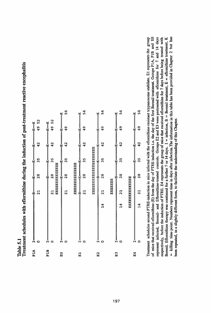
Table
5.
1Tr
eatm
ent
sche
dules
wi
th ef
lorn
ithin
e du
ring
the
indu
ction
of
post
-trea
tmen
t re
activ
e en
ceph
aliti
s
CNVi inII cr. Vi
CN Vi <3*
inn
coCN
CQ CN
CNVi in
I *
CN Vi *
in Vi n
10 Vi m
o> vi ^
CNVi
in Vi m
CQ
h o
<Hcu
w w w w w00 W CO CN w « CNw pq
pa w pa paiH
c n pa CN
HO HO
10 Vi in
CNvi
papapapapa
in paV i ro U
Pa papa papa papa papa papa 0 0 wpa w c n papa papa Papa t apa papa COw h p aPa a c n pa
opa
H O
pa
10 Vi in
<r> V, ^
CNVi **
00
HCN
H O
CNpa
10 Vi m
CT*vi *
CNVi
in Vi m
00 CO CN
H O
COW
10 Vi in
CTi Vi
CNVi ^
in £ m
ooPa CO c n Pa pa pa pa pa pa pa pa pa pa pa u pa
CN
asa00(0
J 3
a -aCD
O ViW >>
2 *as §< r*
W
75
Io
Cl-3 2o o *-0 « « 6Si °1 *S
^ *1 |cd g« aa 2«C O*2 £ •£ a
lM0 m >, W•3 |<0 3
■ S CN. wa w — o,d 3
1 2o od5 JS • a orrl iJ0* Sr~J O
Oh
*s 'S^ s ■3 -faj§ C3a I
6 | /■—S CW wI ’S•J §a rg«S a(0 CD
OQ.23•8X!o
•8 I ’8
3 . 9H O £ £ ^
pa
iI a
197
resp
ectiv
ely,
be
fore
the
in
duct
ion
of PT
RE.
E4
repr
esen
ts
the
grou
p of
mice
th
at re
ceiv
ed
eflo
mith
ine
for
7 da
ys
befo
re
bein
g tre
ated
with
Be
reni
l. E
flom
ithin
e th
erap
y wa
s co
ntin
ued
for
a fu
rther
7
or 14
days
. I
= in
fect
ion,
B =
Bere
nil
trea
tmen
t, E
= ef
lom
ithin
e tr
eatm
ent,
K
= ki
lling
tim
e po
int.
Num
bers
re
pres
ent
time
in da
ys
after
in
fect
ion.
The
info
rmat
ion
in th
is tab
le ha
s be
en
prov
ided
in
Chap
ter
2 bu
t ha
s be
en
repe
ated
, in
a sli
ghtly
di
ffere
nt f
orm
, to
facil
itate
the
un
ders
tand
ing
of th
is C
hapt
er.
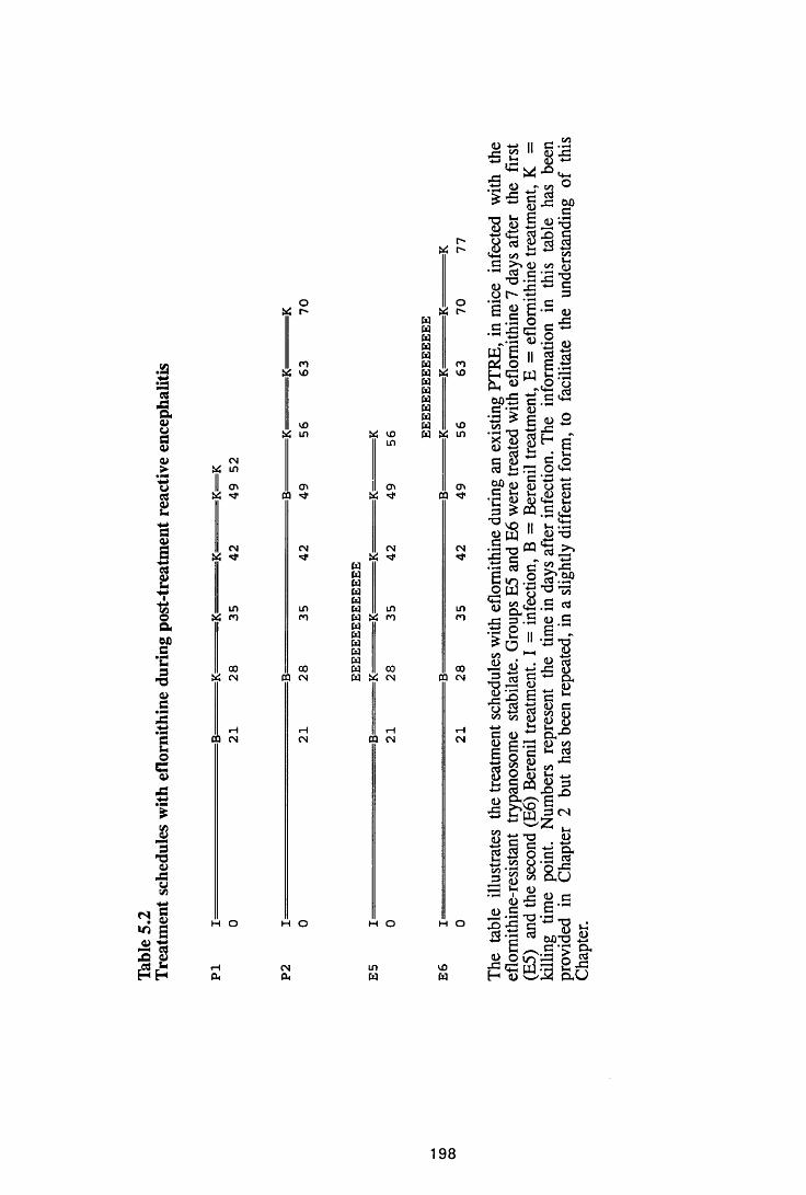
Table
5.
2Tr
eatm
ent
sche
dules
wi
th ef
lorn
ithin
e du
ring
post-
treat
men
t re
activ
e en
ceph
aliti
s
CN « in1 cn
CN
in « n
oo X cn
do CN
h o
oUi C"
roX vo
CO& m
cn CQ ^
CN
LDco
00 DO CN
H o
CNCU
Ui VOin
cn £
CN’CT
CQWWWWM LOw ^ nwwwwww 0 0W « CN
rHCQ CN
M O
o « r-W W W W WW m W « vow w w w ww vo W « in
a* CQ
CN
incn
ooCQ CN
M O
VOW
q -*—»-5 C isjC$ ' S
ll<D
II c 8
# £ >
£3 ■= *J. . V3
G•H ^ • r t •.—*q jG O h *
e 8 e- • G O G .G qH *e q
q
oS 'StUQ-S— G G 0) *3 > Ccn . Ss l §c S hra »-< r3 *-* c 61).' H
m O ra.C &fi G
«*-3f icn
t-Hcn <D ^ 12 3G
q- ^ G *-*' I sg g<2
- 8 £ •r q q3 £ «
^ ^ IICD pj• § -o m - S e ra c
» -.8'-i—ic
ot-HO
E «•> o i2a."3
<D H £ - H BG o O N*q c
, q cd*+H l-lG ,<D•-H Vt_■l-H .—ICD ’O G > >
£ .H S\SP
* o ^5G
cn^ 0 ) * J3 ra c •a - oJ=-2 sS 3 <Scn cn q G q ^
8 sH O
v 2 b g «
cn , Og I O
f i - s 83 o Mi—h V-i g_)q ■£
•S-S-aif sc ^
rH q n—«
raq G
S 'l **U M 8^ o,
qGq ccn A)q q
8 S3j =cn
Whq 3
3 CN
* ^ q
i toOhU
S2 3 6'^"8bJDTD ^e r a •Jo ^ gre 8 jg
i 2
198
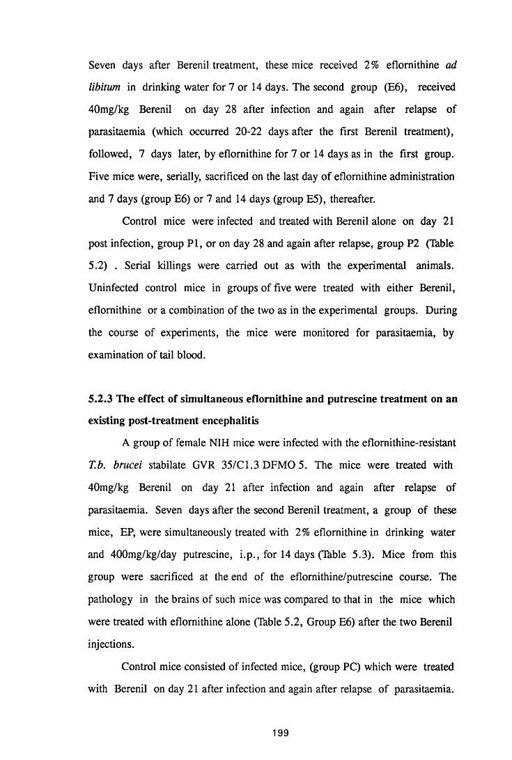
Seven days after Berenil treatment, these mice received 2 % eflomithine ad
libitum in drinking water for 7 or 14 days. The second group (E6), received
40mg/kg Berenil on day 28 after infection and again after relapse of
parasitaemia (which occurred 20-22 days after the first Berenil treatment),
followed, 7 days later, by eflomithine for 7 or 14 days as in the first group.
Five mice were, serially, sacrificed on the last day of eflomithine administration
and 7 days (group E6) or 7 and 14 days (group E5), thereafter.
Control mice were infected and treated with Berenil alone on day 21
post infection, group PI, or on day 28 and again after relapse, group P2 (Thble
5.2) . Serial killings were carried out as with the experimental animals.
Uninfected control mice in groups of five were treated with either Berenil,
eflomithine or a combination of the two as in the experimental groups. During
the course of experiments, the mice were monitored for parasitaemia, by
examination of tail blood.
5.2.3 The effect of simultaneous eflomithine and putrescine treatment on an
existing post-treatment encephalitis
A group of female NIH mice were infected with the eflornithine-resistant
T.b. brucei stabilate GVR 35/C 1.3 DFMO 5. The mice were treated with
40mg/kg Berenil on day 21 after infection and again after relapse of
parasitaemia. Seven days after the second Berenil treatment, a group of these
mice, EP, were simultaneously treated with 2% eflomithine in drinking water
and 400mg/kg/day putrescine, i.p., for 14 days (Table 5.3). Mice from this
group were sacrificed at the end of the eflornithine/putrescine course. The
pathology in the brains of such mice was compared to that in the mice which
were treated with eflomithine alone (Thble 5.2, Group E6) after the two Berenil
injections.
Control mice consisted of infected mice, (group PC) which were treated
with Berenil on day 21 after infection and again after relapse of parasitaemia.
199
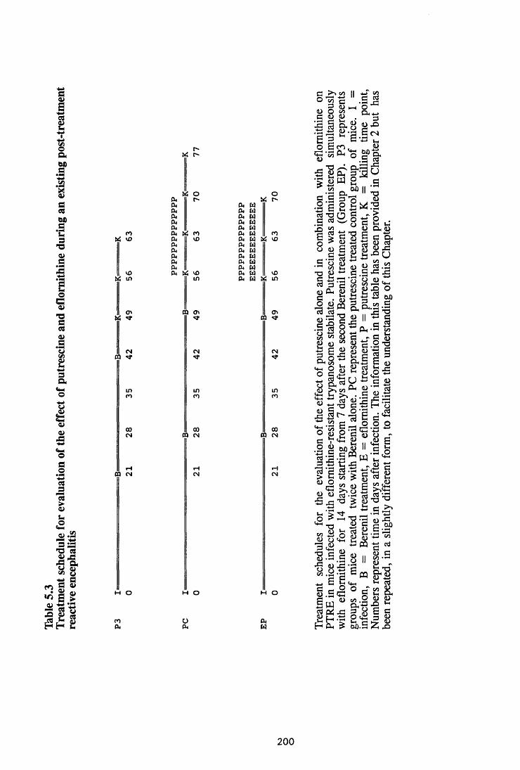
Table
5.
3Tr
eatm
ent
sche
dule
for
eval
uatio
n of
the
effe
ct
of pu
tres
cine
and
eflo
mith
ine
durin
g an
exist
ing
post
-trea
tmen
t re
activ
e en
ceph
aliti
s
co Ui vO
vO U4 i n
CNCQ
inn
ooCN
CQ CN
M o
nP .
v o
p .P.p .P.P. p .cu m coP.O.0.0.0. fu0. US VO
i n
CQ o v •d*
CN
o oCQ CN
H o
0. w 0. w 0. w0. W0. w 0. w
USor-
P4 P . w P . w P . wp . w p . w p . w
mH US v o
VO
Ov CQ N*
CN'it
i nro
ooCQ CN
H O
aO cn 3 0) Qe © .c a
</)—I 4—>c ©© .r v ©Wh
_T cn a c d
oQl 4—>3 © X>B CN
'“ a. f t
> ©W S c* tn ^ U) || £ e c §"73 -8
+-> oo 6 b lJcn © CX ©'’ s ’g e
a •2 d gC • x>Soo
cd„ C5 O h> © & -a © 5rgisile
•S’S go » i .aq S *1 o © ^w 3 C cn K t * nc * Vh
,§ .22 c8 « *3 ■o ® iiq & 8 n Ah
S.&.52-S ^ ^ 12
o C/D 8cn
cn
© D 1/5 ©h a © a . S ’S_ _ _ ii © SS o 5 fe £ a 3L-Li cn , P h+3 C u
Q fe 2 g fej?©
©©
£PH © ^u
c © H
r v cd >% cnB cd —
J*-* ’O C3 73 t“ < o . ,cd
s 2 | § 2cn 5 rP c -^3© P a © o " Vvh © " jo gWh I I C + - I P00 © 11 G fa e c o w —<2
. S O
° o.-s £
© .£ c 1/3S ° j? S'CL
i l l s00.5 Z x>
200
Seven days after the second Berenil administration, they were treated with400mg/kg
putrescine course and 7 days, thereafter. The pathology in the brains of this
group of mice was compared to that in mice treated with eflomithine alone
(group E6) after the two berenil treatments. Mice were monitored for
parasitaemia every 2 days during the course of the experiment.
5.2.4 Preparation of brains for histopathology
At each kill, mice were perfused with approximately 150ml of normal
saline, pH 7.0, through the left ventricle of the heart, to remove peripheral
blood. The brains were removed, fixed in 5% buffered formalin and processed
for histopathology. Three to 4/*m transverse sections were cut serially through
the fore-, mid-, and hindbrain. Immunocytochemistry for GFAP was performed
on some of the sections to assess astrocyte activation, while the serial sections
for all the samples were stained with haematoxylin and eosin for histopathology
(Chapter 2).
5.2.5 In vitro astrocyte preparation and exposure to eflomithine
Cerebral cortices were dissected from 2 to 3-day old NIH mice and
astrocyte preparation performed as described in Chapter 4. When purified and
confluent they were divided into five groups of five flasks each.
Lipopolysaccharide (LPS) was used as a known stimulant and compared to all
other experimental stimuli. LPS at a concentration of 10^g/ml was used alone
or in combination with eflomithine, while eflomithine was used at 0.5mM and
at 5mM, alone and in combination with LPS. One flask in each group was not
exposed to any stimulant and served as the baseline control. The cells were
harvested by treating them with denaturing solution and detaching them with a
rubber policeman, at 2, 4, 6 and 8 hours after exposure. They were then
homogenised and stored at -70°C in phenol resistant plastic tubes. RNA
extraction, cDNA preparation, amplification and detection of cytokine
201
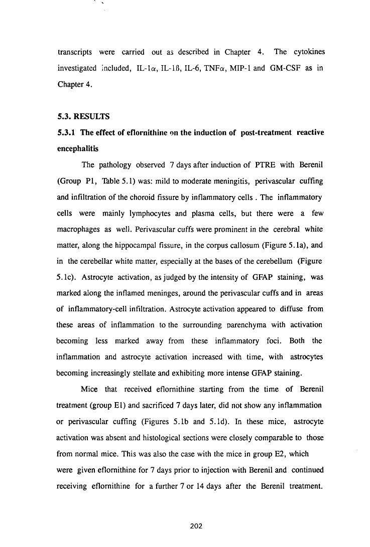
transcripts were carried out as described in Chapter 4. The cytokines
investigated included, IL-lor, IL-IB, IL-6, TNFa, MIP-1 and GM-CSF as in
Chapter 4.
5.3. RESULTS
5.3.1 The effect of eflomithine on the induction of post-treatment reactive
encephalitis
The pathology observed 7 days after induction of PTRE with Berenil
(Group PI, Thble 5.1) was: mild to moderate meningitis, perivascular cuffing
and infiltration of the choroid fissure by inflammatory cells . The inflammatory
cells were mainly lymphocytes and plasma cells, but there were a few
macrophages as well. Perivascular cuffs were prominent in the cerebral white
matter, along the hippocampal fissure, in the corpus callosum (Figure 5.1a), and
in the cerebellar white matter, especially at the bases of the cerebellum (Figure
5.1c). Astrocyte activation, as judged by the intensity of GFAP staining, was
marked along the inflamed meninges, around the perivascular cuffs and in areas
of inflammatory-cell infiltration. Astrocyte activation appeared to diffuse from
these areas of inflammation to the surrounding parenchyma with activation
becoming less marked away from these inflammatory foci. Both the
inflammation and astrocyte activation increased with time, with astrocytes
becoming increasingly stellate and exhibiting more intense GFAP staining.
Mice that received eflomithine starting from the time of Berenil
treatment (group El) and sacrificed 7 days later, did not show any inflammation
or perivascular cuffing (Figures 5.1b and 5 .Id). In these mice, astrocyte
activation was absent and histological sections were closely comparable to those
from normal mice. This was also the case with the mice in group E2, which
were given eflomithine for 7 days prior to injection with Berenil and continued
receiving eflomithine for a further 7 or 14 days after the Berenil treatment.
2 0 2
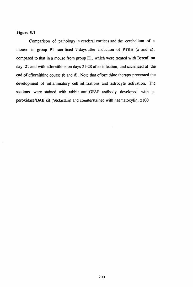
Figure 5.1
Comparison of pathology in cerebral cortices and the cerebellum of a
mouse in group PI sacrificed 7 days after induction of PTRE (a and c),
compared to that in a mouse from group E l, which were treated with Berenil on
day 21 and with eflomithine on days 21-28 after infection, and sacrificed at the
end of eflomithine course (b and d). Note that eflomithine therapy prevented the
development of inflammatory cell infiltrations and astrocyte activation. The
sections were stained with rabbit anti-GFAP antibody, developed with a
peroxidase/DAB kit (Vectastain) and counterstained with haematoxylin. xlOO
203
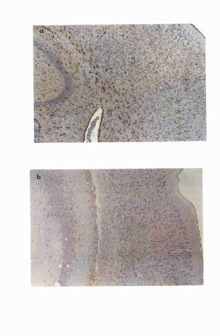
'*?»' > £ * 7 ~ * % W v » £ \ ' >* % v ^ < 1 ? b • » • • i - ' V ' "
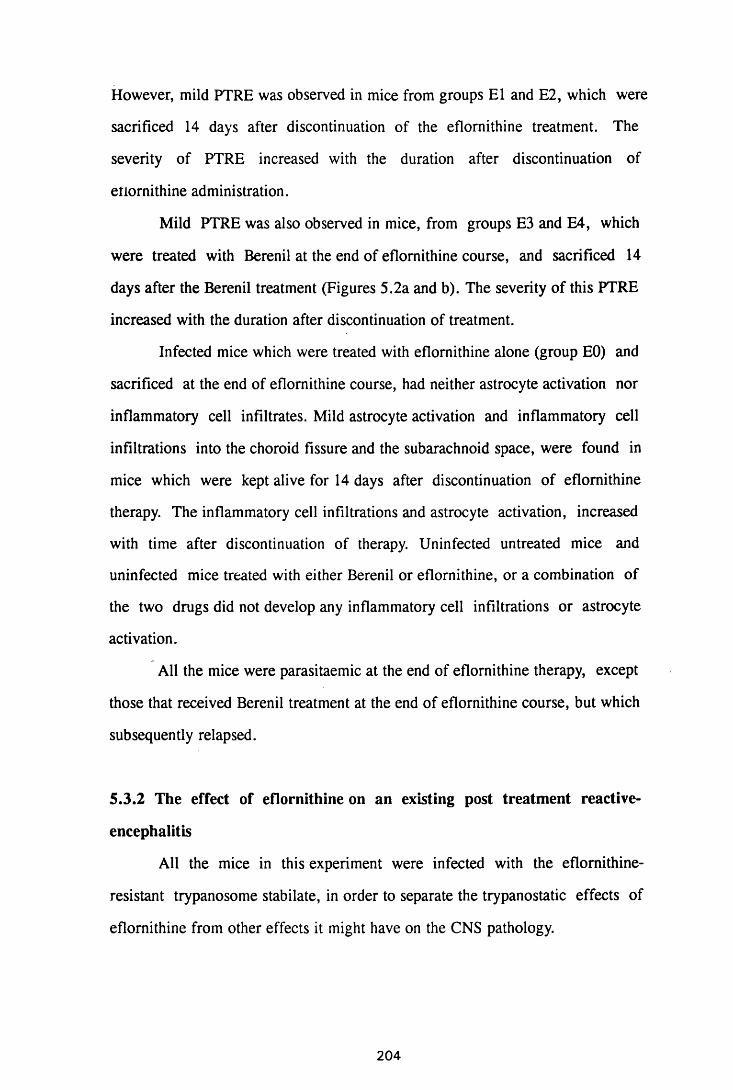
However, mild PTRE was observed in mice from groups El and E2, which were
sacrificed 14 days after discontinuation of the eflomithine treatment. The
severity of PTRE increased with the duration after discontinuation of
etiornithine administration.
Mild PTRE was also observed in mice, from groups E3 and E4, which
were treated with Berenil at the end of eflomithine course, and sacrificed 14
days after the Berenil treatment (Figures 5.2a and b). The severity of this PTRE
increased with the duration after discontinuation of treatment.
Infected mice which were treated with eflomithine alone (group EO) and
sacrificed at the end of eflomithine course, had neither astrocyte activation nor
inflammatory cell infiltrates. Mild astrocyte activation and inflammatory cell
infiltrations into the choroid fissure and the subarachnoid space, were found in
mice which were kept alive for 14 days after discontinuation of eflomithine
therapy. The inflammatory cell infiltrations and astrocyte activation, increased
with time after discontinuation of therapy. Uninfected untreated mice and
uninfected mice treated with either Berenil or eflomithine, or a combination of
the two drugs did not develop any inflammatory cell infiltrations or astrocyte
activation.
All the mice were parasitaemic at the end of eflomithine therapy, except
those that received Berenil treatment at the end of eflomithine course, but which
subsequently relapsed.
5.3.2 The effect of eflomithine on an existing post treatment reactive-
encephalitis
All the mice in this experiment were infected with the eflornithine-
resistant trypanosome stabilate, in order to separate the trypanostatic effects of
eflomithine from other effects it might have on the CNS pathology.
204
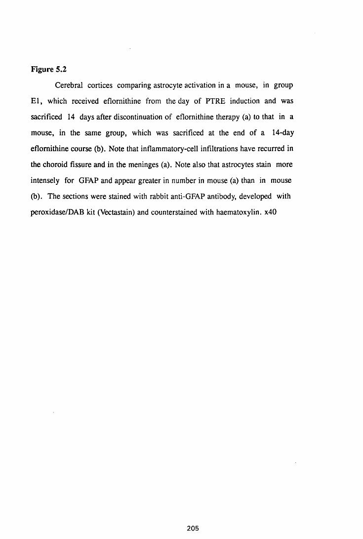
Figure 5.2
Cerebral cortices comparing astrocyte activation in a mouse, in group
E l, which received eflomithine from the day of PTRE induction and was
sacrificed 14 days after discontinuation of eflomithine therapy (a) to that in a
mouse, in the same group, which was sacrificed at the end of a 14-day
eflomithine course (b). Note that inflammatory-cell infiltrations have recurred in
the choroid fissure and in the meninges (a). Note also that astrocytes stain more
intensely for GFAP and appear greater in number in mouse (a) than in mouse
(b). The sections were stained with rabbit anti-GFAP antibody, developed with
peroxidase/DAB kit (Vectastain) and counterstained with haematoxylin. x40
205

:",,/$^r--'- :• . - 'w?.« •
■ ■ 3 :’ ;i v- V
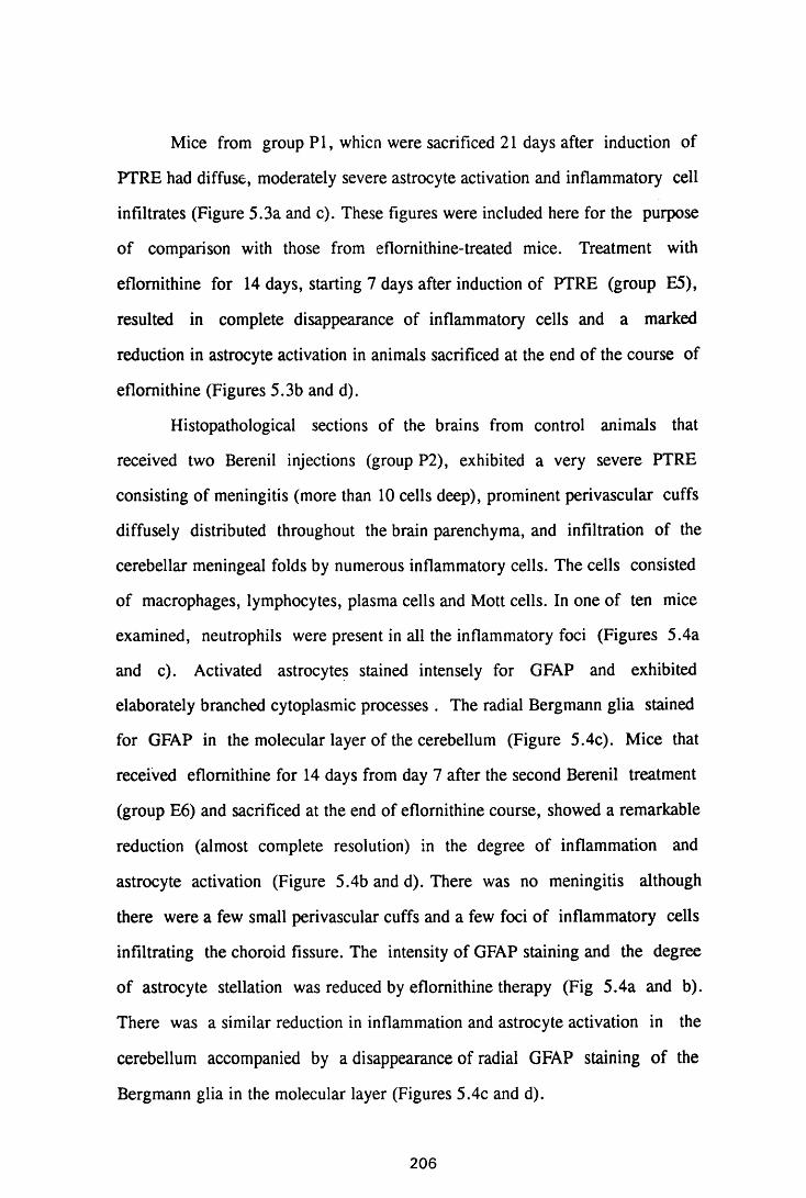
Mice from group PI, whicn were sacrificed 21 days after induction of
PTRE had diffuse, moderately severe astrocyte activation and inflammatory cell
infiltrates (Figure 5.3a and c). These figures were included here for the purpose
of comparison with those from eflornithine-treated mice. Treatment with
eflomithine for 14 days, starting 7 days after induction of PTRE (group E5),
resulted in complete disappearance of inflammatory cells and a marked
reduction in astrocyte activation in animals sacrificed at the end of the course of
eflomithine (Figures 5.3b and d).
Histopathological sections of the brains from control animals that
received two Berenil injections (group P2), exhibited a very severe PTRE
consisting of meningitis (more than 10 cells deep), prominent perivascular cuffs
diffusely distributed throughout the brain parenchyma, and infiltration of the
cerebellar meningeal folds by numerous inflammatory cells. The cells consisted
of macrophages, lymphocytes, plasma cells and Mott cells. In one of ten mice
examined, neutrophils were present in all the inflammatory foci (Figures 5.4a
and c). Activated astrocytes stained intensely for GFAP and exhibited
elaborately branched cytoplasmic processes . The radial Bergmann glia stained
for GFAP in the molecular layer of the cerebellum (Figure 5.4c). Mice that
received eflomithine for 14 days from day 7 after the second Berenil treatment
(group E6) and sacrificed at the end of eflomithine course, showed a remarkable
reduction (almost complete resolution) in the degree of inflammation and
astrocyte activation (Figure 5.4b and d). There was no meningitis although
there were a few small perivascular cuffs and a few foci of inflammatory cells
infiltrating the choroid fissure. The intensity of GFAP staining and the degree
of astrocyte stellation was reduced by eflomithine therapy (Fig 5.4a and b).
There was a similar reduction in inflammation and astrocyte activation in the
cerebellum accompanied by a disappearance of radial GFAP staining of the
Bergmann glia in the molecular layer (Figures 5.4c and d).
206
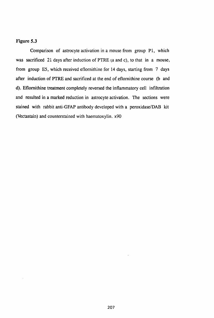
Figure 5.3
Comparison of astrocyte activation in a mouse from group PI, which
was sacrificed 21 days after induction of PTRE ta and c), to that in a mouse,
from group E5, which received eflomithine for 14 days, starting from 7 days
after induction of PTRE and sacrificed at the end of eflomithine course (b and
d). Eflomithine treatment completely reversed the inflammatory cell infiltration
and resulted in a marked reduction in astrocyte activation. The sections were
stained with rabbit anti-GFAP antibody developed with a peroxidase/DAB kit
(Vectastain) and counterstained with haematoxylin. x90
207
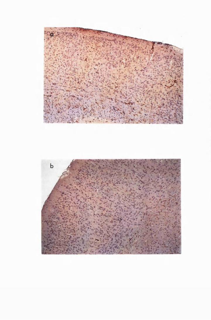
'site'-* v-*-' '■'*•
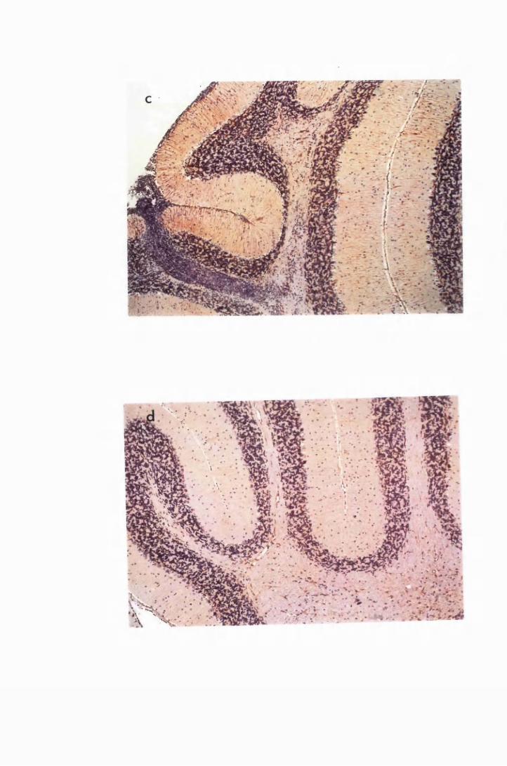
Figure 5.4
A comparison of pathology in the CNS of a mouse, from group P2,
infected with the eflornithine-resistant stabilate of T.b.brucei GVR 35/Cl.3
DFMO 5, treated with Berenil on day 21 after infection and again after relapse
of parasitaemia, to that in a mouse, from group E6, that received eflomithine
for 14 days, from day 7 after the second Berenil treatment. The mouse in Figure
5.4a and c was sacrificed 14 days after the second Berenil treatment while the
one in Figure 5.4b and d was treated with eflomithine for 14 days starting 7
days after the second Berenil treatment, and sacrificed at the end of the
eflomithine course. The photomicrographs show a great reduction in the
inflammation and astrocyte activation in both the cerebral cortex (a and b) and
the cerebellum (c and d). The sections were stained with rabbit anti-GFAP
antibody developed with a peroxidase/DAB kit (Vectastain) and counterstained
with haematoxylin. x90
208

W :
& % ;•• ? * ,& X-.V**'*-* & £ $ % * £ ■ '
i f . t r ; ’.'i<>.,, ,T-Vv '&
*5,vS&J* *'> / *? I**!f t V
r * ' ' .
rtfw£.r:’ '•‘••■;V v -
v ,-« vVv; V - &
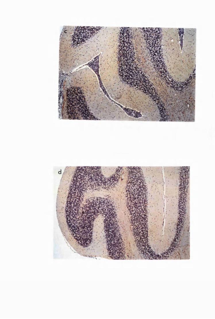
‘ w .• ' . X ’ W . •••
i. » W 1 < • . *1
f ^© k\>? X : v ••
;• • • • .
Although post-treatment reaction was evident in mice sacrificed after 7
days of eflomithine therapy, the intensity of GFAP staining and the degree of
astrocyte stellation were reduced.
5.3.3 The effect of simultaneous eflornithine and putrescine treatment on
an existing post-treatment encephalitis
Mice, in group E6, which were treated with eflornithine alone for 14
days, after the double-Berenil treatment, did not show inflammatory cell
infiltration into the perivascular spaces or the choroid fissure but there was mild
meningitis in the cerebellum (Figure 5.5a and c). However, animals treated
with eflomithine and putrescine simultaneously (group EP), after the double-
Berenil regimen, developed astrocyte activation and inflammatory cell
infiltrations (5.5b and 5.5d), but to a lesser degree than in the infected, Berenil-
treated controls. The pathology in mice that received putrescine alone after the
second Berenil treatment (group PC) was comparable to that seen in the Berenil-
treated controls. Ail the mice were parasitaemic at the end of the putrescine
therapy.
5.3.4 In vitro effects of eflornithine on astrocyte stimulation with
lipopolysaccharide
LPS at a concentration of 10/xg/ml stimulated astrocytes to express
mRNA transcripts for IL-la and -B, IL-6, TNFa, MIP-1 and GM-CSF.
Messenger RNA transcripts for all these cytokines were detectable by 2 hours
after exposure to LPS. Although the cells used for this study are different from
those used in Chapter 4, the variation of the individual cytokine transcripts were
similar to those described in Chapter 4 and the results are presented T&ble 5.4.
However, there are differences in the thickness of the bands. It is most likely
that this was due to the differences sizes of the gel beds and combs. It is also
possible that the different batches of cells produced different amounts of
209
Figure 5.5
Brain sections comparing the pathology from a mouse in group Eo (a and
c), that received eflornithine, to that in a mouse in group EP (b and d), that
received a combination of eflomithine and putrescine, for 14 days starting 7
days after the second Berenil treatment. When eflornithine and putrescine were
administered simultaneously, inflammatory cells infiltrated the choroid fissure
and the perivascular spaces in the cerebrum (b), and the meningeal folds in the
cerebellum (d). However, the degree of astrocyte activation in these two groups
of mice appear very similar. The sections were stained with rabbit anti-GFAP
antibody developed with a peroxidase/DAB kit (Vectastain) and counterstained
with haematoxylin. x90
2 1 0
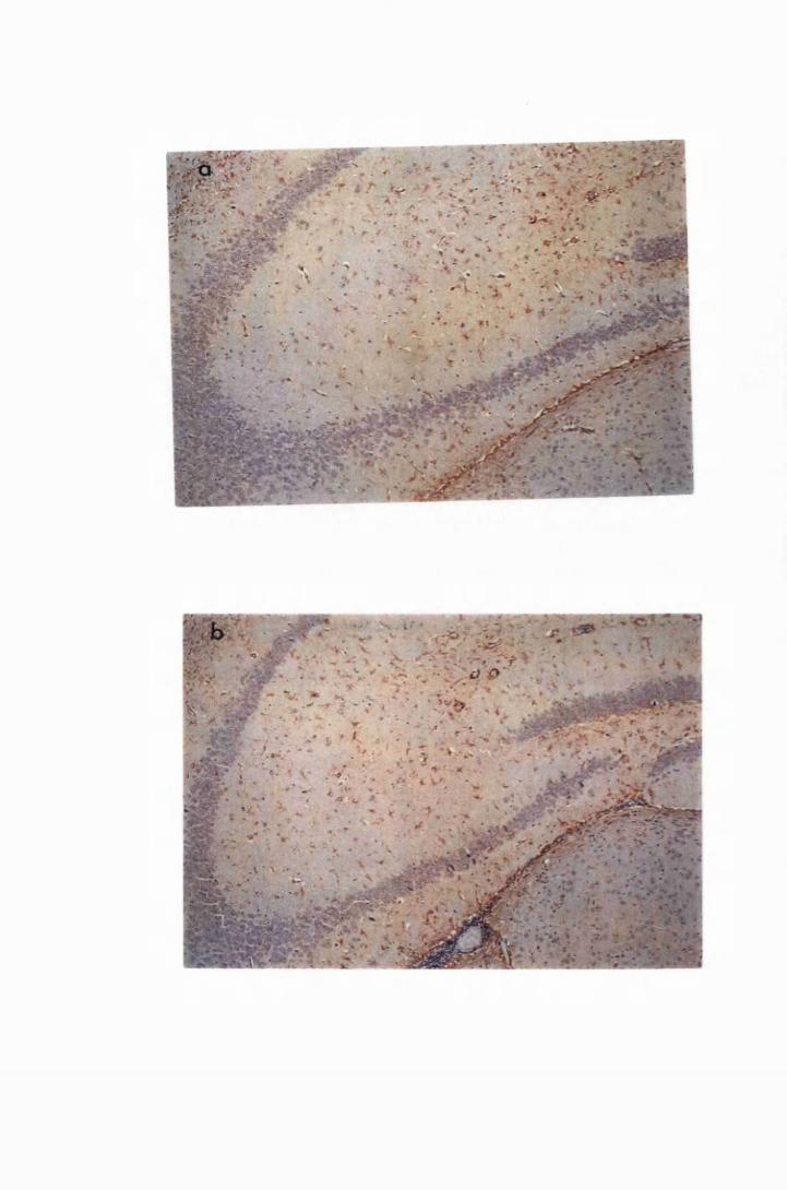
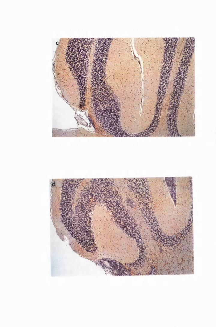
HC8B
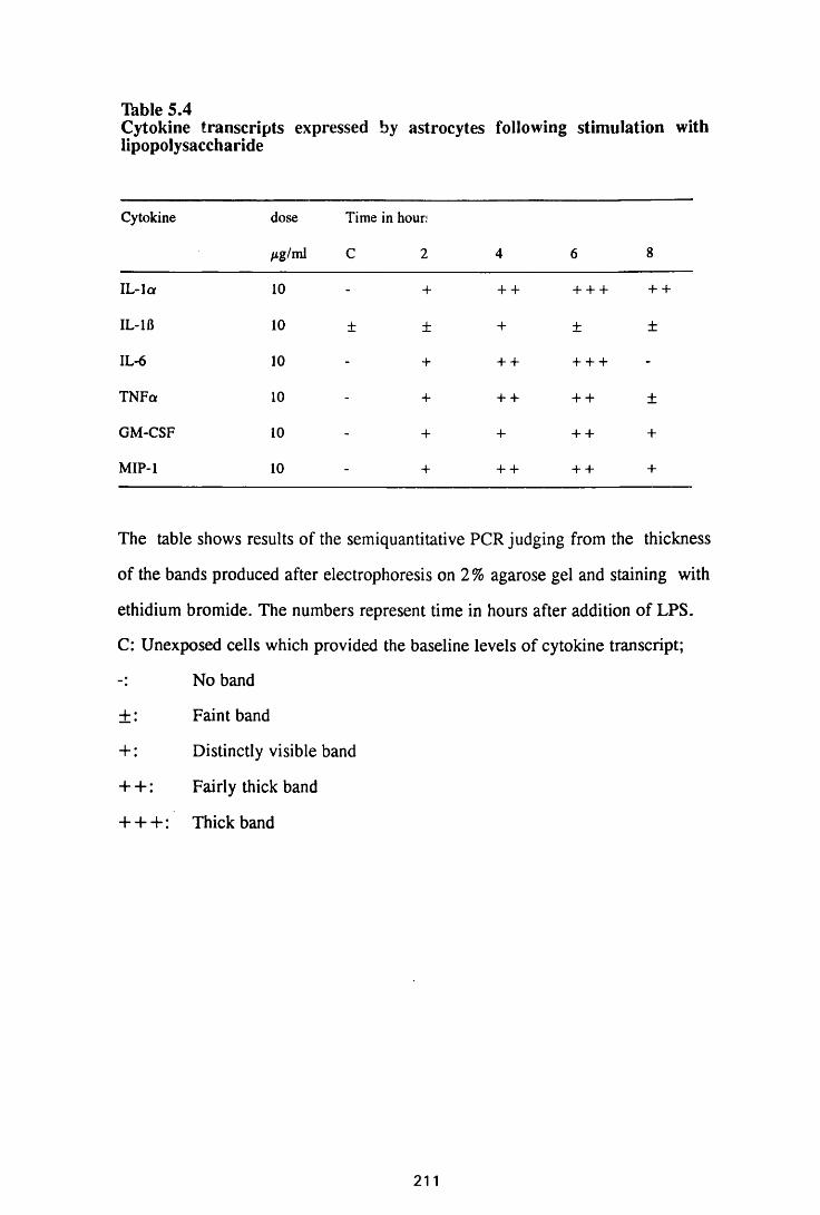
Table 5.4Cytokine transcripts expressed by astrocytes following stimulation with lipopolysaccharide
Cytokine dose Time in hour;
jig/ml C 2 4 6 8
IL -la 10 - + + + + + + + +
IL-lfi 10 ± ± + ± ±
IL-6 10 - + + + + + + -
TNFa 10 - + + + + + ±
GM-CSF 10 - + + + + +
MIP-1 10 - + + + + + +
The table shows results of the semiquantitative PCR judging from the thickness
of the bands produced after electrophoresis on 2% agarose gel and staining with
ethidium bromide. The numbers represent time in hours after addition of LPS.
C: Unexposed cells which provided the baseline levels of cytokine transcript;
No band
±: Faint band
+ : Distinctly visible band
+ + : Fairly thick band
+ + + : Thick band
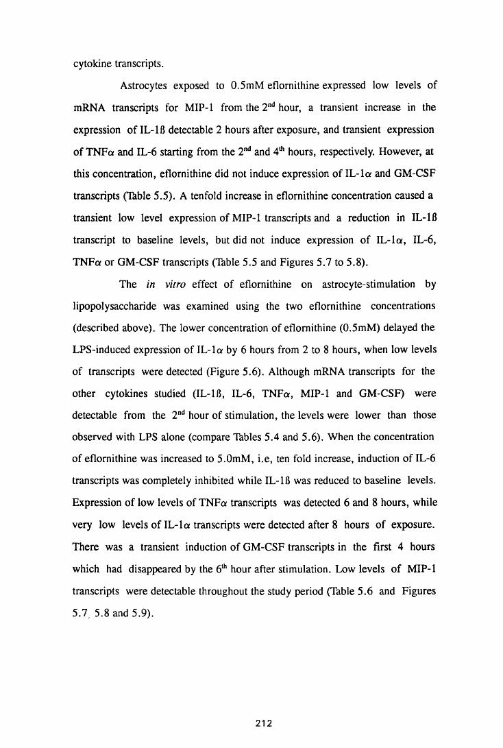
cytokine transcripts.
Astrocytes exposed to 0.5mM eflornithine expressed low levels of
mRNA transcripts for MIP-1 from the 2nd hour, a transient increase in the
expression of IL-lfl detectable 2 hours after exposure, and transient expression
of TNFa and IL-6 starting from the 2nd and 4th hours, respectively. However, at
this concentration, eflornithine did not induce expression of IL-la and GM-CSF
transcripts (Thble 5.5). A tenfold increase in eflomithine concentration caused a
transient low level expression of MIP-1 transcripts and a reduction in IL-1B
transcript to baseline levels, but did not induce expression of IL-la, IL-6,
TNFa or GM-CSF transcripts (Thble 5.5 and Figures 5.7 to 5.8).
The in vitro effect of eflornithine on astrocyte-stimulation by
lipopolysaccharide was examined using the two eflomithine concentrations
(described above). The lower concentration of eflomithine (0.5mM) delayed the
LPS-induced expression of IL-la by 6 hours from 2 to 8 hours, when low levels
of transcripts were detected (Figure 5.6). Although mRNA transcripts for the
other cytokines studied (IL-lfl, IL-6, TNFa, MIP-1 and GM-CSF) were
detectable from the 2nd hour of stimulation, the levels were lower than those
observed with LPS alone (compare Thbles 5.4 and 5.6). When the concentration
of eflomithine was increased to 5.0mM, i.e, ten fold increase, induction of IL-6
transcripts was completely inhibited while IL-16 was reduced to baseline levels.
Expression of low levels of TNFa transcripts was detected 6 and 8 hours, while
very low levels of IL-la transcripts were detected after 8 hours of exposure.
There was a transient induction of GM-CSF transcripts in the first 4 hours
which had disappeared by the 6th hour after stimulation. Low levels of MIP-1
transcripts were detectable throughout the study period (Table 5.6 and Figures
5.7. 5.8 and 5.9).
2 1 2
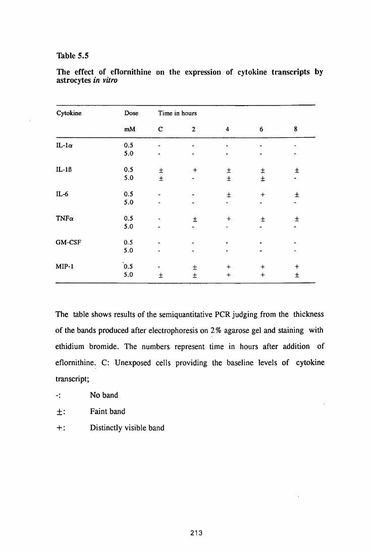
Table 5.5
The effect of eflornithine on the expression of cytokine transcripts by astrocytes in vitro
Cytokine Dose
mM
Time in hours
C 2 4 6 8
IL -la 0.5 _ _ _ _
5.0 - - - - -
IL-1B 0.5 ± + ± ± ±5.0 ± - ± ± -
IL-6 0.5 _ _ ± + ±5.0 - - - - -
TNFa 0.5 _ ± + ± ±5.0 - - - - -
GM-CSF 0.5 _ _ _ _ _
5.0 - - - - -
MIP-1 0.5 _ ± + + +5.0 ± ± + + ±
The table shows results of the semiquantitative PCR judging from the thickness
of the bands produced after electrophoresis on 2 % agarose gel and staining with
ethidium bromide. The numbers represent time in hours after addition of
eflornithine. C: Unexposed cells providing the baseline levels of cytokine
transcript;
No band
± : Faint band
+: Distinctly visible band
213
Figure 5.6
Gel electrophoresis showing the expression of mRNA transcripts for IL-
la (top) and TNFa (bottom) in cultured astrocytes stimulated with lO^ig/ml LPS
(L), 0.5mM eflomithine (E) and a combination of LPS and eflornithine (E+L).
This Figure shows that 0.5mM eflornithine delays the LPS-induced expression
of IL-la transcript, while it has no effect on the expression of TNFa. The
numbers, 2, 4, 6 and 8 represent the time in hours after stimulation. M =
123bp ladder (Gibco); + = the positive control; W = water, used as a negative
control; C = unexposed cells.
214

E + L E L
M + 8 6 4 2 W 8 6 4 2 8 6 4 2 C
IL -la(625bp)
TNFa(692bp)
M 8 6 4 2 8 6 4 2 + 8 6 4 2 C
E + L E L
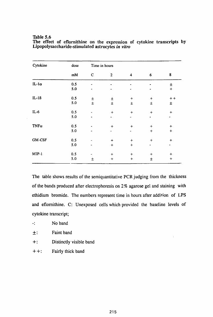
Thble 5.6The effect of eflornithine on the expression of cytokine transcripts by Lipopolysaccharide-stimulated astrocytes in vitro
Cytokine dose Time in hours
mM C 2 4 6 8
IL -la 0.5 _ _ . _ ±5.0 - - - - +
IL-1B 0.5 ± ± + + + +5.0 ± ± ± ± ±
IL-6 0.5 + + + +5.0 - - - - -
TNFa 0.5 . + + + +5.0 - - - + +
GM-CSF 0.5 _ + + + +5.0 - + + - -
MIP-1 0.5 _ + + + +5.0 ± + + ± +
The table shows results of the semiquantitative PCR judging from the thickness
of the bands produced after electrophoresis on 2 % agarose gel and staining with
ethidium bromide. The numbers represent time in hours after addition of LPS
and eflomithine. C: Unexposed cells which provided the baseline levels of
cytokine transcript;
No band
± : Faint band
+: Distinctly visible band
4- + : Fairly thick band
215
Figure 5.7
Gel electrophoresis showing transcripts for the cytokines, IL -la and
TNFa, from cultured astrocytes exposed to lOyug/ml LPS (L) and 5.0mM
eflomithine (E) and a combination of 10jug/ml LPS and 5.0mM eflomithine
(E+L). The numbers, 2, 4, 6 and 8 indicate the time in hours after addition of
stimulant; M = 123bp ladder (Gibco); + = positive control; W = water, used
as a negative control; C= unexposed cells.
216
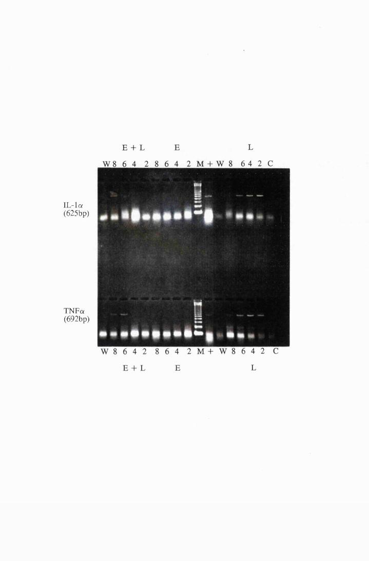
IL-la(625bp)
TNFa(692bp)
Figure 5.8
Gel electrophoresis showing transcripts for the cytokines, IL-lfl and
GM-CSF, from cultured astrocytes exposed to 10^g/ml LPS (L) and 5.0mM
eflomithine (E) and a combination of 10/xg/ml LPS and 5.0mM eflornithine
(E+L). The numbers, 2, 4, 6 and 8 indicate the time in hours after addition of
stimulant; M = 123bp ladder (Gibco); + = positive control; W = water, used
as a negative control; C= unexposed cells.
217
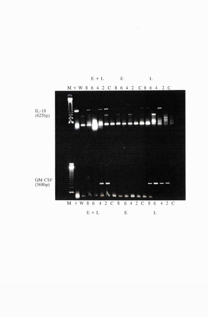
E + L E L
M + W 8 6 4 2 C 8 6 4 2 C 8 6 4 2 C
n6
IL-lfl(625bp)
GM-CSF (368bp)
M + W 8 6 4 2 C 8 6 4 2 C 8 6 4 2 C
E + L E L
Figure 5.9
Gel electrophoresis showing transcripts for the cytokines, IL-6 and MIP-
1, from cultured astrocytes exposed to 10/zg/ml LPS (L) and 5.0mM eflomithine
(E) and a combination of 10^g/ml LPS and 5.0mM eflornithine (E+L). The
numbers, 2, 4, 6 and 8 indicate the time in hours after addition of stimulant; M
= 123bp ladder (Gibco); + = positive control; W = water, used as a negative
control; C = unexposed cells.
218
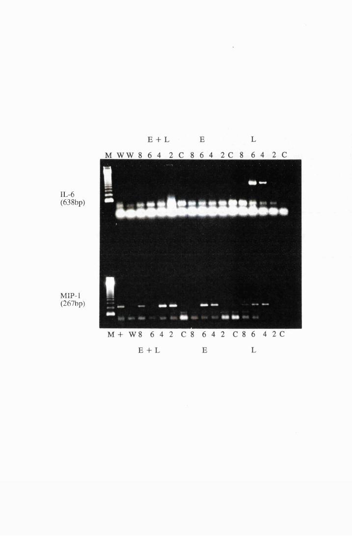
IL-6(638bp)
MIP-1(267bp)
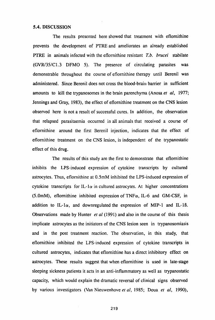
5.4. DISCUSSION
The results presented here showed that treatment with eflomithine
prevents the development of PTRE and ameliorates an already established
PTRE in animals infected with the eflornithine resistant T.b. brucei stabilate
(GVR/35/C1.3 DFMO 5). The presence of circulating parasites was
demonstrable throughout the course of eflornithine therapy until Berenil was
administered. Since Berenil does not cross the blood-brain barrier in sufficient
amounts to kill the trypanosomes in the brain parenchyma (Anosa et al, 1977;
Jennings and Gray, 1983), the effect of eflornithine treatment on the CNS lesion
observed here is not a result of successful cures. In addition, the observation
that relapsed parasitaemia occurred in all animals that received a course of
eflornithine around the first Berenil injection, indicates that the effect of
eflomithine treatment on the CNS lesion, is independent of the trypanostatic
effect of this drug.
The results of this study are the first to demonstrate that eflomithine
inhibits the LPS-induced expression of cytokine transcripts by cultured
astrocytes. Thus, eflornithine at 0.5mM inhibited the LPS-induced expression of
cytokine transcripts for IL-la in cultured astrocytes. At higher concentrations
(5.0mM), eflomithine inhibited expression of TNFa, IL-6 and GM-CSF, in
addition to IL-la, and downregulated the expression of MIP-1 and IL-1B.
Observations made by Hunter et al (1991) and also in the course of this thesis
implicate astrocytes as the initiators of the CNS lesion seen in trypanosomiasis
and in the post treatment reaction. The observation, in this study, that
eflomithine inhibited the LPS-induced expression of cytokine transcripts in
cultured astrocytes, indicates that eflomithine has a direct inhibitory effect on
astrocytes. These results suggest that when eflornithine is used in late-stage
sleeping sickness patients it acts in an anti-inflammatory as well as trypanostatic
capacity, which would explain the dramatic reversal of clinical signs observed
by various investigators (Van Nieuwenhove et al, 1985; Doua et al, 1990),
219
leading to eflornithine being described as the "the resurrection drug".
How the CNS inflammatory events are modified in the presence of
eflomithine is not clear, but it seems unlikely that it compromises the immune
system since its curative effect, as is evident in T.b. gambiense sleeping
sickness, depends on an intact immune system (de Gee, McCann and
Mansfield, 1983; Bitonti et al, 1986). This is further corroborated by the
observations that the administration of 2% eflomithine in drinking water has no
effect on the ability of normal mice to produce a haemagglutination response to
horse red blood cells or oxazalone-sensitised (sensitised during eflomithine
administration) mice to produce a delayed-type hypersensitivity when challenged
with oxazalone (F.W. Jennings, unpublished data).
IL-1 seems to be a key factor in triggering a cascade of events that
might lead to development of the CNS inflammatory lesion and its inhibition
might be critical in modulating inflammatory reactions of the CNS. For
instance, Merrill (1991) found that natural and recombinant forms of IL-1 and
TNFa had a synergistic effect, amplifying microglial numbers four-fold over
TNFa alone; however, anti IL-1 antibodies completely blocked this effect while
anti-TNFa antibodies only partially did so. Furthermore, IL-1 or TNFa alone
stimulated astrocytes to double in number and to triple when both cytokines
were used together, in mixed glial cultures (Merrill, 1991). In addition,
astrocytes underwent some putative differentiation that resulted in stellation
when exposed to IL-1, but not TNFa, in only 3 days after addition of the
cytokine. The effect on morphology was more dramatic when both cytokines
were used together and was amplified by long term exposure (Merrill, 1991).
It has been proposed that some autocrine event involving IL-1, TNFa
and a third factor, possibly IL-6 and/or platelet derived growth factor (PDGF),
might work in tandem or in synergy to promote astroglia proliferation and
differentiation (anti-IL-6 and anti-PDGF antibodies inhibit proliferation and
220
astroglial differentiation in mixed cultures) (Merrill, 1991). Both PDGF and IL-
6 are some of the factors produced by astrocytes in response to IL-1 stimulation
(Merrill, 1991). It is possible that PDGF and IL-6 are induced by and work in
synergy with IL-1 and TNFa to produce proliferation and differentiation of
astrocytes in late-stage trypanosomiasis. The results of this study show that
exposure of cultured astrocytes to eflornithine, downregulates the LPS-induction
of mRNA transcripts for IL-115, IL-6, TNFa, MIP-1 and GM-CSF. It is possible
that this occurs due to the lack of the autocrine activity of IL-la on astrocytes
themselves, which may be needed to boost the levels of these cytokines to those
observed with LPS alone. By inhibiting production of IL-1, eflornithine may be
acting directly on astrocytes inhibiting their activation, thus downregulating the
effect of this cytokine in the initiation and propagation of the CNS
inflammation.
IL-1 increases expression oflCAM -land ELAM-1 on endothelial
cells, altering the adhesive properties of the vascular walls, thus initiating
leukocyte extravasation. It also induces the endothelial cells to produce
chemotactic cytokines such as IL-8, macrophage chemotactic protein and colony
stimulating factors which lead to leukocyte recruitment and proliferation
(Broudy et al, 1987; Sieff et al, 1988; Montovani and Dejana, 1989). By
preventing IL-1 production, eflomithine might curtail a cascade of events that
could lead to the breakdown of the blood-brain barrier and could therefore
prevent leukocyte extravasation and the resultant perivascular cuffs. IL-1 is a
polyclonal activator of lymphocytes and it is possible that when eflomithine
inhibits its production in the CNS by astrocytes, then the proliferative signal to
the lymphocytes and plasma cells infiltrating the meninges and perivascular
cuffs is removed, leading to the disappearance of these cells.
A tenable hypothesis is that diffusible paracrine factors derived from
microglial cells or cell-surface molecules, trigger the changes seen in astrocytes
in mixed glial cultures (Merrill, 1991). Indeed, a bi-directional effect might
221
occur with astrocytes triggering microglial cells to proliferate as well. Microglia
do play a major role in gliosis in vivo and physically abut reactive astrocytes in
brain tissue (Jordan and Thomas, 1988). They also produce factors in vitro such
as glia-promoting factors (GPF) which stimulate astroglia proliferation (Giulian
and Baker, 1985). In addition, activated mononuclear leukocytes and therefore
presumably microglia, produce factors which stimulate astrocytic proteinases, a
sign of astrocytic differentiation (Bever, Snyder, Endres, Morgan, Postlethwaite
and Whitaker, 1989). This seems to suggest that the reduction in astrocyte
activation following eflornithine therapy in the T.b. brucei-chronic mouse model
studied here, is secondary to the disappearance of inflammatory cells. This is
corroborated by the observation in this chapter that the inflammatory cells are
the first to disappear followed by a reduction in astrocyte activation. However,
the observation in the current study and earlier on (Chapter 3), that astrocyte
activation disappeared faster when T.b. brucei-infected mice were treated with
subcurative eflomithine regimen than when they were treated with the curative
Mel Cy/MK436 combination regimen (Jennings, 1991c), suggested that
eflomithine inactivates the astrocytes themselves.
Astrocytes produce both IL-3 and GM-CSF in response to IL-1 and
TNFa both in vivo and in vitro (Nishida, Nikai, Kawakami, Aihara, Nishino
and Hirari, 1989; Farrar, Vinocour and Hill, 1989). Although microglia do not
proliferate in response to IL-1 in vitro, they multiply in response to IL-3 and
GM-CSF (Frei et al, 1985; 1986; Giulian and Ingeman, 1988). It is possible
that microglial hyperplasia contributes to the gliosis seen in late-stage
trypanosomal infections and PTRE. Then, the inhibition of IL-1-induced, IL-3
and GM-CSF production by astrocytes, might explain how eflomithine
downregulates the cerebral hypercellularity in the T.b. brucei chronic mouse
model. It is possible that eflomithine inhibits production of IL-1 from cells other
than astrocytes, including, lymphocytes, macrophages, endothelial cells and
222
microglia to produce the dramatic reduction in inflammation observed in this
study.
Higher doses of eflornithine prevented the LPS-induction of mRNA
transcripts for IL-115, IL-6, TNFa and GM-CSF in addition to IL-la. The
higher dose of eflomithine used in this study was 5mM eflomithine in culture
medium. This dosage is over ten times the concentration of eflomithine expected
to reach the CNS considering only 30% of plasma serum levels of eflomithine
reach the CSF (Levin, Csejtey and Byrd, 1983; Bacchi and McCann, 1987).
Since in a clinical intravenous regimen of 15g/day, plasma levels approach
ImM, it thus seems possible that peak levels of 100-300/xM are attainable in the
CSF (Van Nieuwenhove et al, 1985; Bacchi and McCann, 1987). This would be
therapeutic since 50-100^M completely inhibits the growth of T.b. brucei in
vitro (Giffin, McCann, Bitonti and Bacchi, 1986). While the 5mM eflomithine
used in this study is higher than the levels of eflornithine expected to reach the
CNS after oral administration, there is a chance that such high levels might
reach the CNS following the disruption cf the blood-brain barrier which seems
to occur in late-stage sleeping sickness, especially after subcurative therapy
(Waitumbi et al, 1988; Jennings et al, 1977). If the blood-brain barrier was
disrupted and such levels of eflornithine were attained, it seems possible that
eflornithine would be useful in downregulating the resultant inflammatory
reaction that would occur in such cases.
Eflomithine competitively inhibits ornithine decarboxylase (ODC), an
enzyme required for the biosynthesis of polyamines, which in turn are required
by rapidly proliferating cells. It may therefore inhibit the proliferation of the
astrocytes and the infiltrating inflammatory cells that form the CNS lesion in
late-stage trypanosomiasis. Indeed it has been proposed that the inhibition of
ODC by eflomithine is responsible for preventing proliferation of astrocytes
during reactive gliosis (Zini, Zoli, Grimaldi, Melo-Pich, Biagini, Fuxe and
Agnati, 1990). Blood-brain barrier disruption has been reported to evoke an
223
eflornithine-reversible activation of ODC (Koenig, Goldstone and Lu, 1989a;
1989b). Such a disruption might permit entry of factors and inflammatory cells
that lead to reactive gliosis (Bologa, Cole, Chiappelli, Saneto and De Vellis,
1988; Giulian et al, 1988; Norton, Aquino, Hozumi, Chiu and Brosnan,. 1992).
These observations raise the possibility that conditions resulting in a
compromised blood-barrier allow entry of blood-borne astrocyte growth
factors/cell types that mediate an increase in GFAP through an ODC-dependent
pathway (O'Callaghan and Seidler, 1992). The observation here that putrescine,
a key polyamine synthesised by ODC, inhibited the eflomithine-induced
amelioration of PTRE would support this hypothesis. Furthermore, the
reduction in astrocyte activation occurred after the disappearance of the blood-
derived inflammatory cells (macrophages, lymphocytes and plasma cells) and by
inference the blood-derived factors. This would support the hypothesis raised
earlier in this study, that astrocytes and infiltrating inflammatory cells are acting
in co-operation to perpetuate the pathological lesion in late-stage
trypanosomiasis and that this lesion is blood-brain barrier disruptive. It is
possible that removal of inflammatory cells reduces the CNS cytokine levels to
below the threshold required for diffuse astrocyte activation. The studies here
show that eflornithine inhibits expression of transcripts for IL-la, IL-6, TNF
and GM-CSF by LPS-stimulated astrocytes in vitro, indicating a direct
suppressive effect of eflornithine on astrocytes. One way in which eflomithine
might achieve this is by depleting polyamines which are required for RNA
synthesis. Indeed, it has been reported that ODC, the enzyme inhibited by
eflomithine, is a regulatory subunit of RNA polymerase 1 (Russell and Durrie,
1978) and that 13-galactosidase mRNA elongation in Escherichia coli, is slowed
by polyamine depletion (Morris and Hansen, 1973). The observation in this
study that the PTRE observed when putrescine was administered with
eflomithine, was not as severe as in the double-Berenil treated control, would
224
indicate that eflornithine is doing more than just inhibiting ODC.
The finding that eflornithine can prevent the occurrence of a severe
post-treatment reactive encephalitis and by inference the "reactive arsenical
encephalopathy", irrespective of its trypanostatic action, suggests that
eflomithine supportive treatment in combination with melarsoprol would be an
advantage even in T.b. rhodesiense infection in which it is not curative (Bacchi,
et al, 1990). Post-mortem material from a T.b. rhodesiense-monkey model, in
which eflomithine has been found to be completely refractory, showed that there
was a modulation of CNS inflammation compared to materials from animals that
died during the course of a curative melarsoprol regimen (J.M. Ndung'u,
personal communication). Moreover, PTRE was reported to be absent in
T.b.brucei infected-mice after treatment with a combination of low doses of
eflomithine and melarsoprol (Jennings, 1988a; 1988b).
In conclusion, the results presented here suggest a dual role of
eflomithine in preventing and ameliorating the meningoencephalitis found in
late-stage trypanosomiasis: 1) In inhibiting the production of IL-1 by astrocytes,
eflomithine prevents a cascade of events that would initiate and propagate the
meningeal inflammatory process. Thus, it would inhibit the IL-1-induced
migration of endothelial cells and expression of adhesion molecules on
endothelial, as well as on glial cells, and therefore, the subsequent leukocyte
extravasation and spread within the CNS. Eflornithine would also inhibit the IL-
1-induced proliferation of infiltrating inflammatory cells. The downregulation of
the IL-I-induced production of cytokines by the infiltrating inflammatory cells,
the brain vascular endothelial cells, and the intrinsic glial and neuronal cells,
would essentially remove the inflammatory boost provided by such cytokines. 2)
By inhibiting ODC, eflornithine inhibits the synthesis of polyamines that would
be required for the IL-1-induced, proliferation of inflammatory cells within the
CNS.
The clinical implications of such effects cannot be overemphasised.
225
When used in conjunction with other trypanocides, eflornithine would prevent
and/or attenuate post-treatment reactive encephalopathy, even when the infecting
trypanosomes are eflornithine-insensitive. In addition, eflornithine is likely to be
beneficial in the treatment of other CNS inflammatory conditions, such as viral
and bacterial meningoencephalitis, that feature astrogliosis and inflammatory
cell infiltrations.
2 2 6
CHAPTER 6
THE EFFECT OF AZATHIOPRINE ON ASTROCYTE ACTIVATION
IN VIVO AND IN VITRO
227
6.1 INTRODUCTION
The pathogenesis of the CNS disease in HAT, and of the reactive
encephalopathy following treatment with melarsoprol is poorly understood
(Adams et al, 1986; Haller et al, 1986). Evidence from experimental infections
of laboratory and domestic animals, an^ from pathological lesions in the brains
of HAT patients, implicate the host's immune response to the invading parasite
as the underlying mechanism (Galvao-Castro et al, 1978; Greenwood and
Whittle, 1980; Vickerman and Barry, 1982; Milord and Pepin, 1992; Cegielski
and Durack, 1991). However, anti-inflammatory therapy with corticosteroids to
prevent and/or attenuate reactive encephalopathy, yielded equivocal results and
their value in that respect remains uncertain (Foulkes, 1975; Arroz, 1987; Pepin
et al, 1989). Recently, the non-steroidal anti-inflammatory drug, azathioprine,
has been shown to prevent and ameliorate the Berenil-induced infiltration of the
CNS, by mononuclear inflammatory cells, in T.b. brucei-infected mice
(Jennings et al, 1989; Hunter et al, 1992b).
Azathioprine is used in prevention of heterograph graft rejection
(Elion, 1967; 1972). It has been reported to inhibit renal homograft rejections in
dogs (Caine and Murray, 1961; Zukoski, 1965), prolong renal allograft in
Rhesus monkeys (Dicke et al, 1971), and inhibits rejection of full thickness
corneal xenograft in rabbits (Leibowitz and Elliott, 1966). It has also been
shown to inhibit the secondary phase of adjuvant arthritis in a mouse model of
arthritis (Whittington, 1970; Perper et al, 1971), and the development of
experimental allergic encephalitis in rats, a model of multiple sclerosis
(Ronsenthale et al, 1969; Babington and Wedeking, 1971). To date,
azathioprine is still used in combination with other immunosuppressive drugs for
prevention and prolongation of organ transplants in man (Isoniemi, Ahonen,
Tikkanen, Van Willerbrand, Krogerus, Eklund, Hockerstedt, Salmela and
Hayry, 1993; Vereerstraeten, 1993).
Since astrocytes have been implicated in the genesis of the CNS lesion
228
during trypanosome infections (Chapters 4 and 5), it is possible that modulating
astrocyte activation might prevent the cascade of events that lead to the
encephalitis and possibly the PTRE following subcurative trypanocidal therapy.
The aim of the present study was to determine how azathioprine,
affects astrocyte activation, as judged by the intensity of GFAP staining and
morphological changes, in vivo, and by the production of cytokines, in vitro.
6.2 MATERIALS AND METHODS
6.2.1 The effect of azathioprine on the induction of post-treatment reactive
encephalitis
Two groups of female NIH mice were infected with 4xl04 parasites of
T.b.brucei GVR 35/Cl.5 (Tkble 6.1). One group, BC1, was treated with
40mg/kg Berenil on day 26 after infection. Groups of five mice were sacrificed
serially 6 days after this Berenil treatment and every 7 days thereafter. Another
group of mice, Al, was infected and on days 24 to 32 after infection, they
received lOOmg/kg azathioprine and Berenil on day 26 after infection (2 days
after the start of azathioprine administration). Groups of five mice were
sacrificed serially on the last day of azathioprine therapy and every 7 days
thereafter.
Control mice consisted of infected untreated mice (group IC),
uninfected mice treated with either Berenil on day 26 or azathioprine on days 24
to 32, and uninfected mice treated with a combination of the drugs. Some mice
from the experimental groups, and the infected control groups, were retained
and monitored for parasitaemia.
229
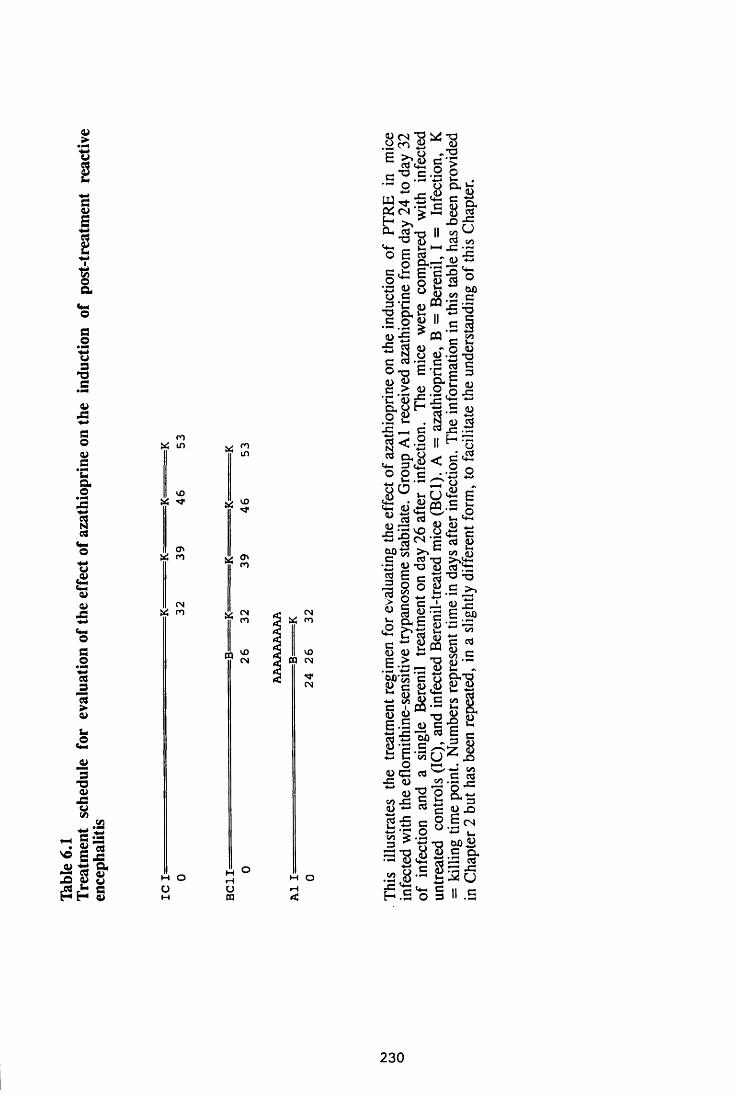
Tabl
e 6.
1Tr
eatm
ent
sche
dule
for
eval
uatio
n of
the
effe
ct
of az
athi
oprin
e on
the
indu
ctio
n of
post
-trea
tmen
t re
activ
e en
ceph
aliti
s
n
10 Ui **
<T>
CN m rn
in
O' n
cnm
CQID
INt< n
10CQ CN
M O O
CN
M O
S p i l e 'SE >-8 c l B-S C .l 1• <- +-> V-* ,‘ O o CL ^m Z e & c f .
o £ c3 o cl,—£ <£: £ '£ o s <u ctf
CO </J<L>
O *3 — « 8 h w) O c ^ 2 c3 r <U « £ «T3 cl i-> it *-* Is.5.2 > C I u-5 CQ 'c 2£ cfl 5j - O D
E-c | §rv ce 2 £ . 2 ,o . eC u r x : ^ uo g S 'S 1<& < .2 || H •'gts ^ X3 " . o^ 2 ^ o 0O O C • -r- Q8 ° i-o«a e& (U^ « .£ fa
C —* +-> ■*—»"-1 CT o <u
JSigvo ~c/l 4)op 3 £»fc - „
•S 2 - S - 8 ^ =e «3’a i3
S ^ *■* n-l 55 5E -S -s^ 2 -Sf'l S 8 g-!
g 3 2f ^ g S E - I c z |O H-l •£ •3 wr . s i “ «-gg a -CO £ £ i c 32 ~ c 22 § e 8 •§ ^-*-> *r^ o ■*-» i_
3 i l ! , s ti’S,• - I 'S .s ' O 3
e3.■ -Ci2 u
C ii c
230
6.2.2 The effect of azathioprine on an existing post-treatment reactive
encephalitis
Two groups of female NIH mice infected with 4xl04 trypanosomes of
the T.b. brucei stabilate GVR 35/C 1.5, ' ~re treated with 40mg/kg Berenil on
the 26th day of infection (Table 6.2). A group of these mice (A2) received
lOOmg/kg azathioprine i.p. for 7 or 14 days starting from day 32 after infection
(7 days after the first Berenil injection), and sacrificed at the end of azathioprine
course. The remainder, group A3, were treated again with 40mg/kg Berenil
after relapse of parasitaemia, and with azathioprine, at lOOmg/kg for 7 or 14
days from the day of the second Berenil treatment. Five mice were sacrificed at
the end of azathioprine course, and the rest were sacrificed serially every 7 days
thereafter in groups of five. Blood from tail snips was examined twice every
week to monitor parasitaemia, throughout the study period.
These mice were compared to infected mice treated with Berenil
alone, once (BC1) or twice (BC2), and sacrificed the same way as the
experimental animals. Uninfected controls in groups of five were included for
each drug regimen.
6.2.3 Preparation of brains for histopathology
At each kill, mice were perfused with approximately 150ml of normal
saline, pH 7.0, through the left ventricle of the heart to remove peripheral
blood. The brains were removed, fixed in 5% buffered formalin and processed
for histopathology. Three to 4^m transverse sections were cut serially through
the fore-, mid-, and hindbrain. Immunocytochemistry for GFAP was performed
on the sections to assess astrocyte activation. Serial sections from all the
samples were stained with haematoxylin and eosin for histopathology (Chapter
2).
231
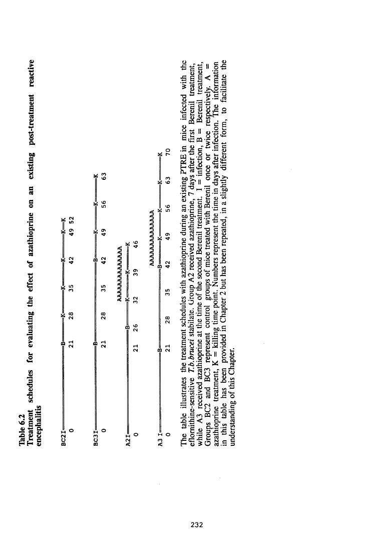
Table
6.
2Tr
eatm
ent
sche
dules
for
ev
alua
ting
the
effe
ct
of az
athi
oprin
e on
an ex
istin
g po
st-tr
eatm
ent
reac
tive
ence
phal
itis
VS CN
II mSfJ O'
oo
o
CNOOQ
vj cnvO
VO
CQ CNN*
i n
coCN
vj <0
CQ
CQ
vOCN
oc oO CNCQ ft
H O
or"
c o X v o
v oi n
CQ
OV
CN
00CN
CQ CN
M O CO
<
s s5 E | <4 -)
(D *s h ^
■c - a ii o s<D *X3
ccj o>
6 S C g * . S 3
3 1 §■s & &<D.a ts "
e
C ci'O *0C/3• h r—X 1
„ o
S e e o c. — <D \3 ,oe « m - c tS
c ■2 .s gS '— O »_ Jrj
l_ \ 0 O CD fflj
U 2 C C H ^ < 2 ^ ^ T 3
Oh ^ C ^ cn
11 Z ’S ’S-••3.5.5?
A ) ^ ~ u u ^^ C/Sv E «> c c <d PQ . 5 _rt' C E o ^ . S
g, & s s i T3- ■ c l i * r : § *o S "S S gg S g g g g -•§'8(5 * & £& > « u a g. 2 OJ "O . 2 cn ©
f g § e s 'SS <D <4- -2 Cd8 3 8 ° s i s a l l
• — n , . r - j j o£ 3 -* o^ O ' * - ! Co
Uc
„ c - 6 J Q . - Uh
3 0 < d _ 2 2 3 . £ ’o CX<U . 5 C <L) CC3
O jE-C i2 fli e £o ^ 2 o \ s ~ ° W>. g cn C - 2 - O
6 ‘r- <u (U ■—i <D& c a a . ’S
c* g - C S > y C cx, D . II O£ -Ci O CD £(D * j ’jS ** * ^ ■S K ^ j c
| y e «s . § « « g iS-a>c | asq C <D «J 2 -C 3 <D O ■*-3 ? £<n „ «
i U g S ^ . 2 c n PQ * c i3X) Ohi3 • — j? o «.V Oh» **23X5-S 2 O ' ? P cd
H <d > 'J c3 . 0
Wh
5i&.Cu
x: oW )c
CDT3C3
232
6.2.4 In vitro astrocyte preparation and exposure to azathioprine
Cerebral cortices were dissected from 2 to 3-day old NIH mice and
astrocyte preparation performed as described in Chapter 4. When purified and
confluent they were divided into four groups of five flasks each.
Lipopolysaccharide (LPS) was used as a known stimulant and compared to all
other experimental stimuli. LPS and azathioprine were used at a concentration
of 10jig/ml alone or in combination with each other. One flask in each group
was not exposed to any stimulant and served as the baseline control. The cells
were harvested by treating them with denaturing solution and scraping them off
with a cell scraper, at 2, 4, 6 and 8 hours after exposure. They were then
homogenised and stored at -70°C in phenol-resistant plastic tubes. RNA
extraction, cDNA preparation, amplification and detection of cytokine
transcripts were carried out as described in Chapter 4. The cytokines
investigated included, IL-la, IL-IB, IL-6, TNFa, MIP-1 and GM-CSF as in
Chapter 4.
6.3 RESULTS
6.3.1 The effect of azathioprine on induction of post-treatment reactive
encephalitis
Mice in group BC1, that were sacrificed 6 days after induction of
PTRE, exhibited a mild to moderate meningitis, perivascular cuffing and
infiltration of choroid fissure by inflammatory cells, mainly lymphocytes and
plasma cells, with a few macrophages. Perivascular cuffs, were prominent in
the cerebral white matter, along the hippocampal fissure, in the corpus
callosum, and in the cerebellar white matter, especially at the base of the
cerebellum. Astrocyte activation, as judged by the intensity of GFAP staining,
was marked along the inflamed meninges, around the perivascular cuffs and in
areas of inflammatory-cell infiltration. Astrocyte activation appeared to diffuse
233
from these areas of inflammation to the surrounding parenchyma with activation
becoming less marked away from these inflammatory foci. Both the
inflammation and astrocyte activation increased with time, with astrocytes
becoming increasingly stellate and exhibiting more intense GFAP stainir.g.
Treatment of mice with azathioprine for 8 days starting from day 24
followed by a Berenil injection on day 26 after infection (group Al), prevented
the infiltration of inflammatory cells into the CNS. However, astrocyte
activation occurred along the meninges, around the blood vessels and the
choroid plexus, and in the white matter in both the cerebrum and the
cerebellum, although the extent of astrocyte activation was less than in the mice
treated with Berenil alone, as assessed by the intensity of GFAP staining and
the degree of astrocyte stellation (Figures 6.1a to f)- However, the astrocyte
activation at this time was greater than in infected untreated mice sacrificed on
day 32 after infection.
Uninfected mice, treated with either or both of the two drugs in this
experiment did not develop CNS lesions. Blood from tail snips was examined
twice every week for parasites, throughout the duration of experiment.
6.3.2 The effect of azathioprine on an existing post-treatment reactive
encephalitis
The group of mice that was treated with azathioprine for 7 or 14 days
starting from 6 days after the Berenil treatment on day 26 after infection (group
A2), developed focal, mild inflammatory cell infiltrations, and astrocyte
activation along the meninges, the choroid fissure and the blood vessels.
Although the astrocyte activation in this group of animals was less than in the
Berenil-treated infected controls (group BC1), it was more marked than in mice
that received azathioprine from two days before the Berenil injection (compare
Figures 6.1 a,c and e with Figures 6.2a to c).
234
Figure 6.1
Comparison of the CNS pathology and astrocyte activation in a mouse,
from group BC1, infected with T.b.brucei GVR 35/C 1.5 and treated with
Berenil 26 days later and sacrificed 6 days after Berenil therapy (a, c and e),
to that in a mouse, from group Al, infected the same way, treated with Berenil
on day 26 after infection and with azathioprine from the 24th to 32nd day of
infection, and sacrificed at the end of azathioprine therapy (b, d and f).
Azathioprine stops the meningitis and perivascular cuffing in the cerebral cortex
(b); perivascular cuffing in the hippocampus and infiltration into the choroid
fissure (d), and meningitis and perivascular cuffing in the cerebellum (f).
Astrocytes stain more intensely, appear larger and are more diffusely distributed
in animals that received Berenil alone. The sections were stained with rabbit
anti-GFAP antibody, developed with peroxidase/DAB kit (Vectastain) and
counterstained with haematoxylin. x90
235
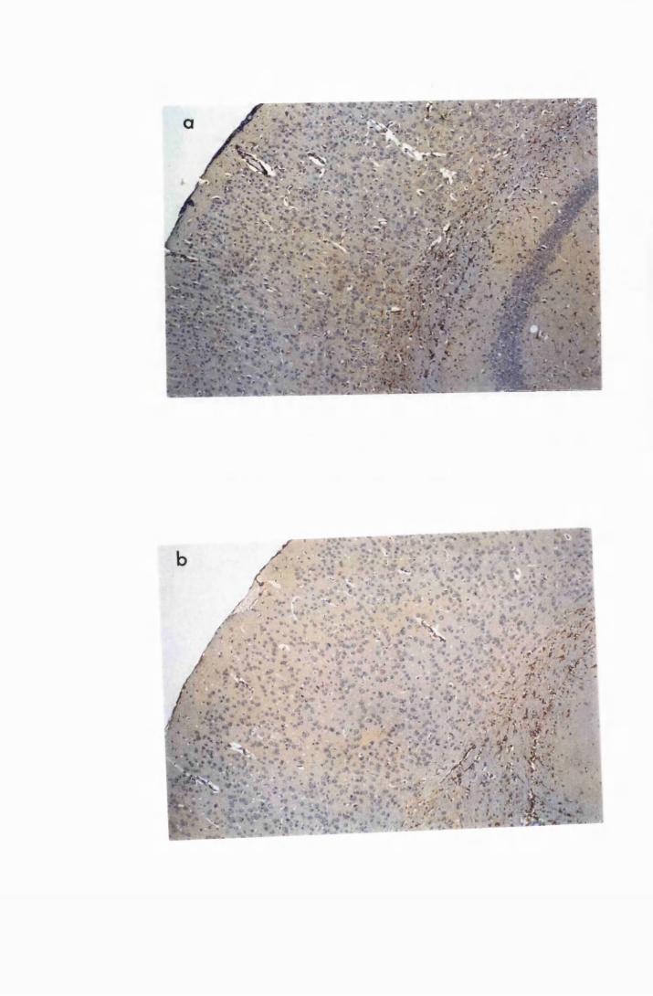
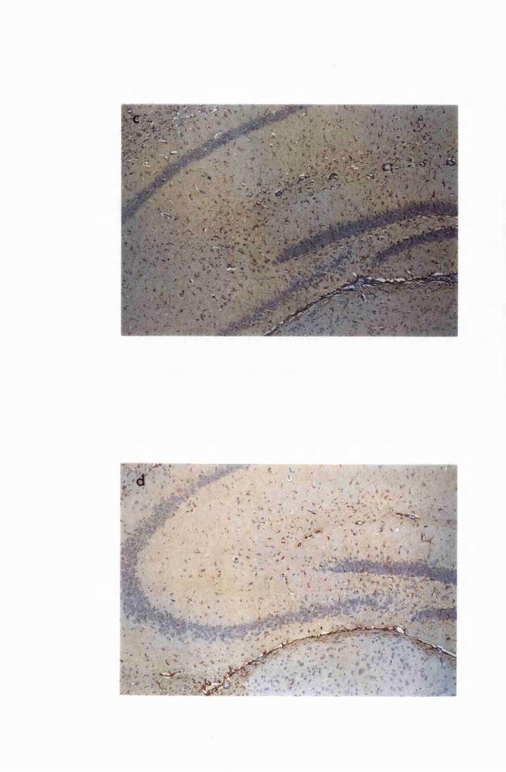
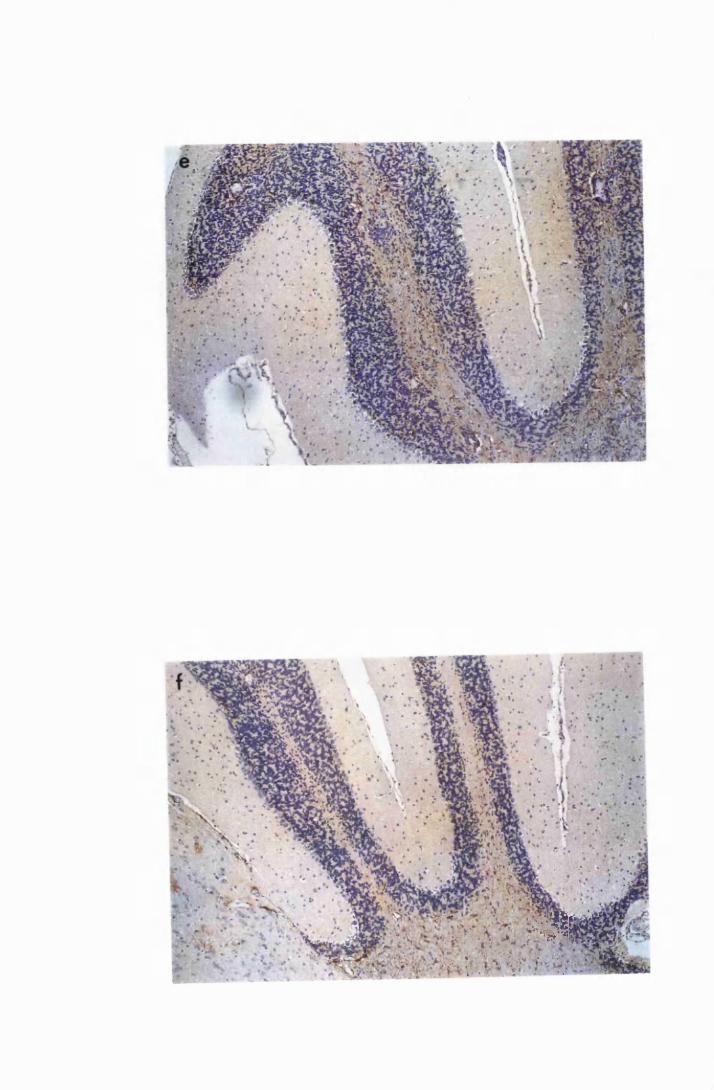
v ' ' '<Ji|% *• *' * 4 ' t S'I * . M ' • - 4
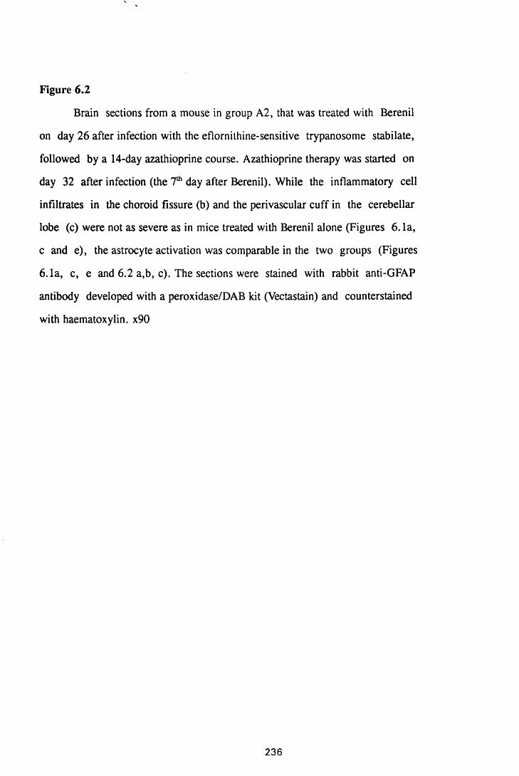
Figure 6.2
Brain sections from a mouse in group A2, that was treated with Berenil
on day 26 after infection with the eflornithine-sensitive trypanosome stabilate,
followed by a 14-day azathioprine course. Azathioprine therapy was started on
day 32 after infection (the 7th day after Berenil). While the inflammatory cell
infiltrates in the choroid fissure (b) and the perivascular cuff in the cerebellar
lobe (c) were not as severe as in mice treated with Berenil alone (Figures 6. la,
c and e), the astrocyte activation was comparable in the two groups (Figures
6.1a, c, e and 6.2 a,b, c). The sections were stained with rabbit anti-GFAP
antibody developed with a peroxidase/DAB kit (Vectastain) and counterstained
with haematoxylin. x90
236
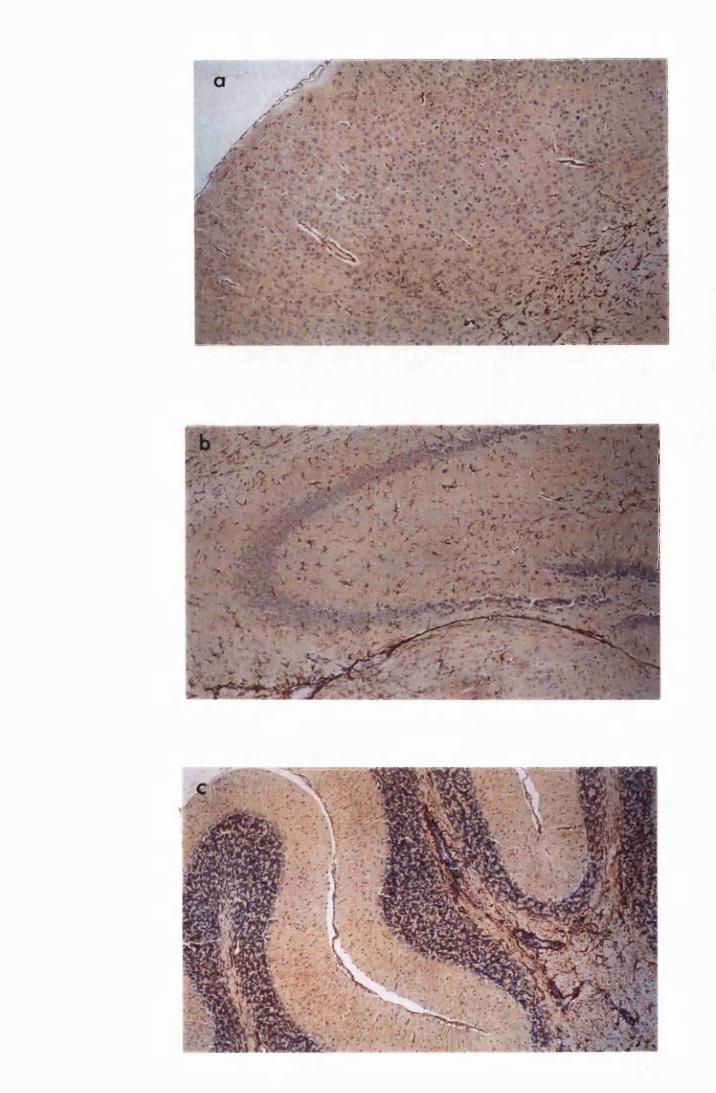
z
The mice that received azathioprine for 7 days from the day of the
second Berenil injection (group A3), showed a marked reduction in the
meningitis and perivascular cuffing. However, azathioprine did not appear to
affect the degree of astrocyte activation except in the cerebral grey matter where
it was reduced. The number of astrocytes and degree of astrocyte stellation in
the hippocampus and the cerebellum were comparable to those in the animals
that received two Berenil treatments without azathioprine (group BC2).
However, there was a slight decrease in the intensity of GFAP staining of the
activated astrocytes (Figures 6.3a to d).
In this group of animals (A3), severe inflammatory-cell infiltration
into the choroid fissure, the perivascular spaces and the meninges was evident 7
days after discontinuation of azathioprine administration.
6.3.3 In vitro effects of azathioprine on astrocyte activation
LPS at a concentration of 10/xg/ml stimulated astrocytes to express
mRNA transcripts for IL-la and -3, IL-6, TNFa, MIP-1 and GM-CSF (Figures
6.4a to c). Messenger RNA transcripts for all these cytokines were detectable by
2 hours after exposure to LPS, with those of IL-6 and IL-1B reaching peak
levels on the 4th and 6th respectively and remaining at this level for the rest of
the experimental duration. Those of MIP-1 remained at the same level except
for a slight decrease detected at the 4lh hour. Transcripts for IL-la seem to have
peaked at the sixth hour and were declining by the 8th hour, while those of GM-
CSF had disappeared by the 8th hour after peaking between the 4th and the 6th
hours.
Azathioprine, at 10/xg/ml, neither induced the expression of mRNA
transcripts for any of the cytokines studied, nor did it inhibit the LPS-induced
expression of these cytokine transcripts by astrocytes when it was introduced
simultaneously with 10/xg/ml LPS (Figures 6.4a, b and c).
237
Figure 6.3
The effect of azathioprine on the CNS pathology in mice treated with
Berenil on day 21 after infection with the eflornithine-sensitive T.b.brucei , and
again after relapse of parasitaemia. The sections shown in Figure 6.3a, c, and e
were from a mouse in group BC3, which was sacrificed 6 days after the second
Berenil, while the sections shown in Figure 6.3b, d and f came from a mouse in
group A3, which received azathioprine for 7 days from the day of the second
Berenil injection and was sacrificed at the end of azathioprine therapy. As a
result of azathioprine treatment, there is a marked reduction in the inflammatory
cell infiltration, but little reduction in the intensity of GFAP stain in the cerebral
grey mater (compare a and b) and no noticeable change in the state of astrocyte
activation in the hippocampus (compare c and d) and the cerebellum (compare e
and f). The sections were stained with rabbit anti-GFAP antibody developed
with a peroxidase/DAB kit (Vectastain) and counterstair.ed with haematoxylin.
x90
238
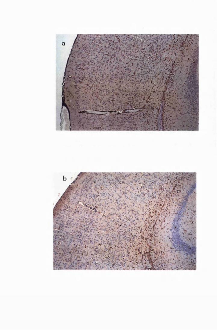
■-J& &
& : # J C W i ® 8 $ W
* •-■ *& ■ ?■; ’ ■■£!■ s$ 5• f ■ V . / ^ > # s $ s r
*-*«&** •/<; ... v , ■’& i
i ' "K*r . ';
T* a*"'*}••■•* ,:\''
«s*
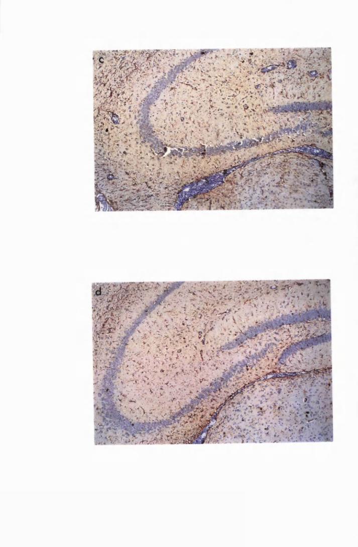
' • «>*•*•* r . ♦
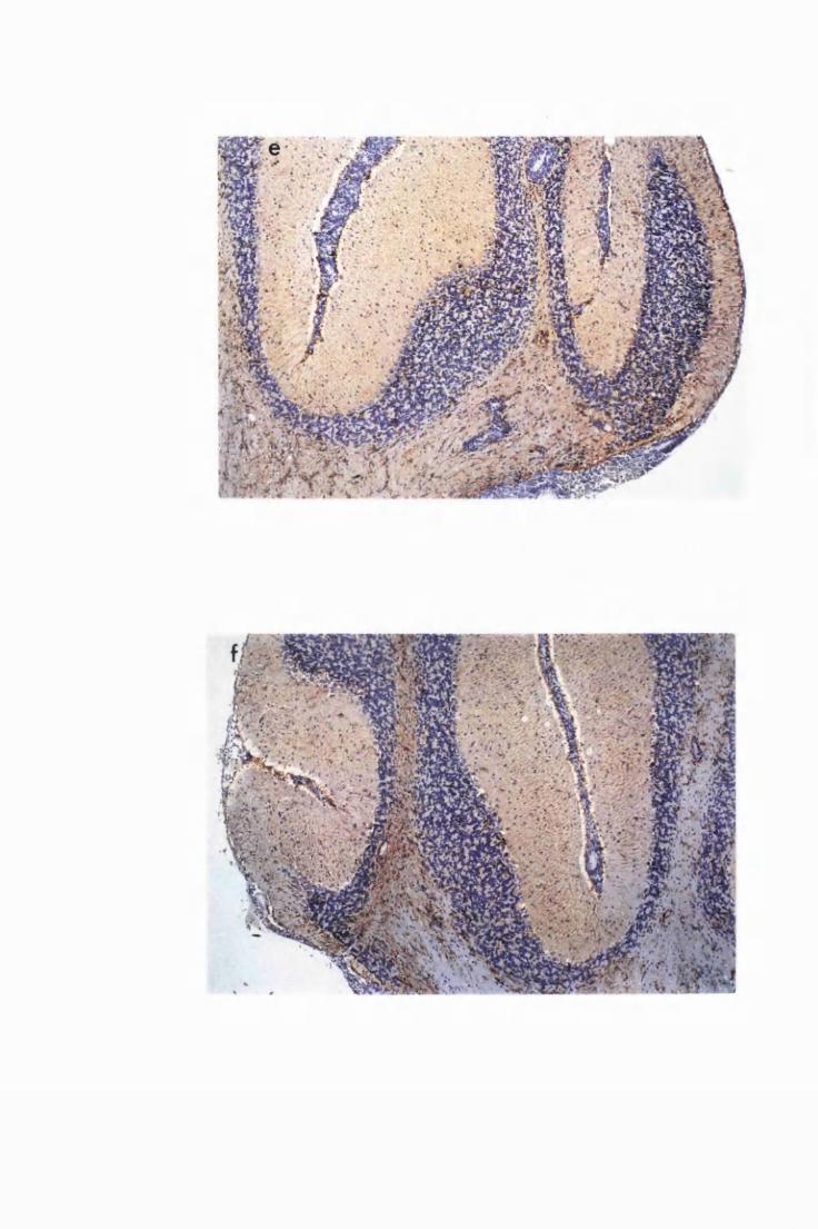
n >»«
Figure 6.4
Gel electrophoresis showing transcripts for the cytokines: a) IL-la b)
MIP-1 and IL-6 and c)IL-lfl and GM-CSF expren«ed by cultured astrocytes after
exposure to 10/xg/ml LPS (L), or 10/xg/ml azathioprine (A), or a combination of
LPS and azathioprine (A+L). Azathioprine has no effect on LPS-induced
expression of cytokine transcripts.The numbers 2, 4, 6 and 8 indicate time in
hours after addition of stimulant, M = 123bp ladder (Gibco); + =
positive control; W = water, used as a negative control; C = unexposed cells.
239
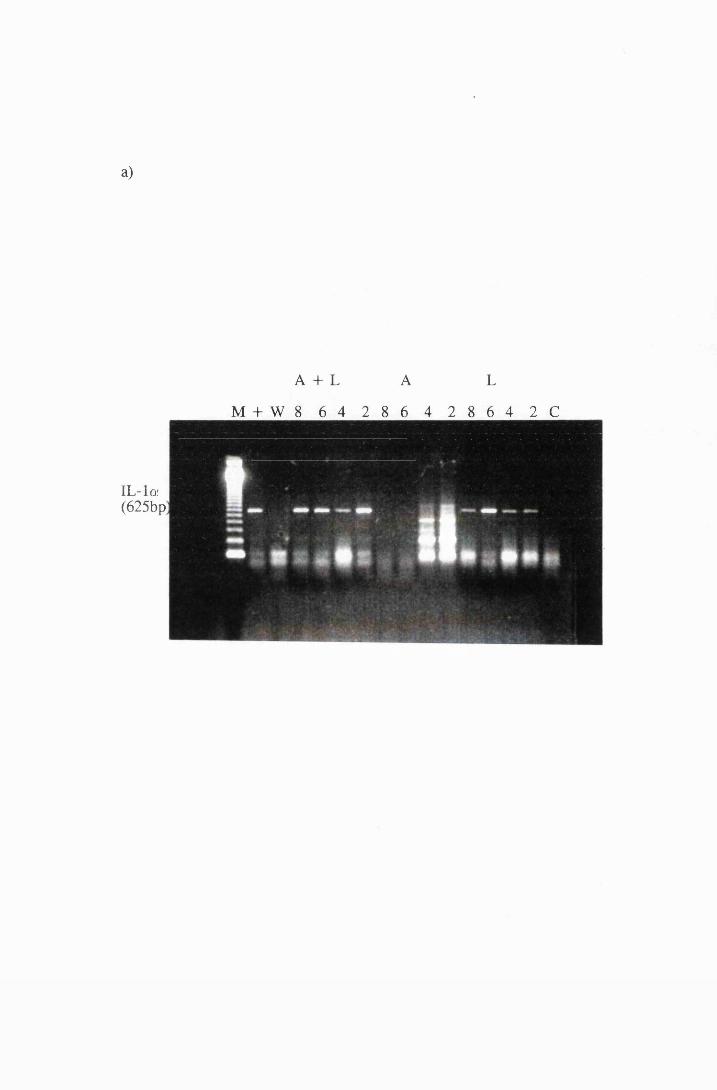
a)
A + L A L
M + W 8 6 4 2 8 6 4 2 8 6 4 2 C
IL-lor(625bp
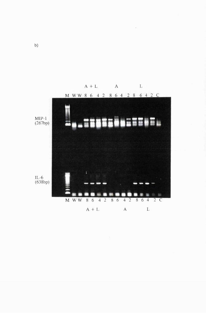
b)
A + L A L
M W W 8 6 4 2 8 6 4 2 8 6 4 2 C
MIP-1(267bp)
IL-6(638bp)
M W W 8 6 4 2 8 6 4 2 8 6 4 2 C
A + L A L
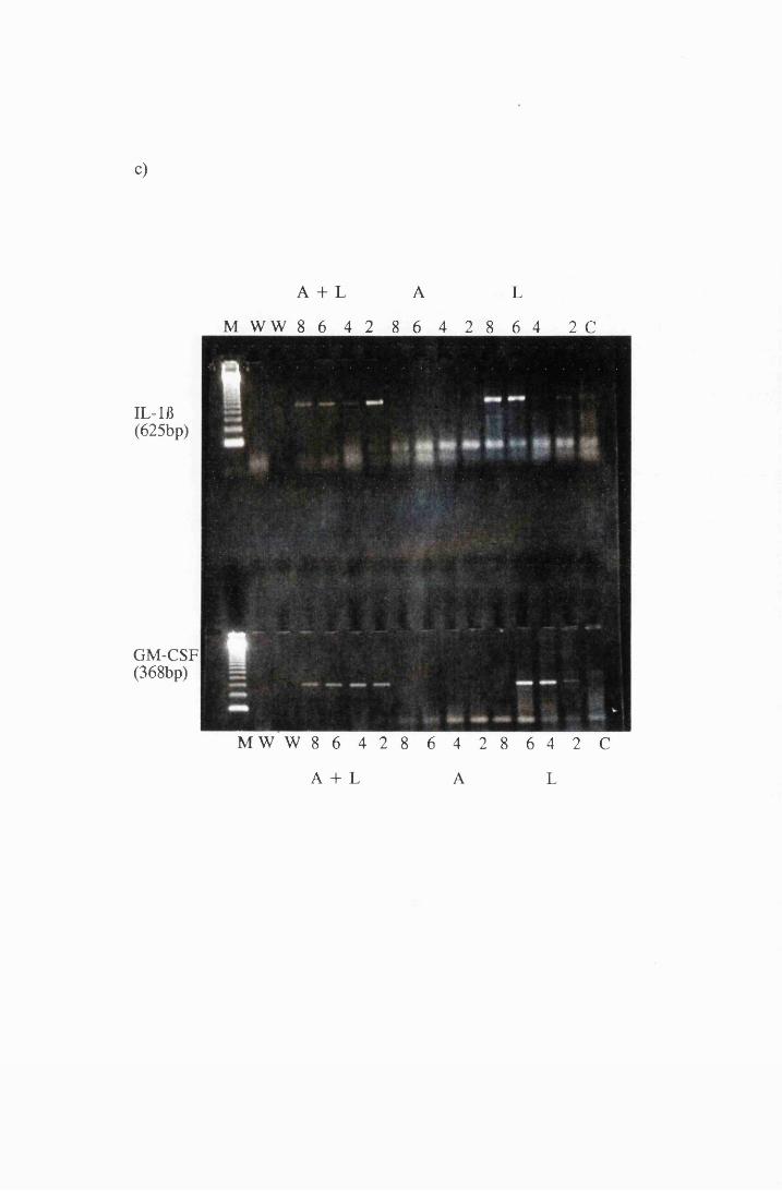
c)
A + L A L
M W W 8 6 4 2 8 6 4 2 8 6 4 2 C
IL-lfl(625bp)
GM-CSF(368bp)
M W W 8 6 4 2 8 6 4 2 8 6 4 2 C
A + L A L
6.4 DISCUSSION
The results presented here show varied effects of azathioprine on the
PTRE in T.b. brucei-infected mice. The varied effects seemed to depend on the
time at which azathioprine was administered. Thus, when azathioprine was
administered prior to and at the time of PTRE induction, it prevented the
development of inflammatory cell infiltration and appeared to arrest astrocyte
activation when compared to the Berenil treated controls. This suggested that
by inhibiting the infiltration of inflammatory cells azathioprine indirectly
prevented the progress of astrocyte activation, but unlike eflomithine did not
reverse the activation that had already taken place. Indeed, the results of this
study showed that the astrocyte activation after 7 or 14 days of azathioprine
administration, starting from the day of PTRE induction (day 26 after infection
when Berenil is injected), was just slightly more than in the activation seen 32
days after infection in untreated mice, but less than the Berenil-treated controls.
These results lend support to the hypothesis raised in this thesis that the
infiltrating inflammatory cells produce the additional signal (probably
cytokines), to provide the threshold for diffuse astrocyte activation.
The results of this study further showed that azathioprine
administration at the time of the second Bei^iil injection removed the
infiltrating inflammatory cells but did not appear to affect the astrocyte
activation. Moreover, the in vitro studies presented here, demonstrated that
azathioprine at a concentration known to inhibit cultured lymphocyte functions,
had no effect on the ability of LPS-induced astrocytes to produce cytokines.
Together, these results suggested that azathioprine had no direct inhibitory effect
on astrocytes. Other workers (Rosenthale et al, 1969; Babington and Wedeking,
197 1), using an experimental allergic encephalitis- (EAE)-rat model, reported
that azathioprine prevented the clinical development of the disease but that the
microscopical neural lesions were not prevented. Since the neural lesions in
EAE consist of demyelination and gliotic scarring, these microscopic findings
240
support the finding in this study that azathioprine does not directly inhibit
astrocyte activation.
Previous workers have reported that azathioprine prevents and
reverses the mononuclear inflammatory cell infiltration into the CNS of
T.b.brucei-infectod mice (Jennings et al, 1989; Hunter et al, 1992b). However,
the results of this study using the intensity of staining of the astrocyte specific
marker, GFAP, showed that azathioprine neither prevents nor reverses astrocyte
activation. The current study further demonstrated that azathioprine had no
effect on the in vitro production of cytokines by LPS-stimulated astrocytes.
Thken together these findings indicated that azathioprine had only a partial effect
on prevention and amelioration of PTRE. This is not suprising since this drug is
a specific inhibitor of lymphocyte functions. Evidence for a special effect of
azathioprine on the lymphocyte population includes: 1. An inhibitory effect on
the primary antibody response to T cell-dependent antigens, such as, the
heterologous red-blood cells (Nathan, Beiber, Elion and Hitchings, 1961),
tetanus toxoid (Friedman, Gelfand and Bemheimer, 1971) Candida (Kakuk,
Balogh, Szabolcsi and Hal my, 1972), human serum albumin (Meyer zum
Buschenfelde and Freudenberg, 1969) but not to T cell-independent antigens,
such as the pneumococcal polysaccharide (SIII). 2. The inhibition of in vitro T-
lymphocyte rosette formation around heterologous RBCs (Vas and Lowenstein,
1965; Bach and Bach, 1972), the cytocidal capacity of in Wvo-stimulated rat
lymphocytes (Wilson, 1965), production of specific antibody by lymphoid cells
following in vitro but not in vivo sensitisation (Rollinghoff, Schrader and
Wagner, 1973). 3. The inhibitory in vivo effects on the lymphocyte behaviour,
its synergisms with thymectomy (House, Boak and Draper, 1974) and the
inhibition of the effector capacity of T cells in azathioprine-treated patients
(Dawkins and Mastaglia, 1973; Richens, Williams, Gough and Ancill, 1974a;
1974b).
241
When azathioprine and its active metabolite, 6-mercaptopurine (6-
MP), are appropriately administered during the primary immune response,
normal development of immunological memory (as judged by normal booster
reaction) may be impaired even to the extent of producing temporary but
specific immune paralysis or tolerance (Nathan et al, 1961; Schwartz, 1965),
with azathioprine showing greater potential for immunosuppression than 6-MP
(Nathan et al, 1961). The results presented here showed an absence of
inflammatory cell infiltration into the CNS when azathioprine was administered
just before or at the time of PTRE induction. Since the infiltrating cells in PTRE
of T.b. brucei-infected mice consist mainly of lymphocytes and plasma cells, it
is possible that azathioprine acted on these cells causing temporary immune
paralysis, thus preventing the infiltration and proliferation of lymphocytes in the
CNS at this time.
The timing of azathioprine administration seems to be critical since it
appears to have an effect on the induction phase of immune response but not on
an already established response. The results of this study confirm this, as it was
found that administration of azathioprine at the time of PTRE induction
prevented infiltration of inflammatory cells while it was ineffective when
administered 7 days after induction of PTRE. Similar observations have been
made in various experimental procedures: azathioprine stops the enlargement of
germinal centres and lymphoid follicles in lymph nodes and spleen of rodents
during the first set reaction but not the second set reaction of graft rejection
(Marmor, 1967; Skopinska, Rychlikowa and Nouza, 1968; Laden, 1972;
Swingle, Grant and Valle, 1973); and it (azathioprine) is highly effective in
preventing the development of clinical EAE in rats when given during the first 7
days and less so when given later (Babington and Wedeking, 1971). Moreover,
the presence of azathioprine within the first 24 hours of lymphocyte culture was
found to be necessary for the in vitro inhibition of: a two-way mixed leukocyte
reaction (Vas and Lowenstein, 1965; Bach and Bach, 1972), production of
242
specific antibodies after in vitro stimulation of lymphocytes (Rollinghoff et al,
1973) and cytocidal capacity of cultured rat lymphocytes after in vivo
stimulation (Wilson, 1965). Azathioprine and 6-MP can also abolish or delay
the primary humoral response to both heterologous cellular, or soluble protein
antigens, inhibiting the switch from IgM to IgG production; the prolongation of
IgM production being attributable to a lack of normal feedback control (Uhr and
Braumann, 1961). In multiple sclerosis patients, azathioprine has been reported
to slow down the clinical deterioration associated with this disease, the effect
being more marked in patients who had the disease for less than 2 years.
Patients who had been affected for longer durations did not show any significant
reduction in the rate of disease progression (Patzold and Pocklington, 1980;
Muller, Sauermann and Kreiner, 1981)
This study showed that administration of azathioprine starting 7 days
after Berenil treatment, neither reversed the cellular infiltration nor the astrocyte
activation in the resultant PTRE in T.b. brucei-infected mice. It is possible that
at this time, the lymphocytes had proceeded into the secondary phase of immune
response and were insensitive to azathioprine. Furthermore, macrophages which
occur secondary to lymphocytes in an immune response and which might not
respond to azathioprine (Gotjamanos, 1971; Kaufman and McIntosh, 1971),
could have infiltrated the inflammatory foci by this time.
The observation, in this study, that azathioprine almost entirely
stopped the infiltration and proliferation of lymphocytes when given at the time
of the second Berenil treatment, seemed to suggest that the lymphocytes were
still in the induction-phase response even at this time. This was not suprising
considering that trypanosomes undergo antigenic variation, which could ensure
that lymphocytes are constantly in the induction phase, reacting to the new
antigenic type of trypanosomes. The observation that delaying azathioprine
administration for 7 days after Berenil removed this effect on lymphocytes, and
243
knowing that Berenil clears parasites from the circulatory system, suggested that
the new antigenic types emanate from the systemic circulation to constantly
present a new challenge to the infiltrating lymphocytes. It would appear that
when the systemic circulation was cleared of parasites by Berenil, and
lymphocytes given time to accommodate to the trypanosomes in the CNS, they
proceeded to the secondary phase of response becoming insensitive to
azathioprine. An alternative possibility is that the immunosuppression that
occurs during trypanosome infections affects the lymphocytes in such a way that
they are arrested in the induction phase.
Administration of azathioprine at the time of the second Berenil
injection cleared the bulk of the infiltrating cells but a few inflammatory foci
remained. Most of the remaining cells were macrophages which are a
component of the infiltrating cells in the trypanosome-induced CNS lesion and
PTRE, and which might not respond to azathioprine. The effect of azathioprine
on macrophages is controversial. Some authors found azathioprine to have no
effect on the ability of activated macrophages to phagocytose particulate matter
such as carbon (Gotjamanos, 1971) or colloidal gold (Kaufman and McIntosh,
1971). Other workers, have shown that 6-MP has a suppressive effect on the
ability of macrophages to phagocytose I4C and their ability to migrate from
capillary tubes regardless of changing macrophage inflammatory factor (MIF)
concentration (Philip and Zweiman, 1973).
When the course of azathioprine therapy was extended to 14 days,
there was a marked reduction in the infiltration of inflammatory cells and a
slight reduction in the astrocyte activation. It is possible that this reduction in
astrocyte activation was due to the reduced stimulatory boost from the
disappearing inflammatory cells, since the in vitro studies confirmed that
azathioprine had no direct effect on astrocytes. It would appear that prolonging
the course of azathioprine therapy might be useful in the amelioration of an
established PTRE. Unfortunately, at the doses used in this study (lOOmg/kg,
244
lower doses were ineffective), the drug was poorly tolerated, with most of the
mice dying before the completion of the 14-day course. Similar intolerance of
azathioprine has been reported in Rhesus monkeys in the prevention of renal
allograft-rejection (Dicke et al, 1971) and in the treatment of arthritis in rats
(Jouanneau, Brouilhet, Kahan, Charles and Delbarre, 1973). Long term (1-5
years) azathioprine therapy has been reported in multiple sclerosis patients
(Patzold and Pocklington, 1980; Muller et al, 1981; Milanese, La Mantia,
Salmaggi, Campi, Bortolami, Tajoli, Nespolo and Corridori, 1988). The
outcome of these studies were conflicting: while Milanese et al (1988) reported
a significant reduction of disease progression in azathioprine treated patients
(favourable outcome seen only after 3 years of therapy), other researchers found
a high incidence of clinical deterioration, with 64.1% of these patients
developing severe side effects (Muller et al, 1981). The side effects included
gastric complaints, considerable leukocyte depression, oedema, alopecia,
strumata, increase in transaminases, recrudescence of mycosis and pyoderma
(Muller et al, 1981).
The observation here that azathioprine did not reverse the astrocyte
activation, and that cellular infiltrates recurred within 7 days of discontinuation
of azathioprine therapy (compared with 14 or more days after eflomithine, a
drug that stops astrocyte activation) seems to support the hypothesis proposed
earlier in this thesis that activated astrocytes initiate inflammatory cell-
infiltration into the CNS. These results further suggest that the effects of
azathioprine on lymphocytes are short lived, with the result that lymphocytes
become active and proliferate as soon as azathioprine therapy is withdrawn.
Indeed it has been reported that a burst of mitotic activity occurs shortly after
therapy with azathioprine is stopped (Skopinska et al, 1968).
It would appear that administration of azathioprine for the prevention
of PTRE would require an exact knowledge of the stage of the inflammatory
245
lesion in the brain. Using the current tools of diagnosis, it is impossible to know
the exact stage of the reaction in the CNS of sleeping sickness patients and any
improvement would depend on a chance "correct" timing. However, the use of
azathioprine at the time of trypanocidal therapy could be beneficial since
administration at the time of Berenil treatment (even after relapse of
parasitaemia) seemed to slow down the progress of PTRE. It is possible that
azathioprine therapy might arrest the development of clinical PTRE, especially
when used in combination with the first-time trypanocidal treatment (i.e non
relapsed patients).
In experimental T.b. brucei infections of mice, it has been observed
that the use of three consecutive doses of azathioprine at the time of the first
Berenil treatment reduced the incidence of hind-limb paralysis to 25 % from
50% in Berenil treated controls (F.W. Jennings, personal communication). This
seems to suggest that the beneficial effects of azathioprine could be utilised
without prolonging the duration of therapy and avoiding the accompanying
adverse side effects. While azathioprine might not reverse the already present
pathological changes during a relapse infection, its simultaneous use with a
trypanocidal agent might prevent development of clinical PTRE.
It has been reported that in the prevention of graft rejection,
azathioprine works best at safe dosages when used in triple-combination with
other anti-inflammatory drugs especially corticosteroids and cyclosporin A
(Vereerstraeten, 1993; Isoniemi et al, 1993). This possibility has not been
investigated in models of trypanosomiasis. One wonders whether investigating
this possibility would be worthwhile considering that the combined anti
inflammatory therapy, would have to be used in conjunction with trypanocidal
drugs, in subjects that are likely to be already immunosuppressed.
246
CHAPTER 7
GENERAL DISCUSSION AND CONCLUSIONS
247
This thesis concerns the role of astrocytes in the pathogenesis of CNS
trypanosomiasis and in the encephalopathy that results from treatment of late-
stage trypanosomiasis with drugs such as melarsoprol in HAT and Berenil in
experimental T.b. brucei infections in mice. The working hypothesis of the
present investigations, was that astrocytes initiate and propagate the CNS
pathology during trypanosome infections and in PTRE. The objectives of the
study were: to determine when astrocytes become activated during the course of
trypanosome infection in mice and during curative and subcurative trypanocidal
therapy; how astrocytes respond to trypanosomes in vitro with respect to
cytokine production; and how drugs, reported to attenuate the inflammatory cell
infiltrations during PTRE, affect astrocyte activation in vivo and in vitro.
Astrocyte activation, occurred initially where the blood-brain barrier
is incomplete, at sites where early trypanosome invasion has been reported to
occur, and was followed by infiltration of inflammatory cells into the choroid
fissure, perivascular spaces and the meninges. This suggests that astrocyte
activation is the first CNS response to invasion by trypanosomes and that the
activated astrocytes initiate the inflammatory cell infiltration, possibly by
producing inflammatory mediators. The correlation of the early astrocyte
activation, with cytokine production, the knowledge that astrocytes are capable
of producing cytokines and that cytokines initiate meningeal inflammation,
would suggest that cytokines, produced by the activated astrocytes, initiate
leukocyte extravasation into the brains of T.b. brucei-infected mice.
Histological examination of the brains of mice with PTRE, caused by
subcurative treatment with Berenil, revealed pathology similar to that in infected
untreated mice, with the exception that it occurred faster and was more
pronounced. A second Berenil treatment made the lesions even more
pronounced and did not kill the trypanosomes in the brain. This suggested that
the cells and mediators of inflammation in infected untreated and in
248
subcuratively treated mice were the same. It was proposed that PTRE is an
enhanced reaction to persistent infection within the brain, resulting from a
temporary disruption of the blood-brain bamer, and subsequent influx of
metabolites and inflammatory mediators from the circulatory system. The blood-
derived metabolites and inflammatory mediators facilitate trypanosome survival,
and enhance inflammation and astrocyte activation within the CNS.
Pure cultures of astrocytes responded to trypanosomes, trypanosome
lysate and VSG by expressing mRNA transcripts for the cytokines, IL-la and
-B, IL-6, TNFa, MIP-1 and GM-CSF, indicating that astrocytes are likely to be
a major source of these cytokines in the brains of T.b. brucei infected mice; and
that trypanosomes within the brains of such mice are a potent stimulus for
astrocyte activation. Production of IL-1 and TNF in the CNS would act on
endothelial cells, microglia and astrocytes themselves, causing: migration,
proliferation, induction of adhesion molecule expression, and production of
leukocyte chemotactic cytokines. The overall effect of such cytokines would
lead to extravasation of leukocytes into the CNS, proliferation of the infiltrating
inflammatory cells, microglia and astrocytes, and production of more cytokines,
essentially creating a self-perpetuating lesion.
Administration of eflornithine prior to and around day 21 of infection
with an eflomithine-resistant trypanosome stabilate, prevented the occurrence of
astrocyte activation and the subsequent inflammatory cell infiltration. When
eflornithine was administered during an existing PTRE, induced by a single or
double Berenil treatment, there was regression of both inflammatory cell
infiltrations and astrocyte activation. These results suggested that eflornithine
had an inhibitory effect on both the inflammatory cells and the astrocytes. The
finding that eflornithine, inhibited the LPS-induced expression of cytokine
transcripts by cultured astrocytes confirmed a direct effect of eflornithine on
astrocytes. This suggested that the disappearance of astrocyte activation in the
brains of eflornithine-treated, T.b. brucei-'miociod mice was not a consequence of
249
the disappearing inflammatory cells. Since the trypanosomes used in this study
were eflornithine resistant, the remarkable reversal of the CNS pathology in the
face of persistent infection demonstrates the extraordinary potential of
eflornithine therapy in the late stages of African trypanosomiasis. Therefore,
eflornithine may be used alone, for the treatment of trypanosomiasis, when the
infecting trypanosomes are sensitive, and in combination with other
trypanocides when the infecting trypanosomes are refractory to eflornithine. In
addition, eflornithine would be of use in the prevention and, possibly, reversal
of PTRE.
Treatment of mice with azathioprine, after day 21 of infection with
T.b. brucei or just prior to treatment of such mice with Berenil, prevented the
infiltration of inflammatory cells but did not prevent the onset of astrocyte
activation. Administration of azathioprine during an established PTRE (starting
7 days after Berenil treatment) did not affect the astrocyte activation and only
slightly reduced the number of infiltrating inflammatory cells. It was further
shown that azathioprine did not affect the LPS-induced expression of cytokine
transcripts in vitro. Azathioprine is a known inhibitor of lymphocytes, and, as
the results in chapter 6 show, does not affect astrocyte activation either at the
onset or after it has occurred. This would suggest that the onset and mediation
of astrocyte activation during T.b. brucei infections in mice is not dependent on
inflammatory cells, supporting the hypothesis that astrocyte activation is the
CNS response to trypanosomes. The effect of azathioprine on inflammatory
cells, depended on the time it was administered: when administered just prior to
the first Berenil, or at the same time as the second Berenil, it prevented the
infiltration of inflammatory cells. In such situations, there was a a slight
decrease in the GFAP staining intensity, reflecting a slight reduction in astrocyte
activation. It was suggested that this reduction in astrocyte activation, resulted
from the loss of stimulatory boost from the disappearing inflammatory cells and
250
not from a direct effect of azathioprine on astrocytes. It would appear that
administration of azathioprine at the time of trypanocidal therapy might prevent
development of clinical PTRE and by inference the development of RAE in
man.
In conclusion, these studies show: 1. that the inflammatory cell
infiltration during trypanosome infections in mice is a secondary event to a
response from within the CNS, 2. that astrocytes play an important role in this
initial CNS response to trypanosomes and in inducing the subsequent leukocyte
infiltration and proliferation, through production of inflammatory mediators,
including cytokines, and 3. that eflornithine, a drug that affects both the
astrocytes and the infiltrating inflammatory cells is more potent in preventing
the Berenil-induced PTRE in mice than azathioprine, a drug that affects only the
inflammatory cells. It would appear that identifying the mediators (such as
cytokines) of CNS inflammation; and specifically inhibiting these mediators
would give a better prognosis than using anti-inflammatory agents that affect a
specific cell type. Future studies using specific cytokine inhibitors, such as anti
cytokine antibodies or cytokine-receptor antagonists, would be very useful in
addressing this question. Eflornithine might also be useful in other
inflammatory conditions such as bacterial meningitis and viral meningitis.
Indeed, the in vitro results presented in this study showed that eflornithine
reverses the LPS-induction of cytokine production by astrocytes. Although the
cell infiltrations in bacterial meningitis consist mainly of polymorphonuclear
cells, the cytokines produced by astrocytes, some of which were discussed here,
would also have an activating and proliferative effect on those cells.
251
REFERENCES
252
Aay, C. and Borst, P. (1972) The gel electrophoresis of DNA. Biochemistry and
Biophysiology Acta 269, 192-200
Abaru, D.E. and Matovu, F.S. (1981) Berenil in the treatment of early and late stage human
trypanosomiasis cases. Proceedings of the International Scientific Council of
Trypanosomiasis Research and Control. Arusha (Tanzania). Nairobi OAU/STRC
Publication 112, 194-198
Abbas, A.K., Litchman, A.H., and Pober, J.S ., eds. (1991) Cellular and molecular
immunology. Philadelphia. W.B. Saunders.
Abney, E.R., Williams, B.P. and Raff, M.C. (1983) Tracing the development of
oligodendrocytes from precursor cells using monoclonal antibodies, fluorescence-
activated cell sorting and cell culture. Developmental Biology 100, 166-177
Ackerman, S.B. and Seed, J.R . (1976) Immunosuppression during Trypanosoma brucei
gambiense infection in field vole, Microtus montanus. Clinical and Experimental
Immunology 25, 152-158
Adams, J.H ., Graham, D.I. and Gennarelli, T.A. (1983) Head injury in man and
experimental animals: neuropathology. Acta Neurochirugia Supplementum (Wien) 32,
15-30
Adams, J.H . and Graham, D. eds. (1988) Microscopy of the normal nervous system. In "An
Introduction to Neuropathology" Churchill Livingstone pp 25-43
Adams, J.H ., Haller, L., Boa, F.Y., Doua, F., Dago, A. and Konian, K. (1986) Human
African Trypanosomiasis {T.b. gambiense): a study of 16 fatal cases of sleeping
sickness with some observations on acute reactive arsenical encephaolopathy.
Neuropathology and Applied Neurobiology 12, 81-94
Aderka, D., Moar, Y., Novick, D., Engelmann, H., Kahn, Y., Levo, Y., Wallach, D. and
Revel, M. (1993) Interleukin 6 inhibits the proliferation of B-chronic lymphocytic
leukaemia cells that is induced by tumor necrosis factor-alpha or beta. Blood 81, 2076-
2084
Aerts, D., True, P., Penchemier, L., Claes, Y. and Le Ray, D. (1992) A kit for in vitro
isolation of trypanosomes in the field: first trial with sleeping sickness patients in the
253
Congo Republic. Transactions of the Royal Society of Tropical Medicine and Hygiene
86, 394-395
Aiyedun, B.A., Amodu, A.A., Bidwell, D.E., Bone, G .J., Buck, A.A., Coulm, J ., Frezil,
J .L ., Kent, N.H., Mattern, P., Njogu \.R ., Voller, A. and Wery, M. (1976)
Parallel evaluation of serological tests applied in African trypaosomiasis: a WHO
collaborative study. Bulletin of the World Health Orgaization 54, 141
Akol, G.W.O. and Murray, M. (1982) Early events following challenge of cattle with tsetse
infected with Trypanosoma congolense: development of the local skin reaction.
Veterinary Record 110, 295-302
Alam, R ., Forsythe, P.A., Stafford, S., Lett-Brown, M.A. and Grant, J.A . (1992)
Macrophage inflammatory protein 1 alpha activates basophils and mast cells. Journal of
Experimental Medicine 176, 781-786
Anosa, V.O., Jennings, F.W. and Urquhart, G.M. (1977) The effect of splenectomy on
anaemia in Trypanosoma brucei infection in mice. Journal of Comparative Pathology
87, 1-11
Apted, F.I.C. (1957) Four year experience of melarsen oxide/BAL in the treatment of late
stage Rhodesian sleeping sickness. Transactions of the Royal Society of Tropical
Medicine and Hygiene 51, 75-86
Apted, F.I.C. (1962) Sleeping sickness in Tanganyika, past, present and the future.
Transactions of the Royal Society of Tropical Medicine and Hygiene 56, 15-29
Apted, F .I.C (1970) Clinical manifestations and diagnosis of sleeping sickness. In "The
African Trypanosomiases". Mulligan, H.W. ed. London Allen and Unwin pp661-683
Arroz, J.O .L. (1987) Short report. Melarsoprol and reactive encephalopathy in Trypanosoma
brucei rhodesiense. Transactions of the Royal Society of Tropical Medicine and
Hygiene 81, 192
Askonas, B.A. (1985) Macrophages as mediators of immunosuppression in murine African
trypanosomiasis. Current Topics in Microbiology and Immunology 117, 119-127
Askonas, B.A., Corsini, A.C., Clayton, C.E. and Oglivie, B.M. (1979) Functional
254
depression of T- and B-memory cells and other lymphoid cell subpopulations during
trypanosomiasis. Parasitology 36, 313-321
Asonganyi, T., Lando, G. and Ngu, J.L . (1989) Serum antibodies against human myelin
proteins in Gambian trypanosomiasis. nales de la Societe Beige de Medicine
Tropicale 69, 213-221
Assoku, R .K .G ., Hazzlet, C.A. and Tizard, I. (1979) Immunosuppression in experimental
African trypanosomiasis: polyclonal B cell activation and mitogenicity of trypanosome-
derived saturated fatty acids. International Archives of Allergy and Applied
Immunology 59, 298-307
Babington, R.G. and Wedeking, P.W. (1971) The influence of cinanserin and selected
pharmacologic agents on experimental allergic encephlomyelitis. Journal of
Pharmacology and Experimental Therapeutics 177, 454-460
Bacchi, C.J. and McCann, P.P. (1987) Parasitic protozoa and polyamines. In "Inhibition of
Polyamine Metabolism" McCann, P.P., Pegg, A.E. and Sjeordsma, A. eds. Academic
Press pp 317-345
Bacchi, C.J., Nathan, H.C., Clarkson, A.B. jr., Bienen, E .J., Bitonti, A.J., McCann,
P.P. and Sjeordsma, A. (1987) Effects of ornithine decarboxylase inhibitors DL-a-
difluoromethylomithine and alpha-monofluoromethyldehydroomithine methyl ester
alone and in combination with suramin against Trypanosoma brucei brucei central
nervous system models. American Journal of Tropical Medicine and Hygiene 36, 46-52
Bacchi, C.J., Nathan, H.C., Hunter, S.H., McCann, P.P. and Sjoerdsma, A. (1980)
Chemotherapeutic target in trypanosomes. Science 210, 332-334
Bacchi, C.J., Nathan, H.C., Livingston, T., Valladares, G., Saric, M., Sayer, P., Njogu,
A.R. and Clarkson, A .B Jr (1990) Differential susceptibility to DL- alpha-
difluoromethylomithine in clinical isolates of Trypanosoma brucei rhodesiense.
Antimicrobial Agents and Chemotherapy 34, 1183-1188
Bach, M.A. and Bach, J.F. (1972) Activities of immunosuppressive agents in vitro. II.
Different timing of azathioprine and methotrexate in inhibition and stimulation of mixed
lymphocyte reaction. Clinical and Experimental Immunology 11, 89-98
255
Bafort, J.M ., Schmidt, H. and Molyneux, D.H. (1987) Development of Trypanosoma
brucei in suckling mouse brain following intracerebral injection. Transactions of the
Royal Society of Tropical Medicine and Hygiene 81, 487-490
Bagasra, O., Schell, R.F. and Le Frock, T.L. (1981) Evidence of depletion of la
macrophages and associated immunosuppression in African trypanosomiasis. Infection
and Immunity 32, 188-193
Bailey, N.M. (1967) The use of Berenil in the treatment of human trypanosomiasis. East
African Trypanosomiasis Research Organization report 1966, pp83
Bailey, N.M. (1968) Oral Berenil in the treatment and prophylaxis of human trypanosomiasis.
Transactions of the Royal Society of Tropical Medicine and Hygiene 62, 122
Baker, J.R.,(1970) Techniques for the detection of trypanosome infections. In "The African
Trypanosomiases". Mulligan, H.W. ed. Allen and Unwin, London. pp67-88
Bakheit, M., Olsson, T., Van der Meide, P. and Kristensson, K. (1990) Depletion of
CD8+ T-cells suppresses growth of Trypanosoma b. brucei and interferon gamma
production in infected rats. Clinical Experimental Immunology 81, 195-199
Barna, B.P., Estes, M .L., Jacobs, B.S., Hudson, S. and Ransohoff, R.M. (1990) Human
astrocytes proliferate in response to tumor necrosis factor. Journal of
Neuroimmunology 30, 239-243
Barnet, S.F. (1947) Bovine trypanosomiasis in Kenya with special reference to its treatment
with phenanthidinium. Veterinary Record 59, 459-462
Barres, B.A, Chun, L.L. and Corey, D.R (1988) Ion channel expression by white matter
glia. 1. Type 2 astrocytes and oligodendrocytes. Glia 1, 10-30
Barret-Connor E., Ugoretz, R .J. and Braude A.I. (1973) Disseminated intravascular
coagulation in trypanosomiasis. Archives of Internal Medicine 131, 574-577
Basson W., Page, M.L., and Myburgh, D.P. (1977) Human trypanosomiasis in Southern
Africa. South African Medical Journal 51, 453-457
Bauman, H ., Jahreis, G.P, Sauder, D.N. and Koj, A (1984) Human keratinocytes and
monocytes release factors which regulate the synthesis of major acute phase plasma
256
proteins in hepatic cells from man, rat and mouse. Journal of Biological Chemistry 259,
7331-7342
Benveniste, E.N., Sparacio, S.M., Norris, J.G ., Grannet, H.E. and Fuller, H.E. (1990)
Induction and regulation of interleukin 6 gene expression in rat astrocytes. Journal of
Neuroimmunology 30, 201-212
Bertrand, E., Sentilhes, L., Ducasse, B., Vacher, P. and Boudin, L. (1965) Le attentte du
coeur dans la trypanosomiase Africaine a Trypanosoma gambiense a propos notament
des constatations ECG chez 60 malades. Medicine Tropicale 25, 603
Bertrand, E., Lobiere, R., Barabe, P. and Ette, M. (1971) La coeur dans la trypansomiase
Africaine. Coer et Medicine Interne 10, 391-396
Beutler, B. and Cerami, A. (1986) Cachectin and tumor necrosis factor as two sides of the
same biological coin. Nature 320, 584-588
Beutler, B., and Cerami, A.(1989) The biology of cachectin/TNF: a primary mediator of the
host response. Anual of Reviews in Immunology 7, 625-630
Beutler, B., Greenwald, D., Hulmes, J.D., Chang, M., Pan, Y., Math iso n, J , Ulevitch,
R. and Cerami, A. (1985a) Identity of tumor necrosis factor and the macrophage-
secreted factor. Nature 161, 552-554
Beutler, B., Milsark, I.W. and Cerami, A. (1985b) Cachectin/tumor necrosis factor:
production, distribution and metabolic fate in vivo. Journal of Immunology 135, 3972-
3977
Bever, C.T., Snyder, D.S., Endres, R.O., Morgan D.D., Postlethwaite, A. and Whitaker,
J.N . (1989) Activation of astrocytic lysosomal proteinases by factors released by
mononuclear leukocytes. Neurochemistry Research 14, 27-41
Bevilacqua, M.P., Pober, J.S ., Wheeler, M .E., Cotran, R.S. and Gimbrone, M.A., j r
(1985) Interleukin 1 acts on cultured human endothelium to increase the adhesion of
polymorphonuclear leukocytes, monocytes and related cell lines. Journal of Clinical
Investigations 76, 2003-2011
Bevilacqua M.P., Pober, J.S ., Majeau, G .R., Fiers, W., Cotran, R.S. and Gimbrone
M.A. j r (1986a) Recombinant tumor necrosis factor induces procoagulant activity in
257
cultured human vascular endothelium: characterisation and comparison with the actions
of interleukin 1. Proceedings of the National Academy of Science USA 83, 4533-4537
Bevilacqua, M.P., Schleef, R.R., Gimbrone, M.A. jr. and Loskutoff, D.J. (1986b)
Regulation of fibrinolytic system of cultured human vascular endothelium. Clinical
Investigations 78, 587-591
Beyer, R (1978) In " Enzyme-activated Irreversible Inhibitors". Seiler N., Jung M.J, and
Koch-weser J. eds. Elsevier Amsterdam pp 27-41.
Bignami, A. and Dahl, D. (1976) The astroglial response to stabbing: Immunofluorescent
studies with antibodies to astrocyte specific protein (GFAP) in mammalian and sub
mammalian vertebrates. Neuropathology and Applied Neurobiology 2, 99-101
Bignami, A., Eng, L., Dahl, D. and Uyeda, C. (1972) Localisation of glial fibrillary acidic
protein by immunofluorescence. Brain Research 43, 429-435
Bitonti, A.J., McCann, P.P. and Sjeordsma, A. (1986) Necessity of antibody response with
treatment of African trypanosomiasis with a-difluoromethylomithine. Biochemistry and
Pharmacology 35, 331-334
Black, J.A . and Waxman, S.G. (1988) The perinodal astrocytes. Glia 1, 169-183
Black, J.A ., Friedman, B., Waxman, S.G., Elmer, L.W. and Angelides, K.J. (1989)
Immunoultrastructural localisation of sodium channels at nodes of Ranvier and
perinodal astrocytes in rat optic nerve. Proceedings of the Royal Society. London,
Biology 238, 39-51
Blakett, K. and Ngu, J.L . (1976) Immunological studies in congestive cardiomyopathy in
Cameroon. British Heart Journal 38, 605-611
Boa, F.Y., Traore, M.A., Doua, F., Kouassi-Traore, M.T., Kouassi, B.E. and Giordano,
C. (1988) Present clinical aspects of African trypanosomiasis due to Trypanosoma
brucei gambiense: analysis of 300 cases in Daloa focus, Cote D’Ivoire. Bulletin Societe
Pathologie Exotique 81, 427-444
Bologa, L., Cole, R., Chiappelli, F., Saneto, R.P. and De Vellis, J . (1988) Expression of
glial fibrillary acidic protein by differentiated astrocytes is regulated by serum
258
a n ta g o n i s t i c f a c to r s . B r a in R e s e a r c h 4 5 7 , 2 9 5 - 3 0 2
B o n e , G . J . a n d C h a r l i e r , J . ( 1 9 7 5 ) L 'h a e m a g g l u t i n a t i o n i n d i r e c t e n c a p i l l a i r e : u n e m e th o d d e
d i a g n o s t i c d e l a t r y p a n o s o m i a s e a p p l i c a b l e s u r l e t e r r a in . A n n a le s d e l a S o c i e t e B e ig e
d e M e d i c i n e T r o p ic a l e 5 , 5 5 9 - 5 6 9
B o r e h a m , P . F .L . ( 1 9 6 8 ) I m m u n e r e a c t io n ? - d k in n in f o r m a t io n in c h r o n ic t r y p a n o s o m ia s i s .
B r i t i s h J o u r n a l o f P h a r m a c o lo g y a n d T h e r a p e u t i c s 3 2 , 4 9 3 - 5 0 4
B o r e h a m , P . F .L . ( 1 9 7 0 ) K in n in r e le a s e a n d i m m u n e r e a c t io n in h u m a n t r y p a n o s o m ia s i s
c a u s e d b y Trypanosoma rhodesiense. T r a n s a c t io n s o f th e R o y a l S o c i e ty o f T r o p ic a l
M e d i c i n e a n d H y g i e n e 6 4 , 3 9 4 - 4 0 0
B o r e h a m , P .F .L . ( 1 9 8 5 ) A u to c o id s : t h e i r r e le a s e a n d p o s s ib l e r o l e in t h e p a th o g e n e s i s o f
A f r i c a n t r y p a n o s o m i a s i s . I n " I m m u n o lo g y a n d P a t h o g e n e s i s o f T r y p a n o s o m ia s i s "
T iz a r d , I . e d . C R C P r e s s p p 4 5 - 6 6
B o r e h a m , P . F .L . a n d G o o d w i n , L . G . ( 1 9 7 0 ) T h e r e le a s e o f k in in a s a r e s u l t o f a n t ig e n
a n t i b o d y r e a c t i o n s in t r y p a n o s o m i a s i s . P h a r m a c o lo g i c a l R e s e a r c h C o m m u n i c a t i o n 1 ,
1 4 4 - 1 4 5
B o r o w y , N . K . , S t e r n b e r g , J . M . , S c h r e i b e r , D . , N o n n e n g a s s e r , C . a n d O v e r a t h , P . (1 9 9 0 )
S u p p r e s s iv e m a c r o p h a g e o c c u r r i n g in m u r in e Trypanosoma brucei-i n f e c t i o n in h ib i t T -
c e l l r e s p o n s e s in vivo a n d in vitro. P a r a s i t e I m m u n o l o g y 1 2 , 2 3 3 - 2 4 6
B o r s t , P . a n d C r o s s , G . A . M . ( 1 9 8 2 ) M o l e c u l a r b a s i s f o r t r y p a n o s o m e a n t i g e n ic v a r ia t i o n .
C e l l 2 9 , 2 9 1 - 3 0 3
B o r s t , P . , F a c e - F o w l e r , F . , F r a s c h , A . C . , H o e i j m a k e r s , J . H . H . a n d W e i j e r s , P . J . (1 9 8 0 )
C h a r a c t e r i s a t i o n o f D N A f r o m Trypanosoma brucei a n d r e la te d t r y p a n o s o m e s b y
r e s t r i c t i o n a n d e n d o n u c l e a s e d ig e s t i o n . M o l e c u l a r B io c h e m is t r y a n d P a r a s i to l o g y 1 ,
2 2 1 - 2 2 4
B o w m a n , I . B . R . a n d F l y n n , I .W . ( 1 9 7 6 ) O x i d a t i v e m e ta b o l i s m o f t r y p a n o s o m e s . I n
" B i o lo g y o f t h e K i n e to p la s t id a " L u m s d e n , W .H .R . a n d E v a n s , D .A . e d s . V o l. 1
A c a d e m ic P r e s s , L o n d o n p p 4 3 5 - 4 7 6
B r a c k , C h . , D e l a n , E . , R i o u , G . a n d F e is ty , B . ( 1 9 7 2 ) M o l e c u l a r o r g a n i s a t i o n o f th e
k i n e t o p l a s t D N A o f Trypanosoma cruzi t r e a te d w i th B e r e n i l , a D N A in t e r a c t in g d r u g .
259
Journal of Ultrastructure Research 39, 568-579
Bradford, M. (1976) Biorad protein assay method. Analytical chemistry 72, 248-254
Brener, C.A., Tam, A.W., Nelson, P.A., Engleman, E.G., Suzuki, N., Fry, K.E and
Larick (1989) Message amplification phenotyping (MAPPing): A technique to
simultaneously measure small numbers of mRNAs from small numbers of cells.
Biotechniques 7, 1096-1103
Broden, A. (1904) Les infections a trypanosomes au Congo chez l'homme et les animaux.
Bulletin Societe Etudes Colon (Brussels) February
Broudy, V.C., Kaushansky, K., Harlan, J.M . and Adamson, J.W. (1987) Interleukin 1
stimulates human endothelial cells to produce granulocyte macrophage colony
stimulating factor and granulocyte colony stimulating factor. Journal of Immunology
139, 464-468
Bruce, D. ed. (1895) The preliminary report on the tsetse-fly disease or Nagana in Zululand.
Umbombo, Zululand. Bennett and David Durban pp. 1-28.
Bruce, D. ed. (1897) Further report on the tsetse-fly disease or Nagana, in Zululand. (Dated
29 May 1896, appeared 4 Feb, 1897) Harrison and Sons London pp 1-69.
Bruce, D., Hamerton, A.R., Bateman, H.R., Mackie, F.P. and Bruce, M. (1911) Further
researches on the development of Trypanosoma vivax in laboratory bred Glossina
palpalis. Report of the Sleeping Sickness Commission of the Royal Society 11, 50-54
Burgess, W., and Metcalf, D. (1980) The nature and action of colony stimulating factors.
Blood 56, 947-958
Bursell, E. (1960) The effect of temperature on the consumption of fat during pupal
development in Glossina. Bulletin of Entomological Research 70, 187-196
Bussolino, F., Wang, J.M ., Defilippi, P., Turrini, F., Sanavio, F., Edgell, J.S ., Aglietta,
M., Arese, P. and Montovani, A.(1989) Granulocyte- and granulocyte-macrophage
colony stimulating factors induce human endothelial cells to migrate and proliferate.
Nature 337, 471-473
Butler, J .L ., Falkoff, J.J.M . and Fauci, A.S. (1984) Development of a human T cell
260
hybridoma secreting separate B cell growth and differentiation factors. Proceedings
of the National Academy of Science, USA 81, 2475-2478
Caine, R.Y. and Murray, J.E . (1961) Inhibition of the rejection of renal homografts in dogs
by Burroughs Wellcome 57-322. Surgery Forum (Transplantation) 12, 118
Calwell, H.G. (1937) The pathology of brain in Rhodesian trypanososmiasis. Transactions of
the Royal Society of Tropical Medicine and Hygiene 30, 611-624
Camussi, G., Bussolino, F., Salvidio, G. and Baglioni, C. (1987) Tumor necrosis
factor/cachectin stimulates peritoneal macrophages, polymorphonuclear neutrophils and
vascular endothelial cells to synthesize and release platelet activating factor. Journal of
Experimental Medicine 166, 1390-1404
Capbern, A., Giroud, C., Baltz, T. and Mattern, P. (1977) Trypanosoma equiperdum:
etude des variation antigeniques au cours de la trypanosomose experi men tale du lapin.
Experimental Parasitology 42, 6-13
Castellani, A. (1903) Trypanosoma in sleeping sickness. British Medical Journal part 1, 1218
Castro, C., Craig, S.P., and Castaneda, M. (1981) Genome organization and ploidy number
in Trypanosoma cruzi. Molecular Biology and Parasitology 4, 273- 282
Cattand, P. and de Raadt, P. (1991) Laboratory diagnosis of trypanosomiasis. In "Diagnosis
of Important Parasitic Diseases" Clinics in Laboratory Medicine 11, 899-908
Cattand, P., Miezan, B.T. and de Raadt, P. (1988) Human African trypanosomiasis: use of
double centrifugation of cerebrospinal fluid to detect trypanosomes. Bulletin of the
World Health Organisation 66, 83-86
Cegielski, J .R and Durack, D. (1991) Protozoal infections of the central nervous system. In
"The Infections of the Central Nervous system" Scheld, W.M., Whitley, R.J. and
Durack, D.T. eds. Raven Press, New York pp801-824
Challier, A., Gouteux, J.P. and Coosemans, M. (1983) La limit geographique entre les sous
especes Glossina palpalis palpalis (Rob Desv) et G.p. gambiensis vander plank
(Diptera Glossinidae) en Afrique occidantale. Cahiers Office de la Recherche Scientific
et Technique de Outre Mer. Serie Entomologie Medicale et Parasitologie 21, 207-220
Chalmers, A.H., Knight, P.R. and Atkinson, M.R. (1967) Conversion of azathioprine into
261
mercaptopurine and mercaptoimidazole derivatives in vitro and during
immunosuppressive therapy. Australian Journal of Experimental Biology and Medical
and Sciences 45, 681-691
Chan, J ., Salmon, D.T., Nimer, D.S., Golde, D.W. and Gasson, J.C.(1986) Regulation of
expression of human granulocyte-macrophage colony stimulating factor. Proceedings of
the National Academy of Science USA 83, 8669-8673
Chen, T.C., Hinton, D.R., Apuzzo, M.L. and Hofman, F.M. (1993) Differential effects of
tumor necrosis factor alpha on proliferation, cell surface antigen expression, and
cytokine interactions in malignant gliomas. Neurosurgery 32, 85-94
Chien, A., Edgar, D.B. and Trela, J.M . (1976) Deoxyribonucleic acid polymerase from
extreme thermophile, Thermus aquaticus. Journal of Bacteriology 127, 1550-1557
Chisari, F.V. (1977) Immunoregulatory properties of human plasma in very low density
lipoproteins. Journal of Immunology 119, 2129-2136
Chomczinsky, P. and Sacchi, N. (1987) Single-step method for RNA isolation by Acid-
Guanidinium Thiocyanate-Phenol-Chloroform extraction. Analytical Biochemistry 162,
156-159
Chung, I.Y. and Benveniste, E.N. (1990) Tumor necrosis factor production by astrocytes:
Induction by lipopolysaccharide, interferon gamma and interleukin 1. Journal of
Immunology 144, 2999-3005
Clapier, P., (1921) Constantation de Trypanosoma gambiense en grande abondance dans le
liquid d'hydrocele. Bulletin de la Societe de Pathologie Exotique 14, 378
Clark, R. (1840) Observations on the disease Lethargus: with cases and pathology. London
Medical Gazette Series n , 970-977
Clarkson, A.B. jr, Bacchi, C.J., Mellow, G.H., Nathan, H.C., McCann, P.P. and
Sjoerdsma, A. (1983) Efficacy of difluoromethyl ornithine and bleomycin in a mouse
model of central nervous system African trypanosomiasis. Proceedings of the National
Academy of Science USA 80, 5729-5733
Clarkson, A.B. jr, Bienen, E .J., Bacchi, C.J., McCann, P.P., Hunter, S.H. and
Sjoerdsma, A. (1984) New combination of treatment for experimental late-stage
African trypansomiasis: DFMO with Suramin. American Journal of Tropical Medicine
and Hygiene 33, 1073-1077
Coceani, F., Lees, J ., Mane ilia, J ., Belizario, J °nd Dinarello, C.A. (1993) Interleukin 6
and tumor necrosis factor in cerebrospinal fluid: changes during pyrogen fever. Brain
Research 612, 165-171.
Cohen, C. (1973) Trypanosomiasis in the Witwatersrand. South African Medical Journal 47,
485-491
Collins, T., Korman, A.J., Wake, C.T., Boss, J.M ., Kappes, D.J., Fiers, W., Adult,
K.A., Gimbrone, M.A. jr., Stominger, J.L . and Pober, J.S . (1984) Immune
interferon induces multiple class II major histocompatibility complex genes and the
associated invariant chain gene in human endothelial cells and dermal fibroblasts.
Proceedings of the National Academy of Sciene USA 81, 4917-4921
Conway, E.M. and Rosenberg, R.D. (1988) Tumor necrosis factor suppresses transcripts of
thrombomodulin gene in endothelial cells. Molecular Cell Biology 8, 5588-5592
Corsini, A.C., Clayton, C., Askonas, B.A. and Ogilvie, B.M. (1977) Suppressor cells and
loss of B cell potential in mice infected with T. brucei. Clinical and Experimental
Immunology 29, 122-131
Corson, J .F (1936) Experimental transmission of Trypanosoma rhodesiense by Glossina
morsitans from man to sheep and back to man. Journal of Tropical Medicine and
Hygiene 39, 125-126
Creagan, E.T., Kovach, J.S ., Moertel, C.G., Frytak, S. and Kvols, L.K.(1988) A phase I
clinical trial of recombinant human tumor necrosis factor. Cancer 62, 2467-2471
Cross, G.A.M. (1975) Identification, purification and properties of clone specific
glycorprotein antigens consisting the surface coat of Trypanosoma brucei. Parasitology
71, 393-418
Curtis, L.K ., Deheer, D.H. and Edgington, T.S. (1977) In vivo suppression of primary
immune response by species of low density serum lipoproteins. Journal of Immunology
118, 648-652
263
Cuzner, M .L., Hayes, G.M., Newcombe, J. and Woodroofe, M.N. (1988) The nature of
inflammatory components during demyelination in multiple sclerosis. Journal of
Neuroimmunology 20, 203-209
Davies, J.N.P. (1962) "The Cause of Sleeping Sickness" parts 1 and 2. East African Medical
Journal 39, 81-145
Davis, C.E., Robbins, R.S., Weller, R.D. and Braude, A.I. (1974) Thrombocytopaenia in
experimental trypanosomiasis. Journal of Clinical Investigation 53, 1359-1367
Dawkins, R.L. and Mastaglia, F.L. (1973) Cell-mediated cytotoxicity to muscle in
polymyositis. Effect of immunosuppression. New England Journal of Medicine 288,
434-438
Dedhar, S., Gaboury, L., Galloway, R and Eaves, C. (1988) Human granulocyte-
macrophage colony-stimulating factor is a growth factor active on a variety of cell types
of nonhematopoietic origin. Proceedings of the National academy of Science USA 85,
9253-9257
de Gee, A.L.W., McCann, RR and Mansfield, J.M . (1983) Role of antibody in the
elimination of trypanosomes after DL-a-difluoromethylomithine chemotherapy. Journal
of Parasitology 69, 818-822
Dembic, Z., Loetscher, H., Gubler, U., Pan, Y.C., Lahm, H.W., Gentz, R., Brockhaus,
M. and Lesslauer, W.(1990) Two human TNF receptors have similar extracellular,
but distinct intracellular, domain sequences. Cytokine 2, 231-237
Dempsey, W.L. and Mansfield, J.M . (1983) Lymphocyte function in experimental African
trypanosomiasis. VI. Parasite-specific immunosuppression. Journal of Immunology 130
2896-2898
de Miranda, P., Beacham, L.M., Creagh, T.H. and Elion, G.B. (1973) The metabolic fate
of the methyl nitroimidazole moiety of azathioprine in the rat. Journal of Pharmacology
and Experimental Therapeutics 187, 588-601
de Raadt, P. (1966) Reactive encephalopathy occurring as a complication during treatement of
T. rhodesiense infections with non-arsenical drugs. East African Trypanosomiasis
264
Research Organisation Report pp85-86
de Raadt, P. (1974) Immunity and antigenic immune phenomena in African trypanosomiasis.
In "Trypanosomiasis and Leishmaniasis". Ciba Foundation Symposium 2u. Elsevier.
Excerpta Medica. North Holland pp 199-216
de Raadt, P. (1984) African trypanosomiasis. Medicine international 2, 140-150
de Raadt, P. (1986) Integrated sleeping sickness control. In "Proceedings of the CEC
International Symposium. Ispra, Italy 4, 147-151
de Raadt, P., Van Hoeve, K., Onyango, R .J. and Kimber, C.D. (1965a) Berenil in a case
of human trypanosomiasis. East African Trypanosomiasis Research Organization
Annual report, 1965. Government Printer, Entebbe p58
de Raadt, P., Van Hoeve, K., Bailey, N.M. and Kinyanjui, E.N. (1965b) Observations on
the use of berenil in the treatment of human trypanosomiasis. East African
Trypanosomiasis Research Organization report. Annual Report 1965 Government
Printer, Entebbe p60
de Raadt, P. and Seed J.R . (1977) Trypanosomes causing disease in man in Africa. Parasitic
Protozoa 1, 175-137
Dhib-Jalbut, S.S. and McFarlin, D.E.(1989) Macrophages, microglia and other antigen
presenting cells in neurological disorders. Progress in Neuroendocrine Immunology 2,
86-90
Dhib-Jalbut, S.S., Kufta, C.V., Flerlage, M.M., Shimijo, N. and McFarlin, D.E.(1990)
Adult human glial cells can present target antigens to HLA-restricted cytotoxic T cells.
Journal of Neuroimmunology 29, 203-208
Dicke, H.W., Marquet, R.C., Heyshek, G.A. and Balner, H. (1971) Effects of
immunosuppressive treatment and leukocyte antigen matching on kidney allografts in
Rhesus monkeys. Transplantation Proceedings 3, 484
Diehl, E .J. and Risby, E.L.(1974) Serum changes in rabbits experimentally infected with
Trypanosoma gambiense. American Journal of Tropical Medicine and Hygiene 23,
2019-2022
Dinarello, C.A. (1991) Interleukin 1 and interleukin 1 antagonism. Blood 77, 1627-1652
Dinarello, C.A. (1992a) The biology of interleukin 1. Chemistry and Immunology 51, 1-32
Dinarello, C.A. (1992b) Role of interleukin 1 in infectious diseases. Immunology Reviews
127, 119-146
Dinarello, C.A. and Wolff, S.M. (1993) The role of interleukin 1 in disease. New England
Journal of Medicine 328, 106-113
Dixon, H. (1967) Lipid metabolism in trypanosomes. Transactions of the Royal Society of
Tropical Medicine and Hygiene 61, 135
Docampo, R. and Stoppani, A.O.M. (1979) Generation of superoxide anion and hydrogen
peroxide induced by nifurtimox in Trypanosoma cruzi. Archives of Biochemistry and
Biophysics 197, 317-321
Docampo, R. and Moreno, S.N.J (1984) Free radical metabolites in the mode of action of
chemotherapeutic agents and phagocytic cells on Trypanosoma cruzi. Reviews in
Infectious Diseases 6, 223-228
Doua, F., Boa, F.Y., Schecter, R, Miezan, T.W., Diai, D., Sanon, S.J.R ., de Raadt, R,
Haegele, K.D., Sjeordsma, A. and Konian, K. (1990) Treatment of late stage
gambiense infection with alpha difluoromethyl ornithine (efluomithine): efficacy and
tolerance in 14 cases in Cote d'lvore. American Journal of Tropical Medicine and
Hygiene 37, 525-533
Doyle, J .J ., Hirumi, H., Hirumi, K., Lupton, E.N., and Cross, G.A.M. (1980) Antigenic
variation in clones of animal-infective Trypanosoma brucei derived and maintained in
vitro. Parasitology 80, 359-369
Duggan, A.J. (1962) A survey of sleeping sickness in Northern Nigeria from the earliest time
to the present day. Transactions of the Royal Society of Tropical Medicine and Hygiene
56, 439-486
Duggan, A .J., and Hutchingson, M.P., (1966) Sleeping sickness in Europeans: a review of
109 cases. Journal of Tropical Hygiene 69, 124-131
Durum, S.K., Schmidt, J.A . and Oppenheim, J .J . (1985) Interleukin 1: an immunological
perspective. Annual Reviews of Immunology 3, 263-287
266
Dutton, J.E . (1902) "Preliminary note upon a Trypanosome occuring in the blood of man".
Thomson Yates Laboratory Reports 4, 455
Dustin, M.L. and Springer, T.A. (1988) Lymphocyte function associated-antigen 1 (LFA) is
one of at least 3 mechanisms for lymphocyte adhesion to cultured endothelial cells.
Journal of Cell Biology 107, 321-331
Eardly, D.D. and Jayawardena, A.N. (1977) Suppressor cells in mice infected with
Trypanosoma brucei. Journal of Immunology 119, 1029-1033
Eisenbarth, G.S., Walsh, ES. and Nirenberg, M. (1979) Monoclonal antibodies to a plasma
membrane antigen of neurones. Proceedings of the National Academy of Science, USA
76, 4913-4917
Elion, G.B. (1967) Biochemistry and pharmacology of purine analogues. Federation
Proceedings. 26, 898-904
Elion, G.B. (1972) Significance of azathioprine metabolites. Proceedings of the Royal Society
of Tropical Medicine 65, 257-260
Emeis, J .J . and Kooistra, T. (1986) Interleukin 1 and lipopolysaccharide induce an inhibitor
of tissue-type plasminogen activator in vivo and in cultured endothelial cells. Journal of
Experimental Medicine 163, 1260-1260
Emery, D.L., Akol, G.W.O., Murray, M., Morrison, W T and Moloo, S.K. (1980) The
early events in the pathogenesis of African trypanosomiasis in domestic livestock. In
"The Host-Invader Interplay" Van der Bossche, H. ed. Beerse, Belgium, June-July
1980 Elsvier, North Holland Biomedical Press, Amsterdam. pp 345-356
Eng, L.F. (1985) The major protein of glial intermediate filaments in differentiated
astrocytes. Journal of Neuroimmunology 8, 203-204
Engelmann, H ., Holtmann, H., Brakebusch, C., Avni, Y.S., Sarov, I., Nophar, Y.,
Hadas, E., Leitner, O. and Wallach, D.(1990) Antibodies to a soluble form of a
tumor necrosis factor receptor have TNT-like activity. Journal of Biological Chemistry
265, 14497- 14504
Esievo, K.A.N. (1979) In vitro production of neuraminidase (sialidase) by Trypanosoma vivax.
Proceedings of the International Scientific Council for Trypanosomiasis Research and
267
Control, Younde, Cameroun. OAU/STRC Publication Number 110, pp205-210
Esievo, K.A.N. and Saror, D.I. (1983) Leukocyte response in experimental Trypanosoma
vivax infection in cattle. Journal of Comparative Pathology 93, 165-169
Esiri, N.M. (1980) Multiple sclerosis: A quantitative and qualitative study of Immunoglobulin-
containing cells in the central nervous system. Neuropathology and Applied
Neurobiology 6, 9-21
Eustace, D., Han, X., Gooding, R., Rowbottom, A., Richens, P. and Heyderman, E.
(1993) Interleukin 6 (IL-6) functions as an autocrine growth factor in cervical
carcinomas in vitro. Gynaecologic Oncology 50, 15-9
Fairbairn, H. and Godfrey, D.G. (1957) The local reaction in man at the site of infection
with T. rhodesiense. Annals of Tropical Medicine and Parasitology 51, 464-492
Fairbairn, H. and Godfrey, D.G. (1958) Sections cut through a chancre developing in
human volunteers previously exposed to the bite of an infected tsetse (laboratory
demonstration). Transactions of the Royal Society of Tropical Medicine and Hygiene
52, 21-22
Fairclough, R. (1963a) Summary of use of Berenil in Kenya. Proceedings of the International
Scientific Council for Trypanosomiasi Research and Control. OAU/STRC Publication
Number 100, p81
Fairclough, R. (1963b) Observations on the use of Berenil in trypanosomiasis of cattle in
Kenya. Veterinary Record 75, 1107
Fairlamb, A.H., Blackburn, P., Ulrich, P., Chait, B.T., and Cerami, A.(1985)
Trypanothione: A novel bis(glutathionyl) spermidine cofactor for glutathione reductase
in trypanosomatids. Science 227, 1485-1487.
Fairlamb, A.H. and Bowman, I.B.R. (1980) Studies on glycerol kinase and its role in ATP
synthesis in Trypanosoma brucei. Molecular Biology and Parasitology 1, 315-333
Farrar, W.L., Vinocour, M. and Hill, J.M . (1989) In situ hybridisation histochemistry
localisation of IL-3 mRNA in mouse brain. Blood 73, 137-140
Ferrante, A. and Allison, A.C. (1983a) Alternative pathway activation of complement by
268
African trypanosomes lacking a glycoprotein coat. Parasite Immunology 5, 491-498
Ferrante, A. and Allison, A.C.(1983b) Natural agglutinins to African trypanosomes. Parasite
Immunology 5, 539-546
Fierz, W., Endler, B., Reske, K., Wekerle, H. and Fontana, A. (1985) Astrocytes as
antigen-presenting cells. 1. Induction of la antigen expression on astrocytes by T cells
via immune interferon and its effect on antigen presentation. Journal of Immunology
134, 3785-3793
Fierz, W. and Fontana, A. (1986) The role of astrocytes in the interaction between the
immune and the nervous system. In "Astrocytes: Cell Biology and Pathology of
Astrocytes" Fedoroff, S. and Vernadakis, A. eds. Volume 3 Academic Press pp 203-
229
Fink, E. and Dann, O. (1974) The specific curative and prophylactic activity of diamidines
against Trypanosoma brucei rhodesiense and T. congolense. Revue de Elevage et de
Medicine Veterinaire des Pays Tropicoux Supplementes 297-300
Fink, E., Sayer, P. and Schmidt. H. (1983) Meningoencephali^s in Trypanosoma brucei
rhodesiense infection of the white mouse. Tropenmedizin und Parasitologie 30, 206-211
Fischer, H.G., Nitzgen, B., Germann, T., Degitz, K., Daubener, W. and Hadding, U.
(1993) Differentiation driven by granulocyte-macrophage colony stimulating factor
endows microglia with interferon gamma dependent antigen presentation function.
Journal of Neuroimmunology 42, 87-96
Fisher, M.P. and Dingman, C.W. (1971) The role of molecular conformation in determining
the electrophoretic properties of polynucleotide in agarose-acrylamide composite gels.
Biochemistry 10, 1895-1899
Flynn, I.W., and Bowman, I.B.R. (1974) The action of trypanocidal arsenical drugs on
Trypanosoma brucei and T. rhodesiense. Comparative Biochemistry and Physiology
48B, 261-273.
Fontana, A., Otz, U., de Week, A.L. and Grob, P.S. (1981) Glial cell stimulating factor
(GSF): a new lymphokine, 2 cellular sources and partial purification of human GSF.
Journal of Neuroimmunology 2, 73-81
269
Fontana, A., Kristensson, F., Dubs, R., Gemsa, D. and Weber, E. (1982) Production of
prostaglandin E and interleukin 1-like factor by cultured astrocytes and C6 glioma cells.
Journal of Immunology 239, 2413-2419
Fontana, A., Hangartner, H., de Tribolet, N. and Weber, E. (1984) Glioblastoma cells
release interleukin 1 and factors inhibiting interleukin 2-mediated effects. Journal of
Immunology 132, 1837-1844
Foulkes, J.R . (1975) An evaluation of predinisolone as a routine adjunct to the treatment of
Trypanosoma rhodesiense. Journal of Tropical Medicine and Hygiene 78, 72-74
Francis, T.I. (1972) Visceral complications of Gambian trypanosomiasis in a Nigerian.
Transactions of the Royal Society of Tropical Medicine and Hygiene 66, 140-144
Frater-Schroder, M., Risau, W., Hallman, R., Gautschi, P. and Bohlen, P. (1987) Tumor
necrosis factor alpha, a potent inhibitor of endothelial cell growth in vitro, is angiogenic
in vivo. Proceedings of the National Academy of Science USA 84, 5277- 5281
Freeman, J ., Hudson, K.M., Longstaffe, J.A . and Terry, R .J. (1973) Immunodepression
in trypanosome infections. Parasitology 67, xxiii
Frei, K ., Bodmer, S., Schwerdel, C., and Fontana, A., (1985) Astrocytes of the brain
synthesise interleukin 3-like factors. Immunology 135, 4044-4047
Frei, K ., Bodmer, S., Schwerdel, C. and Fontana, A. (1986) Astrocyte derived interleukin
3 as a growth factor for microglial cells and peritoneal macrophages. Journal of
Immunology 137, 3521-3527
Frei, K ., Leist, T.P., Meager, A., Gallo, P., Leppert, D., Zinkernagel, P.M. and
Fontana, A. (1988) Production of B cell stimulatory factor 2 and interferon gamma in
the central nervous system during viral meningitis and encephalitis. Evaluation in a
murine model infection and in patients. Journal of Experimental Medicine 168, 449-453
Frei, K ., Malipiero, U.V., Leist, T.P., Zinkernagel, P.M., Schwab, M.E. and Fontana,
A. (1989) On the cellular source and function of interleukin 6 produced in the central
nervous system in viral diseases. European Journal of Immunology 19, 689-694
Friedheim, E.A.H. (1949) Mel B in the treatment of human trypanosomiasis. American
270
Journal of Tropical Medicine 29, 173-180
Friedman, E.A., Gelfand, M. and Bernheimer, H.P. (1971) Synergisms in
immunosuppression. 1. Effect of methyl-prednisolone, azathioprine, chlorambucil and
radiation on tetanus antitoxin production. Transplantation 11, 479-486
Frohman, E.M ., Frohman, T.C., Dustin, M .L., Vayuvegula, B., Choi, B., Gupta, A.,
Van der Noort, P. and Gupta, S. (1989) The induction of intercellular adhesion
molecule 1 expression on human fetal astrocytes by interferon gamma, tumor necrosis
factor a, lymphotoxin, and interleukin-1: relevance to intracerebral antigen
presentation. Journal of Neuroimmunology 23, 117-124
Fruit, J ., Santoro, F., Afchain, D., Durallet, G. and Capron, A. (1977) Les
immunocomplexes circulant dans la trypanosomiase Africaine experi men tale. Annales
de la societe Beige de Medicine Tropicale 57, 257-266
Fuller, C. (1924) Tsetse in Transvaal and surrounding territories. A historical review. 9th
and 10th Reports Director of Veterinary Education and Research. Department of
Agriculture South Africa, 317-376.
Fussganger, R. and Bauer, F. (1958) Berenil: ein neues chemotherapeuticum in der
vetemarmedizin. Medizin und Chemie VI, 504-531
Gall, D. (1954) Chemoprophylaxis of sleeping sickness with diamidines. Annals of Tropical
Medicine and Parasitology 48, 242-258
Gall, D., Hutchinson, M.P. and Yates, W. (1957) The erythrocyte sedimentation rate in
sleeping sickness. Annals of Tropical Medicine and Parasitology 51, 136-149
Gallais, P., CoIIomb, H ., Miletto, G., Dutertre, J . and Berardbadier (1956) L'apport de la
pneumo-encephalographie dans L'interAfricaine. Interet doctrinal et therapeutique
Medicinale Tropicale 16, 5
Galvao-Castro, B., Huchmann, A., and Lambert, P-H. (1978) The role of host immune
response in the development of tissue lesions associated with African trypanosomiasis in
mice. Clinical and Experimental Immunology 33, 12-24
Gamble, J .R ., Harlan, J.M ., Klebanoff, S.J. and Vadas, M.A. (1985) Stimulation of the
adherence of neutrophils to umbilical vein endothelium by human recombinant tumor
necrosis factor. Proceedings of the National Academy of Sciences USA 82, 8667-8671
Gardiner, P.R., Ibtthi, R., Gathou, H., Nelson, R. and Moloo, S.K. (1986) Further
studies of cyclical transmission and antigenic variation of ILDaR 1 serodeme of
T.vivax. Parasitology 92, 581-593
Gasson, J.C ., Weisbert, R .H., Kaufman, S.E., Clark, S.C., Hewick, R.M., Wong, G.G.
and Golde, D.W. (1984) Purified human granulocyte-macrophage colony stimulating
factor: direct action on neutrophils. Science 226, 1339-1342
Gastl, G.A., Abrams, J.S ., Nanus, D.M., Oosterkamp, R., Silver, J ., Liu, F., Chen, M.,
Albino, A.P. and Bander, N .H.(1993) Interleukin 10 production by human carcinoma
cell lines and its relationship to interleukin 6 expression. International Journal of
Cancer 55, 96-101
Geigy, R ., Mwambu, P.M., and Kaufmann, M. (1971) Sleeping sickness in Musoma
district, Tanzania IV. Examination of wild mammals as a potential reservoir for T.
rhodesiense. Acta Tropica 28, 211-220
Geigy, R., Mwambu, P.M. and Onyango, R.J. (1972) Additional animal reservoirs of
T. rhodesiense sleeping sickness. Acta Tropica 29, 199
Gelfand, M. and Friedlander, J . (1963) Jaundice in Rhodesian sleeping sickness. Report on
two European cases. Transactions of the Royal Society of Tropical Medicine and
Hygiene 57, 290-292
Gerrard, T.L., Dyer, D.R., Zoon, K.C., Nedden, D.Z. and Siegel, J.P. (1988) Modulation
of class I and class II histocompatibility antigens on human T cell lines by IFN gamma.
Journal of Immunology 140, 3450-3055
Giacomini, P., Tecce, R., Gambari, R ., Sacchi, A., Fisher, P.B. and Natali, P.G. (1988)
Recombinant human IFN-y, but not IFNa or IFNB, enhances MHC- and non-MHC-
encoded glycoproteins by a protein synthesis-dependent mechanism. Journal of
Immunology 140, 3073-3081
Giffin, B.F., McCann, P.P., Bitonti, A.J. and Bacchi, C.J. (1986) Polyamine depletion
following exposure to DL-alpha-difluoromethyl ornithine both in vivo and in vitro
272
initiate morphological alterations and mitchondrial activation in a monomorphic strain
of Trypanosoma brucei brucei. Journal of Protozoology 33, 238-243
Gillet, J ., Bone, G., Lowa, P., Rona, A.M. and Schechter, P. (1986) Alpha-
difluoromethylomithine induces protective immunity in mice inoculated with
Plasmodium berghei sporozoites. Transactions of the Royal Society of Tropical
Medicine and Hygiene 80, 236-239
Giovarelli, M., Santoni, A., Jemma, C., Mussoc, T., Gillfrida, A.M., Cavalo, G.,
Landolfo, S. and Forni, T. (1988) Obligatory role of interferon gamma in induction of
lymphokine activated and T lymphocyte killer activity but not in boosting of natural
cytotoxicity. Journal of Immunology 141, 2831- 2836
Giulian, D. and Baker, J .J . (1985) Peptides released by amoeboid microglia regulate
astroglial proliferation. Journal of Cell Biology 101, 2411-2415
Giulian, D. and Ingeman, J.E . (1988) Colony stimulating factors as promoters of amoemboid
microglia. Journal of Neuroscience 8, 4707-4717
Giulian, D. and Lachman, L.B. (1985) Interleukin 1 stimulation of astroglial proliferation
after brain injury. Science 228, 497-499
Giulian, D., Woodward, J ., Young, D.G., Krebs, J.F. and Lachman, L.B. (1988)
Interleukin 1 injected into mammalian brain stimulates astrogliosis and
neovascularistion. Journal of Neuroscience 8, 2485-2490
Giulian, D., Chen, J ., Ingeman, J.E ., George, J.K . and Naponnen, M. (1989) The role of
mononuclear phagocytes in wound healing after traumatic injury to adult mammalian
brain. Journal of Neuroscience 9, 4416-4429
Glasgow, J.P. (1970) The Glossina community. In "The African Trypanosomiases" Mulligan, H.W.,
ed. Allen and Unwin, London pp348-381
Goldman, R.S., Finkbeiner, S.M. and Smith, S.J. (1991) Endothelin induces a sustained
rise in intracellular calcium in hippocampal astrocytes. Neuroscience Letters 123, 4-8
Goodwin, J.G . (1970) The pathology of African trypanosomiasis. Transactions of the Royal
Society of Tropical Medicine and Hygiene 64, 696-807
Goodwin, L.G. (1974) The African scene: mechanisms of pathogenesis. In” Trypanosmiasis
273
and Leishmaniasis". Ciba Foundation Symposium 20. Elsvier. Excerpta Medica. North
Holland pp 107-124
Goodwin, L.G. (1985) Introduction. British Medical Bulletin 41, 103-104
Goodwin, L.G. and Richards, W.H.R. (1960)Pharmacologically active peptides in the blood
and urine of animals infected with Babesia rhodaini and other pathogenic organisms.
British Journal of Pharmacology and Chemotherapy 15, 152
Goodwin, L.G., Green, D.G., Guy, M.W. and Voller, A. (1972) Immunosuppression
during trypanosomiasis. British Journal of Experimental Pathology 53, 40-43
Goodwin, L.G. and Guy, M.W., (1973) Tissue fluid in rabbits infected with Trypanosoma
(Trypanozoon) brucei. Parasitology 66, 499-513
Gotjamanos, T. (1971) The effect of azathioprine on phagocytic activity and morphology of
reticuloendothelial organs in mice. Pathology 3, 387-388
Grabstein, K.H., Urdal, D.L., Tushiski, U.J., Mochizuki, D.Y., Price, V.L., Cantrell,
M.A, Gillis, S. and Conlon, P.J. (1986) Introduction to macrophage tumoricidal
activity by granulocyte-macrophage colony stimulating factor. Science 232, 506-508
Gramse, M. Brevario, F., Pintucci, G., Millet, I., Dejana, E., Van Damme, J ., Donati,
M.B. and Mussoni, L .(1986) Enhancement by interleukin 1 (IL-1) of plasminogen
activator inhibitor (PA-1) activity in cultured human endothelial cells. Biochemistry and
Biophysical Research Communication 139, 720-727
Gray, A.R. and Luckins, A.G, (1976) Antigenic variation in salivarian trypanosomes. In
"Biology of Kinetoplastida" Lumsden, W.H.R and Evans, M. eds. Volume 1.
Academic Press. New York pp 493-506
Greenwood, B.M. and Whittle, H.C. (1973) Cerebrospinal fluid IgM in patients with
sleeping sickness. Lancet 2, 525-527
Greenwood, B.M. and Whittle, H.C. (1980) The pathogenesis of sleeping sickness.
Transactions of the Royal Society of Tropical Medicine and Hygiene 74, 716-725
Greenwood, B.M., Whittle, H.C. and Molyneux, D.H. (1973) Immunosuppression in
Gambian trypanosomiasis. Transactions of the Royal Society of Tropical Medicine and
274
Hygiene 67, 846-850
Griffin, L. (1978) African trypanosomiasis in sheep and goats: a review. Veterinary Bulletin
48, 819-825
Gutteridge, W.E. (1969) Some effects of Pentrmidine di-isethionate on Crithidia fasculata.
Journal of Protozoology 16, 306-311
Gutteridge, W.E. (1985) Existing chemotherapy and its limitations. British Medical Bulletin
41, 162-168
H^jduk, S.L., England, P.T. and Smith, D.H. (1990) African trypanosomiasis in "Tropical
and Geographical Medicine" Warren, K.S. and Mahmoud, A.A.F. eds. second
edition. New York McGraw-Hill 268-281
Haller, L., Adams, H., Merouze, E and Dago, A. (1986) Clinical and Pathological aspects
of human African trypanosomiasis, Trypanosoma brucei gambiense, with particular
reference to reactive arsenical encephalopathy. Neuropathology and Applied
Neurobiology 12, 81-94
Hama, T., Miyamoto, M., Tsukui, H., Nishio, C. and Hatanaka, H. (1989) Interleukin 6
as a neurotrophic factor for promoting the survival of cultured basal forebrain
cholinergic neurons from postnatal rats. Neruoscience Letters 104, 340-344
Handman, E. and Burgess, A.W. (1979) Stimulation bv granulocyte-macrophage colony
stimulating factor of Leishmania tropica killing by macrophages. Journal of
Immunology 122, 1134-1137
Harding, R.D. and Hutchingson, M.P. (1948) Sleeping sickness of an unusual type in Sierra
Leone and its attempted control. Transactions of the Royal Society of Tropical
Medicine and Hygiene 41, 14-81
Harries, A.D and Wirima, J .J . (1988) African trypanosomiasis in a Caucasian associated
with anaphylactic shock. Transactions of the Royal Society of Tropical Medicine and
Hygiene 82, 578
Hatson, W.S. (1975) Studies on the early development of Trypanosoma brucei in the vector
Glossina morsitans. PhD Thesis. University of Glasgow
Hawking, F. (1940) Three cases of trypanosomiasis relapsing during treatment with Bayer 205
275
(Germanin). Transactions of the Royal Society of Tropical Medicine and Hygiene 34,
217-226
Hawking, F. (1963) History of Chemotherapy. In: "Experimental Chemotherapy" Schnitzer,
R.J. and Hawking, F. eds. Volume 1, New York Academic Press ppl-25
Hawking, F. and Greenfield, J.C. (1941) Two autopsies on rhodesiense sleeping sickness;
visceral lesions and significance of chages in cerebrospinal fluid. Transactions of the
Royal Society of Tropical Medicine and Hygiene 35, 155-164
Hayaishi, O. (1989) Prostaglandin D2 and sleep. Annals of the New York Academy of
Science USA 559, 374-381
Hazuda, D.J., Strickler, J ., Kueppers, F., Simon, P.L. and Young, P.R. (1990)
Processing of precursor interleukin 1 beta and inflammatory disease. Journal of
Biological Chemistry 265, 6318-6322
Hecker, H. (1980) Application of morphometry to pathogenic trypanosomes (Protozoa:
Mastigophora). Pathology Research and Practice 166, 203-217
Heisch, R.B., McMahon, J.P. and Mason-Bahr, P.E.C. (1958) The isolation of
Trypanosoma rhodesiense from a Bushbuck. British Medical Bulletin ii, 1203-1204
Henderson-Begg, A. (1946) Heterophile antibodies in trypanosomiasis. Transactions of the
Royal Society of Tropical Medicine and Hygiene 40, 331-339
Henderson, G.B. and Fairlamb, A.H. (1987) Trypanothione metabolism: a chemotherapeutic
target in trypanosomatids. Parasitology Today 3, 312-314
Henning, M.W. (1956) In "Animal Diseases in South Africa " 3rd Edition. Central News
Agency Ltd. South Africa pp 716-766
Herbert, W.J. and Lumsden, W.H.R. (1976) Trypanosoma brucei: a rapid "matching"
method for estimating the host's parasitaemia. Experimental Parasitology 40, 427-431
Herbert, W.J., Paratti, D., Van Meirvenne, N. and Lennox, B. (1980) An accidental
laboratory infection with a trypanosome strain of defined stock. II Studies in serological
response of the patient and the identity of infecting organism. Journal of Infection 2,
113-124
276
Hertz, L., McFarlin, D.E. and Waksman, B.H. (1990) Astrocytes: auxiliary cells for
immune responses in the central nervous system. Immunology Today 11, 265-268
Hirano, T., Teranishi, T., Lin, B. and Onoue, K. (1984) Human helper T cell factor(s):
Demonstration of a human B cell differentiation factor acting on Staphylococcus
aureas, Cowan 1 stimulated B cells. Journal of Immunology 133, 798-802
Hirano, T., Yasukawa, K., Harada, H., Taga, T., Watanabe, Y., Matsuda, T., Nakajima, K.,
Koyama, K., Iwamtsu, A., Tsunasawa, S., Sakiyama, F., Matsui, H ., Takahara,
Y., Taniguchi, T. and Kishimoto, T. (1986) Complementary DNA for a novel human
interleukin (BSF-2) that induces B lymphocytes to produce immunoglobulin. Nature
324, 73-76
Hirschberg, H ., Braathen, L. and Thorsby, E. (1982) Antigen presentation by vascular
endothelial cells and epidermal Langerhan cells. Immunology Reviews 66, 57-77
Hoare, C.A. (1970) The systematic description of the mammalian trypanosomes of Africa. In
"The African Trypanosomiases" Mulligan, H.W. ed. Allen and Unwin. London pp 24-59
Hoare, C.A. ed. (1972) The Trypanosomes of Mammals, A Zoological Monograph.
Blackwell Scientific Publishers. Oxford
House, A.K., Boak, J.L . and Draper, V. (1974) In vitro spleen leukocyte migration and
phytohaemagglutinnin-stimulated lymphocyte transformation of immunosuppressed
mice cells. Clinical Experimental Immunology 17, 189-198
Houssiau, F.A., Bukasa, K., Sindic, C.J.M ., Van Damme, J . and Van Snick, J . (1988)
Elevated levels of 26K human hybridoma growth factor (interleukin 6) in cerebrospinal
fluid of patients with acute infection of the central nervous system. Clinical and
Experimental Immunology 71, 320-323
Houssiau, F.A. and Van Snick, J . (1992) IL-6 and the T cell response. Research in
Immunology 143, 740-743
Hudson, K .M ., Byner, C., Freeman, J. and Terry, R .J. (1976) Immunodepression, high
IgM levels and evasion of the immune response in murine trypanosomiasis. Nature 264,
256-258
Hudson, K.M. and Terry, R .J. (1979) Immunodepression and the course of infection in
277
mice. Parasite Immunology 1, 317-326
Huet, G ., Lemesre, J.L ., Grard, G., Noireau, F., Boutignon, F., Dieu, M.C., Jannin, J.
and Degand P. (1990) Serum lipid and lipoprotein abnormalities in human African
trypanosomiasis. Transactions of the Royal Society of Tropical Medicine and Hygiene
84, 792-794
Hunter, C.A., Gow, J.W., Kennedy, P.G.E., Jennings, F.W. and Murray, Max (1991)
Immunopathology of experimental African sleeping sickness: detection of cytokine
mRNA in the brains of Trypanosoma brucei brucei- infected mice. Infection and
Immunity 59, 4636-4640
Hunter, C.A., Jennings, F.W., Adams, H., Murray, Max and Kennedy, P.G.E. (1992a)
Subcurative chemotherapy and fatal post-treatment reactive enephalopathies in African
trypanosomiasis. Lancet 339, 956-958
Hunter, C.A., Jennings, F.W., Kennedy, P.G.E. and Murray, Max (1992b) The use of
Azathioprine to ameliorate post treatment encephalopathy associated with African
trypanosomiasis. Neuropathology and Applied Neurobiology 18, 619-625
Hunter, C.A., Jennings, F.W., Kennedy, P.G.E., and Murray, Max (1992c) Astrocyte
activation correlates with cytokine production in central nervous system of
Trypanosoma brucei bruceit'mfected mice. Laboratory Investigation 67, 635-642
Hunter, C.A., Jennings, F.W., Tierney, J.F., Murray, Max and Kennedy, P.G.E.
(1992d) Correlation of autoantibody titres with central nervous system pathology in
African Trypanosomiasis. Journal of Immunology 41, 143-148
Hunter, C.A. and Kennedy, P.G.E. (1992) Immunopathology of the central nervous system
of human African trypanosomiasis. Journal of Neuroimnmunology 36, 91-95
Hurst, E.W. (1959) The lesions produced in the central nervous system by certain organic
arsenical compounds. Journal of Pathology and Bacteriology 77, 523-533.
Hurwitz, A.A., Lyman, W.D., Guida, M.P., Calderon, T.M. and Berman, J.W. (1992)
Tumor necrosis factor-a induces adhesion molecule expression on human fetal
astrocytes. Journal of Experimental Medicine 176, 1631-1636
278
Hutchinson, M.P. and Watson, H.J.C. (1962) Berenil in the treatment of T.gambiense
infection in man. Transactions of tne Royal Society of Tropical Medicine and Hygiene
56, 227-230
Ishibashi, T., Shikama, Y., Kimura, H., Kawaguchi, M., Uchida, T., Yamamoto, T.,
Okano, A., Akiyama, Y., Hirano, T. and Kishimoto, T. (1993) Thrombopoeitic
effects of interleukin 6 in long term administration in mice. Experimental Haematology
21, 640-646
Isoniemi, H .M ., Ahonen, J ., Tikkanen, M .J., Van Willerbrand, E.O., Krogerus, L.,
Eklund, B.H., Hockerstedt, K.V., Salmela, K.E and Hayry P.J. (1993) Long term
consequences of different immunosuppressive regimens for renal allografts.
Transplantation 55, 494-499
Itazi, O.K.A. and Enyaru, J.C. (1973) Nature of proteins excreted in the urine of rabbits
infected with T brucei subgroup organisms. Transactions of the Royal Society of
Tropical Medicine and Hygiene 67, 263
Jackson, D.G., Owen, M .J. and Voorheis, P.H.(1985) A new method for the rapid
purification of both membrane-bound and released form of the variant surface
glycoprotein from Trypanosoma brucei. Biochemistry Journal 230, 195-202
Janssens, P.G. and De Muynck, A. (1977) Clinical trials with Nifurtimox in African
trypanosomiasis. Annales Societe Beige de Medicine Tropicale 57, 475-479
Jayawardena, A.N. and Waksman, B.H. (1977) Suppressor cells in experimental
trypanosomiasis. Nature 265, 539-541
Jenkins, A.R. and Robertson, D.H.H. (1959) Hepatic dysfunction in human
Trypanosomiasis. II. Serum proteins in T. rhodesiense infections and observations on
the alterations found after treatment and during convalenscence. Transactions of the
Royal Society of Tropical Medicine and Hygiene 53, 524-533
Jennings, F. W. (1988a) Chemotherapy of trypanosomiasis: the potentiation of melarsoprol by
concurrent difluoromethyl ornithine (DFMO) treatment. Transactions of the Royal
Society of Tropical Medicine and Hygiene 82, 572-573
Jennings, F.W. (1988b) The potentiation of arsenicals with difluoromethyl ornithine (DFMO);
279
experimental studies in murine trypanosomiasis. Bulletin de la Societe de Pathologie
exotique 81, 595-607
Jennings, F.W. (1990) Future prospects for the chemotherapy of human trypanosomiasis. 2.
Combined chemotherapy and African trypanosomiasis. Transactions of the Royal
Society of Tropical Medicine and Hygiene 84, 618-621
Jennings, F.W. (1991a) Chemotherapy of trypanosomiasis: the potentiation of antimonial
compounds by difluoromethyl ornithine (DFMO). Tropical Medicine and Parasitology
42, 135-138
Jennings, F.W. (1991b) Chemotherapy of CNS trypanosomiasis: the combined use of
arsenicals and nitro-compounds. Tropical Medicine and Parasitology 42, 139-142
Jennings, F.W. (1991c) Chemotherapy of CNS trypanosomiasis: combination chemotherapy
with a 5-nitroimidazole (MK 436), an arsenical (Cymelarsen) and suramin. Tropical
Medicine and Parasitology 42, 157-160
Jennings, F.W. (1992) Chemotherapy of CNS-trypanosomiasis: the combined use of
diminazene aceturate or Pentamidine with DL-alpha-difluoromethyl ornithine (DFMO).
Tropical Medicine and Parasitology 43, 106-109
Jennings, F.W., Whitelaw, D.D. and Urquhart, G.M. (1977) The relationship between
duidtion of infection with Trypanosoma brucei in mice and the efficacy of
chemotherapy. Parasitology 75, 143-153
Jennings, F.W., Whitelaw, D.D., Holmes, P.H., Chizyuak, H.G.B. and Urquhart, G.M.
(1979) The brain as a source of relapsing Trypanosoma brucei infection after
chemotherapy. International Journal for Parasitology 9, 381-384
Jennings, F.W. and Gray, G.D. (1983) Relapsed parasitaemia following chemotherapy of
chronic T.brucei infections in mice and its relation to cerebral trypanosomes.
Contributions to Microbiology and Immunology 7, 147-154
Jennings, F.W., Ulrich, P.C. and Cerami, A. (1987) Chemotherapy of Trypanosomiasis: the
use of guanylhydrazone compounds in the treatment of experimental murine
trypanosomiasis. Tropical Medicine and Parasitology 38, 181- 186
280
Jennings, F.W., McNeil, P.E., Ndung'u, J.M . and Murray, Max (1989) Trypanosomiasis
and encephalitis: possible aetiology and treatment. Transactions of the Royal Society of
Trooical Medicine and Hygiene 83, 518-519
Jobling, S.A., Auron, P.E., Gurka, G., Webb, A.C., McDonald, B., Rosenwasser, L.J.
and Gehrke, L. (1988) Biological activity and receptor binding of human pro-
interleukin 1 beta and subpeptides. Journal of Biological Chemistry 263, 16372-16378
Jones, I.G ., Lowenthal, M.N. and Buyst, H. (1975) Electrocardiographic changes in African
trypanosomiasis caused by Trypanosoma brucei rhodesiense. Transactions of the Royal
Society of Tropical Medicine and Hygiene 69, 388-395
Jordan, F.L. and Thomas, W.E. (1988) Brain macrophages: Question of origin and
interrelationship. Brain Research Reviews 13, 165-178
Jordan, A.M., ed. (1986) Parasites, Vectors and Hosts. In " Trypanosomiasis control and
African rural development" Longman Inc. New York
Jouanneau, M. Brouilhet, H. Kahan, A., Charles, A., and Delbarre, F.(1973) The effect
of immunosuppression on polyarthritis induced by Mycoplasma arthritidis in rats.
Biomedicine (Express) 19, 156-159
Kaggwa, E., Munyua, W.K. and Mugera, G.M. (1984) Pathogenicity of Trypanosoma
brucei brucei in dogs. Bulletin of Animal Health and Production in Africa 32, 360-368
Kakuk, G. Balogh, E., Szabolcsi, M. and Halmy, K. (1972) Experimental investigation of
immune reactivity against Candida albicans 2. The effect of azathioprine (Imuran) on
immune reactivity against Candida albicans. Mykosen 15, 73-82
Kasahara, T., Mukaida, N., Yam ash it a, K., Yagisawa, H ., Akahoshi, T. and
Matsushima, K. (1991) Interleukin 1 and tumor necrosis factor-a induction of
interleukin 8 and monocyte chemotactic and activating factor messenger RNA
expression in human astrocytoma cell line. Immunology 74, 60-67
Katende, J.M ., Nantulya, V.M. and Musoke, A.J. (1987) Comparison between bloodstream
and procyclic form trypanosomes for serodiagnosis of African human trypanosomiasis.
Transactions of the Royal society of Tropical Medicine and Hygiene 81, 607-608
Kaufman, D.B. and McIntosh, R.M. (1971) The Effects of azathioprine on reticulo-
281
endothelial clearance. Clinical Research 19, 221
Kaufman, D.B. and McIntosh, R.M. (1972) The effects of azathioprine in experimentally-
induced immune complex disease in rabbits. Toxicology and Applied Pharmacology 22,
440-448
Kawakami, M., Ishibashi, S., Ogawa, II., Murase, T., Takaku, F. and Shibata, S. (1986)
Cachectin/TNF as well as interleukin 1 induces prostacyclin synthesis in cultured
vascular endothelial cells. Biochemistry and Biophysical Research Communication 141,
482-487
Kayembe, D. and Wery, M. (1972) Observations sur la sensibilite aux diamidines de souches
de Trypanosoma gambiense recemment isolees en Republique du Zaire. Annales de la
Societe Beige de Medicine Tropicale 52, 1-8
Keevil, A.J. (1934) Subsequent histories of six cases of Trypanosoma rhodesiense infection
treated with Bayer 205 or Forneau 309. Transactions of the Royal Society of Tropical
Medicine and Hygiene 28, 101
Kelley, S. and Schillinger, D. (1983) Improved field diagnostic technique for trypanosomiasis
by use of a minicentrifuge. Veterinary Record 113, 219
King, H ., Lourie, E.M. and Yorke, W. (1937) New Trypanocidal Substances. Lancet 2,
1360-1363
Kishimoto, T., Akira, S. and Taga, T. (1992) Interleukin 6 and its receptor: a paradigm for
cytokines. Science 258, 593-597
Klein, F.K. (1909) Positive infektionsverxuche mit Trypansoma brucei durch Glossina
palpalis. Deutsche Medizinische Wochenschrift 35, 469-470.
Knowles, G ., Black, S.J. and Whitelaw, D.D. (1987) Peptidase in plasma of mice infected
with Trypanosoma brucei brucei. Parasitology 95, 291-300
Kobayashi, A. and Tizard, I. (1976) The response to Trypanosoma congolense infection in
calves: determination of immunoglobulins IgGp IgG2, IgM and C3 levels and the
complement fixing antibody titres during the course of infection. Tropenmedizin und
Parasitologie 27, 411-417
282
Koehavong, P. and Thilly, W.G. (1989) Fidelity of DNA polymerases in DNA amplification.
Proceedings of the National Academy of Science, USA 86, 9253-9257
Komba, E.K., Odiit, M., Mbulamberi, D.B., Chimfwembe, E.C. and Nantulya, V.M.
(1992) Multicenter evaluation of an antigen-detection ELISA for the diagnosis of
Trypanosoma brucei rhodesiense sleeping sickness. Bulletin of World Health
Organisation 70, 56-61
Koenig, H ., Goldstone, A.D. and Lu, C.Y. (1989a) Blood-brain barrier breakdown in cold-
injured brain is linked to a biphasic stimulation of ornithine decarboxylase activity and
polyamine synthesis: both are cordinately inhibited by verapamil, dexamethasone, and
aspirin. Journal of Neurochemistry 52, 101-109
Koenig, H ., Goldstone, A.D. and Lu, C.Y (1989b) Polyamines mediate the reversible
opening of the blood-brain barrier by the intracarotid infusion of hyperosmolal
mannitol. Brain Research 483, 110-116
Koten, J.W. and de Raadt, P. (1969) Myocarditis in Trypanosoma rhodesiense infections.
Transactions of the Royal Society of Tropical Medicine and Hygiene 63, 485-489
Krueger, J.M . (1990) Somnogenic activity of immune response modifiers. Trends in
Pharmacological Sciences 11, 122-126
Kurland, J .I ., Broxmeyer, H.E., Pelus, L.M., Bockman, R.S. and Moore, M.A.S. (1978)
The role of monocyte macrophage-derived colony stimulating factor and prostaglandin
E in the positive and negative feedback control of myeloid stem cell proliferation.
Blood 52, 388-407
Kuzoe, F.A.S. (1989) Current knowledge on epidemiology of sleeping sickness. Annales de la
Societe Beige de Medicine Tropicale 69, 217-220
Kuzoe, F.A.S. (1993a) African trypanosomiasis. In "Tropical disease research progress 1991-
1992" 11th Programme Report of the UNDP/World Bank/WHO Special Programme for
Research and Training in Tropical diseases. WHO Geneva 1993.
Kuzoe, F.A.S. (1993b) Current situation of African trypanosomiasis. Acta Tropica 54, 153-
162
Laden, A.M.K. (1972) The effects of treatment on the arterial lesions of rat and rabbit cardiac
283
allografts. Transplantation, 13, 281-290
Lambert, P.H., Berney, M. and Kazyumba, G.L. (1981) Immune complexes in the serum
and cerebrospinal fluid of sleeping sickness. Correlation with polyclonal B-cell
activation and with intracerebral immunoglobulin synthesis. Journal of Clinical
Investigation 67, 77-85
Lambo, T.A. (1966) Neuropsychiatric syndromes associated with human African
trypanosomiasis in tropical Africa. Acta Psychatrica Scandinavia 42, 474-484
Lambretch, F.L. (1964) Aspects of evolution and ecology of tsetse flies and trypansomiasis in
prehistoric African environment. Journal of African History 5, 1-24
Lancien, J . (1991) Lutte contre la maladie du sommeil dans la sud-est Ouganda par le
piegaege dees glossines. Annales de la Societe Beige de Medicine Tropicale 71
(supplement 1) 35-47
Lane, J.E . and Neuhoff, J.V. (1980) Phenylketonuria: clinical and experimental
considerations revealed by animal models. Naturwissenschaften 67, 227
Lanham, S.M. (1968) Separation of trypanosomes from the blood of infected rats and mice by
anion-exchange. Nature 218, 1273-1274
Lanham, S.M. and Godfrey, D.G. (1970) Isolation of salivarian trypanosomes from man and
mammals using DEAE-cellulose. Experimental Pararifology 28, 521-534
Latov, N., Nilayer, E.A., Zimmerman, E., Johnson, W.G., Silverman, A.D., Dejandini,
R. and Cote, L.(1979) Fibrillary astrocytes proliferate in response to brain injury.
Developmental Biology 72, 381-387
Lavier, G. and Leroux, R. (1939) Lesons cardiaque dans la malde du sommiel. Bulletin de la
Societe de Pathologie exotique 32, 927
Le, J. and Vilcek, J . (1987) Tumor necrosis factor and interleukin 1: Cytokines with multiple
overlapping biological activities. Laboratory Investigation 56, 234-248
Leary, A.G., Ikebuchi, K., Hirai, Y., Wong, G.G., Yang, Y.C., Clark, S.C. and Ogawa,
W. (1988) Synergism between interleukin 6 and interleukin 3 in supporting
proliferation of human hematopoietic stem cells: comparison with interleukin 1 alpha.
284
Blood 71, 1759-1763
Lee S.C., Liu, W., Brosnan, C.F. and Dickson, D.W. (1992) Characterisation of primary
human fetal CNS dissociated cultures with an emphasis on microglia. Laboratory
Investigation 67, 465-476
Lee, S.C., Liu, W., Dickson, D.W., Berman, J.W. and Brosnan, C.F. (1993a) Cytokine
production by human fetal microglia and astrocytes: differential induction by
lipopolysaccharide and IL-lfi. Journal of Immunology 150, 2659-2667
Lee, S.C., Liu, W., Roth, R, Dickison, D.W., Berman, J.W. and Brosnan, C.F. (1993b)
Macrophage colony stimulating factor in human fetal astrocytes and microglia:
differential regulation by cytokines and LPS, and modulation of class II MHC on
microglia. Journal of Immunology 150, 594-604
Leibovich, S.J., Polverini, P.J., Shephard, H.M., Wiseman, D.M., Shively, V. and
Nuseir, N. (1987) Macrophage induced angiogenesis is mediated by tumor necrosis
factor-alpha. Nature 329, 630-632
Leibowitz, H.M. and Elliott, J.H . (1966) Chemotherapeutic immunosuppression of the
comeal graft reactrion. II. Combined systemic anti-metabolite and topical corticosteroid
therapy. Archives of Opthalmology 76, 338-344
Lenk, H ., Thnnenberger, S., Muller, U. and Shiga, T. (1988) Human pharmacological
investigation of a human recombinant tumor necrosis factor preparation (PAC-4D) a
phase-1 trial. Archiv fuer Geschwulstforchung 58, 89-97
Le Port, L-R. (1935) Les plexo-choroidites rhomboidiennes au debut de la malde du sommiel.
Bulletin Medicine du Katanga 12, 41-42
Lerea, L.S. and McCarthy, K.D. (1991) Neurone-associated astroglial cells express beta and
alpha 1-adrenergic receptors in vitro. Brain Research 521, 7-14
Lester, H.M.O. (1933) The characteristics of some Nigerian strains of polymorphic
trypanosomes. Annals of Tropical Medicine and Parasitology 27, 361
Levine, R.A., Wardlaw, S.C. and Patton, C.L. (1989) Detection of haematoparasites using
quantitative buffy coat analysis tubes. Parasitology Today 5, 132-133
Levin, A., Csejtey, J . and Byrd, D.J. (1983) Brain, CSF and tumor pharmacokinetics of
285
alpha-difluoromethyl ornithine in rats and dogs. Cancer Chemotherapy and
Pharmocology 10, 196-199
Libby, P., Ordovas J.M ., Birinyi, L.K., Auger, K.R. and Dinarello, C.A. (1986a)
Inducible interleukin 1 gene-expression in human vascular smooth muscle cells. Journal
of Clinical Investigation 78, 1432-1438
Libby, P., Ordovas, K.R., Auger, K.R., Robbins, A.H., Birinyi, L.K. and Dinarello, A.
(1986b) Endotoxin and tumor necrosis factor induce interleukin 16-expression in adult
human vascular endothelium. American Journal of Pathology, 124, 179-185
Libby, P., Warner, S.J.C. and Friedman, G.B. (1988) Interleukin 1: a mitogen for human
vascular smooth muscle cells that induces the release of growth-inhibitory prostanoids.
Journal of Clinical Investigations 81, 487-498
Lieberman, A.P., Pitha, P.M., Shin, P.M. and Shin, M.L. (1989) Production of tumor
necrosis factor and other cytokines by astrocytes stimulated with lipopolysaccharide or
a neurotropic virus. Proceedings of National Academy of Science USA 86, 5348-6352
Lin, H. and Gordon, S. (1979) Secretion of plasminogen activator by bone marrow-derived
mononuclear phagocytes and its enhancement by colony-stimulating factor. Journal of
Experimental Medicine 150, 231-245
Liu, M.K. and Pearson, T.W. (1987) Detection of circulating trypanosomal antigens by
double antibody ELISA using antibodies to procyclic trypanosomes. Parasitology 95,
277-290
Llorente, L., Crevon, M-C., de France, T., Banchereau, J . and Galanaud, P. (1989)
Interleukin (IL-) 4 counteracts the effect of IL-2 on antigen activated human B cells.
European Journal of Immunology 19, 765-769
Locksley, R.M ., Heinzel, F.P., Shephard, H.M ., Agosti, J ., Eessalu, T.E., Aggarrwal,
B.B. and Harlan, J.M . (1987) Tumor necrosis factor alpha and beta differ in their
capacities to generate IL-1 release from human endothelial cells. Journal of
Immunology 139, 1891-1895
Loetscher, H ., Pan, Y.C., Lahm, H.W., Gentz, R., Brockhaus, M., Tabuchi, H. and
286
Lesslauer, W. (1990) Molecular cloning and expression of the human 55kd tumor
necrosis factor receptor. Cell 61, 351-359
Longstaffe, J.A ., Freeman, J. and Hudson, K.M. (1973) Immunosuppression in
trypanosomiasis: some thymus dependent and thymus independent responses.
Transactions of the Royal Society of Tropical Medicine and Hygiene 76, 264-265
Losos, G J . and Ikede, B.O. (1972) Review of pathology of diseasees in domestic and
laboratory animals caused by Trypanosoma congolense, T. vivax, T. brucei, T.
rhodesiense and T gambiense. Veterinary Pathology 9, Supplement pp 1-71
Lotz, M., Jirik, F., Kabouridis, P., Tsoukas, C., Hirano, T., Kishimoto, T. and Carson,
D.A. (1988) B cell stimulating factor 2/interleukin 6 is a costimulant for human
thymocytes. Journal of Experimental Medicine 167, 1253-1258
Low, G.C. and Mott, F.W. (1904) The examinations of the tissues of the case of sleeping
sickness in a European. British Medical Journal i, 1000
Luk, G.D., Goodwin, G., Marton, L .J., and Baylin, S.G., (1981) Polyamines are
necessary for the survival of human small cell-lung cancer in culture. Proceedings of
the National Academy of Science USA 78, 2355-2358
Luckins, A.G. (1976) The immune response of zebu cattle to infection with T. congolense and
T. vivax. Annals of Tropical Medicine and Parasitology 70, 133-145
Luckins A.G., Gray, A.R. and Rae, P. (1978) Comparison of the diagnostic value of serum
immunoglobulin levels, an enzyme-linked immunosorbent assay and a fluorescent
antibody test in experimental infections with Trypanosoma evansi in rabbits. Annals of
Tropical Medicine and Parasitology 72, 429- 441
Luckins, A.G., Boid, R ., Rae, P., Mahmoud, M.M ., El Malik, K.H. and Gray, A.R.
(1979) Serodiagnosis of infection with Trypanosoma evansi in camels in the Sudan.
Tropical Animal Health and Production 11, 1-12
Lumsden, W.H.R., Kimber, C.D. and Strange, M. (1977) Trypanosoma brucei:detection of
low parasitaemia by miniature anion-exchanger/centrifugation technique. Transactions
of the Royal Society of Tropical Medicine and Hygiene 71, 421-424
Lumsden, W.H.R., Kimber, C.D., Evans, D.A. and Doig, S.J. (1979) Trypanosoma
brucei: miniature anion-exchange centrifugation technique for detection of low
parasitaemias: adaptation for field use. Transactions of the Royal Society of Tropical
Medicine and Hygiene 73, 312-317
McCann, P.P., Bitonti, A.J., Bacchi, C.J. and Clarkson, A.B., jr. (1987) Use of
difluoromethyl ornithine (DFMO, Efluornithine) for late stage African trypanosomiasis.
Transactions of the Royal Society of Tropical Medicine and Hygiene 81, 701
McCarron, R.M ., Kempski, O., Spatz, M. and McFarlin, D.E. (1985) Presentation of
myelin basic protein by murine vascular endothelial cells. Journal of Immunology 134,
3100-3103
McCarron, R.M ., Spatz, M., Kempski, O., Hogan, R.N., Muehl, L. and McFarlin, D.E.
(1986) Interaction between myelin basic protein-sensitized T lymphocytes and murine
cerebral vascular endothelial cells. Journal of Immunology 137, 1893-1896.
Mackenzie, P.K.I. and Boreham, P.F.L. (1974) Autoimmunity in trypanosome infections.
Immunology 26, 1225-1238
Mackenzie, A.R., Boreham, P.F.L. and Facer, C.A. (1973) Autoantibodies in African
trypansomiasis. Transactions of the Royal Society of Tropical Medicine and Hygiene
67, 268
Mackenzie, P.K.I.-, Boyst, W.P., Emslie, V.W., Lander, K.P., and Swanepoel, R. (1975)
Immunosuppression in ovine trypanosomiasis. Veterinary Record 97, 452-453
MacLennan, K .J.R . (1968) Some recent findings concerning the use of Berenil for the
treatment of bovine trypanosomiasis in Northen Nigeria. Transactions of the Royal
Society of Tropical Medicine and Hygiene 62, 139-140
Magnus, E ., Vervoort, T., and Van Meirvenne, N. (1978) A card agglutination test with
stained trypanosomes (CATT) for serological diagnosis of T.b. gambiense
trypanosomiasis. Annales Societe Beige Medicinale Tropicale 58, 169-176
Maimone, D., Gregory, S., Arnason, B.G.W. and Reder, A.T. (1991) Cytokine levels in
the CSF and serum of patients with multiple sclerosis. Journal of Neuroimmunology
32, 67-74
288
Malkovsky, M. and Medawar, P.B. (1984) Is immunological tolerance a consequence of
interleukin 2 deficit during recognition of antigen? Immunology Today 5, 340-348
Male, D., Price, G., Hughes, C. and Lantos, P.L. (1990) Lymphocyte migration to brain
modelled in vitro: control by lymphocyte activation, cytokines and antigen. Cell
Immunology 127, 1-11
Malone, D.G., Pierce, J.H ., Falko, J.P. and Metcalf, D.D. (1988) Production of
granulocyte-macrophage colony stimulating factor by primary cultures of unstimulated
rat microvascular endothelial cells. Blood 71, 684-689
Malipiero, U.V., Frei, K. and Fontana, A. (1990) Production of hemopoietic colony
stimulating factors by astrocytes. Journal of Immunology 144, 3816-3821
Mansfield, J.M . and Bagasra, O. (1978) Lymphocyte function in experimental African
trypanosomiasis: 1. B cell responses to helper T cell-independent and -dependent
antigens. Journal of Immunology 120, 759-765
Manson-Bahr, P.E.C. and Charters, A.D. (1963) Myocarditis in African trypanosomiasis.
Transactions of the Royal Society of Tropical Medicine and Hygiene 57, 110
Manuelidis, E.E., Robertson, D.H.H., Amberson J.M ., Pola, K.M. and Haymaker, W.
(1965) Trypanosoma rhodesiense encephalitis. Clinicopathological study of five cases of
encephalitis and one of Mel B hemorrhagic encephalopathy. Acta Neuropathologica 5,
176-204.
Marmor, L. (1967) Experimental prevention of nerve homograft rejection by use of
immunosuppressive drugs. Journal of Neurosurgery 27, 415-418
Mathias, S., Perez, R. and Diffley, P. (1990) The kinetics of gene expression and maturation
of IL-la after induction with the surface coat of Trypanosoma brucei rhodesiense or
lipopolysaccharide. Journal of Immunology 145, 3450-3455
Mattern, P. (1962) Beta-2-macroglobulin rachie importante chez des maldes ateints de
trypanosomiase Africaine. Annales Institut Pasteur 102 , 64-72
Mattern, P., Masseyeff, R., Michel, R. and Peretti, R. (1961) Etounde immunochimique de
la 8-macroglobulin des serums de malades attaints de trypanosomiase Africaine a T.
gambiense. Annales Institut Pasteur (Paris) 101, 382-392
289
Mayor-Withey, K.S., Clayton, C.E., Roelants, G.E. and Askonas, B.A. (1978)
Trypanosomiasis leads to extensive proliferation of B, T and null cells in spleen and
bone marrow. Clinical and Experimental Immunology 34, 359-363
Mbala, P.T., Blakett, K ., Mb on i for, C.L., Leke, R. and Etoundi, R. (1988) Functional
and immunopathological disturbances in T. gambiense human African trypanosomiasis.
Bulletin de la Societe de Pathologie Exotique et des ses Filiales 81, 490-501
Mbulamberi, D.B. (1989) Clinical and Laboratories features of late stage Rhodesian sleeping
sickness in south eastern Uganda. Proceedings of the 20th International Scientific
Council for Trypanosomiasis Research and Control meeting. Mombasa, Kenya.
Publication 115, pp234-244
Meade, C.J. and Mertin, J . (1975) The mechanism of immunoinhibition by arachidonic and
linoleic acid: effects on the lymphoid and the reticuloendothelial systems. International
Archives of Allergy and Applied Immunology 51, 2-24
Mellors, A. (1985) Phospholipases of trypanosomes. In" Immunology and Pathogenesis of
Trypanosomiasis" Tizard, I. eds. CRC Press pp 67-74
Merrill, J.E . (1991) Effects of interleukin 1 and tumor necrosis factor-a on astrocytes,
microglia, oligodendrocytes, and glial precursors in vitro. Developmental Neuroscience
13, 130-137
Merrill, J .E ., Kono, D.H., Clayton, J ., Ando, D.G., Hinton, D.R. and Hofman, F.M.
(1992) Inflammatory leukocytes and cytokines in the peptide-induced disease of
experimental allergic encephalomyyelitis in SJL and BIO.PL mice Proceedings of the
National Academy of Sciences USA 89, 574-578
Metcalf, D. and Nicola, N.A. (1983) Proliferative effects of purified granulocyte colony-
stimulating factor (G-CSF) on normal mouse haemopoietic cells. Journal of Cellular
Physiology 116, 198-206
Metcalf, B.W., Bey, P., Danzin, C., Jung, M., Casara, P. and Vervet, J.P. (1978)
Catalytic irreversible inhibition of mammalian ornithine decarboxylase (EC 4.1.1.17)
by substrate and product analogues. Journal of American Chemistry Society 100, 2551-
290
2553
Meyer zum Buschenfelde, K.H. and Freudenberg, J. (1969) The effect of azathioprine on
the formation of circulating antibodies in laboratory animals. Kliniche Wochenschrift
47, 379-382
Milanese, C., La Mantia, L., Salmaggi, A. Campi, A., Bortolami, C., Tajoli, L.,
Nespolo, A. and Corridori, F. (1988) Double blind controlled randomized study on
azathioprine efficacy in multiple sclerosis. Italian Journal of Neuroscience 9, 53-57
Milord, F. and Pepin, J . (1992) African trypanosomiasis: more aggressive treatment or more
aggressive research?. Lancet 340, 250-251
Milord, F., Pepin, J ., Loko, L., Ethier, L. and Mpia, B. (1992) Efficacy and toxicity of
eflomithine for treatment of Trypanosoma brucei gambiense sleeping sickness. Lancet
340, 652-655
Miossec. P., Cavendeer, D. and Ziff, M. (1986) Production of IL-1 by human endothelial
cells. Journal of Immunology 136, 2486-2491
Miossec, P. and Ziff, M. (1986) Immune interferon enhances the production of IL-1 by
human endothelial cells stimulated by lipolysaccharide. Journal of Immunology 137,
2848-2852
Moens, F., de Wilde, M. and Ngato, K. (1984) Essai de traitement au nifurtimox de la
Trypanosomiase humaine Africaine. Annales Societe Beige de Medicine Tropicale 64,
37-43
Molyneux, D.H., de Raadt, P. and Seed, J.R (1984) African human trypanosomiasis. In
"Recent advances in Tropical Medicine". Gillies, H.M. ed. Churchill Livingstone,
Edinburgh pp 39-62
Molyneux, D.H. and Ashford, R.N., eds. (1983) The biology of Trypanosoma and
Leishmania. In "Parasites of Man and Domestic Animals" Thylor, A.E.R. and Francis,
T.I., London.
Montovani, A. and Dejana, E. (1989) Cytokines as communication signals between
leukocytes and endothelial cells. Immunology Today 10, 370-372
Moore, K.W., Viera, P., Florentino, D.F., Trounstine, M .L., Khan, T.A. and
291
Mossman, T.R. (1990) Homology of cytokine synthesis inhibitory factor (IL-10) to
Epstein-Barr Virus gene BCRF1. Science 248, 1230-1234
Moore, K .L., Emson, C.T. and Emson, N.L. (1989) Tumor necrosis factor leads to the
internalisation and degradation of thrombomodulin from the surface of bovine aortic
endothelial cells in culture. Blood 73, 159-165
Morris, D.R. and Hansen, M.T. (1973) Influence of polyamine limitation on chain growth
rates of beta-galactosidase and its messenger ribonucleic acid. Journal of Bacteriology
116, 588-592
Morrison, W.I. and Murray, Max (1979) Lymphoid changes in African trypanosomiasis. In
"Pathogenicity of Trypanosomes" Losos, G.J. and Chouinard, A. eds. IDRC Ottawa
number 132 pp 154-160
Morrison, W.I., Murray, M. and Sayer, P.D. (1979) Pathogenesis of tissue lesions in
T.brucei infections. In "Pathogenicity of Trypanosomes" Lossos, G.J. and Chouinard,
A. eds. IDRC number 132 pp 171-177
Morrison, W.I, Murray, Max and Bovell, D.I. (1981a) Response of the murine lymphoid
system to a chronic infection with Trypanosoma congolense 1. The spleen. Laboratory
Investigation 45, 547-557
Morrison, W.I., Murray, Max, Sayer, P.D. and Preston, J.M . (1981b) The pathogenesis
of experimentally induced Trypansoma brucei infection in the dog: I. Tissue and organ
damage. American Journal of Pathology 102, 168-181
Morrison, W.I., Murray, Max, Sayer, P.D. and Preston, J.M . (1981c) The pathogenesis of
experimentally-induced Trypansoma brucei infection in the dog: II. Changes in the
lymphoid organs. Journal of Pathology 102, 182-194
Morrison, W.I., Murray, Max and Hinson, C.A. (1982) The response of the murine
lymphoid system to a chronic infection with Trypanosoma congolense II. The
lymphnodes, thymus and liver. Journal of Pathology 138, 273-288
Morrison, W.I., Murray, Max, Whitelaw, D.D. and Sayer, P.D. (1983) Pathology of
infection with Trypanosoma brucei: disease syndromes in dogs, and cattle resulting
from severe tissue damage. Contributions to Microbiology and Immunology 7, 103-119
Morse, J.H ., White, L.D. and Goodman, D.S. (1977) Inhibition of lymphocyte
proliferation stimulated by lectins and allogeneic cells by normal plasma lipoproteins.
Journal of Experimental Medicine 140, 1791-1803
Mosley, B., Urdal, D.L., Prickett, K.S., Larsen, A., Cosman, D., Conlon, P.J., Gillis, S.
and Dower, S.K. (1987) The interleukin 1 receptor binds the human interleukin 1
alpha precursor but not the interleukin 1 beta precursor. Journal of Biological
Chemistry 262, 2941-2944
Moser, D.R. Cook, G.A., Ochs, D.E., Barley, C.P., McKane, M.R. and Donelson, J.E .
(1989) Detection of Trypanosoma congolense and Trypanosoma brucei subspecies by
DNA amplification using the polymerase chain reaction. Parasitology 99, 57-66
Mosser, D.M. and Robert, J.F. (1982) Trypanosoma brucei: Recognition in vitro of 2
developmental forms by murine macrophages. Experimental Parasitology 54, 310-316
Mott, F.W. (1899) The changes in the central nervous system of 2 cases of negro lethargy:
Sequel to Dr. Manson's clinical report, British Medical Journal (ii), 1666
Mott, F. W.(1906) Histological observations on sleeping sickness and other trypanosome
infections, Reports of the Sleeping Sickness Commission of the Royal Society 7, 3-46
Muller, J ., Sauerman, W. and Kreiner, R. (1981) Further experiences with Imurek
treatment in multiple sclerosis. Psychiatry, Neurology and Medical Psychology (Leipz)
33, 527-531
Mullis, L.B. and Faloona, F.A. (1987) Specific synthesis of DNA in vitro via a polymerase
catalysed chain reacton. Methods in Enzymology 155, 335-350
Murphy, S., Pearce, B., Jeremy, J . and Dandona, P. (1988) Astrocytes as eicosanoid-
producing cells. Glia 1, 241-245
Murray, Max (1974) The pathology of African trypanosomiasis. In" Progress in Immunology
II". North Holland Brent, L. and Hoborow, J. eds. Volume 4 pp 181-192
Murray, Max (1979) Anaemia of the bovine African Trypanosomiasis: an overview. In
"Pathogenicity of Trypanosomes" Losos, G.J. and Chouinard, A. eds. IDRC Ottawa
ppl21-132
293
Murray, P.K., Jennings, F.W., Murray, Max and Urquhart, G.M. (1974a) The nature of
immunosuppression in Trypanosoma brucei infections in mice. I. The role of
macrophages. Immunology 27, 815-824
Murray, P.K., Jennings, F.W., Murray, Max and Urquhart, G.M. (1974b) The nature of
immunosuppression in Trypanosoma brucei infections in mice. II. The role of T and B
lymphocytes. Immunology 27, 825-840
Murray, Max, Murray, P.K., Fisher, E.W. and Urquhart, G.M. (1974) The pathology of
Trypanosoma brucei infection in the rat. Research in Veterinary Science 16, 77-84
Murray, Max, Lambert, P.H. and Morrison, W.I. (1975) Renal lesions in experimental
trypanosomiasis studies in rats and mice. Research in Veterinary Sciences 16, 70-76
Murray, Max, Murray, P.K., and McIntyre W.I.M. (1977) An improved parasitological
technique for the diagnosis of African trypanosomiasis. Transactions of the Royal
Society of Tropical Medicine and Hygiene 71, 325-326
Murray, Max, Morrison, W.I., Emery, D.L., Akol, G.W.O., Masake, R.A. and Moloo,
S.K. (1979) Pathogenesis of trypanosome infections in cattle. In " The Host Pathogen
Relationship and Enviromental Impact of Control Procedures" IAEA-SM 240/19 Vienna
Austria pp 15-32
Murray, Max and Morrison, W.I. (1980) Pathogenesis and pathology of African
trypanosomiasis in domestic livestock. In " Report of the Expert Consultation on
Research on Trypanosomiases". 1-5 October, 1979. FAO/UN Rome. AGA-801.
Appendix VIII, pp 70-75
Musoke, A .J., Nantulya, V.M., Barbet, A.F., Kironde, F. and McGuire, T.C. (1981)
Bovine immune responses to African trypanosomes: specific antibodies to variable
surface glycoprotein of Trypanosoma brucei. Parasite Immunology 3, 97-106
Naccache, P.H., Faucher, N., Borgeat, P., Gasson, J.C. and DiPersio, J.F. (1988)
Granulocyte-macrophage colony stimulating factor modulates the excitation-response
coupling sequence in human neutrophils. Journal of Immunology 140, 3541-3546
Nachman, R .L., Hajjar, K.A., Silverstein, R.L. and Dinarello C.A (1986) Interleukin 1
294
induces endothelial cells to synthesise plasminogen activator inhibitor. Journal of
Experimental Medicine 163, 1595-1600
Nagai, M., Saigusa, T., Shimada, Y., Inagawa, H ., Oshima, H. and Iriki, M. (1988)
Antibody to tumor necrosis factor (TNF) reduces endotoxin fever. Experientia 44, 606-
607
Nantulya, V.M. (1989) An antigen detection enzyme immunoassay for the diagnosis of
T.rhodesiense sleeping sickness. Parasite Immunology 11, 69-75
Nantulya, V.M., Doua, F. and Molisho, S. (1992) Diagnosis of Trypanosoma brucei
gambiense sleeping sickness using an antigen detection enzyme-linked immunosorbent
assay. Transactions of the Royal Society of Tropical Medicine and Hygiene 86, 42-45
Nantulya, V.M., Musoke, A.J., Rurangirwa, F.R., Minja, S. and Saigar, N. (1985)
Diagnosis of African trypanosomiasis using monoclonal antibodies. In Proceeedings of
the VII International Congress of Protozoology Nairobi, Kenya. Abstract No. 174
ppl73
Nantulya, V.M., Musoke, A.J., Rurangirwa, F.R.. Saigar, N. and Minja, S. (1987)
Monoclonal antibodies that distinguish Trypanosoma congolense, T.vivax and T. brucei.
Parasite Immunology 9, 421-431
Nathan H.C., Beiber. S., Elion, G.B. and Hitchings, Ci.H. (1961) Detection of agents
which interfere with the immune response. Proceedings of the Society for Experimental
Biology 107, 796-799
Nathaniel, E .J.H . and Nathaniel, D.R. (1981) The reactive astrocyte. Advances in Cell and
Neurobiology 2, 249-301
Nash, T.A.M. (1937) Climate, the vital factor in the ecology of Glossina. Bulletin of
Entomological Research 28, 75-128
Ndung'u, J.M . (1990) PhD Thesis. "Clinical and Immunological Aspects of Heart Damage
in Dogs Infected with Trypanosoma brucei". University of Glasgow, U.K.
Neary, J.T ., Laskey, R., Van Breemen, C., Blicharska, J ., Norenberg, L.D. and
Norenberg, M.D. (1991) ATP-evoked calcium signal stimulates protein
295
phosophorylation/dephosphorylation in astrocytes. Brain Research 566, 89-94
Neujean, G. (1950) Contribution a l'etudde des liquides rachidiens et cephaliques dans
Trypanosoma gambiense. Annales de la Societe Beige de Medicine Tropicale 30, 1129-
1387
Newton, B.A. (1974) The chemotherapy of trypanosomiasis and leishmaniasis with special
references to Chagas disease. Ciba Foundation Symposium 20. Elsevier. Excerpta
Medica. North Holland pp285-307
Nielsen, K., Shephard, J ., Holmes, W. and Tizard, I. (1978) Experimental bovine
trypanosomiasis. Changes in catabolism of serum immunoglobulins and complement
components in infected cattle. Immunology 35, 811-816
Nishida, T., Nikai, S., Kawakami, T., Aihara, K., Nishino, N. and Hirari, Y. (1989)
Dexamethasone regulation of cytokine mRNA induced by IL-1 in the astrocytoma cell
line U373. FEBS Letter 16, 388-395
Nobel, M. and Murray, K. (1984) Purified astrocytes promote the in vitro division of a
bipotential progenitor cell. EMBO Journal 3, 2243-2247
Norton, W.T., Aquino, D.A., Hozumi, I., Chiu, F.C. and Brosnan, C.F. (1992)
Quantitative aspects of reactive gliosis: a review. Neurochemistry Research 17, 877-
885
Nossal, G.J.V. (1989) Immunological tolerance: Collaboration between antigen and
lymphokines. Science 245, 147-153
O'Callaghan, J.P., Miller, D.B. and Reinhard, J.F., jr. (1990) Characterisation of origins
of astrocyte response to injury using the dopaminergic neurotoxicant, 1 methyl-4-
phenyl-l,2,3,6-tetrahydropyridine. Brain Research 521, 73-80
O'Callaghan, J.R and Seidler, F.J. (1992) l-methyl-4-phenyl-l,2,3,6-tetrapyridine (MPTP)-
induced astrogliosis does not require activation of ornithine decarboxylase.
Neuroscience Letters 148, 105-108
Ochman, R. (1905) Trypanosomiasis beim Schweine. Berlina Tieraerztliche Wochenschrift
19, 337
Oganda, T. (1974) Clinical Mel B resistance in Rhodesian sleeping sickness. East African
296
Medical Journal 51, 56-59
Oliff, A. (1988) The role of tumor necrosis factor (cachectin) in cachexia. Cell 54, 141-142
Olsson, T., Bakheit, M., Edlund, C., Hojeberg, B., Van der Meide, P. and Kristensson
(1991) Bidirectional activating signals between T. brucei and CD8+ cells: a
trypanosome-released factor triggers interferon gamma production that stimulates
parasite growth. European Journal of Immunology 21, 2447-2454
Onyango, R .J., de Raadt, P., Cunningham, M.P. and Van Hoeve, K. (1965) Periodicity
and infectivity of trypanosomes in man infected with Trypanosoma rhodesiense. East
African Trypanosomiasis Research Organization report 1964, pp32-34
Onyango, R .J., Van Hoeve, K. and de Raadt, P. (1966) Epidemiology of Trypanosoma
rhodesiense sleeping sickness in Alego location, Central Nyanza, Kenya. 1. Evidence
that cattle may act as reservoir hosts of trypanosomes infective to man. Transactions of
the Royal Society of Tropical Medicine and Hygiene 60, 175-182
Onyango, R .J., Bailey, N.M., Okach, R.W., Mwangi, E.K. and Oganda, T. (1970) The
use of Berenil for the treatment of human trypanosomiasis. Eatro Report 1969 pl21
Oppenheim, J .J ., Kovacs, E .J., Matsushima, K. and Durum, K. (1986) There is more
than 1 interleukin 1. Immunology Today 7, 45-56
Ormerod, W.E. (1967) Thxonomy of sleeping sickness trypanosomes. Journal of
Parasitology 53, 824-830
Ormerod, W.E. (1970) Pathogenesis and Pathology of trypanosomiasis in man. In "The
African Trypanosomiases" Mulligan, H.W., ed., Unwin and Allen, London pp587-601
Pace, J .L ., Russell, S.W., Torres, B.A., Johnson, H.M. and Gray, P.W. (1983)
Recombinant mouse gamma interferon induces the priming step in macrophage
activation for tumor cell killing. Journal of Immunology 130, 2011-2013
Paris, J ., Murray, Max and McOdimba, F. (1982) A comparative evaluation of the
parasitological techniques currently available for the diagnosis of African
trypanosomiasis in cattle. Acta Tropica 39, 307-317
Parish, N.M ., Morrison, W.I and Pearson, T.W. (1985) Identification of an antigen
297
specific to Trypanosoma congolense using monoclonal antibodies. Journal of
Immunology 134, 593-597
Patel, T.B., Booth, R.F.G. and Clark, J.B. (1977) Inhibition of acetoacetic oxidation by
brain mitochondria from the suckling rat by phenylpyruvate and ketoisocaproate.
Journal of Neurochemistry 29, 1151-1153
Patzold, U. and Pocklington, P. (1980) Azathioprine in multiple sclerosis- a 3-year
controlled study of its effectiveness. Journal of Neurology 223, 97-117
Paul, W.E. (1989) Pleitropy and redundancy: T cell derived lymphokines in the immune
response. Cell 57, 521-524
Paul, N. and Ruddle, N.(1988) Lymphotoxin. Annual Reviews of Immunology 6 , 407-438
Pearson, T.W., Roelants, G.E, Pinder, M., Lundin, L.B. and Mayor-Withey, K.S.
(1979) Immunedepression in trypanosome-infected mice. III. Suppressor cells.
European Journal of Immunology 9, 200-204
Pegg, A.E. and McCann, P.P. (1982) Polyamine metabolism and function. American Journal
of Physiology 243, C212-C221
Pentreath, V.W. (1989) Neurobiology of sleeping sickness. Parasitology Today 5, 215-218
Pentreath, V.W., Rees, K., Owolabi, O.A., Philip, K.A. and Doua, F. (1990) The
somnogenic T-lymphocyte suppressor PGD2 is selectively elevated in CSF of advanced
sleeping sickness patients. Transactions of Royal Society of Tropical Medicine and
Hygiene 84, 795-799
Pepin, J ., Milord, F., Guern, C. and Schecter, P.J. (1987) Difluoromethyl ornithine for
arseno-resistant Trypanosoma brucei gambiense sleeping sickness. Lancet ii, 1431-1433
Pepin, J ., Milord, F., Guern, C., Mpia, B., Ethier, L. and Mansisa, D. (1989a) Trial of
prednisolone for prevention of melarsoprol-induced ecephalopathy in gambiense
sleeping sickness. Lancet i, 1246-1250
Pepin, J ., Milord, F., Mpia, B., Meurice, F., Ethier, L., Degroof, D. and Bruneel, H.
(1989b) An open clinical trial of nifurtimox for arseno-resistant Trypanosoma brucei
gambiense sleeping sickness in central Zaire. Transactions of the Royal society of
Tropical Medicine and Hygiene 83, 514-517
Pepin, J . and Milord, F. (1991) African trypanosomiasis and drug-induced encephalopathy:
Risk factors and pathogenesis. Transactions of the Royal Society of Tropical Medicine
and Hygiene 85, 222-224
Pepin, J ., Milord, F., Meurice, F., Ethier, L., Loko, L. and Mpia, B. (1992) High-dose
nifurtimox for arseno-resistant Trypanosoma brucei gambiense sleeping sickness: an
open trial in central Zaire. Transactions of the Royal Society of Tropical Medicine and
Hygiene 86, 254-256
Perper, R .J., Alvarez, B., Colombo, C. and Schroder, H. (1971) The use of a standardised
adjuvant arthritis assay to differentiate between anti-inflammatory and
immunosuprressive agents. Proceedings of the Society for Exprimental Biology and
Medicine New York 137, 506-512
Perussia, B., Mangoni, L., Engers, H.D. and Trinchieri, G. (1980) Interferon production
by murine and human lymphocytes in response to alloantigens. Journal of Immunology
125, 1589-1595
Peruzzi, M. (1928) Final report on the League of Nations on human trypanosomiasis. Section
5, 245-336
Philip, S.M. and Zweiman, B. (1973) Mechanisms in the suppression of delayed
hypersensitivity in the guinea pig by 6-mercaptopurine. Journal of Experimental
Medicine 137, 1494-1510
Picot, S., Peyron, F., Vuilez, J.P., Barbe, G., Marsh, K. and Thomas, P.A. (1990)
Tumor necrosis factor production by human macrophages stimulated with Plasmodium
falciparum. Infection and Immunity 58, 214-216
Plimmer, H.G. and Bradford, J.R.C. (1899) A preliminary note on the morphology and
distribution of the organism found in the tsetse-fly disease. Proceedings of the Royal
Society of Biology 65, 274
Pober, J.S ., Collins, T. and Gimbrone, M.A., jr. (1983) Lymphocytes recognize human
vascular endothelial and dermal fibroblasts la antigens induced by recombinant immune
interferon. Nature 305, 726-729
Pober, J.S ., Gimbrone, M.A., jr., Cotran, R., Reiss, G., Burkakoff, S. Fiers, W. and
Ault, K. (1983) la expression by vascular endothelium is inducible by activated T cells
and by human 7-interferon. Journal of Experimental Medicine 157, 1339-1353
Pober J.S ., Gimbrone, M.A., jr., Lapierre, L.A., Mendrick, K.L., Fiers, W., Rothlein,
R. and Springer, T.A. (1987a) Overlapping patterns of activation of human
endothelial cells by inteleukin 1, tumor necrosis factor, and immune interferon. Journal
of immunology 137, 1893-1896
Pober, J.S ., Lapierre, L.A., Stoplen, A.H., Brock, T.A., Springer, T.A., Fiers, W.,
Bevilacqua, M.P., Mendrick, D.L. and Gimbrone, M.A., jr. (1987b) Activation of
cultured human endothelial cells by recombinant lymphotoxin: comparison with tumor
necrosis factor and interleukin 1 species. Journal of Immunology 138, 3319-3324
Poltera, A.A. (1980) Immunopathological and chemotherapeutic studies in experimental
trypanosomiasis with special reference to the heart and brain. Transactions of the Royal
Society of Tropical Medicine and Hygiene 74, 706-715
Poltera, A.A. (1985) Pathology of African trypanosomiasis with reference to experimental
African trypansomiasis and infections of the central nervous system. British Medical
Bulletin 41, 169-174
Poltera, A.A. and Cox, J.N. (1977) Pancarditis with valvulitis in endomyocardial fibrosis
(EMF) and in human African trypanosomiasis. A comparative histological study of
four Ugandan cases. Virchows Archiv A Pathological Anatomy and Histology 375, 53-
70
Poltera, A.A., Cox, J.N . and Owor, R. (1976) Pancarditis involving the conducting system
and all valves in human African trypanosomiasis. British Heart Journal 38, 827-837
Poltera, A.A., Owor, R. and Cox, J.N . (1977) Pathological aspects of human African
trypanosomiasis (HAT) in Uganda. A post mortem survey of 14 cases. Virchows
Archives 373, 249-265
Poltera, A.A., Hochmann, A., Rudin, W. and Lambert, P.H. (1980a) Trypanosoma brucei
brucei: a model for cerebral trypanosomiasis in mice. An immunological,
histopathological and electron microscopic study. Clinical and Experimental
300
Immunology 40, 496-507
Poltera, A.A., Hochmann, A. and Lambert, P.H. (1980b) A model for cardiopathoiogy
induced by Trypanosoma brucei brucei in mice: a histopathologic and
immunopathologic study. American Journal of Pathology 99, 325-338
Poltera, A.A. and Sayer, P.D. (1983) Cardiac lymph drainage in experimental African
trypanosomiasis in vervet monkeys. Bulletin de la Societe de Pathologie Exotique 76,
614-621
Raff, M .C , Abney, E.R., Cohen, J ., Lindsay, R. and Nobel, M. (1983a) Two types of
astrocytes in cultures of developing rat white matter: Differences in morphology,
surface gangliosides and growth characteristics. Journal of Neuroscience 3, 1289-1300
Raff, M.C., Miller, R.H. and Nobel, M.(1983b) A glial progenitor cell that develops in vitro
into an astrocyte or an oligodendrocyte depending on the culture medium. Nature 303,
390-396
Raivich, G., Gehrmann, J. and Kreutzberg, G.W. (1991) Increase in macrophage colony-
stimulating factor receptors in the regenerating rat mcial nucleus. Journal of
Neuroscience Research 30, 682-686
Ramilo, O., Saez-Llorens, X., Mertsola, J ., Jafari, H., Olsen, K.D., Hansen, E .J.,
Yoshinaga, M., Ohkawara, S., Naruchi, H. and McCracken, J.H . (1990) Tumor
necrosis factor/Cachectin and interleukin 1 initiate meningeal inflammation. Journal of
Experimental Medicine 172, 497-507
Ranscht, B., Clapshaw, P.A., Price, J ., Nobel, M. and Seifert, W. (1982) Development of
oligodendrocytes and Schwann cells studied with a monoclonal antibody
galactocerebroside. Proceedings of the National Academy of Sciences, USA 79, 2709-
2713
Raseroka, B.H. and Ormerod, W.E. (1986) The trypanocidal effects of drugs in different
parts of the brain. Transactions of the Royal Society of Tropical Medicine and Hygiene
80, 634-641
Resnick, L., Di Marzo-Veronese, Schupbach, J ., Tourtellotte, W.W., Ho, D.D., Muller,
301
F., Shapshak, P., Vogt, M., Groopman, J.E ., Markham, P.D. and Gallo, R.C.
(1985) Intrablood-brain barrier synthesis HTLV-specific IgG in patients with
neurological symptoms associated with AIDS or AIDS-related complex. New England
Journal of Medicine 313, 1498-1504
Richardson, J.P., Jenni, L., Beecroft, R.P., and Pearson, T.W. (1986) Procyclic tsetse fly
midgut forms and culture forms of African trypanosomes share stage- and species-
specific surface antigens identified by monoclonal antibodies. Journal of Immunology
136, 2259-2264
Richens, E .R., Williams, M .J., Gough, K.R. and Ancill, R .J. (1974a) Leukocyte migration
studies in Crohn's disease using Crohn's colon homogenate and mitochondrial and
microsomal fractions. Gut 15, 19-23
Richens, E.R, Williams M .J., Gough, K.R. and Ancill, R .J. (1974b) Mixed-lymphocyte
reaction as a measure of immunological competence of lymphocytes from patients with
Crohns's disease. Gut 15, 24-28
Rifkin, M.R. and Landsberger, F.R. (1990) Trypanosome variant surface glycoprotein
transfer to targeted membranes: A model for the pathogenesis of trypanosomiasis. Cell
Biology 87, 801-805
Robertson, D.H.H. (1963) Treatment of sleeping sickness (mainly due to Trypanosoma
rhodesiense) with melarsoprol. 1. Reactions observed during treatment. Transactions of
the Royal Society of Tropical Medicine and Hygiene 57, 122-133
Robertson, D.H.H. and Jenkins, J.R . (1959) Hepatic dysfunction in human trypanosomiasis
1. Abnormalities of excretory function, seroflocculation phenomena and other tests of
hepatic function with observations of the alterations of these tests during treatment and
convalescence. Transactions of the Royal Society of Tropical Medicine and Hygiene
53,511-523
Robins-Browne, R.M ., Schneider, J. and Metz, J. (1975) Thrombocytopaenia in
trypanosomiasis. American Journal of Tropical Medicine and Hygiene 24, 226-231
Rollinghoff, M., Schrader, J . and Wagner, H. (1973) Effect of azathoprine and cytosine
arabinoside on humoral and cellular immunity in vitro. Clinical and Experimental
I m m u n o lo g y 1 5 , 2 6 1 - 2 7 0
R o l l o , I . M . a n d W i l l i a m s o n , J (1 9 5 1 ) A c q u i r e d r e s i s t a n c e to " M e la r s e n " T r y p a r s a m id e a n d
a m i d i n e in p a th o g e n i c t r y p a n o s o m e s a f t e r t r e a tm e n t w i th " M e la r s e n " a lo n e . N a tu r e 1 6 7 ,
1 4 7 - 1 4 8
R o s e , N . R . , F r i e d m a n , H . a n d F a h e y , J . L . e d s . ( 1 9 8 6 ) M a n u a l o f c l i n i c a l l a b o r a to r y
i m m u n o lo g y . T h i r d e d i t i o n . A m e r ic a n S o c ie ty f o r M ic r o b io lo g y .
R o s e n t h a l e , M . E . , D a k t o , L . J . , K a s s a r i c h , J . a n d S c h n e i d e r , F . ( 1 9 6 9 ) C h e m o t h e r a p y o f
e x p e r i m e n t a l a l l e r g i c e n c e p h a l i t i s ( E A E ) ( 1 ) A r c h iv e s I n t e r n a t i o n a l P h a r m a c o d y n a m i e
e t d e T h e r a p ie 1 7 9 , 2 5 1 - 2 7 5
R o s e n w a s s e r , S . J . , W e b b , A . C . , C l a r k , B .D . , I r i e , S . , C h a n g , L . , D i n a r e l l o , C . A . ,
G e h r k e , L . , W o l f f , S . M . , R i c h , A . a n d A u r o n , P .E . ( 1 9 8 6 ) E x p r e s s io n o f
b i o lo g i c a l l y a c t i v e h u m a n in te r l e u k in 1 s u b p e p t id e s b y t r a n s f e c te d s im ia n C O S c e l l s .
P r o c e e d i n g s o f t h e N a t io n a l A c a d e m y o f S c ie n c e s U S A 8 3 , 5 2 4 3 - 5 2 4 6
R o s s , J . P . J . ( 1 9 7 1 ) T h e d e te c t io n o f c i r c u la t in g t r y p a n o s o m a l a n t i b o d i e s b y c a p i l l a r y tu b e
a g g lu t i n a t i o n t e s t . A n n a l s o f T r o p ic a l M e d ic in e a n d P a r a s i t o l o g y 6 5 , 3 2 7 - 3 3 3
R o t , A . , K r i e g e r , M . , B r u n n e r , T . , B i s c h o f f , S . C . , S c h a l l , T . J . a n d D a h i n d e n , C .A .
( 1 9 9 2 ) R A N T E S a n d m a c r o p h a g e i n f la m m a to r y p r o t e i n 1 a lp h a in d u c e t h e m ig r a t io n
a n d a c t iv a t io n o f n o r m a l h u m a n e o s in o p h i l g r a n u lo c y te s . J o u r n a l o f E x p e r im e n ta l
M e d i c i n e 1 7 6 , 1 4 8 9 - 1 4 9 5
R o u z e r , C . A . a n d C e r a m i , A . ( 1 9 8 0 ) H y p e r t r i g l y c e r i d a e m i a a s s o c i a t e d w i th T.b. brucei
i n f e c t i o n s in r a b b i t s . R o le o f d e f e c t iv e t r i g l y c e r i d e r e m o v a l . M o l e c u l a r B io c h e m is t r y
a n d P a r a s i t o l o g y 2 , 3 1 - 3 8
R u d d l e , N . H . , L i , C . B . , T a n g , W .L . , G r a y , P .W . a n d M c G r a t h , K . M . (1 9 8 7 )
L y m p h o t o x i n : c lo n i n g , r e g u la t i o n a n d m e c h a n is m o f k i l l i n g . C i b a F o u n d a t io n
S y m p o s i u m 1 3 1 , 6 4 - 8 2
R u d d l e , N . H . a n d W a k s m a n , B . H . ( 1 9 6 8 ) C y to to x ic i t y m e d ia te d b y s o l u b l e a n t ig e n a n d
l y m p h o c y t e s in d e la y e d h y p e r s e n s i t i v i ty . 3 . A n a ly s i s o f m e c h a n i s m J o u r n a l o f
E x p e r i m e n t a l M e d i c i n e 1 2 8 , 1 2 6 7 - 1 2 7 9
303
R u d k i n , B . B . , M a r m o n t , P .S . a n d S e i l e r , N . (1 9 8 4 ) D e c r e a s e d p r o t e i n - s y n t h e t i c a c t i v i t y is
a n e a r ly c o n s e q u e n c e o f s p e r m id in e d e p le t io n in r a t h e p a to m a t i s s u e c u l t u r e c e l l s .
B io c h e m ic a l J o u r n a l 2 1 7 , 7 3 1 - 7 4 1
R u s s e l l , D . H . a n d D u r r i e , B . G . M . e d s . (1 9 7 8 ) P r o g r e s s in C a n c e r R e s e a r c h a n d T h e r a p > .
P o l y a m i n e s a s B io c h e m ic a l M a r k e r s o f N o r m a l a n d M a l ig n a n t G r o w th . V o lu m e 8
R a v e n P r e s s . N e w Y o rk p p 8 9 - 1 0 5
S a g e , L . , H a m b r e y , P . N . , W e r c h o l a , G . M . , M e l l o r s , A . a n d T i z a r d , I . ( 1 9 8 1 )
L y s o p h o s p h o l i p a s e 1 in Trypanosoma brucei. T r o p e n m e d iz in u n d P a r a s i t o l o g i e 3 2 ,
2 1 5 - 2 2 0
S a i k i , R . K . , G e l f a n d , D . H . , S t o f f e l , S . J . , S c h a r f , R . , H i r u c h i , G . T . , H o r n , K . B . , M u l l i s ,
K . B a n d E r l i c h , H . A . ( 1 9 8 8 ) P r i m e r d i r e c te c te d e n z y m a t i c a m p l i f i c a t i o n o f D N A
p o l y m e r a s e . S c i e n c e 2 3 9 , 3 8 7 - 4 9 1
S a l m , A . K . a n d M c C a r t h y , K .D . ( 1 9 9 2 ) T h e e v id e n c e f o r a s t r o c y t e s a s a t a r g e t f o r c e n t r a l
n o r a d r e n e r g i c a c t i v i t y : e x p r e s s io n o f a d r e n e r g ic r e c e p to r s . B r a in R e s e a r c h 2 9 , 2 6 5 - 2 7 5
S a l s e r , J . S . , H u t c h i n s o n , D . J . a n d B a l i s , M . E . ( 1 9 6 0 ) S tu d ie s o n th e m e c h a n i s m o f a c t i o n o f
6 - m e r c a p t o p u r i n e in c e l l - f r e e p r e p a r a t i o n s . J o u r n a l o f B io lo g ic a l C h e m i s t r y 2 3 9 , 4 2 9 -
4 3 2
S a m b r o o k , J . , F r i t s c h , E .F . a n d M a n i a t i s , T . ( 1 9 8 9 ) M o l e c u l a r c lo n in g : A L a b o r a to r y
M a n u a l . C o ld S p r in g H a r b o u r L a b o r a to r i e s P r e s s . N e w Y o rk .
S a t o h , T . , N a k a m u r a , S . , T a g a , T . , M a t s u d a , T . , H i r a n o , T . , K i s h i m o t o , T . a n d K a z i r o ,
Y . ( 1 9 8 8 ) I n d u c t io n o f n e u r o n a l d i f f e r e n t i a t i o n in P C 12 c e l l s b y B c e l l s t im u la to r y
f a c t o r 2 / i n t e r l e u k in 6 . M o l e c u l a r a n d C e l l B io lo g y 8 , 3 5 4 6 - 3 5 4 9
S a t o h , J . - I , K a s t r u k o f f , L .F . a n d K i m , S .U . ( 1 9 9 1 ) C y t o k in e i n d u c e d e x p r e s s i o n o f
i n t e r c e l l u l a r a d h e s io n m o l e c u l e - 1 in c u l t u r e d h u m a n o l ig o d e n d r o c y t e s a n d a s t r o c y te s .
J o u r n a l o f N e u r o p a t h o l o g y a n d E x p e r im e n ta l N e u r o l o g y 5 0 , 2 1 5 - 2 2 6
S a u k k o n e n , K . , S a n d e , S . , C i o f f e , C . , W o lp e , S . , S h e r r y , B . , C e r a m i , A . a n d T u o m a n e n ,
E . ( 1 9 9 0 ) T h e r o l e o f c y to k i n e s in th e g e n e r a t i o n o f i n f l a m m a t i o n a n d t i s s u e d a m a g e in
e x p e r i m e n t a l g r a m - p o s i t i v e m e n in g i t i s . J o u r n a l o f E x p e r i m e n t a l M e d i c i n e 1 7 1 , 4 3 9 - 4 4 8
S c h e c h t e r , P . J . , B a r lo w , J . L . R . a n d S j o e r d s m a , A . ( 1 9 8 7 ) C l in i c a l a s p e c t s o f i n h ib i t i o n o f
304
ornithine decarboxylase with emphasis on therapeutic trials of eflomithine
(difluoromethyl ornithine) in cancer and protozoan diseases. In "Inhibition of
Polyamine Biosynthesis" McCann, P.P., Pegg, A.E, and Sjoerdsma, A. eds.
Academic Press Incoporated, Orlando, Florida, pp345-364.
Schechter, P.J. and Sjoerdsma, A. (1986) Difluoromethyl ornithine in the treatment of
African trypanosomiasis. Parasitology Today 2, 223-224
Schultzberg, M., Ambatsis, M., Samuelsson, E.B., Kristensson, K. and Van Meirvenne,
N. (1988) Spread of Trypanosoma brucei to the nervous sensory ganglia. Journal of
Neuroscience Research 21, 56-61
Schultzberg, M., Olsson, T., Samuelsson, E.B., Maehlen, J . and Kristensson, K. (1989)
Early major histocompatibility complex (MHC) class I induction in hypothalamic,
supraoptic and paraventricular nuclei in trypanosome-infected rats. Journal of
Neuroimmunology 24, 105-112
Schwartz, R.A. (1965) Immunosuppressive drugs. Progress in Allergy 9, 246-306
Scott, J.M ., Pegram, R .G ., Holmes, P.H., Pay, T.W.F., Knight, P.A., Jennings, F.W.
and Urquhart, G.M. (1977) Immunosuppression in bovine trypanosomiasis: field
studies using foot-and-mouth disease vaccine and clostridial vaccine. Tropical Animal
Health and Production 9, 159-165
Sedgwick, J.D., Mobner, R., Schwender, S. and ter Meulen, V. (1991) Major
histocompatibility expressing non-haematopoietic astroglial cells prime only CD8+ T
lymphocytes: Astroglial cells as perpetuators but not initiators of CD4+ cell responses
in the central nervous system. Journal of Experimental Medicine 173, 1235-1246
Seed, J.R . (1969) Trypanosoma gambiense and T. lewisi: increased vascular permeability and
skin lesions in rabbits. Experimental Parasitology 26, 214-223
Seed, J.R . (1977) The role of immunoglobulin in immunity to Trypanosoma brucei
gambiense. International Journal of Parasitology 7, 55-70
Seed, J.R . (1978) Competition among serologically different clones of Trypanosoma brucei
gambiense. Journal of Protozoology 25, 526-529
305
Seed, J.R . (1980) The possible role of aromatic amino acid catabolites in the pathogenicity of
T.b. gambiense. In "The Host Invader Interplay" Van der Bossche H. ed. Elsevier
North-Holland, Armsterdam 273-283
Seed, J .R ., Cornille, R.L., Risby, E.L. and Gam, A.A. (1969) The presence of
agglutinating antibody in the IgM immunoglobulin fraction of rabbit anti-serum during
experimental African trypanosomiasis. Parasitology 59, 283-292
Seed, J.R , Hall, J.E . and Sechelski, J. (1982) Phenylalanine metabolism in Microtus
monatus chronically infected with T.b. gambiense. Comparative Biochemistry and
Physiology 71B, 209-215
Seed, J .R ., Hall, J.E . and Price, C.C. (1983) A physiological mechanism to explain
pathogenesis in African trypanosomiasis. Contributions to Microbiology and
Immunology 7, 83-94
Seed, J.R . and Sechelski, J . (1977) Tryptophol levels in mice injected with pharmacological
doses of tryptophol and the effects of pyrozol and ethanol on these levels. Life Science
21, 1603-1610
Seelentag, W.K., Mermod, J .J ., Montesano, R. and Vassalli, P. (1987) Additive effects of
IL-1 and TNF-a on the accumulation of the three granulocyte and macrophage colony
stimulating factor mRNAs in human endothelial cells. EMBO Journal 6, 2261-2265
Seidenfeld, J ., Gray, J.W. and Marton, L.J. (1981) Depletion of 9L rat brain tumor cell
polyamine content by treatment with DL-alpha-difluoromethyl ornithine inhibits
proliferation and the G1 to S transition. Experimental Cell Research 131, 209-216
Seiler, N. and Heby, O. (1988) Regulation of cellular polyamines in mammals. Acta
Biochimica Biophysica Hungary 23, 1-35
Selm^j, K.W., Farooq, M., Norton, W.T., Raine, C.S. and Brosnan, C.F. (1990)
Proliferation of astrocytes in vitro in response to cytokines: A primary role for tumor
necrosis factor. Journal of Immunology 144, 129-135
Service, M.W. (1978) A short history of early medical entomology. Journal of Medical
Entomology 14, 603-626
Sherr, C.J. (1990) Colony stimulating factors. In "Peptide Growth Factors and Their
306
Receptors". Sporn, M.B. and Roberts, A.B. eds. Springer Berlin, Hieldelberg, New
York pp 667-698
Sieff, C.A., Niemeyer, C.M., Mentzer, S J. a».J Faller, D.V. (1988) Interleukin 1, tumor
necrosis factor and the production of CSFs by cultured mesenchymal cells. Blood 72,
1316-1323
Silberstein, D.S. and David, J.R . (1987) The regulation of human eosinophil function by
cytokines. Immunology Today, 8, 380-385
Sileghem, M ., Darji, A., Hamers, R. amd De Beatselier, R, (1989a) Modulation of
interleukin 1 production and interleukin 1 release during experimental trypanosome
infections. Immunology 68, 137-139
Sileghem, M ., Darji, A., Hamers, R. and De Beatselier, R, (1989b) Dual role of
macrophages in the supppression of interleukin 2 production and interleukin 2 receptor
expression in trypanosome-infected mice. European Journal of Immunology 19, 829-
835
Sims, P. and Gutteridge, W.E. (1978) Inhibitory action of 5-nitrofuran (SQ 18506) against
nucleic acid synthesis in Trypanosoma cruzi. Biochemistry and Pharmacology 27, 2815-
2820
Sims, P. and Gutteridge, W.E. (1979) Mode of action of 5-nitrofuran (SQ 18506) against
Trypanosoma cruzi. International Journal of Parasitology 9, 61-67
Sironi, M ., Brevario, F., Proserpio, P., Biondi, A., Vecchi, A., Damme, Y.J., Dejana, E.
and Montovani, A. (1989) Interleukin 1 stimulates interleukin 6 production in
endothelial cells. Journal of Immunology 142, 549-553
Sjoerdsma, A. and Schechter, R .J. (1984) Chemotherapeutic implications of polyamine
biosynthesis inhibition. Clinical Pharmacology and Therapeutics 35, 287-300
Skopinska, E., Rychlikowa, M. and Nouza, K. (1968) Immunosuppressive drugs in
lymphocyte mixed cutures. Bulletin Academie Polanaise Sciences. Serie des Science
Biologiques 16, 385-389
Skoskiewicz, M .J., Calvin, R.B., Schneeberger, E. E., and Russell, P.S. (1985)
Widespread and selective induction of major histocompatibility complex determined
antigens by gamma interferon. Journal of Experimental Medicine 162, 1645-1664
Sloof, P., Bos, J .L ., Konings, Borst, P., Gutteridge, W.E. and Leon, W. (1983a)
Characterisation of satellite DNA in Trypanosoma brucei and Trypanosoma cruzi.
Journal of Molecular Biology 167, 1-21
Sloof, P., Menke, H .H., Caspers, M.P. and Borst, P. (1983b) Size fractionation of
Trypanosoma brucei DNA: localization of the 177bp repeat satelite DNA and a variant
surface glycotprotein gene in a mini chromosomal DNA fraction. Nucleic Acids
Research 11, 3889-3901
Smith, M .E., Gibbs, M.A., Forno, L.S. and Eng, L.F. (1987) (3H)Thymidine labelling of
astrocytes in experimental allergic encephalitis. Journal of Neuroimmunology 15, 309-
321
Smith, C.A., Davis, T., Anderson, D., Solam, L., Beckmann, M.P., Jerzy, R., Dower,
S.K., Cosman, D. and Goodwin, R.G. (1990) A receptor for tumor necrosis factor
defines an unusual family of cellular and viral proteins. Science 248, 1019-1023
Sommer, I., Lagenaur, C. and Schachner, M. (1981) Recognition of Bergmann glia and
ependymal cells in the mouse nervous system by monoclonal antibody. Journal of Cell
Biology 90, 448-458
Sommer, I. and Schachner, M. (1981) Monoclonal antibodies (01 to 04) to oligodendrocyte
cell surfaces: an immunocytological study in the central nervous system. Developmental
Biology 83, 311-327
Sommer, I., Lagenaur, C. and Schachner, M. (1982) Stage-specific 05-011 antigens on
oligodendrocyte-cell surfaces detected by monoclonal antibodies. Society of
Neuroscience. Abstract 8:246
Sontheimer, H ., Minturn, J .E ., Black, J.A ., Waxman, S.G. and Ransom, B.R. (1990)
Specificity of cell-cell coupling in rat optic nerve astrocytes in vitro. Proceedings of the
National Academy of Science USA, 87 9833-9837
Souza, L.M ., Boone, T.C., Gabrilove, J ., Lai, P.H., Zsebo, K.M ., Murdock, D.C.,
Chazin, V.R., Bruszewski, J ., Lu, H., Chen, K .K., Barendt, J ., Platzer, E.,
308
Moore, M.A.S., Martelsmann, R. and Welle, K. (1986) Recombinant human
granulocyte-macrophage colony stimulating factor. Effects on normal and leukemic
myeloid cells. Science, 232, 61-65
Stephens, J.W.W. and Fantham, H.B. (1910) On the peculiar morphology of a trypanosome
from a case of sleeping sickness and the possibility of its being a new species
(!Trypanosoma rhodesiense). Proceedings of the Royal Society (B) 83, 28
Steiger, R.F. (1973) On the ultrastructure of Trypanosoma (Trypanozoon) brucei in the course
of its life cycles and some related aspects. Acta Tropica 30, 64-68
Steinert, M. and Pays, E. (1985) Genetic control of antigenic variation in trypanosomes.
British Medical Bulletin 41, 149-159
Stern, D.M., Bank, I., Bawroth, P.P, Cassimeris, J ., Kisiel, W., Fenton II, J.W.,
Dinarello, C., Chess, L. and Jaffe, E.A. (1985) Self regulation of procoagulant
events on the endothelial cell surface. Journal of Experimental Medicine 163, 1223-
1235
Studier, F.W. (1972) Bacteriophage T7. Science 176, 367-376
Sunkara, P.S., Prakash, N.J. and Rosenberger, A.L., (1982) An essential role for
polyamines in tumor metastasis. FEBS Letter 150, 379-399
Sunkara, P.S., Prakash, H .J., Mayer, G.D. and Sjoerdsma, A. (1983) Tumor suppression
with a combination of alpha-difluoromethyl ornithine and interferon. Science 219, 851-
853
Suzumura, A., Sawada, M., Yamamoto, J . and Marunouchi, T. (1990) Effects of colony
stimulating factors on isolated microglial in vitro. Journal of Neuroimmunology 30,
111-120
Swingle, K.F., Grant, T.J. and Valle, P.M. (1973) Effect of immunosuppressive drugs on a
localised graft versus host reaction in the rat. Proceedings of the Society for
Experimental Biology and Medicine 142, 1329-1331
Ihnaka, Y., Adams, D.H., Hubscher, S., Hirano, H., Siebenlist, U. and Shaw, S. (1993)
T cell adhesion induced by proteoglycan-immobilized cytokine MIP-1 beta. Nature 361,
309
79-82
Tao-Cheng, J.H . and Brightman, M.W. (1988) Development of membrane interactions
between brain endothelial cells and astrocytes in vitro. International Journal of
Developmental Neuroscience 6, 25-37
Tao-Cheng, J.H ., Bressler, J.P. and Brightman, M.W. (1992) Astroglial membrane
structure is affected by agents that raise cyclic AMP and by phosphatidylcholine
phospholipase C. Journal of Neurocytology 21, 458-467
Taylor, A.E.R. and Smith, V. (1983) Micro-counter immunoelectrophoresis: a rapid
screening technique for trypanosomiasis. Transactions of the Royal Society of Tropical
Medicine and Hygiene 77, 481-486
Temii, S.E. and Mbwabi, D.L. (1975) Treatment of human African trypanosomiasis at East
African Trypanosomiasis Research Organization with a note on the use of berenil in
early cases. East African Journal of Medical Research 2, 35
Thery, C., Hetier, E., Evrard, C. and Mallat, M. (1990) Expression of macrophage colony
stimulating factor gene in the mouse brain during development. Journal of
Neuroscience Research 26, 129-133
Thery, C., Stanley, E.R. and Mallat, M. (1992) Interleukin 1 and tumor necrosis factor-a
stimulate the production of colony stimulating factor 1 by murine astrocytes. Journal of
Neurochemistry 59, 1183-1186
Tizard, I., Nielsen, K .H., Seed, J.R . and Hall, J.E . (1978) Biology of active products from
African trypanosomes. Microbiology Reviews 81, 664-681
Tooth, G. (1950) Studies in mental illness on the Gold coast. Colonial Research Publication
No 6. London HMSO
Tomonaga, M ., Golde, D.W. and Gasson, J.C. (1986) Biosynthetic (recombinant) human
granulocyte-macrophage colony stimulating factor: effect on normal bone marrow and
leukaemia cell lines. Blood 67, 31-36
Tosato, G. and Pike, S.E. (1988) Interferon beta 2/interleukin 6 is a co-stimulant for human
T lymphocytes. Journal of Immunology 141, 1556-1562
Tuomanen, E. (1993) Breaching the blood-brain barrier. Scientific American (February) 56-
310
II
60
Tracey, K .J., Wei, H., Monague, K.R., Fong, Y., Hesse, D.G., Nguyen, H.T., Kuo,
G.C., Beutler, B., Cotran, R.S., Cerami, A. and Lowry, S.F. (1988)
Cachectin/Tumor necrosis factor induces cachexia, anaemia and inflammation. Journal
of Experimental Medicine 167, 1211-1227
Traugott, U., Reinhertz, E.L. and Raine, C.S. (1983) Multiple Sclerosis: distribution of T
cell subsets within acute chronic lesions. Science 219, 308-310
Traugott, U., Scheinberg, L.C. and Raine, C.S. (1985) On the presence of la positive
endothelial cells and astrocytes in multiple sclerosis lesions and its relevance to antigen
presentation. Journal of Neuroimmunology 8 , 1-14
Trinchieri, G. and Perussia, B. (1985) Immune interferon: a pleiotrophic lymphokine with
multiple effects. Immunology Today 6 , 131-136
Tsuchihashi, Y., Kitamura, T. and Fujita, S. (1981) Immunofluorescence studies of the
monocytes in the injured rat brain. Acta Neuropathologica 53, 213-219
Turner, M .J. (1985) Biochemistry of Surface antigens. British Medical Bulletin 41, 137-143
Uhr, J.W. and Braumann, N.P. (1961) Antibody formation: the suppression of antibody
formation by passively administered antibody. Journal of Experimental Medicine 113,
935
Urquhart, G.M ., Murray, Max, Murray, P.K., Jennings, F.W. and Bate, E. (1973)
Immunosuppression in Trypanosoma brucei infections in rats and mice. Transactions of
the Royal Society of Tropical Medicine and Hygiene 67, 528-535
Uyttenhove, C., Coulie, P.G. and Van Snick, J. (1988) T cell growth and differentiation
induced by interleukin HPI/IL-6, the murine hybridoma growth factor. Journal of
Experimental Medicine 167, 1417-1427
Van Bogaert, L. (1958) La trypanosomiase Africaine Handbuch der speiellen pathologischem
Anatomie und histologie (ed W. Scholz) 13ster band 2ster teil. Erkrankungen des
sentralen Nervensystems II, 1089-1099
Van Damme, J ., Bjan Beeumen, J ., Decock, B., Van Snick, J ., De Ley, M. and Billiau,
311
A. (1988) Separation and comparison of two monokines with lymphocyte-activating
factor activity: IL-1 beta and hybridoma growth factor (HGF). Identification of HGF as
IL-6. Journal of Immmunology 140, 1534-1541
Van der Ploeg, L.H.T., Valerio, D., De Lange, T., Bernards, A., Borst, P. and Grosveld,
F.G. (1982) An analysis of cosmic clones of nuclear DNA from Trypanosoma brucei
shows that the genes for surface variable glycoprotein are clustered in the genome.
Nucleic Acid Research 10, 5905-5923
Vandvik, B., Skolenberg, B., Forsgren, M., Sternstedt, G., Jeansson, S. and Norrby, E.
(1985) Long term persistence of intrathecal virus-specific antibody responses after
herpes simplex virus encephalitis. Journal of Neurolology 231, 307-312
Van Hinsberg, V.W., Kooistra, T., Van den Berg, E.A., Princen, H.M., Fiers, W. and
Emeis, J.J.(1988) Tumor necrosis factor increases the production of plasminogen
activator inhibitor in human endothelial cells in vitro and in rats in vivo. Blood 72,
1467-1473
Van Hoeve, K. and Grainge, E.B. (1966) Berenil sensitivity in vitro of trypanosomes of the
T. brucei subgroup and T. congolense group. East African Trypanosomiasis Research
Organization Report 1966 p.63
Van Hoof, C.M.T. (1947) Observations on Trypanosomiasis in the Belgian Congo.
Transactions of the Royal Society of Tropical Medicine and Hygiene 40, 728-761
Van Meirvenne, N., Janssens, P.G., Magnus E. (1975) Antigenic variation in syringe
passaged populations of Trypanosoma (trypanozoon) brucei 1. Rationalization of the
experimental approach. Annales de la Societe Beige de Medicine Tropicale 55, 1-23
Van Meirvenne, N., Magnus, E. and Vervoort, T. (1977) Comparison of antigenic types
produced by trypanosome strains of subgenus Trypanozoon. Annales de la Societe
Beige de Medicine Tropicale 55, 409-423
Van Nieuwenhove, S. (1992) Advances in sleeping sickness therapy. Annales de la Societe
Beige de Medicine Tropicale 72, 39-51
Van Nieuwenhove, S., Schechter, P.J., de Clercq, J ., Bone, G., Hurke, J . and
Sjoerdsma, A.(1985) Treatment of Gambian sleeping sickness in the Sudan with
312
DFMO (difluoromethyl ornithine) an inhibitor of ornithine decarboxylase; First field
trial. Transactions of the Royal Society of Tropical Medicine and Hygiene 79, 692-698.
Van Nieuwenhove, S. and de Clercq, J. (1989) Nifurtimox (Lampit) treatment in late stage
of gambiense sleeping sickness. In "The Proceedings of the International Scientific
Council for Trypanosomiasis Research and Control". Seventeenth meeting. Publication
Number 112, pp206-208
Van Snick, J . (1990) Interleukin 6: an overview. Annual Reviews of Immunology 8 , 253-278
Van Snick, J ., Cayphus, S., Szikova, J-P., Renauld, J-C., Van Roost, E., Boon, T. and
Simpson, R .J. (1988) cDNA cloning of murine interleukin-HPl: homology with
interleukin 6. European Journal of Immunology 18, 193-197
Vas, M. and Lowenstein, L. (1965) The effect of predinisolone or azathioprine in mixed
leucocyte cultures. In "Histocompatibility Testing" Amos, D.B. and Van Rood, V.,
eds. William and Wilkins, Copenhagen.pp 213-215
Veeken, H .J.G .M ., Ebeling, M.C.A. and Dolmans, W.M.V. (1989) Trypanosomiasis in a
rural hospital in Tanzania. A retrospective study of its management and the results of
treatment. Tropical and Geographical Medicine 411, 113-117
Vereerstraeten, P. (1993) Renal transplantation at the Free University of Brussels. Revue
Medicale Bruxelles 14, 78-82
Vickerman, K. (1969) On the surface coat and flagella adhesion in trypanosomes. Journal of
Cell Science 5, 163-193
Vickerman, K., (1985) Developmental cycles and biology of pathogenic trypanosomes.
British Medical Bulletin 41, 105-114
Vickerman, K. and Barry, J.D. (1982) African Trypanosomiasis in "Immunology of
Parasitic Diseases" Cohen, S. and Warren, K.S. eds. 2nd Edition Blackwell Scientific
Publications pp204-260
Vidovic, M., Sparacio, S.M., Elovitz, M. and Benveniste, E. N. (1990) Induction and
regulation of class II major histocompatibility complex mRNA in astrocyte cultures by
interferon gamma and tumor necrosis factor. Journal of Neuroimmunology 30, 189-200
313
Vilcek, J . and Lee, T.H. (1991) Tumor necrosis factor - new insights into the molecular
mechanisms of its multiple actions. Journal of Biological Chemistry 266, 7313-7316
Voller, A. (1977) Serology of African trypanosomiasis. Annales de la Societe Beige de
Medicine Tropicale 57, 273-279
Voller, A., Bidwell, D.E. and Bartlett, A. (1975) Diagnostic serology of tropical diseases.
Journal of Immunological Methods 46, 1-29
Voorheis, H.P. (1980) Fatty acid uptake by bloodstream foms of Trypanosoma brucei and
other forms of kinetoplastida. Molecular Biochemistry and Parasitology 1, 177-186
Waage, A., Halstein, A. and Espevik, T. (1987) Association between tumor necrosis factor
alpha in serum and fatal outcome in patients with meningococcal disease. Lancet i, 355-
357
Waitumbi, J.N ., Sayer, P.D. and Gould, S.S. (1988) Evidence of blood-cerebrospinal
barrier permeability impairment in Trypanosoma rhodesiense infection in vervet
monkeys. Bulletin de la Societe de Pathologie Exotique 81, 502-505
Walldmann, T.A. (1993) The IL-2/IL-2 receptor system: a target for rational immune
intervention. Immunology Today 14, 264-270
Wang, J.M ., Chen, Z.G., Cianciolo, G .J., Snyderman, R., Brevario, E , Dejana, E. and
Montovani, A. (1989) Production of a retroviral P15E-related chemotaxis inhibitor by
IL-l-treated endothelial cells. Journal of Immunology 142, 2012-2017
Watson, H.J.C. (1962) The domestic pig as a reservoir of T. gambiense. In" International
Scientific committee for Trypanosomiasis research. Ninth meeting, Conakry CCTA
p327
Wekerle, H ., Linington, C., Lassman, H. and Meyerman, R. (1986) Cellular immune
reactivity within central nervous system. Trends in Neuroscience 9, 271-277
Wellde, B.T., Chumo, D.A., Reardon, M .J., Mwangi, J ., Asenti, A., Mbwambi, D.,
Abinya, A., Wanyama, L. and Smith, D.H. (1989) Presenting features of Rhodesian
sleeping sickness in the Lambwe valley of Kenya in 1978. Annals of Tropical Medicine
and Parasitology 83, 73-89
Wellhausen, S.R. and Mansfield, J.M . (1979) Lymphocyte function in experimental African
314
trypanosomiasis. II Splenic suppressor cell-activity. Journal of Immunology 122, 818-
824
Wellhausen, S.R. and Mansfield, J.M . (1980) Characteristics of splenic suppressor cell-
target cell interactions in experimental Ai.^an trypanosomiasis. Cellular Immunology
54, 414-424
Wery, M., Van Wetter, P., Wery-Paskoff, S., Van Meirvenne, N. and Mesatewa, M.
(1970) The diagnosis of human trypanosomiasis (T. gambiense) by the use of
fluorescent antibody test. 2. First results of field application. Annales de la Societe
Beige de Medicine Tropicale 50, 711-730
Whiteside, E.F. (1962a) The control of cattle trypanosomiasis with drugs in Kenya. East
African Agricultural Journal 28, 67
Whiteside, E.F. (1962b) Interactions between drugs, trypanosomes and cattle in the field. In
"Drugs, Parasites and Hosts" Goodman, L.G. and Nimmo-Smith, J. eds. J&A
Churchill Ltd., London pp 117-141
Whittington, H. (1970) Effects of azathioprine and phenylbutazone in rat adjuvant arthritis.
British Journal of Pharmacology 40, 167-168
Whittle, H.C., Mohamed, I., and Greenwood B.M., (1980) Immune complexes in
Gambian sleeping sickness. Transactions of the Royal Society of Tropical Medicine and
Hygiene 74, 833-834
Williamson, J. (1970) Review of chemotherapeutic and chemoprophylactic agents. In "The
African trypansomiases" Mulligan H.W. ed. Allen and Unwin London pp 125-221
Williamson, J. (1983) Drug sensitivity of pleomorphic Trypanosoma rhodesiense.
Transactions of the Royal Society of Tropical Medicine and Hygiene 77, 192-193
Williamson, J. and Lourie, E.M. (1948) Melarsen and melarsen oxide. Nature London 17,
103-105
Wilson, D.B. (1965) Quantitative studies on the behaviour of sensitised lymphocytes in vitro.
II. Inhibitory influence of the immune suppressor, Imuran, on destructive reaction of
senstised lymphoid cells agaisnt homologous targed cells. Journal of Experimental
315
Medicine 122, 167-172
Wolpe, S.D. and Cerami, A. (1989) Macrophage inflammatory protein 1 and 2: members of
a novel superfamily of cytokines. FASEB Journal 3, 2565- 2573
Wolswyk, G. and Noble, M. (1989) Identification of an adult-specific glial progenitor cell.
Development 105, 387-399
Wong, G.G. and Clark, S.C. (1988) Multiple actions of IL-6 within a cytokine network.
Immunology Today 9, 137-139
Wong, G.H., Clark-Lewis, I., McKimm-Breschkin, J .L ., Harris, A.W. and Schrader,
J.W. (1983) Interferon gamma induces enhanced expression of la and H-2 antigens on
B lymphoid, macrophage and myeloid cells. Journal of Immunology 131, 788-793
Wong, G.H., Bartlett, P., Clark-Lewis, I., Battye, F. and Schrader, J . (1984) Inducible
expression of H-2 and la antigens on brain cells. Nature 310, 688-691
Woo, P.K.T. (1969) The haematocrit centrifuge for the detection of trypanosomes in blood.
Canadian Journal of Zoology 47, 921-923
Woo, P.K.T. and Kobayashi, A. (1975) Studies in the anaemia of experimental African
trypanosomiasis. 1. A preliminary communication on the mechanisms of anaemia.
Annales de la Societe Beige de Medicine Tropicale 55, 37-45
Woodruff, J .J . and Woodruff, J.F. (1976) Influenza A virus interaction with murine
lymphocytes. 1. The influence of influenza A/Japan 305 (H2N2) on the pattern of
migration of recirculating lymphocytes. Journal of Immunology 117, 852-858
World Health Organisation (1986) Epidemiology and Control of African trypanosomiasis.
Report of a WHO Expert Committee. WHO Technical Report Series 739, 118-121
World Health Organisation (1987) Tropical disease research: A global partnership 8th
program report. World Health Organisation, Geneva.
Yasukawa, K ., Hirano, T., Watanabe, Y., Muratani, K., Matsuda, T., Nakai, S. and
Kishimoto, T. (1987) Structure and expression of human B cell stimulatory factor-2
(BSF-2/IL-6) gene. EMBO Journal 6 , 2939-2945
Yong, Y.W., Moumdjian, R ., Yong, F.P., Ruijs, T.C.G., Freedman, M.S., Cashman, N.
and Antel, J.P. (1991) Interferon gamma promotes proliferation of adult human
316
astrocytes in vitro and reactive gliosis in the adult mouse brraiin in vivo. Proceedings of
the National Academy of Science USA 88, 7016-7020
Yoshimura, T., Matsushima, K., Tanaka, S., Robinson E .A . , A ppela, E., Oppenheim,
J .J . and Leonard, E.J.(1987) Purification of a human nmonocyte-derived neutrophil
chemotactic factor that has a peptide sequence similarity with other host defence
cytokines. Proceedings of the National Academy of Science j USA 84, 9233-9237
Zini, I., Zoli, M., Grimaldi, R., Merlo-Pich, E., Biagini, G., IFuixe, K. and Agnati, L.F.
(1990) Evidence for a role of neosynthetized putrescine in tlhe increase of glial fibrillary
acidic protein immunoreactivity induced by a mechaniceal lesion in the rat brain.
Neuroscience Letters 120, 13-16
Zukoski, C.F. (1965) Canine renal homografts. Prolongation <off homografts by delayed
antimetabolite administration. Journal of American Mediccall Association 191, 1017-
1018
GLASGOW3 1 7 UNr r 5RSITY
UbitnRY