astrocytes: emerging stars in leukodystrophy pathogenesis
TRANSCRIPT
144
Translational Neuroscience
Review Article • DOI: 10.2478/s13380-013-0118-1 • Translational Neuroscience • 4(2) • 2013 • 144-164
* E-mail: [email protected]
Introduction
In the middle of the nineteenth century Rudolf Virchow described for the first time neuroglia as a connective tissue that glued nervous elements together [1]. Later studies of Camillo Golgi allowed to recognise the cellular nature of glia [2,3], then identified as a group of cells distinguishable from neurons. Central nervous system (CNS) glial cells encompass oligodendrocytes, the myelin forming cells, microglial cells, the brain/spinal cord resident macrophages, and astrocytes that are the most numerous component and account for a large portion of total brain volume (20-50%). Their name was coined in 1891 by Michael von Lenhossek who called “astrocytes” the star-shaped cells found in the CNS. Once considered only passive scaffolds for neurons, in the last two decades astrocytes have been shown to play multiple roles in brain physiology thanks to the development of new technologies to study their in vivo function. Astrocytes are also emerging as important contributors to the pathogenesis of a variety of neurological diseases.
Astrocytes are distributed throughout the CNS, in the grey and white matter. Two morphologically distinct astrocyte populations can be found. The protoplasmic astrocytes of the grey matter exhibit many branches uniformly distributed around the cell body, each of which gives rise to finely branching processes. The fibrous astrocytes are mainly localised along myelinated fiber tracts in the white matter and are characterized by many long fiber-like processes [4,5]. The intermediate filament glial fibrillary acid protein (GFAP) is the specific astrocyte marker, even though the variability of the GFAP expression in mature astrocytes in healthy CNS can limit its in vivo use [6,7]. The dense network of specialized, finely branched processes extending from the cell body allows astrocytes to contact and ensheath cerebral blood vessels and neuronal synapses whose development and functionality are strictly regulated by astrocytes. Due to their strategic location and structural and biochemical features astrocytes are the cells optimized by the natural selection to sense and dynamically respond to changes in the CNS microenvironment. By means of functionally
specialized molecular settings at astrocyte-neuron contacts astrocytes deliver energy substrates to neurons and regulate their activity by controlling neurotransmitter metabolism and, more in general, the homeostasis of synaptic transmission. Astrocyte endfeet also project toward blood vessels participating in the formation and maintenance of the blood-brain barrier (BBB) where they regulate water and solute exchange between the blood circulation and the neural milieu. Astrocytes also contact the pial membrane and the ependymal layer lining the ventricles, thus representing the main neural cell type interfacing with the external environment. Moreover, via specific intercellular communication structures, the gap junctions, astrocytes are structurally and functionally interconnected in a highly organized network allowing them to coordinate their activities like a cell syncytium, by transfer of small molecules like ions and second messengers. Through gap junction communication, astrocytes can also modulate the activity of adjacent cells, like oligodendrocytes and neurons, over long distances.
1Department of Cell Biology and Neuroscience, Istituto Superiore di Sanità,
Viale Regina Elena 299, 00161 Rome, Italy
2Unit of Neurodegenerative Disorders Laboratory of Molecular Medicine,
Bambino Gesù Pediatric Research Hospital, Piazza S. Onofrio 4, 00165 Rome, Italy
Angela Lanciotti1, Maria Stefania Brignone1,
Enrico Bertini2,Tamara C. Petrucci1,
Francesca Aloisi1,Elena Ambrosini1*
Received 18 March 2013 accepted 30 April 2013
Astrocytes: emergIng stArs In leukodystrophy pAthogenesIs AbstractAstrocytes are the predominant glial cell population in the central nervous system (CNS). Once considered only passive scaffolding elements, astrocytes are now recognised as cells playing essential roles in CNS development and function. They control extracellular water and ion homeostasis, provide substrates for energy metabolism, and regulate neurogenesis, myelination and synaptic transmission. Due to these multiple activities astrocytes have been implicated in almost all brain pathologies, contributing to various aspects of disease initiation, progression and resolution. Evidence is emerging that astrocyte dysfunction can be the direct cause of neurodegeneration, as shown in Alexander’s disease where myelin degeneration is caused by mutations in the gene encoding the astrocyte-specific cytoskeleton protein glial fibrillary acidic protein. Recent studies point to a primary role for astrocytes in the pathogenesis of other genetic leukodystrophies such as megalencephalic leukoencephalopathy with subcortical cysts and vanishing white matter disease. The aim of this review is to summarize current knowledge of the pathophysiological role of astrocytes focusing on their contribution to the development of the above mentioned leukodystrophies and on new perspectives for the treatment of neurological disorders.
keywords• Leukodystrophies • Glial cells • Myelin • Ion homeostasis • CNS diseases • Alexander’s disease • Megalencephalic leukoencephalopathy with subcortical cysts (MLC) • Vanishing white matter disease.
© Versita Sp. z o.o.
145
Considering their key role in maintaining tissue homeostasis, astrocytes not only carry out essential functions in the healthy CNS but are also involved in virtually all pathological processes. Astrocytes respond to a variety of pathological insults by activating complex biochemical processes and by undergoing morphological changes, collectively called “reactive astrogliosis”, which can eventually lead to cell proliferation and scar formation. Moreover, astrocytes contribute to the brain defence response through their antioxidant and immunomodulatory activities. There is increasing evidence that alterations in astrocyte functionality play a crucial role in neurodegenerative diseases, inflammatory demyelinating diseases, infections, metabolic diseases, intoxication, leukodystrophies, epilepsy, migraine and schizophrenia (reviewed by [7]). Of major interest is the involvement of astrocytes in the pathological events leading to myelin disturbance. Although already hypothesized in the past, this possibility has been recently confirmed by the identification of disorders in which myelin degeneration occurs as a consequence of a primary astrocyte defect. Alexander’s disease (AxD), a leukodystrophy caused by mutations in the GFAP gene, is the prototypic disease, but recently new clinical entities, particularly among genetic leukodystrophies, like megalencephalic leukoencephalopathy with subcortical cysts (MLC) and childhood ataxia with central hypomyelination (CACH)/vanishing white matter disease (VWM), have been added to this group.
In this review we summarize general astrocyte functions in the healthy CNS and provide some examples of neurological diseases whose pathogenic mechanisms directly alter astrocyte physiology. We will then discuss in more detail the genetic leukodystrophies in which a primary pathogenic role for astrocytes has been described and the possibility to target these cells for therapeutic intervention.
Astrocytes in cns physiology
Evidence accumulated over the last 20 years has revealed that astrocytes are mainly devoted to the maintenance of CNS homeostasis at several
levels. At the molecular level they continuously check the entire CNS by regulating the concentration and exchange of ions, water, neurotransmitters, neurohormones, and energy and metabolic substrates. Moreover, astrocytes control cellular and organ homeostasis, being involved in neurogenesis and synaptogenesis and in the formation and maintenance of the BBB. These fundamental activities are directly related to specialized plasmamembrane domains that are distributed along astrocyte processes in a polarized manner and are equipped with specific proteins and macromolecular complexes, enabling astrocytes to exert different functions depending on the environmental context. Although astrocyte plasmamembrane domains
are functionally linked among them, for clarity reasons in the next sections we shall discuss separately the molecular organization and intercellular interactions and activities of each astrocytic functional domain.
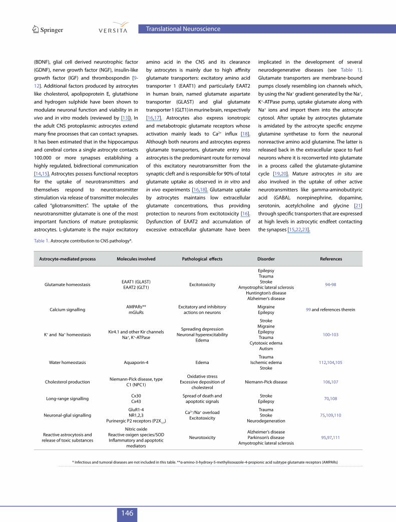
Astrocytes and neurons: the synaptic domain (Figure 1)
Astrocyte-neuron relationships start during development when astrocytes regulate neurogenesis by guiding and supporting neuronal migration, survival and process extension [8]. Astrocytes participate in the formation, maintenance and remodelling of synapses mainly through release of trophic factors such as brain-derived neurotrophic factor
Figure 1. Astrocyte-neuron relationships. Astrocytes are involved in the uptake and release of neurotransmitters, trophic factors and energy substrates for neurons and in the control of ion homeostasis. Astrocytes respond with Ca2+ elevations to neurotransmitters released during synaptic activity and, in turn, control neuronal excitability and synaptic transmission through the Ca2+-dependent release of gliotransmitters such as glutamate, ATP, GABA and D-serine. The uptake of the neurotransmitter glutamate from the synaptic cleft by astrocytes occurs via Na+-dependent excitatory amino acid transporters (EAATs). Glutamate is then converted into glutamine by glutamine synthetase (GS) and released back to neurons where it is converted to glutamate by glutaminase. Na+, K+-ATPase provides the Na+-mediated driving force for glutamate uptake. Trophic factors like BDNF, GDNF, NGF, IGF and thrombospondin, produced and released by astrocytes regulate synapse formation, maintenance and remodelling. Glutamatergic activation induces lactate release from astrocytes via monocarboxylate transporters (MCT). Astrocytes also control water and ion exchange in the synaptic cleft through water (AQP4) and ion (Kir4.1) channels and ion exchangers (Na+/H+ exchanger, Na+/Ca+2 exchanger). Kir4.1 is the main channel involved in potassium buffering in astrocytes. K+ ions travel through the astrocyte syncytium via gap junctions or are siphoned in the blood circulation. Carbonic anhydrase (CA) in astrocytes converts CO2 into H+ and HCO3-. Two HCO3- are transported into the extracellular space along with one Na+ via the Na+-HCO3- co-transporter (NBC), thereby increasing the extracellular ion buffering power. Excess H+ in neurons is extruded via the Na+/H+ exchanger (NHE).
Translational Neuroscience
146
(BDNF), glial cell derived neurotrophic factor (GDNF), nerve growth factor (NGF), insulin-like growth factor (IGF) and thrombospondin [9-12]. Additional factors produced by astrocytes like cholesterol, apolipoprotein E, glutathione and hydrogen sulphide have been shown to modulate neuronal function and viability in in vivo and in vitro models (reviewed by [13]). In the adult CNS protoplasmic astrocytes extend many fine processes that can contact synapses. It has been estimated that in the hippocampus and cerebral cortex a single astrocyte contacts 100.000 or more synapses establishing a highly regulated, bidirectional communication [14,15]. Astrocytes possess functional receptors for the uptake of neurotransmitters and themselves respond to neurotransmitter stimulation via release of transmitter molecules called “gliotransmitters”. The uptake of the neurotransmitter glutamate is one of the most important functions of mature protoplasmic astrocytes. L-glutamate is the major excitatory
amino acid in the CNS and its clearance by astrocytes is mainly due to high affinity glutamate transporters: excitatory amino acid transporter 1 (EAAT1) and particularly EAAT2 in human brain, named glutamate aspartate transporter (GLAST) and glial glutamate transporter 1 (GLT1) in murine brain, respectively [16,17]. Astrocytes also express ionotropic and metabotropic glutamate receptors whose activation mainly leads to Ca2+ influx [18]. Although both neurons and astrocytes express glutamate transporters, glutamate entry into astrocytes is the predominant route for removal of this excitatory neurotransmitter from the synaptic cleft and is responsible for 90% of total glutamate uptake as observed in in vitro and in vivo experiments [16,18]. Glutamate uptake by astrocytes maintains low extracellular glutamate concentrations, thus providing protection to neurons from excitotoxicity [16]. Dysfunction of EAAT2 and accumulation of excessive extracellular glutamate have been
implicated in the development of several neurodegenerative diseases (see Table 1). Glutamate transporters are membrane-bound pumps closely resembling ion channels which, by using the Na+ gradient generated by the Na+, K+-ATPase pump, uptake glutamate along with Na+ ions and import them into the astrocyte cytosol. After uptake by astrocytes glutamate is amidated by the astrocyte specific enzyme glutamine synthetase to form the neuronal nonreactive amino acid glutamine. The latter is released back in the extracellular space to fuel neurons where it is reconverted into glutamate in a process called the glutamate-glutamine cycle [19,20]. Mature astrocytes in situ are also involved in the uptake of other active neurotransmitters like gamma-aminobutityric acid (GABA), norepinephrine, dopamine, serotonin, acetylcholine and glycine [21] through specific transporters that are expressed at high levels in astrocytic endfeet contacting the synapses [15,22,23].
Astrocyte-mediated process molecules involved pathological effects disorder references
Glutamate homeostasis EAAT1 (GLAST)EAAT2 (GLT1) Excitotoxicity
EpilepsyTrauma Stroke
Amyotrophic lateral sclerosisHuntington’s diseaseAlzheimer’s disease
94-98
Calcium signalling AMPARs**mGluRs
Excitatory and inhibitory actions on neurons
MigraineEpilepsy 99 and references therein
K+ and Na+ homeostasis Kir4.1 and other Kir channelsNa+, K+-ATPase
Spreading depressionNeuronal hyperexcitability
Edema
Stroke MigraineEpilepsyTrauma
Cytotoxic edemaAutism
100-103
Water homeostasis Aquaporin-4 EdemaTrauma
Ischemic edemaStroke
112,104,105
Cholesterol production Niemann-Pick disease, type C1 (NPC1)
Oxidative stressExcessive deposition of
cholesterolNiemann-Pick disease 106,107
Long-range signalling Cx30Cx43
Spread of death and apoptotic signals
StrokeEpilepsy 70,108
Neuronal-glial signallingGluR1-4NR1,2,3
Purinergic P2 receptors (P2X1/7)
Ca2+/Na+ overloadExcitotoxicity
Trauma Stroke
Neurodegeneration75,109,110
Reactive astrocytosis and release of toxic substances
Nitric oxideReactive oxigen species/SODInflammatory and apoptotic
mediators
NeurotoxicityAlzheimer’s diseaseParkinson’s disease
Amyotrophic lateral sclerosis95,97,111
Table 1. Astrocyte contribution to CNS pathology*.
* Infectious and tumoral diseases are not included in this table. **α-amino-3-hydroxy-5-methylisoxazole-4-propionic acid subtype glutamate receptors (AMPARs)
Translational Neuroscience
147
Increasing experimental evidence indicates that gliotransmitters released from astrocytes actively participate in modulating synaptic activity. Astrocytes react to synaptically released neurotransmitters with intracellular calcium elevations, which in turn induce the regulated secretion of gliotransmitters, such as glutamate, adenosine triphosphate (ATP), GABA and D-serine [24], mainly through Ca2+-dependent vesicular release ([25] and references therein) but also via other mechanisms (reviewed by [26,27]). Release of gliotransmitters is also driven by a volume regulatory response stimulated by osmotic imbalance conditions following the release of osmotically active solutes from the neuronal cytoplasm into the extracellular space [27]. These findings have led to build-up a new model of neuron-glia inter-communication, the so called “tripartite synapse”, comprising the pre- and post-synaptic neurons and the astrocyte, where the latter integrates neuronal inputs and modulates synaptic activity [22]. Other soluble factors produced by astrocytes in vitro and in vivo, such as neurosteroids [28], growth factors and cytokines [29], can also influence synaptic activity.
Astrocytes also support brain activity by supplying neurons with energy substrates. They contribute to take up glucose, the primary source of energy for the brain, from the blood circulation and to deliver it to neurons. Apart from neurons which mainly rely on oxidative metabolism, astrocytes rely more on glycolytic metabolism to generate ATP and lactate from glucose [30,31]. Glucose enters astrocytes via specific glucose transporters (GLUT) and its degradation to lactate represents the main energy source for neurons, particularly during intense neuronal activity. In the “astrocyte-neuron lactate shuttle model” (ANSL) proposed by Magistretti and collaborators [32] astrocytes respond to glutamatergic activation by increasing the rate of glucose utilization and the production of lactate which is released in the extracellular space through monocarboxylate transporters to be taken up by neurons (reviewed by [33] and references therein). Neurons can use both lactate and glucose as energetic substrates, and the issue of lactate versus glucose as energetic
supply for neurons has been debated [34]. Moreover, the CNS astrocytes represent the main storage site for glycogen [35]. Up to 40% of glucose entering astrocytes is metabolised into glycogen molecules which can be rapidly mobilized without requirement of ATP to generate energy substrates, particularly lactate ([33] and references therein). In areas of intense synaptic density the accumulation of glycogen in astrocytes is increased and its utilization can sustain neuronal activity [35] but also buffer blood hypoglycaemia which disturbs cerebral metabolism and neuronal function [36,37]. The coupling between astrocyte glycogen accumulation and its mobilization during neuronal activity is sustained by the observation that neurotransmitters like glutamate can regulate glycogen release at synaptic sites ([33] and reference therein). The activity-dependent mechanisms of glucose utilization involve Na+-coupled glutamate uptake in astrocytes and the activation of the Na+, K+-ATPase which triggers glucose uptake from the blood and its processing [38].
Another important task of astrocytes is to optimize synapse functionality by maintaining the interstitial space homeostasis through a tight control of water and ion fluxes [39]. To this end, astrocytes are well equipped with ion channels and transporters for the uptake of K+ ions and for proton exchange, like the Na+/H+ exchanger, bicarbonate transporters, and the vacuolar type ATPase [40]. It is remarkable that most of the ATP produced within astrocytes is for cell pumping requirements to maintain ionic homeostasis [31]. Particularly important is the astrocyte-mediated control of K+ homeostasis since repetitive firing of action potentials in neurons induces a rise in extracellular K+ ions that can compromise neuronal function, if not quickly buffered in the space surrounding the synapses [41]. Studies in cultured cells have demonstrated that astrocytes have a much higher capacity for K+ uptake compared to neurons [42]. It was initially proposed that astrocytes, due to their high permeability to K+ ions, could favour passive uptake of K+ ions that would diffuse to distant sites through the glial syncytium, a process named K+ spatial buffering [41,43]. More recently using knock-out (KO) mouse models it has been demonstrated that
K+ spatial buffering, rather than being a passive process, is mediated by the activity of specific channels, particularly the inward-rectifying Kir4.1 channels [44] which are clustered in specific functional domains in the perisynaptic and perivascular astrocyte endfeet. Due to the weakly rectifying nature of Kir4.1 the same channel can drive both inward and outward K+ movements in astrocytes [45]. Other molecules like the Na+, K+-ATPase pump, co-transporters of the Slc12a family and chloride channels contribute to K+ buffering by astrocytes as observed in vitro and in vivo models [42,46-48].
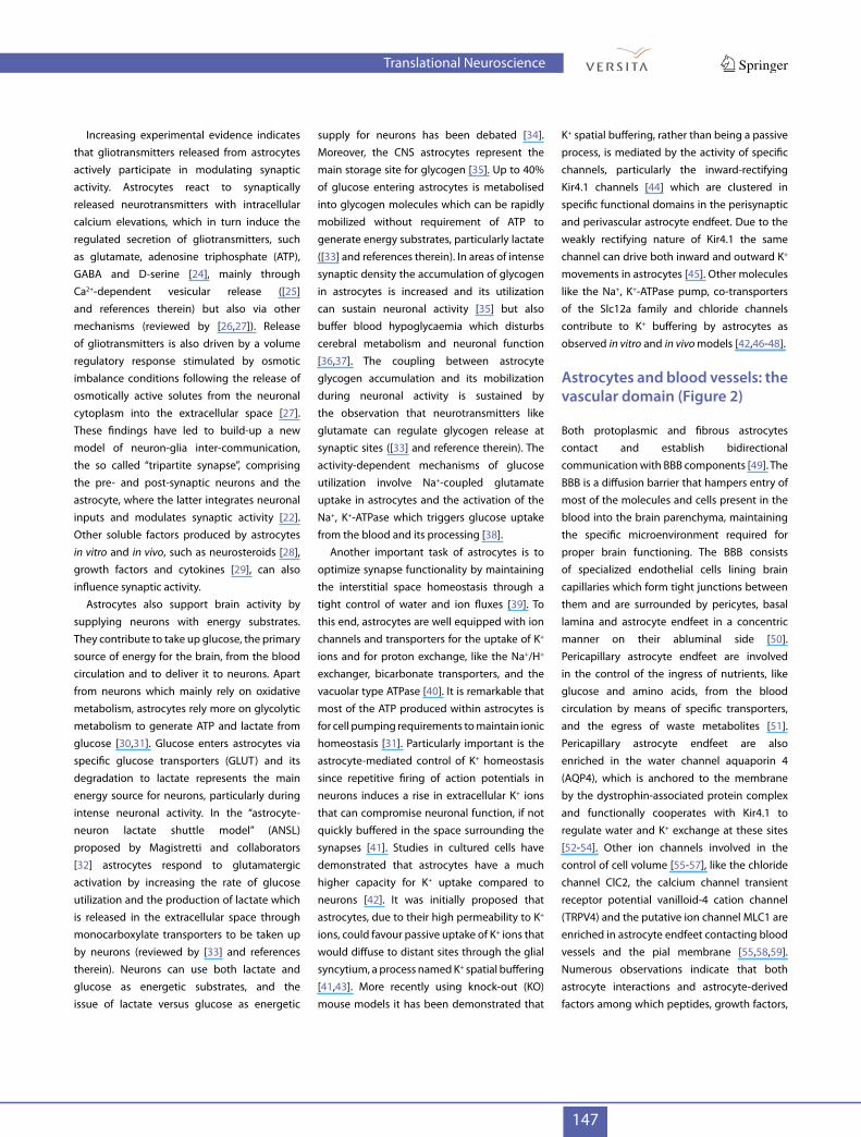
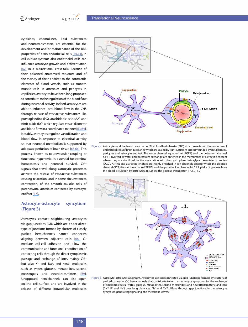
Astrocytes and blood vessels: the vascular domain (Figure 2)
Both protoplasmic and fibrous astrocytes contact and establish bidirectional communication with BBB components [49]. The BBB is a diffusion barrier that hampers entry of most of the molecules and cells present in the blood into the brain parenchyma, maintaining the specific microenvironment required for proper brain functioning. The BBB consists of specialized endothelial cells lining brain capillaries which form tight junctions between them and are surrounded by pericytes, basal lamina and astrocyte endfeet in a concentric manner on their abluminal side [50]. Pericapillary astrocyte endfeet are involved in the control of the ingress of nutrients, like glucose and amino acids, from the blood circulation by means of specific transporters, and the egress of waste metabolites [51]. Pericapillary astrocyte endfeet are also enriched in the water channel aquaporin 4 (AQP4), which is anchored to the membrane by the dystrophin-associated protein complex and functionally cooperates with Kir4.1 to regulate water and K+ exchange at these sites [52-54]. Other ion channels involved in the control of cell volume [55-57], like the chloride channel ClC2, the calcium channel transient receptor potential vanilloid-4 cation channel (TRPV4) and the putative ion channel MLC1 are enriched in astrocyte endfeet contacting blood vessels and the pial membrane [55,58,59]. Numerous observations indicate that both astrocyte interactions and astrocyte-derived factors among which peptides, growth factors,
Translational Neuroscience
148
cytokines, chemokines, lipid substances and neurotransmitters, are essential for the development and/or maintenance of the BBB properties of brain endothelial cells [60,61]. In cell culture systems also endothelial cells can influence astrocyte growth and differentiation [62] in a bidirectional cross-talk. Because of their polarized anatomical structure and of the vicinity of their endfeet to the contractile elements of blood vessels, such as smooth muscle cells in arterioles and pericytes in capillaries, astrocytes have been long proposed to contribute to the regulation of the blood flow during neuronal activity. Indeed, astrocytes are able to influence local blood flow in the CNS through release of vasoactive substances like prostaglandins (PG), arachidonic acid (AA) and nitric oxide (NO) which regulate vessel diameter and blood flow in a coordinated manner [63,64]. Notably, astrocytes regulate vasodilatation and blood flow in response to electrical activity so that neuronal metabolism is supported by adequate perfusion of brain tissue [65,66]. This process, known as neurovascular coupling or functional hyperemia, is essential for cerebral homeostasis and neuronal survival. Ca2+ signals that travel along astrocytic processes activate the release of vasoactive substances causing relaxation, and in some circumstances contraction, of the smooth muscle cells of parenchymal arterioles contacted by astrocyte endfeet [67].
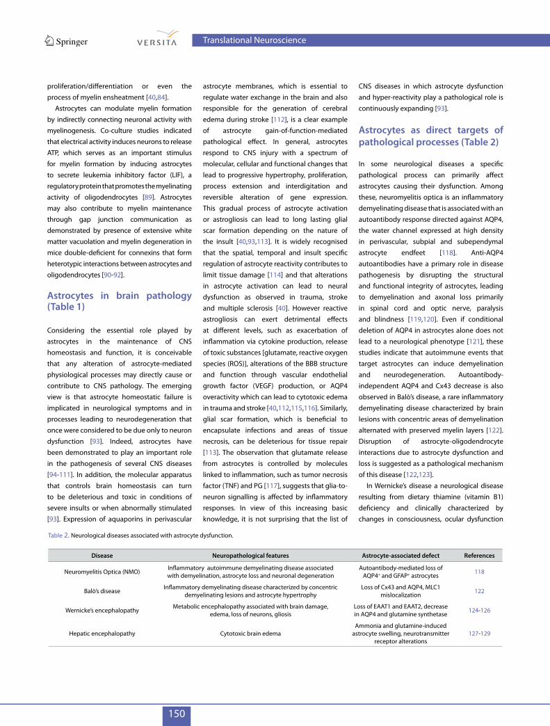
Astrocyte-astrocyte syncytium (Figure 3)
Astrocytes contact neighbouring astrocytes via gap junctions (GJ), which are a specialized type of junctions formed by clusters of closely packed hemichannels named connexins aligning between adjacent cells [68]. GJ mediate cell-cell adhesion and allow the communication and functional coordination of contacting cells through the direct cytoplasmic passage and exchange of ions, mainly Ca2+ but also K+ and Na+, and small molecules such as water, glucose, metabolites, second messengers and neurotransmitters [69]. Unopposed hemichannels can also open on the cell surface and are involved in the release of different intracellular molecules
Figure 2. Astrocytes and the blood brain barrier. The blood brain barrier (BBB) structure relies on the properties of endothelial cells of brain capillaries which are sealed by tight junctions and surrounded by basal lamina, pericytes and astrocyte endfeet. The water channel aquaporin-4 (AQP4) and the potassium channel Kir4.1 involved in water and potassium exchange are enriched in the membranes of astrocytic endfeet where they are stabilized by the association with the dystrophin-dystroglycan associated complex (DGC). At this site astrocyte endfeet are highly enriched in ion channels among which the chloride channel ClC2, the calcium channel TRPV4 and the putative ion channel MLC1. Uptake of glucose from the blood circulation by astrocytes occurs via the glucose transporter-1 (GLUT1).
Figure 3. Astrocyte-astrocyte syncytium. Astrocytes are interconnected via gap junctions formed by clusters of packed connexin (Cx) hemichannels that contribute to form an astrocyte syncytium for the exchange of small molecules (water, glucose, metabolites, second messengers and neurotransmitters) and ions (Ca+2, K+ and Na+) over long distances. Na+ and Ca+2 diffuse through gap junctions in the astrocyte syncytium generating signalling and metabolic waves.
Translational Neuroscience

149
into the extracellular space. In the CNS, glial cells express the highest level of connexins (connexins 30 and 43 representing the main astrocyte specific connexins), GJ channels and hemichannels ([69] and references therein). By means of GJ astrocytes form a network that is visualised experimentally by the injection of a dye in one cell and its subsequent spreading to adjacent cells. Astrocyte coupling generates a multicellular structural and functional network, considered as a syncytium, that is not only essential for physiological functions but may also play a role in CNS disorders [70]. Through this network astrocytes are thought to rapidly dissipate K+ and glutamate from highly active synapses to other brain areas or blood circulation, avoiding their harmful accumulation [71]. Passage of Ca2+ ions between adjacent astrocytes provides these cells with a specific form of excitability and represents the major way by which astrocytes encode and transmit information. During the propagation of Ca2+ waves several calcium-dependent pathways and biochemical cascades are activated which have functional consequences for astrocyte themselves and for neighbouring cells (for a detailed review see [72]). Connexins also allow direct contacts between astrocytes and oligodendrocytes (see below).
It has been shown that, in addition to Ca2+ waves, electrical or mechanical stimulation of cultured astrocytes induces metabolic waves that are mediated by Ca2+-dependent release of glutamate which in turn triggers a Na+ wave in the astrocyte network due to the activity of the Na+-dependent glutamate transporters [72,73]. The propagation of this signal is spatially and temporally connected with the activation of glucose uptake, which is also dependent on astrocyte glutamate transporters, thus allowing a concerted neurometabolic coupling between glucose utilization and neuronal activity [74]. The important role of Ca2+ and Na+ ions in regulating astrocyte function is demonstrated by the tight control of the intracellular concentration of these two ions exerted by several transporters and ionic channels or ion exchangers present in the astrocyte plasma membrane [75]. In light of these findings neuron-glia relationships should be considered not only at the single cell level, but as a
more complex array of interactions between neuronal and glial networks.
Astrocyte relationships with oligodendrocytes and myelin (Figure 4)
Astrocyte production of growth factors which promote neuronal survival in normal ([76], see above) and injured brain [77] is also essential for survival of oligodendrocytes, the myelin forming cells. The importance of astrocytes as regulators of oligodendrocyte proliferation and differentiation in vitro was recognized more than 20 years ago when astrocytes were identified as a major source of proliferation and differentiation factors, in particular platelet-derived growth factors (PDGFα) and fibroblast growth factor 2 (FGF2), for oligodendrocyte progenitor cells [78-80]. Subsequently, astrocyte-conditioned media and several astrocyte-derived soluble factors, like neurotrophin-3 (NT-3), IGF-1 and ciliary neurotrophic factor (CNTF), were found to support oligodendrocyte progenitor cell survival [81-83]. Although astrocytes have
long been considered the main players in the inhibition of CNS repair via formation of the glial scar (see below), it is now accepted that astrocytes regulate myelin formation and that understanding this process could help develop strategies for CNS repair and remyelination [84]. Astrocyte influence on myelination is supported by the observation that oligodendrocytes remyelinate preferentially in areas containing astrocytes and that endogenous remyelination can be favoured by transplanting astrocytes into demyelinated lesions [85,86]. In vitro studies using a myelinating culture system made of embryonic spinal cord cells plated on monolayers of astrocytes provided additional evidence that astrocytes, particularly when activated, efficiently support myelination by secreting pro-myelinating factors [87,88]. Moreover, depending on their activation status astrocytes can promote myelination by releasing cytokines and growth factors like BDNF, CTNF, GDNF, IGF, NGF, C-X-C motif chemokine 12 (CXCL12), PDGF, FGF and factors implicated in extracellular matrix remodelling which may affect oligodendrocyte progenitor
Figure 4. Astrocyte relationship with oligodendrocytes and myelin. Astrocytes produce several factors that contribute to oligodendrocyte progenitor cell (OPC) proliferation and differentiation, such as PDGFα and FGF2, and survival like NT-3, IGF-1, CNTF. Astrocytes also respond to ATP released by neurons by producing LIF which promotes the myelinating activity of oligodendrocytes. Astrocytes may also contribute to myelin maintenance via gap junction communication.
Translational Neuroscience
150
proliferation/differentiation or even the process of myelin ensheatment [40,84].
Astrocytes can modulate myelin formation by indirectly connecting neuronal activity with myelinogenesis. Co-culture studies indicated that electrical activity induces neurons to release ATP, which serves as an important stimulus for myelin formation by inducing astrocytes to secrete leukemia inhibitory factor (LIF), a regulatory protein that promotes the myelinating activity of oligodendrocytes [89]. Astrocytes may also contribute to myelin maintenance through gap junction communication as demonstrated by presence of extensive white matter vacuolation and myelin degeneration in mice double-deficient for connexins that form heterotypic interactions between astrocytes and oligodendrocytes [90-92].
Astrocytes in brain pathology (table 1)
Considering the essential role played by astrocytes in the maintenance of CNS homeostasis and function, it is conceivable that any alteration of astrocyte-mediated physiological processes may directly cause or contribute to CNS pathology. The emerging view is that astrocyte homeostatic failure is implicated in neurological symptoms and in processes leading to neurodegeneration that once were considered to be due only to neuron dysfunction [93]. Indeed, astrocytes have been demonstrated to play an important role in the pathogenesis of several CNS diseases [94-111]. In addition, the molecular apparatus that controls brain homeostasis can turn to be deleterious and toxic in conditions of severe insults or when abnormally stimulated [93]. Expression of aquaporins in perivascular
astrocyte membranes, which is essential to regulate water exchange in the brain and also responsible for the generation of cerebral edema during stroke [112], is a clear example of astrocyte gain-of-function-mediated pathological effect. In general, astrocytes respond to CNS injury with a spectrum of molecular, cellular and functional changes that lead to progressive hypertrophy, proliferation, process extension and interdigitation and reversible alteration of gene expression. This gradual process of astrocyte activation or astrogliosis can lead to long lasting glial scar formation depending on the nature of the insult [40,93,113]. It is widely recognised that the spatial, temporal and insult specific regulation of astrocyte reactivity contributes to limit tissue damage [114] and that alterations in astrocyte activation can lead to neural dysfunction as observed in trauma, stroke and multiple sclerosis [40]. However reactive astrogliosis can exert detrimental effects at different levels, such as exacerbation of inflammation via cytokine production, release of toxic substances [glutamate, reactive oxygen species (ROS)], alterations of the BBB structure and function through vascular endothelial growth factor (VEGF) production, or AQP4 overactivity which can lead to cytotoxic edema in trauma and stroke [40,112,115,116]. Similarly, glial scar formation, which is beneficial to encapsulate infections and areas of tissue necrosis, can be deleterious for tissue repair [113]. The observation that glutamate release from astrocytes is controlled by molecules linked to inflammation, such as tumor necrosis factor (TNF) and PG [117], suggests that glia-to-neuron signalling is affected by inflammatory responses. In view of this increasing basic knowledge, it is not surprising that the list of
CNS diseases in which astrocyte dysfunction and hyper-reactivity play a pathological role is continuously expanding [93].
Astrocytes as direct targets of pathological processes (table 2)
In some neurological diseases a specific pathological process can primarily affect astrocytes causing their dysfunction. Among these, neuromyelitis optica is an inflammatory demyelinating disease that is associated with an autoantibody response directed against AQP4, the water channel expressed at high density in perivascular, subpial and subependymal astrocyte endfeet [118]. Anti-AQP4 autoantibodies have a primary role in disease pathogenesis by disrupting the structural and functional integrity of astrocytes, leading to demyelination and axonal loss primarily in spinal cord and optic nerve, paralysis and blindness [119,120]. Even if conditional deletion of AQP4 in astrocytes alone does not lead to a neurological phenotype [121], these studies indicate that autoimmune events that target astrocytes can induce demyelination and neurodegeneration. Autoantibody-independent AQP4 and Cx43 decrease is also observed in Balò’s disease, a rare inflammatory demyelinating disease characterized by brain lesions with concentric areas of demyelination alternated with preserved myelin layers [122]. Disruption of astrocyte-oligodendrocyte interactions due to astrocyte dysfunction and loss is suggested as a pathological mechanism of this disease [122,123].
In Wernicke’s disease a neurological disease resulting from dietary thiamine (vitamin B1) deficiency and clinically characterized by changes in consciousness, ocular dysfunction
disease neuropathological features Astrocyte-associated defect references
Neuromyelitis Optica (NMO) Inflammatory autoimmune demyelinating disease associated with demyelination, astrocyte loss and neuronal degeneration
Autoantibody-mediated loss of AQP4+ and GFAP+ astrocytes 118
Balò’s disease Inflammatory demyelinating disease characterized by concentric demyelinating lesions and astrocyte hypertrophy
Loss of Cx43 and AQP4, MLC1 mislocalization 122
Wernicke’s encephalopathy Metabolic encephalopathy associated with brain damage, edema, loss of neurons, gliosis
Loss of EAAT1 and EAAT2, decrease in AQP4 and glutamine synthetase 124-126
Hepatic encephalopathy Cytotoxic brain edemaAmmonia and glutamine-induced
astrocyte swelling, neurotransmitter receptor alterations
127-129
Table 2. Neurological diseases associated with astrocyte dysfunction.
Translational Neuroscience
151
and ataxia, decrease of the astrocytic glutamate transporters EAAT1 and EAAT2 and disturbance of glutamatergic neurotransmission are considered the main cause of the loss of neurons, gliosis [124] and structural damage (lesions in thalamus and cortex) observed in patients [106]. Abnormalities in GABA transporters, GFAP, glutamine synthetase, and AQP4 have also been reported, which may result in brain edema, oxidative stress, inflammation and white matter damage [124-126]. Hepatic encephalopathy, an alteration of the CNS primarily caused by hepatic insufficiency, liver failure and cirrhosis, results in a decrease in ammonia detoxification and higher ammonia levels in the systemic circulation. Excessive ammonia entry into the brain leads to astrocyte swelling. Swelling impairs astrocyte homeostatic ability and predisposes to neuronal dysfunction and cytotoxic brain edema, the major cause of mortality [127]. Hepatic encephalopathy is considered as a primary astrocytopathy [128,129] since astrocytes are the only CNS cell type that can detoxify ammonia by glutamate to glutamine conversion.
Astrocyte dysfunction as primary cause of brain diseases: focus on cystic leukodystrophies (table 3)
The first example of a disease with a primary astrocyte defect is Alexander’s disease (AxD), a leukodystrophy in which mutations in the gene encoding the astrocyte intermediate
filament GFAP causes severe myelin alterations and neurodegeneration [130,131]. Leukodystrophies are a heterogeneous group of rare and untreatable genetic diseases characterised by defects in CNS myelin formation or maintenance. Most leukodystrophies manifest as early as in the first year of life or later in childhood or adolescence and represent an important cause of progressive mental and motor disability. Although considerable advances in the diagnosis have been made over the last decade due to improvement in magnetic resonance imaging (MRI) and gene sequencing technology, the definitive diagnosis of many leukodystrophies remains a challenge and requires combined clinical, genetic, biochemical and neuroradiological investigations [132]. Most of the clinically characterized leukodystrophies are still of unknown cause, while in some cases the gene mutation is known but the function of the mutated proteins or the pathogenetic mechanisms leading to myelin defects remain elusive [133]. Pathological mutations can target genes encoding myelin components or genes encoding proteins whose dysfunction indirectly leads to myelin damage. Different types of classification of these myelin disorders have been made based on either pathological defect, biochemical and genetic data or on the combination of histological and clinical criteria [134]. Depending on the type of defect observed, leukodystrophies have been classified into 2 main groups: i)
hypomyelinating leukodystrophies, in which a lower amount of myelin is observed due to defects in myelin production or to myelin poor quality, are generally caused by mutations in genes encoding myelin structural proteins, such as myelin proteolipid protein (PLP) [135]; and ii) demyelinating leukodystrophies in which myelin degenerates because of loss of some enzymatic activity essential for myelin biogenesis and maintenance, like peroxisomal or lysosomal enzymatic activities, or because of astrocyte dysfunctions that lead to progressive cystic or spongy (vacuolating) degeneration of myelin [135,136]. This latter group encompasses AxD, megalencephalic leukoencephalopathy with subcortical cysts and vanishing white matter disease, three leukodystrophies characterized by defects in genes expressed in astrocytes or associated with specific astrocyte function (Table 3). In the next section the role of astrocytes in these leukodystrophies is discussed. The clinical and neuropathological characterization of these diseases has been described in detail elsewhere [137-139].
Alexander’s disease (Axd)
AxD (OMIM 203450) is a degenerative neurological disorder that is inherited in an autosomal dominant manner and is caused by heterozygous mutations in the astrocyte specific type III intermediate filament GFAP gene [132]. About 95% of patients show
Table 3. Leukodystrophies caused by a primary astrocyte defect.
leukodystrophy gene defect Astrocyte-mediated pathological mechanisms
clinical and histopathological featuresshared among cystic leukodystrophies
Alexander’s disease (AxD)
Mutations in the glial fibrillary acidic protein (GFAP) gene
(130)
Accumulation of Rosenthal fibers, abnormal GFAP
polimerization, defects in proteasomal degradation and
authophagy processes, decrease in glutamate trasporters
Macrocephaly, ataxia, spasticity, epilepsy,aggravation of clinical conditions after minor head trauma and infections; limited genotype-phenotype correlation.
Swollen white matter, astrocyte vacuolation
Megalencephalic leukoencephalopathy with
subcortical cysts (MLC)
Mutations in the MLC1 or HEPACAM
gene(168,178)
Altered regulation of ion and fluid homeostasis and cell
volume
Macrocephaly, ataxia, spasticity, mild cognitive decline, severe motor dysfunctions, epilepsy, subcortical cysts,
aggravation of clinical conditions after minor head trauma. No genotype-phenotype correlation.
Swollen white matter, astrocyte vacuolation
Vanishing white matter syndrome (VWM) or childhood
ataxia with central nervous system hypomyelination (CACH)
Mutations in the Eukariotic Translation
Initiation Factor 2 (EIF-2B) gene (187)
Astrocyte maturation defect, abnormal response to stress
conditions
Ataxia, spasticity, loss of vision, severe motor dysfunctions, mild cognitive decline, epilepsy, aggravation of clinical
symptoms after minor stress conditions. Limited genotype-phenotype correlation. Swollen white matter
Translational Neuroscience
152
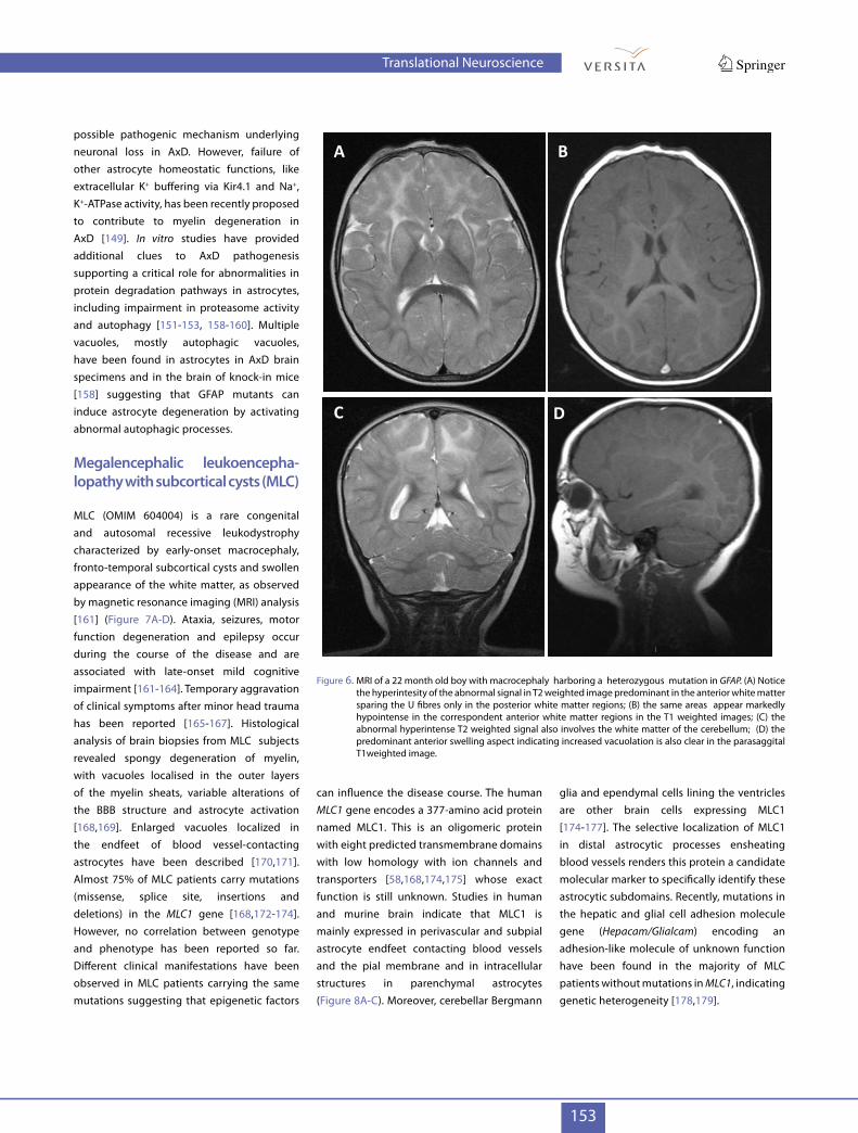
mutations, including de novo mutations, in the GFAP gene. Mutations can affect different parts of the GFAP protein, at either the NH2 or COOH terminal, with hot spot mutation areas [140]. Evidence of genotype-phenotype correlation in AxD is limited [141]. Three forms of the disease exist: infantile, juvenile and adult-onset. The infantile onset form is very aggressive and is characterized by seizures, bulbar dysfunction, psychomotor regression symptoms and low life expectancy [142]. When compared to a healthy control (Figure 5A-D) MRI analysis of AxD affected brain reveals macrocephaly and widespread abnormalities in the anterior white matter (Figure 6 A-D). The juvenile and adult forms of AxD are not associated with macrocephaly, have longer life expectancy and milder symptoms, white matter damage being less severe and sometimes absent. Aggravation of clinical symptoms can occur after trauma or inflammatory events [140,142-144].Histopathological studies performed on bioptic and autoptic brain samples indicate that pathological changes in the CNS of aggressive infantile forms include loss of oligodendrocytes, cystic degeneration and loss of myelin in the white matter and variable loss of neurons, most commonly in the hippocampus, striatum, and neocortex [145,146].
The pathological hallmark of AxD is the presence of Rosenthal fibers, that are ubiquitinated protein aggregates composed of GFAP, vimentin, small heat shock proteins (including αB-crystallin and Hsp27) and plectin, in the cytoplasm of astrocytes [147,148]. Since GFAP is not expressed in neurons or oligodendrocytes, myelin degeneration and oligodendrocyte loss in the AxD brain [149] are secondary to alterations in astrocyte functions [137]. A direct relationship between expression of disease-associated GFAP mutants, Rosenthal fiber accumulation in astrocytes and neurodegeneration was demonstrated first in mice carrying additional copies of the wild-type human GFAP gene and expressing elevated levels of GFAP protein, which replicated some features of human AxD, and in knock-in mice carrying the pathological gene mutations found in patients [149,150]. While these studies suggest that high levels of wild-type GFAP can reproduce the astrocyte
phenotype of AxD, studies performed in cultured astrocytes showed that mutant GFAP protein accumulates more rapidly and at higher levels than the wild-type protein [151,152]. Thus, the precise mechanism through which GFAP mutations lead to accumulation of Rosenthal fibers and the pathogenicity of the proteinaceous aggregates themselves are not completely understood. The dominant nature of pathological GFAP mutations, along with the accumulation of protein aggregates in brain astrocytes, have led to a hypothesis of a toxic gain-of-function pathological mechanism with impairment of normal astrocyte supportive
functions [153]. Other studies have suggested that astrocyte-mediated pathological effects in AxD are due to both oxidative stress and reduction in glial glutamate transporter function [154-156]. It was been shown that GLT-1 transcript and protein are markedly downregulated in astrocytes overexpressing mutant GFAP and that hippocampal neurons are more vulnerable to glutamate-induced excitotoxicity when co-cultured with astrocytes overexpressing mutant GFAP [157]. These observations connect GFAP mutations to GLT-1 dysfunction and impairment of neuron-astrocyte interactions, suggesting a
Figure 5. Normal MRI of a 4 year old boy. (A) Normal myelination in a T2 weighted image; (B) the same aspect in a FLAIR weighted image with normal relative hypointensity of the white matter; (C) the normal hypointense T2 weighted signal also involves the white matter of the cerebellum; (D) relative hyperintesity of the white matter is clear in the parasaggital T1 weighted image.
A B
C D
Translational Neuroscience
153
possible pathogenic mechanism underlying neuronal loss in AxD. However, failure of other astrocyte homeostatic functions, like extracellular K+ buffering via Kir4.1 and Na+, K+-ATPase activity, has been recently proposed to contribute to myelin degeneration in AxD [149]. In vitro studies have provided additional clues to AxD pathogenesis supporting a critical role for abnormalities in protein degradation pathways in astrocytes, including impairment in proteasome activity and autophagy [151-153, 158-160]. Multiple vacuoles, mostly autophagic vacuoles, have been found in astrocytes in AxD brain specimens and in the brain of knock-in mice [158] suggesting that GFAP mutants can induce astrocyte degeneration by activating abnormal autophagic processes.
megalencephalic leukoencepha-lopathy with subcortical cysts (mlc)
MLC (OMIM 604004) is a rare congenital and autosomal recessive leukodystrophy characterized by early-onset macrocephaly, fronto-temporal subcortical cysts and swollen appearance of the white matter, as observed by magnetic resonance imaging (MRI) analysis [161] (Figure 7A-D). Ataxia, seizures, motor function degeneration and epilepsy occur during the course of the disease and are associated with late-onset mild cognitive impairment [161-164]. Temporary aggravation of clinical symptoms after minor head trauma has been reported [165-167]. Histological analysis of brain biopsies from MLC subjects revealed spongy degeneration of myelin, with vacuoles localised in the outer layers of the myelin sheats, variable alterations of the BBB structure and astrocyte activation [168,169]. Enlarged vacuoles localized in the endfeet of blood vessel-contacting astrocytes have been described [170,171]. Almost 75% of MLC patients carry mutations (missense, splice site, insertions and deletions) in the MLC1 gene [168,172-174].However, no correlation between genotype and phenotype has been reported so far. Different clinical manifestations have been observed in MLC patients carrying the same mutations suggesting that epigenetic factors
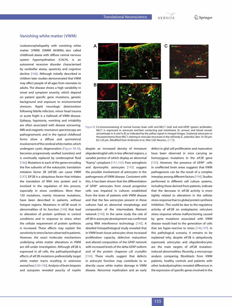
can influence the disease course. The human MLC1 gene encodes a 377-amino acid protein named MLC1. This is an oligomeric protein with eight predicted transmembrane domains with low homology with ion channels and transporters [58,168,174,175] whose exact function is still unknown. Studies in human and murine brain indicate that MLC1 is mainly expressed in perivascular and subpial astrocyte endfeet contacting blood vessels and the pial membrane and in intracellular structures in parenchymal astrocytes (Figure 8A-C). Moreover, cerebellar Bergmann
glia and ependymal cells lining the ventricles are other brain cells expressing MLC1 [174-177]. The selective localization of MLC1 in distal astrocytic processes ensheating blood vessels renders this protein a candidate molecular marker to specifically identify these astrocytic subdomains. Recently, mutations in the hepatic and glial cell adhesion molecule gene (Hepacam/Glialcam) encoding an adhesion-like molecule of unknown function have been found in the majority of MLC patients without mutations in MLC1, indicating genetic heterogeneity [178,179].
Figure 6. MRI of a 22 month old boy with macrocephaly harboring a heterozygous mutation in GFAP. (A) Notice the hyperintesity of the abnormal signal in T2 weighted image predominant in the anterior white matter sparing the U fibres only in the posterior white matter regions; (B) the same areas appear markedly hypointense in the correspondent anterior white matter regions in the T1 weighted images; (C) the abnormal hyperintense T2 weighted signal also involves the white matter of the cerebellum; (D) the predominant anterior swelling aspect indicating increased vacuolation is also clear in the parasaggital T1weighted image.
A B
C D
Translational Neuroscience
154
In cultured human and rat astrocytes MLC1 is localized along the plasma membrane, at astrocyte-astrocyte contacts and in many intracellular vesicles [170,180,181]. As for GFAP in AxD, MLC1 is an astrocyte specific protein not found in oligodendrocytes [174-176], indicating that also in MLC myelin degeneration is secondary to astrocyte dysfunction. To date the molecular mechanisms causing MLC-associated brain damage are not completely understood. Swollen white matter, fluid cysts and myelin vacuolation observed in the MLC brain and preferential localization of MLC1 at the brain barriers has led to suggest that MLC1 is involved in astrocyte-mediated regulation of brain homeostasis through transport of water and/or ions between astrocytes and the blood or cerebrospinal fluid. Consistent with this hypothesis we have recently shown that in cultured astrocytes MLC1 interacts with the b subunit of the Na+, K+-ATPase complex and is part of a macromolecular protein complex that includes Kir4.1, AQP4, syntrophin, dystrobrevin, caveolin-1 and the cation channel TRPV4. This complex is involved in the cellular response to hyposmotic stress in primary rat astrocytes and human astrocytoma cells overexpressing MLC1 [182,183]. We have shown that MLC1 functionally cooperates with TRPV4 to activate intracellular Ca2+ influx in astrocytes in hyposmotic conditions and that pathological MLC1 mutations affect this pathway [183]. TRPV4-mediated calcium influx is the trigger leading to activation of the regulatory volume decrease (RVD), which compensates hyposmotic stress-induced cell swelling, including astrocyte swelling [184,185]. Hyposmotic shock is known to cause an abrupt, osmotic driven cell swelling due to ion and water movement across the plasmalemma, which is followed by RVD activation. Together with the observation that ATP induces abnormal Ca2+ currents in patient-derived macrophages (Petrini S. et al., manuscript under revision) the data reported above, highlight the possibility that MLC1 mutations alter intracellular Ca2+ homeostasis which then leads to defects in cell volume regulation, particularly in stress conditions. Defects in a RVD-induced chloride current have been reported also in rat astrocytes upon siRNA-mediated MLC1
downregulation and in MLC patient-derived lymphoblastoid cell lines after hyposmotic stimulation [56]. Altogether, these findings corroborate the idea that mutated MLC1 causes an altered reaction of astrocytes to osmotic changes, which accounts for brain damage in MLC. This model implies that MLC1 exerts its functions mainly in response to physiological changes in the extracellular ionic composition (i.e. during development or intense neuronal activity) or to pathological insults (i.e. trauma or inflammatory reaction). In this respect, we have observed upregulation of MLC1 protein
expression in astrocytes in the brain of patients with the inflammatory demyelinating disease multiple sclerosis, [182] and other neurological diseases with a major microglia activation (Alzheimer’s disease, prion disease) (Sbriccoli, Ambrosini - unpublished data). The recent finding that the Hepacam/Glialcam protein is essential to transport MLC1 and the chloride channel ClC2, another channel involved in the regulation of cell volume in response to osmotic stress [57,179] in cultured rat astrocytes, supports the idea that alteration of this process is responsible for MLC pathogenesis.
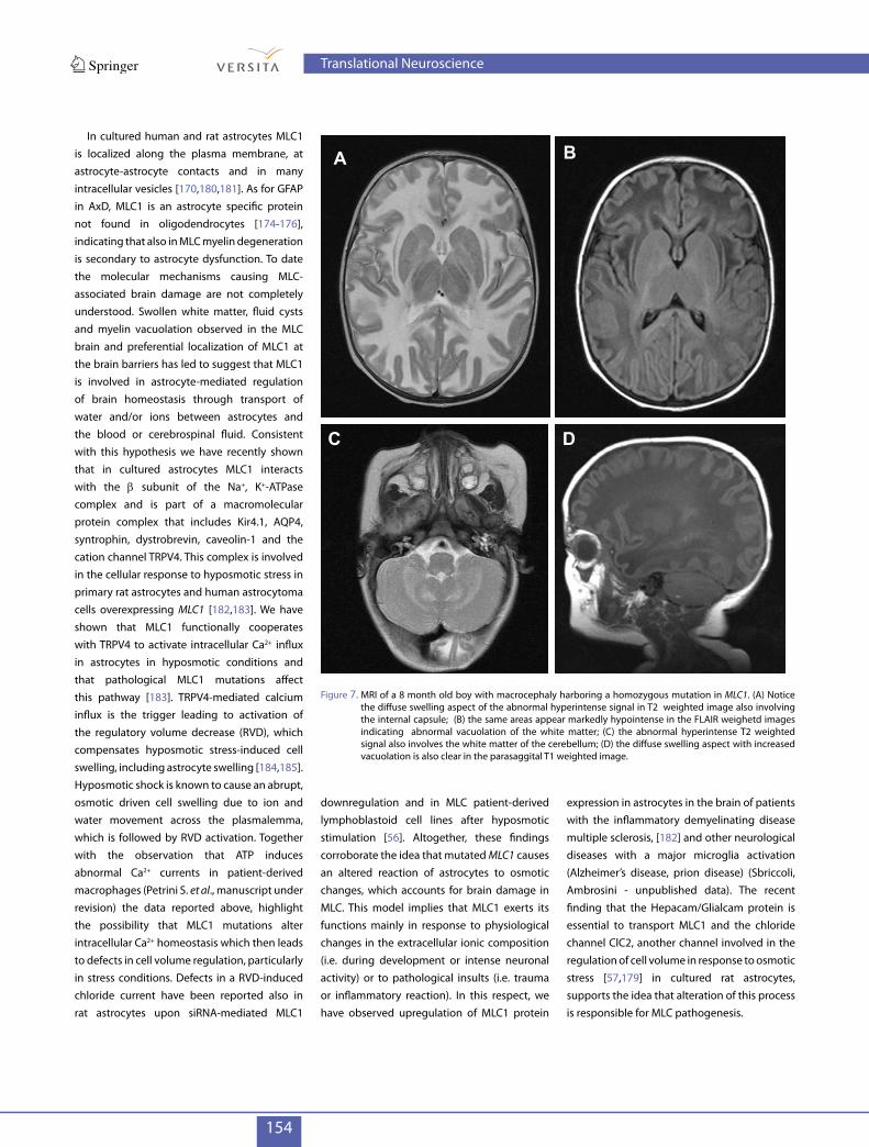
Figure 7. MRI of a 8 month old boy with macrocephaly harboring a homozygous mutation in MLC1. (A) Notice the diffuse swelling aspect of the abnormal hyperintense signal in T2 weighted image also involving the internal capsule; (B) the same areas appear markedly hypointense in the FLAIR weighetd images indicating abnormal vacuolation of the white matter; (C) the abnormal hyperintense T2 weighted signal also involves the white matter of the cerebellum; (D) the diffuse swelling aspect with increased vacuolation is also clear in the parasaggital T1 weighted image.
A B
C D
Translational Neuroscience
155
Vanishing white matter (VWm)
Leukoencephalopathy with vanishing white matter (VWM) (OMIM 603896), also called childhood ataxia with diffuse central nervous system hypomyelination (CACH), is an autosomal recessive disorder characterized by cerebellar ataxia, spasticity and cognitive decline [186]. Although initially described in children later studies demonstrated that VWM may affect people of all ages from neonates to adults. The disease shows a high variability in onset and symptom severity, which depend on patient specific gene mutations, genetic background and exposure to environmental stressors. Rapid neurologic deterioration following febrile infection, minor head trauma or acute fright is a hallmark of VWM disease. Epilepsy, hypotonia, vomiting and irritability are often associated with disease worsening. MRI and magnetic resonance spectroscopy are pathognomonic and in the typical childhood forms show a diffuse and symmetrical involvement of the cerebral white matter, which undergoes cystic degeneration (Figure 9A-D),becomes progressively rarefied (vanishes) and is eventually replaced by cerebrospinal fluid [186]. Mutations in each of the genes encoding the five subunits of the eukaryotic translation initiation factor 2B (eIF2B) can cause VWM [187]. EIF2B is a ubiquitous factor that initiates the translation of RNA into protein and is involved in the regulation of this process, especially in stress conditions. More than 120 mutations, mainly missense mutations, have been described in patients, without hotspot regions. Mutations in eIF2B result in abnormalities of its function [188] that lead to alteration of protein synthesis in control conditions and in response to stress when the cellular requirement of protein synthesis is increased. These effects may explain the sensitivity to stress factors observed in patients. However, the exact molecular mechanisms underlying white matter alterations in VWM are still under investigation. Although eIF2B is expressed in all cells, the pathophysiological effects of eIF2B mutations preferentially target white matter tracts resulting in extensive axonal loss [188-190]. Analysis of brain biopsies and autopsies revealed paucity of myelin
despite an increased density of immature oligodendroglial cells in less affected regions, a variable portion of which display an abnormal “foamy” cytoplasm [191,192]. Poor astrogliosis and dysmorphic astrocytes [193] suggest the possible involvement of astrocytes in the pathogenesis of VWM disease. Consistent with this, it has been shown that the differentiation of GFAP+ astrocytes from neural progenitor cells was impaired in cultures established from the brain of patients with VWM disease and that the few astrocytes present in these cultures had an abnormal morphology and composition of the intermediate filament network [190]. In the same study the role of eIF2B in astrocyte development was confirmed using RNA interference technology [190]. A detailed histopathological study revealed that in VWM brain tissue astrocytes show increased proliferation capacity, defective maturation and altered composition of the GFAP network with increased levels of the delta GFAP isoform and of the protein chaperon aB crystallin [194]. These results suggest that defects in astrocyte function may contribute to or directly cause white matter damage in VWM disease. Abnormal myelination and an early
defect in glial cell proliferation and maturation have been observed in mice carrying an homozygous mutations in the eIF2B gene [195]. However, the presence of GFAP+ cells in unaffected brain areas suggests that VWM pathogenesis can be the result of a complex interplay among different factors [194]. Studies performed in different cell culture systems, including those derived from patients, indicate that the decrease in eIF2B activity is more tightly related to alterations in the cellular stress response than to global protein synthesis inhibition. This could be due to the regulatory activity of eIF2B on endoplasmic reticulum stress response whose malfunctioning caused by gene mutations associated with VWM disease would lead to the generation of cells that are hyper-reactive to stress [196,197]. In this pathological scenario, it remains to be explained why, despite eIF2B is ubiquitously expressed, astrocytes and oligodendrocytes are the main targets of eIF2B mutation-induced abnormalities. Recently, a microarrays analysis comparing fibroblasts from VWM patients, healthy controls and patients with other leukodystrophies revealed differences in the expression of specific genes involved in the
Figure 8. Co-immunostaining of normal human brain with anti-MLC1 (red) and anti-GFAP (green) antibodies. MLC1 is expressed in astrocyte end-feet contacting pial membrane (A, arrows) and blood vessels (arrowheads in A and in B) as indicated by the yellow signal in merged images. Scattered astrocytes in the parenchyma show MLC1 staining in vesicular structures in the cell body (C, asterisks). Bars A=50 µm; B,C=20 µm. (Modified from Ambrosini et al., Mol. Cell. Neurosc., [177]).
Translational Neuroscience
156
regulation of mRNA splicing and mitochondrial metabolism. In the same study splicing dysregulation of genes important for glial cell maturation, like PLP in oligodendrocytes and GFAP in astrocytes, were observed in eIF2B mutated fetal brains compared with control brains [136]. Another recent study showed in the brain of VWM patients accumulation of the high molecular weight extracellular matrix component hyaluronan [198], which inhibits oligodendrocyte and astrocyte progenitor cell differentiation [198-200], further supporting the pathogenetic importance of abnormalities in glial cell maturation processes in this disease.
lesson from cystic leukodystrophies
The study of cystic leukodystrophies has revealed that failure of astrocyte functions due to specific gene mutations can lead to myelin degeneration. Although the molecular mechanisms underlying these diseases are not completely understood, it is now established that defects in astrocyte maturation, astrocyte functional impairment due to accumulation of toxic substrates, and/or failure of specific astrocyte-mediated homeostatic pathways can affect myelin formation and maintenance
leading to cystic or spongiform myelin degeneration. Dysfunctional astrocytes could affect myelination also by acting on oligodendrocyte cell development. Whatever the mechanism leading to the astrocyte defect, an important concept that emerges from the study of leukodystrophies is that loss of astrocyte functionality can make the brain more vulnerable to changes in tissue homeostasis that are caused by different types of pathological insults and stress conditions, thus aggravating tissue damage. Indeed aggravation of clinical symptoms is commonly observed in patients affected by cystic leukodystrophies after trauma or febrile episodes, all conditions known to perturb brain homeostasis ([201] and reference therein, [202,203]). This body of evidence suggests that in disorders caused by a primary astrocyte defect environmental factors perturbing CNS homeostasis can modify the clinical course of the disease independently of the type of mutation, thus hampering genotype-phenotype correlations.
An interesting hypothesis is that brain damage in genetic leukodystrophies could be the result of failure of some astrocyte-mediated regulatory pathways influencing glial cell homeostatic control during brain development [204]. Further investigations on the pathogenesis of astrocyte-mediated leukodystrophies and the availability of new disease models are needed to elucidate whether gene-environmental interactions during development are modulated at the astrocyte level and how genetic defects in astrocytes can affect CNS developmental processes.
concluding remarks and perspectives
During the past 20 years it has become clear that astrocytes exert complex and essential functions in the healthy CNS including regulation of synaptic transmission and information processing by neuronal circuits. Recently, unexpected roles of astrocytes in higher brain functions such as learning and memory, sleep behaviour and regulation of breathing, have been described [205-207].
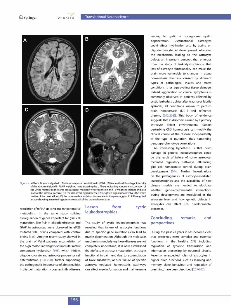
Figure 9. MRI of a 14 year old girl with 2 heterocompound mutations in eIF2Be. (A) Notice the diffuse hypointensity of the abnormal signal in FLAIR weighted image sparing the U fibers indicating abnormal vacuolation of the white matter; (B) the same areas appear markedly hyperintense in the T2 weighted images and also involve the internal capsule; (C) the abnormal hyperintense T2 weighted signal also involves the white matter of the cerebellum; (D) the increased vacuolation is also clear in the parasaggital FLAIR weighted image showing a marked hypointense signal of the brain white matter.
A B
C D
Translational Neuroscience
157
The speculative concept that astrocytes contribute to the pathogenesis of neurodegenerative diseases is turning into reality as strong evidence proves astrocyte involvement in Alzheimer’s disease, amyotrophic lateral sclerosis, Huntington’s disease, cerebral edema and stroke, as a consequence of either loss or gain of astrocyte function [7,208]. However, it is also clear that these disorders arise from a complex combination of abnormalities in neurons, glial cells and immune system cells that we are only beginning to understand. The identification of the genetic defect and the elucidation of the pathogenetic mechanism underlying AxD and other cystic leukodystrophies, has opened a new avenue for understanding the impact that specific astrocyte functional impairment has on myelin and neuron degeneration. Advancing knowledge in this field may have significant implications for the development of therapeutic strategies for these rare leukodystrophies and other common CNS diseases. The majority of drugs currently used for brain diseases target neuronal proteins like receptors, channels or transporters. Therapeutic strategies may benefit by a stronger focus on the homeostatic control functions of astrocytes. Although
some complex aspects of astrocyte physiology must be taken into account, such as regional differences in astrocyte phenotypes indicating that these cells represent a heterogeneous cell population [209,210], astrocytes are emerging as new potential pharmacological targets for CNS diseases [211]. The dissection of the molecular mechanisms of astrocyte reactivity is allowing identification of specific molecules whose function can be blocked or stimulated for therapeutic purposes [212,214]. The development of new technologies allows generating glial cells, including astrocytes, from embryonic stem cells or from the reprogrammed induced pluripotent stem cells (iPSCs) derived from skin fibroblasts [215-217]. These advancements have paved the way for the generation of easily accessible human cellular models to study astrocyte role in CNS development and the pathological mechanisms of astrocyte-mediated diseases [218,219]. These technologies are also providing new opportunities for regenerative therapies involving transplantation of astrocytes or glial progenitor cells [219,220-225]. Transplanted progenitor cells differentiating into astrocytes were reported to improve disease outcome in a mouse model of amyotrophic lateral
sclerosis characterized by abnormal astrocytes overexpressing mutant superoxide dismutase (SOD), thus demonstrating the feasibility and efficacy of transplantation-based astrocyte replacement for this disease [225]. Transplantation of astrocytes genetically engineered to produce therapeutic molecules is another strategy under investigation [221,223]. Recently, cortical human astrocytes have been dedifferentiated into cells with a neural stem/progenitor cell phenotype, indicating that restoration of multipotency from human astrocytes can be exploited for the reprogramming of endogenous CNS cells in neurological disorders [226]. A better knowledge of astrocyte contribution to genetic leukodystrophies will help develop astrocyte-based therapeutic strategies for these rare pathologies and for other neurological diseases.
Acknowledgments
We apologize to those authors whose original work could not be referenced due to space limitations. This work has been supported by Telethon (grant n. GGP11188B) and ELA Foundation (grant n. 2009-002C5). AL is the recipient of an ELA foundation fellowship (grant n. 2012-021F2).
References
[1] Virchow R., Cellular pathology as based upon physiological and pathological histology, translated from German by Chance B., 1859, 2nd ed., reproduced by Dover Publications, New York, 1971, 356-382
[2] Golgi C., Sulla struttura della sostanza grigia del cervello (comunicazione preventiva), Gazzetta Medica Italiana, Lombardia, 1873, 33, 244-246
[3] Golgi C., Opera omnia, Hoepli, Milano, 1903[4] Ramon Y., Cajal S., Histologie du systeme nerveux de l’homme et des
vertebres, Maloine, Paris, 1909[5] Peters A., Palay S.L., Webster H.F., The fine structure of the
nervous system: the neurons and supporting cells, W.B. Saunders, Philadelphia, 1976, 232-248
[6] Bignami A., Stoolmiller A.C., Astroglia-specific protein (GFA) in clonal cell lines derived from the G26 mouse glioma, Brain Res., 1979, 163, 353-357
[7] De Keyser J., Mostert J.P., Koch M.W. J., Dysfunctional astrocytes as key players in the pathogenesis of central nervous system disorders, Neurol. Sci., 2008, 267, 3-16
[8] Powell E.M., Geller H.M., Dissection of astrocyte-mediated cues in neuronal guidance and process extension, Glia, 1999, 26, 73-83
[9] Zaheer A., Zhong W., Uc E.Y., Moser D.R., Lim R., Expression of mRNAs of multiple growth factors and receptors by astrocytes and glioma cells: detection with reverse transcription-polymerase chain reaction, Cell. Mol. Neurobiol., 1995, 15, 221-37
[10] Ullian E.M., Sapperstein S.K., Christopherson K.S., Barres B.A., Control of synapse number by glia, Science, 2001, 291, 657-661
[11] Christopherson K.S., Ullian E.M., Stokes C.C., Mullowney C.E., Hell J.W., Agah A., et al., Thrombospondins are astrocyte-secreted proteins that promote CNS synaptogenesis, Cell, 2005, 120, 421-433
[12] Stevens B., Allen N.J., Vazquez L.E., Howell G.R., Christopherson K.S., Nouri N., et al., The classical complement cascade mediates CNS synapse elimination, Cell, 2007, 131, 1164-1178
[13] Allaman I., Belanger M., Magistretti P.J., Astrocyte-neuron metabolic relationships: for better and worse, Trends Neurosci., 2011, 34, 75-87
[14] Bushong E.A., Martone M.E., Jones Y.Z., Ellisman M.H., Protoplasmic astrocytes in CA1 stratum radiatum occupy separate anatomical domains, J. Neurosci., 2002, 22, 183-192
Translational Neuroscience
158
[15] Halassa M.M., Fellin T., Takano H., Dong J.H., Haydon P.G., Synaptic islands defined by the territory of a single astrocyte, J. Neurosci., 2007, 27, 6473-6477
[16] Rothstein J.D., Dykes-Hoberg M., Pardo C.A., Bristol L.A., Jin L., Kuncl R.W., et al., Knockout of glutamate transporters reveals a major role for astroglial transport in excitotoxicity and clearance of glutamate, Neuron, 1996, 16, 675-686
[17] Rauen T., Taylor W.R., Kuhlbrodt K., Wiessner M., High-affinity glutamate transporters in the rat retina: a major role of the glial glutamate transporter GLAST-1 in transmitter clearance, Cell Tissue Res., 1998, 291, 19-31
[18] Verkhratsky A., Kirchhoff F., NMDA receptors in glia, Neuroscientist, 2007, 13, 28-37
[19] Bak L.K., Schousboe A., Waagepetersen H.S., The glutamate/GABA-glutamine cycle: aspects of transport, neurotransmitter homeostasis and ammonia transfer, J. Neurochem., 2006, 98, 641-53
[20] McKenna M.C., The glutamate-glutamine cycle is not stoichiometric: fates of glutamate in brain, J. Neurosci. Res., 2007, 85, 3347-3358
[21] Kimelberg H.K., Receptors on astrocytes - what possible functions?, Neurochem. Int., 1995, 26, 27-40
[22] Perea G., Navarrete M., Araque A., Tripartite synapses: astrocytes process and control synaptic information, Trends Neurosci., 2009, 32, 421-431
[23] Shigetomi E., Bowser D.N., Sofroniew M.V., Khakh B.S., Two forms of astrocyte calcium excitability have distinct effects on NMDA receptor-mediated slow inward currents in pyramidal neurons, J. Neurosci., 2008, 28, 6659-6663
[24] Haydon P.G., Carmignoto G., Astrocyte control of synaptic transmission and neurovascular coupling, Physiol. Rev., 2006, 86,1009-1031
[25] Zorec R., Araque A., Carmignoto G., Haydon P.G., Verkhratsky A., Parpura V., Astroglial excitability and gliotransmission: an appraisal of Ca2+ as a signalling route, ASN Neuro., 2012, 4, e00080
[26] Malarkey E.B., Parpura V., Mechanisms of glutamate release from astrocytes, Neurochem. Int., 2008, 52, 142-1454
[27] Evanko D.S., Zhang Q., Zorec R., Haydon P.G., Defining pathways of loss and secretion of chemical messengers from astrocytes, Glia, 2004, 47, 233-240
[28] Garcia-Segura L.M., Melcangi R.C., Steroids and glial cell function, Glia, 2006, 54,485-498
[29] Stellwagen D., Malenka R.C., Synaptic scaling mediated by glial TNF-alpha, Nature, 2006, 440, 1054-1059
[30] Abi-Saab W.M., Maggs D.G., Jones T., Jacob R., Srihari V., Thompson J., et al., Striking differences in glucose and lactate levels between brain extracellular fluid and plasma in conscious human subjects: effects of hyperglycemia and hypoglycemia, J. Cereb. Blood Flow Metab., 2002, 22, 271-279
[31] Turner D.A., Adamson D.C., Neuronal-astrocyte metabolic interactions: understanding the transition into abnormal astrocytoma metabolism, J. Neuropathol. Exp. Neurol., 2011, 70, 167-176
[32] Pellerin L., Magistretti P.J., Sweet sixteen for ANLS, J. Cereb. Blood Flow. Metab., 2012, 32, 1152-1166
[33] Bélanger M., Allaman I., Magistretti P.J., Brain energy metabolism: focus on astrocyte-neuron metabolic cooperation, Cell Metab., 2011, 14, 724-738
[34] Dienel G.A., Brain lactate metabolism: the discoveries and the controversies, J. Cereb. Blood Flow Metab., 2012, 32, 1107-1138
[35] Brown A.M., Baltan Tekkok S., Ransom B.R., Energy transfer from astrocytes to axons: the role of CNS glycogen, Neurochem. Int., 2004, 45, 529-536
[36] Tsacopoulos M., Magistretti P.J., Metabolic coupling between glia and neurons, J. Neurosci., 1996, 16, 877-885
[37] Amaral A.I., Effects of hypoglycaemia on neuronal metabolism in the adult brain: role of alternative substrates to glucose, J. Inherit. Metab. Dis., 2012, [Epub ahead of print] doi: 10.1007/s10545-012-9553-3
[38] Magistretti P.J., Pellerin L., Cellular bases of brain energy metabolism and their relevance to functional brain imaging: evidence for a prominent role of astrocytes, Cereb.Cortex., 1996, 6, 50-61
[39] Kimelberg H.K., Nedergaard M., Functions of astrocytes and their potential as therapeutic targets, Neurotherapeutics, 2010, 7, 338-353
[40] Sofroniew M.V., Vinters H.V., Astrocytes: biology and pathology, Acta Neuropathol., 2010, 119, 7-35
[41] Gardner-Medwin A.R., Analysis of potassium dynamics in mammalian brain tissue, J. Physiol., 1983, 335, 393-426
[42] Walz W., Role of astrocytes in the clearance of excess extracellular potassium, Neurochem. Int., 2000, 36, 291-300
[43] Kofuji P., Newman E.A., Potassium buffering in the central nervous system, Neuroscience, 2004, 129, 1045-1056
[44] Djukic B., Casper K.B., Philpot B.D., Chin L.S., McCarthy K.D., Conditional knock-out of Kir4.1 leads to glial membrane depolarization, inhibition of potassium and glutamate uptake, and enhanced short-term synaptic potentiation, J. Neurosci., 2007, 27, 11354-1165
[45] Butt A.M., Kalsi A., Inwardly rectifying potassium channels (Kir) in central nervous system glia: a special role for Kir4.1 in glial functions, J. Cell. Mol. Med., 2006, 10, 33-44
[46] D’Ambrosio R., Gordon D.S., Winn H.R., Differential role of KIR channel and Na(+)/K(+)-pump in the regulation of extracellular K(+) in rat hippocampus, J. Neurophysiol., 2002, 87, 87-102
[47] Wang D.D., Bordey A., The astrocyte odyssey, Prog. Neurobiol., 2008, 86, 342-367
[48] Amzica F., Massimini M., Glial and neuronal interactions during slow wave and paroxysmal activities in the neocortex, Cereb. Cortex., 2002, 12, 1101-1113
[49] Peters A., Palay S.L., Webster H.D., The fine structure of the nervous system, 3rd ed., Oxford University Press, New York, 1991
[50] Abbott N.J., Ronnback L., Hansson E., Astrocyte-endothelial interactions at the blood-brain barrier, Nat. Rev. Neurosci., 2006, 7, 41-53
[51] Pellerin L., Magistretti P.J., Neuroenergetics: calling upon astrocytes to satisfy hungry neurons, Neuroscientist, 2004, 10, 53-62
[52] Connors N.C., Adams M.E., Froehner S.C., Kofuji P., The potassium channel Kir4.1 associates with the dystrophin-glycoprotein complex via alpha-syntrophin in glia, J. Biol. Chem., 2004, 279, 28387-28392
Translational Neuroscience

159
[53] Dalloz C., Sarig R., Fort P., Yaffe D., Bordais A., Pannicke T., et al., Targeted inactivation of dystrophin gene product Dp71: phenotypic impact in mouse retina, Hum. Mol. Genet., 2003, 12, 1543-54
[54] Amiry-Moghaddam M., Ottersen O.P., The molecular basis of water transport in the brain, Nat. Rev. Neurosci., 2003, 4, 991-1001
[55] Benfenati V., Amiry-Moghaddam M., Caprini M., Mylonakou M.N., Rapisarda C., Ottersen O.P., et al., Expression and functional characterization of transient receptor potential vanilloid-related channel 4 (TRPV4) in rat cortical astrocytes, Neuroscience, 2007, 148, 876-892
[56] Ridder M.C., Boor I., Lodder J.C., Postma N.L., Capdevila-Nortes X., Duarri A., et al., Megalencephalic leucoencephalopathy with cysts: defect in chloride currents and cell volume regulation, Brain, 2011, 134, 3342-3354
[57] Ernest N.J., Weaver A.K., Van Duyn L.B., Sontheimer H.W., Relative contribution of chloride channels and transporters to regulatory volume decrease in human glioma cells, Am. J. Physiol. Cell. Physiol., 2005, 288, C1451-C1460
[58] Teijido O., Martínez A., Pusch M., Zorzano A., Soriano E., Del Río J.A., et al., Localization and functional analyses of the MLC1 protein involved in megalencephalic leukoencephalopathy with subcortical cysts, Hum. Mol. Genet., 2004, 13, 2581-2594
[59] Walz W., Chloride/anion channels in glial cell membranes, Glia, 2002, 40, 1-10
[60] Hayashi Y., Nomura M., Yamagishi S., Harada S., Yamashita J., Yamamoto H., Induction of various blood-brain barrier properties in non-neural endothelial cells by close apposition to co-cultured astrocytes, Glia, 1997, 19, 13-26
[61] Nico B., Ribatti D., Morphofunctional aspects of the blood-brain barrier, Curr. Drug Metab., 2012, 13, 50-60
[62] Mi H., Haeberle H., Barres B.A., Induction of astrocyte differentiation by endothelial cells, J. Neurosci., 2001, 21, 1538-1547
[63] Iadecola C., Nedergaard M., Glial regulation of the cerebral microvasculature, Nat. Neurosci., 2007, 10, 1369-1376
[64] Koehler R.C., Roman R.J., Harder D.R., Astrocytes and the regulation of cerebral blood flow, Trends Neurosci., 2009, 32, 160-169
[65] Schummers J., Yu H., Sur M., Tuned responses of astrocytes and their influence on hemodynamic signals in the visual cortex, Science, 2008, 320, 1638-1643
[66] Petzold G.C., Murthy V.N., Role of astrocytes in neurovascular coupling, Neuron, 2011, 71, 782-97
[67] Carmignoto G., Gómez-Gonzalo M., The contribution of astrocyte signalling to neurovascular coupling, Brain Res. Rev., 2010, 63, 138-48
[68] Yeager M., Harris A.L., Gap junction channel structure in the early 21st century: facts and fantasies, Curr. Opin. Cell. Biol., 2007, 19, 521-528
[69] Theis M., Giaume C., Connexin-based intercellular communication and astrocyte heterogeneity. Brain Res., 2012, 1487, 88-98
[70] Dere E, Zlomuzica A., The role of gap junctions in the brain in health and disease, Neurosci. Biobehav. Rev., 2012, 36, 206-217
[71] Volterra A., Meldolesi J., Astrocytes, from brain glue to communication elements: the revolution continues, Nat. Rev. Neurosci., 2005, 6, 626-640
[72] Scemes E., Giaume C., Astrocyte calcium waves: what they are and what they do, Glia, 2006, 54, 716-725
[73] Charles A., Teaching resources. Glial intercellular waves, Sci. STKE, 2005, 290, tr19
[74] Bernardinelli Y., Magistretti P.J., Chatton J.Y., Astrocytes generate Na+-mediated metabolic waves, Proc. Natl. Acad. Sci. USA, 2004, 101, 14937-14942
[75] Parpura V., Verkhratsky A., Homeostatic function of astrocytes: Ca(2+) and Na(+) signaling, Transl. Neurosci., 2012, 3, 334-344
[76] Dreyfus C.F., Dai X., Lercher L.D., Racey B.R., Friedman W.J., Black I.B., Expression of neurotrophins in the adult spinal cord in vivo, J. Neurosci. Res., 1999, 56, 1-7
[77] Schwartz J.P., Taniwaki T., Messing A., Brenner M., Somatostatin as a trophic factor. Analysis of transgenic mice overexpressing somatostatin in astrocytes, Ann. N.Y. Acad. Sci., 1996, 22, 29-35
[78] Bögler O., Wren D., Barnett S.C., Land H., Noble M., Cooperation between two growth factors promotes extended self-renewal and inhibits differentiation of oligodendrocyte-type-2 astrocyte (O-2A) progenitor cells, Proc. Natl. Acad. Sci. USA, 1990, 87, 6368-63672
[79] Noble M., Murray K., Stroobant P., Waterfield M.D., Riddle P., Platelet-derived growth factor promotes division and motility and inhibits premature differentiation of the oligodendrocyte/type-2 astrocyte progenitor cell, Nature, 1988, 333, 560-562
[80] Richardson W.D., Pringle N., Mosley M.J., Westermark B., Dubois-Dalcq M., A role for platelet-derived growth factor in normal gliogenesis in the central nervous system, Cell, 1988, 53, 309-319
[81] Barres B.A., Schmid R., Sendnter M., Raff M.C., Multiple extracellular signals are required for long-term oligodendrocyte survival, Development, 1993, 118, 283-295
[82] Sendtner M., Kreutzberg G.W., Thoenen H., Ciliary neurotrophic factor prevents the degeneration of motor neurons after axotomy, Nature, 1990, 345, 440-441
[83] Chernausek S.D., Insulin-like growth factor-I (IGF-I) production by astroglial cells: regulation and importance for epidermal growth factor-induced cell replication, J. Neurosci. Res., 1993, 34, 189-197
[84] Barnett S.C., Linington C., Myelination: Do Astrocytes Play a Role?, Neuroscientist, 2012, http://nro.sagepub.com/content/early/2012/11/05/1073858412465655
[85] Blakemore W.F., Crang A.J., The relationship between type-1 astrocytes, Schwann cells and oligodendrocytes following transplantation of glial cell cultures into demyelinating lesions in the adult rat spinal cord, J. Neurocytol., 1989, 18, 519-528
[86] Franklin R.J., Crang A.J., Blakemore W.F., Transplanted type-1 astrocytes facilitate repair of demyelinating lesions by host oligodendrocytes in adult rat spinal cord, J. Neurocytol., 1991, 20, 420-430
[87] Sorensen A., Moffat K., Thomson C., Barnett S.C., Astrocytes, but not olfactory ensheathing cells or Schwann cells, promote myelination of CNS axons in vitro, Glia, 2008, 56, 750-63
[88] Nash B., Thomson C.E., Linington C., Arthur A.T., McClure J.D., McBride M.W., et al., Functional duality of astrocytes in myelination, J. Neurosci., 2011, 31, 13028-13038
Translational Neuroscience

160
[89] Ishibashi T., Dakin K.A., Stevens B., Lee P.R., Kozlov S.V., Stewart C.L., et al., Astrocytes promote myelination in response to electrical impulses, Neuron, 2006, 49, 823-932
[90] Kleopa K.A., Orthmann-Murphy J., Sargiannidou I., Gap junction disorders of myelinating cells, Rev. Neurosci., 2010, 21, 397-419
[91] Tress O., Maglione M., Zlomuzica A., May D., Dicke N., Degen J., et al., Pathologic and phenotypic alterations in a mouse expressing a connexin47 missense mutation that causes Pelizaeus-Merzbacher-like disease in humans, PLoS Genet., 2011, 7, e1002146
[92] Lutz S.E., Zhao Y., Gulinello M., Lee S.C., Raine C.S., Brosnan C.F., Deletion of astrocyte connexins 43 and 30 leads to a dysmyelinating phenotype and hippocampal CA1 vacuolation, J. Neurosci., 2009, 29, 7743-7752
[93] Verkhratsky A., Olabarria M., Noristani H.N., Yeh C.Y., Rodriguez J.J., Astrocytes in Alzheimer’s disease, Neurotherapeutics, 2010, 7, 399-412
[94] Coulter D.A., Eid T., Astrocytic regulation of glutamate homeostasis in epilepsy, Glia, 2012, 60, 1215-1226
[95] Rothstein J.D.,Current hypotheses for the underlying biology of amyotrophic lateral sclerosis, Ann. Neurol., 2009, 65 Suppl 1, S3-S9
[96] Faideau M., Kim J., Cormier K., Gilmore R., Welch M., Auregan G., et al., In vivo expression of polyglutamine-expanded huntingtin by mouse striatal astrocytes impairs glutamate transport: a correlation with Huntington’s disease subjects, Hum. Mol. Genet., 2010, 19, 3053-3067
[97] Simpson J.E., Ince P.G., Lace G., Forster G., Shaw P.J., Matthews F., et al., Astrocyte phenotype in relation to Alzheimer-type pathology in the ageing brain, Neurobiol. Aging, 2010, 31, 578-590
[98] Wang Y., Qin Z.H., Molecular and cellular mechanisms of excitotoxic neuronal death, Apoptosis, 2010, 15, 1382-1402
[99] Carmignoto G., Haydon, P.G., Astrocyte calcium signaling and epilepsy, Glia, 2012, 60, 1227-1233
[100] Schroder W., Seifert G., Huttmann K., Hinterkeuser S., Steinhauser C., AMPA receptor-mediated modulation of inward rectifier K+ channels in astrocytes of mouse hippocampus, Mol. Cell. Neurosci., 2002, 19, 447-458
[101] D’Ambrosio R., Maris D.O., Grady M.S., Winn H.R., Janigro D., Impaired K(+) homeostasis and altered electrophysiological properties of post-traumatic hippocampal glia, Neurosci., 1999, 19, 8152-8162
[102] Capendeguy O., Horisberger J.D., Functional effects of Na+,K+-ATPase gene mutations linked to familial hemiplegic migraine, Neuromolecular. Med., 2004, 6, 105-116
[103] Sicca F., Imbrici P., D’Adamo M.C., Moro F., Bonatti F., Brovedani P., et al., Autism with seizures and intellectual disability: possible causative role of gain-of-function of the inwardly-rectifying K+ channel Kir4.1, Neurobiol. Dis., 2011, 43, 239-247
[104] Nico B., Ribatti D., Role of aquaporins in cell migration and edema formation in human brain tumors, Exp. Cell. Res., 2011, 317, 2391-2396
[105] Benga O., Huber V.J., Brain water channel proteins in health and disease. Mol. Aspects Med., 2012, 33, 562-578
[106] Liu J.P., Tang Y., Zhou S., Toh B.H., McLean C., Li H., Cholesterol involvement in the pathogenesis of neurodegenerative diseases, Mol. Cell. Neurosci., 2010, 43, 33-42
[107] Chen G., Li H.M., Chen Y.R., Gu X.S., Duan S., Decreased estradiol release from astrocytes contributes to the neurodegeneration in a mouse model of Niemann-Pick disease type C, Glia, 2007, 55, 1509-1518
[108] Kovács R., Heinemann U., Steinhäuser C., Mechanisms underlying blood-brain barrier dysfunction in brain pathology and epileptogenesis: role of astroglia, Epilepsia, 2012, 6, 53-59
[109] Verkhratsky A., Parpura V., Recent advances in (patho)physiology of astroglia, Acta Pharmacol. Sin., 2010, 31, 1044-1054
[110] Arundine M., Tymianski M., Molecular mechanisms of calcium-dependent neurodegeneration in excitotoxicity, Cell Calcium, 2003, 34, 325-337
[111] Mirza B., Hadberg H., Thomsen P., Moos T., The absence of reactive astrocytosis is indicative of a unique inflammatory process in Parkinson’s disease, Neuroscience, 2000, 95, 425-432
[112] Zador Z., Stiver S., Wang V., Manley G.T., Role of aquaporin-4 in cerebral edema and stroke, Handb. Exp. Pharmacol., 2009, 190, 159-170
[113] Sofroniew M.V., Molecular dissection of reactive astrogliosis and glial scar formation, Trends Neurosci., 2009, 32, 638-647
[114] Eddleston M., Mucke L., Molecular profile of reactive astrocytes-implications for their role in neurological disease, Neuroscience, 1993, 54, 15-36
[115] Argaw A.T., Asp L., Zhang J., Navrazhina K., Pham T., Mariani J.N., et al., Astrocyte-derived VEGF-A drives blood-brain barrier disruption in CNS inflammatory disease, J. Clin. Invest., 2012, 122, 2452-2468
[116] Colombo E., Cordiglieri C., Melli G., Newcombe J., Krumbholz M., Parada L.F., et al., Stimulation of the neurotrophin receptor TrkB on astrocytes drives nitric oxide production and neurodegeneration, J. Exp. Med., 2012, 209, 521-535
[117] Rossi D., Volterra A., Astrocytic dysfunction: insights on the role in neurodegeneration, Brain Res. Bull., 2009, 80, 224-232
[118] Hinson S.R., McKeon A., Lennon V.A., Neurological autoimmunity targeting aquaporin-4, Neuroscience, 2010, 168, 1009-1018
[119] Jarius S., Aboul-Enein F., Waters P., Kuenz B., Hauser A., Berger T., et al., Antibody to aquaporin-4 in the long-term course of neuromyelitis optica, Brain, 2008, 131, 3072-3080
[120] Ratelade J., Verkman A.S., Neuromyelitis optica: aquaporin-4 based pathogenesis mechanisms and new therapies, Int. J. Biochem. Cell. Biol., 2012, 44, 1519-1530
[121] Haj-Yasein N.N., Vindedal G.F., Eilert-Olsen M., Gundersen G.A., Skare Ø., Laake P., et al., Glial-conditional deletion of aquaporin-4 (Aqp4) reduces blood-brain water uptake and confers barrier function on perivascular astrocyte endfeet, Proc. Natl. Acad. Sci. USA, 2011, 108, 17815-17820
[122] Masaki K., Suzuki S.O., Matsushita T., Yonekawa T., Matsuoka T., Isobe N., et al., Extensive loss of connexins in Baló’s disease: evidence for an auto-antibody-independent astrocytopathy via impaired
Translational Neuroscience
161
astrocyte-oligodendrocyte/myelin interaction, Acta Neuropathol., 2012, 123, 887-900
[123] Matsuoka T., Suzuki S.O., Iwaki T., Tabira T., Ordinario A.T., Kira J., Aquaporin-4 astrocytopathy in Baló’s disease, Acta Neuropathol., 2010, 120, 651-660
[124] Hazell A.S., Astrocytes are a major target in thiamine deficiency and Wernicke’s encephalopathy, Neurochem. Int., 2009, 55, 129-135
[125] Hazell A.S., Rao K.V., Danbolt N.C., Pow D.V., Butterworth R.F., Selective down-regulation of the astrocyte glutamate transporters GLT-1 and GLAST within the medial thalamus in experimental Wernicke’s encephalopathy, J. Neurochem., 2001, 78, 560-568
[126] Hazell A.S., Sheedy D., Oanea R., Aghourian M., Sun S., Jung J.Y., et al., Loss of astrocytic glutamate transporters in Wernicke encephalopathy, Glia, 2010, 58, 148-156
[127] Hilgier W., Olson J.E., Brain ion and amino acid contents during edema development in hepatic encephalopathy, J. Neurochem., 1994, 62, 197-204
[128] Schliess F., Görg B., Häussinger D., Pathogenetic interplay between osmotic and oxidative stress: the hepatic encephalopathy paradigm, Biol. Chem., 2006, 387, 1363-1370
[129] Butterworth R.F., Altered glial-neuronal crosstalk: cornerstone in the pathogenesis of hepatic encephalopathy, Neurochem. Int., 2010, 57, 383-388
[130] Brenner M., Johnson A.B., Boespflug-Tanguy O., Rodriguez D., Goldman J.E., Messing A., Mutations in GFAP, encoding glial fibrillary acidic protein, are associated with Alexander disease, Nat. Genet., 2001, 27, 117–120
[131] Li R., Messing A., Goldman J.E., Brenner M., GFAP mutations in Alexander disease, Int. J. Dev. Neurosci., 2002, 20, 259-268
[132] Kohlschütter A., Eichler F., Childhood leukodystrophies: a clinical perspective, Expert Rev. Neurother., 2011, 11, 1485-1496
[133] Kohlschütter A., Bley A., Brockmann K., Gärtner J., Krägeloh-Mann I., Rolfs A., et al., Leukodystrophies and other genetic metabolic leukoencephalopathies in children and adults, Brain Dev., 2010, 32, 82-89
[134] Di Rocco M., Biancheri R., Rossi A., Filocamo M., Tortori-Donati P., Genetic disorders affecting white matter in the pediatric age, Am. J. Med. Genet. B. Neuropsychiatr. Genet., 2004, 129B, 85-93
[135] Boespflug-Tanguy O., Labauge P., Fogli A., Vaurs-Barriere C., Genes involved in leukodystrophies: a glance at glial functions, Curr. Neurol. Neurosci. Rep., 2008, 8, 217-229
[136] Huyghe A., Horzinski L., Hénaut A., Gaillard M., Bertini E., Schiffmann R., et al., Developmental splicing deregulation in leukodystrophies related to EIF2B mutations, PLoS One, 2012, 7, e38264
[137] Messing A., Brenner M., Feany M.B., Nedergaard M., Goldman J.E., Alexander disease, J. Neurosci., 2012, 32, 5017-5023
[138] Bugiani M., Boor I., Powers J.M., Scheper G.C., van der Knaap M.S., Leukoencephalopathy with vanishing white matter: a review, J. Neuropathol. Exp. Neurol., 2010, 69, 987-996
[139] van der Knaap M.S., Scheper G.C., Megalencephalic Leukoencephalopathy with Subcortical Cysts, In: Pagon R.A., Bird
T.D., Dolan C.R., Stephens K., Adam M.P. (Eds.) GeneReviews™ [Internet], Seattle (WA), University of Washington, Seattle; 1993-2003 Aug 11 [updated 2011 Nov 03]
[140] Messing A., Goldman J.E., Alexander Disease, Elsevier, Amsterdam, 2004
[141] Yoshida T., Nakagawa, M., Clinical aspects and pathology of Alexander disease, and morphological and functional alteration of astrocytes induced by GFAP mutation, Neuropathology, 2012, 32, 440-446
[142] Salvi F., Aoki Y., Della Nave R., Vella A., Pastorelli F., Scaglione C., et al., Adult Alexander’s disease without leukoencephalopathy, Ann. Neurol., 2005, 58, 813-814
[143] Barkovich A.J., Messing A., Alexander disease: not just a leukodystrophy anymore, Neurology, 2006, 66, 468-469
[144] van der Knaap M.S., Ramesh V., Schiffmann R., Blaser S., Kyllerman M., Gholkar A., et al., Alexander disease: ventricular garlands and abnormalities of the medulla and spinal cord, Neurology, 2006, 66, 494-498
[145] Tanaka K.F., Takebayashi H., Yamazaki Y., Ono K., Naruse M., Iwasato T., et al., The murine model of Alexander disease: analysis of GFAP aggregate formation and its pathological significance, Glia, 2007, 55, 617-631
[146] van der Voorn J.P., Pouwels P.J.W., Salomons G.S., Barkhof F., van der Knaap M., Unraveling pathology in juvenile Alexander disease: serial quantitative MR imaging and spectroscopy of white matter, Neuroradiology, 2009, 51, 669–675
[147] Iwaki T., Kume-Iwaki A., Liem R.K., Goldman J.E., Alpha B-crystallin is expressed in non-lenticular tissues and accumulates in Alexander’s disease brain, Cell, 1989, 57, 71-78
[148] Der Perng M., Su M., Wen S.F., Li R., Gibbon T., Prescott A.R., et al., The Alexander disease-causing glial fibrillary acidic protein mutant, R416W, accumulates into Rosenthal fibers by a pathway that involves filament aggregation and the association of alpha B-crystallin and HSP27, Am J. Hum. Genet., 2006, 79, 197-213
[149] Messing A., Head M.W., Galles K., Galbreath E.J., Goldman J.E., Brenner M., Fatal encephalopathy with astrocyte inclusions in GFAP transgenic mice, Am. J. Path., 1998, 152, 391-398
[150] Hagemann T.L., Gaeta S.A., Smith M.A., Johnson D.A., Johnson J.A., Messing A., Gene expression analysis in mice with elevated glial fibrillary acidic protein and Rosenthal fibers reveals a stress response followed by glial activation and neuronal dysfunction, Hum. Mol. Genet., 2005, 14, 2443-2458
[151] Tang G., Xu Z., Goldman J.E., Synergistic effects of the SAPK/JNK and the proteasome pathway on glial fibrillary acidic protein (GFAP) accumulation in Alexander disease, J. Biol. Chem., 2006, 281, 38634-38643
[152] Tang G., Perng M.D., Wilk S., Quinlan R., Goldman J.E., Oligomers of mutant glial fibrillary acidic protein (GFAP) Inhibit the proteasome system in alexander disease astrocytes, and the small heat shock protein alphaB-crystallin reverses the inhibition, J. Biol. Chem., 2010, 285, 10527-10537
Translational Neuroscience
162
[153] Mignot C., Boespflug-Tanguy O., Gelot A., Dautigny A., Pham-Dinh D., Rodriguez D., Alexander disease: putative mechanisms of an astrocytic encephalopathy, Cell. Mol. Life Sci., 2004, 61, 369-385
[154] Hagemann T.L., Connor J.X., Messing A., Alexander disease-associated glial fibrillary acidic protein mutations in mice induce Rosenthal fiber formation and a white matter stress response, J. Neurosci., 2006, 26, 11162-11173
[155] Hagemann T.L., Boelens W.C., Wawrousek E.F., Messing A., Suppression of GFAP toxicity by alphaB-crystallin in mouse models of Alexander disease, Hum. Mol. Genet., 2009, 18, 1190-1199
[156] Tian R., Wu X., Hagemann T.L., Sosunov A.A., Messing A., McKhann G.M., et al., Alexander disease mutant glial fibrillary acidic protein compromises glutamate transport in astrocytes, J. Neuropathol. Exp. Neurol., 2010, 69, 335-345
[157] Mignot C., Delarasse C., Escaich S., Della Gaspera B., Noé E., Colucci-Guyon E., et al., Dynamics of mutated GFAP aggregates revealed by real-time imaging of an astrocyte model of Alexander disease, Exp. Cell. Res., 2007, 313, 2766-2779
[158] Tang G., Yue Z., Talloczy Z., Goldman J.E., Adaptive autophagy in Alexander disease-affected astrocytes, Autophagy, 2008, 4, 701-713
[159] Cho W., Messing A., Properties of astrocytes cultured from GFAP over-expressing and GFAP mutant mice, Exp. Cell. Res., 2009, 315, 1260-1272
[160] Chen B., Moreland J., Zhang J., Human brain functional MRI and DTI visualization with virtual reality, Quant. Imaging Med. Surg., 2011, 1, 11-16
[161] van der Knaap M.S., Barth P.G., Stroink H., Van Nieuwenhuizen O., Arts W.F., Hoogenraad F., et al., Leukoncephalopathy with swelling and a discrepantly mild clinical course in eight children, Ann. Neurol., 1995, 7, 324-334
[162] Singhal B.S., Gorospe J.R., Naidu S., Megalencephalic leukoencephalopathy with subcortical cysts, J. Child Neurol., 2003, 18, 646-652
[163] Topku M., Saatci I., Topcuoglu M.A., Kose G., Kunak B., Megalencephaly and leukodystrophy with mild clinical course: a report on 12 new cases, Brain Dev., 1998, 20, 142-153
[164] Goutières F., Boulloche J., Bourgeois M., Aicardi J., Leukoencephalopathy, megalencephaly, and mild clinical course. A recently individualized familial leukodystrophy. Report on five new cases, J. Child Neurol., 1996, 11, 439-444
[165] Ben-Zeev B., Gross V., Kushnir T., Shalev R., Hoffman C., Shinar Y., et al., Vacuolating megalencephalic leukoencephalopathy in 12 Israeli patients, J. Child. Neurol., 2001, 16, 93-99
[166] Riel-Romero R.M., Smith C.D., Pettigrew A.L., Megalencephalic leukoencephalopathy with subcortical cysts in two siblings owing to two novel mutations: case reports and review of the literature, J. Child. Neurol., 2005, 20, 230-234
[167] Bugiani M., Moroni I., Bizzi A., Nardocci N., Bettecken T., Gärtner J., et al., Consciousness disturbances in megalencephalic leukoencephalopathy with subcortical cysts, Neuropediatrics, 2003, 34, 211-214
[168] Leegwater P.A., Yuan B.Q., Van Der Steen J., Mulders J., Konst A.A., Boor P.K., et al., Mutations of MLC1 (KIAA0027), encoding a putative membrane protein, cause megalencephalic leukoencephalopathy with subcortical cysts, Am. J. Hum. Genet., 2001, 68, 831-838
[169] Pascual-Castroviejo I., Viaño J., Pascual-Pascual S.I., Quiñones D., Congenital and evolving vascular disorders associated with cutaneous hemangiomas: case report, Neuropediatrics, 2009, 40, 148-151
[170] Duarri A., de Heredia M.L., Capdevila-Nortes X., Ridder M.C., Montolio M., Lopez-Hernandez T., et al., Knockdown of MLC1 in primary astrocytes causes cell vacuolation: a MLC disease cell model, Neurobiol. Dis., 2011, 43, 228-238
[171] Leegwater P.A., Boor P.K., Yuan B.Q., van der Steen J., Visser A., Konst A.A., et al., Identification of novel mutations in MLC1 responsible for megalencephalic leukoencephalopathy with subcortical cysts, Hum. Genet., 2002, 110, 279-283
[172] Patrono C., Di Giacinto G., Eymard-Pierre E., Santorelli F.M., Rodriguez D., De Stefano N., et al., Genetic heterogeneity of megalencephalic leukoencephalopathy and subcortical cysts, Neurology, 2003, 61, 534-537
[173] Boor I.P.K., de Groot K., Mejaski-Bosnjak V., Brenner C., van der Knaap M.S., Scheper G.C., et al., Megalencephalic leukoencephalopathy with subcortical cysts: an update and extended mutation analysis of MLC1, Hum. Mutat., 2006, 27, 505-512
[174] Teijido O., Casaroli-Marano R., Kharkovets T., Aguado F., Zorzano A., Palacín M., et al., Expression patterns of MLC1 protein in the central and peripheral nervous systems, Neurobiol. Dis., 2007, 26, 532-545
[175] Boor P.K., de Groot K., Waisfisz Q., Kamphorst W., Oudejans C.B., Powers J.M., et al., MLC1 a novel protein in distal astroglial processes, J. Neuropathol. Exp. Neurol., 2005, 64, 412-419
[176] Schmitt A., Gofferje V., Weber M., Meyer J., Mössner R., Lesch K.P., The brain-specific protein MLC1 implicated in megalencephalic leukoencephalopathy with subcortical cysts is expressed in glial cells in the murine brain, Glia, 2003, 44, 283-295
[177] Ambrosini E., Serafini B., Lanciotti A., Tosini F., Scialpi F., Psaila R., et al., Biochemical characterization of MLC1 protein in astrocytes and its association with the dystrophin-glycoprotein complex, Mol. Cell. Neurosci., 2008, 37, 480-493
[178] López-Hernández T., Ridder M.C., Montolio M., Capdevila-Nortes X., Polder E., Sirisi S., et al., Mutant GlialCAM causes megalencephalic leukoencephalopathy with subcortical cysts, benign familial macrocephaly, and macrocephaly with retardation and autism, Am. J. Hum. Genet., 2011, 88, 422-432
[179] Jeworutzki E., López-Hernández T., Capdevila-Nortes X., Sirisi S., Bengtsson L., Montolio M., et al., GlialCAM, a protein defective in a leukodystrophy, serves as a CIC-2 Cl- channel auxiliary subunit, Neuron, 2012, 73, 951-961
[180] Duarri A., Teijido O., López-Hernández T., Scheper G.C., Barriere H., Boor I., et al., Molecular pathogenesis of megalencephalic leukoencephalopathy with subcortical cysts: mutations in MLC1 cause folding defects, Hum. Mol. Genet., 2008, 17, 3728-3739
Translational Neuroscience

163
[181] Lanciotti A., Brignone M.S., Camerini S., Serafini B., Macchia G., Raggi C., et al., MLC1 trafficking and membrane expression in astrocytes: role of caveolin-1 and phosphorylation, Neurobiol. Dis., 2010, 37, 581-595
[182] Brignone M.S., Lanciotti A., Macioce P., Macchia G., Gaetani M., Aloisi F., et al., The beta1 subunit of the Na, K-ATPase pump interacts with megalencephalic leucoencephalopathy with subcortical cysts protein 1 (MLC1) in brain astrocytes: new insights into MLC pathogenesis, Hum. Mol. Genet., 2011, 20, 90-103
[183] Lanciotti A., Brignone M.S., Molinari P., Visentin S., De Nuccio C., Macchia G., et al., Megalencephalic leukoencephalopathy with subcortical cysts protein 1 functionally cooperates with the TRPV4 cation channel to activate the response of astrocytes to osmotic stress: dysregulation by pathological mutations, Hum. Mol. Genet., 2012, 21, 2166-2180
[184] Benfenati V., Caprini M., Dovizio M., Mylonakou M.N., Ferroni S., Ottersen O.P., et al., An aquaporin-4/transient receptor potential vanilloid 4 (AQP4/TRPV4) complex is essential for cell-volume control in astrocytes, Proc. Natl. Acad. Sci. USA, 2011, 108, 2563-2568
[185] Benfenati V., Amiry-Moghaddam M., Caprini M., Mylonakou M.N., Rapisarda C., Ottersen O.P., et al., Expression and functional characterization of transient receptor potential vanilloid-related channel 4 (TRPV4) in rat cortical astrocytes, Neuroscience, 2007, 148, 876-892
[186] Bugiani M., Boor I., Powers J.M., Scheper G.C., van der Knaap M.S., Leukoencephalopathy with vanishing white matter: a review, J. Neuropathol. Exp. Neurol., 2010, 69, 987-996
[187] van der Knaap M.S., Leegwater P.A., Könst A.A., Visser A., Naidu S., Oudejans C.B., et al., Mutations in each of the five subunits of translation initiation factor eIF2B can cause leukoencephalopathy with vanishing white matter, Ann. Neurol., 2002, 51, 264-270
[188] Fogli A., Schiffmann R., Hugendubler L., Combes P., Bertini E., Rodriguez D., et al., Decreased guanine nucleotide exchange factor activity in eIF2B-mutated patients, Eur. J. Hum. Genet., 2004, 12, 561-566
[189] van der Knaap M.S., Kamphorst W., Barth P.G., Kraaijeveld C.L., Gut E., Valk J., Phenotypic variation in leukoencephalopathy with vanishing white matter, Neurology, 1998, 51, 540-547
[190] Dietrich J., Lacagnina M., Gass D., Richfield E., Mayer-Pröschel M., Noble M., et al., EIF2B5 mutations compromise GFAP+ astrocyte generation in vanishing white matter leukodystrophy, Nat. Med., 2005, 11, 277-283
[191] Wong K., Armstrong R.C., Gyure K.A., Morrison A.L., Rodriguez D., Matalon R., et al., Foamy cells with oligodendroglial phenotype in childhood ataxia with diffuse central nervous system hypomyelination syndrome, Acta Neuropathol., 2000, 100, 635-646
[192] Rodriguez D., Gelot A., della Gaspera B., Robain O., Ponsot G., Sarlieve L.L., et al., Increased density of oligodendrocytes in childhood ataxia with diffuse central hypomyelination (CACH)
syndrome: neuropathological and biochemical study of two cases, Acta Neuropathol., 1999, 97, 469-480
[193] Francalanci P., Eymard-Pierre E., Dionisi-Vici C., Boldrini R., Piemonte F., Virgili R., et al., Fatal infantile leukodystrophy: a severe variant of CACH/VWM syndrome, allelic to chromosome 3q27, Neurology, 2001, 57, 265-270
[194] Bugiani M., Boor I., van Kollenburg B., Postma N., Polder E., van Berkel C., et al., Defective glial maturation in vanishing white matter disease, J. Neuropathol. Exp. Neurol., 2011, 70, 69-82
[195] Geva M., Cabilly Y., Assaf Y., Mindroul N., Marom L., Raini G., et al., A mouse model for eukaryotic translation initiation factor 2B-leucodystrophy reveals abnormal development of brain white matter, Brain, 2010, 133, 2448-2461
[196] Kantor L., Pinchasi D., Mintz M., Hathout Y., Vanderver A., Elroy-Stein O., A point mutation in translation initiation factor 2B leads to a continuous hyper stress state in oligodendroglial-derived cells, PLoS One, 2008, 3, e3783
[197] van der Voorn J.P., van Kollenburg B., Bertrand G., Van Haren K., Scheper G.C., Powers J.M., et al., The unfolded protein response in vanishing white matter disease, J. Neuropathol. Exp. Neurol., 2005, 64, 770-775
[198] Huyghe A., Horzinski L., Hénaut A., Gaillard M., Bertini E., Schiffmann R., et al., Developmental splicing deregulation in leukodystrophies related to EIF2B mutations, PLoS One, 2012, 7, e38264
[199] Bugiani M., Postma N., Polder E., Dieleman N., Scheffer P.G., Sim F.J., et al., Hyaluronan accumulation and arrested oligodendrocyte progenitor maturation in vanishing white matter disease, Brain, 2013, 136, 209-222
[200] Sloane J.A., Batt C., Ma Y., Harris Z.M., Trapp B., Vartanian T., Hyaluronan blocks oligodendrocyte progenitor maturation and remyelination through TLR2, Proc. Natl. Acad. Sci. USA, 2010, 107, 11555-11560
[201] McAllister T.W., Neurobiological consequences of traumatic brain injury, Dialogues Clin. Neurosci., 2011, 13, 287-300
[202] Biddle C., The neurobiology of the human febrile response, AANA J., 2006, 74, 145-50
[203] Combes V., Guillemin G.J., Chan-Ling T., Hunt N.H., Grau G.E., The crossroads of neuroinflammation in infectious diseases: endothelial cells and astrocytes, Trends Parasitol., 2012, 28, 311-319
[204] Molofsky A.V., Krencik R., Ullian E.M., Tsai H.H., Deneen B., Richardson W.D., et al., Astrocytes and disease: a neurodevelopmental perspective, Genes Dev., 2012, 26, 891-907
[205] Gibbs M.E., Bowser D.N., Hutchinson D.S., Loiacono R.E., Summers R.J., Memory processing in the avian hippocampus involves interactions between beta-adrenoceptors, glutamate receptors, and metabolism, Neuropsychopharmacology, 2008, 33, 2831-2846
[206] Halassa M.M., Florian C., Fellin T., Munoz J.R., Lee S.Y., Abel T., et al., Astrocytic modulation of sleep homeostasis and cognitive consequences of sleep loss, Neuron, 2009, 61, 213-219
[207] Gourine A.V., Kasymov V., Marina N., Tang F., Figueiredo M.F., Lane S., et al., Astrocytes control breathing through pH-dependent release of ATP, Science, 2010, 329, 571-575
Translational Neuroscience
164
[208] Christensen R.K., Petersen A.V., Perrier J.F., How do glial cells contribute to motor control, Curr. Pharm. Des., 2013, [Epub ahead of print] doi: CPD-EPUB-20130121-5
[209] Verkhratsky A., Sofroniew M.V., Messing A., de Lanerolle N.C., Rempe D., Rodríguez J.J., et al., Neurological diseases as primary gliopathies: a reassessment of neurocentrism, ASN Neuro., 2012, 4, e00082
[210] Oberheim N.A., Goldman S.A., Nedergaard M., Heterogeneity of astrocytic form and function, Methods Mol. Biol., 2012, 814, 23-45
[211] Kimelberg H.K., Nedergaard M., Functions of astrocytes and their potential as therapeutic targets, Neurotherapeutics, 2010, 7, 338-353
[212] Jeong S.R., Kwon M.J., Lee H.G., Joe E.H., Lee J.H., Kim S.S., et al., Hepatocyte growth factor reduces astrocytic scar formation and promotes axonal growth beyond glial scars after spinal cord injury, Exp. Neurol., 2012, 233, 312-222
[213] Fontana A.C., de Oliveira Beleboni R., Wojewodzic M.W., Ferreira Dos Santos W., Coutinho-Netto J., Grutle N.J., et al., Enhancing glutamate transport: mechanism of action of Parawixin1, a neuroprotective compound from Parawixia bistriata spider venom, Mol. Pharmacol., 2007, 72, 1228-1237
[214] Escartin C., Bonvento G., Targeted activation of astrocytes: a potential neuroprotective strategy, Mol. Neurobiol., 2008, 38, 231-241
[215] Gupta K., Patani R., Baxter P., Serio A., Story D., Tsujita T., et al., Human embryonic stem cell derived astrocytes mediate non-cell-autonomous neuroprotection through endogenous and drug-induced mechanisms, Cell Death Differ., 2012, 19, 779-787
[216] Krencik R., Zhang S.C., Directed differentiation of functional astroglial subtypes from human pluripotent stem cells, Nat. Protoc., 2011, 6, 1710-1717
[217] Goldman S.A., Nedergaard M., Windrem M.S., Glial progenitor cell-based treatment and modeling of neurological disease, Science, 2012, 338, 491-495
[218] Juopperi T.A., Kim W.R., Chiang C.H., Yu H., Margolis R.L., Ross C.A., et al., Astrocytes generated from patient induced pluripotent stem
cells recapitulate features of Huntington’s disease patient cells, Mol. Brain, 2012, 5, 17
[219] Noble M., Davies J.E., Mayer-Pröschel M., Pröschel C., Davies S.J., Precursor cell biology and the development of astrocyte transplantation therapies: lessons from spinal cord injury, Neurotherapeutics, 2011, 8, 677-693
[220] Davies S.J., Shih C.H., Noble M., Mayer-Proschel M., Davies J.E., Proschel C., Transplantation of specific human astrocytes promotes functional recovery after spinal cord injury, PLoS One, 2011, 6, e17328
[221] Lee H.J., Lim I.J., Park S.W., Ko Y.B., Kim S.U., Human neural stem cells genetically modified to express human nerve growth factor (NGF) gene restore cognition in mouse with ibotenic acid-induced cognitive dysfunction, Cell Transplant., 2012, 21, 2487-2496
[222] Jin Y., Neuhuber B., Singh A., Bouyer J., Lepore A., Bonner J., et al., Transplantation of human glial restricted progenitors and derived astrocytes into a contusion model of spinal cord injury, J. Neurotrauma, 2011, 28, 579-594
[223] Van Dycke A., Raedt R., Verstraete A., Theofilas P., Wadman W., Vonck K., et al., Astrocytes derived from fetal neural progenitor cells as a novel source for therapeutic adenosine delivery, Seizure, 2010, 19, 390-396
[224] Hayashi K., Hashimoto M., Koda M., Naito A.T., Murata A., Okawa A., et al., Increase of sensitivity to mechanical stimulus after transplantation of murine induced pluripotent stem cell-derived astrocytes in a rat spinal cord injury model, J. Neurosurg. Spine, 2011, 15, 582-593
[225] Lepore A.C., O’Donnell J., Kim A.S., Williams T., Tuteja A., Rao M.S., et al., Human glial-restricted progenitor transplantation into cervical spinal cord of the SOD1 mouse model of ALS, PLoS One, 2011, 6, e25968
[226] Corti S., Nizzardo M., Simone C., Falcone M., Donadoni C., Salani S., et al., Direct reprogramming of human astrocytes into neural stem cells and neurons, Exp. Cell. Res., 2012, 318, 1528-1541
Translational Neuroscience