pathogenesis of tuberculosis and other mycobacteriosis
TRANSCRIPT
Enferm Infecc Microbiol Clin. 2018;36(1):38–46
w ww.elsev ier .es /e imc
Continuing medical education: Mycobacterial infections
Pathogenesis of tuberculosis and other mycobacteriosis�
Pere-Joan Cardona
Unitat de Tuberculosi Experimental, Institut Germans Trias i Pujol, Centro de Investigación Biomédica en Red de Enfermedades Respiratorias, Universitat Autònoma de Barcelona,
Badalona, Barcelona, Spain
a r t i c l e i n f o
Article history:
Received 9 October 2017
Accepted 12 October 2017
Available online 24 December 2017
Keywords:
Mycobacterium tuberculosis
Dynamic hypothesis
Pathogenicity
a b s t r a c t
The evolution between Mycobacterium tuberculosis infection and active tuberculosis is multifactorial and
involves different biological scales. The synthesis of ESAT-6 or the induction of alveolar macrophage
necrosis are key, but to understand it, it is necessary to consider the dynamics of endogenous and exoge-
nous reinfection, drainage of lung parenchyma and respiratory mechanics, local fibrosis processes and
blood supply. Paradoxically, the immune response generated by the infection is highly protective (90%)
against active tuberculosis, although as it is essentially based on the proliferation of Th1 lymphocytes, it
cannot prevent reinfection. Severe immunosuppression can only explain 10% of active tuberculosis cases,
while the remainder are attributable to comorbidities, a proinflammatory environment and an unknown
genetic propensity. The pathogenic capacity of environmental mycobacteria is discrete, linked to deficits
in the innate and acquired immune response. The ability to generate biofilms and the ability of M. ulcerans
to generate the exotoxin mycolactone is remarkable.
© 2017 Elsevier Espana, S.L.U. and Sociedad Espanola de Enfermedades Infecciosas y Microbiologıa
Clınica. All rights reserved.
Patogénesis de la tuberculosis y otras micobacteriosis
Palabras clave:
Mycobacterium tuberculosis
Hipótesis dinámica
Patogenicidad
r e s u m e n
La evolución entre la infección por Mycobacterium tuberculosis y la tuberculosis activa es multifactorial e
implica diferentes escalas biológicas. La síntesis de ESAT-6 o la inducción de la necrosis de los macrófagos
alveolares son claves, pero para entenderla se requiere tener en cuenta las dinámicas de reinfección endó-
gena y exógena, el drenaje del parénquima pulmonar y la mecánica respiratoria, los procesos de fibrosis
locales y la irrigación sanguínea. Paradójicamente, la respuesta inmune generada por la infección es alta-
mente protectora (90%) contra la tuberculosis activa, aunque al basarse esencialmente en la proliferación
de linfocitos Th1 no puede evitar la reinfección. La inmunosupresión severa tan solo puede explicar un 10%
de los casos de tuberculosis activa, mientras que el resto es favorecido por comorbilidades, un ambiente
proinflamatorio y una propensión genética desconocida. La capacidad patogénica de las micobacterias
ambientales es discreta, ligada a déficits en la respuesta inmune innata y adquirida. Remarcable es la
capacidad de generación de biofilms y la capacidad de M. ulcerans para generar la exotoxina micolactona.
© 2017 Elsevier Espana, S.L.U.
y Sociedad Espanola de Enfermedades Infecciosas y Microbiologıa Clınica. Todos los derechos reservados.
DOI of original article: https://doi.org/10.1016/j.eimc.2017.10.015� Please cite this article as: Cardona P-J. Patogénesis de la tuberculosis y otras micobacteriosis. Enferm Infecc Microbiol Clin. 2018;36:38–46.
E-mail address: [email protected]
2529-993X/© 2017 Elsevier Espana, S.L.U. and Sociedad Espanola de Enfermedades Infecciosas y Microbiologıa Clınica. All rights reserved.
P.-J. Cardona / Enferm Infecc Microbiol Clin. 2018;36(1):38–46 39
The entry of bacillus
The quality and quantity of infectious aerosols; and the surfactant
Infected aerosols must be deposited on the pulmonary alveolus
to be able to generate infection. In fact, this is one of keys of the suc-
cess of Mycobacterium tuberculosis: its ability to infect the alveolar
macrophage (AM). It is true that there are certain “protective” fac-
tors that can prevent its infective capacity. Firstly, the quality of the
aerosol. Not all patients are able to generate a sufficient quantity of
susceptible aerosol particles to be able to penetrate the alveolus.1
Secondly, the quality of the surfactant that prevents the collapse
of the alveoli. The surfactant remains a surfactant and, as such, it
has the ability to destroy the lipophilic wall of the mycobacterium,
as it may be destroyed by the AM when it is phagocytised.2 In
any case, it is not known to what extent this factor is important
for evaluating the infectious dose. What is known is that there is
close contact between patients that have not been infected who are
constantly exposed to infectious aerosols and who have active pul-
monary tuberculosis (APTB).3 It must be taken into account that the
people most likely to suffer from active tuberculosis (ATB) are those
that have been continuously in contact with a case of ATB, i.e. more
than six hours a day for a period which depends on the diagnostic
delay, and is somewhere between 60 and 90 days in countries with
a good healthcare system.4 This means that to develop ATB, a sin-
gle infection is not enough. A process of continuous reinfection is
required.5
The alveolar space and alveolar macrophage
The physiological function of the pulmonary alveolus must
always be taken into account to understand the essence of M. tuber-
culosis infection. The alveolus is a very delicate structure, made
up of epithelial cells, type I pneumocytes, or flat alveolar cells,
which make up 95% of the surface. They have a very low thick-
ness to allow the diffusion of gases, which in turn have to cross the
endothelial cells of the capillaries that cover the alveoli. In turn,
these cells are firmly stuck to each other to prevent the entry of
plasma. This fact is significant, since it allows a low surface tension
to be maintained, thanks to the surfactant generated by the type II
pneumocytes. However, there is a negative counterpart: it prevents
the entry of antibodies. In the same way, each alveolus has its AM
which is dedicated to constantly cleaning this space.6 It must be
taken into account that the alveolus expands approximately every
six seconds to allow the entry of external air, and with it all types
of particles and pathogens. The function of the AM is to keep the
alveolus clean to allow the exchange of gases and to prevent at all
cost any inflammatory development that could break its delicate
structure. The AM is a kind of “Mr Clean”, not “police”, dedicated to
identifying pathogens in order to immediately generate an inflam-
matory response, as would be the case with Langerhans cells in
the skin. This cleaning also includes the surfactant, converted into
alveolar fluid, which not only serves to maintain surface tension but
also cleans the alveolar space since it is constantly drained with the
respiratory movement towards the bronchioles, the bronchial tree
and the pharynx to be swallowed and directed towards the stom-
ach. We drain approximately 500 ml of alveolar fluid towards the
stomach every day.
Necrosis of the alveolar macrophage as an essential virulence
mechanism
When the viable bacillus is phagocytised by the AM, it spreads
its pathogenic capacity by secreting 6 kDa early secretory anti-
genic target (ESAT-6). This peptide is essential to prevent the
phagosome–lysosome union and apoptosis and eventually allows
entry of the bacillus into the cytoplasm.7 In this way, the bacil-
lus makes the most of its multiplication capacity in a single AM,
between approximately 5–6 division cycles, to achieve a con-
centration of between 32 and 64 bacilli.8 This process develops
over 5–6 days, considering that each division cycle in M. tuber-
culosis requires around 24 hours, causing necrosis of the AM.9
Then, the bacilli become extracellular and are phagocytised by
the AM from the interstitial space once more, which replaces the
necrotised one, and by the AM of neighbouring alveoli, which are
reached due to the constant drainage generated by the move-
ment of inspiration/expiration. The process is repeated at least once
more, generating up to 1000 bacilli, causing sufficient generation
of chemokines by the infected AMs to produce an inflammatory
response.
With the inflammation, the balance is broken when an exudate
is generated in the capillaries which destroys the tightness of the
alveolus and allows polymorphonuclear (PMN) cells to enter, nor-
mally neutrophils and monocytes, in proportions that will depend
on the type of chemokines and cytokines secreted by the AMs. At the
same time, it allows a more vigorous cleaning of the affected alveoli,
draining into the lymph nodes through the afferent lymphatic cap-
illaries. This is how M. tuberculosis first infects the macrophages
of the nodules, generating lymphadenitis, and the dendritic cells
(fig. 1).
The ganglionic stage. Induction of the immune response
The dendritic cells process M. tuberculosis and present epitopes
that mostly correspond to the most abundant antigens secreted:
ESAT-6 and the antigen 85 complex (Ag85 A, B or C). The latter
is responsible for the construction of the cell wall, since it allows
two essential molecules to be joined: arabinogalactan mycolate
and trehalose dimycolate.11 The antigen presentation essentially
stimulates the CD4 T cells, whose subtype depends on the type
of chemokines and cytokines that transport the drained lym-
phatic fluid and which are essentially Th1, Th2, Th17 or Treg.
CD8 T cells may also be generated on a smaller scale. Gener-
ally, the dominant subtype is Th1, responsible for generating
interferon gamma, which allows the infected macrophages to be
activated.
Although it is still not known for sure which immunologi-
cal parameter is the determining one to evaluate the protective
response against M. tuberculosis, what is clear is that it is protec-
tive and prevents the development of ATB in approximately 90%
of cases. The Heimbeck studies in Ulleval Hospital in Oslo between
1924 and 1946 demonstrated that the nursing students that had
a positive tuberculin test on admission had a very high protec-
tion against ATB in comparison with those that had a negative
test. Specifically, in the first group there were 22 cases out of 668
(3.3%) and in the second group 97 out of 284 (34.2%), i.e. protec-
tion of more than 90%. In the case of students that had a negative
tuberculin test and had been vaccinated with bacillus Calmette-
Guérin, the incidence was 35 cases in 501 (6.9%), i.e. they were
protected, but less than in the case of natural infection. The mortal-
ity also reflected this trend, with 0/668, 10/284 and 3/501 observed,
respectively.12
The bacillus journey towards extrapulmonarydissemination
Normally, the bacillus journey ends in the lymph node, but not
necessarily. When lymphadenitis is caused, this may progress and
release bacilli towards the efferent capillaries, which reach the vena
cava and pass into the right atrium and ventricle to be once more
transported towards the lungs. In this way, new infectious foci

40 P.-J. Cardona / Enferm Infecc Microbiol Clin. 2018;36(1):38–46
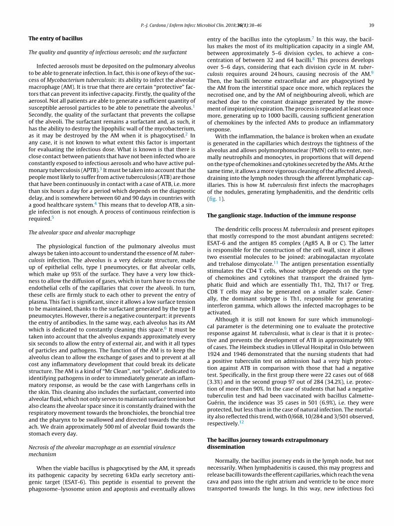
Fig. 1. Infectious cycle of M. tuberculosis. 1. Entry of bacilli into the pulmonary alveolus through an aerosol drop. 2. Phagocytosis by an alveolar macrophage (AM) and
subsequent multiplication inside. 3. Destruction of the AM, local dissemination of M. tuberculosis, phagocytosis by other AMs, and generation of a local inflammatory response
dominated by monocytes (3a) or PMN cells (3b), thanks to which the bacilli can be drained into the regional lymph node, where Th1 or Th17 lymphocytes proliferate. 4. The
lymphocytes are attracted by the inflammatory response of the lesions and activate the infected AMs or attract more PMN cells, depending on whether the immune response
is Th1 (4a) or Th17 (4b), respectively. In the first case, there is a control of the bacillary population and the dormant bacilli are drained through the foam macrophages (5a)
until it is controlled by encapsulation of the lesion (6). In the second, the lesions continue growing in size thanks to the entry of PMN cells and the extracellular bacillary
growth in the NET, generating new peripheral lesions. In this case, the bacillary concentration is much higher and from here the drainage is much more significant, whether
through the alveolar fluid or on a systemic level through neovascularisation of the granuloma (5b). In the lungs, the bacilli in the alveolar fluid (7) tend to be drained into the
gastrointestinal tract (8), although they may form part of new aerosols, generating new lesions (1).
Adapted from Cardona.10
may be generated, especially if the bacilli are released en masse
in the form of clumps, which obstruct capillaries, block circulation,
destroy the tightness of the alveolar space and enter into it. They
may also recolonise previously generated lesions which, as they
are in a process of inflammation, have greater vascularisation and
permeability. Finally, these bacilli may simply pass into the venous
capillaries, reach the left atrium and ventricle, and disseminate sys-
temically. The bacillus may potentially colonise any organ. This is
linked to the characteristics of vascularisation. In some organs, the
endothelial cells allow a greater permeability, such as bone tissue,
especially in children when it is in the development phase, or the
kidneys, and so these organs are common targets. In contrast, the
meninges are much less commonly affected; mass entry of bacilli
into the bloodstream is required, something which is associated
with disseminated or active miliary TB, manifesting in the first few
weeks after infection.13
An important point is that the lesions generated by M. tuber-
culosis develop new vessels which are more permeable and fragile
than the structural ones, allowing both reinfection of the lesion
and propagation of the bacillus towards the pulmonary venous
capillaries.14
Another common dissemination route is the one generated by
draining the bacillus itself through the alveolar fluid, which allows
it to enter into the pharyngeal cavity and penetrate the mucous
membrane, via small wounds, affecting the cervical lymph nodes;
or causing intestinal ATB in the event that gastric acidification is not
sufficient to destroy the bacillus. Furthermore, pleural ATB remains
a variant of pulmonary TB. The mesenchymal cells of the pleural
serosa are in charge of monitoring any small disturbance in the
most superficial pulmonary parenchyma and if they detect any
small lesion, they generate a mass influx of PMN cells and mono-
cytes to isolate it and generate fibrous tissue around it.15
Extrapulmonary ATB represents approximately 30% of ATB cases
and often indicates a delay in immune response, mostly affecting
children below 5 years or people with immunodepression. How-
ever, there is a lot of geographical variability, something which may
be interpreted as a possibility for there being a genetic factor that
facilitates it.16
P.-J. Cardona / Enferm Infecc Microbiol Clin. 2018;36(1):38–46 41
Control of lesions via immune response and local structures
The “dynamic hypothesis”: process of endogenous reinfection
In most cases, the proliferation of specific Th1 cells against
the infection takes place in time to prevent the development of
ATB. The Th1 cells are drained through the efferent vessels to be
incorporated into pulmonary circulation and are mostly directed
towards the infectious foci, given that the inflammation allows a
greater probability of attraction. The Th1 cells come into contact
with the infected macrophages and, through interferon gamma
synthesis they activate them, allowing most of the bacilli to be
destroyed. However, there is a small percentage that manage to
enter into a metabolic slowing-down or dormancy phase, which
allows them to survive by maintaining minimal metabolic activity.
The activated macrophages, apart from maintaining these dormant
bacilli inside, also phagocytise the remaining necrosed tissue to a
point where it cannot metabolise the enormous load of fatty acids
(especially cholesterol). This means that lipid bodies are produced,
which accumulate within the cytoplasm and transform into foam
cells.17 These cells end up being drained by the alveolar fluid and it
is during this process that they may be destroyed. This releases
the bacilli, which may form part of the aerosols that are con-
stantly generated in the bronchioles and may reinfect the lungs
(Fig. 1).
This process was the origin of the so-called “dynamic hypothe-
sis” which involves latent tuberculosis infection (LTBI) as a constant
endogenous reinfection process. Among other things, this hypoth-
esis is able to explain why chemoprophylaxis with isoniazid works,
taking into account that there is no action against dormant bacilli.
The idea is that constant levels of isoniazid over 6–9 months
allows the dormant bacilli to be drained into the gastrointesti-
nal tract, preventing the risk of reinfection. The prolonged period
is explained because it is the average time to be able to drain
all the lesions before they fibrose and calcify, destroying the
bacilli from the inside by generating multifactorial stress around
them.18
Importance of interlobular septa in encapsulating lesions
There is another local control mechanism that allows the lesion
to be controlled: interlobular septa. These structures divide the pul-
monary parenchyma in sections of approximately 1 cm3 and serve
to transmit the mechanical force generated by the diaphragm.19 In
this way, the lungs can be inflated, allowing their expansion and
preventing the delicate structure of the alveoli from being exces-
sively strained due to the displacement required by respiratory
movement. This structure is maintained by fibroblasts that have a
special capacity for capturing surrounding mechanical variations,
such as those generated with conformation of a lesion, even if it
is very small such as that caused by initial infection due to M.
tuberculosis (around 0.5 mm).20 Therefore, the fibroblasts begin to
weave a capsule around the lesion, which ends up isolating it in
approximately 10 days.
It is interesting to highlight that infection due to M. tuberculosis
generates a protective immune response.21 There are no muta-
tion phenomena, nor are there big variations between the different
lineages that have suffered different evolutionary contexts.22 The
immune response is initially generated against several antigens but
progressively focuses on vital antigens secreted for M. tuberculosis.
This specialisation has a rationale. On the one hand, the bacillus in
its active form is capable of causing damage to the host, thanks to
its capacity to destroy macrophages. On the other hand, the ability
of the bacillus to escape and cause reinfection after generating an
immune response has already been described. Therefore, the focus
against the bacilli in multiplication is a successful strategy, even
more so taking into account that the dormant bacilli end up being
drained into the gastrointestinal tract to eventually be expelled
(Fig. 1).
Immunity based on lymphocytes explains the reactive nature of
the immune response
The other lesson to be learned is that due to the cellular nature
of the immune response, it is always “after” the infection. This is
completely the opposite of a humoral immune response, which in
this case is not effective due to the lack of entry of antibodies into
the alveolar space. Therefore, the host that has already generated
a specific immune response gains a few days, due to memory cells
which allow the bacillary population to be reduced. This is key for
preventing excessive extrapulmonary dissemination and/or local
bacillary concentration, needed for generating ATB.23 Even so, due
to the fact that the multiplication of the bacillus is initially silent
and does not generate an inflammatory response, these lympho-
cytes cannot be attracted in any way to the infectious focus. The
conclusion from this information is that, in reality, infection and
reinfection due to M. tuberculosis cannot be prevented.24 There is
much desperation in the field of vaccinology in this area. In reality,
it is impossible to achieve a prophylactic vaccine, as has been stated
up to now.10 No vaccine is capable of preventing infection: the aim
is to prevent ATB.
The origin of pulmonary tuberculosis. Size matters
Clinical practice in diagnosing TB
Bearing in mind the reaction of the host, it is difficult to explain
the development of APTB. Although infection seems inevitable, the
consequences for the host are practically zero. Extraordinarily small
lesions are generated, which do not cause any type of significant
dysfunction. In fact, it is difficult to explain why a lesion that is 1 mm
in diameter can become a 10-mm lesion. Why 10 mm? Because it
is the size of the lesion that is required to be able to be detected via
a chest X-ray scan.25
Years of combating tuberculosis have allowed effective clinical
practice to be standardised. When ATB is suspected, a tuberculin
test or interferon-gamma release assay should be performed to
evaluate whether there has been an infection and if it has generated
an immune response. If it is positive, a chest X-ray is performed. If
a lesion is detected, it is considered that the patient has APTB and
to be able to be detected, this lesion must be at least 10 mm in size.
Otherwise, it is considered that the patient has LTBI. In the event
of a recent infection, it has been shown that the most significant
risk of developing APTB is during the first two years, decreasing
exponentially to zero after eight years.5
Importance of the size of the lesion
The question is: how can a 10-mm lesion develop from a 1-mm
lesion, bearing in mind that the local control mechanisms work so
well?
At this point, it is important to take into account the formulation
of the question. In all the publications that study the progression
of the tuberculosis infection to ATB, this issue is never raised. The
questions focus on the reactivation capacity of the dormant bacilli
or the development of some type of local immunodepression. No
one considers what is actually seen in clinical practice: APTB is
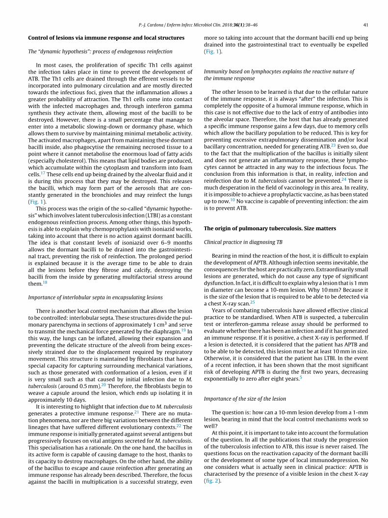
characterised by the presence of a visible lesion in the chest X-ray
(fig. 2).
42 P.-J. Cardona / Enferm Infecc Microbiol Clin. 2018;36(1):38–46
Fig. 2. Progression of latent tuberculosis infection (LTBI) to active tuberculosis
(ATB). (A) Common representation; the difference is in the activity of the bacil-
lus, omitting the fact that the main characteristic that clinically distinguishes LTBI
from ATB is the size of the lesions (B).
Extracted from Cardona.10
Tropism of the upper pulmonary lobes in the developmentof APTB
Physiology of the upper pulmonary lobes
A characteristic aspect of the development of APTB in adults is
the involvement of the upper pulmonary lobes. The theory with the
most current success for explaining this phenomenon is that oxy-
gen pressure is higher in this location, which allows M. tuberculosis
to grow better. This theory, the “unitary theory”, is attractive due
to its simple explanation, but in reality, it does not withstand sys-
tematic analysis.5 The reason for the accumulation of oxygen in this
location is because the displacement of the pulmonary parenchyma
due to respiratory movement is much less than at the base. This is
due to the structure of the lung, which is held at its apex so that
the expansion movement of the diaphragm can effectively gener-
ate the expansion of the parenchyma. Furthermore, the force of
gravity causes the interlobular septa of the upper lung to be con-
stantly tensed and have less reactivity than those situated at the
base. The lack of movement also causes a deficiency in vascular
drainage, which is why there is a lower exchange of gases leading
to hyperoxygenation. However, despite this increase favouring an
acceleration in the capacity of the bacillus to multiply, a fact that is
doubtful from a microbiological standpoint, it does not explain the
disproportionate growth of the lesions.26
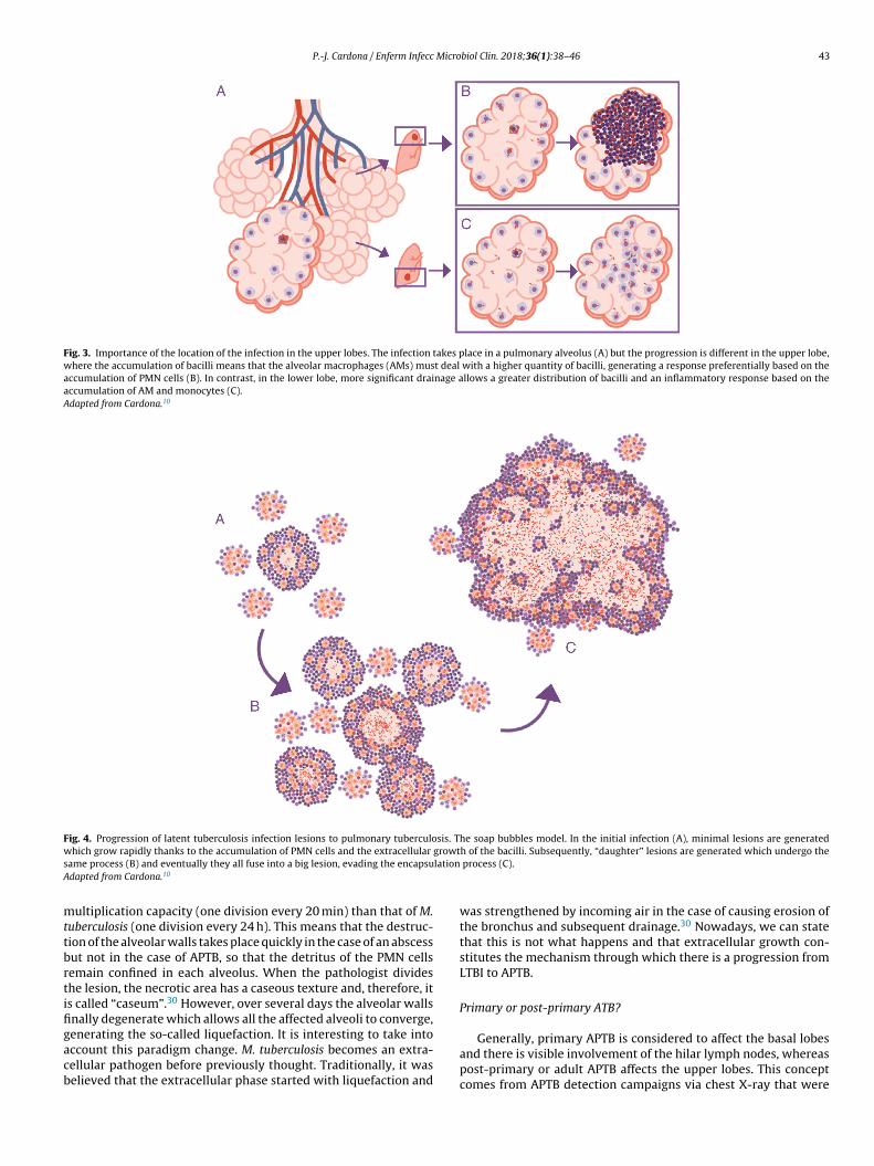
The response is in alveolar drainage
What is really important is the reduction in draining the inte-
rior of the alveolus itself, since this causes an accumulation of
extracellular bacilli locally. This means that the AM which has
to phagocytise them faces a high bacillary concentration, causing
more cellular destruction and a response from chemokines and
cytokines that facilitates the intensive accumulation of PMN cells6
(Fig. 3).
The role of PMN cells in the development of APTB. Thereturn of a forgotten actor
The experience of pathologists in the pre-antibiotic era
During the modern phase of TB study, which starts with the
declaration of a global emergency in 1993, PMN cells were consid-
ered secondary actors in pathogenesis, when they were not simply
ignored. However, after reviewing the experiences of pathologists
that studied APTB in the pre-antibiotic era, it is found that two
types of lesions were described: proliferative (or “tubercle”), which
contains a small bacillary population composed of epithelioid cells
and fibroblasts that progress towards fibrosis and calcification; and
exudative tuberculosis or local neutrophilic condensation, which
contains a high bacillary population and is related to the develop-
ment of APTB.27
Lessons learned from the ATB model in mice. The soap bubbles
model
The development of the experimental model using mice with
the C3HeB/FeJ strain has allowed a very significant leap forward
in understanding progression from LTBI to APTB. In this model,
it has been possible to demonstrate that M. tuberculosis strength-
ens the formation of neutrophil extracellular traps, a mechanism
developed by the PMN cells to deal with infections caused by
extracellular pathogens. It consists of cell lysis to release enzymes
contained in the lysosomes, which in turn traps the pathogens via
tissue formation with nuclear DNA. However, in the case of M.
tuberculosis, they do not manage to destroy it. In contrast, the neu-
trophil extracellular traps make a platform for extracellular growth
of the bacillus.28 In this way, it causes exponential growth in the
size of the lesion, which involves the generation of surrounding
satellite (or “daughter”) lesions and a final fusion process between
all of them over a time period of between 10 and 15 days. This
prevents the protective encapsulation mechanism and it is capa-
ble of generating APTB in the form of alveolar infiltration by PMN
cells that finally liquefies and cavitates. The previously mentioned
constant reinfection from contact with cases of APTB clearly helps
this phenomenon. In the case of reinfection, it is located adjacently,
the same as constant endogenous reinfection, as a consequence of
draining dormant bacilli from previous lesions (Fig. 1) and those
that come from haematogenous dissemination that manage to pass
the filter of the lymph nodes. The evolution of this process is very
similar to the dynamic of the formation of soap bubbles. That is
why a parallel has been made and it has been possible to show that
the induction of this type of lesion depends on three factors: rapid
growth of the initial lesion (as with PMN infiltration), the formation
of surrounding “daughter lesions” and the fusion of all of them29
(Fig. 4).
Abscess or caseum?
What would be the difference between a pulmonary abscess
and APTB? The answer is time. Pulmonary abscesses have a very
rapid progression, generated by pathogens with a much quicker
P.-J. Cardona / Enferm Infecc Microbiol Clin. 2018;36(1):38–46 43
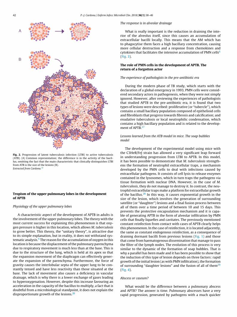
Fig. 3. Importance of the location of the infection in the upper lobes. The infection takes place in a pulmonary alveolus (A) but the progression is different in the upper lobe,
where the accumulation of bacilli means that the alveolar macrophages (AMs) must deal with a higher quantity of bacilli, generating a response preferentially based on the
accumulation of PMN cells (B). In contrast, in the lower lobe, more significant drainage allows a greater distribution of bacilli and an inflammatory response based on the
accumulation of AM and monocytes (C).
Adapted from Cardona.10
Fig. 4. Progression of latent tuberculosis infection lesions to pulmonary tuberculosis. The soap bubbles model. In the initial infection (A), minimal lesions are generated
which grow rapidly thanks to the accumulation of PMN cells and the extracellular growth of the bacilli. Subsequently, “daughter” lesions are generated which undergo the
same process (B) and eventually they all fuse into a big lesion, evading the encapsulation process (C).
Adapted from Cardona.10
multiplication capacity (one division every 20 min) than that of M.
tuberculosis (one division every 24 h). This means that the destruc-
tion of the alveolar walls takes place quickly in the case of an abscess
but not in the case of APTB, so that the detritus of the PMN cells
remain confined in each alveolus. When the pathologist divides
the lesion, the necrotic area has a caseous texture and, therefore, it
is called “caseum”.30 However, over several days the alveolar walls
finally degenerate which allows all the affected alveoli to converge,
generating the so-called liquefaction. It is interesting to take into
account this paradigm change. M. tuberculosis becomes an extra-
cellular pathogen before previously thought. Traditionally, it was
believed that the extracellular phase started with liquefaction and
was strengthened by incoming air in the case of causing erosion of
the bronchus and subsequent drainage.30 Nowadays, we can state
that this is not what happens and that extracellular growth con-
stitutes the mechanism through which there is a progression from
LTBI to APTB.
Primary or post-primary ATB?
Generally, primary APTB is considered to affect the basal lobes
and there is visible involvement of the hilar lymph nodes, whereas
post-primary or adult APTB affects the upper lobes. This concept
comes from APTB detection campaigns via chest X-ray that were
44 P.-J. Cardona / Enferm Infecc Microbiol Clin. 2018;36(1):38–46
performed after the Second World War. At that time, when the
incidence of APTB and LTBI were very significant, they began to
observe calcified lesions in the lower lobe and lymph nodes in
adults, whereas APTB manifested in the upper lobes. In 1967, the
“unitary theory” was announced, which gave a global sense to those
observations, taking into account that it was believed that once a
person was infected, they could not be reinfected.31 The interpreta-
tion was that lesions in the upper lungs that had remained dormant
and had subsequently been reactivated had arisen from old lesions
in the pre-immune phase. It has now been shown that this is not
true. Firstly, although infection gives good protection against ATB,
it does not prevent reinfection. Secondly, molecular epidemiolog-
ical studies have shown that the location of APTB depends on the
host’s immune response.32 In the case of children under five years
or immunosuppressed adults, the proliferation of the bacillus does
not require the advantage given by location in the upper lung, and
so it may develop in both areas. In the case of immunocompetent
adults, it generally develops in the upper lung due to the advan-
tage from the lack of drainage and the induction of an inflammatory
response dominated by PMN cells. One of the elements that is often
not taken into account in ATB developed in patients that contracted
ATB in the pre-antibiotic era, i.e. before the 1950s, is that very sig-
nificant lesions were often resolved via classic methods, either with
“rest cures”, pneumothorax or thoracoplasty, which aimed to con-
trol lesions through fibrosis. In all these cases, eradication of the
bacillary population was very different but generally ineffective.
Hence, in this case the possibility of generating ATB, especially
when reaching old age, was much higher than in the case where
LTBI was detected and, therefore, no lesion was observed in the
chest X-ray.
Conclusion: why do 10% of those infected develop ATB?
This is the big question! On the one hand, the most important
antigens, whose expression is very stable, have been identified;
on the other hand, the nature of the cell immune response does
not prevent infection but it does have good efficacy at preventing
ATB. In this sense, the onset of AIDS has helped to understand the
importance of CD4 lymphocytes. However, it only explains 10% of
ATB cases.33 Bacillus circulations through pulmonary drainage or
the bloodstream, respiratory movements, the encapsulation capac-
ity of the interlobular septa, together with the capacity of external
aerosols to effectively cause infections cause great complexity that
inhibits finding markers with a sufficient predictive value. What is
clear is that there are comorbidity factors that facilitate the devel-
opment of ATB. Tobacco use34 or domestic pollution through the
use of solid fuels35 can be clearly related to a deficient capacity in
pulmonary drainage. However, there is a factor that appears to be
very important, which is those people that have a proinflammatory
environment and therefore facilitate PMN cell attraction to lesions.
This factor may explain why ATB affects more males36 and patients
with type 2 diabetes mellitus.37 In fact, in most cases, the induction
of ATB is included within the conceptual framework of the “damage
theory”,38 by which the induction of ATB may be due to immun-
odepression but is mostly due to an excessive response from the
host, in this case in the form of PMN infiltrates.
Historically, there is a lot of evidence for familial susceptibility
to ATB. However, up to now no replicable specific association has
been recognised.39 Finally, it must always be taken into account
that patients that have had APTB and have been cured with standard
treatment without suffering a relapse have a greater probability
of having another episode of APTB through exogenous reinfection.
This may be due to two factors: a susceptibility due to the same
genetic factor or factors that already facilitated the induction of the
first APTB and/or local sequelae caused by the first episode.40
Environmental mycobacteriosis
Environmental mycobacteria have a lot of interest to be able
to verify TB pathogenesis. They are microorganisms that live in
the natural world and that share many aspects with M. tubercu-
losis, not only in terms of the characteristics of the cell wall, rich
in mycolic acids, but also by sharing different antigens capable of
generating protective responses.41 However, on a pathogenic level
they have barely been studied and it is difficult to draw conclu-
sions. Clinical interest has arisen, firstly, with pulmonary disease
generated in elderly populations,42 causing a disease very similar
to APTB in patients that are negative for HIV; secondly, in patients
that are positive for HIV, demonstrating the value of the Th1-
type immune response for preventing disseminated infections.43
In the same way, these mycobacteria are capable of generating
lymphadenopathy and infections in the skin, bone or joints.42 It
should be underlined that one of its peculiarities is that, in general,
its optimum growth temperature is around 30 ◦C. This competitive
disadvantage limits its pathogenic activity to superficial epithelia
where the temperature may be close to this value.
Since the secretion of ESAT-6 seems to be one of the key factors
in the pathogenesis of M. tuberculosis, the environmental mycobac-
teria may be classified in relation to this aspect.
ESAT-6 or ESAT-6-like secreting species
Mycobacterium kansasii is capable of generating lesions similar
to APTB in individuals exposed to aerosols in water with a high con-
centration of this bacillus. The incidence of pulmonary disease due
to M. kansasii is very low. The explanation for this is probably due to
the fact that in reality, a very high infectious dose is required. This
means that in reality M. tuberculosis has other pathogenic mecha-
nisms that have not been known to be evaluated, surely related to
the capacity to generate a necrotic response. It has recently been
possible to demonstrate that environmental mycobacteria induce
a rapid production of apoptosis, a fact that is linked to molecular
differences in one of the main components of the cell wall: lipoara-
binomannan, different from M. tuberculosis, which is capable of
causing cell necrosis.44
Mycobacterium marinum also generates very similar lesions to
those induced by M. tuberculosis, as reflected in extensive studies
performed on the zebrafish model.45 However, due to the fact that
its optimum growth temperature is around 30 ◦C, its capacity to
generate human pathogenesis is limited and is focused on locations
with a similar temperature, such as the surface of the skin, and is
always linked to epithelium disruption due to physical causes that
allow the bacillus to be introduced into the skin.
Non-ESAT-6-secreting
In this case, the very extensive group of M. avium-intracellulare
must be mentioned, which emerged as a significant medical prob-
lem by causing disseminated infections in patients with AIDS.42
This group was already known for its capacity to generate lym-
phadenopathy in children in Nordic countries, such as in the case
of Mycobacterium malmoense,46 or respiratory infections in patients
with bronchiectasis,47 an aspect surely linked to its capacity to gen-
erate biofilms.48 This aspect has also been especially important in
both fast- and slow-growth mycobacteria in order to be able to sup-
port itself in drinking water pipes, plumbing, showers, etc.,49 and to
be able to generate sufficiently significant infectious doses to enter
into contact with different human epithelia, whether cutaneous or
respiratory, and to be able to generate infectious processes in peo-
ple with an innate or acquired immunity deficiency. Or to colonise
prosthesis or medical devices.
P.-J. Cardona / Enferm Infecc Microbiol Clin. 2018;36(1):38–46 45
M. ulcerans also deserves to be mentioned, a microorganism
whose natural habitat is rivers or lakes and which requires incu-
bation at a temperature below 32 ◦C.42 This bacillus is also capable
of synthesising an exotoxin, mycolactone, with devastating effects
on the cutaneous epithelium. This toxin is capable of inducing
a cytotoxic effect on all the skin’s cell lines and suppressing
the production of cytokines, preventing among other things the
microbicidal capacity induced by the interferon gamma in the
macrophages; the capacity for maturation and migration of den-
dritic cells, as well as its capacity to stimulate cell immunity.
In lymphocytes, it inhibits the capacity to generate IL-2 and the
antigen-dependent capacity to produce cytokines in Th1, Th2 and
Th17 lymphocytes. In fact, mycolactone also affects the process of
attracting lymphocytes, causing a depletion of T-cells in the periph-
eral lymph nodes.50 It is therefore the most important pathogenic
factor detected in a mycobacteria environment.
Conflicts of interest
The author declares that there are no conflicts of interest.
Acknowledgements
To Júlia Gallardo Andrés, for her magnificent illustrations. To
Paula Cardona, for reviewing the manuscript.
References
1. Escombe AR, Oeser C, Gilman RH, Navincopa M, Ticona E, Martínez C, et al.The detection of airborne transmission of tuberculosis from HIV-infectedpatients, using an in vivo air sampling model. Clin Infect Dis. 2007;44:1349–57,http://dx.doi.org/10.1086/515397
2. Arcos J, Sasindran SJ, Fujiwara N, Turner J, Schlesinger LS, Torrelles JB. Humanlung hydrolases delineate Mycobacterium tuberculosis-macrophage interac-tions and the capacity to control infection. J Immunol. 2011;187:372–81,http://dx.doi.org/10.4049/jimmunol.1100823
3. Ma N, Zalwango S, Malone LL, Nsereko M, Wampande EM, Thiel BA, et al. Clinicaland epidemiological characteristics of individuals resistant to M. tuberculosisinfection in a longitudinal TB household contact study in Kampala, Uganda. BMCInfect Dis. 2014;14:352, http://dx.doi.org/10.1186/1471-2334-14-352
4. Storla DG, Yimer S, Bjune GA. A systematic review of delay in thediagnosis and treatment of tuberculosis. BMC Public Health. 2008;8:15,http://dx.doi.org/10.1186/1471-2458-8-15
5. Cardona PJ. Reactivation or reinfection in adult tuberculosis: is thatthe question? Int J Mycobacteriol. 2016;5:400–7, http://dx.doi.org/10.1016/j.ijmyco.2016.09.017
6. Cardona PJ. The progress of therapeutic vaccination with regard to tuberculosis.Front Microbiol. 2016;7:1536, http://dx.doi.org/10.3389/fmicb.2016.01536
7. Mitchell G, Chen C, Portnoy DA. Strategies used by bacteria to growin macrophages. Microbiol Spectr. 2016;4, http://dx.doi.org/10.1128/microbiolspec.MCHD-0012-2015
8. Lee J, Remold HG, Ieong MH, Kornfeld H. Macrophage apoptosis in response tohigh intracellular burden of Mycobacterium tuberculosis is mediated by a novelcaspase-independent pathway. J Immunol. 2006;176:4267–74. Available from:http://www.ncbi.nlm.nih.gov/pubmed/16547264
9. Behar SM, Divangahi M, Remold HG. Evasion of innate immunity by Mycobac-terium tuberculosis: is death an exit strategy? Nat Rev Microbiol. 2010;8:668–74,http://dx.doi.org/10.1038/nrmicro2387
10. Cardona PJ. What we have learned and what we have missed in tuberculosispathophysiology for a new vaccine design: searching for the “Pink Swan”. FrontImmunol. 2017;8:556, http://dx.doi.org/10.3389/fimmu.2017.00556
11. Kremer L, Maughan WN, Wilson RA, Dover LG, Besra GS. The M.tuberculosis antigen 85 complex and mycolyltransferase activity. LettAppl Microbiol. 2002;34:233–7. Available from: http://www.ncbi.nlm.nih.gov/pubmed/11940150
12. Heimbeck J. BCG vaccination of nurses. Tubercle. 1948;29:84–8. Available from:http://www.ncbi.nlm.nih.gov/pubmed/18909014
13. Wallgren A. The time-table of tuberculosis. Tubercle. 1948;29:245–51. Availablefrom: http://www.ncbi.nlm.nih.gov/pubmed/18101320
14. Osherov N, Ben-Ami R. Modulation of host angiogenesis as a microbialsurvival strategy and therapeutic target. PLoS Pathog. 2016;12:e1005479,http://dx.doi.org/10.1371/journal.ppat.1005479
15. Mutsaers SE, Prele CM, Brody AR, Idell S. Pathogenesis of pleural fibro-sis. Respirology. 2004;9:428–40, http://dx.doi.org/10.1111/j.1440-1843.2004.00633.x
16. Kulchavenya E. Extrapulmonary tuberculosis: are statistical reportsaccurate? Ther Adv Infect Dis. 2014;2:61–70, http://dx.doi.org/10.1177/2049936114528173
17. Cáceres N, Tapia G, Ojanguren I, Altare F, Gil O, Pinto S, et al. Evolu-tion of foamy macrophages in the pulmonary granulomas of experimentaltuberculosis models. Tuberculosis. 2009;89:175–82, http://dx.doi.org/10.1016/j.tube.2008.11.001
18. Cardona PJ. A dynamic reinfection hypothesis of latent tuberculosis infection.Infection. 2009;37:80–6, http://dx.doi.org/10.1007/s15010-008-8087-y
19. Webb WR. Thin-section CT of the secondary pulmonary lobule: anatomyand the image – the 2004 Fleischner lecture. Radiology. 2006;239:322–38,http://dx.doi.org/10.1148/radiol.2392041968
20. Gil O, Díaz I, Vilaplana C, Tapia G, Díaz J, Fort M, et al. Granuloma encapsula-tion is a key factor for containing tuberculosis infection in minipigs. PLOS ONE.2010;5:e10030, http://dx.doi.org/10.1371/journal.pone.0010030
21. Mollenkopf HJ, Kursar M, Kaufmann SHE. Immune response to postprimarytuberculosis in mice: Mycobacterium tuberculosis and Miycobacterium bovisbacille Calmette-Guérin induce equal protection. J Infect Dis. 2004;190:588–97,http://dx.doi.org/10.1086/422394
22. Coscolla M, Copin R, Sutherland J, Gehre F, de Jong B, Owolabi O, et al.M. tuberculosis T cell epitope analysis reveals paucity of antigenic variationand identifies rare variable TB antigens. Cell Host Microbe. 2015;18:538–48,http://dx.doi.org/10.1016/j.chom.2015.10.008
23. Jung YJ, Ryan L, LaCourse R, North RJ. Properties and protective valueof the secondary versus primary T helper type 1 response to airborneMycobacterium tuberculosis infection in mice. J Exp Med. 2005;201:1915–24,http://dx.doi.org/10.1084/jem.20050265
24. Kaufmann SH, Evans TG, Hanekom WA. Tuberculosis vaccines: time fora global strategy. Sci Transl Med. 2015;7:276fs8, http://dx.doi.org/10.1126/scitranslmed.aaa4730
25. Andreu J, Cáceres J, Pallisa E, Martinez-Rodriguez M. Radiological manifesta-tions of pulmonary tuberculosis. Eur J Radiol. 2004;51:139–49, http://dx.doi.org/10.1016/j.ejrad.2004.03.009
26. Bru A, Cardona PJ, Bru A, Cardona PJ. Mathematical modeling of tuberculosisbacillary counts and cellular populations in the organs of infected mice. PLOSONE. 2010;5:e12985, http://dx.doi.org/10.1371/journal.pone.0012985
27. Cardona PJ. The key role of exudative lesions and their encapsulation: lessonslearned from the pathology of human pulmonary tuberculosis. Front Microbiol.2015;6:612, http://dx.doi.org/10.3389/fmicb.2015.00612
28. Marzo E, Vilaplana C, Tapia G, Diaz J, Garcia V, Cardona PJ. Damaging role of neu-trophilic infiltration in a mouse model of progressive tuberculosis. Tuberculosis(Edinb). 2014;94:55–64, http://dx.doi.org/10.1016/j.tube.2013.09.004
29. Prats C, Vilaplana C, Valls J, Marzo E, Cardona PJ, López D. Local inflamma-tion, dissemination and coalescence of lesions are key for the progressiontoward active tuberculosis: the bubble model. Front Microbiol. 2016;7:33,http://dx.doi.org/10.3389/fmicb.2016.00033
30. Grosset J. Mycobacterium tuberculosis in the extracellular compartment: anunderestimated adversary. Antimicrob Agents Chemother. 2003;47:833–6.Available from: http://www.pubmedcentral.nih.gov/articlerender.fcgi?artid=149338&tool=pmcentrez&rendertype=Abstract
31. Stead WW. Pathogenesis of a first episode of chronic pulmonarytuberculosis in man: recrudescence of residuals of the primary infec-tion or exogenous reinfection? Am Rev Respir Dis. 1967;95:729–45,http://dx.doi.org/10.1164/arrd.1967.95.5.729
32. Geng E, Kreiswirth B, Burzynski J, Schluger NW. Clinical and radiographic corre-lates of primary and reactivation tuberculosis: a molecular epidemiology study.JAMA. 2005;293:2740–5, http://dx.doi.org/10.1001/jama.293.22.2740
33. World Health Organization Global tuberculosis report 2016. WHO; 2017.34. Schneider NK, Novotny TE. Addressing smoking cessation in tuberculosis
control. Bull World Health Organ. 2007;85:820–1, http://dx.doi.org/10.2471/BLT.07.043794
35. Jubulis J, Kinikar A, Ithape M, Khandave M, Dixit S, Hotalkar S, et al. Modifiablerisk factors associated with tuberculosis disease in children in Pune, India. Int JTuberc Lung Dis. 2014;18:198–204, http://dx.doi.org/10.5588/ijtld.13.0314
36. Neyrolles O, Quintana-Murci L. Sexual inequality in tuberculosis. PLoS Med.2009;6:e1000199, http://dx.doi.org/10.1371/journal.pmed.1000199
37. Hodgson K, Morris J, Bridson T, Govan B, Rush C, Ketheesan N. Immuno-logical mechanisms contributing to the double burden of diabetesand intracellular bacterial infections. Immunology. 2015;144:171–85,http://dx.doi.org/10.1111/imm.12394
38. Casadevall A, Pirofski L. The damage-response framework of microbialpathogenesis. Nat Rev Microbiol. 2003;1:17–24, http://dx.doi.org/10.1038/nrmicro732
39. Naranbhai V. The role of host genetics (and genomics) in tuberculosis. MicrobiolSpectr. 2016;4, http://dx.doi.org/10.1128/microbiolspec.TBTB2-0011-2016
40. Verver S, Warren RM, Beyers N, Richardson M, van der Spuy GD, BorgdorffMW, et al. Rate of reinfection tuberculosis after successful treatment is higherthan rate of new tuberculosis. Am J Respir Crit Care Med. 2005;171:1430–5,http://dx.doi.org/10.1164/rccm.200409-1200OC
41. Poyntz HC, Stylianou E, Griffiths KL, Marsay L, Checkley AM, McShaneH. Non-tuberculous mycobacteria have diverse effects on BCG efficacyagainst Mycobacterium tuberculosis. Tuberculosis (Edinb). 2014;94:226–37,http://dx.doi.org/10.1016/j.tube.2013.12.006
42. Tortoli E. Clinical manifestations of nontuberculous mycobacteria infec-tions. Clin Microbiol Infect. 2009;15:906–10, http://dx.doi.org/10.1111/j.1469-0691.2009.03014.x

46 P.-J. Cardona / Enferm Infecc Microbiol Clin. 2018;36(1):38–46
43. Halstrom S, Price P, Thomson R. Review: environmental mycobacte-ria as a cause of human infection. Int J Mycobacteriol. 2015;4:81–91,http://dx.doi.org/10.1016/j.ijmyco.2015.03.002
44. Bohsali A, Abdalla H, Velmurugan K, Briken V. The non-pathogenic mycobac-teria M. smegmatis and M. fortuitum induce rapid host cell apoptosis viaa caspase-3 and TNF dependent pathway. BMC Microbiol. 2010;10:237,http://dx.doi.org/10.1186/1471-2180-10-237
45. Meijer AH. Protection and pathology in TB: learning from the zebrafishmodel. Semin Immunopathol. 2016;38:261–73, http://dx.doi.org/10.1007/s00281-015-0522-4
46. Henriques B, Hoffner SE, Petrini B, Juhlin I, Wahlen P, Kallenius G. Infectionwith Mycobacterium malmoense in Sweden: report of 221 cases. Clin Infect Dis.1994;18:596–600, http://dx.doi.org/10.1093/clinids/18.4.596
47. Wickremasinghe M, Ozerovitch LJ, Davies G, Wodehouse T, ChadwickMV, Abdallah S, et al. Non-tuberculous mycobacteria in patients with
bronchiectasis. Thorax. 2005;60:1045–51, http://dx.doi.org/10.1136/thx.2005.046631
48. Johansen TB, Agdestein A, Olsen I, Nilsen SF, Holstad G, Djønne B. Biofilmformation by Mycobacterium avium isolates originating from humans, swineand birds. BMC Microbiol. 2009;9:159, http://dx.doi.org/10.1186/1471-2180-9-159
49. Williams MM, Yakrus MA, Arduino MJ, Cooksey RC, Crane CB, Banerjee SN,et al. Structural analysis of biofilm formation by rapidly and slowly grow-ing nontuberculous mycobacteria. Appl Environ Microbiol. 2009;75:2091–8,http://dx.doi.org/10.1128/AEM. 00166-09
50. Sarfo FS, Phillips R, Wansbrough-Jones M, Simmonds RE. Recent advances:role of mycolactone in the pathogenesis and monitoring of Mycobac-terium ulcerans infection/Buruli ulcer disease. Cell Microbiol. 2016;18:17–29,http://dx.doi.org/10.1111/cmi.12547