cor, a novel carbon monoxide resistance gene, is essential for mycobacterium tuberculosis...
TRANSCRIPT
doi:10.1128/mBio.00721-13. 4(6): .mBio
. PathogenesisMycobacterium tuberculosisEssential for , a Novel Carbon Monoxide Resistance Gene, Iscor2013.
et al.Vineetha M. Zacharia, Paolo S. Manzanillo, Vidhya R. Nair, PathogenesisMycobacterium tuberculosisResistance Gene, Is Essential for
, a Novel Carbon Monoxidecor
http://mbio.asm.org/content/4/6/e00721-13.full.htmlUpdated information and services can be found at:
MATERIALSUPPLEMENTAL http://mbio.asm.org/content/4/6/e00721-13.full.html#SUPPLEMENTAL
REFERENCES
http://mbio.asm.org/content/4/6/e00721-13.full.html#ref-list-1This article cites 58 articles, 31 of which can be accessed free at:
CONTENT ALERTS
more>>article), Receive: RSS Feeds, eTOCs, free email alerts (when new articles cite this
http://journals.asm.org/subscriptions/To subscribe to another ASM Journal go to:
http://mbio.asm.org/misc/contentdelivery.xhtmlInformation about Print on Demand and other content delivery options:
http://mbio.asm.org/misc/reprints.xhtmlInformation about commercial reprint orders:
m
bio.asm.org
on Septem
ber 24, 2014 - Published by
mbio.asm
.orgD
ownloaded from
mbio.asm
.org on S
eptember 24, 2014 - P
ublished by m
bio.asm.org
Dow
nloaded from
cor, a Novel Carbon Monoxide Resistance Gene, Is Essential forMycobacterium tuberculosis Pathogenesis
Vineetha M. Zacharia,a Paolo S. Manzanillo,b Vidhya R. Nair,a Denise K. Marciano,a Lisa N. Kinch,c Nick V. Grishin,c Jeffery S. Cox,b
Michael U. Shiloha
Department of Internal Medicine, University of Texas Southwestern Medical School, Dallas, Texas, USAa; Department of Microbiology and Immunology, University ofCalifornia at San Francisco, San Francisco, California, USAb; Howard Hughes Medical Institute and Department of Biochemistry, University of Texas Southwestern MedicalCenter, Dallas, Texas, USAc
V.M.Z, P.S.M., J.S.C., and M.U.S. contributed equally to this article.
ABSTRACT Tuberculosis, caused by Mycobacterium tuberculosis, remains a devastating human infectious disease, causing twomillion deaths annually. We previously demonstrated that M. tuberculosis induces an enzyme, heme oxygenase (HO1), that pro-duces carbon monoxide (CO) gas and that M. tuberculosis adapts its transcriptome during CO exposure. We now demonstratethat M. tuberculosis carries a novel resistance gene to combat CO toxicity. We screened an M. tuberculosis transposon library forCO-susceptible mutants and found that disruption of Rv1829 (carbon monoxide resistance, Cor) leads to marked CO sensitivity.Heterologous expression of Cor in Escherichia coli rescued it from CO toxicity. Importantly, the virulence of the cor mutant isattenuated in a mouse model of tuberculosis. Thus, Cor is necessary and sufficient to protect bacteria from host-derived CO.Taken together, this represents the first report of a role for HO1-derived CO in controlling infection of an intracellular pathogenand the first identification of a CO resistance gene in a pathogenic organism.
IMPORTANCE Macrophages produce a variety of antimicrobial molecules, including nitric oxide (NO), hydrogen peroxide(H2O2), and acid (H�), that serve to kill engulfed bacteria. In addition to these molecules, human and mouse macrophages alsoproduce carbon monoxide (CO) gas by the heme oxygenase (HO1) enzyme. We observed that, in contrast to other bacteria, my-cobacteria are resistant to CO, suggesting that this might be an evolutionary adaptation of mycobacteria for survival within mac-rophages. We screened a panel of ~2,500 M. tuberculosis mutants to determine which genes are required for survival of M. tu-berculosis in the presence of CO. Within this panel, we identified one such gene, cor, that specifically confers CO resistance.Importantly, we found that the ability of M. tuberculosis cells carrying a mutated copy of this gene to cause tuberculosis in amouse disease model is significantly attenuated. This indicates that CO resistance is essential for mycobacterial survival in vivo.
Received 29 August 2013 Accepted 17 October 2013 Published 19 November 2013
Citation Zacharia VM, Manzanillo PS, Nair VR, Marciano DK, Kinch LN, Grishin NV, Cox JS, Shiloh MU. 2013. cor, a novel carbon monoxide resistance gene, is essential forMycobacterium tuberculosis pathogenesis. mBio 4(6):e00721-13. doi:10.1128/mBio.00721-13
Editor Eric Rubin, Harvard School of Public Health
Copyright © 2013 Zacharia et al. This is an open-access article distributed under the terms of the Creative Commons Attribution-Noncommercial-ShareAlike 3.0 Unportedlicense, which permits unrestricted noncommercial use, distribution, and reproduction in any medium, provided the original author and source are credited.
Address correspondence to [email protected].
Mycobacterium tuberculosis infects one-third of the world’spopulation and causes 8 million active cases of disease an-
nually. Tissue macrophages are the primary cells within whichmuch of the host-M. tuberculosis interaction occurs, and macro-phages utilize multiple strategies to control M. tuberculosis infec-tion, including elaboration of reactive oxygen and nitrogen inter-mediates, acidification of the phagosome, fusion of phagosomeswith lysosomes, delivery of antimicrobial peptides to the phagoly-sosome, and autophagic engulfment of M. tuberculosis (1, 2).However, a complete understanding of the immune mechanismsthat control M. tuberculosis infection is lacking. Likewise, how M.tuberculosis resists host immunity and persists indefinitely is onlybeginning to be elucidated.
Sustained in vivo and intracellular survival of M. tuberculosis, ahallmark of latent infection, has selected for bacteria that can resisthost antimicrobial systems (3). These include oxidative radicalsproduced by the respiratory burst, NO toxicity from induciblenitric oxide synthase (NOS2), and phagosomal acidification (4).
In response, M. tuberculosis has evolved strategies to interfere withhost defenses such as excluding NOS2 from the phagosome (5, 6)and arresting phagosome maturation to prevent acidification (7).Furthermore, M. tuberculosis can intrinsically resist the toxicitymediated by host pathways such as hydrogen peroxide throughcatalase (8, 9), NO through DNA repair (10), protein degradation(11), and antioxidant defense (12), and acidification through ad-aptation (13), cell wall stabilization (14, 15), and nitrate respira-tion (16). Thus, in many cases, the presence of a host antimicro-bial pathway has selected for mycobacterial genes that can preventbacterial death.
Humans and mice produce CO by the enzyme heme oxygenase(HO1) (17, 18), which catalyzes the degradation of heme intobiliverdin, iron, and CO in a reaction requiring O2 and NADPH(19, 20). HO1 is primarily expressed within alveolar, liver, andspleen macrophages and is induced by inflammatory mediatorssuch as lipopolysaccharide, tumor necrosis factor, interleukin-1,and oxidative stress (21). The CO is exhaled, with the average,
RESEARCH ARTICLE
November/December 2013 Volume 4 Issue 6 e00721-13 ® mbio.asm.org 1
m
bio.asm.org
on Septem
ber 24, 2014 - Published by
mbio.asm
.orgD
ownloaded from
nonsmoking human exhaling approximately 2 ppm (22, 23),while patients with a variety of infectious and inflammatory con-ditions produce significantly more (21, 22, 24–26). No studieshave been performed to date on the CO concentration in exhaledair from individuals with tuberculosis.
We (27) and others (28) have shown that M. tuberculosis infec-tion of macrophages and mice induces HO1. HO1-derived COthen induces a set of ~50 genes known as the dormancy regulonvia a two-component signal transduction system mediated by thesensor histidine kinases DosS and DosT, and this induction isdiminished in HO1-deficient macrophages and with chemical in-hibition of HO1 (27). Recently, it was shown that HO1-deficientmice are more susceptible to Mycobacterium avium infection (29,30) and M. tuberculosis infection (30) than wild-type mice, al-though this enhanced susceptibility was attributed to either inap-propriate granuloma formation (29, 30) or macrophage toxicityfrom heme accumulation (30) and not to an antimicrobial activityof CO. To that end, the identification of a specific CO resistancegene in M. tuberculosis, an organism that is highly evolved forhuman survival, would provide strong evidence that HO1-derivedCO functions as an antibacterial mechanism in humans.
Although CO has been present since the beginning of life (31),little is known about how bacteria survive in its presence, as COcan be toxic to proteins containing iron and other transition met-als. Weigel and Englund (32) showed that CO halts aerobic growthof Escherichia coli by rapidly inhibiting ATP production and pre-venting DNA replication. More recent work has confirmed thatE. coli, Pseudomonas aeruginosa, and Staphylococcus aureus aresusceptible to exogenous CO (33–35). In contrast, M. tuberculosisis resistant to high levels of CO (27). Because heme oxygenase isinduced during an M. tuberculosis infection (27, 28), the CO pro-duced is sensed by M. tuberculosis (27, 28), and M. tuberculosis isresistant to CO (27), we hypothesized that M. tuberculosis carriesresistance genes for CO.
Here we identify a novel host-pathogen interaction centeredon the effects of host-derived CO on mycobacterial pathogenesis.We found that M. tuberculosis carries a CO resistance gene that isboth necessary and sufficient for CO resistance and that this geneis essential for long-term survival of M. tuberculosis in mice. Thisrepresents the first identification of a CO resistance gene in M.tuberculosis and highlights the critical importance of HO1 in con-trolling infection of an intracellular pathogen.
RESULTSIdentification of Rv1829 as a CO resistance gene. Because HO1 isimportant for control of M. tuberculosis during mouse infection(29, 30), and because we previously showed that M. tuberculosis isresistant to exogenous CO (27), we hypothesized that M. tubercu-losis carries CO resistance genes. We created a transposon libraryusing the M. tuberculosis Erdman strain and the �MycomarT7phage developed by Sassetti et al. (36) and screened for mutantsthat would not grow in the presence of CO. We screened ~2,500individual mutants on plates and identified one mutant whosetransposon insertion was mapped to Rv1829 that was unable togrow in the presence of CO both on plates (Fig. 1A and B) and inliquid culture (Fig. 1C). Because it conferred carbon monoxideresistance, we named the gene cor. Notably, cor mutant bacteriademonstrated normal growth under aerobic conditions (Fig. 1A,B, and C; CO). The gene organization of the region encompassingcor does not indicate that it is part of an operon (Fig. 2C). None-
theless, to exclude the possibility that the transposon insertionresulted in a polar disruption of neighboring genes, we cloned corunder the control of its native promoter and integrated the con-struct into the attB site in the cor mutant. Heterologous expressionof cor rescued the susceptibility of the mutant bacteria to grow inthe presence of CO on plates (Fig. 1A) and in liquid culture(Fig. 1C). We confirmed that Cor was absent in the cor mutant andthat its synthesis was restored in the complemented strain using ananti-Cor antibody (Fig. 1D). Thus, we conclude that the cor mu-tant phenotype is due specifically to disruption of cor and not to apolar effect on neighboring genes, including the neighboringSoxR-like transcription factors.
cor is a gene of unknown function in M. tuberculosis that en-codes a 164-amino-acid protein (Fig. 2A). It is an evolutionarilyancient gene that is found in bacteria, archea, and plants and con-tains a DUF151 (domain of unknown function) domain (Fig. 2A).There are �500 sequences in the CDART database containing thisdomain, including other pathogenic mycobacteria (M. avium,M. kansasii, M. abscessus, and M. leprae) and other pathogens suchas Rhodococcus sp. and corynebacteriae (Fig. 2B), and the genomicorganization of the cor region is shared between mycobacteria andRhodococcus sp. (Fig. 2C), suggesting shared functions. Bioinfor-matics and molecular modeling analysis using the crystal structureof the Cor homologue from Thermotoga maritima demonstratethat Cor likely dimerizes (Fig. 2D). We purified recombinant Corin E. coli for biochemical experiments and found using native gelelectrophoresis that Cor forms a dimer (Fig. 2E; compare the de-naturing gel on the left to the native gel on the right). The structureof the Thermotoga maritima homologue is novel and thereforeprovides little insight with respect to function. However, theDUF151 domain containing wild rice Oryza minuta proteinOmBBD was recently reported to exhibit nuclease activity (37).We tested recombinant Cor purified from E. coli as a 6�His–maltose-binding protein (MBP) fusion for nuclease activity andfound that it lacked DNase or RNAse activity compared to His-MBP alone (see Fig. S1 in the supplemental material). Thus, corappears to be a CO resistance gene of unknown function.
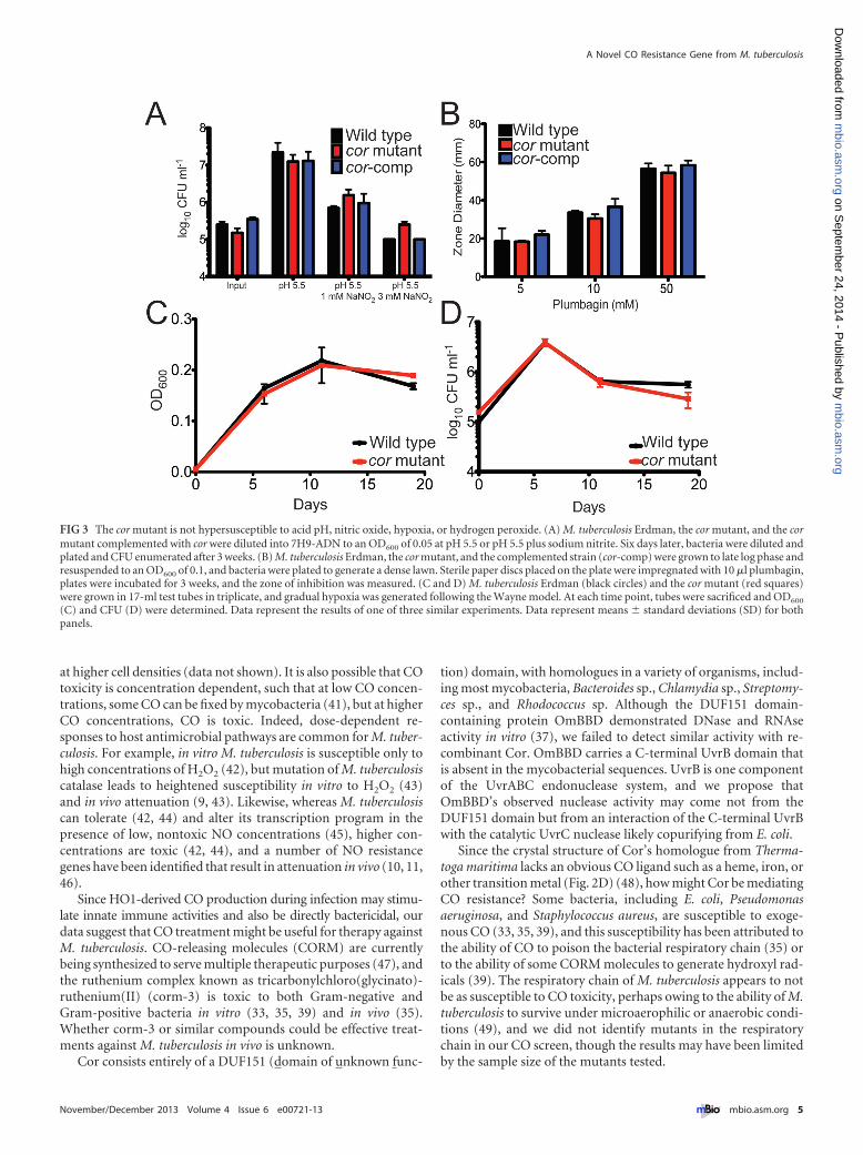
The cor mutant is not hypersusceptible to multiple stresses.Although we isolated the cor mutant in a screen for CO sensitivity,cor may encode a general stress survival factor rather than provid-ing isolated protection against CO toxicity. Therefore, we testedthe ability of the mutant to survive when exposed to conditionsexpected to exist in vivo, namely, exposure to acid pH, NO, oxi-dative stress, and hypoxia. We found that the growth phenotype ofthe cor mutant when exposed to acid pH (Fig. 3A), NO (Fig. 3A),hydrogen peroxide via plumbagin treatment (Fig. 3B), or hypoxia(Fig. 3C and D) was indistinguishable from that of the wild type.Thus, we conclude that among the stresses tested, the mutantstrain is increasingly susceptible to CO only.
The cor mutant develops a reducing environment. On thebasis of the crystal structure of the Thermatoga maritima homo-logue, we hypothesized that Cor might have enzymatic activity.Therefore, we tested if the cor mutant has a different small-molecule metabolite pool than wild-type bacteria when exposedto CO and asked if differences in metabolites might provide in-sight into Cor’s function. We extracted a small-molecule fractionfrom wild-type and cor mutant bacteria grown in the presence andabsence of CO and profiled the mycobacterial metabolite pool.We found that for the roughly 150 metabolites surveyed, theNAD� and mycothione levels were significantly reduced in the
Zacharia et al.
2 ® mbio.asm.org November/December 2013 Volume 4 Issue 6 e00721-13
m
bio.asm.org
on Septem
ber 24, 2014 - Published by
mbio.asm
.orgD
ownloaded from

CO-treated mutant compared to the CO-treated wild type at the10-day time point (Fig. 4A and B), indicating that the cor mutanthas a dysregulated redox environment with accumulation of re-ducing equivalents (the full list of metabolites is available as Ta-ble S1 in the supplemental material). We also found that the cormutant had significantly elevated levels of unsaturated (Fig. 4C toE) and saturated (Fig. 4F) long-chain fatty acids, likely indicativeof increased anabolism of fatty acids. This result is consistent witha previous study of an M. tuberculosis WhiB3 mutant that developsa reducing environment and concomitant anabolism of a varietyof lipids (38).
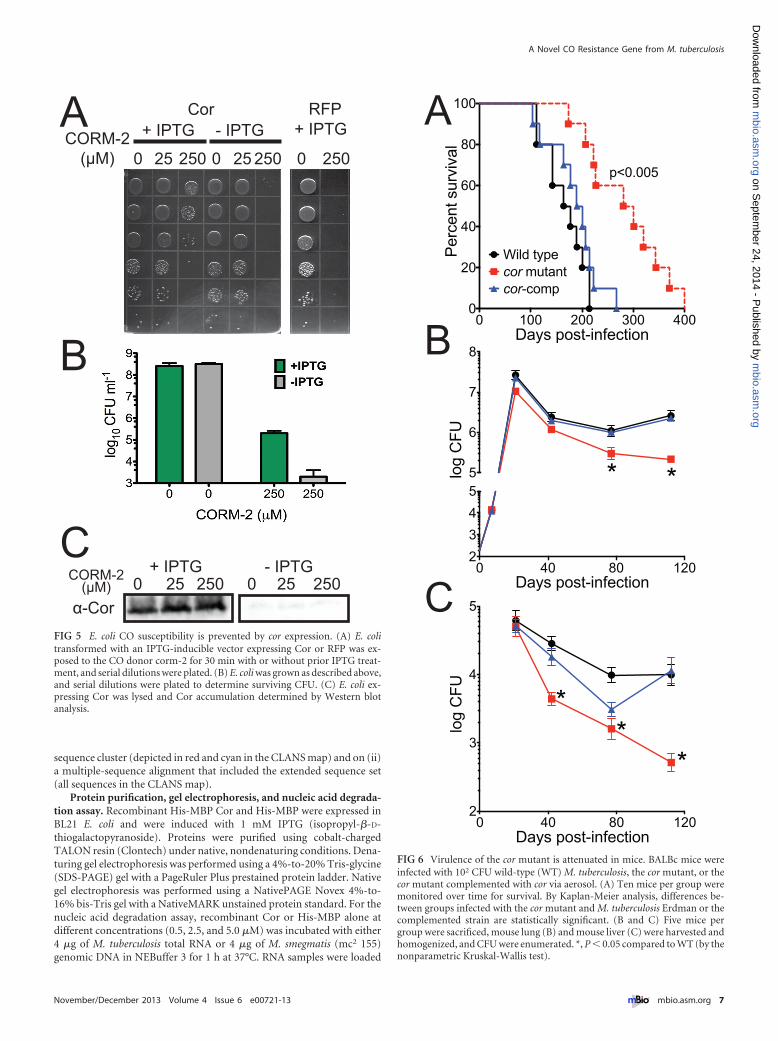
Expression of M. tuberculosis Cor in E. coli is sufficient torescue CO toxicity. To ask if Cor alone could confer CO resis-tance, we transformed E. coli with a vector containing cor underthe control of a T7 promoter. Both E. coli transformed with theCor expression vector but left uninduced and E. coli expressing anirrelevant protein, red fluorescent protein (RFP), were markedlysusceptible to CO treatment (Fig. 5A and B), as has been previ-ously reported (39). In contrast, when E. coli was induced to ex-press Cor and exposed to CO, we observed significantly reducedCO toxicity such that Cor-expressing E. coli demonstrated a 3-to-4-log improvement in growth (Fig. 5A and B). Exogenous addi-tion of recombinant Cor to CO-treated E. coli did not rescue E. colifrom CO toxicity (data not shown). We confirmed expression ofCor with our anti-Cor polyclonal antibody (Fig. 5C). Thus, al-though the precise activity of Cor remains unknown, it is able toprotect E. coli from CO toxicity.
Virulence of the cor mutant is attenuated in mice. To assessthe role of Cor in vivo, we compared the outcomes of mouse in-fection with wild-type M. tuberculosis, the cor mutant, and thecomplemented strain. Mice infected with the cor mutant survivedsignificantly longer than mice infected with the wild type or thecomplemented strain (Fig. 6A). Indeed, by 250 days postinfection,all the mice infected with the wild-type strain or the comple-mented strain had succumbed to infection, while more than 50%of the mutant mice were still alive. Likewise, the virulence of thecor mutant was attenuated in both mouse lung (Fig. 6B) andmouse liver (Fig. 6C) by an organ CFU assay, and this attenuationwas most profound at later time points during the infection. Im-portantly, complementation restored full virulence, indicatingthat the attenuated phenotype of the cor mutant was due to dis-ruption of cor.
DISCUSSION
Our report identifies a novel host-pathogen relationship centeredon the gas carbon monoxide. It was recently shown that HO1-deficient mice are more susceptible to M. tuberculosis infection(30), highlighting a role for HO1 in controlling M. tuberculosis. Tocombat CO in vivo, M. tuberculosis carries at least one CO resis-tance gene, cor. Although the precise biochemical mechanism ofprotection is unknown, the cor mutant appears to protect againstaccumulation of excess reducing equivalents. Further, cor can pro-tect a heterologous bacterium, E. coli, from CO toxicity. Impor-
FIG 1 Identification of cor (Rv1829) as a CO resistance gene. (A) Serial dilutions of M. tuberculosis Erdman, cor mutant bacteria, or the cor mutantcomplemented with cor (cor-comp) were plated and exposed to ambient air or CO (0.2%) for 3 weeks. (B) Quantitation of CFU data from the experimentrepresented by panel A (one of three similar experiments shown). (C) Western blots of lysates of M. tuberculosis Erdman, the cor mutant, or the complementedstrain were probed with anti-Cor antibody. WT, wild type. (D) M. tuberculosis Erdman, the cor mutant, and the complemented strain were grown in 7H9 liquidmedium in the presence or absence of CO, and CFU were enumerated. *, P � 0.05 compared to M. tuberculosis Erdman (by Student’s t test).
A Novel CO Resistance Gene from M. tuberculosis
November/December 2013 Volume 4 Issue 6 e00721-13 ® mbio.asm.org 3
m
bio.asm.org
on Septem
ber 24, 2014 - Published by
mbio.asm
.orgD
ownloaded from

tantly, cor is important for M. tuberculosis survival after aerosolinfection in mice.
Previous work demonstrated that some mycobacteria can uti-lize CO as a carbon source (40) by fixing CO (41). This raises theissue of how a gas such as CO could act as a potential metabolite,a signaling molecule, and a toxin. Notably, although the attenu-
ated M. tuberculosis H37Ra strain can grow on CO as its sole car-bon source (40), CO-dependent growth of virulent M. tuberculosisstrains such as H37Rv, CDC 1551, and Erdman has yet to be dem-onstrated. Whether CO is toxic might also depend on the environ-ment experienced by the bacteria. For example, we found that M.tuberculosis was more resistant to CO when it was exposed to CO
B
AE
Actinobacteria
Bacteroidetes
Mycobacteria
Viridiplantae Crenarchaeota
Euryarchaeota
Proteobacteria
Cyanobacteria
Chlamydiae
Firmi-cutes
Root
Bacterial Kingdom
Archaeal Kingdom
Eukaryotic Kingdom
B
A
E Scale0k 1k 2k 3k 4k 5k 6k 7k 8k 9k 10kMycobacterium tuberculosis H37Rv
pgsA2Rv1823
Rv1824Rv1825
gcvH cfp17SoxR TF
Rv1829 SoxR TF Rv1831gcvB
Rv1833c
Rhodococcus sp. RHA1 pgsA
COG3879COG-Sbp
COG3879gcvH COG1716
SoxR TFCOG1259 SoxR gcvP1 COG1408
C
D E
A
B
2066
146
kDakDa
1015253555
1 2 3 4
Thermotoga Maritima [1sj5] MRKAWVKTLALDRVSNTPVVILGIEGTNRVLPIWIGAAEGHALALAMEKMEFPRPLTHDLLLSVLESLEARVDKVIIHSLSecondary Structure EEEEEEEEEEE------EEEEEEE----EEEEEEE-HHHHHHHHHHHHH------HHHHHHHHHH----EEEEEEEEEEEMycobacterium tuberculosis MGEVRVVGIRVEQPQNQPVLLLREANGDRYLPIWIGQSEAAAIALEQQGVEPPRPLTHDLIRDLIAALGHSLKEVRIVDLMycobacterium leprae MGEVRVVGIRVEQPQNQPVLLLREANGDRYLPIWIGQSEAAAIALEQQGIEPPRPLTHDLIRDVIAALGHSLKEVRIVDLMycobacterium smegmatis MAEVRVVGIRVEQPQNQPVLLLRESNGDRYLPIWIGQSEAAAIALEQQGVEPARPLTHDLIRDLIAALGHSLKEVRIVDLRhodococcus fascians MSEMRVVGIRVEQPQNQPVLLLRESDEDRYLPIWIGQTEAAAIALEQQGVQPARPLTHDLVKNLISALGHELKEVRIVDLStreptomyces sp. AA4 MSEMRVVGVRVELPANQPILLLRETEGERYLPIWIGSVEATAIALEQQGVRPARPLTHDLLKEVIGALGRELEQVVITDL
Thermotoga Maritima [1sj5] KDNTFYATLVIRDLTAALIDIDSRPSDAIILAVKTGAPIFVSDNLVEKHSIELEVNER---------DLIN--------Secondary Structure E--EEEEEEEEEE----EEEEEE-HHHHHHHHHHHH--EEEE-HHHHHH-EEE-HHHH---------HHHH--------Mycobacterium tuberculosis QEGTFYADLIFDRN----IKVSARPSDSVAIALRVGVPIYVEEAVLAQAGLLIPDESDEEATTAVREDEVEKFKEFLDSMycobacterium leprae QEGTFYADLIFDRN----IKVSARPSDSVAIALRVGVPIYVEDVVLAQAGLLIPDENDEEVGGAVREDEVEKFKEFLDSMycobacterium smegmatis QEGTFYADLIFDRD----IKVSARPSDSVAIALRVGVPIYVEEAVLAEAGLLIPDENDEEESGTVREDEVEKFKEFLDSRhodococcus fascians QEGTFYADLVFDKD----IRVSARPSDSVAIALRAGVPIYAEEPVLAEAGLLMPDER---------EDEVEKFKEFLESStreptomyces sp. AA4 KEGTFFAELVFDGD----IRVSARPSDSVALALRIGVPIHAVDAVLEEAGLIIPDEQ---------EDEVEKFREFLDS
!
FIG 2 Cor is a conserved, ancient protein. (A) Alignment of Rv1829 from M. tuberculosis to orthologues from Thermotoga maritima, M. leprae, M. smegmatis,Rhodococcus fascians, and Streptomyces species AA4. Sequence alignment of Rv1829 homologues shows conserved amino acids (in rainbow colors) by conser-vation, with invariant nonhydrophobic residues colored in red. This color scheme is identical in the modeled crystal structure in panel D. (B) Species-distributiontaxonomic tree in “sunburst” format. The distribution and evolutionary conservation of all known homologues of Cor are shown. (C) Alignment of the Corgenomic region from M. tuberculosis with the orthologous genomic region from Rhodococcus sp. demonstrates conservation of multiple surrounding genes. (D)The Cor sequence was mapped to the representative DUF151 structure, which highlighted a conserved surface cleft formed at the interface (also relativelyconserved) of two DUF151 monomers (shown in red). (E) Cor was purified from E. coli as a 6�His-tagged protein and then run on a denaturing SDS-PAGE gel(left panel) or a native gel (right panel) with appropriate molecular mass markers. The predicted molecular masses of cor are 18 kDa for the monomer and 36 kDafor the dimer. In the left panel, lanes are total lysate (lane 1), flowthrough (lane 2), final wash (lane 3), and imidazole elution (lane 4).
Zacharia et al.
4 ® mbio.asm.org November/December 2013 Volume 4 Issue 6 e00721-13
m
bio.asm.org
on Septem
ber 24, 2014 - Published by
mbio.asm
.orgD
ownloaded from
at higher cell densities (data not shown). It is also possible that COtoxicity is concentration dependent, such that at low CO concen-trations, some CO can be fixed by mycobacteria (41), but at higherCO concentrations, CO is toxic. Indeed, dose-dependent re-sponses to host antimicrobial pathways are common for M. tuber-culosis. For example, in vitro M. tuberculosis is susceptible only tohigh concentrations of H2O2 (42), but mutation of M. tuberculosiscatalase leads to heightened susceptibility in vitro to H2O2 (43)and in vivo attenuation (9, 43). Likewise, whereas M. tuberculosiscan tolerate (42, 44) and alter its transcription program in thepresence of low, nontoxic NO concentrations (45), higher con-centrations are toxic (42, 44), and a number of NO resistancegenes have been identified that result in attenuation in vivo (10, 11,46).
Since HO1-derived CO production during infection may stimu-late innate immune activities and also be directly bactericidal, ourdata suggest that CO treatment might be useful for therapy againstM. tuberculosis. CO-releasing molecules (CORM) are currentlybeing synthesized to serve multiple therapeutic purposes (47), andthe ruthenium complex known as tricarbonylchloro(glycinato)-ruthenium(II) (corm-3) is toxic to both Gram-negative andGram-positive bacteria in vitro (33, 35, 39) and in vivo (35).Whether corm-3 or similar compounds could be effective treat-ments against M. tuberculosis in vivo is unknown.
Cor consists entirely of a DUF151 (domain of unknown func-
tion) domain, with homologues in a variety of organisms, includ-ing most mycobacteria, Bacteroides sp., Chlamydia sp., Streptomy-ces sp., and Rhodococcus sp. Although the DUF151 domain-containing protein OmBBD demonstrated DNase and RNAseactivity in vitro (37), we failed to detect similar activity with re-combinant Cor. OmBBD carries a C-terminal UvrB domain thatis absent in the mycobacterial sequences. UvrB is one componentof the UvrABC endonuclease system, and we propose thatOmBBD’s observed nuclease activity may come not from theDUF151 domain but from an interaction of the C-terminal UvrBwith the catalytic UvrC nuclease likely copurifying from E. coli.
Since the crystal structure of Cor’s homologue from Therma-toga maritima lacks an obvious CO ligand such as a heme, iron, orother transition metal (Fig. 2D) (48), how might Cor be mediatingCO resistance? Some bacteria, including E. coli, Pseudomonasaeruginosa, and Staphylococcus aureus, are susceptible to exoge-nous CO (33, 35, 39), and this susceptibility has been attributed tothe ability of CO to poison the bacterial respiratory chain (35) orto the ability of some CORM molecules to generate hydroxyl rad-icals (39). The respiratory chain of M. tuberculosis appears to notbe as susceptible to CO toxicity, perhaps owing to the ability of M.tuberculosis to survive under microaerophilic or anaerobic condi-tions (49), and we did not identify mutants in the respiratorychain in our CO screen, though the results may have been limitedby the sample size of the mutants tested.
FIG 3 The cor mutant is not hypersusceptible to acid pH, nitric oxide, hypoxia, or hydrogen peroxide. (A) M. tuberculosis Erdman, the cor mutant, and the cormutant complemented with cor were diluted into 7H9-ADN to an OD600 of 0.05 at pH 5.5 or pH 5.5 plus sodium nitrite. Six days later, bacteria were diluted andplated and CFU enumerated after 3 weeks. (B) M. tuberculosis Erdman, the cor mutant, and the complemented strain (cor-comp) were grown to late log phase andresuspended to an OD600 of 0.1, and bacteria were plated to generate a dense lawn. Sterile paper discs placed on the plate were impregnated with 10 �l plumbagin,plates were incubated for 3 weeks, and the zone of inhibition was measured. (C and D) M. tuberculosis Erdman (black circles) and the cor mutant (red squares)were grown in 17-ml test tubes in triplicate, and gradual hypoxia was generated following the Wayne model. At each time point, tubes were sacrificed and OD600
(C) and CFU (D) were determined. Data represent the results of one of three similar experiments. Data represent means � standard deviations (SD) for bothpanels.
A Novel CO Resistance Gene from M. tuberculosis
November/December 2013 Volume 4 Issue 6 e00721-13 ® mbio.asm.org 5
m
bio.asm.org
on Septem
ber 24, 2014 - Published by
mbio.asm
.orgD
ownloaded from
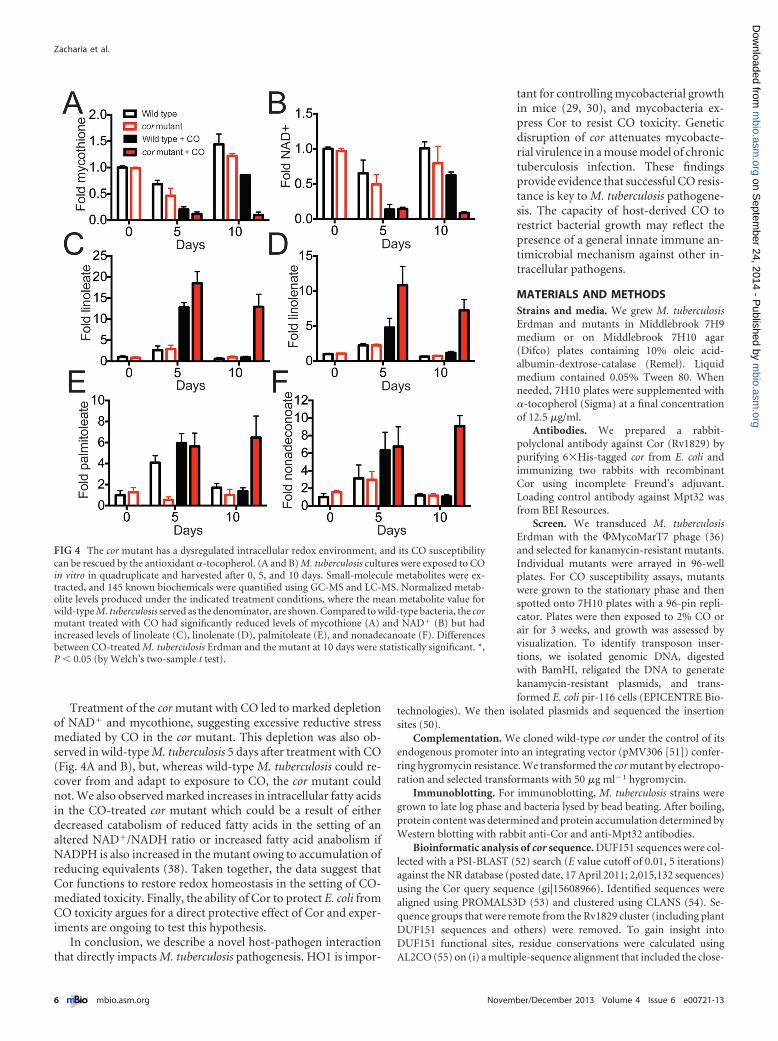
Treatment of the cor mutant with CO led to marked depletionof NAD� and mycothione, suggesting excessive reductive stressmediated by CO in the cor mutant. This depletion was also ob-served in wild-type M. tuberculosis 5 days after treatment with CO(Fig. 4A and B), but, whereas wild-type M. tuberculosis could re-cover from and adapt to exposure to CO, the cor mutant couldnot. We also observed marked increases in intracellular fatty acidsin the CO-treated cor mutant which could be a result of eitherdecreased catabolism of reduced fatty acids in the setting of analtered NAD�/NADH ratio or increased fatty acid anabolism ifNADPH is also increased in the mutant owing to accumulation ofreducing equivalents (38). Taken together, the data suggest thatCor functions to restore redox homeostasis in the setting of CO-mediated toxicity. Finally, the ability of Cor to protect E. coli fromCO toxicity argues for a direct protective effect of Cor and exper-iments are ongoing to test this hypothesis.
In conclusion, we describe a novel host-pathogen interactionthat directly impacts M. tuberculosis pathogenesis. HO1 is impor-
tant for controlling mycobacterial growthin mice (29, 30), and mycobacteria ex-press Cor to resist CO toxicity. Geneticdisruption of cor attenuates mycobacte-rial virulence in a mouse model of chronictuberculosis infection. These findingsprovide evidence that successful CO resis-tance is key to M. tuberculosis pathogene-sis. The capacity of host-derived CO torestrict bacterial growth may reflect thepresence of a general innate immune an-timicrobial mechanism against other in-tracellular pathogens.
MATERIALS AND METHODSStrains and media. We grew M. tuberculosisErdman and mutants in Middlebrook 7H9medium or on Middlebrook 7H10 agar(Difco) plates containing 10% oleic acid-albumin-dextrose-catalase (Remel). Liquidmedium contained 0.05% Tween 80. Whenneeded, 7H10 plates were supplemented with�-tocopherol (Sigma) at a final concentrationof 12.5 �g/ml.
Antibodies. We prepared a rabbit-polyclonal antibody against Cor (Rv1829) bypurifying 6�His-tagged cor from E. coli andimmunizing two rabbits with recombinantCor using incomplete Freund’s adjuvant.Loading control antibody against Mpt32 wasfrom BEI Resources.
Screen. We transduced M. tuberculosisErdman with the �MycoMarT7 phage (36)and selected for kanamycin-resistant mutants.Individual mutants were arrayed in 96-wellplates. For CO susceptibility assays, mutantswere grown to the stationary phase and thenspotted onto 7H10 plates with a 96-pin repli-cator. Plates were then exposed to 2% CO orair for 3 weeks, and growth was assessed byvisualization. To identify transposon inser-tions, we isolated genomic DNA, digestedwith BamHI, religated the DNA to generatekanamycin-resistant plasmids, and trans-formed E. coli pir-116 cells (EPICENTRE Bio-
technologies). We then isolated plasmids and sequenced the insertionsites (50).
Complementation. We cloned wild-type cor under the control of itsendogenous promoter into an integrating vector (pMV306 [51]) confer-ring hygromycin resistance. We transformed the cor mutant by electropo-ration and selected transformants with 50 �g ml�1 hygromycin.
Immunoblotting. For immunoblotting, M. tuberculosis strains weregrown to late log phase and bacteria lysed by bead beating. After boiling,protein content was determined and protein accumulation determined byWestern blotting with rabbit anti-Cor and anti-Mpt32 antibodies.
Bioinformatic analysis of cor sequence. DUF151 sequences were col-lected with a PSI-BLAST (52) search (E value cutoff of 0.01, 5 iterations)against the NR database (posted date, 17 April 2011; 2,015,132 sequences)using the Cor query sequence (gi|15608966). Identified sequences werealigned using PROMALS3D (53) and clustered using CLANS (54). Se-quence groups that were remote from the Rv1829 cluster (including plantDUF151 sequences and others) were removed. To gain insight intoDUF151 functional sites, residue conservations were calculated usingAL2CO (55) on (i) a multiple-sequence alignment that included the close-
FIG 4 The cor mutant has a dysregulated intracellular redox environment, and its CO susceptibilitycan be rescued by the antioxidant �-tocopherol. (A and B) M. tuberculosis cultures were exposed to COin vitro in quadruplicate and harvested after 0, 5, and 10 days. Small-molecule metabolites were ex-tracted, and 145 known biochemicals were quantified using GC-MS and LC-MS. Normalized metab-olite levels produced under the indicated treatment conditions, where the mean metabolite value forwild-type M. tuberculosis served as the denominator, are shown. Compared to wild-type bacteria, the cormutant treated with CO had significantly reduced levels of mycothione (A) and NAD� (B) but hadincreased levels of linoleate (C), linolenate (D), palmitoleate (E), and nonadecanoate (F). Differencesbetween CO-treated M. tuberculosis Erdman and the mutant at 10 days were statistically significant. *,P � 0.05 (by Welch’s two-sample t test).
Zacharia et al.
6 ® mbio.asm.org November/December 2013 Volume 4 Issue 6 e00721-13
m
bio.asm.org
on Septem
ber 24, 2014 - Published by
mbio.asm
.orgD
ownloaded from
sequence cluster (depicted in red and cyan in the CLANS map) and on (ii)a multiple-sequence alignment that included the extended sequence set(all sequences in the CLANS map).
Protein purification, gel electrophoresis, and nucleic acid degrada-tion assay. Recombinant His-MBP Cor and His-MBP were expressed inBL21 E. coli and were induced with 1 mM IPTG (isopropyl-�-D-thiogalactopyranoside). Proteins were purified using cobalt-chargedTALON resin (Clontech) under native, nondenaturing conditions. Dena-turing gel electrophoresis was performed using a 4%-to-20% Tris-glycine(SDS-PAGE) gel with a PageRuler Plus prestained protein ladder. Nativegel electrophoresis was performed using a NativePAGE Novex 4%-to-16% bis-Tris gel with a NativeMARK unstained protein standard. For thenucleic acid degradation assay, recombinant Cor or His-MBP alone atdifferent concentrations (0.5, 2.5, and 5.0 �M) was incubated with either4 �g of M. tuberculosis total RNA or 4 �g of M. smegmatis (mc2 155)genomic DNA in NEBuffer 3 for 1 h at 37°C. RNA samples were loaded
CORM-2 (μM)
0 25 250 0 25 250+ IPTG - IPTG
+ IPTG - IPTG
0 25 250 0 25 250 0 250
+ IPTGCor RFPA
B
Cα-Cor
CORM-2 (μM)
FIG 5 E. coli CO susceptibility is prevented by cor expression. (A) E. colitransformed with an IPTG-inducible vector expressing Cor or RFP was ex-posed to the CO donor corm-2 for 30 min with or without prior IPTG treat-ment, and serial dilutions were plated. (B) E. coli was grown as described above,and serial dilutions were plated to determine surviving CFU. (C) E. coli ex-pressing Cor was lysed and Cor accumulation determined by Western blotanalysis.
0 40 80 12023455
6
7
8
Days post-infection
log
CFU* *
0 100 200 300 4000
20
40
60
80
100
Days post-infection
Perc
ent s
urvi
val
Wild typecor mutantcor-comp
p<0.005
0 40 80 1202
3
4
5
Days post-infection
log
CFU *
**
A
B
C
FIG 6 Virulence of the cor mutant is attenuated in mice. BALBc mice wereinfected with 102 CFU wild-type (WT) M. tuberculosis, the cor mutant, or thecor mutant complemented with cor via aerosol. (A) Ten mice per group weremonitored over time for survival. By Kaplan-Meier analysis, differences be-tween groups infected with the cor mutant and M. tuberculosis Erdman or thecomplemented strain are statistically significant. (B and C) Five mice pergroup were sacrificed, mouse lung (B) and mouse liver (C) were harvested andhomogenized, and CFU were enumerated. *, P � 0.05 compared to WT (by thenonparametric Kruskal-Wallis test).
A Novel CO Resistance Gene from M. tuberculosis
November/December 2013 Volume 4 Issue 6 e00721-13 ® mbio.asm.org 7
m
bio.asm.org
on Septem
ber 24, 2014 - Published by
mbio.asm
.orgD
ownloaded from
with RNA sample loading buffer (Sigma-Aldrich) and were run on a 1.2%ethidium bromide-stained formaldehyde gel, whereas DNA samples wereloaded with DNA loading dye (New England Biolabs) and were run on a1% ethidium bromide-stained agarose gel.
Metabolomics. M. tuberculosis Erdman and the cor mutant weregrown in quadruplicate in roller bottles and exposed to air or 0.2% CO.At time zero (no treatment), day 5, and day 10, 1 � 109 bacteria werepelleted and resuspended in cold 100% methanol. Acetonitrile and waterwere then added for a final 40:40:20 ratio, the samples bead beated,and the supernatants collected and dried. The samples were then ana-lyzed by gas chromatography-mass spectrometry (GC-MS) and liquidchromatography-MS (LC-MS) and statistically significant differences de-termined by Welch’s two-sample test (Metabolon, Inc.).
Susceptibility to acid, nitric oxide, hypoxia, and plumbagin. Wegrew M. tuberculosis Erdman and the cor mutant to the late log phase,washed cells in phosphate-buffered saline (PBS), resuspended the cells atan optical density at 600 nm (OD600) of 0.1 in 7H9 at either pH 6.6 or 5.0in the absence or presence of NaNO2 (5 mM), and then measured OD600
daily. Alternatively, after washing the cells, we resuspended the bacteria in7H9-ADN (7H9, 0.2% glycerol, 0.05% Tween 80, 0.5% bovine serumalbumin, 0.2% dextrose, 0.085% NaCl) at pH 5.5 and exposed the bacteriato NaNO2 (3 mM) for 5 days. We determined CFU by plating serial dilu-tions of the suspensions on 7H10 agar plates. For growth under condi-tions of hypoxia, cells were grown in 17-ml test tubes in triplicate andgradual hypoxia was generated following the Wayne model (56). At eachtime point, tubes were sacrificed and CFU recorded. For testing ofplumbagin via disc diffusion, M. tuberculosis strains (wild type, cor mu-tant, and cor complement) were grown to an OD600 of 0.6 to 0.8 and wereback diluted to an OD600 of 0.1. Cultures were uniformly spread onto7H10 plates supplemented with oleic acid, albumin fraction V, and dex-trose, excluding catalase. Four discs (Oxoid/Thermo Scientific) wereplaced onto each plate, and 10 �l of plumbagin dissolved in ethanol wasspotted onto the discs (0 mM, 20 mM, and 100 mM). Treatment of discswith plumbagin was performed in duplicate per strain used in the exper-iment. The zone of inhibition surrounding the discs was measured inmillimeters.
CO treatment of E. coli. All BL21(DE3) E. coli cultures were grownunder aerobic conditions at 37°C with shaking at 250 rpm. The E. colistrain containing the pJ401 IPTG-inducible expression vector (DNA 2.0,Inc.) with the cor gene insert and the control E. coli strain (pJ401-RFP)were grown in LB medium plus kanamycin (50 �g/ml) to an OD600 of 0.35and induced with IPTG (1 mM final concentration). The cultures wereinduced for 3 h with IPTG to allow maximal expression of protein (de-tected by Coomassie; data not shown). The cultures were then back-diluted to an OD600 of 0.1 in M9 minimal salts (BD Difco) media supple-mented with MgSo4 and CaCl2. At an OD600 of 0.3, E. coli cultures weretreated with tricarbonyldichlororuthenium (II) dimer (corm-2; Sigma-Aldrich) as previously described by Tavares et al. (39). Briefly, corm-2 wasprepared as stock solutions dissolved in dimethyl sulfoxide (DMSO) andused at a final concentration of 0 �M (DMSO only), 25 �M, or 250 �M.E. coli bacteria were treated for 30 min at the indicated concentrations andwere serially diluted and spotted onto square LB agar plates with kanamy-cin (50 �g/ml) and incubated overnight at 37° C. For bacterial enumera-tion, E. coli bacteria were spread onto plates and also incubated overnightat 37° C to determine CFU. Cor protein expression was determined byWestern blot analysis using the anti-Cor antibody. Lysates from ~3 � 107
bacteria were loaded per well.Mouse infections. We infected BALBc mice (Jackson Laboratories)
using a Madison aerosol exposure chamber to deliver ~200 bacilli permouse. Prior to aerosolization, bacteria were washed repeatedly and son-icated to generate a single-cell suspension. At day zero, we plated totalorgan homogenates from both lungs (5 mice per group) to determine theinitial inoculum per M. tuberculosis strain. At subsequent time points, weplated serial dilutions of organ homogenates from lung (left lung) or liver(left lobe) from 5 mice per group. Animal experiments were reviewed and
approved by the Institutional Animal Care and Use Committee of Uni-versity of California at San Francisco and University of Texas (UT) South-western. For organ CFU comparisons, statistically significant differenceswere determined by the nonparametric Kruskal-Wallis test. For survivalexperiments (10 mice per strain), mice were sacrificed when they had lost15% of their maximal body weight as we had previously demonstratedthat this degree of weight loss predicted imminent mouse death (57, 58).Mouse survival curves were compared by Kaplan-Meier analysis.
SUPPLEMENTAL MATERIALSupplemental material for this article may be found at http://mbio.asm.org/lookup/suppl/doi:10.1128/mBio.00721-13/-/DCSupplemental.
Figure S1, EPS file, 17.9 MB.Table S1, PDF file, 0.1 MB.
ACKNOWLEDGMENTS
We thank C. Sassetti (University of Massachusetts) for the �MycomarT7phage, Z. Zheng for help with immunohistochemistry and immunofluo-rescence, and members of the Cox and Shiloh laboratories for helpfulsuggestions.
Funding for this work was provided by NIH grants AI076632 andAI099439 (M.U.S.), AI081727 (J.S.C.), DK081668 (D.K.M.), and5T32AI007520 (V.M.Z.). M.U.S. acknowledges support from the DiseaseOriented Clinical Scholars Program at UT Southwestern.
REFERENCES1. Huynh KK, Joshi SA, Brown EJ. 2011. A delicate dance: host response to
mycobacteria. Curr. Opin. Immunol. 23:464 – 472.2. Liu PT, Modlin RL. 2008. Human macrophage host defense against
Mycobacterium tuberculosis. Curr. Opin. Immunol. 20:371–376.3. Nathan C, Shiloh MU. 2000. Reactive oxygen and nitrogen intermediates
in the relationship between mammalian hosts and microbial pathogens.Proc. Natl. Acad. Sci. U. S. A. 97:8841– 8848.
4. Rohde K, Yates RM, Purdy GE, Russell DG. 2007. Mycobacteriumtuberculosis and the environment within the phagosome. Immunol. Rev.219:37–54.
5. Davis AS, Vergne I, Master SS, Kyei GB, Chua J, Deretic V. 2007.Mechanism of inducible nitric oxide synthase exclusion from myco-bacter ia l phagosomes. PLOS Pathog. 3:e186. doi :10.1371/journal.ppat.0030186.
6. Deghmane AE, Soualhine H, Bach H, Sendide K, Itoh S, Tam A, NoubirS, Talal A, Lo R, Toyoshima S, Av-Gay Y, Hmama Z. 2007. Lipoamidedehydrogenase mediates retention of coronin-1 on BCG vacuoles, leadingto arrest in phagosome maturation. J. Cell Sci. 120:2796 –2806.
7. Sturgill-Koszycki S, Schlesinger PH, Chakraborty P, Haddix PL, CollinsHL, Fok AK, Allen RD, Gluck SL, Heuser J, Russell DG. 1994. Lack ofacidification in Mycobacterium phagosomes produced by exclusion of thevesicular proton-ATPase. Science 263:678 – 681.
8. Zhang Y, Heym B, Allen B, Young D, Cole S. 1992. The catalase-peroxidase gene and isoniazid resistance of Mycobacterium tuberculosis.Nature 358:591–593.
9. Li Z, Kelley C, Collins F, Rouse D, Morris S. 1998. Expression of katGin Mycobacterium tuberculosis is associated with its growth and persis-tence in mice and guinea pigs. J. Infect. Dis. 177:1030 –1035.
10. Darwin KH, Nathan CF. 2005. Role for nucleotide excision repair invirulence of Mycobacterium tuberculosis. Infect. Immun. 73:4581– 4587.
11. Darwin KH, Ehrt S, Gutierrez-Ramos JC, Weich N, Nathan CF. 2003.The proteasome of Mycobacterium tuberculosis is required for resistanceto nitric oxide. Science 302:1963–1966.
12. Bryk R, Lima CD, Erdjument-Bromage H, Tempst P, Nathan C. 2002.Metabolic enzymes of mycobacteria linked to antioxidant defense by athioredoxin-like protein. Science 295:1073–1077.
13. Vandal OH, Pierini LM, Schnappinger D, Nathan CF, Ehrt S. 2008. Amembrane protein preserves intrabacterial pH in intraphagosomal Myco-bacterium tuberculosis. Nat. Med. 14:849 – 854.
14. Vandal OH, Roberts JA, Odaira T, Schnappinger D, Nathan CF, Ehrt S.2009. Acid-susceptible mutants of Mycobacterium tuberculosis share hy-persusceptibility to cell wall and oxidative stress and to the host environ-ment. J. Bacteriol. 191:625– 631.
15. Abramovitch RB, Rohde KH, Hsu FF, Russell DG. 2011. aprABC: a
Zacharia et al.
8 ® mbio.asm.org November/December 2013 Volume 4 Issue 6 e00721-13
m
bio.asm.org
on Septem
ber 24, 2014 - Published by
mbio.asm
.orgD
ownloaded from
Mycobacterium tuberculosis complex-specific locus that modulates pH-driven adaptation to the macrophage phagosome. Mol. Microbiol. 80:678 – 694.
16. Tan MP, Sequeira P, Lin WW, Phong WY, Cliff P, Ng SH, Lee BH,Camacho L, Schnappinger D, Ehrt S, Dick T, Pethe K, Alonso S. 2010.Nitrate respiration protects hypoxic Mycobacterium tuberculosis againstacid- and reactive nitrogen species stresses. PLoS One 5:e13356. doi:10.1371/journal.pone.0013356.
17. Sjostrand T. 1952. The formation of carbon monoxide by the decompo-sition of haemoglobin in vivo. Acta Physiol. Scand. 26:338 –344.
18. Tenhunen R, Marver HS, Schmid R. 1968. The enzymatic conversion ofheme to bilirubin by microsomal heme oxygenase. Proc. Natl. Acad. Sci.U. S. A. 61:748 –755.
19. Maines MD. 1988. Heme oxygenase: function, multiplicity, regulatorymechanisms, and clinical applications. FASEB J. 2:2557–2568.
20. Maines MD. 2004. The heme oxygenase system: past, present, and future.Antioxid. Redox Signal. 6:797– 801.
21. Ryter SW, Morse D, Choi AM. 2004. Carbon monoxide: to boldly gowhere NO has gone before. Sci. STKE 2004:RE6. doi:10.1126/stke.2302004re6.
22. Biernacki WA, Kharitonov SA, Barnes PJ. 2001. Exhaled carbon mon-oxide in patients with lower respiratory tract infection. Respir. Med. 95:1003–1005.
23. Paredi P, Shah PL, Montuschi P, Sullivan P, Hodson ME, KharitonovSA, Barnes PJ. 1999. Increased carbon monoxide in exhaled air of patientswith cystic fibrosis. Thorax 54:917–920.
24. Uasuf CG, Jatakanon A, James A, Kharitonov SA, Wilson NM, BarnesPJ. 1999. Exhaled carbon monoxide in childhood asthma. J. Pediatr. 135:569 –574.
25. Antuni JD, Kharitonov SA, Hughes D, Hodson ME, Barnes PJ. 2000.Increase in exhaled carbon monoxide during exacerbations of cystic fibro-sis. Thorax 55:138 –142.
26. Paredi P, Kharitonov SA, Barnes PJ. 2003. Exhaled carbon monoxide inlung disease. Eur. Respir. J. 21:197–198. doi:10.1183/09031936.02.00071802.(Author reply.)
27. Shiloh MU, Manzanillo P, Cox JS. 2008. Mycobacterium tuberculosissenses host-derived carbon monoxide during macrophage infection. CellHost Microbe 3:323–330.
28. Kumar A, Deshane JS, Crossman DK, Bolisetty S, Yan BS, Kramnik I,Agarwal A, Steyn AJ. 2008. Heme oxygenase-1-derived carbon monoxideinduces the Mycobacterium tuberculosis dormancy regulon. J. Biol.Chem. 283:18032–18039.
29. Regev D, Surolia R, Karki S, Zolak J, Montes-Worboys A, Oliva O,Guroji P, Saini V, Steyn AJ, Agarwal A, Antony VB. 2012. Hemeoxygenase-1 promotes granuloma development and protects against dis-semination of mycobacteria. Lab. Invest. 92:1541–1552.
30. Silva-Gomes S, Appelberg R, Larsen R, Soares MP, Gomes MS. 2013.Heme catabolism by heme oxygenase-1 confers host resistance to myco-bacterium infection. Infect. Immun. 81:2536 –2545.
31. Bach FH. 2006. Carbon monoxide: from the origin of life to molecularmedicine. Trends Mol. Med. 12:348 –350.
32. Weigel PH, Englund PT. 1975. Inhibition of DNA replication in Esche-richia coli by cyanide and carbon monoxide. J. Biol. Chem. 250:8536 – 8542.
33. Nobre LS, Seixas JD, Romão CC, Saraiva LM. 2007. Antimicrobialaction of carbon monoxide-releasing compounds. Antimicrob. AgentsChemother. 51:4303– 4307.
34. Davidge KS, Sanguinetti G, Yee CH, Cox AG, McLeod CW, Monk CE,Mann BE, Motterlini R, Poole RK. 2009. Carbon monoxide-releasingantibacterial molecules target respiration and global transcriptional regu-lators. J. Biol. Chem. 284:4516 – 4524.
35. Desmard M, Davidge KS, Bouvet O, Morin D, Roux D, Foresti R,Ricard JD, Denamur E, Poole RK, Montravers P, Motterlini R, Bocz-kowski J. 2008. A carbon monoxide-releasing molecule (corm-3) exertsbactericidal activity against Pseudomonas aeruginosa and improves sur-vival in an animal model of bacteraemia. FASEB J. 23:1023–1031.
36. Sassetti CM, Boyd DH, Rubin EJ. 2003. Genes required for mycobacte-rial growth defined by high density mutagenesis. Mol. Microbiol. 48:77– 84.
37. You MK, Shin HY, Kim YJ, Ok SH, Cho SK, Jeung JU, Yoo SD, Kim JK,Shin JS. 2010. Novel bifunctional nucleases, OmBBD and AtBBD1, areinvolved in abscisic acid-mediated callose deposition in Arabidopsis.Plant Physiol. 152:1015–1029.
38. Singh A, Crossman DK, Mai D, Guidry L, Voskuil MI, Renfrow MB,Steyn AJ. 2009. Mycobacterium tuberculosis WhiB3 maintains redox ho-meostasis by regulating virulence lipid anabolism to modulate macro-phage response . PLoS Pathog. 5:e1000545. doi :10 .1371/journal.ppat.1000545.
39. Tavares AF, Teixeira M, Romão CC, Seixas JD, Nobre LS, Saraiva LM.2011. Reactive oxygen species mediate bactericidal killing elicited by car-bon monoxide-releasing molecules. J. Biol. Chem. 286:26708 –26717.
40. Park SW, Hwang EH, Park H, Kim JA, Heo J, Lee KH, Song T, Kim E,Ro YT, Kim SW, Kim YM. 2003. Growth of mycobacteria on carbonmonoxide and methanol. J. Bacteriol. 185:142–147.
41. King GM. 2003. Uptake of carbon monoxide and hydrogen at environ-mentally relevant concentrations by mycobacteria. Appl. Environ. Micro-biol. 69:7266 –7272.
42. Chan J, Xing Y, Magliozzo RS, Bloom BR. 1992. Killing of virulentMycobacterium tuberculosis by reactive nitrogen intermediates producedby activated murine macrophages. J. Exp. Med. 175:1111–1122.
43. Ng VH, Cox JS, Sousa AO, MacMicking JD, McKinney JD. 2004. Roleof KatG catalase-peroxidase in mycobacterial pathogenesis: counteringthe phagocyte oxidative burst. Mol. Microbiol. 52:1291–1302.
44. O’Brien L, Carmichael J, Lowrie DB, Andrew PW. 1994. Strains ofmycobacterium tuberculosis differ in susceptibility to reactive nitrogenintermediates in vitro. Infect. Immun. 62:5187–5190.
45. Voskuil MI, Schnappinger D, Visconti KC, Harrell MI, Dolganov GM,Sherman DR, Schoolnik GK. 2003. Inhibition of respiration by nitricoxide induces a Mycobacterium tuberculosis dormancy program. J. Exp.Med. 198:705–713.
46. Venugopal A, Bryk R, Shi S, Rhee K, Rath P, Schnappinger D, Ehrt S,Nathan C. 2011. Virulence of mycobacterium tuberculosis depends onlipoamide dehydrogenase, a member of three multienzyme complexes.Cell Host Microbe 9:21–31.
47. Motterlini R, Otterbein LE. 2010. The therapeutic potential of carbonmonoxide. Nat. Rev. Drug Discov. 9:728 –743.
48. Boczkowski J, Poderoso JJ, Motterlini R. 2006. Co-metal interaction:vital signaling from a lethal gas. Trends Biochem. Sci. 31:614 – 621.
49. Wayne LG. 1976. Dynamics of submerged growth of Mycobacteriumtuberculosis under aerobic and microaerophilic conditions. Am. Rev. Re-spir. Dis. 114:807– 811.
50. Rubin EJ, Akerley BJ, Novik VN, Lampe DJ, Husson RN, Mekalanos JJ.1999. In vivo transposition of mariner-based elements in enteric bacteriaand mycobacteria. Proc. Natl. Acad. Sci. U. S. A. 96:1645–1650.
51. Stover CK, de la Cruz VF, Fuerst TR, Burlein JE, Benson LA, BennettLT, Bansal GP, Young JF, Lee MH, Hatfull GF, Snapper SB, BarlettaRG, Jacobs, WR, Jr, Bloom BR. 1991. New use of BCG for recombinantvaccines. Nature 351:456 – 460.
52. Altschul SF, Madden TL, Schäffer AA, Zhang J, Zhang Z, Miller W,Lipman DJ. 1997. Gapped BLAST and PSI-BLAST: a new generation ofprotein database search programs. Nucleic Acids Res. 25:3389 –3402.
53. Pei J, Tang M, Grishin NV. 2008. PROMALS3D web server for accuratemultiple protein sequence and structure alignments. Nucleic Acids Res.36:W30 –W34.
54. Frickey T, Lupas A. 2004. CLANS: a Java application for visualizingprotein families based on pairwise similarity. Bioinformatics 20:3702–3704.
55. Pei J, Grishin NV. 2001. AL2CO: calculation of positional conservation ina protein sequence alignment. Bioinformatics 17:700 –712.
56. Wayne LG, Hayes LG. 1996. An in vitro model for sequential study ofshiftdown of Mycobacterium tuberculosis through two stages of nonrep-licating persistence. Infect. Immun. 64:2062–2069.
57. Manzanillo PS, Shiloh MU, Portnoy DA, Cox JS. 2012. Mycobacteriumtuberculosis activates the DNA-dependent cytosolic surveillance pathwaywithin macrophages. Cell Host Microbe 11:469 – 480.
58. Ohol YM, Goetz DH, Chan K, Shiloh MU, Craik CS, Cox JS. 2010.Mycobacterium tuberculosis MycP1 protease plays a dual role in regula-tion of ESX-1 secretion and virulence. Cell Host Microbe 7:210 –220.
A Novel CO Resistance Gene from M. tuberculosis
November/December 2013 Volume 4 Issue 6 e00721-13 ® mbio.asm.org 9
m
bio.asm.org
on Septem
ber 24, 2014 - Published by
mbio.asm
.orgD
ownloaded from