listeria infections: epidemiology, pathogenesis and treatment
TRANSCRIPT

Complimentary Contributor Copy

Complimentary Contributor Copy

BACTERIOLOGY RESEARCH DEVELOPMENTS
LISTERIA INFECTIONS
EPIDEMIOLOGY, PATHOGENESIS
AND TREATMENT
No part of this digital document may be reproduced, stored in a retrieval system or transmitted in any form orby any means. The publisher has taken reasonable care in the preparation of this digital document, but makes noexpressed or implied warranty of any kind and assumes no responsibility for any errors or omissions. Noliability is assumed for incidental or consequential damages in connection with or arising out of informationcontained herein. This digital document is sold with the clear understanding that the publisher is not engaged inrendering legal, medical or any other professional services.
Complimentary Contributor Copy

BACTERIOLOGY RESEARCH DEVELOPMENTS
Additional books in this series can be found on Nova’s website
under the Series tab.
Additional e-books in this series can be found on Nova’s website
under the e-book tab.
ALLERGIES AND INFECTIOUS DISEASES
Additional books in this series can be found on Nova’s website
under the Series tab.
Additional e-books in this series can be found on Nova’s website
under the e-book tab.
Complimentary Contributor Copy

BACTERIOLOGY RESEARCH DEVELOPMENTS
LISTERIA INFECTIONS
EPIDEMIOLOGY, PATHOGENESIS
AND TREATMENT
ANDINO ROMANO
AND
CARMINE F. GIORDANO
EDITORS
Nova Science Publishers, Inc. New York
Complimentary Contributor Copy

Copyright © 2012 by Nova Science Publishers, Inc.
All rights reserved. No part of this book may be reproduced, stored in a retrieval system or
transmitted in any form or by any means: electronic, electrostatic, magnetic, tape, mechanical
photocopying, recording or otherwise without the written permission of the Publisher.
For permission to use material from this book please contact us:
Telephone 631-231-7269; Fax 631-231-8175
Web Site: http://www.novapublishers.com
NOTICE TO THE READER
The Publisher has taken reasonable care in the preparation of this book, but makes no expressed or
implied warranty of any kind and assumes no responsibility for any errors or omissions. No
liability is assumed for incidental or consequential damages in connection with or arising out of
information contained in this book. The Publisher shall not be liable for any special,
consequential, or exemplary damages resulting, in whole or in part, from the readers’ use of, or
reliance upon, this material. Any parts of this book based on government reports are so indicated
and copyright is claimed for those parts to the extent applicable to compilations of such works.
Independent verification should be sought for any data, advice or recommendations contained in
this book. In addition, no responsibility is assumed by the publisher for any injury and/or damage
to persons or property arising from any methods, products, instructions, ideas or otherwise
contained in this publication.
This publication is designed to provide accurate and authoritative information with regard to the
subject matter covered herein. It is sold with the clear understanding that the Publisher is not
engaged in rendering legal or any other professional services. If legal or any other expert
assistance is required, the services of a competent person should be sought. FROM A
DECLARATION OF PARTICIPANTS JOINTLY ADOPTED BY A COMMITTEE OF THE
AMERICAN BAR ASSOCIATION AND A COMMITTEE OF PUBLISHERS.
Additional color graphics may be available in the e-book version of this book.
Library of Congress Cataloging-in-Publication Data
Library of Congress Control Number: 2012936694
Published by Nova Science Publishers, Inc. † New York
ISBN: 978-1-62081-647-9 (eBook)
Complimentary Contributor Copy

Contents
Preface vii
Chapter I The Behavior of Listeria monocytogenes during
the Manufacture and Storage of Greek Protected
Designation of Origin (PDO) Cheeses 1 Apostolos S. Angelidis and Alexandros Govaris
Chapter II Epidemic Clones of Listeria Monocytogenes:
Detection, Transmission and Virulence 35 Sara Lomonaco
Chapter III Listeria: Epidemiology, Pathogenesis and Novel
Potential Treatments 67 Lucila Saavedra, Augusto Bellomio, Elvira M. Hebert,
Carlos Minahk, Nadia Suarez, and Fernando Sesma
Chapter IV Sublethal Damage in Listeria monocytogenes after
Non-Thermal Treatments, and Implications for Food Safety 99 A. Silva, C. M. Belda-Galbis, S. F. Zanini, D. Rodrigo,
P. Martorell, and A. Martínez
Chapter V Listeria Infections Review: Epidemiology,
Pathogenesis and Treatment 115 Karina Pellicer
Chapter VI Epidemiology of Listeria monocytogenes in RTE Fermented
Meat and Smoked Fish Products 129 D. Meloni, A. Mureddu, F. Piras, R. Mazza, S. Lamon,
S. G. Consolati, F. Fois, and R. Mazzette
Chapter VII Epidemiology of Invasive Listeriosis in Clinical Cases in Navarra
(Spain): Comparison between 1995-2005 and 2006-2011 141 V. Garrido, A. I. Vitas, I. García-Jalón, L. Torroba,
and A. Navascués
Complimentary Contributor Copy

Contents vi
Chapter VIII Listeria Infections: Epidemiology, Pathogenesis and Treatment 155 Imane Saleh, Nisreen Alwan, Elie Barbour, Esam Azhar,
and Steve Harakeh
Chapter IX Listeria in Wildlife of Russia 167 I. Yegorova, Ju. Selyaninov, and V. Fertickov
Index 177
Complimentary Contributor Copy

Preface
Microbial food-borne illnesses have a great impact not only on public health but also
represent high economic costs for many countries around the world. Listeria monocytogenes,
is a gram-positive facultative intracellular pathogen. In this book, the authors discuss the
epidemiology, pathogenesis and treatment of Listeria infections. Topics include the behavior
of L. monocytogenes in Greek PDO cheeses and preventing the pathogen's proliferation;
epidemic clones of Listeria monocytogenes; description of outbreaks, pathogenesis and
technology for controlling Listeria; sublethal damage in Listeria monocytogenes after non-
thermal treatments and implications for food safety; and Listeria monocytogenes in RTE
fermented meat and smoked fish products.
Chapter I - Among the numerous traditional cheese varieties produced throughout Greece
today, 21 cheeses have been granted the Protected Designation of Origin (PDO) status,
whereas others are awaiting recognition. The Greek PDO cheeses include all types of cheeses
(hard, semi-hard and soft cheeses). Several studies worldwide have reported the occurrence of
Listeria spp. including Listeria monocytogenes in raw milk from sheep and goats.
The majority of the Greek PDO cheeses are manufactured by a mixture of sheep and goat
milk, which can be used raw, or after pasteurization. According to the European Commission
Regulation (EC) 2073/2005 and its amendment by Regulation (EC) 1441/2007 food business
operators are responsible for ensuring product compliance with the new food safety criteria
including those specified for the food-borne pathogen L. monocytogenes. The ability of L.
monocytogenes to proliferate during the manufacture and storage of permissive ready-to-eat
foods is probably the most important factor regarding the risk for listeriosis. Currently, there
are published studies pertaining to the behavior of L. monocytogenes in seven PDO cheeses
(Feta, Galotyri, Graviera, Kasseri, Katiki, Manouri and Pichtogalo Chanion). This chapter
aims at reviewing the available scientific literature regarding the behavior of L.
monocytogenes in these Greek PDO cheeses, as well as the different interventions that have
been proposed aiming at preventing the pathogen’s proliferation.
Chapter II - Although many different serotypes of Listeria monocytogenes have been
isolated from foods, only a few such as 1/2a, 1/2b, and 4b account for the vast majority of
clinical cases, and in particular most outbreaks of listeriosis have involved a small number of
closely related clones in serotypes 1/2a and 4b. An epidemic clone (EC) of L. monocytogenes
has been defined as groups of isolates that are genetically related and presumably of a
common ancestor, but are implicated in different, geographically and temporally unrelated
Complimentary Contributor Copy

Andino Romano and Carmine F. Giordano viii
outbreaks. Since the introduction of this concept in 2002, four ECs have been recognized:
ECI, ECII, and ECIV in serotype 4b and ECIII in serotype 1/2a. Most recently, in 2011, a
novel serotype 1/2a EC that had been causing disease in Canada for the past twenty years was
detected and designated as ECV. As L. monocytogenes continues to spread throughout the
world and cause many different outbreaks, it is reasonable to assume that other novel ECs
could be identified in the future. In order to control L. monocytogenes, determining the routes
by which ECs and outbreak clones (OCs) are transmitted to foods will be extremely helpful.
Consequently, the availability of subtyping assays capable of discerning closely related ECs
and OCs is extremely important. In the past few years, the number and quality of subtyping
assays has increased thanks to the development of DNA-based methods such as pulsed-field
gel electrophoresis (PFGE), considered the gold-standard for its high discriminatory power
(i.e. the ability to correctly differentiate unrelated strains). PFGE has proven to be a very
accurate and reproducible method in subtyping L. monocytogenes. However, new sequence-
based techniques, such as multilocus-sequence typing (MLST) and single nucleotide
polymorphism (SNP) typing, are also currently being used. In particular, multi-virulence-
locus sequence typing (MVLST) has been able to accurately identify and differentiate all five
ECs of L. monocytogenes. This technique also proved very useful in the identification of ECs,
when PFGE failed to identify strains belonging to the same ECs due to the presence of
different PFGE profiles. Therefore, the use of novel DNA-sequence-based molecular
subtyping strategies able to correctly determine the clonal relationship among different
isolates is essential to accurately recognize outbreaks and epidemics and identify their routes
of transmission. This chapter will review the five currently known ECs of L. monocytogenes,
including listeriosis outbreaks caused by these ECs during the past few years, and the latest
molecular subtyping methods developed to identify them.
Chapter III - Microbial food-borne illnesses have a great impact not only in public health
but also represent high economic costs for many countries around the world. Listeria
monocytogenes, a Gram-positive facultative intracellular pathogen, is estimated to cause
nearly 1,600 illnesses each year in the United States. Listeriosis may have different clinical
syndromes from a non-invasive form in healthy human usually associated to mild
gastroenteritis to severe invasive form, especially during pregnancy and in people with
compromised immune system. In this case, it can be a serious and sometimes fatal disease.
The transmission can occur by different ways, being the most important one the consumption
of contaminated food, especially ready-to-eat products. Many efforts have been made in order
to control o minimize the presence of this pathogen in food and food processing areas.
Besides the new antilisterial technologies that are being explored such as surface
pasteurization or ozone treatments, antimicrobial peptides called bacteriocins produced by
GRAS microorganisms such as lactic acid bacteria arise as a potential solution in this field.
There are many Listeria-active bacteriocins described so far in the literature and products
containing purified or semi-purified bacteriocins are already in the market. Moreover, some
anti-listerial bacteriocins proved to be active not only in food environments but also in
listeria-infected mice models. In this chapter, an updated description of outbreaks,
pathogenesis as well as the new technology for controlling Listeria is presented.
Chapter IV - The presence of microorganisms in foods during production, packaging,
transport and storage is unavoidable. Since January 2005, Regulation 178/2002/EC or
General Food Law requires the traceability of the food chain in all its stages (EU, 2002). This
directive will enforce the introduction of security mechanisms and controls for foodstuffs.
Complimentary Contributor Copy

Preface ix
One of the microorganisms of concern, mainly for ready-to-eat foods, is Listeria
monocytogenes. Between 2002 and 2006 there was a significant increase in reported cases of
human listeriosis in Member States of the European Union (EU) (EFSA, 2007). In 2007 there
were 1,639 reported cases, of which 1,635 were subsequently confirmed, in 29 countries.
The rate of global reporting was 0.35 cases per 100,000 inhabitants. The proportion of
samples exceeding the legal limit for Listeria monocytogenes in Member States was higher in
ready-to-eat foods based on fish, followed by meat products and cheeses (EFSA and ECDC,
2011). Consequently, knowledge of microbial inactivation and growth behavior is very
important for food safety and shelf-life assessment. Important aspects that should be
considered as emerging risks are the changes that could take place after sublethal injury, i.e.,
changes in virulence. This review aims to evaluate Listeria monocytogenes inactivation and
growth in foodstuffs by simulating the supply chain in order to verify the relation existing
between the various treatments that could be used by the industry to increase product lifetime
without risk to consumers and sublethal damage.
Chapter V - Listeria monocytogenes is a pathogen that affects animals and human beings;
it can produce potentially fatal infections in susceptible individuals. This bacterium can cause
miscarriages in pregnant women and meningitis in newborns, children and adults who are
immunosuppressed. Most cases of Listeriosis are sporadic; although outbreaks have been
described due to food consumption. The ability of L. monocytogenes to resist stressful
environmental conditions makes this pathogen a food industry concern.
Chapter VI - Listeria monocytogenes is an ubiquitous organism, widely distributed in the
environment. The principal reservoirs are soil, forage and water. Other reservoirs include
healthy humans and animals or infected domestic and wild animals. L.monocytogenes is also
the etiologic agent of listeriosis, which occurs in humans and animals. Since the beginning of
the 1980’s L. monocytogenes has been recognized as an emerging food-borne pathogen after
several sporadic and epidemic cases of listeriosis occurred in Europe and the US. Recently
the incidence of sporadic cases rose again in Europe. Two main forms of listeriosis have been
described in humans: febrile gastroenteritis in healthy individuals and life-threatening
invasive infections in susceptible individuals, with the latter posing a serious problem to
public health. In fact, invasive human listeriosis is a rare but severe infection, typically
causing septicemia, encephalitis and meningitis in defined high-risk groups: young, old,
pregnant and immune-compromised, the so called “YOPI”. Listeriosis is the fifth most
common zoonotic disease in Europe, less common than other diseases (eg. by Escherichia
coli 0157:H7, Campylobacter jejuni or Salmonella spp.). It has an incidence of 3.3 cases per
1.000.000 population per year, an estimated case fatality rate of 20 up to 30% and the highest
hospitalization rate (90%) of all food -borne pathogens with additional long term sequelae in
some patients. In Europe, 55.6% of all human listeriosis cases are reported in patients aged
above 60 years, approximately 2.5 times higher than those reported in any other age group.
Therefore, although this disease continues to occur in association with pregnancy, it is now
predominantly an infection of immune-compromised individuals amongst the older sections
of the populations. Data on listeriosis in USA show a similar marked reduction-trend. The
majority (99%) of the infections caused by L.monocytogenes are thought to be foodborne.
The pathogen is able to survive at a broad range of temperature (from 0 to 45°C) and pH
(from 4.5 to 9.0), high salt concentrations (10%) and low water activity values (0.92). These
properties, together with the severity of human listeriosis infections, make L. monocytogenes
of particular concern for manufacturers of cold stored “ready to eat” (RTE) foods (Romanova
Complimentary Contributor Copy

Andino Romano and Carmine F. Giordano x
et al., 2002; Van Coillie et al., 2004; Shen et al., 2006). RTE food is a large, heterogeneous
category of foodstuffs and can be subdivided in many different ways. According to the Codex
definition, RTE include any food that is normally consumed in its raw state or any food
handled, processed, mixed, cooked or otherwise prepared into a form in which it is normally
consumed without further processing. L.monocytogenes has been isolated from a wide variety
of RTE products capable of supporting its growth and is responsible for numerous outbreaks
associated with the consumption of RTE meat, poultry, dairy, fish and vegetable products.
Seafood products have been reported to be contaminated with L.monocytogenes and several
reports of outbreaks or sporadic cases have been linked to these products in particular with
cold or hot-smoked salmon, “gravad” salmon, shrimps, mussels, fermented fish, fish and
seafood salads. Concerning RTE meat products, a large outbreak occurred in 2008 in Canada
causing 22 deaths and 57 confirmed cases. Although fermented pork meat RTE products as
dry and semi-dry sausages have been rarely implicated in food poisoning, more risks should
be linked to the consumption of these products because mainly in the manufacturing of
traditional products, an empirical application of the hurdles technology often occurs. In the
following, through an up-to-date review of (personal and non) published data, the
epidemiology of L.monocytogenes in two selected RTE food categories will be discussed:
fermented meat and smoked fish products. Fermented meat products are often contaminated
and are produced without any lethal processing step while smoked fish products (in particular
cold smoked ones) are frequently contaminated, have no lethal processing steps and permits
growth during an extended storage period.
Chapter VII - The incidence of human listeriosis in Navarra (Spain) was monitored
during two different periods of time (1995-2005 and 2006-2011) by active surveillance in
collaboration with the main hospitals within this region. A total of 72 cases of invasive
listeriosis were detected, with an average incidence rate of 0.75/100,000 inhabitants. The
incident rate shows a tendency to increase, as the first period (40 cases within 11 years)
showed a rate of 0.65/100,000 while the second period (32 cases within 6 years) showed a
rate of 0.86/100,000. Over the whole period studied, 44.4% of the cases were diagnosed
among aged population (32 cases out 72), the group most affected by listeriosis, while case
fatality (including fetal death) was 57.9% in pregnant women (n=11 out of 19 pregnancy-
associated cases). Most of the isolated strains belonged to serotype 4b (n=38 out of 65 strains;
58.5%), but a significant increase of 1/2a serotype has been observed in recent years
(P≤0.05). In addition, serotype 1/2c was isolated from a clinical case, and to the best of our
knowledge, it is the first clinical isolation of this serotype in the region. In this chapter, the
epidemiology of human listeriosis and how to improve the current Spanish surveillance
system will be discussed.
Chapter VIII - Listeria is a motile, Gram-positive, rod-shaped coccobacillus. It is a non-
sporeforming, facultatively anaerobic bacterium that can cause a life-threatening disease to
both humans and animals known as Listeriosis. Listeria can frequently be isolated from soil,
water, food products and vegetation. It is a major food-borne pathogen worldwide, accounting
for about 28% of food-related deaths in the USA alone. In 2006, Listeriosis was reported in
23 European Union Member States and it ranked fifth among the most common zoonotic
infections in Europe after Campylobacter, Salmonella, Yersinia and Verotoxin-Producing
Escherichia coli (VTEC) infections. The most pathogenic species of Listeria is Listeria
monocytogenes that often causes food-borne infections in immuno-compromised hosts,
including newborns and the elderly. As a facultative intracellular parasitic bacterium,
Complimentary Contributor Copy

Preface xi
L. monocytogenes invades a variety of host cells, such as hepatocytes, fibroblasts and
epithelial cells, multiplying in the cytoplasm of these cells. This pathogen is one of the
reasons of meningoencephalitis and abortion in ruminants. In neonates, it is the third most
common cause of bacterial meningitis after E. coli and Streptococcus agalactiae. In most
Listeria infections cases, the symptoms spontaneously clear in about seven days. However,
patients at high risk such as pregnant women require antimicrobial treatment to prevent halt
and development of more severe diseases. The duration of antimicrobial treatment depends on
the severity of the infection. In Listeriosis treatment, the initial choice of antimicrobials is
usually Ampicillin. Some studies also reported a successful treatment using the synergism
present in the Trimethoprim-sulfamethoxazole preparations.
Chapter IX - Based on long-term monitoring studies carried out among wild ungulates
and aquatic organisms living in woodland and hunting areas and freshwater basins of Russia,
we determined a carrier state for both pathogenic and nonpathogenic Listeria species of sika
deer, red deer, wild boar, as well as herbivorous and carnivorous fish species. Among the
eight currently known listeria species, only two ones, namely Listeria innocua and Listeria
monocytogenes have been found in hot- and cold-blooded representatives of the fauna, with
Listeria innocua being most often isolated from the samples tested. The prevalence rates
among aquatic organisms were 0.8% for L. monocytogenes and 5.2% for L. innocua, while in
wild cloven-hoofed species the rates were 2.2 to 12% and 1.5 to 29%, respectively. For wild
cloven-hoofed animals, listeria in most cases were found in feces, and only in two cases the
agent was found in brain and lymph node of a hunted sika deer, and in the liver of a died wild
boar. The carriage state levels among cloven-hoofed species significantly depended on the
forms of economic activities and compliance with sanitation requirements. Among fish, the
carrier state for listeria was determined in both herbivorous and carnivorous fish species. L.
monocytogenes was mainly found in bream, carp, white bream and perch, while L. innocua in
bream, pike, white bream, rudd, crucian carp and perch. Listeria of both pathogenic and
nonpathogenic species were most frequently found in fish skin and gill tissues. The carried
out investigations also indicated that the soil in feeding grounds and rotting plant residues are
natural reservoirs for listeria, with the immigration of juvenile fish not checked for listeria
carrier state and carcasses of drowned listeria-carrying wildlife being another source of
listeria introduction into aquatic fauna of freshwater basins, beside the known ones.
In order to identify phylogenetic relationships among the identified isolates, a pulse-
electrophoresis of a restricted chromosomal DNA (REA-PFGE) was carried out. The results
of the investigations into genetic variability among the isolates collected from wild cloven-
hoofed animals using REA-PFGE revealed a variety of pulse electrotypes, suggesting
multiple sources of infection. In freshwater fish populations a circulation of three L.
monocytogenes clonal variants was found, with the pulse-electrotype isolates as found in
different fish species, caught in the waters of the same river, having 100% coincidence. In
addition, the pulse electrotype of a perch isolate was identical to a restriction profile of an
isolate from sika deer feces found in the same region.
Complimentary Contributor Copy

Complimentary Contributor Copy

In: Listeria Infections ISBN: 978-1-62081-639-4
Editors: A. Romano and C. F. Giordano © 2012 Nova Science Publishers, Inc.
Chapter I
The Behavior of Listeria
monocytogenes during the
Manufacture and Storage
of Greek Protected Designation
of Origin (PDO) Cheeses
Apostolos S. Angelidis,1 and Alexandros Govaris
2 1Laboratory of Milk Hygiene and Technology,
Faculty of Veterinary Medicine, Aristotle University of Thessaloniki,
Thessaloniki, Greece 2Laboratory of Hygiene of Foods of Animal Origin,
Faculty of Veterinary Medicine, University of Thessaly, Karditsa, Greece
Abstract
Among the numerous traditional cheese varieties produced throughout Greece today,
21 cheeses have been granted the Protected Designation of Origin (PDO) status, whereas
others are awaiting recognition. The Greek PDO cheeses include all types of cheeses
(hard, semi-hard and soft cheeses).
Several studies worldwide have reported the occurrence of Listeria spp. including
Listeria monocytogenes in raw milk from sheep and goats. The majority of the Greek
PDO cheeses are manufactured by a mixture of sheep and goat milk, which can be used
raw, or after pasteurization.
According to the European Commission Regulation (EC) 2073/2005 and its
amendment by Regulation (EC) 1441/2007 food business operators are responsible for
ensuring product compliance with the new food safety criteria including those specified
Correspondence: Dr Alexandros Govaris,
Laboratory of Hygiene of Foods of Animal Origin, Faculty of Veterinary
Medicine, University of Thessaly, 224 Trikalon Street, 43100 Karditsa, Greece. Tel. +30 24410 66086, Fax:
+30 24410 66087 Email: [email protected]
Complimentary Contributor Copy

Apostolos S. Angelidis and Alexandros Govaris 2
for the food-borne pathogen L. monocytogenes. The ability of L. monocytogenes to
proliferate during the manufacture and storage of permissive ready-to-eat foods is
probably the most important factor regarding the risk for listeriosis.
Currently, there are published studies pertaining to the behavior of L. monocytogenes
in seven PDO cheeses (Feta, Galotyri, Graviera, Kasseri, Katiki, Manouri and Pichtogalo
Chanion). This chapter aims at reviewing the available scientific literature regarding the
behavior of L. monocytogenes in these Greek PDO cheeses, as well as the different
interventions that have been proposed aiming at preventing the pathogen’s proliferation.
Introduction
Cheese is a highly nutritious food and a great variety of cheeses are manufactured
throughout the world. In Greece there are several traditional cheeses produced at the
industrial or artisanal scale both in the mainland, as well as in the Greek islands (Anifantakis,
1991; Litopoulou-Tzanetaki and Tzanetakis, 2011). Twenty one of these traditional Greek
cheeses have been granted Protected Designation of Origin (PDO) status (European
Commission, 1996, 2002, 2011). As is the case for other Mediterranean countries, PDO
cheeses are food commodities bearing strong cultural, social and economic importance. The
milk used for PDO cheesemaking should be of good quality, collected from lactating breeds
of ruminants raised in austerely specified geographical regions at least 10 days after
parturition, and the use of condensed milk, milk powder, caseinates, milk protein
concentrates, preservatives or colorants is prohibited (Greek Codex of Foodstuffs and
Beverages, 2009).
In general, cheeses can be classified using various classification schemes such as those
based on their final moisture content, or their technology of manufacture.
Table 1. Physicochemical specifications and characteristics of selected PDO Greek cheeses
Cheese Cheese type Milk type1
Ripening
(days) 2
Moisture
(%)3
FDM
(%)4
Feta Soft, white-brined O (30% C) 60 56 43
Galotyri Soft, acid-curd O, C, O/C 605 75 40
Graviera Agrafon Hard O (30% C) 90 38 40
Graviera Naxou Hard B (20% C/O) 90 38 40
Graviera Kritis Hard O (20% C) 90 38 40
Kasseri Semi-hard O (20% C) 90 45 40
Katiki Domokou Soft, acid-curd C, C/O n/a 75 40
Manouri Soft, whey-cheese O, C, O/C n/a 60 70
Pichtogalo Chanion Soft, acid-curd C, O, C/O n/a 65 50 1 B, bovine milk; C, caprine milk; O, ovine milk. Percentages added in parentheses refer to the
maximum allowable content of the alternative milk in the milk mixture. 2 The “ripening” of commercially produced Galotyri is much shorter (ca. 1 week or less).
3 Maximum values imposed by the Greek Codex of Foodstuffs and Beverages.
4 Minimum values imposed by the Greek Codex of Foodstuffs and Beverages.
Based on their moisture content the 21 Greek PDO cheeses can be classified as hard
cheeses (Graviera Agrafon, Graviera Naxou, Graviera Kritis, Kefalograviera, Ladotyri
Complimentary Contributor Copy

The Behavior of Listeria monocytogenes during the Manufacture and Storage … 3
Mytilinis and San Michali), semi-hard cheeses (Batzos, Formaela Arachovas Parnassou,
Kasseri, Metsovone and Sfela) and soft cheeses (Anevato, Feta, Galotyri, Kalathaki Limnou,
Katiki Domokou, Kopanisti, Manouri, Pichtogalo Chanion, Xynomyzithra Kritis and Xygalo
Siteias). Table 1 presents the specifications outlined in the Greek Codex of Foodstuffs and
Beverages (2009) pertaining to the physicochemical characteristics of the Greek PDO cheeses
discussed in this chapter, whereas Table 2 presents physicochemical data retrieved from the
literature on these PDO cheeses. The microbiological safety of most cheeses is dependent on
some crucial factors such as the application (or absence thereof) of thermal treatment
(thermization, pasteurization) to the raw milk, growth of lactic acid bacteria, the rate and
extent of acidification during cheese manufacture and ripening, heating of curd in certain
cheese types, the concentration of the salt in the aqueous phase (SWP) of the cheese and the
extent and type of microbial contamination(s) during the various steps of cheesemaking.
Raw milk can contain low levels of L. monocytogenes (Greenwood et al., 1991;
Rodriguez et al., 1994; Meyer-Broseta et al., 2003). The pasteurization of raw milk, however,
which inactivates the low levels of L. monocytogenes, is not sufficient to eliminate later risks
of L. monocytogenes contamination in dairy products. Indeed, the pathogen is often isolated
from the food-processing environment, including cheese factories (Kornacki and Gurtler,
2007; Kousta et al., 2010), and several outbreaks of listeriosis have been attributed to the
consumption of cheeses (mostly soft cheeses) contaminated with L. monocytogenes after
manufacturing (Linan et al. 1988; Alterkruse et al., 1998; De Buyser et al., 2001; Lunden et
al., 2004; Dewaal et al., 2006). Owing to their technology of manufacture and their resulting
physicochemical properties, soft, unripened cheeses present a higher incidence and potential
for growth or survival of L. monocytogenes than other cheese varieties and are considered
foods with an increased risk for listeriosis (Schuchat et al., 1992). Hence, a recent scientific
report of the European Food Safety Authority (EFSA) proposed that each Member State of
the EU should conduct a survey on L. monocytogenes in selected categories of ready-to-eat
(RTE) foods (including soft cheeses) at retail (EFSA, 2009). In order to satisfy the EFSA
requirements, a study involving samples of soft cheeses available in the Greek retail market
was conducted recently. A total of 137 soft cheese samples were tested for L. monocytogenes
as well as other Listeria species both qualitatively and quantitatively. None of the cheeses
analyzed were found positive for L. monocytogenes (0%; 95% CI = 0.0 - 2.2%). Three
samples (2.2%; 95% CI = 0.5 - 6.3%) were positive for other Listeria spp. with populations
ranging from <5 to 4.5 x 10² CFU/g (Angelidis et al., 2012). The reported prevalence
estimates of L. monocytogenes from studies conducted on soft and fresh cheeses in different
countries are quite variable. Estimates range from 0% to values as high as 87%, with the
majority of studies reporting prevalence estimates below 10% (Lianou and Sofos, 2007;
Ryser, 2007). More recent data from the EU indicate a major improvement in the
contamination status of soft and semi-soft cheeses at retail. According to the 2008
Community summary report, only 0.2% of the 2116 soft and semi-soft cheese samples tested
(single, retail units collected from 25 EU Member States) were deemed as non-compliant, i.e.
contained more than 100 cfu/g of L. monocytogenes, while the respective percentage for batch
samples was 2.8%. Also, approximately 1.3% of the soft and semi-soft cheeses (single or
batch samples from either processing plants or retail) made from pasteurized milk were
positive for L. monocytogenes (presence in 25 g) (EFSA, 2010).
Complimentary Contributor Copy

Table 2. Physicochemical characteristics of selected PDO Greek cheeses
Cheese S/M%1,2
NaCl (%)1 pH
1 aw
Feta
4.20±1.12
(n=19, Nega and
Moatsou, 2012)
2.94 (Anifantakis, 1991)
2.83±0.15
(n=4, Konteles et al., 2009)
2.14±0.1 (Govaris et al., 2011)
4.41 (Anifantakis, 1991)
4.43-4.56 (Belessi et al., 2008)
4.58±0.01 (n=4, Konteles et al., 2009)
4.55±0.03 (Govaris et al., 2011)
4.68±0.35 (n=19, Nega and Moatsou, 2012)
0.957-0.966
(Belessi et
al., 2008)
Galotyri
2.76 (Anifantakis, 1991)
1.54±0.12
(n=8, Papageorgiou et al., 1998)
1.8±0.6 (artisan); 1.8±0.1
(industrial)
(n=4, Rogga et al., 2005)
3.9 (Anifantakis, 1991)
4.35±0.08 (n=8, Papageorgiou et al., 1998)
4.0±0.1(artisan); 3.8 ± 0.04 (industrial)
(n=4, Rogga et al., 2005)
3.8- 4.0 (Samelis and Kakouri, 2007)
3.9-4.4 (Kykkidou et al., 2007)
“Greek
Graviera”
1.49 (Anifantakis, 1991)
1.6±0.3 (n=2, plant-ripened);
1.4±0.3 (n=2, pilot-ripened)
(Samelis et al., 2010)
5.94 (Anifantakis, 1991)
5.6±0.1 (Giannou et al., 2009a)
5.6±0.2 (n=2, plant-ripened); 5.6±0.1
(n=2, pilot-ripened) (Samelis et al., 2010)
0.948±0.006
(Giannou et
al., 2009a)
Graviera
Naxou
3.93±0.59 (n=3, Nega
and Moatsou, 2012) 5.45±0.04 (n=3, Nega and Moatsou, 2012)
Graviera
Kritis
5.18-5.48 (Kandarakis
et al., 1998) 5.02±1.13
(n=6, Nega and
Moatsou, 2012)
5.49-5.51 (Kandarakis et al., 1998)
5.56±0.23 (n=6, Nega and Moatsou, 2012)
Kasseri
5.39±0.92 (n=3,
Anastasiou et al., 2007)
3.92±1.31 (n=9, Nega
and Moatsou, 2012)
3.1 (Anifantakis, 1991)
ca. 2.2-2.4 (traditional);
ca. 2.6-3.1 (acidified)
(Kaninarides et al., 1995)
5.7 (Anifantakis, 1991)
4.8, 5.3 (Genigeorgis et al., 1991)
5.69±0.01 (traditional); 5.48± 0.04 (acidified)
(n=6, Kaninarides et al., 1995)
5.4-5.6 (Moatsou et al., 2001)
5.67±0.19 (n=3, Anastasiou et al., 2007)
5.67±0.17 (n=9, Nega and Moatsou, 2012)
Complimentary Contributor Copy

Cheese S/M%1,2
NaCl (%)1 pH
1 aw
Katiki
Domokou
4.5-4.6 (Panagou, 2008)
4.5-4.6 (Mataragas et al., 2008)
4.5-4.6 (Kagkli et al., 2010)
4.39±0.04 (n=2, Angelidis et al., 2012)
0.983±0.007
(n=2, Angelidis
et al., 2012)
Manouri
2.90±0.02 (spring);
2.65±0.07 (summer)
(n=3, Lioliou et al., 2001)
6.30 (n=3, Papageorgiou et
al., 1996)
0.83 (Anifantakis, 1991)
2.28±0.08
(n=3, Papageorgiou et al., 1996)
5.45, 5.9 (Anifantakis, 1991)
7.09±0.01 (spring); 7.33±0.07 (summer)
(n=3, Lioliou et al., 2001)
5.19±0.92 (n=23, Angelidis et al., 2012)
0.973±0.015
(n=23,
Angelidis et al.,
2012)
Pichtogalo
Chanion
1.02±0.38 (n=62,
Papageorgiou et al., 1998)
1.25±0.04 (n=2,
Theodoridis et al., 2006)
4.36±0.25 (n=62,Papageorgiou et al., 1998)
4.23±0.19 (n=2, Theodoridis et al., 2006)
0.990±0.003
(n=62,
Papageorgiou et
al., 1998) 1 The S/M%, NaCl% and pH values of cheeses can vary depending on differences during manufacture (e.g. choice of starters and ripening conditions) and may
also change during retail storage from the values of cheeses immediately upon their manufacture. For experiments in which market cheese samples were
used, the reported values refer to those measured at the beginning of each experiment. For experiments where cheesemaking was conducted by the
investigators, the values are those measured upon completion of the cheese manufacture (including cheese ripening, where applicable). The reader should
consult the respective citations for more details. 2 S/M, Salt (NaCl) in moisture.
Complimentary Contributor Copy

Apostolos S. Angelidis and Alexandros Govaris 6
The published data on the prevalence of L. monocytogenes in Greek cheeses, however
remain quite limited (Theodoridis et al., 1998; Angelidis et al., 2006; Filiousis et al., 2009;
Angelidis et al., 2012).
The European legislation specifies microbiological criteria for L. monocytogenes in
Ready-To-Eat (RTE) foods (European Commission, 2005; 2007). According to these
Regulations, manufacturers of RTE foods must be able to demonstrate to the competent
authorities the L. monocytogenes-food category in which their products belong to (i.e.,
whether the food products support or not the growth of L. monocytogenes). Food products
“with pH ≤ 4.4 or aw ≤ 0.92, products with pH ≤ 5.0 and aw ≤ 0.94 and products with a shelf-
life of less than five days” are considered as RTE foods that are unable to support L.
monocytogenes growth. For RTE foods that do not meet the fore-mentioned physicochemical
and shelf-life limits, one way of providing scientific justification regarding the ability or not
to support the growth of the pathogen is through the conduct of challenge tests. In the
following sections, the studies in which challenge tests have been employed to decipher the
behavior of L. monocytogenes in Greek PDO cheeses are discussed. Such studies have been
conducted thus far for seven of the Greek PDO cheeses, namely Feta, Kasseri, Pichtogalo
Chanion, Graviera, Manouri, Galotyri and Katiki Domokou.
Feta Cheese
Feta is by far the most popular cheese in Greece and probably the most popular white-
brined cheese in the world. Most of the small ruminants’ milk produced in Greece is used for
cheesemaking and particularly for the manufacture of white-brined cheeses such as Feta
(Moatsou and Govaris, 2011). In Greek, the name Feta means “slice” and most likely
originates from the cheese’s properties which allow it to be cut in slices without falling apart.
Feta is a soft, rennet-coagulated cheese produced by ovine milk or by a mixture of ovine with
caprine milk. When caprine milk is used for Feta cheese manufacture it should not exceed
30% in the final mixture. The fat content of the milk used for Feta cheese manufacture should
be at least 6% (Greek Codex of Foodstuffs and Beverages, 2009). Feta is ripened and stored
in rectangular tinned or lacquered metal containers covered in brine and undergoes lactic
fermentation. An older traditional practice still used at a lower scale is the ripening and
preservation of Feta in wooden barrels. In recent years, however, for retail marketing
purposes, Feta cheese blocks are frequently also sold under Modified Atmosphere Packaging
(MAP) (Alichanidis and Polychroniadou, 2008). Most of the Feta cheese produced in Greece
is from pasteurized milk, although in certain cases, e.g. under artisanal production practices or
when the farmers themselves produce Feta cheese, thermized or even raw milk can also be
used. When pasteurized milk is used for Feta cheese manufacture, lactic acid bacterial strains
(starters) are added to the milk, in addition to calcium chloride in a concentration of up to 0.2
g per Kg of milk. Traditionally, the thermophilic yogurt starter cultures, i.e. Streptococcus
salivarius subsp. thermophilus and Lactobacillus delbrueckii subsp. bulgaricus are used.
However, the use of mesophilic lactic starters such as Lactococcus lactis subsp. lactis or
Lactobacillus plantarum alone, or in combination with thermophilic starters are also
frequently employed in order to achieve more rapid acidification during the first ripening
stage (Kandarakis et al., 2001; Karageorgis et al., 2005).
Complimentary Contributor Copy

The Behavior of Listeria monocytogenes during the Manufacture and Storage … 7
The characteristics of mature Feta cheese should be as follows: maximum moisture
content 56%, minimum fat-in-dry matter (FDM) 43%. Feta cheese has a mandatory ripening
period of at least 2 months. Fully ripened Feta cheese has an aw value of ca. 0.96 (Bintsis,
2006) and a pH value that should be approximately 4.3 - 4.4. The cheesemaking technology
of Feta and other white brined cheeses has been presented in a recent review by Moatsou and
Govaris (2011).
Owing to its popularity, the effects of various technological steps on the physicochemical
characteristics and organoleptic properties of Feta cheese have been extensively studied by
investigators working in the field of food science and technology. Consequently, researchers
have studied i) the use of different and/or adjunct starter cultures for the manufacture of Feta
such as the use of selected mesophilic starter cultures (Litopoulou-Tzanetaki et al., 1993;
Katsiari et al., 2002; Sarantinopoulos et al., 2002; Karageorgis et al., 2005), the application of
concentrated lyophilized cultures directly on the cheese milk (Pappa and Anifantakis, 2001a,
2001b) or the use of heat-shocked cultures (Vafopoulou et al., 1989) on the manufacture,
microbiological, physicochemical and organoleptic characteristics of Feta, ii) the effects of
standardization for casein/fat or the use of different ratios or concentrations of caprine and
ovine milk on the properties of Feta (Mallatou et al., 1994; Pappas et al., 1994; Tsigkros et
al., 2003), iii) the evolution of the lipolysis and proteolysis (Katsiari et al., 2000; Moatsou et
al., 2002; Georgala et al., 2005; Nega and Moatsou, 2012) and of the microbial populations
(Tzanetakis and Litopoulou-Tzanetaki, 1992; Xanthopoulos et al., 2000; Manolopoulou et al.,
2003; Vassiliadis et al., 2009) during cheese ripening, the microbiology of brines used to
mature Feta cheese (Bintsis et al., 2000) and the migration of water-soluble nitrogenous
compounds from the cheese blocks into the brine (Michaelidou et al., 2005), iv) the effect of
other technological variables such as draining time and aging (Pappas et al., 1996), draining
temperature (Kandarakis et al., 2001), rennet type (Kandarakis et al., 1999; Moatsou et al.,
2004), microbial coagulants (Alichanidis et al., 1984), salting method and storage time
(Pappas et al., 1996) and reduction of sodium content (Katsiari et al., 1997). In addition,
studies have focused on the microbiology of Feta cheese from different manufacturers
(Rantsiou et al., 2008) and the isolation or determination of selected nutrients (Efthymiou,
1967; Michaelidou et al., 1998; Zlatanos et al., 2002; Kondyli et al., 2002). Lalos et al.
(1996) investigated the effects of using cold stored milk and different heat treatments on the
quality of the resulting Feta. Mauropoulos and Arvanitoyannis (1999) have described the
application of the HACCP system to the Feta cheese production line. Recently,
Moschopoulou et al. (2010) reported on the microbiological and physicochemical changes
after the application of high-hydrostatic pressure treatments at the 15th
day of ripening of Feta
cheese.
The Behavior of L. monocytogenes during the Manufacture
and Ripening of Feta Cheese
The first and, to date, most thorough study regarding the fate of L. monocytogenes during
the manufacture, ripening and storage of Feta was conducted in 1989 by Papageorgiou and
Marth. It should be noted that this study was conducted in the US and bovine milk was used
for cheesemaking. Papageorgiou and Marth (1989) investigated the ability of two different L.
monocytogenes strains (the clinical isolate strain Scott A, and the strain incriminated for the
Complimentary Contributor Copy

Apostolos S. Angelidis and Alexandros Govaris 8
listeriosis soft-cheese outbreak in Los Angeles, strain CA) to grow during the cheese-making
process and to survive during ripening and storage of the cheese. Pasteurized milk was
inoculated with ca. 5 x 10³
CFU/mL of L. monocytogenes and 1% of the traditional
thermophilic yogurt starters (L. bulgaricus, S. thermophilus) were added at a 1:1 ratio. After
the addition of rennet, the coagulum was cut at ca. pH 6.4, transferred to rectangular metal
hoops and drained for 6 h at room temperature. Subsequently the cheese was ripened initially
at 22C in sterile 12% salt brine for 24 h, then for 4 days in 6% brine until the pH had
dropped to 4.3 - 4.4, and finally at 4C until 90 days of ripening.
According to the results of this study, no measureable growth of L. monocytogenes
occurred in the contaminated milk during the time preceding the addition of rennet. The
pathogen was entrapped in the cheese curd, where, on average, L. monocytogenes counts were
0.9 log cfu/g higher than those in the inoculated milk. In contrast, the whey contained only
about 3% of the inoculated cells. Pathogen counts in the curd increased during the whey
drainage period as well as during the first two days of ripening at 22C. The combined
population average increase (i.e. both due to cell concentration in the curd and due to actual
growth during this period) was calculated to be ca. 2.3 log cfu/g. The observed L.
monocytogenes population increase paralleled the drop in the pH from an initial value of 6.65
to 5.0 during the first two days. However, once the pH of the 2-day-old cheese had reached
4.6, growth of the pathogen ceased and L. monocytogenes counts in the cheese remained
essentially unchanged during the following 3 days of ripening at 22C (days 2 to 5). No
appreciable difference in the behavior of the two strains was observed during the first 5 days
of manufacture and ripening.
After 5 days at 22C, the Feta cheese in 6% brine was transferred at 4C for completion
of ripening. The populations of both strains decreased significantly during storage at 4C,
with L. monocytogenes CA being less resistant. Nevertheless, high numbers of both strains
could be enumerated even after 90 days of storage, i.e. a time period exceeding the mandatory
2-month ripening period. Hence, compared to the populations enumerated in the respective 2-
day-old cheeses, L. monocytogenes Scott A displayed a reduction in counts of ca. 1.3 log
cfu/g, whereas strain CA was reduced by about 3.1 log cfu/g.
The results of this study indicated that when the milk used for Feta cheesemaking had
been contaminated with high numbers of L. monocytogenes, the pathogen could readily grow
during the first stage or ripening when both the acidity and the temperature were favorable for
L. monocytogenes proliferation and that the subsequent ripening under unfavorable conditions
(pH = 4.3 at 4C) was insufficient to eliminate the pathogen.
The milking period of ewes and goats is seasonal and relatively short, and milk
production culminates in the mid- to late-spring. Hence, the year-round production of ovine
and caprine milk cheeses is not feasible. Therefore, the freezing of either milk or the freezing
and frozen storage of cheese curd after drainage is an option that enables more year-round
manufacture of cheese in the Mediterranean countries (Alichanidis et al., 1981). A study
conducted by Papageorgiou et al. (1997) tested the ability of two strains of L. monocytogenes
(Scott A and CA) to survive during frozen storage of Feta cheese curd. The strains were
inoculated to pasteurized ewe’s milk at ca. 1-2 x 10⁶ cfu/mL which was made into Feta
cheese using the standard procedures described previously (Papageorgiou and Marth, 1989).
After 5 h of curd drainage at 21-22C (pH = 5.4, moisture content = 59.7%, FDM = 57.1%),
200-g cheese pieces (5.5 x 5.5 x 6 cm) were placed in sterile stomacher bags and frozen at -
Complimentary Contributor Copy

The Behavior of Listeria monocytogenes during the Manufacture and Storage … 9
38C overnight, and subsequently stored at either -18C or -38C for up to 7.5 months. The
survival of L. monocytogenes during frozen storage was evaluated at 15-day intervals after
thawing of samples at 35 ± 1C within 1 h. Two analyses were conducted per sample. One
sub-sample consisted of 10 g of cheese from the outer 1.5-2.0 cm of the curd and the other
consisted of 10 g from the center of the cheese curd.
The results of the study showed that L. monocytogenes Scott A was much more capable
of surviving frozen storage than strain CA. Furthermore, Scott A cells survived better in the
outer parts of the frozen cheese curd. For instance, up to 83% of the number of
L. monocytogenes Scott A cells survived 5.5 months of frozen storage at -38C. An
explanation offered by the authors is the formation of smaller ice crystals in the surface of the
curd than in the center. The results of this study emphasize that freezing of unripened Feta
cheese curd contaminated with high numbers of L. monocytogenes will lead to a progressive
decrease in viable pathogen populations but freezing is not sufficient to eliminate L.
monocytogenes from the curd.
Ramsaran et al. (1998) studied the survival of L. monocytogenes during the manufacture
and storage of Feta cheese using a mesophilic starter culture. Both pasteurized and raw milk
was used in the experimental trials standardized at a protein to fat ratio of 0.96, but the
species-origin of milk was not specified. Three experimental conditions were tested: raw or
pasteurized milk plus 1% (v/v) Lactococcus lactis subsp. lactis (culture 188) and raw milk
plus a nisin-producing culture formed by combining culture 188 (2% v/v) and L. lactis subsp.
lactis ATCC 11454 (3% v/v). The milk had been inoculated with 104 CFU/mL of a
bioluminescent L. monocytogenes strain prior to the addition of starter. Kid goat lipase was
added (300 μg per 4 L of milk) and the cheese was ripened for 1 h before addition of rennet
(0.012 % v/v). After 1 h the curd was cut and stirred gently for aprox. 20 min, dipped into
forms and allowed to drain for 2 h. The curd was then placed at 18C, and after 24 h it was
dry-salted (3% of the curd weight). One week later the curd was placed in 7% brine and
stored at 2 ± 1C.
The populations of L. monocytogenes in the cheese made using raw milk were increased
by about 1 log cfu/g at 24 h and remained practically unchanged for up to 75 days. In the
trials involving pasteurized milk, an initial decline in the populations of L. monocytogenes
was observed during curd formation (2 h), but the subsequent average increase was greater
compared to the raw-milk trials. As a result, the 24-h L. monocytogenes counts of the raw and
pasteurized milk trials were practically indistinguishable. The increase in the counts of L.
monocytogenes between 2 and 24 h maybe at least partially attributed to bacterial
concentration, i.e. entrapment of Listeria in the cheese curd. Compared to the raw milk trials,
the counts of L. monocytogenes in the pasteurized milk trials after 55 days of storage were
increased by another 0.5 log cfu/g.
The behavior of the pathogen in the trials using the nisin-producing strain during the first
24 h of cheese manufacture was essentially similar to that of the pasteurized milk trials, and
after 24 h, the L. monocytogenes counts displayed overall a decreasing trend during ripening,
but there was noticeable variation around the average count estimates of each sampling point.
The authors used the word “erratic” to describe the behavior of L. monocytogenes after the
first 24 h and suggested that the effect of nisin might have been influenced by
physicochemical changes during ripening and storage. Nonetheless, in the nisin-producing
starter trials, the average L. monocytogenes counts at each sampling point (10, 20, 30, 40, 55
Complimentary Contributor Copy

Apostolos S. Angelidis and Alexandros Govaris 10
and 75 days) was lower by 0.5 to 1.5 log cfu/g compared to the lower average count of the
two other experimental conditions. For instance, at 75 days, the average counts in the cheese
of the nisin-producing starter trials were the same as those of the initial inoculum. The authors
suggested that the use of a nisin-producing starter culture constitutes an additional hurdle to
the growth of L. monocytogenes in Feta cheese. However, even in the nisin-producing starter
trials, no appreciable effect on the survival of the pathogen was noted.
Genigeorgis et al. (1991) studied the growth and survival of a five-strain cocktail of L.
monocytogenes in 24 types of market cheeses available in the U.S., during aerobic storage at
4, 8 or 30C. The aim of the study was to establish the pathogen’s potential to grow in
contaminated market cheeses at refrigeration or abuse temperatures, simulating a post-
manufacture, cross-contamination scenario, where cheeses may get contaminated after the
opening of the package, e.g., via contact with raw foods. Therefore the aim and design of this
study was quite different than the two previously discussed studies. Hence, in the Genigeorgis
et al. study four different market samples of Feta cheese (two reported as “Domestic Feta”
and two reported as “Imported Feta”) were used. Portions of the cheeses were aseptically
removed and placed in sterile petri dishes or test tubes prior to inoculation with ca. 4 log
cfu/g. The pH of three of the samples was equal to 4.3, while the fourth sample had a pH of
4.2, in accordance to the expected pH values of fully ripened Feta cheese.
The authors reported that Feta and Kasseri (a pasta-filata Greek PDO cheese, discussed at
a later section) were among the cheeses in which L. monocytogenes was unable to initiate
growth at any of the storage temperatures. Instead, in contaminated Feta cheese samples the
populations of L. monocytogenes after 4 days of storage at 30C or 8 days of storage at 4 or
8C were reduced by more than 2.04 log cfu/g. The results showed that L. monocytogenes
could not proliferate in Feta cheese with a pH lower than 4.6.
It should be also noted that the species origin of the four Feta cheese samples is not
specified in the Genigeorgis et al. study either.
Belessi et al. (2008) studied the survival and acid resistance of a non-pathogenic species
of the genus Listeria, L. innocua, in Feta cheese, both in the presence and in the absence of
fungi. Commercial Feta cheese samples with pH values ranging from 4.43 to 4.56, fat content
between 22 and 24% and moisture content between 55 and 57% were used in the study. In a
first set of experiments, Feta cheese portions were inoculated with 105 cfu/g of L. innocua and
stored at 3, 5, 10 or 15C. The results of these trials were in agreement with previously
published observations concerning L. monocytogenes, as L. innocua failed to multiply in the
contaminated product at either storage temperature. In fact, gradual decreases in the L.
innocua counts were observed during storage at all temperatures. In experiments involving
artificial contamination of Feta cheese with a cocktail of mold spores, the pH of the cheese
increased with time during storage at 5, 10 and 15C, due to fungal growth and this enhanced
the survival of L. innocua more than the respective survival during storage at 3C. In
addition, following storage at 3 and 10C, L. innocua cells that had been inoculated in Feta
cheese were capable of surviving subsequent exposure for 3 h in broth of pH 2.5, in contrast
to cultures that had not been inoculated in Feta cheese. The investigators concluded that the
growth of fungi on the surface of Feta cheese may compromise the safety of Feta by
increasing the pH of the cheese, enhancing the survival of the bacterium and potentially
leading to the development of acid-resistant Listeria populations. Acid-adapted Listeria cells
may be more resistant to gastric acid during the digestive process and thereby a higher
Complimentary Contributor Copy

The Behavior of Listeria monocytogenes during the Manufacture and Storage … 11
proportion of surviving cells may reach the intestine. This is an excellent study that highlights
the complexity of microbial interactions in foods and their potential implications for food
safety. The conduct of an analogous study in the future using L. monocytogenes would be of
great significance regarding the behavior of the pathogen when introduced as a post-
processing contaminant in Feta cheese.
Recently two studies dealing with possible physical or compositional and packaging
interventions in order to enhance the safety of Feta cheese with respect to L. monocytogenes
were published (Konteles et al., 2009; Govaris et al., 2011). Konteles et al. (2009) evaluated
the effects of -irradiation on the populations of L. monocytogenes NCTC 10357 as well as on
the colour, texture and sensory properties of Feta cheese during cold storage. Two sets of
experiments were conducted. In the first set, L. monocytogenes was inoculated in pasteurised
ovine milk (103 cfu/mL; “pre-process” contamination) and then 1% v/v of mesophilic starters
(L. lactis subsp. lactis and L. lactis subsp. cremoris) and rennet were added. The curd was cut
and drained at 16 - 18C for 5 h and then placed in tin-coated vessels where the blocks were
turned and dry-salted twice with 3% w/w coarse NaCl over the next 12 h. After 5 days of
ripening in the vessels at 16-18C, the expelled whey was poured-off and replaced with sterile
brine (7% w/v NaCl), and the cheese ripened until the pH fell below 4.6. The completion of
the 2-month ripening took place at 3 - 4C. By the end of ripening, the populations of L.
monocytogenes in “pre-process” contaminated Feta cheese had reached ca. 2.0 x 10⁵ cfu/g.
Contaminated Feta samples in brine were vacuum-packaged and subjected to irradiation
doses of 1.0, 2.5 or 4.7 KGy and stored at 4C for 30 days. Several physicochemical
(moisture, fat, salt content and pH), color, texture and sensorial parameters were evaluated
immediately following the irradiation process (day 0), as well as after storage of the irradiated
samples at 4C for 30 days. No statistical significant differences were recorded among the
irradiated and control (non-irradiated) samples for all the above measured physicochemical
characteristics. On the day of irradiation, none of the applied doses was sufficient to eliminate
L. monocytogenes in any of the “pre-process” test samples. The maximum reduction in the L.
monocytogenes populations (3.8 log cfu/g) was recorded in samples receiving the highest
irradiation dose (4.7 KGy). After 30 days of storage of irradiated samples at 4C, significant
reductions in the L. monocytogenes populations (compared to day 0) were observed only in
the samples that had been exposed to irradiation, i.e. the small reductions in the L.
monocytogenes populations observed in non-irradiated samples between days 0 and 30 were
not statistically significant.
In the “post-process” contamination trials, Feta cheese was manufactured as described
above, albeit without using contaminated milk, and ripened for 2 months. The brine used for
the final packaging (i.e. prior to irradiation) was contaminated with ca. 103 L. monocytogenes
cfu/mL. Following irradiation with either 2.5 or 4.7 KGy, the counts of L. monocytogenes
decreased at levels below the limit of enumeration (10 cfu/g) in all samples. L.
monocytogenes populations remained below the limit of enumeration also after 30 days of
storage of these irradiated samples at 4C. On the contrary, in the non-irradiated samples, the
populations of L. monocytogenes remained practically unchanged during the 30-day storage
period. It should be noted that irradiation did not seem to influence the texture of Feta, but the
highest dose produced detectable changes in the cheese’s color by increasing the cheese’s
“redness” and decreasing its “yellowness” and “lightness”, and also altered Feta’s typical
aroma, which however, was “restored” after one month of cold storage.
Complimentary Contributor Copy

Apostolos S. Angelidis and Alexandros Govaris 12
Recently, Govaris et al. (2011) studied the activity of oregano and thyme essential oils
against L. monocytogenes when the pathogen is introduced as a post-processing contaminant
to the cheese. The Feta cheese used for the experiments was obtained from a dairy plant in
Greece two months after its production. The average physicochemical values of the cheese
used in the experiments were: 49.5 for FDM, 53.4% for moisture content, 2.1% for NaCl, and
4.55 for pH. One hundred-g blocks of Feta were surface-inoculated (104 cfu/g) with two
strains of L. monocytogenes (Scott A and Lmk), which were used either as single strain
inocula or as a cocktail at equal concentrations. Thirty min after inoculation with L.
monocytogenes the inoculated samples were sprayed with essential oil (EO) derived from
thyme (0.1 mL/100 g), or from oregano (0.1 or 0.2 mL / 100g). Unsprayed samples served as
controls. Immediately after applying the EOs, the cheese blocks were packaged under MAP
(50% CO2 and 50% N2) and stored at 4C for 36 days. Uninoculated, yet sprayed test samples
were subjected to sensory evaluation and proven acceptable by trained panellists.
The L. monocytogenes counts in Feta cheese samples treated with EOs were significantly
lower than those of control samples at all sampling points. In Feta cheese sprayed with
oregano EO at the lower dose (0.1 mL/100g) viable L. monocytogenes populations could be
detected up to 18 days of storage at 4C, whereas in samples sprayed with the higher dose of
oregano EO (0.2 mL/100g) viable L. monocytogenes populations could be detected only up to
14 days of storage. The antilisterial effect of thyme EO at the concentration tested was similar
to that of the oregano EO used at the same level (0.1 mL/100g). Significant strain-dependent
differences were not observed in this study. In addition, no significant differences in the
behavior of L. monocytogenes were observed between the trials involving single strains vs.
those involving the strain cocktail. This study revealed an antilisterial action of oregano and
thyme EOs when applied on the surface of mature Feta cheese stored under MAP at 4C.
Kasseri Cheese
Kasseri is another Greek PDO cheese and probably the second most popular traditional
cheese in Greece. It is a “pasta-filata”-type, semi-soft cheese made from raw or pasteurized
ovine milk. Occasionally, a mixture of ovine with caprine milk is used (Anifantakis, 1991).
However, when caprine milk is used for Kasseri cheese manufacture it should not exceed
20% in the final mixture. When pasteurized milk is used for Kasseri cheese manufacture,
thermophilic or combinations of thermophilic and mesophilic starters (Kaminarides et al.,
1999; Anastasiou et al., 2007) are added to the milk, in addition to calcium chloride in a
concentration of up to 0.2 g per Kg of milk. The minimum fat content of the milk should be
6% (Greek Codex of Foodstuffs and Beverages, 2009).
For the manufacture of Kasseri cheese, pasteurized milk is cooled to 32C, inoculated
with starter and sufficient rennet is added to allow for coagulation in 35 - 40 min. After milk
coagulation, the curd is cut in small pieces (i.e. corn-sized) and allowed to stand for 5 - 10
min. The curd is subsequently heated under continuous stirring at 38 - 40C for ca. 5 min, and
then collected from the bottom of the cheese vat, pressed, cut into large blocks and left to
drain (ripen) at 15-18C until the pH reaches 5.2 - 5.3. Then the acidified curd (“baski”) is
ready for further processing, which includes cutting of the curd into long, thin slices, which
are immersed and kneaded into hot (75 - 80C) water until a homogenous compact structure
Complimentary Contributor Copy

The Behavior of Listeria monocytogenes during the Manufacture and Storage … 13
is formed. The hot curd is transferred onto a table where it is further kneaded and stretched by
hand (traditionally), dry-salted and placed into cylindrical moulds. While in the moulds, the
cheese is dry-salted 12 - 14 times and turned 5 - 6 times during 2 - 3 days at 18C. The cheese
ripens for at least 3 months at a temperature than should not exceed 18C, being typically
15C or less (Anifantakis, 1991; Moatsou et al., 2001; Alichanidis and Polychroniadou, 2008;
Greek Codex of Foodstuffs and Beverages, 2009). The moisture content of Kasseri should not
exceed 45% and the minimum FDM is set at 40% (Greek Codex of Foodstuffs and
Beverages, 2009).
Kaminarides et al. (1995, 1999) compared different methods of Kasseri cheesemaking
and studied the effects of using concentrated thermophilic and mesophilic cultures and of the
acidification temperature on the quality of Kasseri cheese. The effects of milk pasteurization
and other technological parameters on the characteristics of Kasseri cheese made from raw or
pasteurized ewes’ milk without the addition of starter cultures have been investigated in detail
by Moatsou et al. (2001). Anastasiou et al. (2007) have demonstrated that Streptococcus
macedonicus, a new species isolated from naturally fermented Kasseri (Tsakalidou et al.,
1998) can be used as an adjunct starter in Kasseri cheese production. Finally, Arvanitoyannis
and Mavropoulos (2000) have described the application of the HACCP system to the Kasseri
cheese production line.
The Behavior of L. monocytogenes as a Post-Processing Contaminant
in Kasseri Cheese during Storage
There is only one study with limited information regarding the fate of L. monocytogenes
introduced as a post-processing contaminant in Kasseri cheese (Genigeorgis et al., 1991). In
this US study, among many different market cheeses, two samples of Kasseri, one
“Domestic” (pH = 4.8, SWP = 5.8%) and one “imported” (pH = 5.3, SWP = 5.52%) were
inoculated (ca. 4.0 log cfu/g) with a five-strain inoculum of L. monocytogenes (Scott A, V7,
RM-1, VPH-1 and VPH-2). Inoculated samples were stored aerobically at 4, 8 or 30C. The
authors reported L. monocytogenes population reductions of more than ca. 2 log cfu/g after 4,
8 or 6 days of storage of the “domestic” sample at 30, 8 or 4C, respectively. Reductions of
similar magnitude (i.e. more than ca. 2 log cfu/g) were reported for the “imported” sample
after 8, 24 or 36 days of storage at 30, 8 or 4C, respectively. The authors proposed that the
higher pH and lower SWP value of the “imported” sample may have led to more prolonged
survival of L. monocytogenes during storage at low temperatures compared to that observed
for the “domestic” sample.
Pichtogalo Chanion Cheese
Pichtogalo Chanion is a soft, acid-curd, spreadable, white cheese made in the island of
Crete and specifically in the region of Chania. Caprine, ovine, or a mixture of caprine with
ovine milk can be used and the milk can be raw or pasteurized. When pasteurized milk is used
for Pichtogalo Chanion cheese manufacture, mesophilic starters (Papageorgiou et al., 1998)
are added to the milk, in addition to calcium chloride in a concentration of up to 0.2 g per Kg
Complimentary Contributor Copy

Apostolos S. Angelidis and Alexandros Govaris 14
of milk. The milk coagulation is done at 18-25C within 2 h and the curd is left to sour for
approximately 24 h at room temperature. Thus, the pH of the curd is lowered to 4.2 - 4.6. The
curd is then transferred to cheese cloths for ca. 8 h for draining. Then, 1% salt is added and
the cheese is ready for consumption or retailing under refrigeration (Papageorgiou et al.,
1998). The moisture content of Pichtogalo Chanion should not exceed 65% and the minimum
FDM is set at 50% (Greek Codex of Foodstuffs and Beverages, 2009). Papageorgiou et al.
(1998) demonstrated that high-quality Pichtogalo Chanion cheese can be produced using a
mixture of pasteurized ovine and caprine milk and 4% of mesophilic starter culture.
The Behavior of L. monocytogenes during the Manufacture and Storage
of Pichtogalo Chanion Cheese
Theodoridis et al. (2006) studied the fate of L. monocytogenes during the manufacture
and storage of Pichtogalo Chanion cheese. For the manufacture of Pichtogalo Chanion cheese
equal volumes of pasteurized ovine and caprine milk were used. The milk was inoculated
with different levels (2 x 10², 2 x 10
3, or 2 x 10
5 cfu/mL) of two L. monocytogenes strains
(Scott A and CA). After warming the milk to 23oC, 4% (v/v) of the starter culture were added
followed by addition of rennet. The starter culture consisted of L. lactis subsp. lactis (1.3%),
Lactobacillus casei subsp. pseudoplantarum (1.3%), Lb. casei subsp. casei (1%) and L. lactis
biovar diacetylactis (0.4%). These strains had been isolated and biochemically verified as
such from traditionally manufactured Pichtogalo Chanion cheese (Papageorgiou et al., 1998).
In one of the high-inoculum trials, raw milk was used and no starter was added. Coagulation
took place within 2 h, and after keeping the curd at 23C for 24 h for souring, the curd was
transferred to hoops to drain for 8 h. After draining, 1% NaCl was added and the cheese was
refrigerated. Two days later, half of the refrigerated cheese was frozen at -20C in 150-g
portions. For frozen samples, enumeration of L. monocytogenes was being conducted after
thawing (at 35 ± 1oC within 1 h) of the samples and duplicate 10-g samples were tested (one
from the outer 1.5-2.0 cm of the cheese and one from the center).
The average composition of the Pichtogalo Chanion cheese was: 64.5% moisture, 55.3%
FDM, 1.3% NaCl. The average pH of the 2-day cheese was 4.23. Although minor differences
were observed between trials, the populations of L. monocytogenes remained essentially
unchanged during the first 12 h of cheesemaking in all trials and up to 24 h in all but two
trials, in which reductions of up to 1.3 log cfu/g were recorded. During subsequent storage at
4C, however, i.e. after souring of the curd for 24 h, significant decreases in the L.
monocytogenes populations were noted in all trials and the pathogen was inactivated to
undetectable levels at 5 to 20 days, depending on the level of the initial inoculum. The
average estimated time required for one decimal reduction in the populations of L.
monocytogenes during the post-souring, sharp inactivation period at 4C was calculated to be
1.8 days. The results of the freezing experiments showed that L. monocytogenes populations
declined substantially during the first 15 days of frozen storage, but a small fraction of cells
(1% or less) remained viable for up to 90 days.
Overall, the data have shown that the conditions prevailing during Pichtogalo Chanion
cheese manufacture are unfavorable for the growth and survival of L. monocytogenes. Since,
however, Pichtogalo Chanion in an unripened cheese that is ready for consumption 2 days
Complimentary Contributor Copy

The Behavior of Listeria monocytogenes during the Manufacture and Storage … 15
after manufacture, the authors emphasized that every effort must be made to keep this cheese
pathogen-free (Theodoridis et al., 2006). Hence, the collection of raw milk must be conducted
in a manner that minimizes contamination and the milk should be properly pasteurized.
Graviera Cheese
Graviera is considered as the finest among the traditional Greek hard-cheeses. Many
types of Graviera cheese are produced and traded in Greece, distinguished by the name of the
production region. Three of these types of Graviera (Graviera Agrafon, Graviera Naxou,
Graviera Kritis) have been granted PDO status (European Commission, 1996). Graviera
Agrafon is produced using ovine milk or mixtures of ovine with up to 30% caprine milk.
Graviera Naxou, on the other hand is manufactured using bovine milk. Mixtures of bovine
with ovine and caprine milk are also used, provided that small-ruminant milk does not exceed
20%. Finally, Graviera Kritis is made using ovine milk or mixtures of ovine with up to 20%
caprine milk (Greek Codex of Foodstuffs and Beverages, 2009). Thermized or pasteurized
milk should be used for the manufacture of Graviera Agrafon and Graviera Kritis, whereas
raw or pasteurized milk for Graviera Naxou. Nowadays, most of the Graviera cheese is
manufactured using pasteurized milk. In modern cheesemaking facilities, following
pasteurization, the milk is cooled to 34 - 36C. Calcium chloride (up to 0.2 g per Kg of milk),
thermophilic and mesophilic lactic acid bacterial cultures (and sometimes also
propionibacteria) and rennet are added and coagulation usually takes place in 30 - 40 min.
The curd is cut in small pieces (approximately the size of rice grains), left undisturbed for a
few minutes and then “cooked” at 48 - 52C under continuous stirring for approximately 30
min before molding (Anifantakis, 1991). The molds are pressed, drained and then placed into
18 - 20% brine for 2 - 5 days. Ripening of the cheese takes place in wooden shelves in well-
ventilated rooms at 12 - 18oC and 85 - 95% relative humidity. Repeated surface dry-saltings
and turnings of cheese heads are performed during the ripening period. The minimum
ripening time for Graviera is set at 3 months. There are only a limited number of published
studies, which however have provided useful data on the microbiological, physicochemical
and organoleptic characteristics of Graviera cheese. The physicochemical characteristics and
microbiological composition of Graviera Kritis, made from raw ovine milk, at different stages
of manufacture and ripening has been described by Litopoulou Tzanetaki and Tzanetakis
(2011), whereas Zerfiridis et al. (1984) reported on the characteristics of Graviera cheese
made from pasteurized bovine milk. Kandarakis et al. (1998) studied the effects of using
different starters on the gross and microbiological composition of Graviera Kritis and Samelis
et al. (2010) studied the lactic acid bacterial flora and reported on the detection of bacteriocin
genes in ripened Graviera cheeses.
The Behavior of L. monocytogenes during the Manufacture and Storage
of Graviera Cheese
Giannou et al. (2009a) studied the fate of L. monocytogenes on fully ripened Greek
Graviera cheese stored at 4, 12, or 25oC in air or vacuum packages. Thermized (63
oC for 30 s)
Complimentary Contributor Copy

Apostolos S. Angelidis and Alexandros Govaris 16
milk (ovine: caprine ratio of 90:10), rennet and a commercial starter mixture (S. thermophilus,
L. lactis subsp. lactis, L. lactis subsp. diacetylactis and leuconostocs) were used for
cheesemaking. In one of the experimental batches Enterococcus faecium KE82, a Graviera
cheese isolate producing enterocin (Giannou et al., 2009b) was also added to the starter
culture. Approximately 103 cfu/cm
2 of a five-strain L. monocytogenes cocktail (Scott A, ISS
G79, a soft-cheese isolate, ISS G185, a blue-veined cheese isolate, WSLC 1294, a goat
cheese isolate and WSLC 1482, a soft cheese isolate) were applied on the surface of fully
ripened cheese pieces and the contaminated pieces were either vacuum packed or stored
aerobically at 4, 12, or 25oC. L. monocytogenes growth was not observed in any of the
inoculated experimental cheese batches. However, the death rate of the pathogen was slow,
particularly at the lower storage temperatures. Hence, populations of L. monocytogenes could
be enumerated even after 90 days of storage at 4oC and the highest survival was noted for
cheeses stored at 4oC under vacuum (less than 1 log reduction, compared to the initial
inoculum). The pathogen survival did not display any significant differences between the
batches manufactured with the standard starter culture and the batch manufactured with the
additional use of E. faecium. The authors proposed that the complete growth inhibition of L.
monocytogenes should be attributed to the combined antimicrobial effect of several factors,
i.e. the relatively low pH and aw values, the organic acid content and the presence of various
bacteriocins (enterocins and plantaricins) produced mainly by indigenous, non-starter LAB.
In conclusion, this study satisfactorily addressed the behaviour of the pathogen, should fully
ripened Graviera cheeses be accidentally contaminated in retail.
In order to get a more comprehensive outlook into the behaviour of L. monocytogenes
during the manufacture of Graviera cheese, the above detailed study was subsequently
followed by additional challenge studies where the pathogen was introduced as a contaminant
in two distinct stages, early during the cheesemaking process (Samelis et al., 2009b).
Therefore, the following study addressed the behaviour of L. monocytogenes in the core of
Graviera cheese during processing and storage by inoculating the pathogen into the thermized
(63oC for 30 s) milk (90% ovine, 10% caprine) used for cheesemaking. For the thermized
milk challenge experiments a three-strain (Scott A, ISS G79 and ISS G185) cocktail (ca. 4
log cfu/mL) was used and a composite commercial starter was added (S. thermophilus,
L. lactis subsp. lactis, L. lactis subsp. lactis var. diacetylactis and leuconostocs) in half of the
inoculated samples. Inoculated samples were incubated at 37oC for 6 h and then at 18
oC for
an additional 66 h to simulate the conditions during milk curdling and initial cheese ripening,
respectively. In separate experiments, fresh Graviera curd (pH = 6.3 ± 0.2) prepared using the
initial steps of a standard in-plant Graviera cheese manufacturing protocol was inoculated (ca.
3 log cfu/g) with a cocktail consisting of a “non-pathogenic” L. monocytogenes strain and two
L. innocua strains, namely LMG 11387 and M58, both isolated from raw milk (Samelis et al.,
2009a). After stirring of the curd to ensure uniform distribution of the inoculum, the
contaminated curd was transferred to small perforated cheese molds (12 cm in diameter) and
left to dry. The fresh mini cheeses were kept at 18oC overnight, immersed in 20% brine for 24
h at 12oC, drained at 12
oC for an additional 24 h and then transferred to a ripening room
(18oC, 90% RH) for 20 days. Subsequently, the cheeses were vacuum-packed and stored at
4oC for up to 60 days after manufacture (Samelis et al., 2009b). In the thermized milk trials,
L. monocytogenes increased by ca. 2 logs in the absence of starter culture and the growth
occurred essentially within the first 6 h post-inoculation (i.e. at 37oC). The average L.
monocytogenes populations increased only marginally in the next 6 h of storage at 18oC,
Complimentary Contributor Copy

The Behavior of Listeria monocytogenes during the Manufacture and Storage … 17
reaching an average maximum of 6.2 log cfu/mL, followed by small reductions thereafter.
Similar L. monocytogenes growth patterns were observed in the milk trials in the presence of
starter culture, although the pathogen’s growth at both 6 and 12 h was significantly reduced
compared to the respective observed growth in the absence of starter. This growth retarding
effect paralleled the faster and more pronounced growth of the starter LAB (as well as drop in
the pH) in the “starter culture” trials compared to the growth of the indigenous LAB (and
drop in the pH) in the trials without added starter. The behavior of L. monocytogenes during
cooking of the curd, however, was not evaluated in this study due to restrictions pertaining to
artificial in-plant contaminations. In the fresh-curd trials, the “non-pathogenic” cocktail
populations decreased (ca. 0.8 - 1 log cfu/g) during the early ripening stages with most of this
decrease occurring essentially within the first 24 h, coinciding with major increases in the
populations of LAB and a significant decrease in the pH (from 6.3 to 5.9). No appreciable
changes in the populations of Listeriae were noted during the remaining ripening period at
18oC or 4
oC, despite the fact that LAB populations increased to 7.5 - 8.5 log CFU/g. Average
day 1 pH values (5.9) decreased to 5.5 by day 11 and remained essentially unchanged
thereafter. The results of these trials indicated that the contamination (with three Listeria spp.
strains) of fresh Graviera cheese curd made using thermized milk and a starter culture
consisting of mesophilic and thermophilic LAB did not result in outgrowth of the inoculum
during subsequent ripening and storage.
Taking into account the fact that natural potential contaminations of thermized milk or
subsequent environmental contaminations during the various steps of cheesemaking should be
much lower than the inoculum levels used in this study, when the collective results of all
experimental trials conducted in this study are considered, it appears that the use of thermized
milk in combination with a commercial, freeze-dried, metabolically active, mixed LAB starter
culture having high acidification potential, should, under proper manufacturing conditions,
lead to the production of Graviera cheese that would be in accordance with the 100 cfu/g limit
of Regulation 2073. The use of pathogenic L. monocytogenes strains in the fresh curd trials
was a realistic (and understandable) experimental restriction noted by the authors. An
analogous study in the future, e.g. in a research pilot plant, enabling the use of a pathogenic L.
monocytogenes cocktail as a contaminant of thermized milk, in which the L. monocytogenes
behavior will be monitored consecutively throughout the entire Graviera manufacture and
ripening process would certainly add valuable information to the conclusions of this study.
Manouri Cheese
Manouri is the only one of three popular Greek whey-cheeses recognized as PDO
(European Commission, 1996). Nowadays, Manouri is being manufactured mainly from
whey derived from ovine milk during the manufacture of hard cheeses. If whey derived from
the manufacture of soft cheeses is used, then its fat content is enriched by the addition of
cream (Anifantakis, 1991). According to Greek standards, Manouri must be manufactured
using ovine, caprine or mixtures of ovine with caprine milk whey with a minimum fat content
of 2.5% (Greek Codex of Foodstuffs and Beverages, 2009). For Manouri cheese manufacture,
milk whey is filtered to remove any existing curd particles. The whey is gradually heated
under continuous stirring so that its temperature reaches 88 - 90oC within 40 - 45 min. When
Complimentary Contributor Copy

Apostolos S. Angelidis and Alexandros Govaris 18
the temperature is about 70 - 75 oC, 1% NaCl is added as well as ovine or caprine milk cream
at a ratio of up to 25%. At approximately 80oC, the first flakes of denatured whey protein
(and co-precipitated caseins from the added milk or cream) are surfaced and the rate of
stirring is gradually reduced and eventually paused, and heating continues until the
temperature reaches 88-90oC. The denatured whey proteins (coagulum) are “cooked” at this
temperature for 15 - 30 min and then the floating “curd” is collected (scooped) and placed in
cheese cloths to drain (for ca. 4 - 5 h). After draining, the cheese is usually packaged in
polyethylene cylindrical bags and refrigerated (4 - 5 oC) until retail distribution (Greek Codex
of Foodstuffs and Beverages, 2009). As is the case for other whey cheeses, the final chemical
composition of Manouri can vary depending on the composition of the initial whey used for
manufacture and further depending on the composition (and quantity) of the full-fat milk or
cream added during the heating step. Nonetheless, the Greek Codex of Foodstuffs and
Beverages specifies a maximum moisture content of 60% and a minimum FDM of 70%.
Lioliou et al. (2001) have examined the main chemical changes as well as the microbial
changes occurring on the surface and the interior of Manouri during storage at 4oC, when the
cheese is manufactured by the traditional procedure, i.e. using fat-rich whey derived during
the manufacture of Batzos, a semi-hard cheese. Mauropoulos and Arvanitoyannis (1999) have
provided an example of implementation of the HACCP system during the manufacture of
Manouri cheese.
The Behavior of L. monocytogenes During Storage of Manouri Cheese
Manouri is a high-pH, high-moisture, fresh, whey cheese. There is only one published
study on the behavior of L. monocytogenes in whey cheeses (including Manouri), in which
the fate of the pathogen was monitored after inoculating (5 x 10² cfu/g) freshly made Manouri
cheese (i.e. cheese immediately upon manufacture) that had been drained for 5 h
(Papageorgiou et al., 1996). The experimental Manouri cheese had been manufactured with
the addition of 80 Kg of cream (63% fat) to 1000 liters of whey during heating when the
whey temperature was 68 - 70oC and with the addition of 15 Kg NaCl when the temperature
had reached 73 - 75oC. The cheese had initial pH 6.3, FDM 71.7%, moisture 52.2% (average
values). Portions of Manouri were inoculated with two strains of L. monocytogenes (Scott A
and CA), the inoculum was mixed thoroughly into the cheese mass and the inoculated cheese
was stored at 5, 12 or 22oC in plastic containers. At 5
oC, following a short lag phase of ca. 1
day, L. monocytogenes exhibited exponential growth with generation times of 18.5 and 17.8 h
for strains CA and Scott A, respectively. As a result, maximum L. monocytogenes populations
exceeding 8 log cfu/g were recorded after two weeks of refrigerated storage with no
appreciable change in the cheese pH. At the 12oC-storage experiments, the L. monocytogenes
generation times were approximately 3-fold shorter compared to those recorded at 5oC. As a
result, maximum L. monocytogenes concentrations were recorded much sooner (5 - 7 days
post-inoculation) and exceeded 8.5 log cfu/mL. As was the case in the 5oC-experimental
trials, the pH of the Manouri remained essentially unchanged (i.e., ca. 6.3) during this period.
As expected, pronounced L. monocytogenes growth was also noted during storage at room
temperature with analogously high pathogen concentrations being achieved after only 2-3
days. The results of this study highlight the “vulnerability” of Manouri to potential post-
processing listerial contaminations. The extensive heat treatment applied to the whey (and to
Complimentary Contributor Copy

The Behavior of Listeria monocytogenes during the Manufacture and Storage … 19
the milk or cream mixture) during cheese processing is deemed adequate to eliminate
expected counts of food borne pathogenic vegetative cells that may be present in the raw
milk, cream or whey. However, like other unripened whey-cheeses, in the case of Manouri
(high moisture, high pH, low salt) storage at low temperature constitutes the only post-
processing antimicrobial hurdle. Owing to its elaborate physiologic adaptive mechanisms, L.
monocytogenes can readily multiply at low temperatures in the absence of significant
additional stresses (Angelidis et al., 2002; Angelidis and Smith, 2003). Strict hygienic
practices must therefore be employed during the handling and packaging of Manouri in order
to avoid any contaminations after the heat-treatment step (collection, draining and packaging)
and post-packaging pasteurization of whey-cheeses has been recommended (Papageorgiou et
al., 1996). For Anthotyros, other interventions such as irradiation or addition of nisin have
been proposed for the inactivation or the control of L. monocytogenes proliferation (Tsiotsias
et al., 2002; Samelis et al., 2003). In 1998 the results of two surveys regarding the prevalence
of L. monocytogenes and Listeria spp. in the Greek whey cheeses Anthotyros, Myzithra and
Manouri were published (Theodoridis et al., 1998). The surveys were conducted in 1990 and
1996 and 167 commercial samples were tested in each survey. The reported percentages of
positive whey cheeses for Listeria spp. and L. monocytogenes were 15% and 13.8% (1990),
and 6% and 1.8% (1996), respectively. Although the number of the Greek whey cheeses
examined in a subsequent study (Angelidis et al., 2012) was relatively small (n = 63), the
results of the latter study may indicate an improvement with respect to the hygienic status of
whey cheeses, given that only one sample (of Anthotyros) tested positive for Listeria spp. and
no samples were found positive for L. monocytogenes (Angelidis et al., 2012).
Galotyri Cheese
Galotyri is a soft, acid-curd, spreadable cheese, usually made from ovine milk
(Anifantakis, 1991), although the use of caprine milk or a mixture of ovine with caprine milk
is also permitted (Greek Codex of Foodstuffs and Beverages, 2009). In the traditional
manufacturing process, immediately after milking, the milk is boiled and placed in a clay pot
where it is left for 24 h. It is then salted (3 - 4% NaCl), mixed and left for another two days
with periodical stirring. Subsequently, the naturally acidified cheese is placed in cheese cloths
or leather bags. The same procedure is repeated using milk from subsequent milkings until
the bags are filled. Then the bags are closed hermetically and transferred to cold (< 8oC) and
dry storage rooms, where the cheese ripens for at least two months in the case it is
manufactured from raw milk. Often, the cheese ripens in wooden barrels instead of bags
(Litopoulou-Tzanetaki and Tzanetakis, 2011). In recent years Galotyri is also produced
commercially using pasteurized milk and starters in single batches. Consequently, “ripening”
is shortened to one week or less at 4oC. In this sense commercially available Galotyri can be
considered a “fresh” (non-ripened) acidic (pH of ca. 4), low-salt (< 2%) cheese (Rogga et al.,
2005). The maximum allowable moisture content and the minimum FDM are 75% and 40%,
respectively (Greek Codex of Foodstuffs and Beverages, 2009). A recent study examined the
microbiological and safety status of 12 batches of commercial Galotyri produced by two
plants (one industrial and one artisan) and marketed in Epirus, a northwestern province of
Greece (Samelis and Kakouri, 2007). L. monocytogenes was detected (< 10 cfu/g) at the time
Complimentary Contributor Copy

Apostolos S. Angelidis and Alexandros Govaris 20
of purchase in one industrial and two artisanal batches, but all batches were negative via
culture enrichment after one week of storage at 4oC (Samelis and Kakouri, 2007). Another
study examined the effects of treatment with nisin, on the microbiological and sensory
properties of Galotyri during aerobic storage at 4oC (Kykkidou et al., 2007). Nisin, at 50 or
150 IU/g, was added after the manufacture of the cheese from pasteurized milk with the
addition of commercial starters at a dairy processing plant. Based on the sensory evaluation
(appearance and aroma) and microbiological acceptability (with a limit of 5 log cfu/g of
yeasts) results of the study, nisin treatments extended the shelf life of fresh Galotyri during
aerobic storage at 4oC by ca. 7 (50 IU/g) to 21 (150 IU/g) days.
The Behavior of L. monocytogenes during the Manufacture and Storage
of Galotyri Cheese
The behavior of L. monocytogenes, as a post-processing contaminant in Galotyri cheese
has been investigated by Papageorgiou et al. (1998) and Rogga et al. (2005). In the first study
(Papageorgiou et al., 1998), Galotyri was obtained from local markets and upon transfer to
the laboratory the cheese was inoculated (3.5 - 7.5 105 cfu/g) with L. monocytogenes
(strains Scott A or CA). The inoculum was mixed thoroughly into the cheese mass and the
inoculated cheese was stored at 5 or 12oC in plastic containers. The average physicochemical
composition of the cheeses was determined just prior to the inoculations (moisture 68%,
FDM 50.1%, NaCl 1.54%, pH 4.35) and remained essentially unchanged during storage. The
populations of both L. monocytogenes strains decreased significantly during storage at 5oC,
particularly during the first week of storage, reaching low (< 10 cfu/g) levels after one to two
weeks. Viable L. monocytogenes could be detected for up to 16 - 24 days post-inoculation.
Similarly, significant reductions in the L. monocytogenes populations were also observed
during the 12oC-storage trials. A noticeable L. monocytogenes “tailing” effect was evident in
all trials.
The study addressed the behavior of L. monocytogenes introduced as a post-processing
contaminant in Galotyri cheese. Given the numerous handling steps (described above)
involved during the manufacture of Galotyri, a future study simulating L. monocytogenes
contamination scenarios after the milk-boiling step would provide essential additional data
regarding the behavior of the pathogen during the initial steps of cheese manufacture.
In the second study, the survival of L. monocytogenes was evaluated as a post-processing
contaminant of freshly produced Galotyri cheese (Rogga et al., 2005), and additional factors
were investigated compared to the study by Papageorgiou et al. (1998). The experimental
setting involved the use of Galotyri produced at a commercial scale (purchased from the retail
outlets of two local dairy plants), as well as “pilot” Galotyri, prepared in the laboratory in
order to evaluate the effects of certain deviations of modern Galotyri production practices
from the traditional manufacturing practices. The manufacture of pilot cheeses also enabled
their inoculation while at the beginning of draining, a processing step when the risk for cross-
contamination is higher (for instance, during placement of the curd to a potentially
contaminated cheese cloth).
Both types of commercial Galotyri were produced from pasteurized ovine milk with the
addition of commercial or natural starters and their manufacturing process involved a short
Complimentary Contributor Copy

The Behavior of Listeria monocytogenes during the Manufacture and Storage … 21
draining (10 - 18 h at 15oC) and holding period under refrigeration (4 - 5 days after
preparation). A five-strain cocktail L. monocytogenes (Scott A, N-7144 and N-7155, two meat
isolates, DC-LM1 and DC-LM4, two cheese isolates) was used to inoculate (ca. 103 or 10
7
cfu/g) commercially manufactured Galotyri (containing less than 2% salt), of the artisan (pH
= 4) or of the industrial (pH = 3.8) type. The “pilot” Galotyri was manufactured using freshly
milked ovine milk, heated to 90oC over a gas burner with the addition of 3% NaCl.
After milk cooling, 0.7% (w/v) of a commercially produced industrial or artisan cheese
was added as starter. After 2 h at room temperature, the milks were transferred at 30oC for 2
days, being periodically agitated. The coagulated milks were placed in disinfected cheese
cloths and left to drain (6 - 8 h at 15oC followed by overnight draining at 4
oC) by hanging.
After cheese draining, cheese portions were placed in plastic containers and inoculated with
the same five-strain inoculum (ca. 103
cfu/g). Contaminated samples were stored aerobically
at 4 or 12oC. The highly structured experimental design enabled the investigators to reveal
interesting differences in the microbiological and physicochemical composition between the
different types of Galotyri and assign or at least correlate the observed differences with
differences in manufacturing practices.
Some of the many interesting findings of this study are briefly summarized as follows:
overall, the commercial artisan Galotyri had a higher content of yeasts and bacterial
contaminants associated with plant hygiene (e.g. enterobacteria) than the respective industrial
product, and in the “pilot” cheeses, the batch-to-batch inoculation practice resulted in
enhanced presence of the above contaminants. L. monocytogenes declined rapidly in both
types of commercial cheese, particularly during the first 3 days of storage, when average
declines of 1.3 - 1.6 and 3.7 - 4.6 log cfu/g were noted in cheese samples inoculated with the
low- and high inocula, respectively.
Interestingly, these declines in the populations of L. monocytogenes were independent of
the cheese type or the storage temperature. At 4oC, the survival of L. monocytogenes during
the subsequent storage period was characterized by a strong “tailing” effect, independent of
the initial inoculation level. Thus, 1.4 - 1.8 log cfu/g of survivors could be enumerated after 4
weeks of storage of all cheeses at 4oC. At 12
oC, low numbers (1.2 - 1.7 log cfu/g) of L.
monocytogenes also survived for 14 days. The survival of L. monocytogenes in “pilot”
Galotyri appeared to be affected by both the origin of the batch-to-batch starter inoculum, as
well as by storage temperature.
In artisan-type “pilot” cheese made using natural starters (pH ca. 4.4; NaCl ca. 3%) L.
monocytogenes was inactivated at 14 and 21 days of storage at 12 and 4oC, respectively.
However, when an industrial starter was used during “pilot” Galotyri cheesemaking, viable
populations of the pathogen could be enumerated at the end of the observation period (28
days of storage at both temperatures).
Both studies have provided evidence that Galotyri does not support the growth of L.
monocytogenes when the pathogen is introduced as a post-manufacturing contaminant.
Rather, the pathogen counts decreased with a rate that was more pronounced during the first
days post-inoculation. However, the strong L. monocytogenes “tailing” effect exhibited in
both studies, and in particular the fact that the survival of L. monocytogenes was independent
of the initial inoculum in the Rogga et al. (2005) study suggest that inactivation of even a low
post-processing contamination of Galotyri cheese under commercial conditions of distribution
and storage many not be assured.
Complimentary Contributor Copy

Apostolos S. Angelidis and Alexandros Govaris 22
Katiki Domokou Cheese
Katiki is a soft, fresh, acid-curd, spreadable cheese produced by caprine milk or mixtures
of it with ovine milk in the area of Domokos (central Greece). Katiki is an unripened cheese
consumed within a few days after manufacture. Raw or pasteurized milk can be used, but
nowadays the cheese is manufactured mainly using pasteurized milk and a mesophilic starter
culture (Panagou, 2008; Litopoulou-Tzanetaki and Tzanetakis, 2011). The milk is heated at
75oC for 30 s, cooled at 27-28
oC and placed in a cheese vat with or without the addition of
rennet to sour and curdle at 20-22oC. The curd is then transferred to cheese cloths for
draining. The drained cheese is then salted, mixed, packaged and stored at a temperature
lower than 4oC (Greek Codex of Foodstuffs and Beverages, 2009; Litopoulou-Tzanetaki and
Tzanetakis, 2011). The maximum allowable moisture content and the minimum FDM values
for Katiki are set to 75% and 40%, respectively (Greek Codex of Foodstuffs and Beverages,
2009).
The Behavior of L. monocytogenes During Storage of Katiki Domokou
Cheese
Panagou (2008) inoculated (ca. 106 cfu/g) commercially manufactured Katiki obtained
from a local market (pH = 4.5 - 4.6) with a five-strain cocktail (NTCT 10527, and the cheese
isolates Scott A, LMBF-123, LMBF-131 and LMBF-133) of L. monocytogenes. The
contaminated cheeses were stored at 5, 10, 15 or 20oC. The emphasis of this work was on the
use of neural networks (elaborate predictive microbiology tools) to determine the survival of
L. monocytogenes in Katiki during storage at constant temperatures. The results of the study
showed clear inactivation trends in the populations of L. monocytogenes at all storage
temperatures. Interestingly, an initial “shoulder”, followed by a log-linear declining phase and
an extended “tailing” phase, characterized all inactivation curves (Panagou, 2008). Similar
observations regarding the reductions of L. monocytogenes during storage at different
temperatures were later also observed in artificially contaminated processed cheese
(Angelidis et al., 2010). As a result, although the populations of L. monocytogenes in
contaminated Katiki were rapidly decreased (albeit, with different rates, depending on storage
temperature) in all trials during the first 1 - 2 weeks of storage, L. monocytogenes surviving
populations of up to 2 log cfu/g could be enumerated until the end of the experiments (ca. 10
days at 20oC or 40 days at 5
oC). The average populations of LAB increased from 6.2 - 6.5
cfu/g to 8.0 - 8.2 cfu/g during storage and a small decrease in the pH was noted during the
same time period (from 4.6 - 4.5 to 4.2 - 4.3). The author proposed that the presence of
“tailing” could be attributed to possible heterogeneity in the L. monocytogenes cells and
potentially to the development of defensive/adaptive mechanisms in response to acid stress.
A subsequent study was published in the same year, focusing again on modeling of the
survival of L. monocytogenes during storage of commercial samples of Katiki cheese
(Mataragas et al., 2008). In this study, the L. monocytogenes strain cocktail, the inoculum
level and the storage temperatures were the same as those described by Panagou (2008).
However, acid-adapted L. monocytogenes cells were also used in certain experimental trials.
The reported results regarding the growth of LAB and the drop in pH in the contaminated
Complimentary Contributor Copy

The Behavior of Listeria monocytogenes during the Manufacture and Storage … 23
cheeses during storage were identical to those reported by Panagou (2008) and did not differ
between experiments involving acid-adapted or non-acid adapted L. monocytogenes cells.
Interestingly, however, and in contrast to the shape of the survival curves in the first study,
the shape of the survival curves (of both the acid-adapted and non-acid adapted inoculum
trials) in the Mataragas et al. (2008) study was bi-phasic, i.e., the rapid inactivation of the
pathogen proceeded without an initial “shoulder” phase. Instead, following a rapid log-linear
decrease in the populations of L. monocytogenes during the first 5-10 days of storage, the
subsequent inactivation was much lower, displaying a “tail”. As a result, L. monocytogenes
surviving populations (1-2 log cfu/g) could be enumerated until the end of storage (11 days at
20oC or 40 days at 5
oC).
Acid-adapted L. monocytogenes cells were inactivated at the same rate as non-acid-
adapted cells within a given storage temperature. However, the survival was temperature-
dependent, with faster inactivation rates observed at elevated storage temperatures. This study
illustrated the usefulness of challenge tests in combination with mathematical modeling as
tools for describing and estimating bacterial growth in foods.
A third study pertaining to the survival of L. monocytogenes in artificially contaminated
commercial Katiki cheese was published in the following year (Kagkli et al., 2009). Five
strains of L. monocytogenes were either used individually or as a cocktail for inoculation of
Katiki cheese. Contaminated cheeses were inoculated at ca. 106 cfu/g and stored at the same
temperatures as in previous studies (Panagou, 2008; Mataragas et al., 2008). In order to
monitor the fate, during storage, of individual strains comprising the cocktail inoculum,
several colonies were being picked from the highest dilution from L. monocytogenes-selective
plates at several time points throughout cheese storage and subjected to Pulse-Field Gel
Electrophoresis typing. The results regarding the growth of LAB and the drop in pH in the
contaminated cheeses during storage were identical to those previously reported by the other
two Katiki cheese studies (Panagou, 2008; Mataragas et al., 2008). Similar to the results of
previous studies L. monocytogenes counts decreased with storage time, with the L.
monocytogenes populations remaining viable for a longer storage time at low temperatures.
However, the shape of the inactivation curves appeared to be neither tri-phasic (Panagou,
2008), nor bi-phasic (Mataragas et al., 2008).
The results also indicated that different strains displayed a different survival capability at
different storage temperatures. For instance, L. monocytogenes Scott A was the most
persistent strain both individually or when mixed with other L. monocytogenes strains,
surviving the longest at all tested temperatures. The increased survival capacity of this
particular strain over other L. monocytogenes strains has been also demonstrated in previously
conducted, single-strain inoculum challenge studies involving other Greek PDO cheeses (e.g.,
Papageorgiou et al., 1996, 1998b). All three published studies have demonstrated the inability
of L. monocytogenes to proliferate in artificially contaminated, commercial samples of Katiki
cheese (with a pH of 4.5 - 4.6 at the time of inoculation) when the pathogen is introduced as a
post-processing contaminant at high initial levels. Instead, pathogen populations decreased
during storage under both refrigeration (5oC) and temperature abusive conditions (10, 15 or
20oC). It is noteworthy, however, that whereas in all three studies the overall decreasing
trends were similar, there were noticeable differences in the shape of the inactivation curves
among studies, even though the experimental set-up (inoculum preparation, inoculum level,
inoculation procedure and storage of contaminated samples) was very similar.
Complimentary Contributor Copy

Table 3. Behavior of Listeria monocytogenes during the manufacture and storage of selected PDO Greek cheeses when the pathogen is introduced during cheese manufacture, or as a post-processing contaminant
Cheese Contamination stage / inoculum type (strains) /
inoculum level (cfu per mL, g or cm2) Comments Behavior of L. monocytogenes Reference
Feta Milk, post pasteurization / single-strain, Scott A
or CA / 5 x 10³.
Thermophilic starters
(L. bulgaricus, S.
thermophilus).
Growth of ca. 1.5 log cfu/g during the initial 2
d of ripening at 22oC. Slow but significant
reduction during ripening and storage at 4oC,
but survival of both strains for more than 90 d.
Strain CA was less tolerant than Scott A.
Papageorgiou
and Marth, 1989
Feta Milk, raw or post pasteurization / one
bioluminescent strain / 104.
Mesophilic starters (L. lactis
subsp. lactis nisin-producing
or not).
Growth (ca. 1.0 log cfu/g) during the initial 24
h of ripening at 18oC. Reduction (ca. 1 log)
during ripening (75 d at 2oC), only in trials
with a nisin-producing starter. Survival
without reduction in non-nisin producing
starter trials for up to 75 d at 2oC.
Ramsaran et al.,
1998
Feta Milk, post pasteurization / strain NCTC 10357 /
103
Mesophilic starters (L. lactis
subsp. lactis, L. lactis subsp.
cremoris).
Populations in the 60-d ripened cheese (at
4oC) were ca. 2 x 10
5.
Konteles et al.,
2009
Feta 60-d ripened cheese / single or cocktail, Scott A
or Lmk / surface contamination 104.
Storage under MAP (50%
CO2, 50% N2).
Fast decline in counts. Undetectable
populations after 30 d of storage at 4oC.
Govaris et al.,
2011
Galotyri
Market samples, post-processing / single-strain,
Scott A or CA / uniform contamination 3.6-7.5 x
105.
Fast reduction during storage at 5 or 12oC.
Viable cells present for up to 27 d at 12oC and
24 d at 5oC.
Papageorgiou et
al., 1998
Galotyri
Market samples or laboratory samples at draining
/ cocktail of Scott A, N-7144, N-7155, DCLM1,
DC-LM4 / uniform contamination 1 x 10³ or 1 x
107.
Commercially and laboratory
manufactured cheeses of the
industrial and artisan types
were tested.
Fast reduction at 4 or 12oC with pronounced
“tailing”. In commercial cheese, survival for
up to 28 d at 4oC, independent of initial
inoculum. Populations declined independent
of storage temperature. In laboratory cheese,
survival throughout 28 d of storage at both
temperatures in industrial-type cheese, but
only for 14 and 21 d of storage at 12 and 4oC,
respectively in artisan-type cheese.
Rogga et al.,
2005
Complimentary Contributor Copy

Cheese
Contamination stage / inoculum type
(strains) / inoculum level
(cfu per mL, g or cm2)
Comments Behavior of L. monocytogenes Reference
Graviera
Fully-ripened (>90 days) cheese /
cocktail of Scott A, ISS G79, ISS G185,
WSLC 1294, WSLC 1482 / surface
contamination 103.
Mesophilic and thermophilic
starters (S. thermophilus, L.
lactis subsp. lactis, L. lactis
subsp. diacetylactis,
leuconostocs) with or without E.
faecium KE82 Ent+. Storage
under vacuum or air at 4, 12 or
25oC.
No growth on the cheese surface. Long
survival with slow reduction rate during
storage. The lower the storage
temperature, the higher and longer the
pathogen survived. Greater survival
under vacuum.
Giannou et al.,
2009a
Graviera
A: Milk, post thermization / cocktail of
Scott A, ISS G79, ISS G185 / 104.
B: Fresh cheese curds (pH=6.3±0.2)
before molding / cocktail of L. innocua
LMG11387 and M58 and “non-
pathogenic” L. monocytogenes / 103.
A: No starter or mesophilic and
thermophilic starters (S.
thermophilus, L. lactis subsp.
lactis, L. lactis subsp.
diacetylactis, leuconostocs).
B: Molded cheeses were kept at
18oC overnight, immersed in
20% brine at 12oC for 24 h,
drained at 12oC for 24 h, ripened
at 18oC for 20 d and then stored
at 4oC under vacuum.
A: Increase during 6 h at 37 and 6 h at
18oC in the absence (ca. 2 log) and
presence (ca. 1.5 log) of starters. Slight
reductions during the following 60 h at
18oC.
B: Small (ca. 0.8 log) decrease in the first
24 h of ripening. No further reduction
until 60 d of ripening.
Samelis et al.,
2009
Kasseri
Market samples / cocktail of Scott A, V7,
RM-1, VPH1, VPH2 / surface
contamination ca. 104
One sample of pH=4.8 and one
sample of pH=5.3 were used.
No growth. “Maximum death” of 2.04
log after 8 d at 30oC, 24 d at 8oC or 36 d
at 4oC (pH=5.3) or 4 d at 30oC, 8 d at 8oC
or 6 d at 4oC (pH=5.3).
Genigeorgis et
al., 1991
Katiki
Market samples / cocktail of Scott A,
NCTC 10527, LMBF-123, LMBF-131,
LMBF-133 / uniform contamination 106
cfu/g.
Application of the neural
network modeling approach to
describe the kinetic behavior of
the pathogen in Katiki.
Fast reduction at 5, 10, 15 or 20oC. Tri-
phasic inactivation curves (“shoulder”,
log-linear and “tailing: phase). Up to 2
log cfu/g survivors by the end of the
experiments (10 d at 20oC or 40 d at
5oC).
Panagou, 2008
Complimentary Contributor Copy

Table 3. (Continued)
Cheese
Contamination stage / inoculum type
(strains) / inoculum level
(cfu per mL, g or cm2)
Comments Behavior of L. monocytogenes Reference
Katiki
Market samples / cocktail of Scott
A, NCTC 10527, LMBF-123,
LMBF-131, LMBF-133 / uniform
contamination 106 cfu/g.
Acid-adapted and non-acid
adapted inoculum was used.
Various models were fitted
to the survival data and the best
model identified through
thorough statistical evaluation.
Fast initial reduction at 5, 10, 15 or 20oC.
Bi-phasic inactivation curves (log-linear
and “tailing: phase). No difference in
survival between acid-adapted and non-
adapted cells. Longer survival at lower
storage temperatures. 1-2 log cfu/g
survivors until the end of the experiments
(11 d at 20oC or 40 d at 5oC).
Mataragas et
al., 2008
Katiki
Market samples / individual
(single-strain) or cocktail of TS124,
TS125, TS128, TS131, TS133 /
uniform contamination 106 cfu/g.
PFGE was used to monitor the
behavior of each strain in the
mixture.
Fast reduction during storage at 5, 10, 15
or 20oC. Faster reduction at higher
temperatures. Different strains displayed
different survival capability at different
storage temperatures. Survivors up to 2 log
cfu/g by the end of the experiments
(10 d at 20oC or 30 d at 5oC).
Kagkli et al.
2009
Manouri
Cheese immediately post-
manufacture / single-strain,
Scott A or CA / uniform
contamination 5 x 10².
Cheese made with the addition
of 80 Kg cream (63% fat content)
and 15 Kg NaCl to 1000 L whey.
Growth to very high cell densities
(> 8 log) during storage at 5, 12 and 22oC.
Papageorgiou
et al., 1996
Pichtogalo
Chanion
Raw milk or milk, post
pasteurization / single-strain,
Scott A or CA / 2 x 10², 2 x 10³ or
2 x 10⁵.
No starter (raw milk trial) or
mesophilic starters (pasteurized
milk trials) (L. lactis subsp. lactis,
L. lactis biovar diacetylactis,
Lb. casei subsp. casei, Lb. casei
subsp. pseudoplantarum).
No change during the first 12 h of
cheesemaking. Significant decrease during
subsequent storage at 4oC. Inactivation
to undetectable levels at 5 to 20 d.
Theodoridis et
al., 2006
Complimentary Contributor Copy

The Behavior of Listeria monocytogenes during the Manufacture and Storage … 27
Conclusion
This work summarized the experimental set-up and the main findings of published
studies dealing with the behavior of L. monocytogenes during the manufacture and storage of
seven PDO Greek cheeses. Table 3 briefly summarizes the behavior of L. monocytogenes
during the manufacture and storage of these seven PDO Greek cheeses when the pathogen is
introduced during cheese manufacture, or as a post-processing contaminant, including some
of the central points of the studies’ experimental set-up. Greek PDO cheeses maintain a good
safety record with respect to listeriosis, being associated wth none of the reported cheese-
borne outbreaks worldwide. In order, to avoid contamination of Greek PDO cheeses with L.
monocytogenes, control measures such as Good Manufacturing Practices (GMP) and HACCP
should be applied by the dairy industry.
References
Alichanidis, E., Polychroniadou, A., Tzanetakis, N., Vafopoulou, A., 1981. Teleme cheese
from deep-frozen curd. Journal of Dairy Science 64, 732-739.
Alichanidis, E., Anifantakis, E.M., Polychroniadou, A., Nanou, M., 1984. Suitability of some
microbial coagulants for Feta cheese manufacture. Journal of Dairy Research 51,
141-147.
Alichanidis, E., Polychroniadou, A., 2008. Characteristics of major traditional regional
cheese varieties of East-Mediterranean countries: a review. Dairy Science and
Technology 88, 495-510.
Alterkruse, S.F., Timbo, B.B., Mowbray, J.C., Bean, N.H., Potter, M.E., 1998. Cheese-
associated outbreaks of human illness in the United States, 1973 to 1992: sanitary
manufacturing practices protect consumers. Journal of Food Protection 61, 1405-1407.
Anastasiou, R., Georgalaki, M., Manolopoulou, E., Kandarakis, I., De Vuyst, L., Tsakalidou,
E., 2007. The performance of Streptococcus macedonicus ACA-DC 198 as starter
culture in Kasseri cheese production. International Dairy Journal 17, 208-217.
Angelidis, A.S., Smith, L.T., Smith, G.M., 2002. Elevated carnitine accumulation by Listeria
monocytogenes impaired in glycine betaine transport is insufficient to restore wild-type
cryotolerance in milk whey. International Journal of Food Microbiology 75, 1-9.
Angelidis, A.S., Smith, G.M., 2003. Role of the glycine betaine and carnitine transporters in
adaptation of Listeria monocytogenes to chill stress in defined medium. Applied and
Environmental Microbiology 69, 7492-7498.
Angelidis, A.S., Chronis, E.N., Papageorgiou, D.K., Kazakis, I.I., Arsenoglou, K.C.,
Stathopoulos, G.A., 2006. Non-lactic acid, contaminating microbial flora in ready-to-
eat foods: A potential food-quality index. Food Microbiology, 23, 95-100.
Angelidis, A.S., Boutsiouki, P., Papageorgiou, D.K., 2010. Loss of viability of Listeria
monocytogenes in contaminated processed cheese during storage at 4, 12 and 22°C.
Food Microbiology 27, 809-818.
Angelidis, A.S., Georgiadou, S.S., Zafiropoulou, V., Velonakis, E.N., Papageorgiou, D.K.,
Vatopoulos, A., 2012. A survey of soft cheeses in Greek retail outlets highlights a low
prevalence of Listeria spp. Dairy Science and Technology 92, 189-201.
Complimentary Contributor Copy

Apostolos S. Angelidis and Alexandros Govaris 28
Anifantakis, E. M., 1991. Greek cheeses: a tradition of centuries. National Dairy Committee
of Greece. Athens, Greece.
Arvanitoyannis, I.S., Mavropoulos, A.A., 2000. Implementation of the hazard analysis
critical control point (HACCP) system to Kasseri/Kefalotyri and Anevato cheese
production lines. Food Control 11, 31-40.
Belessi, C.-I. A., Papanikolaou, S., Drosinos, E. H., Skandamis, P. N., 2008. Survival and
acid resistance of Listeria innocua in Feta cheese and yogurt, in the presence or
absence of fungi. Journal of Food Protection 71, 742-749.
Bintsis, T., Litopoulou-Tzanetaki, E., Davies, R., Robinson, R.K., 2000. Microbiology of
brines used to mature feta cheese. International Journal of Dairy Technology 53,
106-112.
Bintsis, T., 2006. Quality of the brine. In: Brined cheeses. Tamine, A., (Ed.). Blackwell
Publishing, Oxford, U.K.
De Buyser, M., Dufour, B., Maire, M., Lafarge, V., 2001. Implication of milk and milk
products in food-borne diseases in France and different industrialized countries.
International Journal of Food Microbiology 67, 1-17.
Dewaal, C.S., Hicks, G., Barlow, K., Alderton, L., Vegosen, L., 2006. Foods associated with
foodborne illness outbreaks from 1990 through 2003. Food Protection Trends 26,
466–473.
Efthymiou, C., 1967. Major free fatty acids of Feta cheese. Journal of Dairy Science 50,
20-24.
European Commission, 1996. Commission Regulation (EC) No. 1107/96 of 12 June 1996 on
the registration of geographical indications and designations of origin under the
procedure laid down in Article 17 of Council Regulation (EEC) No 2081/921107/96.
Official Journal of the European Union L148, 1-16.
European Commission, 2002. Commission Regulation (EC) No 1829/2002 of 14 October
2002 amending the Annex to Regulation (EC) No 1107/96 with regard to the name
‘Feta’. Official Journal of the European Union L277, 10-14.
European Commission, 2005. Commission Regulation (EC) No. 2073/2005 of 15 November
2005 on microbiological criteria for foodstuffs. Official Journal of the European Union
L338, 1-26.
European Commission, 2007. Commission Regulation (EC) No 1441/2007 of 5 December
2007 amending Regulation (EC) No 2073/2005 on microbiological criteria for
foodstuffs. Official Journal of the European Union L 332, 12-29.
European Commission, 2011. Commission implementing Regulation (EU) No 766/2011 of
29 July 2011 entering a name in the register of protected designations of origin and
protected geographical indications [Ξύγαλο Σητείας (Xygalo Siteias)/Ξίγαλο Σητείας
(Xigalo Siteias) (PDO)]. Official Journal of the European Union L 200, 12-13.
European Food Safety Authority (EFSA), 2009. Report of the Task Force on Zoonoses Data
Collection on proposed technical specifications for a survey on Listeria monocytogenes
in selected categories of ready-to-eat food at retail in the EU. EFSA Journal 300, 1-66.
EFSA, 2010. The Community summary report on trends and sources of zoonoses, zoonotic
agents and food-borne outbreaks in the European Union in 2008. EFSA Journal 8,
1-368.
Complimentary Contributor Copy

The Behavior of Listeria monocytogenes During the Manufacture and Storage … 29
Filiousis, G., Johansson, A., Frey, J., Perreten, V., 2009. Prevalence, genetic diversity and
antimicrobial susceptibility of Listeria monocytogenes isolated from ope-air markets in
Greece. Food Control 20, 314-317.
Genigeorgis, C., Carniciu, M., Dutulescu, D., Farver, T.B., 1991. Growth and survival of
Listeria monocytogenes in market cheeses stored at 4 to 30C. Journal of Food
Protection 54, 662-668.
Georgala, A., Moschopoulou, E., Aktypis, A., Massouras, T., Zoidou, E., Kandarakis, I.,
Anifantakis, E., 2005. Evolution of lipolysis during the ripening of traditional Feta
cheese. Food Chemistry 93, 73-80.
Giannou, E., Kakouri, A., Matijasic, B.B., Rogelj, I., Samelis, J., 2009a. Fate of Listeria
monocytogenes on fully ripened Greek Graviera cheese stored at 4, 12, or 25°C in air or
vacuum packages: in situ PCR detection of a cocktail of bacteriocins potentially
contributing to pathogen inhibition. Journal of Food Protection 72, 531-538.
Giannou, E., Lianou, A., Kakouri, A., Kallimanis, A., Drainas, C., Samelis, J., 2009b.
Identification and biopreservation potential of Enterococcus spp. Isolated from fully
ripened Graviera, a traditional hard Greek cheese. Italian Journal of Food Science 21,
135-147.
Govaris, A., Botsoglou, E., Sergelidis, D., Chatzopoulou, P. S., 2011. Antibacterial activity
of oregano and thyme essential oils against Listeria monocytogenes and Escherichia
coli O157:H7 in feta cheese packaged under modified atmosphere. LWT – Food
Science and Technology 44, 1240-1244.
Greek Codex of Foodstuffs and Beverages, 2009. Article 83: Cheeses products. National
Printing Office, Athens, Greece.
Greenwood, M.H., Roberts, D., Burden, P., 1991. The occurrence of Listeria species in milk
and dairy products: a national survey in England and Wales. International Journal of
Food Microbiology 12, 197-206.
ISO 11290-1:1996/Amd 1-2004, 2004. Microbiology of food and animal feedingstuffs –
Horizontal method for the detection and enumeration of Listeria monocytogenes – Part
1: Detection method. International Organization for Standardization, Geneva,
Switzerland.
Kagkli, D.-M., Iliopoulos, V., Stergiou, V., Lazaridou, A., Nychas, G.-J., 2009. Differential
Listeria monocytogenes strain survival and growth in Katiki, a traditional Greek soft
cheese, at different storage temperatures Applied and Environmental Microbiology 75,
3621-3626.
Kaminarides, S.E., Siaravas, V., Potetsianaki, I., 1995. Comparison of 2 methods of making
kneaded plastic cheese from ewe’s milk. Lait 75, 181-189.
Kaminarides, S., Paraschopoulos, N., Beri, I., 1999. Combined effects of concentrated
thermophilic and mesophilic cultures and conditions of curd acidifications on the
manufacture and quality of kasseri cheese. International Journal of Dairy Technology
52, 11-19.
Kandarakis, I.G., Moschopoulou, E.E., Moatsou, G.A., Anifantakis, E., 1998. Effect of
starters on gross and microbiological composition and organoleptic characteristics of
Graviera Kritis cheese. Lait 78, 557-568.
Kandarakis, I., Moschopoulou, E., Anifantakis, E., 1999. Use of fermentation produced
chymosin from E. coli in the manufacture of Feta cheese. Milchwissenschaft 54, 24-26.
Complimentary Contributor Copy
Apostolos S. Angelidis and Alexandros Govaris 30
Kandarakis, I., Moatsou, G., Georgala, A., Kaminarides, S., Anifantakis, E., 2001. Effect of
draining temperature on the biochemical characteristics of Feta cheese. Food
Chemistry, 72, 369-378.
Karageorgis, S.B., Papageorgiou, D.K., Mantis, A.I., Georgakis, S.A., 2005. Use of
mesophilic lactic acid bacteria in the manufacture of Feta cheese. Journal of the
Hellenic Veterinary Medical Society 56, 197-218.
Katsiari, M.C., Voutsinas, L.P., Alichanidis, E., Roussis, I.G., 1997. Reduction of sodium
content in Feta cheese by partial substitution of NaCl by KCl. International Dairy
Journal 7, 465-472.
Katsiari, M.C., Alichanidis, E., Voutsinas, L.P., Roussis, I.G., 2000. Proteolysis in reduced
sodium Feta cheese made by partial substitution of NaCl by KCl. International Dairy
Journal 10, 635-646.
Katsiari, M.C., Voutsinas, L.P., Kondyli, E., Alichanidis, E., 2002. Flavour enhancement of
low-fat Feta-type cheese using a commercial adjunct culture. Food Chemistry 79,
193-198.
Kondyli, E., Katsiari, M.C., Masouras, T., Voutsinas, L.P., 2002. Free fatty acids and volatile
compounds of low-fat Feta-type cheese made with a commercial adjunct culture. Food
Chemistry 79, 199-205.
Konteles, S., Sinanoglou, V.J., Batrinou, A., Sflomos, K., 2009. Effects of -irradiation on
Listeria monocytogenes population, colour, texture and sensory properties of Feta
cheese during cold storage. Food Microbiology 26, 157-165.
Kornacki, J.L., Gurtler, J.B., 2007. In: Ryser, E.T., Marth, E.H., (eds) Listeria, listeriosis and
food safety, 3rd edn. CRC Press, Boca Raton, FL.
Kousta, M., Mataragas, M., Skandamis, P., Drosinos, E.H., 2010. Prevalence and sources of
cheese contamination with pathogens at farm and processing levels Food Control 21,
805-815.
Kykkidou, S., Pournis, N., Kostoula, O.K., Savvaidis, I.N., 2007. Effects of treatment with
nisin on the microbial flora and sensory properties of a Greek soft acid-curd cheese
stored aerobically at 4°C. International Dairy Journal 17, 1254-1258.
Lalos, G., Voutsinas, L.P., Pappas, C.P., Roussis, I.G., 1996. Effect of a sub-pasteurization
treatment of cold stored ewe’s milk on the quality of Feta cheese. Milchwissenschaft
51, 78-82.
Lianou, A., Sofos, J.N., 2007. A review of the incidence and transmission of Listeria
monocytogenes in ready-to-eat products in retail and food service environments.
Journal of Food Protection 70, 2172-2198.
Linan, M.J., Mascola, L., Dong Lou, X., Goulet, V., May, S., Salminen, C., Hird, D.W.,
Yonekura, M.L., Hayes, P., Weaver, R., Audurier, A., Plikaytis, B.D., Fannin, S.L.,
Kleks, A., Broome, C.V., 1988. Epidemic listeriosis associated with Mexican-style
cheese. New England Journal of Medicine 319, 823-828.
Lioliou, K., Litopoulou-Tzanetaki, E., Tzanetakis, N., Robinson, R.K., 2001. Changes in the
microflora of manouri, a traditional Greek whey cheese, during storage. International
Journal of Dairy Technology 54, 100-106.
Litopoulou-Tzanetaki, E., Tzanetakis, N., Vafopoulou-Mastrojiannaki, A., 1993. Effect of the
type of lactic starter on microbiological chemical and sensory characteristics of Feta
cheese. Food Microbiology 10, 31-41.
Complimentary Contributor Copy

The Behavior of Listeria monocytogenes During the Manufacture and Storage … 31
Litopoulou-Tzanetaki, E., Tzanetakis, N., 2011. Microbiological characteristics of Greek
traditional cheeses. Small Ruminant Research 101, 17-32.
Lunden, J., Tolvanen, R., Korkeala, H., 2004. Human listeriosis outbreaks linked to dairy
products in Europe. Journal of Dairy Science 87(suppl.), E6-E11.
Mallatou, H., Pappas, C.P., Voutsinas, L.P., 1994. Manufacture of Feta cheese from sheep’s
milk, goats’ milk or mixtures of these milks. International Dairy Journal 4, 641-664.
Manolopoulou, E., Sarantinopoulos, P., Zoidou, E., Aktypis, A., Moschopoulou, E.,
Kandarakis, G., Anifantakis, E., 2003. Evolution of microbial populations during
traditional Feta cheese manufacture and ripening. International Journal of Food
Microbiology 82, 153-161.
Mataragas, M., Stergiou, V., Nychas, G.-J.E., 2008. Modelling survival of Listeria
monocytogenes in the traditional Greek soft cheese Katiki. Journal of Food Protection
71, 1835-1845.
Mauropoulos, A.A., Arvanitoyannis, I.S., 1999. Implementation of hazard analysis critical
control point to Feta and Manouri cheese production lines. Food Control 10, 213-219.
Michaelidou, A., Alichanidis, E., Urlaub, H., Polychroniadou, A., Zerfiridis, G.K., 1998.
Isolation and identification of some major water-soluble peptides in Feta cheese.
Journal of Dairy Science 81, 3109-3116.
Michaelidou, A.-M., Alichanidis, E., Polychroniadou, A. Zerfiridis, G. 2005. Migration of
water-soluble nitrogenous compounds of Feta cheese from the cheese blocks into the
brine. International Dairy Journal 15, 663-668.
Moatsou, G., Kandarakis, I., Moschopoulou, E., Anifantakis, E., Alichanidis, E., 2001. Effect
of technological parameters on the characteristics of kasseri cheese made from raw or
pasteurized ewes’ milk. International Journal of Dairy Technology 54, 69-77.
Moatsou, G., Massouras, T., Kandarakis, I., Anifantakis, E., 2002. Evolution of proteolysis
during the ripening of traditional Feta cheese. Lait 82, 601-611.
Moatsou, G., Moschopoulou, E., Georgala, A., Zoidou, E., Kandarakis, I., Kaminarides, S.,
Anifantakis, E., 2004. Effect of artisanal liquid rennet from kids and lambs abomasa on
the characteristics of Feta cheese. Food Chemistry 88, 517-525.
Moatsou, G., Govaris, A., 2011. White brined cheeses: A diachronic exploitation of small
ruminants milk in Greece. Small Ruminant Research 101, 113-121.
Moschopoulou, E., Anisa, T., Katsaros, G., Taoukis, P., Moatsou, G., 2010. Application of
high-pressure treatment on ovine brined cheese: effect of composition and microflora
throughout ripening. Innovative Food Science and Engineering Technologies 11,
543-550.
Meyer-Broseta, S., Diot, A., Bastian, S., Rivière, J., Cerf, O., 2003. Estimation of low
bacterial concentration: Listeria monocytogenes in raw milk. International Journal of
Food Microbiology 80, 1-15.
Nega, A., Moatsou, G., 2012. Proteolysis and related enzymatic activities in ten Greek cheese
varieties. Dairy Science and Technology 92, 57-73.
Panagou, E.Z., 2008. A radial basis function neural network approach to determine the
survival of Listeria monocytogenes in Katiki, a traditional Greek soft cheese. Journal
of Food Protection 71, 750-759.
Papageorgiou, D.K., Marth, E.H., 1989. Fate of L. monocytogenes during the manufacture,
ripening and storage of Feta cheese. Journal of Food Protection 52, 82-87.
Complimentary Contributor Copy

Apostolos S. Angelidis and Alexandros Govaris 32
Papageorgiou, D.K., Bori, M., Mantis, A., 1996. Growth of Listeria monocytogenes in the
whey cheeses Myzithra, Anthotyros, and Manouri during storage at 5, 12, and 22°C.
Journal of Food Protection 59, 1193-1199.
Papageorgiou, D.K., Bori, M., Mantis, A., 1997. Survival of L. monocytogenes in Frozen
Ewe’s milk and Feta cheese curd. Journal of Food Protection 60, 1041-1045.
Papageorgiou, D.K., Abrahim, A., Bori, M., Doundounakis, S., 1998a. Chemical and
bacteriological characteristics of Pichtogalo Chanion cheese and mesophilic starter
cultures for its production. Journal of Food Protection 61, 688-692.
Papageorgiou, D.K., Bori, M., Mantis, A. 1998b. Survival of Listeria monocytogenes during
storage of soft cheeses Galotyri and Touloumotyri at 5°C and 12°C. Bulletin of the
Hellenic Veterinary Medical Society 49, 48-53.
Pappa, H.C., Anyfantakis, E.M., 2001a. Effect of concentrated starter cultures on the
manufacture of Feta cheese. Milchwissenschaft 56, 325-329.
Pappa, H.C., Anyfantakis, E.M., 2001b. Effect of different concentrated starter cultures on
the proteolysis and organoleptic characteristics of Feta cheese. Milchwissenschaft 56,
384-391.
Pappas, C.P., Kondyli, E., Voutsinas, L.P., Mallatou, H., 1994. Effect of standardization of
ewes’ milk for casein/fat ratio on the composition, sensory and rheological properties
of Feta cheese. International Dairy Journal 4, 763-778.
Pappas, C.P., Kondyli, E., Voutsinas, L.P., Mallatou, H., 1994. Effects of starter level,
draining time and aging on the physicochemical, organoleptic and rheological
properties of feta cheese. Journal of the Society of Dairy Technology 49, 73-78.
Pappas, C.P., Kondyli, E., Voutsinas, L.P., Mallatou, H., 1996. Effects of salting method and
storage time on composition and quality of feta cheese. Journal of the Society of Dairy
Technology 49, 113-118.
Ramsaran, H., Chen, J., Brunke, B., Hill, A., Griffiths, M.W., 1998. Survival of
bioluminescent Listeria monocytogenes and Escherichia coli O157:H7 in soft cheeses.
Journal of Dairy Science 81, 1810-1817.
Rantsiou, K., Urso, R., Dolci, P., Comi, G., Cocolin, L., 2008. Microflora of Fetac cheese
from four Greek manufacturers. International Journal of Food Microbiology 126,
36-42.
Rodriguez, J.L., Gaya, P., Medina, M., Nunez, M., 1994. Incidence of Listeria
monocytogenes and other Listeria spp. in ewe’s raw milk. Journal of Food Protection
57, 571-575.
Rogga, K.J., Samelis, J., Kakouri, A., Katsiari, M.C., Savvaidis, I.N., Kontominas, M.G.,
2005. Survival of Listeria monocytogenes in Galotyri, a traditional Greek soft acid-curd
cheese, stored aerobically at 4°C and 12°C. International Dairy Journal 15, 59-67.
Ryser, E.T., 2007. In: Ryser, E.T., Marth, E.H., (eds) Listeria, listeriosis and food safety, 3rd
edn. CRC Press, Boca Raton, FL.
Samelis, J., Kakouri, A., Rogga, K.J., Savvaidis, I.N., Kontominas, M.G., 2003. Nisin
treatments to control Listeria monocytogenes post-processing contamination on
Anthotyros, a traditional Greek whey cheese, stored at 4°C in vacuum packages. Food
Microbiology 20, 661-669.
Samelis, J., Kakouri, A., 2007. Microbial and safety qualities of PDO Galotyri cheese
manufactured at the industrial or artisan scale in Epirus, Greece. Italian Journal of
Food Science 19, 81-90.
Complimentary Contributor Copy

The Behavior of Listeria monocytogenes During the Manufacture and Storage … 33
Samelis, J., Lianou, A., Kakouri, A., Delbes, C., Rogelj, I., Matijasic, B.B., Montel, M.-C.,
2009a. Changes in the microbial composition of raw milk induced by thermization
treatments applied prior to traditional Greek hard cheese processing. Journal of Food
Protection 72, 783-790.
Samelis, J., Giannou, E., Lianou, A., 2009b. Assuring growth inhibition of listerial
contamination during processing and storage of traditional Greek Graviera cheese:
compliance with the new European Union regulatory criteria for Listeria
monocytogenes. Journal of Food Protection 72, 2264-2271.
Samelis, J., Kakouri, A., Pappa, E.C., Matijasic, B.B., Georgalaki, M.D., Tsakalidou, E.,
Rogelj, I., 2010. Microbial stability and safety of traditional Greek Graviera cheese:
characterization of the lactic acid bacterial flora and culture-independent detection of
bacteriocin genes in the ripened cheeses and their microbial consortia. Journal of Food
Protection 73, 1294-1303.
Sarantinopoulos, P., Kalantzopoulos, G., Tsakalidou, E., 2002. Effect of Enterococcus
faecium on microbiological, physicochemical and sensory characteristics of Greek Feta
cheese. International Journal of Food Microbiology 76, 93-105.
Schuchat, A., Deaver, K.A., Wenger, J.D., Plikaytis, B.D., Mascola, L., Pinner, R.W.,
Reingold, A.L., Broome, C.V., 1992. Role of foods in sporadic listeriosis. I. Case-
control study of dietary risk factors. The Listeria Study Group. Journal of the American
Medical Association 267, 2041–2045.
Theodoridis, A., Abrahim, A., Sarimvei, S., Panoulis, C., Karaioannoglou, P., Genigeorgis,
C., Mantis, A., 1998. Prevalence and significance of Listeria monocytogenes in Greek
whey cheeses. A comparison between the years 1990 and 1996. Milchwissenschaft 53,
147-149.
Theodoridis, A.K., Papageorgiou, D.K., Abrahim, A., Karaioannoglou, P.G., 2006. Fate of
Listeria monocytogenes during the manufacture and storage of Metsovo and Pichtogalo
Chanion cheeses. Italian Journal of Food Science 18, 51-61.
Tsakalidou, E., Zoidou, E., Pot, B., Wassil, L., Ludwig, W., Devriese, L.A., Kalantzopoulos,
G., Schleifer, K.H., Kersters, K., 1998. Identification of streptococci from Greek
Kasseri cheese and description of Streptococcus macedonicus sp. nov. International
Journal of Systematic Bacteriology, 48, 519–527.
Tsigkos, D., Folland, E., Moate, R., Brennan, C.S., 2003. Feta cheese texture: the effect of
caprine and ovine milk concentration. International Journal of Dairy Technology 56,
233-236.
Tsiotsias, A., Savvaidis, I., Vassila, A., Kontominas, M., Kotzekidou, P., 2002. Control of
Listeria monocytogenes by low-dose irradiation in combination with refrigeration in
the soft whey cheese ‘Anthotyros’. Food Microbiology 19, 117-126.
Tzanetakis, N., Litopoulou-Tzanetaki, E., 1992. Changes in numbers and kinds of lactic acid
bacteria in Feta and Telemes, two Greek cheeses from ewes’ milk. Journal of Dairy
Science 75, 1389-1393.
Vafopoulou, A. Alichanidis, E., Zerfiridis, G., 1989. Accelerated ripening of Feta cheese,
with heat-shocked cultures or microbial proteinases. Journal of Dairy Research 56,
285-296.
Vassiliadis, A., Psoni, L., Nikolaou, S., Arvanitis, L., Tzanetakis, N., Litopoulou-Tzanetaki,
E., 2009. Changes in microbial populations, kinds of lactic acid bacteria and
Complimentary Contributor Copy

Apostolos S. Angelidis and Alexandros Govaris 34
biochemical characteristics of Greek traditional feta cheese during ripening.
International Journal of Dairy Technology 62, 39-47.
Xanthopoulos, V., Hatzikamari, M., Adamidis, T., Tsakalidou, E., Tzanetakis, N.,
Litopoulou-Tzanetaki, E., 2000. Heterogeneity of Lactobacillus plantarum isolates
from Feta cheese throughout ripening. Journal of Applied Microbiology 88, 1056-1064.
Zerfiridis, G.K., Vafopoulou-Mastrogiannaki, A., Litopoulou-Tzanetaki, E., 1984. Changes
during ripening of commercial Gruyère cheese. Journal of Dairy Science 67, 1397-
1405.
Zlatanos, S., Laskaridis, S., Feist, C., Sargedos, A., 2002. CLA content and fatty acid
composition of Greek Feta and hard cheeses. Food Chemistry 78, 471-477.
Complimentary Contributor Copy

In: Listeria Infections ISBN: 978-1-62081-639-4
Editors: A. Romano and C. F. Giordano © 2012 Nova Science Publishers, Inc.
Chapter II
Epidemic Clones of Listeria
monocytogenes: Detection,
Transmission and Virulence
Sara Lomonaco* Department of Animal Pathology, Faculty of Veterinary Medicine,
Università degli Studi di Torino, Grugliasco, Torino, Italy
Abstract
Although many different serotypes of Listeria monocytogenes have been isolated
from foods, only a few such as 1/2a, 1/2b, and 4b account for the vast majority of clinical
cases, and in particular most outbreaks of listeriosis have involved a small number of
closely related clones in serotypes 1/2a and 4b [1]. An epidemic clone (EC) of L.
monocytogenes has been defined as groups of isolates that are genetically related and
presumably of a common ancestor, but are implicated in different, geographically and
temporally unrelated outbreaks [1–3]. Since the introduction of this concept in 2002, four
ECs have been recognized: ECI, ECII, and ECIV in serotype 4b and ECIII in serotype
1/2a [1,2]. Most recently, in 2011, a novel serotype 1/2a EC that had been causing
]disease in Canada for the past twenty years was detected and designated as ECV [4].
As L. monocytogenes continues to spread throughout the world and cause many different
outbreaks, it is reasonable to assume that other novel ECs could be identified in the
future. In order to control L. monocytogenes, determining the routes by which ECs and
outbreak clones (OCs) are transmitted to foods will be extremely helpful. Consequently,
the availability of subtyping assays capable of discerning closely related ECs and OCs is
extremely important. In the past few years, the number and quality of subtyping assays
has increased thanks to the development of DNA-based methods such as pulsed-field gel
electrophoresis (PFGE), considered the gold-standard for its high discriminatory power
(i.e. the ability to correctly differentiate unrelated strains). PFGE has proven to be a very
*Address for correspondence: Sara Lomonaco, Department of Animal Pathology Faculty of Veterinary Medicine,
via Leonardo da Vinci 44, 10095 Grugliasco, Torino, Italy. Tel.: +39 011 670 9213; fax: +39 011 670 9224. E-
mail address: [email protected]
Complimentary Contributor Copy

Sara Lomonaco 36
accurate and reproducible method in subtyping L. monocytogenes [5]. However, new
sequence-based techniques, such as multilocus-sequence typing (MLST) and single
nucleotide polymorphism (SNP) typing, are also currently being used [2,6,7]. In
particular, multi-virulence-locus sequence typing (MVLST) has been able to accurately
identify and differentiate all five ECs of L. monocytogenes [2,4]. This technique also
proved very useful in the identification of ECs, when PFGE failed to identify strains
belonging to the same ECs due to the presence of different PFGE profiles [4,8].
Therefore, the use of novel DNA-sequence-based molecular subtyping strategies able to
correctly determine the clonal relationship among different isolates is essential to
accurately recognize outbreaks and epidemics and identify their routes of transmission.
This chapter will review the five currently known ECs of L. monocytogenes, including
listeriosis outbreaks caused by these ECs during the past few years, and the latest
molecular subtyping methods developed to identify them.
1. Introduction
Listeria monocytogenes is the etiologic agent of the sometimes deadly foodborne disease
listeriosis. Although listeriosis is a low incidence disease, it continues to represent a major
public health threat due to its high mortality rate (20-30 %), especially among the most
susceptible population groups such as infants, elderly, pregnant women and their fetuses and
immune-compromised individuals [9].
In the United States alone, the annual medical cost and productivity/premature death loss
due to this disease has been estimated to be more than $2 billion per year [10]. L.
monocytogenes represents a constant issue for the food industry and a serious public health
risk, due to its ability to replicate in a large range of environments and to consequently
contaminate and grow in various ready-to-eat (RTE) foods [1,9]. RTE products are a
particular high-risk food category because they are often ideal substrates for the growth of L.
monocytogenes and are not usually given further heat treatments after manufacture. As a
result, consumption of RTE products has been described as a major cause of listeriosis by the
European Food Safety Authority (EFSA) report on zoonoses [11].
Several different RTE foods such as coleslaw, milk, cheese, deli meats and hot dogs have
been involved in large foodborne outbreaks of listeriosis [12–17]. The largest outbreaks of
listeriosis in North America have been linked to turkey meat, Mexican-style cheese, RTE
meats, and most recently cantaloupe [14, 15, 18, 19].
In September 2011, high numbers of listeriosis cases were reported and linked to the
consumption of cantaloupe, making this the first outbreak associated with this food. The U.S.
Centers for Disease Control and Prevention (CDC) reported 146 cases, with 30 deaths and 1
miscarriage (19), making this the largest listeriosis outbreak in U.S. history, with the highest
number of deaths of any other U.S. foodborne listeriosis outbreak since 1998 [15].
According to data from EFSA, a total of 1,645 confirmed cases of listeriosis in the 26
Member States were reported in Europe in 2009, with an increase of 19.1 % compared to
2008 and the highest number of cases since 2003 [11]. The EU notification rate was of 0.4 per
100,000 inhabitants, with higher rates observed in Denmark, Spain and Sweden. The route of
transmission was only determined for a small fraction of confirmed cases (4.3 %), and in
most of these cases contaminated food was suspected; particularly cheese and milk were
mentioned as the vehicle of infection in 26.6 % of cases [11].
Complimentary Contributor Copy

Epidemic Clones of Listeria monocytogenes 37
In the past, the identification of the food vehicle in foodborne listeriosis outbreaks has
been relatively rare in Europe and only three verified L. monocytogenes outbreaks linked to
cheese and pig meat were reported in 2009 [11]. These outbreaks were responsible for eleven
fatal cases, accounting for almost half of the 23 foodborne-related deaths associated with
outbreaks in the EU in 2009 [11]. Compared to all reported cases of listeriosis in the EU in
2009, the above three outbreaks were associated with the highest proportion of hospitalized
cases and deaths [11]. In Europe, approximately half of all reported cases appear to be
associated with dairy products [20]. In fact, many listeriosis outbreaks were associated with
the consumption of unpasteurized soft cheeses (Switzerland, 1983-1987); unpasteurized milk
(Austria, 1986); Brie made from pasteurized raw milk (France, 1995); contaminated butter
(Finland, 1998-1999) and acid curd cheese made from pasteurized milk (Germany, 2006-
2007) [21–25].
Most recently in 2009, a multinational outbreak in Austria, Germany and the Czech
Republic was reported and linked to the consumption of ‘Quargel’, a curd cheese. This
outbreak accounted for 34 outbreak cases and 8 deaths [26]. In Italy, according to data from
the Epidemiological Bulletin of the Ministry of Health, 277 cases of listeriosis were reported
from 1998 to 2004 while 534 cases were documented from 2005 to 2010 [27], which
translates to an increase in reported cases of about 93 %.
In 1997, a major outbreak of gastrointestinal listeriosis was responsible for over 1,500
cases, particularly children and staff from two elementary schools of the same city in
Northern Italy. This outbreak, linked to the consumption of corn and tuna salad, is
particularly interesting as L. monocytogenes has rarely been associated with these food
vehicles and with gastrointestinal symptoms [28].
In 2003 17 European countries took part in a project to establish a European surveillance
network on Listeria infections in order to develop common methodologies for surveillance of
listeriosis in Europe, based on what previously observed in the U.S. with PulseNet [29-31].
This project resulted in the creation of an electronic database on the genetic profiles of the
strains identified in the different countries and in various foods [29, 30]. The creation of a
common database, harmonization of epidemiologic and microbiological methods and
protocols for collaborative investigation are paramount for detecting and investigating
international outbreaks [29–31]. In fact, listeriosis cases associated with a single outbreak
could potentially be geographically and temporally dispersed thus making the detection of
common source outbreaks very difficult [13, 15].
Another factor that makes tracking the source of listeriosis outbreaks so difficult is the
long incubation period (3-70 days) between consumption of foods contaminated with L.
monocytogenes and the appearance of disease symptoms. Therefore the use of molecular
subtyping methods is critical as these methods are useful for characterizing strains of
foodborne pathogens isolated from different patients and tracking them back through foods to
their sources in processing environments.
In this context, excluding unrelated strains from a specific food-related outbreak is
essential, as such strains may confound epidemiologic investigations.
Although different serotypes of L. monocytogenes have been isolated from foods, only a
few (i.e. 1/2a, 1/2b, and 4b), are responsible for the majority of epidemic and clinical sporadic
cases [1]. In addition, most outbreaks of foodborne listeriosis have involved a small number
of very closely related clones, thus prompting the identification of epidemic clones (ECs) [1].
Complimentary Contributor Copy

Sara Lomonaco 38
2. Epidemic Clones (ECs)
Different genotypic methods demonstrated a close relatedness between strains of L.
monocytogenes belonging to geographically and temporally distinct outbreaks, and that some
strains were more likely than others to be isolated from outbreaks of human listeriosis [1, 32].
In particular, findings from early epidemiologic studies with multilocus enzyme
electrophoresis (MLEE) and later confirmed by techniques such as ribotyping, analysis of
allelic variants of virulence genes and pulsed-field gel electrophoresis (PFGE) revealed how
several major listeriosis outbreaks were caused by closely related strains (33–36). Most
outbreaks of foodborne listeriosis have involved a small number of very closely related
clones, most frequently of serotype 4b or 1/2a, thus highlighting the importance of ECs and
outbreak clones (OCs) [1, 32].
An EC has been defined as a group of genetically related isolates implicated in multiple
unrelated outbreaks that are separated in time and space and are presumably of a common
ancestor [1, 32, 37]. Evidence has shown that ECs of L. monocytogenes caused multiple
outbreaks that were separated by several years or decades [1, 4, 32, 37]. Strains belonging to
the same EC have been shown to share identical or very highly related virulence gene
sequences [2, 8]. However, multiple isolates from the same outbreak within an EC may differ
for some genetic characteristic or by their serotype, suggesting that each represents a distinct
clone [36, 38–42]. In contrast to EC, an OC has been defined as a strain or group of strains
descended asexually from a single ancestral cell that is associated with only a single outbreak
[37]. The OC definition assumes the clonal relationship of all isolates associated with an
outbreak and takes into account the possible existence of slightly different genetic subtypes
within ECs [37]. The two concepts of EC and OC can be useful in differentiating between
long-term and short-term epidemiology of L. monocytogenes, respectively [37].
Five epidemic-associated clonal groups of L. monocytogenes have currently been
identified: three in serotype 4b (ECI, ECII, ECIV) and two in serotype1/2a (ECIII and ECV)
[1, 2, 4, 32]. Since the EC concept was first introduced in 2002 and later revisited in 2003 and
2007, only four ECs were recognized: ECI-IV [1, 2, 32]. In 2011, a novel EC of serotype 1/2a
that had been causing time-linked clusters of cases and outbreaks in Canada for the past 20
years was detected and designated ECV [4]. The repeated involvement of ECs in major
outbreaks with high case-fatality rates has suggested that ECs may be both well-adapted to
foods and food processing plants and highly virulent to humans [1]. Markers specific for a
limited number of ECs have repeatedly been observed worldwide [6, 7, 43–47]. Successful
identification of ECs and OCs will greatly contribute to studies on the distribution, risk
factors and molecular determinants of infectious diseases. In order to allow for appropriate
source tracking and attribution and to design more effective intervention strategies, a better
understanding of the evolution and ecology of L. monocytogenes will be needed. The ability
to differentiate strains within L. monocytogenes using novel molecular subtyping methods has
been crucial for the detection of ECs and OCs, and the sources and routes of transmission to
humans [8, 13, 16]. PFGE, targeting restriction enzyme polymorphisms on a whole-genome
level, is currently considered the gold-standard technique for subtyping foodborne pathogens
in the PulseNet system [5, 13]. PFGE has high discriminatory power (i.e. the ability to
correctly differentiate unrelated strains), good epidemiologic concordance (i.e. the ability to
group epidemiologically related isolates) and good repeatability between laboratories.
Complimentary Contributor Copy

Epidemic Clones of Listeria monocytogenes 39
However, PFGE has some limitations as it is laborious, relatively low through-put, has a long
response time, and requires precise standardization [48]. Due to its high discriminatory
power, PFGE may sometimes confound the epidemiology of L. monocytogenes, as even
strains belonging to the same outbreak may show different PFGE patterns [4, 16, 19, 49]. For
these reasons, alternative methods for subtyping EC strains have been examined in the last
few years. For example, multi-virulence-locus sequence typing (MVLST) was developed
based on the comparison of nucleotide base changes in the sequences of multiple virulence
genes [2, 50]. MVLST was subsequently shown to correctly identify and differentiate all five
ECs and possessed both excellent discriminatory power and excellent epidemiologic
concordance and thus can be considered the best technique for detection and typing of ECs
and OCs [2, 4, 41]. A more detailed description of the molecular methods to detect and screen
for ECs and OCs can be found in sections 4 and 5.
2.1. Epidemic Clone I (ECI)
Among the ECs of L. monocytogenes, ECI was the first to be defined and is now one of
the better characterized ECs. In particular most major foodborne outbreaks of listeriosis in EU
and the U.S. have been caused by closely related L. monocytogenes strains of serotype 4b
[1,32]. A 1981 listeriosis outbreak in Nova Scotia, Canada was responsible for seven adult
cases and 34 perinatal infections and occurred as a result of contamination of vegetables
through the use of sheep manure as fertilizer [17]. In 1985, 142 cases of human listeriosis
resulting in 48 deaths were reported in California, traced to the consumption of a Mexican-
style soft cheese that was contaminated with ECI L. monocytogenes [14]. In western
Switzerland, 122 cases of invasive human disease and 34 deaths due to ECI were reported
from 1983 to 1987 and linked to the consumption of a regional pasteurized soft cheese
"Vacherin" [22]. In the past, one of the largest outbreaks of listeriosis due to ECI in EU was
reported in France in 1992 and linked to the consumption of tongue pork in jelly [51]. This
major outbreak was responsible for 279 cases resulting in 22 abortions and 63 deaths. In
addition to the outbreaks mentioned above, ECI has also been linked to some other outbreaks
[1,32]. Evidence of this association was provided by many different subtyping assays [1,32].
For example, 4b strains of L. monocytogenes associated with different outbreaks (California,
1985; Switzerland, 1983-1987; France, 1992) all shared highly similar MLEE types [33,36].
ECI strains from individual outbreaks have also been observed to share a common ribotype
(DUP1038) [52]. In general, it has been well established that all ECI strains have some
specific characteristics as they harbor restriction fragment length polymorphisms (RFLPs)
that discriminate them from other strains of serotype 4b and several unique and specific
genomic regions normally missing in other serotype 4b strains [53–55]. Moreover, their
genomic DNA is resistant to digestion with Sau3AI, thus suggesting that cytosine nucleotides
at GATC sites are protected by methylation [56,57]. In 2001, a DNA region (fragment 85)
was found to be specific to ECI and was located in a gene presumed to be associated with
cytosine methylation at GATC sites [53]. Subsequent studies confirmed that ECI strains
definitely harbor a gene cassette (85M-85R-85S) with GATC-specific restriction-
modification functions [56]. These results were the first to provide specific markers to be
used for the identification of epidemic-associated strains (53,56). Since then, ECI strains have
been often analyzed and characterized, and the availability of complete genome sequences
Complimentary Contributor Copy

Sara Lomonaco 40
has been very useful in better characterizing ECI and also greatly assisted in the detection of
EC-specific markers [1,3].
2.2. Epidemic Clone II (ECII)
In 1998-99, a multistate outbreak of listeriosis in the U.S. caused by a strain of L.
monocytogenes of serotype 4b resulted in 108 human cases, including 14 deaths and four
miscarriages and was linked to the consumption of contaminated hot dogs [13]. This outbreak
was characterized by heavy disease and economic burdens and badly damaged the perceived
safety of RTE meat products.
Molecular subtyping results from ribotyping (DUP1044A) and PFGE revealed a genetic
profile that had seldom been seen before among clinical isolates [13]. One of the specific
characteristics for these isolates was to have a mutated region 18, a genomic region that had
previously been shown to be conserved among other serotype 4b L. monocytogenes strains;
therefore these former strains were designated ECII [40,58]. In 2002, another multistate
listeriosis outbreak due to consumption of turkey deli products caused 54 illnesses, resulting
in eight deaths and three miscarriages [59].
Isolates from this outbreak showed PFGE profiles that were closely related to those of the
1998-99 outbreak [59]. MVLST subsequently confirmed that the 1998 and 2002 outbreaks
were caused by the same epidemic clone (ECII) [8]. The high genetic relatedness between
isolates from the above two outbreaks was also confirmed with a macroarray-based typing
method and differences were observed only in prophages and sequences of internalin-like
genes, also among isolates within the same outbreak [40].
The same issue was also observed for PFGE, as the same ApaI-PFGE pattern was shared
by some isolates belonging to the two outbreaks and many isolates within the same outbreak
had different ApaI-PFGE patterns [13]. Therefore, the ApaI-PFGE results hindered the
identification of an epidemiologic link between the two outbreaks, as even one or two band
differences in PFGE profiles are deemed significant in CDC's PulseNet protocol and such
profiles may not be considered part of the same epidemic or outbreak [13]. Although no other
outbreaks in the U.S. have been linked to ECII since 2002, ECII strains seems to have
established in a still unknown reservoir in the U.S., especially considering that the 1998 and
2002 outbreaks were characterized by different food vehicles and processing plants [13, 59].
Furthermore, ECII strains were subsequently detected in isolates from U.S. raw poultry
processing plants [60] and from numerous RTE meat and poultry processing plants in U.S.,
tested as part of surveillance and risk-assessment programs by the U.S. Department of
Agriculture [61].
In both cases the presence of ECII strains was confirmed with MVLST (62). Recently,
ECII strains were observed in EU as they were associated with a time-linked cluster of
listeriosis cases that occurred in Belgium in 2006 and 2007, involving 11 patients infected
with identical strains of L. monocytogenes of serotype 4b [63,64]. The above mentioned
findings show that ECII is quite common in the U.S. and is also found in Europe, associated
with time-linked cluster [63] and sporadic cases (see section 6).
As observed for ECI, the availability of complete genome sequences was a key to further
characterizing ECII strains and allowing the identification of different ECII-specific genetic
markers [1, 3].
Complimentary Contributor Copy

Epidemic Clones of Listeria monocytogenes 41
2.3. Epidemic Clone III (ECIII)
A fatal sporadic case of human listeriosis in 1988 in U.S. was caused by the consumption
of turkey franks contaminated with a serotype 1/2a strain of L. monocytogenes [65]. In 2000,
deli turkey meat produced in the same processing facility was linked to a multistate listeriosis
outbreak [16]. Thirty cases of listeriosis were identified in this outbreak, resulting in four
deaths and three miscarriages/stillbirths. Human and food L. monocytogenes isolates from
both episodes were subtyped and results showed the same serotype, and indistinguishable
PFGE profiles and ribotypes (DUP-1053A) (16). PFGE profiles for these isolates had been
very rarely observed before, thus further suggesting these specific strains were indeed
correlated and represented a novel EC, subsequently designated ECIII. A traceback
investigation was carried out and identified a single processing plant as the probable source of
the outbreak, the same plant that had been implicated in the human sporadic case twelve years
earlier [16]. It was therefore suggested that the most likely explanation for the above
mentioned findings is that the outbreak strain had persisted in the same plant for at least
twelve years, occasionally contaminating food.
In general, strains belonging to the same EC are highly related and may often not be
distinguished, even using several molecular typing methods. The backbone genome of L.
monocytogenes is highly clonal and within ECs, backbone genomes are even more conserved
[3, 6, 36, 66, 67]. ECIII strains belonging to the 1988 episode and the 2000 outbreak were
apparently identical when only results from ribotyping, PFGE and MVLST were considered
(2,16). Subsequent whole-genome sequencing of two L. monocytogenes strains from the 1988
sporadic case and two from the 2000 outbreak, allowed the identification of only 11 single
nucleotide polymorphisms (SNPs) in the whole backbone genome between these ECIII
strains [67]. In contrast, 1,274 polymorphic sites that differentiated the 1988 isolates from the
2000 isolates were observed in a 42 kbp prophage, inserted in the chromosomal comK gene
[67]. These findings were attributed to extensive recombination in the comK prophage that
happened within ECIII in a relatively short period of time (twelve years or less) [67]. Food
and human isolates shared the same identical prophage sequences within the 1988 episode
and 2000 outbreak [67]. On the other hand, significant differences in the prophage sequences
were observed between the 1988 episode and the 2000 outbreak, respectively [67]. Findings
from this study also supported the previously reported conclusion that prophages in L.
monocytogenes indeed represent excellent markers for the differentiation of outbreak clones
within ECs [67,68].
2.4. Epidemic Clone IV (ECIV)
In 2003 a subgroup of ECI was defined as ECIa [32], a serotype 4b cluster that included
two outbreaks in Boston, U.S. (1979 and 1983) [12,69], and the 1989 outbreak in the United
Kingdom [70]. The 1979-Boston outbreak was associated with the consumption of
pasteurized milk and involved 23 cases from patients with systemic listeriosis from eight
local hospitals [69]. In 1983, milk was again associated with another outbreak of listeriosis in
humans, which was responsible for 49 cases [12]. Between 1985 and 1989 a marked increase
in the incidence of human listeriosis was observed in England, Wales, and Northern Ireland
and consumption of contaminated paté was the likely source of infection [70].
Complimentary Contributor Copy

Sara Lomonaco 42
Another ECIV outbreak was associated with corn in Italy in 1997 [28]. Interestingly, the
corn outbreak was linked to over 1,500 cases with gastrointestinal symptoms, particularly
children and staff from two elementary schools of the same city in Northern Italy. The
definitive cause of the illness was not identified, but the outbreak was epidemiologically
linked to the consumption of corn and tuna salad [28]. This outbreak is particularly
interesting, as L. monocytogenes has rarely been associated with these symptoms [28].
A common ribotype and similar MLEE profiles were detected among serotype 4b strains
implicated in the first three outbreaks mentioned above, and genomics characteristics specific
for ECI were absent from ECIa thus suggesting that these outbreaks were sufficiently
different from ECI to warrant a different clonal designation - ECIa (53–55,57), subsequently
re-classified as ECIV [2].
Controversy still persists as to whether the 1983-outbreak should be considered as ECIV.
In fact, MVLST results showed that isolates from the 1983-Boston milk outbreak had a
sequence type that differed by a single SNP (lisR85) from those of the other two outbreaks
and thus the 1983-Boston outbreak was excluded from ECIV and presumed to represent a
different clone [2]. However, several later studies observed common MVLST and multiple-
locus variable-number tandem-repeat analysis (MLVA) profiles between strains belonging to
ECIV and strains belonging to the 1983-Boston outbreak, thus suggesting that this outbreak
should be considered part of ECIV [7,42,71]. Moreover, minor genetic differences can be
found between isolates within the same listeriosis outbreaks [32] and differences within the
same EC have been observed [13, 38, 42, 71]. For examples, three slightly different MLVA
profiles were observed for ECIV isolates from the three outbreaks (1979-Boston, 1983-
Boston and 1989-UK) [42]. Additionally, since the above outbreaks occurred more than 20-
30 years ago, it is possible that differences between epidemiologically linked isolates can be
attributed to mutations that occurred during laboratory passages [42]. Overall, the above
findings suggest that the 1983-Boston milk outbreak should presently be considered as part of
ECIV; however, further analysis is needed to confirm this.
Interestingly, ECIV has not been associated with any additional outbreaks of listeriosis,
although markers specific for this clone have been encountered in RTE products in the U.S.
[61] and sporadic cases in Portugal and Italy [7,46]. In general, ECIV can be considered one
of the ECs that has been so far poorly characterized as phenotypic and genetic features unique
to ECIV have yet to be documented [3].
2.5. Epidemic Clone V (ECV)
A nationwide listeriosis outbreak linked to the consumption of RTE meat products
contaminated with serotype 1/2a L. monocytogenes occurred in Canada in 2008, causing at
least 57 cases and 22 deaths [18]. Most isolates associated with this outbreak showed an
identical PFGE pattern for AscI while some isolates had an AscI-PFGE pattern that differed
by a single fragment shift that was associated with the presence of a 33 kbp prophage [18]. In
addition, the presence of a putative mobile genomic island (LGI1) never before seen in other
Listeria genomes, was also observed [18]. Very similar PFGE patterns were also detected in
several serotype 1/2a isolates from surveillance of L. monocytogenes and listeriosis in Canada
from 1995-2004 [72]. These isolates were responsible for a large amount of sporadic cases
and time-linked clusters over a period of several decades in Canada [4,72].
Complimentary Contributor Copy

Epidemic Clones of Listeria monocytogenes 43
Knabel et al., (2012), subtyped a diverse population of 71 L. monocytogenes isolates
obtained since 1995 from Canada [4]. Their results demonstrated the presence of a novel EC
(ECV) within lineage II of L. monocytogenes [4]. In particular, 49 strains were in the same
multilocus sequence typing (MLST)-defined clonal complex (CC8) and had the same
MVLST-defined sequence type or Virulence Type (VT) (VT59) [4)] The marker for LGI1
was present in only 88 % of ECV isolates, probably due to the location of LGI1 on a
putatively mobile genomic island, but was always absent from non-ECV isolates. However, a
specific and stable marker for ECV was found in an internal region in the full genome
sequences available from isolates of the 2008 outbreak [4, 18]. This marker was present in all
ECV isolates tested and only in one non-ECV isolate. This latter isolate proved to be a false-
positive ECV as determined by complete sequencing of the marker gene and by MLST and
MVLST [4]. Overall, ECV was found to be associated with a wide geographic distribution
and was linked to at least 16 time-linked clusters of listeriosis in Canada since 1995, with the
most prominent outbreak being the one in 2008 linked to RTE meat products [4].
Recently, some authors [3] have proposed the term ECV to describe serotype 4b isolates
associated with the 2000 outbreak of listeriosis in North Carolina, U.S., linked to the
consumption of contaminated Mexican-style cheese [73]. This is at odds with the definition of
an epidemic clone, which is defined as " [..] groups of genetically related isolates implicated
in several outbreaks, geographically and temporally independent, and, presumably of a
common ancestor" (1,3,74). In fact, given the definitions of OC and EC, the 2000 outbreak of
listeriosis in North Carolina should be referred to as an OC, as only 12 cases over a two-
month period were observed in a specific city of North Carolina [73]. In particular, two
strains from this outbreak were recently tested with an EC-specific multiplex SNP-typing
assay and they all produced an identical profile that differed from other EC strains, but was
shared with many other non-EC strains [7].
3. Methods for the Screening
and Detection of ECs
The repeated involvement of a few ECs in major outbreaks in recent years [1, 32],
suggests that these clones may be particularly adapted to food processing plants or/and may
also be particularly pathogenic for humans. In addition, the presence of repeated outbreaks of
the same ECs over long periods of time, suggests that these strains may have specific genetic
and phenotypic traits that allow them to persist in environmental niches within food
processing plants during the interval between different outbreaks [3, 32]. If true, it is
reasonable to assume that ECs will be again involved in listeriosis cases in the future.
Therefore, the development of rapid molecular subtyping methods would help in
understanding the short- and long-term transmission of L. monocytogenes and facilitate the
characterization, tracking, and control of L. monocytogenes strains that are represented by
ECs and OCs. Moreover, rapid, simple, inexpensive and accurate screening methods would
enhance the detection and control of ECs.
As previously stated, PFGE is currently considered the gold-standard technique for
subtyping foodborne pathogen; however, it has some limitations such as it is laborious,
requires precise standardization, has a relatively low through-put and long response time, and
Complimentary Contributor Copy

Sara Lomonaco 44
sometimes is too discriminatory and thus confounds epidemiology [4,8,13,49,75]. Therefore,
the availability of several reliable screening and subtyping method for the identification and
characterization of ECs and OCs is essential, and some of the alternative fragment-based and
sequence-based methods developed and utilized in the past are described below.
4. Fragment-Based Methods
4.1. PCR Methods
In 2001, Herd and Kocks conducted subtractive hybridization studies to identify genetic
loci that could be linked to the increased epidemic potential of epidemic-associated
L. monocytogenes strains [53]. The available genome sequence of EGD-e, an experimentally
well-characterized virulent strain, was subtracted from that of a ECI strain from the 1985-
California outbreak [53]. Results indicated the presence of 39 fragments in the ECI strain
which were either absent or strongly divergent from the genome of EGD-e [53]. Findings
from this study have been essential in the identification of EC-specific genetic markers that
could be targeted with PCR.
In 2004, Yildirim et al., characterized serotype 4b L. monocytogenes strains obtained
from foods and food-processing environments and found that a considerable portion of the
analyzed strains had genomic DNA resistant to Sau3AI and harbored genetic markers specific
to ECI (the 85R-85M-85S cassette) [56]. They also searched for additional ECI-specific
genes and two fragments (17B and 133) were found to be conserved among all known ECI
strains and absent among other strains of serotype 4b [56]. PCR primers were designed for all
fragments deemed specific to ECI such as the 85M-, 85R-, and 85S-cassette and the 17B and
133 DNA regions. These genetic markers can be used to specifically detect ECI and were one
of the first markers that could be interrogated with a simple PCR [56].
Concerning ECII, primers were described that provided the first “negative identification”
approach for its detection [58]. In particular, all primers derived from six previously
identified fragments [53] yielded expected amplicons when non-ECII serotype 4b strains
were analyzed. Interestingly, only one primer pair (4bSF7 and 4bSF18) was not able to yield
a PCR product when challenged with ECII strains, thus suggesting that sequences
corresponding to 4bSF7 and 4bSF18 had a divergent nucleotide sequence or were missing in
ECII. The genetic markers identified in this study were the first to specifically identify ECII
by means of a "negative-result" PCR assay [58].
In 2007 a multiplex PCR assay was developed to “positively” detect markers specific for
ECI, II, and III in addition to confirming the identification of Listeria genus, L.
monocytogenes species and serotypes 1/2a and 4b [76]. Chen and Knabel were able to
identify specific genetic markers for each ECs based on findings from whole-genome
sequence comparison of three Listeria strains belonging to ECI, ECII and ECIII and the
availability of 16 newly sequenced L. monocytogenes whole-genome sequences (including 3
other ECIII representative strains) [76]. The ECI-specific target corresponded to fragment
17B, already indicated as an ECI marker by other studies [53, 56]. The genomic region 18
linked to the negative genetic markers for ECII [58] was used to identify a positive marker for
ECII. The ECIII marker was chosen based on the comparison of three ECIII strains and four
Complimentary Contributor Copy

Epidemic Clones of Listeria monocytogenes 45
other serotype 1/2a strains [76]. ECIV markers were not identified as no whole-genome
sequences for ECIV were available at the time. This multiplex PCR correctly identified
samples belonging to ECs, with the exception of two false-positive samples erroneously
identified as ECII, thus showing a 100 % sensitivity and specificity for all seven pairs, and a
97.5 % specificity for ECII primers [76]. This multiplex PCR assay was recently modified in
order to identify ECV, by incorporating primers specific for an internal region within the
genome of ECV isolates [4, 18]. Therefore, this assay is now able to provide a fast,
dependable, and inexpensive method to rapidly screen presumptive-positive
L. monocytogenes isolates for Listeria spp., L. monocytogenes, serotype 1/2a and 4b and EC-
specific markers.
4.2. Hybridization Based Methods
DNA Microarray-Based Methods
DNA microarray-based methods are an extension of the hybridization/array technology
and are based on the use of thousands of oligonucleotide probes, immobilized on small solid
surfaces. The genomic DNA is extracted, labeled and then hybridized to probes of known
sequences on the microarray, and sequences in the sample DNA complementary to regions in
the arrayed genome can then be detected. Comparative genomic hybridization has been used
to characterize many different bacterial species [77–80].
A common approach is represented by spotting most of the coding sequences of a whole-
genome from a reference strain on a microarray [81]. Genetic relatedness is determined
following hybridization of the DNA of the sample strain onto the array, thus detecting which
genes are present (observed hybridization) and which are absent (no hybridization) [81].
Macroarrays were used to confirm that both 1998 and 2002 outbreaks of listeriosis were
caused by ECII [40]. Findings from macroarray typing confirmed how isolates from each
outbreak were closely related, although some differences among isolates within the same
outbreak were observed. Moreover, when 15 randomly chosen serotype 4b clinical isolates
were analyzed from a non-outbreak period in 2003, they appeared to be closely related to
ECII (40).
In a 2004 study, 52 strains of L. monocytogenes were analyzed to compare the resolution
of microarray subtyping in regard to that of PFGE, MLST, and ribotyping. The study
included 16 EC strains (seven for ECI and three each for ECII, ECIII and ECIV) [82]. The
assay produced data that was consistent with PFGE and serotyping, had higher discriminatory
power than ribotyping and good reproducibility [82]. Epidemiologically linked strains were
grouped together according to epidemic.
Three strains were linked to the 1989-UK outbreak and, interestingly, two clinical
isolates grouped together while the third strain, a food isolate, clustered separately [82]. This
is consistent with what was later observed by Miya et al., in 2008 [42]. A ECIII strain linked
to the multistate deli meat-associated epidemic in 2000 clustered with two strains isolated
from the same food-processing plant in 1988 and one isolate from the 1988 human sporadic
case associated with turkey frankfurters [82].
Overall, data obtained with microarray typing correlated well with findings from two-
enzyme PFGE, and showed better resolution than both ribotyping and MLST using
housekeeping genes and ribotyping.
Complimentary Contributor Copy

Sara Lomonaco 46
DNA Probes
In 2010, the specificity of DNA probes derived from genomic regions found in the
genome of ECII, but absent in the available sequenced genomes of ECI, ECIII, and EGD-e
was investigated, in order to identify DNA-based tools that could be used to unambiguously
detect ECII strains [83]. Genomic region 18 was identified as a specific marker for the
identification of ECII [58], although findings from other studies revealed how this region
could also be found in some serotype 4b strains that clearly differed from ECII [40,76].
Therefore new regions were selected and used to design 12 probes [83]. Samples were
represented by 12 ECII strains, 19 isolates that shared the same AscI-PFGE profile as ECII
strains but were not known to have been involved in outbreaks and 29 strains with PFGE
profiles clearly different from those of ECII [83]. Results indicated that five of the tested
probes only hybridized with all tested ECII strains and no hybridization was observed with
any of the other analyzed serotype 4b strains [83]. Overall, this study allowed the
identification of a panel of probes that could unambiguously detect ECII strains [83].
Therefore, developing strain subtyping or detection schemes that combine such probes with
previously identified ECI-specific probes [53, 56], could be useful to accurately monitor the
major ECs of L. monocytogenes.
4.3. Multiple-Locus Variable-Number Tandem Repeat Analysis (MLVA)
MLVA is based on the amplification of tandem repeats (TRs) present in the genome of
different pathogenic bacteria [84]. The primers used in this method have the purpose of
amplifying all possible TRs in the chromosome. The number of TRs is similar among closely
related isolates; size and number of repetitions are subsequently analyzed by a computer and
finally the various TR combinations define the MLVA profile [48, 84]. Consequently, MLVA
is able to provide accurate information on the genetic correlation between different bacterial
strains [48]. MLVA is relatively easy to perform, standardize, automate and interpret because
the generated fragments are of known size and sequence. Moreover, this technique has been
considered by the CDC as one of the candidates for the second generation of molecular typing
methods [85, 86]. However, a potential disadvantage of the method may be linked to the poor
availability of reference sequences and also the absence of TRs in some genera, species and
strains of foodborne pathogens [48].
In 2008, two MLVA assays were used to analyze sets of L. monocytogenes isolates that
also comprised ECs [42, 71]. Miya and colleagues developed and validated a MLVA assay to
rapidly discriminate serotype 4b L. monocytogenes strains and clonal groups. Nine isolates
representative of ECs were included in this study: four strains for ECI (1981, 1985, and 1983-
87 outbreaks); four for ECIV (1979, 1983 and 1989 outbreaks) and one for ECII (1998
outbreak) [42]. It was possible to observe three slightly different MLVA profiles for ECI and
ECIV strains, respectively. ECI, ECII, and ECIV strains grouped in three clusters linked to
EC-associated ribotypes DUP-1038B, DUP-1044A, DUP-1042B, respectively [42]. This
MLVA scheme has been shown to correctly group together epidemiologically related isolates.
One exception was observed as epidemiologically linked ECIV human and food isolates from
the 1989 UK outbreak had different MLVA types [42]. However, ECIV contains some of the
oldest outbreaks and it is therefore possible to attribute differences between epidemiologically
correlated isolates to mutations that occurred during laboratory passage [42].
Complimentary Contributor Copy

Epidemic Clones of Listeria monocytogenes 47
In another recent study by Sperry and colleagues, a novel MLVA method was developed
based on the analysis of eight loci and tested on a set of 193 epidemiologically linked and
non-linked L. monocytogenes isolates [71]. Samples were comprised of 13 ECI strains (1981,
1985, and 1983-87 outbreaks), eight ECII strains (1998 and 2002 outbreak), two ECIII strains
(2000 outbreak and 1988 sporadic listeriosis case) and seven ECIV strains (1983 and 1989
outbreaks) [71]. The ECI isolates grouped together in three main clusters, with only strains
from the 1985 California outbreak differing by one locus from other ECI strains [71]. ECII
isolates showed three slightly different MLVA profiles and grouped together into three
clusters differing by one locus. These findings confirm that slight genetic differences may
occur within strains belonging to the same outbreak and same EC. On the other hand, both
strains from different outbreaks within ECIII and ECIV had identical MLVA profiles [71].
Overall, MLVA showed strong epidemiologic concordance and can represent a fairly
inexpensive high-throughput screening method, useful during epidemiologic investigations of
foodborne illness.
4.4. Pulsed-Field Gel Electrophoresis (PFGE)
PFGE, targeting restriction enzyme polymorphisms on a whole-genome level, is currently
considered the gold-standard technique for subtyping foodborne pathogens. The PulseNet
system (www.cdc.gov/PulseNet) collects from all State Health Laboratories PFGE profiles
that were obtained from bacteria causing foodborne disease in the U.S., in order to identify
multistate outbreaks related to bacterial foodborne illness [5,13]. In fact, the timely
identification of groups of isolates with similar PFGE profiles is useful in epidemiologic
investigations, increasing the chances of highlighing a link between contaminated food and
clinical cases, even in pre-epidemic phase. In particular, the introduction of PulseNet in 2006
has since allowed the detection of a greater number of outbreaks and their sources [85]. The
standardized protocol used by the CDC for PFGE typing of L. monocytogenes uses a primary
enzyme (AscI) for the screening of correlation between isolates and a secondary one (ApaI) to
confirm results and obtain further discrimination [5]. Recently, the PulseNet-standardized
PFGE protocol for L. monocytogenes has been re-evaluated and optimized [87]. Over the
years, PFGE has proven to be very useful in epidemiologic investigations of L.
monocytogenes isolates associated with different epidemics [5, 13, 16, 34, 35, 49, 51] and has
been shown to possess high discriminatory power by correctly differentiating unrelated
strains, good epidemiologic concordance by properly grouping epidemiologically related
isolates and good repeatability between laboratories. However, PFGE has some limitations as
it is laborious, relatively low through-put, has a long response time, and requires precise
standardization [48]. Although PFGE has some limitations, as previously mentioned, this
technique has greatly facilitated the epidemiologic investigations of outbreaks of listeriosis
and has been extensively used for subtyping tens of thousands foodborne pathogens
worldwide [85].
One of the main issues with PFGE typing is related to its high discriminatory power. In
fact PFGE may sometimes provide too much discriminatory power and thus confound
epidemiology, as even strains belonging to the same outbreak can show different PFGE
profiles [4, 16, 19, 49]. For example, ApaI-PFGE hindered the identification of an
Complimentary Contributor Copy

Sara Lomonaco 48
epidemiologic association between the two ECII outbreaks of 1998 and 2002, as the same
ApaI-PFGE pattern was shared by some isolates belonging to the two outbreaks and many
isolates within the same outbreak had different ApaI-PFGE patterns [13]. Also, in the case of
ECV, strains with 12 different PFGE profiles were observed with this EC from 1995 - 2010 in
Canada, making identification of this EC all but impossible [4]. Most recently, four differing
PFGE pattern combinations were observed for the four strains associated with the 2011
cantaloupe outbreak [19]. For the above reasons, alternative methods for detecting and
differentiating ECs have been examined in the last few years.
5. Sequence-Based Methods
5.1. Multilocus Sequence Typing (MLST)
MLST is based on the comparison of nucleotide base changes in the sequences of
multiple housekeeping genes, generally represented by six or seven genes [88, 89]. In order to
determine genetic relatedness, specific nucleotide base changes are considered, rather than the
size of DNA fragments. MLST has been applied to the characterization of several foodborne
pathogens including L. monocytogenes, showing a good correlation with results generated by
PFGE [90–92]. However, it has been reported that housekeeping genes, although useful in
global phylogenetic studies, may not offer enough variability to distinguish among closely
related clones with more recent genetic divergence [2, 50, 93, 94]. In a 2008 study,
sequencing of internal fragments of seven housekeeping genes was carried out on a set of 360
highly diverse isolates of L. monocytogenes and samples were grouped into clonal complexes
(CCs), defined "as groups of profiles differing by no more than one gene from at least one
other profile of the group" (6). Reference strains of large outbreaks were included and found
to belong to specific CCs. For example, CC1 (defined by its central genotype sequence type
1 - ST1) includes a ECI reference strain; CC6 (ST6) comprises a ECII reference strain; ST11
contains a reference strain for ECIII and finally, CC2 (ST2) includes a ECIV reference strain
[6]. Recently CC8 (ST8) has been associated with the newly identified ECV [4].
Another recent MLST study genotyped 300 L. monocytogenes isolates and demonstrated
that some ECs are associated with highly prevalent CCs detected worldwide [43]. In
particular, CC1 and CC2 (containing ECI and ECIV, respectively) were found to have a high
prevalence in several isolates from different sources, which may explain their association
with major listeriosis outbreaks worldwide [43].
On the contrary, CC6 and ST11 (linked to ECII and ECIII, respectively) were rarely
observed, thus possibly indicating that particular conditions allowed the transmission of these
ECs [43]. Future research is needed to determine whether ECs represent a distinct subset or if
they otherwise completely match MLST-defined CCs.
5.2. Multi-Virulence-Locus Sequence Typing (MVLST)
Virulence genes may be more variable between OCs and ECs, and therefore provide
more discriminatory power than housekeeping genes [2, 50, 93, 94]. Therefore, MVLST was
Complimentary Contributor Copy

Epidemic Clones of Listeria monocytogenes 49
developed to include only virulence genes by targeting intragenic regions of three virulence
and three virulence-associated genes [2, 50]. MVLST was subsequently shown to correctly
identify and differentiate all five currently known ECs and possessed both excellent
discriminatory power and epidemiologic concordance [2, 4, 41]. MVLST showed a higher
discriminatory power than PFGE, and was able to resolve strains that were indistinguishable
by ribotyping or PFGE [50]. MVLST was subsequently used to confirm that the two major
listeriosis outbreaks in the U.S. in 1998 and 2002 were part of the same epidemic and
belonged to ECII [8]. A total of 21 isolates were analyzed (10 from the 1998 outbreak and 11
from the 2002 outbreak) and compared with 28 epidemiologically unrelated
L. monocytogenes. Perfect sequence identity was observed for all 21 outbreak strains while
significant variation in the six analyzed genes was observed between genetically diverse
]isolates [8]. A later study focused on the assessment of discriminatory power and
epidemiologic concordance of MVLST through the analysis of 106 well-characterized strains
isolated from outbreaks and from other sources (sporadic, environmental, food) [2]. The
discriminatory power of MVLST was found to be 0.99 and epidemiologic concordance was
E=1 (2), which were further confirmed by the analysis of additional virulence genes and
virulence-associated gene regions (41). Overall, MVLST was able to i) correctly identify the
first three epidemic clones (ECI, ECII, and ECIII); ii) redefine another epidemic clone
(ECIV) and iii) identify a recent novel EC (ECV). MVLST provided results consistent with
other subtyping techniques and was able to correctly differentiate unrelated isolates and detect
the clonality of epidemic/outbreak isolates. Moreover, MVLST can be applied on its own
without necessarily coupling it with other subtyping methods to provide accurate information
on ECs, OCs, serotypes and different lineage of L. monocytogenes [2].
5.3. Prophage Typing
Chen et al. (2005; 2007) demonstrated that isolates within each EC had identical
sequence types and MVLST proved essential for tracking of ECs and useful for
understanding the long-term transmission of L. monocytogenes [2,8]. However, the
identification of the source of specific outbreaks and the differentiation of OCs within the
same EC is needed and important for understanding and investigating the short-term
epidemiology of L. monocytogenes. In 2007, three SNPs that could be used to differentiate
three OCs of ECI were identified [38]. Subsequently, Chen and Knabel identified and
validated SNPs useful in the differentiation of OCs within ECs II, III, and IV [68]. In
particular, 11 SNPs were identified in an arbitrarily chosen prophage locus that were stable
within the outbreaks and able to discriminate the 1998 OC from the 2002 OC. Similarly, the
sequences of putative prophage regions in ECIII and ECIV isolates provided multiple SNPs
that represented accurate epidemiologic markers for distinguishing OCs within these ECs
[68]. Overall, prophage regions in L. monocytogenes were able to provide epidemiologically
relevant SNPs useful in differentiating closely related OCs within ECII, ECIII, and possibly
also ECIV [68]. These SNPs could be incorporated into sequence-based or SNP-based
subtyping schemes for further investigating the short-term epidemiology of
L. monocytogenes.
Subsequently, subtyping based on sequences in the comK prophage junction fragments
(JFs), revealed how different EC strains (including ECII, ECIII, and ECV), were specific to
Complimentary Contributor Copy

Sara Lomonaco 50
individual meat and poultry processing plants [62]. In particular, comK prophage types (PTs)
were found to be unique to individual processing plants, or the same between plants that
manufactured the same type of product [62]. Based on these findings it was suggested that the
concept of defective comK prophage be replaced with that of a rapid adaptation island (RAI),
containing "adaptons" genes that may allow L. monocytogenes to rapidly adapt to different
foods and food processing plants [62]. This model may also explain the ability of
L. monocytogenes to persist, form biofilm and transmit to foods [62]. Overall, prophage
typing by sequencing comK JFs allows the tracking of persistent strains within individual
processing plants, thus presenting an excellent tool for epidemiologic investigations that can
lead to the implementation of more efficient strategies to reduce contamination and improve
the safety of food products.
5.4. SNP-Based Typing
In contrast to MLST and MVLST, which require sequencing of multiple genes, various
methods can be used to detect individual polymorphisms at defined SNP loci (48). In
particular, the interrogation of multiple SNPs can help elucidate the relatedness of strains
(48). For the discrimination of ECs, informative SNPs were defined as those able to
differentiate a specific EC from all other unrelated strains [41].
Multilocus genotyping (MLGT) assays have been recently developed for the high-
throughput subtyping of isolates from lineage I, II, III and IV of L. monocytogenes based on
interrogation of SNPs via multiplexed primer extension reactions [38,95]. A targeted
multilocus genotyping (TMLGT) approach was designed to allow for the simultaneous
classification of L. monocytogenes isolates by lineage, major serotypes, and ECs (ECI, ECII,
ECIII, and ECIV) [96]. Six genomic regions containing the selected SNPs were amplified in a
multiplex PCR and amplicons acted as a template for the subsequent primer extension
reactions utilizing specific probes. Based on DNA sequence alignments, 30 oligonucleotide
probes were designed with nucleotides on the 3′ end specific to the selected SNPs. The 5′ end
of each probes had a distinctive 24-bp sequence tag that was complimentary to
oligonucleotides located on the surface of individual sets of Luminex xMAP fluorescent
polystyrene microspheres (Luminex Corporation), used to sort the different extension
products. The probes served as allele-specific primers in multiplex extension reactions and
after extension and hybridization, the fluorescence intensity was measured for each probe
[96]. The TMLGT assay accurately typed 99.6 % of all isolates in relation to lineage,
serotype, and EC and showed a 99.9 % typeability and 99.8 % accuracy of isolate assignment
[96]. This assay allows the concurrent identification of the four lineages, the four major
serotypes, and four of the five currently described ECs of L. monocytogenes.
Recently, a multiplex minisequencing assay was developed based on six SNPs that were
informative for the four ECs [7]. Minisequencing, based on Snapshot technology™ (Applied
Biosystems) requires the preliminary amplification through simplex or multiplex PCR of
different fragments of genomic DNA where the SNPs to be interrogated are located.
Extension primers are designed to bind in a position immediately adjacent to the selected
SNPs and subsequently, a DNA polymerase extends these primers by incorporating a single
modified dideoxy nucleotide that coincidently terminates and color-codes DNA strand
elongation at each SNP. Specifically, terminator dideoxyNTPs are marked with different
Complimentary Contributor Copy

Epidemic Clones of Listeria monocytogenes 51
colors and emit light at different wavelengths when samples are run through an automated
sequencer.
Conveniently, multiple SNPs can be targeted simultaneously by adding tails of different
lengths to the 5' end of the extension primers, thus making them different sizes. The generated
fragments of different sizes and four different colors are then resolved by capillary
electrophoresis with laser-induced fluorescent (LIF) detection, and the different SNP profile
of each sample is used to detect and identify the specific EC. Multiple SNPs specific for the
different ECs were targeted and detected using this method and when applied to a wide
collection of confirmed and non-confirmed EC isolates, this multiplex SNP-typing assay
correctly identified all known ECs, and also identified nine strains as ECI, ECII or ECIV,
which had been initially misclassified as non-ECs [7]. This assay was intended to provide
SNP profiles for the specific identification of the ECs of L. monocytogenes (ECI-ECIV) that
were known at the time the test was developed. All other non-EC strains share one or more
common SNP-typing profiles, which are different from those of ECs [7]. Since the
development of this assay, another EC (ECV) has been identified [4].
In-silico determination of the SNP profile for ECV using the available whole-genome
sequences for ECV [18] demonstrated that the six selected SNPs should be able to provide a
specific profile for this newly identified clone and no new SNPs are needed in the
minisequencing assay in order to allow the identification of ECV (Lomonaco and Knabel,
unpublished observations).
Preliminary analysis with the above SNP-typing assay were conducted on a single
previously characterized ECV strain [18], which showed a specific profile as expected by the
in-silico determination of the SNP profile (Figure 1) (Lomonaco and Knabel, unpublished
observations).
Figure 1. Minisequencing profile obtained with multiplex SNP typing (7) for a single ECV L.
monocytogenes strain, represented by isolate 08-5923, previously characterized by Gilmour et al., 2010
(18). The observed ECV-specific profile differed from SNP-profiles specific for ECI-ECIV, observed
in Lomonaco et al., (2011) (7). Profile was obtained with Peak Scanner software v1.0 from Applied
Biosystems. Orange peaks correspond to the internal size standard (GS LIZ120) and blue, green, red
and black peaks correspond to G, A, T and C, respectively.
Complimentary Contributor Copy

Sara Lomonaco 52
Further analysis of a larger set of ECV strains will be needed to confirm these results.
This minisequencing assay can be used to screen for all five currently known ECs in more
extensive analysis of food isolates, thus providing valuable insights on the prevalence of these
potentially problematic strains in foods and food processing environments. In general,
subtyping based on the analysis of SNPs has the potential to provide unequivocal data, which
is easy to exchange and compare among different laboratories, has a good potential for
automation, and it may be useful for evaluating the epidemiology and dissemination of ECs.
Moreover, new SNPs could be incorporated in this SNP-typing assay in order to allow the
identification of other ECs that are identified in the future.
5.5. Whole-Genome Sequencing
Several whole-genome sequences of L. monocytogenes have been completed in the last
few years. In 2004, the whole-genome sequences of three L. monocytogenes strains were
obtained for representative food isolates of ECI (1985-California outbreak), ECII (1998 U.S.
outbreak), and ECIII (1988 sporadic case) [66]. More recently, 16 newly sequenced strains
were made available by the Listeria sequencing project of the Broad Institute, including three
ECIII strains isolated from human cases (two associated with the 2000 outbreak and one from
the 1988 sporadic case) [97]. A whole-genome sequence for a ECIV strain associated with the
1997 outbreak in Italy has been recently reported [98] and ECV whole-genome sequences
have been completed for two human isolates associated with the 2008 Canada outbreak (18).
In a recent publication [99], the availability of draft genomes was announced for another ECII
strain of environmental origin (2002 U.S. outbreak) and another ECIV strain from a clinical
case (1979-Boston outbreak). Comparative analysis of available whole-genome sequences of
L. monocytogenes has already greatly contributed to the study and characterization of
epidemic-associated strains and will be essential in the future to identify additional
EC-specific markers and for understanding short-term evolution. In fact, the ecology and
transmission of ECs of L. monocytogenes remain largely unknown and additional
epidemiologic and ecologic investigations are needed. Moreover, an improved understanding
of EC strains may also provide new insights into differences in transmissibility and ability to
cause disease among different L. monocytogenes subtypes.
6. Prevalence of ECs in Non Epidemic-Associated
Sources: Sporadic Cases, Foods and Food
Processing Environments
ECs (particularly ECI and ECII) continue to be detected among isolates from food and
related environment and apparently sporadic cases of listeriosis [7, 44, 45]. Gray et al. (2004)
observed that all four ribotypes previously associated with EC strains were observed from
2.2% to 4.8% in food isolates and were significantly associated with human isolates.
In particular, i) ECI-associated ribotype was observed in 12.8 % of human isolates; ii)
ECII-associated ribotype in 5.7 %; iii) ECIII-associated ribotype in 8.1 % and finally iv)
Complimentary Contributor Copy

Epidemic Clones of Listeria monocytogenes 53
ECIV-associated ribotype in 14.4 % of human isolates [39]. In an earlier study, the ECI-
associated ribotype, although more commonly linked with human epidemic isolates, had also
been observed in 10.5 % of human sporadic cases and 11.8 % of animal cases [36]. Overall, it
has been observed that ECI- and ECIV-associated ribotypes were associated with 41 % of
human sporadic listeriosis cases [52].
In the U.S., one of the first studies to investigate the presence of EC-specific markers in
foods focused on the characterization of several serotype 4b strains of L. monocytogenes
collected from routine surveys of foods and food-processing environments from 1985 to
2000. Findings from this study revealed that 58.8 % of samples were presumably ECI due to
presence of specific genetic markers [56]. Another study has also identified the presence of
ECI and ECII strains in a survey of isolates from various food and environmental samples
from two turkey processing plants in the U.S. [60]. It must be noted that these isolates showed
PFGE profiles that differed from those of established epidemic-associated strains. In addition,
slightly different PFGE profiles were observed for isolates showing ECI- or ECII-specific
markers, obtained from the same plant but in different sampling times [60]. Results from this
study can be attributed to: i) the stable presence of ECs in production facilities or their
frequent reintroduction from a source not currently identified and ii) genomic diversification
of L. monocytogenes isolates with ECI- or ECII-specific markers [60]. Genetic profiles
closely related to ECI were also identified in farm isolates, human clinical cases,
environmental sources and foods [100]. Findings from this study showed how some PFGE
profiles of L. monocytogenes are broadly spread and pandemic (100). EC strains were also
detected in 8 % of L. monocytogenes isolated from RTE foods, when a collection of 502
L. monocytogenes isolates from RTE foods and human clinical cases was analyzed [101].
Interestingly, EC strains were found to be significantly overrepresented among isolates
obtained from deli and seafood salads, a category of samples contaminated by high levels of
L. monocytogenes [101].
In Europe, many studies have investigated the presence of EC-specific markers in L.
monocytogenes isolated from different sources. A 2007 investigation of isolates associated
with clinical cases, food and environment in Italy, resulted in the identification of 16 strains
with ECI-specific markers and 15 with ECII-specific markers [44]. In particular, ECI and
ECII markers were found in 17.6 % and 20.6 % of clinical isolates and in 18 % and 14 % of
food isolates, respectively. Both ECI and ECII markers were found in 20 % of environmental
isolates. The predominance of ECI and ECII between clinical and environmental isolates
(38-40%) compared to those isolated from food (32 %), supports the hypothesis that these
clones are particularly well-adapted to specific environments and able to transmit and cause
disease in humans [44]. The presence of ECI-, ECII-, and ECIII-specific markers was also
observed in 15 samples obtained in another retrospective subtyping study on strains of
L. monocytogenes isolated from apparently sporadic cases of infection occurring in Northern
Italy from 1996 to 2007 [45]. Recently, three strains obtained from human sporadic cases
occurred in Northern Italy in 2004, 2005 and 2006, were characterized and showed profiles
specific for ECIV, ECI and ECII, respectively [7]. EC specific markers were also found in
environmental isolates of L. monocytogenes. Strains from different types of sewage sludge
were collected between 1999 and 2002 from different sewage treatment plants in France and
analyzed [102]. Authors compared PFGE profiles of L. monocytogenes obtained from sludge
with available data on isolates obtained from food and clinical cases. Interestingly, their
results showed how 64.5 % of serotype 4b strains showed PFGE profiles that were closely
Complimentary Contributor Copy

Sara Lomonaco 54
related to profiles observed in human sporadic or epidemic human strains [102]. PFGE
profiles ascribable to ECI and ECIV were also observed in Portugal, for two strains isolated
from sporadic cases [46]. In particular, an isolate from a Portuguese human sporadic case
collected in 2000 had the same PFGE profile as 17 other isolates, including ECI strains
involved in the 1985-California and 1983-87 Switzerland outbreaks [46]. Moreover, two
other isolates from human sporadic cases in 1997 and 1998 shared the sample PFGE profile
as ECIV isolates linked to the 1989-UK outbreak [46]. Another recent study from Portugal
analyzed 1,723 L. monocytogenes isolates obtained from fermented meat sausages, processing
plant environments, and other raw products [103]. ECI and ECII markers were found in 13.58
% and 33.89 % of samples, respectively. Interestingly, in two facilities the presence of both
ECI and ECII isolates was detected and ECI isolates with an indistinguishable PFGE pattern
were also obtained [103].
Finally, concerning countries outside the U.S. and EU, there seems to be only one report
from Japan about the detection of ECI-specific genetic markers. In particular, two strains of
L. monocytogenes isolated from chicken meat and two obtained from patients were
characteristically resistant to digestion with Sau3AI and showed amplification of three
previously identified ECI-specific markers [47].
7. Virulence of Epidemic Clones
L. monocytogenes subtypes appear to differ in their pathogenicity for humans and/or in
their ability to transmit to humans [3, 104]. In general, it has been observed that
L. monocytogenes lineage I strains have been predominantly associated worldwide with
human listeriosis (sporadic cases and outbreaks) and they are also more likely to cause human
disease when compared to lineage II strains [1, 3]. Numerous investigations have examined
whether the epidemic-associated strains were more virulent than other strains of the species
[32, 39, 105, 106]. However, while data on molecular subtyping and genetic characterization
of ECs is readily available, less is known about the in-vitro and in-vivo virulence phenotypes
of these strains in spite of the fact that EC strains have been included in many studies under
the assumption of their enhanced virulence [66,107,108]. However, some studies revealed a
diversity in virulence-associated phenotypes even among EC strains [1, 32, 106] and
controversy still persists on whether EC strains are indeed characterized by a greater
pathogenic potential [3,32].
In general, a significant diversity in virulence of L. monocytogenes strains belonging to
different serotypes and molecular subtypes has been well documented [1,39,104,109,110].
Gray et al., (2004) analyzed food isolates obtained from RTE food products and human
isolates from invasive listeriosis cases that occurred in the U.S., mainly from 1997 to 2001.
EC-associated ribotypes were observed in different percentages, ranging from a minimum of
2.2 % in foods and 5.7 % in human isolates and a maximum of 4.85 % and 14 % in foods and
human isolates, respectively [39]. The same authors also performed a plaque assay on a
subset of 133 isolates to evaluate cytopathogenicity; their results expressed the ability to
spread between mammalian cells (indicated by plaque area) and infectivity. Three ribotypes
were found to differ significantly from the rest in terms of cytopathogenicity. Particularly,
ECI-associated DUP-1038B and ECIV-associated DUP-1042B showed larger plaque areas
Complimentary Contributor Copy

Epidemic Clones of Listeria monocytogenes 55
compared to other ribotypes [39]. In contrast, ECIII-associated DUP-1053A showed a lower
infectivity compared to other ribotypes. These findings are consistent with other studies on
food and human isolates, revealing some subtypes to be more virulent than others [111,112].
However, ECIII comprises serotype 1/2a strains and it is therefore difficult to determine
whether the observed differences in infectivity for DUP-1053A are indeed due to differences
between epidemic and sporadic strains or can be ascribed to differences between serotypes
(1/2a for ECIII and 4b for other EC-associated ribotypes) [39]. Notwithstanding, isolates
showing low in-vitro cytopathogenicity may still be able to cause human disease considering
that ribotype DUP-1053A was considerably more frequent in human isolates than food
isolates (39). It must be noted however, that in-vitro cytopathogenicity cannot be solely
defined on the basis of either plaque area or infectivity alone, since correlations between the
two can be positive, negative, or absent [39].
L. monocytogenes was used as a model organism for the identification of phylogenetic
clades significantly associated with isolation from different sources (e.g. humans, animals,
and food) [113]. A set of 120 geographically and temporally matched L. monocytogenes
isolates from different sources was selected from a larger set of previously characterized
L. monocytogenes isolates [113]. They confirmed what previously observed (39), as isolates
of EC-associated ribotypes (DUP-1038B and DUP-1042B) were classified in two clades
which formed significantly larger plaques than isolates belonging to other ribotypes (P <
0.05) [113]. Interestingly, L. monocytogenes isolates in the above-mentioned clades showed
some of the largest mean plaque sizes observed among all of the source-associated clades
identified in the study [113]. As also previously observed [39], ECIII-associated ribotype
DUP-1053A produced relatively smaller plaques compared to other ribotypes. However, a
potential biological explanation for the association of strains with this ribotype with human
hosts may be in their enhanced invasiveness for human intestinal epithelial Caco-2 cells,
compared to a standard lineage II control strain [113]. Although in-vitro assays do not seem
to provide a full measure of virulence potential of L. monocytogenes isolates, different
findings have provided useful information on the virulence characteristics of
L. monocytogenes isolates and highlighted the need for future studies [39,113].
Great diversity in virulence potential for L. monocytogenes strains has been documented
also for EC-associated strains. For example, a ECI food isolate from the 1985-California
outbreak associated with Mexican-style cheese was described as showing atypical virulence-
associated phenotypic and genotypic characteristics [114]. When compared to standard
laboratory control and other lineage I serotype 4b strains, the analyzed ECI strain revealed: i)
a significantly reduced invasion efficiency in Caco-2 cells; ii) a significantly greater variation
in invasiveness when grown under different conditions; iii) a significantly reduced
exponential growth rate, and finally iv) to carry several authentic atypical point mutations
resulting in premature stop codons (PMSCs), including a nonsense mutation in inlB [114].
Previous studies detected multiple distinct mutations leading to PMSCs in inlA, a key
virulence gene for L. monocytogenes [6,101,114–116]. During the initial stages of an
infection, inlA plays a critical role in crossing the intestinal barrier (117). Isolates with
PMSCs have been shown to represent a substantial fraction of L. monocytogenes strains
isolated from RTE foods in the U.S. (101). L. monocytogenes carrying a PMSC in inlA
produce a truncated form of inlA and such forms have been linked to reduced invasion
efficiency and attenuated virulence [114,116]. A recent study screened for all currently
known PMSC mutations in inlA among a collection of over 1,000 L. monocytogenes isolates
Complimentary Contributor Copy

Sara Lomonaco 56
from RTE foods and human clinical cases in order to determine the prevalence of inlA PMSC
carrying strains and of EC strains [101]. Interestingly, this study represented the first report of
inlA PMSC mutations in isolates associated with ECs, particularly ECI, ECIV and ECIII.
However, inlA PMSC mutations did not appear to be uniformly distributed among ribotypes,
including those associated with ECs [101]. Moreover, although inlA PMSC mutations were
indeed present in a small number of EC strains, these mutations were markedly
underrepresented among ECs [101], consistent with the frequent involvement of these EC
strains in listeriosis cases worldwide, particularly epidemics [1, 39]. In addition, considering
the EC strains that carried a PMSC in inlA, the majority were observed in RTE foods and
only one in a human case (101). On a subsequent study by the same group, a juvenile male
guinea pigs was intragastrically challenged with an ECII strain or a serotype 1/2a strain
carrying a PMSC mutation in inlA in order to generate a strain-specific dose-response curve
[118]. Based on modeling of individual or combined organ data, a significant shift (an
approximate 1.3-log10 increase) in the median infectious dose was observed for the strain
carrying a PMSC in inlA in relation to the ECII strain [118]. Results from this work provide
important data for the implementation and development of current and future risk
assessments.
Other authors have confirmed that even EC strains (particularly ECI and ECIV) of
L. monocytogenes can show attenuated virulence in-vitro [106]. In particular, 15 strains of
L. monocytogenes were obtained from human clinical cases associated with listeriosis
outbreaks worldwide. On the whole, three ECI strains and one ECIV strain showed
particularly low ability to invade both human intestinal epithelial cells and human hepatic
cells and were thus classified as having reduced invasion efficiency [106]. Moreover, a ECIV
strain, earlier classified as virulence-impaired in intragastric guinea pig infection experiments
[116], showed reduced swarming ability in addition to reduced invasion efficiency.
Interestingly, no PMSCs mutations in inlA were observed in the four invasion-attenuated
strains and these strains were deemed very similar and genetically related to fully invasive
strains [106]. However, even though EC strains have shown attenuated virulence in-vitro, a
parallel significant reduction in mortality rates was not observed and thus it is possible to
hypothesize that most outbreaks of human listeriosis are linked to EC strains not because they
are more virulent of other L. monocytogenes strains, but rather due to their greater ability to
persist in the environment and to survive and replicate in foods [106]. Above all, the results of
this study seem to indicate that strains of EC L. monocytogenes cannot be considered a priori
as highly virulent and that differences in virulence also extend to highly clonal groups
commonly associated with listeriosis outbreaks, including ECs [106]. Future studies will be
warranted to more accurately determine the association between different genotypic
characteristics and virulence.
Conclusion
ECs of L. monocytogenes have been associated with major nationwide and international
foodborne outbreaks around the world [1]. In particular, the widespread incidence of ECI in
both human sporadic and epidemic cases of listeriosis suggests its ubiquity in food processing
environments, leading to contamination of food. Overall, findings from the abovementioned
Complimentary Contributor Copy

Epidemic Clones of Listeria monocytogenes 57
studies and the presence of EC strains in different sporadic cases and foods worldwide may
suggest that these strains have specific environmental reservoirs in which they can persist and
transmit during the intervals between outbreaks. Currently, there are still limited data on
prevalence of ECs in isolates from human sporadic cases, animals, food and food processing
plants and natural environments. However, considering the repeated involvement of the same
EC in geographically and temporally distinct outbreaks, it is possible to hypothesize that
these strains have specific environmental niches during the interval between different
outbreaks and also possess the ability to transmit widely around the world.
In general, screening for the presence of EC strains in a wider selections of L.
monocytogenes isolates from different sources will provide valuable data to estimate the
prevalence of ECs-associated samples more precisely and to determine the distribution and
long-term spread of ECs.
Therefore, the continued development of rapid and reliable subtyping and virulence
assays, coupled with the investigation of larger sets of strains, is paramount for improving our
understanding of the detection, transmission and virulence of ECs of L. monocytogenes.
Acknowledgments
The editorial assistance and support of Dr. Steve Knabel and Dr. Daniele Nucera is
gratefully acknowledged. The author would like to apologize for any unintentional omission
of relevant works by other colleagues.
References
[1] Kathariou S. Listeria monocytogenes virulence and pathogenicity, a food safety
perspective. J. Food Prot. 2002;65(11):1811–29.
[2] Chen Y, Zhang W, Knabel SJ. Multi-virulence-locus sequence typing identifies single
nucleotide polymorphisms which differentiate epidemic clones and outbreak strains of
Listeria monocytogenes. J. Clin. Microbiol. 2007;45(3):835–46.
[3] Cheng Y, Siletzky R, Kathariou S. Genomic Divisions/Lineages, Epidemic Clones, and
Population Structure. In: Liu D, ed. Handbook of Listeria monocytogenes. CRC Press;
2008. p. 337-58.
[4] Knabel S, Reimer A, Verghese B, Lok M, Ziegler J, Farber J, et al. Sequence typing
confirms that a predominant Listeria monocytogenes clone caused human listeriosis
cases and outbreaks in Canada from 1988-2010. J. Clin. Microbiol. 2012 Feb 15 [Epub
ahead of print].
[5] Graves LM, Swaminathan B. PulseNet standardized protocol for subtyping Listeria
monocytogenes by macrorestriction and pulsed-field gel electrophoresis. Int. J. Food
Microbiol. 2001;65(1-2):55–62.
[6] Ragon M, Wirth T, Hollandt F, Lavenir R, Lecuit M, Le Monnier A, et al. A new
perspective on Listeria monocytogenes evolution. PLoS Pathog. 2008;4(9):e1000146.
Complimentary Contributor Copy

Sara Lomonaco 58
[7] Lomonaco S, Knabel SJ, Dalmasso A, Civera T, Bottero MT. Novel multiplex single
nucleotide polymorphism-based method for identifying epidemic clones of Listeria
monocytogenes. Appl. Environ. Microbiol. 2011;77(17):6290–4.
[8] Chen Y, Zhang W, Knabel SJ. Multi-virulence-locus sequence typing clarifies
epidemiology of recent listeriosis outbreaks in the United States. J. Clin. Microbiol.
2005;43(10):5291–4.
[9] Swaminathan B, Gerner-Smidt P. The epidemiology of human listeriosis. Microbes
Infect. 2007;9(10):1236–43.
[10] Ivanek R, Gröhn YT, Tauer LW, Wiedmann M. The cost and benefit of Listeria
monocytogenes food safety measures. Crit Rev Food Sci Nutr. 2004;44(7-8):513–23.
[11] EFSA. The European Union summary report on trends and sources of zoonoses,
zoonotic agents and food-borne outbreaks in 2009. 2011 p. 378. Report No.: 9(3):2090.
Available from: doi:10.2903/j.efsa.2011.2090
[12] Fleming DW, Cochi SL, MacDonald KL, Brondum J, Hayes PS, Plikaytis BD, et al.
Pasteurized milk as a vehicle of infection in an outbreak of listeriosis. N. Engl. J. Med.
1985;312(7):404–7.
[13] Graves LM, Hunter SB, Ong AR, Schoonmaker-Bopp D, Hise K, Kornstein L, et al.
Microbiological aspects of the investigation that traced the 1998 outbreak of listeriosis
in the United States to contaminated hot dogs and establishment of molecular
subtyping-based surveillance for Listeria monocytogenes in the PulseNet network. J.
Clin. Microbiol. 2005;43(5):2350–5.
[14] Linnan MJ, Mascola L, Lou XD, Goulet V, May S, Salminen C, et al. Epidemic
listeriosis associated with Mexican-style cheese. N. Engl. J. Med. 1988;319(13):823–8.
[15] Mead PS, Dunne EF, Graves L, Wiedmann M, Patrick M, Hunter S, et al. Nationwide
outbreak of listeriosis due to contaminated meat. Epidemiol. Infect. 2006;134(4):
744–51.
[16] Olsen SJ, Patrick M, Hunter SB, Reddy V, Kornstein L, MacKenzie WR, et al.
Multistate outbreak of Listeria monocytogenes infection linked to delicatessen turkey
meat. Clin. Infect. Dis. 2005;40(7):962–7.
[17] Schlech WF 3rd, Lavigne PM, Bortolussi RA, Allen AC, Haldane EV, Wort AJ, et al.
Epidemic listeriosis--evidence for transmission by food. N. Engl. J. Med.
1983;308(4):203–6.
[18] Gilmour MW, Graham M, Van Domselaar G, Tyler S, Kent H, Trout-Yakel KM, et al.
High-throughput genome sequencing of two Listeria monocytogenes clinical isolates
during a large foodborne outbreak. BMC Genomics. 2010;11:120.
[19] MMWR. Multistate outbreak of listeriosis associated with Jensen Farms cantaloupe ---
United States, August--September 2011. 2011 [Accessed 2012 Jan 30]; Available from:
http://www.cdc.gov/mmwr/preview/mmwrhtml/mm6039a5.htm?s_cid=mm6039a5_w
[20] Lundén J, Tolvanen R, Korkeala H. Human listeriosis outbreaks linked to dairy
products in Europe. J. Dairy Sci. 2004;87(E. Suppl.):E6–E11.
[21] Allerberger F, Guggenbichler JP. Listeriosis in Austria--report of an outbreak in 1986.
Acta Microbiol. Hung. 1989;36(2-3):149–52.
[22] Bille J. Epidemiology of human listeriosis in Europe with special reference to the Swiss
outbreak. Foodborne Listeriosis. Elsevier. Amsterdam: Miller, A.J., Smith, J.L. and
Somkuti,G.A.; 1990. p. 71–4.
Complimentary Contributor Copy

Epidemic Clones of Listeria monocytogenes 59
[23] Goulet V, Jacquet C, Vaillant V, Rebière I, Mouret E, Lorente C, et al. Listeriosis from
consumption of raw-milk cheese. Lancet. 1995;345(8964):1581–2.
[24] Koch J, Dworak R, Prager R, Becker B, Brockmann S, Wicke A, et al. Large listeriosis
outbreak linked to cheese made from pasteurized milk, Germany, 2006-2007.
Foodborne Pathog. Dis. 2010;7(12):1581–4.
[25] Maijala R, Lyytikäinen O, Autio T, Aalto T, Haavisto L, Honkanen-Buzalski T.
Exposure of Listeria monocytogenes within an epidemic caused by butter in Finland.
Int. J. Food Microbiol. 2001;70(1-2):97–109.
[26] Fretz R, Pichler J, Sagel U, Much P, Ruppitsch W, Pietzka AT, et al. Update:
Multinational listeriosis outbreak due to «Quargel», a sour milk curd cheese, caused by
two different L. monocytogenes serotype 1/2a strains, 2009-2010. Euro Surveill.
2010;15(16).
[27] Ministero della Salute. Bollettino Epidemiologico [Internet]. [Accessed 2012 Feb 10]
Available from: http://www.salute.gov.it/malattieInfettive/datidefcons.jsp
[28] Aureli P, Fiorucci GC, Caroli D, Marchiaro G, Novara O, Leone L, et al. An outbreak
of febrile gastroenteritis associated with corn contaminated by Listeria monocytogenes.
N. Engl. J. Med. 2000;342(17):1236–41.
[29] de Valk H, Jacquet C, Goulet V, Vaillant V, Perra A, Simon F, et al. Surveillance of
Listeria infections in Europe. Euro Surveill. 2005;10(10):251–5.
[30] Martin P, Jacquet C, Goulet V, Vaillant V, De Valk H. Pulsed-field gel electrophoresis
of Listeria monocytogenes strains: the PulseNet Europe Feasibility Study. Foodborne
Pathog. Dis. 2006;3(3):303–8.
[31] Swaminathan B, Gerner-Smidt P, Ng L-K, Lukinmaa S, Kam K-M, Rolando S, et al.
Building PulseNet International: an interconnected system of laboratory networks to
facilitate timely public health recognition and response to foodborne disease outbreaks
and emerging foodborne diseases. Foodborne Pathog. Dis. 2006;3(1):36–50.
[32] Kathariou S. Foodborne outbreaks of listeriosis and epidemic-associated lineages of
Listeria monocytogenes. In: Torrence ME, Isaacson RE, eds. Microbial Food Safety in
Animal Agriculture. Oxford, UK: Blackwell Publishing; 2003. p. 243–56.
[33] Bibb WF, Gellin BG, Weaver R, Schwartz B, Plikaytis BD, Reeves MW, et al. Analysis
of clinical and food-borne isolates of Listeria monocytogenes in the United States by
multilocus enzyme electrophoresis and application of the method to epidemiologic
investigations. Appl. Environ. Microbiol. 1990;56(7):2133–41.
[34] Brosch R, Brett M, Catimel B, Luchansky JB, Ojeniyi B, Rocourt J. Genomic
fingerprinting of 80 strains from the WHO multicenter international typing study of
Listeria monocytogenes via pulsed-field gel electrophoresis (PFGE). Int. J. Food
Microbiol. 1996;32(3):343–55.
[35] Buchrieser C, Brosch R, Catimel B, Rocourt J. Pulsed-field gel electrophoresis applied
for comparing Listeria monocytogenes strains involved in outbreaks. Can. J. Microbiol.
1993;39(4):395–401.
[36] Piffaretti JC, Kressebuch H, Aeschbacher M, Bille J, Bannerman E, Musser JM, et al.
Genetic characterization of clones of the bacterium Listeria monocytogenes causing
epidemic disease. Proc. Natl. Acad. Sci. US 1989;86(10):3818–22.
[37] Chen Y, Knabel SJ. Strain Typing. In: Liu D, ed. Handbook of Listeria monocytogenes.
CRC Press; 2008. p. 203–40.
Complimentary Contributor Copy

Sara Lomonaco 60
[38] Ducey TF, Page B, Usgaard T, Borucki MK, Pupedis K, Ward TJ. A single-nucleotide-
polymorphism-based multilocus genotyping assay for subtyping lineage I isolates of
Listeria monocytogenes. Appl. Environ. Microbiol. 2007;73(1):133–47.
[39] Gray MJ, Zadoks RN, Fortes ED, Dogan B, Cai S, Chen Y, et al. Listeria
monocytogenes isolates from foods and humans form distinct but overlapping
populations. Appl. Environ. Microbiol. 2004;70(10):5833–41.
[40] Kathariou S, Graves L, Buchrieser C, Glaser P, Siletzky RM, Swaminathan B.
Involvement of closely related strains of a new clonal group of Listeria monocytogenes
in the 1998-99 and 2002 multistate outbreaks of foodborne listeriosis in the United
States. Foodborne Pathog. Dis. 2006;3(3):292–302.
[41] Lomonaco S, Chen Y, Knabel SJ. Analysis of additional virulence genes and virulence
gene regions in Listeria monocytogenes confirms the epidemiologic relevance of multi-
virulence-locus sequence typing. J. Food Prot. 2008;71(12):2559–66.
[42] Miya S, Kimura B, Sato M, Takahashi H, Ishikawa T, Suda T, et al. Development of a
multilocus variable-number of tandem repeat typing method for Listeria
monocytogenes serotype 4b strains. Int. J. Food Microbiol. 2008;124(3):239–49.
[43] Chenal-Francisque V, Lopez J, Cantinelli T, Caro V, Tran C, Leclercq A, et al.
Worldwide distribution of major clones of Listeria monocytogenes. Emerging Infect.
Dis. 2011;17(6):1110–2.
[44] Franciosa G, Scalfaro C, Maugliani A, Floridi F, Gattuso A, Hodzic S, et al.
Distribution of epidemic clonal genetic markers among Listeria monocytogenes 4b
isolates. J. Food Prot. 2007;70(3):574–81.
[45] Mammina C, Aleo A, Romani C, Pellissier N, Nicoletti P, Pecile P, et al.
Characterization of Listeria monocytogenes isolates from human listeriosis cases in
Italy. J. Clin. Microbiol. 2009;47(9):2925–30.
[46] Neves E, Lourenço A, Silva AC, Coutinho R, Brito L. Pulsed-field gel electrophoresis
(PFGE) analysis of Listeria monocytogenes isolates from different sources and
geographical origins and representative of the twelve serovars. Syst. Appl. Microbiol.
2008;31(5):387–92.
[47] Ochiai Y, Batmunkh O, Ogasawara K, Mochizuki M, Hondo R, Ueda F. Genetic
variation of Listeria monocytogenes isolates from domestic and imported foods in
Japan. Int. J. Food Microbiol. 2008;127(1-2):12–7.
[48] Foley SL, Lynne AM, Nayak R. Molecular typing methodologies for microbial source
tracking and epidemiological investigations of Gram-negative bacterial foodborne
pathogens. Infect. Genet. Evol. 2009;9(4):430–40.
[49] Sauders BD, Fortes ED, Morse DL, Dumas N, Kiehlbauch JA, Schukken Y, et al.
Molecular subtyping to detect human listeriosis clusters. Emerging Infect. Dis.
2003;9(6):672–80.
[50] Zhang W, Jayarao BM, Knabel SJ. Multi-virulence-locus sequence typing of Listeria
monocytogenes. Appl. Environ. Microbiol. 2004;70(2):913–20.
[51] Jacquet C, Catimel B, Brosch R, Buchrieser C, Dehaumont P, Goulet V, et al.
Investigations related to the epidemic strain involved in the French listeriosis outbreak
in 1992. Appl. Environ. Microbiol. 1995;61(6):2242–6.
[52] Jeffers GT, Bruce JL, McDonough PL, Scarlett J, Boor KJ, Wiedmann M. Comparative
genetic characterization of Listeria monocytogenes isolates from human and animal
listeriosis cases. Microbiology (Reading, Engl.). 2001;147(Pt 5):1095–104.
Complimentary Contributor Copy

Epidemic Clones of Listeria monocytogenes 61
[53] Herd M, Kocks C. Gene fragments distinguishing an epidemic-associated strain from a
virulent prototype strain of Listeria monocytogenes belong to a distinct functional
subset of genes and partially cross-hybridize with other Listeria species. Infect. Immun.
2001;69(6):3972–9.
[54] Tran HL, Kathariou S. Restriction fragment length polymorphisms detected with novel
DNA probes differentiate among diverse lineages of serogroup 4 Listeria
monocytogenes and identify four distinct lineages in serotype 4b. Appl. Environ.
Microbiol. 2002;68(1):59–64.
[55] Zheng W, Kathariou S. Differentiation of epidemic-associated strains of Listeria
monocytogenes by restriction fragment length polymorphism in a gene region essential
for growth at low temperatures (4 degrees C). Appl. Environ. Microbiol.
1995;61(12):4310–4.
[56] Yildirim S, Lin W, Hitchins AD, Jaykus L-A, Altermann E, Klaenhammer TR, et al.
Epidemic clone I-specific genetic markers in strains of Listeria monocytogenes
serotype 4b from foods. Appl. Environ. Microbiol. 2004;70(7):4158–64.
[57] Zheng W, Kathariou S. Host-mediated modification of Sau3AI restriction in Listeria
monocytogenes: prevalence in epidemic-associated strains. Appl. Environ. Microbiol.
1997;63(8):3085–9.
[58] Evans MR, Swaminathan B, Graves LM, Altermann E, Klaenhammer TR, Fink RC, et
al. Genetic markers unique to Listeria monocytogenes serotype 4b differentiate
epidemic clone II (hot dog outbreak strains) from other lineages. Appl. Environ.
Microbiol. 2004;70(4):2383–90.
[59] Gottlieb SL, Newbern EC, Griffin PM, Graves LM, Hoekstra RM, Baker NL, et al.
Multistate outbreak of listeriosis linked to turkey deli meat and subsequent changes in
US regulatory policy. Clin. Infect. Dis. 2006;42(1):29–36.
[60] Eifert JD, Curtis PA, Bazaco MC, Meinersmann RJ, Berrang ME, Kernodle S, et al.
Molecular characterization of Listeria monocytogenes of the serotype 4b complex (4b,
4d, 4e) from two turkey processing plants. Foodborne Pathog. Dis. 2005;2(3):192–200.
[61] Ward TJ, Evans P, Wiedmann M, Usgaard T, Roof SE, Stroika SG, et al. Molecular and
phenotypic characterization of Listeria monocytogenes from U.S. Department of
Agriculture Food Safety and Inspection Service surveillance of ready-to-eat foods and
processing facilities. J. Food Prot. 2010;73(5):861–9.
[62] Verghese B, Lok M, Wen J, Alessandria V, Chen Y, Kathariou S, et al. comK prophage
junction fragments as markers for Listeria monocytogenes genotypes unique to
individual meat and poultry processing plants and a model for rapid niche-specific
adaptation, biofilm formation, and persistence. Appl. Environ. Microbiol.
2011;77(10):3279–92.
[63] Yde M, Botteldoorn N, Bertrand S, Collard J, Dierick K. Microbiological and
molecular investigation of an increase of human listeriosis in Belgium, 2006-2007.
Euro Surveill. 2010;15(6).
[64] Kim J-W, Dutta V, Elhanafi D, Lee S, Osborne JA, Kathariou S. A novel restriction-
modification system is responsible for temperature-dependent phage resistance in
Listeria monocytogenes ECII. Appl. Environ. Microbiol. 2012;78(6):1995–2004.
[65] MMWR. Listeriosis associated with consumption of turkey franks. MMWR Morb.
Mortal. Wkly. Rep. 1989;38(15):267–8.
Complimentary Contributor Copy

Sara Lomonaco 62
[66] Nelson KE. Whole genome comparisons of serotype 4b and 1/2a strains of the food-
borne pathogen Listeria monocytogenes reveal new insights into the core genome
components of this species. Nucleic Acids Research. 2004;32:2386–95.
[67] Orsi RH, Borowsky ML, Lauer P, Young SK, Nusbaum C, Galagan JE, et al. Short-
term genome evolution of Listeria monocytogenes in a non-controlled environment.
BMC Genomics. 2008;9:539.
[68] Chen Y, Knabel SJ. Prophages in Listeria monocytogenes contain single-nucleotide
polymorphisms that differentiate outbreak clones within epidemic clones. J. Clin.
Microbiol. 2008;46(4):1478–84.
[69] Ho JL, Shands KN, Friedland G, Eckind P, Fraser DW. An outbreak of type 4b Listeria
monocytogenes infection involving patients from eight Boston hospitals. Arch. Intern.
Med. 1986;146(3):520–4.
[70] McLauchlin J, Hall SM, Velani SK, Gilbert RJ. Human listeriosis and paté: a possible
association. BMJ. 1991;303(6805):773–5.
[71] Sperry KEV, Kathariou S, Edwards JS, Wolf LA. Multiple-locus variable-number
tandem-repeat analysis as a tool for subtyping Listeria monocytogenes strains. J. Clin.
Microbiol. 2008;46(4):1435–50.
[72] Clark CG, Farber J, Pagotto F, Ciampa N, Doré K, Nadon C, et al. Surveillance for
Listeria monocytogenes and listeriosis, 1995-2004. Epidemiol. Infect. 2010;138(4):
559–72.
[73] MacDonald PDM, Whitwam RE, Boggs JD, MacCormack JN, Anderson KL, Reardon
JW, et al. Outbreak of listeriosis among Mexican immigrants as a result of consumption
of illicitly produced Mexican-style cheese. Clin. Infect. Dis. 2005;40(5):677–82.
[74] Orskov F, Orskov I. From the national institutes of health. Summary of a workshop on
the clone concept in the epidemiology, taxonomy, and evolution of the
enterobacteriaceae and other bacteria. J. Infect. Dis. 1983;148(2):346–57.
[75] Riley LW. Molecular Epidemiology of Infectious Diseases: Principles and Practices.
Washington: ASM Press; 2004.
[76] Chen Y, Knabel SJ. Multiplex PCR for simultaneous detection of bacteria of the genus
Listeria, Listeria monocytogenes, and major serotypes and epidemic clones of L.
monocytogenes. Appl. Environ. Microbiol. 2007;73(19):6299–304.
[77] de Greeff A, Wisselink HJ, de Bree FM, Schultsz C, Baums CG, Thi HN, et al. Genetic
diversity of Streptococcus suis isolates as determined by comparative genome
hybridization. BMC Microbiol. 2011;11:161.
[78] den Bakker HC, Moreno Switt AI, Govoni G, Cummings CA, Ranieri ML, Degoricija
L, et al. Genome sequencing reveals diversification of virulence factor content and
possible host adaptation in distinct subpopulations of Salmonella enterica. BMC
Genomics. 2011;12:425.
[79] Marsden GL, Davis IJ, Wright VJ, Sebaihia M, Kuijper EJ, Minton NP. Array
comparative hybridisation reveals a high degree of similarity between UK and
European clinical isolates of hypervirulent Clostridium difficile. BMC Genomics.
2010;11:389.
[80] Vejborg RM, Hancock V, Schembri MA, Klemm P. Comparative genomics of
Escherichia coli strains causing urinary tract infections. Appl. Environ. Microbiol.
2011;77(10):3268–78.
Complimentary Contributor Copy

Epidemic Clones of Listeria monocytogenes 63
[81] Murray AE, Lies D, Li G, Nealson K, Zhou J, Tiedje JM. DNA/DNA hybridization to
microarrays reveals gene-specific differences between closely related microbial
genomes. Proc. Natl. Acad. Sci. US. 2001;98(17):9853–8.
[82] Borucki MK, Kim SH, Call DR, Smole SC, Pagotto F. Selective discrimination of
Listeria monocytogenes epidemic strains by a mixed-genome DNA microarray
compared to discrimination by pulsed-field gel electrophoresis, ribotyping, and
multilocus sequence typing. J. Clin. Microbiol. 2004;42(11):5270–6.
[83] Cheng Y, Kim J-W, Lee S, Siletzky RM, Kathariou S. DNA probes for unambiguous
identification of Listeria monocytogenes epidemic clone II strains. Appl. Environ.
Microbiol. 2010;76(9):3061–8.
[84] Keim P, Price LB, Klevytska AM, Smith KL, Schupp JM, Okinaka R, et al. Multiple-
locus variable-number tandem repeat analysis reveals genetic relationships within
Bacillus anthracis. J. Bacteriol. 2000;182(10):2928–36.
[85] Gerner-Smidt P, Hise K, Kincaid J, Hunter S, Rolando S, Hyytiä-Trees E, et al.
PulseNet USA: a five-year update. Foodborne Pathog. Dis. 2006;3(1):9–19.
[86] Hyytiä-Trees E, Smole SC, Fields PA, Swaminathan B, Ribot EM. Second generation
subtyping: a proposed PulseNet protocol for multiple-locus variable-number tandem
repeat analysis of Shiga toxin-producing Escherichia coli O157 (STEC O157).
Foodborne Pathog. Dis. 2006;3(1):118–31.
[87] Halpin JL, Garrett NM, Ribot EM, Graves LM, Cooper KL. Re-evaluation,
optimization, and multilaboratory validation of the PulseNet-standardized pulsed-field
gel electrophoresis protocol for Listeria monocytogenes. Foodborne Pathog. Dis.
2010;7(3):293–8.
[88] Maiden MC, Bygraves JA, Feil E, Morelli G, Russell JE, Urwin R, et al. Multilocus
sequence typing: a portable approach to the identification of clones within populations
of pathogenic microorganisms. Proc. Natl. Acad. Sci. US 1998;95(6):3140–5.
[89] Maiden MC. Multilocus sequence typing of bacteria. Annu. Rev. Microbiol.
2006;60:561–88.
[90] Meinersmann RJ, Phillips RW, Wiedmann M, Berrang ME. Multilocus sequence typing
of Listeria monocytogenes by use of hypervariable genes reveals clonal and
recombination histories of three lineages. Appl. Environ. Microbiol. 2004;70(4):
2193–203.
[91] Revazishvili T, Kotetishvili M, Stine OC, Kreger AS, Morris JG Jr, Sulakvelidze A.
Comparative analysis of multilocus sequence typing and pulsed-field gel
electrophoresis for characterizing Listeria monocytogenes strains isolated from
environmental and clinical sources. J. Clin. Microbiol. 2004;42(1):276–85.
[92] Salcedo C, Arreaza L, Alcalá B, de la Fuente L, Vázquez JA. Development of a
multilocus sequence typing method for analysis of Listeria monocytogenes clones. J.
Clin. Microbiol. 2003;41(2):757–62.
[93] Gomes AR, Vinga S, Zavolan M, de Lencastre H. Analysis of the genetic variability of
virulence-related loci in epidemic clones of methicillin-resistant Staphylococcus aureus.
Antimicrob. Agents Chemother. 2005;49(1):366–79.
[94] Packard ER, Parton R, Coote JG, Fry NK. Sequence variation and conservation in
virulence-related genes of Bordetella pertussis isolates from the UK. J. Med. Microbiol.
2004;53(Pt 5):355–65.
Complimentary Contributor Copy

Sara Lomonaco 64
[95] Ward TJ, Ducey TF, Usgaard T, Dunn KA, Bielawski JP. Multilocus genotyping assays
for single nucleotide polymorphism-based subtyping of Listeria monocytogenes
isolates. Appl. Environ. Microbiol. 2008;74(24):7629–42.
[96] Ward TJ, Usgaard T, Evans P. A targeted multilocus genotyping assay for lineage,
serogroup, and epidemic clone typing of Listeria monocytogenes. Appl. Environ.
Microbiol. 2010;76(19):6680–4.
[97] Broad Institute of Harvard and MIT. Listeria monocytogenes Sequencing Project
[Internet]. http://www.broadinstitute.org/. [Accessed 2012 Feb 3]. Available from:
http://www.broadinstitute.org/annotation/genome/listeria_group/GenomesIndex.html
[98] den Bakker HC, Fortes ED, Wiedmann M. Multilocus sequence typing of outbreak-
associated Listeria monocytogenes isolates to identify epidemic clones. Foodborne
Pathog. Dis. 2010;7(3):257–65.
[99] Chen Y, Strain EA, Allard M, Brown EW. Genome sequences of Listeria
monocytogenes strains J1816 and J1-220, associated with human outbreaks. J.
Bacteriol. 2011;193(13):3424–5.
[100] Fugett EB, Schoonmaker-Bopp D, Dumas NB, Corby J, Wiedmann M. Pulsed-field gel
electrophoresis (PFGE) analysis of temporally matched Listeria monocytogenes isolates
from human clinical cases, foods, ruminant farms, and urban and natural environments
reveals source-associated as well as widely distributed PFGE Types. J. Clin. Microbiol.
2007;45:865–73.
[101] Van Stelten A, Simpson JM, Ward TJ, Nightingale KK. Revelation by single-nucleotide
polymorphism genotyping that mutations leading to a premature stop codon in inlA are
common among Listeria monocytogenes isolates from ready-to-eat foods but not
human listeriosis cases. Appl. Environ. Microbiol. 2010;76(9):2783–90.
[102] Kerouanton A, Roche SM, Marault M, Velge P, Pourcher A-M, Brisabois A, et al.
Characterization of isolates of Listeria monocytogenes from sludge using pulsed-field
gel electrophoresis and virulence assays. J. Appl. Microbiol. 2010;108(4):1380–8.
[103] Ferreira V, Barbosa J, Stasiewicz M, Vongkamjan K, Moreno Switt A, Hogg T, et al.
Diverse geno- and phenotypes of persistent Listeria monocytogenes isolates from
fermented meat sausage production facilities in Portugal. Appl. Environ. Microbiol.
2011;77(8):2701–15.
[104] Wiedmann M, Bruce JL, Keating C, Johnson AE, McDonough PL, Batt CA. Ribotypes
and virulence gene polymorphisms suggest three distinct Listeria monocytogenes
lineages with differences in pathogenic potential. Infect. Immun. 1997;65(7):2707–16.
[105] Kim SH, Bakko MK, Knowles D, Borucki MK. Oral inoculation of A/J mice for
detection of invasiveness differences between Listeria monocytogenes epidemic and
environmental strains. Infect. Immun. 2004;72(7):4318–21.
[106] Roberts AJ, Williams SK, Wiedmann M, Nightingale KK. Some Listeria
monocytogenes outbreak strains demonstrate significantly reduced invasion, inlA
transcript levels, and swarming motility in vitro. Appl. Environ. Microbiol.
2009;75(17):5647–58.
[107] Peterson LD, Faith NG, Czuprynski CJ. Growth of L. monocytogenes strain F2365 on
ready-to-eat turkey meat does not enhance gastrointestinal listeriosis in intragastrically
inoculated A/J mice. Int. J. Food Microbiol. 2008;126(1-2):112–5.
Complimentary Contributor Copy

Epidemic Clones of Listeria monocytogenes 65
[108] Donaldson JR, Nanduri B, Burgess SC, Lawrence ML. Comparative proteomic analysis
of Listeria monocytogenes strains F2365 and EGD. Appl. Environ. Microbiol.
2009;75(2):366–73.
[109] Raybourne RB. Virulence testing of Listeria monocytogenes. J AOAC Int.
2002;85(2):516–23.
[110] Roche SM, Gracieux P, Milohanic E, Albert I, Virlogeux-Payant I, Témoin S, et al.
Investigation of specific substitutions in virulence genes characterizing phenotypic
groups of low-virulence field strains of Listeria monocytogenes. Appl. Environ.
Microbiol. 2005;71(10):6039–48.
[111] Larsen CN, Nørrung B, Sommer HM, Jakobsen M. In vitro and in vivo invasiveness of
different pulsed-field gel electrophoresis types of Listeria monocytogenes. Appl.
Environ. Microbiol. 2002;68(11):5698–703.
[112] Nørrung B, Andersen JK. Variations in virulence between different electrophoretic
types of Listeria monocytogenes. Lett. Appl. Microbiol. 2000;30(3):228–32.
[113] Nightingale KK, Lyles K, Ayodele M, Jalan P, Nielsen R, Wiedmann M. Novel method
to identify source-associated phylogenetic clustering shows that Listeria
monocytogenes includes niche-adapted clonal groups with distinct ecological
preferences. J. Clin. Microbiol. 2006;44(10):3742–51.
[114] Nightingale KK, Milillo SR, Ivy RA, Ho AJ, Oliver HF, Wiedmann M. Listeria
monocytogenes F2365 carries several authentic mutations potentially leading to
truncated gene products, including inlB, and demonstrates atypical phenotypic
characteristics. J. Food Prot. 2007;70(2):482–8.
[115] Jacquet C, Doumith M, Gordon JI, Martin PMV, Cossart P, Lecuit M. A molecular
marker for evaluating the pathogenic potential of foodborne Listeria monocytogenes. J.
Infect. Dis. 2004;189(11):2094–100.
[116] Nightingale KK, Ivy RA, Ho AJ, Fortes ED, Njaa BL, Peters RM, et al. inlA premature
stop codons are common among Listeria monocytogenes isolates from foods and yield
virulence-attenuated strains that confer protection against fully virulent strains. Appl.
Environ. Microbiol. 2008;74(21):6570–83.
[117] Lecuit M, Ohayon H, Braun L, Mengaud J, Cossart P. Internalin of Listeria
monocytogenes with an intact leucine-rich repeat region is sufficient to promote
internalization. Infect. Immun. 1997;65(12):5309–19.
[118] Van Stelten A, Simpson JM, Chen Y, Scott VN, Whiting RC, Ross WH, et al.
Significant shift in median guinea pig infectious dose shown by an outbreak-associated
Listeria monocytogenes epidemic clone strain and a strain carrying a premature stop
codon mutation in inlA. Appl. Environ. Microbiol. 2011;77(7):2479–87.
Complimentary Contributor Copy

Complimentary Contributor Copy

In: Listeria Infections ISBN: 978-1-62081-639-4
Editors: A. Romano and C. F. Giordano © 2012 Nova Science Publishers, Inc.
Chapter III
Listeria: Epidemiology, Pathogenesis
and Novel Potential Treatments
Lucila Saavedra*1, Augusto Bellomio
2, Elvira M. Hebert
1,
Carlos Minahk2, Nadia Suarez
1, and Fernando Sesma
1
1Centro de Referencia para Lactobacilos (CERELA-CONICET), Tucumán -Argentina
2Instituto Superior de Investigaciones Biológicas (INSIBIO-CONICET),
Tucumán-Argentina
Abstract
Microbial food-borne illnesses have a great impact not only in public health but also
represent high economic costs for many countries around the world. Listeria
monocytogenes, a Gram-positive facultative intracellular pathogen, is estimated to cause
nearly 1,600 illnesses each year in the United States.
Listeriosis may have different clinical syndromes from a non-invasive form in
healthy human usually associated to mild gastroenteritis to severe invasive form,
especially during pregnancy and in people with compromised immune system. In this
case, it can be a serious and sometimes fatal disease. The transmission can occur by
different ways, being the most important one the consumption of contaminated food,
especially ready-to-eat products. Many efforts have been made in order to control o
minimize the presence of this pathogen in food and food processing areas.
Besides the new antilisterial technologies that are being explored such as surface
pasteurization or ozone treatments, antimicrobial peptides called bacteriocins produced
by GRAS microorganisms such as lactic acid bacteria arise as a potential solution in this
field.
There are many Listeria-active bacteriocins described so far in the literature and
products containing purified or semi-purified bacteriocins are already in the market.
*
Corresponding author: Dr. Lucila Saavedra. CERELA-CONICET, Chacabuco 145, San Miguel de Tucumán,
T4000ILC, Argentina, Tel.: +54 381 4310465 ext: 153; fax: +54 381 4005600, E-mail address: lucila@
cerela.org.ar.
Complimentary Contributor Copy

Lucila Saavedra, Augusto Bellomio, Elvira M. Hebert et al. 68
Moreover, some anti-listerial bacteriocins proved to be active not only in food
environments but also in listeria-infected mice models.
In this chapter, an updated description of outbreaks, pathogenesis as well as the new
technology for controlling Listeria is presented.
Introduction
Listeria belongs to the family Listeriaceae, which are non-spore-forming Gram-positive,
motile, rod shaped, facultative-anaerobic bacteria [1-2]. Listeria spp. and specially L.
monocytogenes are ubiquitous. In fact, L. monocytogenes can be isolated from polluted water,
sludge, soil and plants as well as from different animal sources including wild, domestic and
farm animals [3-5]. L. monocytogenes appears even to be a habitual resident of the human gut
and can be isolated from fecal samples in 2-10% of healthy population [6-7]. Furthermore, the
presence of L. monocytogenes in raw foods from vegetable or animal origin is very common.
Although L. monocytogenes is killed by cooking, it can grow under food storage conditions,
being the presence of Listeria spp. in cooked foods an usual finding [8]. In fact, L.
monocytogenes can survive and even multiply under conditions that are considered
unfavorable for other bacteria. This bacterium can grow in a temperature range from -1.5° to
45° C, environmental pH between 4.1 and 9.6 and it is even able to grow at high salt
concentrations (10% NaCl), requiring a water activity as low as 0.92 (11.5% NaCl). In
addition, L. monocytogenes can survive although not grow in freezing conditions and in dry
environments [9-10].
L. monocytogenes is the only member of the family recognized as a human pathogen
because it can invade and replicate in human cells, causing the disease known as listeriosis
[11]. On the other hand, L. ivanovii can cause abortions in ruminants and may even affect
humans occasionally [12]. The remaining species of the genus (L. innocua, L. seeligeri, L.
welshimeri, L. grayi, L. marthii and L. rocourtiae) are not pathogenic.
Listeriosis is a serious disease that mainly affects immunocompromised persons. Patients
under immunosuppressive therapy and HIV infected alongside people suffering from various
types of cancer are the most susceptible population to contract severe forms of the disease
[13-14]. Other factors that increase the probability of getting sick are diabetes, alcoholism,
liver or kidney disease, newborns, elderly and pregnant women. 16-27% of all cases of
listeriosis occur in pregnant women [13] and patients with AIDS have about 65-145% more
probability that a healthy person to become ill base on an estimation carried out in Emory
University School of Medicine, Atlanta, Georgia [15].
Listeriosis is a worldwide disease with a sporadic incidence but often severe and fatal.
1,685 confirmed cases were reported from 26 European Union countries in 2009 [16], while
1,600 cases per year are reported in the United States [17]. Importantly, nearly 90% of those
patients need hospitalization and 250 die [18]. Even though listeriosis was first reported in
rabbits in 1926 by Murray et al. [19] and as a human disease by Nyfeldt in 1929 [20]; it was
finally established as a foodborne disease in as late as 1983. Actually, the first outbreak of
listeriosis in which epidemiologists could reliably link the source of contamination with L.
monocytogenes occurred in Nova Scotia, Canada in 1981, where 18 people died upon
ingestion of contaminated coleslaw [21]. Indeed, the transmission of the disease occurs
mainly by contaminated ready-to-eat food, especially dairy products, meat and fish [22-25].
Complimentary Contributor Copy

Listeria: Epidemiology, Pathogenesis and Novel Potential Treatments 69
There is only one outbreak of neonatal listeriosis that was attributed to the use of
contaminated mineral oil in Costa Rica in 1989 [26].
The transmission may also occur from mother to fetus during pregnancy and may lead to
stillbirth, septicemia at birth or neonatal meningitis [13]. In addition, babies can become
infected during birth from their mothers, who carry the pathogen in the gastrointestinal tract
or perianal region [25].
Even though Listeria is almost constantly present in foods, the disease is very rare. The
minimum infective dose seems to be high and that would be the reason for its low frequency.
However, listeriosis is a major public health concern worldwide because the severity of the
disease and especially because epidemics are generally associated with industrially processed
foods. In fact, Listeria epidemics are particularly more frequent in developed countries [7].
Among major outbreaks occurred recently, it can be mentioned the Canadian outbreak in
Quebec during the summer of 2008. Nearly 40 people got sick because of contaminated
cheese. Luckily, only two deaths were reported [27]. Almost 250 people in total were
afflicted with listeriosis that year in Canada, but most cases have been isolated ones and not
associated with outbreaks. Another important cheese-related outbreak was reported in
Germany from October 2006 till February 2007. In this case, as many as 189 patients were
affected [28]. Recently, a major multi-state outbreak took place in the United States, which
turned out to be the third most deadly foodborne outbreak in the United States. This outbreak
was associated with whole cantaloupes from Jensen Farms, Colorado. 146 people contracted
listeriosis and 30 patients died i.e. a 20% mortality rate. In addition, a pregnant woman had a
miscarriage [29]. Other multi-state outbreaks were reported before in the United States such
as the 1998-1999 outbreaks where at least 50 people were affected in 11 states with a
mortality rate of 12% (adult population). The vehicles for transmission were identified as hot
dogs and deli meats produced under several brand names by one manufacturer. In fact, the
outbreak ceased after the manufacturer recalled contaminated production lots of hot dogs and
deli meats [30]. Another major multi-state outbreak was reported in 2005, where 108 cases
were described in 24 states with 14 deaths and 4 miscarriages or stillbirths. This outbreak was
associated with meat frankfurters [31].
Ivanek et al. thoroughly reviewed available data on habitats, hosts and routes of
transmission and suggested that three recent human practices contribute in increasing the
pathogen load in foods: the storage for long time of refrigerated ready-to-eat foods, feeding of
domestic ruminants with L. monocytogenes contaminated silage and the spreading of
wastewater in agricultural fields or water sources [4].
The Codex Alimentarius Commission (CAC) adopted in 2009 new international
standards, codes of practice and guidelines to improve global food safety and defend the
health of consumers [32]. Among those directives, the Commission adopted parameters for
microbiological testing and environmental monitoring for L. monocytogenes in ready-to-eat
foods. A maximum level was set for certain food products where L. monocytogenes usually
cannot grow (100 cfu/g, based on the horizontal method for the detection and enumeration of
L. monocytogenes - ISO 11290-2:1998), while no L. monocytogenes should be present in a 25
g analytical unit in ready-to-eat products where growth is potentially possible under the
conditions of storage (based on ISO 11290-1:1996). The parameters will certainly help
producers to control and prevent contamination of ready-to-eat foods with this bacterium.
Complimentary Contributor Copy

Lucila Saavedra, Augusto Bellomio, Elvira M. Hebert et al. 70
Genome Sequences of Listeria Species
In 2001, the genome sequences of L. monocytogenes EGDe (serotype 1/2a) and
L. innocua CLIP 11262 (serotype 6a) were first reported by Glaser et al. [33]. Genome
analysis of L. monocytogenes EGDe shows a high number of putative transport systems,
transcriptional regulators and surface and secreted proteins. This is consistent with the ability
of this bacterium to colonize a wide range of ecosystems [33]. Particularly, L. monocytogenes
EGDe genome encodes for 331 transport proteins, including phosphotransferase system
proteins which curiously doubled the number present in E. coli). In addition, the capacity of
coding many transcriptional regulators allows this bacterium to coordinate gene expression in
different environmental conditions inside and outside the host. Moreover, the genome of
L. monocytogenes EGDe encodes for 133 surface proteins and 86 secreted proteins.
Interestingly, the comparative analysis between L. monocytogenes and L. innocua genomes
showed that 30 of 133 surface proteins and 23 putative secreted proteins are absent in L.
innocua [34].
In recent years, the number of sequenced genomes (complete or incomplete) of different
L. monocytogenes serotypes from various sources has grown enormously; many of them are
available online. Examples of genomes recently sequenced are: L. monocytogenes F2365
(serotype 4b), F6854 (serotype 1/2a) and H7858 (serotype4b) [35]; nonpathogenic L.
monocytogenes serovar 4a strain M7 [36]; L. monocytogenes strains J1816 and J1-220,
associated with human outbreaks [37]; L. monocytogenes strain HCC23 [38]. The website of
the NCBI microbial genomes has bioprojects for 94 genomes of L. monocytogenes, many of
them still in draft form (http://www.genomesonline.org; http://www.ncbi.nlm.nih.gov
/bioproject).
To date, all sequenced genomes of L. monocytogenes are presented as circular
chromosomes with sizes ranging from 2.7 to 3.2 Mb with an average G + C content of 38%.
These genomes encode approximately to 2,900 genes and more than 60% of these genes have
known functions. The expression analysis of complete genome of L. monocytogenes EGDe
both in vivo and in vitro trials showed that this bacterium expresses more than 98% of these
ORFs in any experimental condition [33, 39].
Recently, the genome of L.monocytogenes ScottA (serotype 4b) strain has been released
[40]. Since its isolation during the Massachusetts listeriosis outbreak in 1983, the serovar 4b
strain Scott A has been widely used as a reference strain for efficacy testing of food
processing and preservation techniques, establishment of detection methods in foods, growth
and heat resistance studies, and virulence studies [40].
An interesting feature of L. monocytogenes genomes is the fact that there is an important
conservation of gene organization, and lack of inversion or "shifts" of long genomic
fragments [41-42]. This fact might be attributed to the low occurrence of transposons and
insertion sequences (IS) present among genomes of this specie.
Genome evolution in Listeria involved limited loss and acquisition of genes. Limited
gene loss in Listeria did include loss of virulence-associated genes, likely associated with
multiple transitions to a saprophytic lifestyle. Listeria thus provides an example of a group of
bacteria that appears to evolve through a loss of virulence rather than acquisition of virulence
characteristics. While Listeria includes a number of species-like clades, many of these
putative species include clades or strains with atypical virulence associated characteristics.
Complimentary Contributor Copy

Listeria: Epidemiology, Pathogenesis and Novel Potential Treatments 71
This information will allow the development of genetic and genomic criteria for pathogenic
strains, including development of assays that specifically detect pathogenic Listeria strains
[43]
The large number of genomic sequences of L. monocytogenes strains and other species,
either virulent or not, facilitates the application of comparative genomics to identify novel
virulence factors. Furthermore, the functional genomic analyses are important to define the
role of potential virulence genes and determine their functions during infection. Among the
genes associated with pathogenicity, inlA, inlJ and vip encode for LPXTG proteins anchored
to the peptidoglycan and aut encodes a surface protein with autolytic activity called Auto
[34]. Another gene associated with pathogenicity is srtA which encodes the sortasa
responsible for anchoring LPXTG proteins and several other proteins to the peptidoglycan
[44-47].
Life Cycle of Listeria monocytogenes
Inside the Host
Even though L. monocytogenes can behaves as a saprophyte when it is present in the
environment, it can become an intracellular parasite by living in the cytosol of a wide range
of host cell types [48]. As saprophyte, it is found mainly in soil and water and thus can
contaminate vegetables. Moreover, Listeria can be present in food processing environments
forming biofilms [49-50].
Humans can be infected by L. monocytogenes by the ingestion of contaminated foods.
After the intake of the pathogen, Listeria cells have to deal with harsh environments in the
gastrointestinal tract characterized by high osmolarity, low oxygen pressure, low pH, and
presence of bile [51]. Nonetheless, Listeria is able to survive due to the expression of key
proteins such as glutamate decarboxilase that confers acid resistance as described by Colin
Hill's group [52], the osmolyte transporter encoded by opuC [53], the bile salt hydrolase and
the bile exclusion system (BilE) that are crucial for bile stress [54-55]. The key regulator of
these responses is the Sigma-B factor (σB) [56]. Sigma factors are proteins that may act as
RNA polymerase subunits, regulating polymerase affinity for different promoters.
L. monocytogenes has four alternative sigma factors (σB, σ
C, σ
H, σ
L), but only the stress-
related σB is associated to virulence [56]. It was also suggested that σ
B might modulate the
expression dependent on PrfA, the master regulator of Listeria pathogenesis [57]. However, it
is generally accepted that σB role appears to be restricted to the gastrointestinal phase before
cell infection [39].
Once in the gut, Listeria may enter into the host through the intestinal mucosa by two
different mechanisms: on one hand, the bacterium can translocate across the M cells
(microfold cells) of Peyer’s patches [58]. On the other, L. monocytogenes enters by a more
efficient pathway, infecting enterocytes i.e. non-phagocytic cells [59], by means of internalin
A (InlA) a protein that is covallently anchored to the bacterial cell wall [60]. InlA was first
described in 1991 by a seminal work from the Cossart's lab [61]. A few years later, the same
group reported for the first time that InlA binds to the transmembrane glycoprotein
E-cadherin, which acts as a mammalian cell receptor for internalin A [62].Internalin
A-E-cadherin interaction is Ca2+
-dependent and occur at the lipid rafts of enterocyte plasma
Complimentary Contributor Copy

Lucila Saavedra, Augusto Bellomio, Elvira M. Hebert et al. 72
membranes [63]. Upon interaction, a complex signaling process is triggered, which in a
simplified view, involves clustering of caveolin-1 and phosphorilation of E-cadherin. This
event in turn leads to the ubiquitination of E-cadherin and the association of clathrin, α- and
β-catenins [64-65]. These proteins, the natural link between E-cadhein and actin, induce the
remodeling of actin, a crucial step for bacterial invasion [66]. Finally, Listeria cells end up
being taken by the so-called “zipper” mechanism, a clathrin-mediated process that is similar
but not identical to the classical endocytic mechanism in clathrin-coated pits [64, 67].
Interestingly, the zipper mechanism was first characterized when the uptake of
L. monocytogenes by the other main internalin (InlB) was being studied [68]. InlB is a
lipoteichoic acid non-covalently bound protein; hence it may be easily released from the cell
wall. InlB targets the ubiquitous Met receptor tyrosine kinase [69] enabling L. monocytogenes
to enter many cell types, in vitro at least [70]. The entry of Listeria by this second mechanism
shares some similarities with the InlA-mediated uptake since it also involves receptor
ubiquitination, clathrin recruitment and actin rearrangements, but it has its own features:
besides the interaction between InlB N-terminal domain with Met [69], InlB C-terminal
domain may bind to glycosaminoglycans [71] as well as C1q, a complement-related protein
[72], the three interactions combined guarantees a successful uptake. Upon interaction with
InlB, Met dimerizes, undergoes auto-phosphorylation and triggers the recruitment and
activation of adaptor proteins crucial for the downstream events [73] i.e. phosphatidylinositol
3-kinase type I recruitment to the uptake site and the concomitant activation of Rac-1 and the
complex signaling cascade leading the actin polymerization [74]. Interestingly, Rac-1
activation is associated to lipid rafts, therefore, even though L. monocytogenes cell do not
interact with cholesterol-rich microdomains in this mechanism, lipid rafts are still involved in
the signaling cascade that leads to the cytoskeleton rearrangement [75]. It was very recently
shown that septins, GTPases that form filaments and are associated with cellular membrane
and cytoskeleton networks during cell division, may take part in the binding of Met receptor
to actin [76-77].
Upon endocytosis, L. monocytogenes is able to escape from the vacuole where is trapped.
It was suggested that L. monocytogenes can alter the endocytic vacuole by inhibiting Rab5a-
mediated Rac translocation [78], which would activate the NADPH oxidase activity that
produces the highly toxic superoxide anion [79]. In this way, Listeria is able to delay the
cellular antibacterial response. Secondly, L. monocytogenes expresses a cholesterol-
dependent cytolysin called listeriolysin O (LLO) [80], which needs for optimal activity both a
low pH such as the endosomal pH [81] and the activation via reduction of disulfide bonds by
the host γ-interferon-inducible lysosomal thiol reductase [82]. LLO would form small pores
upon oligomerization in the vacuole membrane [81, 83] allowing Ca2+
release from the
vacuole and the rise of the endosomal pH hence inhibiting vacuole maturation [84-85]. Since
Ca2+
induces the fusion of Listeria-containing vacuoles with lysosomes, the leakage of this
divalent cation would prevent that crucial step [86]. Another important contribution of LLO
to L. monocytogenes escape from endosomes is that allows the translocation of two
phospholipases (PI-PLC and PC-PLC) hence they can hydrolyze the phospholipids present in
the endosomal outer leaflet as well as in the inner leaflet of the plasma membrane [87]. These
two phospholipases are other important factors for Listeria escape. One phospholipase is
specific for phosphatidylinositol (PI-PLC) while the other is a broad range phospholipase
C (PC-PLC) [88]. They definitely enhance the rupture of vacuole membrane letting Listeria
free in the cytosol. There are some reports suggesting that PI-PLC but not PC-PLC alongside
Complimentary Contributor Copy

Listeria: Epidemiology, Pathogenesis and Novel Potential Treatments 73
LLO would be important for the disruption of endosomal membrane [83, 88] while PC-PLC
might be essential for disrupting the double-membrane vacuoles that are formed upon cell to
cell spread (see below) [88-89]. However, this is not a completely accepted hypothesis since
most of the literature cites that LLO as well as both phopholipases may work together in
releasing Listeria from both single and double membrane endosomes [65, 90]. Interestingly,
even though LLO undoubtedly helps L. monocytogenes escaping from the vacuole, its activity
is not absolutely required in human cell hosts. On the other hand, PI-PLC and PC-PLC are
completely indispensable weapons for that task [91].
One cellular mechanism designed for degradation of cellular components in the
cytoplasm is the so-called autophagy [92], essentially involves the sequestration of cellular
material in an endoplasmic reticulum-derived double-membrane vacuole: the “phagosome”
[93]. This very same mechanism is used by cells to control intracellular parasites as part of an
innate defense mechanism [94]. As soon as L. monocytogenes escapes from the first
endosome it may be trapped by the autophagosome, delaying Listeria replication in the
cytosol [95]. However, L. monocytogenes can also escape from this second “cellular cage”
aided mainly by the bacterial factor ActA [96]. This protein helps Listeria cells to avoid being
recognized by the autophagic machinery because it recruits Arp2/3 complex and Ena/VASP
to the bacterial surface in a sort of camouflage [97]. LLO and PC-PLC are also important
tools that allow L. monocytogenes to avoid being killed by autophagy [96, 98]. Recently,
another protein, internalin K (InlK) was identified in helping Listeria disguises from
autophagic recognition [99]. In this case, InlK recruits the major vault protein, which in turn
decorates bacterial surface thus avoiding recognition [99].
After escaping from primary endosomes and autophagosomes, L. monocytogenes can
grow in the cytosol and then spread from one cell to another without facing the extracellular
milieu, thus avoiding complement and antibodies [100]. ActA is the key protein behind
L. monocytogenes spreading [101]. This surface protein is able to recruit Arp2/3 complex
[102], which in turn activates actin polymerization to finally form a long structure (5 µm
average) called the “comet tail”, because actin polymerizes specifically at one end of Listeria
cells as it was shown in the classical paper from the Portnoy's group [103]. Bacteria get close
to the plasma membrane and then form protrusions till reach and penetrate the neighbor host
cell without cellular lysis, being trapped by a double-membrane compartment in the new cell
[103]. Then L. monocytogenes is able to escape from this vacuole by means of phospholipases
and LLO in order to start a new cycle [91].
An important feature of cytosolic L. monocytogenes is that it can switch its own
metabolism. On one hand, it starts using glycerol as the main carbon energy source [104] but
also it is able to use hexose phosphates as carbon source [105], using the hexose phosphate
transporter (Hpt) as a permease to steal these metabolites from the host [106]. In fact,
L. monocytogenes switches from using glycolysis to using the pentose phosphate cycle as the
predominant pathway of sugar metabolism [107]. On the other hand, the nitrogen sources for
intracellular Listeria are arginine and ethanolamine that comes from the hydrolysis of the
phospholipid phosphatidylethanolamine by broad range PC-PLC phospholipase [108].
Interestingly, the switch of carbon source is closely related to Listeria virulence. In this
regard, saprophytic Listeria take up sugars as carbon energy source by using the phosphoenol
piruvate transport system (PTS), these carbohydrates indirectly inhibit the activity of PrfA,
the master regulator of L. monocytogenes virulence factors [109]. PrfA is a a symmetrical
homodimer belonging to the cAMP receptor protein (Crp)/fumarate nitrate reductase
Complimentary Contributor Copy

Lucila Saavedra, Augusto Bellomio, Elvira M. Hebert et al. 74
regulator (Fnr) family of bacterial transcription factors, which is tightly regulated in several
ways [110]. On one hand, its expression is thermo-regulated i.e. it is almost completely
silenced at 30°C while it is efficiently transcribed at 37°C [111]. Moreover, its expression is
modulated by three differentially regulated promoters [112]. Worthily, σB factor can bind to
one of these promoters, prfAp (2). Therefore, the main regulator for Listeria stress resistance
outside cells (see above) can also partially regulate PrfA expression [113]. On the other hand,
it was hypothesized that a cofactor would bind and activate PrfA upon host cell entry;
however, such ligand is still uncharacterized [114]. A modern model concerning PrfA activity
regulation was proposed recently [115-116]. Briefly, it states that in saprophytic life, sugars
would be transported by the PTS and therefore would receive a phosphate group from the
EIIB subunit. In this condition, PrfA would be inactive. However, during intracellular life
where PTS-independent carbon substrates are being consumed, EIIA would keep
phosphorylated thus preventing close interaction with PrfA that can be freed from that
complex. Finally, Freitag et al. completed this model by proposing that free PrfA would only
be fully active upon binding with a cellular cofactor, which would trigger conformational
changes in the same way other cAMP receptor proteins are regulated [114]. The
conformational change activation theory is supported by the description of a number of PrfA
mutants, which are locked in the ON position i.e. they are ever active and have a different
conformation as compared to wild type PrfA [117].
Once L. monocytogenes translocates the small intestine and replicates in phagocytic cells
located close to the site of infection, it can be transported in the blood by macrophages to the
spleen and the liver where they are killed by neutrophils and Kuppfer cells in people in good-
health [118-119]. However, if the immune system is compromised or does not respond
promptly, Listeria can further proliferate not only in macrophages but also in hepatocytes
[120]. In this regard, it was recently demonstrated by Seveau in an elegant study that LLO
alone can drive the internalization of Listeria monocytogenes in hepatic cells [121]. This
finding does not rule out the role that internalins may play in liver infection; actually, it has
been proposed that all three factors i.e. InlA, InlB and LLO may act in a concerted manner in
order to guarantee a successful bacterial entry. Interestingly, LLO-mediated uptake in the
hepatic cell line studied by Vadia et al does not involve clathrin but depends almost
exclusively on pore formation [121]. From the liver and spleen Listeria can spread throughout
the body, but especially to the central nervous system generating meningo-encephalitis and
brain sepsis, life-threatening Listeria-associated clinical syndromes because of the tropism
that L. monocytogenes has for the brain and meninges [122]. Even though there is still no
conclusive data about the virulence factors involved in brain infection because only cell
culture experiments have been carried out so far [90], some information is available. Since
L. monocytogenes has to deal with the highly impermeable blood-brain barrier, Greiffenberg
et al. used human brain microvascular endothelial cells, to get a close approach to in vivo
conditions, and found that Listeria cells are taken up by these cells in an InlB-dependent and
wortmannin-insensitive way [123]. In addition to InlB, an important role for the cell wall-
anchored autolysin IspC was proposed. In fact it has been proposed that IspC is necessary for
crossing the blood-cerebrospinal fluid barrier since ΔIspC mutant exhibited a marked defect
in adhesion to and invasion of sheep choroid plexus epithelial cells [124]. An early report
stated that the onset of meningo-encephalitis was heavily dependent on the presence of
persistent bacteremia [125]. However, this conclusion was challenged since Freitag´s group
were able to rule out the involvement of free bacteria in CNS invasion by treating infected
Complimentary Contributor Copy

Listeria: Epidemiology, Pathogenesis and Novel Potential Treatments 75
mice with systemic gentamicin. They found that Listeria was still able to reach CNS even in
the absence of free bacteria in the bloodstream [126]. Furthermore, a few years ago, an
unexpected reservoir of L. monocytogenes was uncovered: it was found that Listeria can
infect bone marrow myelomonocytic cells during infection, which in turn can release infected
cells into the circulation that ultimately invade the central nervous system (CNS), being even
more efficient to infect brain as compared to free bacteria [127].
If L.monocytogenes infects a pregnant woman, it can reach the uterus, cross the placental
barrier and infect the fetus inducing abortion, stillbirth or neonatal meningitis [128]. Even
though it can be found during the whole pregnancy, it is most commonly seen during the third
trimester [122]. The mechanism underneath fetoplacental infection was studied in detail in
animal models, which let the authors reach firm conclusions on the matter. They
unquestionably concluded that both InlA and InlB act in a concerted and interdenpendent
manner to cross the placental barrier [129]. InlA would bind to the syncytiotrophoblast
maternal-fetal interface and InlB, but not InlA, might trigger the downstream signaling
cascade that allows L. monocytogenes to cross that border [129].
Detection of Listeria in Clinical
and Food Samples
Listeriosis is commonly diagnosed by the isolation of L. monocytogenes from a normally
sterile site such as blood, cerebrospinal fluid, placenta, meconium, pleural or pericardial fluid.
Currently, there are several methods for the detection and identification of L. monocytogenes
in food samples and specimens from animal or human. Identification traditionally involved
culture methods based on the use of microbiological media to selectively grow and enumerate
bacteria followed by the characterization of Listeria spp. based on colony morphology, sugar
fermentation and haemolytic properties. These methods are the “gold standards”; they are
usually sensitive, inexpensive, provide quantitative results and they do not require
sophisticated equipment. However, they require several manipulations, many chemicals,
media and reagents and they are lengthy. Taking into account these disadvantages, different
rapid methods were developed based on molecular techniques or on antibodies; these tests are
very sensitive and allow identification of L. monocytogenes within 48 h. In addition, there are
several tests for the sub-species characterization that are suitable in epidemiological
investigations.
1. Methodology for Listeria Isolation from Food Samples
Three of the most common methods used to detect L. monocytogenes in foods are those
developed by the United States Food and Drug Administration (FDA) [130], the Association
of Official Analytical Chemists (AOAC) [131] and the International Organization of
Standards (ISO Standardization) 11290 [132-134]. These methods vary in the type of
selective media that are used. Both the FDA and the ISO methods include a pre-enrichment
step allowing injured L. monocytogenes cells to recover while AOAC method do not require
this step. In the FDA method the pre-enrichment consists of an incubation at 30°C for 4 h in
Complimentary Contributor Copy

Lucila Saavedra, Augusto Bellomio, Elvira M. Hebert et al. 76
Listeria Enrichment broth (LEB) without selective agents, then the selective agents
(acriflavin, nalidixic acid and cycloheximide) are added and the incubation continue for a
total time of 48 h at 30°C. The AOAC method also uses selective enrichment in a broth
containing acriflavin and naladixic acid for 48 h at 30°C. The ISO protocol uses a primary
enrichment in half Fraser broth at 30°C for 24 h in the presence of selective agents (acriflavin
and nalidixic acid) at half concentrations, and then an aliquot is transferred to Fraser broth
containing the selective agents at full concentration for an additional enrichment during
48 h at 37°C.
After selective enrichment, cultures are plated on Oxford agar (FDA and AOAC
methods), PALCAM agar (FDA and ISO methods) or Agar Listeria Ottavani and Agosti
(ALOA for the ISO method) for isolation of possible colonies of L. monocytogenes. Oxford
agar contains acriflavin, cefofetan, colistin, cycloheximide, phosfomycin, lithium chloride as
well as esculin as selective agents; esculin allows detection of β-D-glucosidase activity by
Listeria, causing a blackening of the colony and surrounding medium. The PALCAM agar
contains lithium chloride, polymixin B, acriflavine, ceftazidime, esculin and phenol red; L.
monocytogenes grows as grey-green colored colony with a black zone. The plating media
PALCAM and Oxford have been developed for the selective isolation of listeriae on the basis
of the hydrolysis of esculin, which contribute to the differentiation of the listeriae from the
other bacteria [135]. However, these media are not able to differentiate L. monocytogenes
colonies from the other listeriae colonies particularly from those of L. innocua [136]. Thus,
different chromogenic media such as ALOA chromogenic medium have been developed
[137]; ALOA agar contains lithium chloride, nalidixic acid, ceftazidime, cycloheximide (or
amphotericin B), polymyxin B solution and L-α-phosphatidylinositol; Listeria grows as blue-
green regular round colonies while L. monocytogenes also shows an opaque halo which helps
to easily differentiate them from other species of Listeria [134]. Additional variations of
ALOA medium are commercialized under different names such as BBL® CHROMagar
Listeria (Becton Dickinson Diagnostics), BCM® chromogenic agar test (Biosynth
International), CompassL.mono® (Biokar Diagnostics).
A different substrate for phosphatidylinositol-specific phospholipase C (PI-PLC) is 5-
bromo-4-chloro-3-indolyl-myo-inositol-1-phosphate (X-IP) that stains colonies blue when is
cleaved by PI-PLC [138]. Bio-Rad Laboratories have developed RAPID’L.Mono agar, a
selective chromogenic medium based on X-IP for the detection of PI-PLC-positive L.
monocytogenes and L. ivanovii which are differentiated by the inability of L. monocytogenes
to metabolize D-xylose [139]. L. monocytogenes produces blue colonies (PI-PLC positive)
without yellow halo (xylose negative) while L. ivanovii produces green-blue colonies (PI-
PLC positive) with yellow halo (xylose positive).
2. Identification of Isolated Cultures
Typical Listeria colonies, grown on the above mentioned media, are selected for
identification to the species level using additional tests including the Gram-staining reaction,
catalase, motility, haemolysis and carbohydrate utilization. Listeria strains are Gram-positive
rods, aerobic and facultatively anaerobic, non-spore forming, catalase-positive, oxidase-
negative, and fermentative in sugars. Most Listeria strains are motile at 28°C and non-motile
at 37°C [140]. Fermentation of different sugars with the production of acid without gas allows
Complimentary Contributor Copy

Listeria: Epidemiology, Pathogenesis and Novel Potential Treatments 77
differentiation of the species of Listeria (Table 1). Commercial identification kits provide a
simple and rapid alternative to traditional biochemical assays, which are time consuming. In
addition, some strips test such as API Listeria (bio-Mérieux, Marcy-Etoile, France) and
Micro-ID® (Remel, USA) have been validated and are now incorporated into standard
methodology [130].
The Christie, Atkins, Munch–Petersen (CAMP) test is a very useful tool to differentiate
among hemolytic Listeria isolates; L. monocytogenes, L. ivanovii and L. seeligeri. This test is
used in the ISO and AOAC protocols and it is optional to be done in the FDA method. The
test consists of streaking a β-hemolysin-producing Staphylococcus aureus strain
(ATCC49444 or 25923, NCTC 7428 or 1803, CIP 5710) and Rhodococcus equi (ATCC 6939
or NCTC 1621) parallel to each other on sheep blood agar plate. Suspect Listeria cultures are
streaked perpendicularly, in between the two indicator microorganisms, without touching
them. After 24- and 48-h incubation at 35° C, the zone of influence of the vertical streaks is
examined for hemolysis. Hemolysis of L. monocytogenes and to a lesser degree L. seeligeri is
enhanced near the S. aureus streak; L. ivanovii hemolysis is enhanced near the R. equi streak.
Other species non-hemolytic do not react in this test. Sometimes, CAMP test cannot correctly
differentiate between L. monocytogenes and L. ivanovii [141]. Hence, commercially available
disks impregnated with the β-lysin of S. aureus (REMEL, Lenexa, KS) can be used as a
simple test to differentiate between haemolytic activities of Listeria [130]. As mentioned
above, D-xylose and L-rhamnose fermentation can also be used to differentiate L.
monocytogenes from the other two haemolytic species L. ivanovii and L. seeligeri (Table 1).
Another bacterial enzyme used for the identification of L. monocytogenes is D-alanyl
aminopeptidase, an enzyme produced by all Listeria species except for L. monocytogenes
[142]. This property is exploited in the Oxoid Biochemical Identification System Mono
(OBIS, ThermoScientific) and in the Monocytogenes ID Disc (Biolife, Milan, Italy); a
substrate D-alanyl-7-amido-4-methylcoumarin or DL-alanine-β-naphthylamide, respectively,
is hydrolyzed releasing a coloured compound.
3. Alternative Rapid Identification Methods
Before choosing a new available commercial test for identifying L. monocytogenes, it is
important to know whether it has been validated and approved by regulatory authorities such
as FDA, ISO or AOAC.
Immunological Methods
There are several immunoassays available as commercial kits that they are validated by
regulatory authorities (993.12. 2000). However, only a few kits are specific for L.
monocytogenes detection, such as VIDAS™LMO 2 (Bio-Mérieux) and Transia ™ Plate
Listeria monocytogenes (Diffchamb SA, Lyon, France). VIDAS L. monocytogenes II (LMO
2) test is a commercially validated available enzyme-linked fluorescent assay (ELFA) that
allows rapid screening of samples for the presence of L. monocytogenes, since it reduces the
detection time to two days [143]. Transia ™ Plate Listeria monocytogenes (Diffchamb) is an
Complimentary Contributor Copy

Lucila Saavedra, Augusto Bellomio, Elvira M. Hebert et al. 78
enzyme-linked immunosorbent assay (ELISA), based on the detection of the
L. monocytogenes specific P60 protein (invasion-associated protein encoded by the iap gene).
Nucleic Acid-Based Methods
Detection of Listeria spp. and L. monocytogenes by molecular methods is becoming more
popular now since these methods are accurate, rapid, sensitive and specific.
DNA Hybridization
DNA hybridization is the simplest molecular method used for the differentiation of
L. monocytogenes from other Listeria species by means of oligonucleotide probes directed
against specific virulence factor genes. There are validated commercially kits for testing pure
cultures or foods such as GeneTrak DNA hybridization kit (Neogen Corporation, Lansing
MI,USA) and AccuProbe® Listeria monocytogenes culture identification test (Gen-Probe
Inc., San Diego, USA).
Polymerase Chain Reaction (PCR) Based Methods
The polymerase chain reaction (PCR) is a rapid, specific, and sensitive method for the
confirmation of Listeria. PCR-based strategies for the detection of L. monocytogenes have
targeted a number of sequences such as the hemolysin gene hlyA [144-146], the invasion-
associated protein p60 iap gene [147], the internalin B inlB gene [148] and 16S rRNA [149].
The BAX® System PCR Assay for L. monocytogenes (DuPont-Qualicon) is a commercially
PCR test for the detection of L. monocytogenes in foods and environmental samples. There
are also several commercially available kits for the detection of L. monocytogenes by real-
time PCR, such as the iQ-Check® Listeria monocytogenes II kit (Bio-Rad), MicroSeQ®
Listeria monocytogenes detection Kit (Life Technologies), TaqMan® Listeria monocytogenes
Detection Kit (Life Technologies), The LightCycler® Listeria monocytogenes Detection Kit
(Roche/Biotecon).
Table 1. Differentiation of Listeria species
Species Haemolysis Catalase Oxidase Phosphatidylinositol
Phospholipase C
Fermentation of
L-Rhamnose D-Mannitol D-Xylose
L. monocytogenes + + - + + - -
L. innocua - + - - +/- - -
L. ivanovii + + - + - - +
L. grayi - + - - +/- + -
L. seeligeri + + - - - - +
L. welshimeri - + - - +/- - +
From [130].
Sheep blood agar sab, +/- indicates variable biotypes; on ALOA and similar media.
Complimentary Contributor Copy

Listeria: Epidemiology, Pathogenesis and Novel Potential Treatments 79
New Technologies in Food Processing
to Control Listeria monocytogenes
Ready-to-eat (RTE) products are completely processed foods that are consumed without
further cooking. Therefore, there is a possibility of the occurrence of food-borne illnesses if
contamination by pathogenic bacteria occurs. In this regard, the ability of L. monocytogenes
to grow at refrigeration temperatures allows it an easy survival and proliferation in RTE
products. The economic impact of listeriosis due to big product recalls, severity of the
disease, hospitalization and treatment costs has drawn the attention of researchers towards the
development of innovative preventive strategies to control the spread of L. monocytogenes in
foods and food processing environments. Physical, chemical, biological treatments and
different combination among them are emerging to control or avoid L. monocytogenes growth
in food industry, fulfilling the consumer demand for minimally processed, ready-to-eat foods,
without any loss in sensory and nutritional attributes [150]
In this section we review new technological developments for the control of spoilage
microorganisms, mainly Listeria spp. control.
High Pressure Procesing
High Pressure Processing (HPP) is a non-thermal method of food processing where food
is subjected to elevated pressures (approximately to 300-700 MPa for vegetative
microorganisms inactivation), with or without the addition of heat. HPP retains food quality,
maintains natural freshness, and extends microbiological shelf life. The process is also known
as high hydrostatic pressure processing (HHP) and ultra high-pressure processing (UHP)
[151] [152]. Sensitivity to high pressure varied among Listeria strains [153]. Pressure ≥200
MPa causes irreversible protein denaturation, rupture of cytoplasmic membrane and leakage
of cell contains [154]. Simpson and Gilmour reported that high pressure inactivation of
Listeria follows a second-order relationship with processing temperature but in other studies
appear to be more sensitive to the effects of pressurization at low temperatures [155-157].
The matrix of the food product has influence on Listeria food control by HPP. For example,
food with high levels of proteins or glucose favored survival of the pathogen during high
pressure treatment but the level of fat in food did not consistently affect survival of Listeria
[158]. Even though HPP may be ineffective to prevent L. monocytogenes growth in certain
food products [159], combinations with others antimicrobial compounds yielded good results.
For example, food formulation containing bacteriocins [160-161], lactoperoxidase /
lactoferrin and lysozyme system [162], or potassium lactate [163] synergistically enhanced
the action of high pressure processing against Listeria.
Ultrasonic Processing of Food
Ultrasonic treatment can be used to kill bacteria. When applied in a liquid medium, sound
waves at a frequency of 20 to 100 kHz may lead to cavitation and changes in pressure that
Complimentary Contributor Copy

Lucila Saavedra, Augusto Bellomio, Elvira M. Hebert et al. 80
finally have a lethal effect on cells [164], which may be enhanced by mild heat treatment
[165]. According to Ugarte et al. ultrasonic treatment of L. monocytogenes at 35 and 50°C
enhanced inactivation, but at an upper temperature limit for thermosonication (65ºC) was
evident above which no added killing due to ultrasound was observed [166].
Berrang et al. tested the ultrasonication effect to break up biofilm architecture and allow
chemical sanitizers to contact cells directly. Those results indicated that the addition of
ultrasonication can improve the effectiveness of chlorine or quaternary ammonium sanitizers
against L. monocvtogenes biofilms [167].
Pulse Electric Fields
Pulse electric fields (PEF) is one of the most appealing non-thermal technologies for
preservation of liquid foods due to reduced heating effects compared to traditional
pasteurization methods. A typical PEF system is based on a high voltage pulse generator with
a treatment chamber [168].
About the effects of PEF on Listeria food control, Reina et al. observed that the PEF
lethal effect on L. monocytogenes Scott A was a function of the field strength and treatment
time during milk processing [169]. Also, higher field strength or longer treatment time
resulted in a greater reduction of viable cells. With treatment at 25°C, 1- to 3-log reductions
of L. monocytogenes were observed. A 4-log reduction of the bacterium was obtained by
increasing the treatment temperature to 50°C.
The combination of PEF with others methods, for example with nisin and ozone resulted
in a synergistic effect [170-171].
Ozone Treatment
Ozone is a powerful antimicrobial agent that is suitable for application in food. Molecular
ozone or its decomposition products rapidly inactivate microorganisms. When applied to
food, ozone is generated on-site and it decomposes quickly, leaving no residues. Ozone is
suitable for decontaminating products, equipment, food contact surfaces, and processing
environment [172]. There are several reports on the effectiveness of ozone in killing Listeria
spp. In general, the log CFU reduction depends of time, ozone concentration, temperature,
food matrix treatment and the stage of growth of the microorganisms.
Listeria cells from the early log phase of growth are more sensitive than cells from
stationary phase and at increasing ozone concentration their elimination are more effective.
Regarding the food matrix, the treatments have been more successful in liquids than in
solids foods [172-175].
The use of ozone in the processing of foods has recently come to the forefront as a result
of the approval by the U.S. Food and Drug Administration of ozone as an anti-microbial
agent for food treatment, storage and processing (Federal Register, 2001).
Complimentary Contributor Copy

Listeria: Epidemiology, Pathogenesis and Novel Potential Treatments 81
Ionizing Irradiation
Irradiation is a safe, effective, environmentally clean and energy efficient process to
improve food safety and quality. Ionizing irradiation may employ gamma rays (cobalt-60 and
cesium- 137 as radioactive sources), x-rays (machine generated) or e-beam (high-energy
electrons) [152]. This process was shown to be effective in killing L. monocytogenes in food
[176] [177].
Phage Technology
In recent years, there has been a revival of interest in the use of phages to treat bacterial
infections. In fact, phage therapy as a tool to overcome the problem of increasing levels of
antibiotic resistance has become widely publicized. Accordingly, the idea that phage could be
applied to food products as biocontrol agents has also received more interest among
researchers.
In this regard, Carlton et al. studied the bacteriophage P100 for controlling L.
monocytogenes in foods [178]. They were able to obtain a significant reduction (at least 3.5
logs) or even a complete eradication of Listeria. Moreover, no evidence for phage resistance
in the Listeria isolates recovered from samples was found. These results indicate that P100
can provide an effective and safe measure for the control of Listeria contamination in foods
and production equipment.
On the other hand, Dykes and Moorhead concluded that the use of nisin combined with
listeriophage LH7 has potential to control L. monocytogenes in foods [179]. In 2006, L.
monocytogenes phage was approved by the FDA as a food antimicrobial [180].
Non-Thermal Plasma Technology
Atmospheric plasmas containing high levels of bactericidal molecules (> 100 ppm ozone,
nitric oxides, peroxides, etc.) are generated with minimal power under room-temperature
conditions in seconds to minutes, with little or no product heating (less than 5 °C).
Atmospheric plasma technology (APT) requires a few hundred watts of power and a supply
of compressed air or other gas; sometimes, a gas blend is used depending on the reactive gas
species being generated [181].
Song et al. evaluated the efficacy of atmospheric pressure plasma (APP) in sliced cheese
and ham inoculated by 3-strains cocktail of L. monocytogenes (ATCC 19114, 19115, and
19111, LMC). In sliced cheese, more than 8 log reductions could be achieved in 120s at
150W. In contrast, reductions after 120s ranged from 0.25 to 1.73 log CFU/g in sliced ham.
No viable cells were detected at 125 and 150 W of APP treatment in sliced cheese,
irrespective of plasma exposure time, after 1 week at a detection limit of 101 CFU/g. Song et
al. concluded that the inactivation effects of APP on L. monocytogenes are strongly
dependent on the type of food [182]. In the same trend, Yun et al. demonstrate that APP
treatment is effective for inactivation of L. monocytogenes and applicable for disposable food
Complimentary Contributor Copy

Lucila Saavedra, Augusto Bellomio, Elvira M. Hebert et al. 82
containers but they concluded that the type of material is crucial and appropriate treatment
conditions should be considered for achieving satisfactory inactivation levels [183].
Pulse Ultraviolet Light
Pulsed Ultraviolet (UV)-Light is an emerging processing technology, which has a
potential to decontaminate food products. UV-light is pulsed several times per second and
each pulse lasts between 100 ns and 2ms. The pulsed UV-light has a modest energy input
which can yield high peak power dissipation [184].
Ozer et al. investigated the efficacy of pulsed UV-light to inactivate of Escherichia coli
O157:H7 and L. monocytogenes Scott A on salmon fillets. The study demonstrated that about
one log reduction (c. 90%) of E. coli O157:H7 or L. monocytogenes could be achieved at 60s
treatment at 8 cm distance without affecting the quality. Similar results were obtained by
Keklik et al. on unpackaged and vacuum-packaged chicken frankfurters inoculated with
Listeria monocytogenes Scott A on the top surfaces [185].
Active Packaging System
Modified atmosphere packaging (MAP; 40% CO2/30% O2/30% N2) and vacuum
packaging (VP) involve the removal of headspace air, followed by reintroduction of gases
that have a different composition than air. In the literature there are controversial findings
related to the inhibitory effect of VP/MAP systems on the growth of L. monocytogenes in
model systems or foods [186-187].
L. monocytogenes is minimally affected by CO2 levels below 50% [188]. However, there
are studies showing that combination of different antimicrobials such as bacteriocins,
irradiation, essencial oils, acid organic salts, etc. with MAP may improve the efficacy of the
treatment [189-190]. These methodologies are close to a new generation of food packaging
technologies called Active Packaging (AP).
Active packing involves the incorporation of certain additives into packaging systems,
such as oxygen scavengers, carbon dioxide or chlorine dioxide emitters, ionizing irradiation
systems. If antimicrobial agents are used the process is referred as Antimicrobial Packing
(AMP) [152]. In this regard, the bacteriocins nisin and pediocin were shown to be effective in
controlling L. monocytogenes using this approach [191-193].
Antimicrobials
Antimicrobials compounds like natural or synthetic chemicals and biological compounds
are frequently used to control L. monocytogenes in foods. A wide range of products have been
assessed to control L. monocytogenes in foods. Among them it can be mentioned weak
organic acid and ester such as propionate, sorbate, benzoate, [194]; other chemicals such as
EDTA [195]; plant extracts such as carvacrol, thymol, eugenol [196], enzymes such as
Complimentary Contributor Copy

Listeria: Epidemiology, Pathogenesis and Novel Potential Treatments 83
lysozime[197-199].In addition the use of a combination of two or more of them in
combination can be particularly fruitful [200-201].
Bacteriocins
Besides the new antilisterial technologies described above, antimicrobial peptides called
bacteriocins produced by GRAS microorganisms such as lactic acid bacteria (LAB) arise as a
potential solution not only in food preservation but also for therapeutically applications in
Listeria infections [202-205]. The use of bacteriocin-producing LAB proved to be
competitive in the food market. Their application as functional cultures helps to standardize
the fermentation process in addition to inhibit the growth of sensitive pathogens such as
Listeria. The potential of LAB as biopreservative cultures is based on its prevalent presence
during fermentation and storage of food.
Bacteriocins comprise ribosomally synthesized proteinaceous compounds released
extracellularly by bacteria that interfere with the growth of other bacteria [202]. Bacteriocins
can be classified into four major classes:
I. Lantibiotic peptides;
II. Small (< 10 kDa) non- modified peptides. This class is further subdivided into
IIa, pediocin-like peptides (anti-listerial peptides having the amino acid motif
YGNGV/L in the N-terminal part of the peptide) and IIb, multi-component
peptides;
III. Large (> 10 kDa) proteins, and
IV. Cyclic peptides.
Paying particular attention to class I and II, there are many Listeria-active bacteriocins
described so far in the literature [202, 206-214]. They could be used in the industry alone as
food preservatives or in combination with other hurdles to increase the antilisteria effect in
the so-called hurdle technology, like we described in some cases above.
There are three typical applications of bacteriocins for the bio-preservation of food: 1. the
addition, of purified bacteriocins to food products; 2. the inoculation of a food product with
bacteriocin-producing LAB that will synthesize the peptide in the product; and 3. the use of
an ingredient in food processing that has been previously fermented with abacteriocin-
producing bacterium [215].
From a regulatory standpoint, although there are patents for the use of bacteriocin
(lacticin 4137, U.S. patent 6,833,150) or fermented products containing bacteriocins from
LAB (ALTA ™ 2341, Quest International, Sarasota Fla.), nisin is the only approved in the
United States (FDA and FSIS) and in Europe for use in food.
The use of bacteriocin or its bacteriocinogenic strain as biopreservative for food has been
extensively reported [208, 216-218]. On the other hand, there is an increase research
exploring the potential use of antilisterial bacteriocins as in vivo therapeutic agents.
Bacteriocins are excellent candidates to substitute antibiotics since they have restricted target
specificity which minimizes their impact on commensal microbiota. Furthermore, most
bacteriocins are active at low concentrations [203]. We have shown that synthetic enterocin
Complimentary Contributor Copy

Lucila Saavedra, Augusto Bellomio, Elvira M. Hebert et al. 84
CRL35, an antilisterial class IIa bacteriocin, in combination with some antibiotics is effective
against L. innocua 7 and L. monocytogenes FBUNT cells [219-220]. Another important
feature is that given the nature of bacteriocins, it is unlikely that these substances reach the
small intestine, and can affect the intestinal microbiota balance. Most analyzed bacteriocins
so far are sensitive to the intestinal proteases, especially alpha-chymotrypsin and trypsin.
One of the first reports about the in vivo effectiveness of the bacteriocins in the treatment
of Listeria infections was conducted by Ingham et al. [221]. In this work the group used a
mouse model of listeriosis. In this case animals were intravenously administered with a
pathogenic strain of L. monocytogenes. A fraction of these animals was treated with purified
bacteriocin piscicolin 126 before and after challenge with Listeria. These authors found that
mice that had been given the bacteriocin had a reduced charge of Listeria in the organs tested,
which also resulted in minor clinical signs of listeriosis.
Gardiner et al. have investigated the fate of the two-component lantibiotic lacticin 3147,
when administered orally. They found both ex vivo and in an animal model (pig), that when
the bacteriocin was administered orally or tested in the different liquids simulating the
different compartments of the gastrointestinal system, it quickly lost its activity [222].
Corr et al. gave the first confirmation that the production of a bacteriocin (salivaricin
Abp118) corresponded to the main anti-infective mechanism of a probiotic strain
(Lactobacillus salivarius UCC118) [223].
An interesting fact was obtained by Bernbom et al., who observed inhibition of
L. monocytogenes in an in vitro intestinal system with continuous flow using a strain
producing pediocin AcH [224]. Recently, we found that synthetic enterocin CRL35 is more
effective in the inhibition of translocation of L. monocytogenes FBUNT than the use of the
bacteriocinogenic strain [205].
Conclusion
The progress in studies on the genome, transcriptome and proteome of L.monocytogenes
will allow the discovery of new virulence factors involved in pathogenesis allowing the
development of methodologies and technologies for rapid detection and treatment. This will
ultimately result in further measures in the food industry to reduce the incidence of human
listeriosis.
References
[1] Family III. Listeriaceae fam. nov. In: Vos P, Garrity G, Jones D, Krieg NR, Ludwig W,
Rainey FA, et al., editors. Bergey's Manual of Systematic Bacteriology: Volume 3: The
Firmicutes (Bergey's Manual of Systematic Bacteriology. London: Springer; 2009. p.
244-.
[2] Validation List, n. List of new names and new combinations previously effectively, but
not validly, published. Int J SysEvolMicrobiol. [10.1099/ijs.0.022855-0]. 2010;60 (3):
469-72.
Complimentary Contributor Copy

Listeria: Epidemiology, Pathogenesis and Novel Potential Treatments 85
[3] Technical, NOTES. Determination of proteolysis by dye binding. J Dairy Sci.
1966;49:504.
[4] Ivanek, R;Gröhn, YT, Wiedmann, M. Listeria monocytogenes in multiple habitats and
host populations: review of available data for mathematical modeling. Foodborne
Pathog Dis. [10.1089/fpd.2006.3.319]. 2006;3(4):319-36.
[5] Liu, D. Epidemiology. In: Liu D, editor. Handbook of Listeria monocytogenes: CRC
Press; 2008. p. 27-59.
[6] Farber, JM, Peterkin, PI. Listeria monocytogenes, a food-borne pathogen.
Microbiological Reviews. 1991;55(3):476-511.
[7] Who/Fao. Risk assessment of Listeria monocytogenes in ready-to-eat foods: World
Health Organization, Food and Agriculture Organization of the United Nations; 2004.
[8] Thévenot, D;Dernburg, A, Vernozy-Rozand, C. An updated review of Listeria
monocytogenes in the pork meat industry and its products. J ApplMicrobiol.
[10.1111/j.1365-2672.2006.02962.x]. 2006;101(1):7-17.
[9] Farber, JM;Coates, F, Daley, E. Minimum water activity requirements for the growth of
Listeria monocytogenes. Lett ApplMicrobiol. [10.1111/j.1472-765X.1992.tb00737.x].
1992;15(3):103-5.
[10] Martin, W, Jim, M. Biology. In: Dongyou L, editor. Handbook of Listeria
monocytogenes: CRC Press; 2008. p. 3-25.
[11] Vázquez-Boland, JA;Kuhn, M;Berche, P;Chakraborty, T;Domínguez-Bernal,
G;Goebel, W, et al. Listeria pathogenesis and molecular virulence determinants. Clin
Microbiol Rev. [10.1128/CMR.14.3.584-640.2001]. 2001;14(3):584-640.
[12] Guillet, C;Join-Lambert, O;Le Monnier, A;Leclercq, A;Mechaï, F;Mamzer-Bruneel,
MF, et al. Human listeriosis caused by Listeria ivanovii. EmergInfec Dis.
[10.3201/eid1601.091155]. 2010;16(1):136-8.
[13] Lamont, RF;Sobel, J;Mazaki-Tovi, S;Kusanovic, JP;Vaisbuch, E;Kim, SK, et al.
Listeriosis in human pregnancy: a systematic review. J Perin Med.
[10.1515/JPM.2011.035]. 2011;39(3):227-36.
[14] Rocourt, J;Jacquet, C, Reilly, A. Epidemiology of human listeriosis and seafoods. Inter
J Food Microbiol. 2000;62(3):197-209.
[15] Jurado, RL;Farley, MM;Pereira, E;Harvey, RC;Schuchat, A;Wenger, JD, et al.
Increased risk of meningitis and bacteremia due to Listeria monocytogenes in patients
with human immunodeficiency virus infection. Clin Infec Dis.. 1993;17(2):224-7.
[16] ECDC. European Centre for Disease Prevention and Control publishes Annual
epidemiological report 2011. Euro Surveillance: Bulletin Européen Sur Les Maladies
Transmissibles = European Communicable Disease Bulletin. 2011;16(45).
[17] Exterkate, FA, Veer, GJCMD. Optimal growth of {IStreptococcus cremoris} HP in
milk is related to B- and K-casein degradation. Appl Microbioland Biotechnol.
1987;25:471-5.
[18] Scallan, E;Hoekstra, RM;Angulo, FJ;Tauxe, RV;Widdowson, M-A;Roy, SL, et al.
Foodborne illness acquired in the United States--major pathogens. EmergInfec Dis.
[10.3201/eid1701.091101p1]. 2011;17(1):7-15.
[19] Murray, EGD;Webb, RA, Swann, MBR. A disease of rabbits characterised by a large
mononuclear leucocytosis, caused by a hitherto undescribed bacillus Bacterium mono-
cytogenes (n.sp.). J. Pathol. Bacteriol.. [10.1002/path.1700290409]. 1926;29(4):407-
39.
Complimentary Contributor Copy

Lucila Saavedra, Augusto Bellomio, Elvira M. Hebert et al. 86
[20] Nyfeldt, AA. Etiologie de la mononucleuse infectieuse. CR Soc Biol. 1929;101:590-2.
[21] Schlech, WF, 3rd;Lavigne, PM;Bortolussi, RA;Allen, AC;Haldane, EV;Wort, AJ, et al.
Epidemic listeriosisevidence for transmission by food. New Engl. J. Med.
[10.1056/NEJM198301273080407]. 1983;308(4):203-6.
[22] Pinner, RW;Schuchat, A;Swaminathan, B;Hayes, PS;Deaver, KA;Weaver, RE, et al.
Role of foods in sporadic listeriosis. II. Microbiologic and epidemiologic investigation.
The Listeria Study Group. JAMA: J Am Med Assoc. 1992;267(15):2046-50.
[23] Schuchat, A;Deaver, KA;Wenger, JD;Plikaytis, BD;Mascola, L;Pinner, RW, et al. Role
of foods in sporadic listeriosis. I. Case-control study of dietary risk factors. The Listeria
Study Group. JAMA: J Am Med Assoc. 1992;267(15):2041-5.
[24] Mead, PS;Slutsker, L;Dietz, V;McCaig, LF;Bresee, JS;Shapiro, C, et al. Food-related
illness and death in the United States. Emerg Infec Dis. 1999;5(5):607-25.
[25] Schlech, WF, 3rd. Foodborne listeriosis. Clinical Infectious Diseases: An Official
Publication of the Infectious Diseases Society of America. [10.1086/314008].
2000;31(3):770-5.
[26] Schuchat, A;Lizano, C;Broome, CV;Swaminathan, B;Kim, C, Winn, K. Outbreak of
neonatal listeriosis associated with mineral oil. Pediatr Infec Dis J. 1991;10(3):183-9.
[27] Gaulin, C;Ramsay, D, Bekal, S. Widespread listeriosis outbreak attributable to
pasteurized cheese, which led to extensive cross-contamination affecting cheese
retailers, Quebec, Canada, 2008. J Food Prot. [10.4315/0362-028X.JFP-11-236].
2012;75(1):71-8.
[28] Koch, J;Dworak, R;Prager, R;Becker, B;Brockmann, S;Wicke, A, et al. Large listeriosis
outbreak linked to cheese made from pasteurized milk, Germany, 2006-2007.
Foodborne Pathog Dis. [10.1089/fpd.2010.0631]. 2010;7(12):1581-4.
[29] CDC. Multistate outbreak of listeriosis associated with Jensen Farms cantaloupe--
United States, August-September 2011. MMWR Morbidity and Mortality Weekly
Report. 2011;60(39):1357-8.
[30] CDC. Update: multistate outbreak of listeriosis--United States, 1998-1999. MMWR
Morbidity and Mortality Weekly Report. 1999;47(51-52):1117-8.
[31] Mead, PS;Dunne, EF;Graves, L;Wiedmann, M;Patrick, M;Hunter, S, et al. Nationwide
outbreak of listeriosis due to contaminated meat. Epidemiol Infec.
[10.1017/S0950268805005376]. 2006;134(4):744-51.
[32] (CAC), CAC. Guidelines on the application of general principles of food hygiene to the
control of Listeria monocytogenes in ready-to-eat foods. 2007.
[33] Glaser, P;Frangeul, L;Buchrieser, C;Rusniok, C;Amend, A;Baquero, F, et al.
Comparative genomics of Listeria species. Science. 2001 Oct 26;294(5543):849-52.
[34] Cabanes, D;Sousa, S, Cossart, P. Listeria Genomics In: Wiedmann M, Zhang W,
editors. Genomics of Foodborne Bacterial Pathogens NY: Springer; 2011. p. 141-70.
[35] Nelson, KE;Fouts, DE;Mongodin, EF;Ravel, J;DeBoy, RT;Kolonay, JF, et al. Whole
genome comparisons of serotype 4b and 1/2a strains of the food-borne pathogen
Listeria monocytogenes reveal new insights into the core genome components of this
species. Nucleic Acids Res. 2004;32(8):2386-95.
[36] Chen, J;Xia, Y;Cheng, C;Fang, C;Shan, Y;Jin, G, et al. Genome sequence of the
nonpathogenic Listeria monocytogenes serovar 4a strain M7. J. Bacteriol. 2011
Sep;193(18):5019-20.
Complimentary Contributor Copy

Listeria: Epidemiology, Pathogenesis and Novel Potential Treatments 87
[37] Chen, Y;Strain, EA;Allard, M, Brown, EW. Genome sequences of Listeria
monocytogenes strains J1816 and J1-220, associated with human outbreaks. J.
Bacteriol. 2011 Jul;193(13):3424-5.
[38] Steele, CL;Donaldson, JR;Paul, D;Banes, MM;Arick, T;Bridges, SM, et al. Genome
sequence of lineage III Listeria monocytogenes strain HCC23. J. Bacteriol. 2011
Jul;193(14):3679-80.
[39] Toledo-Arana, A;Dussurget, O;Nikitas, G;Sesto, N;Guet-Revillet, H;Balestrino, D, et
al. The Listeria transcriptional landscape from saprophytism to virulence. Nature. 2009
Jun 18;459(7249):950-6.
[40] Briers, Y;Klumpp, J;Schuppler, M, Loessner, MJ. Genome sequence of Listeria
monocytogenes Scott A, a clinical isolate from a food-borne listeriosis outbreak. J.
Bacteriol. 2011 Aug;193(16):4284-5.
[41] Buchrieser, C;Rusniok, C;Kunst, F;Cossart, P, Glaser, P. Comparison of the genome
sequences of Listeria monocytogenes and Listeria innocua: clues for evolution and
pathogenicity. FEMS Immunol Med. Microbiol. 2003 Apr 1;35(3):207-13.
[42] Ragon, M;Wirth, T;Hollandt, F;Lavenir, R;Lecuit, M;Le Monnier, A, et al. A new
perspective on Listeria monocytogenes evolution. PLoS Pathog. 2008;4(9):e1000146.
[43] den Bakker, HC;Cummings, CA;Ferreira, V;Vatta, P;Orsi, RH;Degoricija, L, et al.
Comparative genomics of the bacterial genus Listeria: Genome evolution is
characterized by limited gene acquisition and limited gene loss. BMC Genomics.
2010;11:688.
[44] Bierne, H;Mazmanian, SK;Trost, M;Pucciarelli, MG;Liu, G;Dehoux, P, et al.
Inactivation of the srtA gene in Listeria monocytogenes inhibits anchoring of surface
proteins and affects virulence. Mol. Microbiol. 2002 Feb;43(4):869-81.
[45] Cabanes, D;Sousa, S;Cebria, A;Lecuit, M;Garcia-del Portillo, F, Cossart, P. Gp96 is a
receptor for a novel Listeria monocytogenes virulence factor, Vip, a surface protein.
EMBO J. 2005 Aug 3;24(15):2827-38.
[46] Sabet, C;Lecuit, M;Cabanes, D;Cossart, P, Bierne, H. LPXTG protein InlJ, a newly
identified internalin involved in Listeria monocytogenes virulence. Infect Immun. 2005
Oct;73(10):6912-22.
[47] Pucciarelli, MG;Calvo, E;Sabet, C;Bierne, H;Cossart, P, Garcia-del Portillo, F.
Identification of substrates of the Listeria monocytogenes sortases A and B by a non-gel
proteomic analysis. Proteomics. 2005 Dec;5(18):4808-17.
[48] Lecuit, M. Human listeriosis and animal models. Microbes Infect. 2007
Aug;9(10):1216-25.
[49] Lunden, JM;Autio, TJ, Korkeala, HJ. Transfer of persistent Listeria monocytogenes
contamination between food-processing plants associated with a dicing machine. J.
Food Prot. 2002 Jul;65(7):1129-33.
[50] Renier, S;Hebraud, M, Desvaux, M. Molecular biology of surface colonization by
Listeria monocytogenes: an additional facet of an opportunistic Gram-positive
foodborne pathogen. Environ Microbiol. 2011 Apr;13(4):835-50.
[51] Schuppler, M, Loessner, MJ. The Opportunistic Pathogen Listeria monocytogenes:
Pathogenicity and Interaction with the Mucosal Immune System. Int. J. Inflam.
2010;2010:704321.
Complimentary Contributor Copy

Lucila Saavedra, Augusto Bellomio, Elvira M. Hebert et al. 88
[52] Cotter, PD;O'Reilly, K, Hill, C. Role of the glutamate decarboxylase acid resistance
system in the survival of Listeria monocytogenes LO28 in low pH foods. J. Food Prot.
2001 Sep;64(9):1362-8.
[53] Sleator, RD;Wouters, J;Gahan, CG;Abee, T, Hill, C. Analysis of the role of OpuC, an
osmolyte transport system, in salt tolerance and virulence potential of Listeria
monocytogenes. Appl. Environ. Microbiol. 2001 Jun;67(6):2692-8.
[54] Begley, M;Sleator, RD;Gahan, CG, Hill, C. Contribution of three bile-associated loci,
bsh, pva, and btlB, to gastrointestinal persistence and bile tolerance of Listeria
monocytogenes. Infect Immun. 2005 Feb;73(2):894-904.
[55] Sleator, RD;Wemekamp-Kamphuis, HH;Gahan, CG;Abee, T, Hill, C. A PrfA-regulated
bile exclusion system (BilE) is a novel virulence factor in Listeria monocytogenes. Mol
Microbiol. 2005 Feb;55(4):1183-95.
[56] Chaturongakul, S;Raengpradub, S;Wiedmann, M, Boor, KJ. Modulation of stress and
virulence in Listeria monocytogenes. Trends Microbiol. 2008 Aug;16(8):388-96.
[57] Ollinger, J;Bowen, B;Wiedmann, M;Boor, KJ, Bergholz, TM. Listeria monocytogenes
sigmaB modulates PrfA-mediated virulence factor expression. Infect Immun. 2009
May;77(5):2113-24.
[58] Corr, S;Hill, C, Gahan, CG. An in vitro cell-culture model demonstrates internalin- and
hemolysin-independent translocation of Listeria monocytogenes across M cells.
Microb. Pathog. 2006 Dec;41(6):241-50.
[59] Lecuit, M. Understanding how Listeria monocytogenes targets and crosses host barriers.
Clin Microbiol Infect. 2005 Jun;11(6):430-6.
[60] Vazquez-Boland, JA;Kuhn, M;Berche, P;Chakraborty, T;Dominguez-Bernal,
G;Goebel, W, et al. Listeria pathogenesis and molecular virulence determinants. Clin.
Microbiol. Rev. 2001 Jul;14(3):584-640.
[61] Gaillard, JL;Berche, P;Frehel, C;Gouin, E, Cossart, P. Entry of L. monocytogenes into
cells is mediated by internalin, a repeat protein reminiscent of surface antigens from
gram-positive cocci. Cell. 1991 Jun 28;65(7):1127-41.
[62] Mengaud, J;Ohayon, H;Gounon, P;Mege, RM, Cossart, P. E-cadherin is the receptor for
internalin, a surface protein required for entry of L. monocytogenes into epithelial cells.
Cell. 1996 Mar 22;84(6):923-32.
[63] Seveau, S;Bierne, H;Giroux, S;Prevost, MC, Cossart, P. Role of lipid rafts in E-
cadherin-- and HGF-R/Met--mediated entry of Listeria monocytogenes into host cells. J
Cell Biol. 2004 Aug 30;166(5):743-53.
[64] Bonazzi, M;Veiga, E;Pizarro-Cerda, J, Cossart, P. Successive post-translational
modifications of E-cadherin are required for InlA-mediated internalization of Listeria
monocytogenes. Cell Microbiol. 2008 Nov;10(11):2208-22.
[65] Pizarro-Cerda, J, Cossart, P. Listeria monocytogenes membrane trafficking and
lifestyle: the exception or the rule? Annu Rev Cell Dev. Biol. 2009;25:649-70.
[66] Lecuit, M;Hurme, R;Pizarro-Cerda, J;Ohayon, H;Geiger, B, Cossart, P. A role for
alpha-and beta-catenins in bacterial uptake. Proc Natl Acad Sci U S A. 2000 Aug
29;97(18):10008-13.
[67] Bonazzi, M;Vasudevan, L;Mallet, A;Sachse, M;Sartori, A;Prevost, MC, et al. Clathrin
phosphorylation is required for actin recruitment at sites of bacterial adhesion and
internalization. J. Cell Biol. 2011 Oct 31;195(3):525-36.
Complimentary Contributor Copy

Listeria: Epidemiology, Pathogenesis and Novel Potential Treatments 89
[68] Veiga, E, Cossart, P. Listeria hijacks the clathrin-dependent endocytic machinery to
invade mammalian cells. Nat. Cell Biol. 2005 Sep;7(9):894-900.
[69] Shen, Y;Naujokas, M;Park, M, Ireton, K. InIB-dependent internalization of Listeria is
mediated by the Met receptor tyrosine kinase. Cell. 2000 Oct 27;103(3):501-10.
[70] Khelef, N;Lecuit, M;Bierne, H, Cossart, P. Species specificity of the Listeria
monocytogenes InlB protein. Cell Microbiol. 2006 Mar;8(3):457-70.
[71] Jonquieres, R;Pizarro-Cerda, J, Cossart, P. Synergy between the N- and C-terminal
domains of InlB for efficient invasion of non-phagocytic cells by Listeria
monocytogenes. Mol Microbiol. 2001 Nov;42(4):955-65.
[72] Braun, L;Ghebrehiwet, B, Cossart, P. gC1q-R/p32, a C1q-binding protein, is a receptor
for the InlB invasion protein of Listeria monocytogenes. EMBO J. 2000 Apr
3;19(7):1458-66.
[73] Dokainish, H;Gavicherla, B;Shen, Y, Ireton, K. The carboxyl-terminal SH3 domain of
the mammalian adaptor CrkII promotes internalization of Listeria monocytogenes
through activation of host phosphoinositide 3-kinase. Cell Microbiol. 2007
Oct;9(10):2497-516.
[74] Bierne, H;Miki, H;Innocenti, M;Scita, G;Gertler, FB;Takenawa, T, et al. WASP-related
proteins, Abi1 and Ena/VASP are required for Listeria invasion induced by the Met
receptor. J Cell Sci. 2005 Apr 1;118(Pt 7):1537-47.
[75] Seveau, S;Tham, TN;Payrastre, B;Hoppe, AD;Swanson, JA, Cossart, P. A FRET
analysis to unravel the role of cholesterol in Rac1 and PI 3-kinase activation in the
InlB/Met signalling pathway. Cell Microbiol. 2007 Mar;9(3):790-803.
[76] Mostowy, S;Janel, S;Forestier, C;Roduit, C;Kasas, S;Pizarro-Cerda, J, et al. A role for
septins in the interaction between the Listeria monocytogenes INVASION PROTEIN
InlB and the Met receptor. Biophys J. 2011 Apr 20;100(8):1949-59.
[77] Mostowy, S;Nam Tham, T;Danckaert, A;Guadagnini, S;Boisson-Dupuis, S;Pizarro-
Cerda, J, et al. Septins regulate bacterial entry into host cells. PLoS One.
2009;4(1):e4196.
[78] Alvarez-Dominguez, C;Madrazo-Toca, F;Fernandez-Prieto, L;Vandekerckhove,
J;Pareja, E;Tobes, R, et al. Characterization of a Listeria monocytogenes protein
interfering with Rab5a. Traffic. 2008 Mar;9(3):325-37.
[79] Prada-Delgado, A;Carrasco-Marin, E;Bokoch, GM, Alvarez-Dominguez, C. Interferon-
gamma listericidal action is mediated by novel Rab5a functions at the phagosomal
environment. J Biol Chem. 2001 Jun 1;276(22):19059-65.
[80] Cossart, P;Vicente, MF;Mengaud, J;Baquero, F;Perez-Diaz, JC, Berche, P. Listeriolysin
O is essential for virulence of Listeria monocytogenes: direct evidence obtained by gene
complementation. Infect Immun. 1989 Nov;57(11):3629-36.
[81] Beauregard, KE;Lee, KD;Collier, RJ, Swanson, JA. pH-dependent perforation of
macrophage phagosomes by listeriolysin O from Listeria monocytogenes. J. Exp. Med.
1997 Oct 6;186(7):1159-63.
[82] Singh, R;Jamieson, A, Cresswell, P. GILT is a critical host factor for Listeria
monocytogenes infection. Nature. 2008 Oct 30;455(7217):1244-7.
[83] Goldfine, H;Knob, C;Alford, D, Bentz, J. Membrane permeabilization by Listeria
monocytogenes phosphatidylinositol-specific phospholipase C is independent of
phospholipid hydrolysis and cooperative with listeriolysin O. Proc Natl Acad Sci U S A.
1995 Mar 28;92(7):2979-83.
Complimentary Contributor Copy

Lucila Saavedra, Augusto Bellomio, Elvira M. Hebert et al. 90
[84] Henry, R;Shaughnessy, L;Loessner, MJ;Alberti-Segui, C;Higgins, DE, Swanson, JA.
Cytolysin-dependent delay of vacuole maturation in macrophages infected with Listeria
monocytogenes. Cell Microbiol. 2006 Jan;8(1):107-19.
[85] Repp, H;Pamukci, Z;Koschinski, A;Domann, E;Darji, A;Birringer, J, et al. Listeriolysin
of Listeria monocytogenes forms Ca2+-permeable pores leading to intracellular Ca2+
oscillations. Cell Microbiol. 2002 Aug;4(8):483-91.
[86] Shaughnessy, LM;Hoppe, AD;Christensen, KA, Swanson, JA. Membrane perforations
inhibit lysosome fusion by altering pH and calcium in Listeria monocytogenes
vacuoles. Cell Microbiol. 2006 May;8(5):781-92.
[87] Schnupf, P, Portnoy, DA. Listeriolysin O: a phagosome-specific lysin. Microbes Infect.
2007 Aug;9(10):1176-87.
[88] Smith, GA;Marquis, H;Jones, S;Johnston, NC;Portnoy, DA, Goldfine, H. The two
distinct phospholipases C of Listeria monocytogenes have overlapping roles in escape
from a vacuole and cell-to-cell spread. Infect Immun. 1995 Nov;63(11):4231-7.
[89] Marquis, H, Hager, EJ. pH-regulated activation and release of a bacteria-associated
phospholipase C during intracellular infection by Listeria monocytogenes. Mol.
Microbiol. 2000 Jan;35(2):289-98.
[90] Cossart, P. Illuminating the landscape of host-pathogen interactions with the bacterium
Listeria monocytogenes. Proc Natl Acad Sci U S A. 2011 Dec 6;108(49):19484-91.
[91] Alberti-Segui, C;Goeden, KR, Higgins, DE. Differential function of Listeria
monocytogenes listeriolysin O and phospholipases C in vacuolar dissolution following
cell-to-cell spread. Cell Microbiol. 2007 Jan;9(1):179-95.
[92] Seglen, PO;Gordon, PB, Holen, I. Non-selective autophagy. Semin Cell Biol. 1990
Dec;1(6):441-8.
[93] Klionsky, DJ. Autophagy: from phenomenology to molecular understanding in less
than a decade. Nat Rev Mol Cell Biol. 2007 Nov;8(11):931-7.
[94] Rich, KA;Burkett, C, Webster, P. Cytoplasmic bacteria can be targets for autophagy.
Cell Microbiol. 2003 Jul;5(7):455-68.
[95] Py, BF;Lipinski, MM, Yuan, J. Autophagy limits Listeria monocytogenes intracellular
growth in the early phase of primary infection. Autophagy. 2007 Mar-Apr;3(2):117-25.
[96] Birmingham, CL;Canadien, V;Gouin, E;Troy, EB;Yoshimori, T;Cossart, P, et al.
Listeria monocytogenes evades killing by autophagy during colonization of host cells.
Autophagy. 2007 Sep-Oct;3(5):442-51.
[97] Yoshikawa, Y;Ogawa, M;Hain, T;Yoshida, M;Fukumatsu, M;Kim, M, et al. Listeria
monocytogenes ActA-mediated escape from autophagic recognition. Nat Cell Biol.
2009 Oct;11(10):1233-40.
[98] Ray, K;Marteyn, B;Sansonetti, PJ, Tang, CM. Life on the inside: the intracellular
lifestyle of cytosolic bacteria. Nat Rev Microbiol. 2009 May;7(5):333-40.
[99] Dortet, L;Mostowy, S;Samba-Louaka, A;Gouin, E;Nahori, MA;Wiemer, EA, et al.
Recruitment of the major vault protein by InlK: a Listeria monocytogenes strategy to
avoid autophagy. PLoS Pathog. 2011 Aug;7(8):e1002168.
[100] Mackaness, GB. The influence of immunologically committed lymphoid cells on
macrophage activity in vivo. J Exp Med. 1969 May 1;129(5):973-92.
[101] Kocks, C;Gouin, E;Tabouret, M;Berche, P;Ohayon, H, Cossart, P. L. monocytogenes-
induced actin assembly requires the actA gene product, a surface protein. Cell. 1992
Feb 7;68(3):521-31.
Complimentary Contributor Copy

Listeria: Epidemiology, Pathogenesis and Novel Potential Treatments 91
[102] Welch, MD;Iwamatsu, A, Mitchison, TJ. Actin polymerization is induced by Arp2/3
protein complex at the surface of Listeria monocytogenes. Nature. 1997 Jan
16;385(6613):265-9.
[103] Tilney, LG, Portnoy, DA. Actin filaments and the growth, movement, and spread of the
intracellular bacterial parasite, Listeria monocytogenes. J Cell Biol. 1989 Oct;109(4 Pt
1):1597-608.
[104] Eisenreich, W;Dandekar, T;Heesemann, J, Goebel, W. Carbon metabolism of
intracellular bacterial pathogens and possible links to virulence. Nat Rev Microbiol.
2010 Jun;8(6):401-12.
[105] Eylert, E;Schar, J;Mertins, S;Stoll, R;Bacher, A;Goebel, W, et al. Carbon metabolism
of Listeria monocytogenes growing inside macrophages. Mol Microbiol. 2008
Aug;69(4):1008-17.
[106] Chico-Calero, I;Suarez, M;Gonzalez-Zorn, B;Scortti, M;Slaghuis, J;Goebel, W, et al.
Hpt, a bacterial homolog of the microsomal glucose- 6-phosphate translocase, mediates
rapid intracellular proliferation in Listeria. Proc Natl Acad Sci U S A. 2002 Jan
8;99(1):431-6.
[107] Joseph, B;Przybilla, K;Stuhler, C;Schauer, K;Slaghuis, J;Fuchs, TM, et al.
Identification of Listeria monocytogenes genes contributing to intracellular replication
by expression profiling and mutant screening. J. Bacteriol. 2006 Jan;188(2):556-68.
[108] Goldfine, H;Johnston, NC, Knob, C. Nonspecific phospholipase C of Listeria
monocytogenes: activity on phospholipids in Triton X-100-mixed micelles and in
biological membranes. J Bacteriol. 1993 Jul;175(14):4298-306.
[109] Milenbachs, AA;Brown, DP;Moors, M, Youngman, P. Carbon-source regulation of
virulence gene expression in Listeria monocytogenes. Mol Microbiol. 1997
Mar;23(5):1075-85.
[110] de las Heras, A;Cain, RJ;Bielecka, MK, Vazquez-Boland, JA. Regulation of Listeria
virulence: PrfA master and commander. Curr Opin Microbiol. 2011 Apr;14(2):118-27.
[111] Johansson, J;Mandin, P;Renzoni, A;Chiaruttini, C;Springer, M, Cossart, P. An RNA
thermosensor controls expression of virulence genes in Listeria monocytogenes. Cell.
2002 Sep 6;110(5):551-61.
[112] Freitag, NE, Portnoy, DA. Dual promoters of the Listeria monocytogenes prfA
transcriptional activator appear essential in vitro but are redundant in vivo. Mol.
Microbiol. 1994 Jun;12(5):845-53.
[113] Nadon, CA;Bowen, BM;Wiedmann, M, Boor, KJ. Sigma B contributes to PrfA-
mediated virulence in Listeria monocytogenes. Infect Immun. 2002 Jul;70(7):3948-52.
[114] Freitag, NE;Port, GC, Miner, MD. Listeria monocytogenes - from saprophyte to
intracellular pathogen. Nat .Rev. Microbiol. 2009 Sep;7(9):623-8.
[115] Joseph, B;Mertins, S;Stoll, R;Schar, J;Umesha, KR;Luo, Q, et al. Glycerol metabolism
and PrfA activity in Listeria monocytogenes. J Bacteriol. 2008 Aug;190(15):5412-30.
[116] Stoll, R;Mertins, S;Joseph, B;Muller-Altrock, S, Goebel, W. Modulation of PrfA
activity in Listeria monocytogenes upon growth in different culture media.
Microbiology. 2008 Dec;154(Pt 12):3856-76.
[117] Vega, Y;Rauch, M;Banfield, MJ;Ermolaeva, S;Scortti, M;Goebel, W, et al. New
Listeria monocytogenes prfA* mutants, transcriptional properties of PrfA* proteins and
structure-function of the virulence regulator PrfA. Mol. Microbiol. 2004
Jun;52(6):1553-65.
Complimentary Contributor Copy

Lucila Saavedra, Augusto Bellomio, Elvira M. Hebert et al. 92
[118] Conlan, JW. Early pathogenesis of Listeria monocytogenes infection in the mouse
spleen. J. Med. Microbiol. 1996 Apr;44(4):295-302.
[119] Conlan, JW, North, RJ. Neutrophils are essential for early anti-Listeria defense in the
liver, but not in the spleen or peritoneal cavity, as revealed by a granulocyte-depleting
monoclonal antibody. J. Exp. Med. 1994 Jan 1;179(1):259-68.
[120] Gaillard, JL;Jaubert, F, Berche, P. The inlAB locus mediates the entry of Listeria
monocytogenes into hepatocytes in vivo. J. Exp. Med. 1996 Feb 1;183(2):359-69.
[121] Vadia, S;Arnett, E;Haghighat, AC;Wilson-Kubalek, EM;Tweten, RK, Seveau, S. The
pore-forming toxin listeriolysin O mediates a novel entry pathway of L. monocytogenes
into human hepatocytes. PLoS Pathog. 2011 Nov;7(11):e1002356.
[122] Doganay, M. Listeriosis: clinical presentation. FEMS Immunol Med Microbiol. 2003
Apr 1;35(3):173-5.
[123] Greiffenberg, L;Goebel, W;Kim, KS;Weiglein, I;Bubert, A;Engelbrecht, F, et al.
Interaction of Listeria monocytogenes with human brain microvascular endothelial
cells: InlB-dependent invasion, long-term intracellular growth, and spread from
macrophages to endothelial cells. Infect Immun. 1998 Nov;66(11):5260-7.
[124] Wang, L, Lin, M. A novel cell wall-anchored peptidoglycan hydrolase (autolysin),
IspC, essential for Listeria monocytogenes virulence: genetic and proteomic analysis.
Microbiology. 2008 Jul;154(Pt 7):1900-13.
[125] Berche, P. Bacteremia is required for invasion of the murine central nervous system by
Listeria monocytogenes. Microb Pathog. 1995 May;18(5):323-36.
[126] Drevets, DA;Jelinek, TA, Freitag, NE. Listeria monocytogenes-infected phagocytes can
initiate central nervous system infection in mice. Infect Immun. 2001 Mar;69(3):
1344-50.
[127] Join-Lambert, OF;Ezine, S;Le Monnier, A;Jaubert, F;Okabe, M;Berche, P, et al.
Listeria monocytogenes-infected bone marrow myeloid cells promote bacterial invasion
of the central nervous system. Cell Microbiol. 2005 Feb;7(2):167-80.
[128] Abram, M;Schluter, D;Vuckovic, D;Wraber, B;Doric, M, Deckert, M. Murine model of
pregnancy-associated Listeria monocytogenes infection. FEMS Immunol Med
Microbiol. 2003 Apr 1;35(3):177-82.
[129] Disson, O;Grayo, S;Huillet, E;Nikitas, G;Langa-Vives, F;Dussurget, O, et al.
Conjugated action of two species-specific invasion proteins for fetoplacental listeriosis.
Nature. 2008 Oct 23;455(7216):1114-8.
[130] Hitchins, AD, Jinneman, K. Chapter 10: Detection and Enumeration of Listeria
monocytogenes. Food and Drug Administration's Bacteriological Analytical Manual
(online)2011.
[131] 993.12., AOM. Listeria monocytogenes in Milk and Dairy Products. Selective
Enrichment and Isolation Method. Official Methods of Analysis of AOAC
INTERNATIONAL. In: O, Volume I, Agricultural Chemicals; Contaminants; Drugs,
Horwitz W., ed. AOAC INTERNATIONAL, Gaithersburg, MD, USA, 138–141. 2000.
[132] Standardization, IOf. Microbiology of food and animal feeding stuffs –Horizontal
method for the detection and enumeration of Listeria monocytogenes – Part 1:
Detection method. International Standard ISO 11290-1, Geneva, Switzerland. 1996.
[133] Standardization, IOf. Microbiology of food and animal feeding stuffs –Horizontal
method for the detection and enumeration of Listeria monocytogenes – Part 2:
Enumeration method. International Standard ISO 11290–2, Geneva, Switzerland. 1998.
Complimentary Contributor Copy

Listeria: Epidemiology, Pathogenesis and Novel Potential Treatments 93
[134] Standardization, IOf. Microbiology of food and animal feeding stuffs – Horizontal
method for the detection and enumeration of Listeria monocytogenes – Part 1:
Detection method. International Standard ISO 11290-1, AMENDMENT 1:
Modification of the isolation media and the haemolysis test, and inclusion of precision
data, Geneva, Switzerland. 2004.
[135] McLauchlin, J. Listeria monocytogenes, recent advances in the taxonomy and
epidemiology of listeriosis in humans. J Appl Bacteriol. [Review]. 1987 Jul;63(1):1-11.
[136] Poyski, FT;Parenipye, RN;Lashbrook, LC;Peterson, ME;Pelroy, GA, Eklund, MW.
Selective and differential medium for isolation of Listeria monocytogenes from foods.
J. Food Prot. 1993;56:326-9.
[137] Vlaemynck, G;Lafarge, V, Scotter, S. Improvement of the detection of Listeria
monocytogenes by the application of ALOA, a diagnostic, chromogenic isolation
medium. J .Appl .Microbiol. [Comparative Study]. 2000 Mar;88(3):430-41.
[138] Restaino, L;Frampton, EW;Irbe, RM;Schabert, G, Spitz, H. Isolation and detection of
Listeria monocytogenes using fluorogenic and chromogenic substrates for
phosphatidylinositol-specific phospholipase C. J. Food Prot. 1999 Mar;62(3):244-51.
[139] Lauer, WF;Facon, JP, Patel, A. Evaluation of a chromogenic medium for identification
and differentiation of Listeria monocytogenes in selected foods. J. AOAC Inter. 2005
Mar-Apr;88(2):511-7.
[140] Gasanov, U;Hughes, D, Hansbro, PM. Methods for the isolation and identification of
Listeria spp. and Listeria monocytogenes: a review. FEMS Microbiol Rev. [Research
Support, Non-U.S. Gov't Review]. 2005 Nov;29(5):851-75.
[141] Vazquez-Boland, JA;Dominguez, L;Fernandez, JF;Rodriguez-Ferri, EF;Briones,
V;Blanco, M, et al. Revision of the validity of CAMP tests for Listeria identification.
Proposal of an alternative method for the determination of haemolytic activity by
Listeria strains. Acta Microbiol. Hung. 1990;37(2):201-6.
[142] Clark, AG, McLaughlin, J. Simple color tests based on an alanyl peptidase reaction
which differentiate Listeria monocytogenes from other Listeria species. J Clin
Microbiol. [Research Support, Non-U.S. Gov't]. 1997 Aug;35(8):2155-6.
[143] Silbernagel, KM;Jechorek, RP;Kaufer, AL;Johnson, RL;Aleo, V;Brown, B, et al.
Evaluation of the VIDAS Listeria (LIS) immunoassay for the detection of Listeria in
foods using demi-Fraser and Fraser enrichment broths, as modification of AOAC
Official Method 999.06 (AOAC Official Method 2004.06). J. AOAC Inter. [Multicenter
Study]. 2005 May-Jun;88(3):750-60.
[144] Rodriguez-Lazaro, D;Hernandez, M, Pla, M. Simultaneous quantitative detection of
Listeria spp. and Listeria monocytogenes using a duplex real-time PCR-based assay.
FEMS Microbiol Lett. [Research Support, Non-U.S. Gov't]. 2004 Apr 15;233(2):
257-67.
[145] Rodriguez-Lazaro, D;Hernandez, M;Scortti, M;Esteve, T;Vazquez-Boland, JA, Pla, M.
Quantitative detection of Listeria monocytogenes and Listeria innocua by real-time
PCR: assessment of hly, iap, and lin02483 targets and AmpliFluor technology. Appl.
Environ. Microbiol. [Comparative Study Research Support, Non-U.S. Gov't]. 2004
Mar;70(3):1366-77.
[146] Rodriguez-Lazaro, D;Jofre, A;Aymerich, T;Hugas, M, Pla, M. Rapid quantitative
detection of Listeria monocytogenes in meat products by real-time PCR. Appl. Environ
Complimentary Contributor Copy

Lucila Saavedra, Augusto Bellomio, Elvira M. Hebert et al. 94
Microbiol. [Comparative Study Research Support, Non-U.S. Gov't]. 2004
Oct;70(10):6299-301.
[147] Cocolin, L;Rantsiou, K;Iacumin, L;Cantoni, C, Comi, G. Direct identification in food
samples of Listeria spp. and Listeria monocytogenes by molecular methods. Appl.
Environ Microbiol. 2002 Dec;68(12):6273-82.
[148] Jung, YS;Frank, JF;Brackett, RE, Chen, J. Polymerase chain reaction detection of
Listeria monocytogenes on frankfurters using oligonucleotide primers targeting the
genes encoding internalin AB. J Food Prot. [Research Support, Non-U.S. Gov't
Research Support, U.S. Gov't, Non-P.H.S.]. 2003 Feb;66(2):237-41.
[149] Sontakke, S, Farber, JM. The use of PCR ribotyping for typing strains of Listeria spp.
EurJEpidemiol. 1995 Dec;11(6):665-73.
[150] Gandhi, M, Chikindas, ML. Listeria: A foodborne pathogen that knows how to survive.
Int J Food Microbiol. 2007 Jan 1;113(1):1-15.
[151] Aymerich, T;Picouet, PA, Monfort, JM. Decontamination technologies for meat
products. Meat Sci. 2008 Jan;78(1-2):114-29.
[152] Tokarskyy , O, Marshall, DL. Novel Technologies for Microbial Spoilage Prevention.
In: Toldrá F, editor. Handbook of Meat Processing. Oxford, UK: Wiley-Blackwell;
2010. p. 263-86.
[153] Tay, A;Shellhammer, TH;Yousef, AE, Chism, GW. Pressure death and tailing behavior
of Listeria monocytogenes strains having different barotolerances. J Food Prot. 2003
Nov;66(11):2057-61.
[154] Lado, BH, Yousef, AE. Alternative food-preservation technologies: efficacy and
mechanisms. Microbes Infect. 2002 Apr;4(4):433-40.
[155] Simpson, RK, Gilmour, A. The effect of high hydrostatic pressure on Listeria
monocytogenes in phosphate-buffered saline and model food systems. J Appl
Microbiol. 1997 Aug;83(2):181-8.
[156] Ritz, M;Tholozan, JL;Federighi, M, Pilet, MF. Morphological and physiological
characterization of Listeria monocytogenes subjected to high hydrostatic pressure. Appl
Environ Microbiol. 2001 May;67(5):2240-7.
[157] Ritz, M;Tholozan, JL;Federighi, M, Pilet, MF. Physiological damages of Listeria
monocytogenes treated by high hydrostatic pressure. Int J Food Microbiol. 2002 Nov
15;79(1-2):47-53.
[158] Simpson, RK, Gilmour, A. The effect of high hydrostatic pressure on the activity of
intracellular enzymes of Listeria monocytogenes. Lett Appl. Microbiol. 1997
Jul;25(1):48-53.
[159] Lakshmanan, R, Dalgaard, P. Effects of high-pressure processing on Listeria
monocytogenes, spoilage microflora and multiple compound quality indices in chilled
cold-smoked salmon. J. Appl. Microbiol. 2004;96(2):398-408.
[160] Chung, YK;Vurma, M;Turek, EJ;Chism, GW, Yousef, AE. Inactivation of barotolerant
Listeria monocytogenes in sausage by combination of high-pressure processing and
food-grade additives. J. Food Prot. 2005 Apr;68(4):744-50.
[161] Jofre, A;Garriga, M, Aymerich, T. Inhibition of Listeria monocytogenes in cooked ham
through active packaging with natural antimicrobials and high-pressure processing. J.
Food Prot. 2007 Nov;70(11):2498-502.
Complimentary Contributor Copy

Listeria: Epidemiology, Pathogenesis and Novel Potential Treatments 95
[162] Vannini, L;Lanciotti, R;Baldi, D, Guerzoni, ME. Interactions between high pressure
homogenization and antimicrobial activity of lysozyme and lactoperoxidase. Int. J.
Food Microbiol. 2004 Jul 15;94(2):123-35.
[163] Aymerich, T;Jofre, A;Garriga, M, Hugas, M. Inhibition of Listeria monocytogenes and
Salmonella by natural antimicrobials and high hydrostatic pressure in sliced cooked
ham. J. Food Prot. 2005 Jan;68(1):173-7.
[164] Piyasena, P;Mohareb, E, McKellar, RC. Inactivation of microbes using ultrasound:
A review. Int. J. Food Microbiol. 2003 Nov 1;87(3):207-16.
[165] Baumann, AR;Martin, SE, Feng, H. Power ultrasound treatment of Listeria
monocytogenes in apple cider. J. Food Prot. 2005 Nov;68(11):2333-40.
[166] Ugarte-Romero, E;Feng, H, Martin, SE. Inactivation of Shigella boydii 18 IDPH and
Listeria monocytogenes Scott A with power ultrasound at different acoustic energy
densities and temperatures. Food Sci. 2007;4103- 7.
[167] Berrang, ME;Frank, JF, Meinersmann, RJ. Effect of chemical sanitizers with and
without ultrasonication on Listeria monocytogenes as a biofilm within polyvinyl
chloride drain pipes. J. Food Prot. 2008 Jan;71(1):66-9.
[168] Wouters, PC;Dutreux, N;Smelt, JPPM, Lelieveld, HLM. Effects of pulsed electric
fields on inactivation kinetics of Listeria innocua. App. Environ. Microbiol.
1999;12:53-64.
[169] Reina, LD;Jin, ZT;Zhang, QH, Yousef, AE. Inactivation of Listeria monocytogenes in
milk by pulsed electric field. J. Food Prot. 1998 Sep;61(9):1203-6.
[170] Calderón-Miranda, ML;Barbosa-Canovas, GV, Swanson, BG. Transmission electron
microscopy of Listeria innocua treated by pulsed electric fields and nisin in skimmed
milk. Int J. Food Microbiol. 1999;1:31-8.
[171] Unal, R;Kim, JG, Yousef, AE. Inactivation of Escherichia coli O1 57:H7, Listeria
monocytogenes, and Lactobacillus leichmannii by combinations of ozone and pulsed
electric field. J. Food Prot. 2001 Jun;64(6):777-82.
[172] Khadre, Yousef, AE. Sporicidal action of ozone and hydrogen peroxide: a comparative
study. Int. J. Food Microbiol. 2001 Dec 30;71(2-3):131-8.
[173] Wade, WN;Scouten, AJ;McWatters, KH;Wick, RL;Demirci, A;Fett, WF, et al. Efficacy
of ozone in killing Listeria monocytogenes on alfalfa seeds and sprouts and effects on
sensory quality of sprouts. J Food Prot. 2003 Jan;66(1):44-51.
[174] Robbins, JB;Fisher, CW;Moltz, AG, Martin, SE. Elimination of Listeria
monocytogenes biofilms by ozone, chlorine, and hydrogen peroxide. J. Food Prot. 2005
Mar;68(3):494-8.
[175] Fan, L;Song, J;McRae, KB;Walker, BA, Sharpe, D. Gaseous ozone treatment
inactivates Listeria innocua in vitro. J Appl Microbiol. 2007 Dec;103(6):2657-63.
[176] Sommers, CH, Boyd, G. Elimination of Listeria monocytogenes from ready-to-eat
turkey and cheese tortilla wraps using ionizing radiation. J. Food Prot. 2005
Jan;68(1):164-7.
[177] Bari, ML;Nakauma, M;Todoriki, S;Juneja, VK;Isshiki, K, Kawamoto, S. Effectiveness
of irradiation treatments in inactivating Listeria monocytogenes on fresh vegetables at
refrigeration temperature. J. Food Prot. 2005 Feb;68(2):318-23.
[178] Carlton, RM;Noordman, WH;Biswas, B;de Meester, ED, Loessner, MJ. Bacteriophage
P100 for control of Listeria monocytogenes in foods: genome sequence, bioinformatic
Complimentary Contributor Copy

Lucila Saavedra, Augusto Bellomio, Elvira M. Hebert et al. 96
analyses, oral toxicity study, and application. Regul Toxicol Pharmacol. 2005
Dec;43(3):301-12.
[179] Dykes, GA, Moorhead, SM. Combined antimicrobial effect of nisin and a listeriophage
against Listeria monocytogenes in broth but not in buffer or on raw beef. Int. J. Food
Microbiol. 2002;1:71-81.
[180] Sthal, NZ. Antimicrobials move in new directions: A quick look at products debuts and
reformulations. Meat Processing. 2007;4:46-8.
[181] Klockow, P, Keener, KM. Safety and quality assessment of packaged spinach treated
with a novel ozone generation system. Lebensmittel-Wissenschaft and Technologie.
2009;6:1047-53.
[182] Song, HP;Kim, B;Choe, JH;Jung, S;Moon, SY;Choe, W, et al. Evaluation of
atmospheric pressure plasma to improve the safety of sliced cheese and ham inoculated
by 3-strain cocktail Listeria monocytogenes. Food Microbiol. 2009 Jun;26(4):432-6.
[183] Yun, H;Kim, B;Jung, S;Kruk, ZA;Kim, DB;Choe, W, et al. Inactivation of Listeria
monocytogenes inoculated on disposable plastic tray, aluminum foil, and paper cup by
atmospheric pressure plasma. Food Control,. 2010;8:1182-6.
[184] Demirci, A, Panico, L. Pulsed Ultraviolet Light. . Food Science and Technology
International. 2008;14:443-6.
[185] Keklik, NM;Demirci, A, Puri, VM. Inactivation of Listeria monocytogenes on
unpackaged and vacuum-packaged chicken frankfurters using pulsed UV-light. J. Food
Sci.. 2009;8:431-9.
[186] Wilkinson, BHP;Janz, JAM;Morel, PCH;Purchas, RW, Hendriks, WH. The effect of
modified atmosphere packaging with carbon monoxide on the storage quality of master-
packaged fresh pork. Meat Science. 2006;4:605-10.
[187] Sagoo, SK;Little, CL;Allen, G;Williamson, K, Grant, KA. Microbiological safety of
retail vacuum-packed and modified-atmosphere-packed cooked meats at end of shelf
life. J Food Prot. 2007 Apr;70(4):943-51.
[188] Van Houteghem, N;Devlieghere, F;Rajkovic, A;Gomez, SM;Uyttendaele, M,
Debevere, J. Effects of CO2 on the resuscitation of Listeria monocytogenes injured by
various bactericidal treatments. Int. J. Food Microbiol. 2008 Mar 31;123(1-2):67-73.
[189] Jin, T, Zhang, H. Biodegradable polylactic acid polymer with nisin for use in
antimicrobial food packaging. J. Food Sci. 2008 Apr;73(3):M127-34.
[190] Massani, MB;Morando, PJ;Vignolo, GM, Eisenberg, P. Characterization of a multilayer
film activated with Lactobacillus curvatus CRL705 bacteriocins. J. Sci. Food Agric.
Nov 14.
[191] Grower, JL;Cooksey, K, Getty, KJ. Development and characterization of an
antimicrobial packaging film coating containing nisin for inhibition of Listeria
monocytogenes. J. Food Prot. 2004 Mar;67(3):475-9.
[192] Nguyen, VT;Gidley, MJ, Dykes, GA. Potential of a nisin-containing bacterial cellulose
film to inhibit Listeria monocytogenes on processed meats. Food Microbiol. 2008
May;25(3):471-8.
[193] de Fatima, FSN;Pires, AC;Camilloto, GP;Santiago-Silva, P;Espitia, PJ, Silva, WA.
Recent patents on active packaging for food application. Recent Pat. Food Nutr. Agric.
2009 Jun;1(2):171-8.
Complimentary Contributor Copy

Listeria: Epidemiology, Pathogenesis and Novel Potential Treatments 97
[194] Glass, KA;McDonnell, LM;Rassel, RC, Zierke, KL. Controlling Listeria
monocytogenes on sliced ham and turkey products using benzoate, propionate, and
sorbate. J. Food Prot. 2007 Oct;70(10):2306-12.
[195] Chang, Y;Gu, W, McLandsborough, L. Low concentration of
ethylenediaminetetraacetic acid (EDTA) affects biofilm formation of Listeria
monocytogenes by inhibiting its initial adherence. Food Microbiol. 2012 Feb;29(1):
10-7.
[196] Garcia-Garcia, R;Lopez-Malo, A, Palou, E. Bactericidal action of binary and ternary
mixtures of carvacrol, thymol, and eugenol against Listeria innocua. J. Food Sci. 2011
Mar;76(2):M95-100.
[197] Takahashi, H;Kuramoto, S;Miya, S;Koiso, H;Kuda, T, Kimura, B. Use of commercially
available antimicrobial compounds for prevention of Listeria monocytogenes growth in
ready-to-eat minced tuna and salmon roe during shelf life. J. Food Prot. 2011
Jun;74(6):994-8.
[198] Palumbo, D;Iannaccone, M;Porta, A, Capparelli, R. Experimental antibacterial therapy
with puroindolines, lactoferrin and lysozyme in Listeria monocytogenes-infected mice.
Microbes Infect. 2010 Jul;12(7):538-45.
[199] Mastromatteo, M;Lucera, A;Sinigaglia, M, Corbo, MR. Synergic antimicrobial activity
of lysozyme, nisin, and EDTA against Listeria monocytogenes in ostrich meat patties. J
Food Sci. 2010 Sep;75(7):M422-9.
[200] Giannou, E;Kakouri, A;Matijasic, BB;Rogelj, I, Samelis, J. Fate of Listeria
monocytogenes on fully ripened Greek Graviera cheese stored at 4, 12, or 25 degrees C
in air or vacuum packages: in situ PCR detection of a cocktail of bacteriocins
potentially contributing to pathogen inhibition. J Food Prot. 2009 Mar;72(3):531-8.
[201] Solomakos, N;Govaris, A;Koidis, P, Botsoglou, N. The antimicrobial effect of thyme
essential oil, nisin, and their combination against Listeria monocytogenes in minced
beef during refrigerated storage. Food Microbiol. 2008 Feb;25(1):120-7.
[202] Heng, NCK, Wescombe, Philip A., Burton, Jeremy P.,Jack, RWaT, John R. The
Diversity of Bacteriocins in Gram-Positive Bacteria.Chavan MARMA, editor.
Heidelberg Germany: Springer 2007.
[203] Lohans, CT, Vederas, JC. Development of Class IIa Bacteriocins as Therapeutic
Agents. Int J Microbiol.2012:386410.
[204] Lawton, EM;Ross, RP;Hill, C, Cotter, PD. Two-peptide lantibiotics: a medical
perspective. Mini Rev Med Chem. 2007 Dec;7(12):1236-47.
[205] Salvucci, E;Saavedra, L;Hebert, EM;Haro, C, Sesma, F. Enterocin CRL35 inhibits
Listeria monocytogenes in a murine model. Foodborne Pathog Dis. Jan;9(1):68-74.
[206] Barrett, E;Hayes, M;O'Connor, P;Gardiner, G;Fitzgerald, GF;Stanton, C, et al.
Salivaricin P, one of a family of two-component antilisterial bacteriocins produced by
intestinal isolates of Lactobacillus salivarius. Appl. Environ Microbiol. 2007
Jun;73(11):3719-23.
[207] Drider, D;Fimland, G;Hechard, Y;McMullen, LM, Prevost, H. The continuing story of
class IIa bacteriocins. Microbiol Mol. Biol. Rev. 2006 Jun;70(2):564-82.
[208] Castellano, P, Vignolo, G. Inhibition of Listeria innocua and Brochothrix
thermosphacta in vacuum-packaged meat by addition of bacteriocinogenic
Lactobacillus curvatus CRL705 and its bacteriocins. Lett Appl. Microbiol. 2006
Aug;43(2):194-9.
Complimentary Contributor Copy

Lucila Saavedra, Augusto Bellomio, Elvira M. Hebert et al. 98
[209] Saavedra, L;Minahk, C;de Ruiz Holgado, AP, Sesma, F. Enhancement of the enterocin
CRL35 activity by a synthetic peptide derived from the NH2-terminal sequence.
Antimicrob Agents Chemother. 2004 Jul;48(7):2778-81.
[210] Richard, C;Drider, D;Elmorjani, K;Marion, D, Prevost, H. Heterologous expression and
purification of active divercin V41, a class IIa bacteriocin encoded by a synthetic gene
in Escherichia coli. J Bacteriol. 2004 Jul;186(13):4276-84.
[211] Berjeaud, JM, Cenatiempo, Y. Purification of antilisterial bacteriocins. Methods Mol.
Biol. 2004;268:225-33.
[212] Yamazaki, K;Suzuki, M;Kawai, Y;Inoue, N, Montville, TJ. Inhibition of Listeria
monocytogenes in cold-smoked salmon by Carnobacterium piscicola CS526 isolated
from frozen surimi. J. Food Prot. 2003 Aug;66(8):1420-5.
[213] Sabia, C;de Niederhausern, S;Messi, P;Manicardi, G, Bondi, M. Bacteriocin-producing
Enterococcus casseliflavus IM 416K1, a natural antagonist for control of Listeria
monocytogenes in Italian sausages ("cacciatore"). Int J. Food Microbiol. 2003 Oct
15;87(1-2):173-9.
[214] Saavedra, L;Taranto, MP;Sesma, F, de Valdez, GF. Homemade traditional cheeses for
the isolation of probiotic Enterococcus faecium strains. Int J. Food Microbiol. 2003
Dec 1;88(2-3):241-5.
[215] Jones, E;Salin, V, Williams, G. NISIN AND THE MARKET FOR COMMERICIAL
BACTERIOCINS: Texas Agribusiness Market Research Center (TAMRC)2005. Report
No.: CP-01-05.
[216] Ravyts, F;Barbuti, S;Frustoli, MA;Parolari, G;Saccani, G;De Vuyst, L, et al.
Competitiveness and antibacterial potential of bacteriocin-producing starter cultures in
different types of fermented sausages. J. Food Prot. 2008 Sep;71(9):1817-27.
[217] Liu, L;O'Conner, P;Cotter, PD;Hill, C, Ross, RP. Controlling Listeria monocytogenes
in Cottage cheese through heterologous production of enterocin A by Lactococcus
lactis. J Appl Microbiol. 2008 Apr;104(4):1059-66.
[218] De Vuyst, L, Leroy, F. Bacteriocins from lactic acid bacteria: production, purification,
and food applications. J. Mol Microbiol Biotechnol. 2007;13(4):194-9.
[219] Salvucci, E;Hebert, EM;Sesma, F, Saavedra, L. Combined effect of synthetic enterocin
CRL35 with cell wall, membrane-acting antibiotics and muranolytic enzymes against
Listeria cells. Lett Appl. Microbiol. Aug;51(2):191-5.
[220] Minahk, CJ;Dupuy, F, Morero, RD. Enhancement of antibiotic activity by sub-lethal
concentrations of enterocin CRL35. J Antimicrob Chemother. 2004 Feb;53(2):240-6.
[221] Ingham, A;Ford, M;Moore, RJ, Tizard, M. The bacteriocin piscicolin 126 retains
antilisterial activity in vivo. J Antimicrob Chemother. 2003 Jun;51(6):1365-71.
[222] Gardiner, GE;Rea, MC;O'Riordan, B;O'Connor, P;Morgan, SM;Lawlor, PG, et al. Fate
of the two-component lantibiotic lacticin 3147 in the gastrointestinal tract. Appl
Environ Microbiol. 2007 Nov;73(21):7103-9.
[223] Corr, SC;Li, Y;Riedel, CU;O'Toole, PW;Hill, C, Gahan, CG. Bacteriocin production as
a mechanism for the antiinfective activity of Lactobacillus salivarius UCC118. Proc
Natl Acad Sci U S A. 2007 May 1;104(18):7617-21.
[224] Bernbom, N;Licht, TR;Saadbye, P;Vogensen, FK, Norrung, B. Lactobacillus plantarum
inhibits growth of Listeria monocytogenes in an in vitro continuous flow gut model, but
promotes invasion of L. monocytogenes in the gut of gnotobiotic rats. Int J. Food
Microbiol. 2006 Apr 15;108(1):10-4.
Complimentary Contributor Copy

In: Listeria Infections ISBN: 978-1-62081-639-4
Editors: A. Romano and C. F. Giordano © 2012 Nova Science Publishers, Inc.
Chapter IV
Sublethal Damage in Listeria
monocytogenes after Non-Thermal
Treatments, and Implications
for Food Safety
A. Silva1, C. M. Belda-Galbis
2, S. F. Zanini
3,
D. Rodrigo2, P. Martorell
1, and A. Martínez
2
1Departamento de Biotecnología Agroalimentaria,
BIOPOLIS S.L., Valencia, Spain
2Departamento de Conservación y Calidad,
Instituto de Agroquímica y Tecnología de los Alimentos (IATA-CSIC),
Valencia, Spain 3Universidade Federal do Espirito Santo, Brazil
Abstract
The presence of microorganisms in foods during production, packaging, transport
and storage is unavoidable. Since January 2005, Regulation 178/2002/EC or General
Food Law requires the traceability of the food chain in all its stages (EU, 2002). This
directive will enforce the introduction of security mechanisms and controls for foodstuffs.
One of the microorganisms of concern, mainly for ready-to-eat foods, is Listeria
monocytogenes. Between 2002 and 2006 there was a significant increase in reported
cases of human listeriosis in Member States of the European Union (EU) (EFSA, 2007).
In 2007 there were 1,639 reported cases, of which 1,635 were subsequently confirmed, in
29 countries. The rate of global reporting was 0.35 cases per 100,000 inhabitants. The
proportion of samples exceeding the legal limit for Listeria monocytogenes in Member
States was higher in ready-to-eat foods based on fish, followed by meat products and
cheeses (EFSA and ECDC, 2011). Consequently, knowledge of microbial inactivation
and growth behavior is very important for food safety and shelf-life assessment.
Important aspects that should be considered as emerging risks are the changes that could
take place after sublethal injury, i.e., changes in virulence. This review aims to evaluate
Complimentary Contributor Copy

A. Silva, C. M. Belda-Galbis, S. F. Zanini et al. 100
Listeria monocytogenes inactivation and growth in foodstuffs by simulating the supply
chain in order to verify the relation existing between the various treatments that could be
used by the industry to increase product lifetime without risk to consumers and sublethal
damage.
Introduction
Listeria monocytogenes is a microorganism that has become one of the main pathogenic
agents transmitted by foods. It is considered an opportunistic pathogen associated with
mortality rates of 20–30% (Mead et al., 1999; FAO/WHO, 2004). All strains of this species
are considered pathogenic, although their virulence varies according to the susceptibility of
the individuals infected.
The highest risk groups in the population are pregnant women, people over 65, infants
(up to 4 weeks), and immunocompromised people (transplant patients, patients with
leukemia, HIV carriers, dialysis patients, cancer patients, and those suffering from liver
disease, diabetes, and alcoholism).
Between 1995 and 2009, 11 outbreaks of listeriosis associated with cheese consumption
were identified all over the world. 545 people were affected, with estimated mortality rates
between 14% and 30% and hospitalizations up to 100%. In the European Union (EU),
between 2002 and 2006 there was a significant increase in reported cases of human listeriosis
(EFSA, 2007). This trend continued until 2007, according to “The Community Summary
Report on Trends and Sources of Zoonoses, Zoonotic Agents in the European Union” (EFSA,
2011). From 2004 to 2008, rates were estimated at 0.3 cases per 100,000 inhabitants, which is
higher than the rate reported in previous years (0.17 per 100,000 inhabitants) (EC, 2002;
Garvey and McKeown, 2004). In 2007, the highest number of cases was found among people
over 65 (53.1%), at an estimated rate of 1 case/100,000 inhabitants, and among children under
5 (0.51 cases/100,000 inhabitants).
L. monocytogenes has some unique distinctive properties (FSAI, 2005; Luber et al.,
2011). Seeliger and Jones (1986) described L. monocytogenes as a bacterium that grows at
temperatures between 1 ºC and 45 ºC. More recently, it has been assumed that this bacterium
can grow at a minimum temperature between –0.4 ºC and 0 ºC in sterile food rich in nutrients
and with neutral pH (ICMSF, 1996). Its ability to grow at cooling temperatures is particularly
important since it can reproduce and reach critical levels in food stored at low temperatures.
Several studies have been made in relation to the behavior of bacteria in various foods and
under various storage conditions and they show that the bacterium not only survives but can
increase its concentration in the product between 0.5 to 3.0-log during storage under
refrigeration (Ikeda et al., 2003). This allows it to remain viable inside or on the surface of
foods that are stored at low temperatures.
L. monocytogenes is widespread throughout the environment, and is frequently found in
food processing plants, contaminating surfaces in contact with food products and the products
themselves. Thus, L. monocytogenes represents a major concern for both food business
operators and regulatory agencies. It has been involved in sporadic cases following
consumption of milk and milk-based products (cheese), meat, and vegetables. Currently,
however, ready-to-eat foods (RTE foods) that are intended for direct human consumption
without receiving any heat treatment during production, that support L. monocytogenes
Complimentary Contributor Copy

Sublethal Damage in Listeria monocytogenes after Non-Thermal Treatments … 101
growth, and that may become contaminated by the factory environment are the major concern
for regulatory authorities and the industry (EU, 2008), because they seem to be the kind of
foods that contribute most to outbreaks of listeriosis. RTE foods are considered a
heterogeneous group of foods with variations from country to country, depending on habits,
cold chain availability, and current regulations that determine marketing temperature
(FAO/WHO, 2004). Consequently, it is crucial that manufacturers of foods take appropriate
measures to control both contamination by L. monocytogenes and its growth in a product until
the end of its shelf-life. Accordingly, they need to have access to information and documents
regarding the potential growth and inactivation of this microorganism in a particular food,
which they should take into account when calculating a product’s shelf-life (EU, 2008).
Effect of Treatments on Listeria monocytogenes
Measures for Reducing Listeria monocytogenes in Foods
Controlling concentration and growth of L. monocytogenes is an important challenge for
food business operators because it is a ubiquitous, psychrotrophic, facultative anaerobic
bacterium that grows in really hostile environments (Codex Alimentarius Commission, 2002;
Gandhi and Chikindas, 2007; Mosqueda-Melgar et al., 2008) and that forms biofilms on all
the materials commonly used in industry (Møretrø and Langsrud, 2004; Zhu et al., 2005).
In order to obtain safe, high-quality foods it is necessary to promote the monitoring of
raw materials, the application of effective standardized cleaning and disinfection programs,
the implementation of HACCP systems, and the use of preservation techniques that guarantee
safety and quality, keeping market requirements in mind.
To reduce colonization, transmission, and cross-contamination by L. monocytogenes, the
enforcement of good hygienic and manufacturing practices is crucial (Adzitey and Huda,
2010). Measures commonly applied to prevent establishment of L. monocytogenes on food-
processing equipment include the use of chlorine-based disinfectants because of their
effectiveness in removing L. monocytogenes biofilms (Kousta et al., 2010), heat sterilization
of critical areas (Autio et al., 1999), and the addition of sodium benzoate to the lubricants that
are used in food production machines because it prevents the spread of the microorganism
(FSAI, 2005).
In any case, it is necessary to set up processing and environmental monitoring plans to
verify the effectiveness of the preventive, corrective, and control measures applied. These
plans must be specified in the company’s HACCP program (Huss et al., 2000; Adzitey and
Huda, 2010), and must include analyses of samples taken from walls, floors, drains, cold
rooms, processing equipment, raw materials, and processed foods to determine the source of
contamination and prevent transmission if the presence of the pathogen is detected.
Nevertheless, total elimination of L. monocytogenes from the processing environment is
impossible, and post-processing contamination is always more than likely (Huss et al., 2000).
Therefore foods should be submitted to treatments that guarantee their safety. Until now, the
use of chemical preservatives, thermal processing, and/or refrigeration have been the main
technologies used by the food industry to obtain microbiologically safe foods. In response to
consumer demand for fresh, natural, minimally processed products (Raso and Barbosa-
Complimentary Contributor Copy
A. Silva, C. M. Belda-Galbis, S. F. Zanini et al. 102
Cánovas, 2003; Burt, 2004), nowadays foods are pasteurized or subjected to non-thermal
treatments and stored at low temperatures because it is well known that L. monocytogenes
growth depends strongly on temperature. It is able to grow under refrigeration conditions, but,
as temperature decreases, the time needed for adaptation to the environment increases and the
growth rate decreases, even though the reduction in temperature is small and the incubation
temperature is close to the optimum (Belda-Galbis et al., 2011).
Among non-thermal technologies that can be used to reduce the presence of pathogens on
foods, high hydrostatic pressure (HHP) and the application of pulsed electric fields (PEF)
stand out because they are both able to inactivate vegetative cells without altering product
properties (Barbosa-Cánovas et al., 2005 – cited by Saldaña et al., 2010; Raso and Heinz,
2006 – cited by Saldaña et al., 2010; Pina-Pérez et al., 2009b). Their efficiency depends on
treatment intensity, exposure time, temperature, pH, water activity, the composition of the
media in which the bacteria are growing, and even on the type of microorganism at strain
level (Alpas et al., 1999; Saldaña et al., 2010).
Both technologies have shown their effectiveness against L. monocytogenes. With HHP,
Alpas et al. (1999) achieved a 5-log cycle reduction of L. monocytogenes in phosphate buffer,
subjecting the samples to 375 MPa for 15 minutes. Higher reductions have been observed in
food matrices. Chen and Hoover (2003) achieved more than an 8-log cycle reduction by
subjecting inoculated milk samples to 500 MPa for 5 minutes. Working with PEF, Sepulveda
et al. (2005) achieved a 6-log cycle reduction in buffered solution. The same result was
achieved by McDonald et al. (2000) after subjecting inoculated orange juice samples to 6
pulses of 30 kV/cm, with a treatment time of 12 µs. Even so, the use of HHP as the sole post-
processing treatment is not advisable if the consumer wants the food to retain its original
properties, and it does not achieve reductions of 5 or more log cycles when applied on foods
at intensities that do not alter product characteristics, keeping treatment temperature near or
below ambient temperature (Carlez et al., 1993; Ponce et al., 1997; Simpson and Gilmour,
1997; Reina et al., 1998; Fleischman et al., 2004; Saldaña et al., 2010). The best inactivation
results have been obtained after subjecting samples to very long treatments, at high intensities
and/or combined with mild thermal treatments. These processing conditions can cause
undesirable effects on foods (Chen and Hoover, 2003; Tiwari et al., 2009). An alternative is
to combine these non-thermal technologies with natural ingredients that have bacteriostatic
and/or bactericidal properties, because they can improve the effectiveness of these non-
thermal processing techniques and prevent or modify bacterial growth if some cells remain
viable in food after it is processed or if post-processing contamination occurs, maintaining or
improving product characteristics and shelf-life (Aymerich et al., 2005; Nguyen and Mittal,
2007; Ferrer et al., 2009; Pina-Pérez et al., 2009a; Pina-Pérez et al., 2009b; Belda-Galbis et
al., 2010; Espinosa et al., 2010; Belda-Galbis et al., 2011).
Numerous studies have shown that there are many substances derived from plants,
animals, and microorganisms with bactericidal or bacteriostatic effects on L. monocytogenes.
Cranberry extracts and concentrates have been reported to be effective inhibitors for L.
monocytogenes in vitro as well as in ground meat (Apostolidis et al., 2008). Egg white
lysozyme can also kill it and/or prevent its growth in cabbage, lettuce, corn, green beans,
carrots, and cheese (Hughey et al., 1989). The bacteriocin nisin, produced by certain
Lactococcus lactis strains, inactivates L. monocytogenes in milk, meat, and poultry products,
alone or combined with other substances of natural origin (Sivarooban et al., 2007; Kim et
al., 2008a; Ruiz et al., 2009; Ruiz et al., 2010; Mohamed et al., 2011).
Complimentary Contributor Copy

Sublethal Damage in Listeria monocytogenes after Non-Thermal Treatments … 103
Recently, we have shown that very low doses of carvacrol and citral can modify L.
monocytogenes growth at low temperatures, even though the bacterium is growing at
optimum temperature (Figure 1) (Belda-Galbis et al., 2011).
Figure 1. L. innocua growth in the presence of different concentrations of citral (μL/mL) in reference
media at 37 °C. The variation coefficient is expressed by error bars.
Figure 2. L. innocua growth in the presence of 0 and 0.058 mg/mL of lysozyme and lactoferrin (1:1) at
37 ºC. The variation coefficient is expressed by error bars.
All of these results show that at least certain natural substances can be used alone, mixed,
and/or combined with non-thermal technologies in order to obtain healthy, tasty, nutritious,
safe foodstuffs in which the presence and growth of L. monocytogenes can be controlled.
Nevertheless, it is necessary to take into account that the effectiveness of any agent must
be scientifically proven, since there are antimicrobial compounds that have no effect on
Complimentary Contributor Copy
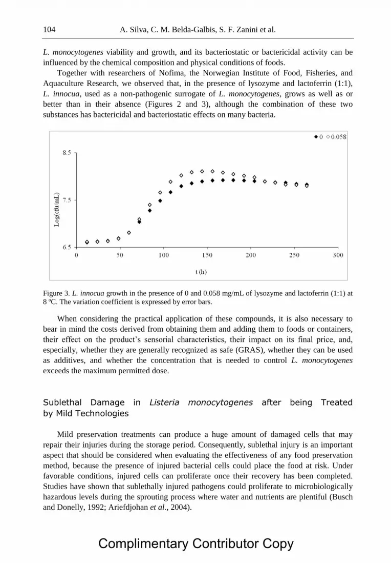
A. Silva, C. M. Belda-Galbis, S. F. Zanini et al. 104
L. monocytogenes viability and growth, and its bacteriostatic or bactericidal activity can be
influenced by the chemical composition and physical conditions of foods.
Together with researchers of Nofima, the Norwegian Institute of Food, Fisheries, and
Aquaculture Research, we observed that, in the presence of lysozyme and lactoferrin (1:1),
L. innocua, used as a non-pathogenic surrogate of L. monocytogenes, grows as well as or
better than in their absence (Figures 2 and 3), although the combination of these two
substances has bactericidal and bacteriostatic effects on many bacteria.
Figure 3. L. innocua growth in the presence of 0 and 0.058 mg/mL of lysozyme and lactoferrin (1:1) at
8 ºC. The variation coefficient is expressed by error bars.
When considering the practical application of these compounds, it is also necessary to
bear in mind the costs derived from obtaining them and adding them to foods or containers,
their effect on the product’s sensorial characteristics, their impact on its final price, and,
especially, whether they are generally recognized as safe (GRAS), whether they can be used
as additives, and whether the concentration that is needed to control L. monocytogenes
exceeds the maximum permitted dose.
Sublethal Damage in Listeria monocytogenes after being Treated
by Mild Technologies
Mild preservation treatments can produce a huge amount of damaged cells that may
repair their injuries during the storage period. Consequently, sublethal injury is an important
aspect that should be considered when evaluating the effectiveness of any food preservation
method, because the presence of injured bacterial cells could place the food at risk. Under
favorable conditions, injured cells can proliferate once their recovery has been completed.
Studies have shown that sublethally injured pathogens could proliferate to microbiologically
hazardous levels during the sprouting process where water and nutrients are plentiful (Busch
and Donelly, 1992; Ariefdjohan et al., 2004).
Complimentary Contributor Copy

Sublethal Damage in Listeria monocytogenes after Non-Thermal Treatments … 105
As indicated earlier, the use of natural antimicrobials is now gaining interest, but, as with
non-thermal or mild thermal technologies, sublethal damage can be produced when they are
used at sublethal concentrations or conditions. This situation can pose a risk to the consumer
because changes that modify the behavior of microorganisms can occur during the repair
process.
Studies conducted in our laboratory, working with 106 cfu/mL of L. monocytogenes,
indicated that the lag phase duration () and the maximum growth rate (µmax) depend on the
citral and carvacrol concentration (p < 0.05).
As the concentration of citral increased from 0.150 µL/mL to 0.250 µL/mL, we observed
an increase in of 1.13 hours and a decrease in µmax of 1.42 log(cfu/mL)/h (p < 0.05). This
behavior was also reflected in the percentage of injured cells when treated L. monocytogenes
was grown in a selective medium containing 5% of sodium chloride (Figures 4 and 5).
Sublethal damage was observed in 20–25% of the cells after being treated by citral. Other
authors have also shown the effect of citral on E. coli, E. coli O157:H7, and S. typhimurium
(Kim et al., 1995).
For carvacrol, the results obtained showed that, when the concentration of carvacrol
increased from 0.100 µL/mL to 0.175 µL/mL, increased by 2.90 hours, whereas µmax
decreased by 0.13 log(cfu/mL)/h (p < 0.05). As in the case of citral, the contact of cells with
carvacrol affected the percentage of injured cells, using 5% sodium chloride as selective
medium (Figures 6 and 7). The percentage of damage by carvacrol that was observed ranged
between 18 and 22% of the cells. Carvacrol has been found to possess in vitro antimicrobial
activity against a broad spectrum of Gram-positive bacteria, including L. monocytogenes
(Gaysinsky et al., 2005; Di Pasqua et al., 2006; Gill and Holley, 2006). Faleiro et al. (2005)
reported that the minimum inhibitory concentration (MIC) of carvacrol ranged from 0.05 to
0.15 µL/mL and the minimum bactericidal concentration (MBC) was 0.25 µL/mL for L.
monocytogenes. According to the reports of Shintani (2006), in the presence of injured cells
there is an increase in in comparison with uninjured cells.
Figure 4. L. monocytogenes sublethal damage (cfu/mL) in the presence of a citral concentration of
0.150 µL/mL, with N0 = 106 log(cfu/mL).
Complimentary Contributor Copy

A. Silva, C. M. Belda-Galbis, S. F. Zanini et al. 106
Figure 5. L. monocytogenes sublethal damage (cfu/mL) in the presence of a citral concentration of
0.250 µL/mL, with N0 = 106 log(cfu/mL).
Figure 6. L. monocytogenes sublethal damage (cfu/mL) in the presence of a carvacrol concentration of
0.100 µL/mL, with N0 = 106 log(cfu/mL).
This extension is required to repair the damage and synthesis of proteins and nucleic
acids, necessary for the multiplication of microorganisms. Injury to pathogens eliminates their
ability to cause disease, but once the cells are repaired pathogenicity is totally restored
(Meyer and Donnelly, 1992).
Sublethal Injury and Changes in Virulence of Pathogens Surviving
the Presence of the Antimicrobial Substances
During the repair process, sublethally damaged microorganisms can acquire new abilities
and modify existing ones. This section describes an example of the effect produced when
Complimentary Contributor Copy

Sublethal Damage in Listeria monocytogenes after Non-Thermal Treatments … 107
antimicrobial substances are used. Changes have been described in microbes resulting from
repairs to damage (Busch and Donelly, 1992; Ariefdjohan et al., 2004). They can be studied
in various ways.
Figure 7. L. monocytogenes sublethal damage (cfu/mL) in the presence of a carvacrol concentration of
0.175 µL/mL, with N0 = 106 log(cfu/mL).
In the literature there are examples where Caco-2 cells have been used to study the
virulence of various microorganisms. In addition to L. monocytogenes, Caco-2 cells have
been used to study the virulence of other pathogenic microorganisms, such as E. coli
O157:H7 (Isumikawa et al., 1998), Campylobacter jejuni, Campylobacter coli (Everest et al.,
1992), Enterobacter sakazakii (Kim and Loessner, 2008), and Bacillus cereus (Minnaard et
al., 2004). In comparison with animal models, this system can be considered as a rapid
laboratory test.
Caco-2 cells were used in our laboratory in a preliminary study to assess the possible
changes in the virulence of L. monocytogenes. The viability of the Caco-2 cells was analyzed
by three different techniques: alamar blue, trypan blue exclusion, and flow cytometry.
Alamar blue is a test based on the ability of viable, metabolically active cells to reduce
resazurin sodium to resorufin and dihydroresorufin, which is measurable by colorimetric
methods. The data obtained after exposure to L. innocua and L. monocytogenes for 24 hours
show that Listeria spp. are able to oxidize resazurin, giving rise to false positives. Therefore
this technique cannot be used to study the virulence of L. monocytogenes after treatment with
antimicrobials.
The trypan blue exclusion test is used to determine the number of viables, based on the
principle that live cells possess intact cell membranes that exclude trypan blue, whereas dead
cells do not. The data show the suitability of this technique for viability studies of Caco-2
cells exposed to Listeria spp. However, it is a subjective, arbitrary, and more time-consuming
technique than flow cytometry, so it is only used as an auxiliary method to complete data
obtained by other ways, such as the Annexin-FITC/IP double staining method.
Annexin V/PI double staining can be used to distinguish between viable [Annexin(–),
PI(–)], necrotic [Annexin(–), PI(+)], and apoptotic cells [early: Annexin(+), PI(–); late:
Complimentary Contributor Copy

A. Silva, C. M. Belda-Galbis, S. F. Zanini et al. 108
Annexin(+), PI(+)]. Annexin V is a member of the family of calcium and phospholipid
binding proteins with a high affinity for phosphatidylserine. The data obtained with this
technique show that after 24 hours of exposure there were no significant differences between
treatments with L. innocua and L. monocytogenes, with ratios of viability, apoptosis, and
necrosis similar to those obtained in untreated samples. These results corroborate the findings
of Pine et al. (1991), who indicated that Caco-2 virulence tests for L. monocytogenes may not
reflect its relative virulence as determined by its mouse LD50. Consequently, efforts have been
made recently to find easier model systems to study the virulence of pathogenic micro-
organisms.
More recently, a new study model has appeared, based on Caenorhabditis elegans, which
has shown its potential for conducting studies of this kind (Thomsen et al., 2006; Forrester et
al., 2007). Previous works have demonstrated that many mammalian pathogenic bacteria and
fungi can infect and cause disease in simple non-vertebrate hosts, such as C. elegans
(Anderson et al., 2006). C. elegans is a simple and economic invertebrate animal model that
is increasingly used for the study of mechanisms of microbial pathogenesis, requiring very
inexpensive, simple growth conditions because it grows on a layer of bacteria in an agar plate
(Diard et al., 2007). C. elegans provides the opportunity simultaneously to explore pathogen
virulence and host defense, and to identify novel pathogen-encoded virulence factors and
universal virulence factors that are relevant in mammalian pathogenesis and specialized
human pathogens (Garsin et al., 2001; Aballay and Ausubel, 2002; Ewbank, 2002; Costi et
al., 2005; Thomsen et al., 2006; Forrester et al., 2007; Jensen et al., 2008b).
Various parameters can be studied using the nematode as a model: life span, egg laying,
mobility, pharyngeal pumping rate, chemotaxis, and the production of new phenotypes. In our
laboratory we studied life span, egg laying, mobility, food choice, and the production of new
phenotypes. A preliminary study was conducted with L. innocua and L. monocytogenes,
trying to find a parameter that would accurately account for the possible virulence changes
that could take place in Listeria spp. after sublethal damage (Silva-Angulo et al., 2011). In
these investigations, C. elegans was used as a model to study sublethal damage and possible
changes in virulence as a consequence of exposure of L. monocytogenes and L. innocua to
various concentrations of carvacrol and citral. The percentage of sublethal damage produced
after each treatment was determined in relation to antimicrobial concentration and exposure
time. The percentage was calculated by the following equation (Busch and Donelly, 1992;
Dykes, 1999):
[1 – (count on TSA-YE-NaCl/count on TSA)]*100
where TSA is the non-selective medium and TSA-YE-NaCl is the selective one.
The results obtained indicated that when C. elegans fed on a lawn of L. monocytogenes
and L. innocua the mobility of the worm was reduced in both cases as compared with E. coli
OP50 as negative control. The “bag of worms” phenotype was also observed when the worm
was fed with L. monocytogenes and L. innocua, respectively. This phenotype was also
observed by Thomsen et al. (2006) in L. monocytogenes. Both L. innocua and L. mono-
cytogenes produced a reduction in egg-laying as compared with OP50. However, this effect
was higher in L. monocytogenes than in L. innocua. These results are promising, indicating
that this nematode could be a good model to study changes of virulence linked to sublethal
damage in L. monocytogenes.
Complimentary Contributor Copy

Sublethal Damage in Listeria monocytogenes after Non-Thermal Treatments … 109
Conclusion
Sublethal injury should be considered as an emerging risk that can affect the development
of safe preservation processes when non-thermal or mild thermal technologies are used alone
or combined as a “hurdle” technology. Safety issues can arise after damaged bacterial cells
repair the injury and the environmental conditions allow them to grow. During the repair
process they can acquire new abilities or changes in their virulence can take place. To avoid
this emerging risk an in-depth validation of minimal processes should be made, focusing on
the number of injured cells produced. For this validation, tests carried out using animal
models such as C. elegans could be a very useful tool.
Acknowledgments
The sublethal damage studies carried out were charged to the AGL2010-22206-CO2-01
project, sponsored by the Spanish Ministry of Science and Innovation. The authors are
grateful to the Departamento de Biotecnología Agroalimentaria, BIOPOLIS S.L for its
collaboration in the studies conducted with C. elegans. They also wish to thank NOFIMA, the
Norwegian Institute of Food Fisheries and Aquaculture Research, for its collaboration in the
study of the effects of lysozyme and lactoferrin on the growth of L. innocua.
References
Aballay, A; Ausubel, F. Caenorhabditis elegans as a host for the study of host-pathogen
interactions. Host-Microbe Interactions: Bacteria 2002, 97-101.
Adzitey, F; Huda, N. Listeria monocytogenes in foods: Incidences and possible control
measures. African Journal of Microbiology Research 2010 4 25, 2848-2855.
Alpas, H; Kalchayanand, N; Bozoglu, F; Sikes, A; Dunne, CP; Ray, B. Variation in
resistance to hydrostatic pressure among strains of food-borne pathogens. Applied and
Environmental Microbiology 1999 65 9, 4248-4251.
Anderson, G; Kenney, S; Millner, P; Beuchat, L; Williams, P. Shedding of foodborne
pathogens by Caenorhabditis elegans in compost-amended and unamended soil. Food
Microbiology 2006 23, 146-153.
Apostolidis, E; Kwon YI; Shetty, K. Inhibition of Listeria monocytogenes by oregano,
cranberry and sodium lactate combination in broth and cooked ground beef systems
and likely mode of action through proline metabolism. International Journal of Food
Microbiology 2008 128, 317-324.
Ariefdjohan, M; Nelson, P; Singh, R; Bhunia, K; Balasubramaniam, M; Singh, N. Efficacy of
high hydrostatic pressure treatment in reducing Escherichia coli O157:H7 and Listeria
monocytogenes in alfalfa seeds. Food Microbiology and Safety 2004 5 69, 117-120.
Autio, T; Hielm, S; Miettinen, M; Sjöberg, AM; Aarnisalo, K; Björkroth, J; Sandholm, TM;
Korkeala, H. Sources of Listeria monocytogenes contamination in a cold-smoked
Complimentary Contributor Copy

A. Silva, C. M. Belda-Galbis, S. F. Zanini et al. 110
rainbow trout processing plant detected by pulsed-field gel electrophoresis typing.
Applied Environmental Microbiology 1999 65, 150-155.
Aymerich, T; Jofré, A; Garriga, M; Hugas, M. Inhibition of Listeria monocytogenes and
Salmonella by natural antimicrobials and high hydrostatic pressure in sliced cooked
ham. Journal of Food Protection 2005 68 1, 173-177.
Belda-Galbis, CM; Martínez, A; Rodrigo, D. Antimicrobial effect of carvacrol on
Escherichia coli K12 growth at different temperatures. In: Mendez-Vilas A, editor.
Science and Technology Against Microbial Pathogens: Research, Development and
Evaluation. Proceedings of the International Conference on Antimicrobial Research
(ICAR2010). Singapore: World Scientific Publishing; 2010; 80-84.
Belda-Galbis, CM; Martínez, A; Rodrigo, D. Seguridad032C: Evaluación in vitro de la
actividad antimicrobiana del citral sobre Listeria innocua a distintas temperaturas. In:
Proceedings of the VI Congreso Nacional de Ciencia y Tecnología de los Alimentos.
Spain; Universitat Politècnica de València; 2011.
Burt S. Essential oils: Their antibacterial properties and potential applications in food - A
review. International Journal of Food Microbiology 2004 94, 223-253.
Busch, S; Donnelly, C. Development of a repair-enrichment broth for resuscitation of heat-
injured Listeria monocytogenes and Listeria innocua. Applied and Environmental
Microbiology 1992 1 58, 14-20.
Carlez, A; Rosec, JP; Richard, N; Cheftel, J. High pressure inactivation of Citrobacter
freundii, Pseudomonas fluorescens and Listeria innocua in inoculated minced beef.
Lebensmittel-Wissenschaft & Technologie 1993 26, 48-54.
Chen, H; Hoover, DG. Modeling the combined effect of high hydrostatic pressure and mild
heat on the inactivation kinetics of Listeria monocytogenes Scott A in whole milk.
Innovative Food Science and Emerging Technologies 2003 4, 25-34.
Codex Alimentarius Commission. Proposed draft guidelines for the control of Listeria
monocytogenes in foods. Report nr. CX/FH 03//8. USA 2002.
Costi, S; Begun, J; Ausubel, F. Review: The worm has turned – microbial virulence modeled
in Caenorhabditis elegans. Trends in Microbiology 2005 3 13, 119-127.
Di Pasqua, R; Hoskins, N; Betts, G; Mauriello, G. Changes in membrane fatty acids
composition of microbial cells induced by addiction of thymol, carvacrol, limonene,
cinnamaldehyde and eugenol in the growing media. Journal of Agricultural and Food
Chemistry 2006 54, 2745-2749.
Diard, M; Baeriswyl, S; Clermont, O; Gouriou, S; Picard, B; Taddei, F; Denamur, E; Matic,
I. Caenorhabditis elegans as a simple model to study phenotypic and genetic virulence
determinants of extraintestinal pathogenic Escherichia coli. Microbes and Infection
2007 9, 214-223.
Dykes, G. Physical and metabolic causes of sub-lethal damage in Listeria monocytogenes
after long-term chilled storage at 4 ºC. Journal of Applied Microbiology 1999 87,
915-922.
EC [European Commission]. Report on trends and sources of zoonotic agents in the
European Union and Norway. Health and Consumer Protection Directorate-General
2002.
EFSA [European Food Safety Authority]. Request for updating the former SCVPH opinion
on Listeria monocytogenes risk related to ready-to-eat foods and scientific advice on
Complimentary Contributor Copy

Sublethal Damage in Listeria monocytogenes after Non-Thermal Treatments … 111
different levels of Listeria monocytogenes in ready-to-eat foods and the related risk for
human illness. The EFSA Journal 2007 599, 1-42.
EFSA [European Food Safety Authority]; ECDC [European Centre for Disease Prevention
and Control]. The European Union summary report on trends and sources of zoonoses,
zoonotic agents and food-borne outbreaks in 2009. EFSA Journal 2011 9 3, 2090.
Espinosa, J; Marco, A; Rodrigo, D; Martínez, A. Antimicrobial activity of Stevia rebaudiana.
In: Fito P; Toldrá F, editors. International Conference on Food Innovation –
FoodInnova 2010: Book of Abstracts. Spain: Universitat Politècnica de València; 2010;
237-237.
EU [European Union]. Regulation (EC) No 178/2002 of the European Parliament and of the
Council of 28 January 2002 laying down the general principles and requirements of
food law, establishing the European Food Safety Authority and laying down
procedures in matters of food safety 2002.
EU [European Union]. Guidance document on Listeria monocytogenes shelf-life studies for
ready-to-eat foods, under Regulation (EC) No 2073/2005 of 15 November 2005 on
microbiological criteria for foodstuffs 2008. Available on: http://ec.europa.eu/food
/food/biosafety/salmonella/docs/guidoc_listeria_monocytogenes_en.pdf [Accessed:
January 21, 2012].
Everest, P; Goossens, H; Butzler, J; Lloyd, D; Knutton, S; Ketley, J; Williams, P.
Differentiated Caco-2 cells as a model for enteric invasion by Campylobacter jejuni
and C. coli. Journal of Medical Microbiology 1992 37, 319-325.
Ewbank, J. Tackling both sides of the host-pathogen equation with Caenorhabditis elegans.
Microbes and Infection 2002 4, 247-256.
Faleiro, L; Miguel, G; Gomes, S; Costa, L; Venâncio, F; Teixeira, A; Figueiredo, AC;
Barroso, JG; Pedro, LG. Antibacterial and antioxidant activities of essential oils
isolated from Thymbra capitata L. (Cav.) and Origanum vulgare L. Journal of
Agricultural and Food Chemistry 2005 53, 8162-8168.
FAO/WHO [Agriculture Organization of the United Nation/World Health Organization].
Risk assessment of Listeria monocytogenes in ready-to-eat foods. Technical report.
Microbiological risk assessment series 5. Rome, Italy 2004,269.
Ferrer, C; Ramón, D; Muguerza, B; Marco, A; Martínez, A. Effect of olive powder on the
growth and inhibition of Bacillus cereus. Foodborne Pathogens and Disease 2009 6 1,
33-37.
Fleischman, GJ; Ravishankar, S; Balasubramaniam, VM. The inactivation of Listeria
monocytogenes by pulsed electric field (PEF) treatment in a static chamber. Food
Microbiology 2004 21, 91-95.
Forrester, S; Milillo, S; Hoose, W; Wiedmann, M; Schwab, U. Evaluation of the
pathogenicity of Listeria spp. in Caenorhabditis elegans. Foodborne Pathogens and
Disease 2007 1 4, 67- 73.
FSAI [Food Safety Authority of Ireland]. General pathogen control for food processing. In:
The control and management of Listeria monocytogenes contamination of food.
Dublin: Food Safety Authority of Ireland; 2005; 11-21. ISBN: 1-904465-29-3.
Gandhi, M; Chikindas, ML. Listeria: A foodborne pathogen that knows how to survive.
International Journal of Food Microbiology 2007 113, 1-15.
Complimentary Contributor Copy

A. Silva, C. M. Belda-Galbis, S. F. Zanini et al. 112
Garsin, D; Sifri, C; Mylonakis, E; Qin, X; Singh, K, Murray, B; Calderwood, S; Ausubel, F.
A simple model host for identifying Gram-positive virulence factors. PNAS 2001 19,
10892-10897.
Garvey, P; McKeown, P. Human listeriosis in Ireland. EPI-Insight (Disease Surveillance
Report of NDSC, Ireland) 2004 5, 4.
Gaysinsky, S; Davidson, PM; Bruce, BD; Weiss, J. Growth inhibition of E. coli O157:H7
and Listeria monocytogenes by carvacrol and eugenol encapsulated in surfactant
micelles. Journal of Food Protection 2005 68, 2559-2566.
Gill, AO; Holley, RA. Disruption of Escherichia coli, Listeria monocytogenes and
Lactobacillus sakei cellular membranes by plant oil aromatics. International Journal of
Food Microbiology 2006 108, 1-9.
Hughey, VL; Wilger, PA; Johnson, EA. Antibacterial activity of hen egg white lysozyme
against Listeria monocytogenes Scott A in foods. Applied and Environmental
Microbiology 1989 55 3, 631-638.
Huss, HH; Jørgensen LV; Vogel BF. Control options for Listeria monocytogenes in seafoods.
International Journal of Food Microbiology 2000 62, 267-274.
ICMSF [International Commission on Microbiological Specifications for Foods].
Microorganismos de los Alimentos: Características de los Patógenos Microbianos.
España: Acribia; 1996; 255-330.
Ikeda, JS; Samelis, J; Kendall, PA; Smith, GC; Sofos, JN. Acid adaptation does not promote
survival or growth of Listeria monocytogenes on fresh beef following acid and nonacid
decontamination treatments. Journal of Food Protection 2003 66, 985-992.
Isumikawa, K; Hirakata, Y; Yamaguchi, T; Takemura, H; Maesaki, S; Tomono, K; Igimi, S;
Kafu, M; Yamada, Y; Kohno, S; Kamihira, S. Escherichia coli O157 interactions with
human intestinal Caco-2 cells and the influence of fosfomycin. Journal of
Antimicrobial Chemotherapy 1998 42, 341-347.
Jensen, A; Thomsen, L; Jorgensen, R; Larsen, M; Roldgaard, B; Christensen, B; Vogel, B;
Gram, L; Ingmer, H. Processing plant persistent strains of Listeria monocytogenes
appear to have a lower virulence potential than clinical strains in selected virulence
models. International Journal of Food Microbiology 2008b 123, 254-261.
Kim, EL; Choi, NH; Bajpai, VK; Kang, SC. Synergistic effect of nisin and garlic shoot juice
against Listeria monocytogenes in milk. Food Chemistry 2008a 110, 375-382.
Kim, JM; Marshall, MR; Cornell, JA; Preston III, IF; Wei, CI. Antibacterial activity of
carvacrol, citral and geraniol against Salmonella typhimurium in culture medium and
on fish cubes. Journal of Food Science 1995 60, 1364-1368.
Kim, K; Loessner, M. Enterobacter sakazakii invasion in human intestinal Caco-2 cells
requires the host cell cytoskeleton and is enhanced by disruption of tight junction.
Infection and Immunity 2008b 2 76, 562-570.
Kousta, M; Mataragas, M; Skandamis, P; Drosinos, EH. Prevalence and sources of cheese
contamination with pathogens at farm and processing levels. Food Control 2010 21,
805-8015.
Luber, P; Scott, C; Christophe, D; Jeff, F; Atin, D; Ewen, CDT. Controlling Listeria
monocytogenes in ready-to-eat foods: Working towards global scientific consensus and
harmonization. Recommendations for improved prevention and control. Food Control
2011 22, 1535-1549.
Complimentary Contributor Copy

Sublethal Damage in Listeria monocytogenes after Non-Thermal Treatments … 113
Mead, PS; Slutsker, L; Dietz, V; McCaig, LF; Breese, JS; Shapiro, C; Griffin, PM; Tauxe,
RV. Food-related illness and death in the United Stated. Emerging Infectious Diseases
1999 5 5, 607-625.
McDonald, CJ; Lloyd, SW; Vitale, MA; Petersson, K; Inning, F. Effects of pulsed electric
fields on microorganisms in orange juice using electric field strengths of 30 and 50
kV/cm. Journal of Food Science 2000 65, 984-989.
Meyer, DH; Donnelly, CW. Effect of incubation temperature on repair of heat-injured
Listeria in milk. Journal of Food Protection 1992 55, 579-582.
Minnaard, J; Moal, V; Coconnier, M; Servin, A; Pérez, P. Disassembly of F-Actin
cytoskeleton after interaction of Bacillus cereus with fully differentiated human
intestinal Caco-2 cells. Infection and Immunity 2004 5 72, 3106-3112.
Mohamed, HMH; Elnawawi, FA; Yousef, AE. Nisin treatment to enhance the efficacy of
gamma radiation against Listeria monocytogenes on meat. Journal of Food Protection
2011 74 2, 193-199.
Møretrø, T; Langsrud S. Listeria monocytogenes: Biofilm formation and persistence in food-
processing environments. Biofilms 2004 1 2, 107-121.
Mosqueda-Melgar, J; Elez-Martínez, P; Raybaudi-Massilia, RM; Martín-Belloso, O. Effects
of pulsed electric fields on pathogenic microorganisms of major concern in fluid foods:
A review. Critical Reviews in Food Science and Nutrition 2008 48, 747-759.
Nguyen, P; Mittal, GS. Inactivation of naturally occurring microorganisms in tomato juice
using pulsed electric field (PEF) with and without antimicrobials. Chemical
Engineering and Processing 2007 46, 360-365.
Pina-Pérez, MC; Silva-Angulo, AB; Muguerza-Marquínez, B; Rodrigo Aliaga, D; Martínez
López, A. Synergistic effect of high hydrostatic pressure and natural antimicrobials on
inactivation kinetics of Bacillus cereus in a liquid whole egg and skim milk mixed
beverage. Foodborne Pathogens and Disease 2009b 6 6, 649-656.
Pina-Pérez, MC; Silva-Angulo, AB; Rodrigo, D; Martínez-López A. Synergistic effect of
pulsed electric fields and CocoanOX 12% on the inactivation kinetics of Bacillus
cereus in a mixed beverage of liquid whole egg and skim milk. International Journal of
Food Microbiology 2009a 130, 196-204.
Pine, L; Kathariou, S; Quinn, F; George, V; Wenger, J; Weaver, R. Cytopathogenic effects in
enterocytelike Caco-2 cells differentiate virulent from avirulent Listeria strains.
Journal of Clinical Microbiology 1991 5, 990-996.
Ponce, E; Pla, R; Mon-Mur, R; Gervilla, R; Guamis, B. Inactivation of Listeria innocua
inoculated in liquid whole egg by high hydrostatic pressure. Journal of Food
Protection 1997 61, 119-122.
Raso, J; Barbosa-Cánovas GV. Nonthermal preservation of foods using combined processing
techniques. Critical Reviews in Food Science and Nutrition 2003 43 3, 265-285.
Reina, LD; Jin, ZT; Zhang, QH; Yousef, AE. Inactivation of Listeria monocytogenes in milk
by pulsed electric field. Journal of Food Protection 1998 61, 1203-1206.
Ruiz, A; Williams, SK; Djeri, N; Hinton Jr, A, Rodrick, GE. Nisin, rosemary, and
ethylenediaminetetraacetic acid affect the growth of Listeria monocytogenes on ready-
to-eat turkey ham stored at four degrees Celsius for sixty-three days. Poultry Science
2009 88, 1765-1772.
Complimentary Contributor Copy

A. Silva, C. M. Belda-Galbis, S. F. Zanini et al. 114
Ruiz, A; Williams, SK; Djeri, N; Hinton Jr, A, Rodrick, GE. Nisin affects the growth of
Listeria monocytogenes on ready-to-eat turkey ham stored at four degrees Celsius for
sixty-three days. Poultry Science 2010 89, 353-358.
Saldaña, G; Puértolas, E; Condón, S; Álvarez, I; Raso, J. Inactivation kinetics of pulsed
electric field-resistant strains of Listeria monocytogenes and Staphylococcus aureus in
media of different pH. Food Microbiology 2010 27, 550-558.
Seeliger, HPR; Jones, D. Genus Listeria Pirie. In: Sncath PHA; Mair NS; Sharpe ME; Holt
JG, editors Bergey's Manual of Systematic Bacteriology, Baltimore: Williams and
Wilkins; 1986; 1235-1245.
Sepulveda, DR; Góngora-Nieto, MM; San-Martín, MF; Barbosa-Cánovas, GV. Influence of
treatment temperature on the inactivation of Listeria innocua by pulsed electric fields.
Lebensmittel-Wissenschaft & Technologie 2005 38, 167-172.
Shintani, H. Importance of considering injured microorganisms in sterilization validation.
Biocontrol Science 2006 11, 91-106.
Silva-Angulo, A; Rodrigo, M; Martínez, A; Martorell P. Studies for selecting a simple model
for analyses of virulence in Listeria monocytogenes 2011. In: 1st CIGR Workshop Food
Safety: Advances and Trends. 14th and 15
th April, Dijon, France.
Simpson, RK; Gilmour, A. The resistance of Listeria monocytogenes to high hydrostatic
pressure in foods. Food Microbiology 1997 14, 567-573.
Sivarooban, T; Hettiarachchy, NS; Johnson, MG. Inhibition of Listeria monocytogenes using
nisin with grape seed extract on turkey frankfurters stored at 4 and 10°C. Journal of
Food Protection 2007 70 4, 1017-1020.
Thomsen, L; Slutz, S; Tan, M; Ingmer, H. Caenorhabditis elegans is a model host for
Listeria monocytogenes. Applied and Environmental Microbiology 2006 2. 72, 1700-
1701.
Tiwari, BK; Valdramidis, VP; O’Donnell CP; Muthukumarappan, K; Bourke, P; Cullen, PJ.
Application of natural antimicrobials for food preservation. Journal of Agricultural and
Food Chemistry 2009 57, 5987-6000.
Zhu, M; Du, M; Cordray, J; Ahn, DU. Control of Listeria monocytogenes contamination in
ready-to-eat meat products. Comprehensive Reviews in Food Science and Food Safety
2005 4, 34-42.
Complimentary Contributor Copy

In: Listeria Infections ISBN: 978-1-62081-639-4
Editors: A. Romano and C. F. Giordano © 2012 Nova Science Publishers, Inc.
Chapter V
Listeria Infections Review:
Epidemiology, Pathogenesis
and Treatment
Karina Pellicer Facultad de Ciencias Veterinarias, Universidad Nacional de La Plata,
La Plata; Provincia de Buenos Aires, República Argentina
Listeria monocytogenes is a pathogen that affects animals and human beings; it can
produce potentially fatal infections in susceptible individuals. This bacterium can cause
miscarriages in pregnant women and meningitis in newborns, children and adults who are
immunosuppressed. Most cases of Listeriosis are sporadic; although outbreaks have been
described due to food consumption. The ability of L. monocytogenes to resist stressful enviro-
nmental conditions makes this pathogen a food industry concern.
Listeria monocytogenes
The genus Listeria includes short rods, with an average of 0.4-0.5 µm in diameter and 0.5
to 2 µm in length, gram positives, which do not produce spores. Cells occur singly, or
arranged in short chains with each other forming a “V” or in groups arranged in parallel along
an axis (Seeliger and Jones, 1986).
Listeria spp. does not form a capsule (Seeliger and Jones, 1986). It is motile by means of
2-3 peritrichous flagella (rarely more than 5 by the usual techniques of color) and does best at
20-25ºC in semisolid media where it develops 3-5 mm below the surface in the form of
inverted umbrella. At 37 ºC it loses mobility because it does not develop flagella.
Listeria is catalase positive, oxidase negative. It is aerobic or facultatively anaerobic;
physocrotrophic and regarded as mesophilic, it can also develop at refrigeration temperature.
Its optimum temperature for development is between 30 to 37ºC. According to the Manual of
Sistematic Bacteriology Bergey, L. monocytogenes develops in the pH range of 6 to 9.
Complimentary Contributor Copy

Karina Pellicer 116
Generally considered that low pH is inhibitory and even lethal to the bacteria, it is however
indicated that it tolerates acidity to pH 4.4 (Pellicer et al., 2009; Giannuzzi and Zaritzky,
1996; Buchanan and Klawitter, 1990; Conner et al., 1986).
There are now seven known species of Listeria, including L. monocytogenes, L. innocua,
L. seeligeri, L. welshimeri, L. ivanovii who have between them a closer relationship than there
are with L. grayi and L. murrayi.
The genus Listeria contains beta-hemolytic species. L. monocytogenes is beta hemolytic,
while L. ivanovii produce a large area or multiples areas of hemolysis on blood agar. If the
reaction is weak or inconclusive betahemolysis can be solved by CAMP test (Christie, Atkins,
Munch-Petersen in Lovett 1988) which relies on the enhancement of the hemolytic activity of
the metabolites of Staphylococcus aureus and Rhodococcus equi in blood agar plate (Lovett,
1988).
On nutrient agar colonies are 0.5 to 1.5 mm in diameter, round, translucent, low convex
and net margins. They are gray with a bluish and greenish blue glow with obliquely
transmitted light. The newly isolated strains show colonies circular, smooth (“S” shape),
white with a shiny surface.
Listeria spp. possesses somatic (O) and flagellar (H) antigens, which allow their
classification into serotypes, the main pathogenic species, L. monocytogenes is classified into
13 serotypes: 1/2a, 1/2b, 1/2c, 3a, 3b, 3c, 4a, 4ab, 4b, 4c, 4d, 4e, 7 (Seeliger and Höhne,
1979), which are all producers of Listeriosis. Most human isolates correspond to serotypes
4b, 1/2a and 1/2b. As for the food-borne isolates, they are also the most frequent.
Listeriosis
Listeria monocytogenes
Since the early 1980s, foodborne transmission has been recognized as a major source of
human Listeriosis. Listeriosis can cause stillbirths, miscarriages, meningitis, or sepsis in
immunocompromised hosts. Case-fatality rates as high as 40% have been reported during
outbreaks (Scuchat, Swaminatan and Broome, 1991).
Listeria monocytogenes is a pathogen which causes miscarriages (Smith et al., 1995) and
mastitis (Gitter et al., 1980) in animals whereas in human beings, it can produce potentially
fatal infections in susceptible individuals. This bacterium can cause miscarriages in pregnant
women and meningitis in newborns, children and adults who are immunosuppressed
(Donelly, 1994; Seeliger, 1961).
Human Listeriosis can occur as epidemics or as sporadic cases, and is primarily an
opportunistic infection primarily affecting pregnant women, newborns, elderly and
immunocompromised adults, including the HIV-infected.
L. monocytogenes infections in healthy adults are usually asymptomatic or in some cases
produce symptoms similar to influenza infection. In other cases, less common, is presented
with diarrhea and abdominal discomfort. In fact, L. monocytogenes is transported in the
gastrointestinal tract of 5 to 10 % of asymptomatic population.
L. monocytogenes is one of the few bacteria that can cross the placenta, which acts as an
effective barrier to prevent seepage of pathogen infection of the fetus via the bloodstream. In
Complimentary Contributor Copy

Listeria Infections Review 117
pregnant women who get Listeriosis, the bacteria can (not always) infect the fetus resulting in
stillborns, premature births or birth of infants with systemic infection by L. monocytogenes
(Saylers and Whitt, 2005; Smith et al., 2009). Neonatal Listeriosis could be septicemic, also
known as early-onset disease (less than 5 days old) due to transplacental infection, or appear
mainly as meningitis after 5 days of age (late-onset disease) due to infection during birth or
immediately before birth. Some infections may be the result of acquisition of the organism
during passage through the birth canal, although late-onset disease can occur following
cesarean delivery. However, several nosocomial clusters of neonatal Listeriosis have been
reported (Schuchart, Swaminathan and Broome, 1991).
Most cases of Listeriosis are often associated with meningitis (80 % of cases) or
septicemia. A great majority of affected individuals have some form of immunodeficiency
such as cancer, alcoholism, diabetes, transplantation, chronic hemodialysis and old age.
It actually has shown a wide range of manifestations including meningoencephalitis,
septicemia, pneumonia, endocarditis, localized abscesses, skin lesions, conjunctivitis, osteitis,
endocarditis, arthritis, cholangitis, conjunctivitis, etc. (ANLIS/INEI, 1999 (Argentina’s
National Laboratory and Health Institutes Administration / National Institute of Infectious
Diseases)).
In the era of transplantation, immunosuppression induced increased cases of Listeria
monocytogenes meningitis in adults, especially in renal transplant patients; it could be
considered an opportunistic infection of endogenous origin (ANLIS/INEI, 1999).
Listeriosis is one of the less common opportunistic infections in patients with AIDS
(Acquired Immunodeficiency Syndrome), but so far it is 300 times more frequent than in the
population that does not have this syndrome.
Pathogenesis
Pathogenesis of Listeria monocytogenes infection´s was well described (Lopez et al.,
2006). Listeria is a facultative intracellular pathogen and uses the host’s cells to survive,
multiply and cause disease. To produce infection, Listeria needs to recognize that it is inside
the body, activating virulence genes in response, including Hpt protein (a membrane
transporter that allows the bacteria to uptake glucose phosphate, in order to nourish and
multiply quickly in host tissues). L. monocytogenes is one of the few bacteria that can cross
placenta, an effective filtration barrier to prevent fetal infection through blood circulation.
Factors that may influence whether invasive disease will occur include the virulence of
the infection organism, the susceptibility of the host, and the size of the inoculum.
Listeria monocytogenes is acquired by ingestion and must find and adhere to the
intestinal mucosa. In humans and animals, Listeria monocytogenes is a clearly invasive
organism that leaves the intestine and crosses the mucosa to reach underlying tissue. Listeria
monocytogenes infection begins with adhesion to intestinal epithelial cells surface, due to
adhesion by means of a 80 kDa membrane protein called internalin as Inl A to intestinal cells
(Inl B and other internalins have been identified) (Pizarro-Cerda and Cossart, 2006). Invasion
of the host cell includes other proteins such as autolysins, a fibronectin-binding protein and
proteins involved in later stages of the intracellular cycle such as ActA (Suarez et al, 2001)
Complimentary Contributor Copy

Karina Pellicer 118
and listeriolysin O (LLO) (Dussurget, Pizzarro-Cerda and Cossart, 2004; Lopez et al., 2006;
Ramaswamy et al., 2007).
In the eukariotic cell, the bacteria are phagocytized. Once in the phagosome it secretes a
hemolysin called LLO that acts on the cholesterol membrane forming pores with subsequent
lysis of the phagosome, giving to L. monocytogenes the ability to escape from the phagocytic
vesicle to the host cell cytosol (Kayal and Charbit, 2006). It was proposed that the survival of
Listeria monocytogenes in macrophages could be due to its ability to escape the phagosome
before phagolysosomal fusion occurs. Listeria monocytogenes also produce catalase and
superoxide dismutase, two enzymes that could help protect it from the oxidative burst in the
phagolysosome (Salyers and Whitt, 1994). Listeria moves from phagocytic vacuoles to the
cytoplasm, where replication is improved by more-favorable growth conditions.
Listeria monocytogenes produces at least two other hemolysins besides LLO, that
includes the secretion of two phospholipases C that damage cell membranes by hydrolyzing
membrane lipids, they are PI-PLC whose substrate is phosphatidylinositol and that might aid
intracellular spread of Listeria; while PC-PLC is a lecitinase which hidrolyzes
phosphatidilcholine, phosphatidilserine and phosphatidiletanolamine (Vázquez-Boland et al.,
1992).
Glucose acts a repressor of the virulence genes of Listeria monocytogenes. Glucose-1-
phosphate (G1P), the precursor metabolite and primary degradation product of glycogen in
mammalian cells, and it has been shown that is essential for the intracellular growth of L.
monocytogenes (Ripio et al., 1997a). In the host cell cytoplasm, Listeria monocytogenes
multiplies rapidly. It has been estimated that the bacteria inside a host cell divide once every
50 minutes, a high rate of growth for an intracellular pathogen.
Intracellular multiplication of Listeria needs hexosaphosphate conveyor (Hpt), a
counterpart of eukaryotic cell´s glucose transporter-6-phosphate (G6P) responsible for entry
into the endoplasmic reticulum G6P from the cytosol (Chico-Calero et al., 2002; Goetz et al.,
2001). This protein (Hpt) is also responsible for entry of fosfomycin inside the bacteria cell,
which explains their sensitivity in vivo associated with resistance in vitro (Scortti et al.,
2006). At 37°C, production of flagella is much decreased, and it is likely that motility due to
flagella is not important as a virulence factor. Another type of motility is clearly important for
virulence, the ability of the bacteria to use host cell actin to move themselves within and
between host cells. Intracellular movement of Listeria occurs by a directional assembly of
actin monomers in a host cell to the poles of the bacterium, apparently a led by ActA surface
protein (Suarez et al., 2001). Listeria monocytogenes can move as rapidly as 1.5 µm/sec using
this model of locomotion.
In the end, most bacteria reach the periphery of the infected cell, in contact with cell
membrane, forming protuberances adjacent to the cell cytoplasmic evaginations shaped at its
end containing a bacterium. These structures are invasive bacteria phagocytosed and enclosed
in a double-membrane phagosome, in whose rupture lecithinase (PC-PLC) seems to play a
key role. As a mechanism that allows the spread of bacteria through the host tissue without
contact with the humoral effectors of the immune system, this phenomenon of direct passage
from cell to cell is crucial in the pathogenesis of infection caused by L. monocytogenes
(Cossart and Mengaud, 1989; López et al., 2006). Most Listeriosis occurs in persons with
impaired cell-mediated immunity due to disease process, medications or pregnancy.
In addition, the local placental suppression of cell mediated immunity necessary to
prevent maternal rejection of the placenta may contribute to susceptibility of the fetus to
Complimentary Contributor Copy

Listeria Infections Review 119
infection with Listeriosis (Redline and Lu, 1987; Schuchart, Swaminathan and Broome,
1991). In the same way, the depressed cell-mediated immunity, deficiencies in immuno-
globulin M and complement activity associated with the neonatal state may contribute to
susceptibility of this host to Listeriosis (Schuchart, Swaminathan and Broome, 1991). Most of
the genes encoding virulence factors are present in all strains of L. monocytogenes, but there
are numerous specific sequences of the major genetic groups in this species (divisions or
lineages) and the different serotypes (Doumith et al., 2004). Virulence genes of L.
monocytogenes are organized into genetic units known as Pathogenicity Island, which are
acquired by the bacteria by horizontal transfer mechanism, sometimes as part of a mobile
genetic element, important in bacterial evolution. Many of the virulence genes are, however,
clustered in the same region of the L. monocytogenes chromosome.
The regulatory protein PrfA is absolutely essential for the expression of virulence in
Listeria (Ripio et al., 1997b). The expression of the virulence regulon by PrfA is dependent
on various environmental signs, including high temperature (37°C), stress conditions and
modifications of components of the cell, and catabolite repression (Gray, Freitag and Boor,
2006; Sokolovic et al., 1993). New virulence factors have been discovered, including new
surface proteins of the type internalin, the SrtA and SrtB sortases (transpeptidases that bind to
different cell wall surface proteins such as internalin), and a bile salt hidrolase (BSH) present
in L. monocytogenes, which allows the bacteria to survive in the intestine (Dussurget, Pizarro-
Cerda and Cossart, 2004; López et al., 2006). In animal models infected with L. mono-
cytogenes, the bacteria first appear in the macrophages and then invade hepatocytes. Most of
their replication probably occurs in the liver. Because L. monocytogenes is an intracellular
pathogen, a cell-mediated host response that kills infected cells is important for eliminating
the microorganism. A failure of the host´s cell-mediated response to control L. mono-
cytogenes allows the bacteria to spread systematically.
Epidemiology
Listeriosis has become a serious public health problem considering that, although it
presents low mortality rates depending on the susceptibility of the host, its mortality is high in
inmunosupressed persons (20 to 30%) and has a long incubation period (5 days-5 weeks)
(Chhabra et al., 1999; Donelly, 1994). The minimum infective dose of L. monocytogenes has
not been established yet (FDA Bad Bug Book) although it has been indicated that the intake
of up to 100 cells does not affect the health of healthy consumers (Jay, 1994; Golnazarian et
al., 1989). Most the cases of Listeriosis are sporadic; though outbreaks have been described
due to food consumption. The ability of L. monocytogenes to resist stressful environmental
conditions makes this pathogen a food industry concern.
Distribution in the Environment
Listeria monocytogenes is a cosmopolitan microorganism (Welshimer and Donker-Voet,
1971). It is found in soil, water and dust. It has been isolated from a variety of domestic and
wild animals. Listeria fecal excretion typically occurs in 1% of a healthy population, in 4.8 %
Complimentary Contributor Copy

Karina Pellicer 120
of refrigerated abattoir workers and in 26 % of people in contact with Listeriosis patients
(Pape y Jhonson, 1986).
Listeria has been isolated from soil with a pH range from 4.8 to 7.6, while in soils with
pH below 3.5 the incidence is very low (Weis and Seeliger, 1975). Plants are also a source of
Listeria, intervening in many infections. L. monocytogenes has been isolated from soy,
decomposing corn, wild grass and shrub leaves. In plant silos its incidence is high (Fenlon,
1986; Nicolett, 1986), especially those of poor quality with high pH and can resist them for
10 to 12 years (Durst, 1975). Many animal Listeriosis cases are attributed to silos as a source
of infection (Fenlon, 1986; Gray, 1960; Gronstol, 1979).
Many animals can serve as host of L. monocytogenes. It is often associated with the
intestinal tract or subclinical infections in domestically animals and humans. Farm animals
are identified as germ reservoir, with a great importance in the epidemiology of Listeriosis. It
can be also isolated from amphibians, fish and insects, with lower epidemiological
significance.
L. monocytogenes has been isolated in water and wastewater (Watkins and Slealth, 1981;
Al-Ghazali and Al-Azawi, 1986); as well as effluent poultry slaughterhouse (Watkins and
Slealth, 1981).
Transmission of Listeriosis
Investigations of large outbreaks of human Listeriosis finally provided epidemiologic and
laboratory support to confirm the suspicion that Listeriosis was a foodborne disease
(Schuchat, Swaminathan, and Broome, 1991). It is evident that the presence of L.
monocytogenes in foods is one of the sources of Listeriosis in humans and is a threat to Public
Health. However, is difficult to assess their presence in the environment. Possible
transmission routes are shown in Figure 1 (Jay, 1994).
Figure 1. Schematic of the modes of spread of Listeria monocytogenes in the environment, animals,
foods and people.
Complimentary Contributor Copy

Listeria Infections Review 121
Gray and Killinger (1966) suggested that the fecal-oral route either directly or indirectly
causes disease in humans and animals. The use of wastewater as fertilizer or irrigation
increases the risk of transmitting Listeriosis (Watkins and Slealth, 1981).
Food handlers with latent infection of Listeria were considered a likely source of
infection (Ralovich, 1974; 1984). Similarly, the presence of bacteria in the soil or decaying
vegetation directly and indirectly contributes to food contamination (Gray, 1960). Soil is a
natural reservoir of this organism and therefore it is more likely that vegetables are
contaminated (Mitscherlich and Marth, 1984). Livestock manure can contaminate soil and
this, in turn, contaminates a crop (Schlech, 1984).
L. monocytogenes has been isolated from cattle, pigs, sheep, turkeys, ducks, and a variety
of other species (Seeliger, 1961; Schuchat, Swaminathan, and Broome, 1991). Because
infected and sometimes healthy animals often eliminate fecal Listeria, Listeriosis was
considered as zoonosis. Human disease was thought to be due to illness occurring in animals,
and the animal host was considered the primary reservoir for the organism (Bojsen-Moller,
1972).The fecal-oral route favors direct transmission between humans.
The direct transmission of sick animals to humans was documented in 1961 by Seeliger.
Foods as Source of Infection
L. monocytogenes can develop at low temperatures in a wide pH range, with a high
concentration of sodium chloride and reduced water activity (Kallipolitis and Higmer, 2001;
Ko et al., 1994). Hence, it can survive and multiply in a great variety of food products (Cole,
Jones and Holyoak, 1990; Begot, Lebert and Lebert, 1997, Bergey´s 1986; Copes et al.,
2000a; Copes et al., 2000b; Monge and Arias-Echandi, 1999; Pellicer et al., 2011). Listeria
has been involved in a large number of food-borne outbreaks transmitted through food all
over the world.
The most hazardous products are those “ready to eat” which are stored at room
temperature for long periods of time (Pellicer et al., 2002a; Pellicer et al., 2002b). Listeria
spp. has been found in a great variety of meats and meat products, even in sausages (Jonson,
Doyle, Cassens, 1990; Encinas et al., 1999).
L. monocytogenes and L. innocua are the most frequently isolated strains in food
processing plants. However, most of the cases of listeriosis transmitted through food are due
to the contamination of raw or cooked foods with L. monocytogenes (Cox et. al., 1989; WHO,
1988). Most of the human and food-borne isolates correspond to serotypes 4b, 1/2a y 1/2b.
Treatment and Prevention
Antibiotic therapy of pregnant women or immunocompromised people who have eaten
food contaminated by L. monocytogenes can prevent the most serious consequences of
disease if it is diagnosed in time, but usually it is not diagnosed early. Listeria monocytogenes
responds poorly to antibiotics currently used in treatment since they hardly penetrate to sites
of infection, mainly the brain. This helps explain the high mortality rate of Listeriosis (20 to
30%) of hospitalized patients.
Complimentary Contributor Copy

Karina Pellicer 122
Penicillin or ampicillin, alone or in combination with gentamicin is recommended for
treatment; although there have been reports of treatment failure with these antibiotics, this
combination therapy is considered the treatment of choice. Although duration of treatment for
Listeriosis has not been standardized, 2 weeks of therapy for uncomplicated sepsis or
meningitis and 4 to 6 weeks for an immunocompromised host is recommended (Schuchart,
Swaminathan and Broome, 1991). In serious diseases such as cerebritis, meningitis or gran-
ulomatosis infantiseptica, early initiation of treatment is essential for infection control. In
vitro studies have demonstrated the efficacy of ampicillin and penicillin with
aminoglycosides. The combination of trimethoprim and sulfamethoxazole has been used
successfully in patients allergic to penicillins, and is currently considered as alternative
therapy in this circumstance.
Recent studies “in vivo” suggest that Listeria incorporates fosfomycin in the cells through
Hpt protein, a membrane transporter that enables bacteria to steal glucose phosphate to feed
and multiply rapidly in the tissues of animal or human hosts; which could introduce an
improvement in the combined antibiotic therapy against Listeria monocytogenes (Scortti et
al., 2006).
Tests to determine if an antibiotic is active or not against one microorganism are
performed in vitro, but the bacteria are in contact with living cells; Listeria lacks Hpt and thus
appears as fosfomycin resistant. However, since Hpt comes into operation when the
bacterium is inside cells, fosfomycin is actually effective against Listeria during infection
(Universia, 2006).
The first signs of an outbreak are cases of stillbirths or serious infections in susceptible
individuals. Although the most publicized cases of Listeriosis are those that involved large
outbreaks, sporadic cases of Listeriosis also occur. Since Listeriosis is frequently associated
with contaminated food consumption, avoiding contamination of foods in the first place
would be the ideal solution. Refrigeration is not as effective in preventing growth of L.
monocytogenes in food as it is for most other pathogens. It is easy to imagine why this germ
is a food´s processors nightmare, due to the fact that at least 5% to 10% of the adults are
asymptomatic carriers of L. monocytogenes.
Moreover, prevention is also in the hands of food consumers, mainly pregnant women
and immunocompromised individuals. They should avoid unpasteurized milk products and
raw eggs, cook all meats, and rinse thoroughly all raw vegetables and fruits before being
eaten. Separate uncooked meats and poultry from vegetables, cooked foods, and ready-to-eat
foods. Wash hands, knives, countertops, and cutting boards after handling and preparing
uncooked foods. Use precooked or ready-to-eat food as soon as possible. Prepare foods
without cross-contamination between raw and cooked foods. Do not store the product in the
refrigerator beyond the use-by date (CDC´s Listeriosis webpage).
A low initial inoculum of Listeria in a food at the time of manufacturing can turn into a
big dose of Listeria for consumers, in relation to shelf life and handling of each product, so
the potential of transmission of Listeriosis should be considered (Schuchat, Swaminathan and
Broome, 1991). In order to prevent the food contamination with Listeria spp., implementation
of assurance quality systems and the manufacturing practice in which the early detection of
the microorganism constitutes the main tool, are required for food industry.
Given that Listeria monocytogenes is transmitted by food consumption, mainly ready to
eat products including meat products, some strategies are proposed in order to prevent the
development of Listeria in those products using the hurdle theory (Leitsner and Gorris, 1995;
Complimentary Contributor Copy

Listeria Infections Review 123
Pellicer et al., 2011). During fermentation and drying of sausage, L. monocytogenes tends to
decrease substantially, however, this organism, which is ubiquitous, psychrotrophic and
relatively resistant to curing ingredients, may survive (Ingham, Beuge, Dropp, Losinski,
2004; Juntilla et al., 1989; Jonson, Doyle, Cassens, 1990). It has been reported that the
undissociated lactic acid inhibits the growth of microorganisms and is frequently used to
extend the shelf life of foods (Barbuddhe, Malik and Bhilegaonkar, 1999). Lactic acid is
effective to inhibit the development of L. monocytogenes when at pH values of 4.5 and 4.6
(Ahmad and Marth, 1990; Ariyapitipun, Mustapha and Clarke, 2000; Sorrels, Engil, Hatfield,
1989). Sodium nitrite when applied individually did not present antimicrobial effects.
However, an enhancement of the bactericidal effect with the combination of lactic acid with
sodium nitrite was observed. Bacteriocins are increasingly used to control the growth of L.
monocytogenes; among them, nisin is an antimicrobial agent that can be used in meat
products (Brandt et. al., 2010; Breukink, Krujiff, 1999), in combination with pH reduction
and the addition of NaCI (Bouttefroy et al., 2000). Nisin is an antimicrobial peptide which
acts against L. monocytogenes as well as other Gram-positive bacteria. Its use is permitted in
dairy products (FAO Nutrition Meeting Report Series Nº 45 A (1969) but still does not have
approval for its application in meat products in South America; some countries such as
Australia and New Zealand are trying to use it, proposing the inclusion of a limit for the use
of nisin in meat products (Food Standards Agency of Australia and New Zealand; October
2007 (A565)).; Directive 95/2/CE ratified by the Commission of European Communities
FAO/WHO, 2007; FDA, 2006). Its potential application is high, especially to avoid the
deterioration caused by Gram-positive bacteria in processed meat such as sausage and meat
paste in which the homogenization of the product allows the better distribution of nisin
(Gonalves and Massaguer, 1999; Ariyapitipun, Mustapha and Clarke, 2000; FDA, 1988;
Pellicer et al., 2011). An enhancement of the bactericidal effect of lactic acid with the
addition of nisin has been demonstrated.
L. monocytogenes and other species of Listeria can be isolated from foods and food
industry, but these other organisms don´t hold the same pathogenic potential as L.
monocytogenes. Molecular methods may facilitate rapid detection of all these organisms in
the food industry, and for this reason, they are an important area of research.
References
Administración Nacional de Laboratorios e Institutos de Salud. Instituto Nacional de
Enfermedades Infecciosas. ANLIS/INEI Dr. Carlos G. Malbrán. Departamento de
Bacteriología, Servicio de Bacteriología Especial. Detección de Listeria
monocytogenes. Vigilancia de Listeriosis. Noviembre de 1999.
Ahmad, N; Marth, E. (1990) Acid-injury of Listeria monocytogenes. Journal of Food
Protection 53: 26-29.
Al-Ghazali, M R; Al-Azawi, S K. (1986) Detection and enumeration of Listeria
monocytogenes in a sawage treatment plant in Iraq. Journal of Applied Microbiology
60: 251-254.
Complimentary Contributor Copy

Karina Pellicer 124
Ariyapitipun, T; Mustapha, A; Clarke, A D. (2000) Inhibition of Listeria monocytogenes
Scott A on vacuum-packaged raw beef treated with polylactic acid, lactic acid and
nisin. Journal of Food Protection 63: 131-136
Barbuddhe, S B; Malik, S V S; Bhilegaonkar, K N. (1999) Growth inhibition of Listeria
monocytogenes by commercial nisin and lactic acid in raw buffalo meat mince. Journal
of Food Science Technology 36: 320-324.
Begot, L; Lebert, I; Lebert, A. (1997) Variability of the response of 66 Listeria
monocytogenes and Listeria innocua strains to different growth conditions. Food
Microbiology 14: 403-412.
Bergey’s Manual of Systematic Bacteriology (1986). Volume 2. Baltimore: Williams and
Wilkins.
Bojsen-Moller, J. (1972) Human Listeriosis: diagnostic, epidemiologic and clinical studies.
Acta Pathologica, Microbiologica et Inmunologica Scandinávica. Sect. B Suppl. 229:
72-92.
Bouttefroy, A; Mansour, M; Linder, M; Millière, J B. (2000) Short communication:
Inhibitory combinations of nisin, sodium chloride, and pH on Listeria monocytogenes
ATCC 15313 in broth by an experimental design approach. International Journal of
Food Microbiology 54: 109-115.
Brandt, A. L., Castillo, A., Harris, K. B., Keeton, J. T., Hardin, M. D., Taylor, T. M. (2010)
Inhibition of Listeria monocytogenes by Food Antimicrobials Applied Singly and in
Combination. Journal of Food Science 75: M557-M563.
Breukink Eefjan; Kruijff Ben de. (1999) The lantibiotic nisin, a special case or not?
Biochimica et Biophysica Acta 1462: 223-234.
Buchanan, R L; Klawitter, L A. (1990) Research note: Effects of temperature and oxygen on
the growth of Listeria monocytogenes at pH 4.5. Journal of Food Science 55: 1754-
1756.
CDC. Center for Disease Control and Prevention. http://www. CDC.gov/ listeria.
Cole, M B; Jones, M V; Holyoak, C. (1990) The effect of pH, salt concentration and
temperature on the survival and growth of Listeria monocytogenes. Journal of Applied
Bacteriology 69: 63-72.
Conner, D E; Brackett, R E; Beuchat, L R. (1986) Effect of temperature, sodium chloride and
pH on growth of Listeria monocytogenes in cabbage juice. Applied and Environmental
Microbiology 52: 59-63.
Copes, J; Pellicer, K; Malvestiti, L; Stanchi, N. (2000) a. Sobrevivencia en tablas de cocina
de madera y plástico inoculadas experimentalmente con Listeria monocytogenes.
Analecta Veterinaria 20: 47-50.
Copes, J; Pellicer, K; Echeverria, G; Stanchi, N; Martinez, C; Leardini, N. (2000) b.
Investigación de Listeria monocytogenes en quesos de pasta blanda. Revista Argentina
de Microbiología 32: 49-52.
Cossart P, Mengaud L. (1989) Listeria monocytogenes. A model system for the study of
intracelular parasitism. Molecular Biology & Medicine 6: 463-474.
Cox, L J; Kleiss, T; Cordier, J L; Cordellana, C; Kondel, P; Perazzini, C; Beumer, R;
Siebenga, A. (1989) Listeria spp. in food processing, non-food and domestic
environments. Food Microbiology 6: 49-61.
Complimentary Contributor Copy

Listeria Infections Review 125
Chhabra, A T; Carter, W H; Linton, R H; Cousin, M A. (1999) A predictive model to
determine the effects of pH, milkfat, and temperature on thermal inactivation of
Listeria monocytogenes. Journal of Food Protection 62: 1143-1149.
Chico-Calero I, Suarez M, Gonzalez-Zorn B, Scortti M, Slaughuis J, Goebel W, et al. Hpt, a
bacterial homolog of the microsomal glucose-6-phosphate translocase, mediates rapid
intracellular proliferation in Listeria. Proceeding National Academy Science USA 2002;
99: 431-436.
Donelly, C W. (1994) Listeria monocytogenes, p. 215-252. In Y H Hui; J R Gorham; K D
Murrell; D O Cliver (ed.), Foodborne disease handbook. Marcel Dekker, Neww York.
Doumith M, Cazalet C, Simoes N, Frangeul L, Jacquet C, Kunst F, et al. (2004) New aspects
regarding evolution and virulence of Listeria monocytogenes revealed by comparative
genomics and DNA arrays. Infection and Immunity 72: 1072-1083.
Durst, J. (1975) The role of temperature factors in epidemiology of Listeriosis. Zentralbl
Bakteriol Microbiol Hyg I., 223:72-81.
Dussurget O, Pizzarro-Cerda J, Cossart P. (2004) Molecular determinants of Listeria
monocytogenes virulence. Annual Reviews Microbiology 58: 587-610.
Encinas, J P; Sanz, J J; García-López, M L; Otero, A. (1999) Short communication:
Behaviour of Listeria spp. in naturally contaminated chorizo (Spanish fermented
sausage). International Journal of Food Microbiology 46: 167-171.
FAO. Nutrition Meeting Report. Series Nº 45.A (1969): http://www.fao. org/docrep/ w4601e/
w4601e03.htm.
FAO/OMS (2007) Directiva 95/2/CE: http://eur-lex.europa.eu/LexUriServ/LexUri
Serv.do?uri=COM:2007:0418:FIN:ES:PDF.
FDA 1988: http://www.accessdata.fda.gov/scripts/fcn/gras_notices/grn 0065.pdf.
FDA: Bad Bug Book http://www.fda.gov/food/foodsafety/foodborneillness/foodborneillness
foodbornepathogensnaturaltoxins/badbugbook/ucm070064.htm
FDA 2006: aditivos http://www.fda.gov/Food/FoodIngredientsPackaging/ FoodAdditives
/ucm073015.htm.
Fenlon, DR. (1986) Rapid quantitative assesment of the distributionof Listeria in silage
implicated in a suspected outbreak of Listeriosis in calves. Veterinary Record 118:
240-242.
Gianuzzi, L; Zaritzky, N. (1996) Effect of ascorbic acid in Comparison to citric and lactic
acid on Listeria monocytogenes inhibition at refrigeration temperatures. Lebensmittel-
Wissenschaft Und Technologie 29: 1-8.
Gitter, M; Bradley, R; Blampied, PH. (1980) Listeria monocytogenes infection in bovine
mastitis. Veterinary Record 107: 390-393.
Goetz M, Bubert A, Wang G, Chico-Calero I, Vazquez-Boland JA, Beck M, et al. (2001)
Microinjectionand growth of bacteria in the cytosol of mammalians host cells.
Proceeding National Academy Science USA 98: 12221-12226.
Gonalzarian, CA; Donelly, CW; Pintauro SJ, and Howard DB. (1989) Comparison of
infectious dose of Listeria monocytogenes F5817 as determined for normal versus
compromised C57B1/6J mice. Journal of Food Protection 52: 696-701
Gonalves, J D; Massaguer, P R. (1999) The effect of antimicrobials in vacuum-packaged hot
dog sausage. IFT´99 Annual Meeting.
Gray, M L. (1960) A possible link in the relationship between silage feeding and Listeriosis.
Journal of American Veterinary Medical Association. 136:205-208.
Complimentary Contributor Copy

Karina Pellicer 126
Gray MJ, Freitag NE, Boor KJ. (2006) How the bacterial pathogen Listeria monocytogenes
mediates the switch fron environmental Dr. Jekyll to pathogenic Mr. Hyde. Infection
and Immunity 74: 2505-2512.
Gray, M L; Killinger, A H. (1966) Listeria monocytogenes and listeric infections.
Bacteriology Reviews. 30: 309-382.
Gronstol, H. (1979) Listeriosis in sheep. Isolation of Listeria monocytogenes from grass
silage. Acta Veterinaria Scandinavica 20:492-497.
Ingham, S. C., Beuge, D. R., Dropp, B. K., Losinski, J. A. (2004) Survival of Listeria
monocytogenes during storage of ready-to-eat meat products processed by drying,
fermentation, and/or smoking. Journal of Food Protection 67: 2698-2702.
Jay, J M. (1994) Microbiología moderna de los Alimentos, 3º edición Wayne State
University. Editorial acribia S.A. Zaragoza.
Jay, J M. (2000) Modern Food Microbiology, sixth ed. Aspen Publishers, Inc., Gaithersburg,
MD, p 679.
Jonson, J L; Doyle, M P; Cassens, R G. (1990) Listeria monocytogenes and other Listeria
spp. in meat and meat products. A review. Journal of Food Protection 53: 81-91.
Juntilla, J; Him, J; Hill, P; Nurmi, E. (1989) Effect of different levels of nitrite and nitrate on
the survival of Listeria monocytogenes during the manufacture of fermented sausage.
Journal of Food Protection 52: 158-161.
Kallipolitis, R M; Higmer, H. (2001) Listeria monocytogenes response regulators important
for stress tolerance and pathogenesis. FEMS Microbiology Letters 204: 111-115.
Kayal S, Charbit A. (2006) Listeriolysin O: a key protein of Listeria monocytogenes with
multiple functions. FEMS Microbiology Reviews 30: 514-529.
Ko, R; Tombras Smith, L; Smith, GM. (1994) Glycine Betaine Confers Enhanced
Osmotolerance and Cryotolerance on Listeria monocytogenes. Journal of Bacteriology
176: 426-431.
López V, Suárez M, Chico-Calero I, Navas J, Martinez-Suárez JV. (2006) Listeria
monocytogenes en alimentos: ¿son todos los aislamientos igual de virulentos? Revista
Argentina de Microbiología 38: 224-234.
Lovett, J. (1988) Isolation and Identification of Listeria monocytogenes in dairy products.
Journal of Association of Official Analytical Chemists 71: 658-660.
Mitscherlich, E; Marth, E H. (1984) Microbial Survival in the Environment: Bacteria and
Rickettsiiae Important in Human and Animal Health. Springer Verlag Berlin.
Monge, Rafael; Arias-Echandi, María L. (1999) Presence of Listeria monocytogenes in fresh
salad vegetables. Revista Biomedica 10: 29-30.
Nicolet, Jacques. (1986) Compendio de Bacteriología Médica y Veterinaria. Ed. Acribia. S.
A. Zaragoza, España.
Organismo de Normas Alimentarias de Australia y Nueva Zelanda (2007) (A565):
http://www.bis.org.in/sf/aug2007/gtbtn07AUS57.doc.
Pape; Johnson. (1986) Medicina Interna T. II. Jy H. Stein. Ed. Salvat. P. 1403.
Pellicer, K; Copes, J; Malvestiti, L; Echeverria, G; Nosetto, E; Stanchi, N. (2002) a. Ready to
eat salads an análisis of health and safety conditions. Analecta Veterinaria 22: 4-6.
Pellicer, K; Copes, J; Malvestiti, L; Lanfranchi, M; Stanchi, N; Echeverria, G; Nosetto, E.
(2002) b. Aislamiento e Identificación de Listeria monocytogenes y Listeria spp. en
embutidos secos obtenidos en mercados de la ciudad de La Plata, Argentina. Revista
Argentina de Microbiología 34: 219-221.
Complimentary Contributor Copy

Listeria Infections Review 127
Pellicer, K; del Hoyo, G; Brocardo, MS; Aliverti, V; Aliverti, F y Copes, J. (2009) Efecto del
ácido clorhídrico y ácido láctico sobre el desarrollo de treinta cepas de Listeria spp.
aisladas de alimentos. Revista de la Facultad de Ciencias Veteterinarias UCV. 50: 19-
22.
Pellicer, K, Copes, J, Giannuzzi, L, Zaritzky, N. (2011) Behavior of Listeria monocytogenes
type1 355/98 (85) in meat emulsions as affected by temperature, pH, water activity, fat
and microbial preservatives. Food Control. 22: 1573-1581.
Pizarro-Cerda J, Cossart P. (2006) Subversion of cellular functions by Listeria
monocytogenes. Journal of Pathology 208: 215-223.
Ralovich, B. (1974) Listeriosis Research. Present Situation and Perspective. Akademiai
Kiado, Budapest.
Ralovich, B. (1984). What is the role of food-stuffs in transportation of Listeria. In
“Microbial Associations and Interactions in Food” ed. I. Kiss, T. Deàk, and K. Incze,
p. 99. Akademiai Kiads. Budapest.
Ramaswamy V, Cressence VM, Rejitha JS, Lekshmi MU, Dharsana KS, Prasad SP, Vijila
HM. (2007) Listeria- review of epidemiology and pathogenesis. Journal of
Microbiology, Immunology and Infection 40: 4-13.
Redline RW and Lu CY. (1987) Role of local immunosuppression in murine fetoplacental
Listeriosis. Journal of Clinical Investigation 79: 1234-1241.
Ripio MT, Brehm K, Lara M, Suarez M, Vazquez-Boland JA. (1997)a A Glucose-1-
phosphate utilization by Listeria monocytogenes is PrfA dependent and coordinately
expressed with virulence factors. Journal of Bacteriology 179: 7174-7180.
Ripio MT, Dominguez-Bernal G, Lara M, Suárez M, Vazquez-Boland JA. (1997)b A Gly
145Ser substitution in the transcriptional activator PrfA causes constitutive
overexpression of virulence factors in Listeria monocytogenes. Journal of Bacteriology
179: 1533-1540.
Saylers, A A; Whitt D D. (1994) Bacterial Pathogenesis. A molecular approach. ASM Press,
Washington DC.
Saylers, A A; Whitt, D D. (2005) Revenge of the Microbes: How Bacterial Resistance is
undermining the Antibiotic Miracle. ASM Press, Washington DC.
Scortti M, Lacharme-LoraL, Wagner M, Chico-Calero I, Losito P, Vazquez-Boland JA.
(2006) Coexpression of virulence and fosomycin susceptibility in Listeria: molecular
basis of an antimicrobial in vitro-in vivo paradox. Nature Medicine 12: 515-517.
Schlech, W F III. (1984) New Perspectives on the gastrointestinal mode of transmission in
invasive Listeria monocytogenes infection. Clinical & Investigative Medicine 7:
321-326.
Schuchat A, Swaminathan B, Broome CV. (1991) Epidemiology of human listeriosis.
Clinical Microbiology Reviews 4:169-83.
Seeliger, H P R. (1961) Listeriosis. New York: Hafner.
Seeliger, H P R; Jones, D. (1986) Genus Listeria. En: P.H.A. Sneath, (ed), Bergey`s Manual
of Systematic Bacteriology. vol 2. The Williams and Wilkins, Baltimore. MD.
Smith, G A; Marques, H; Jones, S; Johnston, N C; Portnoy, D A; Goldfine, H. (1995) The
two distinct phospholipases C of Listeria monocytogenes have overlapping roles in
escape from a vacuole and cell-to-cell spread. Infection and Immunity 63: 4231-4237.
Complimentary Contributor Copy

Karina Pellicer 128
Smith, B; Kemp, M; Ethelberg, S; Schiellemp, P; Bruun, B G; Gerner-Smidt, P; Christensen,
J J. (2009) Listeria monocytogenes: Maternal-faetal infections in Denmark 1994-2005.
Scandinavian Journal of Infectious Diseases 41: 21-25.
Sokolovic Z, Riedel J, Wuenscher M, Goebel W. (1993) Surface-associated, PrfA-regulated
proteins of Listeria monocytogenes synthesized under stress conditions. Molecular
Microbiology 8: 219-227.
Sorrels, K M; Engil, D C; Hatfield, J R. (1989) Effect of pH, acidulate, time, and temperature
on the growth and survival of Listeria monocytogenes. Journal of Food Protection 52:
571-573.
Suarez M, Gonzalez-Zorn B, Vega Y, Chico-Calero I, Vazquez-Boland JA. (2001) A role for
Act A in epitelial cell invasión by Listeria monocytogenes. Cell Microbiology 3:
853-864.
Universia 2006. http://noticias.universia.es/ciencia-nn-tt/noticia/2006/05/17/ 666286/ nuevo-
tratamiento-listeriosis.html.
Vázquez-Boland JA, Kocks C, Dramsi S, Ohayon H, Geoffroy C, Mengaud J, et al. (1992)
Nucleotide sequence of the lechitinase operon of Listeria monocytogenes and possible
role of lechitinase in cell-to-cell spread. Infection and Immunity 60:219-230.
Watkins, J; Slealth, K P. (1981) Isolation and enumeration of Listeria monocytogenes from
sewage, sewage sludge and river water. Journal of Applied Bacteriology. 50: 1-7.
Weis, J; Seeliger, H P R. (1975) Incidence of Listeria monocytogenes in nature. Applied
Microbiology 3:29-32.
Welshimer, H J; Donker-Voet, J. (1971) Listeria monocytogenes in nature. Applied
Microbiology 30:29-32.
WHO (1988) Working group on Foodborne Listeriosis. Foodborne listeriosis. Document Nº
WHO/WHE/FOS/88.5. World Health Organization, Geneva, Switzerland.
Complimentary Contributor Copy

In: Listeria Infections ISBN: 978-1-62081-639-4
Editors: A. Romano and C. F. Giordano © 2012 Nova Science Publishers, Inc.
Chapter VI
Epidemiology of Listeria
monocytogenes in RTE Fermented
Meat and Smoked Fish Products
D. Meloni, A. Mureddu, F. Piras, R. Mazza, S. Lamon,
S. G. Consolati, F. Fois, and R. Mazzette Department of Veterinary Medicine, University of Sassari,
Sassari, Italy
Introduction
Listeria monocytogenes is an ubiquitous organism, widely distributed in the environment.
The principal reservoirs are soil, forage and water. Other reservoirs include healthy humans
and animals (ILSI, 2005) or infected domestic and wild animals (EFSA, 2011).
L.monocytogenes is also the etiologic agent of listeriosis, which occurs in humans and
animals. Since the beginning of the 1980’s L. monocytogenes has been recognized as an
emerging food-borne pathogen after several sporadic and epidemic cases of listeriosis
occurred in Europe and the USA (Kathariou, 2002). Recently the incidence of sporadic cases
rose again in Europe (Gillespie et al., 2006; Goulet et al., 2008; Cairns and Payne, 2009).
Two main forms of listeriosis have been described in humans: febrile gastroenteritis in
healthy individuals and life-threatening invasive infections in susceptible individuals, with the
latter posing a serious problem to public health. In fact, invasive human listeriosis is a rare but
severe infection, typically causing septicemia, encephalitis and meningitis (Vazquez-Boland
et al., 2001; Swaminathan and Gerner-Smidt, 2007) in defined high-risk groups: young, old,
pregnant and immune-compromised, the so called “YOPI” (De Cesare et al., 2007).
Listeriosis is the fifth most common zoonotic disease in Europe, less common than other
diseases (eg. by Escherichia coli 0157:H7, Campylobacter jejuni or Salmonella spp.). It has
an incidence of 3.3 cases per 1.000.000 population per year (Zunabovic et al., 2011), an
estimated case fatality rate of 20 up to 30% (Swaminathan and Gerner-Smidt, 2007) and the
Complimentary Contributor Copy

D. Meloni, A. Mureddu, F. Piras et al. 130
highest hospitalization rate (90%) of all food -borne pathogens with additional long term
sequelae in some patients (Manfreda et al., 2005; Jemmi and Stephan, 2006). In Europe,
55.6% of all human listeriosis cases are reported in patients aged above 60 years,
approximately 2.5 times higher than those reported in any other age group (Denny and
McLauchlin, 2008). Therefore, although this disease continues to occur in association with
pregnancy, it is now predominantly an infection of immune-compromised individuals
amongst the older sections of the populations (Orsi et al., 2011). Data on listeriosis in USA
show a similar marked reduction-trend (Voetsch et al., 2007). The majority (99%) of the
infections caused by L.monocytogenes are thought to be foodborne (Swaminathan and
Gerner-Smidt, 2007). The pathogen is able to survive at a broad range of temperature (from 0
to 45°C) and pH (from 4.5 to 9.0), high salt concentrations (10%) and low water activity
values (0.92) (Ryser and Marth, 2007). These properties, together with the severity of human
listeriosis infections, make L. monocytogenes of particular concern for manufacturers of cold
stored “ready to eat” (RTE) foods (Romanova et al., 2002; Van Coillie et al., 2004; Shen et
al., 2006). RTE food is a large, heterogeneous category of foodstuffs and can be subdivided in
many different ways. According to the Codex definition, RTE include any food that is
normally consumed in its raw state or any food handled, processed, mixed, cooked or
otherwise prepared into a form in which it is normally consumed without further processing
(CAC, 1999). L.monocytogenes has been isolated from a wide variety of RTE products
capable of supporting its growth (McLauchlin, 1996) and is responsible for numerous
outbreaks associated with the consumption of RTE meat, poultry, dairy, fish and vegetable
products (U.S. FDA/USDA/CDC, 2003). Seafood products have been reported to be
contaminated with L.monocytogenes and several reports of outbreaks or sporadic cases have
been linked to these products (Johansson et al., 1999; Medrala et al., 2003) in particular with
cold or hot-smoked salmon, “gravad” salmon, shrimps, mussels, fermented fish, fish and
seafood salads (Ben Embarek, 1994; Rocourt et al., 2000; Gombas et al., 2003). Concerning
RTE meat products, a large outbreak occurred in 2008 in Canada causing 22 deaths and 57
confirmed cases (PHAC, 2009). Although fermented pork meat RTE products as dry and
semi-dry sausages have been rarely implicated in food poisoning, more risks should be linked
to the consumption of these products because mainly in the manufacturing of traditional
products, an empirical application of the hurdles technology often occurs. In the following,
through an up-to-date review of (personal and non) published data, the epidemiology of
L.monocytogenes in two selected RTE food categories will be discussed: fermented meat and
smoked fish products. Fermented meat products are often contaminated and are produced
without any lethal processing step while smoked fish products (in particular cold smoked
ones) are frequently contaminated, have no lethal processing steps and permits growth during
an extended storage period.
Listeria monocytogenes in RTE
Fermented Meat Products
Processed meat products may be contaminated by L.monocytogenes at several stages:
either raw materials are contaminated and the manufacturing process is not sufficient to
sterilize the final product or by contact with contaminated unprocessed raw materials, unclean
Complimentary Contributor Copy

Epidemiology of Listeria monocytogenes in RTE Fermented Meat … 131
surfaces or people (Chasseignaux et al., 2001; Thevenot et al., 2006). At present, the
information on the occurrence of L.monocytogenes in processed pork meat products, are
sporadic, nevertheless several studies have documented the prevalence of the pathogen in
processed raw pork meat products as RTE fermented sausages. Fermentation is one of the
oldest methods of meat preservation and is widely used in making fermented sausages.
Fermented sausages are characterised by their relatively longer shelf life, which is brought
about by production of lactic acid in the fermentation process. According to the length of the
ripening period, fermented sausages are generally classified into dry and semi-dry (Essien,
2000). The manufacturing of fermented sausages utilises curing ingredients, spices and starter
cultures in a fermentation process and at the end of the process they come out as cured meat
products. Traditionally, fermented sausages are made using acid bacteria naturally present in
the meat or with the inoculation of the new batch with an old batch. The introduction of
microflora occurs at the chopping step of the technological process: the mixture is filled in
natural or artificial casings and left to ferment and then dried (Essien, 2000). Generally, in
fermented sausages the development of pathogenic bacteria as L.monocytogenes is inhibited
by a sequence of hurdles which leads to stable and safe products consumed without heat
treatment. The “hurdle technology” concept includes several hurdles, essential in different
stages of the fermentation or ripening process (Barbuti and Parolari, 2002). In the early stages
of the ripening process, the main hurdles are nitrite and salt because they inhibit bacteria as
pseudomonads and other Gram-negative oxidative bacteria (Leistner and Gould, 2002). Other
bacteria are able to multiply, use up the oxygen and cause the decrease of the redox potential.
This in turn favors the selection of lactic acid bacteria, the competitive flora which flourish by
metabolizing the added sugars causing a decrease in pH value: this is of particular importance
for the microbial stability of quick-ripened fermented sausages, which are not greatly dried
(Leistner and Gould, 2002). Only the water activity hurdle is strengthened with time, and this
hurdle is largely responsible for the stability of long-ripened raw sausages (Leistner, 1987).
After drying, the water activity of fermented sausages is <0.90, which inhibit bacterial growth
(Tyopponen et al., 2003). Fermented meat products have moderate rates of consumption and
serving sizes in many countries and have rarely been implicated in food poisoning: the risk
per serving is low (2.1×10⁻¹²
) and the global number of annual cases per 100,000 people was
calculated to be only 0.0000055 (FAO/WHO, 2004). However, mostly in the manufacturing
of traditional RTE fermented products marketed locally or regionally, an empirical
application of the hurdles technology often occurs and these products are regularly
contaminated with L.monocytogenes. Previous surveys carried out on traditional fermented
products at the end of ripening showed a prevalence of 10% in France (Thevenot et al., 2005),
10.6% in Chile (Cordano and Rocourt, 2001) and between 15.2 and 40% in Italy (De Cesare
et al., 2007; Meloni et al., 2009; Meloni et al., 2012). Some manufacturers tend to reduce
drying times of the traditional fermented sausages in order to increase profitability.
Insufficiently dried products may have water activity levels (0.92) which permit the growth of
L.monocytogenes (AFSSA, 2000). Moreover, the pathogen may survive during the processing
of the fermented meat products due to its high tolerance to low pH conditions (between 4.6
and 9.6) and high salt concentrations (up to 10% NaCl) with contamination levels always
lower than 100 cfu/g (Farber and Peterkin, 1991). The latter is true because L.monocytogenes
cannot compete well with lactic acid bacteria which prevail in fermented sausages. However,
without competitive microflora the pathogen is able to multiply and reach high levels of
contamination presenting a major public health concern (Thevenot et al., 2006). In
Complimentary Contributor Copy

D. Meloni, A. Mureddu, F. Piras et al. 132
conclusion, it has been shown that L. monocytogenes poses a microbiological risk in
fermented meat products. The pathogen is able to survive during sausage fermentation,
overcoming the hurdles encountered during the manufacturing process. To decrease the
presence of L.monocytogenes in the traditional fermented sausages at the end of ripening,
food business operators should adhere to accurate application of hurdle technologies.
Products can also become contaminated through contact with work surfaces and equipment,
therefore special care must be taken to ensure that cleaning and disinfecting procedures are
applied correctly. General preventive measures such as Good Hygiene Practices and Good
Manufacturing Practices will help limit the risk of spread L.monocytogenes. HACCP
principles should be applied to each food-processing plant in order to identify and control
sources of L. monocytogenes contamination and dissemination (Thevenot et al., 2006).
Listeria monocytogenes in RTE
Smoked Fish Products
In recent years epidemiological evidence has indicated that listeriosis is often associated
with RTE seafood products (Ben Embarek, 1994; Rocourt et al., 2000; Gombas et al., 2003;
McLauchlin et al., 2004) and in particular with cold or hot-smoked salmon. This product,
normally vacuum packed, with a refrigerated shelf-life of 3-8 weeks, is considered a risk
product for human listeriosis: several studies demonstrated that is an excellent substrate for L.
monocytogenes (Farber, 1991; Nilsson, Huss, and Gram, 1997; Rørvik, 2000). The growth of
this microorganism in smoked salmon has been reported most of all during the storage period
(Gonzalez-Rodriguez et al., 2002), suggesting that the stress applied during the technological
processes may extend the lag phase or reduce the growth rate, but is not able to affect the
vitality of the cells (Serio et al., 2011). L.monocytogenes can contaminate smoked fish at
many levels of the manufacturing chain (Vogel et al., 2001a). Contamination depends on
many factors such as the cleaning and processing procedures, microbiological status of the
raw fish and the existence of the surface persistent L.monocytogenes in the processing plant
(Autio et al., 1999; Rørvik, 2000). Sources and contamination patterns have been often traced
back to the fish processing plants, but the primary origin of the contamination has not been
determined (Katzav et al., 2006). Several authors have reported the colonisation of some
subtypes of L.monocytogenes in fish processing plants, equipment, utensils and brine causing
persistent contamination for months or even years (Rørvik, Caugant and Yndestad, 1995;
López et al., 2008). Numerous studies showed that in-house L. monocytogenes flora
contaminates smoked salmon during processing (Vogel et al., 2001b ; Autio et al., 2003;
Miettinen & Wirtanen, 2005). The ability of some strains to attach to surfaces forming
biofilm may enable these bacteria to persist for long periods in processing plants (Di
Bonaventura et al., 2008). Apart from the processing environment, the raw fish itself has been
regarded as a source of contamination (Gudmundsdottir et al., 2005). There are indications
that L. monocytogenes present in the raw material can proliferate and contaminate the final
products, especially those that are not heat-treated before consumption (Vogel et al., 2001b;
Eklund et al., 2004; Miettinen and Wirtanen, 2005). Although the bacterium has been isolated
from several different environments, it is insufficient to understand the overall ecology of the
transmission dynamics of L.monocytogenes. The prime objective of fish smoking in most part
Complimentary Contributor Copy

Epidemiology of Listeria monocytogenes in RTE Fermented Meat … 133
of the world is still preservation. This may be preservation for very long periods or the
addition of an extra day or two on the expected shelf-life of fresh fish to allow it to be
distributed to remote markets or to keep slightly longer on retail displays. Even when the
process has been greatly reduced, with the aim of attracting new consumers to a subtle taste
difference, or increasing the yeld of finished product from raw material, extended shelf-life
still appears to be expected by manufacturers, retailers and consumers alike (Horner, 1991).
The production of smoked salmon includes several technological steps, such as filleting,
salting (dry-salting or brine microinjection), drying, smoking, trimming and packaging, that
require a lot of handling by workers (Rørvik, 2000). There are two general methods of
smoking fish: hot-smoking and cold-smoking. In these products, both hot and cold smoking
are usually not capable of preventing the growth of the pathogen to high numbers, posing an
increased risk of causing listeriosis.
1. Hot Smoked Fish Products
Hot-smoked fish products are widely consumed in the United States and Europe. In the
European seafood market in particular, hot-smoked products belonging to various families
such as Gadidae, Salmonidae and Clupeidae are present most frequently (Civera and
Manzoni, 1993). These products are classified as mildly heat-preserved seafood products as
they are hot smoked at temperatures > 60° C (Arcangeli, Baldrati and Pirazzoli, 2003). In a
1992 survey, Jemmi and Keush have shown that after experimental inoculation of L.
monocytogenes in fresh fish destined to be hot-smoked, the organism did not survive the
process. It was therefore advanced the hypothesis that contamination of the hot smoked fish
could be related mainly to post-process contamination. The temperatures used for hot-
smoking of fish may vary, however, the most common combinations of time and temperature
reported in the literature are 65°C for 20' and 60° C for 45' (Heintz and Johnson, 1998). Since
the hot-smoking treatment alone is not able to guarantee the safety of the product or to ensure
a satisfactory shelf-life period, it is necessary to submit the products to a pre-treatment of
salting and to strictly respect the cold chain management during the shelf life. Salting is
normally carried out by immersion of the product in a brine solution at 80° salinometers (21.1
g NaCl/100 ml of brine) for a time varying depending on the species, size and degree of
grooming (whole fish, gutted or threaded). This salt concentration is an indicative
compromise to prevent a possible recrystallization of the salt on the surface of the product
after cooking and smoking, and reach the desired salinity in an acceptable time. The sodium
chloride concentration in the aqueous phase of the finished product must also be sufficient to
inhibit the growth of spore-forming bacteria that can withstand the hot smoking process, such
as Clostridium botulinum (Arcangeli, Baldrati and Pirazzoli, 2003). At the end of the process,
before being packed, the fish are cooled to about 0°C. However, if the fish should be vacuum
packed and stored under freezing, it is advisable to bagging at room temperature. This is to
avoid condensation inside the packages and the inevitable "snow effect" (Arcangeli, Baldrati
and Pirazzoli, 2003). In hot-smoked fish products, the prevalence of L. monocytogenes never
exceed 9.12% (Jemmi and Keush, 1992; Ben Embarek, 1994, Jorgensen et al. 1998; Rørvik,
2000). In general, the contamination level is between 100 and 1000 cfu / g, but in several
studies higher levels of contamination, up to 132,000 cfu/g have been reported (Loncarevic et
al., 1996, Cortesi et al., 1997, Jorgensen et al., 1998).
Complimentary Contributor Copy

D. Meloni, A. Mureddu, F. Piras et al. 134
2. Cold Smoked Fish Products
Cold-smoking is the most frequently used method in the preservation of a wide variety of
fish products (fish, molluscs and crustaceans). Vacuum packed cold-smoked salmons
obtained from the species Salmo salar and from other species of the genus Onchorynchus, are
certainly the predominant products. These products, classified as lightly preserved seafood
products, are widely consumed in the United States and Europe and thus are of considerable
economic importance for the seafood market in particular in Northern Europe (Hoffmann et
al., 2003; Cardinal et al., 2004). Cold smoked salmon is usually consumed without any
further cooking (Eklund et al., 1995; Gudmundsdóttir et al., 2005). In addition, salmon has a
high lipid content which may protect microorganisms from a possible thermal treatment or
from freezing and cold-smoking below 20°C, cannot be considered a listericidal treatment.
Moreover, physical and chemical parameters of smoked salmon are usually within values
permitting the growth of L.monocytogenes. In general, the consumed amount of this product
is rather moderate (about 60 g per serving) and the estimated listeriosis risk related to the
number of meals served yearly is very high (5.3 cases per 108 × number of served meals).
Overall, given the moderate frequency of consumption of these products (1-18 meals per
year) the annual rate of listeriosis cases is moderate (0.016 cases per 100,000 inhabitants)
(FAO / WHO, 2004). In the North-European countries, where the consumed amounts are
higher, the estimated listeriosis risk related to the number of meals served is quite similar,
however the annual rate of cases of listeriosis per 100,000 inhabitants is much higher. The
raw materials are often contaminated at negligible levels, while a massive contamination of
the finished products often occurs (Eklund et al., 1995). The prevalence of L.monocytogenes
in smoked fish products has been documented repeatedly (Katzav et al., 2006): varies from
zero (Ben Embarek et al., 1997) to about 30% (Hoffman et al., 2003; Miettinen and Wirtanen,
2005) in raw fish and between 10% and 40% in freshly produced cold-smoked salmon
(Jemmi and Keusch, 1992; Rørvik et al., 2000; Vogel et al., 2001a; Dauphin et al., 2001;
Aguado et al., 2001; Autio et al., 2003; Nakamura et al., 2004; Azevedo et al., 2005;
Miettinen and Wirtanen, 2005). In recent studies performed in Italy, Di Pinto et al. (2010)
reported a prevalence of 34%, Mureddu et al. (2011) of 12% while Gambarin et al. (2011) of
16%. The high prevalence of L.monocytogenes in these products could be due to the low
smoking temperature (20 °C) applied during the cold-salmon processing, as this condition
would be ideal for the proliferation of L.monocytogenes. As reported above, the
characteristics of the product should allow the growth of the pathogen at levels dangerous to
humans (Gombas et al., 2003, Cornu et al., 2006). When the refrigeration temperatures are
observed throughout the storage period, the development of L. monocytogenes is generally
moderate (<100 cfu / g), however the long period of shelf life may allow a significant
increase in the level of contamination, because of the absence of competitive microflora
(Johansson et al., 1999). The ability of L. monocytogenes to grow to a significant level also at
refrigeration temperature, given sufficient time must be taken into account (Little et al.,
2007). Temperature abuse may be a factor in growth to high numbers unless prolonged
storage has occurred (ILSI, 2005). Although it is rather uncommon, contamination levels up
to 104 cfu/g have been reported in samples stored at temperatures of thermal abuse between 8
and 10°C (Loncarevic et al., 1996, Cortesi et al., 1997, Jorgensen et al. 1998; Gnanou-Besse
et al. 2004). Two of the major episodes of febrile gastroenteritis (Sweden 1997 and Finland
1999) have been associated with consumption of cold-smoked fishery products contaminated
Complimentary Contributor Copy

Epidemiology of Listeria monocytogenes in RTE Fermented Meat … 135
at high levels. In particular, in the Finnish outbreak, the contamination level was 190,000
cfu/g. Subsequent investigations proved that the product, manufactured about 20 days earlier,
had been stored at inappropriate temperatures for commercial resale (around 10°C, above the
range of 0-3° C recommended by the manufacturer), allowing the organism to reach very high
levels of contamination (Ericson et al., 1997, Miettinen et al., 1999). Cold smoked fish
confirms itself as one of the most perishable food products: cold smoking processing does not
eliminate the possibility of contamination of raw materials and refrigeration or vacuum
storage are not able to prevent the growth of L. monocytogenes (Duffes, 1999). For these
reasons, consumer education to keep cold chain management during the shelf life becomes
very important to minimize the growth of the pathogen. Shortening of the best-before date is
sometimes useful to reduce the risk of causing food-borne listeriosis (Marklinder et al. 2004;
ILSI, 2005; Garrido, 2008) and to respect the food safety criteria provided by the Commission
Regulation (EC) N° 2073/2005 on microbiological criteria for foodstuffs.
References
Agence Française de Sécurité Sanitaire des Aliments (2000). Rapport de la Commission
d’étude des risques liés à Listeria monocytogenes. http://www.afssa.fr
Aguado V., Vitas A.I., García-Jalón I. (2001). Random amplified polymorphic DNA typing
applied to the study of cross-contamination by Listeria monocytogenes in processed
food products. Journal of Food Protection, 64, 716–720.
Arcangeli G., Baldrati G., Pirazzoli P. (2003). La trasformazione dei prodotti della pesca:
tecnologia, controllo e igiene di lavorazione. Ed. Stazione Sperimentale per l’Industria
delle Conserve Alimentari, Litografica Faenza, 2003.
Autio T. (1999). Sources of Listeria monoctytogenes contamination in a cold-smoked
rainbow trout procesing plant detected by pulsed-field gel electrrophoresis typing.
Applied Environmental Microbiology 65:150-155.
Autio T., Keto-Timonen R., Biorkroth J. and Korkeala H. (2003). Characterization of
persistent and sporadic Listeria monocytogenes strains by pulsed-field gel
electrophoresis (PFGE) and amplified fragment length polymorphism (AFLP).
Systematic and Applied Microbiology 26, (4), 539.
Azevedo I., Regalo M., Mena C., Almeida G., Carneiro L., Teixeira P., Hogg T., Gibbs P.
(2005). Incidence of Listeria spp. in domestic refrigerators in Portugal Food Control,
16:121-4.
Barbuti S., Parolari G. (2002). Validation of manufacturing process to control pathogenic
bacteria in typical dry fermented products. Meat Science 62, (3), 323.
Ben Embarek P.K. (1994). Presence, detection and growth of Listeria monocytogenes in
seafoods: a review. International Journal of Food Microbiology 23: 17.
Ben Embarek P.K., Hansen L.T., Enger O. and Huss H.H. (1997). Occurrence of Listeria
spp. in farmed salmon and during subsequent slaughter: comparison of Listertestk Lift
and the USDA method. Food Microbiology 14: 39.
Cairns B.J., Payne R.J.H. (2009). Sudden increases in listeriosis rates in England and Wales,
2001 and 2003. Emerging Infectious Diseases 15(3): 465-468.
Complimentary Contributor Copy

D. Meloni, A. Mureddu, F. Piras et al. 136
Cardinal M., Gunnlaugsdottir H., Bjoernevik M., Ouisse A., Vallet J.L. (2004). Sensory
characteristics of cold-smoked Atlantic salmon (Salmo salar) from European market
and relationships with chemical, physical and microbiological measurements. Food
Research International 37, 181-93.
Chasseignaux E., Toquin M., Ragimbeau C., Salvat G., Colin P. and Ermel G. (2001).
Molecular epidemiology of Listeria monocytogenes isolates collected from the
environment, raw meat and raw products in two poultry and pork processing plants.
Journal of Applied Microbiology 91, (5), 888.
Civera P., Manzoni P. (1993). Lezioni di Igiene e Controllo dei prodotti della pesca, Ed.
CLU, Torino 1993.
Codex Alimentarius Commission (1999). Report of the 32nd
session of the Codex Committee
on Food Hygiene. Washington, DC, 29 November - 4 December 1999.
Cordano A.M. and Rocourt J. (2001). Occurrence of Listeria monocytogenes in food in
Chile. International Journal of Food Microbiology 70, (1-2), 175.
Cornu M., Beaufort A., Rudelle S., Laloux L., Bergis H., Miconnet N., Serot T., Ignette-
Muller M.L. (2006). Effect of temperature, water-phase salt and phenolic contents on
Listeria monocytogenes growth rates on cold-smoked salmon and evaluation of
secondary models. International Journal of Food Microbiology, 106,159–168.
Cortesi M.L., Sarli T., Santoro A., Murru N., Pepe T. (1997). Distribution and behavior of
Listeria monocytogenes in three lots of naturally-contaminated vacuum-packed smoked
salmon stored at 2 and 10°C. International Journal of Food Microbiology, 37,
209–214.
Dauphin, G., Ragimbeau, C., Malle, P. (2001). Use of PFGE typing for tracing contamination
with Listeria monocytogenes in three cold-smoked salmon processing plants.
International Journal of Food Microbiology 64, 51-61.
De Cesare A., Mioni R. and Manfreda G. (2007). Prevalence of Listeria monocytogenes in
fresh and fermented sausages and ribotyping of contaminating strains. International
Journal of Food Microbiology 120, (1-2), 124.
Denny J., McLauchlin J. (2008). Human Listeria monocytogenes infections in Europe – an
opportunity for improved European Surveillance. Eurosurveillance 13 (13).
Di Bonaventura, G., Piccolomini, R., Paludi, D., D’Orio, V., Vergara, A., Conter, M., Ianieri,
A. (2008). Influence of temperature on biofilm formation by Listeria monocytogenes
on various food-contact surfaces: relationship with motility and cell surface
hydrophobicity. Journal of Applied Microbiology 104, 1552–1561.
Di Pinto A., Novello L., Montemurro F., Bonerba E., Tantillo G. (2010). Occurrence of
Listeria monocytogenes in ready-to-eat foods from supermarkets in Southern Italy. New
Microbiologica, 33, 249-252.
Duffes F. (1999). Improving the control of Listeria monocytogenes in cold smoked salmon.
Trends in Food Science and Technology 10: 211.
Ericsson I.L., Eklow A., Danielsson-Tham M.L., Loncarevic S., Mentzing O., Persson I.,
Unnerstad H., Tham W. (1997). An outbreak of listeriosis suspected to have been
caused by rainbow trout. Journal of Clinical Microbiology, Nov (1997), 2904–290
Essen E. (2000). Sausage manufacture: principles and practice. CRC Press, Woodhead
Publishing Limited.
European Commission (EC) 2005. Regulation (EC) No 2073/2005 of 15 November 2005 on
microbiological criteria for foodstuffs. Off. J. Eur. Union L 338:1.
Complimentary Contributor Copy

Epidemiology of Listeria monocytogenes in RTE Fermented Meat … 137
European Food Safety Authority, European Centre for Disease Prevention and Control; The
European Union Summary Report on Trends and Sources of Zoonoses, Zoonotic Agents
and Food-borne Outbreaks in 2009. 2011. EFSA Journal 2011; 9 (3):2090.
doi:10.2903/j.efsa.2011.2090. Available online: www.efsa.europa.eu/efsajournal.
Eklund, M. W., Poysky, F. T., Paranjpye, R. N., Lashbrook, L. C., Peterson, M. E., & Pelroy,
G. A. (1995). Incidence and sources of Listeria monocytogenes in cold smoked fishery
products and processing plants. Journal of Food Protection, 58, 502-508.
FAO/WHO (2004). Risk assessment of Listeria monocytogenes in ready-to-eat foods:
interpretative summary. FAO/WHO Microbiological risk assessment series n.4.
Farber J.M., Peterkin P. (1991). Listeria monocytogenes, a food-borne pathogen. Microbiology
and Molecular Biology Reviews 55, (3), 476.
Farber J. M. (1991). Listeria monocytogenes in fish products. Journal of Food Protection, 54,
922-924.
Gambarin P., Magnabosco C., Arcangeli G., Losio M., Pavoni E. (2011). Listeria
monocytogenes in semiconserve ittiche e potenziali rischi per il consumatore. Industrie
Alimentari, 19-27.
Garrido V., Torroba L., García-Jalón I., Vitas A.I. (2008). Surveillance of listeriosis in Navarre,
Spain, 1995-2005--epidemiological patterns and characterisation of clinical and food
isolates. Euro Surveillance Dec 4;13(49). pii: 19058.
Gillespie, I.A., Mc Lauchlin, J., Grant, K., Little, C.L., Mithani V., Penman C., Lane X.C.,
Regan, M. (2006). Changing pattern of human listerios, England and Wales, 2001-2004.
Emerging Infectious Diseases, 12 (9), 1361-1366.
Gnanou Besse N., Audinet N., Beaufort A., Colin P., Cornu M., Lombard B. (2004). A
contribution to the improvement of Listeria monocytogenes enumeration in cold-smoked
salmon. International Journal of Food Microbiology 91:119.
Gombas D.E., Chen Y., Clavero R.S., Scott V.N. (2003). Survey of Listeria monocytogenes in
ready-to-eat foods. Journal of Food Protection 66:559.
Gonzalez-Rodriguez, M. N., Sanza, J. J., Santos, J. A., Otero, A., Garcia-Lopez, M. L. (2002).
Numbers and types of microorganisms in vacuum-packed cold-smoked freshwater fish at
the retail level. International Journal of Food Microbiology, 77, 161-168.
Goulet, V., Hedberg, C., Le Monnier, A., de Valk, H. (2008). Increasing incidence of listeriosis
in France and other European countries. Emerging Infectious Diseases 14, 734–740.
Gudmundsdóttir, S., Gudbjörnsdóttir, B., Lauzon, H.L., Einarsson, H., Kristinsson, K.G.,
Kristjánsson, M. (2005). Tracing Listeria monocytogenes isolates from cold-smoked
salmon and its processing environment in Iceland using pulsed-field gel electrophoresis.
International Journal of Food Microbiology 101, 41-51.
Heintz M.L., Johnson J.M. (1998). The incidence of Listeria spp., Salmonella spp and
Clostridium botulinum in smoked fish and shellfish. Journal of Food Protection, 61 (3),
318-323.
Hoffman A., Gall K.L., Norton D.M. and Wiedmann M. (2003). Listeria monocytogenes
contamination patterns for the smoked fish processing environment and for raw fish.
Journal of Food Protection 66:52.
Horner W.F.A. (1991). Water sorption and shelf life of dry cured cod muscle in humid
environments. M. Phil. Thesis, Loughborough University of Technology.
Kathariou S. (2002). Listeria monocytogenes virulence and pathogenicity, a food safety
perspective. Journal of Food Protection 65:1811.
Complimentary Contributor Copy

D. Meloni, A. Mureddu, F. Piras et al. 138
Ilsi Research Foundation-Risk Science Institute - Expert Panel on Listeria monocytogenes in
foods, Achieving continuous improvement in reductions in foodborne listeriosis - a risk-
based approach. (2005). Journal of Food Protection 68, (9), 1932.
Jemmi T., Keusch A., (1992). Behaviour of Listeria monocytogenes during processing and
storage of experimentally contaminated hot-smoked trout. International Journal of Food
Microbiology, 15, 339–346.
Jemmi, T., Stephan, R., (2006). Listeria monocytogenes: food-borne pathogen and hygiene
indicator. Revue Scientifique et technique de l’Office International des Epizoozies, 25
(2):571-580.
Johansson T., Rantala L., Palmu L., Honkanen-Buzalski T. (1999). Occurrence and typing of
Listeria monocytogenes strains in retail vacuum packed fish products and in a production
plant. International Journal of Food Microbiology 47:111.
Jørgensen L.V., Huss H.H. (1998). Prevalence and growth of Listeria monocytogenes in
naturally contaminated seafood. International Journal of Food Microbiology, 42,
127–131.
Katzav, M., Hyvönen, P., Muje, P., Rantala, L., von Wright A. (2006). Pulsed-Field Gel
Electrophoresis typing of Listeria monocytogenes isolated in two Finnish fish farms.
Journal of Food Protection 69, 1443-1447.
Leistner L. (1987). Shelf-stable products and intermediate moisture foods based on meat. Water
activity: theory and applications to food, Marcel Dekker Inc. New York.
Leistner L., Gould G., (2002). Hurdle technologies: combination treatments for food stability,
safety and quality. Kluwer Academic/Plenum Publishers, Food Engineering Series.
Little C.L., Taylor F.C., Sagoo S.K., Gillespie I.A., Grant K. and McLauchlin J. (2007).
Prevalence and level of Listeria monocytogenes and other Listeria species in retail pre-
packaged mixed vegetable salads in the UK. Food Microbiology 24:711.
Loncarevic S., Tham W., Danielsson-Tham M.L. (1996). Prevalence of Listeria monocytogenes
and Listeria spp. in smoked and «gravad» fish. Acta Veterinaria Scandinava, 37, 13–18.
López V., Villator D., Oertiz S., López P., Navas J., Dávila J.C. and Martínez-Suarez J.V.
(2008). Molecular tracking of Listeria monocytogenes in an Iberian pig abattoir and
processing plant. Meat Science 78, (1-2), 130.
Manfreda, G., De Cesare, A., Stella, S., Cozzi, M., Cantoni, C., (2005). Occurrence and
ribotypes of Listeria monocytogenes in Gorgonzola cheeses. International Journal of
Food Microbiology, 34, 45-49.
Marklinder I.M., Lindblad M., Eriksson L.M., Finnson A.M., Lindqvist R. (2004). Home
storage temperatures and consumer handling of refrigerated foods in Sweden. Journal of
Food Protection, 67, 2570–2577.
McLauchlin, J., (1996). The relationship between Listeria and listeriosis. Food Control 7, 4/4:
187-193.
McLauchlin, J., Mitchell, R. T., Smerdon, W. J., Jewell, K. (2004). Listeria monocytogenes and
listeriosis: a review of hazard characterisation for use in microbiological risk assessment
of foods. International Journal of Food Microbiology, 92, 15, 33.
Medrala D., Dabrowski W., Czekajlo-Kolodziej U., Daczkowska-Kozon E., Koronkiewicz A.,
Augustynowicz E., Manzano M. (2003). Persistence of Listeria monocytogenes strains
isolated from products in a Polish fish processing plant over a 1-year period. Food
Microbiology, 20:715.
Complimentary Contributor Copy

Epidemiology of Listeria monocytogenes in RTE Fermented Meat … 139
Meloni D., Galluzzo P., Mureddu A., Piras F., Griffiths M., Mazzette R. (2009). Listeria
monocytogenes in RTE foods marketed in Italy: prevalence and automated EcoRI
ribotyping of the isolates. International Journal of Food Microbiology 129, (2), 166.
Meloni D., Piras F., Mureddu A., Mazza R., Nucera D., Mazzette R. (2012). Sources of Listeria
monocytogenes contamination in traditional fermented sausage processing plants in Italy.
Italian Journal of Food Science, accepted, in press.
Miettinen M.K., Björkroth K., Korkeala H.J. (1999). Characterization of Listeria
monocytogenes from an ice cream plant by serotyping and pulsed-field gel
electrophoresis. International Journal of Food Microbiology, 46, 187–192.
Miettinen H., Wirtanen G. (2005). Prevalence and location of Listeria monocytogenes in
farmed rainbow trout. International Journal of Food Microbiology 104:135.
Nakamura, H., Hatanaka, M., Ochi, K., Nagao, M., Ogasawar, J., Hase, A., Kitase, T., Haruki,
K., Nishikawa, Y. (2004). Listeria monocytogenes isolated from cold-smoked fish
products in Osaka City, Japan. International Journal of Food Microbiology 94, 323-8.
Mureddu A., Conter M., Meloni D., Piras F., Ianieri A., Mazzette R. (2011). Molecular
characterization of L.monocytogenes isolated from ready to eat seafood in Italy. Italian
Journal of Food Science, 23, 106-117.
Nilsson, L., Huss, H. H., & Gram, L. (1997). Inhibition of Listeria monocytogenes on cold-
smoked salmon by nisin and carbon dioxide atmosphere. International Journal of Food
Microbiology, 38, 217-227.
Orsi R.H., den Bakker H.C., Wiedmann M. (2011). Listeria monocytogenes lineages:
Genomics, evolution, ecology, and phenotypic characteristics. International Journal of
Medical Microbiology 301, 79–96.
Public Health Agency Canada, Lessons Learned Report: The Canadian Food Inspection
Agency's Recall Response to the 2008 Listeriosis Outbreak. (2009). Available online:
http://www.phac-aspc.gc.ca/fs-sa/listeria/2008-lessons-lecons-eng.php .
Rocourt J., Jacquet C., Relly A. (2000). Epidemiology of human listeriosis and seafoods.
International Journal of Food Microbiology 62:197.
Romanova, N., Favrin, S., Griffiths, M. W. (2002). Sensitivity of Listeria monocytogenes to
sanitizers used in the meat processing industry. Applied and Environmental Microbiology
68: 6405–6409.
Rørvik, L.M., Caugant, D., Yndestad, M. (1995). Contamination pattern of Listeria
monocytogenes and other Listeria spp. in a salmon slaughterhouse and smoked salmon
processing plant. International Journal of Food Microbiology 25, 19-27.
Rørvik, L. M. (2000). Listeria monocytogenes in the smoked salmon industry. International
Journal of Food Microbiology, 62, 183-190.
Ryser E.T., Marth E.H. (2007). Listeria, listeriosis and food safety, (3rd ed.). CRC Press, Boca
Raton, FL.
Serio A., Chaves-López C., Paparella A. (2011). Listeria monocytogenes isolated from the
smoked salmon industry: Growth potential under different environmental conditions.
Food Control 22 (2011) 2071-2075.
Shen, Y., Liu, Y., Zhang, Y., Cripe, J., Conway, W., Meng, J., Hall, G., Bhagwat, A.A., (2006).
Isolation and Characterization of Listeria monocytogenes Isolates from Ready-To-Eat
Foods in Florida. Applied and Environmental Microbiology, 72, 7, 5073-5076.
Swaminathan B., Gerner-Smidt P. (2007). The epidemiology of human listeriosis. Microbes
Infection 9:1236.
Complimentary Contributor Copy

D. Meloni, A. Mureddu, F. Piras et al. 140
Thévenot D., Delignette-Muller M.L., Christieans S. and Vernozy-Roland C. (2005).
Prevalence of Listeria monocytogenes in 13 dried sausage processing plants and their
products. International Journal of Food Microbiology 102, (1), 85.
Thévenot D., Dernburg A., Christieans S., Vernozy-Roland C. (2006). An updated review of
Listeria monocytogenes in the pork meat industry and its products. Journal of Applied
Microbiology, 101, 7.
Tyopponen S., Petaja E., Mattila-Sndholm T. (2003). Bioprotectives and probiotics for dry
sausages. International Journal of Food Microbiology 83, 233-244.
United States Food and Drug Administration /United States Department of Agriculture /CDC,
(2003). Quantitative assessment of the relative risk to public health from foodborne
Listeria monocytogenes among selected categories of ready-to-eat foods. Available from:
http://www.foodsafety.gov/~dms/lmr2-toc.html.
Van Coillie, E., Werbrouck, H., Heyndrickx, M., Herman, L., Rjipens, N., (2004). Prevalence
and typing of Listeria monocytogenes in Ready-to Eat food products on the Belgian
market. Journal of Food Protection 67, 2480-2487.
Vazquez-Boland J.A., Kuhn M., Berche P., Chakraborty T., Domingues-Bernal G., Goebel W.,
Gonzalez-Zorn B., Wehlamd J. and Kreft J. (2001). Listeria pathogenesis and molecular
virulence determinants. Clinical Microbiolical Review 14, (3), 584.
Voetsch, A.C., Angulo, F.J., Jones, T.F., Moore, M.R., Nadon, C., McCarthy, P., Shiferaw, B.,
Megginson, M.B., Hurd, S., Anderson, B.J., Cronquist, A., Vugia, D.J., Medus, C.,
Segler, S., Graves, L.M., Hoekstra R.M., Griffin P.M. (2007). Reduction in the incidence
of invasive listeriosis in foodborne diseases active surveillance network sites, 1996-2003.
Clinical Infectious Diseases, 44, (4), 513-520.
Vogel, B. F., Huss, H. H., Ojneiyi, B., Ahrens, P., & Gram, L. (2001a). Elucidation of Listeria
monocytogenes contamination routes in cold-smoked salmon processing plants detected
by DNA-based typing methods. Applied and Environmental Microbiology, 67, 2586-
2595.
Vogel, B.F., Jorgensen, L.V., Ojeniyi, B., Huss. H.H., Gram, L. (2001b). Diversity of Listeria
monocytogenes isolates from cold-smoked salmon produced in different smokehouses as
assessed by Random Amplified Polymorphic DNA analyses. International Journal of
Food Microbiology 65, 83-92.
Zunabovic M., Domig K.J., Kneifel W. 2011. Practical relevance of methodologies for
detecting and tracing of Listeria monocytogenes in ready-to-eat foods and manufacture
environments –A review. LWT – Food Science and Technology 44, 351-362.
Complimentary Contributor Copy

In: Listeria Infections ISBN: 978-1-62081-639-4
Editors: A. Romano and C. F. Giordano © 2012 Nova Science Publishers, Inc.
Chapter VII
Epidemiology of Invasive Listeriosis
in Clinical Cases in Navarra (Spain):
Comparison between 1995-2005
and 2006-2011
V. Garrido*1, A. I. Vitas
2, I. García-Jalón
2,
L. Torroba3, and A. Navascués
4
1Animal Health, Instituto de Agrobiotecnología
(CSIC-UPNA-Gobierno de Navarra), Pamplona, Spain 2Microbiology and Parasitology Department,
University of Navarra, Pamplona, Spain 3Navarra Hospital Center, Pamplona, Spain
4Reina Sofía Hospital, Tudela, Spain
Abstract
The incidence of human listeriosis in Navarra (Spain) was monitored during two
different periods of time (1995-2005 and 2006-2011) by active surveillance in
collaboration with the main hospitals within this region. A total of 72 cases of invasive
listeriosis were detected, with an average incidence rate of 0.75/100,000 inhabitants.
The incident rate shows a tendency to increase, as the first period (40 cases within 11
years) showed a rate of 0.65/100,000 while the second period (32 cases within 6 years)
showed a rate of 0.86/100,000. Over the whole period studied, 44.4% of the cases were
diagnosed among aged population (32 cases out 72), the group most affected by
listeriosis, while case fatality (including fetal death) was 57.9% in pregnant women (n=11
out of 19 pregnancy-associated cases). Most of the isolated strains belonged to serotype
*
Corresponding author: Ph.D. Victoria Garrido. Instituto de Agrobiotecnología (IdAB). Universidad Pública de
Navarra (UPNA). Carretera de Mutilva s/n, 31192, Mutilva, Navarra (Spain). Mail: victoria.garrido@unavarra
.es; phone: +34 948168022; fax: +34 948232191.
Complimentary Contributor Copy

V. Garrido, A. I. Vitas, I. García-Jalón et al. 142
4b (n=38 out of 65 strains; 58.5%), but a significant increase of 1/2a serotype has been
observed in recent years (P≤0.05). In addition, serotype 1/2c was isolated from a clinical
case, and to the best of our knowledge, it is the first clinical isolation of this serotype in
the region. In this chapter, the epidemiology of human listeriosis and how to improve the
current Spanish surveillance system will be discussed.
Keywords: L. monocytogenes, listeriosis, surveillance, serotypes
1. Introduction
Listeria monocytogenes (L. monocytogenes) has been recognized as a serious food-borne
pathogen due to the severity of its symptoms (sepsis, meningitis, meningoencefalitis,
rhomboencephalitis, perinatal infections, abortions or death), occuring in persons with
recognized underlying diseases, and the elderly, pregnant women and in some cases, even in
healthy population. It is the seventh zoonoses in the EU with 1,645 confirmed cases in 2009,
but with regard to its high fatality rate, it is the first zoonoses [1]. Moreover, listeriosis is
considered the third most frequent cause of bacterial meningitis, after Streptococcus
pneumoniae and Neisseria meningitidis [2] and although listeriosis has been mainly
diagnosed in immunocompromised patients, elderly or newborns, healthy adults can also be
affected by this pathogen [3].
Despite the severity of their symptoms and their public health impact on developed
countries, listeriosis notification is not mandatory in several countries (including Spain), and
as a result, surveillance of the disease is based on voluntary reports. Currently, there is a
statutory obligation in the EU to report cases of human outbreaks of listeriosis as part of the
Zoonoses Directive2003/99/EC [4] (for epidemiological surveillance purposes, but sporadic
cases of listeriosis are usually not investigated and they could be linked to unnoticed
outbreaks [3]. Even in countries with compulsory notification of listeriosis and efficient
surveillance systems, such as PulseNet in the United States, the number of cases could be
greater than reported [5] due to the occurrence of many sporadic cases and spontaneous
miscarriages which are not investigated. Numerous countries had already established
surveillance programs of listeriosis years ago, such as Finland (1995) or France (2001). In
Spain, the current surveillance of listeriosis is based on voluntary reporting of the cases to the
National System of Microbiology but it is not included in the list of communicable diseases.
A total of 121 listeriosis cases were reported in 2009, covering 25% of all of Spain [1]. Due to
the inexistence of a surveillance system for this disease in Spain, low incidence rates have
been reported. However, when active surveillance methodology is carried out, a large number
of diagnosed cases occurs, as in the case of Italy [6]. In compliance with decision No.
2119/98/EC of the European Parliament and of the Council [7], Navarra, a community of
northern Spain, has a statutory report of cases since 2008 [8], setting up a network for the
epidemiological surveillance and control of communicable diseases. Our research team
started to monitor this disease in Navarra, a small region in northern Spain, in 1995. The
population of this community increased from 532,493 inhabitants in 1995 to 641,293
inhabitants in 2011 [9].
The purpose of this study was to compare the incidence of listeriosis cases in Navarra in
two different periods (1995-2005 and 2006-2011), by applying an active surveillance method,
Complimentary Contributor Copy

Epidemiology of Invasive Listeriosis in Clinical Cases in Navarra (Spain) 143
in order to have reliable data regarding the evolution of the disease rates. The study includes
the characterization of isolated strains and a description of the groups that have been affected
the most, in order to develop the most suitable preventive programs for populations with the
highest risk.
2. Material and Methods
2.1. Established Protocol for Active Surveillance of Listeriosis
in Navarra (Spain)
Since 1995, the principal hospitals of Navarra have actively notified our laboratory of the
cases of human listeriosis. Confirmed cases were reported by physicians, using a standardized
form for documenting the clinical information, disease presentation, underlying conditions
and disease outcome. Information regarding sex, age of the patient, clinical symptoms,
immunosuppressive treatment or underlying disease and evolution of the patient was reported
when available. It is important to point out that our team was notified on the very same day
that a listeriosis case was confirmed.
A case was defined by isolation of L. monocytogenes from a normally sterile site in a
patient with a clinically compatible illness. A perinatal case was considered when an infected
pregnant women, miscarriage, stillbirth or newborn less than one month old was detected.
When the pathogen was isolated from both the pregnant women and her newborn, the event
was considered as a single case. If a patient had more than one underlying disease, the case
was assigned to the condition considered to be the most immunosuppressive. Age was
categorized into 4 age-groups between 1 and ≥60 years old (mother ages were considered in
pregnancy-associated cases). The evolution of listeriosis in the perinatal cases was reported in
terms of recovery of affected women and fetus or newborn evolution.
Risk factors were categorized as known risk factor of listeriosis or not at risk people
(healthy). Known risk factors were defined as malignancies (solid or hematological
malignancies and solid organ transplant), chronic diseases (immunosuppressive treatment,
inflammatory diseases such as colitis or Crohn's disease, diabetes mellitus, alcohol addiction
or corticosteroid theraphy) and pregnant women.
The incidence rate by year was calculated as the ratio between confirmed cases and
Navarra population reported on the first of January of each year [9]. The incidence rates were
expressed per 100,000 inhabitants or per different population groups such as aged population
(number of listeriosis cases in aged persons vs. population of this group) or pregnant women.
Pregnancy associated cases were referred to as the total number of births in each year and the
incidence rate was expressed per 1,000 births.
2.2. Collection of L. monocytogenes Strains
The main hospitals of Navarra were asked to send the documentation (information given
on the forms) and the isolated strains to our research group (Department of Microbiology of
Complimentary Contributor Copy

V. Garrido, A. I. Vitas, I. García-Jalón et al. 144
the University of Navarra) in order to perform the complete characterization of each clinical
case.
From the 72 confirmed listeriosis cases that were detected between 1995 and 2011, only
65 strains were isolated and stored. One colony per patient was isolated and subsequently
characterized (it was assumed that only one serotype and/or macrorestriction pattern would be
found per patient). Strains were confirmed by biochemical methods: characteristic growth in
Aloa Agar (AES, Cedex, France), catalase production, positive Henry illumination, rhamnose
and xylose fermentation (Scharlau, Barcelona, Spain), positive B-hemolysis and Christie
Atkins Munch-Petersen test (CAMP versus S. aureus) in 5% Sheep Blood Agar (Biomerieux,
Marcy l’Etoile, France). All biochemical tests were performed and interpreted as described in
the standardized method for the isolation of L. monocytogenes NF ISO EN 11290-1/A1 [10].
Strain controls used in each experiment were L. monocytogenes ATCC 35152, L. innocua
ATCC 33090, L. welshimeri ATCC 35897, L. ivanovii ATCC 19118, S. aureus ATCC 25923
and R. equi ATCC 6939. The collection of L. monocytogenes isolates was stored in 10%
sterilized skimmed milk at -80ºC.
2.3. Serotyping
Serotyping of L. monocytogenes, based on antibodies that specifically react with somatic
(O antigens) and flagellar (H antigens) antigens, was performed using commercial Listeria
antisera (Denka, Seiken Co., Ldt., Tokyo, Japan) by following the manufacturer's
instructions. Strains were pre-cultured at 37ºC/18 h in Sheep Blood Agar (Biomerieux), then
one colony was streaked in Tryptose Soy Agar supplemented with 0.6 % Yeast extract
(TSAY, Biomerieux) and cultured at 37ºC/18 h. Fresh cultures were suspended with NaCl
0.2% and heat denatured. This suspension was used for the detection of somatic antigens (O
antigens).
Agglutination of each strain was carried out with antisera anti-O (O-I/II), O V/VI, O-I, O-
II, O-VI, O-VII, O-VIII, O-IX). For detection of flagella (H antigens), at least 3 consecutive
cultures at 25ºC/48 h in Motility Agar (Difco, Detroit, USA) were needed. Next, each strain
was grown in Brain Heart Infusion (Microkit, Madrid, Spain) at 25ºC/24 h and fixed with
formaldehyde 1% (Panreac, Barcelona, Spain) for 48 h at 25ºC. Agglutination was carried out
with the anti-H (H-A, H-AB, H-C, H-D) mixing 1:1 (V/V) on a microtiter plate and
incubating at 52ºC/2 h. A collection of L. monocytogenes was used as control strains: ATCC
35152 (1/2a), CECT 936 (1/2b), ATCC 19112 (1/2c), ATCC 19113 (3a), CECT 937 (3b),
CECT 938 (3c), ATCC 19114 (4a), ATCC 13932 (4b), ATCC 19116 (4c), ATCC 19117 (4d).
Strains were classified according to the scheme established by Seeliger and Höhne [11], and
then grouped in 1/2 serogroup (1/2 a, 1/2 b and 1/2c serotypes) and 4 serogroup (3 and 4
serotypes).
2.4. Statistical Analysis
All the statistical analyses were performed with SPSS 16.0 software package. Pearson´s
Chi-Square (X2) test was used for categorical variables. Statistical significance was
considered when the probability was equal to or less than 0.05 (P≤0.05).
Complimentary Contributor Copy

Epidemiology of Invasive Listeriosis in Clinical Cases in Navarra (Spain) 145
3. Results
3.1. Incidence of Listeriosis in Navarra (Spain)
A total of 72 cases of invasive listeriosis have been confirmed in Navarra over 17 years
of surveillance (1995 to 2011), with an average incidence rate of 0.75/100,000 inhabitants.
The mean annual incidence in the first period of time (1995 to 2005) was 0.65/100,000 [3],
while 0.86/100,000 was detected in the second period (2006 to 2011), showing a tendency to
increase but no statistical differences were detected (P>0.05, X2) between the two analyzed
periods.
However, a significant increase of the disease has been observed among the elderly in
recent years, with an incidence rate of 2.38/100,000 in the second period vs. 0.87/100,000 in
the first one. Years 2007 and 2009 were especially dramatic among this group at risk, with
3.6 and 4.2 cases per 100,000 elderly inhabitants, respectively (Figure 1). The mean annual
incidence in the elderly group was 1.4/100,000 inhabitants in the whole study.
As can be observed in Figure 2, the group corresponding to pregnant women was the
second most affected group at risk. Although a low incidence rate per 1,000 births was
detected for the entire study, the number of cases ranged from 0.15 in the first period to 0.65
in the second period.
Interestingly, a high number of cases were detected from 2003 to 2005, showing an
incidence from 0.49 to 0.65/1,000 births, when 10 out of the 19 total pregnancy cases (52.6%)
were diagnosed. Despite the fact that all the affected women recovered, the high fatality
outcome of fetus or newborn detected (57.9%), showed the severity of this disease among
pregnant women.
Figure 1. Incidence trend of listeriosis in Navarra (Spain) from 1995 to 2011 and elderly associated
cases.
Complimentary Contributor Copy

V. Garrido, A. I. Vitas, I. García-Jalón et al. 146
Figure 2. Incidence trend of listeriosis in Navarra (Spain) from 1995 to 2011 and pregnancy-associated
cases.
3.2. Clinical and Epidemiological Data
Table 1 shows the epidemiological information of 72 reported listeriosis cases,
comparing the data in both studied periods. A total of 40 cases (55.6%) were diagnosed
within the first period, while 32 (44.4%) were detected from 2006 to 2011. The case fatality
rate detected for the entire study was quite high (41.7%). From the total reported cases, 90.2%
of them (65 out of 72) had an underlying risk factor, with a fatal outcome of 40.2%. Patients
with an underlying disease were mainly aged persons (n=32). A total of 50 patients (69.4%)
were grouped into the non-perinatal group (known listeriosis risk factors and non-
predisposition risk factors detected), while 19 perinatal infections (pregnant women and/or
newborns) were reported (26.4%).
3.2.1. Non-Perinatal and Perinatal Population
Among the non-perinatal group, the elderly population represented 64.0% of the cases
(32 out of 50 non-perinatal cases), showing other associated predisposition risk factors due to
age and a fatal outcome of 32.5% (12 out of 32). Despite a significant increase (P=0.01) in
the number of cases related to aged population observed between the two analyzed periods
(30% and 62.5%, respectively), the fatality rate in this group decreased (P=0.01) in the
second period (66.6% to 20%, respectively). This fact is the reason for the global decrease in
the fatality rate among the two studied periods, from 52.5% to 28.2% (P=0.037, X2).
Complimentary Contributor Copy

Epidemiology of Invasive Listeriosis in Clinical Cases in Navarra (Spain) 147
Non-elderly population with risk factors to listeriosis was also reported for the entire
study (18 out of 72 cases; 25.0%). This means that 36% of the non-perinatal group (18 out of
50) belonged to this group.
With regard to pregnant women, no significant decrease has been detected in the number
of cases between the two analyzed periods (32.5% to 18.8%; P=0.22), although 10 out of 19
pregnancy-associated cases (52.6%) were diagnosed in the years 2003 to 2005. Despite the
fact that all the pregnant women recovered after receiving treatment, the case fatality rate
among this group was very high (11 cases out of 19; 57.9%). Clinical information showed
that deaths reported in this group were associated to the high fatality rate of the fetus,
resulting in miscarriages, stillbirths or infant deaths within 24 to 48 hours of birth. Fatality
rate of pregnancy associated cases for each of the two periods of time analyzed was similar
(P=0.633).
3.2.2. Risk Factors
As can be observed in Table 1, data regarding elderly people (44.4% of cases) has not
been included in the section of “risk factors”. In this section, age is not considered the main
cause of underlying disease when a chronic or acute disease has been diagnosed. Having
taken into account this consideration, 90.3 % of the diagnosed cases were included in one of
the three identified risk groups (malignancies, chronic diseases and pregnancy).
Solid tumors were detected in 10 patients, while other underlying diseases such liver
disease or alcoholic habits (n=7), solid organ transplant (n=7), diabetes mellitus (n=6),
Crohn's disease or ulcerative colitis (n=3), hematologic malignancy (leukemia n=2)
corticosteroid therapy (n=2) or chronic kidney disease (n=1) and other diseases (n=9) were
also observed in listeriosis patients in this study carried out in Navarra. In addition, one
pregnant woman from the first period (included in the group of risk of pregnancy associated
cases) also had HIV infection.
There is a clear decrease in the fatality outcome of the cases in the risk group between the
first and second period of time (58.8% to 29.0%; P=0.016). A decrease in the number of
deaths is noticeable in the risk group associated to chronic diseases (Crohn's disease or
ulcerative colitis, diabetes mellitus or corticosteroid therapy) with a decrease from 67.7% to
14.3% (P=0.019) in the two periods analyzed.
Finally, it should be pointed out that of the total cases of listeriosis, only 6 of them (8.3%)
were diagnosed in healthy people: 3 of them were approximately 50 years old, 2 were
younger (<20 years old) and the remaining case was a 32-year-old non-pregnant women .
Only 1 case resulted in fatal outcome (1 out of 6; 16.7%).
3.2.3. Clinical Symptoms of Listeriosis
The most common clinical presentations were related to the central nervous system
(n=16; 22.2%), with cases of meningitis (n=9), meningoencephalitis (n=2), brain abscess
(n=2), encephalitis (n=1), rhomboencephalitis (n=1) and ictus (n=1). Septicemia was the
second most frequent presentation in patients with listeriosis (n=15; 20.8%). Other symptoms
were fever, flu-like, vomiting, miscarriage or premature birth (n=15; 20.8%). With regard to
problems associated with organs (11.1%), there were heart diseases (n=4), kidney failure
(n=2), lung (n=1) and liver disease (n=1). Local infections (6.9%) included colitis (n=3),
osteomyelitis or arthritis (n=2). In the remaining group, there were hormonal disorders or
unknown (n=13, not available data).
Complimentary Contributor Copy

V. Garrido, A. I. Vitas, I. García-Jalón et al. 148
Table 1. Epidemiological information of listeriosis cases reported in Navarra over 17 years of surveillance
Epidemiological data First period (1995-2005) Second period (2006-2011) Total (%) H
No. of cases
(%) A
No. of
deaths
% Fatal
outcome B
No. of cases
(%)A
No. of
deaths
% Fatal
outcome B
Clinical form Non-perinatal 26 (65.0) 13 (50.0) 24 (75.0) 6 (25.0) 50 (69.4)
Perinatal 13 (32.5) 8 C (61.5) 6 (18.8) 3 C (50.0) 19 (26.4)
Unknown 1 (2.5) 0 - D 2 (6.2) 0 - D 3 (4.2)
Risk factors
Type or risk
factors
Malignancies E 15 (37.5) 8 (53.3) 11 (34.4) 4 (36.4) 26 (36.1)
Chronic diseases F 6 (15.0) 4 (66.7) 14 (43.8) 2 (14.3) 20 (27.8)
Pregnancy-associated 13 (32.5) 8 C (61.5) 6 (18.8) 3 C (50.0) 19 (26.4)
Total cases at risk 34 (85.0) 20 (58.8) 31 (96.8) 9 (29.0) 65 (90.3)
Not at risk people
5 (12.5) 1 (20.0) 1 (3.1) 0 - D 6 (8.3)
Unknown 1 (2.5) 0 - D 0 - D 0 - D 1 (1.4)
Age group G 1-19 2 (5.0) 1 (50.0) 1 (3.1) 0 - D 3 (4.2)
20-39 13 (32.5) 0 - D 7 (21.9) 0 - D 20 (27.8)
40-59 8 (20.0) 2 (25.0) 4 (12.5) 2 (50.0) 12 (16.7) ≥60 12 (30.0) 8 (66.7) 20 (62.5) 4 (20.0) 32 (44.4)
Unknown 5 (12.5) 2 (40.0) 0 - D D - D 5 (6.9)
Total number of cases 40 (55.6) 21 (52.5) 32 (44.4) 9 (28.1) 72 - D
A. % of the total number of cases per period (n=40 and n=32 respectively). B. % of the number of cases
detected each period in each group. C. Patient considered: newborn (pregnant women recovered).
D. Not applicable (-). E. Solid or hematological malignancies and solid organ transplant. F. Other
diseases: chronic diseases, inflammatory diseases (ulcerative colitis or Crohn's disease), diabetes
mellitus or corticosteroid therapy. G. Age of the pregnant women only (data of newborn death not
included). H. Percentage based on the total number of cases (n=72).
3.3. Serotypes Distribution
A total of 6 serotypes belonging to 2 different serogroups were detected among the 65
available strains, as shown in Table 2. Serotype 4b was the overall predominant serotype in
the entire study (n=38, 58.5%), followed by serotype 1/2a (n=20; 30.7%). A significant
increase in the number of serogroup 1 strains has been observed in the second time period
(P=0.004).
Table 2. Serotype distribution of L. monocytogenes isolated from diagnosed cases in Navarra (Spain) from 1995 to 2011
Period of
time
Serotypes
No.
Serogroup % Total
strains
1/2a 1/2b 1/2c 3b 4b 4d I IV
1995-2005 6 1 0 1 25 0 21.0 79.0 33
2006-2011 14 3 1 0 13 1 56.0 44.0 32
Total 20 4 1 1 38 1 38.5 61.5 65
Complimentary Contributor Copy

Epidemiology of Invasive Listeriosis in Clinical Cases in Navarra (Spain) 149
3.3.1. Distribution of Serotypes among Groups at Risk and Healthy
Population
Four different serotypes were detected within the 30 strains isolated from the 32
diagnosed cases among elderly population. The most frequent serotype was 4b (n=16)
followed by 1/2a (n=12), 1/2b and 4d (n=1, respectively). Most of isolated strains in the first
period belonged to 4b serotype (4b n=8; 1/2a n=2; 2 strains not available), while the
remaining serotypes were detected in the second time period.
From the strains available from pregnant women (n=16 out of 19 diagnosed cases), 4
different serotypes were detected (4b n=10; 1/2a n=4; 1/2b n=1; 1/2c n=1). The serotype 1/2c
was isolated from meconium of a premature baby in 2009. Both, pregnant woman and the
baby recovered.
Four serotypes were detected in the known group of risk (n=14) other than pregnant
women and aged population (4b n= 7; 1/2a n=3; 1/2b n=2; 3b n=1 and 1 case not available -
NA-). The underlying conditions of the 4b serotype were alcoholic habits and or cirrhosis
(n=4), Crohn's disease (n=1), cancer (n=1) and a diabetes patient with heart problems (n=1).
Underlying conditions for serotype 1/2a included heart transplant (n=1), breast cancer (n=1)
and Crohn's disease (n=1).
Two serotypes were detected among the cases diagnosed in healthy people: 4b (n=5) and
1/2a (n=1).
3.3.2. Fatality Rate ad Serotypes
Further analysis was carried out to relate the fatality rate and the serotype of the isolated
strains (data available for both variables in 27 out of 30 cases with fatal outcome). Figure 3
shows that the majority of fatal outcomes were related with serotype 4b (n=22 out 38 4b;
57.9%). Significant differences were observed in the fatal outcomes of patients infected with
serotype 4b versus 1/2a (P=0.011). The fatality rate of clinical patients infected with 4b
serotype, do not decrease along the two periods analyzed P=0.112).
Figure 3. Serotype distribution of L. monocytogenes isolated in clinical cases in Navarra Spain from
1995 to 2011.
Complimentary Contributor Copy

V. Garrido, A. I. Vitas, I. García-Jalón et al. 150
4. Discussion
Based on the average incidence of the entire study, the frequency of listeriosis is
relatively low in Navarra, although a tendency to increase was observed in the last few years,
similar to that which was reported in several European countries (EU) [12-15]. In 2009, the
26 Member States of the EU reported 1,645 cases of confirmed listeriosis human cases,
meaning an increase of 19% (n=264) when compared to 2008. The overall incidence rate was
0.4 cases per 100,000 inhabitants. One of the highest notification rates came from Spain, as a
result of adjusting notification rates with the population coverage in the Spanish surveillance
system (25%). Within each country, a statistically significant increasing trend was noted in
listeriosis rates from 2005 to 2009 in Austria, Denmark, Hungary, Italy, Spain and Sweden,
while statistically significant decreasing trends were noted in the Netherlands [1].
Human listeriosis notification in the EU in 2007 led to an incidence rate of 0.3/100,000
population, with the highest notification rates reported in Denmark (1.1), Finland (0.8),
Sweden (0.6) and Luxembourg (0.6), with a total of 1558 cases of listeriosis reported from 27
Member States of the EU [16]. Spain reported 81 cases, with an incidence rate of 0.2/100,000,
while for the same year a rate of 1.3/100,000 was confirmed in Navarra and also observed in
a study carried out in Madrid in 2007 [17]. Considering that the likelihood of listeriosis in
Navarra is similar to the whole country (similar distribution of groups at risk and similar food
intake habits among Spain communities), the reason for the high rates in Navarra could be the
active surveillance system developed that has been carried out in Navarra since 1995, with the
notification of all sporadic listeriosis cases. It is important to point out that the same
procedures have been used during the entire 17 years of surveillance, so the increase of
disease in the past few years is a reality and can not be attributed to methodological changes
in listeriosis diagnosis. We believe that our incidence rate is more reliable than the official
one mentioned in the EFSA report, because several communities of Spain do not notify
sporadic cases to the National Surveillance System (and afterwards to EFSA), due to the
characteristic of non-mandatory disease. Similar annual average of listeriosis (0.76/100,000
inhabitants) have been observed in Madrid during a 22-year study [17].
Likewise, the active surveillance performed in Italy which involved distribution of
clinical and food questionnaires to hospitals, combined with the characterization of the
isolated strains, resulted in a higher number of listeriosis cases than those reported by
notifications [6].
Coinciding with that which was observed in this study, the most affected population
group within the EU in 2009 was the elderly [1]. As in previous years, the elderly were
especially affected by listeriosis (58.5%; aged >65 years) and the overall case fatality rate was
16.6%, showing a slight decrease with respect to that which was observed in 2008. In the
retrospective study carried out in Madrid (Spain) during 22-years study a significant increase
among the elderly population was observed in agreement with our results [17]. As 91.7% of
listeriosis cases were detected in “risk groups” they should be warned regarding the specific
risks when exposed to the pathogen due to consumption of contaminated food products.
Ingestion of L. monocytogenes is usually quite common and given the ubiquitous
distribution of this bacteria and the high frequency of contamination of raw and industrially
processed foods, high exposure to the population would be expected [18]. Food products are
mainly contaminated with serogroup 1/2 (this includes 1/2a, 1/2b and 1/2c), or serotype 4b as
Complimentary Contributor Copy

Epidemiology of Invasive Listeriosis in Clinical Cases in Navarra (Spain) 151
observed in several studies [19-20]. It is well known that the population is exposed to the
pathogen by ingestion of contaminated food products due to an inadequate treatment for
destroying the pathogen and/or a cross contamination after processing at industry, market or
home level. Delicatessen turkey or ham products and other types of ready-to-eat products
(RTE) such as cheeses, sausages or salads among others, have been linked to listeriosis
outbreaks. Therefore, the increase of serogroup 1 among listeriosis cases in the most recently
studied period (56% vs. 21%) suggests that further studies should be carried out with regard
to periodic surveillance of L. monocytogenes in food products at risk, in order to compare
clinical and food isolated strains (by serotyping and molecular methods). A previous survey
of L. monocytogenes in selected ready-to-eat products carried out in Navarra [19], showed
that smoked fish products were highly contaminated with serotype 4b followed by 1/2a, while
predominant serotypes in sliced cooked ham were 1/2a and 1/2c [19], and the majority of
clinical cases were 4b [3]. However, the increase in serotype 1/2a clinical isolates reported in
this chapter has also been observed by other researchers [6, 21]. Strikingly, few cases of
reported listeriosis have being caused by serotype 1/2c [21] and to the best of our knowledge
this is the first time that a clinical case belonging to serotype 1/2c was isolated in Navarra. It
should also be pointed out that characterization of food and clinical isolates in Navarra [3] by
serological and genetically techniques (Pulsed-field electrophoresis, PFGE) helped us to
relate sporadic listeriosis cases, allowing the detection of possible outbreaks that perhaps
would remain unnoticed when just a few cases are involved (isolated strains of smoked
salmon in 2004 and 10 clinical cases detected since 2002 to 2005). Therefore, we consider the
isolation and rapid characterization of clinical strains by serotyping and PFGE to be essential
for listeriosis surveillance. Despite the fact that this method is laborious and time-consuming,
it is considered to be the most sensitive technique for strain discrimination, and has become
the standard subtyping method for detecting listeriosis outbreaks and for relating sporadic
cases [3, 22-24]. In addition to these procedures, more investigations regarding the virulence
of L. monocytogenes strains are needed, taking into account the decrease in the fatality rate
within recent years (28.1% in the second period vs. 52.5% in the first one), and the decrease
of serotype 4b among isolated clinical strains.
In order to assure the protection of public health in Spain, statutory notification of
listeriosis should be established on a National level due to the severity of this disease and its
high fatality rate. The overall case fatality rate for human listeriosis in Europe in 2009 was
16.6%(270 human deaths), being the first cause of death in the EU followed by Salmonella
(90 deaths) and Campylobacter (40 deaths) [1]. Food safety criteria for L. monocytogenes in
RTE foods have been established by the European Comission [25-27]. Absence of the
pathogen is established in 25 g of RTE products capable of supporting growth (once the
product leaves the industry) and RTE for medical purposes or food for infants, being less than
100 CFU/g for all products (capable or incapable of supporting the growth) at the end of their
shelf-life. We believe that this legislation is sufficient for maintaining the incidence of
listeriosis at low levels, but collaboration on the part of Industry and consumers is also
required. Hygienic measures at the industrial level should be maintained in order to assure
microbiological safety and more specific instructions regarding storage and recommendations
for consumers should be included on the product labels, including special warnings for people
at risk. Consumers play an important role in listeriosis control, especially if they apply correct
hygienic measures and store food products at the correct temperature [28] so as to prevent L.
monocytogenes growth.
Complimentary Contributor Copy

V. Garrido, A. I. Vitas, I. García-Jalón et al. 152
Conclusion
The harmonization of listeriosis surveillance in Spain could be improved by reporting all
sporadic cases throughout the different communities (in addition to suspicious outbreaks),
recovering epidemiological data (clinical information and food habits consumption) and
isolating and characterizing the strain responsible in each case. In addition, all miscarriages or
stillbirth cases should be investigated in search for the possible origin of L. monocytogenes
infection.
Active surveillance of listeriosis on a national level is the only way to determine the real
prevalence of disease for further comparison with the other Member States of the EU or other
countries. Finally, in order to reduce the increasing trend of listeriosis observed, risk groups
should be warned regarding their specific risks when exposed to the pathogen through
educational programs established for this purpose.
Acknowledgments
The contract for V. Garrido, Ph.D., was funded by UPNA (Postdoctoral Fellowship). We
would like to express our appreciation to the following institutions for their close
collaboration: Complejo Hospitalario de Navarra (CHN), Clínica Universitaria de Navarra
(CUN) and Hospital Reina Sofía. We would also like to express our gratitude to José Leiva,
Ph.D. and Jose Luís del Pozo, Ph.D. (Clínica Universitaria de Navarra, CUN). In addition, we
would like to thank Laura Stokes for revising this manuscript.
Transparency Declaration
The authors have no conflict of interest.
References
[1] EFSA & ECDPC.(2011). The European Union Summary Report on Trends and Sources
of Zoonoses, Zoonotic Agents and Food-borne Outbreaks in 2009. The EFSA Journal,
9,2090.
[2] Van de Beek, D., de Gans, J., Spanjaard, L., Weisfelt, M., Reitsma, J. B. & Vermeulen,
M. (2004). Clinical features and prognostic factors in adults with bacterial meningitis.
New England Journal of Medicine,351,1849-1859.
[3] Garrido, V., Torroba, L., Garcia-Jalon, I. & Vitas, A. I. (2008). Surveillance of
listeriosis in Navarre, Spain, 1995-2005-epidemiological patterns and characterisation
of clinical and food isolates. EuroSurveillance,13. Available from:
http://www.eiss.org/images/dynamic/EE/V13N49/art19058.pdf.
[4] EC (2003) Directive 2003/99/EC of the European Parlament of the Council of 17
November 2003 on the monitoring of zoonoses and zoonotic agents, amending Council
Complimentary Contributor Copy
Epidemiology of Invasive Listeriosis in Clinical Cases in Navarra (Spain) 153
Decision 90/424/EC and repealing Council Directive 92/117/EEC. Law 325. Official
Journal of the European Union, 31-40.
[5] Swaminathan, B., Barrett, T. J., Hunter, S. B. & Tauxe, R. V. (2001). PulseNet: the
molecular subtyping network for foodborne bacterial disease surveillance, United
States. Emerging Infectious Diseases, 7, 382-389.
[6] Gianfranceschi, M.V., Gattuso, A., D'Ottavio, M. C., Fokas, S. & Aureli, P. (2007).
Results of a 12-month long enhanced surveillance of listeriosis in Italy.
EuroSurveillance, 12, E7-8.
[7] EC. (1998). Decision No 2119/98/EC of the European Parliament and of the Council of
24 September 1998 setting up a network for the epidemiological surveillance and
control of communicable diseases in the Community.Official Journal L 268,
03/10/1998.
[8] BON, Boletín Oficial de Navarra. (2008). Orden Foral 19/2008, de 15 de Feberero, de
la consejera de salud, por la que se modifican las enfermedades incluidas en el Sistema
de Vigilancia Epidemiológica de Navarra. 28, 2641-2642.
[9] INE (Instituto Nacional de Estadística). [Population of Spain since 1995 to 2011]. 2012
[cited 2011 January 30]. Available from: www. ine.es.
[10] AFNOR. (2005). Microbiology of food and animal feeding stuffs - Horizontal method
for the detection and enumeration of Listeria monocytogenes. Part 1: Detection method
- Amendment 1: Modification of the isolation media of the haemolysis test and
inclusion of precision data. International Standard ISO 11290:1:1997/Amd 1: 2005.
International Organization for Standardization, Geneva.
[11] Seeliger, H. & Höhne. K. (1979). Serotyping of Listeria monocytogenes and related
species. In Norris, T. (Eds.), Methods in Microbiology (pp. 31-49). New York:
Academic Press.
[12] Denny, J. & McLauchlin, J.(2008). Human Listeria monocytogenes infections in
Europe-an opportunity for improved European surveillance. EuroSurveillance13.
[13] Cairns, B.J. & Payne, R.J. (2009). Sudden increases in listeriosis rates in England and
Wales, 2001 and 2003. Emerging Infectious Diseases, 15, 465-468.
[14] Goulet, V., Hedberg, C. Le Monnier, A. & de Valk, H. (2008). Increasing incidence of
listeriosis in France and other European countries. Emerging Infectious Diseases, 14,
734-740.
[15] Kasper, S., Huhulescu, S., Auer, B., Heller, I., Karner, F.,Wurzner, R., Wagner, M. &
Allerberger, F. (2009). Epidemiology of listeriosis in Austria. Wien Klin Wochenschr,
121, 113-119.
[16] EFSA. (2009).The Community Summary Report on Trends and Sources of Zoonoses
and Zoonotic Agents in the European Union in 2007. The EFSA Journal, 223.
[17] Munoz, P., Rojas, L., Bunsow, E., Saez, E., Sanchez-Cambronero, L., Alcala, L.,
Rodriguez-Creixems, M. & Bouza, E. (2012). Listeriosis: An emerging public health
problem especially among the elderly. Journal of Infection, 64, 19-33.
[18] Garrido, V., A.I. Vitas & Garcia-Jalon, I. (2010) The problem of Listeriosis and ready-
to-eat products: Prevalence and persistence. In A. Mendez Vilas (Eds.), Current
Research, Technology and Education Topics in Applied Microbiology and Microbial
Biotechnology (pp. 1182-1189). Badajoz (Spain): Formatex Reseach Center.
Complimentary Contributor Copy

V. Garrido, A. I. Vitas, I. García-Jalón et al. 154
[19] Garrido, V., Vitas, A.I. & García-Jalón, I. (2009). Survey of Listeria monocytogenes in
ready-to-eat products: Prevalence by brands and retail establishments for exposure
assessment of listeriosis in Northern Spain. Food Control, 20, 986-991.
[20] Vitas, A.I., Aguado, V. &. Garcia-Jalon, I. (2004). Occurrence of Listeria
monocytogenes in fresh and processed foods in Navarra (Spain). International Journal
of Food Microbiology, 90, 349-356.
[21] Gilbreth, S.E., Call, J. E., Wallace, F. M., Scott, V. N., Chen, Y. & Luchansky, J. B.
(2005). Relatedness of Listeria monocytogenes isolates recovered from selected ready-
to-eat foods and listeriosis patients in the United States. Applied and Environmental
Microbiology, 71, 8115-8122.
[22] Nucera, D., Lomonaco, S., Bianchi, D. M., Decastelli, L., Grassi, M. A., Bottero, M. T.
& Civera, T. (2010). A five year surveillance report on PFGE types of Listeria
monocytogenes isolated in Italy from food and food related environments. International
Journal of Food Microbiology, 140, 271-276.
[23] Graves, L.M. & Swaminathan, B. (2001). PulseNet standardized protocol for subtyping
Listeria monocytogenes by macrorestriction and pulsed-field gel electrophoresis.
International Journal of Food Microbiology, 65, 55-62.
[24] CDC. (2010). Outbreak of invasive listeriosis associated with the consumption of hog
head cheese-Louisiana. Morbidity and Mortality Weekly Report, 60, 401-405.
[25] EC (2005). Comission Regulation (EC) No. 2073/2005 of 15 of November 2005 on
microbiological criteria for foodstuffs. Law 338. Official Journal of the European
Union.
[26] EC, (2007). Comission Regulation (EC) No. 1441/2007 of 5 December 2007 amending
Regulation (EC) No. 2073/2005 on microbiological criteria for foodstuffs Law 322/12.
Official Journal of the European Union.
[27] EC. (2010). Comission Regulation (EC) No. 365/2010 of 28 April 2010 amending
Regulation (EC) No. 2073/2005 on microbiological criteria for foodstuffs as regards
Enterobacteriaceae in pasteruised milk and other pasteurised liquid dairy products and
Listeria monocytogenes in food grade sal. Law 107/9. Official Journal of the European
Union.
[28] Garrido, V., García-Jalón, I. & Vitas, A. I. (2010). Temperature distribution in Spanish
domestic refrigerators and its effect on Listeria monocytogenes growth in sliced ready-
to-eat ham. Food Control, 21, 896-901.
Complimentary Contributor Copy

In: Listeria Infections ISBN: 978-1-62081-639-4
Editors: A. Romano and C. F. Giordano © 2012 Nova Science Publishers, Inc.
Chapter VIII
Listeria Infections: Epidemiology,
Pathogenesis and Treatment
Imane Saleh1, Nisreen Alwan
2, Elie Barbour
3,
Esam Azhar4 5
, and Steve Harakeh5
1Dubai, United Arab Emirates
2Department of Biology, American University of Beirut, Beirut, Lebanon
3 Department of Animal and Veterinary Sciences,
American University of Beirut, Beirut, Lebanon 4Medical Laboratory Technology Department,
Faculty of Applied Medical Sciences,
King Fahad Medical Research Center,
King Abdulaziz University, Jeddah, Saudi Arabia 5Special Infectious Agents Unit
King Fahad MedicalResearch Center, King Abdulaziz University,
Jeddah, Saudi Arabia
Abstract
Listeria is a motile, Gram-positive, rod-shaped coccobacillus. It is a non-
sporeforming, facultatively anaerobic bacterium that can cause a life-threatening disease
to both humans and animals known as Listeriosis. Listeria can frequently be isolated from
soil, water, food products and vegetation. It is a major food-borne pathogen worldwide,
accounting for about 28% of food-related deaths in the USA alone. In 2006, Listeriosis
was reported in 23 European Union Member States and it ranked fifth among the most
common zoonotic infections in Europe after Campylobacter, Salmonella, Yersinia and
Verotoxin-Producing Escherichia coli (VTEC) infections.
The most pathogenic species of Listeria is Listeria monocytogenes that often causes
food-borne infections in immuno-compromised hosts, including newborns and the
elderly. As a facultative intracellular parasitic bacterium, L. monocytogenes invades a
variety of host cells, such as hepatocytes, fibroblasts and epithelial cells, multiplying in
Complimentary Contributor Copy

Imane Saleh, Nisreen Alwan, Elie Barbour et al. 156
the cytoplasm of these cells. This pathogen is one of the reasons of meningoencephalitis
and abortion in ruminants. In neonates, it is the third most common cause of bacterial
meningitis after E. coli and Streptococcus agalactiae.
In most Listeria infections cases, the symptoms spontaneously clear in about seven
days. However, patients at high risk such as pregnant women require antimicrobial
treatment to prevent halt and development of more severe diseases. The duration of
antimicrobial treatment depends on the severity of the infection. In Listeriosis treatment,
the initial choice of antimicrobials is usually Ampicillin. Some studies also reported a
successful treatment using the synergism present in the Trimethoprim-sulfamethoxazole
preparations.
Introduction
Listeria are either rod shaped or coccobacilli ranging in size between 1-2 μm in length
and are present either individually or in small groups consisting of short chains, palisades or
filaments (Singh et al., 2003). They are Gram-positive, non-sporeforming, facultatively
anaerobic and ubiquitous bacteria (Cocolin et al., 2002).
They belong to the Clostridium-Lactobacillus-Bacillus branch of bacteria (Troxler et al.,
2000). They have peritrichous flagella and are motile however their motility is affected by
temperature. They show “tumbling motility” when grown at 20-25°C but not at 35°C
(Pearson and Marth, 1990).
Six species are included within the listeria genus: L. grayi, L. innocua, L. ivanovii,
L. monocytogenes, L. seeligeriand, and L. welshimeri (Bubert et al., 1999; Cocolin et al.,
2002; Farber and Peterkin, 1991). The classification of listeria is dependent on the somatic
(O) and heat-labile flagellar (H) antigens. Accordingly, Listeria species have been divided
into different serotypes (Table I) (Hitchins, 1998; Pearson and Marth, 1990). Among all the
serotypes, the three most predominant serotypes are: 1/2a, 1/2b and 4b.
Listeria can be present in feces, milk and milk based products, soil, water, silage, sewage
and plants, they are a part of the normal flora of the intestinal tract of healthy people (Seeliger
and Finger, 1976). The optimum growth temperature for Listeria ranges between 25°C to
37°C. In addition, they are still able to grow at lower temperatures but at a slower rate. They
are halophiles and can tolerate salt concentrations ranging from 10% to 20% of NaCl. They
are also pH tolerant and can grow at pH values ranging from 4.0 to 9.5 (Liu et al., 2003;
Pearson and Marth, 1990).
Table I. Serotypes of Listeria species
Listeria species Serotypes
L. innocua 4ab, 6a, 6b
L. ivanovii 5
L. monocytogenes 1/2a, 1/2b, 1/2c, 3a, 3b, 3c, 4a, 4ab, 4b, 4c, 4d, 4e, 7
L. seeligeri 1/2b, 4c, 4d, 6b
L. welshimeri 6a, 6b
Source: (Holko et al., 2002).
Complimentary Contributor Copy

Listeria Infections: Epidemiology, Pathogenesis and Treatment 157
Members of Listeriae are aerobic to micro-aerophilic. They require certain amino acids
for their growth such as thiamine, biotin, riboflavin and thioctic acid are considered essential
for their growth (Pearson and Marth, 1990). All Listeria species exhibit similar
morphologically and physiologically characteristics (Facinelli et al., 1993).
Listeria monocytogenes is considered the most important species in the genus Listeria. It
was first described by Murray et al. (1926) and named as “Bacterium monocytogenes”. It was
called by Pirir in 1927, as “Listerella hepatolytica”. Nyfeldt was the first to confirm its
isolation from humans in 1929 (Farber and Peterkin, 1991). In 1940, it was named “Listeria”
in honor of the pioneer bacteriologist Lord Lister and was given the suffix “monocytogenes”
relating to the presence of a large number of monocytes in the blood of infected animals
(Farber and Peterkin, 1991).
Listeria monocytogenes measures around 1.0-2.0 μm by 0.5 μm. It is a non-capsulated
microaerophilic bacterium. The ability of L. monocytogenes to tolerate a various
environmental conditions gave it access to our food products and was reported to exist
throughout the various stages involved in food production, processing, manufacturing and
distribution (Solano-Lopez and Hernandez-Sanchez, 2000).
L. monocytogenes is a mesophile and can optimally grow at temperatures ranging from
30 to 37°C. It can also tolerate temperatures between -0.4°C to 50°C (Farber and Peterkin,
1991; Singh et al., 2003). Listeria species was isolated from 64% of house refrigerators
according to a study conducted by the Center for Disease Control and Prevention (CDC)
(Edward et al., 2002). It can also withstand different pH's ranging from 5.0 to 9.6, with
optimum growth occurring at neutral to slightly alkaline pH (Farber and Peterkin, 1991).
Listeriosis which is, primarily caused by L. monocytogenes is a zoonotic disease affecting
both humans and animals (Vela et al., 2001). Listeriosis is directly contracted by humans by
occupational exposure through contact with infected animals, or indirectly, by the
consumption of their contaminated products like milk and/or dairy based products (Pearson
and Marth, 1990). As with most bacterial diseases, cross-contamination plays an important
role in the transmission of Listeria to food products (Singh et al., 2003). Listeria is
transmitted to humans by the oral, ocular, cutaneous, respiratory (via inhalation) and
urogenital routes (Pearson and Marth, 1990). Congenital infections may also occur.
Transmission from Person-to-person is possible but extremely rare (Pearson and Marth,
1990).
This review offers an up-to-date synopsis of Listeria infections, epidemiology,
pathogenesis and treatment.
Epidemiology
Listeriosis is a major food-borne disease and accounts annually for around 28% of food-
borne deaths in the USA alone (Mead et al., 1999; Todar, 2003). Listeria is as an “emerging”
bacterium that lately has become more involved in food-borne outbreaks (NIAID, 2005).
The presence Listeria in food is indicative of microbial contamination (Gilot and Content,
2002). Outbreaks of Listeriosis are mainly reported in the developed countries, where most
outbreaks are associated with consumption of manufactured foods and are usually reported to
the right agencies and documented. In the developing world, such as in Africa, Asia and
Complimentary Contributor Copy

Imane Saleh, Nisreen Alwan, Elie Barbour et al. 158
South America, most outbreaks are not recognized and are, therefore, not reported. This is
because people in those countries do not often seek medical advice upon sickness.
Governmental agencies as well do not keep proper surveillance records and doctors do not
always report cases to the right agencies (Rocourt et al., 2003).
Listeria monocytogenes has been involved in several outbreaks and sporadic cases of
food-borne illnesses associated with the consumption of various foods contaminated with the
bacteria especially milk, cheeses made from unpasteurized milk and other dairy based food
products in the USA and Europe (Makino et al., 2005). Listeria monocytogenes has been
isolated from raw milk with a prevalence rate between 1-45%, with an overall incidence rate
of around 4% (Farber et al., 1988; Kells and Gilmour, 2004; Swaminathan and Gerner-Smidt,
2007).
The first outbreak that documented an indirect transmission of L. monocytogenes from
animals to humans was in Canada’s Maritime Provinces in 1983. Consumption of cabbage
which was stored in the cold during the winter season and was contaminated with the bacteria
from infected sheep manure was the source for that outbreak. Another outbreak in California
in 1985 further emphasized the role of food as the vehicle for the spread of Listeriosis. Since
then, Listeria has been implicated in many outbreaks of food-borne illness especially resulting
from consumption of deli meats and turkey, pâté, hot dogs, seafood and fish (Bortolussi,
2008). The most important L. monocytogenes food-borne outbreaks that occurred in Canada,
USA, Switzerland and Japan are shown in Table II.
Table II includes the different foods involved in food-borne Listeriosis outbreaks. The
consumption of cheese made from either pasteurized or non-pasteurized milk was the
causative agent in many outbreaks. In investigating one of the cases, it was found that there
was a fault in the processing of the cheese caused by inadequate heat treatment during
pasteurization, mixing with raw milk post pasteurization, or later by cross-contamination
during further processing and distribution of the dairy products (IFST, 1996).
Listeria is present in many dairy-based foods and constitutes a major problem for the
food industry.
Table II. Outbreaks of human food-borne Listeriosis caused by L. monocytogenes
Country Year Food vehicle Total number of cases
Canada 1981 Coleslaw 41
USA 1983 Pasteurized whole milk 49
USA 1985 Mexican-style soft cheese 142
Switzerland 1983-1987 Soft cheese 122
USA 1994 Chocolate milk 45
Italy 1997 Corn salad 2189
USA 1998-1999 Hot dogs 50
USA 2000 Turkey meat 30
USA 2000-2001 Mexican-style soft cheese 12
Japan 2001 Domestic cheeses 86
Source: (Alwan, N. 2005).
Cheese and milk are the most intensively examined since most Listeriosis outbreaks have
been associated with the consumption of contaminated cheese and/or milk. Listeria has the
Complimentary Contributor Copy

Listeria Infections: Epidemiology, Pathogenesis and Treatment 159
ability to survive all manufacturing and ripening stages of cheese.
It can tolerate the low pH and high salt concentration in various foods especially cheeses. It is
mostly concentrated in the curd and only small numbers appearing in the whey (Faleiro et al.,
2003).
Many types of cheeses especially soft cheeses have been implicated in with human
Listeriosis suggesting that this type of food may be a suitable vehicle for infection with
L. monocytogenes. Surveys, including hundreds of samples, indicate that 1-15% of cheeses
are contaminated with the organism (Low and Donache, 1997). The incidences of
L. monocytogenes in cheeses from various countries were: 17.4% in Italy, 9.2% in Germany,
10% in Austria and 3.3% in France (Rudol and Scherer, 2001). Although pasteurization
should be sufficient to prevent Listeriosis, however, the failure to reach to the desired
temperature in large packages may allow the organism to survive. Listeria can also be spread
by contact with contaminated hands, counter tops and equipment (Bortolussi, 2008).
A study on the presence of Listeria in a Lebanese cheese-based pastry called Halawet El-
Jibn was conducted in 2004. The investigators reported the presence of Listeria species from
the start up product, which is Baladi cheese to the final product, which is Halawet El-Jibn
(Najjar, 2004). In another study conducted by us, using commonly consumed Lebanese dairy
products (Baladi cheese, Kishkand Shankleesh) demonstrated the presence of
L. monocytogenes. Out of one hundred sixty-four dairy samples tested, 30 L. monocytogenes
isolates were identified.Those isolates showed high levels of antimicrobial resistance to
different antimicrobials used (Harakeh et al., 2009).
In Europe, 2065 cases of Listeriosis related to outbreaks were reported between 1991 and
2001 (Rebagliati et al., 2009). In 2006, Listeriosis was reported in 23 European Union
Member States and ranked fifth among the most common cause of zoonotic infection in
Europe after Campylobacter, Salmonella, Yersinia and Verotoxin-Producing Escherichia coli
(VTEC) infections(Rebagliati et al., 2009).
In 2006, 290 cases of human Listeriosis were reported in France, 508 cases in Germany
and 208 in the United Kingdom. In this study, L. monocytogenes occurred in 3.9% of pork
meat, 2.4% of bovine meat, 2.7% of poultry, 2.7% of other or unspecified meats, 12.6% of
fishery products and 1.3% of cheese (Denny and McLauchlin, 2008).
In 2008, a major L. monocytogenes outbreak occurred in Quebec, Canada involving a
strain of L. monocytogenes (LM P93). The outbreak was associated with the consumption of
pasteurized milk cheese. Between June and December 2008, 38 confirmed cases of LM P93
were reported, including 16 maternal-neonatal cases (14 pregnant women and two babies
born to asymptomatic mothers). Testing a variety of cheese brands showed that some have
tested positive for LM P93, leading to two cheese plants contaminated by L. monocytogenes
strains. Pulsed field gel electrophoresis (PFGE) profiles confirmed that a single plant was
associated with the outbreak. Products from these two plants were distributed to more than
300 retailers in the province, leading to a wide cross-contamination of retail stock (Gaulin et
al., 2012).
In 2009, 524 cases of Listeriosis were reported in the USA. Out of those, 99 cases were
associated with pregnancy and the rest were not. The pregnancy status of four cases was
unknown. The serotype 4b was the most commonly identified in 48% of the isolates. In
addition to the death of 19% of the non-pregnancy-associated cases, 13% of the pregnancy-
associated cases have led to fetal death and 7% of the infants born with Listeriosis have died
(CDC, 2009). The 2011 report included 146 reported cases of Listeriosis among 28 states.
Complimentary Contributor Copy

Imane Saleh, Nisreen Alwan, Elie Barbour et al. 160
Most of the infected people were over 60 years old. Thirty death cases were reported, in
which the age of the deceased ranged from 48 to 96 years old. Seven of the illnesses were
related to pregnancy; four were diagnosed in pregnant women and three in newborns. One
miscarriage was reported (CDC, 2011).
Pathogenesis
Listeriosis is an emerging public health hazard. It occurs most frequently in immuno-
compromised individuals including the elderly, pregnant women, newborns and people with
impaired immune systems (Jones, 1990; Munoz et al., 2012).
The case fatality rate of Listeriosis worldwide is about 30-36% (Denny and McLauchlin,
2008; Volokhov et al., 2002). It ranges from 40% to 58% in developing countries and around
10% in the developed ones (Furyk et al., 2011). There are approximately 2,500 cases with
500 deaths caused by Listeriosis each year in the USA (Anon, 1999; White et al., 2002).
Of the six recognized species of the genus Listeria, only L. monocytogenes is an
important human and animal pathogen. It is the most commonly encountered species in food-
borne outbreaks of Listeriosis. It is a facultative intracellular parasite that is common in
temperate regions. It is one of the main causes of acute community-acquired meningitis
among adults in the US (Chan et al., 2001).
Other Listeria species such as L. ivanovii, L. seeligeri, and L. welshimeri have rarely been
involved in human infections (Cocolin et al., 2002).
In humans, L. monocytogenes causes a variety of manifestations ranging from
asymptomatic intestinal carriage and gastroenteritis to invasive and disseminated severe
illnesses. Meningoencephalitis, septicemia and infection of the fetus in pregnant women are
the most serious clinical features of Listeriosis (Stavru et al., 2011).
In pregnant women, Listeria infects the amniotic fluid causing spontaneous abortion late
in the third trimester of pregnancy though several cases of Listeriosis have been found in both
the first and second trimesters (Farber and Peterkin, 1991; Hassan et al., 2001). Pregnant
women rarely contract symptoms such as mild fever, headache, nausea, vomiting, aches and
pains in joints and muscles. They may also suffer from a mild cough or cold. Pregnant
women may be carriers of L. monocytogenes and still give birth to healthy infants (Farber and
Peterkin, 1991; Rebagliati et al., 2009).
Neonates may present clinically with early-onset (< 7 days) or late-onset forms of
infection (≥ 7 days). Those with the early-onset form are often diagnosed in the first 24 hours
of life or with sepsis. Early-onset Listeriosis is usually acquired through the mother by
transplacental transmission. Late-onset neonatal Listeriosis is less common than the early-
onset one. Clinical symptoms may include irritability, fever and loss of appetite. The mode of
acquisition of late-onset Listeriosis is still poorly understood but acquisition of the organism
after birth is implicated, since such cases are not accompanied by maternal infection. The
organism is not spread through breast-feeding (Bortolussi, 2008). In neonates, and within 2-3
days after birth, Listeria causes mononucleosis and pneumonia during the early-onset of the
disease (Hassan et al., 2001; Pearson and Marth, 1990). In utero infection with Listeriosis, a
disease known as “granulomatosis infantisepticum” occurs. It is characterized by lesions in
the liver and placenta. Meningitis may also occur in the late stages of infant Listeriosis.
Complimentary Contributor Copy

Listeria Infections: Epidemiology, Pathogenesis and Treatment 161
Vomiting, rash and cramps are other manifestations that may be encountered in infants
infected with the disease (Farber and Peterkin, 1991; Hassan et al., 2001).
In immuno-compromised individuals, including the elderly, cancer patients undergoing
chemotherapy, AIDS and organ transplant patients, meningitis or bacteremia/septicemia are
the most common clinical features of Listeriosis. Other clinical syndromes associated with
adult Listeriosis include endocarditis, localized abscesses, cutaneous lesions, conjunctivitis,
urethritis and gastroenteritis (Liu et al., 2003; Pearson and Marth, 1990).
The intestinal tract is the major portal of entry for L. monocytogenes. Bacteria might
penetrate the mucosal tissue either directly, via invasion of enterocytes, or indirectly, via
active penetration of the Peyer's patches (Barbuddhe and Chakraborty, 2009). The
pathogenicity of L. monocytogenes is not fully understood and likely to depend on its ability
to grow intracellularly and to secrete hemolysins. Virulence of L. monocytogenes is related to
the production of two toxins: hemolysin (Listeriolysin) and cytotonic toxins. The hemolysin
is a cytolysin that lyses tissue and red blood cells while the cytotonic toxin (similar to cholera
toxin) stimulates cAMP production, causing watery diarrhea. All serotypes of
L. monocytogenes possess a monocytosis activity enabling the pathogen to survive and
multiply in white blood cells (Pearson and Marth, 1990).
Previously, it was thought that the essential virulence factors involved in
L. monocytogenes pathogenesis are protein p60 and listeriolysin O (hemolysin) only. Protein
p60 promotes adhesion and penetration of the bacteria into mammalian cells by inducing
phagocytosis. Inside the phagocytes, listeriolysin O lyses the membrane-bound phagocytic
vacuole (a process essential for survival and multiplication). Once released into the cell
cytoplasm, L. monocytogenes reacts with the host-cell microfilaments and becomes enveloped
in a thick coat of F-actin, which facilitates its penetration into adjacent cells where
listeriolysin O lyses the membrane-bound phagocytic vacuole. This aids with the spread of
the organism by bypassing the humoral immune system of the host. Non-hemolytic mutants
of L. monocytogenes demonstrate virtually no intracellular spread, thus stressing the
importance of hemolysin for intracellular growth (Farber and Peterkin, 1991; Jones, 1990).
Nowadays species-specific strategies for L. monocytogenes intestinal entry are being
studied and these strategies involve different virulent proteins including internalin (InlK)
(Barbuddhe and Chakraborty, 2009). Under normal conditions, the cytosol is protected
against bacterial infection by autophagy which is a cell-autonomous mechanism of innate
immunity. Invasive bacteria, such as L. monocytogenes, have evolved strategies to counteract
autophagy. ActA is a surface protein produced by L. monocytogenes to polymerize actin and
mediate intra- and intercellular movements, which plays an important role in autophagy
escape. Recently, most studies have focused on investigatingthe role of an important
L. monocytogenes surface protein called InlK.
Studies proved that in the cytosol of infected cells, InlK interacts with the Major Vault
Protein (MVP), the main component of cytoplasmic ribonucleoprotein particles named vaults.
Although MVP is involved in a variety of key cellular processes, its role remains indefinable.
It was demonstrated that L. monocytogenes is able, via InlK, to cap its surface with MVP in
order to escape autophagic recognition. The process of actin polymerization previously
described and the InlK-MVP interactions are two processes that favor the L. monocytogenes
infection in the same way. Listeria monocytogenes uses this camouflage strategy to efficiently
survive inside the infected cells (Dortet et al., 2011; Dortet et al., 2012).
Complimentary Contributor Copy

Imane Saleh, Nisreen Alwan, Elie Barbour et al. 162
Treatment
Ampicillin is the drug of choice for the treatment of Listeriosis caused by
L. monocytogenes infections. Several other antibiotics have, also, been effective and are used
as second-line agents (Temple and Nahata, 2000). In vivo, L. monocytogenes is highly
susceptible to Meropenem. Data on the efficacy of Meropenem in the treatment of clinical
cases of Listeriosis are limited. In 2004, Stepanovicet al.described the case of a child with
aplastic anemia who had acquired nosocomial Listeriosisand failed to respond to the initial
Meropenem therapy. The patient’s condition began to improve only after Ampicillin was
introduced to the treatment protocol (Stepanovic et al., 2004).
Pregnant women with signs of fever or sepsis should have blood and urine culture tests to
determine whether their symptoms are caused by listeriosis. In such a case empiric therapy,
including using Ampicillin, should be initiated as a treatment for infection with Listeria. For
infants born to mothers suspected of having Listeriosis, blood cultures should be taken and
antibiotic therapy should be followed (Bortolussi, 2008).
In addition to Ampicillin, Gentamicin should be administered to patients with sepsis.
Listeriosis treatment with antibiotics should be accompanied with agents that penetrate,
distribute and remain stable within the host cells. An extended treatment with high doses of
Ampicillin has a significant outcome in infected neonates (Lamont et al., 2011). In summary,
the current treatment of Listeriosis is made of supportive therapy in parallel with the use of
intravenous antibiotics (Lungu et al., 2011).
When meningitis occurs upon Listeria infection, the treatment of choice is Ampicillin in
synergy with Gentamicin for three weeks or more depending on the case. An alternative
option for people allergic to Ampicillin would be Trimethoprim-sulfamethoxazole. In the case
of bacteremia, Ampicillin and Gentamicin should also be used for two weeks. When
L. monocytogenes infection causes brain abscess, treatment with Ampicillin and Gentamicin
are used for a period between four to six weeks. Listeriosis-linked gastroenteritis is usually
self-limiting but in extreme cases Amoxicillin or Trimethoprim-sulfamethoxazole may be
used (Johns Hopkins medicine, 2012).
Ampicillin is allegedly bacteriostatic to Listeria and this is the reason why it is advisable
to add an aminoglycoside to the treatment. Some other antimicrobials have been tried in
treating Listeriosis, among these is Vancomycin that showed some clinical success as
Listeriosis treatment. However, some patients have developed Listeria meningitis while
receiving Vancomycin. Rifampinis an effective antimicrobial, in vitro, against Listeria, but
very limited clinical experience has been conducted on it and therefore it is dangerous for
monotherapy of any infection (Johns Hopkins medicine, 2012). Future research may
contribute more to the eventual treatment from Listeria infections.
Conclusion
Infections by Listeria monocytogenes cause listeriosis. Listeriosis is caused by the
ingestion of foods that are contaminated with the causative agent or by transplacental passage
of the bacterium from the mother to the neonate. In case of those who are considered as high-
risk individuals, listeriosis may result in diseases as meningoencephalitis and/or septicemia.
Complimentary Contributor Copy

Listeria Infections: Epidemiology, Pathogenesis and Treatment 163
The signs and symptoms of the associated diseases can include headache, fever, nausea,
vomiting and signs of irritation of the meningeal. In the case of neonatal infections, they can
manifest themselves as pneumonia, septicemia and meningitis. Detection of listeria can be
accomplished in the laboratory by culturing blood, cerebrospinal fluid, amniotic fluid or other
tissues suspected in containing the organism. Persons who are considered as high risk should
avoid the consumption of certain foods that are likely to transmit listeriosis. Such foods
include soft cheeses and other dairy based foods especially those made from raw or
unpasteurized milk. In addition to avoiding deli meats, cold salads, foods that are improperly
reheated and ready-to-eat foods may be contaminated too. The use of antimicrobials in the
treatment has resulted in emergence of resistance against commonly used agents. Individuals
who are considered immunocompromised should avoid contaminated foods with this
pathogen, especially when drugs become ineffective in clearing an infection in a host that lost
the appropriate potential of his/her immune system.
References
Alwan, N. 2005. Isolation, molecular characterization, and antimicrobial resistance of
Brucella and Listeria Species in representative Lebanese foods. American University of
Beirut. Lebanon.
Anon, JB. Update: multistate outbreak of Listeriosis-United States, 1998-1999. MMWR
Morb. Mortal Wkly. Rep. 1999; 47:1117-1118.
Barbuddhe, S.B. and Chakraborty, T. Listeria as an enteroinvasive gastrointestinal pathogen.
Curr. Top. Microbiol. Immunol. 2009; 337: 173-195.
Bortolussi, R. Listeriosis: a primer. CMAJ. 2008; 179 (8):795-797.
Bubert, A., Hein, I., Rauch, M., Lehner, A., Yoon, B., Goebel, W. and Wagner, M. Detection
and differentiation of Listeria spp. by a single reaction based on multiplex PCR. Appl.
Environ. Microbiol. 1999; 65(10): 4688-4692.
CDC. National Enteric Disease Surveillance: Listeria Annual Summary. 2009. Available at
http://www.cdc.gov/nationalsurveillance/listeria_surveillance.html.
CDC. Outbreak Highlights, December 8, 2011. Available at: http://www.cdc.gov/listeria/
outbreaks/cantaloupes-jensen-farms/index.html.
Chan, Y.C., Ho, K.H., Tambyah, P.A., Lee, K.H. and Ong, B.L. Listeria
meningoencephalitis: Two cases and a review of the literature. Ann. Acad. Med.
Singapore.2001; 30: 659-663.
Cocolin, L., Rantsiou, K., Iacumin, L., Cantoni, C. and Comi, G. Direct identification in food
samples of Listeria spp. and Listeria monocytogenes by molecular methods. Appl.
Environ. Microbiol. 2002; 68(12):6273-6282.
Denny, J. and McLauchlin, J. Human Listeria monocytogenes Infections in Europe – an
opportunity for improved European surveillance. Euro Surveill.2008; 13(13):pii: 8082.
Dortet, L., Mostowy, S. and Cossart, P. Listeria and autophagy escape, Involvement of InlK,
an internalin-like protein. Autophagy.2012; 8(1):132-134.
Dortet, L., Mostowy, S., Louaka, A.S., Gouin, E., Nahori, M.A., Wiemer, E., Dussurget, O.
and Cossart, P. Recruitment of the Major Vault Protein by InlK: A Listeria
monocytogenes Strategy to Avoid Autophagy. PLoS Path. 2011; 7(8):e1002168.
Complimentary Contributor Copy

Imane Saleh, Nisreen Alwan, Elie Barbour et al. 164
Edward, J., Wingand Stephen, H. Gregory. Listeria monocytogenes: Clinical and
Experimental Update. J. of. Infect. Dis. 2002; 185(1): 18-24.
Facinelli, B., Roberts, M., Giovanetti, E., Casolari, C., Fabio, U. and Varaldo, P. Genetic
basis of tetracycline resistance in food-borne isolates of Listeria innocua.Appl.
Environ. Microbiol.1993; 59(2): 614-616.
Faleiro, M., Andrew, P. and Power, D. Stress response of Listeria monocytogenes isolated
from cheese and other foods. Inter. J. of Food Microbiol. 2003; 84: 207-216.
Farber, J.M. and Peterkin, P.I. Listeria monocytogenes, a food-borne pathogen. Microbiol.
Rev. 1991;55(3): 476-531.
Farber, J.M., Sanders, J.W. and Malcolm, S.A.The presence of Listeria spp. in raw milk in
Ontario.Can. J. Microbiol.1988;34: 95.
Furyk, J. S., Swann, O. and Molyneux, E. Systematic review: neonatal meningitis in the
developing world. Tropical Med .Int. Health.2011; 16(6): 672-679.
Gaulin C, Ramsay D. andBekal S.Widespread listeriosis outbreak attributable to pasteurized
cheese, which led to extensive cross-contamination affecting cheese retailers, Quebec,
Canada, 2008. J. Food Prot.2012; 75(1):71-78.
Gilot, P. and Content, J. Specific identification of Listeria welshimeriand Listeria
monocytogenesby PCR assays targeting a gene encoding a fibronectin-binding protein.
J. Clin. Microbiol.2002;40(2): 698-703.
Graves, L., Hunter, S., Ong, A., Scoonmaker-Bopp, D., Hise, K., Korstein, L.,DeWitt, W.,
Hayes, P., Dunne, E., Mead, P. and Swaminatham, B. Microbiological aspects of the
investigation that traced the 1998 outbreak of Listeriosis in the United States to
contaminated hotdogs and establishment of molecular subtyping-based surveillance for
Listeria monocytogenesin the PulseNet network. J. Clin. Microbiol.2005; 43(5): 2350-
2355.
Harakeh, S., Saleh, I., Zouhairi, O., Baydoun, E., Barbour, E. and Alwan, N. Antimicrobial
resistance of Listeria monocytogenes isolated from dairy-based food products. Sc. of
Total Environ. 2009; 407: 4022-4027.
Hassan, L., Mohammed, H. and McDonough, P.Farm-management and milking practices
associated with the presence of Listeria monocytogenesin New York state dairy herds.
Preventive Vet. Med. 2001; 51: 65-73.
Holko, I., Urbanov, J., Kantikova, M., Pastorova, L. and Kme, V.PCRdetection of Listeria
monocytogenes in milk and milk products anddiffertiation of suspect isolates.Acta.Vet.
BRNO. 2002; 71: 125-131.
IFST.1996. Food safety and cheese. Available at: http://www.ifst.org/hottopls.html.
Johns Hopkins medicine. 2012. Available at:www.hopkinsguide.com.
Jones, D. Food-borne Listeriosis. The Lancet.1990; 336(4):1171.
Kells, J. and Gilmour, A. Incidence of Listeria monocytogenes in two milk processing
environments and assessment of Listeria monocytogenes blood agar for isolation. Int. J.
Food Microbiol.2004; 91:167– 174.
Lamont, R.F., Sobel, J., Mazaki-Tovi, S., Kusanovic, J.P., Vaisbuch, E., Kim, S.K., Uldbjerg,
N. andRomero, R. Listeriosis in human pregnancy: a systematic review. J. Perinat.
Med. 2011; 39(3): 227-236.
Liu, D., Ainsworth, A., Austin, F. and Lawrence, M.Identification of Listeria innocuaby PCR
targeting a putative transcriptional regulator gene. FEMS Microbiol. Lett.2003; 223:
205-210.
Complimentary Contributor Copy

Listeria Infections: Epidemiology, Pathogenesis and Treatment 165
Low, JC and Donache, W. A review of Listeria monocytogenesand Listeriosis. The Vetr. J.
1997; 153: 9-29.
Lungu, B., O'Bryan, C.A., Muthaiyan, A., Milillo, S.R., Johnson, M.G., Crandall, P.G.
andRicke, S.C.Listeria monocytogenes: antibiotic resistance in food production.
Foodborne Pathog. Dis.2011; 8(5):569-578.
Makino, S., Kawamoto, K., Takeshi, K., Okada, Y., Yamasaki, M., Yamamoto, S and Igimi,
S. An outbreak of food-borne Listeriosis due to cheese in Japan, during 2001. Inter. J.
of Food Microbiol. 2005; 104(2):189-96.
McLauchlin, J., Mitchell, R., Smerdon, W. and Jewell, K. Listeria monocytogenesand
Listeriosis: a review of hazard characterization for use in microbiological risk
assessment of foods. Int. J. of Food Microbiol. 2004;92:15-33.
Mead, P.S., Slutsker, L., Dietz , V., McCaig, LF., Bresee, J.S., Shapiro et al. Food-related
illness and death in the United States. Emerg. Infect. Dis. 1999; 5607-5625.
Munoz, P., Rojas, L., Bunsow, E., Saez, E., Sanchez-Cambronero, L., Alcala, L., Rodriguez-
Creixems ,M. andBouza, E. Listeriosis: An emerging public health problem especially
among the elderly. J. Infect. 2012; 64(1):19-33.
Murray, D., Webb., R. and Swann, M. A disease of rabbits characterised by alarge
mononuclear leukocytes, caused by a hitherto undescribed Bacillus Bacterium
monocytogenes (n.sp.). J. Pathol. Bacteriol. 1926; 29: 407– 439.
Najjar, M. 2004. Development of a Hazard Analysis Critical Control Points (HACCP) plan
for Halawet El Jibn (a Lebanese cheese derived pastry).American University of Beirut,
Lebanon.
Olsen, S., Patrick, M. and Hunter, S.Multistate outbreak of Listeria monocytogenesinfection
linked to delicatessen turkey meat. Clin. Infect. Dis. 2005; 40(7): 962-967.
Pearson, L.J. and Marth, E.H. Listeria monocytogenes - threat to a safe food supply: A
review. J. of Dairy Sc. 1990; 73(4): 912-928.
Rebagliati V., Philippi R., Rossi M., Troncoso A. Prevention of foodborne listeriosis. Indian
J. Pathol. Microbiol.2009; 52:145-149.
Rocourt, J., BenEmbarek, P., Toyofuku, H. and Schlundt, J. Quantitative risk assessment of
Listeria monocytogenesin ready-to-eat foods: the FAO/WHO approach. FEMS
Immunology and Medical Microbiol.2003; 35: 263-267.
Rudol, M and Scherer, S.High incidence of Listeria monocytogenes in European red smear
cheese. Int J. Food Microbiol. 2001; 63(1-2): 91-98.
Seeliger, H.P.R. and Finger, H. Listeriosis. The W.B. Saunders Co. Philadelphia.1976;
333-365.
Singh, A., Singh, R.K., Bhunia, A.K. and Singh, N.Efficacy of plant essential oils as
antimicrobial agents against Listeria monocytogenes in hotdogs. Lebensum-Wiss.U.-
Technol. 2003; 36:787-794.
Solano-Lopez, C. and Hernandez-Sanchez, H.Behavior of Listeria monocytogenes during the
manufacture and ripening of Manchego Chihuahua Mexican cheeses. Int. J. of Food
Microbiol.2000; 62: 149-153.
Stavru, F., Archambaud, C.andCossart, P.Cell biology and immunology of Listeria
monocytogenes infections: novel insights. Immunol. Rev. 2011; 240(1): 160-184.
Stepanovic, S., Lazarevic, G.,Jesic, M. and Kos, R. Meropenem therapy failure in Listeria
monocytogenes infection. Europ. J. Clin. Microbiol. Infect. Dis. 2004; 23(6):484-486.
Complimentary Contributor Copy

Imane Saleh, Nisreen Alwan, Elie Barbour et al. 166
Swaminathan B. andGerner-Smidt P.The epidemiology of human listeriosis. Microbes Infect.
2007; 9(10):1236-1243.
Temple, M.E. and Nahata, M.C. Treatment of listeriosis. Ann Pharmacother.2000;
34(5):656-661.
Todar, K. 2003. Listeria monocytogenes and Listeriosis.Todar’s Online Textbook of
Bacteriology. Available at: http://www.Textbookofbacteriology.Net.
Troxler, R., Von Graevenitz, A., Funke, G., Wiedemann, B. and Stock, I. Natural antibiotic
susceptibility of Listeria species: L. grayi, L. innocua, L. ivanovii, L. monocytogenes, L.
seeligeriand L. welshimeri strains. Clin. Microbiol. Infect. 2000; 6(10): 525-535.
Vela, A., Fernandez-GarayzaBal, J., Latre, M, Rodriguez, A. Dominguez, L. and Moreno, M.
Antimicrobial susceptibility of Listeria monocytogenes isolatedfrom
meningoencephalitis in sheep. Inter. J. of Antimicrob. Agents. 2001; 17: 215-220.
Volokhov, D., Rasooly, A., Chumakov, K. and Chizhikov, V. Identification of Listeria
species by microarray-based assay. J. Clini. Microbiol.2002; 40(12): 4720-4728.
Wagner, M. and Allerberger, F. Characterization of Listeria monocytogenesrecovered from
41 cases of sporadic listeriosis in Austria by serotyping and pulsed filed gel
electrophoresis. FEMS Immun. Med. Microbiol. 2003;35: 227-234.
White, D., Zhao, S., Simjee, S., Wagner, D. and Mcdermott, P. Antimicrobial resistance of
food-borne pathogens. Microbes Infect. 2002;4: 405-412.
Complimentary Contributor Copy

In: Listeria Infections ISBN: 978-1-62081-639-4
Editors: A. Romano and C. F. Giordano © 2012 Nova Science Publishers, Inc.
Chapter IX
Listeria in Wildlife of Russia
I. Yegorova1, Ju. Selyaninov
1, and V. Fertickov
2
1National Research Institute for Veterinary Virology and
Microbiology of Russia (NRIVVaMR),
Russian Academy of Agricultural Science (RAAS), Pokrov,
Vladimir region, Russia 2Nationl Park “Zavidovo”, Tver region, Russia
Abstract
Based on long-term monitoring studies carried out among wild ungulates and aquatic
organisms living in woodland and hunting areas and freshwater basins of Russia, we
determined a carrier state for both pathogenic and nonpathogenic Listeria species of sika
deer, red deer, wild boar, as well as herbivorous and carnivorous fish species. Among the
eight currently known listeria species, only two ones, namely Listeria innocua and
Listeria monocytogenes have been found in hot- and cold-blooded representatives of the
fauna, with Listeria innocua being most often isolated from the samples tested. The
prevalence rates among aquatic organisms were 0.8% for L. monocytogenes and 5.2% for
L. innocua, while in wild cloven-hoofed species the rates were 2.2 to 12% and 1.5 to
29%, respectively. For wild cloven-hoofed animals, listeria in most cases were found in
feces, and only in two cases the agent was found in brain and lymph node of a hunted
sika deer, and in the liver of a died wild boar. The carriage state levels among cloven-
hoofed species significantly depended on the forms of economic activities and
compliance with sanitation requirements. Among fish, the carrier state for listeria was
determined in both herbivorous and carnivorous fish species. L. monocytogenes was
mainly found in bream, carp, white bream and perch, while L. innocua in bream, pike,
white bream, rudd, crucian carp and perch. Listeria of both pathogenic and
nonpathogenic species were most frequently found in fish skin and gill tissues.
The carried out investigations also indicated that the soil in feeding grounds and
rotting plant residues are natural reservoirs for listeria, with the immigration of juvenile
fish not checked for listeria carrier state and carcasses of drowned listeria-carrying
wildlife being another source of listeria introduction into aquatic fauna of freshwater
basins, beside the known ones.
Complimentary Contributor Copy

I. Yegorova, Ju. Selyaninov and V. Fertickov 168
In order to identify phylogenetic relationships among the identified isolates, a pulse-
electrophoresis of a restricted chromosomal DNA (REA-PFGE) was carried out. The
results of the investigations into genetic variability among the isolates collected from
wild cloven-hoofed animals using REA-PFGE revealed a variety of pulse electrotypes,
suggesting multiple sources of infection. In freshwater fish populations a circulation of
three L. monocytogenes clonal variants was found, with the pulse-electrotype isolates as
found in different fish species, caught in the waters of the same river, having 100%
coincidence. In addition, the pulse electrotype of a perch isolate was identical to a
restriction profile of an isolate from sika deer feces found in the same region.
Introduction
At present the Russian Federation contains eight administrative- territorial federal
districts, the Central Federal District (CFD) being the largest of them, its total area is 652.8
km2 and the population is 37,121,812 people. Within the territory of the Russian Central
Federal District there are 8 national parks, 17 federal nature reserves and over 20 large water
storage reservoirs. The RF CDF wildlife population structure is represented by more than 40
mammal species, primarily ungulates and fur-bearing animals, and also fowl. The most
prevalent species among the wild ungulates are the European elk (Alces alces L.), the wild
boar (Sus scrofa L.), the red deer (Cervus elaphus L.), the sika deer (Cervus nippon Temm.),
the roe deer (Capreolus capreolus L.) and the Siberian deer (Cervus elapus sibiricus S.).
In the Russian Central Federal District the Ivankovskoye Water Storage Reservoir (or the
Moscow Sea), which has commercial fishing, transport, energy and recreational destination,
is located [N.E. Salnickov and L.M. Sappo, 2005]. The Ivankovskoye Water Storage
Reservoir is inhabited by some 34 fish species of fish. A significant proportion of the fish
catches here (i.e., 78.7 to 89.9%) falls on commercial species like the bream, the silver bream,
the carp and the rudd. The percent of carnivorous species like the pike, the walleye or the
perch is rather low, making only about 5% [V.I. Fertickov, 1998]. Fish and meat products
from wild game animals are a part of the diet of the human population living in the Central
Federal District of Russia, and taking into account that wild animals are a natural reservoir of
Listeria, they create a threat of foodborne listeriosis in humans. The listeriosis occurrence
among wild cloven-hoofed animals were at different times reported by Lemenes et al. (1983),
L. Eriksen et al. (1988), D.O. Trainer, J.B. Hale (1964), M. Cranfield et al. (1985), M.T. Butt
et al. (1991), and others. The data on listeria prevalence in Russian wildlife were not
available, which necessitated carrying out the research works reported here.
Spread of Listeria in Cloven-Hoofed
Wildlife Populations
To study the spread of Listeria, in the period of 2006 to 2009 we carried out monitoring
for listeria in the wildlife of the Moscow, the Kaluga, the Tver and the Vladimir regions being
most densely populated populations of wild cloven-hoofed species. Totally, some 845
samples of various objects were examined for listerial contamination and carriage (see Table
1).
Complimentary Contributor Copy

Listeria in Wildlife of Russia 169
Table 1. Geographical locations of samples collected in the period of 2006 to 2009
No Sampling site (region and/or name of
economic entity)
The material type Number of
samples collected
1. Kaluga region Environmental objects 285
Biological materials 51
2. Moscow and Tver regions Environmental objects 382
Biological materials 41
3. Vladimir region Environmental objects 86
Biological materials 0
The research of wildlife feces showed that the wild artiodactyls inhabiting the areas of
Moscow, the Tver, the Kaluga and the Vladimir regions are gastrointestinal carriers of two of
the eight currently known listerial species, namely Listeria monocytogenes and Listeria
innocua, the listerial carrier levels for wild artiodactyls in various areas apparently depending
on the forms of anthropogenic activities and veterinary service development rates there. Thus,
in the grounds of the Federal Reserve and the National Park located in the Moscow, the Tver
and the Kaluga regions, where all the appropriate preventive measures are taken, listeria
carrier rates were 5.5 and 8.7% for the sika deer, 6.6 and 7.8% for the wild boar, and 0 and
6.0% for the Siberian deer, respectively. For the wildlife species inhabiting the grounds of the
Vladimir region, where the opportunities for taking full-scale veterinary and sanitary and
hunting arrangement measures are rather poor, the above indices were 34.5 and 24.0%,
respectively.
The percentage indices of L. monocytogenes carriers among wild cloven-hoofed animals
ranged from 1.7% to 12% of the population, while for L. innocua the figures were 1.1 to 29%,
respectively. In our opinion, identification of listerial carriers among wild cloven-hoofed
animals within all the year seasons suggests that some part of their population represents
chronic gastrointestinal listeria carriers. These data correlate with the opinion of Dr. J.G.
Chernukha (1988) that zoonoses in wildlife generally proceed chronically, promoting the
pathogen preservation in the Nature for indefinitely long periods.
To identify the natural reservoirs of listeria, the samples of soil from feeding grounds,
decaying plant residues, and feeds (like silage and/or forage mixtures) contaminated with
rodent feces were additionally analyzed. The soil on the feeding grounds and the feeds
contaminated by rodent feces were found to be the major natural reservoirs and sources of
listeria intake into the cloven-hoofed wildlife organism (see Table 2).
It should be noted that some undestroyed remnants of the previous year's feeds (e.g.,
rotting hay or haylage) which are usually stockpiled at the edge of feeding grounds, serve as a
man-made reservoir of listeria in the Nature. Thus, for the 13 samples of decaying plant
residues examined, listeria were found in three of them, including one case with L.
monocytogenes.
Of all the biological/pathological materials sampled from wild cloven-hoofed animals,
only in three of them Listeria cultures were isolated. In one case a L. monocytogenes culture
was isolated from lung and liver of a dead boar that had lived in the National Park
"Zavidovo." A single culture of L. innocua was isolated from the stomach contents of a
drowned sika deer. In addition, a culture of listeria which takes an intermediate position
Complimentary Contributor Copy

I. Yegorova, Ju. Selyaninov and V. Fertickov 170
between L. monocytogenes and L. innocua, was isolated from the brain of a sika deer found in
the Kaluga region hunting grounds.
Table 2. Results of Listeria monocytogenes and Listeria innocua isolation in the samples of environmental objects collected in the period from 2006 to 2009
Sampling region Types of the objects
examined
Amounts of
isolated L.
monocytogenes
cultures
Amounts of
isolated L.
innocua
cultures
% of the
total amount
of samples
Kaluga region Ground 1 7 25.8
Plant residues and
feeds
3 0 42.8
Moscow and
Tver regions
Ground 1 17 19.6
Plant residues and
feeds
1 2 33.3
Vladimir region Ground 0 2 50
Prevalence of Listeria in the Waters
of the Ivankovskoye Storage Reservoir
Some 19 fish species inhabiting the waters of the reservoir were examined for listeria
carriage state. The major part of the investigated fish accounts for the commercial species
such as the bream (33.3%), the river perch (15.9%), the rudd (15.2%), the crucian carp
(10.2%) and the white bream (7.4%). Totally, as many as 52 listerial cultures were isolated
from fish within the entire observation period, 7 of them belonging to the species L.
monocytogenes, and 45 to L. innocua. The frequency of Listeria isolation among the total
amounts of samples analyzed was 6.0%, of which the ones for L. monocytogenes covered
0.8% of the total amount of cases.
Listeria of the two above species were isolated both from carnivorous and herbivorous
fish, as well as from some fish species of a mixed nutrition type. No any dependence of
Listeria isolation frequencies on fish species or age was determined. The listeria were isolated
from fish of any age group ranging from the age of 1 to 9 years (that is the maximal age of the
fish studied).
L. monocytogenes cultures were mainly isolated from fish species like the bream, the
crucian carp, the white bream and the perch, while L. innocua from the bream, the pike, the
white bream, the rudd, the crucian carp, and the perch.
The highest numbers of isolates (namely, 30 cultures) belonging to both pathogenic and
nonpathogenic Listeria species were isolated from fish caught in the waters of the river, on
the banks of which a poultry farm and sewage treatment works are located. The Listeria
cultures of both pathogenic and non-pathogenic species were most often isolated from fish
skin (50% of cases for L. monocytogenes and 47.0% for L. innocua), then from gill filaments
(37.0 and 39%) and from bloodstream and intestinal organs (13.0 and 14.5%), respectively.
Complimentary Contributor Copy

Listeria in Wildlife of Russia 171
In 3 cases, L. monocytogenes were isolated from bloodstream organs (namely, liver
and/or spleen), and in 2 cases, L. innocua from intestine.
From roe, milt or Ligula intestinalis homogenates no any pathogenic or non-pathogenic
Listeria species were isolated. We failed to reveal any patterns or regularities in the
seasonality of listerial carriage state in fish. Listeria carriers could be found in every season.
However, we note that most carriers of pathogenic or nonpathogenic Listeria species were
detected in fish caught in summer periods (40% of all the positive cases), which is probably
due to the seasonal conditions being most optimal for Listeria reproduction and spread in that
period.
In order to determine some additional sources of Listeria entry into the aquafauna and
their carriers, the young fish to be settled into the waters of the storage reservoir were
preliminarily examined. As a result of the investigation carried out, the “invaders” were found
to be carriers of the non-pathogenic listerial species L. innocua. Some non-pathogenic listeria
species were detected in 5 of the 6 young bighead samples and in 100% specimens of young
carp (8 fish specimens).
We also collected and analyzed as many as 104 various samples including biological
materials from wild migratory fowl, semi-aquatic mammals belonging to rodents (e.g.,
muskrat), mollusks (mussels and snails), arthropods (crayfish), river bottom sediments,
aquatic vegetation, surface layers of river waters and carcasses of drowned sika deer. No any
listeria were isolated from the samples of river bottom grounds, water plants represented by a
range of different algae and emergent plant species, and nor in surface layers of river water,
arthropods or molluscs, biological materials collected from wild migratory fowl or semi-
aquatic mammals.
In the course of making the analyses of some pathological materials (like the crura of
diaphragm, liver and/or spleen fragments or stomach contents) collected from 34 carcasses of
drowned sika deer, listeria or their genetic materials were found in two episodes only. In one
case, a PCR of 20% suspension prepared from the crura of diaphragm taken from the carcass
of a drowned sika deer identified some specific fragments of L. monocytogenes genome (hly-
gene), while in the subsequent bacteriological studies we failed to isolate the agent culture. In
the second case a culture of L. innocua was isolated from of the stomach contents of a
drowned sika deer carcass.
Biological Characteristics of L. monocytogenes
Isolates Found in Wildlife
When investigating the principal characteristics of L. monocytogenes isolates, we
determined that the populations of all the examined Listeria cultures were represented by rod-
shaped and nonspore-forming cells. The cultural and morphological features of all the tested
isolates corresponded to a typical representative of L. monocytogenes. Following the
serological structure adopted in the Russian Federation, 33 of the 35 found L. monocytogenes
isolates belonged to serological group I, and only two cultures to serogroup II. From their
ability to produce oxidase, H2S, indole, catalase, to oxidize and ferment glucose, the absence
of any citrate reduction on Simmons medium, the urea digestion on Christensen's agar, and
the recovery of nitrates to nitrites, all the isolates examined did not vary.
Complimentary Contributor Copy

I. Yegorova, Ju. Selyaninov and V. Fertickov 172
Figure. 1. A two-layer blood agar after 48-hour incubation at 37 oC of a number of strain cultures
of the following Listeria species: 1 - L. monocytogenes No 766; 2 - L. monocytogenes No 9-72;
3 - L. monocytogenes No 9-127; 4 - L. monocytogenes No 634, 5 - L. seeligery; 6 - L. welshimeri;
7 - L. innocua.
The analysis of carbohydrates and polyatomic alcohols fermentation by the isolates also
failed to reveal any significant differences among them, though a number of the isolates
additionally fermented sucrose, sorbite, and galactose.
The potential of listerial pathogenic species to raise lysis of animal erythrocytes and to
fermentation or some fractions of chicken egg yolk is known to be the principal feature which
allows their differentiation from the non-pathogenic ones.
In the course of our investigations we examined the capability of the isolates tested to
produce hemolysins applying a two-phase medium we had developed (RF patent for the
invention No 2318022), which surpassed a single-layer medium for sensitivity and allowed
studying the populations heterogeneity for this feature, making clonal analyses, etc. (see
Figure 1).
The studies into hemolytic (hly) and phospholipatic (plcC) activities of the isolates
showed that, except for the two cultures isolated from a spotted deer brain and from hay, all
the L. monocytogenes cultures evoked β-type lysis of erythrocytes and digested
phosphatidylcholine inositol. At the same time the amplification of PCR products conforming
to the calculated hly-gene values was observed on the DNA templates of all the tested L.
monocytogenes isolates, without any exception.
The pathogenicity of the cultures isolated was confirmed by a keratoconjunctival
bioassay with guinea pigs. All the cultures (except for 2 isolates only) induced a purulent
keratoconjunctivitis in guinea pigs on day 2 or 3 post the pathogen inoculation. The cultures
isolated from a spotted deer brain and from silage were an exception, for they did not cause
conjunctivitis in guinea pigs and/or death of white outbred mice when administered
intraperitoneally to the latter ones at an amount of 109 cells.
Complimentary Contributor Copy

Listeria in Wildlife of Russia 173
Genetic Characterization of L. monocytogenes
Isolates
In addition to their monomorphic phenotypic features, some similar clonal variations of
L. monocytogenes should possess a genetic identity as well. Therefore, in the next stage of our
work some genetic characteristics of the isolates were analyzed with REA PFGE method
using restriction enzymes SmaI and ApaI.
On the whole, the obtained results of the cultures genotyping for the both restriction
enzymes were completely identical. The degree of the genetic relationship was assessed
through a Dice’s similarity coefficient calculation (Dsc).
Some three L. monocytogenes clonal variations were determined to circulate within the
fresh water fish population inhabiting the Ivankovskoye Water Storage Reservoir. The first
clonal variation was represented by identical isolates found in the bream and the white bream,
while the second one by identical isolates found in crucian carp yearlings, and the third clonal
variation by isolates taken from 2-year-old perch. The first and the third clonal variations
exhibited a high relationship degree (Dsc = 0.09, see Figure 2).
Figure 2. Pulsed-field separation electrophoregram for Sma I DNA fragments of L. monocytogenes
isolates. Lanes: 1.11 – marker; 2 - perch (Tver region); 3 - spotted deer (Tver region); 4 – crucian carp
(Tver region); 5 – white bream (Tver region); 6 - wild boar (Kaluga region); 7 - bream (Tver region);
8 – soils from feeding grounds (Kaluga region); 9 – crucian carp (Tver region); 10 - S. aureus.
Complimentary Contributor Copy

I. Yegorova, Ju. Selyaninov and V. Fertickov 174
Figure 3. Dendrogram representing the genetic relationships between total L. monocytogenes isolates
based on pulsed-field separation of Sma I DNA fragments. (Tv - Tver region; Ka - Kaluga region;
Vl - Vladimir region; Soi – Soils from feeding grounds; Dee – Sika deer; Mar – Siberian deer; WBo -
Wild boar; Car -crucian carp; Bre – white bream; Per - river perch; Pla - decaying plant residues).
In the course of analyzing the isolates as found in the cloven-hoofed wildlife, we
identified a genetic diversity of the circulating variations. In general, the L. monocytogenes
isolates as found in the wild cloven-hoofed animals inhabiting the same geographic area
comprised some separate groups.
The analysis of phylogenetic relationships allowed to determine the identity (for the both
of the above restriction enzymes) of the isolates from the feces of the sika deer inhabiting the
Kaluga region hunting grounds, and the cultures isolated from plant residues and the feces of
the Siberian deer sampled in the Tver region. In our view, the detection of the genetically
identical isolates found in decaying plant residues in 2008 and in the Siberian deer feces in
2009 in the same hunting ground is of interest. This suggests a long-term circulation of the
same L. monocytogenes clonal variation in wildlife. In addition, the analysis of accounting
records on wildlife movements in the period of 1997-1999 there has shown that there was an
intensive exchange of animals between the hunting grounds then. For example, as many as
158 wild boars taken from the Kaluga region were released into the areas of the Tver region,
while in the Kaluga region some 192 sika deer animals having taken from the Tver region
were released.
Complimentary Contributor Copy

Listeria in Wildlife of Russia 175
We also found genetic similarity high levels (Dsc = 0.05 to 0.14) between the isolates
found in aquatic organisms (namely, perch and carp) and some wild cloven-hoofed animals
(like deer and wild boar) inhabiting the Tver region, indicating that there is an exchange of L.
monocytogenes cultures between cold- and warm-blooded animals.
Phylogenetic relationships of L. monocytogenes isolates are shown in Figure 3.
Conclusion
On the basis of the long-term monitoring studies we have carried out, a wide prevalence
of both pathogenic and nonpathogenic Listeria species among the wildlife of the Central
Federal District of Russia was determined. A number of cloven-hoofed wildlife populations
were shown to be L. monocytogenes gastrointestinal chronic carriers and consequently they
promote the listeriosis natural foci maintenance. Freshwater aquatic organisms are also
natural biological reservoirs of Listeria and may serve as indicators of anthropogenic
pollution of water bodies. We also determined that the Listeria prevalence over aquatic
ecosystems was influenced by human economic activities, for example, the release of juvenile
fish infected.
The study of some biological and, in particular, genetic features of the obtained isolates
revealed the circulation of the same clonal variations both among warm- and cold-blooded
wildlife suggesting there is an exchange of isolates between them.
Based on the data obtained, we developed some adequate measures to prevent listeriosis
in cloven-hoofed wildlife species, which include carrying out monitoring studies at least 2
times a year, obligatory quarantine institution for animals to be acclimatized or reacclimatized
together with their examination for listeria carriage state, feeds checking up for listerial
contamination, carrying out deratization and disinsection procedures in feeds storage places,
and/or sanitation of soils on feeding grounds using biocides produced from tertiary
ammonium compounds and innocuous for animals.
References
D.O. Trainer and J.B. Hale (1964). Wildlife disease report from Wisconsin. Bulletin of the
Wildlife Disease Association, 1, P.P. 4-5.
F. Lemenes, R. Glavits, E. Ivanics, G. Kovacs, and A. Vanyi (1983). Listeriosis in roedeer in
Hungary. Zentralblatt fur Veterinarmedicin, 30, P.P.258-265.
Ju.G. Chernukha (1988). Natural focality of bacterial and protozoal infections. Abstracts of
the All-Union Conf. “The problems of pathology and ecological relationships of warm-
blooded wildlife and livestock diseases” (in Russian), P.P. 11-12.
L. Eriksen, H.E. Larsen, T. Christiansen T., M.M. Jensen, and B. Eriksen B. (1988). An
outbreak of meningo-encephalitis in fallow deer caused by Listeria monocytogenes.
Veterinary Record, 122, P.P. 274-276.
M. Cranfield, M.A. Eckhaus, B.A. Valentine, and J.D. Strandberg (1985). Listeriosis in
Angolan giraffes. Journal of the American Veterinary Medical Association, 187, P.P.
1238 - 1240.
Complimentary Contributor Copy

I. Yegorova, Ju. Selyaninov and V. Fertickov 176
M.T. Butt, A. Weldon, D. Step, A. DeLahunta, C.R. Huxtable (1991). Encephalitic listeriosis
in two adult llamas (Lama glama): Clinical presentations, lesions and
immunofluorescence of Listeria monocytogenes in brain stem lesions. Cornell
Veterinarian, 81, P.P. 251-258.
N.E. Salnickov (2005). Some features of hydrochemical regime formation and water quality
in the Ivankovskoye water storage reservoir in 1996-2000. Gidrobiologicheskiy jurnal.
41, 2, P.P. 73-84.
V.I. Fertickov. The National Park Zavidovo / edited by V.I. Fertickov. - Moscow: Triada-X,
1998.
Complimentary Contributor Copy

Index
A
abuse, 10, 134
access, 101, 157
accounting, x, 37, 155, 174
acid, 2, 10, 13, 15, 16, 19, 22, 26, 28, 30, 32, 34, 37,
71, 72, 76, 82, 88, 96, 97, 112, 113, 123, 124,
131, 157
acidic, 19
acidity, 8, 116
activity level, 131
adaptation, 27, 50, 61, 62, 102, 112
additives, 82, 94, 104
adhesion, 74, 88, 117, 161
adults, ix, 115, 116, 117, 122, 142, 152, 160
Africa, 157
agar, 76, 77, 78, 108, 116, 164, 171, 172
age, ix, 117, 130, 143, 146, 147, 160, 170
agencies, 157
AIDS, 68, 117, 161
alanine, 77
alcoholism, 68, 100, 117
alcohols, 172
alfalfa, 95, 109
algae, 171
allele, 50
amino, 83, 157
aminoglycosides, 122
ammonium, 175
amniotic fluid, 160, 163
amphibians, 120
anaerobic bacteria, 68
anchoring, 71, 87
annotation, 64
annual rate, 134
antibiotic, 81, 98, 122, 162, 165, 166
antibiotic resistance, 81, 165
antioxidant, 111
aplastic anemia, 162
apoptosis, 108
Argentina, 67, 115, 117, 124, 126
arginine, 73
Aristotle, 1
aromatics, 112
arthritis, 117, 147
arthropods, 171
ascorbic acid, 125
Asia, 157
assessment, ix, 40, 49, 85, 93, 96, 99, 111, 137, 140,
154, 164
asymptomatic, 116, 122, 159, 160
atmosphere, 29, 82, 96, 139
atmospheric pressure, 81, 96
attribution, 38
Austria, 37, 58, 150, 153, 159, 166
authorities, 6, 77, 101
automate, 46
automation, 52
B
bacillus, 85
bacteremia, 74, 85, 161, 162
bacteria, viii, 3, 30, 33, 46, 47, 62, 63, 67, 68, 70, 74,
75, 76, 79, 83, 90, 98, 100, 102, 104, 105, 108,
116, 117, 118, 119, 121, 122, 123, 125, 131, 132,
133, 135, 150, 156, 158, 161
bacterial cells, 104, 109
bacterial infection, 81, 161
bacterial pathogens, 91
bacterial strains, 6, 46
bacteriocins, viii, 16, 29, 67, 79, 82, 83, 84, 96, 97,
98
bacteriophage, 81
Complimentary Contributor Copy

Index 178
bacteriostatic, 102, 104, 162
bacterium, ix, x, 10, 59, 68, 69, 70, 71, 80, 83, 90,
100, 101, 103, 115, 116, 118, 122, 132, 155, 157,
162
banks, 170
barriers, 88
base, 39, 48, 68
beef, 96, 97, 109, 110, 112, 124
Belgium, 40, 61
bile, 71, 88, 119
bioassay, 172
biopreservation, 29
biosafety, 111
biotin, 157
births, 117, 143, 145
blood, 74, 75, 77, 78, 116, 117, 157, 162, 163, 164,
172
blood circulation, 117
blood cultures, 162
blood-brain barrier, 74
bloodstream, 75, 116, 170, 171
bonds, 72
bone, 75, 92
bone marrow, 75, 92
brain, xi, 74, 121, 147, 162, 167, 170, 172, 176
brain abscess, 147, 162
brain stem, 176
breast cancer, 149
buffalo, 124
C
Ca2+, 71, 72, 90
cabbage, 102, 124, 158
calcium, 6, 12, 13, 90, 108
cancer, 68, 100, 117, 149, 161
candidates, 46, 83
capillary, 51
capsule, 115
carbohydrate(s), 73, 76, 172
carbon, 73, 82, 96, 139
carbon dioxide, 82, 139
carbon monoxide, 96
carboxyl, 89
casein, 7, 32, 85
category b, 36
cation, 72
cattle, 121
CDC, 36, 40, 46, 47, 86, 122, 124, 130, 140, 154,
157, 159, 163
cell culture, 74
cell division, 72
cell line, 74
cell membranes, 107, 118
cell surface, 136
cellulose, 96
central nervous system, 74, 92, 147
cerebrospinal fluid, 74, 75, 163
cesium, 81
cheese, vii, 1, 2, 3, 5, 6, 7, 8, 9, 10, 11, 12, 13, 14,
15, 16, 17, 18, 19, 20, 21, 22, 23, 24, 25, 27, 28,
29, 30, 31, 32, 33, 34, 36, 39, 43, 55, 58, 59, 62,
69, 81, 86, 95, 96, 97, 98, 100, 102, 112, 154,
158, 159, 164, 165
chemical(s), 18, 30, 75, 79, 80, 82, 95, 101, 104,
134, 136
chemotaxis, 108
chemotherapy, 161
chicken, 54, 82, 96, 172
children, ix, 37, 42, 100, 115, 116
Chile, 131, 136
chlorine, 80, 82, 95, 101
cholangitis, 117
cholera, 161
cholesterol, 72, 89, 118
chopping, 131
choroid, 74
chromosome, 46, 119
chronic diseases, 143, 147, 148
chymotrypsin, 84
circulation, xi, 75, 168, 174, 175
cirrhosis, 149
classes, 83
classification, 2, 50, 116, 156
cleaning, 101, 132
clinical isolation, x, 142
clinical presentation, 92, 147
clinical symptoms, 143
clinical syndrome, viii, 67, 74, 161
clonality, 49
clone, vii, 35, 38, 40, 42, 43, 49, 51, 57, 61, 62, 63,
64, 65
clustering, 65, 72
clusters, 38, 42, 43, 46, 47, 60, 117
CNS, 74
CO2, 12, 24, 82, 96, 109
cobalt, 81
coding, 45, 70
codon, 64, 65
colitis, 143, 147
collaboration, x, 109, 141, 151, 152
colonisation, 132
colonization, 87, 90, 101
color, 11, 50, 93, 115
combination therapy, 122
combined effect, 110
Complimentary Contributor Copy

Index 179
commercial, 16, 17, 19, 20, 21, 22, 23, 24, 30, 34,
77, 124, 135, 144, 168, 170
communication, 124, 125
community(ies), 142, 150, 152, 160
comparative analysis, 70
complement, 72, 73, 119
complexity, 11
compliance, vii, xi, 1, 33, 142, 167
composition, 14, 15, 18, 20, 21, 29, 31, 32, 33, 34,
82, 102, 104, 110
compost, 109
compounds, 7, 30, 31, 79, 82, 83, 97, 103, 104, 175
computer, 46
concordance, 38, 47, 49
condensation, 133
conflict, 152
conflict of interest, 152
conjunctivitis, 117, 161, 172
consensus, 112
conservation, 63, 70
consumer education, 135
consumers, ix, 27, 69, 100, 119, 122, 133, 151
consumption, viii, ix, x, 3, 14, 36, 37, 39, 40, 41, 42,
43, 59, 61, 62, 67, 100, 115, 119, 122, 130, 131,
132, 134, 150, 152, 154, 157, 158, 159, 163
containers, 6, 18, 20, 21, 82, 104
contaminant, 11, 12, 13, 16, 17, 20, 21, 23, 24, 27
contaminated food, viii, 36, 47, 67, 71, 122, 150,
151, 163
contamination, 3, 10, 11, 15, 17, 20, 21, 24, 25, 26,
27, 30, 32, 33, 39, 50, 56, 68, 69, 79, 81, 86, 87,
101, 102, 109, 111, 112, 114, 121, 122, 131, 132,
133, 134, 135, 136, 137, 139, 140, 150, 157, 158,
159, 164, 168, 175
control measures, 27, 101, 109
controversial, 82
cooking, 17, 68, 79, 133, 134
cooling, 21, 100
correlation, 46, 47, 48
correlations, 55
corticosteroid therapy, 147, 148
cost, 36, 58
Costa Rica, 69
cough, 160
covering, 142
crop, 121
crystals, 9
culture, 9, 10, 14, 16, 17, 20, 22, 27, 30, 33, 75, 78,
88, 91, 112, 162, 169, 171
culture media, 91
culture medium, 112
cycles, 102
cytometry, 107
cytoplasm, xi, 73, 118, 156, 161
cytosine, 39
cytoskeleton, 72, 112, 113
Czech Republic, 37
D
dairy industry, 27
damages, 94
database, 37
death rate, 16
deaths, x, 36, 37, 39, 40, 41, 42, 69, 130, 147, 148,
151, 155, 157, 160
decomposition, 80
decontamination, 112
deficiencies, 119
degradation, 73, 85, 118
denaturation, 79
Denmark, 36, 128, 150
Department of Agriculture, 140
depth, 109
detectable, 11
detection, 15, 29, 33, 37, 38, 40, 43, 44, 46, 47, 51,
54, 57, 62, 64, 69, 70, 75, 76, 77, 78, 81, 84, 92,
93, 94, 97, 122, 123, 135, 144, 151, 153, 174
developed countries, 69, 142, 157
developing countries, 160
diabetes, 68, 100, 117, 143, 147, 148, 149
dialysis, 100
diaphragm, 171
diarrhea, 116, 161
diet, 168
digestion, 39, 54, 171
directives, 69
discomfort, 116
discrimination, 47, 50, 63, 151
disease rate, 143
diseases, ix, xi, 28, 38, 59, 122, 129, 140, 142, 147,
148, 153, 156, 157, 162, 175
disinfection, 101
distribution, 16, 18, 21, 38, 43, 57, 60, 123, 148, 149,
150, 154, 157, 158
divergence, 48
diversification, 53, 62
diversity, 54, 55, 62
DNA, viii, xi, 35, 39, 44, 45, 46, 48, 50, 61, 63, 78,
125, 135, 140, 168, 172, 173, 174
DNA polymerase, 50
doctors, 158
dogs, 36, 40, 58, 69, 158
draft, 52, 70, 110
drainage, 8
drugs, 163
Complimentary Contributor Copy
Index 180
dry matter, 7
drying, 123, 126, 131, 133
E
E-cadherin, 71, 88
ecological preferences, 65
ecology, 38, 52, 132, 139
ECs, vii, 35, 37, 38, 39, 41, 42, 43, 44, 46, 48, 49,
50, 52, 53, 54, 56, 57
editors, 84, 86, 111, 114
educational programs, 152
effluent, 120
egg, 108, 112, 113, 172
elderly population, 146, 147, 149, 150
electric field, 80, 95, 102, 111, 113, 114
electron microscopy, 95
electrons, 81
electrophoresis, viii, xi, 35, 38, 51, 57, 59, 60, 63,
64, 65, 110, 135, 137, 139, 151, 154, 159, 166,
168
elementary school, 37, 42
ELISA, 78
elk, 168
elongation, 50
emitters, 82
emulsions, 127
encephalitis, ix, 74, 129, 147, 175
encoding, 94, 119, 164
endocarditis, 117, 161
endothelial cells, 74, 92
energy, 73, 81, 82, 95, 168
energy input, 82
enforcement, 101
England, 29, 41, 135, 137, 153
entrapment, 9
environment, ix, 3, 52, 53, 56, 62, 71, 80, 89, 100,
101, 120, 129, 132, 136, 137
environmental conditions, ix, 70, 109, 115, 119, 139,
157
environmental contamination, 17
environments, viii, 30, 36, 37, 44, 52, 53, 56, 64, 68,
71, 79, 101, 113, 124, 132, 137, 140, 154, 164
enzyme, 38, 45, 47, 59, 77
enzyme-linked immunosorbent assay, 78
enzymes, 82, 94, 98, 118
epidemic, vii, ix, 35, 37, 38, 39, 40, 43, 44, 45, 47,
49, 52, 53, 54, 55, 56, 57, 58, 59, 60, 61, 62, 63,
64, 65, 129
epidemic clone (EC), vii, 35
epidemiologic, 37, 38, 40, 47, 48, 49, 50, 52, 59, 60,
86, 120, 124
epidemiologic studies, 38
epidemiological investigations, 60, 75
epidemiology, vii, x, 38, 39, 44, 47, 49, 52, 58, 62,
93, 120, 125, 127, 130, 136, 139, 142, 157, 166
epithelial cells, xi, 56, 74, 88, 117, 155
equipment, 75, 80, 81, 101, 132, 159
erythrocytes, 172
ester, 82
eukaryotic, 118
Europe, ix, x, 31, 36, 37, 40, 53, 58, 59, 83, 129,
133, 134, 136, 151, 153, 155, 158, 159, 163
European Commission, vii, 1, 2, 6, 15, 17, 28, 110,
136
European market, 136
European Parliament, 111, 142, 153
European Union (EU), viii, ix, x, 3, 28, 33, 36, 40,
54, 58, 68, 99, 100, 101, 110, 111, 137, 142, 150,
151, 152, 153, 154, 155, 159
evidence, 21, 58, 81, 89, 132
evolution, 7, 38, 52, 57, 62, 70, 87, 119, 125, 139,
143
exclusion, 71, 88, 107
excretion, 119
experimental condition, 9, 10, 70
experimental design, 21, 124
exploitation, 31
exposure, 10, 81, 102, 107, 108, 150, 154, 157
extracts, 82, 102
F
factories, 3
facultative intracellular pathogen, vii, viii, 67, 117
false positive, 107
families, 133
farmers, 6
farms, 64, 138, 163
fat, 6, 7, 9, 10, 11, 12, 17, 18, 26, 30, 32, 79, 127
fatty acids, 28, 30, 110
fauna, xi, 167
FDA, 75, 76, 77, 81, 83, 119, 123, 125, 130
febrile gastroenteritis, ix, 59, 129, 134
feces, xi, 156, 167, 168, 169, 174
Federal Register, 80
Federal Reserve, 169
fermentation, 6, 29, 75, 77, 83, 123, 126, 131, 144,
172
fetus, 69, 75, 116, 118, 143, 145, 147, 160
fever, 147, 160, 162, 163
fibroblasts, x, 155
filtration, 117
Finland, 37, 59, 134, 142, 150
Complimentary Contributor Copy

Index 181
fish, vii, ix, x, xi, 68, 99, 112, 120, 130, 132, 133,
134, 137, 138, 139, 151, 158, 167, 168, 170, 171,
173, 175
fishing, 168
flora, 15, 27, 30, 33, 131, 156
fluid, 75, 113
fluorescence, 50
Food and Drug Administration, 75, 80, 92, 140
food chain, viii, 99
food habits, 152
food industry, ix, 36, 79, 84, 101, 115, 119, 122, 123,
158
food intake, 150
food poisoning, x, 130, 131
food production, 101, 157, 165
food products, x, 6, 50, 54, 69, 79, 81, 82, 83, 100,
121, 135, 140, 151, 155, 157, 158, 164
food safety, vii, ix, 1, 11, 30, 32, 57, 58, 69, 81, 99,
111, 135, 137, 139
foodborne illness, 28, 47
Ford, 98
formaldehyde, 144
formation, 9, 61, 74, 97, 113, 136, 176
fragments, 44, 46, 48, 49, 50, 61, 70, 171, 173, 174
France, 28, 37, 39, 53, 77, 114, 131, 137, 142, 144,
153, 159
freezing, 8, 9, 14, 68, 133, 134
freshwater, xi, 137, 167, 168
fruits, 122
fungi, 10, 28, 108
fusion, 72, 90, 118
G
gamma radiation, 113
gamma rays, 81
gastroenteritis, viii, ix, 59, 67, 129, 134, 160, 161,
162
gastrointestinal tract, 69, 71, 98, 116
gel, viii, 35, 38, 57, 59, 60, 63, 64, 65, 87, 110, 135,
137, 139, 154, 159, 166
gene expression, 70, 91
genes, 15, 33, 38, 39, 40, 44, 45, 48, 50, 60, 61, 63,
65, 70, 71, 78, 91, 94, 117, 118, 119
genetic diversity, 29, 174
genetic marker, 40, 44, 53, 54, 60, 61
genome, 38, 39, 40, 41, 43, 44, 45, 46, 47, 51, 52,
58, 62, 63, 64, 70, 84, 86, 87, 95, 171
genomic regions, 39, 46, 50
genomics, 42, 62, 71, 86, 87, 125
genotype, 48
genotyping, 50, 60, 64, 173
genus, 10, 44, 62, 68, 87, 115, 116, 134, 156, 157,
160
geographical origin, 60
Georgia, 68
Germany, 37, 59, 69, 86, 97, 159
gill, xi, 167, 170
glucose, 79, 91, 117, 118, 122, 125, 171
glutamate, 71, 88
glycerol, 73
glycine, 27
glycogen, 118
glycolysis, 73
glycosaminoglycans, 72
goat milk, vii, 1
GRAS, viii, 67, 83, 104
grass, 120, 126
Greece, vii, 1, 2, 6, 12, 15, 19, 22, 28, 29, 31, 32
Greek PDO cheese, vii, 1, 2, 3, 6, 10, 12, 23, 27
grouping, 47
growth rate, 55, 102, 105, 132, 136
growth temperature, 156
GTPases, 72
guidelines, 69, 110
H
habitats, 69, 85
harmonization, 37, 112, 152
headache, 160, 163
health, 36, 62, 69, 74, 119, 126
heart disease, 147
hemodialysis, 117
hepatocytes, x, 74, 92, 119, 155
heterogeneity, 22, 172
high fat, 142, 145, 147, 151
HIV, 68, 100, 116, 147
hospitalization, ix, 68, 79, 130
host, x, 62, 70, 71, 72, 73, 74, 85, 88, 89, 90, 108,
109, 111, 112, 114, 117, 118, 119, 120, 121, 122,
125, 155, 161, 162, 163
host population, 85
human brain, 74, 92
human immunodeficiency virus, 85
Hungary, 150, 175
Hunter, 58, 63, 86, 153, 164, 165
hunting, xi, 167, 169, 170, 174
hybridization, 44, 45, 46, 50, 62, 63, 78
hydrogen, 95
hydrogen peroxide, 95
hydrolysis, 73, 76, 89
hydrophobicity, 136
hygiene, 21, 86, 138
hypothesis, 53, 73, 133
Complimentary Contributor Copy

Index 182
I
Iceland, 137
ideal, 36, 122, 134
identification, viii, 31, 36, 37, 38, 39, 40, 41, 44, 46,
47, 49, 50, 51, 52, 53, 55, 63, 75, 76, 77, 78, 93,
94, 163, 164, 169
identity, 49, 173, 174
illumination, 144
images, 152
immersion, 133
immigrants, 62
immigration, xi, 167
immune system, viii, 67, 74, 118, 160, 161, 163
immunity, 118
immunocompromised, 68, 100, 116, 121, 122, 142,
163
immunodeficiency, 117
immunofluorescence, 176
immunoglobulin, 119
immunosuppression, 117, 127
in vitro, 64, 70, 72, 84, 88, 91, 95, 98, 102, 105, 110,
118, 122, 127, 162
in vivo, 65, 70, 74, 83, 84, 90, 91, 92, 98, 118, 122,
127
incidence, ix, x, 3, 30, 36, 41, 56, 68, 84, 120, 129,
137, 140, 141, 142, 143, 145, 150, 151, 153, 158,
165
incubation period, 37, 119
individuals, ix, 36, 100, 115, 116, 117, 122, 129,
160, 161, 162
industrialized countries, 28
industry, ix, 83, 85, 100, 101, 123, 139, 140, 151
infants, 36, 100, 117, 151, 159, 160, 161, 162
infection, ix, xi, 36, 41, 53, 55, 56, 58, 62, 71, 74, 75,
85, 89, 90, 92, 116, 117, 118, 120, 121, 122, 125,
127, 129, 147, 152, 156, 159, 160, 161, 162, 163,
165, 168
inflammatory disease, 143, 148
influenza, 116
ingestion, 68, 71, 117, 151, 162
ingredients, 102, 123, 131
inhibition, 16, 29, 33, 84, 96, 97, 111, 112, 124, 125
initiation, 122
injury(ies), ix, 99, 104, 109, 123
innate immunity, 161
inoculation, 10, 12, 16, 18, 20, 21, 23, 64, 83, 131,
133, 172
inoculum, 10, 13, 14, 16, 17, 18, 20, 21, 22, 23, 24,
25, 26, 117, 122
inositol, 76, 172
insects, 120
insertion, 70
institutions, 152
interface, 75
interferon, 72
internalization, 65, 74, 88, 89
international standards, 69
intervention, 38
intervention strategies, 38
intestinal tract, 120, 156, 161
intestine, 11, 117, 119, 171
intravenous antibiotics, 162
intravenously, 84
inversion, 70
ionizing radiation, 95
Iraq, 123
Ireland, 111, 112
irradiation, 11, 19, 30, 33, 81, 82, 95
irrigation, 121
irritability, 160
islands, 2
isolation, x, 7, 55, 70, 75, 76, 93, 98, 142, 143, 144,
151, 153, 157, 164, 170
issues, 47, 109
Italy, 35, 37, 42, 52, 53, 60, 77, 111, 129, 131, 134,
136, 139, 142, 150, 153, 154, 158, 159
J
Japan, 54, 60, 139, 144, 158, 165
joints, 160
justification, 6
K
keratoconjunctivitis, 172
kidney, 68, 147
kidney failure, 147
kill, 79, 102
kinetics, 95, 110, 113, 114
L
lactic acid, viii, 3, 6, 15, 27, 30, 33, 67, 83, 98, 123,
124, 125, 131
Lactobacillus, 6, 14, 34, 84, 95, 96, 97, 98, 112, 156
lactoferrin, 79, 97, 103, 104, 109
landscape, 87, 90
lead, 9, 17, 50, 69, 79
leakage, 72, 79
Lebanon, 155, 163, 165
legislation, 6, 151
lesions, 117, 160, 161, 176
leucine, 65
Complimentary Contributor Copy

Index 183
leukemia, 100, 147
lifetime, ix, 100
ligand, 74
light, 51, 82, 96, 116
lipids, 118
lipolysis, 7, 29
liquids, 80, 84
lithium, 76
liver, xi, 68, 74, 92, 100, 119, 147, 160, 167, 169,
171
liver disease, 100, 147
livestock, 175
loci, 44, 47, 50, 63, 88
locus, viii, 36, 39, 42, 47, 49, 57, 58, 60, 62, 63, 92
loss of appetite, 160
Louisiana, 154
low temperatures, 13, 19, 23, 61, 79, 100, 102, 103,
121
lubricants, 101
Luo, 91
lymph, xi, 167
lymph node, xi, 167
lymphoid, 90
lysis, 73, 118, 172
lysosome, 90
lysozyme, 79, 95, 97, 102, 103, 104, 109, 112
M
machinery, 73, 89
macrophages, 74, 90, 91, 92, 118, 119
magnitude, 13
majority, vii, ix, 1, 3, 35, 37, 56, 117, 130, 149, 151
malignancy, 147
mammal(s), 168.171
mammalian cells, 54, 89, 118, 161
man, 169
management, 111, 133, 135, 164
manufacturing, x, 3, 16, 17, 19, 20, 21, 27, 101, 122,
130, 132, 135, 157, 159
manure, 39, 121, 158
marketing, 6, 101
mass, 18, 20
mastitis, 116, 125
materials, 101, 130, 135, 169, 171
matrix, 79, 80
matter, iv, 75
measurements, 136
meat, vii, ix, x, 21, 36, 37, 40, 41, 42, 43, 45, 50, 54,
58, 61, 64, 68, 69, 85, 86, 93, 94, 97, 99, 100,
102, 113, 114, 121, 122, 124, 126, 127, 130, 136,
138, 139, 140, 158, 159, 165, 168
meconium, 75, 149
media, 75, 76, 93, 102, 103, 110, 114, 115, 153
median, 56, 65
medical, 36, 97, 151, 158
medicine, 162, 164
Mediterranean, 2, 8, 27
mellitus, 147, 148
membranes, 91, 112
meninges, 74
meningitis, ix, xi, 69, 75, 85, 115, 116, 117, 122,
129, 142, 147, 152, 156, 160, 161, 162, 163, 164
metabolism, 73, 91, 109
metabolites, 73, 116
metabolizing, 131
methodology, 77, 142
methylation, 39
mice, viii, 64, 68, 75, 84, 92, 97, 125, 172
micro-aerophilic, 157
microbial cells, 110
microbiota, 83
microinjection, 133
microorganism(s), viii, 63, 67, 77, 79, 80, 83, 99,
100, 101, 102, 105, 106, 107, 108, 113, 114, 119,
122, 123, 132, 134, 137
microspheres, 50
migration, 7
miscarriage, 36, 69, 143, 147, 160
miscarriages, ix, 40, 41, 69, 115, 116, 142, 147, 152
mixing, 144, 158
model system, 82, 108, 124
models, viii, 26, 68, 75, 87, 107, 109, 112, 119, 136
modifications, 88, 119
moisture, 2, 5, 7, 8, 10, 11, 12, 13, 14, 18, 19, 20, 22,
138
moisture content, 2, 7, 8, 10, 12, 13, 14, 18, 19, 22
mold, 10
molds, 15, 16
molecules, 81
mollusks, 171
monoclonal antibody, 92
monocytogenes clonal variants, xi, 168
monomers, 118
mononucleosis, 160
Moon, 96
morphology, 75
mortality, 36, 56, 69, 100, 119, 121
mortality rate, 36, 56, 69, 100, 119, 121
Moscow, 168, 169, 170, 176
motif, 83
mucosa, 71, 117
multilocus-sequence typing (MLST), viii, 36
multiples, 116
multiplication, 106, 118, 161
Complimentary Contributor Copy

Index 184
multi-virulence-locus sequence typing (MVLST),
viii, 36, 39
muscles, 160
muskrat, 171
mussels, x, 130, 171
mutant, 74, 91
mutation, 56, 65
mutations, 42, 46, 55, 56, 64, 65
myeloid cells, 92
N
NaCl, 4, 5, 11, 12, 14, 18, 19, 20, 21, 26, 30, 68, 108,
131, 133, 144, 156
national parks, 168
nausea, 160, 163
NCTC, 11, 24, 25, 26, 77
necrosis, 108
nematode, 108
neonates, xi, 156, 160, 162
nervous system, 75
Netherlands, 150
neural network, 22, 25, 31
neutral, 100, 157
neutrophils, 74
New England, 30, 152
New Zealand, 123
NH2, 98
nitrates, 171
nitric oxide, 81
nitrite, 123, 126, 131
nitrogen, 73
nonsense mutation, 55
North America, 36
Northern Ireland, 41
Norway, 110
nucleic acid, 106
nucleotide sequence, 44
nucleotides, 39, 50
nutrient(s), 7, 100, 104, 116
nutrition, 170
O
oil, 12, 69, 86, 97, 112
old age, 117
oligomerization, 72
omission, 57
operon, 128
opportunities, 169
optimization, 63
organ, 56, 143, 147, 148, 161
organism, ix, 55, 117, 121, 123, 129, 133, 135, 159,
160, 161, 163, 169
organs, 84, 147, 170, 171
osteomyelitis, 147
outbreak clones (OCs), viii, 35, 38
oxygen, 71, 82, 124, 131
ozone, viii, 67, 80, 81, 95, 96
P
parallel, 56, 77, 115, 162
parasite, 71, 91, 160
parasites, 73
pasta, 10, 12, 124
pasteurization, vii, viii, 1, 3, 13, 15, 19, 24, 26, 30,
67, 80, 158, 159
patents, 83, 96
pathogenesis, vii, viii, 68, 71, 84, 85, 88, 92, 108,
118, 126, 127, 140, 157, 161
pathogens, ix, 30, 37, 38, 46, 47, 48, 60, 83, 85, 102,
104, 106, 108, 109, 112, 122, 130, 166
pathology, 175
PCR, 29, 44, 50, 62, 78, 93, 94, 97, 163, 164, 171,
172
penicillin, 122
peptidase, 93
peptide, 83, 97, 98, 123
peptides, viii, 31, 67, 83
perforation, 89
perinatal, 39, 142, 143, 146, 147, 148
peritoneal cavity, 92
permit, 131
pertussis, 63
pH, ix, 5, 6, 7, 8, 10, 11, 12, 13, 14, 16, 18, 19, 20,
21, 22, 23, 25, 68, 71, 72, 88, 89, 90, 100, 102,
114, 115, 120, 121, 123, 124, 125, 127, 128, 130,
131, 156, 157, 159
phage, 61, 81
phagocytic cells, 71, 74, 89
phagocytosis, 161
phenol, 76
phenomenology, 90
phenotype(s), 54, 64, 108
Philadelphia, 165
phosphate(s), 73, 74, 76, 91, 94, 102, 117, 118, 122,
125, 127
phosphatidylcholine, 172
phosphatidylethanolamine, 73
phosphatidylserine, 108
phospholipids, 72, 91
phosphorylation, 72, 88
physicians, 143
physicochemical characteristics, 3, 7, 11, 15
Complimentary Contributor Copy
Index 185
physicochemical properties, 3
Physiological, 94
pigs, 56, 121, 172
placenta, 75, 116, 117, 118, 160
placental barrier, 75
plants, 3, 19, 20, 38, 40, 43, 50, 53, 57, 61, 68, 87,
100, 102, 121, 132, 136, 137, 139, 140, 156, 159,
171
plaque, 54, 55
plasma membrane, 72, 73
plexus, 74
pneumonia, 117, 160, 163
point mutation, 55
policy, 61
pollution, 175
polymer, 96
polymerase, 71, 78
polymerization, 72, 73, 91, 161
polymorphism, viii, 36, 58, 60, 64, 135
polymorphisms, 38, 41, 47, 50, 57, 61, 64
polystyrene, 50
polyvinyl chloride, 95
population, ix, x, 8, 13, 30, 36, 43, 68, 69, 100, 116,
117, 119, 129, 141, 142, 143, 146, 149, 150, 168,
169, 173
population group, 36, 143, 150, 168
Portugal, 42, 54, 64, 135
potassium, 79
poultry, x, 40, 50, 61, 102, 120, 122, 130, 136, 159,
170
pregnancy, viii, ix, x, 67, 69, 75, 85, 92, 118, 130,
141, 143, 145, 146, 147, 159, 160, 164
premature death, 36
preparation, 21, 23
preservation, 6, 70, 80, 83, 94, 101, 104, 109, 113,
114, 131, 133, 134, 169
prevalence rate, 158
prevention, 97, 112, 122
preventive programs, 143
principles, 86, 111, 132, 136
probability, 68, 144
probe, 50
probiotic(s), 84, 98, 140
producers, 69, 116
product compliance, vii, 1
profitability, 131
project, 37, 52, 109
proliferation, vii, 2, 8, 19, 79, 91, 125, 134
proline, 109
Protected Designation of Origin (PDO), v, vii, 1, 2
protection, 65, 151
proteins, 18, 70, 71, 72, 74, 79, 83, 87, 89, 91, 92,
106, 108, 117, 119, 128, 161
proteolysis, 7, 31, 32, 85
proteome, 84
prototype, 61
public health, vii, viii, ix, 36, 59, 67, 69, 119, 129,
131, 140, 142, 151, 153, 160, 165
pulsed-field gel electrophoresis (PFGE), viii, 35, 38,
59, 135
purification, 98
Q
quaternary ammonium, 80
R
rash, 161
raw materials, 101, 130, 134
raw milk, vii, 1, 3, 6, 9, 14, 15, 16, 19, 26, 31, 32, 33,
37, 158, 164
REA, xi, 168, 173
reactions, 50
reagents, 75
reality, 150
recognition, vii, 1, 59, 73, 90, 161
recombination, 41, 63
recommendations, iv, 151
recovery, 104, 143, 171
recreational, 168
recrystallization, 133
red blood cells, 161
regulations, 101
regulator gene, 164
regulatory agencies, 100
reintroduction, 53, 82
rejection, 118
relevance, 60, 140
repair, 104, 105, 106, 109, 110, 113
repetitions, 46
replication, 73, 91, 118, 119
repression, 119
repressor, 118
reproduction, 171
requirements, xi, 3, 85, 101, 111, 167
resale, 135
researchers, 7, 79, 81, 104, 151
reserves, 168
residues, xi, 80, 167, 169, 170, 174
resistance, 10, 28, 61, 70, 71, 74, 81, 88, 109, 114,
118, 159, 163, 164, 166
resolution, 45
response, 22, 39, 43, 47, 56, 59, 72, 101, 117, 119,
124, 126, 164
Complimentary Contributor Copy

Index 186
response time, 39, 43, 47
restriction enzyme, 38, 47, 173, 174
restriction fragment length polymorphis, 39, 61
restrictions, 17
retail, 3, 5, 6, 16, 18, 20, 27, 28, 30, 96, 133, 137,
138, 154, 159
reticulum, 73, 118
riboflavin, 157
risk, vii, ix, xi, 2, 3, 20, 33, 36, 38, 40, 56, 85, 86,
100, 104, 105, 109, 110, 111, 121, 129, 131, 132,
134, 137, 138, 140, 143, 145, 146, 147, 148, 149,
150, 151, 152, 156, 162, 165
risk assessment, 56, 111, 137, 138, 165
risk factors, 33, 38, 86, 143, 146, 147, 148
risks, ix, x, 3, 99, 130, 150, 152
RNA, 71, 91
rodents, 171
rods, 76, 115
room temperature, 8, 14, 18, 21, 121, 133
routes, viii, 35, 38, 69, 120, 140, 157
rowing, 91
Russia, xi, 167, 168, 175
S
safety, 3, 10, 11, 19, 27, 32, 33, 40, 50, 96, 101, 126,
133, 138, 151, 164
salinity, 133
salmon, x, 82, 94, 97, 98, 130, 132, 134, 135, 136,
137, 139, 140, 151
salmonella, 111
Salmonella, ix, x, 62, 95, 110, 112, 129, 137, 151,
155, 159
salt concentration, ix, 68, 124, 130, 131, 133, 156,
159
salt tolerance, 88
salts, 82
saprophyte, 71, 91
Saudi Arabia, 155
scavengers, 82
science, 7
scripts, 125
seafood, x, 53, 130, 132, 133, 134, 138, 139, 158
seasonality, 171
second generation, 46
secrete, 161
secretion, 118
security, viii, 99
sediments, 171
seed, 114
sensitivity, 45, 118, 172
sepsis, 74, 116, 122, 142, 160, 162
sequencing, 41, 43, 48, 50, 52, 58, 62
sewage, 53, 128, 156, 170
sex, 143
shape, 23, 116
sheep, vii, 1, 31, 39, 74, 77, 121, 126, 158, 166
shelf life, 20, 79, 96, 97, 122, 123, 131, 133, 134,
137
shellfish, 137
shoot, 112
showing, 45, 48, 53, 55, 82, 145, 146, 150
signalling, 89
signs, 84, 119, 122, 162, 163
sika deer feces, xi, 168
silver, 168
Singapore, 110, 163
single nucleotide polymorphism (SNP), viii, 36
single-nucleotide polymorphism, 62, 64
skin, xi, 117, 167, 170
sludge, 53, 64, 68, 128
small intestine, 74, 84
smoking, 126, 132, 133, 134
SNP, viii, 36, 42, 43, 49, 50, 51, 52
sodium, 7, 30, 101, 105, 107, 109, 121, 123, 124,
133
software, 51, 144
solid surfaces, 45
solution, viii, 67, 76, 83, 102, 122, 133
sorption, 137
South America, 123, 158
Spain, x, 36, 110, 111, 137, 141, 142, 143, 144, 145,
146, 148, 149, 150, 151, 152, 153, 154
species, x, xi, 3, 9, 10, 13, 29, 44, 45, 46, 54, 61, 62,
68, 70, 71, 75, 76, 77, 78, 81, 86, 92, 93, 100,
116, 119, 121, 123, 133, 134, 138, 153, 155, 156,
157, 159, 160, 161, 166, 167, 168, 169, 170, 171,
172, 175
specifications, 2, 3, 28
spleen, 74, 92, 171
spontaneous abortion, 160
spore, 68, 76, 133
sprouting, 104
stability, 33, 131, 138
standardization, 7, 32, 39, 43, 47
state, x, xi, 69, 119, 130, 164, 167, 170, 171, 175
sterile, 8, 10, 11, 75, 100, 143
stillbirth, 69, 75, 143, 152
stock, 159
stomach, 169, 171
storage, vii, viii, x, 2, 5, 7, 8, 9, 10, 11, 12, 13, 14,
16, 18, 19, 20, 21, 22, 23, 24, 25, 26, 27, 29, 30,
31, 32, 33, 68, 69, 80, 83, 96, 97, 99, 100, 104,
110, 126, 130, 132, 134, 138, 151, 168, 171, 175,
176
streptococci, 33
Complimentary Contributor Copy

Index 187
stress, 22, 27, 71, 74, 88, 119, 126, 128, 132
structure, 12, 73, 91, 171
style, 30, 36, 39, 43, 55, 58, 62, 158
substitution(s), 30, 65, 127
substrate, 76, 77, 118, 132
substrates, 36, 74, 87, 93
sucrose, 172
supply chain, ix, 99
suppression, 118
surface layer, 171
surfactant, 112
surveillance, x, 37, 40, 42, 58, 61, 140, 141, 142,
145, 148, 150, 151, 152, 153, 154, 158, 163, 164
survival, 3, 9, 10, 13, 14, 16, 20, 21, 22, 23, 24, 25,
26, 29, 31, 79, 88, 112, 118, 124, 126, 128, 161
survivors, 21, 25, 26
susceptibility, 29, 100, 117, 118, 119, 127, 166
Sweden, 36, 134, 138, 150
Switzerland, 29, 37, 39, 54, 92, 93, 128, 158
symptoms, xi, 37, 42, 116, 142, 147, 156, 160, 162,
163
syndrome, 117
synergistic effect, 80
synthesis, 106
T
tandem repeats, 46
target, 44, 83
Task Force, 28
taxonomy, 62, 93
TCC, 124
techniques, viii, 36, 38, 49, 70, 75, 101, 102, 107,
113, 115, 151
technological developments, 79
technology(ies), vii, viii, x, 2, 3, 7, 45, 50, 67, 68, 80,
81, 82, 83, 84, 93, 94, 101, 102, 103, 109, 130,
131
temperature, ix, 7, 8, 10, 13, 17, 18, 21, 22, 23, 24,
25, 30, 61, 68, 79, 80, 81, 95, 100, 101, 102, 103,
113, 114, 115, 119, 124, 125, 127, 128, 130, 133,
134, 136, 151, 156, 159
territorial, 168
territory, 168
testing, 65, 69, 70, 78
texture, 11, 30, 33
therapeutic agents, 83
therapy, 68, 81, 97, 121, 122, 162, 165
thermal treatment, vii, 3, 102, 134
tin, 11
tissue, 117, 118, 161
toxicity, 96
toxin, 63, 92, 161
trafficking, 88
traits, 43
transcription, 74
transcription factors, 74
translocation, 72, 84, 88
transmembrane glycoprotein, 71
transmission, viii, 30, 36, 38, 43, 48, 49, 52, 57, 58,
67, 68, 69, 86, 101, 116, 120, 121, 122, 127, 132,
157, 158, 160
transplant, 100, 117, 143, 147, 148, 149, 161
transplantation, 117
transport, viii, 27, 70, 73, 88, 99, 168
transportation, 127
treatment, vii, xi, 18, 20, 30, 31, 53, 79, 80, 81, 82,
84, 95, 100, 102, 107, 108, 109, 111, 113, 114,
121, 122, 123, 131, 133, 134, 143, 147, 151, 156,
157, 158, 162, 163, 170
trial, 26
triggers, 72
tropism, 74
trypsin, 84
tumors, 147
Turkey, 158
tyrosine, 72, 89
U
U.S. Centers for Disease Control and Prevention, 36
U.S. Department of Agriculture, 40, 61
U.S. history, 36
ulcerative colitis, 147, 148
ultrasound, 80, 95
uniform, 16, 24, 25, 26
United Kingdom (UK), 41, 42, 45, 46, 54, 59, 62, 63,
94, 138, 159
United Nations, 85
United States (US), viii, ix, x, 27, 36, 58, 59, 60, 63,
67, 68, 69, 75, 77, 83, 85, 86, 92, 110, 125, 129,
133, 134, 140, 142, 144, 153, 154, 155, 157, 158,
159, 160, 163, 164, 165
United, viii, 27, 36, 41, 58, 59, 60, 67, 68, 69, 75, 83,
85, 86, 111, 113, 133, 134, 140, 142, 153, 154,
155, 159, 163, 164, 165
updating, 110
urban, 64
urea, 171
urethritis, 161
urinary tract infection, 62
urinary tract, 62
urine, 162
USDA, 130, 135
uterus, 75
UV, 82, 96
Complimentary Contributor Copy

Index 188
V
vacuole, 72, 73, 90, 127, 161
vacuum, 11, 15, 16, 25, 29, 32, 82, 96, 97, 124, 125,
132, 133, 135, 136, 137, 138
Valencia, 99
validation, 63, 109, 114
variables, 7, 144, 149
variations, 76, 101, 173, 174, 175
varieties, vii, 1, 3, 27, 31
vegetables, 39, 71, 95, 100, 121, 122, 126
vegetation, x, 121, 155, 171
vehicles, 37, 40, 69
Verotoxin-Producing Escherichia coli (VTEC), x,
155, 159
vesicle, 118
vessels, 11
vomiting, 147, 160, 163
vulnerability, 18
W
Wales, 29, 41, 135, 137, 153
Washington, 62, 127, 136
wastewater, 69, 120, 121
water, ix, x, 7, 12, 31, 68, 69, 71, 85, 102, 104, 119,
120, 121, 127, 128, 129, 131, 136, 155, 156, 168,
171, 173, 175, 176
water quality, 176
wavelengths, 51
weapons, 73
white blood cells, 161
World Health Organization (WHO), 59, 85, 100,
101, 111, 121, 123, 128, 131, 134, 137, 165
wild animals, ix, 119, 129, 168
wild cloven-hoofed species, xi, 167, 168
wild type, 74
wildlife, xi, 167, 168, 169, 174, 175
Wisconsin, 175
woodland, xi, 167
workers, 120, 133
worldwide, vii, x, 1, 27, 38, 47, 48, 54, 56, 57, 68,
69, 155, 160
worms, 108
X
x-rays, 81
Y
yearlings, 173
yield, 44, 65, 82
yolk, 172
Complimentary Contributor Copy