childhood vitiligo – epidemiology, clinical
TRANSCRIPT
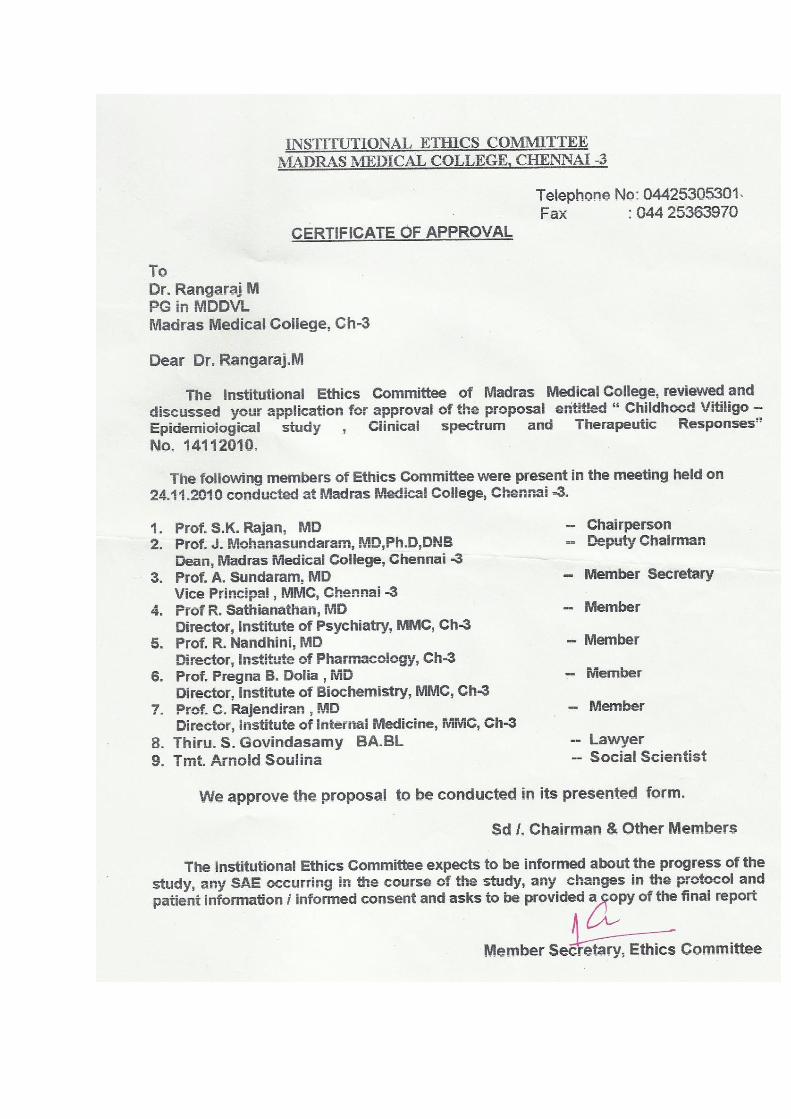
CHILDHOOD VITILIGO – EPIDEMIOLOGY,CLINICAL SPECTRUM AND THERAPEUTIC
RESPONSES
Dissertation Submitted in
Partial fulfillment of the University regulations for
MD DEGREE INDERMATOLOGY, VENEREOLOGY AND LEPROSY
(BRANCH XII A)
MADRAS MEDICAL COLLEGE
THE TAMILNADU DR.M.G.R. MEDICAL UNIVERSITYCHENNAI, INDIA.
APRIL 2012
CERTIFICATE
Certified that this dissertation titled “CHILDHOOD
VITILIGO – EPIDEMIOLOGY, CLINICAL SPECTRUM AND
THERAPEUTIC RESPONSES” is a bonafide work done by
Dr. M. RANGARAJ, Post graduate student of the Department of
Dermatology, Venereology and Leprosy, Madras Medical College,
Chennai – 3, during the academic year 2009 – 2012. This work has not
previously formed the basis for the award of any degree.
Prof. V. KANAGASABAI, M.D.,
DeanMadras Medical CollegeChennai-600003.
Prof. S. JAYAKUMAR MD.,D.D.,Head of the Department,Department of Dermatology,Madras Medical College& Rajiv GandhiGovt.General Hospital,Chennai-3.
DECLARATION
I, Dr.M. RANGARAJ solemnly declare that this dissertation
titled “CHILDHOOD VITILIGO – EPIDEMIOLOGY, CLINICAL
SPECTRUM AND THERAPEUTIC RESPONSES” is a bonafide
work done by me at Madras Medical College during 2009-2012 under
the guidance and supervision of Prof. S. JAYAKUMAR, M.D.,D.D.,
Professor and head department of Dermatology, Madras Medical
College,Chennai-600003.
This dissertation is submitted to The Tamil Nadu
Dr.M.G.R.Medical University, Chennai towards partial fulfillment of
the rules and regulations for the award of M.D Degree in Dermatology,
venereology and leprology (BRANCH – XII A)
PLACE :
DATE :
(Dr. M. RANGARAJ)
SPECIAL ACKNOWLEDGEMENT
My sincere thanks to
Prof. V.Kanagasabai, M.D.,
Dean,
Madras Medical College
for allowing me to do this dissertation and utilize
the Institutional facilities.
ACKNOWLEDGEMENT
I am gratefully indebted to Professor and Head of the Department
of Dermatology Dr.S.Jayakumar, M.D.,D.D., for his invaluable
advice, guidance and encouragement throughout the study. I would like
to express my sincere and heartfelt gratitude to Dr.V.Sudha,
MD,DV,DD., Director and Professor, Institute of Venereology, for her
kindness and support throughout the study.
I express my sincere gratitude to Prof.R.Arunadevi, MD.,DD.,
Head of the Department of Leprosy for her guidance and support.
I sincerely thank Dr.C.Janaki, M.D.,D.D., Additional Professor of
Dermatology (Mycology) for her priceless support. I am grateful to
Dr.V.Sampath, M.D., Additional Professor, Department of
Dermatology for his invaluable guidance and help.
I thank my Professor and Head of the department of Occupational
and Contact Dermatitis ,Dr.S.Nirmala.,M.D., for her help and support.
I also thank Dr.Priyavarthini M.D., for her advice and encouragement.
I also thank Additional Professor institute of venereology
Dr.P.Elangovan,M.D.,D.V., for his timely help.
I humbly thank my Co-Guide Dr.J.Manjula M.D.,D.N.B., for
her valuable guidance throughout my work.
I extend my gratitude to my Assistant professors, Dr.R.Madhu
M.D.,D.ch, Dr.SamuelJeyaraj Daniel M.D., DVL.,
Dr.Vijayabaskaran M.D.,D.ch., Dr. G.K.Tharini, M.D.,
Dr.S.Anupama Roshan, D.D.V.L., and Dr.N.Hema, M.D, Assistant
professors, Department of Dermatology for their kind support and
encouragement.
I also thank my Assistant Professors Dr.V.Thirunavukkarrasu,
M.D.,D.V., Dr.P.Prabhakaran,M.D.,(D.V.L) , Dr.V.N.S.Ahamed
Sherrif, M.D (D.V.L), Dr.Umameheshwari,M.D.,(D.V.L)., and
Dr.R.Sowmiya M.D., (D.V.L)., of Institute of Venereology for their
able guidance.
I express my thanks to my Former Professor and Head of the
Department of Dermatology Dr.D.Prabhavathy, M.D.,D.D, and
Former Professor and Head of the Department of Occupational and
contact dermatitis Dr.V.Somasundaram, M.D.,D.D for their constant
support and motivation.
I express my thanks to my former assistant professors,
Dr.Kumaravel.M.D.,D.D, Dr.A.Hameedullah, M.D.,D.D, Dr.Afthab
JamelaWahab, M. D.,D.D.,Dr.N.Saravanan, MD(DVL),Dch.,
Department of Occupational and Contact Dermatitis for their support
and help.
I am inclined to thank my former Assistant professors, Institute of
Venereology, Dr.K.Venkateswaran,M.D.,D.V., Dr.S.Arunkumar
M.D., D.V., Dr. P. Mohan MD.,D.V.,and Dr.S.Kalaivani, M.D.,D.V
for their kindness.
I extend my sincere thanks to The Director and
Professor, Institute of Plastic surgery, Madras Medical College,
Dr. R.Ananthsubramaniyam M.S., MCH for his support and
encouragement.
I am also grateful to all paramedical staffs for rendering timely
help to complete my study.
My hearty thanks to all my beloved friends for their wishes and
cooperation amidst their busy schedule throughout my study.

CONTENTS
S.No Title Page
No
1 INTRODUCTION 1
2 REVIEW OF LITERATURE 2
3 AIM OF THE STUDY 38
4 MATERIALS AND METHODS 39
5 OBSERVATIONS 52
6 DISCUSSION 66
7 CONCLUSION 74
REFERENCES
PROFORMA
MASTER CHART
1
INTRODUCTION
Vitiligo is a common dermatologic disorder in children and one
that has been observed since ancient times. It is characterized by
asymptomatic, well-demarcated, ivory-white macules and patches that
may be localized or generalized. Vitiligo is common in India affecting
3-4% of Indian population2. Childhood vitiligo is a special subtype and is
seen in significant proportion of vitiligo patients. There are only a few
clinical studies in the past which address the clinical spectrum of vitiligo
in children.
This study on eighty cases of childhood vitiligo will cover the
epidemiology, clinical spectrum and review its therapeutic responses.
This study was undertaken in view of the seriousness of the problem in
children.
2
REVIEW OF LITERATURE
DEFINITION
Vitiligo is an acquired primary, usually progressive
melanocytopenia of unknown etiology, clinically manifested by
circumscribed achromic macules often associated with leukotrichia and
histologically by degeneration and disappearance of melanocytes in the
involved skin and not uncommonly in the pigment epithelium of the eyes,
leptomeninges and inner ear.
The disease affects subjects of either sex with a heritable
constitutional predilection.
SYNONYMS
Sufaid Bagh Phubhari Phuleri
Switra Bars Bahak
Kilas Palita Kodha
Sweta Kushta Dhawal Kustha
HISTORY
The origin of the term vitiligo is obscure, like the disease itself.
Some believe that it is derived from the Latin words ‘vitelius’ meaning
3
vale, i.e. pale pink flesh of calf, since the clinical lesions resembled the
white patches of a spotted calf, while other believed that it originated
from the Latin word ‘vitium’, meaning blemish. The Roman physician
Celsus first used the term vitiligo in the second country AD. It is
interesting to note that the Rigveda (6000 BC or earlier) named
leukoderma as Kilas, meaning a white spotted deer2.
EPIDEMIOLOGY
The highest incidence has been recorded in India and Mexico.
Based on the studies done so far, it is roughly estimated to be 3-4% in
India2.
INCIDENCE AND HOST FACTORS
There is no particular sex predilection. Female predominance
probably reflect their greater concern for cosmetic disfigurement and
related to the social and marital problems.
Vitiligo mostly affects people with Fitzpatrick’s skin types III
and IV2.
The disease may start at any stage. Onset of the unilateral
dematomal type is usually in childhood within 10 years of age whereas
4
most cases with bilateral non- dermatomal lesions (vitiligo vulgaris type)
begin in the second to fourth decades of life. A few instances of vitiligo
lesions present at birth have been reported as cases of congenital vitiligo5.
Koebner’s phenomenon is observed in 6-20% of cases of vitiligo
vulgaris2. Minor trauma such as scratch mark, laceration, or stitches on
the skin results in the development of a corresponding linear depigmented
macule, usually in 2-4 weeks. This isomorphic phenomenon indicates an
abnormal pattern of cutaneous response to trivial physical trauma.
HEREDOFAMILIAL ASPECTS
At present the consensus is that the familial incidence is between
20% and 30%2.
VITI gene, which maps to chromosome 2p16, has also been
associated with vitiligo2.
A familial incidence of diabetes mellitus and thyroid disease has
been commonly noted in cases of vitiligo. Atopy is another familial
association.

5
ETIOLOGY:
THEORIES ON THE PATHOGENESIS:
Theories on the pathogenesis of vitiligo centered on mechanisms
for the destruction of melanocytes as there are no melanocytes present in
the fully evolved white macules. Traditionally there have been three
hypothesis to explain vitiligo.
1. Neural Hypothesis
2. Self destructive Theory
3. Autoimmune Theory
1. NEURAL HYPHOTHESIS8,9
Evidences in favour of neural hypothesis include:
Stress and emotional trauma is a known initiating or precipitating
factor in vitiligo, the common embryologic origin of melanocytes and the
nervous system, dermatomal distribution of segmental vitiligo,
demonstration of direct contact between cutaneous free nerve endings and
epidermal melanocytes in vitiligo macules, demonstration of
neuropeptides in the skin and their ability to regulate melanocyte
differentiation has given more strength to this hypothesis.
6
These alterations are said to induce melanocyte dysfunction and
melanocyte injury by promoting the production of melanocytotoxic
compounds and by decreasing the natural detoxification.
At present, however the role of nervous system in vitiligo, if any, is
poorly understood.
2. SELF DESTRUCTIVE THEORY 9
A. B. Lerner postulated that melanocytes in vitiligo have lost an
intrinsic protective mechanism that eliminates toxic intermediates or
metabolites in the melanogenesis pathway.
Melanocytes synthesize melanin by oxidation of tyrosine to
dihydroxyphenylalanine (DOPA) and to dopaquinone, which by a
multistep reaction forms indoles. All the intermediates in the biosynthesis
of melanin are phenols, excessive production or accumulation of phenolic
radicals or intermediates within the melanocyte could damage the cell10.
It has been suggested that melatonin receptor and melatonin could
play a key role in vitiligo. Melatonin is known to stimulate the
melanogenic pathway without the production of melanins, leading to an
accumulation of toxic intermediates which causes injury to keratinocytes
7
and melanocytes with release of specific cellular proteins that initiate a
secondary autoimmune reaction.
The presence of high levels of Hydrogen peroxide (H2O2) and low
levels of catalase11 in epidermis of vitiligenous skin suggests that there is
an increased oxidative stress in vitiligo patients12. Several pathways could
be involved in overproduction of H2O2 in vitiligo13.
An abnormality in tetrabiopterin metabolism leads to defective
recycling of 6BH4 causing formation of H2O2.
Over production of H2O2 with increased levels of monoamine
oxidase A from inhibition of thioredoxin / thioredoxin reductase by
calcium and increased nitric oxide synthase activities.
3. AUTOIMMUNE THEORY14
The association of vitiligo with autoimmune diseases suggested an
immunologic basis for vitiligo.
A. Humoral immunity
There is an increased frequency of organ-specific auto antibodies
in patients with vitiligo, even in the absence of any associated disease in
8
up to 30% of patients. Antibodies to thyroid tissue, gastric parietal cell,
adrenal cytoplasm and pancreatic islet cell have been demonstrated.
More recently, autoantibodies to a transcription factor called
SOX10 have been found in vitiligo associated with APECED.
B. Cell mediated immunity
In marginal skin from progressive lesions of generalized and
inflammatory vitiligo, an infiltrate of skin-homing (CLA+) cytotoxic
T cells expressing granzyme/perforin is often found close to the
remaining melanocytes. This infiltrate is composed of CD8 T cells, CD4
T cells and subsets of macrophages, and this correlates with the increased
number of CLA+ MART-1 reactive CD8 T cells in the peripheral blood
of patients with progressive vitiligo. These specific cytotoxic T cells react
against the melanocyte differentiating antigens in vitiligo patients14.
4. OTHER HYPOTHETICAL THEORIES
A. Convergence theory suggests that genetic factors, stress,
accumulation of toxic compounds, infection, autoimmunity, mutations,
altered cellular environment, and impaired melanocyte migration and
proliferation can all contribute to this disease15.
9
B. An intrinsic defect of the structure and function of rough
endoplasmic reticulum in melanocytes of vitiligo patients16.
C. Deficiency of Melanocyte growth factor.
D. Viral origin.
E. Dysregulation of Melanocyte apoptosis.
CLINICAL FEATURES7
A typical lesion is a well defined depigmented (milky white or
chalky) macule, round to oval in shape, to fairly distinct often with
scalloped margins, measures from few mm to many cms in diameter,
showing a variable number of depigmented (white) hairs and without any
change in the skin texture.
The number, size, shape, and location of individual macules vary
widely. Frequently the initial macule occurs on the exposed areas (such as
the dorsal surface of hands, elbows, feet, legs, knees, neck and face),
body folds (such as axillae, groin, and sub mammary region in women),
lips or genitalia.
10
The initial unifocal lesion may be followed by the appearance of
new lesions elsewhere. In less than 25% of cases the onset may be
multifocal2. Onset of the lesions is usually insidious. The disease is
progressive in nature as a rule and course is virtually unpredictable and
may be quite erratic, it may be jerky, indolent or rapid. While some
lesions may show signs of repigmentation, new lesions may develop on
other parts of the body simultaneously. There is an episodic phase of
rapid extension of lesions after remaining quiescent over a long period of
time.
Although no definite precipitating factor is ascertained, many
factors have been incriminated which include local trauma, itching,
friction, infection, infestations, gastrointestinal disturbances, emotional
upset, pregnancy, parturition and surgery.
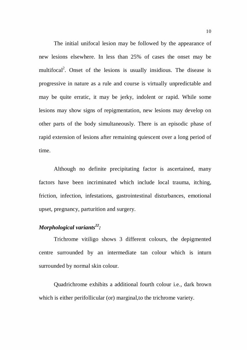
Morphological variants22:
Trichrome vitiligo shows 3 different colours, the depigmented
centre surrounded by an intermediate tan colour which is inturn
surrounded by normal skin colour.
Quadrichrome exhibits a additional fourth colour i.e., dark brown
which is either perifollicular (or) marginal,to the trichrome variety.
11
Pentachrome vitiligo exhibits 5 shades of colours white, tan, brown
hyperpigmented, blue grey hyperpigmented and normal.
INFLAMMATORY VITILIGO
Shows erythematous raised border and may be associated with
pruritus.
VALECEO VITILIGO2
In most instances extension of vitiligo is gradual, whereas in
valeceo vitiligo there is a very rapid extension and spread of lesions.
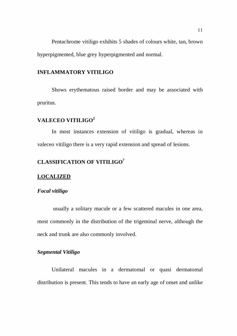
CLASSIFICATION OF VITILIGO7
LOCALIZED
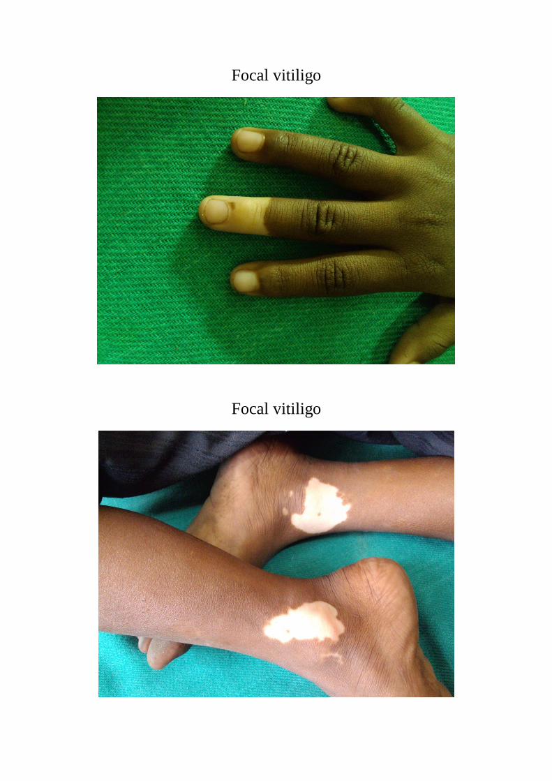
Focal vitiligo
usually a solitary macule or a few scattered macules in one area,
most commonly in the distribution of the trigeminal nerve, although the
neck and trunk are also commonly involved.
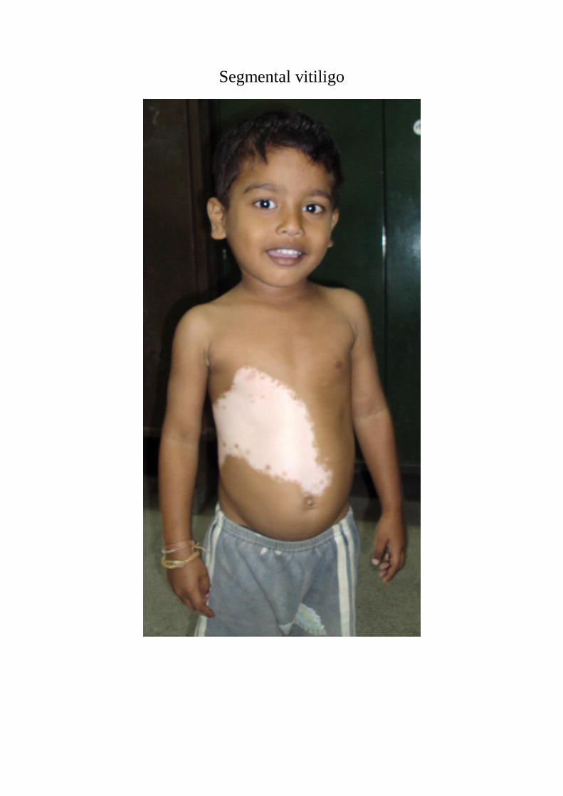
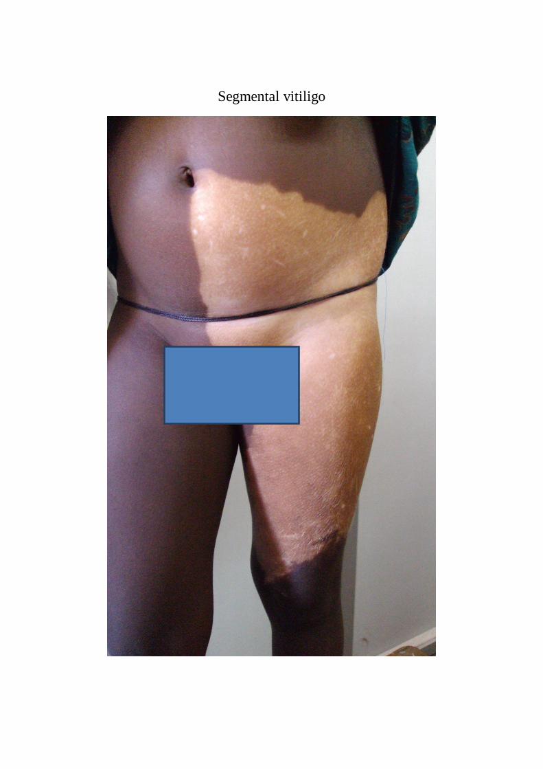
Segmental Vitiligo
Unilateral macules in a dermatomal or quasi dermatomal
distribution is present. This tends to have an early age of onset and unlike
12
the other types, is not associated with thyroid disease or other
autoimmune disease. This type occurs more commonly in children7.
Alteration of neural peptides has been implicated in the pathogenesis of
this type. More than one half of the patients with segmental vitiligo have
patches of white hair, known as poliosis7.
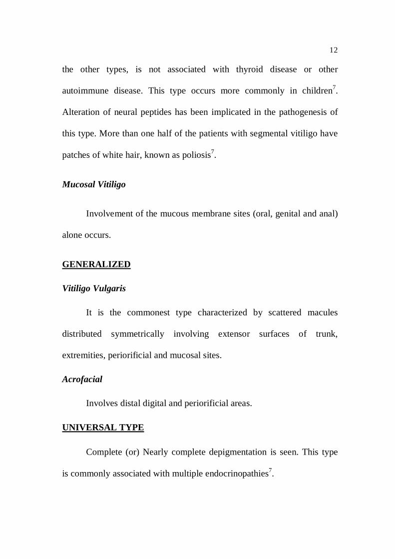
Mucosal Vitiligo
Involvement of the mucous membrane sites (oral, genital and anal)
alone occurs.
GENERALIZED
Vitiligo Vulgaris
It is the commonest type characterized by scattered macules
distributed symmetrically involving extensor surfaces of trunk,
extremities, periorificial and mucosal sites.
Acrofacial
Involves distal digital and periorificial areas.
UNIVERSAL TYPE
Complete (or) Nearly complete depigmentation is seen. This type
is commonly associated with multiple endocrinopathies7.
13
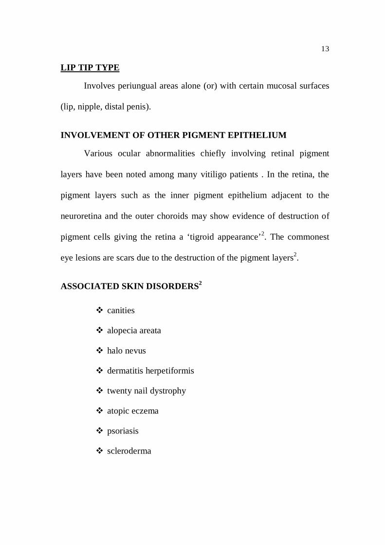
LIP TIP TYPE
Involves periungual areas alone (or) with certain mucosal surfaces
(lip, nipple, distal penis).
INVOLVEMENT OF OTHER PIGMENT EPITHELIUM
Various ocular abnormalities chiefly involving retinal pigment
layers have been noted among many vitiligo patients . In the retina, the
pigment layers such as the inner pigment epithelium adjacent to the
neuroretina and the outer choroids may show evidence of destruction of
pigment cells giving the retina a ‘tigroid appearance’2. The commonest
eye lesions are scars due to the destruction of the pigment layers2.
ASSOCIATED SKIN DISORDERS2
canities
alopecia areata
halo nevus
dermatitis herpetiformis
twenty nail dystrophy
atopic eczema
psoriasis
scleroderma
14
lichen planus
lichen simplex
discoid lupus erythematosus
malignant melanoma
ichthyosis.
ASSOCIATED SYSTEMIC DISORDERS
Pernicious anemia
Addition’s disease
Grave’s disease
hyperthyroidism
hypothyroidism
hyperparathyroidism
diabetes mellitus
More than 10% of patients with pernicious anemia have been
reported to develop vitiligo2. In a different series, vitiligo has been
reported to be associated with thyroid disorders in 0.6% - 38% of
patients2. Diabetes mellitus was reported to occur in 1%-7% of vitiligo
patients2. An intimate relationship between vitiligo and melanoma has
been reported in one study.
15
SYNDROMES ASSOCIATED:
Vogt-Koyanagi-Harada syndrome: It is a rare syndrome affecting
children, especially of south-east Asian origin. Characteristic features are
uveitis, aseptic meningitis, dysacusia, alopecia, poliosis and vitiligo.
Uveitis is the presenting feature and vitiligo may appear later, during the
chronic stage (fourth stage) of the disease (adolescence or adulthood).
Vitiligo lesions tend to be symmetrical, involving the head, neck and
trunk. The sacral region is a common site of involvement with vitiligo.
Poliosis may involve the scalp, eyebrows and eyelashes.
Alezzandrini’s syndrome: This syndrome is characterized by
segmental vitiligo(cheek), poliosis, ipsilateral uveitis resulting in
decreased visual acuity and same-sided partial hearing loss. Manifestation
starts during adolescence.
PATTERN & DISTRIBUTION:
Vitiligo can exhibit 2 general pattern unilateral with a dermatomal
distribution or bilateral which is very common. Although vitiligo can
occur on any part of the body, there are characteristic patterns of
involvement. The most frequently involved sites are the face, dorsum of
16
hands, axilla, umbilicus, nipples, sacrum and inguinal region. It also
commonly involves sites of friction - back of hand, elbow, knee, ankles,
shoulder strap and waist band areas, other commonly involved sites are
scalp and neck. Palms and soles involvement is common. Mucosal
involvement is frequent, the genitalia, gingiva and lips may be involved.
VITILIGO VULGARIS
It is the most common type7. The lesions may occur at various
body sites often bilaterally, the lesions may be either symmetrical (or)
asymmetrical.
SEGMENTAL VITILIGO
It is characterized by unilateral macules in a dermatomal
distribution. It tends to be earlier in onset. 20% of children have this
pattern 3.It is more stable. It is not familial and is unlikely to be associated
with other autoimmune diseases. Koebnerization is not characteristic.
Trigeminal area is the commonest site more than 50% of cases. Neck and
Trunk involved in 23% and 17% respectively. Upto 13% may have
multiple sites of involvement2.
17
FOCAL
20% of children with vitiligo have focal pattern.
ACROFACIAL
It is the least common type in children23.
HAIR IN VITILIGO
Depigmentation of body and scalp hair occurs in 9-45% of vitiligo
patients3. Nearly 50% of segmental vitiligo arc associated with poliosis.
The perifollicular and interfollicular skin is affected. Leukotrichia
indicates poor prognosis.
HISTOPATHOLOGY:
Histopathology as a means of diagnosis is rarely employed in
vitiligo but it useful when other cause of hypopigmentation needs to be
excluded. The histopathological changes classically associated with
vitiligo include a complete absence of melanocytes in the basal layer of
the epidermis with loss of melanin in the epidermis. The upper dermis
often has sparse superficial perivascular collection of lymphocytes with a
few melanophages.
18
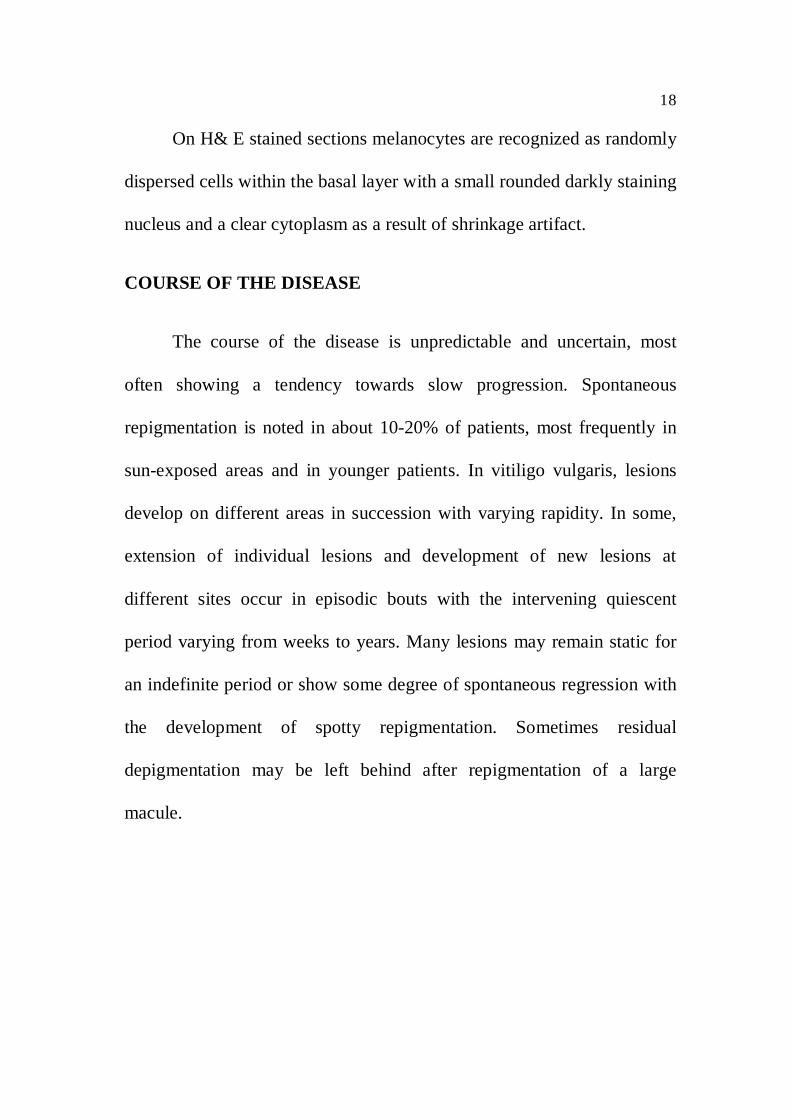
On H& E stained sections melanocytes are recognized as randomly
dispersed cells within the basal layer with a small rounded darkly staining
nucleus and a clear cytoplasm as a result of shrinkage artifact.
COURSE OF THE DISEASE
The course of the disease is unpredictable and uncertain, most
often showing a tendency towards slow progression. Spontaneous
repigmentation is noted in about 10-20% of patients, most frequently in
sun-exposed areas and in younger patients. In vitiligo vulgaris, lesions
develop on different areas in succession with varying rapidity. In some,
extension of individual lesions and development of new lesions at
different sites occur in episodic bouts with the intervening quiescent
period varying from weeks to years. Many lesions may remain static for
an indefinite period or show some degree of spontaneous regression with
the development of spotty repigmentation. Sometimes residual
depigmentation may be left behind after repigmentation of a large
macule.
19
DIFFERENTIAL DIAGNOSIS
post inflammatory hypopigmentation
pityriasis alba
indeterminate hansen
pityriasis versicolor
post kala azar dermal leishmaniasis
chemical leukoderma
piebaldism
idiopathic guttate hypomelanosis
naevus depigmentosus
albinism
ash leaf macule
waardenburg syndrome
woolf’s syndrome
halo naevus
syphilis and yaws
lupus erythematosus
incontinentia pigmenti
20
PROGNOSIS
These factors indicate poor prognostic factors:
1) Lesions on the so-called resistant sites, such as bony prominences,
non-fleshy areas, non-hairy areas and mucosal areas. They
comprise the sides of the ankles, front of the wrists, back of the
elbows, dorsum of feet and hands, palms, soles, nipples and areola.
2) The greater the percentage of associated white hair, the worse the
prognosis.
3) Extensive long-standing disease.
4) Associated systemic disorders.
5) Familial background.
6) Old age.
7) Iatrogenic factors, including injudicious administration of topical
and systemic medication, particularly photochemo-therapeutic
agent)
21
TREATMENT MODALITIES
Treatment of vitiligo at any age remains a challenge for clinicians,
more so during childhood. None of the available therapies is absolutely
effective, and the disease runs a relapsing course. With any of the
treatment modalities, >75% repigmentation (achieved by approximately
60% of treated children) is considered as the best therapeutic response34.
Vitiligo beginning in childhood can be associated with significant
psychological trauma that may have lasting effects on the persons
selfesteem. Since vitiligo is not a life threatening cutaneous condition
many children with little skin involvement in cosmetically unimportant
areas do not wish to treat their disease.
Few studies have assessed therapeutic responses in paediatric
patients with vitiligo. Presently there is no universally effective medical
or surgical modality for vitiligo. However there are number of active
therapeutic approaches that are known to be effective. Most
repigmentation therapies are directed to stimulate the melanocytic
reserves in the hair follicle. Non surgical repigmentation therapies
represent the first line active treatment modality in vitiligo44. If the area to
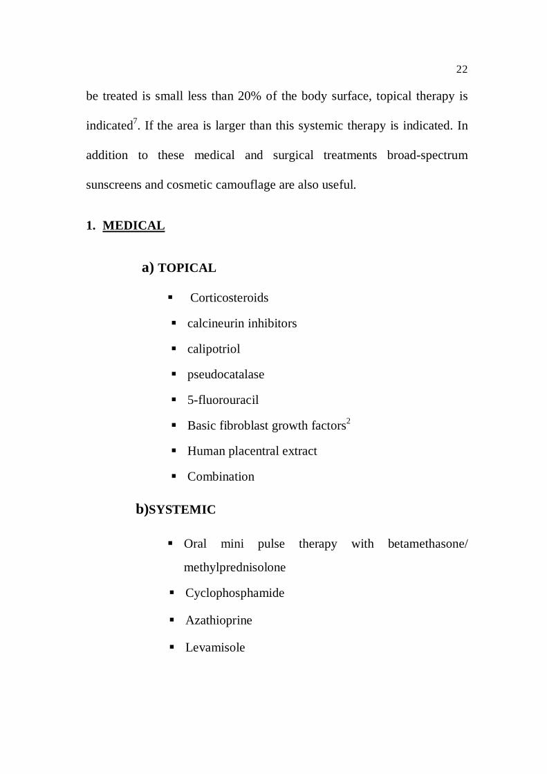
22
be treated is small less than 20% of the body surface, topical therapy is
indicated7. If the area is larger than this systemic therapy is indicated. In
addition to these medical and surgical treatments broad-spectrum
sunscreens and cosmetic camouflage are also useful.
1. MEDICAL
a) TOPICAL
Corticosteroids
calcineurin inhibitors
calipotriol
pseudocatalase
5-fluorouracil
Basic fibroblast growth factors2
Human placentral extract
Combination
b)SYSTEMIC
Oral mini pulse therapy with betamethasone/
methylprednisolone
Cyclophosphamide
Azathioprine
Levamisole
23
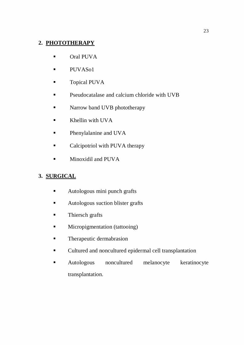
2. PHOTOTHERAPY
Oral PUVA
PUVASo1
Topical PUVA
Pseudocatalase and calcium chloride with UVB
Narrow band UVB phototherapy
Khellin with UVA
Phenylalanine and UVA
Calcipotriol with PUVA therapy
Minoxidil and PUVA
3. SURGICAL
Autologous mini punch grafts
Autologous suction blister grafts
Thiersch grafts
Micropigmentation (tattooing)
Therapeutic dermabrasion
Cultured and noncultured epidermal cell transplantation
Autologous noncultured melanocyte keratinocyte
transplantation.
24
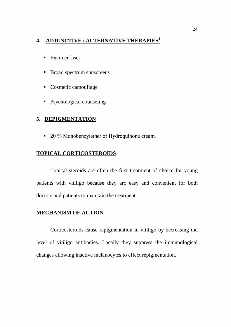
4. ADJUNCTIVE / ALTERNATIVE THERAPIES4
Excimer laser
Broad spectrum sunscreens
Cosmetic camouflage
Psychological counseling
5. DEPIGMENTATION
20 % Monobenzylether of Hydroquinone cream.
TOPICAL CORTICOSTEROIDS
Topical steroids are often the first treatment of choice for young
patients with vitiligo because they arc easy and convenient for both
doctors and patients to maintain the treatment.
MECHANISM OF ACTION
Corticosteroids cause repigmentation in vitiligo by decreasing the
level of vitiligo antibodies. Locally they suppress the immunological
changes allowing inactive melanocytes to effect repigmentation.
25
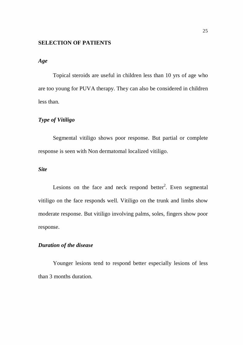
SELECTION OF PATIENTS
Age
Topical steroids are useful in children less than 10 yrs of age who
are too young for PUVA therapy. They can also be considered in children
less than.
Type of Vitiligo
Segmental vitiligo shows poor response. But partial or complete
response is seen with Non dermatomal localized vitiligo.
Site
Lesions on the face and neck respond better2. Even segmental
vitiligo on the face responds well. Vitiligo on the trunk and limbs show
moderate response. But vitiligo involving palms, soles, fingers show poor
response.
Duration of the disease
Younger lesions tend to respond better especially lesions of less
than 3 months duration.
26
TREATMENT PROTOCOLS:
Topical therapy using 0.1% betamethasone valerate is successful in
repigmenting some vitiliginous lesions. Low to mid potency steroids are
used in young children. High potency preparation can be used in older
children. They should be applied only to limited (or) localized areas of
involvement.
Steroids should be applied two times daily for 3wks, then skipped
for 1 week and then two times daily as above for 2 months. If there is no
response it is discontinued. If there is good response it is continued along
with monitoring every 2 months for every side effects. The earliest
response is seen after 3 wks. The duration of treatment strongly depends
on the response. The mean duration of treatment varies from 5 to 8
months. It can be even prolonged for a period of 21 months. An
acceptable response is regarded as 50% or greater return of pigmentation
to all involved skin areas. A good response occurs in 30-50% of patients.
SIDE EFFECTS
Atrophy , Striae ,Telangiectasia, Glaucoma if used for a prolonged
period on eyelid, Hypothalamic - pituitary adrenal axis suppression, if
used for a prolonged period.
27
SYSTEMIC STEROIDS
Oral minipulse therapy with betamethasone
This pulse therapy seems to be beneficial in the treatment of
vitiligo by it’s immunomodulatory effect.
Mechanism of Action:
Steroids prevent damage to the melanocytes in the vitiligo by
decreasing levels of offending complements and antibody mediated
cytotoxicity against melanocytes.
Selection of Patients
Child with extensive and rapidly spreading vitiligo.
Treatment Protocol
Tab.Betamethasone 5mg is given as a single oral dose after
breakfast on two consecutive days each week. The response is evaluated
every 2-4 months and the side effects are recorded.
RESPONSE TO TREATMENT
The treatment duration for oral minipulse therapy varies between
6 and 24 months. Treatment is discontinued after all depigmented areas
regained pigment or when there has been no further repigmentation for
28
atleast 4 months. It arrests the progression of vitiligo within 1-3 months
in 89% of children on treatment. Within 2-4 months 80% of the patients
show spontaneous repigmentation of existing lesions which progresses
with continued treatment. The extent of repigmentation varies indifferent
patients and even in different lesions in the same patient. It varies from
10% to 90%.
SIDE EFFECTS
Weight gain, headache, transient mild weakness, acne, cushing’s
disease, diabetes mellitus, perioral dermatitis, glaucoma, cataract,
herpes zoster.
CONTRAINDICATIONS
Diabetes mellitus, tuberculosis, gastro intestinal disease, glaucoma,
hypertension and osteoporosis.
INVESTIGATIONS
Blood Sugar
X-ray chest
29
Topical Tacrolimus and Topical Pimecrolimus
Tacrolimus is a macrolide lactone produced by Streptomyces
tsukubaensis. Tacrolimus acts on the immune system and directly on skin
cells. It binds to a receptor within the cell called the FK binding proteins.
This resulting drug-protein complex inhibits calcineurin (a calcium-
dependent phosphatase transmitting chemical) that in turn reduces the
activity of T-lymphocytes in the immune system. As a consequence, T-
cells fail to release their cytokines.
Tacrolimus by its immunomodulating activity found to be useful in
treating vitiligo. It is available as 0.03% and 0.1% ointment and it has to
be applied twice daily for a period of 3 to 6 months, and recent reports
claimed it as a success even when used alone as a monotherapy.
Pimecrolimus is available as 1% cream.
NARROW BAND UVB PHOTOTHERAPY
Narrow band UVB therapy is more effective than PUVA therapy in
terms of repigmentation4. Other advantages are: (a) since no oral
medication is required, no side effects of psoralens are seen; (b) it can
also be used in pregnancy and childhood; and (c) no post-exposure eye
30
protection is necessary; and (d) exposure time is shorter than with PUVA.
Hence, narrow band UVB therapy is now considered the treatment of
choice for stable vitiligo. Narrow band fluorescent bulbs of Philips TL-
QJ, each of 100W with an emission spectrum of 311 nm are used.
Response to treatment correlated with localization of the lesions
and the patient’s compliance. The risk of skin cancer with narrow-band
UVB is unknown at present and there are insufficient human data
available to provide recommendations. If no response is observed after 6
months, further therapy should be discouraged4 or the treatment be
limited to specific areas Targeted UVB or focussed microphototherapy
with a spectrum of 280 to 315 nm also has excellent results but the cost is
prohibitive and the size of the area to be treated is restricted.
SURGICAL MODALITIES
MINIATURE PUNCH GRAFTING
This procedure consist of taking 1.5-2.5mm sized punch autografts
from normal skin and grafting them in appropriately space and punched
out chambers at the recipient site.
31
TEST GRAFTING
Trial grafting in a small area of vitiligo should be undertaken with
4-5 minigrafts to confirm the stability of the disease before attempting
repigmentation in other involved areas4.
PROCEDURE
Give premedication (antibiotic) and calculate the approximate
number of required grafts after spaced markings at the recipient site. For
a large patch, multiple sittings are required.
DONOR SITE
Donor site is the extensor aspect of the thigh, gluteal region, medial
aspect of the upper arm, or behind the ear lobe and retroauricular area.
Prepare it surgically (viz, with shaving, cetrimide, spirit, povidone iodine)
and give local anesthesia (1% lignocaine without adrenaline)
intradermally to raise a wheal 5 cm in diameter or larger depending upon
the number of grafts required.
Rotate a 2.5 mm punch until the cutting edge descends to the depth
of the upper dermis. Take the requisite number of cuts adjacent to one
32
another, with intervening normal skin (rows of 10-15). Lift the edge and
cut through the upper dermis close to the epidermis to free the grafts
individually from the base. Transfer to a bowl containing gauze
moistened with normal saline. Achieve hemostasis by pressure. Dress
with a double layer of collgen mesh, gauze and Elastoplast bandage.
RECIPIENT SITE
Surgically prepare the area and give local anesthesia (1%
lignocaine injected both intradermally and subcutaneously). Rotate a 2
mm punch until the cutting edge descends to the depth of the mid-dermis.
Lift the edge, cut through the mid-dermis to free it from the base and
discard. Take such cuts spaced 5-10 mm apart as per the geographical
pattern of the patch. Transfer the stored grafts individually to these
dilated punched out areas, ensuring that the dermal side of the graft is in
direct contact with the recipient dermis, and even out the edges. For
hemostasis and a proper take up of graft, apply firm pressure with a moist
gauze piece. Dress with a double layer of framycetin tulle, gauze
and elastocrepe bandage. Immobilize the part where required
(rest, splints, etc.).
33
POST OPERATIVE MEDICATIONS
Antibiotics and anti-inflammatory drugs are administered for 8-10
days and PUVA or PUVASOL for 3-6 months is given to enhance
repigmentation4. Oral and local steroids may be needed if the pigment
spread is slow.
COURSE OF GRAFTS
The grafts turn pink brown, dark brown and finally black . They are
taken up by 8 to 10 days. Perigraft pigmentation starts by 1 month, and
spreads to cover the patch in the next 3-6 months4. The average spread
varies from 5-10mm and in dark individuals up to 15 mm has been
observed.
COMPLICATIONS
These include faulty or no pigmentation due to graft rejection
(bleeding, shearing movement, upside-down graft, infection and improper
immobilization), cobble-stoning (raised grafts), sinking pits (sunk-in
grafts), polka dot appearance, hypo- or depigmented junctional line,
34
scarring, allergic reactions, blotchy pigmentation, color discrepancy, and
reactivation of viti1igo.
Cobble-stoning and sinking pits can he prevented by ensuring that
the grafts are placed at the level of the adjacent skin. If the graft is raised,
either cut away its under-surface or deepen the recipient chamber.
ADVANTAGES
This is a simple, safe and inexpensive office procedure that, being
an extended biopsy technique, needs no special training for a
dermatologist. It has a high success rate and excellent cosmetic results.
Large lesions and any site except the angle of mouth can be treated. Areas
of residual vitiligo between grafts or wherever grafts are rejected can be
re-grafted.
THIN THIERCH’S SPLIT THICKNESS SKIN GRAFTING
This procedure consists of grafting a very thin, split thickness skin
graft consisting of the epidermis and part of the upper papillary dermis
onto the dermabraded patch of vitiligo, and securing it with pressure or
with surgical glue and local immobilization. It is very useful for large
35
patches and where immediate results are desirable4. Use of meshed split
thickness skin grafts has also been reported to treat large areas of vitiligo.
DONOR SITE
A thin split thickness graft is obtained from routine donor sites
with a simple razor blade, Silver’s knife or mechanized dermatome by the
basic skin grafting technique.
RECIPIENT SITE
The recipient bed is prepared in a similar fashion as in the spot and
regional dermabrasion technique by evenly dermabrading the vitiliginous
lesion and 1 cm of the surrounding normal skin to obtain a good raw
surface. The graft is then placed with its dermal surface facing the
abraded bed to extend 3-5 mm beyond its border. Its edges are evened out
to prevent curling/beading and a ‘stuck on’ appearance. Pressure dressing
with elastocrepe bandage is given after covering the graft with framycetin
tulle or surgical glue is applied in droplet form all along the overlapped
and free edges of the graft to ensure firm contact. A flexible collodion
dressing can also be used as an alternative. The part is immobilized if
required.
36
POSTOPERATIVE DRESSING AND MEDICATIONS
The recipient site dressing is changed after 24 hours. A seroma,
hematoma or any air bubble is drained. The donor site dressing is
removed after 10-15 days. Antibiotics and anti-inflammatory drugs are
given for 8-10 days. Pressure garments can be given after graft uptake for
2-3 months to flatten the graft and obtain a good color match.
COMPLICATIONS
These are graft rejection (due to bleeding, movement, infection and
upside down placement of graft), ‘stuck on tyre patch’ appearance,
perigraft halo of depigmentation, superficial scarring at the donor site,
and reactivation of vitiligo.
ADVANTAGES
Large areas can be covered in a single sitting. The grafted areas are
skin colored immediately after removal of the dressing. It is a simple and
safe procedure with good results. The procedure is less time consuming
than other procedures of grafting for vitiligo. The graft survival chances
are good because it is a thin split thickness graft.
37
DISADVANTAGES
Large areas have to be abraded if the geographical pattern of the
vitiligo patch is to be followed. Some degree of hyperpigmentation and
contracture will occur at the grafted site. Superficial scarring and vitiligo
can develop at the donor site. The fingers, lips, palms and soles are
difficult to graft. Benign thin, Thiersch’s grafts are vulnerable to trauma.
Multiple sittings are required for large lesions. Surgical expertise is
required to obtain thin and adequate sized thin graft.
38
AIM OF THE STUDY
To study the epidemiology, clinical spectrum and therapeutic
responses in childhood vitiligo with an aim to observe the following
parameters:
1. Prevalence of vitiligo in children under 12 yrs.
2. Age and sex distribution, associated family history.
3. Sites of involvement and type of vitiligo.
4. Associated autoimmune disorders and syndromes.
5. Therapeutic responses to various modalities of treatment
39
MATERIALS AND METHODS
This was a prospective study conducted at Rajiv Gandhi
Government General Hospital, Chennai in the Department
of Dermatology for a period of over 1 ½ years from november 2009 to
June 2011.
During this period, all children less than 12 years of age were
screened for vitiligo. Only untreated patients were included in the study.
A total of 80 children with vitiligo of both sexes were enrolled. They
were questioned in detail regarding the age of onset, site of initial lesion,
duration of disease, progression and associated cutaneous disorder.
Precipitating factors such as trauma, illness, stress and contact with
chemicals were specifically asked for. History of ocular symptoms and
systemic illness like diabetes, thyroid dysfunction, anaemia and
Addison’s disease were recorded. History of vitiligo, premature canities
or any other autoimmune disorder in the family was noted.
40
A detailed dermatological examination was carried out and a
thorough systemic examination was made to record any associated
systemic disorders.
The diagnosis of vitiligo was made based on clinical features and if
needed skin biopsy. Trichrome vitiligo, quadrichrome vitiligo and
associated cutaneous disorders were specifically looked for. In each case,
body charting, extent of body surface involvement, leukotrichia and
Kobnerization was recorded.
Each case was classified into recognized patterns of vitiligo namely
vitiligo areata, segmental vitiligo, acrofacial vitiligo, lip tip vitiligo,
vitiligo mucosae, vitiligo vulgaris and vitiligo universalis.
The patients in our study were divided into 3 groups.
1) Group A treated with Topical steroids and immuno modulators.
2) Group B treated with NBUVA therapy
3) Group C treated with surgical modalities with mini punch grafting
and split skin grafting.
41
The above therapeutic modalities were adopted according to the
age of the patients, type of vitiligo, site of vitiligo and extent of vitiligo.
A detailed history regarding the onset, duration and course of the
disease, presence and absence of precipitating factor, family history,
associated skin and systemic problems, were recorded.
Dermatological assessment of the disease was carried out using
down the sites of involvement total body surface area involved, total
number of factors, size and distribution of the patches, presence of white
hair in the patch.
Details regarding the margin of the patch, skin texture, presence or
absence of perifollicular pigmentation, Koebner’s phenomenon,
associated with skin and systemic problems were noted. Focal sepsis was
ruled out by referring the patient to ENT and Dental OPD for checkup.
Other associations if any are noted and referred to respective departments
for evaluation.
After collecting the preliminary reports the patients were assessed
and divided based on inclusion and exclusion criteria.
42
Since all the three modalities of treatment selected belong to
different modes (medical, physical and surgical) they cannot be
compared. So, the therapeutic response to each modality has been
studied.
GENERAL PRINCIPLES
1) Patient should be explained the nature of the disease and its
impredictable cause and prognosis.
2) Reassurance is essential.
3) Diet rich in proteins, Vitamin B complex, Vitamin E and minerals
such as copper, iron and zinc should be supplemented.
4) Avoidance of precipitating factors and drugs like choroquine, -
interferon and -blockers.
5) Avoidance of sun exposure, sunscreens may be used if necessary.
6) Avoidance of detergents and substances containing phenolic
compounds.
43
EFFICACY PARAMETERS
The primary efficacy variable was the percentage change in
depigmentation from the baseline to the end of study period. (i.e. 6
months)
The efficacy parameter namely the Physician’s Global
Improvement assessment was computed at the end of 6 months of the
study.
During the initial assessment, estimation of body surface area
(BSA) involvement was assessed using Vitiligo Area Scoring Index
(VASI). The body was divided into five separate and mutually exclusive
regions: Face and Neck, Upper extremities (excluding Hands), Lower
extremities (excluding Feet), Hands and Feet and Trunk. Buttocks were
included with the lower extremities.
One hand unit, which encompasses the palm plus the volar surface
of all the digits is approximately 1% of total body surface area and was
used as a guide to estimate the baseline percentage of vitiligo
involvement of any body region. To eliminate variation in hand size, we
44
defined a hand unit to be the volar hand, including the fingers of single
investigator.
All the patients were followed up once in every two weeks, to look
for any macular repigmentation and presence of adverse effects. Any
new depigmented patches that developed during the study were also
estimated using the hand unit method and were included in the VASI
calculation.
For each body region the VASI was determined by the product of
the area of vitiligo in hand units (which were set at 1% per unit) and the
extent of depigmentation within each hand unit measured patch.
Standardized assessment for estimating the degree of pigmentation
to derive the VITILIGO AREA SCORING INDEX (VASI).
At 100% depigmentation, No pigment is present.
At 90%, only specks of pigment are present.
At 75%, the depigmented area exceeds the pigmented area.
At 50%, the depigmented area and pigmented area are equal.
At 25%, the pigmented areas exceed the depigmented area
At 10%, only specks of depigmentation are present.
45
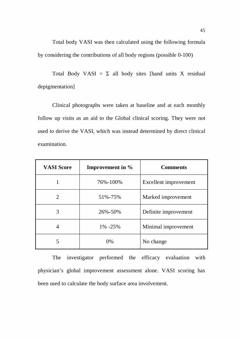
Total body VASI was then calculated using the following formula
by considering the contributions of all body regions (possible 0-100)
Total Body VASI = all body sites [hand units X residual
depigmentation]
Clinical photographs were taken at baseline and at each monthly
follow up visits as an aid to the Global clinical scoring. They were not
used to derive the VASI, which was instead determined by direct clinical
examination.
VASI Score Improvement in % Comments
1 76%-100% Excellent improvement
2 51%-75% Marked improvement
3 26%-50% Definite improvement
4 1% -25% Minimal improvement
5 0% No change
The investigator performed the efficacy evaluation with
physician’s global improvement assessment alone. VASI scoring has
been used to calculate the body surface area involvement.
46
Limitations of the study:
The treatment results were based only on physician’s global
improvement assessment score.
PATIENT SELECTION:
In the study of 80 patients under 12 years of age,
As per the VASI scoring:
59 cases had BSA involvement <20% and
21 cases had BSA involvement >20%
Out of 59 cases with BSA <20%, 17 cases were stable cases of
vitiligo .
Vitiligo cases <20% BSA were treated with topical steroids and
immunomodulators.
Stable vitiligo <20% BSA were treated with surgical modality of
treatment.
The rest 21 cases with >20% BSA were treated with physical
modality namely NB UVB therapy and the responses were
commuted as below.
47
STATISTICAL ANALYSIS
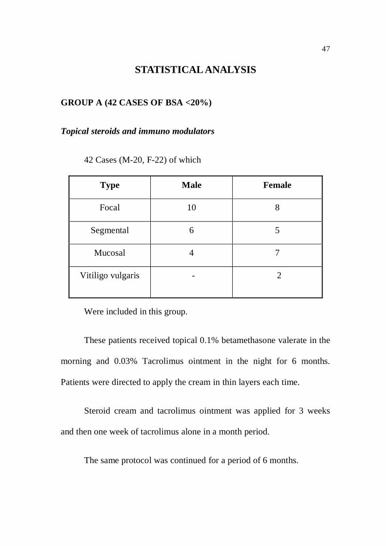
GROUP A (42 CASES OF BSA <20%)
Topical steroids and immuno modulators
42 Cases (M-20, F-22) of which
Type Male Female
Focal 10 8
Segmental 6 5
Mucosal 4 7
Vitiligo vulgaris - 2
Were included in this group.
These patients received topical 0.1% betamethasone valerate in the
morning and 0.03% Tacrolimus ointment in the night for 6 months.
Patients were directed to apply the cream in thin layers each time.
Steroid cream and tacrolimus ointment was applied for 3 weeks
and then one week of tacrolimus alone in a month period.
The same protocol was continued for a period of 6 months.
48
The patients were reviewed once in 2 weeks for the response and
side effects. After completing the treatment they were carefully followed
up for any evidence of relapse.
In this group, the mucosal vitiligo cases were treated with mild
steroid like triamcinolone acetonide and tacrolimus 0.03% cream in the
same manner as above for 6 months, and followed up.
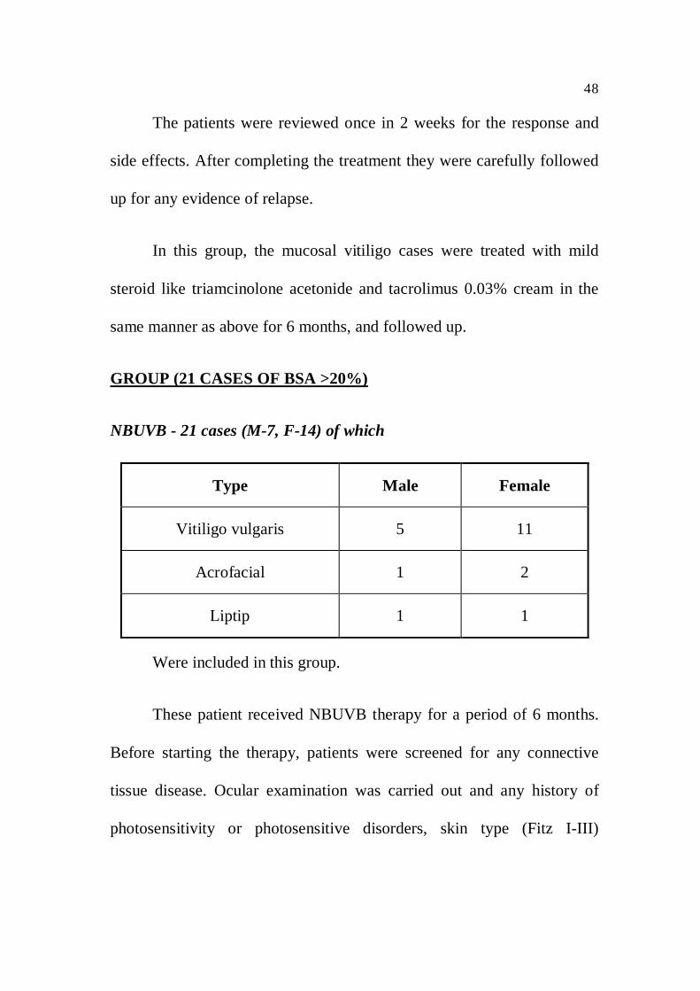
GROUP (21 CASES OF BSA >20%)
NBUVB - 21 cases (M-7, F-14) of which
Type Male Female
Vitiligo vulgaris 5 11
Acrofacial 1 2
Liptip 1 1
Were included in this group.
These patient received NBUVB therapy for a period of 6 months.
Before starting the therapy, patients were screened for any connective
tissue disease. Ocular examination was carried out and any history of
photosensitivity or photosensitive disorders, skin type (Fitz I-III)
49
concomitant radiotherapy, chemotherapy or immunosuppressive therapy
and claustrophobics were excluded.
NBUVB therapy was given thrice weekly on alternate days starting
with one unit dose of 0.25 J/cm2 independent of the skin type.
The dose was increased by 20% each treatment. The optimal
constant dose was achieved when minimal erythema occurred in the
lesions. During the treatment the eyes were protected by UV- blocking
goggles. All patients were kept their underwear on to shield their genitals
from radiation exposure.
Children were advised to protect their skin against excessive
exposure to natural sunlight especially between 11.00am to 3.00pm
during sunny days on both treatment as well as non treatment days. A
sunscreen with high SPF (25 or higher) was applied on sun exposed
areas.
Pre and post treatment photographs were taken to see the response
to therapy.
50
Informed written consent was obtained from the parents. In each
case, the nature of treatment was carefully explained, including details of
the possible benefits and side effects.
At each visit, they are evaluated for any adverse effects like
sunning sensation, itching, erythema.
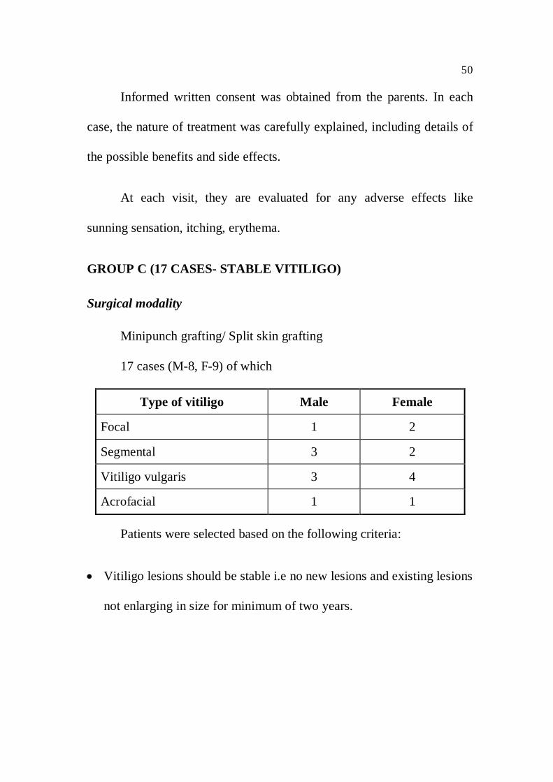
GROUP C (17 CASES- STABLE VITILIGO)
Surgical modality
Minipunch grafting/ Split skin grafting
17 cases (M-8, F-9) of which
Type of vitiligo Male Female
Focal 1 2
Segmental 3 2
Vitiligo vulgaris 3 4
Acrofacial 1 1
Patients were selected based on the following criteria:
Vitiligo lesions should be stable i.e no new lesions and existing lesions
not enlarging in size for minimum of two years.
51
For lesions present on mobile and immobile areas punch grafting was
done.
For lesions on immobile areas alone split skin grafting was done.
Patient’s parent consent for surgery.
Psychologically stable patients with realistic expectations.
Out of 17 cases, 13 cases were treated with punch graft and 4 cases
with split skin graft.
52
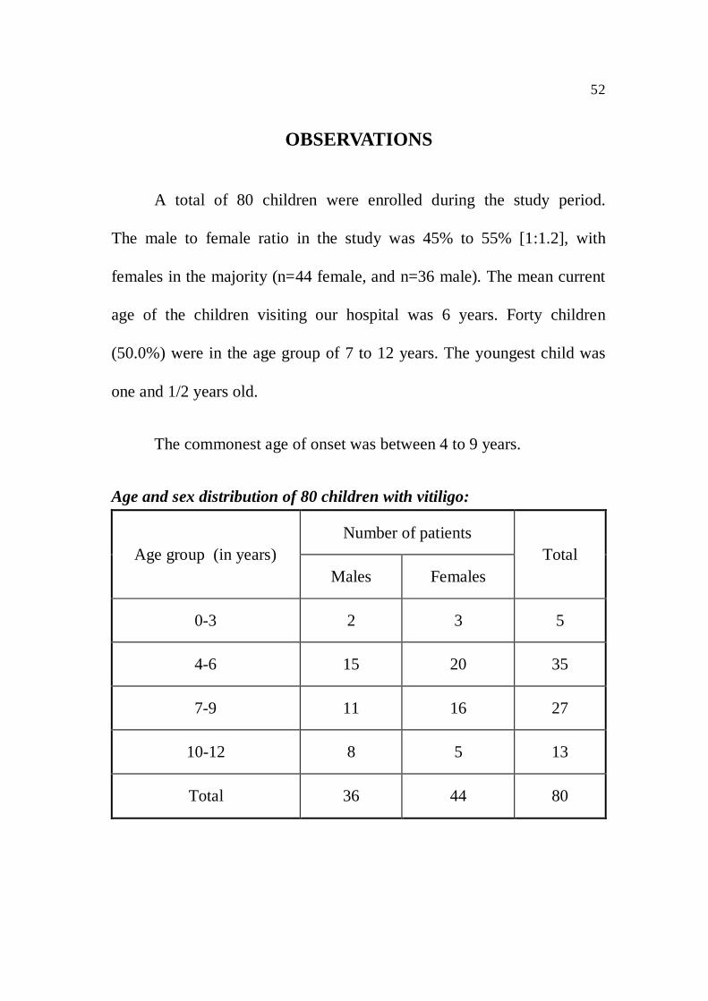
OBSERVATIONS
A total of 80 children were enrolled during the study period.
The male to female ratio in the study was 45% to 55% [1:1.2], with
females in the majority (n=44 female, and n=36 male). The mean current
age of the children visiting our hospital was 6 years. Forty children
(50.0%) were in the age group of 7 to 12 years. The youngest child was
one and 1/2 years old.
The commonest age of onset was between 4 to 9 years.
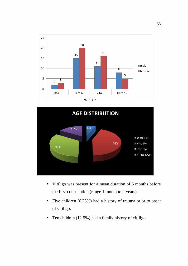
Age and sex distribution of 80 children with vitiligo:
Age group (in years)Number of patients
TotalMales Females
0-3 2 3 5
4-6 15 20 35
7-9 11 16 27
10-12 8 5 13
Total 36 44 80
53
Vitiligo was present for a mean duration of 6 months before
the first consultation (range 1 month to 2 years).
Five children (6.25%) had a history of trauma prior to onset
of vitiligo.
Ten children (12.5%) had a family history of vitiligo.
54
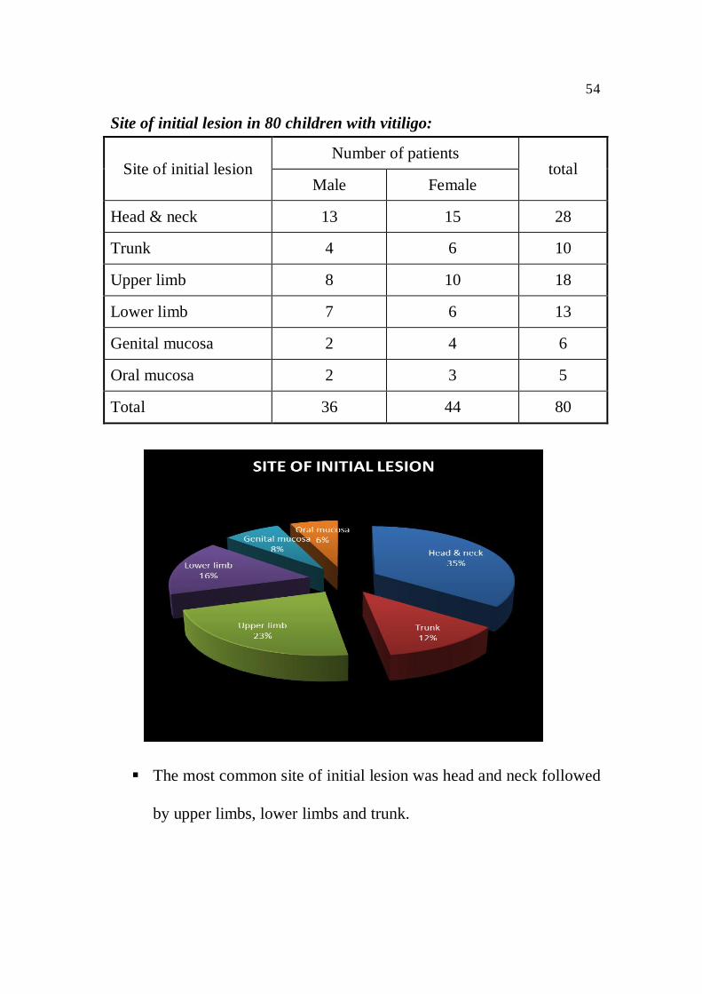
Site of initial lesion in 80 children with vitiligo:
Site of initial lesionNumber of patients
totalMale Female
Head & neck 13 15 28
Trunk 4 6 10
Upper limb 8 10 18
Lower limb 7 6 13
Genital mucosa 2 4 6
Oral mucosa 2 3 5
Total 36 44 80
The most common site of initial lesion was head and neck followed
by upper limbs, lower limbs and trunk.
55
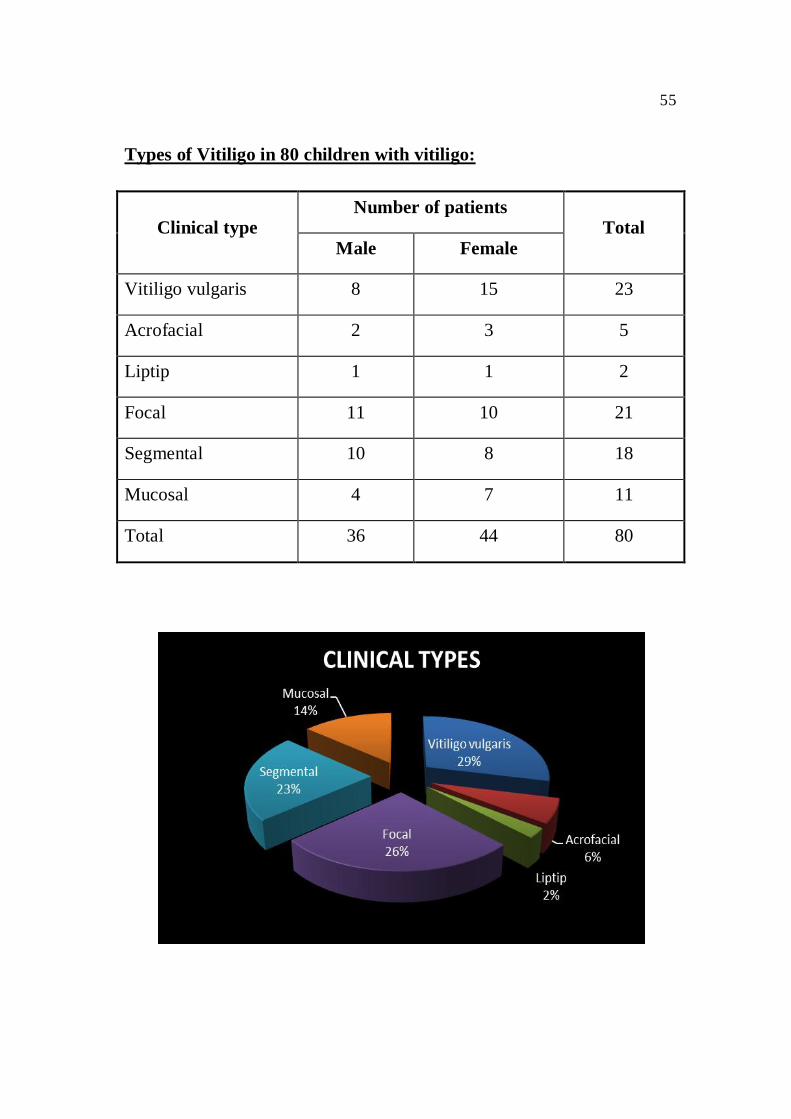
Types of Vitiligo in 80 children with vitiligo:
Clinical typeNumber of patients
TotalMale Female
Vitiligo vulgaris 8 15 23
Acrofacial 2 3 5
Liptip 1 1 2
Focal 11 10 21
Segmental 10 8 18
Mucosal 4 7 11
Total 36 44 80
56
The most common type was
vitiligo vulgaris seen in children 23 cases (28.75%) , followed by
focal type in 21cases(26.25%)
segmental type in 18 cases(22.50%)
mucosal type in 11 cases(13.75%)
acrofacial type in 5 cases(6.25%)
liptip type in 2 cases(2.5%)
Among the segmental type of vitiligo in children, trigeminal
dermatome was most commonly involved in 12children (15%)
In 80 children, 59 cases (73.75%) had body surface area involved
less than 20%.
Leukotrichia was present in 12 children (15%), while Kobner
phenomenon was observed in 17 children (21.25%).
57
21 children (26.25%) had an associated cutaneous disorder. These were
Twenty nail dystrophy in 3(2.5%)
Nail pitting in 9(11.25%)
Halo naevi in 2(1.6%)
Alopecia areata in 3 (2.5%)
Premature canities in 2 (1.6%)
lichen striatus in 2(1.6%)
Ten children (12.5%) had an associated ocular disorder. These were
Eyelid vitiligo in 5 (6.25%)
Reduced visual acuity (myopia) in 4 (5%)
Conjunctivitis in 1 (1.25%) child.
SYSTEMIC ASSOCIATONS:
Juvenile rheumatoid arthritis was seen in one (1.25%) child
Hypothyroidism in 3(3.75%) children.
58
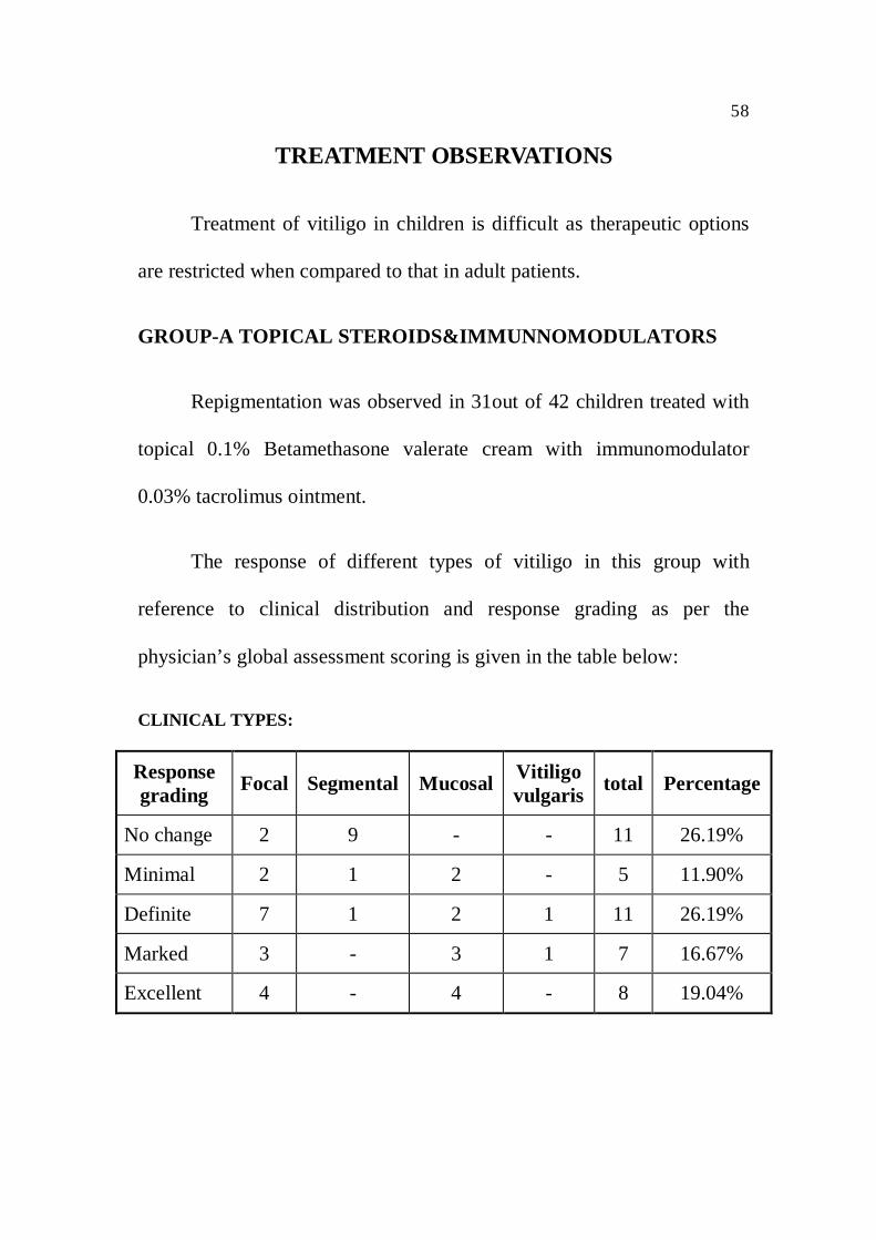
TREATMENT OBSERVATIONS
Treatment of vitiligo in children is difficult as therapeutic options
are restricted when compared to that in adult patients.
GROUP-A TOPICAL STEROIDS&IMMUNNOMODULATORS
Repigmentation was observed in 31out of 42 children treated with
topical 0.1% Betamethasone valerate cream with immunomodulator
0.03% tacrolimus ointment.
The response of different types of vitiligo in this group with
reference to clinical distribution and response grading as per the
physician’s global assessment scoring is given in the table below:
CLINICAL TYPES:
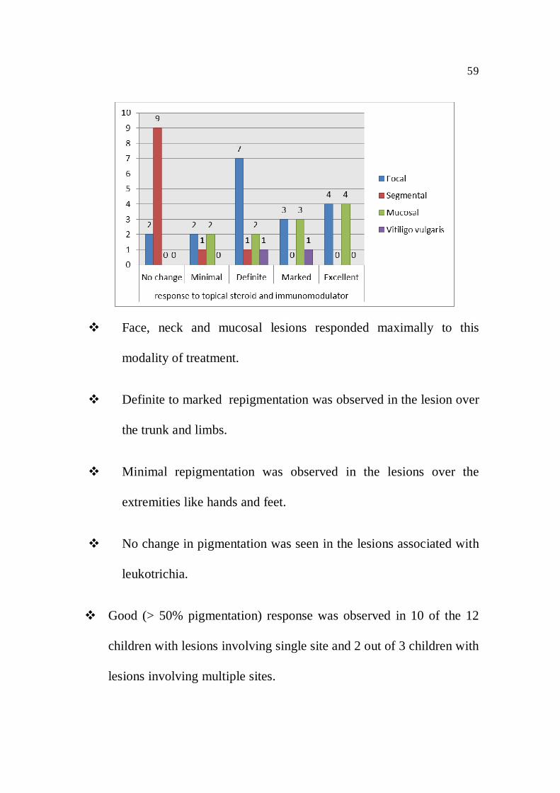
Responsegrading Focal Segmental Mucosal Vitiligo
vulgaris total Percentage
No change 2 9 - - 11 26.19%
Minimal 2 1 2 - 5 11.90%
Definite 7 1 2 1 11 26.19%
Marked 3 - 3 1 7 16.67%
Excellent 4 - 4 - 8 19.04%
59
Face, neck and mucosal lesions responded maximally to this
modality of treatment.
Definite to marked repigmentation was observed in the lesion over
the trunk and limbs.
Minimal repigmentation was observed in the lesions over the
extremities like hands and feet.
No change in pigmentation was seen in the lesions associated with
leukotrichia.
Good (> 50% pigmentation) response was observed in 10 of the 12
children with lesions involving single site and 2 out of 3 children with
lesions involving multiple sites.
60
Better response was observed in lesions involving <5% skin surface
overall in this group.
No significant difference in response was observed in relation to
duration of lesions.
No drop outs were found in this group. No adverse effects (atrophy,
telangiectasia) were seen. No relapse was observed during the follow
up period.
Out of the 42 cases treated in this group, 10 cases (all focal cases) had
rapid progression of lesions with onset of new lesions. They were
treated with oral mini pulse with tab. Prednisolone 30mg
(as betamethasone in oral form was not available in our hospital)
divided into two doses 15mg each on two consecutive days every
weekend. The progression of lesions were arrested within 1-3 months
in 8 out of 10 cases and within 4 months in the rest 2 cases.
The patients were continuing the topical steroids and
immunomodulators simultaneously during this intermittent therapy.
These patients on mini pulse steroid therapy not only responded by
the progression of lesions but had earlier repigmentation of lesion as
well compared to others not receiving mini pulse therapy in 80% of
cases.
61
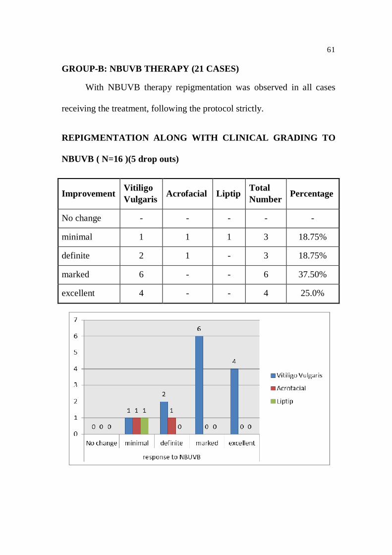
GROUP-B: NBUVB THERAPY (21 CASES)
With NBUVB therapy repigmentation was observed in all cases
receiving the treatment, following the protocol strictly.
REPIGMENTATION ALONG WITH CLINICAL GRADING TO
NBUVB ( N=16 )(5 drop outs)
Improvement VitiligoVulgaris Acrofacial Liptip Total
Number Percentage
No change - - - - -
minimal 1 1 1 3 18.75%
definite 2 1 - 3 18.75%
marked 6 - - 6 37.50%
excellent 4 - - 4 25.0%
62
Out of 21 children, 5 children discontinued (VV-3, Acrofacial-1,
Liptip-1) therapy as they were not able to attend the phototherapy centre
three times a week and were school going. Rest 16 children managed to
complete 6 month of therapy regularly.
Out of 16 children excellent repigmentation was observed in 4 out
of the 13 (3 dropouts) children with vitiligo vulgaris type, marked
repigmentation was observed in rest 7 children of vitiligo vulgaris type
and 1 out of 2 (one drop out) cases of acrofacial type, minimal to definite
repigmentation was seen in one acrofacial type and one liptip types
(one dropout).
GROUP-C: SURGICAL THERAPY
Out of 17 cases who had stable vitiligo lesions, 13 cases were
treated with miniature punch grafting for patients having lesions in both
mobile and immobile areas and the rest 4 cases with spit skin grafting for
those having lesions in immobile areas alone.
63
PUNCH GRAFTING
Pigmentation started appearing in 1½ to 2 months after grafting.
Uniform perigraft and perifolicular pigmentation was seen and several
such pigmented islands coalesced together to cover the affected area
within 3-6 months. Majority of cases showed 7-10mm pigmentation.
All the patients were followed up for 6 months and the results were
observed after 6 months. 9 out of 13 cases showed fair to marked
cosmetic matching with the normal surrounding skin.
Almost all the donor sites healed with scarring within 1-2 months.
The scars were superficial and acceptable to the patients.
Cobble stoning was seen in 5 patients in the initial stages but
slowly faded within a period of 4-6 months . Keloidal tendency was
noted in 2 cases later subsided with application of topical steroids.
Depigmentation of grafts were seen in 2 cases associated with
leukotrichia.
64
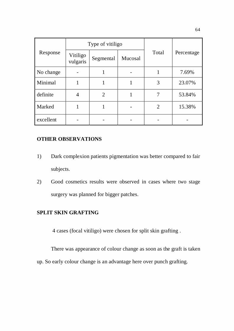
ResponseType of vitiligo
Total PercentageVitiligovulgaris Segmental Mucosal
No change - 1 - 1 7.69%
Minimal 1 1 1 3 23.07%
definite 4 2 1 7 53.84%
Marked 1 1 - 2 15.38%
excellent - - - - -
OTHER OBSERVATIONS
1) Dark complexion patients pigmentation was better compared to fair
subjects.
2) Good cosmetics results were observed in cases where two stage
surgery was planned for bigger patches.
SPLIT SKIN GRAFTING
4 cases (focal vitiligo) were chosen for split skin grafting .
There was appearance of colour change as soon as the graft is taken
up. So early colour change is an advantage here over punch grafting.
65
This modality was best for facial lesions and lesions close to the
hair line and poor at the extremities. Amongst the clinical types, focal and
segmental showed better response than acral vitiligo.
Tire-pattern appearance was seen in 2 cases and donor sites showed
scaring in all cases.
Postoperative phototherapy in the form of NBUVB was given to
the graft site 3 times a week and better results were seen after 6 months
during follow up.
Focal vitiligo
Focal vitiligo
Segmental vitiligo
Segmental vitiligo
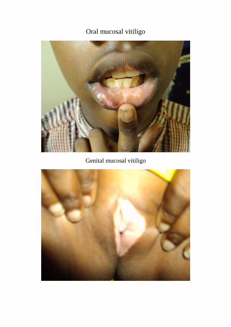
Oral mucosal vitiligo
Genital mucosal vitiligo
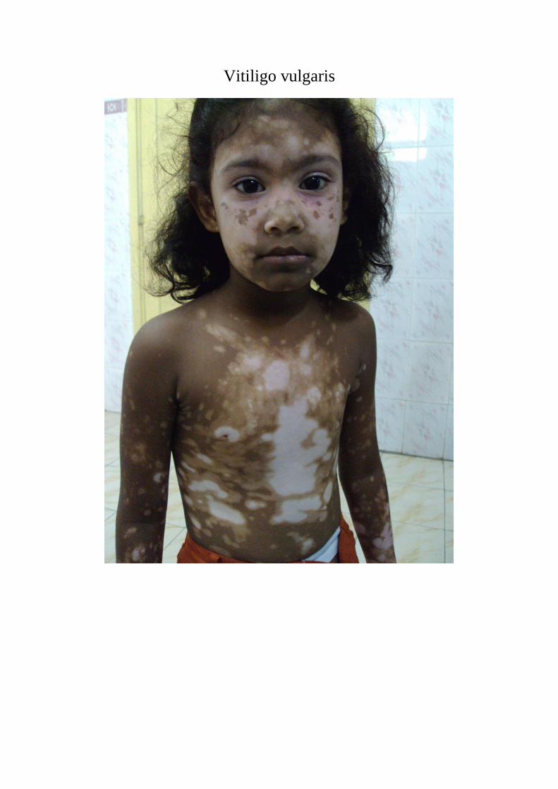
Vitiligo vulgaris
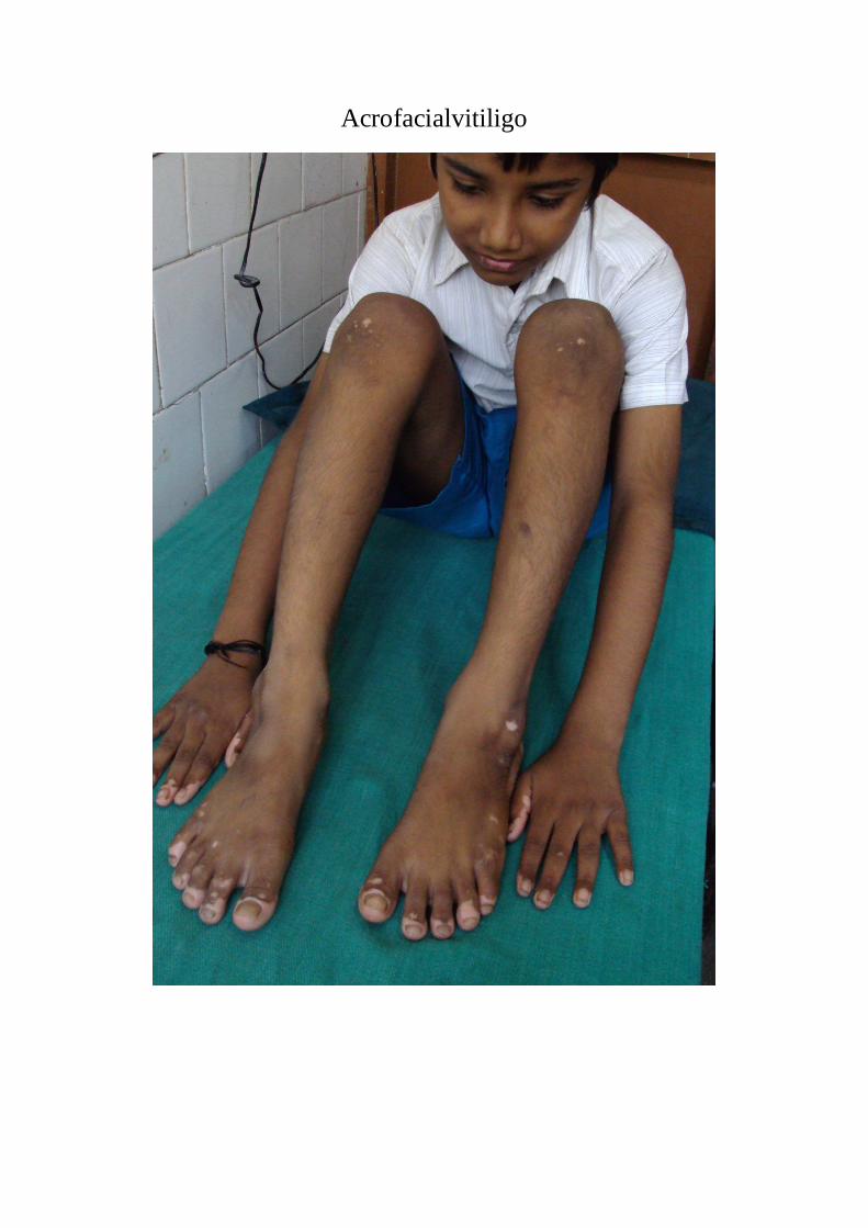
Acrofacialvitiligo
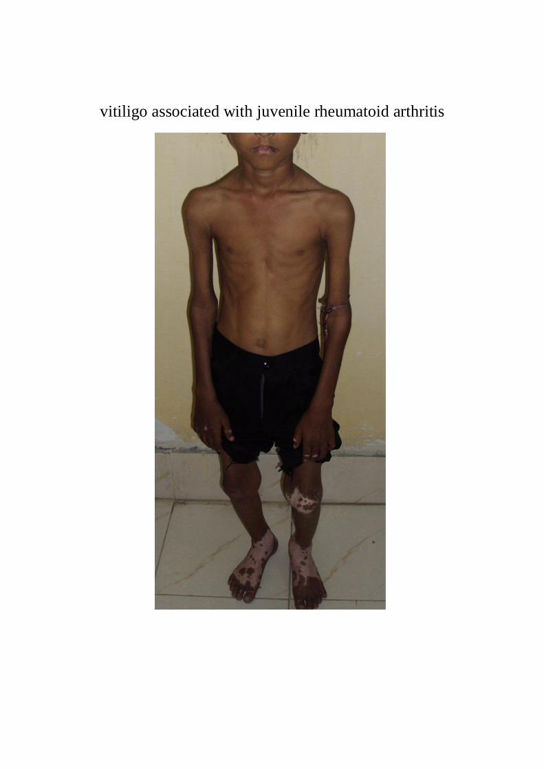
vitiligo associated with juvenile rheumatoid arthritis
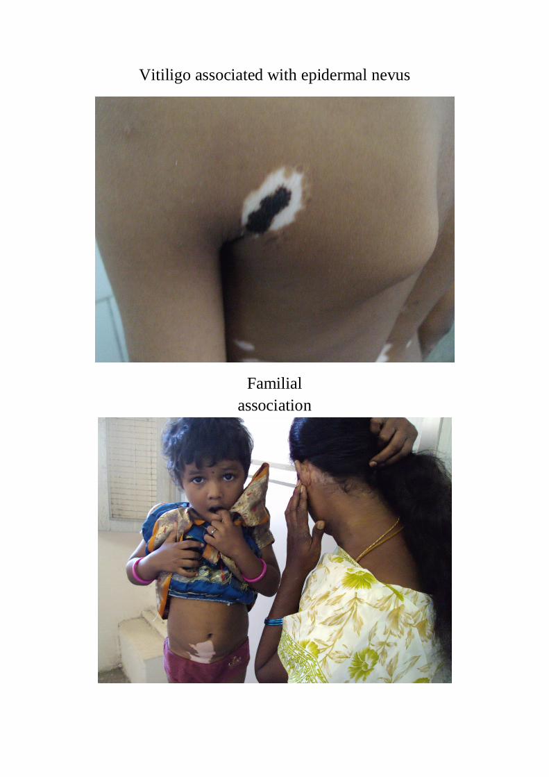
Vitiligo associated with epidermal nevus
Familialassociation
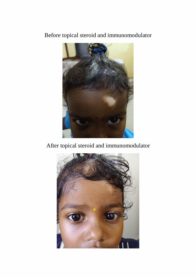
Before topical steroid and immunomodulator
After topical steroid and immunomodulator
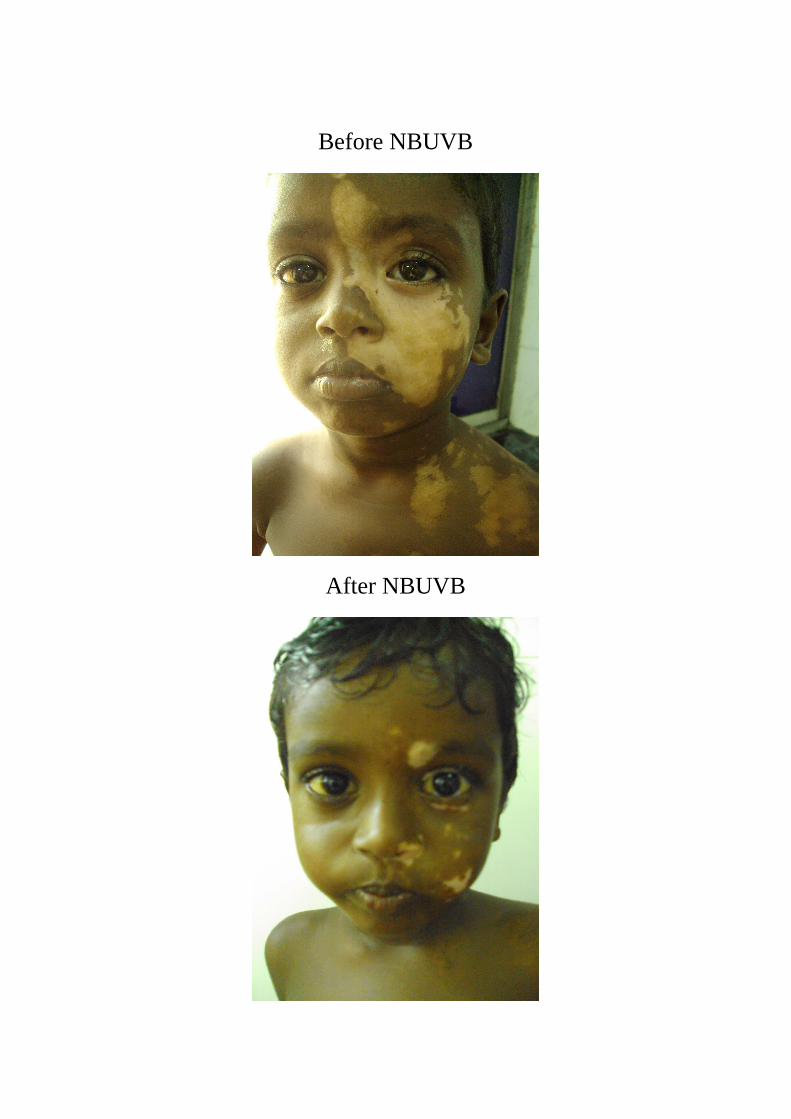
Before NBUVB
After NBUVB
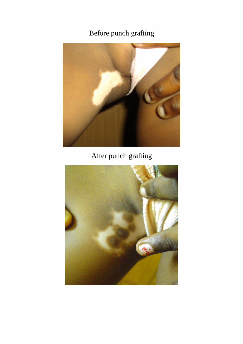
Before punch grafting
After punch grafting
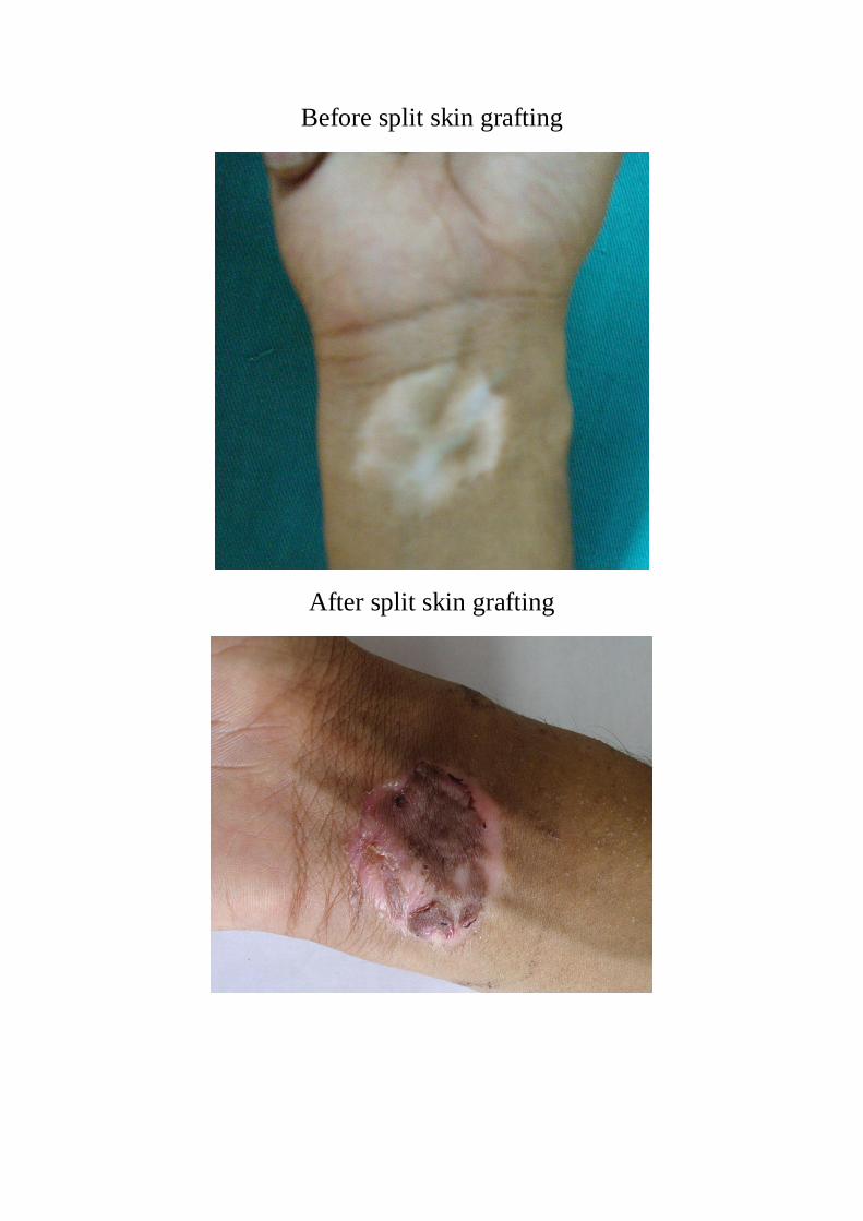
Before split skin grafting
After split skin grafting
66
DISCUSSION
Vitiligo is a common disease in India having a prevalence of
0.46 – 8.8%73. Majority (>50%) of this population develop the disease
before 20 years of age group making vitiligo an important aspect in
Paediatric Dermatology. Indian studies on childhood vitiligo have
reported the prevalence to be 2.6%76
AGE DISTRIBUTION:
In our study of 80 children, the commonest age of presentation
was between 4 and 9 years which is in contrast to the study of Belliappa
et al75 where the commonest age of presentation was between 7 and 12
years.
The youngest child in our study was 1 ½ years old similar to
Belliappa et al study where the youngest child was 1 year old. Earlier
studies have reported cases of congenital vitiligo. But our study did not
have any case of congenital vitiligo.
67
SEX DISTRIBUTION:
The Prevalence of vitiligo was found to be higher in girls than in
boys 44 Vs 36 in our study of 80 cases.
The male to female ratio was 1: 1.2.
In earlier studies as well, girls were affected more than boys.
However boys and girls were affected equally in Zhi Hu85 et al study.
AGE OF ONSET:
In our study, the commonest age of onset of the disease was
between 4 to 9 years constituting 77.5% of the cases. Belliappa et al
reported that 68.9% of cases had onset of disease between 4 and 8 years
of age similar to our study.
When comparing the age at presentation and age of onset, age of
presentation in most cases is between 4 and 9 years and age of onset is
also between 4 and 9 years. This shows that the patients and their parents
are more aware of the nature of the disease and its course and present to
the physician earlier to seek treatment.
68
DURATION AND PROGRESSION:
The duration of depigmentation varied from 1 month to 2 years.
The mean duration before they first seek treatment was 6 months. This is
in contrast to Belliappa et al study where mean duration of disease was 14
months.
SITE OF INITIAL LESION:
In our study the most common site of initial lesion was head and
neck followed by upper limbs, trunk and lower limbs in that order.
Belliappa et al also reported that the most common site of onset was head
and neck. Jaisankar et al76 reported the various sites of onset as lower
limbs, head and neck, upper limbs and thorax in that order.
FAMILY HISTORY:
In our study, 12.5% of children had family history of vitiligo.
In Belliappa et al study family history was present in 14.8% of children,
Jaisankar et reported very low incidence as 3.3% and in Halder
et al33study there was 35% family history.
69
TYPE OF VITILIGO:
In our study, vitiligo vulgaris was the most common clinical type
seen in 28.75% closely followed by focal type in 26.25%, segmental type
in 22.50%. In earlier studies on childhood vitiligo as well, vitiligo
vulgaris was the most common type reported. Belliappa et al in their
study of 122 children reported was the most common type is 36.9% and
segmental vitiligo as the second most frequent type occurring in 27%.
Lip-Tip vitiligo was the least common type seen in our study
population which is similar to study of Belliappa et al ,Halder et al but
Jaisankar et al reported acrofacial type as the least common.
Mucosal Vitiligo was seen in 13.75% in our study, Belliappa et al
also had similar figures of 13.10%.
Among segmental type, trigeminal dermatome was most
commonly involved in 15% in our study consistent with all other studies
done earlier.
70
BODY SURFACE AREA INVOLVEMENT :
Our study showed Body surface area of less than 20% in 73.75% of
children in contrast to 95.9% in Belliappa et al study.
SPECIAL FEATURES:
Leucotrichia was present in 15% of children with vitiligo in
contrast to 41.8% in Belliappa et al study. Kobner phenomenon was
observed in 21.25% and Belliappa et al reported in 24.6% of patients.
CUTANEOUS & AUTO IMMUNE DISORDERS ASSOCIATED:
In our study, Alopecia areata was seen in 2.5%, Halo Nevi in 1.6%
of children. Belliappa et al reported 2.5% of alopecia areata similar to our
study and 4.9% Halo Nevi.
OTHER CUTANEOUS DISORDERS:
Premature canities was seen in 1.6%, epidermal nevus in 3.75%,
Lichen striatus in 1.6%, Twenty nail dystrophy in 2.5% and nail pitting in
15%. Belliappa et al reported premature canities in 1.6% similar to our
study.
71
TREATMENT
The treatment modalities selected here were based on the available
medications and facilities in our hospital set up.
Group A: Topical steroid and immunomodulator
Repigmentation was seen in 73.80% of cases treated with both
topical 0.1% betamethasone and 0.03% tacrolimus ointment. To the best
of our knowledge, no other study in childhood vitiligo has combined both
topical steroid and immunomodulator for treatment , rather compared the
efficacy of both proving that topical immunomodulator is an effective
alternative to topical steroid60.
A good response (>50% pigmentation) was found in 35.71% in our
study. Response was very good for focal and mucosal lesions.
Segmental patches and those associated with leukotrichia did not
respond well to this modality.
Mucosal vitiligo usually a resistant form, responded well on
combining both topical steroid and immunomodulator , so this modality
is best suited for mucosal lesions.
72
Oral minipulse steroid with prednisolone is an effective treatment
option for controlling the rapid disease spread in childhood vitiligo and
with addition of topical steroid and immunomodulator the extent of
repigmentation was also significant consistent with the previous studies
of Pasricha64 et al and Majid62 et al.
GROUP B (NBUVB)
A good response (>50%repigmentation) was observed in 62.50%
of treated cases. After a mean interval of 32 times of phototherapy 50%
repigmentation was achieved similar to A.J kanwar65 et al study. The
adverse effects were mild and transient and none discontinued the therapy
because of side effects. Vitiligo vulgaris type responded well and liptip
type responded least similar to other studies on this modality.
GROUP C
PUNCH GRAFTING: In our study over 69.22% cases showed
definite to marked repigmentation as compared to Khandpur et al study of
83.3%. The graft site showed appreciable perigraft spread of
pigmentation and good cosmetic colour match to the surrounding skin in
73
70% of treated cases. Vitiligo vulgaris responded well and segmental type
responded least.
SPIT SKIN GRAFTING: Excellent cosmetic matching was seen
in 75% of grafts involving larger areas. Obvious colour change seen after
grafting is an advantage in this procedure.
Further similar such studies need to be done at regular intervals
with newer modalities of treatment.
74
CONCLUSION
Vitiligo incidence during the study period was 0.27%.
The incidence of vitiligo in children was 15.87% of the total
number of vitiligo patients over a period of 1 ½ years of study.
Females were predominantly affected than males which might be
due to their increased cosmetic concern.
There was a positive family history in 12.5% of children.
Most common age group affected include 4-6 years.
Most common site of initial lesion was head and neck followed by
upper limb, lower limb and trunk.
Most common clinical type was vitiligo vulgaris followed by focal
type then segmental. Lip tip type was least common type.
Cutaneous association was seen in 26.25%.
Body surface area involving < 20% was found in 73.75%.
75
Localized facial and mucosal lesions best respond to topical steroid
and immunomodulator combination. The compliance of the
patients was very good with this treatment modality.
NBUVB therapy is an effective and safe modality to treat
generalized vitiligo with cosmetically acceptable repigmentation.
Punch grafting proves to be easier, faster and least expensive
method of treatment in stable vitiligo cases.
Split skin grafting carries a distinct advantage over mini punch
grafting in producing excellent cosmetic matching over larger areas
using fewer grafts.
Further several such studies need to be done on a larger scale to
compare the epidemiology, clinical spectrum and various newer
modalities of treatment ,their responses, adverse effects and to derive a
standard protocol for treating this less studied entity childhood vitiligo.
BIBLIOGRAPHY
1) Handa S, Dogra S, Epidemiology of childhood vitiligo: a study of
625 patients from north India. Pediatr Dermatol. 2003 May-Jun
;20(3):207-10.
2) Sandipan Dhar, Pijush Dutta, Rajib Malakar; vitiligo , pigmentary
disorders; IADVL textbook of dermatology, 3rd edition, vol 1 ,
ch.25 ,749-760.
3) Mosher DB, OrtonneJP, Fitzpatrick TB : Disorder of pigmentation
in: Fitzpatrick’s TB, EisenAZ , Wolff K , et al. Dermatology in
General medicine, 3rd edition Mc Graw Hill, new York: 1987;
794-876
4) Sathish S.Savant: vitiligo surgery: IADVL textbook of dermatology,
3rd edition , vol 2,ch56, 1728-1748.
5) Chandra S, Kumar A, Singh KK, Mohan L. Congenital vitiligo.
Indian J Dermatol Venereol Leprol 1992;58:339
6) Kanwar AL, Dhar S, Kaur S. Vitiligo in children. Ind J Dermatol
1993;38:47-52.
7) Rebat M. halder, Sumayah J.Taliaferro ,Vitiligo;classification of
vitiligo and clinical variants and treatment edition 7th fitzpatrick’s
dermatology in General Medicine ; 617-622.
8) Lerner AB. Vitiligo. J Invest Dermatol 1959; 32:285-310.
9) Lerner AB. On the etiology of Vitiligo and gray hair. Am J Med
1971:51:141-7.
10) Bleehen SS, Pathak MA, Hori Y, Fitzpatrick TB. Depigmentation of
skin with 4-isopropylcatechol, mercaptoamines, and other
compounds. J Invest Dermatol 1968:50: 103-17.
11) Schallreuter KU, Wood JM, Berger J. Low catalase levels in
epidermis of patients with vitiligo. J Invest Dermatol 1991; 97:
1081-5.
12) Schallreuter KU et al: Biochemical theory of vitiligo: A role of
pteridines in pigmentation, in Vitiligo, edited by SK Hann, JJ
Nordlund, London, Blackwell science, 2000 p151.
13) Schallreuter KU, Wood JM, Ziegler I etal. Defective
tetrahydrobiopterin and catecholamine biosynthesis in the
Depigmentation disorder vitiligo. Biochim BiophysActa
1994:122:181-92.
14) Bystryn JC: Theories on the pathogenesis of Depigmentation:
Immune hypothesis, in Vitiligo, edited by SK Hann, JJ Nordlund,
London, Blackwell science, 2000 p129.
15) Das PK et al: A Symbiotic concept of autoimmunity and tumour
immunity: Lessons from vitiligo. Trends Immunol 22:130, 2001.
16) Le Poole IC et al: Review of etiopathomechanism of vitiligo: A
Convergence theory, Exp Dermatol 2:145, 199.3
17) Boissy RE: The intrinsic (genetic theory) for the cause of Vitiligo, in
Vitiligo, edited by SK Hann, JJ Nordlund, London, Blackwell
science, 2000, p123.
18) Nordlund JJ. Vitiligo: A review of some facts lesser known about
depigmentation. Indian J Dermatol 2011;56:180-9
19) Sarin KC, Kumar AS. A clinical study of vitiligo. Indian J Dermatol
Venereol Leprol 1977; 43:311-314.
20) Koranne RV, Sehgal VN, Sachdeva KG. Clinical profile of vitiligo
in North India. Indian J Dermatol Venereol Leprol 1986; 52:81-83.
21) Kanwar AL, Dhar S, Kaur S. Vitiligo in children. Ind J Dermatol
1993;38:47-52.
22) Ortonne J.P, Bahadoran P, Thomas B. Fitzpatrick, David B. Mosher
and Yoshiaki Hori: Fitzpatrick’s Textbook of Dermatology in
General Medicine, sixth edition, chapter-90.
23) Jaisankar TJ, Baruah MC, Garg BR. Vitiligo in children. Int J
Dermatol. 1992; 31(9):6213
24) Schwartz RA, Janniger CK. Vitiligo. Cutis. 1997; 60:239-44.
25) Hann SK, Chang JH, Lee HS, Kim SM. The classification of
segmental vitiligo on the face. Yonsei Med J. 2000;41:209-12.
26) Cunliffe WJ, Hall R, Newel DJ et al, Vitiligo, thyroid disease and
autoimmunity. Br J Dermatol 1968: 80:135-9.
27) Dawber RPR. Clinical Associations of Vitiligo, Postgrad Med J
1970: 46:276-7.
28) Romano G, Moretti G, Benedetto A et al. Skin lesions in diabetes
mellitus prevalence and clinical correlations. Diab Res. Clin Pract.
1998; 39(2):101-106.
29) Dunlop D, Eighty six cases of Addison’s disease. BMJ 1963: ii:887-
91.
30) Obermayer – Straub P, Manns MP: Autoimmune polyglandular
syndromes. Baillieres Clin Gastroenterol 1998; 12:293-315.
31) Grunnet I, Howitz J, Reyman F, et al. Vitiligo and pernicious
anemia, Arch Dermatol 1970;101:82-85.
32) Cho M, Cohen PR, Duvie M. Vitiligo and alopecia areata in patients
with human immunodeficiency virus infection. South Med. J. 1995
Apr; 88(4):489-91.
33) Halder RM, Grimes PE, Cowan CA, Enterline JA, Chakrabarti SG,
Kenney JA. Childhood vitiligo. J Am Acad Dermatol. 1987; 16:948-
54.
34) Palit A, Inamadar AC. Childhood vitiligo. Indian J Dermatol
Venereol Leprol 2012;78:30-41
35) Taieb A. Intrinsic and extrinsic pathomechanisms in vitiligo.
Pigment Cell Res. 2000;13:41-7.
36) Hann SK, Lee HJ. Segmental vitiligo: clinical findings in 208
patients. J Am Acad Dermatol. 1996; 35:671-4.
37) Lee DJ, Modlin RL. Breaking tolerance—another piece added to the
vitiligo puzzle. J Invest Dermatol. 2004; 124: xii-xv.
38) Iacovelli P, Sinagra JLK, Vidolin AP, Marenda S, Capitanio B,
Leone G, et al. Relevance of thyroiditis and of other autoimmune
diseases in children with vitiligo. Dermatology. 2005:210:26-30.
39) Kakourou T, Kanaka-Gantenbein C, Papadopoulou A, Kaloumenou
E, Chrousos GP. Increased prevalence of chronic autoimmune
(Hashimoto’s) thyroiditis in children and adolescents with vitiligo. J
Am Acad Dermatol. 2005; 53:220-3.
40) Laberge G, Mailloux CM, Gowan K, Holland P, Bennett DC, Fain
PR, et al. Early disease onset and increased risk of other
autoimmune disease in familial generalized vitiligo. Pigment Cell
Res. 2005; 18:300-5.
41) Roberts A, Kaye LC, Memon A, Parslew R, Kaye SB. Unilateral
poliosis of the eyelashes in children associated with vitiligo. J
AAPOS. 2005; 9:295-6.
42) Scherschum L, Kim JJ, Lim HW. Narrow-band ultraviolet B is a
useful and well-tolerated treatment for vitiligo. J Am Acad Dermatol
2001;44:999-1003
43) Ando I, Chi HI, Nakagawa H, Otsuka F. Difference in clinical
features and HLA antigensbetween familial and non-familial vitiligo
of non-segmental type. Br J Dermatol. 1993;129:408-10.
44) Njoo MD, Spuls PI, Bos JD, Westerhof W, Bossuyt PMM.
Nonsurgical repigmentation therapies in vitiligo: meta-analysis of
the literature. Arch Dermatol. 1998; 134:1532-40.
45) Grimes PE, Soriano T, Dytoc MT. Topical tacrolimus for
repigmentation of vitiligo. J Am Acad Dermatol. 2002; 47:789-91.
46) Lepe V, Moncada B, Castandeo-Cazares JP, Torres-Alvarez MB,
Ortiz CA, Torres- Rubalcava AB. A doubleblind randomized trial of
0.1% tacrolimus vs 0.05% clobetasol for the treatment of childhood
vitiligo. Arch Dermatol. 2003; 139:581-5.
47) Plettenberg H, Assmann T, Ruzicka T. Childhood vitiligo and
tacrolimus. Arch Dermatol. 2003; 139:651-4.
48) Caelles IP, Pinto PH, Casado LDEL, Laguna RDL. Focal
hypertrichosis during topical tacrolimus therapy for childhood
vitiligo. Pediatr Dermatol. 2005; 22:86-7.
49) Roelandts R. Photo (chemo) therapy for vitiligo. Photodermatol
Photoimmunol Photomed. 2003; 19:1-4.
50) Westerhof W, Nieuweboer-Krobotova L. Treatment of vitiligo with
UV-B radiation vs topical psoralen plus UV-A. Arch Dermatol.
1997; 133:1525-8.
51) Njoo MD, Bos JD, Westerhof W. Treatment of generalized vitiligo
in children with narrow-band (TL-01) UVB radiation therapy.
J Am Acad Dermatol. 2000; 42:245-53.
52) Kanwar AJ, Dogra AJ. Narrow-band UVB for the treatment of
generalized vitiligo in children. Clin Exp Dermatol. 2005; 30:
332-6.
53) Kanwar AJ, Dogra S, Parsad D, Kumar B. Narrow-band UVB for
the treatment of vitiligo: an emerging effective and well-tolerated
therapy. Int J Dermatol. 2005; 44:57.
54) Brazelli V, Prestinari F, Castello M, Bellani E, Roveda E,
Barbagallo T, et al. Useful treatment of vitiligo in 10 children with
UV-B Narrowband (311 nm). Pediatr Dermatol. 2005; 22:257-61.
55) Menchini G, Tsoureli-Nikita E, Hercogova J. Narrowband UVB
micro-phototherapy: a new treatment for vitiligo. J Eur Acad
Dermatol Venereol. 2003; 17:171-7.
56) Leone G, Iacovelli P, Paro Vidolin A, Picardo M. Monochromatic
excimer light 308 nm in the treatment of vitiligo: a pilot study. J Eur
Acad Dermatol Venereol. 2003; 17:531-7.
57) Gupta S, Kumar B. Epidermal grafting in vitiligo: influence of age,
site of lesion, and type of disease on outcome. J Am Acad Dermatol.
2003; 49:99-104.
58) Khandpur S, Sharma VK, Manchanda Y. Comparison of mini-punch
grafting versus split-skin grafting in chronic stable vitiligo.
Dermatol Surg. 2005; 31:436-41.
59) Ongenae K, Dierckzsens L, Brochez L, Van Geel N, Naeyaert JM.
Quality of life and stigmatization profile in a cohort of vitiligo
patients and effect of the use of camouflage. Dermatology. 2005;
210:279-85.
60) choi, c. w., chang, s. e., bak, h., choi, j. h., park, h. s., huh, c. h., kim,
c. w., kim, s. e., mun, s. k., kim, b. j. and kim, m. n. (2008), topical
immunomodulators are effective for treatment of vitiligo. The
Journal of Dermatology, 35: 503–507. doi: 10.1111/j.1346-
8138.2008.00511.x
61) Bhuvana K, Sarala N, Singh G, Kumar TN, Effect of 0.1%
tacrolimus ointment in localized vitiligo: an open uncontrolled trial.
Indian J Dermatol. 2011 Jul; 56(4):4456.
62) Majid I, Masood Q, Hassan I, Khan D, Chisti M. Childhood vitiligo
: Response to methylprednisolone oral minipulse therapy and topical
fluticasone combination. Indian J Dermatol 2009;54:124-7
63) Ghrarami R. Therapy For an Extensive Childhood Vitiligo (le).
Indian J Dermatol Venereol Leprol 2000;66:161
64) Pasricha JS, Khaitan BK. Oral mini-pulse therapy with
betamethasone in vitiligo patients having extensive or fast-spreading
disease.Int J Dermatol. 1993 Oct;32(10):753-7.
65) Kanwar, A. J. and Dogra, S. (2005), Narrow-band UVB for the
treatment of generalized vitiligo in children. Clinical and
Experimental Dermatology, vol 30: 332–336.
66) Kumar YH, Rao GR, Gopal K, Shanti G, Rao KV. Evaluation of
narrow-band UVB phototherapy in 150 patients with vitiligo. Indian
J Dermatol Venereol Leprol 2009;75:162-6
67) Khandpur S, Sharma VK, Manchanda Y. Comparison of minipunch
grafting versus split-skin grafting in chronic stable vitiligo.
Dermatol Surg. 2005 Apr ;31(4):436-41
68) Veronica Lepe, Benjamin Moncada, Juan Pablo Castanedo-Cazares,
Maria Bertha Torres-Alvarez, Carlos A. Ortiz, and Antonio B.
Torres-Rubalcava, A Double-blind Randomized
69) Trial of 0.1% Tacrolimus vs 0.05% Clobetasol for the Treatment of
Childhood Vitiligo , Arch Dermatol. 2003;139(5):581-585.
70) Farah Sameem, Sheikh Javeed Sultan, and Qazi Masood Ahmad,
Split Thickness Skin Grafting in Patients with Stable
Vitiligo,Journal of Cutaneous and Aesthetic Surgery J Cutan Aesthet
Surg. 2011 Jan-Apr; 4(1): 38–40.
71) Srinivas CR, Rai R, Kumar PU. Meshed split skin graft for
extensive vitiligo. Indian J Dermatol Venereol Leprol 2004;70:165-7
72) Lerner AB. Vitiligo. J Invest Dermatol 1959; 32:285-310.
73) Handa S,Kaur I. Vitiligo: Clinical findings in 1436 patients. J
Dermatol 1999; 26:653-657.
74) Njoo MD, Westerhof W. Vitiligo: pathogenesis and treatment. Am J
Clin Dermatol 2002; 2 (3): 167-181.
75) Belliappa PR, Priya KS, Umashankar N, Vivekananda, Leena and
Lokanath. Characteristics of Childhood Vitiligo in Bangalore with
special reference to associated Ocular abnormalities. e-Journal of the
Indian Society of Teledermatology, 2011;Vol 4, No.3:1-10.
76) Jaishankar TJ, Baruah MC, Garg BR. Vitiligo in children. Int J
Dermatol 1992; 31:621-623.
77) Porter J, Beuf A, Nordlund J, et al. Psychological reaction to chronic
skin disorders. Gen Hosp Psychiatry 1979; 1:73-77.
78) Cho S, Kang HC, Hahm JH. Characteristics of vitiligo in Korean
children. Paediatr Dermatol 2000;17:189-193.
79) Handa S,Kaur I. Vitiligo: Clinical findings in 1436 patients. J
Dermatol 1999; 26:653-657.
80) Mosher DB, Fitapatrick TB, Ortonne JP, Hori Y. Hypomelanoses
and hypermelanoses. In; Freedberg IM, Eisen AZ, Wolff K, Austen
KF, Goldsmith LA, Katz SI, et al, editors. Dermatology in general
medicine. 5 ed. New York: McGraw-Hill; 1999; pg. 945-1017.
81) Dutta AK, Dutta PK. Pigmentory disorders. In Valia RG, Valia AR,
Sidappa K Editors. Assoc. Dermatol. Venereol. Leprol. Textbook
and Atlas of Dermatology. 2nd Edition. Bhalani Publishing House.
2001, 607-654.
82) Hann SK, Lee HJ. Segmental Vitiligo: clinical findings in 208
patients. J AmAcad Dermatol 1996; 35:671-674.
83) Drake LA, Dinehart SM, Farmer ER, Goltz RW, Graham GF,
Hordinsky MH, et al. Guidelines of care for vitiligo. J Am Acad
Dermatol 1996; 35:620-626.
84) Albert D, Wagoner M, Pruett R, et al. Vitiligo and disorders of the
retinal pigment epithelium. Br. J Opthalmol. 1983; 67:153-156.
85) Zhi Hu, Jiang-Bo Liu, Sui-Sui Ma, et al. Profile of childhood vitiligo
in China: An analysis of 541 patients. Pediatric Dermatology, 23:
114–116.
86) Plettenberg H, Assmann T, Ruzicka T. Childhood Vitiligo and
Tacrolimus. Arch Dermatol 2003; 139: 651-654.

Sl.No. age in years sex age of onset in yrs DU in mths FH SK SYM ASYM KB H in P H in G Nail Mucosa Type1 4 m 4 6 - + - N N N - V.V2 12 f 11 12 - + - N N N - SM3 5 f 5 5 - + - N N N + AF4 10 m 10 1 - M - N N N - V.V5 9 f 9 4 - + - L N N - F6 9 m 9 5 + + - N N P + SM7 4 f 4 7 - + - N N N - SM8 5 m 5 3 - + - N N N - M9 11 m 11 1 - + - N N N - V.V
10 3 f 3 3 - + - N N N - LT11 11 f 11 5 - + - N N N - V.V12 6 f 6 1 - M - N N N + SM13 6 m 6 6 - + - N N P - AF14 8 f 8 4 - + - N N N - V.V15 5 m 5 1 + + - L N N - SM16 5 f 5 3 - + - N N N - F17 8 m 7 12 - + - N N N - V.V17 8 m 7 12 - + - N N N - V.V18 4 f 4 1 - M - N N N - V.V19 6 f 5 12 - + - L N N - F20 10 f 10 2 - + - N N N - SM21 9 m 9 3 - + - N N P + M22 3 m 3 6 - M - N N N - V.V23 5 m 5 1 - + - N N N - SM24 11 f 10 10 + + - N N N - V.V25 5 f 5 3 - + - N N N - F27 6 f 6 5 + + - N N N + V.V28 8 f 8 2 - + - N N N - M29 9 m 9 6 - + - N N N - SM30 4 m 4 3 - + - N N N - V.V31 9 f 9 7 - + - N N P + F32 6 f 6 5 - + - N N N - SM33 12 m 11 10 - M - L N N - F34 7 f 7 5 - + - N N N - V.V35 5 f 5 3 - + - N N N - F36 7 m 7 6 - + - N N N + V.V37 11 f 11 2 - + - L N N - F38 6 f 6 2 - + - N N P - AF39 8 f 8 5 + + - N N N - SM40 2 m 2 2 - M - N N N - V.V

41 6 m 6 4 - + - N N N - M42 5 m 5 7 - + - L N N - SM43 9 f 9 3 - + - N N N - F44 6 f 6 4 - + - N N N + V.V45 10 m 10 1 + + - N N N - F46 11 m 10 9 - + - N N P - AF47 4 f 4 3 - M - N N N + V.V48 7 f 7 6 - + - N N N - V.V49 8 m 8 4 - + - N N N - V.V50 1.5 f 1 5 - + - N N N - SM51 8 f 8 2 - + - L N N - M52 9 m 8 10 + + - N N N - V.V53 6 f 6 3 - M - N N N - F54 9 f 9 4 - + - N N N + SM55 4 m 3 9 - + - N N N + M56 12 m 11 10 - + - N N N + F57 6 f 6 3 - + - N N N - SM58 5 f 5 4 - M - N N N - F58 5 f 5 4 - M - N N N - F59 12 m 11 12 - + - N N P - M60 7 f 7 6 - + - N N P - V.V61 6 m 6 3 - + - N N N - V.V62 7 f 7 5 - + - L N N - F63 8 m 7 10 + M - N N N - V.V64 5 f 5 3 - + - N N N - SM65 4 m 4 4 - M - N N N - F66 5 m 5 6 - + - N N N + AF67 7 m 7 2 - M - N N P - F68 2 f 1.5 5 - + - N N N - V.V70 7 f 7 2 - + - N N N - M71 5 f 5 7 + + - N N N + F72 8 f 8 5 - + - N N P - SM73 6 m 5 9 - + - L N N - F74 6 m 5 8 - + - N N N + M75 8 f 7 10 - + - N N P - LT76 5 f 5 4 - + - N N N - F77 6 m 6 1 - + - N N N - F78 10 m 10 3 - + - L N N - SM79 6 f 5 8 - + - N N N + F80 7 f 6 6 - + - N N N - SM

Sl.No ENT Eye Dental Endo Hb in gms% TC Nt in % Lym in % Es in % mono in % motion O/C RBS in mg% LFT X- ray chest S.A in % Tt Du in mths EXC MKD DEF MIN NR S/E1 N N N N 11 7500 55 34 3 - - 82 WNL N >20 NUVB 6 B2 N N C N 13 6700 65 32 4 - - 98 - - 5 S 63 N N N N 12 8900 57 40 6 2 - 122 - - 1 TS+IM 64 T N G N 11 11000 56 31 2 - - 120 - - 1 TS+IM 65 N N C N 11 8700 67 36 3 - - 87 - - >20 NUVB 66 N N N N 10 10700 72 35 7 - - 94 - - 4 S 67 T EV N N 13 9800 57 37 10 - O,C 87 - - >20 NUVB 68 N N C N 12 5600 64 29 3 - - 90 - - 2 TS+IM 6 I9 N N N N 13 10600 73 33 6 - - 89 - - >20 NUVB 6
10 P N G N 10 3500 72 36 7 - - 100 - - 5 S 611 N M G N 11 13500 78 31 4 - - 123 WNL N 5 S 612 N N N N 9 8900 65 38 5 - - 140 - - 2 TS+IM 613 T N C N 12 7500 56 37 5 - - 112 - - >20 NUVB 6 -14 N EV N N 10 4700 77 34 4 - - 87 WNL N 3 TS+IM 6 I,B15 N N C N 13 6700 67 40 3 - - 98 - - >20 NUVB 6 -16 P N N N 12 8900 56 39 8 - - 113 - - 4 TS+IM 617 N N N N 10 4800 76 32 5 - - 142 - - 5 S 618 N N C N 9 11800 78 36 9 - - 136 - - 5 S 619 N N C N 12 10500 65 38 3 - - 123 - - <1 TS+IM 620 T N N H 13 6700 76 33 4 - - 107 - N <1 TS+IM 621 P N N N 11 9800 58 41 5 - - 89 - N >20 NUVB 6 B22 T EV C N 11 7800 76 34 6 - - 93 - - 1 TS+IM 623 N N N N 11 11900 67 36 2 - - 123 - - >20 NUVB 624 N N G N 12 12500 55 32 8 - - 154 - - 1 TS+IM 625 N N N N 13 4500 78 36 5 - - 117 - - >20 NUVB 6 -26 N my C N 10 12000 71 33 4 - - 143 WNL N 1 TS+IM 627 N N N N 12 5600 63 41 6 - - 132 - - <1 TS+IM 628 P N N N 10 7600 68 36 7 - - 165 - - 1 TS+IM 629 N N N N 11 5600 73 33 4 - - 134 - - >20 NUVB 630 N N N N 9 8700 59 38 5 - - 123 - - 1 TS+IM 631 N N C N 12 8000 73 34 7 - - 143 - - <1 TS+IM 632 N N N N 11 6700 65 35 8 - - 135 - - >20 NUVB 633 T N N N 10 7800 72 39 3 - - 121 WNL - 1 TS+IM 634 N C G N 12 9500 62 33 4 - - 111 - N 1 TS+IM 635 N N C N 13 10200 65 36 6 - - 87 - - 1 TS+IM 636 N N N N 11 11200 78 38 5 - - 97 - - >20 NUVB 637 P N C N 10 9800 75 39 3 - O,C 88 - N 1 TS+IM 638 N N N H 9 5600 54 34 7 - - 90 - - 5 S 639 N EV N N 11 7600 68 33 8 - - 85 - - >20 NUVB 6 -40 N N G N 11 3400 71 36 4 - O,C 90 - - 1 TS+IM 641 N N N N 11 5800 68 32 5 - - 110 - - >20 NUVB 642 T co N N 11 8700 57 31 9 - - 120 - N >20 NUVB 6 B
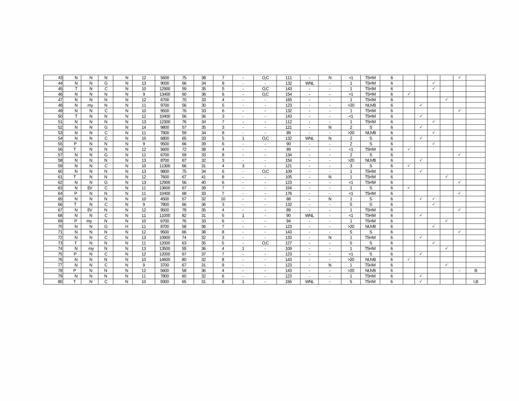
43 N N N N 12 5600 75 38 7 - O,C 111 - N <1 TS+IM 644 N N G N 13 9000 66 34 8 - - 132 WNL - 1 TS+IM 645 T N C N 10 12900 59 35 5 - O,C 143 - - 1 TS+IM 646 N N N N 9 13400 60 36 6 - O,C 154 - - <1 TS+IM 647 N N N N 12 6700 70 33 4 - - 165 - - 1 TS+IM 648 N my N N 11 9700 56 30 5 - - 123 - - >20 NUVB 649 N N C N 10 9500 76 33 6 - - 132 - - 1 TS+IM 650 T N N N 12 10400 56 36 3 - - 143 - - <1 TS+IM 651 N N N N 13 12300 76 34 7 - - 112 - - 1 TS+IM 652 N N G N 14 9800 57 35 3 - - 121 - N 2 S 653 N N C N 11 7800 59 34 8 - - 89 - - >20 NUVB 654 N N C N 10 6800 65 33 5 1 O,C 132 WNL N 2 S 655 P N N N 9 9500 66 39 6 - - 90 - - 2 S 656 T N N N 12 5600 72 38 4 - - 89 - - <1 TS+IM 657 N N G N 11 6700 59 33 8 - - 134 - - 2 S 658 N N N N 13 8700 67 32 3 - - 154 - - >20 NUVB 659 N N C N 10 11300 66 31 4 3 - 121 - - 3 S 660 N N N N 13 9800 75 34 6 - O,C 109 - - 1 TS+IM 661 T N N N 12 7600 67 41 8 - - 105 - N 1 TS+IM 662 N N G N 13 12900 56 40 6 - - 123 - - <1 TS+IM 663 N EV C N 11 13600 67 39 7 - - 154 - - 1 S 664 P N N N 11 10400 68 33 7 - - 176 - - <1 TS+IM 665 N N N N 10 4500 57 32 10 - - 88 - N 1 S 666 T N C N 9 7800 66 36 3 - - 132 - - 5 S 667 N EV N N 12 9500 78 35 4 - - 89 - - 1 TS+IM 668 N N C N 11 11000 82 31 5 1 - 90 WNL - <1 TS+IM 669 P my N N 10 6700 76 33 6 - - 94 - - 1 TS+IM 670 N N G H 11 8700 58 36 7 - - 123 - - >20 NUVB 671 N N N N 12 9500 66 38 8 - - 143 - - 5 S 672 N N C N 13 10900 74 32 3 - - 133 - N 1 TS+IM 673 T N N N 11 12000 63 35 5 - O,C 127 - - 5 S 674 N my N N 13 13500 55 36 4 1 - 109 - - 1 TS+IM 675 P N C N 12 12000 67 37 7 - - 123 - - <1 S 676 N N N N 10 14600 80 32 8 - - 143 - - >20 NUVB 677 N N C N 9 3700 67 31 9 - - 123 - N 1 TS+IM 678 P N N N 12 5600 58 36 4 - - 143 - - >20 NUVB 6 - B79 N N N N 11 7800 60 32 6 - - 123 - - 1 TS+IM 680 T N C N 10 9300 65 31 8 1 - 156 WNL - 5 TS+IM 6 I,B
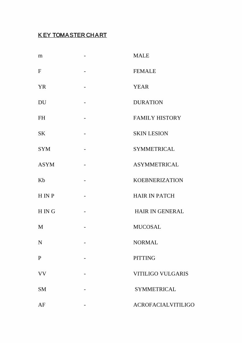
KEY TO MASTER CHART
m - MALE
F - FEMALE
YR - YEAR
DU - DURATION
FH - FAMILY HISTORY
SK - SKIN LESION
SYM - SYMMETRICAL
ASYM - ASYMMETRICAL
Kb - KOEBNERIZATION
H IN P - HAIR IN PATCH
H IN G - HAIR IN GENERAL
M - MUCOSAL
N - NORMAL
P - PITTING
VV - VITILIGO VULGARIS
SM - SYMMETRICAL
AF - ACROFACIALVITILIGO
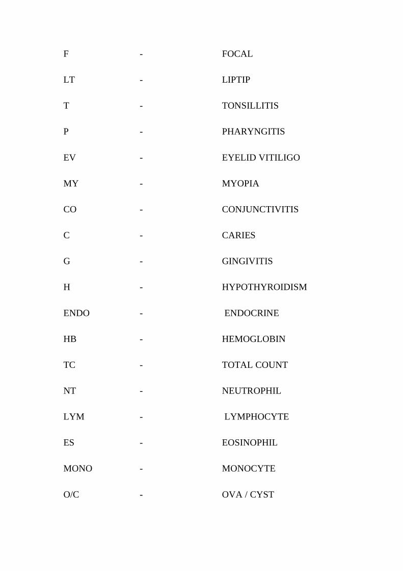
F - FOCAL
LT - LIPTIP
T - TONSILLITIS
P - PHARYNGITIS
EV - EYELID VITILIGO
MY - MYOPIA
CO - CONJUNCTIVITIS
C - CARIES
G - GINGIVITIS
H - HYPOTHYROIDISM
ENDO - ENDOCRINE
HB - HEMOGLOBIN
TC - TOTAL COUNT
NT - NEUTROPHIL
LYM - LYMPHOCYTE
ES - EOSINOPHIL
MONO - MONOCYTE
O/C - OVA / CYST
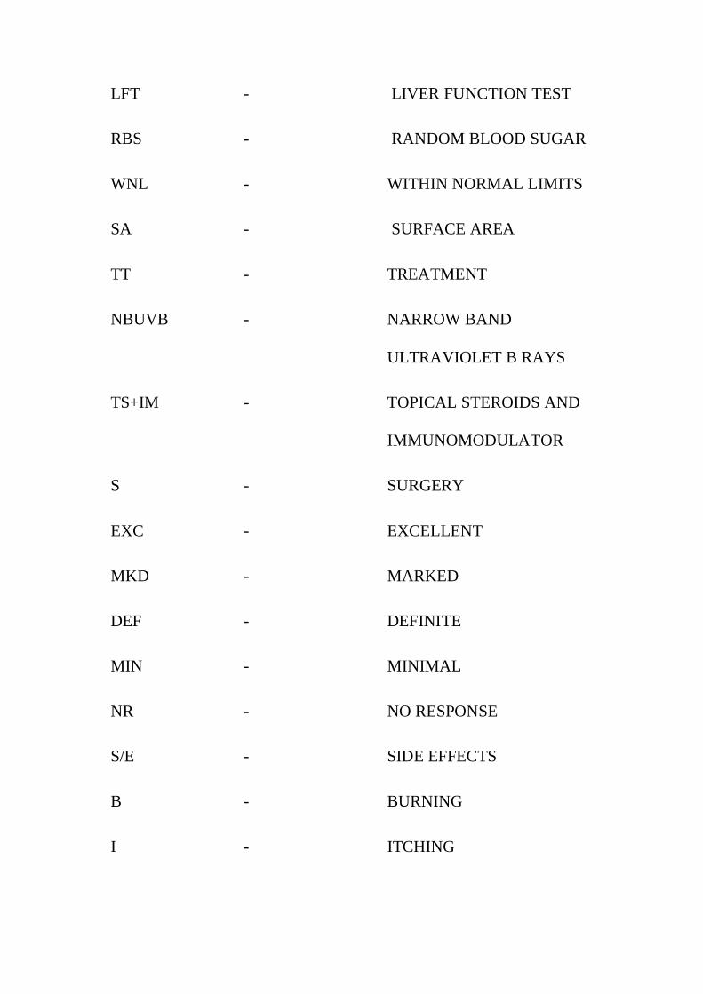
LFT - LIVER FUNCTION TEST
RBS - RANDOM BLOOD SUGAR
WNL - WITHIN NORMAL LIMITS
SA - SURFACE AREA
TT - TREATMENT
NBUVB - NARROW BAND
ULTRAVIOLET B RAYS
TS+IM - TOPICAL STEROIDS AND
IMMUNOMODULATOR
S - SURGERY
EXC - EXCELLENT
MKD - MARKED
DEF - DEFINITE
MIN - MINIMAL
NR - NO RESPONSE
S/E - SIDE EFFECTS
B - BURNING
I - ITCHING
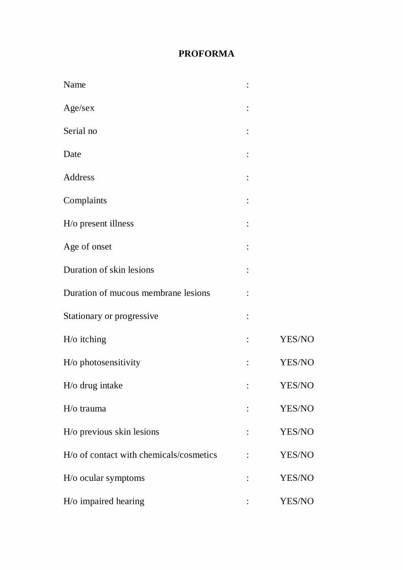
PROFORMA
Name :
Age/sex :
Serial no :
Date :
Address :
Complaints :
H/o present illness :
Age of onset :
Duration of skin lesions :
Duration of mucous membrane lesions :
Stationary or progressive :
H/o itching : YES/NO
H/o photosensitivity : YES/NO
H/o drug intake : YES/NO
H/o trauma : YES/NO
H/o previous skin lesions : YES/NO
H/o of contact with chemicals/cosmetics : YES/NO
H/o ocular symptoms : YES/NO
H/o impaired hearing : YES/NO
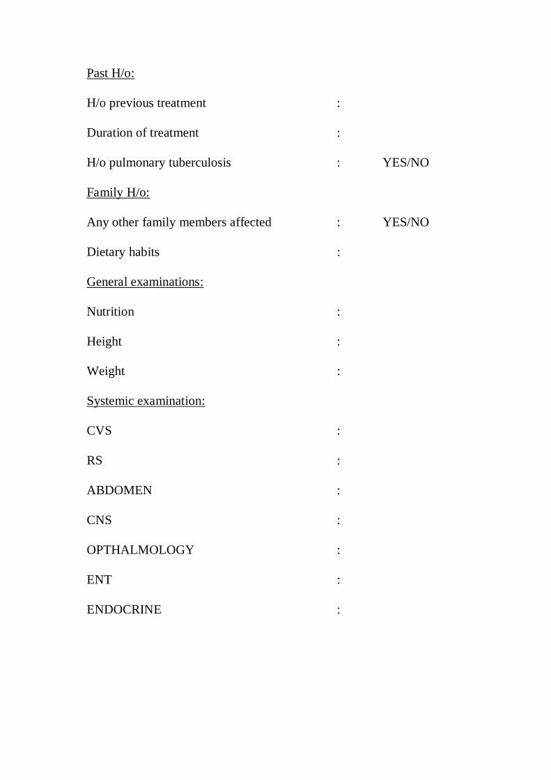
Past H/o:
H/o previous treatment :
Duration of treatment :
H/o pulmonary tuberculosis : YES/NO
Family H/o:
Any other family members affected : YES/NO
Dietary habits :
General examinations:
Nutrition :
Height :
Weight :
Systemic examination:
CVS :
RS :
ABDOMEN :
CNS :
OPTHALMOLOGY :
ENT :
ENDOCRINE :
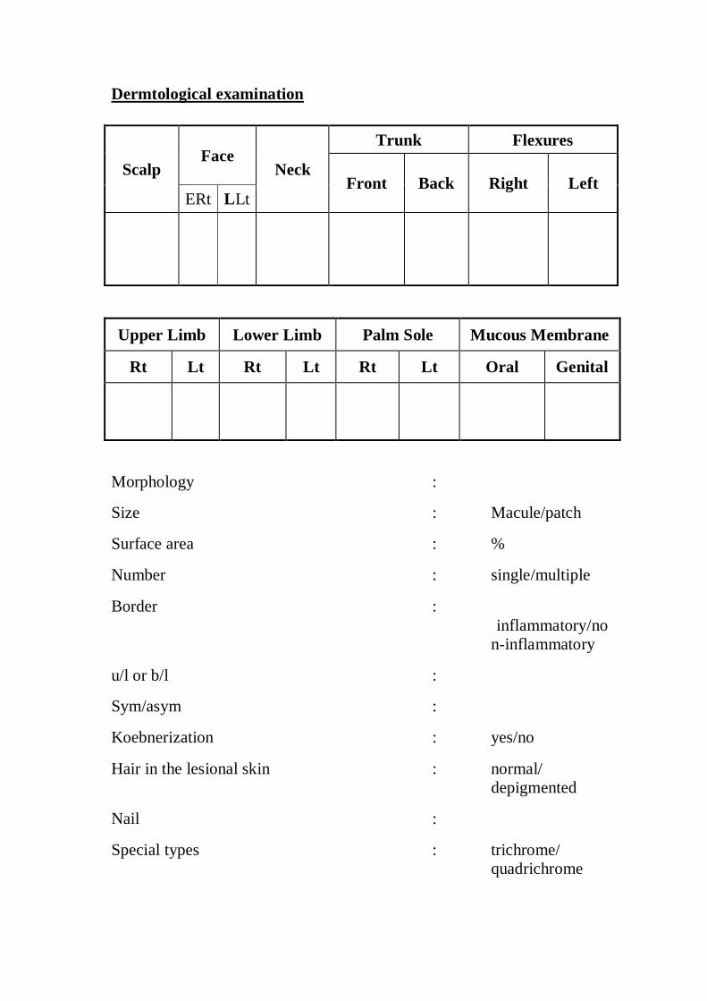
Dermtological examination
ScalpFace
Neck
Trunk Flexures
Front Back Right LeftERt LLt
Upper Limb Lower Limb Palm Sole Mucous Membrane
Rt Lt Rt Lt Rt Lt Oral Genital
Morphology :
Size : Macule/patch
Surface area : %
Number : single/multiple
Border : inflammatory/non-inflammatory
u/l or b/l :
Sym/asym :
Koebnerization : yes/no
Hair in the lesional skin : normal/depigmented
Nail :
Special types : trichrome/quadrichrome
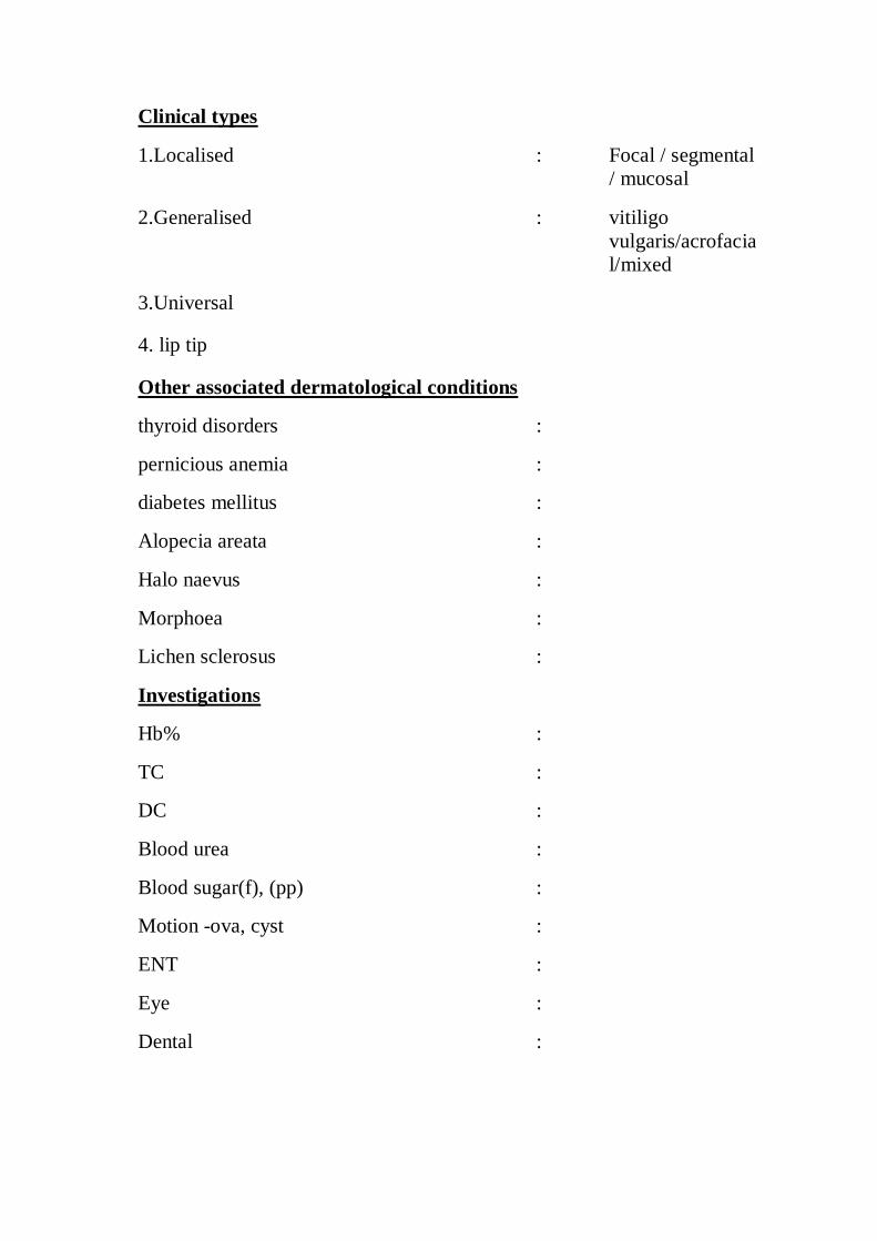
Clinical types
1.Localised : Focal / segmental/ mucosal
2.Generalised : vitiligovulgaris/acrofacial/mixed
3.Universal
4. lip tip
Other associated dermatological conditions
thyroid disorders :
pernicious anemia :
diabetes mellitus :
Alopecia areata :
Halo naevus :
Morphoea :
Lichen sclerosus :
Investigations
Hb% :
TC :
DC :
Blood urea :
Blood sugar(f), (pp) :
Motion -ova, cyst :
ENT :
Eye :
Dental :
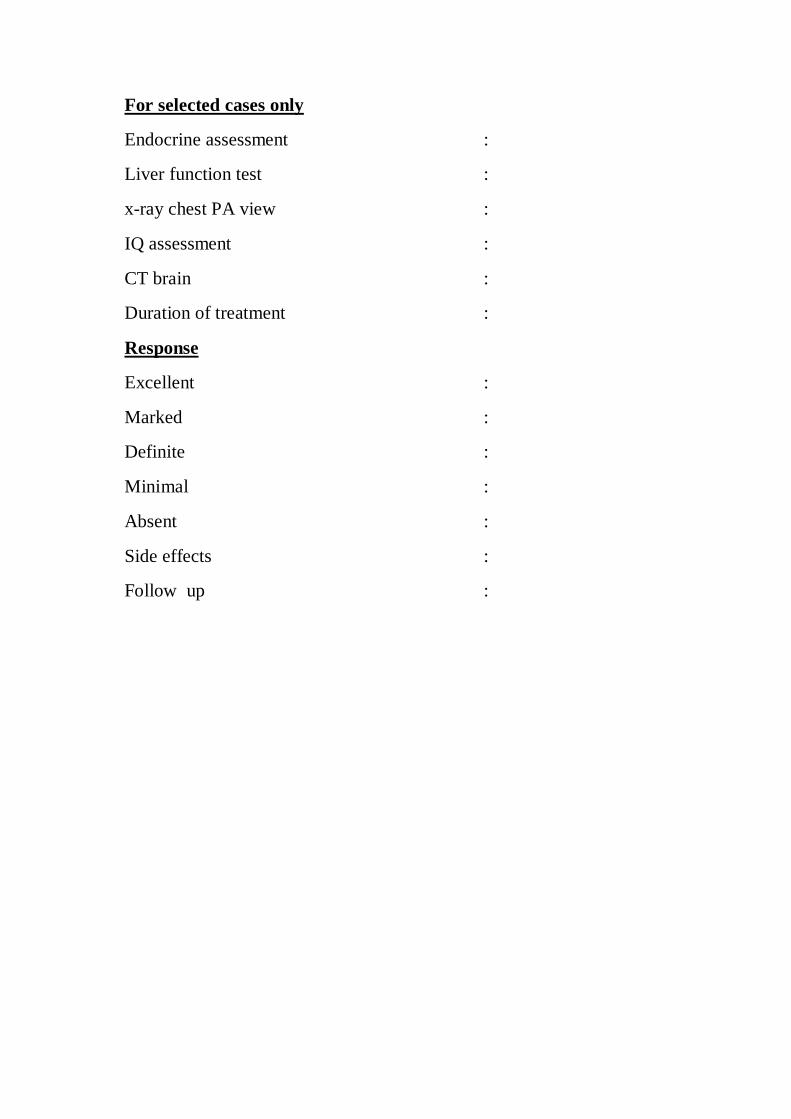
For selected cases only
Endocrine assessment :
Liver function test :
x-ray chest PA view :
IQ assessment :
CT brain :
Duration of treatment :
Response
Excellent :
Marked :
Definite :
Minimal :
Absent :
Side effects :
Follow up :