questions and (some) answers on reactive astrocytes - cea
TRANSCRIPT

HAL Id: cea-02290440https://hal-cea.archives-ouvertes.fr/cea-02290440
Submitted on 17 Aug 2021
HAL is a multi-disciplinary open accessarchive for the deposit and dissemination of sci-entific research documents, whether they are pub-lished or not. The documents may come fromteaching and research institutions in France orabroad, or from public or private research centers.
L’archive ouverte pluridisciplinaire HAL, estdestinée au dépôt et à la diffusion de documentsscientifiques de niveau recherche, publiés ou non,émanant des établissements d’enseignement et derecherche français ou étrangers, des laboratoirespublics ou privés.
Questions and (some) answers on reactive astrocytesCarole Escartin, Océane Guillemaud, Maria-angeles Carrillo-de Sauvage
To cite this version:Carole Escartin, Océane Guillemaud, Maria-angeles Carrillo-de Sauvage. Questions and (some) an-swers on reactive astrocytes. Glia, Wiley, 2019, �10.1002/glia.23687�. �cea-02290440�
Questions and (some) answers on reactive astrocytes
Journal: GLIA
Manuscript ID GLIA-00125-2019.R2
Wiley - Manuscript type: Review Article
Date Submitted by the Author: n/a
Complete List of Authors: Escartin, Carole; UMR9199, MIRCenGuillemaud, OcéaneCarrillo-De Sauvage, Maria-Angeles
Topics:
Astrocytes, Neurodegenerative diseases, Neuropathy, Cytoskeletal proteins, Ageing, Mechanisms of glia cell injury, Regulation of gene expression, Growth factor, cytokine and chemokine receptors, Immune Function
Techniques:
Histological techniques, Cellular and Developmental Neuroscience, Transgenic animals, Molecular Neuroscience, Transcriptome analysis (DD-PCR, CHIPS, SAGE), Neurophysiology, Cute isolation of neural cells, Knock-out methodology, Cell culture techniques, Techniques to measure cell prolifaration, necrosis and apoptosis
Key Words: Reactive astrocytes, Brain diseases, Neuron-glia interactions, Astrogliosis, Animal models
John Wiley & Sons, Inc.
GLIA

1
1 Questions and (some) answers on reactive astrocytes23 Carole Escartin 1,2, Océane Guillemaud 1,2, Maria-Angeles Carrillo-de Sauvage 1,2
45 1 Commissariat à l’Energie Atomique et aux Energies Alternatives, Département de la Recherche Fondamentale, Institut de 6 Biologie François Jacob, MIRCen, F-92260 Fontenay-aux-Roses, France7 2 Centre National de la Recherche Scientifique, Univ. Paris Sud, Univ. Paris-Saclay, UMR 9199, Neurodegenerative Disease 8 Laboratory, F-92260 Fontenay-aux-Roses, France9
101112
13 Corresponding author14 Carole Escartin PhD, HDR15 UMR 9199 (CNRS, CEA, Univ. Paris Sud)16 MIRCen17 18, route du Panorama18 92265 Fontenay-aux-roses Cedex19 France20 Tel (0033) 1 46 54 72 3321 [email protected] Running title25 Q&A on reactive astrocytes26
2728 Acknowledgments29 We are extremely grateful to all researchers that positively replied to our request and provided their own definition of 30 reactive astrocytes. We thank them for their valuable input and support. By alphabetical order: Nicola Allen, Alfonso Araque, 31 Luis Barbeito, Dwight Bergles, Giorgio Carmignoto, Colm Cunningham, Marc Freeman, Elena Galea, Vittorio Gallo, Magdalena 32 Götz, Philip Haydon, Helmut Kettenmann, Schuichi Koizumi, Andras Lakatos, C. Justin Lee, Shane Liddelow, Albee Messing, 33 Keith Murai, Christopher Norris, James O'Callaghan, Seiji Okada, Stéphane H.R. Oliet, Aude Panatier, Luc Pellerin, Gertrudis 34 Perea, Gabor Petzold, Frank Pfrieger, Stefanie Robel, David Rowitch, Alberto Serrano-Pozo, Swetlana Sirko, Michael 35 Sofroniew, Harald Sontheimer, Christian Steinhauser, Raymond Swanson, Alexei Verkhratsky, Andrea Volterra. Their full 36 affiliation and reply are provided as Supplementals. 37 We apologize for not being able to cite all original studies relevant to this review, due to space limitations.38 We thank Prof. C. Barcia, Dr. E. Hernández-Garzón, Dr. S. Liddelow, Prof M. Sofroniew, Dr. M. Valiente and Prof. A. 39 Verkhratsky, for providing original images for Fig. 2. We thank Dr. Lucile Ben Haim for her critical review of the manuscript.40 Our team is supported by CEA, CNRS and grants from the French National Research Agency (grants # 2010-JCJC-1402-1, 2011-41 BSV4-021-03 and ANR-16-TERC-0016-01), from Fondation Vaincre Alzheimer (grant # FR-15015), Fédération pour la 42 Recherche sur le Cerveau and Association Huntington France. OG is a recipient of a doctoral fellowship from the CEA.4344 Conflict of Interest45 The authors declare no conflict of interest4647 Word count48 Main Text: 943749 References: 739050 Figure & Table Legends: 1778
Page 1 of 57
John Wiley & Sons, Inc.
GLIA

2
51 Abstract 52 Astrocytes are key cellular partners for neurons in the central nervous system. Astrocytes react to virtually all 53 types of pathological alterations in brain homeostasis by significant morphological and molecular changes. This 54 response was classically viewed as stereotypical and is called astrogliosis or astrocyte reactivity. It was long 55 considered as a non-specific, secondary reaction to pathological conditions, offering no clues on disease-causing 56 mechanisms and with little therapeutic value.57 However, many studies over the last thirty years have underlined the crucial and active roles played by astrocytes 58 in physiology, ranging from metabolic support, synapse maturation and pruning to fine regulation of synaptic 59 transmission. This prompted researchers to explore how these new astrocyte functions were changed in disease, 60 and they reported alterations in many of them (sometimes beneficial, mostly deleterious). More recently, cell-61 specific transcriptomics revealed that astrocytes undergo massive changes in gene expression when they become 62 reactive. This observation further stressed that reactive astrocytes may be very different from normal, non-63 reactive astrocytes and could influence disease outcomes. To make the picture even more complex, both normal 64 and reactive astrocytes were shown to be molecularly and functionally heterogeneous. Very little is known about 65 the specific roles that each subtype of reactive astrocytes may play in different disease contexts.66 In this review, we have interrogated researchers in the field to identify and discuss points of consensus and 67 controversies about reactive astrocytes, starting with their very name. We then present the emerging knowledge 68 on these cells and future challenges in this field. 697071 Keywords 72 Reactive astrocytes, brain diseases, neuron-glia interactions, astrogliosis, animal models7374
75 Main Points 76 - Astrocytes react to brain homeostasis alteration by morphological and molecular changes
77 - This response is complex, heterogeneous and subject to controversies
78 - New tools, models and concepts will help better understand this widespread reaction
Page 2 of 57
John Wiley & Sons, Inc.
GLIA
3
79 1. Introduction80 Astrocytes have key functions in the brain (Verkhratsky & Nedergaard, 2018). New discoveries are made 81 regularly on their active roles in ion homeostasis, vascular coupling, synaptic plasticity, circuit building, synapse 82 turnover, waste clearance or higher functions like sleep-wake cycle, food intake or memory (Verkhratsky & 83 Nedergaard, 2018). One peculiar feature of these glial cells, first observed by 19th century anatomopathologists 84 is that they look different in the diseased brain [Fig. 1, (P. Chaslin, 1891; Ramón y Cajal)]. The original observations 85 reported morphological and histological changes, with increased fibrillary structures and more visible cellular 86 elements in scarring brain tissue from patients [Fig. 1, see also (Liddelow & Barres, 2017)]. Such changes in 87 astrocyte appearance are observed in the brain of patients suffering from a broad range of brain pathologies 88 (e.g. epilepsy, neurodegenerative disease, brain cancer, ischemia, infection, axotomy, invasive injury, toxin 89 exposure), as well as in animal models thereof (Fig. 2, and references in legend). Astrocyte reactivity, Astrogliosis, 90 Astrocyte activation, Reactive gliosis or Astrocytosis are among the terms used to describe such molecular, 91 morphological and functional changes in astrocytes. Even if definitions of astrocyte reactivity were proposed by 92 Prof. M. Sofroniew (Sofroniew, 2009, 2014a; Sofroniew & Vinters, 2010) and others (Pekny et al., 2016), a review 93 of the literature shows that the use and meaning of these different terms are quite broad and inconsistent. 94 Indeed, we surveyed ~40 researchers in the field (see complete list in Supplemental Table 1), and found that 95 they have quite different visions of what reactive astrocytes are, or are not (Table 1a) and they do not agree on 96 the nomenclature (Table 1b). 97 Beyond the discrepant terminology, some common features of reactive astrocytes emerge from this survey. 98 A broad definition could be summarized as “astrocytes sense and respond to an abnormal situation in the brain. 99 They change at the morphological, biochemical, transcriptional and functional levels. These changes are
100 maintained while the pathological stimulus is present but some aspects may resolve. Reactive astrocytes are 101 heterogeneous and may have various effects on disease progression”. Indeed, reactive astrocyte heterogeneity 102 is underlined by most researchers and contested by none of them (Table 1a). This opens new areas of research 103 to understand its origin and consequences (Section 5). 104 Other points that remain controversial will be covered later in this review: Are glial fibrillary acid protein 105 (GFAP) and morphological changes appropriate markers of reactive astrocytes (Section 4)? Can we identify better 106 markers (Section 6)? Do reactive astrocytes do good or bad things in disease (Section 8)? Do they die (Section 107 9)? Should aging astrocytes be considered as reactive (Section 12)? Other points were not necessarily raised by 108 surveyed researchers, but are still actively studied: What are the exact triggers and downstream signaling 109 pathways controlling this response (see Section 7)? Where do reactive astrocytes come from and what do they 110 become (see Section 10)? How do reactive astrocytes interact with other cell types involved in innate and 111 adaptive immunity (see Section 11)? Addressing all these questions will help provide a refined vision of this 112 complex brain response.113 The aim of this review is not to provide an extensive overview of the now abundant literature on reactive 114 astrocytes, but instead, discuss emerging research questions and unresolved issues on this topic.115116117 2. How to best name them? 118 But first, it is important to name our research subject and define what we are talking about. Our survey of 119 researchers in the field of astrocytes, showed that even three broadly-used terms had different inferred 120 meanings (Table 1b). Many surveyed PIs agreed that the very definition of astrocyte reactivity was ambiguous 121 and that a clear nomenclature would benefit the field. 122 The heterogeneity of this response and the variety of “reactive states” (see Section 5), call for a broad and 123 inclusive definition that can further be refined in each context. Indeed, all recognized that drastic changes happen 124 to astrocytes in pathological conditions, with some core and some disease-specific alterations (Table 1a, 125 Supplemental Text). We thus need words to describe this phenomenon. What are the most appropriate ones? 126 There was some agreement that astrocyte activation is misleading because it can apply to physiological activation 127 like transient Ca2+ signaling in response to normal neuronal activity (see Section 4-Timescale). It is thus better to 128 avoid this term. Another point of partial convergence was that astrogliosis implied something different (e.g.
Page 3 of 57
John Wiley & Sons, Inc.
GLIA

4
129 stronger or irreversible; with scarring, proliferation or immune infiltration), which definitely does not apply to all 130 disease conditions (Table 1b). We are therefore left with the term astrocyte reactivity. The problem with this 131 expression, as stressed by several PIs, is that it implies an “ability” to become (See Table 1a). As discussed by Dr. 132 A. Serrano-Pozo, reaction would be more correct to define the final state of astrocytes (see Supplemental Text). 133 We thus propose to avoid the use of astrocyte activation and astrogliosis and instead use reactive astrocytes or 134 astrocyte reaction and add to stroke/epilepsy/Alzheimer disease to further define it. Expressions like scar-forming 135 astrocytes, proliferating reactive astrocytes, phospho-STAT3+ reactive astrocytes are also very useful to 136 functionally or molecularly define these cells (see details in Section 5). We will use this nomenclature in our 137 review, but a more widespread agreement on terminology would be beneficial to the astrocyte community.138139140 3. Why bother with reactive astrocytes? 141 There are more than 17,500 articles recovered in PubMed with the query “Reactive astrocyte” OR “Astrocyte 142 reactivity” OR “Astrogliosis”, with more than 400 articles published each year since 2000. Why does such a broad 143 and somehow ill-defined concept trigger this significant interest? Indeed, if astrocytes become reactive in 144 response to something already going wrong, why should we care? It may already be too late, the initial 145 pathological event may have started well before and triggered an irreversible disease cascade.146 It is striking that astrocyte reaction is such a widespread response, reported in virtually all brain diseases, 147 brain regions, and multiple species including invertebrates, like Drosophila, many mammals and Humans 148 [(Kremer, Jung, Batelli, Rubin, & Gaul, 2017; Sofroniew & Vinters, 2010), see Fig. 2]. It is thus very important to 149 better understand what it means for an astrocyte to engage in this phenotype switch and how it can influence 150 surrounding cells.151 Astrocyte reaction is by definition a secondary event; yet, it may still impact disease progression, which lasts 152 for months or years in the case of tumor, neurodegenerative disease or epilepsy. More importantly, astrocytes 153 are reactive at the clinically visible stages of the disease, when patients seek diagnosis and treatment. Turning 154 reactive astrocytes into beneficial partners for vulnerable neurons exposed to a chronic disease or to the long-155 term consequences of an acute injury, would be a valuable therapeutic strategy.156 In addition, as astrocytes react to altered homeostasis in the brain, they are endogenous biomarkers for brain 157 diseases. Development of non-invasive imaging techniques, like magnetic resonance imaging (MRI) or positron 158 emission tomography (PET) to visualize when and where astrocytes become reactive would help disease 159 diagnostic. Available imaging methods are not quite specific for reactive astrocytes, they rather detect 160 neuroinflammation as a whole (i.e. reactive glial cells, sometimes infiltration of peripheral immune cells) or 161 associated changes, for example in brain metabolism (Aiello et al., 2018; Carrillo-de Sauvage et al., 2015; Lavisse 162 et al., 2012). Indeed, astrocytes may significantly contribute to imaging signals, such as those measured with 163 blood oxygenation level-dependent (BOLD) functional MRI or [18F]-fluorodeoxyglucose uptake by PET, and 164 therefore their reactive state may impact these measurements (Carter et al., 2019; Mishra, 2017). Refined 165 approaches that are more selective for reactive astrocytes are being developed (Carter et al., 2019; Ligneul et 166 al., 2019; Rodriguez-Vieitez et al., 2016; Scholl et al., 2015). Such improvements will be facilitated by basic studies 167 that establish the molecular and functional profile of reactive astrocytes (see Section 6) but also of their microglia 168 or neuronal neighbors, allowing the identification of new and specific astrocyte targets. 169 Last, some astrocyte proteins like GFAP or their break-down products may also end up in the cerebrospinal 170 fluid of patients subject to traumatic brain injury [TBI, (Halford et al., 2017)], but also in more progressive diseases 171 like Creutzfeldt-Jakob disease or Alzheimer disease (AD) [for review, see (Carter et al., 2019; Perez-Nievas & 172 Serrano-Pozo, 2018)]. 173 Therefore, defining how astrocytes change during disease may in fine lead to new biomarkers, better 174 diagnosis tools, and even original therapeutic strategies, which are long-sought goals for many diseases. 175176
Page 4 of 57
John Wiley & Sons, Inc.
GLIA

5
177 4. What are the defining features of reactive astrocytes? 178 Several common features are observed in very different cases of brain injuries and diseases. They can be used 179 to define the core hallmarks of reactive astrocytes. 180181 Morphological changes. In pathological conditions, astrocytes display morphological changes, including 182 hypertrophy of soma and main processes, but not only (see Fig. 2). This was already noted by earlier pathologists 183 using impregnation techniques to achieve sparse labeling of astrocytes (Fig. 1). Diolistic labeling, dye-filling or 184 expression of a cytosolic or membrane-tagged fluorescent protein reveal the complex and highly ramified 185 astrocyte morphology. These methods evidence subtle alterations in astrocyte morphology that could be missed 186 by cytoskeleton labeling with GFAP antibodies (See Fig. 2B, D, E, H, I, J). They show process polarization towards 187 a lesion [(Bardehle et al., 2013), Fig. 2H] or ramification changes while the overall 3D domain covered by a single 188 astrocyte is not massively disrupted [(Wilhelmsson et al., 2006), Fig. 2E]. In more severe diseases like epilepsy, 189 astrocytes may also retract their fine peripheral processes, become asymmetric and overlap more with their 190 neighbors [(Oberheim et al., 2008; Sun & Jakobs, 2012), Fig. 2J]. An extreme case is the glial scar caused by 191 mechanical lesions of brain or spinal parenchyma (Fig. 2F), or severe focal lesions like tumors (Fig. 2R). Astrocytes 192 become elongated and assemble to form a compact and permanent scar. Other cell types, such as fibroblasts, 193 oligodendrocyte progenitor cells (OPC), ependymal cells or pericytes, may also contribute to this scar (Adams & 194 Gallo, 2018; Sabelstrom, Stenudd, & Frisen, 2014). This question and the overall effect of the glial scar on axonal 195 regrowth are still debated (Anderson et al., 2016; Silver, 2016). Readers are referred to excellent reviews on the 196 topic (Adams & Gallo, 2018; Sofroniew, 2018).197198 Molecular changes. A defining feature of reactive astrocytes is their overexpression of intermediate filament 199 proteins like GFAP or vimentin. The “increased number and size of fibrils in neuroglia of sclerotic brain tissue in 200 epileptic patients” was noted by anatomopathologists already at the end of the 19th century [see Fig. 1, (P. 201 Chaslin, 1891)]. Electron microscopy also evidences large bundles of filaments in reactive astrocytes (Fig. 2M’). 202 In AD brains, GFAP accumulates to the extent that it forms protein aggregates resembling Rosenthal fibers in 203 reactive astrocytes (Wegiel & Wisniewski, 1994b). The surveyed researchers broadly acknowledged the validity 204 of GFAP as a marker for reactive astrocytes (Table 1a), but several of them also underlined its limits (see further 205 discussion in Section 6).206 Owing to refined methods to isolate astrocytes and investigate their transcriptome at the genome-wide level, 207 recent studies revealed that astrocyte reaction involves massive transcriptional changes that go well beyond 208 Gfap induction. Hundreds of genes are either up- or down-regulated in astrocytes in AD models and patients 209 (Ceyzériat et al., 2018; Orre, Kamphuis, Osborn, Jansen, et al., 2014; Sekar et al., 2015), in a mouse model of 210 hyperammonia (Lichter-Konecki, Mangin, Gordish-Dressman, Hoffman, & Gallo, 2008), or multiple sclerosis (MS) 211 (Itoh et al., 2018), following spinal cord injury [SCI, (Anderson et al., 2016)], cortical stab wound injury [SWI, (Sirko 212 et al., 2015)], middle cerebral artery occlusion (MCAO) or lipopolysaccharide (LPS) injection (Zamanian et al., 213 2012). Several genes induced in reactive astrocytes both in the LPS and MCAO models were identified. Among 214 them, Serpina3n and Lcn2 were further validated as strongly, but transiently induced following LPS injection or 215 MCAO (Zamanian et al., 2012). They are also induced in other disease models (Itoh et al., 2018; Suk, 2016; 216 Switonski, Szlachcic, Krzyzosiak, & Figiel, 2015). Change in transcriptome is a conserved feature of astrocyte 217 reaction, similarly observed in Drosophila glia (Lu et al., 2017).218 Interestingly, genes that are down-regulated in reactive astrocytes may also hold the key to understanding 219 the roles of these cells in disease. Some genes associated with important astrocyte functions like the potassium 220 channel KIR4.1 [Kcnj10, (Nwaobi, Cuddapah, Patterson, Randolph, & Olsen, 2016)], the glutamate transporter 221 GLT1 (Slc1a2) or glutamine synthase GS [GluI, (Sheldon & Robinson, 2007)], are repeatedly reported as down-222 regulated in disease. Likewise, reduced expression of several homeostatic astrocyte genes is reported in mouse 223 models of SWI (Sirko et al., 2015) and TBI (Shandra et al., 2019) (see also Section 10), but there is no established 224 list of genes down-regulated in reactive astrocytes across multiple diseases. Overall, the significant phenotypic 225 alterations observed in reactive astrocytes involve large-scale transcriptome modifications and functional 226 changes (see Section 8).
Page 5 of 57
John Wiley & Sons, Inc.
GLIA

6
227228 Migration & proliferation. The very first observers of astrocytes noticed increased numbers of nuclei in sclerotic 229 tissue of epileptic brains, and discussed the proliferative capacity of neuroglia [Fig. 1, (P. Chaslin, 1891; Ramón y 230 Cajal)]. But more recent studies based on two-photon imaging of astrocyte reaction, fate mapping and 231 bromodeoxyuridine (BrdU) labeling in animal models, challenged this dogma [(Bardehle et al., 2013; Buffo et al., 232 2008), Fig. 2H]. Immunostaining with Ki67 or Proliferating Cell Nuclear Antigen in patients confirmed that only a 233 small percentage of reactive astrocytes undergoes proliferation (Perez-Nievas & Serrano-Pozo, 2018). These 234 proliferative astrocytes appear to require a stimulus from outside the central nervous system (CNS): they are in 235 direct contact with the lesion in SCI [(Wanner et al., 2013), Fig. 2F], are exposed to blood-borne substrates 236 following SWI (Sirko et al., 2013), or their cell bodies are in apposition to blood vessels (Bardehle et al., 2013). 237 Notably, proliferative astrocytes perform only one or two rounds of division (Bardehle et al., 2013; Sirko et al., 238 2013). Two-photon microscopy also showed that reactive astrocytes do not migrate like microglia or 239 macrophages do after a focal mechanical injury (Bardehle et al., 2013; Nimmerjahn, Kirchhoff, & Helmchen, 240 2005). Similarly, in the spinal cord, astrocytes remain in their allocated regional domain and do not migrate to a 241 nearby SWI (Tsai et al., 2012).242243 Timescale. Increased GFAP and nestin protein expression is detected as soon as 90 min after mouse euthanasia 244 and preparation of acute slices, showing that molecular changes may occur rapidly (and even ex vivo) (Takano et 245 al., 2014). In addition, In different striatal injury models caused by acute neurotoxin injection, Gfap mRNA levels 246 are induced 6 h later, while GFAP protein levels increase significantly only after 12-72 h, depending on the model 247 (O'Callaghan, Kelly, VanGilder, Sofroniew, & Miller, 2014). Astrocyte reaction can be viewed as a change of state 248 or a conversion (Table 1a). By becoming reactive, astrocytes undergo a set of morphological, transcriptional and 249 functional changes that transform them into different cells, with acquired, lost or altered properties and 250 functions (see Section 8). This is different from a rapid stimulation by neurotransmitters for example, which will 251 produce transient movements of perisynaptic processes (Bernardinelli et al., 2014), Ca2+ signals or gliotransmitter 252 release (Araque et al., 2014). This physiological response (which is better qualified as activation, see Table 1b 253 and Section 2), will not necessarily be long-lasting or associated with large scale transcriptional changes that shift 254 astrocyte phenotype. On the contrary, reactive astrocytes may persist over months and even years in chronic 255 brain diseases, although they may evolve overtime. 256257 Reversibility. Astrocyte reaction is reversible and may resolve. Manipulation of specific signaling cascades in vitro 258 and in vivo can normalize the transcriptome of astrocytes. Whether such “de-activated” astrocytes are truly 259 normal or are in a different state remains to be fully explored. Indeed, it is known that a given stimulus has a 260 different effect on astrocytes, when they were previously exposed to a first injury, a process known as priming 261 (Hennessy, Griffin, & Cunningham, 2015). A very elegant study recently showed that grafting reactive astrocytes 262 in a normal mouse spinal cord is sufficient to revert them to a non-reactive state (Hara et al., 2017). Conversely 263 (but less surprising), grafting normal astrocytes in an injured environment turns naïve astrocytes into reactive 264 astrocytes based on both morphological and molecular criteria, showing that reactive astrocytes are plastic and 265 regulated by environmental cues. Among the reported signals involved in maintaining a non-reactive state or 266 resolving astrocyte reaction are fibroblast growth factor (Kang et al., 2014), β1 integrin (Hara et al., 2017; Robel 267 et al., 2009), transforming growth factor α (Rothhammer et al., 2018), microbiome-derived tryptophan 268 metabolites (Rothhammer et al., 2016), suppressor of cytokine signaling 3 [SOCS3, (Ceyzériat et al., 2018)] or 269 Sonic Hedgehog [SHH, (Garcia, Petrova, Eng, & Joyner, 2010)). Such signaling molecules offer unique 270 opportunities to tune astrocyte reaction and evaluate the consequences on specific outcomes in different 271 disease models (see Section 8).272273274 5. Do all astrocytes react the same way? 275 As nearly all surveyed researchers noted (Table 1a), the global expression “reactive astrocytes” falls short of 276 describing the different responses that astrocytes may express in different disease conditions or even in response
Page 6 of 57
John Wiley & Sons, Inc.
GLIA

7
277 to the same stimulus (Table 2). As Prof. A. Messing puts it, citing Leo Tolstoy, “All happy families are alike; each 278 unhappy family is unhappy in its own way", suggesting that astrocytes react in a specific manner in each 279 pathological situation. This was nicely illustrated by two-photon monitoring of astrocyte responses following 280 focal SWI. All astrocytes proximal to the lesion became hypertrophic but only half of them polarized their 281 processes towards the lesion, the other half remained static and only 10% of them underwent proliferation 282 [(Bardehle et al., 2013), Fig. 2H]. Likewise, a lineage tracing method was used to determine whether astrocytes 283 derived from the same developmental clone display similar morphological changes following SWI or 284 experimental autoimmune encephalomyelitis (EAE). In both cases, reactive astrocytes from the same clone 285 tended to behave similarly, indicating that they are controlled by intrinsic cues established during development. 286 However, some astrocytes reacted differently than other cells in the same clone, showing that environmental 287 signals further diversify astrocyte response [(Bribian, Perez-Cerda, Matute, & Lopez-Mascaraque, 2018; Martin-288 Lopez, Garcia-Marques, Nunez-Llaves, & Lopez-Mascaraque, 2013), Fig. 2B].289 The heterogeneity of reactive astrocytes is also evidenced by spatial gradients in the intensity of response, 290 which culminates at the core of ischemia injury, SCI, mechanical lesion or epileptic focus (see Fig. 2A for example). 291 This is defined as topographical heterogeneity (Anderson, Ao, & Sofroniew, 2014) and is basically determined by 292 the distance to the injury core. As the concentration of molecular stimuli decreases with distance to the injury 293 core, so does astrocyte reaction, at least based on morphological changes or GFAP induction (see Table 2). As 294 Prof. S. Sirko explains it “as in Newton's third law, the action and the reaction [of astrocytes] are equivalent in 295 magnitude”. But this apparent continuum in the intensity of astrocyte reaction may in fact hide different, discrete 296 reactive states. For example, proliferative reactive astrocytes are only found proximal to the lesion [(Herrmann 297 et al., 2008; Wanner et al., 2013), Fig. 2F]. Nearly ten years ago, Prof. M. Sofroniew described three categories 298 of astrocyte reactivity: mild to moderate; severe diffuse and severe with glial scar. It was a noticeable effort to 299 better define this process and recognize its heterogeneity (Sofroniew & Vinters, 2010). This classification is based 300 on measurable criteria (proliferation, process extension, maintenance or loss of exclusive 3D domains) but there 301 is no absolute quantitative categories (e.g. to what extent astrocytes need to elongate their processes to fall into 302 the mild or severe category?). This classification may thus be difficult to translate to any disease or model, and 303 does not take advantage of objective molecular markers. Indeed, it was recently shown that reactive astrocytes 304 that form the glial scar and lose their 3D domain have a different molecular profile than hypertrophic reactive 305 astrocytes located farther away. “Scar-forming” astrocytes express specific transcripts such as chondroitin sulfate 306 proteoglycans (e.g. Acan, Pcan) and N-Cadherin (Cdh2), while hypertrophic reactive astrocytes express several 307 members of the β-catenin pathway (e.g. Ctnnb1, Plaur) (Hara et al., 2017). Interestingly, both types of reactive 308 astrocytes overexpress Gfap and Vim compared to normal astrocytes (Hara et al., 2017). In addition, scar-forming 309 astrocytes proliferate more than more distant hypertrophic reactive astrocytes [see Section 6, Table 2, (Wanner 310 et al., 2013)]. Overall, these data suggest that the scar-forming astrocytes are a specific reactive astrocyte 311 subtype and not a mere exacerbation of hypertrophic reactive astrocytes. 312 Other sources of heterogeneity have been described, such as regional differences, even more than 20 years 313 ago (Table 2). Astrocytes from different CNS regions exposed to the same toxic stimulus (Amyloid β) respond 314 differently in vitro (Hoke, Canning, Malemud, & Silver, 1994), showing that astrocyte heterogeneity is at least 315 partially intrinsic, as it is maintained in a dish. Likewise, astrocytes from gray and white matter react differently 316 to SWI (Mattugini et al., 2018). This could be due to pre-existing differences between astrocytes from distinct 317 brain regions (Boisvert, Erikson, Shokhirev, & Allen, 2018; Chai et al., 2017; Itoh et al., 2018; John Lin et al., 2017; 318 Lanjakornsiripan et al., 2018; Morel et al., 2017; Torigoe, Yamauchi, Zhu, Kobayashi, & Murakami, 2015). Another 319 demonstration of different molecular classes of reactive astrocytes came from the Barres laboratory. They 320 reported that the molecular changes induced in astrocytes by LPS injection or MCAO only partially overlap 321 (Zamanian et al., 2012). This led to the description of A1 and A2 reactive astrocytes, each characterized by a set 322 of 10-13 genes, in addition to a panel of 10-13 common genes (i.e. pan reactive) including Gfap, Vimentin and 323 Serpina3n (Liddelow et al., 2017). The molecular profile of LPS-induced A1 reactive astrocytes was replicated 324 quite faithfully by treating immunopanned rodent astrocytes with interleukin 1α (IL1α), complement 1q (C1q) 325 and tumor necrosis factor (TNF) (Liddelow et al., 2017). A detailed functional characterization of A1 reactive 326 astrocytes showed that they release factors that are toxic to neurons and oligodendrocytes, are less synaptogenic
Page 7 of 57
John Wiley & Sons, Inc.
GLIA

8
327 and have defective phagocytosis. But this dual classification may be quite restrictive to define other potential 328 types of reactive astrocytes existing in the complex world of brain diseases (Cunningham, Dunne, & Lopez-329 Rodriguez, 2018). Indeed, many intermediate molecular profiles were observed after treatment of 330 immunopanned astrocytes with different molecules, not only A1 or A2 (Liddelow et al., 2017). In a complex in 331 vivo multicellular environment with multiple stimuli, reactive astrocyte diversity may even be stronger. Both A1 332 and A2 genes are induced concomitantly in different models, such as mouse models of AD (Ceyzériat et al., 2018), 333 Tauopathy (Litvinchuk et al., 2018) or ischemia (Liddelow et al., 2017).334 Overall, some markers may display graded changes, proportional to the intensity of the initial injury [GFAP, 335 cytokines, (Sofroniew, 2014a)], while others may undergo an all-or-none response, being present only in some 336 forms of reaction [e.g. A1 versus A2 reactive astrocytes (Liddelow et al., 2017); scar-forming versus hypertrophic 337 reactive astrocytes (Hara et al., 2017), ephrin type-B receptor 1 (EphB1) versus interleukin (IL)6-induced reactive 338 astrocytes (G. E. Tyzack et al., 2017), Table 2]. 339 There are several origins and several manifestations of reactive astrocyte heterogeneity (Fig. 3, Table 2). 340 Indeed, as stated by Prof. A. Volterra « the combination of the intrinsic features of a given astrocytic population, 341 of its microenvironment and the type of insult will concur to produce different types of reactivity and different 342 functional outcomes». More precisely, astrocyte reaction is determined by core changes (induction of Gfap 343 expression, morphological plasticity), combined with disease-specific changes on a preexisting heterogeneous 344 background (Fig. 3), resulting in an extreme variety of possible reactive astrocyte subtypes. This leads us to the 345 next question, very central to the field.346347348 6. What are the appropriate markers of reactive astrocytes? 349 As discussed earlier, GFAP overexpression and morphological changes are the most commonly used of 350 reactive astrocyte markers (Liddelow & Barres, 2017). Both are easy to monitor on different types of samples 351 (see Fig. 2, Table 2). Gfap is one of the most consistently induced gene in transcriptomic datasets of reactive 352 astrocytes, confirming its usefulness as a reactive marker [(Hol & Pekny, 2015; Liddelow et al., 2017; Orre, 353 Kamphuis, Osborn, Jansen, et al., 2014; Zamanian et al., 2012), see also Table 2 and Section 4]. It is important to 354 note however, that the level of Gfap induction can be very different between conditions (see Table 2 and Section 355 5) or even between cells (Pekny, Wilhelmsson, Tatlisumak, & Pekna, 2019), but GFAP expression globally 356 increases at the population level, in a wide range of brain diseases. 357 Are GFAP induction and morphological changes enough to identify reactive astrocytes? Several surveyed PIs 358 think that these indexes can be misleading (Table 1a). Indeed, additional markers would be helpful with some 359 specific forms of reactive astrocytes that display unconventional morphological alterations or lower GFAP 360 immunoreactivity (see Section 9). In addition, they would be useful to define specific types of reactive astrocytes. 361 Genome-wide transcriptional profiling can identify potential common and class-specific genes (Hara et al., 2017; 362 Liddelow et al., 2017). How many markers are needed to define a class? For example, of the ~10 genes forming 363 the A1 and A2 panels, should they all be induced in a given condition to qualify a cell as an A1 reactive astrocyte 364 or 2-3 are enough? In fact, the expression of only a few protein markers (Complement 3, Complement factor b 365 and MX dynamin-like GTPase 1) was tested in the brain of patients and these three genes were not in the original 366 A1 panel [(Liddelow et al., 2017), Fig. 2T, U]. When the full panel of A1 genes is tested, not every single gene of 367 the A1 cassette is induced in several diseases, like in vitro and in vivo models of Parkinson disease (Yun et al., 368 2018) or AD mouse models (Ceyzériat et al., 2018). In addition, individual reactive astrocytes co-expressing A1 369 and A2 genes are observed in the MCAO rodent model and in the aging mouse brain (Clarke et al., 2018), 370 suggesting that it may be difficult to define classes of reactive astrocytes with only a few genes. 371 To improve the panel of reactive astrocyte molecular markers, it would be useful to extend the transcriptomic 372 analysis of reactive astrocytes performed by Zamanian et al. in LPS and MCAO models (Zamanian et al., 2012), to 373 many different, genetic and sporadic, acute and chronic, degenerative and inflammatory diseases. It would help 374 define with greater power the core sets of genes systematically induced or down-regulated in reactive astrocytes, 375 and identify disease-specific markers. Of course, such analysis would provide even more insight if performed on 376 human samples. However, post mortem delays induce noise and artifacts and the physical isolation of astrocytes
Page 8 of 57
John Wiley & Sons, Inc.
GLIA

9
377 is quite difficult, although it may be possible to gain some insight into astrocyte-specific transcriptomic changes 378 by co-expression analysis on bulk samples (Kelley, Nakao-Inoue, Molofsky, & Oldham, 2018). A method of choice 379 to study cell diversity at the molecular level is single-cell RNAseq (scRNAseq) (Svensson, Vento-Tormo, & 380 Teichmann, 2018) or single-nuclei RNAseq (snRNAseq) (Habib et al., 2017). These methods have gained 381 significant momentum, and several landmark papers have reported brain cell heterogeneity, including glial cells, 382 throughout development or between regions (Macosko et al., 2015; Pollen et al., 2014; Zeisel et al., 2018). There 383 are only few articles on brain diseases, and to date, only on other glial cells like microglia (Hammond et al., 2018; 384 Masuda et al., 2019). Such unsupervised approaches will help define populations of reactive astrocytes with 385 associated gene markers in different conditions (see Section 13).386 Another option to classify reactive astrocytes is to combine molecular with functional indexes. A good 387 example is the variable capacity of reactive astrocytes to proliferate. As mentioned earlier, only a subset of 388 reactive astrocytes undergo cell division. These proliferative astrocytes may thus be considered as a different 389 class, of particular interest because they may repopulate the damaged brain (see Section 10). But other specific 390 functions (either gained, lost or altered, see Section 8) may also serve as functional markers of reactive 391 astrocytes, like enhanced phagocytic capacities around plaques, as shown for “disease associated microglia” 392 (Keren-Shaul et al., 2017). For example, only a fraction of reactive astrocytes phagocyte myelin debris in the brain 393 of patients with MS or leukoencephalopathy [(Ponath et al., 2017), Fig. 2N]. A classification based on production 394 of γ-amino-butyric acid (GABA) and brain-derived neurotrophic factor (BDNF) by reactive astrocytes was 395 proposed by Prof. C.J. Lee [see Table 1a, and Supplemental text, (Chun et al., 2018)], since reactive astrocytes 396 produce more GABA in AD (Jo et al., 2014b; Z. Wu, Guo, Gearing, & Chen, 2014). This is a potentially interesting 397 functional classification of reactive astrocytes, but it needs to be further explored, to evaluate whether this is 398 translatable to multiple diseases, animal models and of course patients. For example, contrary to AD models, 399 astrocyte GABA production is reduced in a mouse model of Huntington disease [HD, (Jo et al., 2014b; Wojtowicz, 400 Dvorzhak, Semtner, & Grantyn, 2013)]. Importantly, a good marker needs to be easy to use and compatible with 401 other exploratory techniques like electrophysiology or functional imaging. Therefore, mRNA and protein markers 402 remain favored options.403 A problem that may be very difficult to overcome is the dynamic, flexible and context-dependent phenotype 404 of astrocytes. It was nicely discussed for astrocytes in physiology (Poskanzer & Molofsky, 2018), and it may even 405 be exacerbated in disease, when astrocytes express gene markers of other cell types and cell identities become 406 blurrier (see Section 10). It may thus be impossible to establish fixed classes and we rather should try to define 407 states, dependent on previous phenotype, activated signaling and environment (see Fig. 3). The ambitious 408 Human Cell Atlas project that aims to define all cells in the human body with a range of single-cell approaches 409 will probably provide the astrocyte field with new tools and concepts to better address the challenge of reactive 410 astrocyte classification. 411412413 7. What are the molecular cascades triggered in reactive astrocytes? 414 First, astrocytes sense a pathological signal. This signal can be extracellular (e.g. cytokines, purines, 415 aggregated proteins, myelin debris), trans-cellular (e.g. transmembrane adhesion molecules like ephrins or 416 integrins), membrane bound [e.g. phosphatidyl-serine (Chung et al., 2013)], as well as intracellular (e.g. 417 aggregated proteins, nucleic acids from infecting pathogens, ions like Ca2+), [see (Buffo, Rolando, & Ceruti, 2010; 418 Cunningham et al., 2018; Kang & Hebert, 2011; Sofroniew, 2014a) for review]. Interestingly, a recent study 419 reported that astrocytes are also very sensitive to environmental pollutants (Wheeler et al., 2019). To sense all 420 these molecular triggers, astrocytes are equipped with a wide range of membrane or intracellular receptors, 421 including G Protein-coupled receptors (GPCR, like metabotropic glutamate or P2Y purinergic receptors), 422 ionotropic receptors (e.g. P2X purinergic receptors), multimeric cytokine receptors (e.g. IL6 family receptors), 423 Toll like receptors [TLR, although their expression by astrocytes is quite disputed (Cunningham et al., 2018)] or, 424 tyrosine kinase receptors (e.g. Epidermal growth factor receptor), [see (Kang & Hebert, 2011; Verkhratsky & 425 Nedergaard, 2018) for review]. It is important to note however that there is rarely a direct and formal 426 demonstration that each of these stimuli can trigger a full reactive program (i.e. resulting in morphological as
Page 9 of 57
John Wiley & Sons, Inc.
GLIA

10
427 well as complex transcriptional changes), without the involvement of other cell types like microglia. Indeed, this 428 is typically tested by applying high doses of these compounds in vitro or through injection or overexpression in 429 vivo. It is therefore difficult to know whether they activate astrocytes directly or through microglial cells (see 430 Section 11). Interestingly, astrocytes are also mechano-sensitive. They can detect changes in their environment 431 mechanical properties, discriminate soft from stiff material and adjust by changing their own stiffness 432 (Moeendarbary et al., 2017; Moshayedi et al., 2014).433 After stimulus sensing, a step of signal transduction takes place and converge to the nucleus. This can occur 434 through direct shuffling of activated down-stream transcription factors to the nucleus after phosphorylation, 435 dephosphorylation or release from inhibitors. Signal transducer and activator of transcription 3 (STAT3), nuclear 436 factor B (NF-B) and nuclear factor of activated T-cells (NFAT), the downstream effectors of the Janus kinase 437 (JAK)-STAT3, NF-B and calcineurin-NFAT pathways respectively, are regulated by such a mechanism (Ceyzériat, 438 Abjean, Carrillo-de Sauvage, Ben Haim, & Escartin, 2016; Sompol & Norris, 2018; Q. Zhang, Lenardo, & Baltimore, 439 2017). Many signaling cascades are associated with astrocyte reaction (L. Ben Haim, Carrillo-de Sauvage, 440 Ceyzeriat, & Escartin, 2015; Buffo et al., 2010; Kang & Hebert, 2011), but the STAT3 pathway seems to play a 441 prominent role in different disease conditions, acting as a master regulator of reactive astrocytes [(L. Ben Haim, 442 Ceyzeriat, et al., 2015; Ceyzériat et al., 2016; Herrmann et al., 2008), Fig. 2O]. Ca2+ signaling may also be involved 443 in astrocyte reaction. Ca2+ can activate or inhibit many downstream signaling intermediates like the phosphatase 444 calcineurin (Sompol & Norris, 2018) or the transcriptional repressor Pumilio 2 (Kanemaru et al., 2013), which will 445 activate other downstream pathways like the NFAT or N-cadherin pathways respectively and trigger important 446 transcriptional changes.447 Activation of specific transcription factors will induce expression of target genes such as Gfap or cytokines 448 but also of transcription factors or retro-inhibitors of other pathways (e.g. SOCS3 for the JAK-STAT3 pathway or 449 IB for the NF-B pathway), which will further shape the transcriptome of reactive astrocytes. This step may 450 involve successive waves of transcriptional regulation, starting with induction of immediate early genes (Jenab 451 & Quinones-Jenab, 2002; Priller, Reddington, Haas, & Kreutzberg, 1998; Y. E. Wu, Pan, Zuo, Li, & Hong, 2017), 452 followed by subsequent waves of gene induction. The transcription of some genes may be repressed. Indeed, 453 the number of down-regulated genes may even be higher than those over-expressed, as observed AD mouse 454 astrocytes (Orre, Kamphuis, Osborn, Jansen, et al., 2014). Importantly, the precise time course of transcriptional 455 induction is not very well known. It was established for specific transcripts or proteins (mostly GFAP and 456 cytokines) in mice exposed to neurotoxins (O'Callaghan et al., 2014) or LPS (Biesmans et al., 2015; Norden, 457 Trojanowski, Villanueva, Navarro, & Godbout, 2016). Transcriptional profiling of astrocytes was performed in a 458 mouse model of epilepsy, in two brains regions and three time points following pilocarpine injection (Clasadonte 459 et al., 2016). This extensive analysis showed subtle differences between disease stages or brain regions (see 460 Table 2). Such genome-wide, longitudinal analysis of relevant in vivo models of other brain diseases would 461 provide key insight into the temporal regulation of astrocyte reaction. 462 Some changes observed in reactive astrocytes do not require transcriptional induction, such as morphological 463 plasticity. The molecular mechanisms underlying astrocyte morphological changes and process motility are 464 mostly studied in vitro, where these cells are grown in 2D and usually have a much simpler morphology 465 (sometimes even no processes) [see (Schiweck, Eickholt, & Murk, 2018), for review]. But in vivo, reactive 466 astrocytes do not migrate [(Bardehle et al., 2013), Section 2] and show little motility of main processes compared 467 to microglia (Nimmerjahn et al., 2005), calling for in vivo validation of the signaling described in vitro. For 468 example, knockout of the Rho GTPase cdc42 reduces astrocyte polarization in vitro in the scratch wound assay, 469 while it exacerbates it after SWI in vivo (Robel, Bardehle, Lepier, Brakebusch, & Gotz, 2011). As mentioned earlier 470 (Section 3), reactive astrocytes not only present hypertrophy, but also subtle morphological changes. For 471 example, reactive astrocytes retract their processes following stimulation of Slit-Robo signaling by neuroblasts 472 migrating from the subventricular zone after stroke. This cdc42-dependent remodeling of astrocyte cytoskeleton 473 facilitates neuroblast migration towards lesion sites (Kaneko et al., 2018). 474 Less is known about mechanisms of epigenetic regulation in reactive astrocytes in disease. Based on what 475 happens during astrocyte development, it is suspected that epigenetic processes (chromatin remodeling through
Page 10 of 57
John Wiley & Sons, Inc.
GLIA

11
476 DNA methylation or histone post-translational modifications, as well as expression of regulatory microRNA) could 477 influence astrocyte conversion into reactive cells (Neal & Richardson, 2018).478 The profound transcriptional changes occurring in reactive astrocytes translate into functional alterations 479 that may impact virtually all astrocyte functions.480481482 8. Do reactive astrocytes do good things?483 Reactive astrocytes are the usual suspects in many diseases, but they are now considered by many, as 484 beneficial partners for neurons, as illustrated in Table 1a. An overview of the literature shows that depending on 485 the strategy employed to modulate reactive astrocytes (e.g. pharmacological or genetic approaches, targeting of 486 astrocyte proteins or signaling pathways, ablation of astrocytes), the outcomes vary (L. Ben Haim, Carrillo-de 487 Sauvage, et al., 2015; Cunningham et al., 2018). Of course, the disease studied and its model itself (e.g. transgenic 488 versus knock-in, in vivo versus in vitro, species) will influence how astrocyte reaction will play out. More 489 importantly, some models do not reproduce astrocyte reaction well. For example, most HD mouse models do 490 not display reactive astrocytes, while GFAP up-regulation is robustly detected in the caudate-putamen of HD 491 patients (Faideau et al., 2010; Selkoe, Salazar, Abraham, & Kosik, 1982; Vonsattel et al., 1985). This could be due 492 to low GFAP expression in the mouse striatum compared to other brain regions (Chai et al., 2017), significant 493 species differences (Oberheim et al., 2009; Y. Zhang et al., 2016) or a simple failure of mouse models to 494 recapitulate all HD features. 495 How do reactive astrocytes functionally impact the diseased brain? Virtually all astrocyte functions are 496 reported to be altered in disease: neurotransmitter uptake (Escartin et al., 2006; Sheldon & Robinson, 2007), 497 gliotransmitter release (Jo et al., 2014a; Z. Wu et al., 2014), metabolic activity (Escartin et al., 2007; Gavillet, 498 Allaman, & Magistretti, 2008; Valenza et al., 2010), ion buffering (Tong et al., 2014), release of cytokines, 499 complement factors or trophic factors (Chou et al., 2008; Lian et al., 2015; Sofroniew, 2014b), phagocytosis 500 (Gomez-Arboledas et al., 2018; Liddelow et al., 2017; Morizawa et al., 2017), production and detoxification of 501 reactive oxygen species [ROS, (Allaman et al., 2010; Cassina et al., 2008; Ye et al., 2015)]. These functions may 502 be enhanced or reduced by the reactive state. For example, reactive astrocytes may display enhanced or reduced 503 connectivity through gap junctions, depending on the disease (Escartin & Rouach, 2013; Pannasch & Rouach, 504 2013). They may display deficits in glutamate uptake (Sheldon & Robinson, 2007), or show enhanced uptake 505 capacity when reaction is induced by the cytokine ciliary neurotrophic factor (CNTF) (Escartin et al., 2006). 506 Reactive astrocytes may release more GABA in AD mice (Jo et al., 2014b; Z. Wu et al., 2014) or do the opposite 507 in HD mice (Wojtowicz et al., 2013). 508 It is beyond the scope of this review to list all their described functional changes and contributions to specific 509 diseases. Readers are referred to recent reviews on astrocytes in HD (Khakh et al., 2017), AD (Chun & Lee, 2018; 510 Osborn, Kamphuis, Wadman, & Hol, 2016; Perez-Nievas & Serrano-Pozo, 2018), MS (Wheeler & Quintana, 2019), 511 SCI (Adams & Gallo, 2018; Sofroniew, 2018), TBI (Burda, Bernstein, & Sofroniew, 2016), stroke (Pekny et al., 512 2019), and epilepsy (Coulter & Steinhauser, 2015; Robel & Sontheimer, 2016). In general, deficits in normal 513 astrocyte functions are reported and the existence of “killer astrocytes” releasing toxic molecule(s) was even 514 proposed in amyotrophic lateral sclerosis [ALS, (Haidet-Phillips et al., 2011; Nagai et al., 2007)] and other 515 neurodegenerative diseases [(Liddelow et al., 2017), see Section 5]. But there are also strong evidence for 516 beneficial reactive astrocytes in acute or chronic diseases. For example, over the years, the Sofroniew laboratory 517 has convincingly demonstrated that scar-forming reactive astrocytes demarcate damaged tissue (Bush et al., 518 1999; Faulkner et al., 2004) and even promote axonal regrowth through the lesion (Anderson et al., 2016). 519 Likewise, STAT3-dependent reactive astrocytes reduce degeneration of axotomized motoneurons in the facial 520 motor nucleus, and promotes synapse maintenance through thrombospondin 1 release (G. E. Tyzack et al., 2014). 521 CNTF-induced reactive astrocytes display enhanced glutamate uptake, promote metabolic resilience and protect 522 neurons against excitotoxicity (Beurrier et al., 2010; Escartin et al., 2006; Escartin et al., 2007). Even A1 neurotoxic 523 reactive astrocytes may have some beneficial roles in specific diseases, for example by killing damaged, 524 dysfunctional or infected neurons (Liddelow et al., 2017).
Page 11 of 57
John Wiley & Sons, Inc.
GLIA

12
525 A way to study common functional features of reactive astrocytes, is to activate (or inhibit) a specific signaling 526 pathway to trigger reactive conversion (see Section 8). For example, JAK-STAT3 pathway activation in astrocytes 527 induces many reactive markers and is sufficient to alter synaptic transmission and plasticity in the hippocampus 528 (Ceyzériat et al., 2018). Activation of the NF-B pathway by expression of a constitutive form of the upstream 529 IB kinase (IKK) in adult astrocytes also induces a reactive profile and Purkinje cell loss in the cerebellum (Lattke 530 et al., 2017; Oeckl, Lattke, Wirth, Baumann, & Ferger, 2012). Such approaches are useful to isolate functional 531 changes in reactive astrocytes, independently of a specific disease condition. Alternatively, blocking astrocyte 532 reaction may help uncover the net effect of reactive astrocytes in each disease context [see (L. Ben Haim, Carrillo-533 de Sauvage, et al., 2015)]. This requires an efficient strategy that i) only impacts the reactive state and not normal 534 astrocyte functions, ii) does not affect other cell types, and iii) inhibits all (or only the targeted) classes of reactive 535 astrocytes. With new and maybe more selective molecular markers, it will be important to extensively validate 536 the efficiency and universality of such methods and determine how these “de-activated” astrocytes are different 537 from non-reactive astrocytes. 538 Overall, the range of functional changes displayed by reactive astrocytes and their effects on disease are 539 extremely large. They are governed by the pre-existing astrocyte molecular and functional profile, in addition to 540 the pathological trigger itself (Fig. 3, Section 5). Indeed, even reactive astrocytes activated by the same pathway 541 can have different effects on specific disease outcomes. STAT3 signaling in reactive astrocytes has detrimental 542 effects in mouse models of AD (Ceyzériat et al., 2018; Reichenbach et al., 2019), or hypoxia (Hristova et al., 2016), 543 no significant effect in the context of acute mitochondria toxicity (O'Callaghan et al., 2014) but has beneficial 544 effects in neonatal white matter injury (Nobuta et al., 2012), after SCI, by promoting glial scar formation 545 (Anderson et al., 2016; Herrmann et al., 2008; Okada et al., 2006), and in HD models, by reducing mutant 546 huntingtin aggregation (L. Ben Haim, Ceyzeriat, et al., 2015). Intriguingly, a recent study by Tyzack et al. suggested 547 that the upstream activator (IL6 or EphB1-ephrine-B1 signaling) controls the final molecular profile induced by 548 STAT3 in reactive astrocytes [(G. E. Tyzack et al., 2017), Table 2].549 It is now important to define what reactive astrocytes do in each situation and the underlying signaling 550 cascades, before targeting these cells for therapy. There are new opportunities and yet many challenges to use 551 reactive astrocytes for therapeutic purposes.552553554 9. Do reactive astrocytes eventually die?555 There may be extreme forms of responses leading to the loss of key astrocyte proteins and functions or even 556 their death. Astrocytes are considered more resilient than neurons, but they may degenerate as well. As seen 557 with TUNEL or caspase stainings, astrocytes may undergo apoptotic cell death in ischemia (Giffard & Swanson, 558 2005), in ALS mice (Rossi et al., 2008), or in the brain of AD patients (Perez-Nievas & Serrano-Pozo, 2018). 559 Alternatively, mouse astrocytes may die by necrosis at the infarct core after stroke (Lukaszevicz et al., 2002). 560 Major alterations of astrocyte morphology, including severe process atrophy or fragmentation, swollen or 561 vacuolated cell bodies are described in brain samples from patients with AD, stroke or infection for example, and 562 it is called clasmatodendrosis [see references in (Perez-Nievas & Serrano-Pozo, 2018; Tachibana et al., 2019)]. It 563 is also reproduced in animal models of these diseases (Olabarria, Noristani, Verkhratsky, & Rodriguez, 2010; 564 Sullivan, Bjorkman, Miller, Colditz, & Pow, 2010). In addition, astrocytes may be directly or primarily targeted by 565 the disease process, like in Alexander disease caused by Gfap mutation (Messing, 2018) or neuromyelitis optica 566 caused by autoantibodies against the astrocyte protein aquaporin 4 [see (Verkhratsky, Zorec, & Parpura, 2017) 567 for a complete review].568 Interestingly, in a mouse model of tauopathy (Bussian et al., 2018) and in AD patients (Bhat et al., 2012), 569 astrocytes express markers of cellular senescence (p16INK4a and β-galactosidase). Genetic ablation of senescent 570 cells prevents tau pathology and cognitive decline (Bussian et al., 2018), suggesting that they have deleterious 571 effects. It is not possible however, to know whether senescent astrocytes are responsible alone or if senescent 572 microglia also play a role. Interestingly, interleukins IL6 and IL1β are classic senescence markers (Salminen et al., 573 2011) and senescent astrocytes in AD brains express high levels of GFAP (Bhat et al., 2012), suggesting that 574 senescence could be yet another specific reactive state.
Page 12 of 57
John Wiley & Sons, Inc.
GLIA

13
575 Overall, such atypical astrocyte responses characterized by major loss of key homeostatic astrocyte proteins, 576 senescence and/or altered morphology will need to be further characterized by genome wide and refined 577 temporal analysis to really qualify as a form of astrocyte reaction. Do astrocytes first become reactive and then 578 adopt this dysfunctional, senescent phenotype and die, or these are distinct non-overlapping processes?579580581 10.Where do reactive astrocytes come from or what do they become? 582 Reactive astrocytes are in a very distinct state from astrocytes in physiological conditions. Thus, one may 583 wonder whether reactive astrocytes 1) can further transform into other cell types and 2) come only from the 584 conversion of a non-reactive astrocyte. Both questions have important therapeutic implications, especially if 585 reactive astrocytes are able to generate new neurons. 586 As mentioned earlier, only a subset of reactive astrocytes proliferate (e.g. juxta-vascular or scar-forming 587 astrocytes), they only undergo one round of division and invariably generate two sister astrocyte cells (see 588 Section 4). Strikingly, the in vitro situation is quite different. In a culture medium promoting cell proliferation, 589 acutely isolated reactive astrocytes are able to generate different cell types, including neurons (Gotz, Sirko, 590 Beckers, & Irmler, 2015). It shows that some permissive signals are lacking in the brain environment [like SHH 591 signaling, (Sirko et al., 2013)] or conversely, that restrictive factors prevent astrocytes from returning to a 592 multipotent state and differentiating into neurons [like Notch signaling, (Magnusson et al., 2014)]. Significant 593 efforts are made to identify and target such factors, to promote the neurogenic potential of reactive astrocytes 594 and their reconstruction of damaged neuronal circuits [for a complete review, see (Lei, Li, Ge, & Chen, 2019)].595 Regarding the origin of reactive astrocytes, scar-forming reactive astrocytes were proposed to come from 596 other proliferating cell types, like progenitors cells (Benner et al., 2013; Faiz et al., 2015) or ependymal cells 597 (Sabelstrom et al., 2013). Fate mapping by Cre-reporters or viral targeting suggests that this phenomenon is 598 marginal, with the majority of reactive astrocytes originating from previously labelled astrocytes (Buffo et al., 599 2008; Ren et al., 2017), but it may be insult- and region-dependent (see Gotz et al., 2015, for a complete 600 discussion). 601 These two questions are intimately linked to cell identity. At present, there is no single univocal marker for 602 brain cell types [as discussed for reactive astrocytes in Section 6, see (Gotz et al., 2015), for a complete 603 discussion]. For example, GFAP is also expressed by progenitor cells. A type of thalamic glial cell expresses both 604 Connexin43 and Olig2, which are typical astrocyte and OPC markers respectively (Griemsmann et al., 2015). Such 605 blurred cell identities are even exacerbated in disease. Reactive astrocytes express nestin and other makers of 606 neural stem cells (Gotz et al., 2015). Likewise, transcriptomic studies reveal that reactive astrocytes overexpress 607 many genes characteristic of microglia (e.g. Cst7, Trem2, Cts) (Ceyzériat et al., 2018; Orre, Kamphuis, Osborn, 608 Jansen, et al., 2014). Indeed, cells co-expressing astrocyte and microglia markers are observed in the spinal cord 609 of an ALS rat model (Trias et al., 2013), the deafferented mouse hippocampus or the brain of AD, Lewy body 610 dementia or stroke patients (Wilhelmsson et al., 2017). Moreover, reactive astrocytes may lose some of their 611 defining functional features like input resistance, membrane currents and gap-junction coupling, as described in 612 human brains and in a mouse model of sclerotic mesial temporal lobe epilepsy (Bedner et al., 2015). They may 613 also express lower levels of key astrocyte proteins, as discussed earlier (Section 4, 9), and as shown after mild 614 repetitive TBI with loss of GLT1, GS, KIR4.1 and even GFAP (Shandra et al., 2019). This will complicate their 615 identification as astrocytes.616 When astrocytes become reactive, they undergo profound transformations, losing their original features and 617 “disguising” as other cells. Refined fate mapping methods with specific drivers and tight time control will be 618 valuable to trace the origin and future of single astrocytes in disease.619620621 11. How do reactive astrocytes interact with other innate immune cells? 622 The dialogue between reactive astrocytes and other cellular partners involved in inflammation and immunity 623 (e.g. microglia, macrophages, lymphocytes) is quite complex. It is generally admitted that microglial cells react 624 first to many injury signals, in particular danger- and pathogen-associated molecular patterns, and then activate
Page 13 of 57
John Wiley & Sons, Inc.
GLIA

14
625 astrocytes through cytokine release (Sofroniew, 2014b). There are several reports of microglial cells driving 626 astrocyte towards a specific reactive state. LPS-activated microglia produce IL1α, C1q and TNF and induce A1 627 reactive astrocytes (Liddelow et al., 2017). But microglia made reactive by methotrexate chemotherapy induce 628 astrocytes to express A2 genes, not A1 (Gibson et al., 2019). Alternatively, when they release vascular endothelial 629 growth factor B (VEGF-B) during EAE, microglial cells induce a possibly different deleterious type of reactive 630 astrocyte, which needs to be fully characterized (Rothhammer et al., 2018). In TBI, microglia drives a beneficial 631 form of astrocyte reaction through down-regulation of P2Y1 receptors (Shinozaki et al., 2017).632 Overall, these studies suggest that microglia dictate the profile of reactive astrocytes, but it may go the other 633 way around. Indeed, reactive astrocytes overexpress many cytokines, chemokines and signaling molecules that 634 could activate neighboring microglial cells (Ceyzériat et al., 2018; Orre, Kamphuis, Osborn, Jansen, et al., 2014; 635 Zamanian et al., 2012). Astrocyte-specific NF-B activation induces significant astrocyte reaction that secondarily 636 causes microglia activation (Oeckl et al., 2012; Ouali Alami et al., 2018). Accordingly, astrocytes may become 637 strongly reactive while microglial is barely affected after viral infection (Ortinski et al., 2010), or CNTF exposure 638 (Lavisse et al., 2012), showing that astrocytes have the ability to become reactive on their own. 639 Reactive astrocytes may also interact with peripheral immune cells. A recent study showed that regulatory T 640 cells reduce astrocyte reaction (including A1 markers) after stroke, through amphiregulin, IL6 and STAT3 signaling 641 (Ito et al., 2019). Alternatively, phospho-STAT3+ reactive astrocytes found around brain metastatic cells reduce 642 CD8+ lymphocyte recruitment and increase the number of CD74+ macrophage/microglia, which are more 643 permissive to metastatic proliferation [(Priego et al., 2018), Fig. 2R]. Proliferating, juxta-vascular reactive 644 astrocytes were also shown to inhibit monocyte infiltration following SWI (Frik et al., 2018). Using reporters to 645 track expression of two genes typical of different phagocyte states, Locatelli et al., also showed that astrocytes 646 release factors that convert polarized phagocytes expressing inducible nitric oxide synthase into arginase-647 expressing cells in vitro (Locatelli et al., 2018).648 Overall, reactive astrocytes engage in complex bi-directional communications with other immune cells. It 649 remains to be determined where and when these interactions take place, as peripheral immune cells entry into 650 the brain is tightly regulated. Microglial cells and peripheral immune cells themselves show heterogeneous 651 phenotypes, adding yet another layer of complexity to understand the role of reactive astrocytes in disease (Song 652 & Colonna, 2018). 653654
655 12.Are aging astrocytes a type of reactive astrocytes? 656 The human aging brain displays higher GFAP immunoreactivity and mRNA levels [see references in (Munger 657 et al., 2019)], and it was assumed that aging causes progressive astrocyte reaction. But this idea is not consensual 658 (see Table 1a). It is possible for example that the analyzed subjects were exposed to some pathological stimuli 659 (mild trauma, micro-infarcts, inflammation or infection) that caused GFAP increase, not aging itself. Again, 660 transcriptomics studies in mice provide a fuller view on what happens to astrocytes with aging. Results showed 661 that aging astrocytes express higher levels of genes linked to immunity and inflammation (Boisvert et al., 2018; 662 Orre, Kamphuis, Osborn, Melief, et al., 2014). Another study showed that 2-year-old astrocytes express several 663 pan and A1 genes at higher levels than young astrocytes (Clarke et al., 2018). Aging astrocytes also display 664 reduced expression of some genes linked to cholesterol or ROS detoxification (Boisvert et al., 2018; Clarke et al., 665 2018). But overall, the molecular changes occurring in aging mouse astrocytes are rather limited. Interestingly, 666 there are regional differences, with larger variations in hippocampal and striatal astrocytes than in their cortical 667 counterparts, even if Gfap is induced in all brain regions (Boisvert et al., 2018; Clarke et al., 2018). A genome-668 wide study in Humans also evidenced significant, region-dependent changes in astrocyte-specific genes with 669 aging (Soreq et al., 2017).670 When analyzed in a healthy aging brain, astrocytes display mild reactive changes. However, aging astrocytes 671 have a high probability to have encountered previous pathological conditions in their lifetime, and they may thus 672 be in a primed state that will change their subsequent response (Cunningham et al., 2018). Astrocyte reaction
Page 14 of 57
John Wiley & Sons, Inc.
GLIA

15
673 and possible priming in the aging brain need to be thoroughly analyzed, as aging is a major risk factor for several 674 brain diseases.675676677 13. Which new approaches and tools will move the field forward? 678 There are now many techniques and models to study astrocytes, some mentioned in the previous paragraphs 679 [e.g. new transgenic mice, purification methods, human induced pluripotent stem cells (iPSC)-derived astrocytes 680 (Almad & Maragakis, 2018; Guttenplan & Liddelow, 2019)]. In addition, refined cellular and molecular methods 681 developed in other fields could prove very useful to study reactive astrocytes like spatial transcriptomics 682 (Rodriques et al., 2019; Wang et al., 2018), scRNAseq (Svensson et al., 2018), Cas9 genome editing and screening 683 (Shalem, Sanjana, & Zhang, 2015) and multiplexed immunostainings (Goltsev et al., 2018). Miniaturization (in 684 terms of quantity of input sample), extension to large brain regions and multiplexing to hundreds or thousands 685 of target genes or proteins will achieve unprecedented resolution to decipher how astrocytes react in a given 686 disease context and define better molecular markers (see Section 6). But in the end, it is the function that 687 matters. How will these molecularly defined reactive astrocytes perform their normal functions? This is easier 688 tested in vitro where several functions can be quickly screened, [e.g. phagocytosis, glutamate uptake, catabolism 689 of specific metabolites, production of cytokines/trophic factors/complement factors, toxicity towards other cell 690 types, effect on synapses, (Diaz-Amarilla et al., 2011; Escartin et al., 2007; Liddelow et al., 2017; G. E. Tyzack et 691 al., 2014)]. It may be trickier to explore astrocyte functions in vivo or at least in ex vivo settings like acute brain 692 slices or acutely dissociated astrocytes. It is important to combine functional assessment with some 693 morphological or molecular markers to ascertain that probed astrocytes are indeed reactive and ideally provide 694 some insight into their molecular features. Therefore, methods with cellular resolution like electrophysiology 695 combined with fluorescent detection of markers, multidimensional cytometry and two-photon imaging with 696 functional sensors are methods of choice to establish how specific types of reactive astrocytes function. Several 697 elegant studies recently succeeded in providing new insight into the complexity of astrocyte functions in the 698 normal brain (Bindocci et al., 2017; Chung et al., 2013; Martin, Bajo-Graneras, Moratalla, Perea, & Araque, 2015; 699 Nimmerjahn & Bergles, 2015) and in disease (Bardehle et al., 2013; Delekate et al., 2014; John Lin et al., 2017; 700 Yu et al., 2018). Future studies will need to implement such refined methods in relevant disease models, to study 701 reactive astrocyte responses at the single-cell level.702 Importantly, it is now clear that human astrocytes are more complex than their rodent counterparts and 703 express specific genes (Oberheim et al., 2009; Y. Zhang et al., 2016). In vitro, human and mouse astrocytes are 704 reactive to different stimuli and overexpress different genes [(Tarassishin, Suh, & Lee, 2014), Table 2]. It is thus 705 important to eventually go back to human samples to confirm findings obtained in animal models (Arranz & De 706 Strooper, 2019). Patient brains are a valuable resource but with associated bias due to post mortem delay, 707 comorbidities and other confounding factors. Derivation of human astrocytes from iPSCs taken from patients is 708 opening new research opportunities to probe disease-specific changes (G. Tyzack, Lakatos, & Patani, 2016). 709 However, the study of reactive astrocytes may be particularly difficult, as iPSC-derived astrocytes (and cultured 710 astrocytes in general) tend to be already reactive, especially when exposed to serum. It is thus required to 711 optimize differentiation and culture protocols to obtain a mature phenotype, reactive to subsequent stimulation 712 (Perriot et al., 2018). It will also be important to define which subtype of reactive astrocytes is modeled and to 713 confirm that this subtype truly exists in vivo.714 Research on astrocytes is at an exciting stage, with potent approaches, new models and refined concepts to 715 study their role in the normal and diseased brain. 716717718 Concluding remarks719 It is now becoming obvious that broad and quite old expressions like Astrocyte reactivity or Astrogliosis, hide 720 in fact a striking complexity with subtle regulations. Our survey highlights that several aspects of astrocyte 721 reaction are still unclear or even controversial, starting with their very name. But there is a strong interest in 722 better understanding this specific transformation of astrocytes in response to a pathological stimulus. Reactive
Page 15 of 57
John Wiley & Sons, Inc.
GLIA

16
723 astrocytes may have significant impact on disease progression, diagnosis or even treatment. Therefore, we 724 should now aim to disentangle this widespread and complex brain response to disease and define which aspects 725 are amenable to therapeutic improvement.
Page 16 of 57
John Wiley & Sons, Inc.
GLIA

17
726 References727 Acaz-Fonseca, E., Duran, J. C., Carrero, P., Garcia-Segura, L. M., & Arevalo, M. A. (2015). Sex 728 differences in glia reactivity after cortical brain injury. Glia, 63(11), 1966-1981. doi: 729 10.1002/glia.22867730 Adams, K. L., & Gallo, V. (2018). The diversity and disparity of the glial scar. Nat Neurosci, 21(1), 9-15. 731 doi: 10.1038/s41593-017-0033-9732 Aiello, M., Cavaliere, C., Fiorenza, D., Duggento, A., Passamonti, L., & Toschi, N. (2018). 733 Neuroinflammation in Neurodegenerative Diseases: Current Multi-modal Imaging Studies and 734 Future Opportunities for Hybrid PET/MRI. Neuroscience. doi: 735 10.1016/j.neuroscience.2018.07.033736 Allaman, I., Gavillet, M., Belanger, M., Laroche, T., Viertl, D., Lashuel, H. A., & Magistretti, P. J. (2010). 737 Amyloid-beta aggregates cause alterations of astrocytic metabolic phenotype: impact on 738 neuronal viability. J Neurosci, 30(9), 3326-3338. doi: 30/9/3326 [pii]739 10.1523/JNEUROSCI.5098-09.2010740 Almad, A., & Maragakis, N. J. (2018). A stocked toolbox for understanding the role of astrocytes in 741 disease. Nat Rev Neurol, 14(6), 351-362. doi: 10.1038/s41582-018-0010-2742 Anderson, M. A., Ao, Y., & Sofroniew, M. V. (2014). Heterogeneity of reactive astrocytes. Neurosci 743 Lett, 565, 23-29. doi: 10.1016/j.neulet.2013.12.030744 Anderson, M. A., Burda, J. E., Ren, Y., Ao, Y., O'Shea, T. M., Kawaguchi, R., . . . Sofroniew, M. V. 745 (2016). Astrocyte scar formation aids central nervous system axon regeneration. Nature, 746 532(7598), 195-200. doi: 10.1038/nature17623747 Araque, A., Carmignoto, G., Haydon, P. G., Oliet, S. H., Robitaille, R., & Volterra, A. (2014). 748 Gliotransmitters travel in time and space. Neuron, 81(4), 728-739. doi: 749 10.1016/j.neuron.2014.02.007750 Arranz, A. M., & De Strooper, B. (2019). The role of astroglia in Alzheimer's disease: pathophysiology 751 and clinical implications. Lancet Neurol. doi: 10.1016/S1474-4422(18)30490-3752 Barcia, C., Ros, C. M., Annese, V., Gomez, A., Ros-Bernal, F., Aguado-Yera, D., . . . Herrero, M. T. 753 (2011). IFN-gamma signaling, with the synergistic contribution of TNF-alpha, mediates cell 754 specific microglial and astroglial activation in experimental models of Parkinson's disease. Cell 755 Death Dis, 2, e142. doi: 10.1038/cddis.2011.17756 Barcia, C., Sr., Mitxitorena, I., Carrillo-de Sauvage, M. A., Gallego, J. M., Perez-Valles, A., & Barcia, C., 757 Jr. (2013). Imaging the microanatomy of astrocyte-T-cell interactions in immune-mediated 758 inflammation. Front Cell Neurosci, 7, 58. doi: 10.3389/fncel.2013.00058759 Bardehle, S., Kruger, M., Buggenthin, F., Schwausch, J., Ninkovic, J., Clevers, H., . . . Gotz, M. (2013). 760 Live imaging of astrocyte responses to acute injury reveals selective juxtavascular proliferation. 761 Nat Neurosci, 16(5), 580-586. doi: 10.1038/nn.3371762 Bedner, P., Dupper, A., Huttmann, K., Muller, J., Herde, M. K., Dublin, P., . . . Steinhauser, C. (2015). 763 Astrocyte uncoupling as a cause of human temporal lobe epilepsy. Brain, 138(Pt 5), 1208-1222. 764 doi: 10.1093/brain/awv067765 Ben Haim, L., Carrillo-de Sauvage, M. A., Ceyzeriat, K., & Escartin, C. (2015). Elusive roles for reactive 766 astrocytes in neurodegenerative diseases. Front Cell Neurosci, 9, 278. doi: 767 10.3389/fncel.2015.00278768 Ben Haim, L., Ceyzeriat, K., Carrillo-de Sauvage, M. A., Aubry, F., Auregan, G., Guillermier, M., . . . 769 Escartin, C. (2015). The JAK/STAT3 Pathway Is a Common Inducer of Astrocyte Reactivity in 770 Alzheimer's and Huntington's Diseases. J Neurosci, 35(6), 2817-2829. doi: 771 10.1523/JNEUROSCI.3516-14.2015772 Ben Haim, L., & Rowitch, D. H. (2017). Functional diversity of astrocytes in neural circuit regulation. 773 Nat Rev Neurosci, 18(1), 31-41. doi: 10.1038/nrn.2016.159774 Benner, E. J., Luciano, D., Jo, R., Abdi, K., Paez-Gonzalez, P., Sheng, H., . . . Kuo, C. T. (2013). Protective 775 astrogenesis from the SVZ niche after injury is controlled by Notch modulator Thbs4. Nature, 776 497(7449), 369-373. doi: 10.1038/nature12069
Page 17 of 57
John Wiley & Sons, Inc.
GLIA

18
777 Bernardinelli, Y., Randall, J., Janett, E., Nikonenko, I., Konig, S., Jones, E. V., . . . Muller, D. (2014). 778 Activity-dependent structural plasticity of perisynaptic astrocytic domains promotes excitatory 779 synapse stability. Curr Biol, 24(15), 1679-1688. doi: 10.1016/j.cub.2014.06.025780 Beurrier, C., Faideau, M., Bennouar, K. E., Escartin, C., Kerkerian-Le Goff, L., Bonvento, G., & 781 Gubellini, P. (2010). Ciliary neurotrophic factor protects striatal neurons against excitotoxicity by 782 enhancing glial glutamate uptake. PLoS One, 5(1), e8550. 783 Bhat, R., Crowe, E. P., Bitto, A., Moh, M., Katsetos, C. D., Garcia, F. U., . . . Torres, C. (2012). Astrocyte 784 senescence as a component of Alzheimer's disease. PLoS One, 7(9), e45069. doi: 785 10.1371/journal.pone.0045069786 Biesmans, S., Acton, P. D., Cotto, C., Langlois, X., Ver Donck, L., Bouwknecht, J. A., . . . Nuydens, R. 787 (2015). Effect of stress and peripheral immune activation on astrocyte activation in transgenic 788 bioluminescent Gfap-luc mice. Glia, 63(7), 1126-1137. doi: 10.1002/glia.22804789 Bindocci, E., Savtchouk, I., Liaudet, N., Becker, D., Carriero, G., & Volterra, A. (2017). Three-790 dimensional Ca2+ imaging advances understanding of astrocyte biology. Science, 356(6339). doi: 791 10.1126/science.aai8185792 Boisvert, M. M., Erikson, G. A., Shokhirev, M. N., & Allen, N. J. (2018). The Aging Astrocyte 793 Transcriptome from Multiple Regions of the Mouse Brain. Cell Rep, 22(1), 269-285. doi: 794 10.1016/j.celrep.2017.12.039795 Bribian, A., Perez-Cerda, F., Matute, C., & Lopez-Mascaraque, L. (2018). Clonal Glial Response in a 796 Multiple Sclerosis Mouse Model. Front Cell Neurosci, 12, 375. doi: 10.3389/fncel.2018.00375797 Buffo, A., Rite, I., Tripathi, P., Lepier, A., Colak, D., Horn, A. P., . . . Gotz, M. (2008). Origin and progeny 798 of reactive gliosis: A source of multipotent cells in the injured brain. Proc Natl Acad Sci U S A, 799 105(9), 3581-3586. 800 Buffo, A., Rolando, C., & Ceruti, S. (2010). Astrocytes in the damaged brain: molecular and cellular 801 insights into their reactive response and healing potential. Biochem Pharmacol, 79(2), 77-89. 802 doi: 10.1016/j.bcp.2009.09.014803 Burbach, G. J., Dehn, D., Del Turco, D., Staufenbiel, M., & Deller, T. (2004). Laser microdissection 804 reveals regional and cellular differences in GFAP mRNA upregulation following brain injury, 805 axonal denervation, and amyloid plaque deposition. Glia, 48(1), 76-84. doi: 10.1002/glia.20057806 Burda, J. E., Bernstein, A. M., & Sofroniew, M. V. (2016). Astrocyte roles in traumatic brain injury. Exp 807 Neurol, 275 Pt 3, 305-315. doi: 10.1016/j.expneurol.2015.03.020808 Bush, T. G., Puvanachandra, N., Horner, C. H., Polito, A., Ostenfeld, T., Svendsen, C. N., . . . Sofroniew, 809 M. V. (1999). Leukocyte infiltration, neuronal degeneration, and neurite outgrowth after 810 ablation of scar-forming, reactive astrocytes in adult transgenic mice. Neuron, 23(2), 297-308. 811 Bussian, T. J., Aziz, A., Meyer, C. F., Swenson, B. L., van Deursen, J. M., & Baker, D. J. (2018). Clearance 812 of senescent glial cells prevents tau-dependent pathology and cognitive decline. Nature, 813 562(7728), 578-582. doi: 10.1038/s41586-018-0543-y814 Butt, A. M., & Colquhoun, K. (1996). Glial cells in transected optic nerves of immature rats. I. An 815 analysis of individual cells by intracellular dye-injection. J Neurocytol, 25(6), 365-380. 816 Carrillo-de Sauvage, M. A., Flament, J., Bramoulle, Y., Ben Haim, L., Guillermier, M., Berniard, A., . . . 817 Escartin, C. (2015). The neuroprotective agent CNTF decreases neuronal metabolites in the rat 818 striatum: an in vivo multimodal magnetic resonance imaging study. J Cereb Blood Flow Metab, 819 35(6), 917-921. doi: 10.1038/jcbfm.2015.48820 Carter, S. F., Herholz, K., Rosa-Neto, P., Pellerin, L., Nordberg, A., & Zimmer, E. R. (2019). Astrocyte 821 Biomarkers in Alzheimer's Disease. Trends Mol Med. doi: 10.1016/j.molmed.2018.11.006822 Cassina, P., Cassina, A., Pehar, M., Castellanos, R., Gandelman, M., de Leon, A., . . . Radi, R. (2008). 823 Mitochondrial dysfunction in SOD1G93A-bearing astrocytes promotes motor neuron 824 degeneration: prevention by mitochondrial-targeted antioxidants. J Neurosci, 28(16), 4115-825 4122. doi: 10.1523/JNEUROSCI.5308-07.2008826 Ceyzériat, K., Abjean, L., Carrillo-de Sauvage, M. A., Ben Haim, L., & Escartin, C. (2016). The complex 827 STATes of astrocyte reactivity: How are they controlled by the JAK-STAT3 pathway? 828 Neuroscience, 330, 205-218. doi: 10.1016/j.neuroscience.2016.05.043
Page 18 of 57
John Wiley & Sons, Inc.
GLIA

19
829 Ceyzériat, K., Ben Haim, L., Denizot, A., Pommier, D., Matos, M., Guillemaud, O., . . . Escartin, C. 830 (2018). Modulation of astrocyte reactivity improves functional deficits in mouse models of 831 Alzheimer's disease. Acta Neuropathol Commun, 6(1), 104. doi: 10.1186/s40478-018-0606-1832 Chai, H., Diaz-Castro, B., Shigetomi, E., Monte, E., Octeau, J. C., Yu, X., . . . Khakh, B. S. (2017). Neural 833 Circuit-Specialized Astrocytes: Transcriptomic, Proteomic, Morphological, and Functional 834 Evidence. Neuron, 95(3), 531-549 e539. doi: 10.1016/j.neuron.2017.06.029835 Charron, G., Doudnikoff, E., Canron, M. H., Li, Q., Vega, C., Marais, S., . . . Bezard, E. (2014). 836 Astrocytosis in parkinsonism: considering tripartite striatal synapses in physiopathology? Front 837 Aging Neurosci, 6, 258. doi: 10.3389/fnagi.2014.00258838 Chaslin, P. (1891). Contribution à l'étude de la sclérose cerebrale. Arch. Med. Exp. Anat. Pathol., 3, 839 305-340. 840 Chaslin, P. (1891). Contribution à l’étude de la sclérose cérébrale. . In M. Charcot. (Ed.), Archives de 841 médecine expérimentale et d’anatomie pathologique. Paris: G. Masson, éditeur 842 Chou, S. Y., Weng, J. Y., Lai, H. L., Liao, F., Sun, S. H., Tu, P. H., . . . Chern, Y. (2008). Expanded-843 polyglutamine huntingtin protein suppresses the secretion and production of a chemokine 844 (CCL5/RANTES) by astrocytes. J Neurosci, 28(13), 3277-3290. doi: 10.1523/JNEUROSCI.0116-845 08.2008846 Chun, H., An, H., Lim, J., Woo, J., Lee, J., Ryu, H., & Lee, C. J. (2018). Astrocytic proBDNF and Tonic 847 GABA Distinguish Active versus Reactive Astrocytes in Hippocampus. Exp Neurobiol, 27(3), 155-848 170. doi: 10.5607/en.2018.27.3.155849 Chun, H., & Lee, C. J. (2018). Reactive astrocytes in Alzheimer's disease: A double-edged sword. 850 Neurosci Res, 126, 44-52. doi: 10.1016/j.neures.2017.11.012851 Chung, W. S., Clarke, L. E., Wang, G. X., Stafford, B. K., Sher, A., Chakraborty, C., . . . Barres, B. A. 852 (2013). Astrocytes mediate synapse elimination through MEGF10 and MERTK pathways. Nature, 853 504(7480), 394-400. doi: 10.1038/nature12776854 Clarke, L. E., Liddelow, S. A., Chakraborty, C., Munch, A. E., Heiman, M., & Barres, B. A. (2018). 855 Normal aging induces A1-like astrocyte reactivity. Proc Natl Acad Sci U S A, 115(8), E1896-E1905. 856 doi: 10.1073/pnas.1800165115857 Clasadonte, J., Morel, L., Barrios-Camacho, C. M., Chiang, M. S., Zhang, J., Iyer, L., . . . Yang, Y. (2016). 858 Molecular analysis of acute and chronic reactive astrocytes in the pilocarpine model of temporal 859 lobe epilepsy. Neurobiol Dis, 91, 315-325. doi: 10.1016/j.nbd.2016.03.024860 Cordeau, P., Jr., Lalancette-Hebert, M., Weng, Y. C., & Kriz, J. (2008). Live imaging of 861 neuroinflammation reveals sex and estrogen effects on astrocyte response to ischemic injury. 862 Stroke, 39(3), 935-942. 863 Coulter, D. A., & Steinhauser, C. (2015). Role of astrocytes in epilepsy. Cold Spring Harb Perspect 864 Med, 5(3), a022434. doi: 10.1101/cshperspect.a022434865 Cunningham, C., Dunne, A., & Lopez-Rodriguez, A. B. (2018). Astrocytes: Heterogeneous and Dynamic 866 Phenotypes in Neurodegeneration and Innate Immunity. Neuroscientist, 1073858418809941. 867 doi: 10.1177/1073858418809941868 Delekate, A., Fuchtemeier, M., Schumacher, T., Ulbrich, C., Foddis, M., & Petzold, G. C. (2014). 869 Metabotropic P2Y1 receptor signalling mediates astrocytic hyperactivity in vivo in an Alzheimer's 870 disease mouse model. Nat Commun, 5, 5422. doi: 10.1038/ncomms6422871 Diaz-Amarilla, P., Olivera-Bravo, S., Trias, E., Cragnolini, A., Martinez-Palma, L., Cassina, P., . . . 872 Barbeito, L. (2011). Phenotypically aberrant astrocytes that promote motoneuron damage in a 873 model of inherited amyotrophic lateral sclerosis. Proc Natl Acad Sci U S A, 108(44), 18126-874 18131. doi: 10.1073/pnas.1110689108875 Escartin, C., Brouillet, E., Gubellini, P., Trioulier, Y., Jacquard, C., Smadja, C., . . . Bonvento, G. (2006). 876 Ciliary neurotrophic factor activates astrocytes, redistributes their glutamate transporters GLAST 877 and GLT-1 to raft microdomains, and improves glutamate handling in vivo. J Neurosci, 26(22), 878 5978-5989.
Page 19 of 57
John Wiley & Sons, Inc.
GLIA

20
879 Escartin, C., Pierre, K., Colin, A., Brouillet, E., Delzescaux, T., Guillermier, M., . . . Bonvento, G. (2007). 880 Activation of astrocytes by CNTF induces metabolic plasticity and increases resistance to 881 metabolic insults. J Neurosci, 27(27), 7094-7104. 882 Escartin, C., & Rouach, N. (2013). Astroglial networking contributes to neurometabolic coupling. 883 Front. Neuroenergetics, 5(4). doi: 10.3389/fnene.2013.00004884 Faideau, M., Kim, J., Cormier, K., Gilmore, R., Welch, M., Auregan, G., . . . Bonvento, G. (2010). In vivo 885 expression of polyglutamine-expanded huntingtin by mouse striatal astrocytes impairs 886 glutamate transport: a correlation with Huntington's disease subjects. Hum Mol Genet, 19(15), 887 3053-3067. doi: 10.1093/hmg/ddq212888 Faiz, M., Sachewsky, N., Gascon, S., Bang, K. W., Morshead, C. M., & Nagy, A. (2015). Adult Neural 889 Stem Cells from the Subventricular Zone Give Rise to Reactive Astrocytes in the Cortex after 890 Stroke. Cell Stem Cell, 17(5), 624-634. doi: 10.1016/j.stem.2015.08.002891 Faulkner, J. R., Herrmann, J. E., Woo, M. J., Tansey, K. E., Doan, N. B., & Sofroniew, M. V. (2004). 892 Reactive astrocytes protect tissue and preserve function after spinal cord injury. J Neurosci, 893 24(9), 2143-2155. 894 Frik, J., Merl-Pham, J., Plesnila, N., Mattugini, N., Kjell, J., Kraska, J., . . . Gotz, M. (2018). Cross-talk 895 between monocyte invasion and astrocyte proliferation regulates scarring in brain injury. EMBO 896 Rep, 19(5). doi: 10.15252/embr.201745294897 Garcia, A. D., Petrova, R., Eng, L., & Joyner, A. L. (2010). Sonic hedgehog regulates discrete 898 populations of astrocytes in the adult mouse forebrain. J Neurosci, 30(41), 13597-13608. doi: 899 10.1523/JNEUROSCI.0830-10.2010900 Gavillet, M., Allaman, I., & Magistretti, P. J. (2008). Modulation of astrocytic metabolic phenotype by 901 proinflammatory cytokines. Glia, 56(9), 975-989. 902 Gibson, E. M., Nagaraja, S., Ocampo, A., Tam, L. T., Wood, L. S., Pallegar, P. N., . . . Monje, M. (2019). 903 Methotrexate Chemotherapy Induces Persistent Tri-glial Dysregulation that Underlies 904 Chemotherapy-Related Cognitive Impairment. Cell, 176(1-2), 43-55 e13. doi: 905 10.1016/j.cell.2018.10.049906 Giffard, R. G., & Swanson, R. A. (2005). Ischemia-induced programmed cell death in astrocytes. Glia, 907 50(4), 299-306. 908 Goltsev, Y., Samusik, N., Kennedy-Darling, J., Bhate, S., Hale, M., Vazquez, G., . . . Nolan, G. P. (2018). 909 Deep Profiling of Mouse Splenic Architecture with CODEX Multiplexed Imaging. Cell, 174(4), 968-910 981 e915. doi: 10.1016/j.cell.2018.07.010911 Gomez-Arboledas, A., Davila, J. C., Sanchez-Mejias, E., Navarro, V., Nunez-Diaz, C., Sanchez-Varo, R., . 912 . . Gutierrez, A. (2018). Phagocytic clearance of presynaptic dystrophies by reactive astrocytes in 913 Alzheimer's disease. Glia, 66(3), 637-653. doi: 10.1002/glia.23270914 Gotz, M., Sirko, S., Beckers, J., & Irmler, M. (2015). Reactive astrocytes as neural stem or progenitor 915 cells: In vivo lineage, In vitro potential, and Genome-wide expression analysis. Glia, 63(8), 1452-916 1468. doi: 10.1002/glia.22850917 Griemsmann, S., Hoft, S. P., Bedner, P., Zhang, J., von Staden, E., Beinhauer, A., . . . Steinhauser, C. 918 (2015). Characterization of Panglial Gap Junction Networks in the Thalamus, Neocortex, and 919 Hippocampus Reveals a Unique Population of Glial Cells. Cereb Cortex, 25(10), 3420-3433. doi: 920 10.1093/cercor/bhu157921 Guttenplan, K. A., & Liddelow, S. A. (2019). Astrocytes and microglia: Models and tools. J Exp Med, 922 216(1), 71-83. doi: 10.1084/jem.20180200923 Habib, N., Avraham-Davidi, I., Basu, A., Burks, T., Shekhar, K., Hofree, M., . . . Regev, A. (2017). 924 Massively parallel single-nucleus RNA-seq with DroNc-seq. Nat Methods, 14(10), 955-958. doi: 925 10.1038/nmeth.4407926 Haidet-Phillips, A. M., Hester, M. E., Miranda, C. J., Meyer, K., Braun, L., Frakes, A., . . . Kaspar, B. K. 927 (2011). Astrocytes from familial and sporadic ALS patients are toxic to motor neurons. Nat 928 Biotechnol, 29(9), 824-828. doi: 10.1038/nbt.1957
Page 20 of 57
John Wiley & Sons, Inc.
GLIA

21
929 Halford, J., Shen, S., Itamura, K., Levine, J., Chong, A. C., Czerwieniec, G., . . . Wanner, I. B. (2017). 930 New astroglial injury-defined biomarkers for neurotrauma assessment. J Cereb Blood Flow 931 Metab, 37(10), 3278-3299. doi: 10.1177/0271678X17724681932 Hammond, T. R., Dufort, C., Dissing-Olesen, L., Giera, S., Young, A., Wysoker, A., . . . Stevens, B. 933 (2018). Single-Cell RNA Sequencing of Microglia throughout the Mouse Lifespan and in the 934 Injured Brain Reveals Complex Cell-State Changes. Immunity. doi: 935 10.1016/j.immuni.2018.11.004936 Hara, M., Kobayakawa, K., Ohkawa, Y., Kumamaru, H., Yokota, K., Saito, T., . . . Okada, S. (2017). 937 Interaction of reactive astrocytes with type I collagen induces astrocytic scar formation through 938 the integrin-N-cadherin pathway after spinal cord injury. Nat Med, 23(7), 818-828. doi: 939 10.1038/nm.4354940 Hennessy, E., Griffin, E. W., & Cunningham, C. (2015). Astrocytes Are Primed by Chronic 941 Neurodegeneration to Produce Exaggerated Chemokine and Cell Infiltration Responses to Acute 942 Stimulation with the Cytokines IL-1beta and TNF-alpha. J Neurosci, 35(22), 8411-8422. doi: 943 10.1523/JNEUROSCI.2745-14.2015944 Herrmann, J. E., Imura, T., Song, B., Qi, J., Ao, Y., Nguyen, T. K., . . . Sofroniew, M. V. (2008). STAT3 is a 945 critical regulator of astrogliosis and scar formation after spinal cord injury. J Neurosci, 28(28), 946 7231-7243. 947 Hill, S. J., Barbarese, E., & McIntosh, T. K. (1996). Regional heterogeneity in the response of 948 astrocytes following traumatic brain injury in the adult rat. J Neuropathol Exp Neurol, 55(12), 949 1221-1229. 950 Hoke, A., Canning, D. R., Malemud, C. J., & Silver, J. (1994). Regional differences in reactive gliosis 951 induced by substrate-bound beta-amyloid. Exp Neurol, 130(1), 56-66. doi: 952 10.1006/exnr.1994.1185953 Hol, E. M., & Pekny, M. (2015). Glial fibrillary acidic protein (GFAP) and the astrocyte intermediate 954 filament system in diseases of the central nervous system. Curr Opin Cell Biol, 32, 121-130. doi: 955 10.1016/j.ceb.2015.02.004956 Hristova, M., Rocha-Ferreira, E., Fontana, X., Thei, L., Buckle, R., Christou, M., . . . Peebles, D. (2016). 957 Inhibition of Signal Transducer and Activator of Transcription 3 (STAT3) reduces neonatal 958 hypoxic-ischaemic brain damage. J Neurochem, 136(5), 981-994. doi: 10.1111/jnc.13490959 Ito, M., Komai, K., Mise-Omata, S., Iizuka-Koga, M., Noguchi, Y., Kondo, T., . . . Yoshimura, A. (2019). 960 Brain regulatory T cells suppress astrogliosis and potentiate neurological recovery. Nature, 961 565(7738), 246-250. doi: 10.1038/s41586-018-0824-5962 Itoh, N., Itoh, Y., Tassoni, A., Ren, E., Kaito, M., Ohno, A., . . . Voskuhl, R. R. (2018). Cell-specific and 963 region-specific transcriptomics in the multiple sclerosis model: Focus on astrocytes. Proc Natl 964 Acad Sci U S A, 115(2), E302-E309. doi: 10.1073/pnas.1716032115965 Jenab, S., & Quinones-Jenab, V. (2002). The effects of interleukin-6, leukemia inhibitory factor and 966 interferon-gamma on STAT DNA binding and c-fos mRNA levels in cortical astrocytes and C6 967 glioma cells. Neuro Endocrinol Lett, 23(4), 325-328. 968 Jo, S., Yarishkin, O., Hwang, Y. J., Chun, Y. E., Park, M., Woo, D. H., . . . Lee, C. J. (2014a). GABA from 969 reactive astrocytes impairs memory in mouse models of Alzheimer's disease. Nat Med. doi: 970 10.1038/nm.3639971 Jo, S., Yarishkin, O., Hwang, Y. J., Chun, Y. E., Park, M., Woo, D. H., . . . Lee, C. J. (2014b). GABA from 972 reactive astrocytes impairs memory in mouse models of Alzheimer's disease. Nat Med, 20(8), 973 886-896. doi: 10.1038/nm.3639974 John Lin, C. C., Yu, K., Hatcher, A., Huang, T. W., Lee, H. K., Carlson, J., . . . Deneen, B. (2017). 975 Identification of diverse astrocyte populations and their malignant analogs. Nat Neurosci, 20(3), 976 396-405. doi: 10.1038/nn.4493977 Kaneko, N., Herranz-Perez, V., Otsuka, T., Sano, H., Ohno, N., Omata, T., . . . Sawamoto, K. (2018). 978 New neurons use Slit-Robo signaling to migrate through the glial meshwork and approach a 979 lesion for functional regeneration. Sci Adv, 4(12), eaav0618. doi: 10.1126/sciadv.aav0618
Page 21 of 57
John Wiley & Sons, Inc.
GLIA

22
980 Kanemaru, K., Kubota, J., Sekiya, H., Hirose, K., Okubo, Y., & Iino, M. (2013). Calcium-dependent N-981 cadherin up-regulation mediates reactive astrogliosis and neuroprotection after brain injury. 982 Proc Natl Acad Sci U S A, 110(28), 11612-11617. doi: 10.1073/pnas.1300378110983 Kang, W., Balordi, F., Su, N., Chen, L., Fishell, G., & Hebert, J. M. (2014). Astrocyte activation is 984 suppressed in both normal and injured brain by FGF signaling. Proc Natl Acad Sci U S A, 111(29), 985 E2987-2995. doi: 10.1073/pnas.1320401111986 Kang, W., & Hebert, J. M. (2011). Signaling pathways in reactive astrocytes, a genetic perspective. 987 Mol Neurobiol, 43(3), 147-154. doi: 10.1007/s12035-011-8163-7988 Kelley, K. W., Nakao-Inoue, H., Molofsky, A. V., & Oldham, M. C. (2018). Variation among intact tissue 989 samples reveals the core transcriptional features of human CNS cell classes. Nat Neurosci, 21(9), 990 1171-1184. doi: 10.1038/s41593-018-0216-z991 Keren-Shaul, H., Spinrad, A., Weiner, A., Matcovitch-Natan, O., Dvir-Szternfeld, R., Ulland, T. K., . . . 992 Amit, I. (2017). A Unique Microglia Type Associated with Restricting Development of Alzheimer's 993 Disease. Cell, 169(7), 1276-1290 e1217. doi: 10.1016/j.cell.2017.05.018994 Khakh, B. S., Beaumont, V., Cachope, R., Munoz-Sanjuan, I., Goldman, S. A., & Grantyn, R. (2017). 995 Unravelling and Exploiting Astrocyte Dysfunction in Huntington's Disease. Trends Neurosci, 996 40(7), 422-437. doi: 10.1016/j.tins.2017.05.002997 Kim, W. R., Kim, J. Y., Moon, Y., Kim, H. J., Kim, H., & Sun, W. (2012). Regional difference of reactive 998 astrogliosis following traumatic brain injury revealed by hGFAP-GFP transgenic mice. Neurosci 999 Lett, 513(2), 155-159. doi: 10.1016/j.neulet.2012.02.023
1000 Kremer, M. C., Jung, C., Batelli, S., Rubin, G. M., & Gaul, U. (2017). The glia of the adult Drosophila 1001 nervous system. Glia, 65(4), 606-638. doi: 10.1002/glia.231151002 Lanjakornsiripan, D., Pior, B. J., Kawaguchi, D., Furutachi, S., Tahara, T., Katsuyama, Y., . . . Gotoh, Y. 1003 (2018). Layer-specific morphological and molecular differences in neocortical astrocytes and 1004 their dependence on neuronal layers. Nat Commun, 9(1), 1623. doi: 10.1038/s41467-018-03940-1005 31006 Lattke, M., Reichel, S. N., Magnutzki, A., Abaei, A., Rasche, V., Walther, P., . . . Baumann, B. (2017). 1007 Transient IKK2 activation in astrocytes initiates selective non-cell-autonomous 1008 neurodegeneration. Mol Neurodegener, 12(1), 16. doi: 10.1186/s13024-017-0157-01009 Lavisse, S., Guillermier, M., Herard, A. S., Petit, F., Delahaye, M., Van Camp, N., . . . Escartin, C. (2012). 1010 Reactive Astrocytes Overexpress TSPO and Are Detected by TSPO Positron Emission 1011 Tomography Imaging. J Neurosci, 32(32), 10809-10818. doi: 32/32/10809 [pii]1012 10.1523/JNEUROSCI.1487-12.20121013 LeComte, M. D., Shimada, I. S., Sherwin, C., & Spees, J. L. (2015). Notch1-STAT3-ETBR signaling axis 1014 controls reactive astrocyte proliferation after brain injury. Proc Natl Acad Sci U S A, 112(28), 1015 8726-8731. doi: 10.1073/pnas.15010291121016 Lei, W., Li, W., Ge, L., & Chen, G. (2019). Non-engineered and Engineered Adult Neurogenesis in 1017 Mammalian Brains. Front Neurosci, 13, 131. doi: 10.3389/fnins.2019.001311018 Lian, H., Yang, L., Cole, A., Sun, L., Chiang, A. C., Fowler, S. W., . . . Zheng, H. (2015). NFkappaB-1019 Activated Astroglial Release of Complement C3 Compromises Neuronal Morphology and 1020 Function Associated with Alzheimer's Disease. Neuron, 85(1), 101-115. doi: 1021 10.1016/j.neuron.2014.11.0181022 Lichter-Konecki, U., Mangin, J. M., Gordish-Dressman, H., Hoffman, E. P., & Gallo, V. (2008). Gene 1023 expression profiling of astrocytes from hyperammonemic mice reveals altered pathways for 1024 water and potassium homeostasis in vivo. Glia, 56(4), 365-377. doi: 10.1002/glia.206241025 Liddelow, S. A., & Barres, B. A. (2017). Reactive Astrocytes: Production, Function, and Therapeutic 1026 Potential. Immunity, 46(6), 957-967. doi: 10.1016/j.immuni.2017.06.0061027 Liddelow, S. A., Guttenplan, K. A., Clarke, L. E., Bennett, F. C., Bohlen, C. J., Schirmer, L., . . . Barres, B. 1028 A. (2017). Neurotoxic reactive astrocytes are induced by activated microglia. Nature, 541(7638), 1029 481-487. doi: 10.1038/nature210291030 Ligneul, C., Palombo, M., Hernandez-Garzon, E., Carrillo-de Sauvage, M. A., Flament, J., Hantraye, P., . 1031 . . Valette, J. (2019). Diffusion-weighted magnetic resonance spectroscopy enables cell-specific
Page 22 of 57
John Wiley & Sons, Inc.
GLIA

23
1032 monitoring of astrocyte reactivity in vivo. Neuroimage, 191, 457-469. doi: 1033 10.1016/j.neuroimage.2019.02.0461034 Litvinchuk, A., Wan, Y. W., Swartzlander, D. B., Chen, F., Cole, A., Propson, N. E., . . . Zheng, H. (2018). 1035 Complement C3aR Inactivation Attenuates Tau Pathology and Reverses an Immune Network 1036 Deregulated in Tauopathy Models and Alzheimer's Disease. Neuron. doi: 1037 10.1016/j.neuron.2018.10.0311038 Locatelli, G., Theodorou, D., Kendirli, A., Jordao, M. J. C., Staszewski, O., Phulphagar, K., . . . 1039 Kerschensteiner, M. (2018). Mononuclear phagocytes locally specify and adapt their phenotype 1040 in a multiple sclerosis model. Nat Neurosci, 21(9), 1196-1208. doi: 10.1038/s41593-018-0212-31041 Lu, T. Y., MacDonald, J. M., Neukomm, L. J., Sheehan, A. E., Bradshaw, R., Logan, M. A., & Freeman, 1042 M. R. (2017). Axon degeneration induces glial responses through Draper-TRAF4-JNK signalling. 1043 Nat Commun, 8, 14355. doi: 10.1038/ncomms143551044 Lukaszevicz, A. C., Sampaio, N., Guegan, C., Benchoua, A., Couriaud, C., Chevalier, E., . . . Onteniente, 1045 B. (2002). High sensitivity of protoplasmic cortical astroglia to focal ischemia. J Cereb Blood Flow 1046 Metab, 22(3), 289-298. doi: 10.1097/00004647-200203000-000061047 Macosko, E. Z., Basu, A., Satija, R., Nemesh, J., Shekhar, K., Goldman, M., . . . McCarroll, S. A. (2015). 1048 Highly Parallel Genome-wide Expression Profiling of Individual Cells Using Nanoliter Droplets. 1049 Cell, 161(5), 1202-1214. doi: 10.1016/j.cell.2015.05.0021050 Magnusson, J. P., Goritz, C., Tatarishvili, J., Dias, D. O., Smith, E. M., Lindvall, O., . . . Frisen, J. (2014). A 1051 latent neurogenic program in astrocytes regulated by Notch signaling in the mouse. Science, 1052 346(6206), 237-241. doi: 10.1126/science.346.6206.2371053 Martin-Lopez, E., Garcia-Marques, J., Nunez-Llaves, R., & Lopez-Mascaraque, L. (2013). Clonal 1054 astrocytic response to cortical injury. PLoS One, 8(9), e74039. doi: 1055 10.1371/journal.pone.00740391056 Martin, R., Bajo-Graneras, R., Moratalla, R., Perea, G., & Araque, A. (2015). GLIAL CELL SIGNALING. 1057 Circuit-specific signaling in astrocyte-neuron networks in basal ganglia pathways. Science, 1058 349(6249), 730-734. doi: 10.1126/science.aaa79451059 Masuda, T., Sankowski, R., Staszewski, O., Bottcher, C., Amann, L., Scheiwe, C., . . . Prinz, M. (2019). 1060 Spatial and temporal heterogeneity of mouse and human microglia at single-cell resolution. 1061 Nature, 566(7744), 388-392. doi: 10.1038/s41586-019-0924-x1062 Mattugini, N., Merl-Pham, J., Petrozziello, E., Schindler, L., Bernhagen, J., Hauck, S. M., & Gotz, M. 1063 (2018). Influence of white matter injury on gray matter reactive gliosis upon stab wound in the 1064 adult murine cerebral cortex. Glia, 66(8), 1644-1662. doi: 10.1002/glia.233291065 Messing, A. (2018). Alexander disease. Handb Clin Neurol, 148, 693-700. doi: 10.1016/B978-0-444-1066 64076-5.00044-21067 Mishra, A. (2017). Binaural blood flow control by astrocytes: listening to synapses and the 1068 vasculature. J Physiol, 595(6), 1885-1902. doi: 10.1113/JP2709791069 Moeendarbary, E., Weber, I. P., Sheridan, G. K., Koser, D. E., Soleman, S., Haenzi, B., . . . Franze, K. 1070 (2017). The soft mechanical signature of glial scars in the central nervous system. Nat Commun, 1071 8, 14787. doi: 10.1038/ncomms147871072 Morel, L., Chiang, M. S. R., Higashimori, H., Shoneye, T., Iyer, L. K., Yelick, J., . . . Yang, Y. (2017). 1073 Molecular and Functional Properties of Regional Astrocytes in the Adult Brain. J Neurosci, 1074 37(36), 8706-8717. doi: 10.1523/JNEUROSCI.3956-16.20171075 Morizawa, Y. M., Hirayama, Y., Ohno, N., Shibata, S., Shigetomi, E., Sui, Y., . . . Koizumi, S. (2017). 1076 Reactive astrocytes function as phagocytes after brain ischemia via ABCA1-mediated pathway. 1077 Nat Commun, 8(1), 28. doi: 10.1038/s41467-017-00037-11078 Moshayedi, P., Ng, G., Kwok, J. C., Yeo, G. S., Bryant, C. E., Fawcett, J. W., . . . Guck, J. (2014). The 1079 relationship between glial cell mechanosensitivity and foreign body reactions in the central 1080 nervous system. Biomaterials, 35(13), 3919-3925. doi: 10.1016/j.biomaterials.2014.01.0381081 Munger, E. L., Edler, M. K., Hopkins, W. D., Ely, J. J., Erwin, J. M., Perl, D. P., . . . Raghanti, M. A. 1082 (2019). Astrocytic changes with aging and Alzheimer's disease-type pathology in chimpanzees. J 1083 Comp Neurol, 527(7), 1179-1195. doi: 10.1002/cne.24610
Page 23 of 57
John Wiley & Sons, Inc.
GLIA

24
1084 Nagai, M., Re, D. B., Nagata, T., Chalazonitis, A., Jessell, T. M., Wichterle, H., & Przedborski, S. (2007). 1085 Astrocytes expressing ALS-linked mutated SOD1 release factors selectively toxic to motor 1086 neurons. Nat Neurosci, 10(5), 615-622. 1087 Nagy, J. I., Li, W., Hertzberg, E. L., & Marotta, C. A. (1996). Elevated connexin43 immunoreactivity at 1088 sites of amyloid plaques in Alzheimer's disease. Brain Res, 717(1-2), 173-178. 1089 Neal, M., & Richardson, J. R. (2018). Epigenetic regulation of astrocyte function in neuroinflammation 1090 and neurodegeneration. Biochim Biophys Acta Mol Basis Dis, 1864(2), 432-443. doi: 1091 10.1016/j.bbadis.2017.11.0041092 Nimmerjahn, A., & Bergles, D. E. (2015). Large-scale recording of astrocyte activity. Curr Opin 1093 Neurobiol, 32, 95-106. doi: 10.1016/j.conb.2015.01.0151094 Nimmerjahn, A., Kirchhoff, F., & Helmchen, F. (2005). Resting microglial cells are highly dynamic 1095 surveillants of brain parenchyma in vivo. Science, 308(5726), 1314-1318. 1096 Nobuta, H., Ghiani, C. A., Paez, P. M., Spreuer, V., Dong, H., Korsak, R. A., . . . Waschek, J. A. (2012). 1097 STAT3-Mediated astrogliosis protects myelin development in neonatal brain injury. Ann Neurol. 1098 doi: 10.1002/ana.236701099 Norden, D. M., Trojanowski, P. J., Villanueva, E., Navarro, E., & Godbout, J. P. (2016). Sequential 1100 activation of microglia and astrocyte cytokine expression precedes increased Iba-1 or GFAP 1101 immunoreactivity following systemic immune challenge. Glia, 64(2), 300-316. doi: 1102 10.1002/glia.229301103 Nwaobi, S. E., Cuddapah, V. A., Patterson, K. C., Randolph, A. C., & Olsen, M. L. (2016). The role of 1104 glial-specific Kir4.1 in normal and pathological states of the CNS. Acta Neuropathol, 132(1), 1-21. 1105 doi: 10.1007/s00401-016-1553-11106 O'Callaghan, J. P., Kelly, K. A., VanGilder, R. L., Sofroniew, M. V., & Miller, D. B. (2014). Early activation 1107 of STAT3 regulates reactive astrogliosis induced by diverse forms of neurotoxicity. PLoS One, 1108 9(7), e102003. doi: 10.1371/journal.pone.01020031109 Oberheim, N. A., Takano, T., Han, X., He, W., Lin, J. H., Wang, F., . . . Nedergaard, M. (2009). Uniquely 1110 hominid features of adult human astrocytes. J Neurosci, 29(10), 3276-3287. doi: 1111 10.1523/JNEUROSCI.4707-08.20091112 Oberheim, N. A., Tian, G. F., Han, X., Peng, W., Takano, T., Ransom, B., & Nedergaard, M. (2008). Loss 1113 of astrocytic domain organization in the epileptic brain. J Neurosci, 28(13), 3264-3276. 1114 Oeckl, P., Lattke, M., Wirth, T., Baumann, B., & Ferger, B. (2012). Astrocyte-specific IKK2 activation in 1115 mice is sufficient to induce neuroinflammation but does not increase susceptibility to MPTP. 1116 Neurobiol Dis, 48(3), 481-487. doi: 10.1016/j.nbd.2012.06.0101117 Okada, S., Nakamura, M., Katoh, H., Miyao, T., Shimazaki, T., Ishii, K., . . . Okano, H. (2006). 1118 Conditional ablation of Stat3 or Socs3 discloses a dual role for reactive astrocytes after spinal 1119 cord injury. Nat Med, 12(7), 829-834. 1120 Olabarria, M., Noristani, H. N., Verkhratsky, A., & Rodriguez, J. J. (2010). Concomitant astroglial 1121 atrophy and astrogliosis in a triple transgenic animal model of Alzheimer's disease. Glia, 58(7), 1122 831-838. 1123 Orre, M., Kamphuis, W., Osborn, L. M., Jansen, A. H., Kooijman, L., Bossers, K., & Hol, E. M. (2014). 1124 Isolation of glia from Alzheimer's mice reveals inflammation and dysfunction. Neurobiology of 1125 Aging, 35(12), 2746-2760. doi: 10.1016/j.neurobiolaging.2014.06.0041126 Orre, M., Kamphuis, W., Osborn, L. M., Melief, J., Kooijman, L., Huitinga, I., . . . Hol, E. M. (2014). 1127 Acute isolation and transcriptome characterization of cortical astrocytes and microglia from 1128 young and aged mice. Neurobiology of Aging, 35(1), 1-14. doi: 1129 10.1016/j.neurobiolaging.2013.07.0081130 Ortinski, P. I., Dong, J., Mungenast, A., Yue, C., Takano, H., Watson, D. J., . . . Coulter, D. A. (2010). 1131 Selective induction of astrocytic gliosis generates deficits in neuronal inhibition. Nat Neurosci, 1132 13(5), 584-591. doi: nn.2535 [pii]1133 10.1038/nn.2535
Page 24 of 57
John Wiley & Sons, Inc.
GLIA

25
1134 Osborn, L. M., Kamphuis, W., Wadman, W. J., & Hol, E. M. (2016). Astrogliosis: An integral player in 1135 the pathogenesis of Alzheimer's disease. Prog Neurobiol, 144, 121-141. doi: 1136 10.1016/j.pneurobio.2016.01.0011137 Ouali Alami, N., Schurr, C., Olde Heuvel, F., Tang, L., Li, Q., Tasdogan, A., . . . Baumann, B. (2018). NF-1138 kappaB activation in astrocytes drives a stage-specific beneficial neuroimmunological response 1139 in ALS. Embo J, 37(16). doi: 10.15252/embj.2017986971140 Pannasch, U., & Rouach, N. (2013). Emerging role for astroglial networks in information processing: 1141 from synapse to behavior. Trends Neurosci. doi: 10.1016/j.tins.2013.04.0041142 Pekny, M., Pekna, M., Messing, A., Steinhauser, C., Lee, J. M., Parpura, V., . . . Verkhratsky, A. (2016). 1143 Astrocytes: a central element in neurological diseases. Acta Neuropathol, 131(3), 323-345. doi: 1144 10.1007/s00401-015-1513-11145 Pekny, M., Wilhelmsson, U., Tatlisumak, T., & Pekna, M. (2019). Astrocyte activation and reactive 1146 gliosis-A new target in stroke? Neurosci Lett, 689, 45-55. doi: 10.1016/j.neulet.2018.07.0211147 Perez-Nievas, B. G., & Serrano-Pozo, A. (2018). Deciphering the Astrocyte Reaction in Alzheimer's 1148 Disease. Front Aging Neurosci, 10, 114. doi: 10.3389/fnagi.2018.001141149 Perriot, S., Mathias, A., Perriard, G., Canales, M., Jonkmans, N., Merienne, N., . . . Du Pasquier, R. 1150 (2018). Human Induced Pluripotent Stem Cell-Derived Astrocytes Are Differentially Activated by 1151 Multiple Sclerosis-Associated Cytokines. Stem Cell Reports, 11(5), 1199-1210. doi: 1152 10.1016/j.stemcr.2018.09.0151153 Pike, C. J., Cummings, B. J., & Cotman, C. W. (1995). Early association of reactive astrocytes with 1154 senile plaques in Alzheimer's disease. Exp Neurol, 132(2), 172-179. 1155 Pollen, A. A., Nowakowski, T. J., Shuga, J., Wang, X., Leyrat, A. A., Lui, J. H., . . . West, J. A. (2014). Low-1156 coverage single-cell mRNA sequencing reveals cellular heterogeneity and activated signaling 1157 pathways in developing cerebral cortex. Nat Biotechnol, 32(10), 1053-1058. doi: 1158 10.1038/nbt.29671159 Ponath, G., Ramanan, S., Mubarak, M., Housley, W., Lee, S., Sahinkaya, F. R., . . . Pitt, D. (2017). 1160 Myelin phagocytosis by astrocytes after myelin damage promotes lesion pathology. Brain, 1161 140(2), 399-413. doi: 10.1093/brain/aww2981162 Poskanzer, K. E., & Molofsky, A. V. (2018). Dynamism of an Astrocyte In Vivo: Perspectives on Identity 1163 and Function. Annu Rev Physiol, 80, 143-157. doi: 10.1146/annurev-physiol-021317-1211251164 Priego, N., Zhu, L., Monteiro, C., Mulders, M., Wasilewski, D., Bindeman, W., . . . Valiente, M. (2018). 1165 STAT3 labels a subpopulation of reactive astrocytes required for brain metastasis. Nat Med. doi: 1166 10.1038/s41591-018-0044-41167 Priller, J., Reddington, M., Haas, C. A., & Kreutzberg, G. W. (1998). Stimulation of P2Y-purinoceptors 1168 on astrocytes results in immediate early gene expression and potentiation of neuropeptide 1169 action. Neuroscience, 85(2), 521-525. 1170 Ramón y Cajal, S. (1925). Contribution à la connaissance de la névroglie cérébrale et cérébelleuse 1171 dans la paralysie générale progressive. Trab. Lab. Invest. biol. (Vol. 23, pp. 157-216).1172 Reichenbach, N., Delekate, A., Plescher, M., Schmitt, F., Krauss, S., Blank, N., . . . Petzold, G. C. (2019). 1173 Inhibition of Stat3-mediated astrogliosis ameliorates pathology in an Alzheimer's disease model. 1174 EMBO Mol Med. doi: 10.15252/emmm.2018096651175 Ren, Y., Ao, Y., O'Shea, T. M., Burda, J. E., Bernstein, A. M., Brumm, A. J., . . . Sofroniew, M. V. (2017). 1176 Ependymal cell contribution to scar formation after spinal cord injury is minimal, local and 1177 dependent on direct ependymal injury. Sci Rep, 7, 41122. doi: 10.1038/srep411221178 Robel, S., Bardehle, S., Lepier, A., Brakebusch, C., & Gotz, M. (2011). Genetic deletion of cdc42 1179 reveals a crucial role for astrocyte recruitment to the injury site in vitro and in vivo. J Neurosci, 1180 31(35), 12471-12482. doi: 10.1523/JNEUROSCI.2696-11.20111181 Robel, S., Mori, T., Zoubaa, S., Schlegel, J., Sirko, S., Faissner, A., . . . Gotz, M. (2009). Conditional 1182 deletion of beta1-integrin in astroglia causes partial reactive gliosis. Glia, 57(15), 1630-1647. doi: 1183 10.1002/glia.208761184 Robel, S., & Sontheimer, H. (2016). Glia as drivers of abnormal neuronal activity. Nat Neurosci, 19(1), 1185 28-33. doi: 10.1038/nn.4184
Page 25 of 57
John Wiley & Sons, Inc.
GLIA

26
1186 Rodriguez-Vieitez, E., Saint-Aubert, L., Carter, S. F., Almkvist, O., Farid, K., Scholl, M., . . . Nordberg, A. 1187 (2016). Diverging longitudinal changes in astrocytosis and amyloid PET in autosomal dominant 1188 Alzheimer's disease. Brain. doi: 10.1093/brain/awv4041189 Rodriques, S. G., Stickels, R. R., Goeva, A., Martin, C. A., Murray, E., Vanderburg, C. R., . . . Macosko, E. 1190 Z. (2019). Slide-seq: A scalable technology for measuring genome-wide expression at high spatial 1191 resolution. Science, 363(6434), 1463-1467. doi: 10.1126/science.aaw12191192 Rossi, D., Brambilla, L., Valori, C. F., Roncoroni, C., Crugnola, A., Yokota, T., . . . Volterra, A. (2008). 1193 Focal degeneration of astrocytes in amyotrophic lateral sclerosis. Cell Death Differ, 15(11), 1691-1194 1700. doi: 10.1038/cdd.2008.991195 Rothhammer, V., Borucki, D. M., Tjon, E. C., Takenaka, M. C., Chao, C. C., Ardura-Fabregat, A., . . . 1196 Quintana, F. J. (2018). Microglial control of astrocytes in response to microbial metabolites. 1197 Nature, 557(7707), 724-728. doi: 10.1038/s41586-018-0119-x1198 Rothhammer, V., Mascanfroni, I. D., Bunse, L., Takenaka, M. C., Kenison, J. E., Mayo, L., . . . Quintana, 1199 F. J. (2016). Type I interferons and microbial metabolites of tryptophan modulate astrocyte 1200 activity and central nervous system inflammation via the aryl hydrocarbon receptor. Nat Med, 1201 22(6), 586-597. doi: 10.1038/nm.41061202 Sabelstrom, H., Stenudd, M., & Frisen, J. (2014). Neural stem cells in the adult spinal cord. Exp 1203 Neurol, 260, 44-49. doi: 10.1016/j.expneurol.2013.01.0261204 Sabelstrom, H., Stenudd, M., Reu, P., Dias, D. O., Elfineh, M., Zdunek, S., . . . Frisen, J. (2013). Resident 1205 neural stem cells restrict tissue damage and neuronal loss after spinal cord injury in mice. 1206 Science, 342(6158), 637-640. doi: 10.1126/science.12425761207 Salminen, A., Ojala, J., Kaarniranta, K., Haapasalo, A., Hiltunen, M., & Soininen, H. (2011). Astrocytes 1208 in the aging brain express characteristics of senescence-associated secretory phenotype. Eur J 1209 Neurosci, 34(1), 3-11. doi: 10.1111/j.1460-9568.2011.07738.x1210 Schiweck, J., Eickholt, B. J., & Murk, K. (2018). Important Shapeshifter: Mechanisms Allowing 1211 Astrocytes to Respond to the Changing Nervous System During Development, Injury and 1212 Disease. Front Cell Neurosci, 12, 261. doi: 10.3389/fncel.2018.002611213 Scholl, M., Carter, S. F., Westman, E., Rodriguez-Vieitez, E., Almkvist, O., Thordardottir, S., . . . 1214 Nordberg, A. (2015). Early astrocytosis in autosomal dominant Alzheimer's disease measured in 1215 vivo by multi-tracer positron emission tomography. Sci Rep, 5, 16404. doi: 10.1038/srep164041216 Sekar, S., McDonald, J., Cuyugan, L., Aldrich, J., Kurdoglu, A., Adkins, J., . . . Liang, W. S. (2015). 1217 Alzheimer's disease is associated with altered expression of genes involved in immune response 1218 and mitochondrial processes in astrocytes. Neurobiology of Aging, 36(2), 583-591. doi: 1219 10.1016/j.neurobiolaging.2014.09.0271220 Selkoe, D. J., Salazar, F. J., Abraham, C., & Kosik, K. S. (1982). Huntington's disease: changes in striatal 1221 proteins reflect astrocytic gliosis. Brain Res, 245(1), 117-125. 1222 Serrano-Perez, M. C., Martin, E. D., Vaquero, C. F., Azcoitia, I., Calvo, S., Cano, E., & Tranque, P. 1223 (2011). Response of transcription factor NFATc3 to excitotoxic and traumatic brain insults: 1224 identification of a subpopulation of reactive astrocytes. Glia, 59(1), 94-107. doi: 1225 10.1002/glia.210791226 Shalem, O., Sanjana, N. E., & Zhang, F. (2015). High-throughput functional genomics using CRISPR-1227 Cas9. Nat Rev Genet, 16(5), 299-311. doi: 10.1038/nrg38991228 Shandra, O., Winemiller, A. R., Heithoff, B. P., Munoz-Ballester, C., George, K. K., Benko, M. J., . . . 1229 Robel, S. (2019). Repetitive Diffuse Mild Traumatic Brain Injury Causes an Atypical Astrocyte 1230 Response and Spontaneous Recurrent Seizures. J Neurosci, 39(10), 1944-1963. doi: 1231 10.1523/JNEUROSCI.1067-18.20181232 Sheldon, A. L., & Robinson, M. B. (2007). The role of glutamate transporters in neurodegenerative 1233 diseases and potential opportunities for intervention. Neurochem Int, 51(6-7), 333-355. doi: 1234 10.1016/j.neuint.2007.03.0121235 Shinozaki, Y., Shibata, K., Yoshida, K., Shigetomi, E., Gachet, C., Ikenaka, K., . . . Koizumi, S. (2017). 1236 Transformation of Astrocytes to a Neuroprotective Phenotype by Microglia via P2Y1 Receptor 1237 Downregulation. Cell Rep, 19(6), 1151-1164. doi: 10.1016/j.celrep.2017.04.047
Page 26 of 57
John Wiley & Sons, Inc.
GLIA

27
1238 Shiow, L. R., Favrais, G., Schirmer, L., Schang, A. L., Cipriani, S., Andres, C., . . . Rowitch, D. H. (2017). 1239 Reactive astrocyte COX2-PGE2 production inhibits oligodendrocyte maturation in neonatal 1240 white matter injury. Glia, 65(12), 2024-2037. doi: 10.1002/glia.232121241 Silver, J. (2016). The glial scar is more than just astrocytes. Exp Neurol, 286, 147-149. doi: 1242 10.1016/j.expneurol.2016.06.0181243 Sirko, S., Behrendt, G., Johansson, P. A., Tripathi, P., Costa, M., Bek, S., . . . Gotz, M. (2013). Reactive 1244 glia in the injured brain acquire stem cell properties in response to sonic hedgehog glia. Cell 1245 Stem Cell, 12(4), 426-439. doi: 10.1016/j.stem.2013.01.0191246 Sirko, S., Irmler, M., Gascon, S., Bek, S., Schneider, S., Dimou, L., . . . Gotz, M. (2015). Astrocyte 1247 reactivity after brain injury-: The role of galectins 1 and 3. Glia, 63(12), 2340-2361. doi: 1248 10.1002/glia.228981249 Sofroniew, M. V. (2009). Molecular dissection of reactive astrogliosis and glial scar formation. Trends 1250 Neurosci, 32(12), 638-647. 1251 Sofroniew, M. V. (2014a). Astrogliosis. Cold Spring Harb Perspect Biol. doi: 1252 10.1101/cshperspect.a0204201253 Sofroniew, M. V. (2014b). Multiple roles for astrocytes as effectors of cytokines and inflammatory 1254 mediators. Neuroscientist, 20(2), 160-172. doi: 10.1177/10738584135044661255 Sofroniew, M. V. (2018). Dissecting spinal cord regeneration. Nature, 557(7705), 343-350. doi: 1256 10.1038/s41586-018-0068-41257 Sofroniew, M. V., & Vinters, H. V. (2010). Astrocytes: biology and pathology. Acta Neuropathol, 1258 119(1), 7-35. 1259 Sompol, P., & Norris, C. M. (2018). Ca(2+), Astrocyte Activation and Calcineurin/NFAT Signaling in 1260 Age-Related Neurodegenerative Diseases. Front Aging Neurosci, 10, 199. doi: 1261 10.3389/fnagi.2018.001991262 Song, W. M., & Colonna, M. (2018). The identity and function of microglia in neurodegeneration. Nat 1263 Immunol, 19(10), 1048-1058. doi: 10.1038/s41590-018-0212-11264 Soreq, L., Consortium, U. K. B. E., North American Brain Expression, C., Rose, J., Soreq, E., Hardy, J., . . 1265 . Ule, J. (2017). Major Shifts in Glial Regional Identity Are a Transcriptional Hallmark of Human 1266 Brain Aging. Cell Rep, 18(2), 557-570. doi: 10.1016/j.celrep.2016.12.0111267 Suk, K. (2016). Lipocalin-2 as a therapeutic target for brain injury: An astrocentric perspective. Prog 1268 Neurobiol, 144, 158-172. doi: 10.1016/j.pneurobio.2016.08.0011269 Sullivan, S. M., Bjorkman, S. T., Miller, S. M., Colditz, P. B., & Pow, D. V. (2010). Structural remodeling 1270 of gray matter astrocytes in the neonatal pig brain after hypoxia/ischemia. Glia, 58(2), 181-194. 1271 doi: 10.1002/glia.209111272 Sun, D., & Jakobs, T. C. (2012). Structural remodeling of astrocytes in the injured CNS. Neuroscientist, 1273 18(6), 567-588. doi: 10.1177/10738584114234411274 Svensson, V., Vento-Tormo, R., & Teichmann, S. A. (2018). Exponential scaling of single-cell RNA-seq 1275 in the past decade. Nat Protoc, 13(4), 599-604. doi: 10.1038/nprot.2017.1491276 Switonski, P. M., Szlachcic, W. J., Krzyzosiak, W. J., & Figiel, M. (2015). A new humanized ataxin-3 1277 knock-in mouse model combines the genetic features, pathogenesis of neurons and glia and late 1278 disease onset of SCA3/MJD. Neurobiol Dis, 73, 174-188. doi: 10.1016/j.nbd.2014.09.0201279 Tachibana, M., Mohri, I., Hirata, I., Kuwada, A., Kimura-Ohba, S., Kagitani-Shimono, K., . . . Taniike, M. 1280 (2019). Clasmatodendrosis is associated with dendritic spines and does not represent 1281 autophagic astrocyte death in influenza-associated encephalopathy. Brain Dev, 41(1), 85-95. doi: 1282 10.1016/j.braindev.2018.07.0081283 Takano, T., He, W., Han, X., Wang, F., Xu, Q., Wang, X., . . . Nedergaard, M. (2014). Rapid 1284 manifestation of reactive astrogliosis in acute hippocampal brain slices. Glia, 62(1), 78-95. doi: 1285 10.1002/glia.225881286 Tarassishin, L., Suh, H. S., & Lee, S. C. (2014). LPS and IL-1 differentially activate mouse and human 1287 astrocytes: role of CD14. Glia, 62(6), 999-1013. doi: 10.1002/glia.22657
Page 27 of 57
John Wiley & Sons, Inc.
GLIA

28
1288 Tong, X., Ao, Y., Faas, G. C., Nwaobi, S. E., Xu, J., Haustein, M. D., . . . Khakh, B. S. (2014). Astrocyte 1289 Kir4.1 ion channel deficits contribute to neuronal dysfunction in Huntington's disease model 1290 mice. Nat Neurosci, 17(5), 694-703. doi: 10.1038/nn.36911291 Torigoe, M., Yamauchi, K., Zhu, Y., Kobayashi, H., & Murakami, F. (2015). Association of astrocytes 1292 with neurons and astrocytes derived from distinct progenitor domains in the subpallium. Sci 1293 Rep, 5, 12258. doi: 10.1038/srep122581294 Trias, E., Diaz-Amarilla, P., Olivera-Bravo, S., Isasi, E., Drechsel, D. A., Lopez, N., . . . Barbeito, L. (2013). 1295 Phenotypic transition of microglia into astrocyte-like cells associated with disease onset in a 1296 model of inherited ALS. Front Cell Neurosci, 7, 274. doi: 10.3389/fncel.2013.002741297 Tsai, H. H., Li, H., Fuentealba, L. C., Molofsky, A. V., Taveira-Marques, R., Zhuang, H., . . . Rowitch, D. 1298 H. (2012). Regional astrocyte allocation regulates CNS synaptogenesis and repair. Science, 1299 337(6092), 358-362. doi: 10.1126/science.12223811300 Tyzack, G., Lakatos, A., & Patani, R. (2016). Human Stem Cell-Derived Astrocytes: Specification and 1301 Relevance for Neurological Disorders. Curr Stem Cell Rep, 2, 236-247. doi: 10.1007/s40778-016-1302 0049-11303 Tyzack, G. E., Hall, C. E., Sibley, C. R., Cymes, T., Forostyak, S., Carlino, G., . . . Lakatos, A. (2017). A 1304 neuroprotective astrocyte state is induced by neuronal signal EphB1 but fails in ALS models. Nat 1305 Commun, 8(1), 1164. doi: 10.1038/s41467-017-01283-z1306 Tyzack, G. E., Sitnikov, S., Barson, D., Adams-Carr, K. L., Lau, N. K., Kwok, J. C., . . . Lakatos, A. (2014). 1307 Astrocyte response to motor neuron injury promotes structural synaptic plasticity via STAT3-1308 regulated TSP-1 expression. Nat Commun, 5, 4294. doi: 10.1038/ncomms52941309 Valenza, M., Leoni, V., Karasinska, J. M., Petricca, L., Fan, J., Carroll, J., . . . Cattaneo, E. (2010). 1310 Cholesterol defect is marked across multiple rodent models of Huntington's disease and is 1311 manifest in astrocytes. J Neurosci, 30(32), 10844-10850. doi: 10.1523/JNEUROSCI.0917-10.20101312 Verkhratsky, A., & Nedergaard, M. (2018). Physiology of Astroglia. Physiol Rev, 98(1), 239-389. doi: 1313 10.1152/physrev.00042.20161314 Verkhratsky, A., Zorec, R., & Parpura, V. (2017). Stratification of astrocytes in healthy and diseased 1315 brain. Brain Pathology, 27(5), 629-644. doi: 10.1111/bpa.125371316 Vonsattel, J. P., Myers, R. H., Stevens, T. J., Ferrante, R. J., Bird, E. D., & Richardson, E. P., Jr. (1985). 1317 Neuropathological classification of Huntington's disease. J Neuropathol Exp Neurol, 44(6), 559-1318 577. 1319 Wang, X., Allen, W. E., Wright, M. A., Sylwestrak, E. L., Samusik, N., Vesuna, S., . . . Deisseroth, K. 1320 (2018). Three-dimensional intact-tissue sequencing of single-cell transcriptional states. Science, 1321 361(6400). doi: 10.1126/science.aat56911322 Wanner, I. B., Anderson, M. A., Song, B., Levine, J., Fernandez, A., Gray-Thompson, Z., . . . Sofroniew, 1323 M. V. (2013). Glial scar borders are formed by newly proliferated, elongated astrocytes that 1324 interact to corral inflammatory and fibrotic cells via STAT3-dependent mechanisms after spinal 1325 cord injury. J Neurosci, 33(31), 12870-12886. doi: 10.1523/JNEUROSCI.2121-13.20131326 Wegiel, J., & Wisniewski, H. M. (1994a). Rosenthal fibers, eosinophilic inclusions, and anchorage 1327 densities with desmosome-like structures in astrocytes in Alzheimer's disease. Acta 1328 Neuropathologica, 87(4), 355-361. doi: 10.1007/bf003136041329 Wegiel, J., & Wisniewski, H. M. (1994b). Rosenthal fibers, eosinophilic inclusions, and anchorage 1330 densities with desmosome-like structures in astrocytes in Alzheimer's disease. Acta 1331 Neuropathol, 87(4), 355-361. 1332 Wheeler, M. A., Jaronen, M., Covacu, R., Zandee, S. E. J., Scalisi, G., Rothhammer, V., . . . Quintana, F. 1333 J. (2019). Environmental Control of Astrocyte Pathogenic Activities in CNS Inflammation. Cell, 1334 176(3), 581-596 e518. doi: 10.1016/j.cell.2018.12.0121335 Wheeler, M. A., & Quintana, F. J. (2019). Regulation of Astrocyte Functions in Multiple Sclerosis. Cold 1336 Spring Harb Perspect Med, 9(1). doi: 10.1101/cshperspect.a0290091337 Wilhelmsson, U., Andersson, D., de Pablo, Y., Pekny, R., Stahlberg, A., Mulder, J., . . . Pekna, M. 1338 (2017). Injury Leads to the Appearance of Cells with Characteristics of Both Microglia and
Page 28 of 57
John Wiley & Sons, Inc.
GLIA
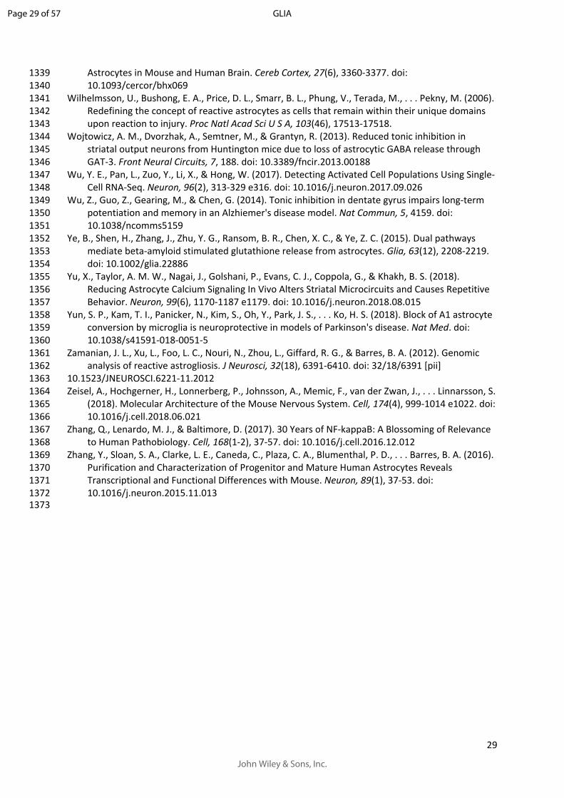
29
1339 Astrocytes in Mouse and Human Brain. Cereb Cortex, 27(6), 3360-3377. doi: 1340 10.1093/cercor/bhx0691341 Wilhelmsson, U., Bushong, E. A., Price, D. L., Smarr, B. L., Phung, V., Terada, M., . . . Pekny, M. (2006). 1342 Redefining the concept of reactive astrocytes as cells that remain within their unique domains 1343 upon reaction to injury. Proc Natl Acad Sci U S A, 103(46), 17513-17518. 1344 Wojtowicz, A. M., Dvorzhak, A., Semtner, M., & Grantyn, R. (2013). Reduced tonic inhibition in 1345 striatal output neurons from Huntington mice due to loss of astrocytic GABA release through 1346 GAT-3. Front Neural Circuits, 7, 188. doi: 10.3389/fncir.2013.001881347 Wu, Y. E., Pan, L., Zuo, Y., Li, X., & Hong, W. (2017). Detecting Activated Cell Populations Using Single-1348 Cell RNA-Seq. Neuron, 96(2), 313-329 e316. doi: 10.1016/j.neuron.2017.09.0261349 Wu, Z., Guo, Z., Gearing, M., & Chen, G. (2014). Tonic inhibition in dentate gyrus impairs long-term 1350 potentiation and memory in an Alzhiemer's disease model. Nat Commun, 5, 4159. doi: 1351 10.1038/ncomms51591352 Ye, B., Shen, H., Zhang, J., Zhu, Y. G., Ransom, B. R., Chen, X. C., & Ye, Z. C. (2015). Dual pathways 1353 mediate beta-amyloid stimulated glutathione release from astrocytes. Glia, 63(12), 2208-2219. 1354 doi: 10.1002/glia.228861355 Yu, X., Taylor, A. M. W., Nagai, J., Golshani, P., Evans, C. J., Coppola, G., & Khakh, B. S. (2018). 1356 Reducing Astrocyte Calcium Signaling In Vivo Alters Striatal Microcircuits and Causes Repetitive 1357 Behavior. Neuron, 99(6), 1170-1187 e1179. doi: 10.1016/j.neuron.2018.08.0151358 Yun, S. P., Kam, T. I., Panicker, N., Kim, S., Oh, Y., Park, J. S., . . . Ko, H. S. (2018). Block of A1 astrocyte 1359 conversion by microglia is neuroprotective in models of Parkinson's disease. Nat Med. doi: 1360 10.1038/s41591-018-0051-51361 Zamanian, J. L., Xu, L., Foo, L. C., Nouri, N., Zhou, L., Giffard, R. G., & Barres, B. A. (2012). Genomic 1362 analysis of reactive astrogliosis. J Neurosci, 32(18), 6391-6410. doi: 32/18/6391 [pii]1363 10.1523/JNEUROSCI.6221-11.20121364 Zeisel, A., Hochgerner, H., Lonnerberg, P., Johnsson, A., Memic, F., van der Zwan, J., . . . Linnarsson, S. 1365 (2018). Molecular Architecture of the Mouse Nervous System. Cell, 174(4), 999-1014 e1022. doi: 1366 10.1016/j.cell.2018.06.0211367 Zhang, Q., Lenardo, M. J., & Baltimore, D. (2017). 30 Years of NF-kappaB: A Blossoming of Relevance 1368 to Human Pathobiology. Cell, 168(1-2), 37-57. doi: 10.1016/j.cell.2016.12.0121369 Zhang, Y., Sloan, S. A., Clarke, L. E., Caneda, C., Plaza, C. A., Blumenthal, P. D., . . . Barres, B. A. (2016). 1370 Purification and Characterization of Progenitor and Mature Human Astrocytes Reveals 1371 Transcriptional and Functional Differences with Mouse. Neuron, 89(1), 37-53. doi: 1372 10.1016/j.neuron.2015.11.0131373
Page 29 of 57
John Wiley & Sons, Inc.
GLIA

30
1374 Figure legends1375 Figure 1. Original drawings of reactive astrocytes in Humans1376 I-II.(P. Chaslin, 1891). Chaslin’s drawings of sclerotic cortical brain sections from an adult epileptic patient, 1377 impregnated by the picro-carmin method. I. This drawing represents a non-lesioned region. A blood vessel (d) 1378 connected by a star-shaped astrocyte (c) is observed, as well as few small nuclei. II. The drawing of a lesioned 1379 tissue shows an accumulation of large bundles of fibers and many nuclei, characterized by Chaslin as “gliotic 1380 tissue”. III-VII.(Ramón y Cajal). Ramon y Cajal’s drawings of cortical (III-VI) and cerebellar (VII) brain sections from 1381 an adult patient suffering from general progressive paralysis, after silver impregnation. III. A reactive astrocyte 1382 with thick branches polarizing towards a blood vessel (a). Thin processes emerge from the other side of the cell 1383 body (b). Microglia cells (e, d) and one neuron (c) are also visible. IV. Large bundles of fibers named “Weigert 1384 fibers” (A), emanating from glial cells, which are barely visible. Note the similitude with Chaslin’s drawing (II). C: 1385 microglia. V. Reactive astrocytes with twisted processes (a, b, c, d) surrounding an infiltration nodule (A). 1386 Degenerating neurons with protein aggregates in their cell bodies are visible (G). D: blood vessels; f: astrocyte 1387 endfeet. VI. Reactive astrocytes with numerous polarized processes have lost their individual, non-overlapping 1388 domains. Astrocyte cell bodies are sometimes in direct contact, suggesting “proliferative astrocyte centers” (A, 1389 C, E), as proposed by Ramon y Cajal. D: blood vessels. VII. Hypertrophic cerebellar astrocytes (B, C, D). Two of 1390 them (C, D) have multinuclear cell bodies, suggesting ongoing cell division. b: blood vessel.
1391
1392 Figure 2. Astrocytes become reactive in a variety of pathological conditions and species.1393 Different methods are used to evidence astrocyte reaction to different diseases, in different species and brain 1394 regions, showing the universality of this response but also some context-specific changes. A. Frontal cortex 1395 mouse brain section following SWI. A gradient of reactive astrocytes (GFAP+, green) is visible around the injury 1396 site. Scale bar: 500 µm (Frik et al., 2018). B. Reactive astrocytes in the cortex of a mouse EAE model. Reactive 1397 astrocytes are labeled with StarTrack multicolor lineage tracing system to identify developmental clones by their 1398 pattern of fluorescent protein expression. Scale bar: 80 µm (Bribian et al., 2018). C. Immunofluorescence for 1399 GFAP and S100β (green), Ki67 (white) and CD31 (blood vessels, red) in the mouse cortex, 5 d after MCAO. Several 1400 reactive astrocytes are proliferating (Ki67+) in the ischemic penumbra (yellow arrowheads) (Frik et al., 2018). D. 1401 3D rendering of the morphology of a normal (blue) and a hypertrophic CNTF-induced reactive astrocyte (brown) 1402 in the mouse striatum. Scale bar: 50 µm (Ligneul et al., 2019). E. A non-reactive (up) and reactive (down) astrocyte 1403 in the mouse cortex, 4 d after cortical lesion. Dye-filling reveals the astrocyte ramified morphology and process 1404 enlargement in reactive astrocytes. Scale bar: 25 µm (Wilhelmsson et al., 2006). F. Immunofluorescence for GFAP 1405 (cyan) and BrdU (red) in the mouse spinal cord after SCI. The border of the lesion core (upper part) presents a 1406 high proportion of BrdU+/GFAP+ proliferative reactive astrocytes forming the glial scar. Scale bar: 25 µm (Wanner 1407 et al., 2013). G. Immunoelectron micrograph of a phagocytic reactive astrocyte in the mouse striatum 3 d after 1408 MCAO. GFAP staining is outlined in blue. Phagocytic inclusions (arrowheads) are visible. Scale bar: 2 µm 1409 (Morizawa et al., 2017). H. Two-photon microscopy of GLAST-eGFP mouse cortex, 28 d after SWI. A GFP+ 1410 astrocyte undergoes cell division (green arrowheads), while some of its processes (yellow arrows) polarize 1411 towards the lesion (yellow star). Scale bar: 20 µm (Bardehle et al., 2013). I. A dextran-filled astrocyte in a rat 1412 transected optic nerve. This reactive astrocyte displays increased caliber of individual fibers and loss of global 1413 complexity. Scale bar: 30 µm (Butt & Colquhoun, 1996). J. Image of 2 adjacent GFP+ astrocytes (white) labeled 1414 with Dil (green) and DiD (red) in the mouse cortex, 1 w after iron injection to induce epilepsy. Their processes 1415 overlap more (box), showing partial loss of domain organization. Scale bar: 20 µm (Oberheim et al., 2008). K. 1416 Reactive astrocytes around an amyloid plaque in the hippocampus of 3xTg-AD mice (GFAP in green and Aβ in 1417 red). Scale bar: 20 µm (Olabarria et al., 2010). L. Reactive astrocytes (GFAP, green), around a vessel overexpress 1418 the chemokine CCL2 (red) in a mouse model of prion disease (ME7), 2 h after TNFα injection in the hippocampus 1419 (Hennessy et al., 2015). M-M’. Electron micrographs of a CNTF-induced reactive (M’) and a control (M) rat 1420 astrocyte. The reactive astrocyte displays an enlarged process filled with bundles of filaments (black star). Scale 1421 bar: 1 µm (Escartin et al., 2006). N. A human perivascular reactive astrocyte (GFAP, brown) close to a 1422 leukoencephalopathy lesion contains phagocytosed LFB+ myelin debris (blue, arrow). Scale bar: 25 µm (Ponath 1423 et al., 2017). O. Triple immunofluorescence for STAT3 (green), GFAP (red), and Huntingtin aggregates (magenta) 1424 in the putamen of a macaque lentiviral model of HD. Reactive astrocytes display STAT3 nuclear labeling. Scale
Page 30 of 57
John Wiley & Sons, Inc.
GLIA

31
1425 bar: 40 µm (L. Ben Haim, Ceyzeriat, et al., 2015). P. Reactive astrocytes around an amyloid plaque in the 1426 hippocampus of an AD patient, visualized by GFAP (black) and Aβ (brown) immunohistochemistry. Note similarity 1427 with image in K. Scale bar: 50 µm (Pike, Cummings, & Cotman, 1995). Q. Electron micrograph showing an 1428 enlarged perivascular reactive astrocyte endfoot enclosing a cortical vessel (V) in the brain of an AD patient. x 1429 7,000 (Wegiel & Wisniewski, 1994a). R. Human cortical reactive astrocytes labeled for GFAP (green) and 1430 phospho-STAT3 (red) around brain metastases from lung cancer (dotted lines). Scale bar: 35 µm (Priego et al., 1431 2018). S-S’. GFAP immunohistochemistry on subtantia nigra pars compacta sections from a control and a 1432 parkinsonian macaque treated with MPTP (C. Barcia et al., 2011). T. Reactive astrocytes in acute lesions of a MS 1433 patient. Triple immunofluorescent staining of reactive astrocytes with GFAP (green), Complement 3 (C3, red) and 1434 activated microglia/macrophages with CD68 (white). Scale bar: 100 µm (Liddelow et al., 2017). U. 1435 Immunofluorescence staining for S100β (green) and in situ hybridization for C3 (red) in an ALS patient. Scale bar: 1436 10 µm (Liddelow et al., 2017). V. 3D rendering of a reactive astrocyte stained with vimentin around human 1437 glioblastoma. Scale bar: 35 µm (C. Barcia, Sr. et al., 2013).
1438
1439 Figure 3. Heterogeneity of reactive astrocytes: causes and manifestations1440 Normal astrocytes are already heterogeneous due to different sources of diversity [listed in blue, see (L. Ben 1441 Haim & Rowitch, 2017), for references]. Upon injury or disease, astrocytes display even more heterogeneity 1442 (listed in brown), depending on the exact molecular trigger (e.g. open versus closed injury, extracellular cytokines 1443 versus intracellular aggregated proteins, aberrant neuronal activity versus cell death), their distance from it 1444 (topography), the exposure time to this stimulus (disease stage) and the signaling cascades activated. The 1445 combination of pre-existing and induced heterogeneity gives rise to potentially very diverse populations of 1446 reactive astrocytes with unique molecular and functional features. However, there are some core changes that 1447 are common to most forms of reactive astrocytes (e.g. GFAP induction, morphological changes, represented in 1448 red).
144914501451 Table legends14521453 Table 1: Reactive astrocytes: definitions, consensus and controversies. 1454 In December 2018 and January 2019, we surveyed 61 researchers actively working in the field of (reactive) 1455 astrocytes with different approaches (molecular, morphological, and functional), on different diseases and 1456 models or in physiology. We asked them the following two questions:14571458 a- What is your definition of astrocyte reactivity, in a few sentences? (Responses listed in Table 1a)1459 b- Do you consider “astrogliosis”, “astrocyte activation” and “astrocyte reactivity” as synonymous terms? 1460 (Responses listed in Table 1b)14611462 We collected 38 replies (see Supplemental Text for full replies) from junior and more senior researchers, from 1463 both genders and 12 countries (see list in Supplemental Table 1). It is not a perfect or unbiased sampling of the 1464 community, but it represents the first attempt to survey different researchers working and publishing on 1465 astrocytes. It shows that some points are consensual while other are more controversial. 1466 Replies were analyzed and grouped under different features or concepts (1st column), addressing different 1467 questions listed in gray boxes above. Respondents, identified by their initials, either agreed to these definitions 1468 (2nd column), agreed and provided further explanations (3rd column) or disagreed (4th column). Pie charts 1469 represent the percentages of responses (green = agree; red = disagree; gray = not mentioned) for features 1470 mentioned by more than 18 respondents or for controversial features.14711472 The definitions given in response to question a are linked to the nomenclature discussed in response to question 1473 b, since several researchers provide specific definitions for each term. For the sake of simplicity, we considered
Page 31 of 57
John Wiley & Sons, Inc.
GLIA
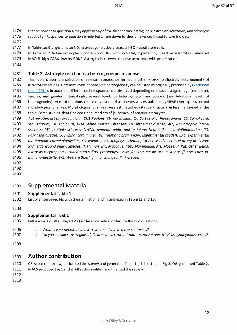
32
1474 that responses to question a may apply to any of the three terms (astrogliosis, astrocyte activation, and astrocyte 1475 reactivity). Responses to question b help better pin down further differences linked to terminology. 14761477 In Table 1a: Glu, glutamate; ND, neurodegenerative diseases; NSC, neural stem cells.1478 In Table 1b: * Active astrocytes = contain proBDNF with no GABA, hypertrophy. Reactive astrocytes = elevated 1479 MAO-B, high GABA, low proBDNF. Astrogliosis = severe reactive astrocyte, with proliferation. 1480
1481 Table 2. Astrocyte reaction is a heterogeneous response 1482 This table presents a selection of relevant studies, performed mostly in vivo, to illustrate heterogeneity of 1483 astrocyte reactions. Different levels of observed heterogeneity can be listed as originally proposed by (Anderson 1484 et al., 2014). In addition, differences in responses are observed depending on disease stage or age (temporal), 1485 species, and gender. Interestingly, several levels of heterogeneity may co-exist (see Additional levels of 1486 heterogeneity). Most of the time, the reactive state of astrocytes was established by GFAP overexpression and 1487 morphological changes. Morphological changes were estimated qualitatively (visual), unless mentioned in the 1488 table. Some studies identified additional markers of (subtypes) of reactive astrocytes.1489 Abbreviation list (by lexical field): CNS Regions: Cb, Cerebellum; Cx, Cortex; Hip, Hippocampus; SC, Spinal cord; 1490 Str, Striatum; Th, Thalamus; WM, White matter. Diseases: AD, Alzheimer disease; ALS, Amyotrophic lateral 1491 sclerosis; MS, multiple sclerosis; NWMI, neonatal white matter injury; Neuroinfla., neuroinflammation; PD, 1492 Parkinson disease; SCI, Spinal cord injury; TBI, traumatic brain injury. Experimental models: EAE, experimental 1493 autoimmune encephalomyelitis; KA, kainate; LPS, lipopolysaccharide; MCAO, Middle cerebral artery occlusion; 1494 SWI, stab wound injury. Species: H, Human; Mc, Macaque; Mm, Mammalian; Ms, Mouse; R, Rat. Other fields: 1495 Astro: astrocytes; CSPG: chondroitin sulfate proteoglycans; IHC/IF, Immuno-histochemistry or -fluorescence; IR, 1496 Immunoreactivity; WB, Western Blotting; =, unchanged; ↗, increase. 149714981499
1500 Supplemental Material1501 Supplemental Table 11502 List of all surveyed PIs with their affiliation and initials used in Table 1a and 1b.
1503
1504 Supplemental Text 11505 Full answers of all surveyed PIs (list by alphabetical order), to the two questions:
1506 a. What is your definition of astrocyte reactivity, in a few sentences?1507 b. Do you consider “astrogliosis”, “astrocyte activation” and “astrocyte reactivity” as synonymous terms?
1508
1509 Author contribution1510 CE wrote the review, performed the survey and generated Table 1a, Table 1b and Fig 3. OG generated Table 2. 1511 MACS produced Fig 1 and 2. All authors edited and finalized the review.15121513
Page 32 of 57
John Wiley & Sons, Inc.
GLIA

Figure 1
Page 33 of 57
John Wiley & Sons, Inc.
GLIA
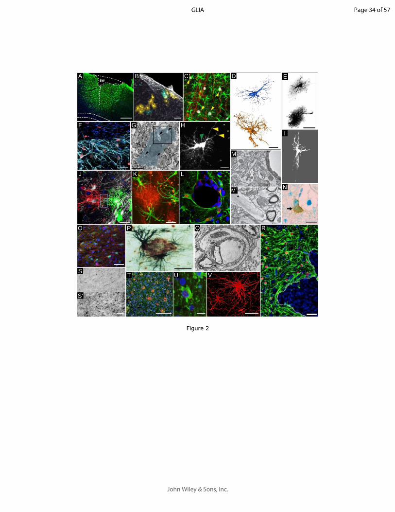
Figure 2
Page 34 of 57
John Wiley & Sons, Inc.
GLIA

Figure 3
180x113mm (300 x 300 DPI)
Page 35 of 57
John Wiley & Sons, Inc.
GLIA

180x206mm (300 x 300 DPI)
Page 36 of 57
John Wiley & Sons, Inc.
GLIA

180x399mm (300 x 300 DPI)
Page 37 of 57
John Wiley & Sons, Inc.
GLIA

180x269mm (300 x 300 DPI)
Page 38 of 57
John Wiley & Sons, Inc.
GLIA

Supplemental text 1
Dr. Nicola Allen 1) Definition: An alteration in astrocyte properties in response to a detrimental insult, for example an infection, a lesion or a stroke. There are core changes that define a reactive astrocyte that happen in response to all insults e.g. upregulation of GFAP, and others that are specific to the type of insult.2) Astrogliosis and astrocyte reactivity are interchangeable; I find astrocyte activation too vague, as it could also apply in a healthy situation where an astrocyte responds to a stimulus for example with an increase in intracellular calcium.
Prof. Alfonso Araque 1) Astrocyte reactivity is a phenotypic alteration manifested as structural changes of astrocyte morphology (e.g., enlargement of cellular processes) and expression regulation of proteins (e.g., GFAP).2) I consider astrogliosis and astrocyte reactivity as synonymous terms. Others would may consider astrocyte activation as synonymous as well. However, as physiologist, I prefer to consider astrocyte activation referred to cellular activity, i.e., astrocyte are activated by signaling molecules such as neurotransmitters, which acting on transmitter receptors activate signaling pathways that regulate cellular activity (e.g., regulating the calcium-based cellular activity that is characteristic of astrocyte physiology).
Prof. Luis Barbeito It's true that the terms we use to describe astrocyte functional or morphological states are somewhat confusing, overlapping and conceived with an "old mind" that must be updated. In my understanding "Reactive astrocytes" preferably refers to a morphological feature of A (hypertrophy, long processes, etc).It also involves GFAP overexpression although this is phenotyping. The definition is linked to the concept of primary or secondary cellular or tissue "damage" or dysfunction, with the potential to induce "activation". Then, reactive astrocytes can be subdivided into multiple subsets by applying immunophenotyping, transcriptome analysis or functional profile, which results in multiple and heterogeneous types of cells. That's the complexity we currently face. While astrocyte reactivity is a morphological feature, astrocyte activation is mostly functional or phenotypic. Could be a link between both but refer to different attributes. Astrogliosis comes from classical neuropathological observations and implies an increased number of astrocytes and hypertrophic.
Prof. Dwight Bergles This sounds like a very worthwhile project, one that will help the field adopt more specific terminology and perhaps spur the development of additional tools to assess phenotypic changes in astrocytes.Although assigning cells to discrete states (e.g. M1 and M2 for microglia and A1 and A2 for astrocytes) is a useful construct and clearly encompasses shared features among cells, I expect that reactivity exists on a continuum, with cells exhibiting different phenotypic changes relative to the timing of the insult (or state of disease) and proximity to other pathology. As we learn more about the diversity among astrocytes, we may find that different regions or different "classes" of astrocytes have different capacities to alter specific pathways associated with a "reactive" status. Likely, such differences will be magnified by age. In addition, some reactive astrocytes that appear in the CNS may also be derived from latent progenitors, rather than through transformation of existing astrocytes.
Page 39 of 57
John Wiley & Sons, Inc.
GLIA

Regarding the terms, I guess I would group these terms together. But, as they are so vague, they don't really carry much value by themselves without additional quantitative descriptors of what criteria were used to assign cells to this category.
Prof. Giorgio Carmignoto 1. I would say that the reactivity of astrocytes, i.e. the change in morphology and function in response to unphysiological situations, such as brain trauma, stroke or diseases, represents a striking example of the high complexity of the astrocytic role in the brain.Reactive astrocytes lose their physiological roles and acquire new actions. Certainly, however, we have not a clear understanding of how these changes develop under pathological conditions and, most importantly, whether and to what extent these astrocytes oppose, or favor, the progression of a specific disease (see Bussian et al . Nature 2018).An important aspect is the calcium signalling that is also affected in reactive astrocytes (in Parkinson, Alzheimer's disease etc….). Given that it regulates the release of gliotransmitters, inflammatory agents etc... its alteration affects the reciprocal communications between astrocytes and other cells, including neurons and microglia. The role of astrocytic calcium signal dysregulations in disease onset and progression remains poorly understood. This is, in my opinion, an important issue that can be adequately addressed in future studies also thanks to genetically encoded calcium indicators.I would add that the "reactive response" of astrocytes is not linked exclusively to chronically altered conditions and it occurs also following episodic dysregulations of neural circuit activities, such as the transient, local hyperactivity of neurons that characterizes epileptic seizures. This strengthens the idea that the "astrocyte reactivity" is not a standardized response and its distinct features (and role) depends on the specific context of the different brain disorders. 2. "Astrogliosis" refers, in general, to the change in astrocyte morphology and the upregulation of GFAP linked to pathological conditions. In contrast, "astrocyte activation" refers to an event that occurs in a physiological context, linked, for example, to a change in calcium signal dynamics or other signaling pathways.The term "astrogliosis" reminds me of the hypertrophy of the astrocytic processes, whereas "astrocyte reactivity" refers more to functional changes. They are not, in my opinion, synonymous terms.
Dr. Colm Cunningham1. I think of astrogliosis as a property of a population while astrocyte reactivity should refer to the activation status of individual cells (since astrocyte phenotype is heterogeneous and may be different even in cells that are relatively close together). "Reactive" as commonly used, suggests that an astrocyte has taken up a different phenotype (i.e. has 'reacted' to some change in its environment) but also implies a propensity to NOW be more reactive than it was previously. This fits with the idea of primed astrocytes as hyperactive to a secondary stimulus (i.e. they are more reactive).2. I think of astrogliosis as representing increased numbers and altered morphology while astrocyte activation simply reflects that they have been in some way activated by pathology or change in the milieu - but this is therefore an ambiguous term - unless one knows what has activated them or what the resulting phenotype is the term 'activated' has no specific meaning. Astrocyte reactivity is probably my favourite because it encompasses how 'reactive' they are: including increased responsiveness to a secondary stimulus.
Prof. Marc Freeman1- I think the phrase astrocyte reactivity can be used in many scenarios. Simply put, I think of it as the set of changes that astrocytes undergo in response to an external stimulus. This is usually studied in the context of “bad” things like injury or disease, and the small things we know about have been lumped in together. For instance, axotomy has been used to study how astrocytes respond to brain trauma. Some of those same responses (e.g. hypertrophy and increased GFAP) also occur in
Page 40 of 57
John Wiley & Sons, Inc.
GLIA

astrocytes in neurodegenerative disease, but I suspect if we performed a much more comprehensive study (like RNA-seq) of how astrocytes respond to axotomy versus neurodegenerative disease that we would see major differences. So I prefer a looser definition, which can include the spectrum of responses that glia exhibit in response to stressful stimuli.2- I think astrogliosis implies that there is some proliferation going on, in addition to changes in gene expression or morphology. The same is not necessarily true for the other terms. I think astrocyte activation and reactivity are fairly interchangeable, and fit with the definition described above.
Prof. Elena Galea 1- The so-called reactive astrocytes are astrocytes undergoing a stress response to tissue damage. Such response is accompanied with: (i) striking morphological change caused by thickening of intermediate filaments composed of GFAP, and (ii) context-specific changes in their transcriptomic profiles, for ‘reactive’ astrocytes are molecularly heterogeneous. As to possible functions, in acute injury, I consider astrocyte reactivity as an adaptive response aimed to isolate the damaged tissue from the healthy one by a scar; the healthy tissue eventually recovers homeostasis with newborn astrocytes from GFAP+ NSC cells. By contrast, in chronic injury astrocyte reactivity might be maladaptive, and render astrocytes chronically dysfunctional with respect to their homeostatic and computational roles. 2. 'Reactive astrocytes' and 'astrogliosis' are equivalent, and they are non-useful terms because they refer to a GFAP-driven morphological change that does not inform about context-specific molecular and functional heterogeneity, and hence it says nothing about the net contribution of such astrocytes to disease pathogenesis. The term 'astrocyte activation' in for physiological contexts, but it might be too vague; if we do not say 'active neurons', we should not say 'active astrocytes' either.
Prof. Vittorio Gallo, PhD 1. Morphological, molecular and functional changes seen in astroglial cells/astrocytes responding to CNS injury and other neurological diseases. 2. No, I don’t consider these as synonymous terms. Astrogliosis involves astrocyte proliferation. Activation and reactivity don’t necessarily involve proliferation. Astrocyte activation could involve intermediate changes that precede a full reactive phenotype.
Prof. Magdalena GötzI guess I would be very simplistic and define a reactive astrocyte as an astrocyte reacting to a major alteration in its environment. I would also add that this is not one state but several (and probably many) states. We have shown for example that in APPPS1 mice where astrocytes are very ‘activated’ (large some, very high GFAP levels) they do not proliferate, while in traumatic and ischemic injuries a subpopulation of them proliferates (see e.g. Sirko et al., 2013, 2015, Frik et al., 2018). So the term reactive astrocytes comprises a) a heterogeneous population and b) different states depending on the alteration in the environment. In regard to your second question I guess each of these terms is not well defined by themselves, so in that sense it is even more difficult to say whether and how much they are overlapping. In ‘colloquial terms’, i.e. in many manuscripts and reviews they are used interchangeable, but by definition they are not.The best suggestion I could come up with is to order them according to the strength/intensity/extend of changes to the homeostatic state – astrocyte activation could be a mild activation, astrocyte reactivity would probably more used for stronger changes and astrogliosis is often used for even stronger stages often accompanied by inflammation. But your review could achieve something very important if you’d suggest more clean definitions.
Page 41 of 57
John Wiley & Sons, Inc.
GLIA
Prof. Philip G HaydonHere is a simple overview of my thoughts. Of course, as you realize, this is a complex process and a few sentences leads to oversimplifications. …1. Reactive astrocytosis is defined and characterized by gross hypertrophy of the cell body and processes of astrocytes in injured and/or diseased areas of the brain. Accompanying these anatomic changes is enhanced expression of, but not limited to, glial cytoskeletal proteins such glial fibrillary acidic protein (GFAP) and vimentin. However, this is not a bistable state - reactive vs non-reactive - but rather there are different shades of grey and significant heterogeneity in astrocyte reactivity. 2- No. I think it is important to clearly define each term being used and use the different terms to discriminate between different states/processes that are on-going in the astrocyte. Too frequently terms are not defined leading to confusion.
Prof. Dr. Helmut Kettenmann1. My definition would be an altered phenotype manifested by morphological alterations, change in
RNA expression pattern and functional parameters due to an alteration in the brain environment related to pathology or injury. This altered phenotype can be quite diverse depending on the type of pathology.
2. I think that these three terms have been used to describe the same event.
Prof. Schuichi Koizumi1- I think that the definition of astrocyte reactivity can be divided into two. The first is to distinguish by form and molecular biological difference, the second is from the aspect of function (beneficial & hazardous etc). In either case, I think that it is necessary for astrocyte to become a state as if it became a different types of cells from normal astrocytes.2- I think that these three words contain the same meaning in part. Astrogliosis means reactive astrocytes, but has a strong implication of becoming a proliferative astrocyte. Astrocyte reactivity and astrocyte activation have almost the same meaning, i.e., they become reactive astrocytes.
Dr. Andras Lakatos1. For me this term encompasses astrocyte responses to any external stimuli, while historically this has been used to describe morphological changes with increases in intermediate filaments in response to injury. It transpires that people still strongly associate this term with a detrimental astrocytic function that is non-permissive for CNS repair. I don’t see the problem of using this term in the right context, but then it should be decorated with adjectives implying the type of pathological or physiological astrocyte state. In the new era of single cell omics it can be further argued if “astrocyte reactivity” should be used as a collective term at all to describe intracellular changes, given the differences in astrocyte responses based on their subpopulation- or region-specific variations in transcription and signalling.2. I regard astrocyte activation and reactivity as interchangeable terms because both words carry the meaning of "response to any stimuli”. However, there is probably more consensus regarding the vast difference between astrocyte activation (reactivity) and the term astrogliosis that latter indicates a cascade of events following activation by pathological stimuli, which may involve at least astrocyte proliferation if not other processes as well, such as activation/influx of various cell types into lesion environments.
Prof. C. Justin Lee NB: […] edited for confidential information about unpublished data
Page 42 of 57
John Wiley & Sons, Inc.
GLIA

I fully agree with you on the fact that reactivity is loosely defined in the past and we need to establish a consensus. …The problem of use of the term, reactive astrocyte is that it is based on the morphological criteria. And traditionally, people have called reactive astrocyte as the one that shows hypertrophy GFAP staining. We have been studying this hypertrophy of astrocytes in depth and we have finally found the molecular mechanism of this hypertrophy (or cause of this hypertrophy). We currently submitted our work, […].
Therefore, my answer to your question #1 is that I would define reactive astrocyte as the one that contain GABA at least in cortical and hippocampal astrocytes. The answer to your question #2 is that the terms “astrogliosis”, “astrocyte activation” and “astrocyte reactivity” cannot be synonymous according to my definition of active and reactive astrocytes. Active astrocytes= contain proBDNF with no GABA. Reactive astrocytes = elevated MAO-B, high GABA, low proBDNF. Astrogliosis= severe reactive astrocyte. […]I don’t think the role of reactive astrocytes in neurodegenerative diseases is “elusive.” The meaning of reactive astrocyte might be elusive, but the role of these GABA positive reactive astrocytes has been clearly laid out in numerous occasions. For example, in Alzheimer’s disease, we and Gong Chen group have clearly demonstrated that the GABA positive reactive astrocytes release GABA to strongly inhibit neighboring neurons to cause learning/memory impairment. Although not published, we have been trying to publish the similar mechanistic story […]In summary, the GABA-containing, GABA-releasing, H2O2-producing reactive astrocytes have critical role in neurodegenerative diseases. Unfortunately, most of these works are not published yet and will be available hopefully soon.
Dr. Shane Liddelow 1. The OED states that 'reactivity' is 'The quality of being reactive or the degree to which something is reactive'. It follows then a 'reactive astrocyte' is any astrocyte that has a FUNCTION that is altered from its normal physiological state, that is, an astrocyte with a function that is altered in response or reaction to some non-physiological setting. There could then be a reactive astrocyte for each individual stimulus (and infinite number of states), or a smaller number of states that collectively respond to a combined number of stimuli.2. Yes. Though I would be less inclined to use 'activation' as it suggests that astrocytes are not normally active in a physiological state - and we know this is untrue. MY personal preference is 'reactivity' but 'gliosis' is similarly useful. Again, I think the most important thing is not so much the nomenclature itself, but what functions are changed (though I do have a pet hate for researchers constantly coming up with their own names for things, rather than sticking with names already in the literature, but adding to the body of knowledge that we have)... but all things must change with time.
Prof. Albee MessingMy first thoughts about a definition of a reactive astrocyte go back to Tolstoy: "All happy families are alike; each unhappy family is unhappy in its own way."But, trying to be more helpful, I’d say my definition is a “change in properties in response to injury or disease, reflected by alterations of morphology, gene expression, and function.” This intentionally excludes anything related to what you might call “normal physiology”. And then your second question of course includes several loaded terms that are difficult to pin down. In my mind astrogliosis fits my definition above most closely – an abnormal state. But obviously astrocytes get “activated” in many normal ways, and in English at least “activation” and “reaction” could both easily apply to normal states, so it gets messy very quickly.
Page 43 of 57
John Wiley & Sons, Inc.
GLIA

I imagine you are contacting me because of Alexander disease. I think the astrocytes here acquire some properties in common with other types of reactive astrocytes, but also have properties that are unique to the disease.
Prof. Keith Murai1. IMO, astrocyte reactivity is the conversion or transformation of the basal state of an astrocyte
to a dynamic, pathological state that normally involves a constellation of structural and molecular changes including astrocytic process hypertrophy and remodeling, upregulation of particular genes (i.e. classical GFAP increase and neuroinflammatory cytokines), activation of specific signaling pathways (i.e. STATs), and potentially re-instatement of cell division (although this is not always the case). However, astrocyte reactivity can differ between brain regions and is influenced by injury type and likely by astrocyte heterogeneity.
2. I consider astrogliosis and astrocyte reactivity as mostly synonymous terms although I tend to think of astrogliosis more in terms of astrocyte reactivity following physical CNS damage (i.e. spinal cord injury) that is accompanied by glial scar formation. Although this may be more related to my past background in spinal cord injury research. I have never used the term 'astrocyte activation'. I reserve the term 'activation' for microglia which has a different time-course and series of cellular changes.
Prof. Christopher Norris I’ll answer both of these together.I think astrocyte reactivity, astrocyte activation, and astrogliosis can overlap, but are not the same things. To me, astrocyte reactivity is an initial (primarily biochemical) response to some form of injury or degeneration. The reaction may vary depending on the area of the brain, the cytoarchitectural context (e.g. gray vs white matter, surrounding neuron subtypes), and perhaps the nature of the insult. For instance, a reactive astrocyte may release trophic factors, or ROs; may adjust its processes around synapses or blood vessels; and/or may adjust the connectivity of its gap junction network.The distinction between reactive and activated astrocytes is harder to articulate (at least for me). I think that once an astrocyte reacts to an injury it can undergo a phenotypic transformation involving induction of unique transcriptional programs, functional changes, and structural changes (e.g. GFAP induction). So, to me, “astrocyte activation” describes the post-phenotypic transformation of reactive astrocytes (which may also vary in nature depending on area of the brain, context, …etc). Astrocyte activation can be short lived (resolvable) or chronic (unresolved). I’ve always thought of astrogliosis as the proliferation of astrocytes, which may persist in an activated state.
Dr. James O’Callaghan1. Astrocyte reactivity is the homotypic response of astrocytes to elaborate gene expression and
proteins following all types of neural (neuronal and glial) injury, including disease, trauma, and chemically-induced damage. The hallmark remains the enhanced expression of GFAP (both biochemically and morphologically because astrocyte reactivity is characterized by the accumulation of glial filaments at the EM level and GFAP remains the major protein component of these filaments, therefore, by definition, if you accumulate glial filaments as a signature of astrocyte reactivity you will accumulate GFAP at immunohisto and immunoassay level. Yes, you can argue about subtypes of GFAP positive astrocytes based on a variety of gene expression and morphological criteria…but by and large they have to be GFAP positive to be an astrocyte and respond with an increase from all types of damage.
2. Yes, as long as these 3 terms refer to responses of astrocytes to CNS injury
Page 44 of 57
John Wiley & Sons, Inc.
GLIA
Prof. Seiji Okada I strongly agree that terminology of astrocyte research has highly ambiguous. For me, non-native speaker for English, it is difficult to understand or explain subtle nuances, but in my opinion,1. Astrocyte reactivity is a cellular response to several kinds of environmental changes for the homeostasis maintenance or tissue repair.
2. I consider “astrocyte activation” and “astrocyte reactivity” as almost same, both term are for the cellular state or ability. Activated or reactive astrocytes can revert to the un-activated or normal state. However, astrogliosis is the “process” of glial scar formation. Glial car is a basically paramagnet, stable tenacious tissue to seclude the injury site from healthy tissue. This process is crucial for the tissue repair after the CNS insult (Please refer Okada et al., Nat Med, 2006).
Dr. Stéphane H.R. Oliet It is a complex question. I am not a specialist on astrocyte reactivity but I think that it does not necessarily imply astrogliosis. Tome, astroglial reactivity, which has become "astroglial activation" for some researchers because they do not like the “reactive” term, corresponds to a rather radical change in the morphology and biochemistry of astrocytes, which may or may not be accompanied by gliosis. This change occurs in response to a stress / stimulation, often of pathological type.But the main mistake would be to think that this reactivity is the only mode of "activation" of astrocytes. A gain or loss of function of these cells can be achieved without reactivity and most of their physiological functions occur in a non-reactive state.
Dr. Aude Panatier 1- Astrocyte reactivity is a modification of the “physiological state” of the astrocyte that will “allow” (or induce) it to integrate and in turn regulate differently the information.It is going from a modification of gene transcription to a modification of astrocyte morphology, and its functional interaction with, blood vessels, synapses and neighboring cells.2- For me “astrogliosis” implicates an increase of the astrocytic domain, which is probably not really the case. It can be the ultimate state of "astrocyte reactivity".So astrogliosis is not always synonymous of astrocyte reactivity.Regarding “astrocyte activation”, for me yes, it can be synonymous of “astrocyte reactivity”.
Prof. Luc Pellerin1. Astrocyte reactivity is closely linked to the notion of phenotypic change. Astrocytes undergo modifications to move from a "resting" state to a "reactive" state. The classical markers of such a phenotypic change are 1) Altered morphology with a thickening of processes 2) enhanced expression of characteristic proteins, GFAP being the gold standard. The appearance of this new phenotype has been observed in several pathological situations and often associated with the notion of inflammation. The presence of cytokines, either produced by reactive astrocytes or acting on astrocytes to induce phenotypic change is characteristic of such an inflammatory state. The notion of proliferation often associated with reactive astrocytes in the past is controversial and contested. Not much is known about the functional consequences of such a phenotypic change although it is likely that it will modify several aspects of their numerous features (e.g. metabolic, trophic, signalling, etc). 2. I do not consider "astrogliosis", "astrocyte activation" and "astrocyte reactivity" completely equivalent. This is particularly true for "astrogliosis".Astrogliosis contains the notion of proliferation, in addition to reactivity
Page 45 of 57
John Wiley & Sons, Inc.
GLIA

Astrocyte activation is the passage from resting astrocyte to reactive astrocyteAstrocyte reactivity is the capacity to become as well as the state of reactive astrocytes
Dr. Gertrudis Perea1. It would be the physiological changes happening into astrocytes triggered by stimuli of different nature. The main changes are typically related with GFAP expression (extension and thickness of the processes); however, other changes take place in shorter time periods, such as Ca2+ signaling, glutamate/GABA uptake, K+ buffering, gap-junction coupling, which contribute to transiently alter the function of astrocytes over the neuronal circuits.2- No, I consider that "astrocyte activation" and "astrocyte reactivity" may represent the physiological response to astrocytes to normal (activation) and abnormal (reactivity) neuronal activity, represented by neurotransmitter release, K buffering, pH measurements, glucose/metabolic state, etc.On the other hand, astrogliosis could be considered as a new physiological and permanent state after neuronal/brain insult.
Prof. Dr. Gabor Petzold1- Astrocyte reactivity is an umbrella term for the signaling mechanisms employed by astrocytes in response to acute or sustained pathological stimuli. The main component of this response can be molecular, functional or morphological, or a combination of those. Moreover, this astroglial phenotype is not only a reaction to brain pathology, but also an important contributor to neuronal regeneration and survival. Whether reactive astrocytes amplify or ameliorate neurodegeneration likely depends on the nature and duration of the stimulus, as well as the presence of co-morbidities and the contribution of other cell types.2- Surprisingly, although changes of astrocytes to brain injury have been known for over a century, the terms describing these alterations have remained highly subjective and ill-defined. To me, these terms describe points on a continuous spectrum. "Astrocyte activation” describes any change in astrocyte function, e.g. molecular or functional, without necessarily implying a pathological event or morphological changes. “Astrocyte reactivity” implies that the stimulus is strong enough to perturb brain homoeostasis, evoking a functional and morphological reaction of astrocytes. “Astrogliosis” is the strongest reaction of astrocytes, and implies morphological changes in astrocytes that result in varying degrees of gliotic scar formation.
Dr. Frank W. Pfrieger1. Reactivity is the ensemble of changes in astrocytes that are evoked by a pathologic condition (genetic, injury etc.)2. No. Astrogliosis or gliosis is a bit ill-defined, but it may mean the same. Astrocyte activation does not necessarily require a pathology.
Dr. Stefanie Robel1. My definition for astrocyte reactivity is pretty broad: Any changes to astrocyte morphology, transcriptome, proteome or function. This can be induced by acute injury, neurological disease or aging. 2. I consider astrocyte activation to be different from astrocyte reactivity. Activity to me means that an astrocyte domain or subdomain is functionally active, for example in response to neuronal activity. I use astrogliosis and astrocyte reactivity synonymous.
Page 46 of 57
John Wiley & Sons, Inc.
GLIA

Prof. David Rowitch1- I consider reactive astrocytes to be defined by marker expression (eg, GFAP) in gray and white matter as well as morphology. They can have both damaging and beneficial functions in the context of injury. There in fact might be heterogeneity in RA across regions of the brain, but this has not been sufficiently defined. 2- No, I consider astrogliosis to be a clinical term signifying brain injury. Activation and reactivity could mean more subtle changes seen in response to cytokines or intrinsic cell death of neighboring cells. But, all these terms are rather non-specific and confusing!
Dr. Alberto Serrano-Pozo “Astrocyte reactivity” = the potential or capacity of astrocytes to mount an astrocyte reaction. Astrocyte reaction (or response) is a change in astrocyte phenotype that is triggered by external noxious stimuli in pathological brain conditions and/or by normal aging, involves the turning on and/or off of specific gene expression programs depending on the trigger, leading to functional changes, and may or not be accompanied by morphological changes and proliferation.“Astrogliosis”, “astrocyte activation” and “astrocyte reactivity” are, in my view, not synonyms and therefore not interchangeable. I believe the term “astrogliosis” should be abandoned because the suffix “-osis” in medicine generally implies a pathological state (i.e. neurosis) and, specifically in pathology, implies proliferation/increased number (i.e. leukocytosis, neurofibromatosis) or invasion/infiltration/spreading (i.e. carcinomatosis, amyloidosis), whereas astrocyte reaction is not necessarily associated with a pathological state (i.e. normal aging) or associated with astrocyte proliferation or migration and invasion. The term “astrocyte activation” suggests that astrocytes can react or “set themselves off” autonomously or for no reason, and fails to recognize that there is usually a trigger of astrocyte reaction (including aging), and that astrocyte reaction may involve inhibition (rather than activation) of some normal functions (i.e. neurotrophic, synaptic). The term ‘reactivity” literally means potential or capacity to react, which is different from the “reaction” itself. Therefore, I prefer the terms “astrocyte reaction” or “astrocyte response” to refer to the results or effects of “astrocyte reactivity”. The concept of “astrocyte reactivity”, devoid of its consequence – the astrocyte reaction -, may fit better with the scenario of normal aging, similar to the concept of “microglia priming”.
Prof. Dr. Swetlana Sirko “astrogliosis”, “astrocyte activation” and “astrocyte reactivity” are related but not synonymous terms. Personally, I prefer to speak about "astrocyte reactivity" as the ability of astroglia to develop an adequate response to a local or systemic pathophysiological stimuli. However, the degree of reactivity (regardless at which level, e.g. transcriptional, morphological, behaviural) is strongly dependent on the type, localization and severity of pathology but also on state of health as well as age of organism. "astrogliosis" as a state of astroglial population associated with chronic, mostly irreversible changes of astrocyte phenotype in context with neuropathologies."astrocyte activation" as a very quick functional adaptation on different physiological (and not necessarily pathological) needs, which can up to a point compensate for natural (or pathological) changes (e.g. activated state of astrocytes in aged brain).In principe, it is like temporal changes in astrocyte state, starting with astrocyte activation (as result of physiological adaptation or exposure to path stimuli) -> astrocyte reactivity (multiple changes in phenotype at different levels)-> astrogliosis (chronic, mostly irreversible changes of astrocyte phenotype). This is, if you want, like determined in the Newton's third law: the action and the reaction are equivalent in magnitude and simultaneous part of an interaction, neither exists without the other.
Page 47 of 57
John Wiley & Sons, Inc.
GLIA

Prof. Michael Sofroniew I would imagine that a definition should be broad and able to encompass different roles and functions. People made have contrasting views about specific things, but the general concept should be inclusive and flexible.
1- ‘Astrocyte reactivity’ is a spectrum of changes in astrocytes that occur in response to all forms and severities of CNS injury and disease. These changes vary with the nature and severity of the insult along a gradated continuum of progressive alterations in molecular expression, progressive cellular hypertrophy and, in severe cases, proliferation and scar formation. The changes associated with astrocyte reactivity are regulated in a context-specific manner by a diverse set of molecular signaling events. The changes undergone by reactive astrocytes have the potential to alter astrocyte activities both through gain and loss of functions that can impact both beneficially and detrimentally on surrounding neural and non-neural cells. Astrocyte reactivity is not an all-or-none response, nor is it a single uniform process, nor is it ubiquitously synonymous with scar formation. Instead, astrocyte is a finely gradated continuum of progressive changes in gene expression and cellular changes that are subtly regulated by complex inter- and intra-cellular signaling mechanisms.
We first proposed and published these definitions 10 years ago because there were no formal modern definitions in the literature at that time (see full quotes from 2 papers below). Sofroniew (2009) Trends Neurosci 32:638-647Sofroniew and Vinters (2010) Acta Neuropathol 119:7-35.In my opinion, the definitions are still valid and I do not know of any new findings that would compel me to change or update them.You mention that there is a lot of variety and contrasting views in the definitions that people have sent you. I think that is interesting and perhaps rather sad that after so many years there is still confusion among ‘experts’… - - It reminds me about the poem of the blind men who argued about their descriptions of the elephant …
2- I consider "astrogliosis" and "astrocyte reactivity" as synonymous and I use them interchangeably. However, I strongly disagree with the use of the term “astrocyte activation” to refer to ‘astrocyte reactivity’. This practice is out of date and is left over from the time when astrocytes in healthy tissue were referred to as “resting”. There is no such thing as a “resting” astrocyte. Astrocytes in healthy tissue continually exhibit physiological activation in the form of transient, ligand-evoked elevations in intracellular calcium ([Ca2+]i) that represent a type of astrocyte excitability, involved in mediating many critical dynamic astrocyte functions, including interactions with synapses and regulation of blood flow as discussed in other articles in this volume. Thus, astrocyte activation can range from physiological contexts in healthy CNS to pathophysiologic contexts that involve responding to CNS injury or disease. In keeping with these definitions, there seems to be no place for the notion of an “activated astrocyte” as a single uniform entity that occurs only in response to injury or disease.(I have published this idea in the article Sofroniew MV (2015) Astrogliosis. Cold Spring Harb Perspect Biol 7:a020420. – the direct quote is below)
Here are some direct quotes from papers:I hope this all helps with your article.
Direct quote on definition of astrocyte reactivity taken from the article Sofroniew MV (2009) Molecular dissection of reactive astrogliosis and glial scar formation. Trends Neurosci 32:638-647.
“In spite of the increasing recognition that astrocytes play central roles in normal CNS function and that reactive astrocytes are primary responders to injury and disease, the concept of reactive astrogliosis seems generally elusive, with no commonly agreed upon definition or model. This article proposes a definition and model of ‘reactive astrogliosis’ that integrates four interdependent
Page 48 of 57
John Wiley & Sons, Inc.
GLIA

key features: (i) reactive astrogliosis is a spectrum of changes in astrocytes that occur in response to all forms and severities of CNS injury and disease including subtle perturbations; (ii) the changes undergone by reactive astrocytes vary with the nature and severity of the insult along a gradated continuum of progressive alterations in molecular expression, progressive cellular hypertrophy and, in severe cases, proliferation and scar formation (Figs. 1,2), (iii) the changes of astrogliosis are regulated in a context-specific manner by specific signaling events that have the potential to modify both the nature and degree of those changes; (iv) the changes undergone during reactive astrogliosis have the potential to alter astrocyte activities both through gain and loss of functions that can impact both beneficially and detrimentally on surrounding neural and non-neural cells.
According to this definition and model, reactive astrogliosis is not an all-or-none response, nor is it a single uniform process, nor is it ubiquitously synonymous with scar formation. Instead, reactive astrogliosis is a finely gradated continuum of progressive changes in gene expression and cellular changes (Figs. 1,2) that are subtly regulated by complex inter- and intra-cellular signaling as discussed in detail below. In its mild and moderate forms, reactive astrogliosis exhibits the potential for resolution if the triggering mechanism has resolved, in which the cells return to an appearance similar to that in healthy tissue (Fig. 2A). The extent to which various molecular changes resolve or persist is not well known. In this context, it is interesting to note that, in healthy tissue, the extensive network of finely branched processes of individual astrocytes occupy contiguous non-overlapping domains 10. In mild-to-moderate reactive astrogliosis, there appears to be preservation of the individual non-overlapping domains of reactive astrocytes in spite of the hypertrophy of the cell body and processes 11 (Fig. 1B, 2A). At its extreme level of activation in response to overt tissue damage and inflammation, reactive astrogliosis involves scar formation that incorporates newly proliferated cells and in which astrocyte processes overlap in manners not seen in healthy tissue 12-14 (Fig. 1C,2B). It deserves mention that astrocytes interact with other cell types, in particular fibromeningeal and other glial cells (such as NG2-positive glia) to form complex glial scars in the CNS 14, 15 (Fig. 2B). The structural changes associated with scar formation are long lasting and persist long after the triggering insult may have resolved (Fig. 1C,2B). The striking potential differences along the continuum of reactive astrocyte responses to insults of different kinds are likely to be of importance when considering the functions and impact of reactive astrogliosis on CNS functions, as discussed below. In other words, reactive astrogliosis of different kinds will have different consequences.”
Direct quote on definition of astrocyte reactivity taken from the article Sofroniew MV, Vinters HV (2010) Astrocytes: biology and pathology. Acta Neuropathol 119:7-35.
Based on a large body of observations in experimental animals, a definition of reactive astrogliosis has recently been proposed [226] that encompasses four key features: (i) reactive astrogliosis is a spectrum of potential molecular, cellular and functional changes in astrocytes that occur in response to all forms and severities of CNS injury and disease including subtle perturbations; (ii) the changes undergone by reactive astrocytes vary with severity of the insult along a gradated continuum of progressive alterations in molecular expression, progressive cellular hypertrophy, and in severe cases, proliferation and scar formation, (iii) the changes of reactive astrogliosis are regulated in a context-specific manner by inter- and intra-cellular signaling molecules; (iv) the changes undergone during reactive astrogliosis have the potential to alter astrocyte activities both through gain and loss of functions that can impact both beneficially and detrimentally on surrounding neural and non-neural cells [226]. Here, we propose to apply and extend this definition to identifying different gradations of reactive astrogliosis and glial scar formation that may be encountered in histopathological examinations of human disorders (Figs. 3,4). According to this definition, reactive astrogliosis is not an all-or-none response, nor is it a single uniform process, nor is it ubiquitously synonymous with scar formation. Instead, reactive astrogliosis is a finely gradated continuum of progressive changes in gene expression and cellular changes. Although the increasing severities of reactive astrogliosis transition seamlessly along a continuum, it is convenient for purposes of description and classification to recognize three broad categories:
Page 49 of 57
John Wiley & Sons, Inc.
GLIA

Mild to moderate reactive astrogliosis. In mild or moderate reactive astrogliosis there is variable up regulation of expression of GFAP and other genes [226], as well as hypertrophy of cell body and processes that can vary in degree but that occurs within the domains of individual astrocytes [257] without substantive intermingling or overlap of processes of neighboring astrocytes or loss of individual domains (Figs. 3b,4b) [226]. There is little or no astrocyte proliferation in mild or moderate reactive astrogliosis, however, the up regulation of GFAP expression in astrocytes that do not express detectable levels of GFAP in healthy tissue (Figs. 3a,4a) can lead to the staining of more cells (Figs. 3b,4b), sometimes giving the false impression of proliferation. Mild or moderate reactive astrogliosis is generally associated with mild non-penetrating and non-contusive trauma, diffuse innate immune activation (viral infections, system bacterial infections) and in areas that are some distance to focal CNS lesions. Because there is little or no reorganization of tissue architecture, if the triggering mechanism is able to resolve, then mild or moderate reactive astrogliosis exhibits the potential for resolution in which the astrocytes return to an appearance similar to that in healthy tissue [226]. The physiological consequences of mild or moderate reactive astrogliosis are not well understood.
Severe diffuse reactive astrogliosis. In severe diffuse reactive astrogliosis there is pronounced up regulation of expression of GFAP and other genes, together with pronounced hypertrophy of cell body and processes, as well as astrocyte proliferation, resulting in considerable extension of processes beyond the previous domains of individual astrocytes. As a result, there is substantive intermingling and overlapping of neighboring astrocyte processes with blurring and disruption of individual astrocyte domains (Figs. 3c,4c). These changes can result in long lasting reorganization of tissue architecture that can extend diffusely over substantive areas without the formation of dense, compact barriers as found in glial scars along borders to necrotic tissue (see below). Severe diffuse reactive astrogliosis is generally found in areas surrounding severe focal lesions, infections or areas responding to chronic neurodegenerative triggers.
Severe reactive astrogliosis with compact glial scar formation. Severe reactive astrogliosis with compact glial scar formation includes changes associated with milder forms, such as pronounced up regulation of GFAP and other genes, and pronounced hypertrophy of cell bodies and processes. In addition, glial scar formation progresses beyond these changes with groups of reactive astrocytes that exhibit pronounced overlapping of reactive astrocyte processes, obliteration of individual astrocyte domains, evidence of substantive astrocyte proliferation and the clear formation of dense, narrow and compact glial scars (Fig. 4d). Recent experimental evidence indicates that these astrocyte scars act as neuroprotective barriers to inflammatory cells and infectious agents, and that they form in particular along borders to severe tissue damage, necrosis, infection or autoimmune-triggered inflammatory infiltration (Fig. 4d) [33, 59, 68, 95, 226, 251]. An important feature of these glial scars is the interaction of reactive astrocytes with other cell types, in particular fibromeningeal and other glial cells (Fig. 4d) [31, 95], and the deposition of a dense collagenous extracellular matrix that contains many molecular cues that inhibit axonal and cellular migration [219]. Triggering insults include penetrating trauma, severe contusive trauma, invasive infections or abscess formation, neoplasm, chronic neurodegeneration, systemically triggered inflammatory challenges. It is noteworthy that the glial scar formation is associated with substantive tissue reorganization and structural changes that are long lasting and persist long after the triggering insult may have resolved.
The findings summarized here show that there are pronounced differences along the continuum of potential responses of reactive astrocyte to insults of different kinds and severities. These differences are likely to be of consequence when considering the functions and impact of reactive astrogliosis on CNS disorders and pathologies.
Direct quote regarding differences between “astrocyte activation” and “astrocyte reactivity” taken from the article Sofroniew MV (2015) Astrogliosis. Cold Spring Harb Perspect Biol 7:a020420.We will not use “activation” or “activated astrocytes” as terms that refer exclusively to astrocyte responses to injury or disease. Astrocytes in healthy tissue continually exhibit physiological activation in the form of transient, ligand-evoked elevations in intracellular calcium ([Ca2+]i) that represent a
Page 50 of 57
John Wiley & Sons, Inc.
GLIA

type of astrocyte excitability, involved in mediating many critical dynamic astrocyte functions, including interactions with synapses and regulation of blood flow as discussed in other articles in this volume. Thus, astrocyte activation can range from physiological contexts in healthy CNS to pathophysiologic contexts that involve responding to CNS injury or disease. In keeping with these definitions, there seems to be no place for the notion of an “activated astrocyte” as a single uniform entity.
Prof. Harald SontheimerFor me reactive astrocytes are:Astrocytes that have undergone both morphological and functional changes as a result of insult or injury typically with the purpose to seal of the location of an injury or disease. Functional changes typically include a global loss proteins involved in ion and neurotransmitter homeostasis and a change in extracellular matrix produced.
Reactive astrocytes and astrogliosis are certainly analogous. The activated astrocytes could refer to astrocytes that show Ca2+ signaling
Prof. Dr. Christian SteinhauserI'm in the astrocyte field since many years, but my impression is that the term 'reactive' astrocyte still lacks a clear definition; everyone understands something different by it.
1. would define an 'reactive' astrocyte as an astrocyte with pathologically altered molecular, functional and/or morphological properties. Given that i) the physiological properties of astrocytes significantly vary during development as well as within and across brain areas (e.g. between hippocampus, cortex, thalamus, Griemsmann et al., 2015, Cereb Cortex), ii) the 'reactive state' usually progresses/worsens with ongoing disease (e.g. in epilepsy, see Bedner et al., 2015, Brain) and ii) in different diseases astrocytes are differently affected, it is clear that there must be a high heterogeneity among 'reactive' astrocytes (certainly not just A1 and A2 astrocytes). Curiously, some colleagues also speak of 'reactive' astrocytes in the physiologically aged brain; I find this usage inappropriate (because an aged brain is not necessarily a diseased brain). 2- 'Astrogliosis' and 'astrocyte reactivity' in my view are synonymous terms. 'Astrocyte activation' of course also occurs in physiology (e.g. when Ca2+ transients are evoked through GPCR activation), therefore the latter term should not be used to describe pathologically altered astrocytes.
Prof. Raymond Swanson 1) I do not think the term reactive astrocyte has any uniform meaning, and I try to avoid the term. My understanding is that it was introduced in the 1800s by pathologists (French, I think) who noted enlarged astrocytes near sites of injury or tumors; hence the term "reactive". It was later found astrocytes with this morphology have increased GFAP, and that later became a defining feature. But, an assumption was made that all cells with these hallmarks are similar to one another and share similar other features. That part is almost certainly wrong; e.g. there are no data that I know of that all cells with reactive morphology have the same changes in gene expression, and lots of reasons to think they do not. This is exactly parallel to the microglia, where the term "activated" pertains to any cell with retracted processes despite the proven fact that the actual gene expression patterns of these cells is very diverse. 2) Yes, I consider them all synonyms, and all equally uninformative.
Prof. Alexej Verkhratsky…indeed in my view we do not have an agreed and proper definition of "reactive astrogliosis";
Page 51 of 57
John Wiley & Sons, Inc.
GLIA

My definition (taken from Verkhratsky A, Zorec R, Parpura V (2017) Stratification of astrocytes in healthy and diseased brain. Brain Pathol 27:629-644 doi:10.1111/bpa.12537) but this was reiterated in many of my papers in last 3-5 years) is:
Reactive astrogliosis is an evolutionary conserved defensive reprogramming of astroglia aimed at: (i) increased neuroprotection and trophic support of nervous tissue; (ii) isolation of the lesioned area; (iii) reconstruction of the damaged blood-brain barrier; and (iv) providing for post-lesion regeneration of brain circuits (10, 177, 178, 232). Activation of astrocytes is a complex process which arguably produces multiple "reactive" phenotypes, which demonstrate disease specificity. Gene expression profiling of reactive astrocytes demonstrated significant context-dependent (ischaemia vs. endotoxin activation) differences (290). All in all, initiation of astrogliotic programme proceeds through a controlled continuum of changes in cellular biochemistry and function that are tuned to the nature and strength of the insult. It is cumbersome and still I believe we may need to expand it even more.
The common definition of astrogliosis as hypertrophy and increase in GFAP/Vim expression is of course trivial and misleading; e.g. GFAP may go up and down in physiological conditions for example in SON in circadian fashion.The recent addition of A1/A2 is welcome as it shows two broad groups of phenotypic outcomes and yet I feel that the reality is much more complex and ether are many more phenotypes which are context, age, disease, comorbidity &c., &c. specific.Finally I do not know what to think about "neurotoxic" phenotypes - obviously evolution never selects for anything detrimental unless it increases survivability at large; so neurotoxic forms might also have positive defensive meaning; (in a way similarly to scar formation which is definitely protective but could be detrimental in spinal cord injury but evolution does not care about species with broken spine...).(….)yes "astrocyte activation", "astrocyte reactivity" and "astrogliosis" refer to the same process; you also forgot another word frequently used: "astrocytosis"
Prof. Andrea Volterra 1. I don't think we have a defined understanding of what a reactive astrocyte is. Previous work indicated that a major feature of reactive astrocytes was their change in morphology, notably the presence of thicker processes, but this was then understood to be an artefact of GFAP (cytoskeletal) staining. Today reactivity is thought to drive a more global "program" change, such as the return to a more immature and plastic state of the astrocyte, accompanied by a large change in its gene expression profile. Since it is increasingly evident that astrocytes are anatomically and biochemically heterogeneous depending on brain areas and circuits, I suspect gene expression changes related to reactivity will depend also on the specific molecular fingerprint of different astrocytic subpopulations. Likewise, they will depend on the specificities of the local microenvironment surrounding an astrocytic population. Moreover, the events triggering reactivity maybe quite different, from microorganism colonization of brain parenchyma, to vascular accidents, to acute traumatic insults to slow processes of protein aggregation and deposition, etc. I suspect astrocytes will turn out to react differently to any of such different conditions. In synthesis, the combination of the intrinsic features of a given astrocytic population, of its microenvironment and the type of insult will concur to produce different types of reactivity and different functional outcomes.2. I do not personally consider "astrogliosis", "astrocyte activation" and "astrocyte reactivity" as synonymous terms. However, we should have a precise definition for each of these phenomena and I don't think we have. So, this leads many authors to interchange them.
Page 52 of 57
John Wiley & Sons, Inc.
GLIA

Dr. Ina WannerAstrocyte reactivity is the active response of viable astrocytes to mechanical injury, non-neural cell contact or a chemical insult in order to protect CNS tissues adjacent to injuries and launch a systemic immune response. Viable astrocytes undergo reactivity, not wounded or dying astrocytes that release diagnostic biomarkers due to integrity compromise [1, 2]. Reactive astrocytes activate signaling pathways like the JAK-STAT cascade that induce gene expression changes. Results are proliferation, functional and structural astrocyte changes that contribute to the formation of a scar and wound healing [3]. Astrocyte shape changes after traumatic CNS injuries or after mechanical trauma or non-neural cell confrontation in vitro are defined by hypertrophy – displaying thick and elongated processes [4-7]. Injury models show a stereotypical astrocyte choreography of aligned elongated processes that wind around microglia or meningeal fibroblasts in co-cultures and that form borders around tears from mechanical injury inflicted by pressure-pulse stretching [4, 5, 7]. Perilesional reactive astrocytes in the injured spinal cord are elongated and associate with each other to bundles that corral non-neural cells and prevent their infiltration. This astrocyte-astrocyte interaction forms a scaffold that establishes the scar border around the lesion limiting its expansion [3, 7]. Astrocyte activation during scar formation is captured by astrocyte-specific protein synthesis after mouse spinal cord injury [8]. Major changes were in protein groups of the biological processes, the JAK-STAT cascade, inflammation, NO biosynthesis, calcium ion transport, gliogenesis and angiogenesis; and of the cellular components, cell adhesion, vesicle and extracellular matrix. These essential protein groups mediate scar assembly and are aimed at re-establishing the neuro-vascular unit and ion homeostasis. Interestingly, quantitative proteomic analysis of in vitro matured human trauma-reactive neocortical astrocytes resulted in changes of the same essential protein groups [manuscript in preparation, [5]. These findings are supported by literature analysis grouping most frequently reported astrocyte protein changes after spinal cord injury in a survey of over 200 publications in the last 30 years. Astrogliosis, astrocyte reactivity and activation are essentially describing similar astroglial processes. In the literature the term “astrogliosis” associates more frequently with histopathology, while the term “astrocyte activation” associates more frequently with expression changes. The term “astrocyte reactivity” is applied in vivo and in vitro models of astrocyte injury responses. Thus, their heritage and common use differ, yet since structural changes are based on molecular and protein changes, these terms can be seen as part of the same astrocyte response program.
1. Halford, J., et al., New astroglial injury-defined biomarkers for neurotrauma assessment. J Cereb Blood Flow Metab, 2017. 37(10): p. 3278-3299.2. Levine, J., et al., Traumatically injured astrocytes release a proteomic signature modulated by STAT3-dependent cell survival. Glia, 2015.3. McGraw, J., G.W. Hiebert, and J.D. Steeves, Modulating astrogliosis after neurotrauma. J Neurosci Res, 2001. 63(2): p. 109-15.4. Wanner, I.B., et al., A new in vitro model of the glial scar inhibits axon growth. Glia, 2008. 56(15): p. 1691-1709.5. Wanner, I.B., An in vitro trauma model to study rodent and human astrocyte reactivity. Methods Mol Biol, 2012. 814: p. 189-219.6. Montgomery, D.L., Astrocytes: form, functions, and roles in disease. Vet Pathol, 1994. 31(2): p. 145-67.7. Wanner, I.B., et al., Glial scar borders are formed by newly proliferated, elongated astrocytes that interact to corral inflammatory and fibrotic cells via STAT3-dependent mechanisms after spinal cord injury. J Neurosci, 2013. 33(31): p. 12870-86.8. Anderson, M.A., et al., Astrocyte scar formation aids central nervous system axon regeneration. Nature, 2016. 532(7598): p. 195-200.
Page 53 of 57
John Wiley & Sons, Inc.
GLIA

Supplemental Table 1
Name First name Country M/F Initials Affiliation
Allen Nicola USA F NAMolecular Neurobiology Laboratory
The Salk Institute for Biological StudiesLa Jolla, CA, USA
Araque Alfonso USA M AADepartment of Neuroscience
University of MinnesotaMinneapolis, MN, USA
Barbeito Luis Uruguay M LB Institut Pasteur of MontevideoMontevideo, URUGUAY
Bergles Dwight USA M DB
Kavli Neuroscience Discovery InstituteThe Solomon H. Snyder Department of
NeuroscienceJohns Hopkins University School of
MedicineBaltimore, MD, USA
Carmignoto Giorgio Italy M GC
Instituto di NeuroscienzeConsiglio Nazionale delle Ricerche
Dip. Scienze BiomedicheUniversità di Padova
Padova, ITALY
Cunningham Colm Ireland M CC
School of Biochemistry and ImmunologyTrinity Biomedical Sciences Institute &
Trinity College Institute of Neuroscience.Trinity College Dublin, IRELAND
Freeman Marc USA M MFVollum Institute
Oregon Health & Science UniversityPortland, OR, USA
Galea Elena Spain F EG
ICREA Institute of NeurosciencesUnitat de Bioquímica
Universitat Autònoma de BarcelonaBellaterra
Barcelona, SPAIN
Gallo Vittorio USA M VG
Children’s Research InstituteWolf-Pack Chair in Neuroscience
George Washington University School of Medicine and Health Sciences
Washington D.C. USA
Götz Magdalena Germany F MG
Ludwig-Maximilians-Universität MünchenDepartment of Physiological Genomics
Planegg-MartinsriedGERMANY
Haydon Philip USA M PH
Department of NeuroscienceTufts Neuroscience Institute
Tufts University School of MedicineBoston, MA, USA
Kettenmann Helmut Germany M HKMax Delbrück Center for Molecular
MedicineBerlin, GERMANY
Koizumi Schuichi Japan M SKDept Neuropharmacology
Interdisciplinary Graduate School of Medecine
Page 54 of 57
John Wiley & Sons, Inc.
GLIA

Univ YamanashiYamanashi, JAPAN
Lakatos Andras UK M AL
Department of Clinical Neurosciences, University of Cambridge & Wellcome-MRC
Cambridge Stem Cell InstituteDepartment of Neurology, Addenbrooke’s Hospital, Cambridge University HospitalsJohn van Geest Centre for Brain Repair,
Cambridge Biomedical Campus, E.D. Adrian Building, Cambridge, UK
Lee C. Justin Korea M CJL
Center for Glia-Neuron InteractionCenter for Neuroscience and Functional
Connectomics Seoul Korea Institute of Science and
Technology (KIST)Seoul, SOUTH KOREA
Liddelow Shane USA M SL
Neuroscience Institute Department of Neuroscience and Physiology
New York University Langone New York, USA
Messing Albee USA M AMWaisman Center
University of Wisconsin-MadisonMadison, WI USA
Murai Keith Canada M KM
Centre for Research in NeuroscienceResearch Institute of the McGill University
Health CentreMcGill University
Montréal, CANADA
Norris Christopher USA M CN
Sanders-Brown Center on Aging& Department of Pharmacology and
Nutritional SciencesUniversity of Kentucky College of Medicine
Lexington, KY, USA.
O'Callaghan James USA M JOCU.S. Centers for Disease Control and
Prevention, Health Effects Laboratory Division, Morgantown, WV, USA
Okada Seiji Japan M SO
Dept.of Immunobiology and NeuroscienceMedical Institute of Bioregulation, Kyushu
UniversityFukuoka, JAPAN
Oliet Stéphane H.R. France M SHRO Neurocentre Magendie
Bordeaux, FRANCE
Panatier Aude France F AP Neurocentre Magendie Bordeaux, France
Pellerin Luc Switzerland M LP Département de PhysiologieLausanne SWITZERLAND
Perea Gertrudis Spain F GePNeuron-Glia Networks Laboratory
Instituto Cajal (CSIC)Madrid, SPAIN
Petzold Gabor Germany M GaP
Neurovascular Diseases LabGerman Center for Neurodegenerative
Diseases (DZNE)Bonn, GERMANY
Pfrieger Frank France M FP Institute of Cellular and Integrative Neurosciences
Page 55 of 57
John Wiley & Sons, Inc.
GLIA

CNRS UPR 3212 Strasbourg, France
Robel Stefanie USA F SR
VA Tech Carilion Research InstituteVT School of Neuroscience
VT Department of Biological SciencesRoanoke, VA, USA
Rowitch David UK M DR
Wellcome-MRC Cambridge Stem Cell Institute
University of CambridgeCambridge, UK
Serrano-Pozo Alberto USA M ASP
Department of Neurology, Massachusetts General Hospital,
Harvard Medical School, Boston, USA
Sirko Swetlana Germany F SS
Physiological Genomics Biomedical Center,Ludwig-Maximilians-University
Grosshaderner Strasse 9Planegg-Martinsried. GERMANY
Sofroniew Michael USA M MSDepartment of Neurobiology
David Geffen School of Medicine at UCLALos Angeles, CA, USA
Sontheimer Harald USA M HS
Virginia Tech School of NeuroscienceCenter for Glial Biology in Health, Disease &
CancerVirginia Tech Carilion Research Institute.
Roanoke, VA, USA
Steinhauser Christian Germany M CSInstitute of Cellular Neurosciences University of Bonn, Medical School
Bonn, GERMANY
Swanson Raymond USA M RS
Department of Neurology, University of California at San Francisco
Neurology ServiceSan Francisco Veterans Affairs Medical
Center San Francisco, CA, USA
Verkhratsky Alexei UK M AVeThe University of Manchester
Faculty of Biology, Medicine and HealthManchester, UK
Volterra Andrea Switzerland M AVo
Département de Neurosciences Fondamentales
Faculté de Biologie et de Médecine Université de LausanneLausanne SWITZERLAND
Wanner Ina USA F IW
Semel Institute for Neuroscience & Human Behavior
IDDRCDavid Geffen School of Medicine, UCLA
Los Angeles, CA, USA
Page 56 of 57
John Wiley & Sons, Inc.
GLIA

TOCI
227x102mm (300 x 300 DPI)
Page 57 of 57
John Wiley & Sons, Inc.
GLIA