intermediate filaments of zebrafish retinal and optic nerve astrocytes and müller glia:...
TRANSCRIPT

SHORT REPORT Open Access
Intermediate filaments of zebrafish retinal andoptic nerve astrocytes and Müller glia: differentialdistribution of cytokeratin and GFAPJoseph R Koke*, Amanda L Mosier, Dana M García
Abstract
Background: Optic nerve regeneration (ONR) following injury is a model for central nervous system regeneration.In zebrafish, ONR is rapid - neurites cross the lesion and enter the optic tectum within 7 days; in mammalsregeneration does not take place unless astrocytic reactivity is suppressed. Glial fibrillary acidic protein (GFAP) isused as a marker for retinal and optic nerve astrocytes in both fish and mammals, even though it has long beenknown that astrocytes of optic nerves in many fish, including zebrafish, express cytokeratins and not GFAP. Weused immunofluorescence to localize GFAP and cytokeratin in wild-type zebrafish and transgenic zebrafishexpressing green fluorescent protein (GFP) under control of a GFAP promoter to determine the pattern ofexpression of intermediate filaments in retina and optic nerve.
Findings: GFAP labeling and GFAP gene expression as indicated by GFP fluorescence was found only in the Müllerglial cells of the retina. Within Müller cells, GFP fluorescence filled the entire cell while GFAP labelling was morerestricted in distribution. No GFAP expression was observed in optic nerves. Cytokeratin labeling of astrocytes wasobserved throughout the optic nerve and less intensely in cells in the retinal inner plexiform layer. The retinal innerlimiting membrane was strongly labeled by anti-cytokeratin.
Conclusions: Studies of astrocyte function during ONR in zebrafish cannot solely rely on GFAP as an astrocytemarker or indicator of reactivity. Future studies of ONR in zebrafish should include evaluation of changes incytokeratin expression and localization in the optic nerve.
IntroductionBecause of the accessibility of the optic nerve, opticnerve regeneration (ONR) is often used for studies ofcentral nervous system regeneration. In fish, typified byzebrafish, regeneration of the optic nerve after injury bycrushing or transectioning is rapid with new neuritescrossing the lesion and entering the optic tectum in asfew as 7 days [1]. In mammals, typified by mice, regen-eration does not take place in the absence of specificmolecular interventions and suppression of astrocytereactivity in the optic nerve [2,3] (for a recent review,see [4]).As part of an ongoing study of ONR in zebrafish [5],
we examined intermediate filament (IF) expression ofastrocytes in the zebrafish retina and optic nerve. Many
previous studies have used the type III IF glial fibrillaryacidic protein (GFAP) as a marker for retinal and opticnerve astrocytes, both in fish and mammals, eventhough it has been known for some time that astrocytesof optic nerves in many fish, including zebrafish, expresscytokeratins rather than GFAP [6,7]. A possible excep-tion are astrocytes of goldfish optic nerve, which, asreported by Nona et al[9], appear GFAP positive bothbefore and after optic nerve injury.
MethodsAll animal use protocols were approved by the TexasState University-San Marcos IACUC (approval #0703_0122_07). Wild-type ZDR zebrafish (Danio rerio,Aquatica Tropicals, Plant City, FL) and transgenic zeb-rafish expressing green fluorescent protein (GFP) undercontrol of a GFAP promoter were acclimated to a 12/12hour light/dark cycle for a minimum of 14 days before
* Correspondence: [email protected] of Biology, Texas State University-San Marcos, San Marcos, TX78666, USA
Koke et al. BMC Research Notes 2010, 3:50http://www.biomedcentral.com/1756-0500/3/50
© 2010 Koke et al; licensee BioMed Central Ltd. This is an Open Access article distributed under the terms of the Creative CommonsAttribution License (http://creativecommons.org/licenses/by/2.0), which permits unrestricted use, distribution, and reproduction inany medium, provided the original work is properly cited.
use. The transgenic fish (Tg(gfap:GFP)mi2001[10]), wereobtained from the Zebrafish International ResourceCenter, Eugene, OR. Optic nerve injury was accom-plished as described in Saul et al. (2009) [5]. For immu-nofluorescent localization of GFAP and cytokeratin,entire fish (N = 3 each of ZDR and (Tg(gfap:GFP)mi2001)were fixed overnight in 4% formaldehyde derived byalkaline depolymerization of paraformaldehyde. Thenboth eyes, optic nerves and brain were dissected outintact. Following washing in PBS, the tissue was cryo-protected by incubation in 30% sucrose-PBS until thetissue sank. The intact eyes, optic nerves, chiasma, andbrain were mounted to permit horizontal sectioning,allowing sections to include retinas from both theinjured and contralateral sides, optic nerves, chiasma,and optic tectum of the brain. Sections were cut at 20μm using a Zeiss Microm cryostat, collected on gelatin-coated coverslips and stored at -80°C until use. Immu-nostaining was performed as previously described [11],using anti-GFAP mAB 131-17719 (Molecular Probes,http://www.invitrogen.com) and anti-KRT 18 mAB(Abgent, San Diego, CA) with appropriate second anti-bodies conjugated respectively to TRITC and Cy5. DAPIwas added to the final wash to stain nuclei. Imaging wasperformed using an Olympus FV1000 confocal micro-scopy system and sized for publication using AdobePhotoShop CS3. Each image presented is a z-projectionof 10 optical sections 1.0 μm thick for the 20× objective(NA 0.95) and 0.4 μm thick for the 60× objective (NA1.4). The objective used is indicated in the figurelegends. Figure 1 is a montage of such images.
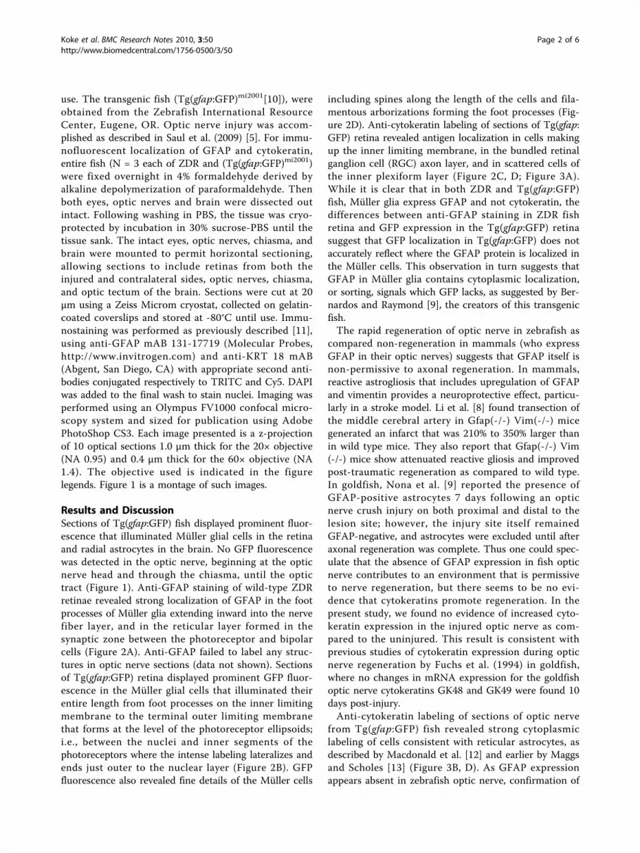
Results and DiscussionSections of Tg(gfap:GFP) fish displayed prominent fluor-escence that illuminated Müller glial cells in the retinaand radial astrocytes in the brain. No GFP fluorescencewas detected in the optic nerve, beginning at the opticnerve head and through the chiasma, until the optictract (Figure 1). Anti-GFAP staining of wild-type ZDRretinae revealed strong localization of GFAP in the footprocesses of Müller glia extending inward into the nervefiber layer, and in the reticular layer formed in thesynaptic zone between the photoreceptor and bipolarcells (Figure 2A). Anti-GFAP failed to label any struc-tures in optic nerve sections (data not shown). Sectionsof Tg(gfap:GFP) retina displayed prominent GFP fluor-escence in the Müller glial cells that illuminated theirentire length from foot processes on the inner limitingmembrane to the terminal outer limiting membranethat forms at the level of the photoreceptor ellipsoids;i.e., between the nuclei and inner segments of thephotoreceptors where the intense labeling lateralizes andends just outer to the nuclear layer (Figure 2B). GFPfluorescence also revealed fine details of the Müller cells
including spines along the length of the cells and fila-mentous arborizations forming the foot processes (Fig-ure 2D). Anti-cytokeratin labeling of sections of Tg(gfap:GFP) retina revealed antigen localization in cells makingup the inner limiting membrane, in the bundled retinalganglion cell (RGC) axon layer, and in scattered cells ofthe inner plexiform layer (Figure 2C, D; Figure 3A).While it is clear that in both ZDR and Tg(gfap:GFP)fish, Müller glia express GFAP and not cytokeratin, thedifferences between anti-GFAP staining in ZDR fishretina and GFP expression in the Tg(gfap:GFP) retinasuggest that GFP localization in Tg(gfap:GFP) does notaccurately reflect where the GFAP protein is localized inthe Müller cells. This observation in turn suggests thatGFAP in Müller glia contains cytoplasmic localization,or sorting, signals which GFP lacks, as suggested by Ber-nardos and Raymond [9], the creators of this transgenicfish.The rapid regeneration of optic nerve in zebrafish as
compared non-regeneration in mammals (who expressGFAP in their optic nerves) suggests that GFAP itself isnon-permissive to axonal regeneration. In mammals,reactive astrogliosis that includes upregulation of GFAPand vimentin provides a neuroprotective effect, particu-larly in a stroke model. Li et al. [8] found transection ofthe middle cerebral artery in Gfap(-/-) Vim(-/-) micegenerated an infarct that was 210% to 350% larger thanin wild type mice. They also report that Gfap(-/-) Vim(-/-) mice show attenuated reactive gliosis and improvedpost-traumatic regeneration as compared to wild type.In goldfish, Nona et al. [9] reported the presence ofGFAP-positive astrocytes 7 days following an opticnerve crush injury on both proximal and distal to thelesion site; however, the injury site itself remainedGFAP-negative, and astrocytes were excluded until afteraxonal regeneration was complete. Thus one could spec-ulate that the absence of GFAP expression in fish opticnerve contributes to an environment that is permissiveto nerve regeneration, but there seems to be no evi-dence that cytokeratins promote regeneration. In thepresent study, we found no evidence of increased cyto-keratin expression in the injured optic nerve as com-pared to the uninjured. This result is consistent withprevious studies of cytokeratin expression during opticnerve regeneration by Fuchs et al. (1994) in goldfish,where no changes in mRNA expression for the goldfishoptic nerve cytokeratins GK48 and GK49 were found 10days post-injury.Anti-cytokeratin labeling of sections of optic nerve
from Tg(gfap:GFP) fish revealed strong cytoplasmiclabeling of cells consistent with reticular astrocytes, asdescribed by Macdonald et al. [12] and earlier by Maggsand Scholes [13] (Figure 3B, D). As GFAP expressionappears absent in zebrafish optic nerve, confirmation of
Koke et al. BMC Research Notes 2010, 3:50http://www.biomedcentral.com/1756-0500/3/50
Page 2 of 6

these cells as astrocytes must depend on morphology,and the pattern of anti-cytokeratin staining seen in Fig-ure 3B is most consistent with the Macdonald et al. [12]description of optic nerve astrocytes and cytokeratin dis-tribution. Neurons would not be expected to label withanti-cytokeratin, and reports of cytokeratin expressionin zebrafish oligodendrocytes are absent from the
literature. Intermediate filaments of mammalian oligo-dendrocytes have been characterized as nestin andvimentin [14]. In contrast to the GFP expressionobserved in Müller glia in retina of Tg(gfap:GFP) fish,no GFP expression was observed in the optic nerves ofthe same fish. At the optic nerve head, a sharp boundarywas present which appeared to exclude GFP-expressing
Figure 1 Montage of images showing Tg(gfap:GFP) and cytokeratin localization in the retinas, optic nerves, and a portion of brainobtained from a fish fixed 24 hours post-optic nerve injury. (20× water immersion, NA 0.95). Prominent GFP expression throughout theMüller glia is visible in retinas of eye associated with the injured optic nerve (injury site, arrow) and the contralateral eye (CL eye), and delimitingwhat appear to be radial glia in a portion of the brain (brain). No GFP expression can be seen in the optic nerve between the retina and optictract. Cytokeratin (magenta) labeling is apparent in the inner limiting membranes and less intensely in the cytoplasm of cells in the innerplexiform layers of the retinas and in the reticular astrocytes of the optic nerve. Blue label is DAPI indicating nuclei. See Figures 2 and 3 forenlarged views of retina and optic nerve.
Koke et al. BMC Research Notes 2010, 3:50http://www.biomedcentral.com/1756-0500/3/50
Page 3 of 6

Müller cells from the optic nerve. Cells showing cytoker-atin labeling in their cytoplasm were found in the opticnerve head, and appeared to extend into the RGC nervefiber layer, retinal inner plexiform layer, and may contri-bute to the inner limiting membrane (Figure 2D, 3C).On the basis of these results, it appears that if Müller
glial cells can be considered astrocytes, zebrafish havetwo populations of astrocytes in their retina, the GFAPexpressing Müller cells, and the cytokeratin expressingreticular astrocytes that appear to extend into the retinafrom the optic nerve, forming the inner limiting
membrane and contributing to the bundled nerve fiberand inner plexiform layers. According to Watanabe andRaff [15], a similar situation exists in mammalian retinawith respect to non-Müller astrocytes entering the retinafrom the optic nerve along retinal vasculature, and in themature retina, locating near the retinal vasculature andnerve fiber layer (although mammalian astrocytes doexpress GFAP and not cytokeratin). Because of theapparent absence of GFAP expression by any cell type inthe zebrafish optic nerve - either injured or uninjured -studies of the role astrocytes may play during ONR in
Figure 2 Images of retina from ZDR (A) and Tg(gfap:GFP) (B, C, D) zebrafish showing localization of anti-GFAP, anti-cytokeratin, andGFP. (A, B, C, 20× water immersion, NA 0.95; D 60× oil immersion, NA 1.4). 2A. An image of retina from a ZDR fish immunostained withanti-GFAP (red) and DAPI (blue). Strong labeling in the foot processes (fp) of Müller glia extending between the RGC nuclei (rgcn) into the innerplexiform layer is apparent. In addition, the Müller glial elements of the outer plexiform layer (opl) are brightly decorated, with fainter labelingextending to the outer limiting membrane. The scale bar represents 55 μm. 2B. Bright GFP fluorescence can be observed in Müller gliaextending from the foot processes (fp) at the inner limiting membrane, through the outer plexiform layer (opl), to the outer limiting membrane(olm). The Müller soma (mgs) are visible among the nuclei of the amacrine cells. Note the distribution of GFAP as indicated by anti-GFAP (Figure2A) is limited to certain regions of the Müller cells and differs from distribution of GFP expressed under a transgene promoter; GFP appears toilluminate all parts of the cell. The scale bar represents 85 μm. 2C. An image of retina from Tg(gfap:GFP) fish has been immunostained for anti-cytokeratin (magenta). As in Figure 2B, GFP fluorescence delineates Müller glia cells from the outer limiting membrane (olm) to the inner limitingmembrane (ilm). Bright anti-cytokeratin labeling can be seen on the inner limiting membrane and in the cytoplasm of cells among the bundledfiber layer (bfl). Less intense cytokeratin labeling is apparent in the inner plexiform layer (ipl). The scale bar represents 70 μm. 2D. Similar toFigure 2C, at higher magnification showing details of the inner segments of the Müller glial cells and anti-cytokeratin labeling of the innerlimiting membrane (ilm). Oblong nuclei characteristic of oligodendrocytes can be seen in the nerve fiber layer. The arrow at lower right indicatesa blood vessel. The scale bar represents 50 μm.
Koke et al. BMC Research Notes 2010, 3:50http://www.biomedcentral.com/1756-0500/3/50
Page 4 of 6

zebrafish cannot rely on GFAP as an marker for astro-cytes or an indicator of reactivity. Future studies of ONRin zebrafish should include evaluation of changes in cyto-keratin expression and localization in the optic nerve.
AcknowledgementsThe authors acknowledge the contributions of undergraduate Kyle Henry,who performed the anti-GFAP labeling. This work was made possible by NSFgrants IOB-0615762 to DMG and DBI-0821252 to JRK and DMG.
Authors’ contributionsAll authors have read and approve the final manuscript. JRK contributed tothe experimental design, supervised the microscopy and prepared the finalimages and manuscript. ALM performed the zebrafish surgeries, dissections,microtechnique and collected the images. DMG conceived and supervisedthe overall project and provided intellectual guidance.
Competing interestsThe authors declare that they have no competing interests.
Received: 5 December 2009 Accepted: 1 March 2010Published: 1 March 2010
Figure 3 Images of retina from Tg(gfap:GFP) (A, B, C) zebrafish showing localization of anti-cytokeratin and GFP (A, 60× oilimmersion, NA 1.4; B, C, 20× water immersion, NA 0.95), and (D) a diagram showing the ribbon structure of zebrafish optic nerve. 3A.A portion of retina labeled with anti-cytokeratin (magenta), DAPI (blue) and GFP. The inner limiting membrane (ilm) appears brightly decoratedwith anti-cytokeratin, which also less intensely labels cytoplasm of cells in the inner plexiform layer (example at arrow). The foot processes (fp)and inner segments of the Müller glia cells are illuminated by GFP and the nuclei of the RGCs (rgcn) by DAPI. The calibration bar represents 45μm. 3B. Image of the optic nerve from the same section as shown in Figures 3A and 3C. Anti-cytokeratin labeling (magenta, arrows) can be seenin the cytoplasm of the optic nerve astrocytes that form the neurolemma of the ribbon-like optic nerve (see Figure 3D). Note the absence ofGFP expression. The calibration bar represents 175 μm. 3C shows a section of retina that includes the optic nerve head, with the dotted linehighlighting the portion of the section where the optic nerve appears exiting the retina. Cells resembling optic nerve astrocytes showing anti-cytokeratin labeled cytoplasm (magenta; example at arrow) appear to stream from the nerve into the retina. Note how the GFP-expressingMüller glial cells appear to form or strongly interact with the physiological cup of the optic nerve head, but then are excluded from the opticnerve itself. The calibration line represents 50 μm. Figure D is a diagram (adapted with permission from Figure 2D, Macdonald et al. [12])illustrating the ribbon nature of the nerve, and the reticular astrocytes (green with red nuclei) forming the neurolemma and extending processesto the nodes (arrow, blue) in the myelin sheath formed by oligodendrocytes (O) on the RGC axons (n). The pattern of anti-cytokeratin stainingseen in Figure 3B is most consistent with the Macdonald et al. [12] description of optic nerve astrocytes and cytokeratin distribution.
Koke et al. BMC Research Notes 2010, 3:50http://www.biomedcentral.com/1756-0500/3/50
Page 5 of 6

References1. Bernhardt RR, Tongiorgi E, Anzini P, Schachner M: Increased expression of
specific recognition molecules by retinal ganglion cells and by opticpathway glia accompanies the successful regeneration of retinal axonsin adult zebrafish. J Comp Neurol 1996, 376:253-264.
2. Cho KS, Chen DF: Promoting Optic Nerve Regeneration in Adult Micewith Pharmaceutical Approach. Neurochem Res 2008, 33(10):2126-2133.
3. Park KK, Liu K, Hu Y, Smith PD, Wang C, Cai B, Xu B, Connolly L, Kramvis L,Sahin M, He Z: Promoting Axon Regeneration in the Adult CNS byModulation of the PTEN/mTOR Pathway. Science 2008, 322:963-966.
4. Garcia DM, Koke JR: Astrocytes as gate-keepers in optic nerveregeneration – A mini-review. Comparative Biochemistry and Physiology,Part A 2009, 152:135-138.
5. Saul KE, Koke JR, Garcia DM: Activating transcription factor 3 (ATF3)expression in the neural retina and optic nerve of zebrafish during opticnerve regeneration. Comp Biochem Physiol A Mol Integr Physiol 2010,155(2010):172-182.
6. Maggs A, Scholes J: Glial domains and nerve fiber patterns in the fishretinotectal pathway. J Neurosci 1986, 6:424-438.
7. Conrad M, Lemb K, Schubert T, Markl J: Biochemical identification andtissue-specific expression patterns of keratins in the zebrafish Daniorerio. Cell Tissue Res 1998, 293:195-205.
8. Li L, Lundkvist A, Andersson D, Wilhelmsson U, Nagai N, Pardo AC, Nodin C,Stahlberg A, Aprico K, Larsson K: Protective role of reactive astrocytes inbrain ischemia.. J Cereb Blood Flow Metab 2008, 28(3):468-481.
9. Nona SN, Thomlinson AM, Bartlett CA, Scholes J: Schwann cells in theregenerating fish optic nerve: evidence that CNS axons, not the glia,determine when myelin formation begins. J Neurocytol 2000, 29:285-300.
10. Bernardos RL, Raymond PA: GFAP transgenic zebrafish. Gene Expr Patterns2006, 6:1007-1013.
11. Weigum SE, Garcia DM, Raabe TD, Christodoulides N, Koke JR: Discretenuclear structures in actively growing neuroblastoma cells are revealedby antibodies raised against phosphorylated neurofilament proteins.BMC Neurosci 2003, 4:6.
12. Macdonald R, Scholes J, Strahle U, Brennan C, Holder N, Brand M,Wilson SW: The Pax protein Noi is required for commissural axonpathway formation in the rostral forebrain. Development 1997,124:2397-2408.
13. Maggs A, Scholes J: Reticular astrocytes in the fish optic nerve: macrogliawith epithelial characteristics form an axially repeated lacework pattern,to which nodes of Ranvier are apposed. J Neurosci 1990, 10:1600-1614.
14. Zerlin M, Levison SW, Goldman JE: Early patterns of migration,morphogenesis, and intermediate filament expression of subventricularzone cells in the postnatal rat forebrain. J Neurosci 1995, 15:7238-7249.
15. Watanabe T, Raff MC: Retinal astrocytes are immigrants from the opticnerve. Nature 1988, 332:834-837.
doi:10.1186/1756-0500-3-50Cite this article as: Koke et al.: Intermediate filaments of zebrafishretinal and optic nerve astrocytes and Müller glia: differentialdistribution of cytokeratin and GFAP. BMC Research Notes 2010 3:50.
Submit your next manuscript to BioMed Centraland take full advantage of:
• Convenient online submission
• Thorough peer review
• No space constraints or color figure charges
• Immediate publication on acceptance
• Inclusion in PubMed, CAS, Scopus and Google Scholar
• Research which is freely available for redistribution
Submit your manuscript at www.biomedcentral.com/submit
Koke et al. BMC Research Notes 2010, 3:50http://www.biomedcentral.com/1756-0500/3/50
Page 6 of 6