gsm 900 mhz exposure of transgenic gfap-mice-a study of histopathology
TRANSCRIPT

Acta Scientiarum Lundensia (open access) Vol. 2012-003 2012-05-15
1
http://www2.msf.lu.se/bpersson/
Volume 2012-003 Citation: (Acta Scientiarum Lundensia) Salford L. G., Brun, A., and Persson, B. R. R., (2012). GSM 900 MHz exposure of Transgenic GFAP-Mice: A study of Histopathology, Tumour induction and BBB leakage, Acta Scientiarum Lundensia, Vol. 2012-003, pp. 1-17, ISSN 1651-5013

Acta Scientiarum Lundensia (open access) Vol. 2012-003 2012-05-15
2
http://www2.msf.lu.se/bpersson/
Research article:
GSM 900 MHz Exposure of Transgenic GFAP-Mice: A study of Histopathology, Tumour induction and BBB leakage
Leif G. Salford1), Arne Brun2), and Bertil R.R. Persson3)
Departments of Neurosurgery1), Neuro Pathology2), Medical Radiation Physics3), Lund University, 22185 Lund Sweden,
Corresponding author: Bertil R.R. Persson, Lund University, Dept. of medical radiation physics, Barngatan 2, S-22185 Lund Sweden E-mail: [email protected]

Acta Scientiarum Lundensia (open access) Vol. 2012-003 2012-05-15
3
http://www2.msf.lu.se/bpersson/
Abstract. In previous work we have studied the effects of electromagnetic fields upon
the promotion of growth of tumours from two different rat glioma cell lines, RG2 and N32, inoculated in the brains of Fischer 344 rats. No promotions of tumour growth due to the EMF could be demonstrated in large series of animals as compared to their matched controls. In the present study we have utilized a newly generated "knock-out" mouse model. By the use of gene targeting, Pekny et al. have created mice deficient for glial fibrillary acidic protein (GFAP), which is an astrocyte-specific protein that forms intermediate filaments in the cytoskeleton of astrocytes. The GFAP-deficient mice utilized by us develop spontaneous tumours, both lymphoblastic (non-Hodgkin) lymphomas and Anaplastic rapidly growing tumours diagnosed as malignant Swannomas. The test system consists of four TEM-cells. A GSM telephone is hooked up to a terminal program on a computer that controls the power and repetition of pulses. The rats were exposed for about 8 hours during 43 days for a total time of 1143 hours.
In male GFAP-mice we found tumours in all 6 controls and in 4 out of 6 exposed. We found, however, no tumours in FM controls and in seven exposed FM mice we fund only 1 tumour. Microwave exposure revealed no significant effect on tumour evolution in the GFAP-mice.
In the CBA-mice microwave exposure gave no significant results of BBB-leakage. But for the CBA-mice BBB-leakage after microwave exposure of CBA-mice was found
probable at a 99% significance-level.
Keywords: GSM, 900 MHz. exposure, GFAP, Transgenic Mice, tumour induction, BBB leakage,

Acta Scientiarum Lundensia (open access) Vol. 2012-003 2012-05-15
4
http://www2.msf.lu.se/bpersson/
Introduction
In earlier work we have studied the effects of electromagnetic fields upon the promotion of growth of tumours from two different rat glioma cell lines, RG2 and N32, inoculated in the brains of Fischer 344 rats. Rats were exposed to 915 MHz microwaves; both continuous wave (CW) and pulse modulated at frequencies 4, 8, 16, and 217 Hz in 0.57 ms pulse lengths. In a previous study of Fischer rats, no promotions of tumour growth due to the EMF could be demonstrated in large series of animals as compared to their matched controls (Salford, et al. 1997).
For study of tumour initiation and promotion, we have utilized a newly generated "knock-out" mouse model (Pekny, M., et al. 1995). By the use of gene targeting, Pekny et al. have created mice deficient for glial-fibrillary-acidic protein (GFAP), which is protein that forms intermediate filaments in the cytoskeleton of the astrocytes (Fuchs and Weber 1994). The disruption of GFAP gene was performed via targeted mutation in E14 embryonic stem cells. Clones were injected into C57BL6J blastocysts, which were subsequently transferred to foster mothers and allowed to develop normally, reach adult- hood and reproduce. However, contrary to normal mice and to other GFAP-deficient mice derived from other strains of ES cells, the GFAP-deficient mice utilized in the present study develop spontaneous tumours, both lymphoblastic (non-Hodgkin) lymphomas and Anaplstic rapidly growing tumours diagnosed as malignant Swannomas (Pekny, M. 1996). A plausible explanation for the tendency to develop tumours could be that a mutation (e.g. during the embryonic stem cell manipulation) took place close to a GFAP locus and then usually segregates with the disrupted GFAP gene. The present study is aimed find out if microwave exposure affects the tendency of tumour induction and BBB-leakage of albumin.
A group of normal CBA mice were exposed for a month to used as normal in comparison with the transgenic GFAP-mice

Acta Scientiarum Lundensia (open access) Vol. 2012-003 2012-05-15
5
http://www2.msf.lu.se/bpersson/
Methods
Microwave exposure
In the present study we expose the whole animal in a TEM-cell with the unique characteristic of having both linear amplitude and phase response versus frequency. Thus it lends itself to extremely broad band sweep frequency testing using a variety of wave forms including CW and pulsed (or modulated) exposure fields. These fields can be accurately generated in the TEM-cell without the distortion that is typically introduced when conventional antennas are used to establish impulse test fields.
Figure 1
The exposure setup with rectangular TEM cell.
The TEM-cell is enclosed in a wooden box that supports the outer conductor and central plate. The outer conductor is made of brass-net and is attached to the inner walls of the box. The centre plate, or septum, is constructed of aluminium and is held up by Teflon braces which are attached at the inner side walls. To allow access to the inside of the cell both ends can be removed. The inside of the cell is ventilated through 18 holes (diam. 18 mm) in the side walls and top of the box and the brass-net allows air to circulate. These holes are also used for examination of the interior during exposure. Probes for monitoring temperature inside the cell or test object are inserted through these holes. The test system consists of four TEM-cells. A GSM telephone is hooked up to a terminal program on a computer that controls the power and repetition of pulses. A power splitter divides the power from the GSM generator into two equal parts that are fed into two of the

Acta Scientiarum Lundensia (open access) Vol. 2012-003 2012-05-15
6
http://www2.msf.lu.se/bpersson/
four cells. The output from the cells is terminated in a 50 Ohms dummy load. Both forward and reflected average powers are measured, with a Bird model 43 power meter, at the inputs and outputs of the cells. The GSM (Global System for Mobile communication) phone used in the present study has a peak output of 2W at the 915 MHz band. With a duty factor of 1/8 (switched "on" one second every eight second) this leads to a time average of 0.25W output leaving the antenna. The average microwave SAR value in the expose mice is in the order of 0.2 W/kg (Malmgren, et al. 2012).
The GFAP-mice were exposed in the TEM Cells for about 8 hours a day during 147 days from November16 1995 to July 9 1996. The total time of exposure was 1143 hours (47.62 days).
The CBA-mice were exposed for about 8 hours a day during 20 days (160 hours).
BBB Albumin leakage
Both controls and exposed animals were sacrificed by perfusion-fixation of the brains under anaesthesia between 20 minutes and 2 hours after the exposure. The brains were perfused with saline for 3-4 minutes, thereafter fixed in 4% formaldehyde for 5-6 minutes and immersion fixed in 4% formaldehyde for more than 24 hours. Whole coronal sections of the brains (3, 7 and 11 mm from the tip of the frontal pole) were dehydrated and embedded in paraffin and sectioned at 5 m. The anaesthesia is necessary to avoid stress and blood pressure rise during perfusion-fixation procedure. Also for ethical reason no animals were sacrificed without anaesthesia.
Albumin was demonstrated with the IgG fraction of rabbit anti rat albumin (Cappel Research Products, Organon Teknika, Västra Frölunda, Sweden) diluted 1:16,000. Fibrinogen was demonstrated with rabbit anti human fibrinogen (Dacopatts AB, Hägersten, Sweden), diluted 1:500. Incubation time for both was over night at 4°C.
Biotinylated swine anti rabbit IgG was used as a secondary antibody. Then avidin, peroxidase conjugated, was coupled to the biotin and visualised with DAB (diaminobenzidine), counterstained with Meyer-HTX (Dacopatt 1994). Standard control procedures were used for both albumin and fibrinogen.
The numbers of immunopositive extravasates were recorded under a microscope. None leakage was rated as normal, occasional minor was rated as 0.5, one leakage was rated 1 and
several leakages was rated 1.5, and 2 with increasing severity.
Statistics
Fisher Exact Probability Test has been applied for a 2x2 Table of Cross-Categorized Frequency Data (Lowry 2012). Freeman-Halton extension of the Fisher exact probability test has been applied for two-rows by four-columns contingency tables (Freeman & Halton 1951).

Acta Scientiarum Lundensia (open access) Vol. 2012-003 2012-05-15
7
http://www2.msf.lu.se/bpersson/
Results
Histopathological examination
Tissue changes found in clinical examination, grossly suspected to represent tumour, proved on histological examination to correspond to lymphoid tissue or inflammatory changes, except in one case. That case was an unexposed cage control with a large tumour in one frontal lobe of the brain with diffuse light positive albumin leakage plus in connection with bleedings at the tumour.
Immunohistochemical methods for albumin detection revealed BBB incompetence and leakage were slightly more enhanced in three cases only and to an insignificant degree or not at all in the majority of cases,
In other cases artifactual positivity were found in connection with tissue damage from slicing the brain for the staining.
In three long term exposure cases there was a diffuse positivity which could theoretically be ascribed to diffusion of earlier leakage which was thus no longer focally peri-vascular. Lack of positivity could also on the same ground be explained by re-sorption of earlier leakage. There is, however, in this group no clear-cut evidence of leakage, old or recent, and thus no sign of persistent vascular damage after exposure.
In the following Tables 1 and 2 is given the protocols of the clinical and pathological examination of all the GFAP mice in the study.

Acta Scientiarum Lundensia (open access) Vol. 2012-003 2012-05-15
8
http://www2.msf.lu.se/bpersson/
Table 1a A study of morphological and histopathological effects of transgenic GFAP-mice, after long term (147 days for a total time of 1143 hours) exposure to GSM 900 MHz at SAR 0.2 W/kg.
Nr Sex Cage Treat- ment
Exposure Time hours
Clinical Exam.
Pathological examination
Score of
Albumin leakage
P8 FM A GSM 185 Paralyzed back-leg
Brain normal with no albumin leakage 0
P9 FM A GSM 1053 No sample -
P10 FM A GSM 1143 Brain normal with no albumin leakage, Spinal cord, kidney, spleen, liver normal. Lymph nodes somewhat plump, Lung separate lymphoid infiltrate
0
P11 FM A GSM 1143 Tumor in intestine
Kidney, spleen normal. Intestine infiltrated with lymphoid tissue. Liver rich on periportal lymphoid infiltrates. Occational lymphoid infiltrates in the lungs
0
P12 FM A GSM 1143 Brain fragmented with several positive albumin leakages, that might be artifacts due to the splitting of the tissue. Normal lung, kidney, spleen and liver.
2?
P13 FM A GSM 1143 Half brain morphologically normal but with positive albumin leakage (rank 1). The kidney shows small multiple lymphoid infiltrates. Spleen rich on pigments that might due to hemolysis. (Iron staining??) Normal liver, intestine, and lung.
1
P14 FM A GSM 1143 Brain normal with no albumin leakage. Lung multiple small lymphoid infiltrates. Normal liver and kidney. Spleen somewhat plump and rich in iron pigments.
0

Acta Scientiarum Lundensia (open access) Vol. 2012-003 2012-05-15
9
http://www2.msf.lu.se/bpersson/
Table 1b A study of morphology and histopathology in unexposed controls of transgenic GFAP-mice.
Nr Sex Cage Clinical
Exam. Pathological examination
Ranking ofAlbumin leakage
P15 FM B Brain artifactually fragmented. Bone implant in basal ganglies. No albumin leakage.
0
P16 FM B Enlarged spleen
Brain partially not adequately perfused with positive vessels but with no signs of albumin leakage. Spleen somewhat plump and rich in iron pigments. (Lymphoma??)
0
P17 FM B Very thin mouse
No sample -
P18 FM B Very fat mouse
Normal brain with no albumin leakage. Normal fat tissue.
0
P19 FM B Half brain rich in vessels with positive albumin leakage (rank 1) that however might be related to the positive vessels. Normal Kidney and lung. Spleen somewhat plump but not as rich in iron pigments as previous mice. Separate scattered giant cells. ”Tumour tissue”: small lymphatic glands and tissue of regular epithelial and glandular cells. Normal pancreas.
1 ?
P20 FM B Half brain with normal structure and no albumin leakage. Perfused only partially. Liver shows small lymphoid infiltrates, somewhat more in the lung but still not significant. Spleen plump with iron pigments as in previous mice. No giant cells. Normal kidney. ”Tumour tissue”: pancreas with small infiltrates. Lymphatic gland.
0
P21 FM B Half brain with normal structure and no albumin leakage but only partially perfused. ”Tumour tissue” in pancreas with separate infiltrates and lymphatic gland. Kidney shows spotty lymphoid infiltrates, some in liver but more pronounced around the bronchial tubes of the lung. These infiltrates might belong to the species? . Spleen plump with iron pigment as in previous mice. No giant cells.
0
P22 FM B Half brain with some infiltrate of lymphoid cells in the meninges but no albumin leakage. A small blood filled cyst in the liver. Small lymphoid infiltrates in the lung. Kidney show large islands of lymphoid infiltrates and”tumour tissue” consist of lymphoid small islands. Spleen plump with iron pigment as in previous mice. No giant cells.
0

Acta Scientiarum Lundensia (open access) Vol. 2012-003 2012-05-15
10
http://www2.msf.lu.se/bpersson/
Table 1c A study of morphological and histopathological effects of transgenic GFAP-mice, after long term (147 days for a total time of 1143 hours) exposure to GSM 900 MHz at SAR 0.2 W/kg.
Nr Sex Bur Treat- ment
Exposure Time hours
Clinical Exam
Pathological examination
Ranking ofAlbumin leakage
P23 M C GSM 1143 Tumour-like tissue at testis
No albumin leakage in the brain 0
P24 M C GSM 1143 Positive albumin leakage in the brain (rank 1) 1
P25 M C GSM 1143 Tumour-like tissue in liver and salivary gland
No albumin leakage in the brain 0
P26 M C GSM 1143 Positive albumin leakage in the brain (rank 1) 1
P27 M C GSM 1143 Tumour in the lung
No albumin leakage in the brain 0
P28 M C GSM 1143 Large tumour on the skin and abdomen
Positive albumin leakage in the brain (rank 1) could be artifact in connection to trauma.
1?
Table 1d A study of morphological and histopathological effects in unexposed controls and cage controls of transgenic GFAP-mice
Nr Sex Cage Control or Cage
Clinical Exam
Pathological examination
Ranking ofAlbumin leakage
P29 M D Control 2 Tumours No albumin leakage in the brain 0
P30 M D Control Tumour in the liver No albumin leakage in the brain 0
P31 M D Control Bleeding in the brain, tumour at the eye
Diffuse positive albumin staining but traumatized or bleeding in one hemisphere
1?
P32 M D Control Tumours along the mesentery
Slight diffuse positive albumin leakage probably from bleeding in lesion. The staining not perfect for evaluation of these damages.
1?
P33 M D Control Subcutaneous tumour
No albumin leakage in the brain 0
P34 M D Control Tumour at intestine and in the prostate
No albumin leakage in the brain 0
P35 M KA Cage Enlarged spleen Positive albumin leakage in the brain (rank 1) partly in connection to damaged area. Poor perfused vessels.
1
P36 M KB Cage ?
P37 KA Cage Enlarged urinary bladder
Positive albumin leakage in the brain (rank 1) 1

Acta Scientiarum Lundensia (open access) Vol. 2012-003 2012-05-15
11
http://www2.msf.lu.se/bpersson/
Table 1d (continued) P38 KB Cage Necrosis in the
frontal brain Large tumour in one frontal lobe. Diffuse slight positive albumin leakage plus 1 in connection with bleedings at the tumour
?
P39 KB Cage Enlarged gland on the neck Tumour?
Extensive albumin leakage (rank 2) but often in connection to tissue damage
2?
P40 M KA Cage Albumin leakage in the brain (rank 1) 1
P41 M KB Cage Tumour in salivary glad
No albumin leakage in the brain 0
P42 M KB Cage No albumin leakage in the brain 0
P43 M KB Cage Tumour in the prostate
Albumin leakage in the brain (rank 1) around a small lesion and diffuse leakage in white matter temporally. Part of cerebellum dislocated to basal brain
1?
Table 2a A study of morphological and histopathological effects in CBA mice, after long term (1 month for a total time of about 160 hours) exposure to GSM 900 MHz at SAR 0.2 W/kg and their controls.
Nr Sex Cage Treat- ment
Exposure Time hours
Clinical Exam.
Pathological examination
Ranking ofAlbumin leakage
M50 FM 0,5 GSM 160 Normal The vessels poorly perfused and positive - White matter paths in striatum light albumin positive, little more than in the white matter
1
M51 FM 0,5 GSM 160 Normal As above but some pathways more positive on albumin staining
1,5
M52 FM 0,5 GSM 160 Normal As above 1,5
M53 FM 0,5 GSM 160 Normal Poorly perfused. Not judge able! -
M54 FM 0,5 GSM 160 Normal No albumin leakage in the brain 0
M55 FM 0,5 GSM 160 Normal Albumin positive (rank 1) 1
M56 FM 0,5 GSM 160 Normal Poorly perfused, albumin positive in connection to damaged section. Path bundles strongly albumin positive.
1
M57 FM 1 Control 0 Normal No albumin leakage in the brain 0
M58 FM 1 Control 0 Normal No albumin leakage in the brain 0
M59 FM 1 Control 0 Normal No albumin leakage in the brain 0
M60 FM 1 Control 0 Normal Albumin positive due to damage. -
M61 FM 1 Control 0 Normal No albumin leakage in the brain 0
M62 FM 1 Control 0 Normal No albumin leakage in the brain 0
M63 FM 1 Control 0 Normal No albumin leakage in the brain 0
Acta Scientiarum Lundensia (open access) Vol. 2012-003 2012-05-15
12
http://www2.msf.lu.se/bpersson/
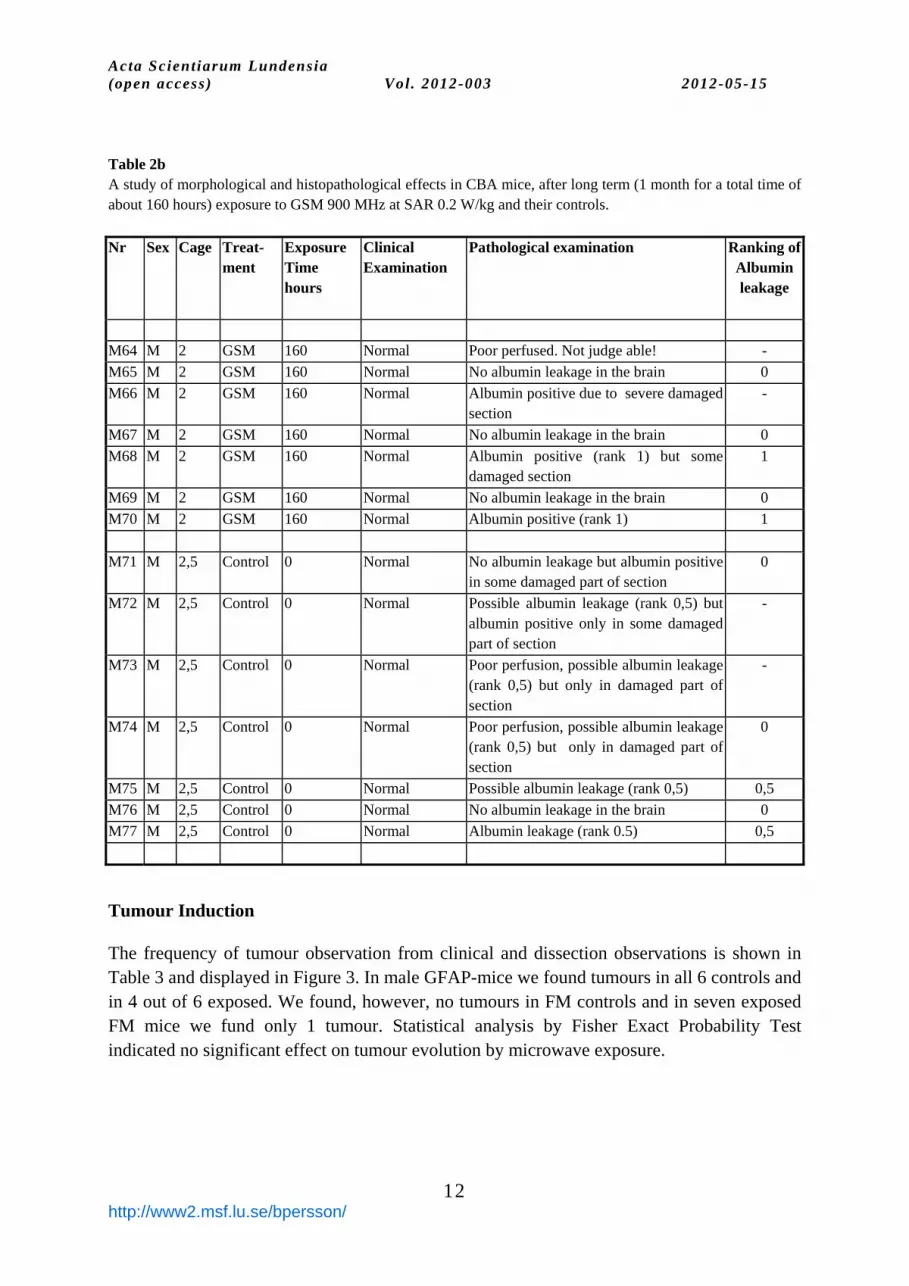
Table 2b A study of morphological and histopathological effects in CBA mice, after long term (1 month for a total time of about 160 hours) exposure to GSM 900 MHz at SAR 0.2 W/kg and their controls.
Nr Sex Cage Treat- ment
Exposure Time hours
Clinical Examination
Pathological examination
Ranking ofAlbumin leakage
M64 M 2 GSM 160 Normal Poor perfused. Not judge able! -
M65 M 2 GSM 160 Normal No albumin leakage in the brain 0
M66 M 2 GSM 160 Normal Albumin positive due to severe damaged section
-
M67 M 2 GSM 160 Normal No albumin leakage in the brain 0
M68 M 2 GSM 160 Normal Albumin positive (rank 1) but some damaged section
1
M69 M 2 GSM 160 Normal No albumin leakage in the brain 0
M70 M 2 GSM 160 Normal Albumin positive (rank 1) 1
M71 M 2,5 Control 0 Normal No albumin leakage but albumin positive in some damaged part of section
0
M72 M 2,5 Control 0 Normal Possible albumin leakage (rank 0,5) but albumin positive only in some damaged part of section
-
M73 M 2,5 Control 0 Normal Poor perfusion, possible albumin leakage (rank 0,5) but only in damaged part of section
-
M74 M 2,5 Control 0 Normal Poor perfusion, possible albumin leakage (rank 0,5) but only in damaged part of section
0
M75 M 2,5 Control 0 Normal Possible albumin leakage (rank 0,5) 0,5
M76 M 2,5 Control 0 Normal No albumin leakage in the brain 0
M77 M 2,5 Control 0 Normal Albumin leakage (rank 0.5) 0,5
Tumour Induction
The frequency of tumour observation from clinical and dissection observations is shown in Table 3 and displayed in Figure 3. In male GFAP-mice we found tumours in all 6 controls and in 4 out of 6 exposed. We found, however, no tumours in FM controls and in seven exposed FM mice we fund only 1 tumour. Statistical analysis by Fisher Exact Probability Test indicated no significant effect on tumour evolution by microwave exposure.

Acta Scientiarum Lundensia (open access) Vol. 2012-003 2012-05-15
13
http://www2.msf.lu.se/bpersson/
Table 3 Tumour evolution in GFAP-mice with and without Microwave exposure. P-values from Fisher test for exposed versus control.
Tumour No Tumour Sum p-1 tail p-2 tail
FM Exposed 1 6 7 0.11 0.11
Control 0 8 8
M Exposed 4 2 6 0.23 0.45
Control 6 0 6
Cage Control 3 5 8
All Control 9 5 14 0.66 1.0
0
20
40
60
80
100
9 / 146 / 64 / 60 / 81 / 7
Per
cent
tum
our
FMEXP FMCON MEXp MCON MALLCON
Figure 3 The frequency of observed tumour from clinical and
histopathological observations.
BBB-leakage of albumin
The results of BBB-leakage of albumin in the GFAP mice are shown in Table 4. Statistical testing considering no sign of leakage versus any sign of leakage in exposed versus control mice revealed no significant effect of increased BBB-leakage of microwave exposure in GFAP mice.
The results of BBB-leakage of albumin in the CBA-mice are shown in Table 5. Statistical testing considering no sign of leakage versus any sign of leakage in exposed versus control mice revealed 1-tail significant effect in of increased BBB-leakage of microwave exposure in CBA-mice. In order to consider all results of ranking as displayed in Table 6 the Freeman-Halton extension of the Fisher exact probability test was allied (Freeman & Halton 1951). This test

Acta Scientiarum Lundensia (open access) Vol. 2012-003 2012-05-15
14
http://www2.msf.lu.se/bpersson/
gave no significant results of BBB-leakage BBB-leakage of microwave exposure in GFAP mice. But for the CBA-mice the probability of the observed array of cell frequencies plus the sum of the probabilities consistent with the observed marginal totals that are equal to or smaller than the probability of the observed array is less than 1%. Thus BBB-leakage after microwave exposure of CBA-mice is probable at a 99% significance level. Table 4. The BBB leakage of albumin in GFAP mice considering No leakage (Ranking = 0) contra leakage (ranking > 0)
No. Mice with Rank Leakage
Number mice
Fisher Exact Probability Test
0 >0 Tot P 1-tail P 2-tail
Exposed 7 5 12 0,2 0.3 Control 6 1 7
Cage 2 5 7 0,2 0,3 Control 4 2 6
Exp 7 5 12 0,6 1 Ctrl+cage 12 8 7
Exp 7 5 12 0,3 0,4 Control 10 3 13
Table 5: The BBB leakage of albumin in CBA-mice considering No leakage (Ranking = 0) contra leakage (ranking > 0).
No. Mice with Rank Leakage
Number mice
Fisher Exact Probability Test
0 >0 Tot P 1-tail P 2-tail
Exposed 4 7 11 0,04 0.08 Control 9 2 11

Acta Scientiarum Lundensia (open access) Vol. 2012-003 2012-05-15
15
http://www2.msf.lu.se/bpersson/
Table 6: The BBB leakage of albumin in CTLA and CBA-mice considering No leakage (Ranking = 0) contra leakage of various ranking (Increasing severity 0;0,5;1;1.5;2) evaluated by using the Freeman-Halton extension of the Fisher exact probability test (Freeman & Halton 1951).
Ranking: 0 0,5 1 1,5 2 Sum *)PA **)PB
GFAP-mice Exposed 7 0 4 0 1 12 0.44 0.33 Control 6 0 1 0 0 7
CBA-mice Exposed 4 0 5 2 0 11 <0.008 <0.005 Control 9 2 0 0 0 11
*)PA = The probability of the observed array of cell frequencies plus the sum of the
probabilities consistent with the observed marginal totals that is equal to or smaller than the probability of the observed array.
**)PB = The probability of the observed array of cell frequencies plus the sum of the probabilities of the observed marginal totals that is smaller than the probability of the observed array.
Note that PA and PB are both non-directional (two-tailed) probabilities.
Discussion and Conclusion
In male GFAP-mice tumours were found in all 6 controls but only in 4 out of the 6 microwave exposed GFAP-mice. In female controls, however, no tumour was found, and in 7 exposed female GFAP-mice only 1 tumour was found. The microwave exposure revealed no significant effect on tumour evolution in the GFAP-mice. In a previous study we performed on Fischer rats with implanted glioma tumours in the brain, we found no effect on tumour growth by long term microwave exposure (Salford, et al. 1997).
In order to mimic the real life situation, with often life-long exposure to the electromagnetic fields emitted by mobile phones, we have investigated in a rat model the effects of repeated exposures under a long period to Global System for Mobile Communication-900 MHz (GSM-900) radiation.
In another study of a total of 56 normal Fischer rats, we exposed 32 once weekly in a 2-h period, for totally 55 weeks, at different average whole-body specific absorption rates (SAR) (of in average 0.6 and 60 mW/kg at the initiation of the experimental period). Sixteen animals were sham exposed and eight animals were cage controls, which never left the animal house. After behavioural tests, 5-7 weeks after the last exposure, the brains were evaluated for histo- pathological alterations such as BBB-leakage of albumin, dark neurons, lipofuscin aggregation and signs of cytoskeletal and neuritic neuronal changes of the type seen in human

Acta Scientiarum Lundensia (open access) Vol. 2012-003 2012-05-15
16
http://www2.msf.lu.se/bpersson/
ageing. In this study, no indications of tumours or significant alteration of any these histopathological parameters was found, when comparing the GSM exposed animals to the sham exposed controls (Grafstrom, et al. 2008).
The results of our studies does not support the indications of an increased risk of developing brain tumours by using mobile phone revealed by epidemiological studies(Cardis, et al. 2011; Hardell, et al. 2011). They rather support the evidences of no association between the use of mobile phone and the risk of brain tumour (Corle, et al. 2012; Xie, et al. 2011).
Other Swedish researchers exposed cultured astroglial and microglial brain cells to 900 MHz Microwave Radiation. Levels of the GFAP, were measured at SAR levels of 27 and 54 W/kg (CW) for 4 or 24 h. No significant differences could be detected at any time and for any of the radiation characteristics. They also exposed Microglial cell cultures to 900 MHz radiation at an SAR of 3 W/kg (mw) for 8 h, and 116 and they appeared to be unaffected by microwave irradiation. Thus the study does not provide evidence for any effect of the microwave radiation on glial cells in culture (Thorlin, et al. 2006).
By applying the Freeman-Halton extension of the Fisher exact probability test (Freeman
& Halton 1951) the results of BBB-leakage of albumin gave no significant results of BBB-leakage microwave exposure in the GFAP mice. But for the CBA-mice BBB-leakage after microwave exposure of CBA-mice was found probable at a 99% significance level. Acute microwave exposure of Fischer rats, however, has in a previous study revealed significant BBB-leakage of albumin (Eberhardt, et al. 2007; Persson, et al. 1999; Persson, et al. 1997; Salford, et al. 2003; Salford, et al. 2001). Acknowledgement: We wish to thank professor Milos Pekny, MD, PhD, at the Institute of Neuroscience and Physiology, University of Gothenburg for giving us the opportunity to use his GFAP mice for this study. We also thank BMA Catarina Blennow and BMA Sussanne Strömblad at the Rausing Laboratory for their excellent technical assistance in the exposure and blood-sampling of all the mice.
References
Cardis E., Varsier, N., Bowman, J. D., Deltour, I., Figuerola, J., Mann, S., Moissonnier, M., Taki, M., Vecchia, P., Villegas, R., Vrijheid, M., Wake, K., and Wiart, J., (2011). Estimation of RF energy absorbed in the brain from mobile phones in the Interphone Study, Occupational and Environmental Medicine, Vol. 68, pp. 686-693, ISSN 1351-0711
Corle C., Makale, M., and Kesari, S., (2012). Cell phones and glioma risk: a review of the evidence, Journal of Neuro-Oncology, Vol. 106, pp. 1-13, ISSN 0167-594X
Eberhardt J., Persson, B. R. R., Malmgren, L., Brun, A., and Salford, L. G., (2007). Blood-brain barrier permeability and nerve cell damage in the rat brain 14 and 28 days after exposure to microwaves from GSM mobile phones Bioelectromagnetics, Vol. (to be submitteed), ISSN
Freeman G. H., and Halton, J. H., (1951). Note on exact treatment of contingency, goodness of fit and other problems of significance, Biometrika, Vol. 39, pp. 141-149, ISSN

Acta Scientiarum Lundensia (open access) Vol. 2012-003 2012-05-15
17
http://www2.msf.lu.se/bpersson/
Grafstrom G., Nittby, H., Brun, A., Malmgren, L., Persson, B. R. R., Salford, L. G., and Eberhardt, J., (2008). Histopathological examinations of rat brains after long-term exposure to GSM-900 mobile phone radiation, Brain Research Bulletin, Vol. 77, pp. 257-263, ISSN 0361-9230
Hardell L., Carlberg, M., and Mild, K. H., (2011). Re-analysis of risk for glioma in relation to mobile telephone use: comparison with the results of the Interphone international case-control study, International Journal of Epidemiology, Vol. 40, pp. 1126-1128, ISSN 0300-5771
Lowry R., (2012). Concepts & Applications of Inferential Statistics. http://vassarstats.net/. Malmgren L., Persson, B. R. R., and Salford, L. G., (2012). SAR-determination in experimental exposure of rats
in a TEM-cell for electromagnetic fields of 915 MHz, Acta Scientiarum Lundensia, Vol. 2012-005, ISSN ISSN: 1651-5013
Pekny M., (1996). Tumour development in GFAP-deficient mice. (Gothenburg). Pekny M., Leveen, P., Pekna, M., Eliasson, C., Berthold, C. H., Westermark, B., and Betsholtz, C., (1995). Mice
lacking Glial Fibrillary Acidic Protein display astrocytes devoid of intermediate filaments but develop and reproduce normally, Embo Journal, Vol. 14, pp. 1590-1598, ISSN 0261-4189
Persson B. R. R., Malmgren, L., Salford, L. G., and Brun, A., (1999). Mobile communications and health: Studies on growth of brain-tumours and on the Blood-Brain Barrier in rats exposed to 900/1800 MHz RF-field, COST 244, Bordeaux, Vol. 19-20 April, ISSN
Persson B. R. R., Salford, L. G., and Brun, A., (1997). Blood-Brain Barrier permeability in rats exposed to electromagnetic fields used in wireless communication, Wireless Networks, Vol. 3, pp. 455-461, ISSN
Salford L. G., Brun, A., Eberhardt, J. L., Malmgren, L., and Persson, B. R. R., (2003). Neuronal damage in mammalian brain of microwaves from GSM mobile telephones, Environmental Heath Perspectives, Vol. 111, pp. 881-883, ISSN
Salford L. G., Brun, A., and Persson, B. R. R., (1997). Brain tumour development in rats exposed to electromagnetic fields used in wireless cellular communication, Wireless Networks, Vol. 3, pp. 463-469, ISSN
Salford L. G., Persson, B. R. R., Malmgren, L., Brun, A., and Lannoye, P., (2001). Telephonie mobile et barrire Sang-cerveau (in french) Mobile Communication and the Blood-Brain Barrier(Marco Pietteur, B-4053 Embourg, Belgique).
Thorlin T., Rouquette, J.-M., Hamnerius, Y., Hansson, E., Persson, M., Bjorklund, U., Rosengren, L., and Ronnback, L., (2006). Exposure of cultured astroglial and microglial brain cells to 900 MHz microwave radiation, Radiation Research, Vol. 166, pp. 409-421, ISSN 0033-7587
Xie Z., He, J., Xie, L., Xie, Z. H., He, J. X., and Xie, L. Y., (2011). Meta analysis of case-control study of mobile phone use and brain tumors, Occupation and Health, Vol. 27, pp. 848-852, ISSN 1004-1257