gfap-cre-mediated transgenic activation of bmi1 results in pituitary tumors
TRANSCRIPT

GFAP-Cre-Mediated Transgenic Activation of Bmi1Results in Pituitary Tumors
Bart A. Westerman1,2, Marleen Blom1, Ellen Tanger1, Martin van der Valk3, Ji-Ying Song3, Marije van
Santen4, Jules Gadiot1, Paulien Cornelissen-Steijger1, John Zevenhoven1, Haydn M. Prosser5,
Anthony Uren1,6, Eleonora Aronica7, Maarten van Lohuizen1,8*
1Division of Molecular Genetics, The Netherlands Cancer Institute, Amsterdam, The Netherlands, 2Department of Oncogenomics, Academic Medical Center, Amsterdam,
The Netherlands, 3Department of Animal-Pathology, The Netherlands Cancer Institute, Amsterdam, The Netherlands, 4Department of Pathology, Academic Medical
Center, Amsterdam, The Netherlands, 5 The Wellcome Trust Sanger Institute, Wellcome Trust Genome Campus, Hinxton, Cambs, United Kingdom, 6Cancer Genomics
group at the MRC Clinical Sciences Centre, London, United Kingdom, 7Department of Neuro-Pathology, Academic Medical Center, Amsterdam, The Netherlands, 8 The
Centre of Biomedical Genetics, Academic Medical Center, Amsterdam, The Netherlands
Abstract
Bmi1 is a member of the polycomb repressive complex 1 and plays different roles during embryonic development,depending on the developmental context. Bmi1 over expression is observed in many types of cancer, including tumors ofastroglial and neural origin. Although genetic depletion of Bmi1 has been described to result in tumor inhibitory effectspartly through INK4A/Arf mediated senescence and apoptosis and also through INK4A/Arf independent effects, it has notbeen proven that Bmi1 can be causally involved in the formation of these tumors. To see whether this is the case, wedeveloped two conditional Bmi1 transgenic models that were crossed with GFAP-Cre mice to activate transgenic expressionin neural and glial lineages. We show here that these mice generate intermediate and anterior lobe pituitary tumors that arepositive for ACTH and beta-endorphin. Combined transgenic expression of Bmi1 together with conditional loss of Rbresulted in pituitary tumors but was insufficient to induce medulloblastoma therefore indicating that the oncogenicfunction of Bmi1 depends on regulation of p16INK4A/Rb rather than on regulation of p19ARF/p53. Human pituitary adenomasshow Bmi1 overexpression in over 50% of the cases, which indicates that Bmi1 could be causally involved in formation ofthese tumors similarly as in our mouse model.
Citation: Westerman BA, Blom M, Tanger E, van der Valk M, Song J-Y, et al. (2012) GFAP-Cre-Mediated Transgenic Activation of Bmi1 Results in PituitaryTumors. PLoS ONE 7(5): e35943. doi:10.1371/journal.pone.0035943
Editor: Alfred Lewin, University of Florida, United States of America
Received April 4, 2011; Accepted March 28, 2012; Published May 4, 2012
Copyright: � 2012 Westerman et al. This is an open-access article distributed under the terms of the Creative Commons Attribution License, which permitsunrestricted use, distribution, and reproduction in any medium, provided the original author and source are credited.
Funding: This research was funded by the Dutch Cancer Society. The funders had no role in study design, data collection and analysis, decision to publish, orpreparation of the manuscript.
Competing Interests: The authors have declared that no competing interests exist.
* E-mail: [email protected]
Introduction
Bmi1 is part of the polycomb repressive complex 1 (PRC1), a
transcriptional repressive complex that silences genes during
embryonic development. PRC1 complexes act through recogni-
tion of H3K27Me3 epigenetic tags on histone 3 [1,2] and through
ubiquitin ligase activity towards K119 on histone 2 [3,4].
Among the Bmi1 target genes are regulators of stem cell self-
renewal, partially by repressing the senescence and apoptosis-
regulating genes p16INK4A and p19Arf [5]. In mice, loss of Bmi1
results in hematopoietic and neural defects leading to an early
death (mostly within 8 weeks). The partial rescue of Bmi1 deletion
by simultaneous deletion of p16INK4A and p19Arf suggested that
p16INK4A/p19Arf independent targets are responsible for the
severe phenotype in mice [6,7]. It was shown that Chk2 deletion
was able to substantially rescue this severe phenotype implicating
mitochondrial function and redox homeostasis as target processes
of Bmi1 [8]. Furthermore, Bmi1 has been shown to repress p21 in
the cerebellum [9] and in the developing forebrain. This latter
repression is mediated through FoxG1 [10,11]. Polycomb proteins
also affect developmental competence of neural precursors during
glial and neural differentiation [12].
In cancer, both p16INK4A and p19Arf dependent as well as
independent oncogenic functions of Bmi1 have been shown [7,13].
The mechanism behind this might be diverse and context
dependent. PRC1 plays a role in DNA double strand break repair
[14]. Furthermore, Bmi1 is required to prevent glial differentiation
in SmoM2 induced medulloblastoma [15], possibly by a similar
mechanism of Ezh2 mediated repression of BMPR1A that affects
JAK/STAT3 mediated glial differentiation in glioma [16]. In
addition, Bmi1 was shown to regulate Twist1 mediated epithelial-
mesenchymal transition [17].
Many reports have shown over expression of Bmi1 in tumors of
astroglial, neural or neuroendocrine origin ([18]; summary in
Table 1). Therefore, it is assumed that the proto-oncogene Bmi1
positively contributes to tumor formation in these cases. However,
a causal role for Bmi1 in the generation of these tumors has not
been demonstrated [19,20]. We show here that Glial fibrillary
acidic protein-Cre (GFAP-Cre) mediated conditional over expres-
sion of Bmi1 generates anterior and intermediate lobe pituitary
tumors. In addition, we show that Bmi1 over expression cannot
substitute for p53 loss in a predisposing genetic model of
medulloblastoma which indicates that the oncogenic function of
PLoS ONE | www.plosone.org 1 May 2012 | Volume 7 | Issue 5 | e35943

Bmi1 is mediated by p16INK4A/Rb mediated effects rather then
through p19ARF/p53 effects.
Materials and Methods
Transgene ModelAll animal experiments have been conducted with approval of
the ethical committee of the Netherlands Cancer Institute under
references 04.003 B21/04.003 B38ext and 04.003 B23/04.003
B39ext. Genetically engineered mice with predispositions leading
to cancer were checked twice every week for the occurrence of
tumors. Animals were immediately sacrificed when a tumor was
detected. All procedures have been conducted according to the
standard operating procedures of the institute.
The cDNA encoding mouse Bmi1 with a downstream internal
ribosome entry site (IRES)-eGFP cassette was cloned in the Rosa
26 targeting construct CEB11. This construct contains three
transcriptional stop (poly A) signals flanked by LoxP sites to enable
removal of this stop cassette upon LoxP-recombination
(Figure 1A). The construct was targeted to the Rosa26 locus by
homologous recombination which was confirmed by Southern blot
analysis (Figure 1A, B). In addition, we generated an indepen-
dent conditional targeting construct with mutant 66/71 LoxP-
recombinase sites enabling reversal of the antisense cloned Bmi1
cDNA (Figure S1A, C and results not shown). The first mouse
model is referred to as Bmi1LSL and the latter as Bmi1Lox66/Lox71.
Detailed information on the transgenes is available at MGI (Mouse
Genome Informatics database, http://www.informatics.jax.org/).
Reference numbers are: Bmi1LSL, MGI:4398910, Gt(ROSA)26-
Sor,tm1(CMV-Bmi1,-EGFP)Nki., also named Bmi-CTS, when
inbred on a FVB background: NKI strain# 1353, when inbred on
a C57BL/6 background: NKI strain# 1656. Bmi1Lox66/Lox71,
MGI:4398914, Gt(ROSA)26Sor,tm2(CMV-Bmi1,EGFP)Nki.,
also named Bmi-CTI, when inbred on a FVB background: NKI
strain# 1148, when inbred on a C57BL/6 background: NKI
strain# 1657.
The Bmi1LSL transgenic strain was subsequently crossed with
Actin-Cre (Acre) strain to analyze the phenotype when Bmi1 is
activated constitutively throughout the mouse. In addition,
transgenic mice were crossed to Glial fibrillary acidic protein
promoter driven Cre (GFAP-Cre or GCre, (MGI#2663939;
Tg(Gfap-cre)2Brn), [21]) to conditionally activate Bmi1 expression
in GFAP expressing cells. Mice carrying conditional mutant forms
of Rb (RbLoxP, [21]) were crossed with the GCre; Bmi1LSL to
analyze the contribution of Bmi1 over expression on top of this
background. All breeding was done on an FVB background. All
tumors were analyzed by the pathologists of the NKI and in case
of doubt, dr. Annemiek Rosemoller, of the VU Medical Center,
Amsterdam was consulted.
Intravenous Injection of Adeno-cre VirusAs a positive control for transgenic expression, we induced
expression of Bmi1 from the Bmi1LSL locus by LoxP recombina-
tion in the liver by intravenous injecting adenovirus which
expressed Cre (Ad5-Cre, obtained from the Gene transfer Vector
Core, University of Iowa). For this, 26109 virus particles per
mouse (estimated 2% infection of hepatocytes) in a total volume of
100 ml was injected. Wild type mice were treated simultaneously as
a reference. One week before the experiment, the mice were given
cyclosporine (Novartis Neoral, 0.1 mg/ml in acidified water) to
reduce immunosupression of adenovirally infected cells. Rosa
reporter mice (R26R) which have a Cre inducible beta galacto-
sidase gene targeted to the Rosa26 locus were used as a control for
the viral infection (based on [22]).
Immunohistochemistry, Electron Microscopy and FACSAnalysisFor immunohistochemistry, tissue was fixed overnight in 10%
neutral buffered formalin. Microwave antigen retrieval was
performed by boiling in sodium citrate buffer for 20 minutes.
The following primary antibodies were used: Bmi1 (mouse
monoclonal clone F6, Upstate, 1:50 (in normal tissue and human
tumors) or 1:200 (in mouse tumors unless otherwise stated)),
ACTH (Organon 11150), hGH (DAKO A570), LH (C93, DAKO
M3502), Prolactine (DAKO A569), TSH (DAKO A574), NCAM-
1 (123C3.D5, Neomarkers MS-204), CAM 5.2 (B&D 349205),
Chormogranine A (DAKO A430), anti-human Ki 67 (MIB-1,
DAKO M7240), anti-mouse Ki 67 (TEC3, DAKO M7249),
Phospho-Histone H3 (Upstate 06-570), glial fibrillary acidic pro-
tein (GFAP, Biotrend; 4650–0100; 1:10), S-100 protein (S-100,
DakoCytomation: Z0311; 1:2000 ), synaptophysin (SYN, Dako-
Cyomation; A0010; 1:100), p75 NTR, also named low affinity
nerve growth factor (NGF) receptor (p-75, Chemicon; AB1554;
1:8000), F4/80 antigen, a 160 kD glycoprotein expressed by
murine macrophages (F4/80, Serotec; MCAP497; 1:400), neuro-
filament (NF, Biotrend; NA1297; 1:2000), Keratin-8 (University of
Iowa; Troma1; 1:600). Antibodies were detected by peroxidase
staining using the Powervision system (Immunologics) followed by
visualization on a Zeiss Axiovert microscope.
For electron microscopy, a selected area from paraffin-
embedded material was dissolved in 1% osmiumtetroxide in
toluene and embedded in epoxyresin LX-112. Light microscopy
sections were stained with toluidine blue. EM sections were stained
with tannic acid, uranyl acetate and lead citrate and examined in a
Philips CM10 transmission electron microscope (FEI, Europe BV,
Eindhoven, the Netherlands).
Detection of eGFP in recombined transgenic ES cells was done
on a Facs scan (BD).
Results
Bmi1 is Over Expressed Efficiently in Two ConditionalTransgenic Mouse StrainsBmi1 is a proto-oncogene which was identified by its ability to
initiate lymphoid tumors [23]. To analyze whether Bmi1 is a bona
fide oncogene in non-lymphoid compartments, we analyzed the
effects of over expression of Bmi1 in mice. For this, we developed a
conditional lox-stop-lox Bmi1 transgene model, further on referred
Table 1. Reported over expression of Bmi1 in astroglial andneural tumors.
Tumor Reference
Central & peripheral Astrocytoma [33]
nervous system tumors Ependymoma [18]
Glioma [13]
[19]
Oligodendroglial tumours [34]
[18]
Medulloblastoma [35]
[36]
Meningoma [18]
Neuroblastoma [37]
doi:10.1371/journal.pone.0035943.t001
Bmi1 Induces Pituitary Tumors
PLoS ONE | www.plosone.org 2 May 2012 | Volume 7 | Issue 5 | e35943

to as Bmi1LSL. In this model, Bmi1 is conditionally over expressed
from the ROSA26 locus, under control of a combined CMV and
beta-Actin (CAG) promoter. This enables constitutive expression
of Bmi1 in the cellular compartment of interest independent of the
transcriptional signals that normally regulate Bmi1 (Figure 1A).
We simultaneously used an alternative transgene strategy, where
the Bmi1 encoding cDNA was cloned in an inverted position
downstream of the CAG promoter. This inverted cDNA can be
reverted in a sense orientation by using Cre mediated recombi-
nation of the LoxP66 and LoxP71 sites that flank the cDNA
(shown in Figure S1A). This second mouse, referred to as
Bmi1Lox66/Lox71, showed identical characteristics as the Bmi1LSL
Figure 1. Bmi1LSL conditional transgenic mice express Bmi1 after Cre mediated activation. (A) Strategy for targeting the CAG promotor-LoxP-PGK-Neo-3xtranscriptionstop-LoxP-Bmi1 cDNA-IRES-Hyg/eGFP cassette into the ROSA26 locus. (B) Southern blot showing germ linetransmission of two out of 7 mice. (C) Western blot showing Adenoviral-Cre mediated expression of Bmi1 in cultured mammary epithelial cells(MECS) of a Bmi1LSL transgene mouse compared to a wild type (WT) mouse. Bmi1 is detected in MECS that received 50 or 100 virus particles per cell.MOI, multiplicity of infection. (D) Immunohistochemistry showing nuclear transgenic Bmi1 expression of hepatocytes of adult mice after adenocre(Ad5Cre) mediated activation using intravenous injection of 109 infectious particles. (E) Immunohistochemistry showing Bmi1 expression in the smallintestine and liver of Actin-Cre (Acre) or Acre;Bmi1 mice of embryonic day 18.5 mice.doi:10.1371/journal.pone.0035943.g001
Bmi1 Induces Pituitary Tumors
PLoS ONE | www.plosone.org 3 May 2012 | Volume 7 | Issue 5 | e35943

targeted mouse (Figure S1C and results not shown). All
subsequent steps described below were performed with the
Bmi1LSL mice.
Proper gene targeting was confirmed by Southern blot
(Figure 1B). Targeted ES cells and in vitro cultured mammary
epithelial cells (MECs) of transgenic mice showed Cre-mediated
recombination as seen from IRES-eGFP mediated expression and
over expression of Bmi1, respectively (Figure S1B, Figure 1C).In addition, intravenous injection of adenovirus encoding the Cre
recombinase in adult transgenic mice resulted in over expression of
Bmi1 in adult hepatocytes, which normally lack Bmi1 expression
(Figure 1D).
Constitutive Over Expression of Bmi1 is Neonatally LethalTo analyze the global effect of constitutive Bmi1 expression in
mice, we generated Actin-cre (Acre); Bmi1LSL mice. These
animals have transgenic over expression during embryogenesis,
as is shown by over expression of Bmi1 in cell types that normally
lack or have low endogenous expression levels such as small
intestine and liver at embryonic day 18.5 (Figure 1E). We never
observed living Acre;Bmi1LSL pups (none out of 23 neonatally
genotyped mice was Acre;Bmi1LSL positive, Table 2). In contrast,
in utero, we observed genetic inheritance of the transgenic allele in
mendelian ratios (7 Acre;Bmi1LSL positives out of 38 mice (18%)
with an expected frequency of 25%, p.0.1) and mice that were
alive were observed until day E15.5-E18.5 suggesting that a
neonatal defect is the reason for the death of the mice. Since Bmi1
deficient mice have strong growth defects, we analyzed growth
during embryonic development (Figure 2A). From this we did not
observe any obvious growth effects compared to matched wild
type littermates. Additional analysis of the heart and brain did not
show any obvious defects. We also looked for hematological
defects since polycomb proteins are regulators of normal
hematopoietic development [24,25]. Although no gross hemato-
logical defects were detected, we found that transgenic mice had a
significant reduction (over 50%) of nucleated red blood cells in the
lungs at embryonic day 18.5, as shown by PTAH staining [26],
Figure 2B). Together these results show that Acre;Bmi1LSL
transgenic mice express high levels of Bmi1 which results in a
neonatally lethal phenotype.
Transgenic Expression of Bmi1 Induces Pituitary TumorsA causal role for Bmi1 in the generation of astroglial or neural
tumors was not observed in earlier studies. We crossed Glial
fibrillary acidic protein promoter driven Cre recombinase (GFAP-
Cre) mice [21] with the Bmi1LSL mice allowing transgenic
expression of Bmi1 in GFAP positive cells, which encompass
different types of mature astrocytes as well as neural progenitors.
Interestingly, these mice generated tumors with a latency of about
one year, which shows that transgenic over expression of Bmi1 is
sufficient to generate solid tumors (Figure 3, upper left panel).
Out of the 16 GFAP-Cre;Bmi1LSL transgenic mice, 8 developed
tumors (50%) of which 6 tumors showed intracranial localization.
In addition, a mammary tumor and a primitive neuroectodermal
tumor (PNET) were found. The intracranial tumors consisted of
cells of uniform size with round to oval nuclei with a fine
chromatin patterning and a moderate to abundant amount of pale
cytoplasm. These tumors are referred to as typical and this was
observed in 5 out of 6 cases. In one case we observed an atypical
form with anaplastic cells with a pleiomorphic nuclei and a thin
rim of pale cytoplasm. This latter form was sometimes observed in
combination with the typical form as well. The highly vascularised
tumors localized to the pituitary gland/hypothalamus as observed
from their caudal localization relative to the brain (Figure 3A,
average at -2,436 0.475 mm bregma (based on coordinates of the
Allen’s mouse brain database, mouse.brain-map.org)). To show
that the tumors are a result of transgenic over expression of Bmi1,
we stained -non transformed- postnatally derived (day 8) pituitary
glands of Gcre;Bmi1LSL mice, which showed enhanced expression
when compared to the wild type control (Figure S2). To confirm
that the tumors were derived from endocrine cells of the pituitary
gland, the tumors were subjected to immunohistochemistry using a
panel of pituitary hormones. This showed that the typical tumors
were positive for adrenocorticotropic hormone (ACTH,
Figure 3B–G, summarized in Table 3). Additional electron
microscopy analysis showed granular secretory vesicles (Figure 3
J and K) as are commonly observed in hormone producing
pituitary adenomas (Figure 3 M and N).The number of Ki67
positive cells (Figure 3 L, Figure S4) ranged from less than
1% to up to 8.4% comparable to human pituitary specimens that
typically show between 1% to 3.8% positivity (MIB index).
Cytokeratin 8/18 as stained by the CAM5.2 antibody was
negative (Figure 3H and results not shown, respectively).
Transgenic Bmi1 expression was observed in the tumors, as
shown by the nuclear staining (Figure 4C1). Additional analysis
of neuroendocrine markers (Figure S3, summarized in Table
S1) showed that the tumors stained positive for the intermediate
lobe pituitary marker beta-endorphin (B-END) and synaptophysin
(SYN), which are commonly observed in pituitary adenomas [27].
No neuronal differentiation was observed both morphologically as
well as indicated by absence of the neuronal marker NF. In
addition, the tumors did not show expression of the astroglial
markers GFAP and S-100 and were negative for the glial/neural
progenitor and hemo-fibrous marker p75NGFR (not shown) and
the glandular epithelial marker KER8. The tumor that showed the
atypical cytomorphology had a different marker profile consisting
of a mosaic pattern of expression of the neuroendocrine markers
(see Table 3). Together, our results show that the majority of the
tumors in the transgenic mice resemble anterior lobe neuroendo-
crine cells of the pituitary gland.
Human pituitary adenomas are derived from the anterior lobe
[28]. To analyze whether BMI1 is over expressed in human
pituitary adenomas, we performed immunohistochemistry. For
this, 13 clinical specimens representing non functioning and
secretory pituitary tumors were analyzed. This showed that 7 out
of 13 (54%) of the specimens analyzed showed over expression of
BMI1 of which 42% was considered strongly positive as compared
to control tissues including normal brain. A representative case is
Table 2. Mendelian distribution of transgenic allele before and after birth.
Genotype Stage Double transgene Total Mendelian ratio Expected ratio
Acre;Bmi1 E12.5–E18.5 7 38 18% 25%
Acre;Bmi1 Neonatal 0 16 0% 25%
doi:10.1371/journal.pone.0035943.t002
Bmi1 Induces Pituitary Tumors
PLoS ONE | www.plosone.org 4 May 2012 | Volume 7 | Issue 5 | e35943

shown in Figure 3O. These results show that a significant portion
of human pituitary adenomas has over expression of BMI1.
Analysis of Transgenic Expression of Bmi1 in aPredisposing Background for MedulloblastomaCombined loss of p53 and Rb mediated by GFAP-Cre in the
cerebellum leads to medulloblastoma [21] and this was shown to
be caused by activation of the Shh pathway mediated by loss of Ptc
[29]. Bmi1 is necessary for the progression of Shh induced
medulloblastoma [15]. Since Bmi1LSL potentially represses
p19ARF/p53 function, we anticipated that GFAP-Cre; Bmi1LSL;
RbLox/Lox mice might form medulloblastomas. To test this
hypothesis, we crossed GFAP-Cre;Bmi1LSL on a RbLox/Lox
background (n= 12). We analyzed the incidence of tumors and
these were histologically classified by two independent pathologists
(Figure 4A and B). From this breeding we did not observe
medulloblastoma upon transgenic over expression of Bmi1 and
instead virtually all tumors observed in the GFAP-Cre;Bmi1LSL;
RbLox/Lox mice were pituitary tumors (Figure 4B). As a control,
we used mice that were deficient for both Rb and p53 (GFAP-Cre;
p53Lox/Lox; RbLox/Lox, n = 6) and these formed medulloblasto-
mas with an early onset as expected (Figure 4B and results not
shown). Addition of transgenic expression of Bmi1 transgene
resulted in a similar amount and penetrance of medulloblastomas
(GCre;p53Lox/Lox;RbLox/Lox; Bmi1LSL, n=4). These data indi-
cate that the oncogenic function of Bmi1 in GFAP positive cells
depends on p16INK4A/Rb regulation and less on p19ARF/p53
regulation.
Earlier reports describing pituitary tumors because of Rb loss
described a similar localization and histology [30,31,32], and we
tested whether Bmi1 overexpression has an enhanced role on top
of Rb loss (GCre;RbLox/Lox;Bmi1LSL n= 12, GCre;RbLox/Lox
n=20). Although we did observe transgenic Bmi1 expression
(Figure 4C) in these mice (in two out of five mice, see histogram
in Figure 4D), no enhanced incidence of pituitary tumors was
observed when compared to mice that were Rb deficient. Since
the oncogenic function Bmi1 is dependent on the p16INK4A/Rb
pathway, deletion of the locus by recombination (which, based on
the lack Bmi1 overexpression, happened in 60% of the cases) is
therefore dominant over inactivation the locus by Bmi1 over
expression (which happens in in 40% of the cases).
Together, our results show that transgenic over expression of
Bmi1 is sufficient to generate adrenocorticotropic pituitary tumors
and a subset of clinical specimens of pituitary adenomas show high
expression of Bmi1. The phenotype of Bmi1 over expression
overlaps with the phenotype observed in Rb deficient mice and
absence of medulloblastoma in GFAP-Cre; Bmi1LSL; RbLox/Lox
mice, shows that the transgenic Bmi1 over expression is insufficient
to functionally inactivate the p19ARF/p53 pathway and indicates
that the oncogenic role of Bmi1 is primarily dependent on
repression of p16INK4A/Rb.
Discussion
Bmi1 is a proto oncogene and it’s over expression has been
observed in many tumors of neural and astroglial origin (Table 1).
Figure 2. Constitutive transgenic expression of Bmi1LSL is neonatally lethal. (A) Histogram showing that constitutive expressingAcre;Bmi1LSL mice have the same growth kinetics as wild type control mice in utero between embryonic day E12.5 and E18.5. (B) Histogram showingthat Acre;Bmi1LSL transgene mice have less nucleated red blood cells in their lungs compared to Acre control mice at embryonic day E18.5.Nucleated red blood cells are visualized using PTAH staining resulting in blue nuclei, as shown in the lower panels. *p =,0.05, NS: not significant.doi:10.1371/journal.pone.0035943.g002
Table 3. IHC analysis of pituitary hormones in Gcre; Bm1LSL
induced tumors.
Marker Typical tumor (n =5) Atypical tumor (n=1)
GH – Mosaic
PRL – Mosaic
ACTH + Mosaic
TSH – Mosaic
FSH/LH n.d. n.d.
NULL n.o. n.o.
n.d., not done, n.o., not observed.doi:10.1371/journal.pone.0035943.t003
Bmi1 Induces Pituitary Tumors
PLoS ONE | www.plosone.org 5 May 2012 | Volume 7 | Issue 5 | e35943
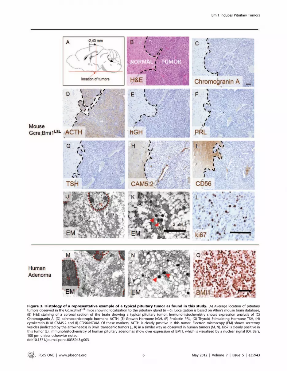
Figure 3. Histology of a representative example of a typical pituitary tumor as found in this study. (A) Average location of pituitarytumors observed in the GCre;Bmi1LSL mice showing localization to the pituitary gland (n = 6). Localization is based on Allen’s mouse brain database,(B) H&E staining of a coronal section of the brain showing a typical pituitary tumor. Immunohistochemistry shows expression analysis of (C)Chromogranin A, (D) adrenocorticotropic hormone ACTH, (E) Growth Hormone hGH, (F) Prolactin PRL, (G) Thyroid Stimulating Hormone TSH, (H)cytokeratin 8/18 CAM5.2 and (I) CD56/NCAM. Of these markers, ACTH is clearly positive in this tumor. Electron microscopy (EM) shows secretoryvesicles (indicated by the arrowheads) in Bmi1 transgenic tumors (J, K) in a similar way as observed in human tumors (M, N). Ki67 is clearly positive inthis tumor (L). Immunohistochemistry of human pituitary adenomas show over expression of BMI1, which is visualized by a nuclear signal (O). Bars,100 mm unless otherwise noted.doi:10.1371/journal.pone.0035943.g003
Bmi1 Induces Pituitary Tumors
PLoS ONE | www.plosone.org 6 May 2012 | Volume 7 | Issue 5 | e35943

Amplification of Bmi1 has been seen in 11% of mantle cell
lymphomas [38] and chromosomal gains have been seen in high-
grade astrocytomas and ovarian cancer [39,40]. Other human
neoplasms, including colon, breast and Laryngeal squamous cell
carcinoma show no amplification of Bmi1 although frequent
protein over expression is observed in these tumors [38]. Over
expression by non-genetic causes is likely to result from
convergence from multiple inputs acting on the transcriptional
and posttranscriptional level [41,42,43,44,37]. By using a trans-
genic over expression model, we show that GFAP-Cre mediated
transgenic over expression of Bmi1 is sufficient to drive the
formation of pituitary tumors, a type of tumor that represents 10 to
25% of all intracranial neoplasms in humans.
The incomplete penetrance and long latency period (1 year) of
tumor formation upon Bmi1 over expression suggested that
additional genetic or epigenetic defects might facilitate the
generation of tumors. Loss of Rb itself has been shown to induce
pituitary tumors in mouse models [30,31,32] and loss of Rb or
p16INK4A expression by hypermethylation is a common mecha-
nism in human pituitary tumors [46,47,48]. We found a higher
penetrance of pituitary tumors in the Rb deficient mice than in the
Bmi1 transgene model which indicates that transgenic Bmi1
expression acts as a predisposing condition facilitating the silencing
of the p16INK4A locus or, alternatively, points to an incomplete
transcriptional repression of the p16INK4A pathway or could
simply reflect the efficiency of Cre-mediated recombination events
Figure 4. Transgenic expression of Bmi1 is sufficient to induce intermediate and anterior lobe pituitary tumors but does not inducemedulloblastoma or glioma. (A) Kaplan Meier survival curves of mice carrying tumors because of GCre induced transgenic expression of Bmi1complemented with loss of Rb. These data show that Bmi1 transgenic mice develop pituitary tumors after about one year. Pituitary tumors are alsoobserved in RbLox/Lox and Bmi1LSL;RbLox/Lox transgenic mice. (B) Histograms showing the relative frequency and penetrance of tumors generated bythe individual genotypic groups. All genotypes shown are GCre positive. Total cohort size: GCre;Bmi1LSL n= 14, GCre;RbLox/Lox n=20, GCre;RbLox/Lox;Bmi1LSL n=12, GCre;p53Lox/Lox;RbLox/Lox n= 6, GCre;p53Lox/Lox;RbLox/Lox; Bmi1LSL n=4, WT mice did not develop tumors, n = 7. (C) IHC resultsshowing transgenic Bm1 expression in tumors raised on a GCre;Bmi1LSL background (5/5), no expression in tumors raised on a Gcre;Rblox/lox
background (0/5) while some of the GCre;Bmi1LSL; Rblox/lox mice were positive (2/5, 40%). These results are summarized in a histogram (D). Bar is50 mm.doi:10.1371/journal.pone.0035943.g004
Bmi1 Induces Pituitary Tumors
PLoS ONE | www.plosone.org 7 May 2012 | Volume 7 | Issue 5 | e35943

for the alleles used. Since no higher incidence of pituitary tumors
was found upon combined Rb loss and Bmi1 over expression, this
indicates that the oncogenic function of Bmi1 is dependent on the
presence of a functional p16INK4A locus, this is substantiated by
the observation that in the latter case, 40% of the tumors are
positive in IHC for Bmi1, indicating that the remaining 60% of the
tumors have inactivated the Rb allele independent of Bmi1 over
expression.
We do not know at which stage GFAP-Cre mediated transgene
activation is accomplished in our transgenic model although
enhanced expression of Bmi1 in Gcre;Bmi1LSL mice in non
transformed cells of the pituitary gland of postnatal day 8 mice
indicates that that transgenic activation has occured before this
time point. The occurrence of pituitary tumors using GFAP-Cre
mediated recombination of Rb has been shown before [29]. GFAP
is expressed at different stages during development, which includes
astrocytes of the sub ventricular zone [49] and in radial glial cells
of the outer sub ventricular zone [50] both of which can act as a
source of neural stem cells. GFAP expression can be activated by
tumor stem cells of pituitary adenomas [51]. In this context it is
interesting to note that transgenic mice generated from a Nestin-
Bmi-1-GFP cassette did not generate tumors [19]. Neural stem
cells isolated from these mice showed significant in vitro effects,
however, in vivo effects were moderate, reflecting the fact that high
activity of p16INK4 and p19ARF are seen in cultures of neural stem
cells, thereby increasing the reliance of cells upon Bmi1. In
contrast, p16INK4 and p19ARF are not detectably expressed by
neural stem/progenitor cells in developing or young adult mice
[52,53] and therefore these neural stem cells are none or weakly
responsive to elevated levels of Bmi1. Since we generated over
expression of Bmi1 in GFAP expressing cells by using a Lox
recombination system, we did not have a transient expression
restricted to stem cell compartment but rather a constitutive over
expression in all progeny of GFAP positive cells throughout later
life, thereby enabling to reveal the oncogenic role of Bmi1.
We show that the tumors in the transgenic mice were localized
to the pituitary gland, although this could be shown for large
intracranial tumors only because of lack material of earlier
neoplastic stages. The majority (n = 5 out of 6) of the tumors
showed a typical cytomorphology and these tumors expressed
ACTH. One tumor expressed multiple markers in a mosaic
pattern and was referred to as an atypical type (Table 3).Neuroendocrine ACTH producing cells normally comprise 10–
30% of the pituitary gland and 10% of pituitary tumors are
positive for ACTH [54], indicating that our model enhances or
selects tumorigenesis in ACTH positive cells. Previously, ACTH
expression in pituitary tumors has been genetically linked to Rb
function [55,56] therefore indicating that our model selects for or
enhances tumor formation in a similar population of cells as cells
that have lost Rb function as discussed above.
Transgenic expression of Bmi1 failed to generate medulloblas-
toma even in the presence of the predisposing deletion of Rb. In
contrast, the p53/Rb double deficient mice control mice
developed medulloblastoma after half a year as expected [21,29]
thereby implicating that Bmi1 over expression is insufficient to
fully functionally repress p19ARF/p53. Hence, our data point to a
Rb mediated function of Bmi1 in our model. The p16INK4A or the
p53 pathways are frequently mutated in glioblastoma [57,58,59],
however we have currently no indication that transgenic over
expression of Bmi1 contributes to the development of glioma/
glioblastoma, although other or additional predisposing lesions
might enable this.
In our mouse model, we identified both anterior and
intermediate lobe tumors. In contrast, human pituitary adenomas
are considered to be derived from the anterior lobe only since all
tumors resemble cell types of the anterior lobe hormone producing
cell types [60]. Human pituitary adenomas show expression of
BMI1 in 54% (our data) to up to 100% [34]. Furthermore, the
pituitary gland might be primed to enable BMI1 to exert its
oncogenic functions because the normal adult pituitary gland
expresses all required PRC1 components RING1, MEL18,
HPH1, RYBP [34]. These data show that over expression of
Bmi1 does not only induce the generation of tumors in mice but
this over expression is also commonly seen in established human
pituitary adenomas and BMI1 could therefore be considered as a
potential drug target for pituitary tumors.
In conclusion, we show here that Bmi1 transgene over
expression is sufficient to drive pituitary tumors and we also show
that more than 50% of clinical pituitary adenomas have high
expression of Bmi1. Furthermore, Bmi1 initiated tumorigenesis is
not enhanced by Rb loss which shows that the oncogenic function
is largely dependent on Rb function and not on p53 function,
which is further substantiated by the observation that Bmi1
overexpression is not sufficient to repress p53 sufficiently to
generate medulloblastomas.
Supporting Information
Figure S1 (A) Comparison of the two targeting constructs for
conditional over expression of Bmi1. Two LoxP recombination
methods are used, the Bmi1LSL construct contains a transcrip-
tional stop sequence that can be removed by LoxP recombination
and the Bmi1Lox66/Lox71 construct contains two partially mutated
LoxP sites that recombine upon Cre expression resulting reversion
of the DNA that is flanked by the LoxP sites which results in the
formation of one recombined LoxP site that has a low chance of
reversal of the recombination process. ES cells that were targeted
with the Bmi1LSL construct (B) or Bmi1LOX66/LOX71 construct (C)
show eGFP expression after Cre mediated activation of the
transgene.
(TIF)
Figure S2 Bmi1 immunohistochemistry of postnatal
day 8 pituitary glands (upper panel) shows enhanced
expression of Bmi1 in Gcre;Bmi1LSL mice (middle panel)
when compared to a wild type controls (lower panel).
The Bmi1 antibody was used at a concentration of 1:50. Bar is
10 mm.
(TIF)
Figure S3 Immunohistochemistry shows that Bmi1
transgenic mice generate pituitary tumors that stain
positive for (A) beta-endorphin (B-END) and (B) synap-
tophysin (SYN). The tumor is negative for (C) the neural marker
neurofilament (NF) as well as for the astroglial and schwann cell
markers (D) GFAP and (E) S100, respectively. No Keratin 8
(KER8) staining was observed (F).
(TIF)
Figure S4 Immunohistochemistry of a typical and an
atypical tumor that were generated in GCre;Bmi1LSL
transgenic mice shows expression of PCNA (A,B), Ki67
(C, D) and phosphorylated Histone H3 (E, F), respec-
tively.
(TIF)
Table S1 IHC analysis of additional markers in Gcre;
BmiPLSLP induced tumors.
(DOC)
Bmi1 Induces Pituitary Tumors
PLoS ONE | www.plosone.org 8 May 2012 | Volume 7 | Issue 5 | e35943

Acknowledgments
We thank colleagues of the NKI animal facility for animal husbandry.
Rahmen Bin Ali is thanked for his help with generating the transgenic
mice. Joaquim Calbo is thanked for his help with the experimental setup
and adenoviral work. We thank Roel Sneepers and Marco Breuer for
advice regarding the animal experiments. We also thank the colleagues of
the animal pathology department for their assistance. Alexandra Pietersen
is thanked for her help with the initial validation experiments of the
transgenic mice. Koen van de Wetering is thanked for sharing data. Hein
te Riele is thanked for thorough reviewing the manuscript. Pathologist J.M.
Rozemuller of the VU Medical Center Amsterdam is thanked for her
analyses.
Author Contributions
Conceived and designed the experiments: BAW AU EA MvL. Performed
the experiments: BAW MB ET MvV JYS JG PCS JZ HMP AU EA MvS.
Analyzed the data: BAW AU EA MvS MvL. Contributed reagents/
materials/analysis tools: BAW JG JZ HMP AU EA. Wrote the paper:
BAW MvV JYS AU EA MvL.
References
1. Muller J, Hart CM, Francis NJ, Vargas ML, Sengupta A, et al. (2002) Histone
methyltransferase activity of a Drosophila Polycomb group repressor complex.Cell. 111(2): 197–208.
2. Ringrose L, Ehret H, Paro R (2004) Distinct contributions of histone H3 lysine 9
and 27 methylation to locus-specific stability of polycomb complexes. Mol Cell.16(4): 641–53.
3. Fang J, Chen T, Chadwick B, Li E, Zhang Y (2004) Ring1b-mediated H2A
ubiquitination associates with inactive X chromosomes and is involved in
initiation of X inactivation. J Biol Chem. 279(51): 52812–5.
4. Wang H, Wang L, Erdjument-Bromage H, Vidal M, Tempst P, et al. (2004)
Role of histone H2A ubiquitination in Polycomb silencing. Nature. 431(7010):
873–8. 57 p.
5. Jacobs JJ, Kieboom K, Marino S, DePinho RA, van Lohuizen M (1999) The
oncogene and Polycomb-group gene bmi-1 regulates cell proliferation and
senescence through the ink4a locus. Nature. 397(6715): 164–8.
6. Molofsky AV, He S, Bydon M, Morrison SJ, Pardal R (2005) Bmi-1 promotes
neural stem cell self-renewal and neural development but not mouse growth and
survival by repressing the p16Ink4a and p19Arf senescence pathways. Genes
Dev. 19: 1432–1437.
7. Bruggeman SW, Valk-Lingbeek ME, van der Stoop PP, Jacobs JJ, Kieboom K,
et al. (2005) Ink4a and Arf differentially affect cell proliferation and neural stem
cell selfrenewal in Bmi1-deficient mice Genes Dev 19: 1438–1443.
8. Liu JH, Song LB, Zhang X, Guo BH, Feng Y, et al. (2008) Bmi-1 expression
predicts prognosis for patients with gastric carcinoma. J Surg Oncol. 97(3):
267–72.
9. Subkhankulova T, Zhang X, Leung C, Marino S (2010) Bmi1 directly represses
p21Waf1/Cip1 in Shh-induced proliferation of cerebellar granule cell
progenitors. Mol Cell Neurosci. 45(2): 151–62.
10. Fasano CA, Dimos JT, Ivanova NB, Lowry N, Lemischka IR, et al. (2007)shRNA knockdown of Bmi-1 reveals a critical role for p21-Rb pathway in NSC
self-renewal during development. Cell Stem Cell. 1(1): 87–99.
11. Fasano CA, Phoenix TN, Kokovay E, Lowry N, Elkabetz Y, et al. (2009) Bmi-1cooperates with Foxg1 to maintain neural stem cell self-renewal in the forebrain.
Genes Dev. 23(5): 561–74.
12. Hirabayashi Y, Suzki N, Tsuboi M, Endo TA, Toyoda T, et al. (2009) Polycomb
limits the neurogenic competence of neural precursor cells to promote astrogenicfate transition. Neuron. 63(5): 600–13.
13. Bruggeman SW, Hulsman D, Tanger E, Buckle T, Blom M, et al. (2007) Bmi1
controls tumor development in an Ink4a/Arf-independent manner in a mousemodel for glioma. Cancer Cell. 12(4): 328–41.
14. Facchino S, Abdouh M, Chatoo W, Bernier G (2010) BMI1 confers
radioresistance to normal and cancerous neural stem cells through recruitmentof the DNA damage response machinery. J Neurosci. 30(30): 10096–111.
15. Michael LE, Westerman BA, Ermilov AN, Wang A, Ferris J, et al. (2008) Bmi1
is required for Hedgehog pathway-driven medulloblastoma expansion. Neopla-
sia. 10(12): 1343–9, 5p following 1349.
16. Lee J, Son MJ, Woolard K, Donin NM, Li A, et al. (2008) Epigenetic-mediated
dysfunction of the bone morphogenetic protein pathway inhibits differentiation
of glioblastoma-initiating cells. Cancer Cell. 13(1): 69–80.
17. Yang MH, Hsu DS, Wang HW, Wang HJ, Lan HY, et al. (2010) Bmi1 is
essential in Twist1-induced epithelial-mesenchymal transition. Nat Cell Biol.
12(10): 982–92.
18. Sanchez-Beato M, Sanchez E, Gonzalez-Carrero J, Morente M, Dıez A, et al.
(2006) Variability in the expression of polycomb proteins in different normal and
tumoral tissues. A pilot study using tissue microarrays. Mod Pathol. 19(5):
684–94.
19. He S, Iwashita T, Buchstaller J, Molofsky AV, Thomas D, et al. (2009) Bmi-1
over-expression in neural stem/progenitor cells increases proliferation and
neurogenesis in culture but has little effect on these functions in vivo. Dev Biol.328(2): 257–72.
20. Yadirgi G, Leinster VH, Acquati S, Bhagat H, Shakhova O, et al. (2011)
Conditional Activation of Bmi1 Expression Regulates Self Renewal, Apoptosis
and Differentiation of Neural Stem/Progenitor Cells in Vitro and In Vivo. StemCells.
21. Marino S, Vooijs M, van Der Gulden H, Jonkers J, Berns A (2000) Induction of
medulloblastomas in p53-null mutant mice by somatic inactivation of Rb in theexternal granular layer cells of the cerebellum. Genes Dev. 14(8): 994–1004.
22. Stec DE, Davisson RL, Haskell RE, Davidson BL, Sigmund CD (1999) Efficientliver-specific deletion of a floxed human angiotensiongen transgene byadenoviral delivery of CRE-Recombinase in vivo. J Biol Chem 274(30):21285–21290.
23. van Lohuizen M, Verbeek S, Scheijen B, Wientjens E, van der Gulden H, et al.(1991) Identification of cooperating oncogenes in E mu-myc transgenic mice byprovirus tagging. Cell. 65(5): 737–52.
24. van der Lugt NM, Domen J, Linders K, van Roon M, Robanus-Maandag E, etal. (1994) Posterior transformation, neurological abnormalities, and severehematopoietic defects in mice with a targeted deletion of the bmi-1 proto-oncogene. Genes Dev. 8(7): 757–69.
25. Ernst T, Chase AJ, Score J, Hidalgo-Curtis CE, Bryant C, et al. (2010)Inactivating mutations of the histone methyltransferase gene EZH2 in myeloiddisorders. Nat Genet. 42(8): 722–6.
26. Bancroft JD, Gamble M (2008) Theory and practice of histological techniques,Churchill Livingstone Elsevier, 131.
27. Johnson MD, Fan X, Bourne P, Walters D (2007) Neuronal differentiation andexpression of neural epitopes in pituitary adenomas.J Histochem Cytochem.55(12): 1265–71.
28. Kovacs K, Horvath E, Vidal S (2001) Classification of pituitary adenomas,Journal of Neuro-Oncology, 54: 121–127.
29. Shakhova O, Leung C, van Montfort E, Berns A, Marino S (2006) Lack of Rband p53 delays cerebellar development and predisposes to large cell anaplasticmedulloblastoma through amplification of N-Myc and Ptch2. Cancer Res.66(10): 5190–200.
30. Maandag EC, van der Valk M, Vlaar M, Feltkamp C, O’Brien J, et al. (1994)Developmental rescue of an embryonic-lethal mutation in the retinoblastomagene in chimeric mice. EMBO J. 13(18): 4260–8.
31. Dannenberg JH, Schuijff L, Dekker M, van der Valk M, te Riele H (2004)Tissue-specific tumor suppressor activity of retinoblastoma gene homologs p107and p130. Genes Dev. 18(23): 2952–62.
32. Foijer F, Delzenne-Goette E, Dekker M, Te Riele H (2007) In vivo significanceof the G2 restriction point. Cancer Res. 67(19): 9244–7.
33. Tirabosco R, De Maglio G, Skrap M, Falconieri G, Pizzolitto S (2008)Expression of the Polycomb-Group protein BMI1 and correlation with p16 inastrocytomas an immunohistochemical study on 80 cases. Pathol Res Pract.204(9): 625–31.
34. Hayry V, Tynninen O, Haapasalo HK, Wolfer J, Paulus W, et al. (2008) Stemcell protein BMI-1 is an independent marker for poor prognosis inoligodendroglial tumours. Neuropathol Appl Neurobiol. 34(5): 555–63.
35. Leung C, Lingbeek M, Shakhova O, Liu J, Tanger E, et al. (2004) Bmi1 isessential for cerebellar development and is overexpressed in human medullo-blastomas. Nature 428(6980): 337–41.
36. Wiederschain D, Chen L, Johnson B, Bettano K, Jackson D, et al. (2007)Contribution of polycomb homologues Bmi-1 and Mel-18 to medulloblastomapathogenesis. Mol Cell Biol. 27(13): 4968–79.
37. Ochiai H, Takenobu H, Nakagawa A, Yamaguchi Y, Kimura M, et al. (2010)Bmi1 is a MYCN target gene that regulates tumorigenesis through repression ofKIF1Bbeta and TSLC1 in neuroblastoma. Oncogene. 29(18): 2681–90. Epub2010 Mar 1.
38. Bea S, Tort F, Pinyol M, Puig X, Hernandez L, et al. (2001) BMI-1 geneamplification and overexpression in hematological malignancies occur mainly inmantle cell lymphomas. Cancer Res. 61(6): 2409–12.
39. Hayry V, Tanner M, Blom T, Tynninen O, Roselli A, et al. (2008) Copynumber alterations of the polycomb gene BMI1 in gliomas. Acta Neuropathol.116(1): 97–102.
40. Yang GF, He WP, Cai MY, He LR, Luo JH, et al. (2010) Intensive expression ofBmi-1 is a new independent predictor of poor outcome in patients with ovariancarcinoma. BMC Cancer. 10: 133.
41. Kranc KR, Bamforth SD, Braganca J, Norbury C, van Lohuizen M, et al. (2003)Transcriptional coactivator Cited2 induces Bmi1 and Mel18 and controlsfibroblast proliferation via Ink4a/ARF. Mol Cell Biol. 23(21): 7658–66.
42. Nowak K, Kerl K, Fehr D, Kramps C, Gessner C, et al. (2006) BMI1 is a targetgene of E2F-1 and is strongly expressed in primary neuroblastomas. NucleicAcids Res. 34(6): 1745–54.
43. Dutton A, Woodman CB, Chukwuma MB, Last JI, Wei W, et al. (2007) Bmi-1 isinduced by the Epstein-Barr virus oncogene LMP1 and regulates the expressionof viral target genes in Hodgkin lymphoma cells. Blood. 109(6): 2597–603.
Bmi1 Induces Pituitary Tumors
PLoS ONE | www.plosone.org 9 May 2012 | Volume 7 | Issue 5 | e35943

44. Godlewski J, Nowicki MO, Bronisz A, Williams S, Otsuki A, et al. (2008)Targeting of the Bmi-1 oncogene/stem cell renewal factor by microRNA-128inhibits glioma proliferation and self-renewal. Cancer Res. 68(22): 9125–30.
45. Li SK, Smith DK, Leung WY, Cheung AM, Lam EW, et al. (2008) FoxM1ccounteracts oxidative stress-induced senescence and stimulates Bmi-1 expression.J Biol Chem. 283(24): 16545–53.
46. Simpson DJ, Hibberts NA, McNicol AM, Clayton RN, Farrell WE (2000) Lossof pRb expression in pituitary adenomas is associated with methylation of theRB1 CpG island. Cancer Research 60: 1211–1216.
47. Ogino A, Yoshino A, Katayama Y, Watanabe T, Ota T, et al. (2005) Thep15(INK4b)/p16(INK4a)/RB1 pathway is frequently deregulated in humanpituitary adenomas. Journal of Neuropathology and Experimental Neurology64: 398–403.
48. Yoshino A, Katayama Y, Ogino A, Watanabe T, Yachi K, et al. (2007)Promoter hypermethylation profile of cell cycle regulator genes in pituitaryadenomas. Journal of Neuro-oncology 83: 153–162.
49. Doetsch F, Caille I, Lim DA, Garcıa-Verdugo JM, Alvarez-Buylla A (1999)Subventricular zone astrocytes are neural stem cells in the adult mammalianbrain. Cell. 97(6): 703–16.
50. Hansen DV, Lui JH, Parker PR, Kriegstein AR (2010) Neurogenic radial glia inthe outer subventricular zone of human neocortex. Nature. 464(7288): 554–561.
51. Tunici P, Yu JS (2009) Pituitary adenoma stem cells. Methods Mol Biol. 568:195–201.
52. Molofsky AV, Pardal R, Iwashita T, Park IK, Clarke MF, et al. (2003) Bmi-1dependence distinguishes neural stem cell self-renewal from progenitorproliferation. Nature. 425(6961): 962–7.
53. Nishino J, Kim I, Chada K, Morrison SJ (2008) Hmga2 promotes neural stemcell self-renewal in young but not old mice by reducing p16Ink4a and p19ArfExpression. Cell. 135(2): 227–39.
54. Gray F, De Girolami U, Poirier J, Escourolle R (2004) Manual of BasicNeuropathology, Butterworth –Heinemann, Philadelphia. 347 p.
55. Guidi CJ, Mudhasani R, Hoover K, Koff A, Leav I, et al. (2006) Functionalinteraction of the retinoblastoma and Ini1/Snf5 tumor suppressors in cell growthand pituitary tumorigenesis. Cancer Res. 66(16): 8076–82.
56. Hinton DR, Hahn JA, Weiss MH, Couldwell WT (1998) Loss of Rb expressionin an ACTH-secreting pituitary carcinoma. Cancer Lett. 126(2): 209–14.
57. Parsons DW, Jones S, Zhang X, Lin JC, Leary RJ, et al. (2008) An integratedgenomic analysis of human glioblastoma multiforme. Science. 321(5897):1807–12.
58. Wang Y, Yang J, Zheng H, Tomasek GJ, Zhang P, et al. (2009) Expression ofmutant p53 proteins implicates a lineage relationship between neural stem cellsand malignant astrocytic glioma in a murine model. Cancer Cell. 15(6): 514–26.
59. Rao SK, Edwards J, Joshi AD, Siu IM, Riggins GJ (2010) A survey ofglioblastoma genomic amplifications and deletions. J Neurooncol. 96(2):169–79.
60. Farrell WE, Clayton RN (2000) Frontiers in Neuroendocrinology. 21: 174–198.
Bmi1 Induces Pituitary Tumors
PLoS ONE | www.plosone.org 10 May 2012 | Volume 7 | Issue 5 | e35943