toxoplasma secreting cre recombinase for analysis of host-parasite interactions
TRANSCRIPT
Toxoplasma secreting Cre recombinase for analysis of host-parasite interactions
Anita A. Koshy1,2, Ashley E. Fouts1,4, Melissa B. Lodoen1,5, Ozan Alkan1,3,6, Helen M.Blau1,3, and John C. Boothroyd1,7
1 Department of Microbiology and Immunology, Stanford University School of Medicine, StanfordCA 94305-5124, USA2 Division of Infectious Disease, Department of Internal Medicine, Stanford University School ofMedicine, Stanford, CA 94305-5107 USA3 Baxter Laboratory in Genetic Pharmacology, Stanford University School of Medicine, StanfordCA 94305-5124, USA
AbstractWe describe the use of site specific recombination to study the host-parasite interactions ofToxoplasma gondii. We present a Toxoplasma strain that efficiently injects a Cre fusion proteininto host cells. In a Cre-reporter cell line, a single parasite invasion induced Cre-mediatedrecombination in 95% of infected host cells. By infecting Cre reporter mice with these parasites,host cell infection could also be monitored in vivo.
Site specific recombination (SSR) is a well established system for genetically modifyingtargeted cells and has been used to spatially and temporally control the expression ofselected genes. SSR should offer the same advantages to the study of intracellular pathogensas it does to the study of developmental or cancer biology1,2. Engineered intracellularpathogens that introduce Cre into host cells would permit the use of SSR to examine theconsequences of removing specific host genes only in the infected cells. We sought toextend the use of the Cre-loxP system to studying the host-pathogen interactions of theeukaryotic parasite Toxoplasma gondii by creating a Toxoplasma strain that could inject Creinto host cells.
Toxoplasma is an obligate intracellular pathogen that has unique organelles – the rhoptriesand dense granules – that are known to secrete effector proteins into host cells duringinvasion3–5. It is a natural pathogen for mice which thus serve as an excellent model for thestudy of human infection6,7. Hence, the combination of a Cre-secreting strain ofToxoplasma and the many mouse strains that have already been engineered to carry loxP-flanked (“floxed”) genes represents a powerful tool for studying Toxoplasma-hostinteractions in vitro and in vivo.
Users may view, print, copy, download and text and data- mine the content in such documents, for the purposes of academic research,subject always to the full Conditions of use: http://www.nature.com/authors/editorial_policies/license.html#terms
7corresponding author: Department of Microbiology and Immunology, Fairchild Science Building, Room D305, Stanford UniversitySchool of Medicine, Stanford CA 94305-5124, USA, Tel. 650-723-7984, Fax. 650-725-6757, [email protected] address: Genentech Inc., 1 DNA Way, South San Francisco, CA 940805present address: University of California Irvine, Molecular Biology & Biochemistry, Irvine, CA 92697-39006present address: Broad Institute of MIT/Harvard, 7 Cambridge Center, Cambridge, MA 02142
Author’s Contributions: A.E.F. and A.A.K. generated the SeCreEt parasites. A.A.K. analyzed the SeCreEt parasites. M.B.L.developed the plasmid for making toxofilin a partner for injecting fusion proteins into host cells. O.K. created the Cre-reporter cellline. H.M.B. oversaw the work of O.K. and J.C.B. oversaw the work of A.A.K., A.E.F., and M.B.L.
NIH Public AccessAuthor ManuscriptNat Methods. Author manuscript; available in PMC 2010 October 1.
Published in final edited form as:Nat Methods. 2010 April ; 7(4): 307–309. doi:10.1038/nmeth.1438.
NIH
-PA Author Manuscript
NIH
-PA Author Manuscript
NIH
-PA Author Manuscript
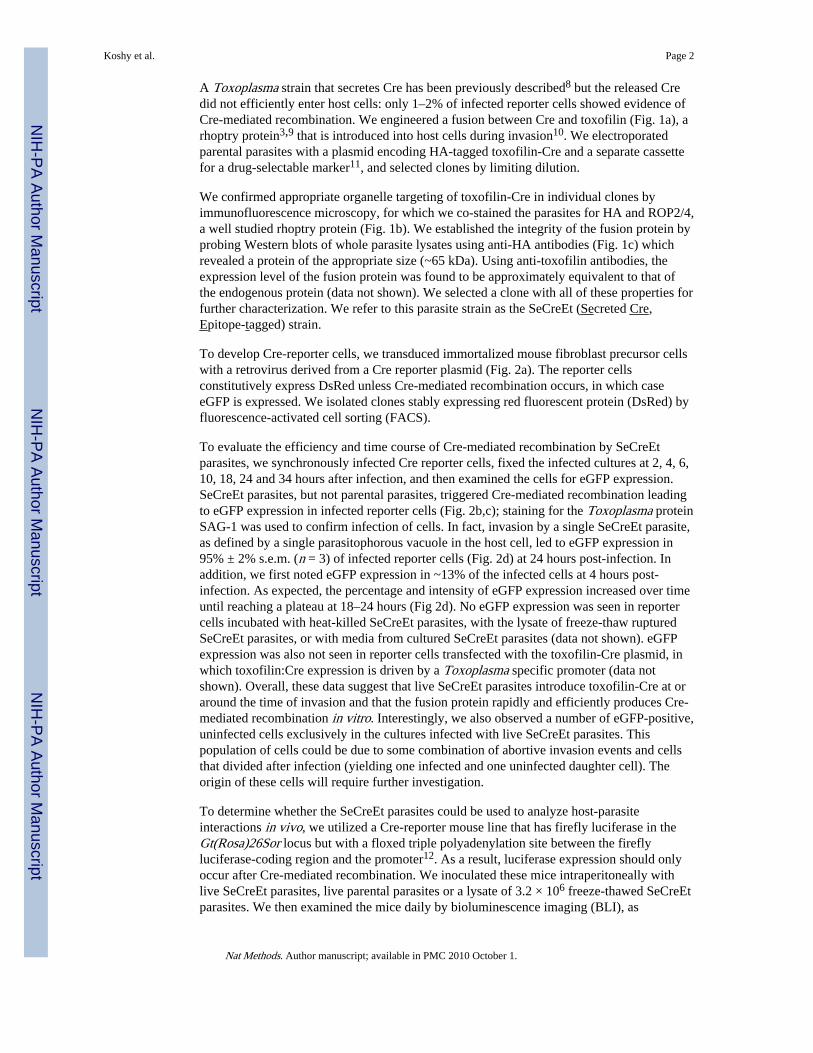
A Toxoplasma strain that secretes Cre has been previously described8 but the released Credid not efficiently enter host cells: only 1–2% of infected reporter cells showed evidence ofCre-mediated recombination. We engineered a fusion between Cre and toxofilin (Fig. 1a), arhoptry protein3,9 that is introduced into host cells during invasion10. We electroporatedparental parasites with a plasmid encoding HA-tagged toxofilin-Cre and a separate cassettefor a drug-selectable marker11, and selected clones by limiting dilution.
We confirmed appropriate organelle targeting of toxofilin-Cre in individual clones byimmunofluorescence microscopy, for which we co-stained the parasites for HA and ROP2/4,a well studied rhoptry protein (Fig. 1b). We established the integrity of the fusion protein byprobing Western blots of whole parasite lysates using anti-HA antibodies (Fig. 1c) whichrevealed a protein of the appropriate size (~65 kDa). Using anti-toxofilin antibodies, theexpression level of the fusion protein was found to be approximately equivalent to that ofthe endogenous protein (data not shown). We selected a clone with all of these properties forfurther characterization. We refer to this parasite strain as the SeCreEt (Secreted Cre,Epitope-tagged) strain.
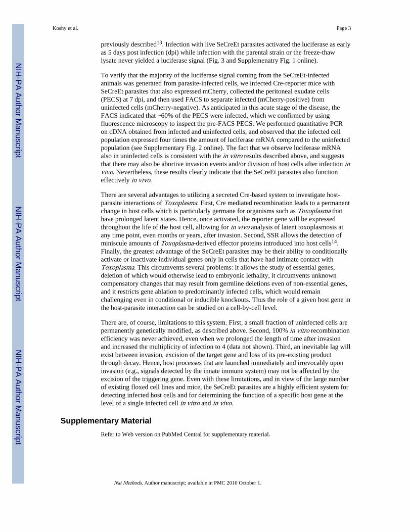
To develop Cre-reporter cells, we transduced immortalized mouse fibroblast precursor cellswith a retrovirus derived from a Cre reporter plasmid (Fig. 2a). The reporter cellsconstitutively express DsRed unless Cre-mediated recombination occurs, in which caseeGFP is expressed. We isolated clones stably expressing red fluorescent protein (DsRed) byfluorescence-activated cell sorting (FACS).
To evaluate the efficiency and time course of Cre-mediated recombination by SeCreEtparasites, we synchronously infected Cre reporter cells, fixed the infected cultures at 2, 4, 6,10, 18, 24 and 34 hours after infection, and then examined the cells for eGFP expression.SeCreEt parasites, but not parental parasites, triggered Cre-mediated recombination leadingto eGFP expression in infected reporter cells (Fig. 2b,c); staining for the Toxoplasma proteinSAG-1 was used to confirm infection of cells. In fact, invasion by a single SeCreEt parasite,as defined by a single parasitophorous vacuole in the host cell, led to eGFP expression in95% ± 2% s.e.m. (n = 3) of infected reporter cells (Fig. 2d) at 24 hours post-infection. Inaddition, we first noted eGFP expression in ~13% of the infected cells at 4 hours post-infection. As expected, the percentage and intensity of eGFP expression increased over timeuntil reaching a plateau at 18–24 hours (Fig 2d). No eGFP expression was seen in reportercells incubated with heat-killed SeCreEt parasites, with the lysate of freeze-thaw rupturedSeCreEt parasites, or with media from cultured SeCreEt parasites (data not shown). eGFPexpression was also not seen in reporter cells transfected with the toxofilin-Cre plasmid, inwhich toxofilin:Cre expression is driven by a Toxoplasma specific promoter (data notshown). Overall, these data suggest that live SeCreEt parasites introduce toxofilin-Cre at oraround the time of invasion and that the fusion protein rapidly and efficiently produces Cre-mediated recombination in vitro. Interestingly, we also observed a number of eGFP-positive,uninfected cells exclusively in the cultures infected with live SeCreEt parasites. Thispopulation of cells could be due to some combination of abortive invasion events and cellsthat divided after infection (yielding one infected and one uninfected daughter cell). Theorigin of these cells will require further investigation.
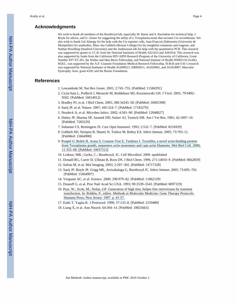
To determine whether the SeCreEt parasites could be used to analyze host-parasiteinteractions in vivo, we utilized a Cre-reporter mouse line that has firefly luciferase in theGt(Rosa)26Sor locus but with a floxed triple polyadenylation site between the fireflyluciferase-coding region and the promoter12. As a result, luciferase expression should onlyoccur after Cre-mediated recombination. We inoculated these mice intraperitoneally withlive SeCreEt parasites, live parental parasites or a lysate of 3.2 × 10⁶ freeze-thawed SeCreEtparasites. We then examined the mice daily by bioluminescence imaging (BLI), as
Koshy et al. Page 2
Nat Methods. Author manuscript; available in PMC 2010 October 1.
NIH
-PA Author Manuscript
NIH
-PA Author Manuscript
NIH
-PA Author Manuscript
previously described13. Infection with live SeCreEt parasites activated the luciferase as earlyas 5 days post infection (dpi) while infection with the parental strain or the freeze-thawlysate never yielded a luciferase signal (Fig. 3 and Supplemenatry Fig. 1 online).
To verify that the majority of the luciferase signal coming from the SeCreEt-infectedanimals was generated from parasite-infected cells, we infected Cre-reporter mice withSeCreEt parasites that also expressed mCherry, collected the peritoneal exudate cells(PECS) at 7 dpi, and then used FACS to separate infected (mCherry-positive) fromuninfected cells (mCherry-negative). As anticipated in this acute stage of the disease, theFACS indicated that ~60% of the PECS were infected, which we confirmed by usingfluorescence microscopy to inspect the pre-FACS PECS. We performed quantitative PCRon cDNA obtained from infected and uninfected cells, and observed that the infected cellpopulation expressed four times the amount of luciferase mRNA compared to the uninfectedpopulation (see Supplementary Fig. 2 online). The fact that we observe luciferase mRNAalso in uninfected cells is consistent with the in vitro results described above, and suggeststhat there may also be abortive invasion events and/or division of host cells after infection invivo. Nevertheless, these results clearly indicate that the SeCreEt parasites also functioneffectively in vivo.
There are several advantages to utilizing a secreted Cre-based system to investigate host-parasite interactions of Toxoplasma. First, Cre mediated recombination leads to a permanentchange in host cells which is particularly germane for organisms such as Toxoplasma thathave prolonged latent states. Hence, once activated, the reporter gene will be expressedthroughout the life of the host cell, allowing for in vivo analysis of latent toxoplasmosis atany time point, even months or years, after invasion. Second, SSR allows the detection ofminiscule amounts of Toxoplasma-derived effector proteins introduced into host cells14.Finally, the greatest advantage of the SeCreEt parasites may be their ability to conditionallyactivate or inactivate individual genes only in cells that have had intimate contact withToxoplasma. This circumvents several problems: it allows the study of essential genes,deletion of which would otherwise lead to embryonic lethality, it circumvents unknowncompensatory changes that may result from germline deletions even of non-essential genes,and it restricts gene ablation to predominantly infected cells, which would remainchallenging even in conditional or inducible knockouts. Thus the role of a given host gene inthe host-parasite interaction can be studied on a cell-by-cell level.
There are, of course, limitations to this system. First, a small fraction of uninfected cells arepermanently genetically modified, as described above. Second, 100% in vitro recombinationefficiency was never achieved, even when we prolonged the length of time after invasionand increased the multiplicity of infection to 4 (data not shown). Third, an inevitable lag willexist between invasion, excision of the target gene and loss of its pre-existing productthrough decay. Hence, host processes that are launched immediately and irrevocably uponinvasion (e.g., signals detected by the innate immune system) may not be affected by theexcision of the triggering gene. Even with these limitations, and in view of the large numberof existing floxed cell lines and mice, the SeCreEt parasites are a highly efficient system fordetecting infected host cells and for determining the function of a specific host gene at thelevel of a single infected cell in vitro and in vivo.
Supplementary MaterialRefer to Web version on PubMed Central for supplementary material.
Koshy et al. Page 3
Nat Methods. Author manuscript; available in PMC 2010 October 1.
NIH
-PA Author Manuscript
NIH
-PA Author Manuscript
NIH
-PA Author Manuscript
AcknowledgmentsWe wish to thank all members of the Boothroyd lab, especially M. Reese and S. Ravindran for technical help, J.Boyle for advice, and G. Zeiner for suggesting the utility of a Toxoplasma strain that secretes Cre recombinase. Wealso wish to thank Gal Almogy for his help with the Cre reporter cells, Jean-Francois Dubremetz (Universite deMontpellier) for antibodies, Marc-Jan Gubbels (Boston College) for his insightful comments and reagents, andNathan Woodling (Stanford University) and the Andreasson lab for help with the quantitative PCR. This researchwas supported by grants to J.C.B. from the National Institutes of Health AI21423 and AI41014. This research wasalso supported by funds from the California HIV/AIDS Research Program of the University of California, GrantNumber F07-ST-201, the Walter and Idun Berry Fellowship, and National Institute of Health NS065116 (AAK).M.B.L. was supported by the A.P. Giannini Foundation Medical Research Fellowship. H.M.B and O.K’s researchwas supported by National Institutes of Health AG009521, EB005011, AG020961, and AG024987; MuscularDystrophy Assn. grant 4320; and the Baxter Foundation.
References1. Lewandoski M. Nat Rev Genet. 2001; 2:743–755. [PubMed: 11584291]
2. Cicin-Sain L, Podlech J, Messerle M, Reddehase MJ, Koszinowski UH. J Virol. 2005; 79:9492–9502. [PubMed: 16014912]
3. Bradley PJ, et al. J Biol Chem. 2005; 280:34245–58. [PubMed: 16002398]
4. Saeij JP, et al. Nature. 2007; 445:324–7. [PubMed: 17183270]
5. Neudeck A, et al. Microbes Infect. 2002; 4:581–90. [PubMed: 12048027]
6. Dubey JP, Sharma SP, Juranek DD, Sulzer AJ, Teutsch SM. Am J Vet Res. 1981; 42:1007–10.[PubMed: 7283229]
7. Subauste CS, Remington JS. Curr Opin Immunol. 1993; 5:532–7. [PubMed: 8216929]
8. Gubbels MJ, Striepen B, Shastri N, Turkoz M, Robey EA. Infect Immun. 2005; 73:703–11.[PubMed: 15664908]
9. Poupel O, Boleti H, Axisa S, Couture-Tosi E, Tardieux I. Toxofilin, a novel actin-binding proteinfrom Toxoplasma gondii, sequesters actin monomers and caps actin filaments. Mol Biol Cell. 2000;11:355–68. [PubMed: 10637313]
10. Lodoen, MB.; Gerke, C.; Boothroyd, JC. Cell Microbiol. 2009. epublished
11. Donald RG, Carter D, Ullman B, Roos DS. J Biol Chem. 1996; 271:14010–9. [PubMed: 8662859]
12. Safran M, et al. Mol Imaging. 2003; 2:297–302. [PubMed: 14717328]
13. Saeij JP, Boyle JP, Grigg ME, Arrizabalaga G, Boothroyd JC. Infect Immun. 2005; 73:695–702.[PubMed: 15664907]
14. Vergunst AC, et al. Science. 2000; 290:979–82. [PubMed: 11062129]
15. Dranoff G, et al. Proc Natl Acad Sci USA. 1993; 90:3539–3543. [PubMed: 8097319]
16. Pear, W.; Scott, M.; Nolan, GP. Generation of high titre, helper-free retroviruses by transienttransfection. In: Robbin, P., editor. Methods in Molecular Medicine: Gene Therapy Protocols.Humana Press; New Jersey: 1997. p. 41-57.
17. Endo T, Yagita K. J Protozool. 1990; 37:133–8. [PubMed: 2319489]
18. Liang X, et al. Ann Neurol. 64:304–14. [PubMed: 18825663]
Koshy et al. Page 4
Nat Methods. Author manuscript; available in PMC 2010 October 1.
NIH
-PA Author Manuscript
NIH
-PA Author Manuscript
NIH
-PA Author Manuscript
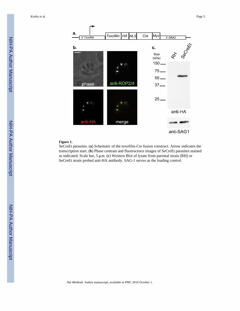
Figure 1.SeCreEt parasites. (a) Schematic of the toxofilin-Cre fusion construct. Arrow indicates thetranscription start. (b) Phase contrast and fluorescence images of SeCreEt parasites stainedas indicated. Scale bar, 5 μm. (c) Western Blot of lysate from parental strain (RH) orSeCreEt strain probed anti-HA antibody. SAG-1 serves as the loading control.
Koshy et al. Page 5
Nat Methods. Author manuscript; available in PMC 2010 October 1.
NIH
-PA Author Manuscript
NIH
-PA Author Manuscript
NIH
-PA Author Manuscript
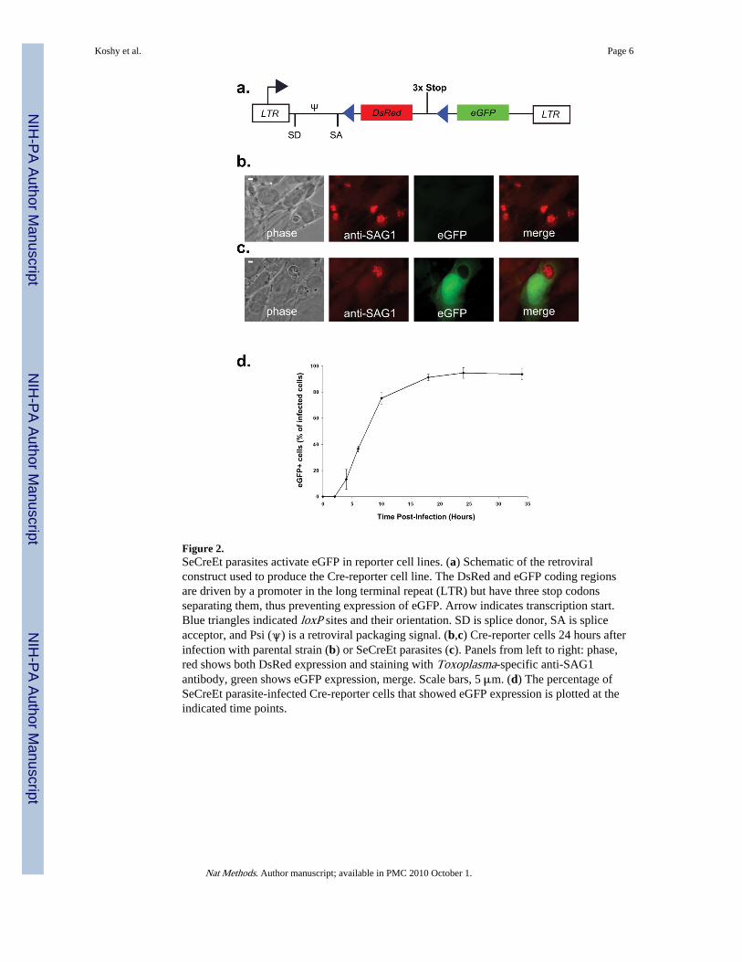
Figure 2.SeCreEt parasites activate eGFP in reporter cell lines. (a) Schematic of the retroviralconstruct used to produce the Cre-reporter cell line. The DsRed and eGFP coding regionsare driven by a promoter in the long terminal repeat (LTR) but have three stop codonsseparating them, thus preventing expression of eGFP. Arrow indicates transcription start.Blue triangles indicated loxP sites and their orientation. SD is splice donor, SA is spliceacceptor, and Psi (ψ) is a retroviral packaging signal. (b,c) Cre-reporter cells 24 hours afterinfection with parental strain (b) or SeCreEt parasites (c). Panels from left to right: phase,red shows both DsRed expression and staining with Toxoplasma-specific anti-SAG1antibody, green shows eGFP expression, merge. Scale bars, 5 μm. (d) The percentage ofSeCreEt parasite-infected Cre-reporter cells that showed eGFP expression is plotted at theindicated time points.
Koshy et al. Page 6
Nat Methods. Author manuscript; available in PMC 2010 October 1.
NIH
-PA Author Manuscript
NIH
-PA Author Manuscript
NIH
-PA Author Manuscript
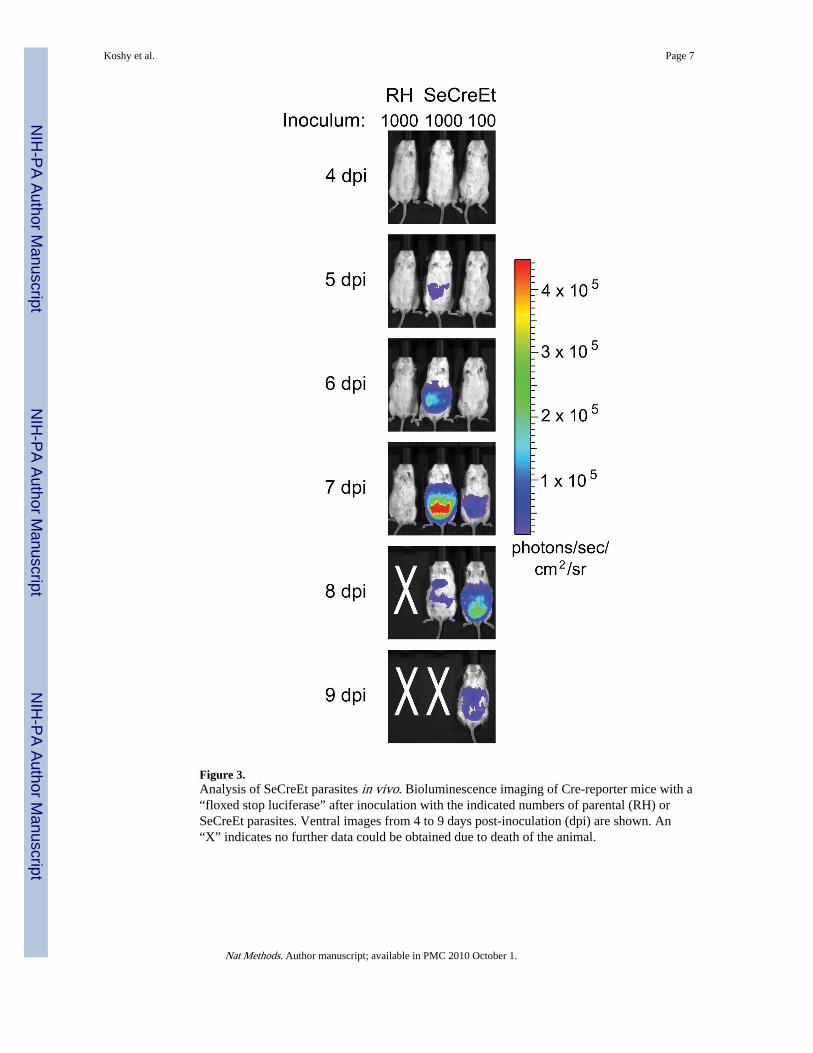
Figure 3.Analysis of SeCreEt parasites in vivo. Bioluminescence imaging of Cre-reporter mice with a“floxed stop luciferase” after inoculation with the indicated numbers of parental (RH) orSeCreEt parasites. Ventral images from 4 to 9 days post-inoculation (dpi) are shown. An“X” indicates no further data could be obtained due to death of the animal.
Koshy et al. Page 7
Nat Methods. Author manuscript; available in PMC 2010 October 1.
NIH
-PA Author Manuscript
NIH
-PA Author Manuscript
NIH
-PA Author Manuscript