cytochemical and stereological analysis of rat cortical astrocytes during development in primary...
TRANSCRIPT

Int..T. Dc\". BioI. 36: -,] 1-32] (1992) 311
Cytochemical and stereological analysis of rat corticalastrocytes during development in primary culture.
Effect of prenatal exposure to ethanol
FERNANDO MAYORDOMO', JAIME RENAU-PIQUERAS", LUIS MEGIAS', CONSUELO GUERRI3,FRANCISCO JOSE IBORRA', INMACULADA AZORIN' and MARC LEDIG4
1Centro Investigacion, Hospital La Fe, Valencia, Spain, 2Departamento de Anatomia, Facultad de Medicina, Granada, Spain,31nstituto Investigaciones Citol6gicas, Valencia, Spain and 4Centre de Neurochimie, Strasbourg, France
ABSTRACT This study has investigated the effect of prenatal alcohol exposure on the qualitativeand quantitative ultrastructure of proliferating and differentiated astrocytes in primary culture as wellas on the cytochemical activity of several subcellular phosphatase markers, including acid phosphatase,uridine diphosphatase, thiamine pyrophosphatase, 5'-nucleotidase and glucose-6-phosphatase. Theastrocytes were obtained from 21-day-fetuses of both control and alcohol-fed rats. Our results showthat several cell components, such as mitochondria, rough endoplasmic reticulum and Iysosomes,exhibit qualitative and/or quantitative ultrastructural changes during the process of astrocytematuration. In some cases these morphological changes are accompanied by variations in thecytochemical activity of enzymes located in these and other cell components, suggesting that theseenzymes, and therefore the functional state of these organelles, are modulated during astrocytedevelopment. When prenatally exposed to ethanol, both proliferating and differentiated astrocytesshowed striking ultrastructural alterations compared with controls, including an increment ofIysosomes as well as a decrease in the values of stereological parameters relative to mitochondria,rough endoplasmic reticulum and Golgi apparatus. Cytochemical analysis of these cells indicates thatprenatal exposure to ethanol decreased the activities of all the enzymes tested, except for acidphosphatase, which was increased in both groups of treated astrocytes. These results suggest thatprenatal exposure to ethanol could affect astrocytes during development in two different but probablycomplementary ways: al by causing a delay in astrocyte maturation and, bJ by inducing a direct toxiceffect on these cells.
KEY WORDS: a.\'lror)'lfs, rhveloJ}!lIen/, prow/al a/(ohol exJ)(JSlue, cJloc!lemiJ/rJ, s/l'/"f'Ology
Introduction
Prenatal exposure to ethanol during development induces inoffspring a wide spectrum of adverse effects, the extreme of whichis fetal alcohol syndrome or FAS (Jones and Smith, 1973), Inhumans, alcohol teratogenicity is mainly characterized by dysfunctionof the central nervous system (CNS), which is manifested by mentalretardation, poor motor coordination, hypotonia, irritability in infancyand hyperactivity in childhood (Abel,1981,1982,1984; Streissguthand Martin, 1983). Many of these symptoms have also beendescribed in laboratory animals (Streissguth and Martin, 1983;Sanchis et a/,. 1986; Driscoll et al., 1990).
Experimental studies in addition to necropsy observations ofFAS-affected children have shown a range of structural and functionalalterations in neurons (Clarren et al., 1978; Dow and Riopelle,
1985; Miller, 1986, 1987. 1990). Since many functional aspectsof these cells in both adult and developing brain depend on theintegrity of neuroglial cells (Bradford, 1986; Kimelberg and Norenberg,1989; McKay, 1989), it has been postulated that the neuronalalterations found in FAS could be due to some initial damage byethanol on astrocytes during development (Kennedy and Mukerji,1986a,b; Miller, 1986, 1987; Guerri ef a/" 1989; Renau-Piqueraset al., 1989c). This hypothesis has been partially confirmed by us
.-I.hhrn'illtiIJl/\"/I.I,d illlhi~ 1)(/1'1'1:C.\. (;()Ig-i appar,llll~: :\cl'a~('. acid pho~phala~e:(~r:\p, g-li<l1 tihril1ary acidic protein: IDI'. ill()~ilH' dipho~phata~e; l'E..-\,
pn'110ltal1~" t:"xpo~t:"d (0 t:"lhanoJ; rER. rOllgh {'lHlopJa"l11i, rt:"liculum; Tl'l'a~c.
thiamint:" p~Tophmphata~('; L.Dl'a~{'. Ilridil1(' clipho~phata~c.
'"'Addrcas for rcprint:J: SeGGi6n MiGro5Gopia EleGtr6nica, Centro Inve5tigac:i6n, Hospital La Fe, Avda. Campanar 21. 46009-Valencia, 5pi:!in. FAX: 34-6-386.8718
0214-62R2!92/S03.00r. URC Pr~',f>rinl~rI in Sp~ In

312 F. A1ayordo1J/o ct a1.
Fig. 1 Micrographs showing several types of Iysosomes (L) in 6- (A,B) and (C,D) 20-day PEA astrocytes. In (A) many endocytlC vesicles (v) accu-
mulate beneath the plasma membrane. (G. Goigi apparatus. IF. intermediate filaments; N. nucleus). A. C, 0 Bars. 1pm; B. O.511m. A x10400; B x23350;C x11600; 0 x9500.
using astrocytes in primary culture obtained from rat fetusesprenatally exposed to ethanol and controls. We have reported thatethanol reduces the content and synthesis in these cells ofproteins, DNAand RNAas well as the activity of several marker andmembrane-bound enzymes (Renau-Piqueras et al.. 1988; Guerri etal.. 1989, 1990c). Moreover, etllanol-exposed astrocytes failed todevelop processes and showed striking cytoskeletal alterations in
both microtubules and intermediate filaments (Renau-Piqueras etal.. 1989c, 1990; Saez et al., 1990). These studies also demon-strated that astrocytes in primary culture are more susceptible tothe toxic effect of ethanol during proliferation than during differen-tiation (Guerri et a/., 1990b).
There are many studies of the ultrastructural changes of mam-malian neuroglial cells and their precursors duringgliogenesis in vivo

TABLE 1
CONTROL ASTROCYTES IN PRIMARY CULTURE. RELATIVESTEREO LOGICAL DATA IMEANfS.D.I
Component Parameter 6 days 20 days Units p
Mitochond. Vv 9.23:t 2.29 7.37 t 2.28 ~mo ~O,O5Sv 1.05tO.13 0.98 f 0.24 /lm-1 n.sNv 1.31 f 0.43 1.25f 0.81 ~lm-3 n.s
rER Vv 6.3H 1.65 l1.44t 2.05 ~lmo ~O.O5Sv 0.45:t 0.06 1.03 t 0.09 ~lm.1 ~O,O5
Goigi Vv 11.2H 4.69 11.74:t 5.94 ~mo n.s
Lysosomes Vv 2.48:t 0.12 4.39 t 0.68 ~mo $0,05
Sv O.lH 0 04 0.28 f 0.02 pm-1 ::;0,05
Nv 0.23 t 0.01 o 24t 0.03 pm-3 n.s
Vv, volume density (in percentage); Sv. surface density; Nv, numericaldensity Reference volume was cytoplasm.
TABLE 2
CONTROL ASTROCYTES IN PRIMARY CULTURE. ABSOLUTE DATAIMEANfS.D.1
Component Parameter 6 days 20 days Units p
Mitochond. A 008 t 0.04 0.09 t 0.05 ~lm2 ~0.05V 0.03 t 0.01 005 tOOl ~m3 ~O05CF O.79:t 0.01 0.70 tOm ~0.05
Lysosomes A 0.10 t 0.07 o 13t 0.06 ~m2 ~0.05V 0.04t 0.01 0.06 t 0.01 ~m3 ::;0.05CF 098t004 0.9H 0.10 n.s
(Vaughn and Peters, 1967; Caley and Maxwell, 1968; Vaughn,1969; Phillips, 1973; Skoff et al., 1976; Parnavelas et al., 1983;Sturrock, 1986). However, although primary cultures of astrocytesconstitute an important tool for studying the biochemical andfunctional characteristics ofthese cells (Hertz et al., 1982; Hansson,1986) little attention has been paidtothe evolution of ultrastructuralfeatures of astrocytes during proliferation and differentiation in vitro(Trimmer et al., 1982; Fedoroff et at.. 1984; Devon and Juurlink,1988) and none to their cytochemical characteristics.
Knowledge of these characteristics could be of interest in orderto better define the functional state of astrocytes during bothphases in normal, experimental and pathological conditions.
In the present work we have therefore analyzed, using quantita-tive electron microscopy and cytochemistry, the possible changesin several subcellular components during the proliferation anddifferentiation periods of cortical astrocytes in primary culture aswell as the effect of prenatal exposure to alcohol (PEA) on these cellcomponents.
Results
Cytochemistry and stereology ofastrocytes 313
and many small vesicles which occasionally were coated. In somemicrographs these coated vesicle's were connected to the cisternae.Several types of Iysosomes, including multivesicular bodies, werealso found in most of the cells. Rough endoplasmic reticulum (rER)appeared both as tubular profiles and as dilated cisternae. In manycases, tubular rER cisternae were dilated at one extreme. Theremainder of the cytoplasm was filled by intermediate filaments,free ribosomes, scattered glycogen granufes and, in some cells.lipid droplets. The intermediate filaments, as previously reported(Renau-Piqueras, 1989c), were more abundant in differentiatedcells in which they were disposed in parallel arrays, whereas inproliferating astrocytes they formed a reticular network. Finally,nascent or free endocytic vesicles were frequently present in bothcell types (Fig 1A).
Six- and 20-day prenatally exposed to alcohol (PEA) astrocytesshowed a gross ultrastructural appearance similar to that ofcontrols. However, some striking alterations were found in severalcell components. Thus, compared with controls, proliferatingastrocytes showed an increased number of Iysosomes (Fig. 1),which were recognized by the morphological criteria (Fawcett, 1981)and by the presence of AcPase activity. These included denseIysosomes, multivesicular bodies. primary Iysosomes, autophagicvacuoles, Iysosomes containing myelinic figures and Iysosomeswith laminated inclusions. Moreover. these cells had a decreasednumber of mitochondria, which were more elongated than in 6-daycontrol astrocytes. GA and rER appeared less developed than incontrol cells. In many astrocytes of both treated groups, intermedi-ate filaments appeared disorganized. 20-day PEAastrocytes mostlyresembled 6-day control astrocytes, although they showed anabundance of Iysosomes, some swollen mitochondria and a lessdeveloped rER and GA. Also seen in some cells were alterations inthe cytoskeletal organization. In these latter cells intermediatefilaments appeared organized in a fashion similar to that found inproliferating control cells.
StereologyResults obtained afterstereological analysis of the four astrocyte
groups are summarized in Tables 1-5 and Fig. 2. Comparison of 6-and 20-day control astrocytes showed significant differences(P<0.05) between stereological parameters concerningmitochondria, rER and Iysosomes. Thus, the Vv of mitochondria of20-day astrocytes was smallerthanthatof6-daycells. However, the
Qualitative observationsIn both Epon and Lowicryl sections, astrocytes showed a similar
ultrastructure. Immunocytochemical observations revealed thatmost cells (",90%) were labeled with the anti-GFAP antibody.
Transverse sections of both 6- and 20-day control astrocytesdemonstrate that cultures were formed by several layers of over-lapping cellular sheets. In these sheets, the adjoining astrocytes
showed junctional complexes which were not stained with the anti-GFAP antibody. In both control groups the cells showed a flat profilewith a cell body containing the nucleus and long processes filled byintermediate filaments. The nucleus was elongated, with a scantamount of condensed chromatin. In many cells, nuclei containedone ormorewell-developed nucleoli. The perinuclear cytoplasm wasoccupied mainly by mitochondria which were more spherical in 6-than in 20.day cells. Cells from both culture periods showed two ormore well developed Golgi complexes composed of 4-5 cisternae A, area; V, volume; CF, coefficient of form
314 F. Mayordomo et al.
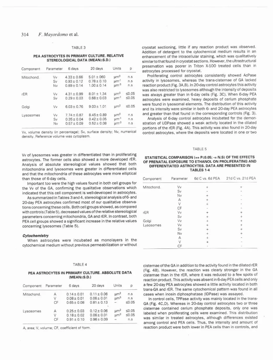
TABLE 3
PEA ASTROCYTES IN PRIMARY CULTURE. RELATIVESTEREOLOGICAL DATA (MEANIS.D.I
Component Parameter 6 days 20 days Units p
Mitochond. Vv 4.3H 0.66 5.01 I 060 ~mo n.s
Sv 0.93IO.12 0.7BIO.10 Jlm"' n.S
Nv 0.89IO.14 1.00IO.14 /lm-3 n.s
rER Vv 4.31 I 0.99 8.0a 1.34 ~mo ::;;0.05
Sv 0.29 I 0.03 0.68 I 0.03 Jlm-1 ";0.05
Goigi Vv 6.03IO.76 9.0H 1.01 ~mo <0.05
Lysosomes Vv 7.74 I 0.87 8A5 I 0.89 ~mo n.SSv 0.35 I 0.04 0.4H 0.05 I1m-' n.S
Nv 0.5H 0.09 0.5H 0.08 Ilm-3 n.s
TABLE 4
PEA ASTROCYTES IN PRIMARY CULTURE. ABSOLUTE DATAIMEANfS.D.}
Component Parameter 6 days 20 days Units p
Mitochond. A 0.14I 0.01 0.1 a 0.06 ~m2 n.sV 0.08 I 0.01 0.06IO.01 ~m3 n.sCF 0.65 f 0.08 0.81 IO.13 <0.05
Lysosomes A 0.25 I 0.03 0.1H 0.06 ~m2 <0.05V 0.18IO.o2 0.06IO.01 ~m3 <005CF 0.91 IO.10 0.96IO.09 n.s
Parameter 6d C VS.6d PEA
Vv +SvNv +A +V +CFVv +Sv +Vv +Vv +Sv +Nv +A +V +
CF
Vv. volume density On percentage): Sv. surface density; Nv, numericaldensity. Reference volume was cytoplasm.
Vv of Iysosomes was greater in differentiated than in proliferatingastrocytes. The former cells also showeda moredevelopedrER.Analysis of absolute stereological values showed that bothmitochondria and Iysosomes were greater in differentiated cellsand that the mitochondria of these astrocytes were more ellipticalthan those of E).{jaycelis.
Important too were the high values found in both celi groups forthe Vv of the GA, confirming the qualitative observations whichindicated that this cell component is well-developed in astrocytes.
As summarized in Tables 3 and 4, stereological analysis of 6- and2o-day PEA astrocytes confirmed most of our qualitative observa-tionsconcerningthese cells. Both cell groups showed, as comparedwith controls (Table 5), decreased values of the relative stereologicalparameters concerning mitochondria. GAand rER. In contrast, bothPEAcell groups showed a significant increase in the relative valuesconcerning Iysosomes (Table 5).
CytochemistryWhen astracytes were incubated as monolayers in the
cytochemical medium without previous permeabilization or without
A, area; V, volume; CF, coefficient of form.
cryostat sectioning, little if any reaction product was observed.Addition of detergent to the cytochemical medium results in anenhancement of the intracellular staining, which was qualitativelysimilarto that found in cryostat sections. However, the ultrastructuralpreservation was poorer in Triton X-lOa treated cells than inastrocytes processed for cryostat.
Proliferating control astrocytes consistently showed AcPaseactivity in Iysosomes, whereas the trans-cisternae of GA lackedreaction product (Fig. 3A,B).ln 20-daycontrol astrocytes this activitywas also restricted to Iysosomes although the intensity of depositswas always greater than in 6-day celis (Fig. 3C). When 6-day PEAastrocytes were examined, heavy deposits of cerium phosphatewere found in lysosomal elements. The distribution of this activityand its intensity were similar in both 6- and 20-day PEA astrocytesand greater than that found in the corresponding controls (Fig. 3).
Analysis of 6-day control astrocytes incubated for the demon-stration of UDPase showed a weak activity located in the dilatedportions of the rER (Fig. 4A). This activity was also found in 20-daycontrol astrocytes, where the deposits were located in one or two
TABLE5
STATISTICAL COMPARISON 1+=P<O.05;-= N.SI OF THE EFFECTSOF PRENATAL EXPOSURE TO ETHANOL ON PROliFERATING AND
DIFFERENTIATED ASTROCYTES. DATA ARE PRESENTED INTABLES 1-4
Component 21d C vs. 21d PEA
Mitochond. +
rER +
++++
+
Goigi
Lysosomes
cisternae of the GAin addition tothe activity found in the dilated rER(Fig. 4B). However, the reaction was clearly stronger in the GAcisternae than in the rER, where it was reduced to a few spots ofreaction product. This activity was absent in 6-day PEAcells and onlya few 2D-dayPEAastrocytes showed a little activity located in bothtrans-GA and rER.The same cytochemical pattern was found in allcases when inosin diphosphatase (IDPase) was assayed.
In control cells. TPPase activity was mainly located in the trans-GA (Fig. 4C,D). Whereas in 2o-day control astrocytes two or threecisternae contained cerium phosphate deposits, only one waslabeled when proliferating cells were examined. This distributionwas similar in treated astrocytes, although differences existedamong control and PEAcells. Thus, the intensity and amount ofreaction product were both lower in PEAcells than in controls. and

(%)50
40
30
20
10
0.04 0.07 0.11
(%)35
f.30
25
20
15
10
5
CONTROL
PEA
0.04 0.12 0.21
AREA ("m2)
0.29 0.5
+ 21-DAYSn 6-DAYS
Fig. 2. Graph showing the distribution of mitochondrial area in controland PEA astrocytes. A total of 200 mitochondrial profiles per group weremeasured using pOint counting procedures (Lindberg and Vorwerk, 1970,19721.
there was some difficulty to observe cells containing reactionproduct. However. the TPPase activity was lower in proliferating thanin differentiated PEA cells. The cytochemical activity of 5'-nucleotidase, as previously reported in detail (Renau-Piqueraset al.,1990, 1992), was restricted in all cases to the plasma membraneand endocytic vesicles. This activity was more intense in 20- thanin 6-day control cells. While in proliferating astrocytes the reactionproduct appeared as small spots, in differentiated astrocytes thecytochemical activity formed a continuous layer on the cell surface.The 5'-nucleotidase activity was clearly lower in PEA cells than incontrols. In 6-day PEAcelis the activity was difficult to see and in 21-day PEAastrocytes it was similar to that found in 6-day control cells.
Finally, we have been unable to demonstrate G-6-Pase activity inthe different groups of astrocytes analyzed.
Discussion
Cortical astrocytes obtained from 21-day-old fetuses, whencultured as primary cultures, grow rapidly during the first 7-10 days
Cylochelllisrry (11/(1slNl'ology of (lslrocWl'S 315
(proliferative pha5;e), after which their number increases slowly(differentiation phase) (Renau-Piqueras et a/., 1989c). Astrocytesin these two phases differ and can be characterized by severalparameters, such as the synthesis of protein, DNA and RNA, theactivity of several enzymes. the amount and distribution pattern ofcytoskeletal proteins as well as the number of plasma membraneconcanavalin Abinding sites (Renau-Piqueras et al., 1988, 1989c,1990, 1992; Guerri et al.. 1989. 1990b,c; Saez et al.. 1990).
Our results show that proliferating and differentiated controlastrocytes also differ in their ultrastructural organization, whichcorrelated to that for 1- and 4-week-old cultures, respectively, in thetype-Ccolonies described by Fedoroff et al. (1984).
In addition to modifications in cytoskeleta! organization, whichhave been described in detail (Trimmer et al., 1982; Fedoroff et al..1984; Renau-Piqueras et al..1989c; Saez et a/., 1990). It appearsthat severa! cell components, such as mitochondria, rER andIysosomes. exhibit qualitative and/or quantitative ultrastructuralchanges during the process of astrocyte maturation in primaryculture. In some cases, these morphological changes are accompa-nied by variations in the cytochemical activity of enzymes located inthese cell components. The increase in the volume density of rERoccurs parallel to that of the UDPase activity, suggesting a func-tional maturation of this cell compartment during astrocyte differen-tiation. Incontrast, other organelles, such as the GA,do not changemorphologically during this process. but display an importantincrease in the cytochemical activity ofthe enzymes analyzed duringdifferentiation. Finally, other cell components, such as Iysosomes,show no changes during the entire culture period in the amount ofcytochemical reaction product per individual organelle. In this lattercase, however. there was an augmentation in number duringculture, thereby resulting in an increment in the total activity ofAcPase per cell.
Thus. the increase which occurs in the activity of variousenzymes during astrocyte maturation in primary culture, appears tobe related to the functional state of different cell components. Thisagrees with previous studies showing that the activity of certainastrocyte marker enzymes, such as glutamine synthetase andbutylcholinesterase, increase mainly during the first weeks ofculture (Guerri et a/., 1989). On the other hand, the activity ofseveral membrane-bound enzymes, including (Na+K)ATPase and5'-nucleotidase (the latter also studied in the present work),showed a striking increment during the differentiation period (Guerriet al., 1989).
An interesting finding is the increment in the volume density ofrER throughout the culture period and which, as mentioned, isaccompanied by an increase in UDPase activity. This, together witha prominent GA and an increase in the TPPase and UDPaseactivities in this cell component during astrocyte differentiation,strongly suggests that these cells, mainly during this latter period,may be active in biosynthesis, glycosylation and transport ofglycoproteins. In fact, during astrocyte maturation in primary culturethere is an increment of both the activity of {Na+K)ATPase and 5'-nucleotidase, which are glycoproteins in nature, as well as of theplasma membrane concanavalin Abinding sites (Renau-Piqueras etal.. 1990, 1992). On the other hand, the presence of abundantclathrin coated vesicles, nascent endocytic vesicles andmultivesicular bodies suggests that astrocytes in primary culturecan also be active in endocytosis, which could be closely relatedwith the functional activityofthese cells in regulating the extracellular
medium in the brain {Bradford, 1986; Kimelberg and Norenberg,

-k ::..,,,,
"f
f \ \,\.
\
jE, :t..;,<
""
316 F. ,Hayordo1}/o ct a!.
r
J .0
'"
..
-.
'\
F,

1989). Recent studies showed that astrocytes in primary culturecan act as vehicles for the translocation of macromolecules suchas transferrin and serum albumin (JuurJink and Devon, 1990) orcationized ferritin (unpublished results).
The results concerning the activities of UDPase. TPPase and 5'-nucleotidase suggest that these enzymes are modulated in the rERand GAduring astrocyte development as occurs in other cell types(Doine et al., 1984).
It is also of interest to point out that whereas the TPPase activityin astrocytes shows a labeling pattern similar to that reported inother tissues. including liver (Morre and Ovtracht. 1977: Farquharand Palade. 1981). the cytochemical activity of UDPase differs fromthat described in hepatocytes. Thus, whereas UDPase is a goodmarker for ER and GAin both adult and fetal hepatocytes (Farquharand Palade, 1981: Robinson and Karnovsky, 1983b: Renau-Piqueraset al.. 1985b. 1987a), this activity was scant in the rER ofdeveloping astrocytes. On the other hand the qualitative andquantitative changes in mitochondria throughout the proliferativeand differentiation periods could be related to changes in thefunctional activity of these organelles in astrocytes. It has beenreported that the activity of several mitochondrial enzymes relatedto energy transduction increases from the proliferative to differen-tiation period (Tholey et al., 1982: Avola et al., 1986: Renau-Piqueraset al.. 1988). As previously suggested (Avola et al.. 1986), theseresults indicate a relationship between the increase in brain energymetabolism and the differentiation of astrocytes in primary culture.
Both 6-and 20-day PEAastrocytes showed striking ultrastructuraland cytochemical alterations as compared with controls. Qualita-tively, the main alteration observed in both treated cell types is anincrease in Iysosomes, mainly autophagosomes, together with anincrease in the size of mitochondria, which is more apparent inproliferating cells. Stereological analysis reveals that prenatalexposure to ethanol decreases the volume density of mitochondriaand GAas well as the numerical and surface density of mitochondriaand rER. respectively. In contrast. stereological parameters con-cerning Iysosomes are clearly increased. Althoughthese alterationswere found in both 6- and 20-day PEAastrocytes, proliferating cellsappeared more affected than differentiated ones, which could berelated to previous findings (Guerri et al., 1990b).
Functional analysis of cell components by means of the study ofthe cytochemical activity of enzyme markers shows that prenatalexposure to alcoho! decreases all the activities tested. except forthat of AcPase, which appears increased per individual lysosome inboth 6- and 20-day cultures. This effect could be interpreted asdamage induced by ethanol or ethanol metabolites on astrocytesduring in utero exposure.
Since the activity Qf the enzymes tested is related to thefunctional activityofthe cell components containing these enzymes.the effect of ethanol on the remaining activities studied could beinterpreted as the result of a delay in the astrocyte maturationinduced by this toxic agent. In fact. it has been shown in previousstudies that prenatal exposure to ethanol alters this process bothin vivo and in primary culture (Renau.Piqueras et al., 1989c; Guerriet al.. 1990c: Miller and Potempa. 1990: Davies and Cox, 1991).
Cytochcmistry ((nd stl'ri'%gy (~(ast"ocyt('.,' 317
It has also been demonstrated that growth kinetics of astrocyticcultures are vulnerable to ethanol in vitro and that these changesare, in part, qualitatively manifested in the ultrastructural configu-rations of cultured astrocytes (Davies and Cox. 1991). On the otherhand, studies performed on brain tissue revealed alterations in glialcells. Thus. after administration of an ethanol diet begun ongestational day 12 and continued until postnatal day 7. the glia weregreatly distended and had effectively isolated the dendrites from thesurrounding neuropil (Smithand Davies. 1990). Alsoobserved wereenlarged glial processes wrapped around the parallel fibres in themolecular layer of the cerebellum (Popova and Shchekalina, 1980:Tavares and Paula-Barbosa, 1984) as well as an increase in theamount of peri neuronal and total glia (Popova and Shchekalina.1980). Studies carried out at different days of pre- and postnataldevelopment showed that prenatal exposure to ethanol inducesoptic nerve hypoplasia in rat. alters the ultrastructure of bothastroglial and oligodendroglial cells and decreases the number ofthese cells (Pmazo-Duran. 1991 in preparation).
Prenatal exposure to ethanol also causes ultrastructural changesin neurons concerning nucleus. mitochondria. ER and GA, whichappeared distended and vesiculated (Clarren et al.. 1990: Smithand Davies. 1990). Some of these alterations are similar to thoseobserved infetalliver(Renau-Piqueras et al.. 1985b. 1987a. 1989a).It has been reported that many of these changes observed inneurons refer to their maturation process. Thus. a significant delayin differentiation of the neurons occurs in several brain regions(Volk, 1984: Magl6czkyet al.. 1990: Smith and Davies. 1990).Moreover, recent studies have reported that the extent ofultrastructural alterations in neurons increased as the mean peakplasma ethanol concentrations increased among the full gestationalexposed animals (Clarren et al.. 1990). However, it remains to beclarified whether these ethanol-induced neuronal alterations aredueto a direct effectofthetoxin on these cells or are a consequenceof the changes observed in astrocytes.
In addition to the effects of prenatal exposure to ethanol on thegrowth kinetics of astrocytes. a direct cytotoxic effect of ethanol onthese cells should also be considered as a complementary mecha-nism. For example. the increase in the number of Iysosomes and,thereby. of the overall activity of AcPase. could be the result of thisdirect toxic effect. Moreover. the decrease in the 5'-nucleotidase.a membrane-bound enzyme which is synthesized in ttle rER.glycosylated in the GA and then transported to the plasma mem-brane. could be due to inhibitory effects of ethanol on proteinsynthesis, glycosylation or glycoprotein transport. In fact. theresults presented here, together with others from previous studieson liver. indicate that prenatal exposure to ethanol alters thefunction of such cell components as rER. GA. and mitochondria.which are directly involved in these functions (Renau-Piqueras et al.,1985a,b. 1987a.b, 1989a.c: Sancho.Telio et al.. 1987: Guerrl etal..1990a). We have also demonstrated. as mentioned above. thatastrocyte cytoskeleton. a cell component involved in glycoproteintransport. is also altered by prenatal exposure to ethanol (Renau-Piqueras et al.. 1989b: Saez et al.. 1990).
It could be of interest to point out that in other experimental or
Fig. 3. AcPase activity (*) in control and PEA astrocytes. This activity IS scarce in both IA. 816- and 20-dayconrrol cells, whereas in PEA cells Iysosomes
show an intense labeling (D and E, 6-days; F, 20-days). No activity was found In the Golgi apparatus (G) (C and D). (rer, rough endoplasmic rericulum;M, mitochondrial. Bars. 0.5,um. A x44550; 8 x40250; C x62200; 0 x3750Q; E x34150; F x65800.

J 1H F. A1ayordon/o et a!.
Fig. 4. UDPase activity in astrocytes. (AI 5-day control astrocytes. IBI 20.cJay control astrocytes. In both cases the UDPase activity was found in thedilated portions of the rER where It was very weak (.) IAI, and in the trans-cisternae of the Golgl apparatus (..) (B). No reaction product was found in PEA
astrocytes. IC-DJ TPPase activity in control (A,B) astrocytes. This activity was located In one-three trans-cisternae (..) of the Goigi apparatus of mostcontrol cells (C. 6-days; D, 20-days;' (M, mitochondria; N. nucleus!. Bars: 0.5).lnl A x73500; B x76800; C x38000. 0 x41300
pathological situations astrocytes show ultrastructural and/orenzymatic changes different to those observed after prenatalalcohol exposure. Thus. TPPase and AcPase showed little changesin cytochemical activity in reactive astrocytes (AI-Aliand Robinson.1982). In contrast, these cells showed an increase in the activity of
G-6-Pase. which was accompanied by an increment of rER (AI-Ali andRobinson. 1982).
Finally. since it has been demonstrated that astrocytes play animportant role in neuronal function in both adult and developingbrain, the results we report here could be related to some of the

neuronal alterations observed in children and animals prenatallyexposed to ethanol.
Materials and Methods
Animal treatmentAdult virgin female Wistar rats with an initial body weight of 200-250 g
were used. Animals were fed before and during gestation as previouslyreported (Sanchfs et al., 1986). Briefly, rats were divided into two diet-basedgroups: (a) the alcoholic animals received an ethanol-liquid diet (5% w/v) inwhich ethanol provided 36% oftotal calories (Lieber and DeCarli, 1976); (b)a control group was given a similar diet, except that maltose-dextrin replacedethanol isocalorically. Both groups were maintained on their diets for 4-5weeks before mating. After this time, females in proestrus or initial estruswere placed overnight in cages with males of the same strain. Theappearance of sperm in the vaginal washing the following morning defineddayO of gestation.
Astrocyte culturesPrimary cultures of astrocytes from 21-day-old fetuses were prepared
from brain hemispheres as described (Renau-Piqueras et al., 1989c). Fe-tuses were obtained under sterile conditions from both control and alcohol-fed rats and astrocytes were cultured without ethanol in the medium. Thepurityof astrocyte cultures was assessed using a mouse anti-GFAPmonoclonalantibody and fluorescence microscopy(Renau-Piqueras et al., 1989c). Sincethese cultures grow rapidly for 7-10 days (proliferative period) after which thecell number increases slowly (differentiation period) (Renau-Piqueras et al.,1989c; Guerri et al., 1990c), all the experiments described below were donein triplicate on 6- and 2O-day cultures.
Electron microscopy and stereologyMonolayers growing in 35-mm Nunc tissue culture plastic dishes were
randomly selected (3 dishes per group and culture period, 3 differentcultures), washed three times in cold phosphate buffered saline and fixed
in 1.5% glutaraldehyde + 1.0% formaldehyde in 0.05 M cacodylate buffer,pH 7.4, for 60 min at 4°C. After washing in this buffer (0.1 M), the
monolayers were postfixed in 2% OS04 containing 0.8% potassiumferrocyanide forGO min at 4°C. The cells were then treated with O.l%tannicacid in buffer for 1 min at room temperature, washed in buffer and stainedin block with 2.0% aqueous uranyl acetate for 120 min at room temperature,dehydrated in cold ethanol, detached from the plastic with propylene oxide
and embedded as monolayers in Epon 812 (10 capsules per dish).For stereo logical analysis, the sampling method was carried out according
to Cruz-Orive and Weibel (1981) and Weibel (1979). Stereological analysisof micrographs was performed using point counting and standard procedures,as described (Weibel, 1979; Renau-Piqueras et al., 1985a). The followingrelative stereo logical parameters were evaluated: a) the volume density, Vvi(mitochondria, rough endoplasmic reticulum (rER), Golgi apparatus(GA) andIysosomes), b} the surface density, Svi (mitochondria, rER and Iysosomes)and, c) the numerical density, Nvi (mitochondria and Iysosomes). Asabsolute stereotogical parameters, the mean transsectioned area, Ai, themean volume, Vi, and the coefficient of form, CF, of mitochondria andIysosomes were determined (Lindberg and Vorwerk, 1970, 1972; Renau"Piqueras et al., 1985a; DePaz et al., 198Ga,b).
The minimum sample size (MSS) in each case was determined by theprogressive mean technique (confidence limit, :t10%) (Williams, 1977). TheMSS for all the stereological parameters analyzed in both control and treatedcells was always less than 17. However, 25 micrographs were analyzed foreach. All the stereological data were statistically analyzed by the ANOVA testusing the Oxstat programme (Oxstat ver. 3.1 (1), IBM).
Cytochemistry
The cytochemical activity of several phosphatases widely used assubcellular markers in various cell types was analyzed (Morn~~and Ovtracht,1977): a) acid phosphatase (AcPase) for the lysosomal system; b) uridinediphosphatase (UDPase) for ER and the transportion of the GA ;c) thiaminepyrophosphatase (TPPase) as a specific markerforthe trans-GA, d) glucose-
Cytochemistry and stereology of astrocytes 319
6-phosphatase (G-6-Pase), for ER ,and e) 5'-nucleotidase for the plasmamembrane.
Since information on the cytochemical activity of these enzymes inastrocytes in primary culture is scarce, several procedures were used forthedemonstration of those enzymes located inside the astrocytes. a) Themonolayers were incubated, after specific fixation, in the cytochemicalmedium, postfixed and embedded in Epon, as described above. b) Fixedcells were incubated in the cytochemical medium containing Triton X-l00 at0.0001% to permeabiJize cell membranes (Robinson, 1985; Baguena-Cervellera et al., 1987), c) Monolayers were detached from the plastic,centrifuged, and cryostat sections (20 J-Lm)were incubated in the corre-sponding cytochemical medium (Renau+Piqueras et al., 1987a). Thesubstrates were as follows: 1.0 mM B-glycerophosphatefor AcPase, 1.0 mMUDP for UDPase, 1.0 mM thiamine pyrophosphate for TPPase, 1.0 mMglucose-6-phosphate for G-6-Pase, and 1.0 mM AMP for 5'-nucleotidase. Inall cases cerium ions were used as capture agent (Robinson and Karnovsky,1983a,b; Renau-Piqueras et al., 1987a). Controls were sections incubatedwithout substrate. After incubation, the cells were processed for electronmicroscopy as described (Renau-Piqueras et al., 1987a). Ultrathin sections(80 nm) were examined at 60 kV without counterstaining.
Immunocytochemistry
For immunocytochemistry the cells were processed as previously described(Renau-Piqueras et al., 1989c). Briefly, the cell mono layers were fixed with0.5% glutaraldehyde and 4.0% formaldehyde in 0.1 M PIPES buffer (pH 7.3)for 60 min at 4°C, detached from the plastic using a rubber policeman, andembedded in Lowicryl K4M. For localization of GFAP with the immunogoldprocedure, ultrathin sections mounted on nickel grids were placed on adroplet of 0.1% BSA-tris buffer (20 mM Tris-HCI, 0.9% NaCI, pH 8.2,containing 0.1% BSA, Type V) supplemented with 5% heat-inactivated fetalcalf serum (FCS) for 30 min at room temperature and then transferred todroplets of 0.1% BSA-Tris containing 1% FCS and a mouse anti-GFAPmonoclonal antibody (10 ~/ml, Boehringer Mannheim GmbH) for 12 h at
4°C in a moist chamber. After three rinses with 0.1% BSA-Tris for 10 mineach, the grids were placed on droplets of 0.1% BSA-Tris containing 0.05%Tween 20, 5% FCS, and goat anti-mouse IgG-gold complex (10 nm, Sigma,1:100 dilution). The incubation was for 12 h at 4°C as above. Aftertwo rinseswith 0.1% BSA-Tris and a rinse in distilled water, the sections were air driedand counterstained with uranyl acetate. Immunocytochemical controls wereincubated in absence of the first antibody.
Acknowledgments .Research supported by grants SAL91-0020-C02 (CICYT) and ASS 90/
0897-2-E. The able technical help of M. T. Huerta and I. Montserrat isgratefully acknowledged.
References
ABEL, E.l. (1981). Behavioral teratology of alcohol. Psychol. 8ull. 90: 564-581.
ABEL, E.l. (1982). Consumption of alcohol during pregnancy: A review of effects ongrowth and development of offspring. Hum. 8iol. 54: 421-453.
ABEL,E.l. (1984) (Ed.). Fetal Alcohol Syndrome and FetalAlcohol Effects. Plenum Press,New York.
Al.AlI, S.Y.A. and ROBINSON, N. (1982). Ultrastructural study of enzymes in reactiveastrocytes: clarification of astrocytic activity. Histochem. J. 14: 311-321.
AVOLA,R., SERRA, I., CURTI,D., LOMBARDO, B., RENIS, M., CONDORElll, D.F.andGIUFFRIDA, A.M. (1986). Nuclear and mitochondrial DNA synthesis and energymetabolism in primary rat glial cell cultures. Neurochem. Res. 11: 789-800.
BAGUENA-CERVEllERA, R., RENAU-PIQUERAS, J., O'CONNOR, J.E. and GRISOllA, S.(1987). Effects of prolonged exposure to ammonia on fluid phase, receptor.mediated and adsorptive (non-specific)endocytosis in cultured neuroblastomacells. A flow and cytochemical study. Histochemistry 87: 445-455.
BRADFORD, H.F.(1986). Glial cells: mechanical and functional supporting cells of thenervous system. In An Introduction CONBurochemisrry. Freeman W,H. and Com-pany, New Yorl<,p. 62

320 F. Alayordo!llo ct al.
CALEY.D,N. and MAXWELL.0.5. (1968). Anelectron microscopic study of the neurogliaduring postnatal development of the rat cerebrum. J. Compo Neurol, 133: 45-70.
CLARREN, S.K.. ALVORD. E.C.. SUMI, S.M.. STREISSGUTH. A.P. and SMITH, D.W.(1978). Brain malformations related to prenatal e~posure to ethanol. J. Pediatr. 92:64-67.
CLARREN.S.K.. ASTLEY.S.J.. BOWDEN.D.M..LAI. H.. MILAM.A.H., RUDEEN,P.K. andSCHOEMAKER.W.J. (1990). Neuroanatomic and neurochemical abnormalities innon-human primate infants exposed to weekly doses of ethanol during gestation.Alcohol C/in. Exp. Res. 14: 674-683.
CRUZ-ORIVE.L.M. and WEIBEL, E.R. (1981). Sampling designs for stereology. J.Microscopy 122: 235-257.
DAVIES, D.L. and COX. W.E. (1991]. Delayed growth and maturation of astroC)1iccultures following exposure to ethanol: electron microscopic observations. BrainRes. 547: 53-61.
DEVON, R.M,and JUURLINK.B.H.J.(1988). Structural complexityof primaryculturesof astrocytes as revealed by transverse sections. Glia 1: 151-155.
DE PAl. P.. BARRIO, J.P. and RENAU-PIQUERAS,J. (1986a). A BASIC program fordetermination of numerical density of cytoplasmic compartments. I. Analysis ofspherical particles. Comput. Bioi. Med. 16: 267-272.
DE PAl. P.. BARRiO, J.P. and RENAU-PIQUERAS,J. (1986b). A BASIC program fordetermination of numerical density of cytoplasmic compartments. II. Analysis ofellipsoids and cylindrical particles. Comput. Bioi. Med. 16: 273-277.
DOINE.A.I., OLIVER,C. and HAND,A.R.(1984), The Goigiapparatus and GERL duringpostnatal differentiation of rat parotid acinar cells: an electron microscopiccytochemical study. J. Histochem. Citochem. 32: 477.485.
DOW, K.E. and RIOPELLE, R.J. (1985). Ethanol neurotoxicity: effects on neuriteformation and neurotrophic factor production in vitro. Science 228: 591-593.
DRISCOLL. C.D. STREISSGUTH. A.P. and RilEY. E.P. (1990). Prenatal alcohol e:o;po-sure: comparability of effects In 11umans and animal models, Neurotoxicol. Teratol.12: 231-237.
FARQUHAR.M.G. and PALADE, G,E. (1981). The Golgiapparatus (complex}-(1954-1981) -from artifact to center stage. J. Cell Bioi. 91: 77s-103s.
FAWCETT.D.W. (1981) (Ed.]. The Cell. WB Saunders Company, Philadelphia. pp. 486-514.
FEDOROFF,S.. NEAL.J.. OPAS. M. and KALNINS.V.I. (1984). Astrocyte cell lineage. III.The morphology of differentiating mouse astrocytes in colony culture, J. Neurocytol.13: 1-20.
GUERRI. C.. GUASCH, R. and RENAU-PIQUERAS,J. (1990a). Chronic and prenatalalcohol treatments induce alteration in the glycosylation of proteins in the trans-Golgi apparatus. Alcoholism: Clin. Exp. Res. 14: 356 (Abstr).
GUERRI.C.. MARQUES.A.. SANCHO-TELLO.M. and RENAU-PIQUERAS.J. (1989). Effectof prenatal exposure to ethanol on membrane.bound enzymes during astrocytedevelopment in vivo and in primary culture. Int. J. Dev. BIOI.33: 239-244.
GUERRI, C.. SAEZ. R.. SANCHO-TELLO,M., MARTiNDE AGUILERA,E.. and RENAU-PIQUERAS. .J. (1990b). Ethanol alters astrocyte development: a study of criticalperiods using primary cultures. Neurochem. Res. 15: 559-565.
GUERRi. C.. SANCHO.TELLO. M., ZARAGOZA. R. and RENAU-PIQUERAS, J. (1990c). The
use of primary culture of astrocytes to study glial development. Effect of ethanol.In Endocrine and Biochemical Development of the Fetus and Neonate (Eds. J.M.Cuezva. A.M.Pascual-leone and M.S. Patel). Plenum Press. NewYork, pp. 201-206,
HANSSON. E. (1986). Primary astroglial cultures. A biochemical and functionalevaluation. Neurochem. Res. 11: 759-763.
HERTZ. L., JUURLiNK. B.H.J.. FOSMARK.H., and SCHOUSBOE. A. (1982). Astrocytesin primary cultures. In Neuroscience Approached Through Cell Culture, Vol. 1. (Ed.P. Pfeiffer). CRC Press, Boca Raton. FL, p. 175.
JONES. K. and SMITH, D.W. (1973). Recognition of the fetal alcohol syndrome in earlyinfancy. Lancet 2: 999.1001.
JUURLlNK,B.H.J. and DEVON.R.M. (1990). Macromoleculartranslocation-a possiblefunction of astrocytes. Brain Res. 533: 73.77.
KENNEDY.L.A. and MUKER.JI.S. (1986a). Ethanol neurotoxicity. 1. Direct effect onreplicating astrocytes. Neurobehav. Toxicol. Teratol. 8: 11-15.
KENNEDY. L.A. and MUKER.JI. S. (1986b). Ethanol neurotoxicity. 2. Effects ondifferentiating astrocytes. Neurobehav. Toxicol. Teratol. 8: 17-21.
KIMELBERG.H.K.and NORENBERG, M.D.(1989). Astrocytes. Sci. Am. April:44-52.LIEBER, C.S. and DECARli, L.M. (1976). Animal models of ethanol dependence and
liver injury in rats and baboons. Fed. Proc. 35: 1232.1236.
LINDBERG, L.G. and VORWERK.P. (1970). On calculating volumes of transectedbodies from two-dimensional micrographs. Lab. Invest, 23: 315-317.
LINDBERG.L.G. and VORWERK.P. (1972). Estimation of volumes of ellipsoid bodiesin conglomerates through random plane sections. Lab. Invest. 27: 384-386.
MAGLOCZKY,Z.. MARTOS, J. and TOMBOL,T. (1990). Effectof prenatal e~posure toethanol on brains of kittens: I. Changes of neurons In lateral geniculate nucleus.1. Hlrnforsch. 31: 671-771.
McKAY. R.D,G. (1989). The origins of cellular diversity in the mammalian centralnervous system. CC!II 58: 815.821.
MillER. M.W.(1986). Effects of alcohol on the generation and migrationof cerebralcortical neurons. Science 233: 1308-1311.
MILLER.M.W. (1987). Effects of prenatal e~posure to alcohol on the distribution andtime of origin of corticospinal neurons in the rat. 1. Compo Neurol. 257: 372-382.
MilLER, M.W. (1990). Effects of prenatal ethanol exposure on cell proliferation in ratcorte~. Proceedings of the International Workshop on .Alcohol and Fetal Develop.-mento, Valencia (Spain).
MILLER.M.W. and POTEMPA, G. (1990). Numbers of neurons and glial in mature ratsomatosensory cortex: effects of prenatal e~posure to ethanol. 1. Compo Neurol.293: 92-102.
MORRE, D.J.and OVTRACHT,L.(1977): Dynamicsof the Golgiapparatus: membranedifferentiation and membrane flow. Int. Rev. Cytol. 5 (Suppl.): 61-188.
PARNAVELAS, J.G., LUDER.R., POLLARD,S,G, SULLIVAN.K. and LIEBERMAN.A.R.(1983). A qualitative and quantitative ultrastructural study of glial cells in thedeveloping visual cortex of the rat. Phil. Trans. Royal. Soc. London B: 301: 55-84.
PHILLIPS, D.E. (19731. Anelectron microscopic study of macroglia and microglia in thelateral funiculus of the developing spinal cord in the foetal monkey. Z. Zell. Mikrosk.Anat. 140: 145-167.
POPOVA, E.N. and SHCHEKALlNA,G.A.(1980). 0 vliyanii alkogolya na glial'nyye kletki(effect of alcohol on glial cells). Z. Neuropatol.Psikhlatr. 80: 539-544.
RENAU-PIQUERAS.J.. GOMEZ-PERRETTA,C., GUERRI, C. and SANCHIS. R. (1985a).Qualitative and quantitative ultrastructural alterations in hepatocytes of ratsprenatally e-.;posed to ethanol with special reference to mitochondria, Golgiapparatus and peroxisomes. Virchows Arch. A Pathol. Anat. 405: 237-251.
RENAU-PIQUERAS.J"
GUERRI,C.. BURGAL,M.. DEPAl, p" SAEZ, R.and MAYORDOMO.F. (1992). Prenatal e~posure to ethanol alters plasma membrane glycoproteins ofastrocytes during development in primary culture as revealed by concanavalin Abinding and 5'-nucleotidase activity. Glia 5: 65-74.
RENAU-PIQUERAS. J., GUERRI, C., MIRAGAll, F., GOMEZ.PERRETTA, C. and BAGUENA.CERVElLERA. R. (1985b). Alterations in the cytochemical activity of severalphosphatases in hepatocytes from rats exposed prenatally to ethanol. VirchowsArch. B Cell Pathol. 49: 249-259.
RENAU-PIQUERAS. .J., GUERRI.C.. SANCHO-TEllO. M.. BAGUENA-CERVELLERA, R.,MIRAGAll, F. (1989a). Effects of prenatal exposure to ethanol on rat liverdevelopment. A review. Int. J. Dev. Bioi. 33: 239-244.
RENAU-PIQUERAS.J., MIRAGALl, F., GUERRI. C. and BAGUENA-CERVEllERA.R.(1987a). Prenatal e~posure to alcohol alters the Golgiapparatus of newborn rathepatoc~1es, J. Hlstochem. Cytochem. 35: 221-228.
RENAU-PIQUERAS, .J., MIRAGAlL. F., GUERRI. C., SANCHO-TELLO, M. and BAGUENA.CERVELLERA, R. (1987b). Prenatal exposure to ethanol alters lateral plasmamembrane and gap junctions of newborn rat hepatocytes as revealed by freeze.fracture. J. Submicrosc. Cytol. 19: 397-404.
RENAU-PIQUERAS, J., SAEZ, R., BURGAL, M. and GUERRI.C. (1990). Ethanol alterscytoskeleton of astrocytes during development in vivo and in primary culture. Cell.Bioi. Int. Rep. 14 (Suppl.): 202 (Abstr.)
RENAU-PIQUERAS. J.. SANCHO-TELLO, M., BAGUENA.CERVELLERA. R, and GUERRI, C.(1989b). Prenatal exposure to ethanol alters the synthesis and glycosylationofproteins in fetal hepatocytes. Alcohol: Clin. Exp. Res. 13: 817-823.
RENAU-PIQUERAS. J.. SANCHO-TELLO, M., ZARAGOZA. R., GUERRI, C. (1988). Effectof ethanol on the development of astrocytes in primary culture. In Alcohol Toxicityand Free Radical Mechanism.Advances in Biosciences. Vol. 71. (Eds. R. Nordmann,R. Rlblere and C. Rouach). Pergamon Press, Oxford, New York, p. 269.
RENAU-PIQUERAS. J,. ZARAGOZA, R., DE PAl, P., BAGUENA-CERVELLERA, R., MEGIAS,L. and GUERRI. C. (1989c). Effects of prolonged ethanol exposure on the glialfibrillary acidic protein-containing intermediate filaments of astrocytes in primaryculture: a quantitative immunofluorescence and immunogold electron microscopicstudy. J. Histochem. Cytochem. 37: 229-240.
ROBINSON, J.M. (1985). Improved localization of intracellular sites of phosphatasesusing cerium and permeabilization. J. Histochem. Cytochem. 31: 1197.1203.
ROBINSON, J.M. and KARNOVSKY, M.J. (1983a). Ultrastructural localization of 5'-nucleotidase in guinea pig neutrophils based upon the use of cerium as capturingagent. J. Histochem. Cytochem. 31: 1190-1196.

ROBINSON. J.M. and KARNOVSKY, M.J. (1983b), Ultrastructural localization of severalphosphatases witt, cerium. J. Histochem. Cytochem, 31: 1197.1208.
SAEZ, R., BURGAL. M.. RENAU-PIQUERAS. J. and GUERRI. C. (1990). Evolution ofseveral cytoskeletal proteins of astroq1es in primary culture. Effect of prenatalalcohol exposure. Neurochem. Res. 16: 737-747.
SANCHIS. R.. SANCHO-TEllO, M. and GUERRI, C. (1986). The effects of chronic alcoholconsumption on pregnant rats and their offspring. AlcoholAlcoholism 21: 295.305.
SANCHO.TElLO, M.. RENAU-PIQUERAS. J.. BAGUENA-CERVELlERA. R. and GUERRI, C.(1987). A biochemical and stereologlCai study of neonatal rat hepatoq1e
subpopulations. Effect of pre- and postnatal exposure to ethanol. Virchows Arch.
B Cell. Pathol. 54: 170-181.
SMITH, D.E. and DAVIES, D.L. (1990). Effect of perinatal administration of ethanol onthe CAl pyramidal cell of the hypocampus and PurkinJe cell of the cerebellum: anultrastructural study. J. Neurocytol. 19: 708-717.
STREISSGUTH. A.P. and MARTIN. J.C. (1983). Prenatal effects of alcohol abuse inhumans and laboratory animals. In The Pathogenesis of Alcoholism (Eds. B. Kisslnand H. Beigllter). Plenum Press. New York. p. 539.
SKOFF, R.P., PRiCE. D.L. and STOCKS, A. (1976). Electron microscopIc auto radiographic
studies of gliogenesis in rat optic nerve. I. Cell proliferation. J. Compo Neural. 169:291-312.
STURROCK, R.R. (1986). Postnatal ontogenesis of astrocytes. In Astrocytes IEds. S.Fedoroff and A. Vernadakis). AcademiC Press. New York. p. 765.
Cytoc!tl'mistry and stl'rl'%gy (~last"o('ytl'S 321
TAVARES. M.A. and PAULA BARBOSA, M.M. (1984). Remodeling of the cerebellar
cortex molecular layer architectonics atter prolonged alcohol consumption in the
adult rat. Neurosci. Lett. 18 (Supp/.): S179.
THOlEY. G.. LEDIG. M. and MANDEL. P. (1982). Modifications In energy metabolismduring the development of chick glial cells and neurons in culture. Neurochem. Res.
7: 27-36.
TRIMMER. P.A.. REIER, P.L OH. T.H. and ENG, L.F. (1982). An ultrastructural andimmunocytochemical study of astroq1ic differentiation in vitro. J. Neuroimmuno/.2: 235-260.
VAUGHN, J.E.11969). An electron microscopic analysIs of gliogenesls In rat optic nerve.Z. Zell Mikrosk. Anat. 94: 293-324.
VAUGHN. J.E. and PETERS. A. 11967). Electron microscopy of early postnatal develop-ment of fibrous astrocytes. Am. J. Anat. 121: 131-152.
VOlK, B. (1984). Cerebellar histogenesis and synaptic maturation following pre- andpostnatal alcohol administration. An elcctronmlcroscopic investigation of the ratcerebellar cortex. Acta Ncuropat/101. 63: 57-65.
WEIBEL. E,R. (1979). Stereological Met/1Ods. Vol.I. Practical Methods for BiologicalMorphometry, Academic Press. Nell>'York.
WILLIAMS. M. (1977). Stereological Techniques. In Practical Methods in ElectronMIcroscopy. Vol. 6 (Ed. A.M.Glauert). Elsevier North Holland. Amsterdam.
.\/lljJ/nf jl!/"jmhliflliioll:.lrllllllll) fl,I)]