efficient gene delivery and selective transduction of astrocytes
TRANSCRIPT

“fncel-07-00106” — 2013/7/3 — 20:28 — page 1 — #1
REVIEW ARTICLEpublished: 05 July 2013
doi: 10.3389/fncel.2013.00106
Efficient gene delivery and selective transduction ofastrocytes in the mammalian brain using viral vectorsNicolas Merienne1, Juliette Le Douce 2, Emilie Faivre 2, Nicole Déglon1* and Gilles Bonvento 2*
1 Laboratory of Cellular and Molecular Neurotherapies, Department of Clinical Neurosciences, Lausanne University Hospital, Lausanne, Switzerland2 Commissariat à l’Energie Atomique et aux Energies Alternatives, Département des Sciences du Vivant, Institut d’Imagerie Biomédicale, Molecular Imaging
Research Center and CNRS CEA URA 2210, Fontenay-aux-Roses, France
Edited by:
Carole Escartin, Molecular ImagingResearch Center, France
Reviewed by:
Carole Escartin, Molecular ImagingResearch Center, FranceSebastian Kügler, UniversitätsmedizinGöttingen, GermanyKeith Murai, McGill University, CanadaVeerle Baekelandt, KatholiekeUniversiteit Leuven, Belgium
*Correspondence:
Gilles Bonvento, Commissariat àl’Energie Atomique et aux EnergiesAlternatives, Département desSciences du Vivant, Institut d’ImagerieBiomédicale, Molecular ImagingResearch Center and CNRS CEA URA2210, 18 route du Panorama, 92265Fontenay-aux-Roses, Francee-mail: [email protected];Nicole Deglon, Laboratory of Cellularand Molecular Neurotherapies,Department of ClinicalNeurosciences, Lausanne UniversityHospital, Pavillon 3, Avernue deBeaumont, 1011 Lausanne,Switzerlande-mail: [email protected]
Astrocytes are now considered as key players in brain information processing becauseof their newly discovered roles in synapse formation and plasticity, energy metabolismand blood flow regulation. However, our understanding of astrocyte function is stillfragmented compared to other brain cell types. A better appreciation of the biology ofastrocytes requires the development of tools to generate animal models in which astrocyte-specific proteins and pathways can be manipulated. In addition, it is becoming increasinglyevident that astrocytes are also important players in many neurological disorders.Targetedmodulation of protein expression in astrocytes would be critical for the development ofnew therapeutic strategies. Gene transfer is valuable to target a subpopulation of cells andexplore their function in experimental models. In particular, viral-mediated gene transferprovides a rapid, highly flexible and cost-effective, in vivo paradigm to study the impact ofgenes of interest during central nervous system development or in adult animals. We willreview the different strategies that led to the recent development of efficient viral vectorsthat can be successfully used to selectively transduce astrocytes in the mammalian brain.
Keywords: viral vectors, astrocytes, CNS, tropism, gene therapy
Astrocytes make up most of the cells in the brain. In addi-tion to well-characterized roles for astrocytes in regulating brainmetabolism and blood flow, there is now an increasing body ofevidence that astrocytes are dynamic regulators of synaptogenesis,synaptic function and network activity. This is conceptualized inthe tripartite synapse model, where pre-synaptic and post-synapticelements of neurons are surrounded and regulated by astrocyteprocesses (Araque et al., 1999; Barres, 2008).
Astrogenesis occurs relatively late in development after mostneurogenesis has completed (Freeman, 2010). Defects in astro-cyte maturation, tripartite synapse formation and plasticity duringearly post-natal development may be responsible for some psychi-atric and neurodegenerative diseases. There is a growing bodyof evidence to support the view that a loss of normal astrocytefunctions or a gain of abnormal effects can contribute to dis-ease processes, and there are now numerous examples of astrocytecontributions to pathological mechanisms in amyotrophic lateralsclerosis (ALS), Huntington’s disease (HD), and brain tumors tocite few of them (for review see Sofroniew and Vinters, 2010).
Despite progress and potential significance, cellular, develop-mental, and systems-level studies of astrocytes still lag far behind
those of neurons. New sophisticated genetic tools to label andmanipulate astrocytes in vivo were recently developed. Additionaltools that allow for temporally controlled deletion of genes, specif-ically in rodent astrocytes, along with improved high resolutionimaging techniques, are enabling researchers to address funda-mental questions in astrocyte biology for the first time. However,these tools need to be more fully expanded and exploited to betterunderstand astrocyte biology in vivo. The situation is complicatedby the recent findings that astrocytes do not represent a homo-geneous cell population across brain regions as well as withinthe same brain region (Zhang and Barres, 2010). So, despiteevidence showing pronounced region- and layer-specific morpho-logical heterogeneity as well as region-specific actions of astrocyteson neuronal functions, currently available tools have had limitedutility for examining functional diversity among astrocytes.
To understand the role of astrocyte signaling in brain function,it is critical to study astrocytes in situ where their complex mor-phology and intimate association with neurons remains intact.Understanding neuron–glia interactions in vivo requires dedicatedexperimental approaches to manipulate each cell type indepen-dently. These approaches include targeted transgenesis and viral
Frontiers in Cellular Neuroscience www.frontiersin.org July 2013 | Volume 7 | Article 106 | 1

“fncel-07-00106” — 2013/7/3 — 20:28 — page 2 — #2
Merienne et al. Viral vectors targeting astrocytes
transduction to overexpress or block the expression of a specificgene in astrocytes.
The past and current approaches of targeted transgenesis wererecently reviewed in a comprehensive paper (Pfrieger and Slezak,2012) and will not be detailed here.
Yet, a very important application of transgene expression isthe visualization of a large population of astrocytes in vivo by afluorescent protein. The use of bacterial artificial chromosomes(BACs) for the production of transgenic mice has opened newopportunities to study gene expression and functions in the brain.The resulting gene expression central nervous system (CNS) atlasprogram GENSAT represents a powerful resource for the scientificcommunity (http://www.gensat.org). However, it remains difficultand time-consuming to target specific cell subpopulations throughtransgenesis, and differences in recombination efficiency betweentransgenic lines complicate the analysis. We will therefore ratherfocus on an alternative approach to genetically manipulate astro-cytes that relies on the use of viral vectors. Indeed, the developmentof highly efficient viral vectors for gene transfer in the CNS is pro-viding new systems for localized and controlled gene expression.Even if such approach requires the stereotaxic injection of the viralvectors in each animal, it significantly reduces the costs of in vivoexperiments, and it can be used in combination with mouse mod-els for conditional gene targeting, providing high flexibility andversatility to replace, modify, induce, or block expression of targetgenes. We will therefore review the recent development in this fieldthat led to the emergence of effective and selective viral vectors fortransducing astrocytes in vivo.
VIRAL VECTORS: POTENT SYSTEM FOR IN VIVO GENEDELIVERY IN BRAINViral vectors offer the possibility to control expression of a trans-gene in adult or developing brain areas and can exploit the uniqueability of viruses to deliver genetic material into mammaliancells. Viral vectors are derived from various viruses and are engi-neered to preserve the transduction efficiency while preventingthe original pathogenicity and, in most cases, the capacity tomultiply (Davidson and Breakefield, 2003). These viral vectorsare often called multiply attenuated and replication-deficient viralvectors (Figure 1). Among the most widely used vectors for CNSapplications are the lentiviral (LVs) and adeno-associated viralvectors (AAVs) which have particularly attractive properties whichinclude, the capacity to infect non-dividing cells, the absence ofcytotoxic or immune response, long-term transgene expressionand large diffusion in the brain. At least for LV, the cloning capac-ity is sufficient to integrate most of the genes of interest (Déglonand Hantraye, 2005). Viral vectors provide a gene transfer toolthat is independent of age and species considered (Kay et al., 2001;Kirik et al., 2003; Lundberg et al., 2008). Along with somatic genetransfer in developing or adult animals, viral vectors can also beused for transgenesis in species in which classical methods are notsuitable, in particular large animals (Yang et al., 2008; Wongsrikeaoet al., 2011).
Natural viruses have a specific pattern of infection, whichreflects the recognition and interaction between viral cap-sid/envelope and receptors expressed on susceptible cells. Simi-larly, the tropism of viral vectors is primarily determined by the
interaction of the viral surface proteins with receptor moleculesexpressed on target cells but other mechanisms could be usedfor subpopulation-restricted gene transfer in the brain. In partic-ular, cell-type-specific promoters, post-transcriptional regulatoryelements, replacement of retroviral envelope proteins with heterol-ogous viral surface proteins, a phenomenon called pseudotyping(Page et al., 1990) or the use of various serotypes (AAV and Adharboring different capsids) have been proposed to dissect andelucidate gene functions in astrocytes.
The first viral vector was obtained by exploiting the naturaltropism of brain cells from the Herpes simplex virus type 1 (HSV-1; Geller and Breakefield, 1988; Federoff et al., 1992). The HSV-1genome is complex and large, but replication-incompetent vec-tors, with a partial (first generation of HSV-1 vectors) or complete(amplicons) deletion of viral genes allow the insertion of verylarge transgenes (around 150 kb). The HSV-1 amplicons are nei-ther pathogenic nor toxic for the infected cells and are retrogradelytransported to the CNS from the peripheral nervous system (PNS;Frampton et al., 2005). These vectors have a widespread tropismfor neurons (Jerusalinsky et al., 2012) and similarly to AAV andadenoviral vectors, their genetic material does not integrate intothe host genome thus reducing the risk of insertional mutagenesis(Manservigi et al., 2010). However, HSV amplicons are difficult toproduce, elicit low levels of adaptive immune responses and mostof the human population is seropositive which limits their clinicalapplications for chronic disorders (Manservigi et al., 2010).
A few years after the apparition of HSV vectors, adenoviral vec-tors (Ad) were derived from the Ad type 5 serotype (Le Gal LaSalle et al., 1993; Horellou et al., 1994). These vectors also have ahigh cloning capacity (approximately 30 kb of double-strandedDNA for gutless Ad) but the tropism of these vectors is not nat-urally oriented to the brain (Arnberg, 2012). Interestingly, a live(replication-competent Ad) vaccine has been safely administeredto humans (Rubin and Rorke, 1994). This vaccine program reflectsthe strong immune response induced by Ad in humans (Whiteet al., 2011), a reason why these vectors are promising candi-dates for tumoral therapy, and are proposed for the treatmentof glioblastoma (Candolfi et al., 2006; Kroeger et al., 2010).
In the mid 1990s, the first AAV (from serotype 2) and LVs werereported (Page et al., 1990; Kaplitt et al., 1994; Naldini et al., 1996).The AAV vectors are derived from the smallest non-envelopedviruses (approximately 20 nm) and have a cloning capacity of5 kb of single-stranded DNA. The AAV2 naturally infects humansbut is non-pathogenic. It is classified as a dependovirus becauseit requires a co-infection with a helper virus such as Ad or HSVto perform its infectious replication cycle. The AAV persists foryears in transduced cells mostly as an extrachromosomal episome(Nakai et al., 2001; Schnepp et al., 2005). To date, more than 100serotypes of AAV have been identified, each of them possessinga specific tropism in the CNS due to the binding of the capsidwith specific receptors (Wu et al., 2006a,b). Fourteen clinical trialsusing AAV gene transfer were performed to assess their potentialtherapeutic value in various neurodegenerative diseases (Crystalet al., 2004; Tuszynski et al., 2005; Kaplitt et al., 2007). In 2012, thefirst AAV gene therapy product was marketed by the EuropeanMedicine Agency (EMEA) for the treatment of patients sufferingfrom lipoprotein lipase deficiency (Yla-Herttuala, 2012).
Frontiers in Cellular Neuroscience www.frontiersin.org July 2013 | Volume 7 | Article 106 | 2

“fncel-07-00106” — 2013/7/3 — 20:28 — page 3 — #3
Merienne et al. Viral vectors targeting astrocytes
FIGURE 1 | Strategies to target astrocytes. Three steps ofviral cycle are used to modify the tropism of viral vectors: (1) theentry, (2) the transcriptional and (3) post-transcriptional regulations.After binding to their respective receptors, LV, AAV, and Ad enter intohost cells via receptor-mediated endocytosis. Viral DNA (AAV and Ad)or RNA (LV) are uncoated in the cytoplasm. The viral DNA remains as
extrachromosomal episomes in the nucleus while viral RNA isintegrated into the host genome after reverse transcription. Fornon-replicative vectors, in most cases only the transgene is expressed.In the case of oncolytic viruses, viral genes encoding structural proteins arenecessary for the encapsidation and production of replicative particles. PIC,pre-integration complex.
Finally, the most extensively characterized LVs are derivedfrom HIV-1, which is a subclass of retroviruses. Retrovirusesare lipid-enveloped particles comprising a homodimer of lin-ear, positive-sense, single-stranded RNA genomes of 7–11 kb.Following entry into target cells, the RNA genome is retro-transcribed into linear double-stranded DNA and integrated intothe cell chromatin (Delelis et al., 2010). To decrease the risk ofinsertional mutagenesis, integration-deficient LVs (IDLV) were
designed (Wanisch and Yanez-Munoz, 2009). These IDLVs arebased on the use of integrase mutations that specifically preventproviral integration, a process that results in the generation ofincreased levels of circular vector episomes in transduced cells.LVs were tested clinically for the treatment of adrenoleukodystro-phy (ALD) and Parkinson’s disease (PD). In the case of ALD, an exvivo approach was used, with the transduction of hematopoieticCD34+ cells and re-infusion of corrected cells in the patients. An
Frontiers in Cellular Neuroscience www.frontiersin.org July 2013 | Volume 7 | Article 106 | 3

“fncel-07-00106” — 2013/7/3 — 20:28 — page 4 — #4
Merienne et al. Viral vectors targeting astrocytes
immunological improvement occurred in the two treated childrenaged 9–12 months in combination with a blockage of the demyeli-nating lesions observed by magnetic resonance imaging (MRI),12–16 months after gene therapy (Cartier et al., 2009, 2012). Ina second study, a dopamine replacement strategy, with an LVthat encodes the three enzymes responsible for the productionof dopamine was tested in a phase I/II clinical trial. Increasingdoses of LV were injected into the striatum of 15 patients withmid-stage PD. An improvement in motor function was observed at6-months relative to pre-treatment assessment (Palfi, 2008; Jarrayaet al., 2009 and see http://www.oxfordbiomedica.co.uk).
STRATEGIES TO TARGET ASTROCYTESThe understanding of astrocyte functions in normal and alteredbrain strongly relies on the availability of experimental systems tospecifically target astrocytes in vivo. However, the first generationCNS viral vectors had a strong neurotropism in vivo (Naldini et al.,1996; Hermens and Verhaagen, 1997; Rabinowitz and Samulski,1998). Indeed, the injection of AAV2 into adult rodent brainswas associated with neuronal transgene expression when usingubiquitous promoters (Bartlett et al., 1998; Mandel et al., 1999;Bjorklund et al., 2000). Similarly, stereotaxic injection into rator mouse brain of LVs pseudotyped with the vesicular stomati-tis virus glycoprotein (VSV-G) with CMV (cytomegalovirus) orPGK (phosphoglycerate kinase 1) promoters, leads to the specifictransduction of neurons with very limited transgene expression inother cell types (Naldini et al., 1996; Kordower et al., 1999; Déglonet al., 2000). Finally, the Ad5 displays a partial neurotropism withthe transduction of other cell types, especially astrocytes (Smithet al., 1996; Bohn et al., 1999; Soudais et al., 2001; Rubio andMartin-Clemente, 2002; Wang et al., 2012).
However, it is important to mention that a number of parame-ters could alter the tropism. These include, amongst other factors,the purity of the vector, the mode of production, the site ofadministration, species, the developmental stage, and normal orpathophysiological conditions. Unfortunately, data gathered inprimary cultures (neurons and astrocytes) are not predictive ofthe in vivo tropism and a systematic evaluation of each vector isstill required. Indeed, VSV-G/LV-GFP under the control of var-ious promoters efficiently transduces primary rat astrocytes andto a lesser extent mouse astrocytes (Englund et al., 2000; Li et al.,2010) while transgene expression is mainly restricted to neuronsin vivo (Naldini et al., 1996; Kordower et al., 1999; Déglon et al.,2000). This phenomenon was also observed with AAV2, whichefficiently targets astrocytes in vitro but not in vivo (Gong et al.,2004). The purification method has also a major impact on thetropism of AAV8. In the mouse hippocampus, the CsCl-purifiedAAV8-CMV-GFP displayed an astroglial pattern in contrast to theexpected neuronal expression obtained with an iodixanol purifi-cation method (Klein et al., 2008). Foust et al. (2009) found thatinjection of AAV9-CMV early enhancer/chicken β actin promoter(CAG)-GFP into the tail vein of adult mice mainly transducesastrocytes throughout the CNS (Foust et al., 2009), whereas thetropism is mainly neuronal after intracerebral injection or intra-venous injection in neonatal mice (Klein et al., 2008). Finally,discrepancies have been observed on the transduction efficiencyand tropism of various AAV serotypes between species (rodent, cat,
and primates; Davidson et al., 2000; Vite et al., 2003; Burger et al.,2004; Gray et al., 2011). Additional studies are therefore still war-ranted to fully characterize the tropism of these vectors in the CNS.However, three strategies to direct viral vectors toward astrocyteshave already been developed: shifting the tropism by favoring theentry of viruses in astrocytes, limiting transgene expression withastrocytes-specific promoters or blocking transgene expression inunwanted cells (Figure 2).
ALTERING THE ENTRY OF VIRAL VECTORSThe tropism of a virus is first determined by its binding witha specific receptor at the surface of the host cell (Lutschg et al.,2011; Arnberg, 2012). Knowledge of the structure and viral capsidsor envelopes and their corresponding receptors provide essen-tial information to specifically target individual cell types and/ordiseased tissues. For example, the tropism of Ad5 vectors is regu-lated by the binding to its primary cellular receptor; the coxsackieand adenoviral vectors receptor (CAR). Tissues refractory to Ad5infection do not express CAR. The limited expression of CAR indopaminergic neurons of the substantia nigra of mice explainsthe poor transduction of these cells and transgene expressionin astrocytes and other non-neuronal cells (Lewis et al., 2010).However, the expression of CAR in the nervous system and inparticular in glial cells has not been extensively examined andCAR-independent forms of Ad have been developed to shift thetropism (Grellier et al., 2011).
As mentioned above, more than 100 serotypes of Ad and AAVwere characterized but only a dozen of them infect cells of theCNS. Indeed, for most of them, only limited data are availableconcerning their receptors and their pattern of expression in thebrain. The earliest and most used serotype is the AAV2, which hasa natural tropism for neurons (Bartlett et al., 1998; Kugler et al.,2003). The binding of AAV2 to its primary receptor, the heparansulfate proteoglycan (HSPG) has been well-characterized, and iscentered around two amino acids on the spikes of the AAV2 capsid(Kern et al., 2003; Opie et al., 2003). However, HSPG is neces-sary, but not wholly sufficient, for the transduction of permissivecells. In addition, fibroblast growth factor receptor 1 (FGFR-1)was identified as a co-receptor of AAV2 (Qing et al., 1999). Thetropism of AAV5 in vivo correlated with the pattern of expres-sion of platelet-derived growth factor receptor (PDGFR)-alpha(Di Pasquale et al., 2003). The AAV1, 5, 7, 8, and 9 not onlyinfect astrocytes in vivo but also neurons and other cells (David-son et al., 2000; Wang et al., 2003; Shevtsova et al., 2005; Cearleyand Wolfe, 2006; Gray et al., 2011). The AAV9 is unique com-pared to other AAV serotypes in that it is capable of crossingthe blood–brain barrier and transducing neurons and/or astro-cytes in the brain depending of the developmental stage (Foustet al., 2009). Recently, it has been shown that AAV9 uses galac-tose at the N-linked glycans as a receptor (Bell et al., 2011; Shenet al., 2011). The identification of the amino acids of the AAV9capsid necessary for binding to galactose opens the possibility tomodify the tropism (Bell et al., 2012). Finally, AAV4 and AAVrh43preferentially target astrocytes (Liu et al., 2005; Lawlor et al., 2009)but the receptors for these serotypes are unknown. AAV4-RSV-βGal and AAVrh43-CAG-eGFP exclusively transduce astrocyteswhen injected into the subventricular zone (SVZ) or the striatum.
Frontiers in Cellular Neuroscience www.frontiersin.org July 2013 | Volume 7 | Article 106 | 4

“fncel-07-00106” — 2013/7/3 — 20:28 — page 5 — #5
Merienne et al. Viral vectors targeting astrocytes
However, AAVrh43-CAG-eGFP infects approximately 3 mm3 ofthe striatum and 2,000 astrocytes per mm3 while AAV8-CAG-eGFP infects 6 mm3 of the striatum and 150,000 neurons permm3 (Lawlor et al., 2009).
Lentiviral vectors are increasingly being used in neuroscienceresearch and are unique in the sense that they are enveloped virusesthat can be pseudotyped (i.e., the original envelope protein can bereplaced by heterologous glycoproteins). The most used pseudo-type for LV is VSV-G which confers some interesting propertiesto the vector (Figure 2). It dramatically broadens LV tropism byfacilitating transduction of various cell types in different species,it stabilizes the vector particles from shear forces during centrifu-gation thereby allowing vector concentration and it directs LV toan endocytic pathway, which reduces the requirements of viralaccessory proteins for transduction (Cockrell and Kafri, 2007).Initial studies suggest that VSV-G/LV enters into cells using phos-phatidylserine (PS), but there is no correlation between the cellsurface PS levels and VSV infection or binding (Coil and Miller,2004). In addition, competition for PS using antagonists does notblock the binding of VSV on target cells. Currently, the receptorsresponsible for VSV-G/LV entry in cells are unknown.
In the CNS, VSV-G/LVs expressing transgenes under the con-trol of ubiquitous promoters have mainly a neuronal tropismwith a limited transgene expression in astrocytes (Naldini et al.,1996; Déglon et al., 2000; Watson et al., 2002). Among the otherenvelopes used to pseudotype LVs, lymphocytic choriomeningitis
virus (LCMV) and Mokola virus (MOK) envelopes result in apartial transduction of astrocytes. In vivo, LV/LCMV infects specif-ically astrocytes in the substantia nigra and in the striatum (Mileticet al., 2004; Cannon et al., 2011). Injection of MOK/LV into thestriatum or the hippocampus leads to the infection of cells that aremainly astrocytes (Pertusa et al., 2008; Colin et al., 2009). Althoughno quantifications were done using LCMV/LV, 70% of cells trans-duced by MOK/LV are astrocytes, 20% are neurons and 10% areother cell types of the striatum. In addition, it is important to notethat the titers and the transduction efficiency of these latter vectorsare usually lower than VSV-G/LV.
In conclusion, specific serotypes or envelopes only partiallyimprove the astrocytic targeting of viral vectors. However, engi-neering chimeric capsids or envelopes targeting astrocytes isdifficult and time-consuming. In order to optimize viral vectorstropism, strategies aiming at restraining transgene expression withastrocytic promoters, or by blocking expression in unwanted cells,mainly in neurons, were developed.
TARGETING ASTROCYTES WITH TRANSCRIPTION REGULATORYELEMENTSDifferent astrocytic promoters have been used to restrict transgeneexpression into glial cells. However, the packaging size of each viralvector limits the type of promoters which can be inserted. Analysisof the transcriptional regulatory elements of the glial fibrillaryacidic protein (GFAP) promoter reveals that 5′-flanking regions
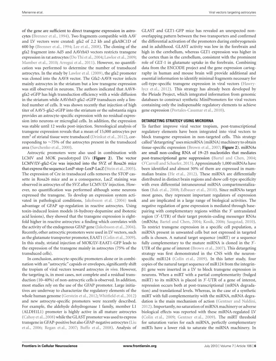
FIGURE 2 | Mechanisms used to restrain the transgene expression
of AAV and LV in astrocytes. (1) To modify the entry, various AAVserotypes or LV pseudotyping with heterologous VSV-G (green) andMOK-G (blue) envelopes were used. The tropism of LV is mainly neuronal(green cells) with the VSV-G envelope and a partial shift toward astrocytes(blue cells) is observed with the MOK-G envelope. AAV1, 2, 5, 7, and 8 mainlytransduce neurons (green) while AAV4, 9, rh43 display a partial astrocytictropism. (2) To restrict transgene expression, astrocytic promoters wereinvestigated (cells in the upper part). Transgene expression under the control
of a PGK promoter (pPGK, green mRNA) leads to a preferentialexpression in neurons, whereas a gfa2 promoter (pgfa2, blue mRNA)results in an astrocytic expression. (3) To block the transgene expression inunwanted cells (lower part), miRNA target (miRT) sequences are integrated inthe 3′-UTR of the vector (red signal on the green mRNA). The miR124 isexclusively expressed in neurons. As a consequence the miR124T is onlyrecognized in neurons and the transgene expression is blocked (mRNAdegraded). miR124, microRNA 124; miR124T, miR-124 target sequence;Tg. transgene.
Frontiers in Cellular Neuroscience www.frontiersin.org July 2013 | Volume 7 | Article 106 | 5
“fncel-07-00106” — 2013/7/3 — 20:28 — page 6 — #6
Merienne et al. Viral vectors targeting astrocytes
of the gene are sufficient to direct transgene expression in astro-cytes (Brenner et al., 1994). Two fragments compatible with AAVand LV vectors were created: gfa2 of 2.2 kb and gfaABC1D of600 bp (Brenner et al., 1994; Lee et al., 2008). The cloning of thegfa2 fragment into Ad5 and AAVrh43 vectors restricts transgeneexpression in rat astrocytes (Do Thi et al., 2004; Lawlor et al., 2009;Mamber et al., 2010; Arregui et al., 2011). However, no quantifi-cation was performed to determine the number of transducedastrocytes. In the study by Lawlor et al. (2009), the gfa2 promoterwas cloned into the AAV8 vector. The Gfa2-AAV8 vector infectsmainly astrocytes in the striatum but a low transgene expressionwas still observed in neurons. The authors indicated that AAV8-gfa2-eGFP has high transduction efficiency with a wide diffusionin the striatum while AAVrh43-gfa2-eGFP transduces only a lim-ited number of cells. It was shown recently that injection of hightiter of AAV5-gfa2-eGFP into the striatum or the substantia nigraprovides an astrocyte-specific expression with no residual expres-sion into neurons or microglial cells. In addition, the expressionwas stable until 12 weeks post-injection. Stereological analysis oftransgene expression reveals that a mean of 15,000 astrocytes permm3 of striatal tissue were transduced (Drinkut et al., 2012), cor-responding to ∼75% of the astrocytes present in the transducedarea (Savchenko et al., 2000).
Astrocytic promoters were also used in combination withLCMV and MOK pseudotyped LVs (Figure 2). The vectorLCMV/LV-gfa2-Cre was injected into the SVZ of Rosa26 micethat express the sequence LoxP-stop-LoxP-LacZ (Stein et al., 2005).The expression of Cre in transduced cells removes the STOP cas-sette in Rosa26 mice and as a consequence, LacZ staining wasobserved in astrocytes of the SVZ after LCMV/LV injection. How-ever, no quantification was performed although some neuronsexpressed the transgene. To develop an expression system acti-vated in pathological conditions, Jakobsson et al. (2004) tookadvantage of GFAP up-regulation in reactive astrocytes. Usingtoxin-induced lesion models (6-hydroxy-dopamine and ibotenicacid lesions), they showed that the transgene expression is eight-fold higher in reactive astrocytes: a finding which correlates withthe activity of the endogenous GFAP gene (Jakobsson et al., 2004).Recently, other astrocytic promoters were used in LV vectors, suchas the glutamate transporter promoter, EAAT1 (Colin et al., 2009).In this study, striatal injection of MOK/LV-EAAT1-GFP leads tothe expression of the transgene mainly in astrocytes (75% of thetransduced cells).
In conclusion, astrocyte-specific promoters alone or in combi-nation with an “astrocytic” capsids or envelopes, significantly shiftthe tropism of viral vectors toward astrocytes in vivo. However,the targeting is, in most cases, not complete and a residual trans-duction (10–40%) of non-astrocytic cells is observed. In addition,most studies rely on the use of the GFAP promoter. Large initia-tives are underway to characterize the regulatory elements of thewhole human genome (Gerstein et al., 2012; Whitfield et al., 2012)and new astrocyte-specific promoters were recently described.For example, the aldehyde dehydrogenase 1 family, member L1(ALDH1L1) promoter is highly active in all mature astrocytes(Cahoy et al., 2008) while the GLAST promoter was used to expresstransgene in GFAP-positive but also GFAP-negative astrocytes (Liuet al., 2006; Regan et al., 2007; Buffo et al., 2008). Analysis of
GLAST and GLT1-GFP mice has revealed an unexpected non-overlapping pattern between the two transporters and confirmedthe differential activation of the promoters during embryogenesisand in adulthood. GLAST activity was low in the forebrain andhigh in the cerebellum, whereas GLT1 expression was higher inthe cortex than in the cerebellum, consistent with the prominentrole of GLT-1 in glutamate uptake in the forebrain. Combiningdata from the ENCODE project and the gene expression cartog-raphy in human and mouse brain will provide additional andessential information to identify minimal fragments necessary forcell-type-specific transgene expression in viral vectors (Hawry-lycz et al., 2012). This strategy has already been developed bythe Pleiade Project, which integrated information from genomicdatabases to construct synthetic MiniPromoters for viral vectorscontaining only the indispensable regulatory elements to achievegene expression (Portales-Casamar et al., 2010).
DETARGETING STRATEGY USING MICRORNATo further improve viral vector tropism, post-transcriptionalregulatory elements have been integrated into viral vectors toblock transgene expression in non-targeted cells. This strategycalled“detargeting”uses microRNA (miRNA) machinery to obtaintissue-specific expression (Brown et al., 2007; Figure 2). miRNAsare small non-coding RNA of 19–25 nucleotides that mediatespost-transcriptional gene suppression (Bartel and Chen, 2004;O’Carroll and Schaefer, 2013). Approximately 1,000 miRNAs havebeen identified and almost 50% of them are expressed in mam-malian brains (He et al., 2012). These miRNA are differentiallydistributed in distinct brain regions and show cell-type specificitywith even differential intraneuronal miRNA compartmentaliza-tion (Bak et al., 2008; Edbauer et al., 2010). Since miRNAs targetmost genes, they represent important regulators of expressionand are implicated in a large range of biological activities. Thenegative regulation of gene expression is mediated through base-pairing with complementary regions within the 3′ untranslatedregion (3′-UTR) of their target protein-coding messenger RNAs(mRNAs; Bartel and Chen, 2004; Kosik, 2006; Saugstad, 2010).To restrict transgene expression in a specific cell population, amiRNA present in unwanted cells but not expressed in targetedcells is chosen. A natural target sequence (miRT) or a sequencefully complementary to the mature miRNA is cloned in the 3′-UTR of the gene of interest (Brown et al., 2007). This detargetingstrategy was first demonstrated in the CNS with the neuron-specific miR124 (Colin et al., 2009). In this latter study, fourcopies of the natural target sequence of miR124 from the integrin-β1 gene were inserted in a LV to block transgene expression inneurons. When a miRT with a partial complementarity (bulgedmiRT) to its miRNA is placed in 3′-UTR of a gene of interest,repression occurs both at post-transcriptional (mRNA degrada-tion) and translational levels. Whereas, in the case of a syntheticmiRT with full complementarity with the miRNA, mRNA degra-dation is the main mechanism of action (Gentner and Naldini,2012). Importantly, no saturation of miRNA machinery or adversebiological effects was reported with these miRNA-regulated LV(Colin et al., 2009; Gentner et al., 2009). The miRT thresholdfor saturation varies for each miRNA, perfectly complementarymiRTs have a lower risk to saturate the miRNA machinery. In
Frontiers in Cellular Neuroscience www.frontiersin.org July 2013 | Volume 7 | Article 106 | 6

“fncel-07-00106” — 2013/7/3 — 20:28 — page 7 — #7
Merienne et al. Viral vectors targeting astrocytes
addition, each miRNA has differential suppressive activity rang-ing from 5 up to >150-fold (Gentner et al., 2009). In this context,miR124 is a promising candidate because it is highly expressedin neurons (Lagos-Quintana et al., 2002; Smirnova et al., 2005;Deo et al., 2006). The insertion of four miR124T sequence in aVSV-G pseudotyped LV (VSV/LV-PGK-LacZ-miR124T) signifi-cantly decreases transgene expression levels and the number ofβ-galactosidase-positive neurons in the striatum of adult mice(Colin et al., 2009). This detargeting approach was used to shiftthe tropism of LV toward astrocytes. Double-immunofluorescencestaining with neuronal and astrocytic markers demonstrated thatcombining mokola pseudotyping and miR124T (MOK/LV-PGK-LacZ-miR124T) resulted in a transgene expression that was almostexclusively restricted to astrocytes, with 89 ± 3% β-galactosidase-S100β-positive cells and 6 ± 4% NeuN-positive cells. This effectwas not restricted to the striatum as similar results were obtainedin the hippocampus and cerebellum.
In conclusion, the use of these three different strategies (modu-lation of viral vector entry, transcription and post-transcriptionalregulations) has enabled the development of efficient gene trans-fer systems to specifically target astrocytes (Figure 3). Thanksto the unique features of these new viral vectors, it has alreadybeen possible to make significant advances in two areas ofresearch related to the development of innovative therapies andthe modeling of neurological disorders.
VIRAL VECTORS TARGETING ASTROCYTES: APPLICATIONSFOR BRAIN DISEASESMODELING BRAIN DISEASESThere is evidence to support the idea that the mechanisms respon-sible for selective neurodegeneration in some brain disorders arenon-cell autonomous and based upon pathological cell–cell inter-actions. The selective death of the neuronal population at riskin each disorder can be better explained by the convergence ofmultiple pathogenic mechanisms which provoke damage withinthe vulnerable neuron and neighboring cell types rather than byautonomous cell mechanisms (Ilieva et al., 2009).
In order to dissect out the specific role of different cell pop-ulations in vivo (neurons, astrocytes, microglia), two differentstrategies were recently used. The first one relies on the use of theCre/loxP system to silence the expression of the mutant proteinin specific cell types by crossing different Cre-expressing trans-genic mice with transgenic mice expressing the mutant proteinflanked by loxP sites in all cell types. The opposite strategy con-sists of selectively expressing the mutant protein in specific celltypes using either specific promoters such as GFAP or by cross-ing different Cre-expressing transgenic mice with transgenic miceexpressing the mutant protein after a STOP cassette flanked byloxP sites.
These two strategies were useful in providing evidence thatastrocytes play a key role in the pathogenesis of ALS (Ilieva et al.,
FIGURE 3 | Effects of the envelope/serotype, promoter, and miRT detargeting on the cellular tropism of LV, AAV and Ad. Overview depicting thetropisms of viral vectors in the CNS. References used for this figure are detailed and cited in the text.
Frontiers in Cellular Neuroscience www.frontiersin.org July 2013 | Volume 7 | Article 106 | 7

“fncel-07-00106” — 2013/7/3 — 20:28 — page 8 — #8
Merienne et al. Viral vectors targeting astrocytes
2009), spinocerebellar ataxia 7 (Custer et al., 2006), HD (Gu et al.,2005; Bradford et al., 2009, 2010), and taupathies (Forman et al.,2005; Dabir et al., 2006). However, an alternative strategy basedupon the use of viral vectors to selectively and locally express themutant protein has also proven to be very useful and comple-mentary to the development of transgenic mice in particular totest whether a local expression is sufficient to induce pathologicalmechanisms. Through the use of a newly developed LV (Colinet al., 2009), a short form of the mutant protein huntingtin (mHtt,responsible for HD), was expressed only in striatal astrocytes andnot in neurons (Faideau et al., 2010). It has been shown that theseglial cells developed a progressive phenotype of reactive astro-cytes that was characterized by a marked decreased expression ofboth glutamate transporters, GLAST and GLT-1, and of glutamateuptake. This reactive phenotype was associated with neuronal dys-function, as observed by a reduction in DARPP-32 and NR2Bexpression. Consistent with the above findings, a histological re-evaluation of potential astrocyte reactivity within postmortembrains of HD patients showed the presence of astrogliosis in thecaudate nucleus of Grade 0 patients and confirmed the colocal-ization of mHtt in astrocytes with a grade-dependent reductionin GLT-1. Through the use of viral vectors that target astrocyteslocally, we were able to show that the presence of mHtt in astrocytesis sufficient to alter the glial glutamate transport capacity early inthe disease process and may contribute to pathogenesis of HD.
GLIOBLASTOMA MULTIFORMGlioblastoma multiform (GBM) is the most common primarytumor developing in the brain from astrocytes. Due to the quickproliferation and its infiltrative nature, complete ablation bysurgery is almost impossible. The prognosis is very poor, witha median survival of 14.6–19.6 months and an inevitable relapsewithin a few months after the resection (Grossman et al., 2010).Viral-mediated gene therapy aiming to reduce glial proliferationrepresents, therefore, an alternative therapy (Murphy and Rabkin,2013). Indeed, GBM is a good candidate for gene therapy becausetumor cells rarely develop metastasis outside of the brain andmost cells in the CNS are post-mitotic, reducing side effects oftherapeutic strategies targeting dividing cells.
However, appropriate viral vectors for the treatment of GBMare different from those developed for the treatment of neu-rodegenerative diseases. For GBM therapy, the aim is to mediatedestruction of proliferating cells. Glial targeting is achieved eitherby the injection of the vector into the tumor mass, by choosing avector which target dividing cells or having a partial tropism forglial cells, as it is the case for Ad (Asadi-Moghaddam and Chiocca,2009).
The first studies used a replication-deficient mouse moloneyleukemia virus (MLV) that infected dividing cells and expressed asuicide gene (thymidine kinase, TK; Ram et al., 1993). Thymidinekinase is a phosphotransferase enzyme that incorporates dGTPanalogs in the presence of ganciclovir instead of cellular dGTPand leads to the blockade of cellular replication (Boivin et al.,1993). But the low transduction efficiency neither improved tumorprogression nor the overall survival time (Ram et al., 1993, 1994;Gunzburg et al., 1995). To improve the efficacy of the treatment,vector-producing cells (VPC releasing MLV particles expressing
the TK suicide gene) were injected into the brain after surgicalresection of the tumor. However, no significant decrease of tumormass occurred despite the bystander effect (Ram et al., 1997; Klatz-mann et al., 1998; Shand et al., 1999; Packer et al., 2000; Rainov,2000; Martinet et al., 2003). As an alternative therapy, Ad-TKwas administered directly to GBM patients but the phase III trialshowed no positive outcome (Cottin et al., 2011). Interestingly,it was shown that the preferential transduction of glioma cells isnot dependent on the expression of known Ad receptors on tumorcells (Candolfi et al., 2006). Expressing the therapeutic suicide geneunder the control of a strong ubiquitous promoter in combinationwith an immune stimulator may increase therapeutic efficacy andprevent relapse (Candolfi et al., 2006; Ghulam Muhammad et al.,2009).
As an alternative strategy to improve the therapeutic efficacy,conditionally replicative or replicative viruses were developed. Theprinciple of oncolytic therapy is to inject directly into the tumoralcells a lytic replicative-competent cytotoxic virus, such as HSV,VSV, Ad, or retroviruses, which will induce apoptosis in prolif-erative cells during replication (Parker et al., 2009; Zemp et al.,2010; Castro et al., 2011; Russell et al., 2012). HSV were initiallyused as lytic viruses in GBM therapy (Zemp et al., 2010). How-ever, the high worldwide HSV seropositivity limits their use inthe clinic and as a consequence has led to the development ofother oncolytic viruses. A deletion of E1B region on Ad genome(Ad-ONYX-15) was introduced to favor apoptosis in infectedglioma cells but the efficiency of this approach was too low toreach a phase II of clinical trial (Moran, 1993; Chiocca et al.,2004). In addition, replicative adenoviral vectors expressing ther-apeutic genes were used to mediate tumoral cells destruction.The candidate genes are inserted in the E3 deleted region and aCAR-independent entry mechanism enhancing the transductionefficiency of tumoral cells has been proposed for these new gener-ation oncolytic viruses. To favor replication in GFAP-positive cells,three copies of glial specific B enhancer were added on the gfa2promoter (gfa2B3), leading to a decreased growth of glioma cells(Horst et al., 2007).
GENE THERAPY FOR NEURODEGENERATIVE DISORDERSDegeneration of the nigro-striatal projection represents the majorpathological hallmark of PD. Preclinical rodent and non-humanprimate models demonstrated a strong protective effect of glialcell line-derived neurotrophic factor (GDNF) on the nigro-striatal dopaminergic system (Gash et al., 1996; Kirik et al., 2000).However, intrathecal infusion of GDNF protein or viral vector-mediated expression of neurturin in the striatum of late stage PDpatients showed no significant clinical benefit (Lang et al., 2006;Marks et al., 2010). Current gene therapeutic trials in the brainpredominantly use AAV2 due to its proven safety record. In the ani-mal and human CNS, AAV2 predominately transduces neurons.However, the expression of neurotrophic factors in neurons mayimpose a serious safety issue since the factors can be secreted fromthe soma, unmyelinated projections, or synaptic sites of trans-duced neurons, thereby delivering a complex signaling-inducingmolecule to potential off-target sites. One alternative strategywould be to restrict their impact to the immediate vicinity ofthe site of the lesion. Through the use of an AAV5 expressing
Frontiers in Cellular Neuroscience www.frontiersin.org July 2013 | Volume 7 | Article 106 | 8

“fncel-07-00106” — 2013/7/3 — 20:28 — page 9 — #9
Merienne et al. Viral vectors targeting astrocytes
GDNF under the expression of GFAP,Drinkut et al. (2012) demon-strated the same efficacy as neuron-derived GDNF. In terms ofsafety, unilateral striatal GDNF expression in astrocytes did notresult in delivery of bio-active GDNF to the contralateral hemi-spheres (potential off-target sites) as was the case when GDNF wasexpressed in neurons. This suggests that astrocytic neurotrophicfactor expression achieved by a viral vector can be considered anefficient alternative to current gene therapeutic strategies.
Astrocyte activation, characterized by hypertrophic somata andprocesses, is an early hallmark in most neurodegenerative condi-tions. The functional impact of this activation on the progressionof these diseases is still elusive and their therapeutic potential isyet unexploited. A recent study has taken advantage of the strongastrocytic tropism of AAV2/5 expressing the astrocyte-specificpromoter Gfa2 to test the potential of astrocyte-targeted thera-peutics in an intact animal model of Alzheimer’s disease (AD;Furman et al., 2012). It was shown that the bilateral adminis-tration of AAV2/5 Gfa2–VIVIT (a synthetic peptide that blocksthe calcineurin (CN)/nuclear factor of activated T cells (NFAT)pathway which regulates several components of the activatedastrocyte phenotype) into the hippocampus of 7- to 8-month-oldAPP/PS1 mice, was associated with reduced glial activation, loweramyloid levels, improved synaptic plasticity, and an improved cog-nitive function at 16–17 months of age. This result represents aproof-of-principle that astrocytes can be considered as significanttherapeutic targets not only in AD but also for other neurode-generative diseases. Because of its specificity, lack of toxicity andcapacity for widespread and long-lasting transgene expression,AAV appears to be an ideal vehicle for directing therapeutics toastrocytes.
CONCLUSION AND PERSPECTIVESThe growing importance of astrocytes in crucial brain functionsand also dysfunctions has led to the development of new genetictools to label and manipulate these glial cells in vivo. Thanks tothese tools that include targeted transgenesis and viral transduc-tion, considerable advances were made in the understanding ofastroglial biology. This first generation of astrocytic viral vectorswas instrumental to start depicting their role in specific brainregions of different species. However, a better determination ofthe numerous functions played by astrocytes during development,in adulthood and disease will require new viral vectors that canfurther resolve the intimate relationship between neurons andglia in the maturing brain (Molofsky et al., 2012). One important
issue relates to the recent but well-accepted notion that astro-cytes do not represent a homogenous population of cells. Thisis, of course, thoroughly demonstrated for neurons (Miller andGauthier, 2007) but is just starting to be studied for astrocytesin particular because of the lack of reliable markers to followthese different cell populations. The launching of recent initia-tives such as the Human Brain Project and ENCODE will increaseour knowledge on the functions of astrocytes and may help torefine strategies previously developed to drive transgene expres-sion into specialized astrocytes at different stages of developmenteither in normal or diseased states. A comprehensive mapping ofthe cell-type-specific expression of miRNAs, the development andin vivo assessment of efficient miRT sequences will also permitone to ameliorate the detargeting strategy. Similarly, the iden-tification of the receptors required for the binding of the viralparticles to astrocyte subpopulations will represent a major steptoward the production of more efficient astrocytic viral vectors.In addition to these strategies which are already used to drivethe tropism of viral vectors toward astrocytes, new viral vectorscould be developed. Among these emerging viral vectors, baculovi-ral vectors take advantage of their natural tropism for astrocytes(Boulaire et al., 2009). Their large genome size (140 kb) is suitablefor the incorporation of large genes of interest and complex reg-ulatory elements (Wang and Wang, 2006). Clinical observationsin patients suffering from neurological pathologies following viralinfections suggest that other viruses could have a cerebrotropism(e.g., alphaviruses or arboviruses; Das et al., 2010; Walker et al.,2012). This illustrates the need for multidisciplinary programs thatwould share the expertise of neurobiologists, virologists, geneti-cists, and clinicians in order to overcome the limitations of currentvectors and discover innovative gene transfer systems. Consider-ing how much more might be discovered about the functions ofnormal or diseased astrocytes, it is tempting to suggest that weare just at the beginning of the development of astrocentric viralvectors.
ACKNOWLEDGMENTSThis work was supported by Agence National de la Recherche(ANR 2011 MALZ 003 02), Commissariat à l’Energie Atomiqueet aux Energies Alternatives (Irtelis PhD International Programfor Juliette Le Douce), Centre National de la Recherche Scien-tifique, and Swiss National Science Foundation 31003A-140945.The authors gratefully acknowledge Dr. Alan R. Young for carefulediting of the manuscript.
REFERENCESAraque, A., Parpura, V., Sanzgiri,
R. P., and Haydon, P. G. (1999).Tripartite synapses: glia, the unac-knowledged partner. Trends Neurosci.22, 208–215. doi: 10.1016/S0166-2236(98)01349-6
Arnberg, N. (2012). Adenovirusreceptors: implications for target-ing of viral vectors. Trends Phar-macol. Sci. 33, 442–448. doi:10.1016/j.tips.2012.04.005
Arregui, L., Benitez, J. A., Razgado,L. F., Vergara, P., and Segovia, J.
(2011). Adenoviral astrocyte-specificexpression of BDNF in the striata ofmice transgenic for Huntington’s dis-ease delays the onset of the motorphenotype. Cell. Mol. Neurobiol.31, 1229–1243. doi: 10.1007/s10571-011-9725-y
Asadi-Moghaddam, K., and Chiocca,E. A. (2009). Gene- and viral-based therapies for brain tumors.Neurotherapeutics 6, 547–557. doi:10.1016/j.nurt.2009.04.007
Bak, M., Silahtaroglu, A., Moller,M., Christensen, M., Rath, M. F.,
Skryabin, B., et al. (2008). MicroRNAexpression in the adult mouse centralnervous system. RNA 14, 432–444.doi: 10.1261/rna.783108
Barres, B. A. (2008). The mys-tery and magic of glia: a per-spective on their roles in healthand disease. Neuron 60, 430–440. doi: 10.1016/j.neuron.2008.10.013
Bartel, D. P., and Chen, C. Z. (2004).Micromanagers of gene expression:the potentially widespread influ-ence of metazoan microRNAs. Nat.
Rev. Genet. 5, 396–400. doi:10.1038/nrg1328
Bartlett, J. S., Samulski, R. J., andMccown, T. J. (1998). Selectiveand rapid uptake of adeno-associated virus type 2 in brain.Hum. Gene Ther. 9, 1181–1186. doi: 10.1089/hum.1998.9.8-1181
Bell, C. L., Gurda, B. L., Van Vliet,K., Agbandje-Mckenna, M., and Wil-son, J. M. (2012). Identificationof the galactose binding domain ofthe adeno-associated virus serotype 9
Frontiers in Cellular Neuroscience www.frontiersin.org July 2013 | Volume 7 | Article 106 | 9

“fncel-07-00106” — 2013/7/3 — 20:28 — page 10 — #10
Merienne et al. Viral vectors targeting astrocytes
capsid. J. Virol. 86, 7326–7333. doi:10.1128/JVI.00448-12
Bell, C. L., Vandenberghe, L. H., Bell, P.,Limberis, M. P., Gao, G. P., Van Vliet,K., et al. (2011). The AAV9 recep-tor and its modification to improvein vivo lung gene transfer in mice.J. Clin. Invest. 121, 2427–2435. doi:10.1172/JCI57367
Bjorklund, A., Kirik, D., Rosenblad,C., Georgievska, B., Lundberg, C.,and Mandel, R. J. (2000). Towardsa neuroprotective gene therapy forParkinson’s disease: use of aden-ovirus, AAV and lentivirus vectors forgene transfer of GDNF to the nigros-triatal system in the rat Parkinsonmodel. Brain Res. 886, 82–98. doi:10.1016/S0006-8993(00)02915-2
Bohn, M. C., Choi-Lundberg, D.L., Davidson, B. L., Leranth, C.,Kozlowski, D. A., Smith, J. C.,et al. (1999). Adenovirus-mediatedtransgene expression in nonhu-man primate brain. Hum. GeneTher. 10, 1175–1184. doi: 10.1089/10430349950018166
Boivin, G., Erice, A., Crane, D. D., Dunn,D. L., and Balfour, H. H. Jr. (1993).Acyclovir susceptibilities of herpessimplex virus strains isolated fromsolid organ transplant recipients afteracyclovir or ganciclovir prophylaxis.Antimicrob. Agents Chemother. 37,357–359. doi: 10.1128/AAC.37.2.357
Boulaire, J., Zhao, Y., and Wang,S. (2009). Gene expression profil-ing to define host response to bac-uloviral transduction in the brain.J. Neurochem. 109, 1203–1214. doi:10.1111/j.1471-4159.2009.06015.x
Bradford, J., Shin, J. Y., Roberts,M., Wang, C. E., Li, X. J., andLi, S. (2009). Expression of mutanthuntingtin in mouse brain astro-cytes causes age-dependent neuro-logical symptoms. Proc. Natl. Acad.Sci. U.S.A. 106, 22480–22485. doi:10.1073/pnas.0911503106
Bradford, J., Shin, J. Y., Roberts, M.,Wang, C. E., Sheng, G., Li, S., et al.(2010). Mutant huntingtin in glialcells exacerbates neurological symp-toms of Huntington disease mice. J.Biol. Chem. 285, 10653–10661. doi:10.1074/jbc.M109.083287
Brenner, M., Kisseberth, W. C., Su, Y.,Besnard, F., and Messing, A. (1994).GFAP promoter directs astrocyte-specific expression in transgenicmice. J. Neurosci. 14, 1030–1037.
Brown, B. D., Gentner, B., Cantore,A., Colleoni, S., Amendola, M., Zin-gale, A., et al. (2007). EndogenousmicroRNA can be broadly exploitedto regulate transgene expressionaccording to tissue, lineage and
differentiation state. Nat. Biotech-nol. 25, 1457–1467. doi: 10.1038/nbt1372
Buffo, A., Rite, I., Tripathi, P., Lep-ier, A., Colak, D., Horn, A. P., et al.(2008). Origin and progeny of reac-tive gliosis: a source of multipotentcells in the injured brain. Proc. Natl.Acad. Sci. U.S.A. 105, 3581–3586. doi:10.1073/pnas.0709002105
Burger, C., Gorbatyuk, O. S., Velardo,M. J., Peden, C. S., Williams, P., Zolo-tukhin, S., et al. (2004). RecombinantAAV viral vectors pseudotyped withviral capsids from serotypes 1, 2,and 5 display differential efficiencyand cell tropism after delivery todifferent regions of the central ner-vous system. Mol. Ther. 10, 302–317. doi: 10.1016/j.ymthe.2004.05.024
Cahoy, J. D., Emery, B., Kaushal,A., Foo, L. C., Zamanian, J. L.,Christopherson, K. S., et al. (2008).A transcriptome database for astro-cytes, neurons, and oligodendrocytes:a new resource for understandingbrain development and function. J.Neurosci. 28, 264–278. doi: 10.1523/JNEUROSCI.4178-07.2008
Candolfi, M., Curtin, J. F., Xiong,W. D., Kroeger, K. M., Liu,C., Rentsendorj, A., et al. (2006).Effective high-capacity gutless ade-noviral vectors mediate transgeneexpression in human glioma cells.Mol. Ther. 14, 371–381. doi: 10.1016/j.ymthe.2006.05.006
Cannon, J. R., Sew, T., Montero,L., Burton, E. A., and Greenamyre,J. T. (2011). Pseudotype-dependentlentiviral transduction of astrocytesor neurons in the rat substantianigra. Exp. Neurol. 228, 41–52. doi:10.1016/j.expneurol.2010.10.016
Cartier, N., Hacein-Bey-Abina, S.,Bartholomae, C. C., Bougneres,P., Schmidt, M., Kalle, C. V., et al.(2012). Lentiviral hematopoieticcell gene therapy for X-linkedadrenoleukodystrophy. Meth-ods Enzymol. 507, 187–198.doi: 10.1016/B978-0-12-386509-0.00010-7
Cartier, N., Hacein-Bey-Abina, S.,Bartholomae, C. C., Veres, G.,Schmidt, M., Kutschera, I., et al.(2009). Hematopoietic stem cell genetherapy with a lentiviral vector inX-linked adrenoleukodystrophy. Sci-ence 326, 818–823. doi: 10.1126/sci-ence.1171242
Castro, M. G., Candolfi, M., Kroeger,K., King, G. D., Curtin, J. F.,Yagiz, K., et al. (2011). Gene ther-apy and targeted toxins for glioma.Curr. Gene Ther. 11, 155–180. doi:10.2174/156652311795684722
Cearley, C. N., and Wolfe, J. H.(2006). Transduction characteris-tics of adeno-associated virus vec-tors expressing cap serotypes 7, 8,9, and Rh10 in the mouse brain.Mol. Ther. 13, 528–537. doi:10.1016/j.ymthe.2005.11.015
Chiocca, E. A., Abbed, K. M., Tatter, S.,Louis, D. N., Hochberg, F. H., Barker,F., et al. (2004). A phase I open-label,dose-escalation, multi-institutionaltrial of injection with an E1B-Attenuated adenovirus, ONYX-015,into the peritumoral region of recur-rent malignant gliomas, in the adju-vant setting. Mol. Ther. 10, 958–966.doi: 10.1016/j.ymthe.2004.07.021
Cockrell, A. S., and Kafri, T. (2007).Gene delivery by lentivirus vectors.Mol. Biotechnol. 36, 184–204. doi:10.1007/s12033-007-0010-8
Coil, D. A., and Miller, A. D. (2004).Phosphatidylserine is not the cell sur-face receptor for vesicular stomatitisvirus. J. Virol. 78, 10920–10926. doi:10.1128/JVI.78.20.10920-10926.2004
Colin, A., Faideau, M., Dufour,N., Auregan, G., Hassig, R.,Andrieu, T., et al. (2009). Engineeredlentiviral vector targeting astrocytesin vivo. Glia 57, 667–679. doi:10.1002/glia.20795
Cottin, S., Gould, P. V., Cantin, L., andCaruso, M. (2011). Gap junctions inhuman glioblastomas: implicationsfor suicide gene therapy. Cancer GeneTher. 18, 674–681. doi: 10.1038/cgt.2011.38
Crystal, R. G., Sondhi, D., Hack-ett, N. R., Kaminsky, S. M., Wor-gall, S., Stieg, P., et al. (2004).Clinical protocol. Administrationof a replication-deficient adeno-associated virus gene transfer vectorexpressing the human CLN2 cDNA tothe brain of children with late infan-tile neuronal ceroid lipofuscinosis.Hum. Gene Ther. 15, 1131–1154.DOI:10.1089/hum.2004.15.1131
Custer, S. K., Garden, G. A., Gill,N., Rueb, U., Libby, R. T.,Schultz, C., et al. (2006). Bergmannglia expression of polyglutamine-expanded ataxin-7 produces neu-rodegeneration by impairing gluta-mate transport. Nat. Neurosci. 9,1302–1311. doi: 10.1038/nn1750
Dabir, D. V., Robinson, M. B., Swan-son, E., Zhang, B., Trojanowski,J. Q., Lee, V. M., et al. (2006).Impaired glutamate transport in amouse model of tau pathology inastrocytes. J. Neurosci. 26, 644–654. doi: 10.1523/JNEUROSCI.3861-05.2006
Das, T., Jaffar-Bandjee, M. C.,Hoarau, J. J., Krejbich Trotot,P., Denizot, M., Lee-Pat-Yuen,
G., et al. (2010). Chikungunyafever: CNS infection and patholo-gies of a re-emerging arbovirus.Prog. Neurobiol. 91, 121–129. doi:10.1016/j.pneurobio.2009.12.006
Davidson, B. L., and Breakefield, X.O. (2003). Viral vectors for genedelivery to the nervous system. Nat.Rev. Neurosci. 4, 353–364. doi:10.1038/nrn1104
Davidson, B. L., Stein, C. S., Heth, J.A., Martins, I., Kotin, R. M., Derk-sen, T. A., et al. (2000). Recombinantadeno-associated virus type 2, 4, and5 vectors: transduction of variant celltypes and regions in the mammaliancentral nervous system. Proc. Natl.Acad. Sci. U.S.A. 97, 3428–3432. doi:10.1073/pnas.97.7.3428
Déglon, N., and Hantraye, P. (2005).Viral vectors as tools to model andtreat neurodegenerative disorders. J.Gene Med. 7, 530–539.
Déglon, N., Tseng, J. L., Bensadoun, J.C., Zurn, A. D., Arsenijevic, Y., PereiraDe Almeida, L., et al. (2000). Self-inactivating lentiviral vectors withenhanced transgene expression aspotential gene transfer system inParkinson’s disease. Hum. Gene Ther.11, 179–190.
Delelis, O., Zamborlini, A., Thierry,S., and Saib, A. (2010). Chromo-somal tethering and proviral inte-gration. Biochim. Biophys. Acta1799, 207–216. doi: 10.1016/j.bbagrm.2009.08.005
Deo, M., Yu, J. Y., Chung, K. H.,Tippens, M., and Turner, D. L.(2006). Detection of mammalianmicroRNA expression by in situhybridization with RNA oligonu-cleotides. Dev. Dyn. 235, 2538–2548.doi: 10.1002/dvdy.20847
Di Pasquale, G., Davidson, B. L., Stein,C. S., Martins, I., Scudiero, D.,Monks, A., et al. (2003). Identifi-cation of PDGFR as a receptor forAAV-5 transduction. Nat. Med. 9,1306–1312. doi: 10.1038/nm929
Do Thi, N. A., Saillour, P., Ferrero, L.,Dedieu, J. F., Mallet, J., and Paunio,T. (2004). Delivery of GDNF by anE1,E3/E4 deleted adenoviral vectorand driven by a GFAP promoter pre-vents dopaminergic neuron degener-ation in a rat model of Parkinson’sdisease. Gene Ther. 11, 746–756. doi:10.1038/sj.gt.3302222
Drinkut, A., Tereshchenko, Y., Schulz, J.B., Bahr, M., and Kugler, S. (2012).Efficient gene therapy for Parkinson’sdisease using astrocytes as hosts forlocalized neurotrophic factor deliv-ery. Mol. Ther. 20, 534–543. doi:10.1038/mt.2011.249
Edbauer, D., Neilson, J. R., Foster,K. A., Wang, C. F., Seeburg, D.
Frontiers in Cellular Neuroscience www.frontiersin.org July 2013 | Volume 7 | Article 106 | 10

“fncel-07-00106” — 2013/7/3 — 20:28 — page 11 — #11
Merienne et al. Viral vectors targeting astrocytes
P., Batterton, M. N., et al. (2010).Regulation of synaptic structureand function by FMRP-associatedmicroRNAs miR-125b and miR-132. Neuron 65, 373–384. doi:10.1016/j.neuron.2010.01.005
Englund, U., Ericson, C., Rosenblad,C., Mandel, R. J., Trono, D., Wic-torin, K., et al. (2000). The use ofa recombinant lentiviral vector forex vivo gene transfer into the ratCNS. Neuroreport 11, 3973–3977.doi: 10.1097/00001756-200012180-00014
Faideau, M., Kim, J. H., Cormier, K.,Gilmore, R., Welch, M., Auregan, G.,et al. (2010). In vivo expression ofpolyglutamine-expanded huntingtinby mouse striatal astrocytes impairsglutamate transport: a correlationwith Huntington’s disease subjects.Hum. Mol. Genet. 19, 3053–3067. doi:10.1093/hmg/ddq212
Federoff, H. J., Geschwind, M. D., Geller,A. I., and Kessler, J. A. (1992). Expres-sion of nerve growth factor in vivofrom a defective herpes simplex virus1 vector prevents effects of axotomyon sympathetic ganglia. Proc. Natl.Acad. Sci. U.S.A. 89, 1636–1640. doi:10.1073/pnas.89.5.1636
Forman, M. S., Lal, D., Zhang, B.,Dabir, D. V., Swanson, E., Lee, V.M., et al. (2005). Transgenic mousemodel of tau pathology in astrocytesleading to nervous system degenera-tion. J. Neurosci. 25, 3539–3550. doi:10.1523/JNEUROSCI.0081-05.2005
Foust, K. D., Nurre, E., Montgomery, C.L., Hernandez, A., Chan, C. M., andKaspar, B. K. (2009). IntravascularAAV9 preferentially targets neona-tal neurons and adult astrocytes.Nat. Biotechnol. 27, 59–65. doi:10.1038/nbt.1515
Frampton, A. R. Jr., Goins, W. F.,Nakano, K., Burton, E. A., and Glo-rioso, J. C. (2005). HSV traffickingand development of gene therapyvectors with applications in the ner-vous system. Gene Ther. 12, 891–901.doi: 10.1038/sj.gt.3302545
Freeman, M. R. (2010). Specificationand morphogenesis of astrocytes. Sci-ence 330, 774–778. doi: 10.1126/sci-ence.1190928
Furman, J. L., Sama, D. M., Gant,J. C., Beckett, T. L., Murphy,M. P., Bachstetter, A. D., et al.(2012). Targeting astrocytes amelio-rates neurologic changes in a mousemodel of Alzheimer’s disease. J.Neurosci. 32, 16129–16140. doi:10.1523/JNEUROSCI.2323-12.2012
Gash, D. M., Zhang, Z. M., Ovadia, A.,Cass, W. A., Yi, A., Simmerman, L.,et al. (1996). Functional recovery inparkinsonian monkeys treated with
GDNF. Nature 380, 252–255. doi:10.1038/380252a0
Geller, A. I., and Breakefield, X.O. (1988). A defective HSV-1 vec-tor expresses Escherichia coli beta-galactosidase in cultured peripheralneurons. Science 241, 1667–1669. doi:10.1126/science.2843986
Gentner, B., and Naldini, L. (2012).Exploiting microRNA regulation forgenetic engineering. Tissue Antigens80, 393–403. doi: 10.1111/tan.12002
Gentner, B., Schira, G., Giustacchini,A., Amendola, M., Brown, B. D.,Ponzoni, M., et al. (2009). Stableknockdown of microRNA in vivo bylentiviral vectors. Nat. Methods 6,63–66. doi: 10.1038/nmeth.1277
Gerstein, M. B., Kundaje, A., Hariharan,M., Landt, S. G., Yan, K. K., Cheng,C., et al. (2012). Architecture of thehuman regulatory network derivedfrom ENCODE data. Nature 489,91–100. doi: 10.1038/nature11245
Ghulam Muhammad, A. K., Can-dolfi, M., King, G. D., Yagiz, K.,Foulad, D., Mineharu, Y., et al.(2009). Antiglioma immunologicalmemory in response to conditionalcytotoxic/immune-stimulatory genetherapy: humoral and cellular immu-nity lead to tumor regression. Clin.Cancer Res. 15, 6113–6127. doi:10.1158/1078-0432.CCR-09-1087
Gong, Y., Chen, S., Sonntag, C. F., Sum-ners, C., Klein, R. L., King, M. A.,et al. (2004). Recombinant adeno-associated virus serotype 2 effec-tively transduces primary rat brainastrocytes and microglia. Brain Res.Brain Res. Protoc. 14, 18–24. doi:10.1016/j.brainresprot.2004.08.001
Gray, S. J., Matagne, V., Bachaboina, L.,Yadav, S., Ojeda, S. R., and Samulski,R. J. (2011). Preclinical differences ofintravascular AAV9 delivery to neu-rons and glia: a comparative studyof adult mice and nonhuman pri-mates. Mol. Ther. 19, 1058–1069. doi:10.1038/mt.2011.72
Grellier, E., Lecolle, K., Rogee, S., Cou-turier, C., D’Halluin, J. C., Hong, S. S.,et al. (2011). A fiber-modified aden-oviral vector interacts with immuno-evasion molecules of the B7 family atthe surface of murine leukemia cellsderived from dormant tumors. Mol.Cancer 10, 105. doi: 10.1186/1476-4598-10-105
Grossman, S. A., Ye, X., Piantadosi, S.,Desideri, S., Nabors, L. B., Rosenfeld,M., et al. (2010). Survival of patientswith newly diagnosed glioblastomatreated with radiation and temozolo-mide in research studies in the UnitedStates. Clin. Cancer Res. 16, 2443–2449. doi: 10.1158/1078-0432.CCR-09-3106
Gu, X., Li, C., Wei, W., Lo,V., Gong, S., Li, S. H., et al.(2005). Pathological cell–cell interac-tions elicited by a neuropathogenicform of mutant Huntingtin con-tribute to cortical pathogenesis inHD mice. Neuron 46, 433–444. doi:10.1016/j.neuron.2005.03.025
Gunzburg, W. H., Saller, R. M., andSalmons, B. (1995). Retroviral vec-tors directed to predefined cell typesfor gene therapy. Biologicals 23, 5–12.doi: 10.1016/1045-1056(95)90003-9
Hawrylycz, M. J., Lein, E. S., Guillozet-Bongaarts, A. L., Shen, E. H., Ng,L., Miller, J. A., et al. (2012). Ananatomically comprehensive atlas ofthe adult human brain transcrip-tome. Nature 489, 391–399. doi:10.1038/nature11405
He, M., Liu, Y., Wang, X., Zhang,M. Q., Hannon, G. J., and Huang,Z. J. (2012). Cell-type-based anal-ysis of microRNA profiles in themouse brain. Neuron 73, 35–48. doi:10.1016/j.neuron.2011.11.010
Hermens, W. T., and Verhaagen, J.(1997). Adenoviral vector-mediatedgene expression in the nervoussystem of immunocompetent Wis-tar and T cell-deficient nude rats:preferential survival of transducedastroglial cells in nude rats. Hum.Gene Ther. 8, 1049–1063. doi:10.1089/hum.1997.8.9-1049
Horellou, P., Vigne, E., Castel, M. N.,Barneoud, P., Colin, P., Perricaudet,M., et al. (1994). Direct intracere-bral gene transfer of an adenoviralvector expressing tyrosine hydroxy-lase in a rat model of Parkinson’sdisease. Neuroreport 6, 49–53. doi:10.1097/00001756-199412300-00014
Horst, M., Brouwer, E., Verwijnen, S.,Rodijk, M., De Jong, M., Hoeben,R., et al. (2007). Targeting malignantgliomas with a glial fibrillary acidicprotein (GFAP)-selective oncolyticadenovirus. J. Gene Med. 9, 1071–1079. doi: 10.1002/jgm.1110
Ilieva, H., Polymenidou, M., andCleveland, D. W. (2009). Non-cellautonomous toxicity in neurodegen-erative disorders: ALS and beyond.J. Cell Biol. 187, 761–772. doi:10.1083/jcb.200908164
Jakobsson, J., Georgievska, B., Ericson,C., and Lundberg, C. (2004). Lesion-dependent regulation of transgeneexpression in the rat brain using ahuman glial fibrillary acidic protein-lentiviral vector. Eur. J. Neurosci.19, 761–765. doi: 10.1111/j.0953-816X.2003.03147.x
Jarraya, B., Boulet, S., Ralph, G. S., Jan,C., Bonvento, G., Azzouz, M., et al.(2009). Dopamine gene therapy forParkinson’s disease in a nonhuman
primate without associated dyskine-sia. Sci. Transl. Med. 1, 2ra4. doi:10.1126/scitranslmed.3000130
Jerusalinsky, D., Baez, M. V., andEpstein, A. L. (2012). Herpes sim-plex virus type 1-based ampliconvectors for fundamental researchin neurosciences and gene therapyof neurological diseases. J. Phys-iol. Paris 106, 2–11. doi: 10.1016/j.jphysparis.2011.11.003
Kaplitt, M. G., Feigin, A., Tang, C.,Fitzsimons, H. L., Mattis, P., Lawlor,P. A., et al. (2007). Safety and tol-erability of gene therapy with anadeno-associated virus (AAV) borneGAD gene for Parkinson’s disease:an open label, phase I trial. Lancet369, 2097–2105. doi: 10.1016/S0140-6736(07)60982-9
Kaplitt, M. G., Leone, P., Samulski, R. J.,Xiao, X., Pfaff, D. W., O’Malley, K. L.,et al. (1994). Long-term gene expres-sion and phenotypic correction usingadeno-associated virus vectors in themammalian brain. Nat. Genet. 8,148–154. doi: 10.1038/ng1094-148
Kay, M. A., Glorioso, J. C., and Nal-dini, L. (2001). Viral vectors for genetherapy: the art of turning infec-tious agents into vehicles of thera-peutics. Nat. Med. 7, 33–40. doi:10.1038/83324
Kern, A., Schmidt, K., Leder, C.,Muller, O. J., Wobus, C. E., Bet-tinger, K., et al. (2003). Identifica-tion of a heparin-binding motif onadeno-associated virus type 2 cap-sids. J. Virol. 77, 11072–11081. doi:10.1128/JVI.77.20.11072-11081.2003
Kirik, D., Annett, L. E., Burger, C.,Muzyczka, N., Mandel, R. J., andBjorklund, A. (2003). Nigrostri-atal alpha-synucleinopathy inducedby viral vector-mediated overexpres-sion of human alpha-synuclein: anew primate model of Parkinson’sdisease. Proc. Natl. Acad. Sci.U.S.A. 100, 2884–2889. doi: 10.1073/pnas.0536383100
Kirik, D., Rosenblad, C., Bjorkland,A., and Mandel, R. J. (2000). Long-term rAAV-mediated gene transferof GDNF in the rat Parkinson’smodel: intrastriatal but not intran-igral transduction promotes func-tional regeneration in the lesionednigrostriatal system. J. Neurosci. 20,4686–4700.
Klatzmann, D., Valery, C. A., Bensimon,G., Marro, B., Boyer, O., Mokhtari,K., et al. (1998). A phase I/II studyof herpes simplex virus type 1 thymi-dine kinase "suicide" gene therapy forrecurrent glioblastoma. Study Groupon Gene Therapy for Glioblastoma.Hum. Gene Ther. 9, 2595–2604. doi:10.1089/hum.1998.9.17-2595
Frontiers in Cellular Neuroscience www.frontiersin.org July 2013 | Volume 7 | Article 106 | 11

“fncel-07-00106” — 2013/7/3 — 20:28 — page 12 — #12
Merienne et al. Viral vectors targeting astrocytes
Klein, R. L., Dayton, R. D., Tatom, J. B.,Henderson, K. M., and Henning, P. P.(2008). AAV8, 9, Rh10, Rh43 vectorgene transfer in the rat brain: effectsof serotype, promoter and purifica-tion method. Mol. Ther. 16, 89–96.doi: 10.1038/sj.mt.6300331
Kordower, J. H., Bloch, J., Ma, S.Y., Chu, Y., Palfi, S., Roitberg, B.Z., et al. (1999). Lentiviral genetransfer to the nonhuman primatebrain. Exp. Neurol. 160, 1–16. doi:10.1006/exnr.1999.7178
Kosik, K. S. (2006). The neu-ronal microRNA system. Nat.Rev. Neurosci. 7, 911–920. doi:10.1038/nrn2037
Kroeger, K. M., Muhammad, A. K.,Baker, G. J., Assi, H., Wibowo, M.K., Xiong, W., et al. (2010). Genetherapy and virotherapy: novel ther-apeutic approaches for brain tumors.Discov. Med. 10, 293–304.
Kugler, S., Lingor, P., Scholl, U., Zolo-tukhin, S., and Bahr, M. (2003).Differential transgene expression inbrain cells in vivo and in vitrofrom AAV-2 vectors with small tran-scriptional control units. Virology311, 89–95. doi: 10.1016/S0042-6822(03)00162-4
Lagos-Quintana, M., Rauhut, R., Yal-cin, A., Meyer, J., Lendeckel, W.,and Tuschl, T. (2002). Identificationof tissue-specific microRNAs frommouse. Curr. Biol. 12, 735–739. doi:10.1016/S0960-9822(02)00809-6
Lang, A. E., Gill, S., Patel, N. K.,Lozano, A., Nutt, J. G., Penn, R.,et al. (2006). Randomized controlledtrial of intraputamenal glial cell line-derived neurotrophic factor infusionin Parkinson disease. Ann. Neurol. 59,459–466. doi: 10.1002/ana.20737
Lawlor, P. A., Bland, R. J., Mouravlev,A., Young, D., and During, M. J.(2009). Efficient gene delivery andselective transduction of glial cellsin the mammalian brain by AAVserotypes isolated from nonhumanprimates. Mol. Ther. 17, 1692–1702.doi: 10.1038/mt.2009.170
Lee, Y., Messing, A., Su, M., and Brenner,M. (2008). GFAP promoter elementsrequired for region-specific andastrocyte-specific expression. Glia 56,481–493. doi: 10.1002/glia.20622
Le Gal La Salle, G., Robert, J. J.,Berrard, S., Ridoux, V., Stratford-Perricaudet, L. D., Perricaudet, M.,et al. (1993). An adenovirus vector forgene transfer into neurons and glia inthe brain. Science 259, 988–990. doi:10.1126/science.8382374
Lewis, T. B., Glasgow, J. N., Glan-don, A. M., Curiel, D. T., andStandaert, D. G. (2010). Transduc-tion of brain dopamine neurons
by adenoviral vectors is modulatedby CAR expression: rationale fortropism modified vectors in PD genetherapy. PLoS ONE 5:e12672. doi:10.1371/journal.pone.0012672
Li, M., Husic, N., Lin, Y., Christensen,H., Malik, I., Mciver, S., et al. (2010).Optimal promoter usage for lentivi-ral vector-mediated transduction ofcultured central nervous system cells.J. Neurosci. Methods 189, 56–64. doi: 10.1016/j.jneumeth.2010.03.019
Liu, G., Martins, I. H., Chiorini, J. A.,and Davidson, B. L. (2005). Adeno-associated virus type 4 (AAV4) tar-gets ependyma and astrocytes inthe subventricular zone and RMS.Gene Ther. 12, 1503–1508. doi:10.1038/sj.gt.3302554
Liu, X., Bolteus, A. J., Balkin, D.M., Henschel, O., and Bordey, A.(2006). GFAP-expressing cells in thepostnatal subventricular zone dis-play a unique glial phenotype inter-mediate between radial glia andastrocytes. Glia 54, 394–410. doi:10.1002/glia.20392
Lundberg, C., Bjorklund, T., Carls-son, T., Jakobsson, J., Hantraye, P.,Deglon, N., et al. (2008). Applica-tions of lentiviral vectors for biol-ogy and gene therapy of neurologicaldisorders. Curr. Gene Ther. 8, 461–473. doi: 10.2174/156652308786847996
Lutschg, V., Boucke, K., Hemmi, S.,and Greber, U. F. (2011). Chemo-tactic antiviral cytokines promoteinfectious apical entry of humanadenovirus into polarized epithelialcells. Nat. Commun. 2, 391. doi:10.1038/ncomms1391
Mamber, C., Verhaagen, J., and Hol,E. M. (2010). In vivo targetingof subventricular zone astrocytes.Prog. Neurobiol. 92, 19–32. doi:10.1016/j.pneurobio.2010.04.007
Mandel, R. J., Snyder, R. O., and Leff,S. E. (1999). Recombinant adeno-associated viral vector-mediated glialcell line-derived neurotrophic fac-tor gene transfer protects nigraldopamine neurons after onset of pro-gressive degeneration in a rat modelof Parkinson’s disease. Exp. Neu-rol. 160, 205–214. doi: 10.1006/exnr.1999.7203
Manservigi, R., Argnani, R., and Mar-coni, P. (2010). HSV recombinantvectors for gene therapy. Open Virol.J. 4, 123–156.
Marks, W. J., Bartus, R. T., Siffert,J., Davis, C. S., Lozano, A., Boulis,N., et al. (2010). Gene delivery ofAAV2-neurturin for Parkinson’s dis-ease: a double-blind, randomised,controlled trial. Lancet Neurol. 9,
1164–1172. doi: 10.1016/S1474-4422(10)70254-4
Martinet, O., Schreyer, N., Reis, E. D.,and Joseph, J. M. (2003). Encapsula-tion of packaging cell line results insuccessful retroviral-mediated trans-fer of a suicide gene in vivo in anexperimental model of glioblastoma.Eur. J. Surg. Oncol. 29, 351–357. doi:10.1053/ejso.2002.1386
Miletic, H., Fischer, Y. H., Neu-mann, H., Hans, V., Stenzel, W.,Giroglou, T., et al. (2004). Selectivetransduction of malignant gliomaby lentiviral vectors pseudotypedwith lymphocytic choriomeningitisvirus glycoproteins. Hum. GeneTher. 15, 1091–1100. doi: 10.1089/hum.2004.15.1091
Miller, F. D., and Gauthier, A. S. (2007).Timing is everything: making neu-rons versus glia in the developingcortex. Neuron 54, 357–369. doi:10.1016/j.neuron.2007.04.019
Molofsky, A. V., Krencik, R., Ullian, E.M., Tsai, H. H., Deneen, B., Richard-son, W. D., et al. (2012). Astrocytesand disease: a neurodevelopmentalperspective. Genes Dev. 26, 891–907.doi: 10.1101/gad.188326.112
Moran, E. (1993). Interaction of ade-noviral proteins with pRB and p53.FASEB J. 7, 880–885.
Murphy, A. M., and Rabkin, S.D. (2013). Current status ofgene therapy for brain tumors.Transl. Res. 161, 339–354. doi:10.1016/j.trsl.2012.11.003
Nakai, H., Yant, S. R., Storm, T. A.,Fuess, S., Meuse, L., and Kay, M. A.(2001). Extrachromosomal recombi-nant adeno-associated virus vectorgenomes are primarily responsiblefor stable liver transduction in vivo.J. Virol. 75, 6969–6976. doi: 10.1128/JVI.75.15.6969-6976.2001
Naldini, L., Blomer, U., Gage, F. H.,Trono, D., and Verma, I. M. (1996).Efficient transfer, integration, andsustained long-term expression of thetransgene in adult rat brains injectedwith a lentiviral vector. Proc. Natl.Acad. Sci. U.S.A. 93, 11382–11388.doi: 10.1073/pnas.93.21.11382
O’Carroll, D., and Schaefer, A. (2013).General principals of miRNA bio-genesis and regulation in the brain.Neuropsychopharmacology 38, 39–54.doi: 10.1038/npp.2012.87
Opie, S. R., Warrington, K. H. Jr.,Agbandje-Mckenna, M., Zolotukhin,S., and Muzyczka, N. (2003). Identi-fication of amino acid residues in thecapsid proteins of adeno-associatedvirus type 2 that contribute toheparan sulfate proteoglycan bind-ing. J. Virol. 77, 6995–7006. doi:10.1128/JVI.77.12.6995-7006.2003
Packer, R. J., Raffel, C., Villablanca, J. G.,Tonn, J. C., Burdach, S. E., Burger, K.,et al. (2000). Treatment of progres-sive or recurrent pediatric malignantsupratentorial brain tumors with her-pes simplex virus thymidine kinasegene vector-producer cells followedby intravenous ganciclovir adminis-tration. J. Neurosurg. 92, 249–254.doi: 10.3171/jns.2000.92.2.0249
Page, K. A., Landau, N. R., and Littman,D. R. (1990). Construction and useof a human immunodeficiency virusvector for analysis of virus infectivity.J. Virol. 64, 5270–5276.
Palfi, S. (2008). Towards gene therapyfor Parkinson’s disease. Lancet Neu-rol. 7, 375–376. doi: 10.1016/S1474-4422(08)70066-8
Parker, J. N., Bauer, D. F., Cody, J. J.,and Markert, J. M. (2009). Oncolyticviral therapy of malignant glioma.Neurotherapeutics 6, 558–569. doi:10.1016/j.nurt.2009.04.011
Pertusa, M., Garcia-Matas, S., Mam-meri, H., Adell, A., Rodrigo, T.,Mallet, J., et al. (2008). Expres-sion of GDNF transgene in astro-cytes improves cognitive deficits inaged rats. Neurobiol. Aging 29, 1366–1379. doi: 10.1016/j.neurobiolaging.2007.02.026
Pfrieger, F. W., and Slezak, M. (2012).Genetic approaches to study glial cellsin the rodent brain. Glia 60, 681–701.doi: 10.1002/glia.22283
Portales-Casamar, E., Swanson, D. J.,Liu, L., De Leeuw, C. N., Banks, K.G., Ho Sui, S. J., et al. (2010). Aregulatory toolbox of MiniPromot-ers to drive selective expression in thebrain. Proc. Natl. Acad. Sci. U.S.A.107, 16589–16594. doi: 10.1073/pnas.1009158107
Qing, K., Mah, C., Hansen, J., Zhou,S., Dwarki, V., and Srivastava, A.(1999). Human fibroblast growthfactor receptor 1 is a co-receptorfor infection by adeno-associatedvirus 2. Nat. Med. 5, 71–77. doi:10.1038/8526
Rabinowitz, J. E., and Samulski,J. (1998). Adeno-associated virusexpression systems for gene trans-fer. Curr. Opin. Biotechnol. 9, 470–475. doi: 10.1016/S0958-1669(98)80031-1
Rainov, N. G. (2000). A phase IIIclinical evaluation of herpes sim-plex virus type 1 thymidine kinaseand ganciclovir gene therapy as anadjuvant to surgical resection andradiation in adults with previouslyuntreated glioblastoma multiforme.Hum. Gene Ther. 11, 2389–2401. doi:10.1089/104303400750038499
Ram, Z., Culver, K. W., Oshiro, E.M., Viola, J. J., Devroom, H. L.,
Frontiers in Cellular Neuroscience www.frontiersin.org July 2013 | Volume 7 | Article 106 | 12

“fncel-07-00106” — 2013/7/3 — 20:28 — page 13 — #13
Merienne et al. Viral vectors targeting astrocytes
Otto, E., et al. (1997). Therapy ofmalignant brain tumors by intra-tumoral implantation of retroviralvector-producing cells. Nat. Med. 3,1354–1361. doi: 10.1038/nm1297-1354
Ram, Z., Culver, K. W., Walbridge, S.,Blaese, R. M., and Oldfield, E. H.(1993). In situ retroviral-mediatedgene transfer for the treatment ofbrain tumors in rats. Cancer Res. 53,83–88.
Ram, Z., Walbridge, S., Shawker, T.,Culver, K. W., Blaese, R. M., andOldfield, E. H. (1994). The effect ofthymidine kinase transduction andganciclovir therapy on tumor vascu-lature and growth of 9L gliomas inrats. J. Neurosurg. 81, 256–260. doi:10.3171/jns.1994.81.2.0256
Regan, M. R., Huang, Y. H., Kim,Y. S., Dykes-Hoberg, M. I., Jin, L.,Watkins, A. M., et al. (2007). Vari-ations in promoter activity reveala differential expression and physi-ology of glutamate transporters byglia in the developing and matureCNS. J. Neurosci. 27, 6607–6619.doi: 10.1523/JNEUROSCI.0790-07.2007
Rubin, B. A., and Rorke, L. B. (1994).“Adenovirus vaccines,” in Vaccines,eds. S. A. Plotkin and E. A. Mor-timer (Philadelphia: W. B. Saunders),475–502.
Rubio, N., and Martin-Clemente, B.(2002). Binding of adenovirus to itsreceptors in mouse astrocytes inducesc-fos proto-oncogene and apopto-sis. Virology 297, 211–219. doi:10.1006/viro.2002.1426
Russell, S. J., Peng, K. W., and Bell,J. C. (2012). Oncolytic virotherapy.Nat. Biotechnol. 30, 658–670. doi:10.1038/nbt.2287
Saugstad, J. A. (2010). MicroRNAsas effectors of brain function withroles in ischemia and injury, neuro-protection, and neurodegeneration.J. Cereb. Blood Flow Metab. 30,1564–1576. doi: 10.1038/jcbfm.2010.101
Savchenko, V. L., Mckanna, J. A., Niko-nenko, I. R., and Skibo, G. G. (2000).Microglia and astrocytes in the adultrat brain: comparative immunocyto-chemical analysis demonstrates theefficacy of lipocortin 1 immunore-activity. Neuroscience 96, 195–203. doi: 10.1016/S0306-4522(99)00538-2
Schnepp, B. C., Jensen, R. L.,Chen, C. L., Johnson, P. R., andClark, K. R. (2005). Character-ization of adeno-associated virusgenomes isolated from human tis-sues. J. Virol. 79, 14793–14803.
doi: 10.1128/JVI.79.23.14793-14803.2005
Shand, N., Weber, F., Mariani, L.,Bernstein, M., Gianella-Borradori,A., Long, Z., et al. (1999). A phase1-2 clinical trial of gene therapyfor recurrent glioblastoma multi-forme by tumor transduction withthe herpes simplex thymidine kinasegene followed by ganciclovir. GLI328European-Canadian Study Group.Hum. Gene Ther. 10, 2325–2335. doi:10.1089/10430349950016979
Shen, S., Bryant, K. D., Brown, S.M., Randell, S. H., and Asokan,A. (2011). Terminal N-linked galac-tose is the primary receptor foradeno-associated virus 9. J. Biol.Chem. 286, 13532–13540. doi:10.1074/jbc.M110.210922
Shevtsova, Z., Malik, J. M., Michel,U., Bahr, M., and Kugler, S. (2005).Promoters and serotypes: targetingof adeno-associated virus vectors forgene transfer in the rat central ner-vous system in vitro and in vivo. Exp.Physiol. 90, 53–59. doi: 10.1113/exp-physiol.2004.028159
Smirnova, L., Grafe, A., Seiler, A.,Schumacher, S., Nitsch, R., andWulczyn, F. G. (2005). Regulationof miRNA expression during neu-ral cell specification. Eur. J. Neurosci.21, 1469–1477. doi: 10.1111/j.1460-9568.2005.03978.x
Smith, G. M., Hale, J., Pasnikowski,E. M., Lindsay, R. M., Wong,V., and Rudge, J. S. (1996).Astrocytes infected with replication-defective adenovirus containing asecreted form of CNTF or NT3show enhanced support of neuronalpopulations in vitro. Exp. Neu-rol. 139, 156–166. doi: 10.1006/exnr.1996.0090
Sofroniew, M. V., and Vinters, H.V. (2010). Astrocytes: biology andpathology. Acta Neuropathol. 119, 7–35. doi: 10.1007/s00401-009-0619-8
Soudais, C., Laplace-Builhe, C., Kissa,K., and Kremer, E. J. (2001). Pref-erential transduction of neurons bycanine adenovirus vectors and theirefficient retrograde transport in vivo.FASEB J. 15, 2283–2285.
Stein, C. S., Martins, I., and David-son, B. L. (2005). The lympho-cytic choriomeningitis virus enve-lope glycoprotein targets lentiviralgene transfer vector to neural pro-genitors in the murine brain. Mol.Ther. 11, 382–389. doi: 10.1016/j.ymthe.2004.11.008
Tuszynski, M. H., Thal, L., Pay, M.,Salmon, D. P., U, H. S., Bakay, R.,et al. (2005). A phase 1 clinical trialof nerve growth factor gene therapy
for Alzheimer disease. Nat. Med. 11,551–555.
Vite, C. H., Passini, M. A., Haskins,M. E., and Wolfe, J. H. (2003).Adeno-associated virus vector-mediated transduction in the catbrain. Gene Ther. 10, 1874–1881. doi:10.1038/sj.gt.3302087
Walker, B. A., Hengst, U., Kim, H. J.,Jeon, N. L., Schmidt, E. F., Heintz, N.,et al. (2012).Reprogramming axonalbehavior by axon-specific viral trans-duction. Gene Ther. 19, 947–955. doi:10.1038/gt.2011.217
Wang, C., Wang, C. M., Clark, K. R.,and Sferra, T. J. (2003). Recombi-nant AAV serotype 1 transductionefficiency and tropism in the murinebrain. Gene Ther. 10, 1528–1534. doi:10.1038/sj.gt.3302011
Wang, C. Y., and Wang, S. (2006).Astrocytic expression of transgenein the rat brain mediated bybaculovirus vectors containing anastrocyte-specific promoter. GeneTher. 13, 1447–1456. doi: 10.1038/sj.gt.3302771
Wang, L., Lin, F., Wang, J., Wu, J., Han,R., Zhu, L., et al. (2012). Expres-sion of mutant N-terminal hunt-ingtin fragment (htt552-100Q) inastrocytes suppresses the secretion ofBDNF. Brain Res. 1449, 69–82. doi:10.1016/j.brainres.2012.01.077
Wanisch, K., and Yanez-Munoz, R. J.(2009). Integration-deficient lentivi-ral vectors: a slow coming of age.Mol. Ther. 17, 1316–1332. doi:10.1038/mt.2009.122
Watson, D. J., Kobinger, G. P., Passini,M. A., Wilson, J. M., and Wolfe, J.H. (2002). Targeted transduction pat-terns in the mouse brain by lentivirusvectors pseudotyped with VSV, Ebola,Mokola, LCMV, or MuLV envelopeproteins. Mol. Ther. 5, 528–537. doi:10.1006/mthe.2002.0584
White, E., Bienemann, A., Taylor, H.,Castrique, E., Bunnun, C., Wyatt, M.,et al. (2011). An evaluation of site-specific immune responses directedagainst first-generation adenoviralvectors administered by convection-enhanced delivery. J. Gene Med. 13,269–282. doi: 10.1002/jgm.1567
Whitfield, T. W., Wang, J., Collins,P. J., Partridge, E. C., Aldred, S.F., Trinklein, N. D., et al. (2012).Functional analysis of transcriptionfactor binding sites in human pro-moters. Genome Biol. 13, R50. doi:10.1186/gb-2012-13-9-r50
Wongsrikeao, P., Saenz, D., Rinkoski,T., Otoi, T., and Poeschla, E. (2011).Antiviral restriction factor transgen-esis in the domestic cat. Nat. Methods8, 853–859. doi: 10.1038/nmeth.1703
Wu, Z., Asokan, A., Grieger, J. C., Govin-dasamy, L., Agbandje-Mckenna, M.,and Samulski, R. J. (2006a). Sin-gle amino acid changes can influencetiter, heparin binding, and tissuetropism in different adeno-associatedvirus serotypes. J. Virol. 80, 11393–11397. doi: 10.1128/JVI.01288-06
Wu, Z., Miller, E., Agbandje-Mckenna,M., and Samulski, R. J. (2006b).Alpha2,3 and alpha2,6 N-linked sialicacids facilitate efficient binding andtransduction by adeno-associatedvirus types 1 and 6. J. Virol. 80, 9093–9103. doi: 10.1128/JVI.00895-06
Yang, S. H., Cheng, P. H., Banta,H., Piotrowska-Nitsche, K., Yang,J. J., Cheng, E. C., et al. (2008).Towards a transgenic model of Hunt-ington’s disease in a non-human pri-mate. Nature 453, 921–924. doi:10.1038/nature06975
Yla-Herttuala, S. (2012). Endgame:glybera finally recommended forapproval as the first gene therapydrug in the European union. Mol.Ther. 20, 1831–1832. doi: 10.1038/mt.2012.194
Zemp, F. J., Corredor, J. C., Lun,X., Muruve, D. A., and Forsyth,P. A. (2010). Oncolytic viruses asexperimental treatments for malig-nant gliomas: using a scourge totreat a devil. Cytokine Growth Fac-tor Rev. 21, 103–117. doi: 10.1016/j.cytogfr.2010.04.001
Zhang, Y., and Barres, B. A. (2010).Astrocyte heterogeneity: an underap-preciated topic in neurobiology. Curr.Opin. Neurobiol. 20, 588–594. doi:10.1016/j.conb.2010.06.005
Conflict of Interest Statement: Theauthors declare that the research wasconducted in the absence of any com-mercial or financial relationships thatcould be construed as a potential con-flict of interest.
Received: 25 April 2013; accepted: 18 June2013; published online: 05 July 2013.Citation: Merienne N, Le Douce J, FaivreE, Déglon N and Bonvento G (2013) Effi-cient gene delivery and selective transduc-tion of astrocytes in the mammalian brainusing viral vectors. Front. Cell. Neurosci.7:106. doi: 10.3389/fncel.2013.00106Copyright © 2013 Merienne, Le Douce,Faivre, Déglon and Bonvento. This isan open-access article distributed underthe terms of the Creative CommonsAttribution License, which permits use,distribution and reproduction in otherforums, provided the original authors andsource are credited and subject to anycopyright notices concerning any third-party graphics etc.
Frontiers in Cellular Neuroscience www.frontiersin.org July 2013 | Volume 7 | Article 106 | 13