trp channel proteins and signal transduction
TRANSCRIPT

TRP Channel Proteins and Signal Transduction
BARUCH MINKE AND BOAZ COOK
Department of Physiology and the Kuhne Minerva Center for Studies of Visual Transduction,
The Hebrew University-Hadassah Medical School, Jerusalem, Israel
I. Introduction 430A. Drosophila phototransduction is a model system for studying TRP 430B. Inositol lipid signaling mediates signal transduction cascades 430
II. Historical Background 432A. The trp phenotype 432B. Mammalian TRP homologs 433
III. Molecular Analysis of the TRP Family 435A. Molecular structure and localization of Drosophila TRP and TRPL 435B. The trp gene is conserved throughout evolution 437C. “TRP-homolog” group 438D. “TRP-related” group 440
IV. Functional Analysis in the Native Tissue 444A. Drosophila light-sensitive channels TRP and TRPL 444B. Activation of TRP and TRPL channels 445C. Role of TRPC3 in pontine neurons 450D. Control of Ca2� influx in vascular smooth muscle cells 452E. TRPC2 regulates Ca2� entry into mouse sperm triggered by egg ZP3 454F. The TRP-related group 454G. NOMPC is a channel that mediates mechanosensory transduction 455H. Summary 455
V. Functional Analysis in Heterologous Systems 456A. Heterologous expression of trpl, but not trp, leads to channels with similar properties
of the native channel 456B. Mechanism of activation of TRPL 457C. Heterologous expression of “TRP-homolog” channels 458D. Heterologous expression of “TRP-related” channels 462
VI. TRP Localizes and Anchors the Transduction Signaling Complex of Drosophila 465VII. Concluding Remarks 466
Minke, Baruch, and Boaz Cook. TRP Channel Proteins and Signal Transduction. Physiol Rev 82: 429–472, 2002;10.1152/physrev.00001.2002.—TRP channel proteins constitute a large and diverse family of proteins that areexpressed in many tissues and cell types. This family was designated TRP because of a spontaneously occurringDrosophila mutant lacking TRP that responded to a continuous light with a transient receptor potential (hence TRP).In addition to responses to light, TRPs mediate responses to nerve growth factor, pheromones, olfaction, mechan-ical, chemical, temperature, pH, osmolarity, vasorelaxation of blood vessels, and metabolic stress. Furthermore,mutations in several members of TRP-related channel proteins are responsible for several diseases, such as severaltumors and neurodegenerative disorders. TRP-related channel proteins are found in a variety of organisms, tissues,and cell types, including nonexcitable, smooth muscle, and neuronal cells. The large functional diversity of TRPs isalso reflected in their diverse permeability to ions, although, in general, they are classified as nonselective cationicchannels. The molecular domains that are conserved in all members of the TRP family constitute parts of thetransmembrane domains and in most members also the ankyrin-like repeats at the NH2 terminal of the protein anda “TRP domain” at the COOH terminal, which is a highly conserved 25-amino acid stretch with still unknownfunction. All of the above features suggest that members of the TRP family are “special assignment” channels, whichare recruited to diverse signaling pathways. The channels’ roles and characteristics such as gating mechanism,regulation, and permeability are determined by evolution according to the specific functional requirements.
Physiol Rev
82: 429–472, 2002; 10.1152/physrev.00001.2002.
www.prv.org 4290031-9333/02 $15.00 Copyright © 2002 the American Physiological Society

I. INTRODUCTION
The role of plasma membrane channel proteins is toregulate the flow of different ions between the cell and itsenvironment. Each channel has a typical permeability todifferent ions, conductance, mechanism of opening andclosing (gating), typical agonists and antagonists, voltagedependence, and additional regulatory characteristics.The gating of channels may be direct by voltage or ligandbinding, or indirect via cascade of molecular events andproduction of second messengers that lead to channelopening. A large volume of data and detailed understand-ing has been accumulated on voltage- and ligand-gatedchannels. However, channel proteins of the TRP family donot strictly fit to any of the above categories. The activa-tion and regulation mechanisms of TRP channels arelargely unknown and highly diverse. Channel activity isaffected by several physical parameters such as osmolar-ity, pH, mechanical force, as well as biochemical interac-tions with external ligands or cellular proteins.
The structure of TRP channels relates them to thesuperfamily of voltage-gated channel proteins character-ized by six transmembrane segments (S1-S6), a pore re-gion (between S5 and S6), and a voltage sensor, althoughthe voltage sensor in S4 is missing in TRPs. Functionally,they may be categorized as nonselective cation channels,although they demonstrate large diversity in the charac-teristics of their permeability to ions and some of themare highly selective for Ca2�. TRP channels have beenfound in many organs, mainly in the brain but also inheart, kidney, testis, lung, liver, spleen, ovaries, intestine,prostate, placenta, uterus, and vascular tissues. They havebeen found in many cell types, including both neuronalcells, such as sensory, primary afferent neurons and non-neuronal tissues such as vascular endothelial cells, epi-thelial cells, and smooth muscle cells. Functionally, themost prominent cellular signaling pathways in whichTRPs play a role are also mediated via PLC. (for reviews,see Refs. 70, 107, 111, 118, 119, 152, 165).
A. Drosophila Phototransduction Is a Model
System for Studying TRP
The archetypal TRP protein was found and exten-sively studied with relation to Drosophila phototransduc-tion (107, 111, 118, 165). The phototransduction cascadeprovides a unique and powerful model system to studyfunctions of specific signaling proteins with relevance tomany transduction cascades. Studying phototransductionin Drosophila offers many advantages. The small size ofthe genome, ease of growth, and short generation makeDrosophila ideally suited for screening large numbers ofmutagenized individuals. Furthermore, the creation of
powerful genetic tools such as balancer chromosomesand P-element-mediated germline transformation com-bined with tissue-specific promoters allow the introduc-tion of cloned genes back into the organism, providing away to study in vitro mutagenized genes in the living fly(107, 175). In Drosophila it is thus easy to screen formutants that have physiological or morphological defects(133). Studies of single cells using electrophysiology andmicrofluorimetry allow characterizing different functionalaspects of responses in great detail. The high sensitivity tolight allows detection and characterization of responsesto activation of single receptor molecules. Moreover, re-sponses to higher light intensities enable the study of fastresponse kinetics and adaptation. Several important andnovel signaling proteins have been identified in Drosoph-
ila, and their role was established using the genetic dis-section approach (107, 111, 118, 165). TRP is an illuminat-ing example for a novel signaling protein of primeimportance whose function has been elucidated due tothe powerful genetic tools and functional tests that havebeen developed in Drosophila. These studies have led toidentification and characterization of TRP as a light-sen-sitive and Ca2�-permeable channel (60, 126, 140), al-though the mechanism that leads to channel opening isnot yet clear. The studies of Drosophila phototransduc-tion have thus indicated that TRP is permeable to Ca2�,and it is the target of a phosphoinositide cascade, leadingto the suggestion that phototransduction in Drosophila
might be analogous to the general and widespread pro-cess of phosphoinositide-mediated Ca2� influx (61).
B. Inositol Lipid Signaling Mediates Signal
Transduction Cascades
Inositol lipid signaling is one of the most widespreadsignal transduction cascades, which exists in virtuallyevery eukaryotic cell. This transduction cascade that ismediated via G protein-activated phospholipase C (PLC)produces two second messengers: diacylglycerol (DAG)and inositol 1,4,5-trisphosphate (InsP3) (14). This cascadebegins by activation of a surface membrane receptor,followed by activation of InsP3 receptors in the endoplas-mic reticulum (ER), leading to Ca2� release, and ends bythe opening of surface membrane channels and Ca2�
influx, which activates a large variety of specific andcritical cellular functions (Fig. 1). A great deal is knownabout the initial stages of this ubiquitous cascade; how-ever, the molecular mechanism underlying activation andgating of the channels is elusive (152).
Currently, the surface membrane nonexcitable Ca2�
influx channels are classified into two categories: store-operated channels (SOC) (100, 152) and store-indepen-dent channels (70, 152). Both channels require PLC for
430 BARUCH MINKE AND BOAZ COOK
Physiol Rev • VOL 82 • APRIL 2002 • www.prv.org

their activation. SOC channels are activated by the reduc-tion in stored Ca2�. Store-independent channels includechannels that are activated by Ca2� elevation in the cy-tosol due to Ca2� release and channels that are activatedby the DAG branch of the inositol-lipid signaling pathway.In both cases the mechanism of channel activation isunknown (107). The Ca2� release activated current(ICRAC) represents activation of the classical SOC chan-nels (134), and recently a TRP-related channel was sug-gested as a molecular candidate for ICRAC channels (215).The paradigm that has been used to demonstrate SOCchannel activation rather than store-independent chan-nels is based on a bypass of InsP3 production. Thusdepletion of the internal stores by a specific Ca2�-ATPaseinhibitor, thapsigargin, which is known to deplete InsP3-sensitive stores (83) or by the Ca2� ionophore ionomycinin Ca2�-free medium in the presence of strong Ca2� buff-
ers in the cell, activates SOC channels. Accordingly, afterstore depletion, application of Ca2� to the external me-dium results in Ca2� influx. The store-independent chan-nels are activated by signaling mechanisms not directlyrelated to the Ca2� stores and, therefore, are not sensitiveto thapsigargin or other Ca2�-mobilizing agents.
At present, the only candidates for the inositide-mediated Ca2� influx channels, which include both SOCchannels and store-independent channels, are proteins ofthe TRP family. In this review we emphasize the impor-tance of studying TRP functions and properties in nativecells and, therefore, first outline the research on TRP andTRP homologs that have been performed on Drosophila
photoreceptors. Then we describe the fast-growing liter-ature on a wide variety of TRP homologs that havebeen recently found in other species. The in vivo proper-ties and possible functions of TRP channels and their
FIG. 1. The phosphoinositide cascade of vision. Cloned genes (for all of which mutants are available) are shown initalics, alongside their corresponding proteins. Upon absorption of light, rhodopsin (ninaE gene) is converted to theactive metarhodopsin state, which activates a heterotrimeric G protein (dGq). This leads to activation of phospholipaseC (PLC�, norpA gene) and subsequent opening of two classes of light-sensitive channels encoded at least in part by trp
and trpl genes, by an as yet unknown mechanism. TRP and TRPL opening leads to the light-induced current (LIC).Deactivation of channel activity is regulated by protein kinas C (PKC, inaC gene). PLC, PKC, and the TRP ion channelform a supramolecular complex with the scaffolding protein INAD (inaD gene, not shown). PLC catalyzes the hydrolysisof phosphatidylinositol 4,5-bisphosphate (PIP2) into the soluble second messenger inositol 1,4,5-trisphosphate (IP3) andthe membrane-bound diacylglycerol (DAG). DAG is recycled to PIP2 by the phosphatidylinositol (PI) cycle shown in anextension of the smooth endoplasmic reticulum called submicrovillar cisternae (SMC, shown on the bottom). DAG isconverted to phosphatidic acid (PA) via DAG kinase (DGK, rdgA gene) and to CDP-DAG via CD synthetase (not shown).After conversion to PI, PI is presumed to be transported back to the microvillar membrane by the PI transfer protein(PITP encoded by the rdgB gene). Both RDGA and RDGB have been immunolocalized to the SMC. At the base of therhabdomere, a system of submicrovillar cisternae has been proposed by analogy to other insects, to represent Ca2�
stores endowed with IP3 receptors (IP3R), which is an internal Ca2� channel that opens and releases Ca2� upon bindingof IP3.
431
Physiol Rev • VOL 82 • APRIL 2002 • www.prv.org

relevance to signal transduction processes are the focusof this review.
II. HISTORICAL BACKGROUND
A. The trp Phenotype
A spontaneously occurring Drosophila mutant wasdesignated transient receptor potential (trp) by Minke etal. (113) because of its unique phenotype. In this mutantthe response to prolonged illumination declines to base-line during light (38, 106, 113) (Fig. 2). Drosophila photo-receptors of wild-type and trp mutants have become apreparation of extensive research because of the ability tostudy TRP within the well-characterized phototransduc-tion cascade in the native tissue (63, 107, 116, 157).
The trp phenotype was originally explained by thehypothesis that some critical factor, which is required forexcitation and needs to be constantly replenished duringillumination, is in short supply in the mutant and cannotbe replenished fast enough due to a mutation in the trp
gene. Indeed, several lines of evidence indicate that thedecline of the light response in trp mutants is due toexhaustion of excitation. Accordingly, the response tocontinuous intense light becomes equivalent to a re-sponse to dim light and then to darkness during illumina-tion (106, 113).
A correlation between the trp phenotype and exhaus-tion of cellular Ca2� was provided by exposure of isolatedDrosophila ommatidia to Ca2�-free medium for a long(1 h) period of time in cells buffered with EGTA to �50nM intracellular Ca2�. The typical trp phenotype wasobtained under such conditions (60). Similar apparent
cellular Ca2� deprivation could be obtained in the iso-lated ommatidia of wild-type (WT) Drosophila during acritical period of development. At this developmentalstage (P14), no response to light can be observed unlessCa2� (�M) is applied by the whole cell recording pipette.Interestingly, the light response under such conditionshas the typical characteristics of the trp phenotype (65).Presumably under such conditions cellular Ca2� is thelimiting factor of excitation as in La3�-treated or Ca2�-deprived WT cells.
A recent study has proposed that the transient pho-toresponse of the trp mutants is due to Ca2�-dependentinactivation of the TRP-like (TRPL) channels rather thanexhaustion of Ca2�-dependent excitation (164). Accord-ing to this model, light-induced influx of Ca2� through theTRPL channels (which remain operative in the trp mutantand have a considerable permeability to Ca2�) activatescalmodulin (CaM), which in turn binds to CaM bindingsites of TRPL (147) and deactivates the channels. In con-trast, other experiments revealed that the trp phenotypeis exacerbated in Ca2�-free medium, thus indicating thatthe negative feedback hypothesis probably does not un-derlie the trp phenotype (37). This conclusion wasstrongly supported by Hardie et al. (67), who found thattrp decay and the associated inactivation are correlatedwith a drastic Ca2�-dependent reduction of phosphatidyl-inositol 4,5-bisphosphate (PIP2) levels in the microvilli,while recovery from inactivation is abolished in mutantsof the PIP2 recycling pathway (rdgB and cds).
A turning point in understanding the trp phenotypeand the possible role of TRP in phototransduction wasbrought about by the discovery that lanthanum (La3�)mimics the trp phenotype in WT flies (72, 181).
FIG. 2. The trp phenotype. Light-induced currents in response to prolonged intense orange lights were recorded involtage-clamped photoreceptors of wild type (WT), the trp mutant, and WT treated with La3�. A peak response and aplateau characterize the light response of WT. The rapid peak-plateau transition is a manifestation of Ca2�-dependentlight adaptation. The response of the trp photoreceptor decays close to baseline during light due to exhaustion ofexcitation. A similar decay of the light response close to baseline is obtained by application of 10 �M La3� to theextracellular medium of WT photoreceptors. The horizontal bar indicates the light monitor. The intensity of the orangelight stimulus is 10-fold dimmer in WT than in the trp mutant or WT treated with La3�. [From Minke and Selinger (111).]
432 BARUCH MINKE AND BOAZ COOK
Physiol Rev • VOL 82 • APRIL 2002 • www.prv.org

1. La3� mimics the trp phenotype in WT
Application of La3� in micromolar concentrations tothe extracellular space of several species of WT fliesaccurately mimics the trp phenotype (Fig. 2) (60, 72, 181).The rate of occurrence of the single photon responsesdecreases in La3�-treated WT flies with incrementing in-tensity of long light stimulation in a similar manner to theeffect of light found in the mutant. Furthermore, applica-tion of La3� to the mutant has virtually no effect (181).The effect of La3� is reversible upon removal of La3� fromthe external solution. Because La3� is a known nonspe-cific Ca2� channel blocker, it was suggested that exhaus-tion of excitation in La3�-treated WT flies or in the trp
mutant, which underlies the trp phenotype, is Ca2� de-pendent. Thus, with the assumption that cellular Ca2� isrequired for sustained excitation, it has been suggestedthat a TRP-mediated mechanism is responsible for replen-ishing cellular Ca2� fast enough during strong illumina-tion and, therefore, TRP is a Ca2� channel/transporter(109, 110).
2. TRP and TRPL are light-activated channels
Whole cell voltage-clamp recordings applied to iso-lated Drosophila ommatidia (55, 156) revolutionized thefield of Drosophila phototransduction and made it possi-ble to examine the hypothesis that TRP is a Ca2� channel/transporter (109). The experiments of Hardie and Minke(60) revealed that the fundamental defect in the trp mu-tant is a change in the ionic selectivity of the light-sensi-tive conductance. The relative Ca2� permeability of thelight-sensitive conductance in trp mutant was reduced bya factor of �10 (60, 151, 158), along with a significantchange in the relative permeability to different monova-lent ions. The reduced Ca2� permeability in the trp mu-tant has also been corroborated by the demonstration ofa reduced light-induced Ca2� influx using Ca2� indicatordyes (59, 140) or Ca2�-selective microelectrodes (139).The effect of the trp mutation on ionic selectivity could bemimicked in WT photoreceptors by bath application ofLa3� in micromolar quantities (60, 181).
The effects of the trp mutation on the permeabilityproperties of the light-sensitive channels and the fact thatin null trp mutants a significant response to light is pre-served led Hardie and Minke (60, 61) to propose that thereare at least two light-sensitive channels. Accordingly, onechannel has the permeation properties of the channelsremaining in the trp mutant, and a second class has highCa2� permeability, which is absent in trp mutants andblocked by La3�.
A significant step forward in the study of TRP be-came possible after the trp gene was cloned and se-quenced by Montell and Rubin (121) and described asencoding a novel membrane protein. Subsequent reanal-ysis of the sequence by Kelly and colleagues (147) re-
vealed significant homologies of the transmembrane do-main to voltage-gated Ca2� channels, the dihydropyridinereceptors, and thus raised the hypothesis that TRP and itshomolog, TRPL, may be channel proteins (147). A secondputative channel gene, trp-like (trpl), was isolated using ascreen for calmodulin binding proteins and was found toshow �40% overall identity with trp (147; for review, seeRefs. 111, 116). This homology suggested that trpl mightencode a second class of light-sensitive channel, respon-sible for the light-induced current recorded in the trp
mutant (60, 147). Subsequent isolation of a null mutant ofthe trpl gene strongly supported the hypothesis that TRPLfunctions as a second light-sensitive channel (126). Underphysiological conditions, the trpl mutation has a relativelysmall but distinct influence on the light response (92).Importantly, eliminating TRP-dependent current in trpl,
either by La3� or by examining the double mutant trpl;trp,
results in complete abolishment of the response to light.Therefore, TRP and TRPL channels make up all the light-activated channels or are required for their activation (fordetails, see Refs. 158, 164; for review, see Refs. 107, 111,116). The isolation of the null trpl mutant allows investi-gating whether the trp phenotype is a property inherent inthe TRPL channels (164). An answer to this question wasprovided by the creation of the double mutants in whichonly a small amount of functional TRP channels remain(126, 158). Interestingly, these mutant alleles show thetypical trp phenotype (92), indicating that the trp pheno-type does not arise only from a specific property unique tothe TRPL channel, as previously suggested (164), but alsofrom low level of TRP in the absence of TRPL.
B. Mammalian TRP Homologs
The first report that TRP-related proteins might alsobe found outside invertebrate photoreceptor came fromPetersen et al. (143), who identified partial sequences ofTRP homologs from Xenopus oocyte and murine braincDNA libraries. Shortly thereafter, the full sequence of ahuman homolog (TRPC1) was reported following homologysearches of EST databases (199, 216). Meanwhile, a largenumber of vertebrate TRP homologs have been clonedand sequenced (see Table 1). The functional role in thenative tissues of vertebrate TRP homologs that have highsimilarity to Drosophila TRP is largely unknown. Theyshow very widespread tissue distribution, but their prop-erties have been mainly inferred from heterologous expres-sion studies. The function of other members of the TRPfamily with low identity to Drosophila TRP can be hypoth-esized from their specific tissue distribution and produc-tion of knockout mice, which suggests important func-tions in both neuronal and nonneuronal cells (Table 1).
433
Physiol Rev • VOL 82 • APRIL 2002 • www.prv.org

TABLE 1. Molecular properties of the “TRP-homolog” and “TRP-related” channels
ChannelNumber of
Amino AcidsIdentity
to TRP, % Expression PatternCloned From the
Following Species Reference Nos.
TRP-homolog channels
TRP 1,275 Photoreceptors ofDrosophila; antenna ofDrosophila duringdevelopment
Drosophila, Calliphora 32, 79, 80, 94, 121, 126,148, 164, 176, 187, 188,214
TRPL 1,124 39 Photoreceptors ofDrosophila
Drosophila 126, 147, 164, 187, 198,208
TRP� 1,128 54 Photoreceptors ofDrosophila
Drosophila 207
TRPC1 759 (�-Isoform)793 (�-Isoform)778 (Xenopus)
40 (Human)42 (Rat)
Brain, heart, ovary, testes(change duringdevelopment)
Xenopus, mouse, rat,bovine, human
30, 50, 98, 99, 173, 185,200, 205, 206, 218, 221
TRPC2 1,172 (Mouse) 25–30 (Mouse) Bovine: testes, spleen,liver; mouse: testes;rat: vomeronasal organ
Bovine, rat, mouse 84, 96, 114, 192, 218
TRPC3 848 (Human)836 (Mouse)
34 Brain mainly, also in lowamount in intestine,lung, prostate,placenta, testis
Human, rat, mouse 19, 51, 95, 114, 193, 217,218
TRPC6 929 (Rat)930 (Mouse)
37 (Mouse) Brain, but also in lung,ovary
Rat, mouse, rabbit 16, 19, 81, 114, 218
TRPC7 862 (Mouse) 33 (Mouse) Heart, lung, eye, but alsoin lower amount inbrain, spleen, testes
Mouse 131
TRPC4 974 (Mouse) 41 (Rabbit) Brain, but also in adrenal,heart, lung, liver,spleen, kidney, testis,uterus, aorta
Bovine, rat, mouse 122, 143, 144
TRPC5 975 (Mouse)974 (Rabbit)
36 (Mouse) Brain, olfactory bulb Rabbit, mouse 132, 145
TRP-related channels
OSM-9 937 Olfactory,mechanosensory andosmosensory neurons
C. elegans 36
VR1 (TRPV1) 828 (Rat) 23 to OSM-9 Dorsal root ganglia,trigeminal nerve
Human, mouse, rat 25, 27
VRL-1 761 (Rat) 78 to VR124 to OSM-9
Spinal cord, spleen, lung,Brain, intestine, vas
deferens
Rat, human, mouse 26
GRC (TRPV2) 756 80 to VRL-1 Spleen, lung, brain Mouse 86OTRPC4 (TRPV4) 871 (Mouse) 41 to VR1
39 to VRL-130 to ECaC
Kidney, heart, liver Mouse 177
ECaC (ECaC1) 730 (Rabbit) 30 to VR1 Intestine, kidney,placenta
Rabbit 73
CaT2 (TRPV5) 723 84 to ECaC73 to CaT1
Kidney Rat 137
CaT1 (ECaC2) 727 (Rat) 75 to ECaC34 to VR126 to OSM-9
Intestine, brain, thymus,adrenal gland
Rat 138
CaT-L (TRPV5) 725 90 to CaT177 to ECaC
Placenta, exocrinepancreas, salivarygland, prostate cancer
Human 202
Polycystin-2 (TRPP) 968 (Human) 23 to CaT1 within aminoacids 596–877
Kidney Human 54
PCL (TRPP) 805 50 to polycystin-2 Kidney Mouse 11, 31Mucolipin-1 (TRPML) 580 (Human) 38 to TRP within amino
acids 331–521Ubiquitous Human 10, 12, 179
NOMPC (TRPN) 1,619 �20% to TRPC channelsover �400 amino acidresidues of TMD
Drosophila bristles Drosophila 196
LTRPC7 (TRP.PLIK)(TRPM7)
1,862 �20 to TRP over �325amino acid residueregion including, TMD,TRP domain
Brain, hematopoieticcells
Mouse 123, 159
LTRPC2 (TRPM2) 1,503 �20 to TRP over �325amino acid residues
Brain, bone marrow,spleen, heart,leukocytes, liver, lung,kidney, prostate, testis,skeletal muscle,ubiquitous
Human 141, 160
434 BARUCH MINKE AND BOAZ COOK
Physiol Rev • VOL 82 • APRIL 2002 • www.prv.org

III. MOLECULAR ANALYSIS
OF THE TRP FAMILY
A. Molecular Structure and Localization
of Drosophila TRP and TRPL
The trp gene encodes a 1,275-amino acid membraneprotein (121). Expression of trp genomic DNA in a trp
mutant (trpCM) background by germ-line transformation
rescued the mutant phenotype (120), indicating that thegene is responsible for the trp mutant phenotype. Themolecular structure of the TRP proteins as depicted inFigure 3 shows multiple domains that are likely to playimportant roles in cellular functions.
1. Ankyrin repeats may regulate TRP activity
At the NH2 terminal of TRP there are three or fourankyrin repeats (147). Ankyrin repeats are 33-amino acid
FIG. 3. Putative domain structure and to-pology of Drosophila TRP, TRPL, and humanTRPC1. The protein sequences contain six pu-tative transmembrane helices S1-S6 (TMD;A) and a putative pore region between S5 andS6 (shaded sphere between S5 and S6 in B andC); S3-S6 shows sequence fragments identicalto the equivalent regions of the dihydropyri-dine receptor (a vertebrate voltage-gated Ca2�
channel). Both TRP and TRPC1 have four con-sensus ankyrin motifs (ankyrin) toward theNH2 terminus. In TRP there is one putativecalmodulin-binding site (CaM) in the se-quence. This site is missing in TRPC1. TheTRP protein has a curious and unique hydro-philic sequence near the COOH terminus con-sisting of nine repeats of the sequence DKD-KKPG/AD (8 � 9). The other features of TRPare PEST sequence, a proline-rich region withthe dipeptide KP repeating 27 time (KP), andan INAD binding domain at the end of theCOOH terminus. The special structural fea-tures of TRPC1 are the coiled-coil (cc) domainnear the NH2 terminus and a dystrophin do-main (dystrophin). [Modified from Minke andSelinger (111).]
435
Physiol Rev • VOL 82 • APRIL 2002 • www.prv.org

residue motifs that mediate specific protein-protein inter-actions with a diverse repertoire of macromolecular tar-gets (166). Ankyrin has been shown to inhibit InsP3 andryanodine receptor-mediated Ca2� release from the inter-nal stores (21, 22). Ankyrin also provides a mechanism forlinking membrane proteins to the cytoskeleton and playsa role in subunit interactions of proteins with two or moresubunits (105). If TRP is a subunit of an oligomeric chan-nel, the ankyrin repeat may play a role in subunit inter-actions or in TRP localization. Importantly, the ankyrinrepeat is highly conserved throughout evolution in mostmembers of the TRP family, strongly suggesting that itplays an essential, although still unknown, role in TRPfunction.
2. TRP displays transmembrane region typical
for voltage-gated channels
The most prominent structure of TRP is the trans-membrane segments S1-S6 and the pore region loop be-tween transmembrane segments S5 and S6, which is typ-ical of voltage-gated channels. The identification of thisregion was the first molecular confirmation of the hypoth-esis that TRP may function as a channel (147). Further-more, parts of this sequence are the most conservedregion of TRP in all members of this family (see below).
3. The TRP domain
A highly conserved region of TRP is found COOHterminal to S6 and includes 25 amino acids, six of which(Glu-Trp-Lys-Phe-Ala Arg) are referred to as the TRP boxbecause of their invariant sequence. The function of thisunique region is still unknown (119).
4. TRP contains a CaM binding domain
TRP contains a putative CaM binding domain some-where in the region of the COOH terminal (residues 628–977) (147). A more defined fragment of the TRP sequencehas been reported to bind CaM in a Ca2�-dependent man-ner (32). Sequence analysis suggests that the most likelyCaM binding site in this region would be residues 705–725, which constitute the equivalent location to the CaMbinding site in TRPL, even though there is no amino acididentity in this region between TRP and TRPL (see be-low). Drosophila photoreceptors contain an abundantamount of CaM and have specific proteins (NINAC) thatstock-pile large amounts of CaM in the cell (149), yet thefunction of CaM in Drosophila phototransduction seemsto be diverse and not entirely clear (6, 164).
5. TRP contains a PEST region but has a slow
turnover rate
The COOH-terminal region near the putative CaMbinding domain contains a PEST sequence, which is a
signal for protein degradation by the calcium-dependentprotease calpain, typical of CaM binding proteins. Thus ithas been expected that TRP will show a fast turnoverrate. In contrast to this expectation, recent studies thatmeasured the turnover of TRP in flies between 1.5 and 9.5days old revealed only 25% turnover of TRP during thattime, indicating a slow turnover of TRP (94).
6. TRP has special structural features
at the COOH terminus
At the COOH-terminal region there is also a proline-rich sequence in which the dipeptide KP is repeated 27times (KP, in Fig. 3). Near the end of the COOH terminal,TRP contains the following amino acid sequence repeatedin tandem nine times: D K D K K P G/A D. This 8 � 9repeats region is likely to be important for protein-proteininteractions. This region is unique to TRP and has notbeen found in any other member of the TRP family.
At the end of the COOH terminal there is a bindingdomain for the PDZ scaffold protein INAD (169, 187, 208)(see sect. VI and Fig. 3).
7. TRPL is a second light-activated channel with CaM
binding domains
The TRP homolog protein TRP-like (TRPL) was iso-lated using a screen to detect CaM binding proteins andshared an overall 40% identity with TRP with muchgreater similarity (70%) in the putative transmembraneregions. Two regions that contain a typical CaM bindingsite were found in the trpl-encoded protein (Fig. 3A). Aregion homologous to the first putative CaM binding sitein TRPL is not present in TRP. One putative CaM bindingregions is capable of forming amphiphilic helices, whichis typical for CaM bindings sites (147, 169, 187). Localiza-tion of the two CaM binding sites (designated CBS1 andCBS2) was determined by CaM overlays on fusion proteinfragments (198). CBS1 is localized within residues 710–728, while CBS2 lies within residues 865–895, where thereis no predicted amphiphilic helix. CBS2 is also unconven-tional in binding CaM in the absence of Ca2� (198). TheCOOH termini of TRP and TRPL show very little homol-ogy (17%); nevertheless, both proteins show a large num-ber of proline residues and thus contain a proline-richdomain at the COOH terminus part.
8. Are the light-activated channels homomers
or heteromultimers?
Both TRP and TRPL have weak but significant ho-mologies to a known channel subunit of the vertebratevoltage-gated Ca2� channel, the dihydropyridine receptor.However, TRP and TRPL contain only one of the fourchannel motifs with the six putative transmembrane do-mains S1-S6. By analogy with voltage-gated K� channels
436 BARUCH MINKE AND BOAZ COOK
Physiol Rev • VOL 82 • APRIL 2002 • www.prv.org

and the cyclic nucleotide-gated channels, both TRP andTRPL represent subunits of putative tetrameric channels.Because null trp and trpl mutants both respond to light,each can function without the other. However, heterolo-gous coexpression studies and coimmunoprecipitationexperiments led to the suggestion that in WT flies thelight-dependent conductance was mediated by at leastthree types of channels, two of which were TRP homo-multimers and TRP-TRPL heteromultimer (209). Biophys-ical measurements of WT, trp, and trpl mutants ques-tioned this conclusion, but these experiments did notexclude the possibility that heteromultimeric TRP-TRPLchannels are important for specific, still uninvestigatedfeatures of the response to light (158).
A recent study has revealed an additional channelsubunit called TRP� (207), which is highly enriched in thephotoreceptor cells. The NH2-terminal domain of TRP�dominantly suppressed the TRPL-dependent transient re-ceptor potential on a trp mutant background, suggestingthat TRPL-TRP� heteromultimers contribute to the pho-toresponse. Furthermore, TRPL and TRP� coimmunopre-cipitate, suggesting that a physical interaction betweenthese proteins forms a heteromeric channel (207). Furtherstudies will be required to unequivocally determine thesubunit composition of the light-sensitive channels.
9. TRP homologs of the blowfly Calliphora and squid
The trp gene of the closely related blowfly Calli-
phora (79) and a more distantly related squid, Loligo
(115), have been cloned and sequenced. The Calliphora
sequence shows 77% sequence identity with Drosophila
TRP, indicating that it is the ortholog of TRP rather thanTRPL. The greatest difference is in the COOH terminal inwhich the KP proline-rich sequence in Calliphora issomewhat shorter (79). The squid sequence is more dis-tantly related, showing only 46% identity to TRP andTRPL, and thus is equally similar to both proteins. As inTRP and TRPL, squid TRP and Calliphora TRP have CaMbinding domains on the COOH terminus, multiple ankyrinrepeats are found in the NH2 termini, which are commonfeature of all TRP homologs, including those in verte-brates (see below).
10. TRP and TRPL are localized to the rhabdomere
Immunogold labeling indicates that the putative light-sensitive channels TRP and TRPL are localized to themicrovillar membrane (187). One immunofluorescencestudy (148) indicates that TRP is particularly localized tothe base of the microvilli in contrast to other studies (32,187, 214). The reason for these differences in immunoflu-orescence localization of TRP is not clear. In addition,TRP is found in photoreceptor axons up to the lamina andmedulla regions in the brain (148). Functional TRP isfound in the antenna of the developing but not in the
mature olfactory system of Drosophila (176). The possi-ble localization of TRP to the base of the microvilli injuxtaposition to the putative InsP3-sensitive Ca2� storesbears important implications on the possible gating mech-anism of TRP (see below). It should be noted that TRP isan abundant protein with amounts equal to that of PLC(i.e., �107 copies per cell, Ref. 79). This is unusual forchannel proteins that are usually expressed in lowamounts. Because the maximal light-induced current(LIC) in response to very intense light is �20 nA (at �60mV holding potential) and single-channel conductance ofTRP is �4 pS, it follows that �7 � 104 channels aresufficient to account for the maximal LIC. Accordingly, asmall fraction (i.e., �1%) of TRP channels is sufficient tomediate the maximal LIC measured during very intenselight. A possible reason for the unusually high levels ofTRP is the second function of this protein as an anchorthat localizes signaling complexes to the microvillar mem-brane (see sect. VI). A support for the notion that the largeamount of TRP is not required for its function as a chan-nel stems from experiments in which TRP was dissoci-ated genetically from the INAD scaffold protein in theinaDP215 mutant. In inaDP215 mutants there is a largereduction of TRP in the microvilli, with minor effects onthe response to light that manifests in somewhat slowresponse termination (32). Similarly, transgenic Drosoph-
ila trp�1272, in which the binding site of TRP with INADwas deleted, shows a gross mislocalization of TRP inyoung flies. However, there was no effect on the responseto light (94, 169). These data implicate that a large portionof TRP proteins are not necessary for production of theresponse to light and thus may have additional long-termroles (see below).
The localization of TRPL has not been extensivelystudied. Nevertheless, TRPL was reported to localize tothe rhabdomere of Drosophila (126). Interestingly, TRP is10-fold more abundant than TRPL (52, 209), and accord-ing to Hardie and colleagues (158), TRP and TRPL con-tribute about equally to the LIC under certain conditions.
B. The trp Gene Is Conserved
Throughout Evolution
By searching Caenorhabditis elegans genome data-base using the sequence that encodes the sixth putativetransmembrane segment (S6) and part of the adjacentCOOH-terminal cytoplasmic domain of Drosophila TRP,Harteneck et al. (70) identified 13 C. elegans TRP mem-bers. These were divided into three groups: short, long,and osm. The “short” group has a considerable similarityto Drosophila TRP and TRPL and therefore will be re-ferred to in this review as the “TRP-homolog” group orTRPC. This group was recently designated classical TRPsby Montell to indicate the very significant similarity of the
437
Physiol Rev • VOL 82 • APRIL 2002 • www.prv.org

mammalian members to the original Drosophila memberof this group (119). Full-length murine homologs, whichconstitute most of the known mammalian members of theTRP-homolog group have been cloned and characterizedby Birnbaumer and colleagues (218) and Schultz and col-leagues (70). Recently, Montell (119) has proposed toclassify the nonclassical TRP channels into five subfami-lies, which include the members of the “long” and “osm”groups. These five subfamilies are distantly related to theDrosophila TRP and TRPL and are referred to as the“TRP-related” group in this review.
Based on the released Drosophila genomic sequence,which accounts for �95% of the euchromatic sequences,a total of 14 members of the TRP family have been re-ported (2). However, it appears that only TRP, TRPL, andTRP� belong to the TRP-homolog group while the rest areTRP-related genes (207).
C. “TRP-Homolog” Group
On the basis of similarity to Drosophila TRP andTRPL sequences, new TRP isoforms have been clonedusing database searches of expressed sequence tags(EST), RT-PCR, or expression-cloning strategies. Sevenmajor groups termed TRPC1–7 have been cloned andsequenced (Table 1; for review, see Ref. 152). The TRPChomologs have been cloned from human, mouse, rat,rabbit, bovine, and Xenopus. Five characteristic featuresof the Drosophila TRP and TRPL proteins have beenfound to be common to most members of the TRP-ho-molog group. 1) The predicted topology of six (but seeRef. 203) transmembrane segments (S1-S6) includes thetypical pore region loop between transmembrane regionsS5 and S6. 2) The charged residues in the putative S4helix, which usually underlies voltage gating, are replacedby noncharged residues. 3) Three or four ankyrin repeats
are found at the NH2 terminal. 4) A proline-rich sequenceis found in the COOH-terminal domain (70). 5) The TRPdomain exists in all members of this group (119).
Birnbaumer and colleagues and Schultz and col-leagues (16, 70, 218) have classified the vertebrate mem-bers of the TRP-homologs group into four subgroupsaccording to their primary amino acid sequence (see Fig.4). Type 1 includes all isoforms of TRPC1. Type 2 includesthe TRPC2 homologs, which have the lowest similarity tothe other groups. Type 3 includes TRPC3, TRPC6, andTRPC7 channel proteins. Type 4 includes TRPC4 andTRPC5 channels and has a higher similarity to the TRPC1group relative to the other groups.
1. TRPC1
TRPC1 was the first mammalian homolog of TRP thatwas cloned independently by two laboratories using ESTdatabase of human fetal brain cDNA library. It was ini-tially called TRPC1 (199) or hTRP-1 (216). This TRP iso-form is the shortest of all members of the TRP-homologgroup (but see TRPC2 pseudogene, below). It has 38–40%identity and 58–62% sequence similarity to Drosophila
TRP, TRPL, and the C. elegans isoform of TRP, whichwas identified during sequencing of chromosome III ofC. elegans. This homology does not allow determination ifTRPC1 is more similar to Drosophila TRP or to TRPL. Inaddition to three putative ankyrin repeats, the NH2 termi-nal was also predicted to have a coiled coil structure atamino acids 256–300. Ninety amino acids at the COOHterminal of the human TRPC1 showed significant homol-ogy to dystrophin (199) (Fig. 3).
A spliced variant of TRPC1 (called TRPC1A) wascloned independently (221). This variant and TRPC1 wereexpressed in Chinese hamster ovary (CHO; Ref. 221) cells,in Sf9 (173, 221) cells, where they were studied function-ally (see below), and in Xenopus oocytes (185). TRPC1
FIG. 4. Pairwise similarity phylogenetic tree of theTRP family. Two distinct subgroups can be identified bytheir phylogenetic relationship. The top group (except forDrosophila TRP and TRPL) shows murine members of the“TRP homolog” (TRPC) group. The bottom group includesseveral members of the “TRP-related” group. The scalerepresents evolutionary distance calculated by Clustalanalysis. [Modified from Strotmann et al. (177).]
438 BARUCH MINKE AND BOAZ COOK
Physiol Rev • VOL 82 • APRIL 2002 • www.prv.org

channels have also been found in other mammalian spe-cies: mouse, bovine, and rat (for review, see Ref. 152).Recently, a Xenopus isoform of TRPC1 was cloned (calledXtrp). The amino acids sequence of Xtrp was 82% identi-cal to the mouse TRPC1, 81% identical to bovine TRPC1,and 84% identical to rat TRPC1 (18). The cloning of aXenopus isoform of TRPC1 and its localization to theoocyte surface membrane is important, since the nativeinositol lipid signaling of Xenopus oocyte has been animportant model system to investigate SOCs (142).
The expression pattern of TRPC1 made by severalgroups (30, 50, 173, 199, 216) shows that TRPC1 is mostabundantly expressed in the brain, heart, testes, ovary,bovine aortic endothelial cells, and salivary glands and isbarely detectable in the liver or adrenal gland. Interest-ingly, the rat ortholog (941 amino acid long with 52%identity to TRP) changes its expression pattern duringbrain development (50), suggesting that it has a role indevelopmental signaling. Injection of antisense oligode-oxyribonucleotide for mammalian TRPC1 inhibited thenative SOC response of the Xenopus oocytes (185); how-ever, the expression pattern of TRPC1 and its physiolog-ical properties suggest that it is not ICRAC that was de-scribed in blood cells. Since a variety of SOC channelshave been described and some of them are TRPs (46, 142),it is possible that specific TRP homologs mediate ICRAC orother SOCs with still unidentified molecular identity. Sup-port for this possibility was found in the report of Birn-baumer and colleagues (218) on carbachol-stimulatedstore-operated Ca2� influx seen in murine Ltk� cells ex-pressing the muscarinic M5 receptor. These cells showloss of Ca2� influx upon coexpression of a mixture ofmurine TRPC1–6 fragments in the antisense orientation.Several additional studies have reported that a reductionof TRPC1 expression using antisense approach led toinhibition of endogenous SOCs (98). Similar results wereobtained in studies of TRPC3 (205) and TRPC4 (146) (forreview, see Ref. 46).
2. TRPC2
The human TRPC2 (also designated Trp2), whichwas the first cloned TRPC2 gene (199), is probably apseudogene since several independent ESTs show muta-tions introducing early stop codons. A bovine homolog ofthis pseudogene, which is mainly expressed in the testes,spleen, and liver, was cloned and sequenced (203). Later,a full-length 1,172-amino acid TRPC2 isoform from mouse(192) and a spliced TRPC2 isoform from bovine (Gen-Bank) were cloned. Thus TRPC2 is the longest of allvertebrate TRP-homolog group, having a longer cytoplas-mic NH2-terminal domain. In contrast to the TRPC1,TRPC2 is tissue specific and in the mouse it was foundonly in the testes and vomeronasal organ (VNO). In con-trast to other TRPC proteins of this group, each having
several consensus glycosylation sites within the putativetransmembrane domain of the protein, TRPC2 has onlyone glycosylation site located in the putative pore region.All other sites are localized to the cytosolic NH2 terminal.TRPC2 shows only 25–30% identity to Drosophila TRP,and it is closer to the TRP-related group than any otherTRP member of the TRP-homolog group (see Table 1 andFig. 4). This is also reflected in the existence of a segmentin the NH2-terminal domain (amino acid 155 to amino acid238) of the type 2 polycystic kidney disease gene (PKD2).PKD2 gene product is categorized as TRP-related channel(see below). This sequence is unique to TRPC2 and isabsent in the other members of the TRP-homolog group.The function of the PKD sequence of TRPC2 is unknown(192).
A rat ortholog of the mouse and bovine TRPC2 geneswas cloned by Liman et al. (96). Importantly, the ratortholog is highly localized and heavily expressed inthe VNO. This organ plays a key role in the detection ofpheromones, which are chemicals released by other rats,and elicits stereotyped sexual behavior (96). TurningTRPC2 gene into a pseudogene in humans fits well withthe hypothesis that the VNO is no longer functionalin apes.
Recent studies revealed that InsP3 is a second mes-senger of VNO receptor neurons of snake (184). This factfits with TRPC2 as a channel, which is activated by theinositol-lipid signaling cascade.
A TRPC2 isoform is highly enriched in the sperm andseems to have an important function in the fertilization ofmice (see below and Ref. 84).
3. TRPC3, TRPC6, and TRPC7
This group, especially the TRPC3 homologs, has beenthoroughly studied by a variety of functional tests and,therefore, significantly contributes to our understandingof TRP function and gating mechanism (see below). Themolecular structure of TRPC3 shows the characteristicfive features of the TRP family listed above. Human,mouse, and rat orthologs of TRPC3 and TRPC6 have beencloned as well as a mouse ortholog of TRPC7.
TRPC3 homologs are predominantly expressed in thebrain and at much lower levels also in ovary, colon, smallintestine, lung, prostate, placenta, and testes (51, 114,218). TRPC6 expression is highest in the brain, but it isalso expressed in the lungs and ovaries. Interestingly, thedevelopment of tumors is associated with downregulationof TRPC6 isoform in a mouse model for an autocrinetumor (24).
TRPC7 from mouse turned out to be very similar(81% identity and 89% similarity) to TRPC3 and also toTRPC6 (75% identity and 84% similarity) while only 33%identical (53% similar) to Drosophila TRP and TRPL.
439
Physiol Rev • VOL 82 • APRIL 2002 • www.prv.org

TRPC7 is mainly expressed in the heart, lung, and eye andat a lower level in the brain, spleen, and testes (131).
4. TRPC4 and TRPC5
TRPC4 and TRPC5 channels are similar in structureto TRPC1. Therefore, several investigators consider thesethree channels types as one group (15, 152), which alsoshare similarity in function (see below). Petersen et al.(143) first cloned a partial sequence of the mouse TRPC4isoform. Then, full-length mouse bovine and rat orthologswere described (122, 144). Another TRP homolog withhigh similarity to TRPC4 was cloned and designatedTRPC5 (132, 145). The rabbit and mouse orthologs ofTRPC5 show 69% sequence identity to bovine TRPC4 and41% identity to Drosophila TRP. The expression patternsof TRPC4 and TRPC5 are very different. TRPC5 mRNA isexpressed predominantly in the brain (145), while TRPC4is expressed in the brain but also in the adrenal gland andat a much lower level in the heart, lung, liver, spleen,kidney, testes, thymus, aorta, and uterus (132). Of specialinterest are TRPC4 isoforms, which are expressed in vas-cular endothelial cells of various species (mouse, human,and bovine) (49; see sect. IVD).
D. “TRP-Related” Group
In contrast to members of the TRP-homolog groupthat were discovered due to sequence homology to Dro-
sophila TRP, the various members of the “TRP-related”group were found following studies aiming to explorespecific sensory transduction pathways or specific ge-netic diseases. These pathways include olfaction and os-molarity in C. elegans (36), defects in mechanosensorytransduction in C. elegans and Drosophila (196), defectsin pain mechanism in primary afferents of dorsal rootganglia (27), Ca2� transport in Ca2� transporting epithe-lial cells (73), polycystic kidney disease in cells express-ing the polycystin genes PKD (31), tumor suppression inthe skin (reviewed in Ref. 119), and mucolipidosis type IVin cells expressing defective mucolipin (10, 179). Thedifferent approaches resulted in a wealth of functionaldata about TRP-related members, in contrast to mostmammalian members of the TRP-homolog group, whosefunctions in most of the native tissues are largely un-known (but see below). In contrast to mammals, none ofthe five C. elegans members of the TRP-related group orother four C. elegans members of the TRP-homolog sub-family has been characterized at the cellular level, neitherin the native cells (but see CUP5, below) nor in heterol-ogous systems.
The TRP-related group (also referred to as nonclas-sical TRP by Montell) has been subdivided into five sub-families by Montell (119). All members of this group sharesignificant homology to Drosophila TRP in the transmem-
brane segments. However, the degree of structural simi-larity to TRP varies considerably among the various sub-families. The TRPV subfamily is the closest to Drosophila
TRP (see below). Some of these subfamilies lack theankyrin repeats or the TRP domain. Nevertheless, theyshare some similar structural features and some of themalso share similar unusual functional features such aspermeability to Mg2� and sensitivity to metabolic stress(see below).
1. TRPV subfamily
This subfamily has all the structural features of theTRP-homolog group and is more similar to this group thanany other subfamily of the TRP-related group (see Fig. 4).The sequence identities to Drosophila TRP are primarilylimited to homology within �50 amino acid residues atthe COOH-terminal ends of S1-S6, the adjacent COOH-terminal region, and conservation of the TRP domain(119). A gene that functions in a subtype of olfactoryneurons of C. elegans and designated osm-9 (36) was thefirst member of this subfamily.
A) THE C. ELEGANS OSM-9. Six recessive alleles of osm-9
have been identified on the basis of defective response toodorants, high osmotic strength, or light touch to the nose(36). A fusion portion between osm-9 and green fluores-cent protein (GFP) revealed that OSM-9 is expressed in asubset of chemosensory, mechanosensory, and osmosen-sory neurons. There is no overlap in expression patternbetween osm-9 and the C. elegans TRP (Z C21.2), whichbelongs to the TRP-homolog group (199) and is expressedin motor neurons, interneurons, vulval muscles, and in-testinal smooth muscles (36). Similarly, it appears thatthere is no overlap in expression pattern between the C.
elegans NOMPC and the above TRPs of C. elegans (196).Osm-9 encodes a predicted protein of 937 amino
acid. As in the other TRP-related members, the similarityof OSM-9 and the TRP-homolog group (including Dro-
sophila TRP and TRPL) is very significant in the vicinityof S6 and the putative pore region. The osm-9 homologalso contains ankyrin motifs in their NH2 terminus. Theseankyrin motifs do not show higher similarity to those ofTRP than to the ankyrin consensus motif. One of thecharacteristic features of the TRP-homolog group, theproline-rich motif at the COOH terminus, is missing inosm-9 and other members of the TRP-related group.
B) THE MAMMALIAN VANILLOID RECEPTOR 1, VR1, AND ITS HOMOLOG
VRL-1. The VR1 member of the TRP-related group wascloned following a search for pain (noxious)-initiated re-ceptor in the peripheral terminals of subgroup of sensoryneurons of the dorsal root ganglia. Julius and colleagues(27) used a functional screening strategy based on capsa-icin-induced Ca2� influx to transfected HEK 293 cells.Capsaicin belongs to chemical group called vanilloids thatare known to induce noxious stimuli. The receptor was
440 BARUCH MINKE AND BOAZ COOK
Physiol Rev • VOL 82 • APRIL 2002 • www.prv.org

discovered using the above screening library and wastherefore named vanilloid receptor 1 (VR1; also desig-nated TRPV1). The cloned VR1 cDNA encodes a proteinof 828 amino acid with the six putative transmembranesegments S1-S6. It also has a typical short hydrophobicregion between S5 and S6 assumed to form the poreregion. Three putative ankyrin repeats are found in theNH2 terminus (27) and a TRP domain near the COOHterminus (119). VR1 is expressed in a subset of sensoryganglion cells of the dorsal root ganglia (DRG) and tri-geminal ganglia. When tested in rat DRG, �40% of allneurons express VR1, predominantly in a population withsmall cell bodies. VR1 is only 23% identical to C. elegans
osm-9 (Table 1).A structurally related gene to VR1 designated VRL-1
(vanilloid receptor-like protein) was cloned after a searchof the GenBank database. Human and mouse orthologs ofVRL-1 have been found in EST databases with 78% iden-tity and 86% similarity to one another and 49% identity and66% similarity to rat VR1 (26). VRL1 (also designatedTRPV2) is only 24% identical to the C. elegans osm-9.Interestingly, heterologously expressed VRL-1 is not acti-vated by vanilloids or low pH (like VR1) but only by heat.VRL-1 is expressed in only 16% of all DRG cells of rat, incells of medium or large diameter, while a similar patternof expression was found in trigeminal ganglia. VRL-1 isalso expressed in tissues other than DRG and spinalcord including lung, spleen, intestine, and brain where itis probably activated by stimuli other than noxiousheat (26).
C) VARIATIONS OF VR1 HAVE BEEN REPORTED THAT DIFFER DUE TO
ALTERNATIVE MRNA SPLICING. Other members of the TRPV sub-family have been reported to have highly similar structurebut distinct mode of activation. An interesting mousemember of this subfamily, which has �80% sequenceidentity to rat VRL-1, was designated “growth factor-reg-ulated channel” (GRC). GRC translocates between the ERand the plasma membrane upon stimulation of cells byinsulin-like growth factor I (IGF-I). The translocation ofendogenous GRC was demonstrated in MIN6 insulinomacells and in heterologous system in CHO cells upon co-expression with GFP (86). Electrophysiological and fluo-rimetrical measurements showed that IGF-I augmentedcalcium entry through translocation of GRC to the plasmamembrane in CHO cells. However, since the CHO cellswere marked by GFP, which is toxic to cells, it was notexcluded that the observed currents and Ca2� influx didnot arise from activation of endogenous channels, whichare sensitive to stress.
D) EPITHELIAL CALCIUM CHANNELS. Epithelial calcium chan-nel (ECaC; also designated ECaC1, CaT2, or TRPV5) (73)and calcium transporter 1 (CaT1, also ECaC2 or TRPV6)(119, 138) were cloned from rabbit kidney and rat duode-num, respectively. These are members of the TRPV sub-family found in 1,25-dihydroxyvitamin D3-responsive epi-
thelial kidney cells (ECaC1) (73) and in Ca2�-transportingcells of the small intestine (CaT1) (138). ECaC1 is aputative apical Ca2� channel in epithelia. In kidney it isabundantly present in the apical membrane of Ca2�-trans-porting cells and it colocalized with 1,25-dihydroxyvita-min D3-dependent calbindin-D28K. The ECaC1 cDNA en-codes a 730-amino acid protein and shows �30%homology to the VR1 channel. It has all the molecularfeatures of the TRP-related group including the S1-S6transmembrane segments with the putative pore regionbetween S5 and S6, three ankyrin binding repeats, andtwo N-linked glycosylation sites in S1-S6 region and a TRPdomain. The protein is predominantly expressed in thekidney, small intestine, and placenta (Table 1).
The cDNA of the calcium transport protein, CaT1,encodes a protein of 727 amino acid residues which hasall the structural features of ECaC1 except that fourankyrin repeats have been identified in the NH2 terminusand the N-glycosylation sites are missing. Nevertheless,CaT1 shows 75% amino acid sequence identity to ECaC ofrabbit (138). Functional studies on CaT1 have suggestedthat CaT1 is a promising candidate of the ICRAC channel(see below and Ref. 215). CaT1 shows 34 and 27% identi-ties to VR1 and osm-9, respectively. CaT1 is mainly dis-tributed in the small intestine of rat, but it was also foundin small amounts in brain, thymus, and adrenal gland.However, it was not found in the kidney, in strikingdifference to ECaC1 (138). This difference in expressionpattern is surprising in light of the high homology be-tween the two genes that normally indicates a commonfunctional role. By screening a rat kidney cortex librarywith CaT1 probe, a cDNA encoding additional channeldesignated CaT2 was isolated (137). CaT2 has 84 and 73%amino acid identities to ECaC1 and CaT1, respectively(137). Unlike ECaC1, CaT2 is kidney specific in rat. A newmember of this subfamily designated CaT-like (CaT-L)has been recently isolated from human. CaT-L is abun-dantly expressed in the placenta, pancreatic acinar cells,and salivary glands. Interestingly, the CaT-L transcriptsare undetectable in benign prostate tissue but are presentat high level in locally advanced prostate cancer, meta-static lesions, and recurrent androgen-insensitive prostateadenocarcinoma, suggesting a possible link between Ca2�
signaling and prostate cancer progression (202).E) THE PUTATIVE OSMORECEPTOR OTRPC4. OTRPC4 (now des-
ignated TRPV3) has structural similarity to VR1, osm-9,
and ECaC, and it is expressed in kidney, heart, and liver(177). The protein is composed of 871 amino acid resi-dues. The hydropathy profile predicts the typical sixtransmembrane segments S1-S6, pore-forming loop be-tween S5 and S6, and three ankyrin repeats at the NH2
terminal. Like other members of this group, OTRPC4lacks the proline-rich motif at the COOH terminal. Se-quence alignment with VR1, VRL-1, and ECaC channelsreveal 41, 39, and 23% overall similarity, respectively. In
441
Physiol Rev • VOL 82 • APRIL 2002 • www.prv.org

situ hybridization analysis revealed high levels ofOTRPC4 transcripts in the inner cortex of the kidney,while higher resolution microscopy indicated a high den-sity in the distal convoluted tubule. This specific localiza-tion and functional tests in heterologous system stronglysuggest that OTRPC4 is involved in cellular osmorecep-tion (177).
2. TRPN subfamily
This subfamily is distantly related to Drosophila
TRP, it does not contain the TRP domain, it is character-ized by a large number of ankyrin repeats, and so far it hasbeen found only in invertebrate species.
A) THE NO MECHANORECEPTOR POTENTIAL C CHANNEL, NOMPC.
NOMPC was identified following a screen for Drosophila
mutants with behavioral defects in coordinated move-ment. The mechanosensory organs of mutants showingthe behavioral defects were examined electrophysiologi-cally, leading to the discovery of the nompC gene. Thismutant displays a large loss of mechanoreceptor currentrelative to control flies. Cloning of the nompC gene wascarried out after mapping and rescue of the nompC mu-tant. NOMPC cDNA encodes a putative channel protein ofunusual structure composed of 1,619 amino acid residues.The 1,150 NH2-terminal amino acid residues consist of 29ankyrin repeats. The remaining 469 amino acid residuesshare a significant similarity to the TRP family with S1-S6transmembrane segments and putative pore region be-tween S5 and S6. Pairwise comparison between NOMPCand the various members of the TRPC family in the chan-nel domain reveals �20% identity and �40% similarity.Thus NOMPC is a distant member of the TRP-homologsubfamily. NOMPC is expressed in the mechanosensoryorgans of Drosophila in agreement with its function as amechanosensory sensitive channel (196). A C. elegans
homolog to NOMPC was identified and revealed highexpression in mechanosensitive neurons of the nematode(196). Because no human ortholog of NOMPC has beenreported, it would be interesting to see in the futurewhether the ability to respond to mechanical stimuli is ageneral feature of TRPs or this activity is specific forinvertebrate mechanotransduction.
3. TRPP subfamily
This subfamily is also distantly related to Drosophila
TRP, because it neither has the ankyrin repeats domainnor the TRP domain (in the mammalian members) (119).The founding member of this subfamily was discovered inmany cases of polycystic kidney disease (PKD, Ref. 205).TRPP channels share �25% amino acid identity with twomembers of the TRP-homolog group TRPC3 and TRPC6in the S5-S6 transmembrane segments and in the poreregion (119).
A) POLYCYSTIN. Polycystin-1 (encoded by PKD1) andpolycystin-2 (encoded by PKD2) are membrane proteinsthat are defective in human autosomal dominant polycys-tic kidney disease (204). Members of this subfamily areconserved through evolution from C. elegans to human.Studies in C. elegans reveal that homologs of the poly-cystins act together in signal transduction pathway insensory neurons (11). Structural analysis of the aminoacid sequence predicts that the human polycystin-2 pro-tein, which is composed of 968 amino acids, has putativesix transmembrane domains S1-S6 with putative pore-forming loop between S5 and S6 segments, typical of theTRP-related group. However, the ankyrin repeats typicalfor the TRP family proteins are missing in the polycystinproteins (31). Although PKD-1 and PKD-2 gene productshave structural similarities, only polycystin-2 seems to bea cationic channel while polycystin-1 seems to be a largerstructural protein (of 4,572 amino acids in human) thatinteracts directly with polycystin-2 (11; see also Ref. 204).An interaction between polycystin-1 and polycistin-2 wasrevealed by coexpression of the two channel proteins inCHO cells. This interaction appears to be crucial forpolycystin-2 function as a channel because only in thepresence of polycystin-1, polycystin-2 translocates to theplasma membrane and function as a channel (54). Thusneither polycystin-1 nor polycystin-2 alone is capable ofproducing currents. Moreover, disease-associated mutantforms of either polycystin protein that are incapable ofheterodimerization do not result in new channel activity(54). In C. elegans, there is a homolog of PKD-1 calledLOV-1 that is composed of 3,125 amino acids and a PKD-2homolog with an overall identity of 27% to the humanPKD-2 (11). Vertebrate polycystins have the Ca2� bindingmotif, EF hand, and a coiled coil domain near the COOHterminal. Heterologous expression of a human polycystin-like (PCL) protein, which shares 50% identity and 71%homology to PKD-2 in Xenopus oocytes, confirms thatPCL forms a nonselective cation channel that can beactivated by Ca2� (31) (see sect. VD2). PKD2 has modestsimilarity to the Ca2� transporting channel, CaT1, in theregion between amino acid residues 596–687 of CaT1,where 23% identity between these proteins has beenfound (138).
4. TRPM subfamily
This subfamily is also distantly related to the TRP-homolog group. The TRPM subfamily is conservedthrough evolution and exists in C. elegans (CED-11,GON-2) and Drosophila. Members of this subfamily aresharing �20% amino acid identity to Drosophila TRP,over �325 residue region that includes S2-S6 transemem-brane segments and the TRP domain. This subfamily alsolacks the ankyrin repeats (119). Originally this subfamilywas designated long TRP (LTRPC) (70) because of the
442 BARUCH MINKE AND BOAZ COOK
Physiol Rev • VOL 82 • APRIL 2002 • www.prv.org

unusually long NH2-terminal region of �750 amino acidresidues relative to the TRP-homolog group. The totallength of this subfamily is 1,000–2,000 amino acids, and itvaries considerably because of large diversity of theCOOH-terminal domain. Members of the TRPM subfamilyhave a potential role in cell cycle regulation and in regu-lation of Ca2� influx in immunocytes (160). Thus C. ele-
gans members of TRPM function in programmed celldeath (CED-11) and mitotic cell division (GON-2; re-viewed in Ref. 119). Importantly, the melastatin memberof this subfamily (TRPM1) has been related to skin canceras a putative tumor suppressor protein (for review, seeRef. 119). An interesting mode of regulating TRPM gatingwas recently described for another member of the TRPMsubfamily, MLSN (210). It was found that an alternativelyspliced short form of MLSN (MLSN-S) interacts directlywith and suppressed Ca2� entry of full-length MLSN(MLSN-L) in HEK293 cells. This suppression appears toresult from inhibition of translocation of MLSN-L to theplasma membrane (210).
A very interesting member of this subfamily is thebifunctional channel protein designated phospholipase C(PLC)-interacting kinase (TRP-PLIK or TRPM7), whichconstitutes the first example of a channel with putativekinase activity. This protein has an NH2-terminal domain�50% identical over 1,250 amino acids residues to that ofTRPM1 fused to COOH terminal with protein kinase do-main (123, 159). Another member of this subfamily nowdesignated TRPM2 (or LTRPC2) also seems to be bifunc-tional, and in addition to the channel-forming domain,which is similar to the channel domains of other membersof this subfamily, there is a NUDT9 homology domain(NUDT9-H) at the COOH terminal. Sequence analysis ofNUDT9 revealed the presence of a signal peptide andNudix box sequence motif. Nudix boxes are found in afamily of diverse enzymes that catalyze the hydrolysis ofnucleoside diphosphates derivates (141). This motif isalso highly conserved in the C. elegans protein EEED8.8.On the basis of these findings combined with heterolo-gous expression of TRPM2 in HEK-293 cells, Scharenbergand colleagues (141) concluded that TRPM2 functionsboth as a cation channel activated by ADP ribose (ADPR)and a specific ADP-ribose pyrophosphatase. TRPM2 ismainly expressed in the brain but also in lower amountsin bone marrow, spleen, heart, leukocytes, liver, and lungs(141). A potentially important function of LTRPC2 in thenative cells has been recently elucidated when LTRPC2was identified in immunocytes. Detailed experimentshave demonstrated that LTRPC2 mediates Ca2� influxinto immunocytes where cellular ADPR and NAD directlyactivate LTRPC2 and enable Ca2� influx. Importantly,NAD-induced activation was suppressed by ATP, suggest-ing that NAD activates TRPC2 when ATP is depleted,while the activated LTRPC2 causes Ca2� influx and apo-
ptosis in these cells linking apoptosis with the metabolismof ADPR and NAD (160).
5. TRPML subfamily
A) MUCOLIPIN-1. Mucolipin-1 (also called mucolipidin, en-coded by the MCOLN1 gene) is a novel membrane proteinthat is defective in mucolipidosis type IV disease, which isa developmental neurodegenerative disorder character-ized by lysosomal storage disorder, abnormal endocytosisof lipids, and accumulation of large vesicles (10, 12, 179).The predicted full-length mucolipin-1 is a 580-amino acidprotein that has the typical six transmembrane S1-S6domains with the putative pore-forming loop between S5and S6. Structure analysis revealed similarity to the TRPfamily between amino acids 331 and 521, which corre-sponds to domains S3-S6, while the pore region is locatedbetween amino acids 496 and 521. Unlike most membersof the TRP family, two proline-rich regions were identifiednear the NH2 terminal. A leucine zipper motif is located atS2, a nuclear localization motif at amino acids 43–60, anda serin lipase motive. In the region that shows similarityto TRP there is 38% identity, whereas in the pore regionthere is 58% identity (179). Mucolipin-1 has no ankyrinrepeats or TRP domain. Unlike other subfamilies, no func-tional analysis of mucolipin is available, and it is still notclear if it functions as a channel protein. Members of theTRPML subfamily are conserved in C. elegans (40% aminoacid identity) and Drosophila (44% identity, Ref. 48). Arecent study on a C. elegans homolog of mucolipin-1designated CUP-5 showed that a loss-of-function muta-tion in CUP-5 resulted in an enhanced rate of fluid-phasemarkers, decreased degradation of endocytosed protein,and accumulation of large vacuoles (48). Overexpressionof the normal CUP-5 caused the opposite effects, thusindicating that CUP-5 is a promising model system forstudying conserved aspects of structure and function ofmucolipin-1 and mucolipidosis type IV (48).
6. Summary
The TRP family of channel proteins reveals a highstructural diversity. This family can be divided on thebasis of primary amino acid sequence into six subfamilies.The founding member of this family, the Drosophila TRP,shares structure homology with all subfamilies mainly atthe region of the ion pore, strongly suggesting that thefundamental function of all members of this family is toconstitute cation channels. Some members without clearpore region may function as regulators of the channels orthey may be responsible for channel translocation into theplasma membrane. Additional structural features that arecommon to most, but not all, subfamilies are the ankyrinrepeats at the NH2 terminal and the TRP domain at theCOOH-terminal side of the transmembrane domain. Be-cause ankyrin repeats are important regions for protein-
443
Physiol Rev • VOL 82 • APRIL 2002 • www.prv.org

protein interactions, such interactions seem to be funda-mental for TRP function, although the actual function ofthese regions in TRP is still unknown. More mysterious isthe function of the TRP domain, which characterizes mostmembers of the TRP family. In addition, one subfamily(TRPM) includes proteins with dual function of channeland enzyme, a property unique to the TRP family. Ingeneral, the diverse structural features of members of theTRP family strongly suggest that members of this familyare involved in a wide range of cellular functions.
IV. FUNCTIONAL ANALYSIS IN THE
NATIVE TISSUE
The discovery of vertebrate TRP channels in a largevariety of cells and tissues each having a putative specificfunction suggests that the channels of the TRP family alsohave diverse and elaborate gating mechanisms thatlargely depend on their cellular environment. This factmakes the few cases in which channel proteins have beenanalyzed in the native cellular environment highly valu-able. Genetic manipulations allow conducting studies ofstructure-function relationship of channel proteins whilethe other cellular components are intact. Such studies inthe native tissue in conjunction with studies in heterolo-gous systems provide powerful tools for studying thefunction of channel proteins. Nevertheless, due to tech-nical difficulties, heterologous expression is still the mainmethod for studying the function of a large number ofTRP isoforms. In the TRP-homolog group, so far onlyDrosophila TRP, TRPL, and mammals TRPC2, TRPC3,and TRPC4 channels have been studied in detail in thenative cells. A few studies in which the effects of knock-out or partial knockout using the antisense approach ofnative TRPs were carried out suggested that some of theTRP homologs contribute to SOC (98, 146, 160, 205, 218).The native Xenopus TRPC1 isoform has been suggested tocontribute to SOC activity of the oocytes, since knockoutof TRPC1 isoform reduces SOC activity (18).
A. Drosophila Light-Sensitive Channels TRP
and TRPL
1. TRP has a small single-channel conductance
The subcellular localization of TRP and TRPL to themicrovilli imposes a considerable difficulty for accesswith patch electrodes. Accordingly, only very recently ithas become possible to record single-channel activities inpatches of Drosophila microvilli (53), as previously re-ported for Limulus (8) and mollusk photoreceptors (125).The light-sensitive conductance in Drosophila has beenstudied mainly under whole cell voltage-clamp conditionsusing the dissociated ommatidial preparation (55, 156).
The newly developed preparation of rhabdomeralmembranes from Drosophila photoreceptors has beenused only under the restrictive conditions of excisedpatches in the absence of Ca2�. Under these conditionsthe channels are constitutively open with a wide range ofconductance amplitudes. Conductance events as large as144 pS seem to arise from coordinated gating of manysmall individual events of 4 pS. This large conductanceseems to arise from openings of TRP and not TRPL chan-nels, since they were blocked by 10 �M La3� and werecompletely absent in a null trp mutant (53). It is not clearif the coordinate openings represent a physiological phe-nomenon, since quantum bumps, which represent coor-dinate openings of many channels, are not observed un-der conditions of constitutive activity of the light-sensitive channels during whole cell recordings (62). It isalso not clear why the activity of TRPL channels, which isobserved under whole cell recordings in the trp mutant(62), is absent in the dissociated rhabdomeral prepara-tion.
The uncertainty as to the physiological significanceof the single-channel recordings emphasizes the impor-tance of noise analysis, which has been used to estimatesingle-channel properties from the so-called “rundowncurrent” (RDC). The RDC represents a spontaneous open-ing of light-sensitive channels, which develops after sev-eral minutes of whole cell recording accompanied byilluminations (62) or under metabolic stress in the dark(4). In the presence of 4 mM external Mg2�, TRPL chan-nels have an apparent conductance of �35 pS and TRPchannels �4 pS. Both channels, however, are blocked bydivalent ions including Mg2�, and under divalent freeconditions, conductance values increase to �70 pS(TRPL) and �35 pS (TRP), respectively (64, 68).
Power spectra have also been calculated from cur-rent noise, providing information on channel kinetics.Power spectra of TRPL channels are consistent with achannel with two open states with mean open times of 2and 0.15 ms. Noise analysis data of heterologously ex-pressed TRPL channels in Drosophila S2 cells were indis-tinguishable from that measured in Drosophila photore-ceptors, and the single-channel conductance of theheterologously expressed TRPL channel was directly con-firmed by recordings of single channels (68). Power spec-tra of TRP channels (in the trpl mutant) are characterizedby higher frequencies with a calculated time constant of0.4 ms.
2. TRP channels are highly permeable to Ca2�
The light-sensitive channels in invertebrate photore-ceptors are essentially nonselective cation channels, andthe channels in Drosophila readily permeate a variety ofmonovalent ions including Na, K, Cs, Li, and even largeorganic cations such as Tris and TEA (55, 156). Moreover,
444 BARUCH MINKE AND BOAZ COOK
Physiol Rev • VOL 82 • APRIL 2002 • www.prv.org

the reversal potential of the LIC in WT and the trpl mutantshows a marked dependence on extracellular Ca2�, indi-cating that TRP channels are permeable to Ca2�.
Our knowledge of the biophysical properties of theTRP and TRPL channels has been facilitated by measure-ments of the in situ conductance in the trp and trpl
mutants. Permeability ratios to monovalent and divalentions in WT and trp flies have been calculated (60) usingthe constant current equation and reversal potential dataobtained under various ionic conditions. With the as-sumption of Goldman-Hodgkin-Katz (GHK) constant fieldtheory, the quantitative dependence implies a relativeCa2� permeability (PCa:PNa) of �40:1 (55, 60). More re-cently, the permeability ratios for a variety of divalent andmonovalent ions ratios have been determined under bi-ionic conditions, where in these experiments Cs� was theonly intracellular cation while the bath contained one of avariety of monovalent or divalent cations. It was foundthat TRPL represents a nonselective cation channel (PCa:PCs � 4.3) and TRP, a channel highly selective for divalentcations (PCa:PCs �100:1). The WT values are explained byapproximately equal summation of TRP and TRPL con-ductance. On the assumption of independent mobility ofions, these permeability data imply that in WT, 45% of theLIC at resting potential is mediated by Ca2�, 25% by Mg2�,and 30% by Na� (using a physiological Ringer solution,Ref. 158).
3. Magnesium conductance and block
Drosophila photoreceptors bathed in divalent freemedium respond to light in an outward current at mem-brane potentials equal to the resting potential (i.e., �50mV). Addition of Mg2� to the bath resulted in light-in-duced inward current, indicating that Mg2� permeates thelight-sensitive channels (60). Consistent with this obser-vation, calculation of permeability ratios indicated thatthe light-sensitive channels have a relatively high perme-ability to Mg2� (60). This highly unusual property of theTRP channels was also recently found in one of the TRPMchannels (123).
TRP and TRPL channels are also blocked by Mg2� atphysiological concentrations. This property is common toa variety of channels (64). For TRPL channels, the blockis moderate (maximally �3-fold with an IC50 of 2–3 mM)and has little, if any, dependence on either voltage orextracellular Ca2�. In WT flies, where the current is dom-inated by the TRP channels, the block is much moresevere and is voltage dependent. The block is relativelyrelieved at both hyperpolarized and depolarized poten-tials, thereby generating a characteristic inward and out-ward rectification, while the block is also stronger in theabsence of extracellular Ca2�. Under these conditions,there is a maximum of �20-fold block at resting potential,with an IC50 of 200 �M Mg2�. In the presence of 1.5 mM
extracellular Ca2�, the maximum block (in 20 mM Mg2�)is �10-fold. The block is reflected in a reduction in single-channel conductance, which may be an important factorin improving signal-to-noise ratio in a similar manner tothe conductance of the vertebrate cGMP-gated channels(213).
The inward and outward rectification seen in thecurrent voltage (I-V) relationship of the WT conductanceis due in part to the voltage-dependent Mg2� block. Asimilar I-V function is found in trpl mutants, indicatingthat this is a property of the trp-dependent channels.Under divalent free conditions, the I-V relationship showsa simple exponential outward rectification. The I-V rela-tionship of TRPL channels shows outward rectificationboth in the presence and absence of divalent ions, sug-gesting that it reflects an inherent property of the chan-nels (64, 68, 158).
4. Ca2� influx
Consistent with the electrophysiological determina-tions, the ability of the light-sensitive channels to perme-ate Ca2� has been directly demonstrated by measure-ments of very large light-induced Ca2� influx signals.Ca2�-selective microelectrodes were applied in vivo(139), and Ca2� indicator dyes were used in both isolatedommatidia of Drosophila (59, 140, 155) and in Calliphora,
in vivo (128). With the use of Ca2� indicators of high andlow affinity (140) or ratiometric indicators (59), it wasfound that during intense lights cellular Ca2� reaches themicromolar range. Ca2� influx was much reduced in thetrp mutant (59, 139). The Ca2� influx measurement inCalliphora, in vivo, has shown that cellular Ca2� is reg-ulated in a graded fashion over a large range of adaptinglight intensities. In dark-adapted photoreceptors, Ca2�
can reach a high level of 200 �M in �20 ms (128). More-over, Ca2� imaging demonstrates colocalization of Ca2�
influx and extrusion. This colocalization ensures that af-ter the cessation of light, removal of Ca2� from the rhab-domere, where the phototransduction machinery resides,is faster than in the cell body. This situation ensures arapid dark adaptation (127).
B. Activation of TRP and TRPL Channels
In Limulus ventral photoreceptors there is strongevidence that light induces release of Ca2� from intracel-lular stores via InsP3 receptors localized in the submi-crovillar cisternae (SMC) and that Ca2� can activate atleast one class of light-sensitive channel (reviewed inRefs. 44, 124). The main evidence includes direct mea-surements of light-induced Ca2� release coincident with,or a few milliseconds before the light-induced current(189), the ability of both InsP3 and Ca2� to excite the
445
Physiol Rev • VOL 82 • APRIL 2002 • www.prv.org
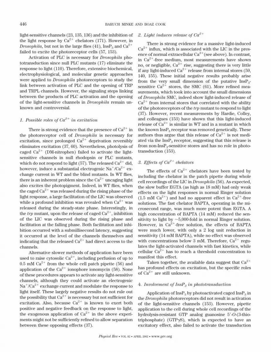
light-sensitive channels (23, 135, 136) and the inhibition ofthe light response by Ca2� chelators (171). However, inDrosophila, but not in the large flies (41), InsP3 and Ca2�
failed to excite the photoreceptor cells (57, 153).Activation of PLC is necessary for Drosophila pho-
totransduction since null PLC mutants (17) eliminate theresponse to light (110). Therefore, extensive biochemical,electrophysiological, and molecular genetic approacheswere applied to Drosophila photoreceptors to study thelink between activation of PLC and the opening of TRPand TRPL channels. However, the signaling steps linkingbetween the products of PLC activation and the openingof the light-sensitive channels in Drosophila remain un-known and controversial.
1. Possible roles of Ca2� in excitation
There is strong evidence that the presence of Ca2� inthe photoreceptor cell of Drosophila is necessary forexcitation, since prolonged Ca2� deprivation reversiblyeliminates excitation (37, 60). Nevertheless, photolysis ofcaged Ca2� (DM-nitrophen) failed to activate the light-sensitive channels in null rhodopsin or PLC mutants,which do not respond to light (57). The released Ca2� did,however, induce a substantial electrogenic Na�/Ca2� ex-change current in WT and the blind mutants. In WT flies,there is an inherent problem since the Ca2� uncaging lightalso excites the photopigment. Indeed, in WT flies, whenthe caged Ca2� was released during the rising phase of thelight response, a large facilitation of the LIC was observedwhile a profound inhibition was revealed when Ca2� wasreleased during the steady-state phase. Interestingly, inthe trp mutant, upon the release of caged Ca2�, inhibitionof the LIC was observed during the rising phase andfacilitation at the falling phase. Both facilitation and inhi-bition occurred with a submillisecond latency, suggestingit occurred at the level of the channels themselves andindicating that the released Ca2� had direct access to thechannels.
Alternative slower methods of application have beenused to raise cytosolic Ca2�, including perfusion of up to0.5 mM Ca2� from the whole cell patch pipette (56) andapplication of the Ca2� ionophore ionomycin (58). Noneof these procedures appears to activate any light-sensitivechannels, although they could activate an electrogenicNa�/Ca2� exchange current and modulate the response tolight itself. These largely negative results do not rule outthe possibility that Ca2� is necessary but not sufficient forexcitation. Also, because Ca2� is known to exert bothpositive and negative feedback on the response to light,the exogenous application of Ca2� in the above experi-ments might not be sufficiently refined to allow separationbetween these opposing effects (37).
2. Light induces release of Ca2�
There is strong evidence for a massive light-inducedCa2� influx, which is associated with the LIC in the pres-ence of normal extracellular Ca2�(see above). In contrast,in Ca2�-free medium, most measurements have shownno, or negligible, Ca2� rise, suggesting there is very littleif any light-induced Ca2� release from internal stores (59,140, 155). These initial negative results probably arisefrom the very small dimension of the putative InsP3-sensitive Ca2� stores, the SMC (61). More refined mea-surements, which took into account the small dimensionsof Drosophila SMC, indeed show light-induced release ofCa2� from internal stores that correlated with the abilityof the photoreceptors of the trp mutant to respond to light(37). However, recent measurements by Hardie, Colley,and colleagues (153) have shown that this light-inducedrelease of Ca2� is similar in WT and in a mutant in whichthe known InsP3 receptor was removed genetically. Theseauthors thus argue that this release of Ca2� is not medi-ated via the InsP3 receptor, suggesting that this release isfrom non-InsP3-sensitive stores and has no role in photo-transduction (153).
3. Effects of Ca2� chelators
The effects of Ca2� chelators have been tested byincluding the chelator in the patch pipette during wholecell recordings of the LIC in Drosophila (56). As expected,the slow buffer EGTA (as high as 18 mM) had only weakeffects on the light responses in normal Ringer solution(1.5 mM Ca2�) and had no apparent effect in Ca2�-freesolutions. The fast chelator BAPTA, operating in the mi-crosecond range, was much more potent than EGTA. Ahigh concentration of BAPTA (14 mM) reduced the sen-sitivity to light by �5,000-fold in normal Ringer solution.However, in Ca2�-free solution, the effects of BAPTAwere much lower, with only a 2 log unit reduction insensitivity (14 mM BAPTA), while no effect was observedwith concentrations below 3 mM. Therefore, Ca2� regu-lates the light-activated channels with fast kinetics, whilecellular Ca2� has to reach a threshold concentration tomanifest this effect.
Taken together, the available data suggest that Ca2�
has profound effects on excitation, but the specific rolesof Ca2� are still unknown.
4. Involvement of InsP3 in phototransduction
Application of InsP3 by photoactivated caged InsP3 inthe Drosophila photoreceptors did not result in activationof the light-sensitive channels (153). However, pipetteapplication to the cell during whole cell recordings of thehydrolysis-resistant GTP analog guanosine 5-O-(3-thio-triphosphate) (GTP�S), which is expected to have anexcitatory effect, also failed to activate the transduction
446 BARUCH MINKE AND BOAZ COOK
Physiol Rev • VOL 82 • APRIL 2002 • www.prv.org

cascade. Therefore, there is probably a problem of acces-sibility of chemicals to the signaling membrane in thepreparation of isolated Drosophila ommatidia duringwhole cell recordings. It is thus possible that these nega-tive results may be attributed to the highly compartmen-talized structure of the phototransduction machinery inDrosophila.
An alternative method of application has been suc-cessfully attempted in the larger flies, Lucilia and Musca
(41, 180). These studies exploited the finding that Luciferyellow applied extracellularly is taken up by the photore-ceptors following intense illumination, possibly via recep-tor-mediated endocytosis (201). Minke and Stephenson(112) introduced extracellular GTP�S into photorecep-tors by application of intense light. The effect of GTP�S inMusca photoreceptors was manifested by a noisy depo-larization in the dark, with properties of the response tolight. Similarly, extracellular application of InsP3 intoMusca or Lucilia retinae accompanied by intense whitelight resulted in a large increase in baseline noise thatpersisted in the dark long (�30 min) after the light. Ap-plication of 2,3-diphosphoglycerate (DPG), an inhibitor ofInsP3 hydrolysis, greatly enhanced the effect of exog-enously applied InsP3 and also enhanced and prolongedthe physiological response to light, suggesting an endog-enous production of InsP3 during illumination. Noise anal-ysis suggested that the InsP3-induced noisy depolarizationis similar to the noisy depolarization induced by dim light(41, 180).
Biochemical studies, which strongly support an es-sential role of PLC, also suggested indirectly that InsP3 isinvolved in fly phototransduction. Evidence for operationof a light-dependent PLC in fly photoreceptors was ob-tained from biochemical experiments in membrane prep-arations of Musca and Drosophila eyes. An eye mem-brane preparation was developed in which light-dependent phosphoinositide hydrolysis could be studiedunder defined conditions, allowing the effects of activa-tors and inhibitors to be analyzed. Musca eyes or Dro-
sophila heads containing intact cells were preincubatedwith [3H]inositol to label the phosphoinositides. Themembrane prepared from these cells was used to analyzethe hydrolysis of phosphoinositides. The Musca eye mem-brane preparation responded to illumination by an in-crease in the accumulation of InsP3 and inositol bisphos-phate (InsP2), the products of polyphosphoinositidehydrolysis by PLC. Addition of DPG substantially de-creased the accumulation of InsP2 and concurrently in-creased the accumulation of InsP3, which thus becomesthe major product of the light-induced phosphoinositidehydrolysis (41). It is apparent that fly membranes areendowed with the enzymatic system necessary to pro-duce InsP3 and to eliminate it after it has been produced.Both the robust light-dependent preferential productionof InsP3 and the efficient turn-off mechanism to stop its
action are consistent with an internal messenger role ofInsP3 in fly phototransduction (for review, see Refs. 107,167).
The Drosophila genomic sequence (2) identifies onlyone InsP3 receptor gene in the Drosophila genome, andmutations in this gene are lethal (1, 194). However, it ispossible to generate mutant photoreceptors in mosaicpatches by inducing mitotic recombination in heterozy-gotes. Intracellular recordings from photoreceptors insuch mosaic patches revealed no differences from WT,leading the authors to conclude that the InsP3 receptorplayed no role in phototransduction (1). A more detailedstudy used mosaic eyes homozygous for a deficiency ofthe InsP3 receptor of Drosophila, where elimination ofInsP3 receptor was confirmed by RT-PCR, Western blotanalysis, and immunocytochemistry (153). In this mutant,all aspects of the photoresponse when examined in detailby whole cell recordings were normal. These results in-dicate that the InsP3 receptor that was eliminated doesnot participate in phototransduction, or that it is redun-dant to a function of another protein. The apparent con-clusion from these experiments is that the InsP3 branch ofthe inositol lipid signaling is not involved in phototrans-duction of Drosophila. However, this conclusion has beenrecently questioned following the use of a novel antago-nist of the InsP3 branch of the inositol lipid signaling.
The membrane-permeant compound 2-aminoethoxy-diphenyl borate (2-APB) has proven effective as a probefor assessing the involvement of InsP3 branch of theinositol lipid signaling in intact cells. 2-APB at 75 �Mblocks receptor-mediated Ca2� store emptying in intactHEK293 cells as well as in several other cells tested (100).In broken cells, 2-APB directly blocks InsP3-mediatedCa2� release from the ER. 2-APB has no effect on InsP3
binding, does not alter InsP3 production through agonist-sensitive PLC, and does not modify the function of ryan-odine receptors or voltage-gated Ca2� channels (100,102). 2-APB applied to the external medium reversiblyand efficiently blocks the endogenous and robust InsP3-mediated signaling pathway of Xenopus oocytes by spe-cifically inhibiting InsP3-induced release of Ca2� frominternal stores. Thus 2-APB seems to operate at a stageafter production of InsP3 but before InsP3 action in me-diating the rise in cellular Ca2� (33). All the above fea-tures of 2-APB, together with very fast penetration intothe signaling region inside the cell, make 2-APB a usefulreagent for studies of the involvement of the InsP3 branchin Drosophila phototransduction. Recent studies indeedshowed that 2-APB is highly effective at reversibly block-ing the response to light of Drosophila flies in a light-dependent manner at a concentration range that coin-cided with its effectiveness in oocytes (33). Furthermore,2-APB had no effect on the opening of the TRP and TRPLchannels themselves, indicating that it operates upstreamof the channels. Accordingly, because 2-APB seems to
447
Physiol Rev • VOL 82 • APRIL 2002 • www.prv.org

block InsP3-induced Ca2� release in vertebrate cells, itcannot be excluded that Drosophila photoreceptors usethe InsP3 branch of the inositol-lipid signaling pathway forlight excitation even if the mechanism and specificity of2-APB action are not clear. The reconciliation of theapparently conflicting knockout of the InsP3R and the2-APB data is likely to shed important new light on themechanism of activation of TRP and TRPL (33). In addi-tion, the same dilemma may apply to the activation mech-anism of vertebrate homologs of the TRPC subfamily,where similar apparently conflicting knockout of InsP3Rsand the 2-APB data have been recently reported (101).Thus, in the triple InsP3 receptor (InsP3-R) knockoutDT40 cells devoided of all three functional InsP3-Rs, SOCactivity was virtually identical to WT cells. However, inboth WT and mutant cells, 2-APB blocked SOC activity inan identical manner (101).
5. Are DAG and polyunsaturated fatty acids second
messengers of excitation?
The DAG branch of the inositol lipid signaling has notbeen considered as a route of excitation in invertebratephototransduction because mutations in an eye-specificPKC (inaC) lead to defects in response termination andadaptation with no apparent effects on activation. Thisnotion has been recently supported by experiments inLimulus showing that application of phorbol esters or thealkaloid indolactam, which are PKC activators, largelysuppressed light-induced Ca2� release and excitation atan early stage of the cascade (39). Contrary to thesefindings, application of phorbol esters and indolactam didexcite mollusk (Lima) photoreceptors in the dark. Theindolactam-induced excitation was also antagonized byPKC inhibitors (40). Phorbol ester also mimicked theeffects of light in rdgB, a retinal degeneration mutant ofDrosophila in which degeneration is largely enhanced byillumination (108).
Recently, a possibility has been considered that DAGdoes not operate solely as activator of PKC but also in aparallel pathway that leads to TRP or TRPL activation, aswell as of mammalian TRPC channels (75; see below).DAG is a potential precursor for formation of polyunsat-urated fatty acids (PUFAs) via DAG lipase, although theactivity of this enzyme has not been demonstrated inDrosophila photoreceptors. Mutations in DAG kinase(rdgA, Fig. 1) that inactivates DAG result in a severe formof light-independent degeneration, although the reasonfor this is unknown (103, 104). Recently, a possible mech-anism has been suggested following the discovery of Har-die and colleagues (35, 154) that in the rdgA mutant TRPand TRPL channels are constitutively open, while elimi-nation of TRP by a mutation (in the double mutant rdgA;
trp) partially rescues both the degeneration and the abil-ity to respond to light. The authors suggest that
elimination of DAG kinase by the rdgA mutation leads toaccumulation of DAG and hence of PUFAs, which consti-tutively activate the TRP and TRPL channels, leading to atoxic increase in cellular Ca2�. Hardie and colleaguesfurther showed that application of PUFAs to isolatedDrosophila ommatidia reversibly activate the TRP andTRPL channels as well as recombinant TRPL channelsexpressed in Drosophila S2 cells. The latter were alsorapidly activated by application of PUFAs in inside-outexcised patches. Application of lipoxygenase inhibitors atlow concentrations induced production of bumplike uni-tary responses followed by activation of macroscopiccurrents at higher concentrations. The authors suggestthat PUFAs may be messengers that directly mediateactivation of TRP and TRPL in Drosophila photorecep-tors (35). It should be noted, however, that genes encod-ing for lipoxygenases have not been found in the Drosoph-
ila genome (2).
6. Constitutive activation of the TRP and TRPL
channels in the dark
The ability to open TRP and TRPL channels in thedark may provide some insight on their gating mecha-nism, since the mechanism of dark openings bypasses atleast some of the phototransduction stages that are acti-vated by light.
RdgA is not the only Drosophila mutant in whichthere is constitutive activation of the light-sensitive chan-nels. Specific mutations in the trp gene also cause consti-tutive activation of the TRP channel, leading to extremelyrapid photoreceptor degeneration in the dark (214, Fig. 5).A new mutant designated TrpP365 does not display thetransient receptor potential phenotype and is character-ized by a near-normal level of the TRP protein and rapid,semi-dominant degeneration of photoreceptors. Despiteits unusual phenotypes, TrpP365 is a trp allele because aTrpP365 transgene induces the mutant phenotype in awild-type background, and a wild-type trp transgene in aTrpP365 background suppresses the mutant phenotype.Moreover, amino acid alterations that could cause theTrpP365 phenotype are found in the transmembrane seg-ment region of the mutant channel protein. These muta-tions are due to the following amino acid substitutions:Pro(500)Thr, His(531)Asn, Phe(550)Ile, and Ser(867)Phe.Whole cell recordings clarified the mechanism underlyingthe retinal degeneration by showing that the TRP chan-nels of TrpP365 are constitutively active, thus presumablyallowing a toxic increase in cellular Ca2� (214, Fig. 6).
Constitutive activation of TRP and TRPL was alsoobtained by metabolic stress, which turned out to be veryefficient in reversibly causing channels openings in thedark in vivo. Accordingly, patch-clamp whole cell record-ings and measurements of Ca2� concentration by ion-selective microelectrodes in intact eyes of normal and
448 BARUCH MINKE AND BOAZ COOK
Physiol Rev • VOL 82 • APRIL 2002 • www.prv.org

mutant Drosophila revealed that anoxia rapidly and re-versibly depolarizes the photoreceptors and induces Ca2�
influx into the photoreceptor cells in the dark (Fig. 7).Furthermore, openings of the light-sensitive channels,which mediate these effects, could be obtained by mito-chondria uncouplers or by depletion of ATP in photore-ceptor cells, while the effects of illumination and all formsof metabolic stress were additive. Effects similar to thosefound in WT flies were also found in mutants with strongdefects in rhodopsin, Gq protein, or phospholipase C, thus
indicating that the metabolic stress operates at a latestage of the phototransduction cascade (Fig. 8). Geneticelimination of both TRP and TRPL channels preventedthe effects of anoxia, mitochondria uncouplers, and de-pletion of ATP, thus demonstrating that the TRP andTRPL channels are specific targets of metabolic stress.These results suggest that a constitutive ATP-dependentprocess is required to keep TRP and TRPL channelsclosed in the dark, a requirement that would make themsensitive to metabolic stress (4). The results also suggest
FIG. 5. Electron micrographs oftransverse sections through the layer ofDrosophila ommatidium TrpP365/trpCM
raised at 19°C and TrpP365/TrpP365. Allsamples were obtained from newlyeclosed adult flies, and all flies weremarked with w. The TrpCM mutant is theoriginal, nearly null, Trp mutant. Theelectron-dense structures are the rhab-domeres, which are composed of mi-crovilli that contain the signaling mole-cules. The trpCM mutation eliminates mostof the TRP protein. The figure shows thatthe degree of degeneration in a newlyeclosed fly depends on the amount of mu-tated TRP having the TrpP365 mutation.[From Yoon et al. (214).]
FIG. 6. Single-cell functional analysisby whole cell recordings of the TrpP365
mutant. A: a typical light-induced current(LIC) of a wild-type cell (left trace) inresponse to an orange stimulus and theabsence of any responses in TrpP365/trpCM and TrpP365/TrpP365 (middle andright traces). The duration of the orangelight stimulus is indicated above eachtrace. B and C compare families of cur-rent traces elicited by series of voltagesteps from photoreceptors of wild-type(left column), the light-insensitive cellsof TrpP365/trpCM (middle column), andTrpP365/TrpP365 homozygotes (right col-
umn). For each experiment, a series ofvoltage steps was applied from a holdingpotential of �20 mV in 20-mV steps (bot-
tom traces). B: membrane currents wererecorded 30 s after establishing thewhole cell configuration with physiolog-ical concentrations (1.5 mM) of Ca2� inthe bath. C: application of 10 mM La3� tothe bath suppressed the membrane cur-rents. The figure shows that the trpP365
mutation eliminated the response to lightdue to constitutive activity of the TRPchannels that could be blocked by La3�.[From Yoon et al. (214).]
449
Physiol Rev • VOL 82 • APRIL 2002 • www.prv.org

that the effects of PUFAs may be indirect, since thesecompounds have been shown in many studies to functionas efficient uncouplers of mitochondria (7). Suppressionof TRP activity is not unique to Drosophila; the NAD-induced activity of LTRPC2 is also suppressed by ATP(see sect. IVD4, Ref. 160).
The constitutive activity of the TRP and TRPL chan-nels may lead to cell death under metabolic stress, notonly in Drosophila but also in vertebrate cells, whichexpress TRPC and TRPM channels (9, 123). Aortic endo-thelial cells have been shown to express an oxidant-activated nonselective cation channel that functions as a
redox sensor. A dominant negative form of TRPC3 abol-ished the oxidant-induced current in aortic cells, suggest-ing that TRPC3 is involved in this conductance (9). Re-cently it has been suggested that LTRPC7 are reporters ofthe metabolic status of cells by being sensitive to Mg-ATP(9). The latter finding is in accord with the findings inDrosophila; however, because it is known that the role ofTRP channels in the fly is to respond to light stimuli, it isconceivable that some of the reported sensitivities of TRPchannels are epiphenomena, while their main role is alto-gether different.
7. TRP and store-operated Ca2� entry
Because Drosophila TRP is the target of inositol lipidsignaling and TRP has a high Ca2� selectivity, Hardie andMinke (63) have proposed that excitation in Drosophila
might be analogous to the process of PI-regulated Ca2�
influx described in a wide variety of mammalian celltypes. Experiments designed to test whether a SOC-likemechanism might operate in Drosophila phototransduc-tion have been largely negative (58). Although both iono-mycin and thapsigargin release Ca2� from intracellularcompartments (58, 155), store depletion by these agents isnot associated with the activation of any currents, nor dothese agents block the response to light during severalminutes (but thapsigargin does block the LIC after longexposure and confers a trp phenotype on WT flies; Ref.37). Application of Ca-BAPTA to the cell via the recordingpipette, buffering Ca2� at concentrations at a level of �10nM, activates an inward current following application ofionomycin (58). However, it is not clear if this effect isdue to store depletion or other effects similar to thoseactivated by metabolic stress. The elimination of theInsP3R by mutation without affecting phototransductionhas also been used as a strong argument against a role ofinositide-mediated store depletion in Drosophila TRP andTRPL activation (1, 153). Thus, although conditions havebeen found in Drosophila to activate TRP and TRPLchannels, which in some studies have been used to dem-onstrate SOC mechanism in vertebrate cells, it is not clearwhether they represent the mechanism operating nor-mally and represent the physiological route of excitation.
C. Role of TRPC3 in Pontine Neurons
At present, TRPC2, TRPC3, TRPC4, and TRPC6 arethe TRPC channels that have been studied in detail in thenative tissue. A recent study by Montell and colleagues(95) has shown that TRPC3 is highly enriched in neuronsof the brain, especially in the forebrain, midbrain, andhindbrain of rat embryo. Because TRPC3 is predomi-nantly expressed in the rat brain during a relatively nar-row developmental period before and after birth (95), itsfunction may be related to developmental processes. Im-
FIG. 7. Anoxia activated the TRP and TRPL channels in both wild-type (WT) (A) and the PLC null mutant (norpAP24; B) as monitored byCa2� influx. The figure shows extracellular voltage change (ERG, top
traces in A and B) and potentiometric measurements with Ca2�-selec-tive microelectrode (ECa, bottom traces in A and B) in response toorange lights and anoxia (N2) in WT Drosophila and norpAP24 mutant.Note that there is no response to light (LM, light monitor) in thenorpAP24 mutant and the initial slow phase of the electrical response toanoxia (before the arrow) is missing in the Ca2� signals of both WT andthe mutant. *Movement artifact. [From Agam et al. (4).]
450 BARUCH MINKE AND BOAZ COOK
Physiol Rev • VOL 82 • APRIL 2002 • www.prv.org

portantly, the temporal and spatial distribution of TRPC3parallels that of the neurotrophin receptor TrkB. Activa-tion of TrkB by brain-derived nerve growth factor (BDNF)
led to production of a PLC-�-dependent nonselective cat-ion current (IBNDF) in isolated pontine neurons. Evidencethat TRPC3 contributes to IBNDF in vivo is provided by the
FIG. 8. Single cell functional analysisby whole cell recordings from Drosoph-
ila photoreceptor showing that depletionof ATP activates the TRP and TRPLchannels of WT cells in the dark. Theconstitutive activity was demonstratedby application of voltage steps in thedark during whole cell recordings fromthe photoreceptor cells during the slowinward current. A: typical light-inducedcurrents (LIC) of a wild-type cell in re-sponse to 3 orange lights was followedby appearance of slow inward current inthe dark when the pipette solution hadno ATP and NAD and the membrane volt-age was held at �50 mV. The top traces
in each pair indicate the duration of theorange light stimuli. B: onset of the darkinward current was accelerated by appli-cation of 0.1 mM DNP (arrow). Note thatno additional response to light was ob-tained after the dark inward current wasinduced. C: comparison of families ofcurrent traces elicited by series of volt-age steps in the range of �100 to �80 mVin steps of 20 mV (lower row), from pho-toreceptors of WT flies in the dark beforethe inward current was induced, at thepeak of the inward current at 1.5 mMexternal Ca2�, after Ca2� was removedfrom the external medium (0 Ca2�) andafter 10 �M La3� was applied to the ex-ternal medium (as indicated). D: effectsof DNP on the trp mutants A relativelyslow induction of a small and noisy in-ward current in the dark was observed innull trp after application of 0.1 mM DNP(arrow) during recordings without ATPand NAD in the recording pipette. Inwardcurrents could not be induced withoutapplication of DNP under similar record-ing conditions. The right traces show afamily of current traces elicited by seriesof voltage steps as in C at 1.5 mM exter-nal Ca2�. E: recording from the trpl;trp
double mutant using pipettes in whichATP and NAD were omitted and 0.1 mMDNP was applied to the external mediumas indicated. No dark inward current wasobserved even 16 min after the beginningof whole cell recordings. The right traces
show a family of current traces elicitedby series of voltage steps as in C at 1.5mM external Ca2�. Note that neitherlight-induced currents nor inward cur-rents in the dark could be induced in thisdouble mutant. [From Agam et al. (4).]
451
Physiol Rev • VOL 82 • APRIL 2002 • www.prv.org

following observations: 1) IBNDF was activated by anti-TRPC3 antibody both at the macroscopic current leveland in single-channel recordings. In these experimentsBNDF and the antibody acted synergistically. 2) Heterol-ogous coexpression of TrkB receptor and TRPC3 in 293Tcells followed by current measurements indicated thatIBNDF was not observed in cells expressing either TrkB orTRPC3 alone. 3) TRPC3 coimmunoprecipitated with TrkBof rat brain, suggesting that the two proteins interact andform a protein complex. 4) IBNDF was blocked by SK&F-96365, a nonspecific inhibitor of TRPC3. Interestingly, theproperties of single-channel conductance that was acti-vated by the anti-TRPC3 antibody in pontine neuronsshowed smaller conductance (i.e., 27 pS, see Table 2) andlonger mean open time (�20 ms) relative to measure-ments in heterologously expressed TRPC3 channels (of�60 pS and �2 ms). In addition, unlike the heterologouslyexpressed TRPC3 channels (100), the native pontineTRPC3 channels could not be activated by the 1-oleoyl-2-acetyl-sn-glycerol (OAG) analog of DAG. Application ofInsP3 activated the pontine TRPC3 channels, and inhibi-tion of either PLC by its antagonist U73122 or the InsP3
receptor by xestospongin (100) inhibited the IBNDF cur-rent. These results are consistent with activation ofTRPC3 via the conformation coupling hypothesis (seebelow), not only in heterologous system (89, 100) but alsoin vivo (95).
D. Control of Ca2� Influx in Vascular Smooth
Muscle Cells
There are several studies reporting that TRP ho-molog channels are involved in the control of vascularsmooth muscle contraction and relaxation (for review,see Ref. 163). Thus TRPC1 has been recently reported tobe involved in SOC activity of resistance arterioles, arter-ies, and veins from human mouse or rabbit (206). TRPC1-specific antibody targeted to the outer vestibule of TRPC1channel identified TRPC1 in native vascular smooth mus-cles. Interestingly, peptide-specific binding of the anti-body selectively blocked SOC activity in these cells, sug-gesting that TRPC1 underlies SOC activity and henceCa2� influx to these cells (206).
In perhaps the most detailed study to date, Mori andcolleagues (81) provided compelling evidence that TRPC6is a requisite component of the �1-adrenoreceptor activa-tor (�1-AR) Ca2�-permeable nonselective cation channel(NSCC) (reviewed in Ref. 163).
1. TRPC6 is the essential component of �1-AR
Ca2�-permeable NSCC channel
The �1-AR is distributed widely in the vascular sys-tem and played a central role in control of systemic bloodpressure via sympathetic innervations. Mori and col-
leagues (81) have found that heterologous expression ofmurine TRP6 in HEK293 cells reproduced almost exactlythe essential biophysical and pharmacological propertiesof �1-AR-NSCC, previously identified in smooth musclesof rabbit portal vein. These properties include activationby DAG, S-shaped I-V relationship, high divalent cationpermeability, single-channel conductance of 25–30 pS,augmentation by flufenamate, and blockade by Ca2�,La3�, Gd3�, SK&F-96365, and amiloride. The level ofTRPC6 mRNA expression was much higher in smoothmuscles of both murine and rabbit portal vein than ofother TRPC members such as TRPC1 or TRPC3. Further-more, treatment of primary cultured portal vein myocyteswith TRPC6 antisense oligonucleotides resulted inmarked inhibition of TRP6 immunoreactivity and selec-tive suppression of �1-AR-activated store depletion-inde-pendent cation current and Ba2� influx.
2. TRPC4 mediates vasorelaxation of blood vessels
A relatively small fraction of TRPC4 channels is ex-pressed in vascular endothelial cells. However, TRPC4 ofendothelial cells is potentially of high functional impor-tance, since Ca2� influx to these cells, presumablythrough TRPC4 channels, may contribute to increase incellular Ca2�, which is necessary for synthesis and re-lease of vasoactive compounds (nitric oxide and prosta-glandins) (49). A strong support for this notion has beenrecently provided by production of knockout mice desig-nated TRP4�/� (49).
Nilius, Flockerzi, and colleagues (49) have studiedprimary cultured mouse aortic endothelial cells (MAECs)of normal mice, which express TRPC4, and comparedthem with similar MAECs of TRP4�/� lacking TRPC4(but not other TRPC channels, as tested in brain tissue)using a protocol that activates SOC channels. To depletethe InsP3-sensitive Ca2� stores they used either InsP3
applied to the patch-clamp pipette or the Ca2� pumpinhibitor tert-butyl-benzohydroquinone (tBHQ). Ca2�
store depletion quickly led to a constitutive inward cur-rent with inward rectification, which was effectivelyblocked by La3� or Mg2� (in Ca2�-free medium) in asimilar manner to the TRP channels of Drosophila. Theconstitutively active channels revealed a high Ca2� per-meability, PCa/PNa � 159.7 (calculated using the constantfield theory). Strikingly, the constitutive SOC current wasmissing in TRP4�/� knockout mice. Furthermore, vaso-active agonists (ACh, ATP) induced an increase of freecellular Ca2�, which was markedly reduced in the knock-out mice. Importantly, stimulation of endothelial cells ofthe knockout mice with the vasoactive agonist revealedthat vasorelaxation of aorta rings was impaired.
The above experiments constitute the first direct ev-idence for the physiological relevance in mature cells ofmammalian TRPC channels and for SOC in endothelial
452 BARUCH MINKE AND BOAZ COOK
Physiol Rev • VOL 82 • APRIL 2002 • www.prv.org

TABLE 2. Functional properties of “TRP-homolog” and “TRP-related” channels
ChannelSingle-Channel
Conductance, pS
RelativePermeability
PCa/PNa
Mechanismof Activation
HeterologousExpression inthe Following
Cells
Species Usedfor Studies in
the NativeTissue
PossibleFunction Reference Nos.
TRP-homolog channels
TRP 4 (in divalent),35 (divalent
free), inDrosophila
110 (PCa/PCs intrpl mutant),
40 (PCa/PNa inWT)
PUFAs?Ca2�?
Sf9, 293T,Xenopus
oocytes
Drosophila Light-activatedchannel
35, 37, 52, 53, 55,57–64, 68, 126,140, 153, 156,158, 190, 209
TRPL 35 (in divalent)in Drosophila,
70 (in divalentfree) inDrosophila
4.3 (PCa/PCs) inDrosophila trp
mutant,3 (PCa/PNa) in
Sf9
PUFAs?DAG (in Sf9)?
293T, S2, Sf9,CHO, Xenopus
oocytes
Drosophila Light-activatedchannel
35, 37, 52, 66, 68,69, 76, 77, 90,91, 129, 130,174, 186, 198,210, 211, 219
TRP� 293T Drosophila Light-activatedchannelsubunit
207
TRPC1 16 (from noiseanalysis)
SOC CHO, Sf9, T293 Xenopus oocytes 18, 97, 98, 99,173, 178, 206,218, 221
TRPC2 SOC?, InsP3 COS-M6 Rat, snake,mouse sperm
Pheromone,acrosomalreaction
84, 96, 184, 192
TRPC3 23 (noiseanalysis),
66 (CHO cells),27 (pontine
neurons)
1.6 DAG, SOC?InsP3 (confor-mationalcoupling)
HEK293, COS,CPAE, 293T,CHO
Rat Neurotrophin-activatedchannel
9, 74, 85, 88, 89,95, 114, 217,218, 220
TRPC6 35, 28 5 DAG CHO, HEK, COS Portal veinmyocytes
�1-AR activated 16, 19, 74, 81,114
TRPC7 5.4 DAG HEK 131TRPC4 42 1, 7, 160 in
nativeendothelialcells
SOC? PLC CHO, HEK Mouse Vasorelaxation 49, 144, 161, 183,185, 197
TRPC5 66, 48 1, 14 SOC? PLC CHO, HEK293 132, 145, 161,178
TRP-related channels
OSM-9 C. elegans Osmoreception,olfaction
36
VR1 35�110
3.8 (heat)9.6 (capsaicin)
PKC, anadamide Xenopus
oocytes,HEK293
Embryonicdorsal rootganglia fromrat
Thermal andinflammatorypain
25, 34, 222
VRL-1 2.9 Hightemperature
Xenopus oocyte,HEK293
Hightemperaturepain
26
GRC PCa/PCs � 0.89 Growth factor CHO Growth factorregulatedchannel
86
OTRPC4 88 (at �60 mV)30 (at �60 mV)
6.3 Osmolarity HEK293 Osmosensorytransduction
177
ECaC 107 Xenopus
oocytes,Rabbit kidney Ca2� transport
in epithel73, 195
CaT2 HEK293 137CaT1 0.5;
42 in divalentfree
130 Xenopus
oocytes, CHOCa2� absorption
in the intestine138, 215
PCL 137 (at 50 mV) �5 Ca2� Xenopus
oocytes,C. elegans; renal
tubular cells,Male mating
(LOV-1)11, 31
Polycystin-2 �6 CHO U937 cells 54LTRPC2 60 Nonselective ADP-ribose HEK-293 Human cells 141, 160LTRPC7,
TRP-PLIK105 2.8 Reduction in
Mg-ATPCHO, HEK-293 DT-40 B cells Channel linked
to cellularmetabolism
123, 159
CHO, Chinese hamster ovary cells; DAG, diacylglycerol; InsP3, inositol 1,4,5-trisphosphate; PLC, phospholipase C; WT, wild type; PUFA,polyunsaturated fatty acid; PKC, protein kinase C.
453
Physiol Rev • VOL 82 • APRIL 2002 • www.prv.org

cells, which seems to be specific to TRPC4 in these cells(49). It is interesting to note that heterologous expressionof mouse TRPC4 and TRPC5 in HEK cells resulted inmarkedly different results (161). In the heterologous HEKcells, TRPC4 could not be activated by store depletion; itshows relatively large single-channel conductance andmuch smaller selectivity to Ca2� relative to Na� (see sect.VC2). Further experiments are required to determine ifthe above differences arise from the different cellularenvironment of the heterologously expressed TRPC4channels.
E. TRPC2 Regulates Ca2� Entry Into Mouse Sperm
Triggered by Egg ZP3
An important function of a TRP homolog channel,TRPC2, in the native cell has been recently demonstratedin the fertilization process of the mammalian egg. Thismultistep process begins by association of the sperm witha glycoprotein constituent of the zona pellucida, ZP3, ofthe egg leading to release of hydrolytic enzymes from thesperm acrosome and remodeling of the sperm surface.This so-called acrosomal reaction is critical for penetra-tion of the sperm into the egg and involves a G protein andPLC-mediated transduction cascade that eventually leadsto activation of SOC channels and sustained increase inintracellular Ca2�. It has been recently shown that TRPC2is essential for activation of the sustained Ca2� influx intosperm by ZP3 (84). The essential role of TRPC2 in theacrosomal reaction was demonstrated by inhibition ofZP3-induced Ca2� influx and acrosomal reaction by anti-TRPC2 antibody (84). The abundant expression of TRPC2isoform in the sperm is also consistent with the role ofTRPC2 in the acrosomal reaction. The same argument isvalid for the localization of TRPC2 to the VNO, whichsuggests that it may function in the specific sensory sys-tem of pheromone detection. Because TRPC2 is a pseudogene in humans, pheromone detection seems to be lost inhumans. However, with regard to fertilization, a proteindifferent from TRPC2 should function in the acrosomalreaction of humans.
F. The TRP-Related Group
Technical difficulties preclude the analysis of severalmembers of this subfamily, such as VRL1, the C. elegans
osm-9, ECaC1, and other channels in their native tissue.An important approach to overcome this difficulty is toproduce knockout mice, in which the specific TRP-relatedchannel has been eliminated, and compare specific func-tion, which is mediated via TRP-related channels. Theirspecific role can thus be studied from the behavior to thecellular level. This approach has been used for TRPC4(see sect. IVD2) and for VR1 channels (see below). In
addition, the cellular localization of these channels tospecific tissue or sensory neurons allows some predic-tions on the function of these channels and comparison ofthese predictions to the actual measured properties inheterologous systems.
1. VR1 channels mediate specific pain
Pain is initiated when peripheral terminals of a sub-group of sensory neurons are activated by noxious chem-icals, pH, and mechanical or thermal stimuli. Nociceptorsare characterized, in part, by their sensitivity to capsaicin(27). Capsaicin, the active ingredient in hot pepper, is anexcitatory neurotoxin that selectively destroys primaryafferent nociceptors in vivo and in vitro. Heterologousexpression of VR1 in HEK293 cells induced necrotic celldeath within several hours of continuous exposure tocapsaicin while control cells were not affected (27).These results are reminiscent of the effect of the trpP365
and the rdgA mutations in Drosophila; they produce sim-ilar effects in vivo presumably via constitutive activity ofthe TRP channels (see above). The natural stimulus thatactivates VR1 channels (also designated TRPV1) seems tobe noxious heat, since the temperature-response profileof VR1 matches very closely those reported for heat-evoked pain response in mammals and in cultured neu-rons. Also, ruthenium red, which blocks VR1 activity inmammalian cells or in oocytes, blocks heat-evoked noci-ceptive response in the rabbit ear (27). In addition, theheat-evoked currents observed in both VR1-expressingcells and cultured sensory neurons are carried throughoutwardly rectifying, nonselective cation channels. Re-duction in tissue pH, which usually results from infection,inflammation, or ischemia, seems also to be endogenousmodulators of VR1. Interestingly, the endogenous canna-binoid receptor agonist anandamide was found to inducevasodilatation by activating vanilloid receptor on perivas-cular sensory neurons. Heterologous expression of VR1 inHEK293 cells and Xenopus oocytes (222) validates thatanandamide is an agonist of this receptor.
An important step toward the establishment of the invivo function of VR1 has been made by the creation ofknockout mice lacking VR1. Sensory neurons from micelacking VR1 are severely deficient in their response to thenoxious stimuli heat (�43°C), protons, or vanilloid com-pounds but exhibited normal responses to noxious me-chanical stimuli (25).
Recently, a functional study of the mechanism under-lying activation of VR1 channels in both the native neu-ronal cells and a heterologous system has been reported.(150). Because PKC is a prominent participant in painsignaling (28, 29, 87), the role of PKC in VR1 activationwas studied. Application of 12-tetradecanoylphorbol 13-acetate (TPA), which activated endogenous PKC, to Xe-
nopus oocytes expressing VR1 markedly increased the
454 BARUCH MINKE AND BOAZ COOK
Physiol Rev • VOL 82 • APRIL 2002 • www.prv.org

amplitude of capsaicin, anandamide, or proton-activatedcurrents, while TPA induced a current by itself. SpecificPKC inhibitors antagonized the above effects. Excisedoutside-out membrane patches of VR1 expressing oocytesshowed pronounced activation of single channels duringapplication of capsaicin or TPA, with a conductance of�110 pS. This single-channel conductance is much largerthan previously reported for the same channel (i.e., 35 pS)(27) (see Table 2). Replacing ATP with the hydroly-sis-resistant analog adenosine 5-O-(3-thiotriphosphate)(ATP�S) activated the channels in a similar manner toTPA, suggesting that phosphorylation of proteins by PKCunderlies channels activation. Importantly, similar single-channel activation by capsaicin, TPA, or the algestic pep-tide bradykinin was observed in patch-clamp cell-at-tached recordings from sensory neurons, isolated fromthe dorsal root ganglia of rat embryos. Recently, it hasbeen demonstrated that bradykinin and nerve growth fac-tor (NGF) release VR1 from PIP2-mediated inhibition.Accordingly, diminution of plasma membrane PIP2 byantibody sequestration of PLC hydrolysis mimics the po-tentiating effects of bradykinin and NGF (34). Modulationof PIP2, which may regulate VR1 function, has been sug-gested also to regulate TRPL activity in heterologoussystem (47). Such a mechanism is unlikely to function invivo, since depletion of PIP2 in the trp mutant that hasonly TRPL channels suppresses, rather than facilitates,TRPL channel activity (67). A modulatory role of anad-amide in PKC-VR1 signaling was examined by applicationof anandamide to excised patches of Xenopus oocytesexpressing VR1. Application of anandamide (10 �M) pro-duced a progressive increase in single-channel activityand in the macroscopic current (150). This and the abovestudies demonstrate again the complex nature of TRPgating in different cells.
2. C. elegans osm-9 may regulate olfaction
and osmoregulation
Functional analysis of C. elegans OSM-9 was basedon behavioral tests in mutant animals. These tests indi-cate an osm-9 gene function in a specific subset (AWA) ofneurons that mediate olfactory transduction, response toosmotic and nose-touch stimuli. It is also involved in slowadaptation to olfaction stimuli (36). No functional studiesat the cellular level are available.
3. The epithelial Ca2� channels ECaC1, CaT1, CaT2,
and OTRPC4
Although the functional properties of ECaC1, CaT1,CaT2, and OTRPC4 (see Fig. 4) have been derived fromheterologous expression studies (see below), their ratherspecific localization to epithelial cells of specific organspoints to the possible function in the native tissue as thegate keeper of 1,25-dihydroxyvitamin D3-dependent active
transepithelial Ca2� transporter in the kidney (73, 137) ora highly Ca2�-selective SOC channel in the duodenum(CaT1) (129). OTRPC4, which functions as an osmorecep-tor and is expressed predominantly in the distal nephronof the kidney, a region exposed to hypotonic solution, isactivated by a decrease in osmolarity, and thus its activa-tion mechanism fits with its localization (177). Their func-tional expression in heterologous cells supports this con-clusion (see below).
G. NOMPC Is a Channel That Mediates
Mechanosensory Transduction
The mechanotransduction channel NOMPC belongsto a unique subfamily of the TRP-related subfamilies(196). The evidence that NOMPC mediates mechanosen-sory transduction came from a highly (10% of control)reduced mechanoreceptor current in mechanosensoryneurons located at the mechanoreceptor organ of theDrosophila nompC mutant (196). Mechanosensory neu-rons reveal a pronounced degree of adaptation to me-chanical stimuli. Thus the intensity-response relationshipbetween the amplitude of the mechanoreceptor currentand displacement of the hair bristle shows a very steepdependence (range of 10 �m) during constant back-ground displacement. The adaptation is manifested by ashift of the intensity response curve with increasing theintensity of the background displacement. Interestingly,one allele of nompC (nompC4) shows an abnormallyrapid adaptation, whereas the amplitude of the mechano-receptor current is normal. This mutant may prove valu-able to study the regulation mechanism of the channel. Atpresent there are no data on the ionic mechanism of themechanoreceptor current and on properties of theNOMPC channels.
H. Summary
The expression pattern of various members of theTRP family to specific cells and tissues may give us a clueregarding their specific functions, which seem to be di-verse. Unfortunately, the absence of selective antagonistsand the difficulty in analyzing many members of the TRPfamily in the native tissue impose a great difficulty inunderstanding the function of these TRP channels and inresolving their mechanism of activation and propertiesunder physiological conditions. The detailed studies inDrosophila that combine genetic dissection with power-ful electrophysiological and single-cell Ca2�-measuringtechniques may also provide important clues regardingthe activation mechanism of at least the mammalianTRPC channels that have striking structural similarity toDrosophila TRP and TRPL. There is a steadily growingresearch on mammalian members of the TRP family that
455
Physiol Rev • VOL 82 • APRIL 2002 • www.prv.org

have been conducted in the native tissue. New studiesshow a crucial role of TRPC2 in the process of acrosomalreaction in sperm, while TRPC3 has a role in developingpontine neurons. Similarly, studies of VR1, TRPC1,TRPC4, and TRPC6 in primary afferent neurons, salivaryglands, and vascular smooth muscles have revealed anessential role in nociceptors, in control of fluid secretionin salivary glands (172), and in control of smooth muscletone, respectively. It thus seems that studies in the nativetissue are indispensable. In the following section, recentstudies on coexpression of two different TRPC channels,which probably form heteromultimeric channels, are re-viewed. The striking differences in channel properties ofhomo- and heteromultimeric channels described below,the possible sensitivity of TRPs to the cellular environ-ment, and the probable protein-protein interactions inmost TRPs emphasize the need to study TRP channels inthe native cellular environment.
V. FUNCTIONAL ANALYSIS
IN HETEROLOGOUS SYSTEMS
A. Heterologous Expression of trpl, But Not trp,
Leads to Channels With Similar Properties
of the Native Channel
Expression of cDNA of trpl and possibly of trp in avariety of expression systems has provided importantsupport that the above gene products function as chan-nels. The expression cells include two insect cell lines(Spodoptera Sf9 cells and Drosophila S2 cells), mamma-lian cell lines (HEK293T and CHO cells), and Xenopus
oocytes (52, 69, 90, 129, 143, 174, 209). Schilling andcolleagues (190) first reported heterologous expression ofTRP. These investigators found that expression of trp
cDNA in Sf9 cells led to the appearance of a novel con-ductance that could be activated by thapsigargin. How-ever, there is only limited similarity between the proper-ties of the heterologously expressed TRP-dependentconductance and the light-induced conductance of Dro-
sophila. The TRP-dependent currents in the Sf9 cellsmarkedly differed from the light-activated current of Dro-
sophila in several main aspects. The I-V relationship isapproximately linear in Sf9 cells expressing TRP, whileshowing inward and outward rectification in Drosophila.
When the holding voltage is stepped, there is instanta-neous current with no time-dependent kinetics in the cellexpressing TRP unlike the LIC. The TRP-dependent cur-rent of Drosophila shows positive and negative feedbackeffects of Ca2� on the LIC, which are completely absent inthe Sf9 cells. The TRP-dependent conductance in Sf9 cellshas low permeability to Ba2� and is not blocked by Mg2�
in concentrations that have a strong blocking effect inDrosophila (158). Xu et al. (209) functionally expressed
TRP and found a novel conductance that could be acti-vated by thapsigargin in HEK293T cells. However, theproperties of this current like those of the Sf9 cellsshowed several quantitative discrepancies from the in situcurrent (e.g., less permeable to Ca2�, less sensitive toblock by either La3� or Mg2� , and linear I-V curve with-out rectification).
Expression of TRP did show appearance of the TRPprotein in the heterologous system in the few cases whereit was investigated (52, 174, 209). Nevertheless, in severalunpublished studies no effect of transient TRP expressionwas found in either HEK293 cells (Minke, unpublishedobservations) or in Drosophila S2 cells. Expression ofTRP in Xenopus oocytes has provided inconsistent data,whereas one study showed enhancement of the endoge-nous Ca2�-activated Cl� current in TRP expressing oo-cytes (143), and another study failed to see any significanteffect when TRP was expressed alone without TRPL (52).Taken together, it seems doubtful whether the heterolo-gously expressed TRP reaches the surface membrane andfunctions as a channel; all reported conductance can beexplained by enhancement of endogenous activity ofchannels of the host cells.
Indeed, the GTP binding protein Rab6, which is re-quired for vesicular trafficking within the Golgi and post-Golgi compartments, turned out to be essential for TRPexpression in normal amounts in the native tissue (168).Thus overexpression of mutant Rab6 protein shows re-duction in both rhodopsin (Rh1) and TRP proteins. Rab6is not the only protein required for TRP expression. Pakand colleagues (93) found a novel protein called INAF,which appears to be required for normal level of TRPexpression and perhaps also for TRP function in the flyeye. Accordingly, a null mutant of INAF specifically re-duced the amount of TRP to a very low level and seems tospecifically affect the TRP activity (93).
The situation is very different for heterologous ex-pression of TRPL, in which the appearance of a nonselec-tive cation conductance has been reported after expres-sion in Sf9 cells (69, 77), S2 cells (68), HEK293 cells (209),CHO cells (69), and Xenopus oocytes (52, 91). In severalcases, a constitutive activity of the TRPL channels waslargely enhanced by coexpressed receptors, which acti-vate endogenous G protein-coupled PI pathways. A com-mon feature of the TRPL expression studies is a consti-tutive spontaneous activity, which increases with timeduring whole cell recording, although Minke and col-leagues (52) and Montell and colleagues (209) reportedthat this was prevented when coexpressed with a largequantity of TRP. The channel properties of TRPL whenexpressed in Drosophila S2 cells were compared with theTRPL-dependent light-sensitive conductance as deter-mined in the native cells of trp mutants during whole cellrecordings (158). A variety of properties including single-channel conductance and open times, ionic selectivity for
456 BARUCH MINKE AND BOAZ COOK
Physiol Rev • VOL 82 • APRIL 2002 • www.prv.org

monovalent and divalent ions, block by Mg2�, and I-Vrelationship were found to be indistinguishable betweennative and heterologously expressed TRPL. The proper-ties of TRPL channels expressed in other expression sys-tems appear similar (90, 130). Thus, in contrast to theapparent failure to heterologously express functionalTRP, heterologous expression of TRPL appears to form aconductance similar to that found in the native tissue,except that some factor is required to prevent constitutiveactivity, which does not occur under normal conditions invivo. The recent cloning of TRP� indeed solved this latterissue (207). Thus heterologous expression of a TRP�-TRPL construct in 293T cells displayed an agonist (ATP)-activated current, which was not observed in controlcells. The TRP�-TRPL expressing cells do not show con-stitutive activity, and the I-V relationships show the out-ward rectification typical for the TRPL channels in situ,strongly suggesting that TRP�-TRPL forms a heteromul-timer (207).
B. Mechanism of Activation of TRPL
1. Receptor-mediated activation
Coexpression of TRPL and the M5 muscarinic recep-tor in Sf9 cells using the baculovirus expression systemreveals an increase in basal Ba2� influx typical of theconstitutive activity of TRPL channels in these cells. Ad-dition of carbachol to these cells produced a large in-crease in cellular Ca2�, which could be blocked by atro-pine and by gadolinium (Gd3�, 0.1–1.0 mM) but wasinsensitive to La3� (10 �M). Depletion of the Ca2� storesby thapsigargin had no effect, suggesting that TRPL is astore-independent channel (76, 77). Similar results werealso obtained by coexpression of TRPL with other hor-mone receptors in Sf9 cells such as the histamine, throm-bin, and thromboxane A2 receptors (129, 130). The typicaloutwardly rectifying TRPL current was observed afterhormonal stimulations following the TRPL expression inthese cells.
2. Activation by InsP3
TRPL channels expressed in Sf9 cells were activatedby inclusion of InsP3 in the recording pipette (43). Al-though heparin blocked activation by InsP3, it could onlypartially counteract activation by a coexpressed bradyki-nin receptor, suggesting an additional alternative route ofexcitation. The authors concluded that the effect of InsP3
was not mediated via a rise in Ca2�, since it was unaf-fected by inclusion of 1 mM EGTA in the pipette. Theauthors suggested that InsP3 might directly gate TRPL viaconformational coupling (see below). Zimmer et al. (219)also found an excitatory effect of InsP3 but reported thatsince this was abolished by prior depletion of stores with
thapsigargin it was an indirect action via a rise in Ca2�.Schultz and colleagues (129) using Sf9 cells did not findany effect of InsP3 but reported that activated Gq/11 sub-units could activate TRPL channels either when overex-pressed or, in a few cases, when applied to inside-outpatches. Although they initially suggested that TRPLchannels were directly gated by the G protein in similarmanner to some K� channels, more recently they con-cluded that their findings are best explained by G protein-mediated activation of PLC (70).
3. Effects of Ca2�-CaM
TRPL was initially isolated on the basis that theexpressed protein binds CaM via two putative CaM bind-ing sites (147). Subsequently, the two CaM binding sites(designated CBS1 and CBS2) have been more accuratelyidentified and studied in vitro and in vivo (32, 164). CBS1binds CaM in a Ca2�-dependent fashion (300–500 nMCa2�). In contrast, CBS2 binds CaM in the absence ofCa2� with dissociation occurring at levels above 5 �M.The authors suggest that dissociation of CaM from CBS2and Ca2�-dependent CaM binding to CBS1 are the mech-anism of TRPL activation (198). Similar inhibition ofTRPC channels by CaM has been recently demonstrated(182). In general, similar dependence of TRPL activationon CaM binding was obtained by expression of TRPL inCHO cells. However, these experiments showed Ca2�
dependence of both CaM binding sites CBS1 and CBS2,although they differ in their affinity to CaM with Ki valuesof 12.3 nM and 1.7 nM for CBS1 and CBS2, respectively.Both sites bind CaM only in the presence of Ca2�, and theCaM antagonist calmidazolium inhibits the Ca2�-depen-dent activation of TRPL current, suggesting modulation ofTRP currents by cellular Ca2� via CaM (186). Lan et al.(91) reported that heterologous expression of TRPL inXenopus oocytes leads to an elevated free Ca2�, whichwas increased by low concentrations of CaM and inhib-ited at high. Zimmer et al. (219) reported that TRPLactivity in CHO cells was enhanced by application of Ca2�
with an EC50 of 500 nM and that this could be reduced bythe CaM antagonist calmidazolium.
Schultz and colleagues (130) have obtained differentresults in Sf9 cells that express TRPL channels togetherwith the histamine receptor. The histamine-inducedTRPL-dependent currents revealed reversible inhibitionby Ca2� (IC50 of 2.3 �M). Consistent with these observa-tions, inside-out patches revealed TRPL channel activitythat was reversibly inhibited by Ca2� (10–50 �M) in thecytosolic side. Interestingly, the CaM inhibitor calmidi-azolium had no inhibitory effect on channel activity, thussuggesting that CaM was not involved in the inhibition(130). Consistent with inhibition of the TRPL channels byCa2� in Sf9 cells, experiments in the native tissue ofDrosophila have shown pronounced inhibition of the LIC
457
Physiol Rev • VOL 82 • APRIL 2002 • www.prv.org

in the trp mutant, which has only TRPL channels. TRPLchannels appear to be negatively regulated by Ca2�, sincein the presence of extracellular Ca2� the response to lightis reduced by approximately threefold (158). Similarlycaged Ca2� released during the rising phase of the lightresponse inhibited the light response in trp mutants (59).To examine if CaM involves TRPL inhibition by Ca2�,Zuker and colleagues (164) generated transgenic flies inwhich TRP channels were absent and the native TRPLchannels were replaced with constructs with CBS1 orCBS2 deleted. Activation by light in these flies was essen-tially unaltered, whereas there were major defects inresponse termination.
Hardie and Raghu (66) examined the effect of Ca2� inS2 cells coexpressing the TRPL channels with a musca-rinic receptor. Releasing caged Ca2� resulted in a pro-nounced facilitation of the spontaneous TRPL activity.Releasing caged InsP3 released Ca2� from internal storesbut had at most a small effect on TRPL activity and noneat all when BAPTA was included in the pipette solution. Incontrast, excitation of TRPL via the coexpressed musca-rinic receptor resulted in a rapid excitation of TRPL chan-nels, which was not blocked by prior depletion of storesby InsP3 or thapsigargin or by inclusion of BAPTA in thepipette. The results suggest that a process requiring Gprotein and PLC, but not InsP3, underlies activation ofheterologously expressed TRPL channels while channelactivity can be modulated by Ca2�-CaM. Taken together,the bulk of data suggest that in heterologous systemsTRPL is constitutively open and that the channel can benegatively and positively regulated by Ca2�-CaM.
4. Activation by PUFAs and DAG
PUFAs have been shown to activate recombinant TRPLexpressed in S2 cells (35). This preparation has an advan-tage over the photoreceptor cells, because it allows mea-surements of single-channel activity. Importantly, PUFAselicited TRPL channel activity in excised patches, suggestingdirect activation of the channel by PUFAs. The fact thatactivation of TRPL by PUFAs was observed in the photore-ceptors of the null norpA mutant, which lacks the photore-ceptor specific PLC (17) and does not respond to light, wasused as a strong argument in favor of direct activation of thechannels without a need for PLC or other upstream proteins.Schilling and colleagues (47), who measured the effects ofDAG analogs and PUFAs on TRPL expressed in Sf9 cells,reported apparently different results. In contrast to the lackof any effect of DAG or DAG analogs on Drosophila photo-receptors, the DAG analog 1-stearoyl-2-arachidonyl-glycerol(SAG) (but not OAG) induced Ca2� influx and elicited sin-gle-channel activity in TRPL-expressing Sf9 cells. Consistentwith the data reported by Hardie and colleagues (35),PUFAs activated both Ca2� influx in intact Sf9 cells express-ing TRPL and elicited single-channel activity in excised
patches. However, in contrast to previous claims that PLC isnot directly involved in single-channel activation of excisedpatches, single-channel activation was elicited by applica-tion of three different PLCs. Furthermore, activation ofTRPL channels in Sf9 cells by PUFAs was markedly atten-uated after inhibition of PLC by the U73122 antagonist (butnot by the control compound U73343) (47). The authorsconcluded that both the hydrolysis of PIP2 and the genera-tion of DAG are required to rapidly activate TRPL channelsafter receptor stimulation and that the effect of PUFAs ismediated at least in part via PLC. The reports are, therefore,contradictory, supporting either direct or PLC-mediated ac-tivation of TRPL by PUFAs. These results may arise fromthe multiple effects that PUFAs have while the activationof photoreceptors may arise indirectly from the effects ofPUFAs as mitochondria uncouplers causing metabolicstress (4).
5. Store-operated Ca2� entry
Almost all studies have found that TRPL is not acti-vated by store depletion. One study has reported activa-tion of TRPL by thapsigargin (211), but the possibility thatthe regulation was due to the associated increase in Ca2�
was not rigorously excluded.In contrast, as mentioned above, heterologously ex-
pressed TRP is presumably activated by store depletion.
6. Coexpression of TRP and TRPL channels
Coexpressing Drosophila TRP and TRPL in Xenopus
oocytes in cRNA ratios of 10:1 reveals a novel inwardcurrent showing inward and outward rectification thatwas activated by Ca2� store depletion. This current wasneither observed in native oocytes nor in oocytes express-ing either TRP or TRPL alone by the same amounts ofcRNA, implicating a cooperative action of TRP and TRPLin the depletion-activated current (52). Heterologous co-expression of TRP and TRPL in 293T cells produced anoutwardly rectifying novel current, which could be acti-vated by store depletion. Immunoprecipitation studiesrevealed physical interaction between TRP and TRPL asfound in the native Drosophila retina (209). Evidence thatTRP and TRPL form heteromultimeric channels was ob-tained by suppression of the TRP-dependent current bydominant negative form of TRP, and vice versa. It wasalso shown that the NH2 terminal and transmembranedomains contributed to subunit assembly. Because eachof the channels by itself can be opened by light, therelevance of coexpression of TRP and TRPL to the nativesystem is still not clear.
C. Heterologous Expression
of “TRP-Homolog” Channels
Functional analysis of the TRPC group has been con-fined mainly to heterologous systems (but see TRPC2,
458 BARUCH MINKE AND BOAZ COOK
Physiol Rev • VOL 82 • APRIL 2002 • www.prv.org

TRPC3, TRPC4, and TRPC6). Not surprisingly, the activa-tion mechanism and biophysical properties remain un-clear and controversial. Thus conflicting results havebeen reported for the same channel for reasons that re-main unclear. A possible explanation may be the use ofdifferent expression system and expression levels of theTRPC homolog and possible interaction with endogenousproteins of the expressing cells.
TRPC1, TRPC4, and TRPC5 homologs seem to formone functional group. However, several studies suggestthat they are activated by store depletion and thereforeconsider them as SOCs (15), while other studies showstore-independent activation (70).
1. TRPC1
The TRPC1 isoform of human was transiently coex-pressed with the M5 muscarinic receptor in COS cells.Activation of the endogenous inositol lipid signaling ofthese cells through the M5 receptor was obtained byapplication of carbachol (CCh). The depletion of the Ca2�
stores leads to the opening of Ca2�-permeable surfacemembrane channels. This entry of Ca2� is the manifesta-tion of the endogenous SOC in control cells. In the co-transfected COS cells with TRPC1 and M5 receptor, asignificantly larger Ca2� influx relative to control cellswas observed (218). Additional studies on a spliced vari-ant of TRPC1 (221) expressed in CHO cells investigatedsome biophysical properties of the expressed TRPC1 ho-molog. Transient expression of TRPC1A induced a con-stitutive inward cationic current in Ca2�-free medium,which was significantly larger than the control. Biophys-ical analysis of this current revealed a nonselective cationcurrent with similar permeability for Na�, Ca2�, or Cs�.The current was enhanced in stably expressing cells per-fused with Ca2� buffer after application of InsP3 or thap-sigargin, which presumably depletes the Ca2� stores andwas inhibited by the lanthanide Gd3�. Based on theseresults, TRPC1A was characterized as a SOC channel.
Expression of the human TRPC1 isoform in the in-sect Sf9 cells using the baculovirus expression system(173) provided a different conclusion. Expression ofTRPC1 caused an increase in basal cytosolic free Ca2� aswell as Ba2� concentration as a function of posttransfec-tion time. The latter indicated activation of an influxpathway. Whole cell recordings indicate that the inwardcurrent was nonselective with respect to Ca2�, Ba2�, orNa� and was blocked by La3�. Both Ba2� influx and thecationic current were unaffected by thapsigargin, InsP3,or internal Ca2� buffers. Thus, according to this study,TRPC1 is a nonselective cationic channel, which is storeindependent. The authors propose (without further evi-dence) that it requires additional subunits to become aSOC. Similar failure to activate human TRPC1 expressedin HEK cells by store depletion was observed by Gros-
chner and colleagues (97). Interestingly, TRPC1 could beactivated (apparently directly and not via PKC) by theDAG analog OAG (Table 2). Previous studies by Schultzand colleagues (74, 100) first showed that DAG and itsanalogs activate TRPC3 and TRPC6 channels, but couldnot activate TRPC1 under the same experimental condi-tion. The difference in the results of the two groups canbe explained by the suppression of the OAG-induced re-sponse in TRPC1 by Ca2�, which was present in theexternal medium of the initial experiments (97).
Expression of TRPC1 in human mandibular glandcell line (HSG) resulted in a three- to fivefold increase inSOC activity measured by thapsigargin-stimulated Ca2�
influx, while similar transfection with TRPC3 did notshow enhancement of SOC activity. Interestingly, trans-fection of HSG cells with antisense TRPC�1 cDNA signif-icantly attenuated the endogenous SOC activity, whilechanges in SOC were associated with correspond-ing changes in the levels of TRPC1 in the plasma mem-brane (98).
2. TRP4 and TRP5
The bovine TRPC4 homolog was studied electro-physiologically after overexpression in HEK cells (144).Depletion of the Ca2� stores in low Ca2�-buffered cells byGTP�S, InsP3, or thapsigargin induced a large inwardlyrectifying current that was not observed in the controlcells. The relative permeability of the expressed channelto cations was PCs:PNa:PCa:PBa � 1:1.1:7.7:12.3. Inability toresolve single-channel activity was interpreted as an indi-cation of very low single-channel conductance. Thus, ac-cording to this study, TRPC4 is a SOC with high perme-ability to divalent ions (144, 197). Results similar to thoseobtained in the TRPC4 isoforms were also obtained afterexpression of TRPC5 in HEK cells (144). The authorsconcluded that these channels have properties reminis-cent of CRAC channels.
Two other studies on mouse isoforms of TRPC4 andTRPC5 (161) and on TRPC5 (131) expressed in HEK cellsreported conflicting results. In TRPC5-expressing cells,activation of the endogenous purinergic receptor by ap-plication of ATP induced a large transient increase incellular Ca2� and inward current only in the presence ofexternal Ca2� (131). Omission of Ca2� from the recordingpipette abolished the ATP-induced current, suggestingthat increase in cellular Ca2� is required for TRPC5 acti-vation. The permeability ratio of the current was PCa:PNa:PCs � 14.3:1.5:1.0, showing Ca2� selectivity. La3� andGd3� and SK&F-6365, an inhibitor of nonselective cationand some Cl� channels, inhibited the Ca2� transients.Single-channel activity during patch-clamp recordings atlow external Ca2� show single-channel conductance of47.6 pS. Ca2� store depletion by thapsigargin had no
459
Physiol Rev • VOL 82 • APRIL 2002 • www.prv.org

effect larger than the control, thus indicating that thischannel is not SOC (see Table 2).
Detailed functional studies of mouse TRPC4 andTRPC5 expressed in HEK cells have shown that these twochannel types are not only structurally related but alsodisplay similarities in their mechanism of regulation andbiophysical properties (161). Contrary to the experimentson bovine and rat orthologs, activation of the mouseisoforms was independent of the filling state of the inter-nal Ca2� stores. Importantly, these channels did not re-spond to InsP3, in contrast to TRPC3 channels (see be-low) (161). Activation via a G protein-coupled PLC wasdemonstrated by activation with CCh or histamine (aftercoexpression with the histamine receptor) by applicationof GTP�S, which activated single channels, and by inhi-bition of the agonist-induced current by the PLC inhibitorU73122. In contrast to the proposed similarity to CRACchannels, the studies on mouse TRPC4 and TRPC5 reveallarge single-channel conductance of 41 and 63 pS in thepresence of Mg2� and similar conductance to both mono-valent and divalent cations. Similarly, single-channel con-ductance of 47.6 pS was measured in another study ofheterologously expressed TRPC5 in HEK293 cells (212).The conductance and the inward and outward rectifica-tion of the I-V relation were markedly dependent onMg2�. In the absence of Mg2�, the rectification was lost,reminiscent of the effect of Mg2� on Drosophila TRP.Surprisingly, La3� at concentrations up to 1 mM facili-tated TRPC4- and TRPC5-dependent currents (161). Thereasons for the marked differences in regulatory and bio-physical properties between species and studies in differ-ent laboratories remain unknown. Interestingly, the ratortholog of TRPC4 lacks the entire putative S2 region. Arat ortholog expressed in Xenopus oocytes was function-ally characterized as SOC (185). It is still not clear if themissing S2 segment is responsible for this phenomenon.Thus the bulk of the most recent studies on TRPC4 andTRPC5 in heterologous systems suggest that these chan-nels are receptor-activated and store-independent non-specific cation channels, in contrast to studies in nativeendothelial cells (49). Importantly, DAG or DAG analogsdo not activate these channels.
3. TRPC2
Only a few studies of the heterologously expressedmouse TRPC2 channel are available. Nevertheless, thespecific expression of rat TRPC2 isoform to the VNOorgan of rat and sperm of mice have already provided avery significant clue as to the functions of TRPC2. Ac-cordingly, TRPC2 may be the target channel of a G pro-tein-coupled receptor that mediates detection of phero-mones.
Heterologous expression of mouse TRPC2 in COScells expressing the muscarinic receptor M6 revealed a
typical SOC. The expressed TRPC2 channels were testedby the same paradigm described above for TRPC1 andshow a significant enhancement of Ca2� influx after storedepletion by thapsigargin (192). These findings seeminglycontradict the expectation of Liman et al. (96) that expectTRPC2 to be store-independent channels, since the VNOorgan where TRPC2 is localized is composed of microvilliand has no Ca2� stores.
4. TRPC3, TRPC6, and TRPC7
The members of this group of TRP channels areclosely related in molecular structure and function. Thehuman TRPC3 has been extensively characterized by anumber of investigators. It was stably expressed in HEKcells (217) and bovine pulmonary artery endothelial cells(85) and transiently expressed in CHO cells (220). Theheterologously expressed TRPC3 channel has been stud-ied by various techniques including whole cell recordingsof macroscopic current, excised patch-clamp recordingsof single channels, as well as fluorescence Ca2� measure-ments using Ca2� indicators. Essentially all investigatorsreported similar properties of the TRPC3 channel (193).TRPC3 forms a nonselective cation channel that is acti-vated by a G protein-coupled receptor, G protein, andPLC. La3�, Gd3�, and SK&F-96365 are potent blockers ofthe channel that has a single-channel conductance of60–66 pS. The I-V relation shows inward and outwardrectification. Most studies also reported that TRPC3 is astore-independent channel and that thapsigargin has noeffect. Nevertheless, several investigators have reportedsome enhancement of the receptor-activated current inTRPC3-expressing cells by thapsigargin treatment (89,218). A different line of evidence supporting the notionthat TRPC3 function as SOC channels was provided bystable transfection of HEK293 cells with a fragment ofhuman TRPC3 in antisense orientation. In the antisensetransfected cells, activity of SOC channels monitored byBa2� influx after store depletion with thapsigargin wasreduced by 32% compared with cells expressing senseTRPC3 (205).
Interestingly, in addition to activation by several ago-nists via a receptor and in few cases by store depletion,TRPC3 could be activated by DAG or its analogs (74, 100).Different studies have reported conflicting data with re-gard to the effects of InsP3. Although Hoffman et al. (74)reported that application of InsP3 has no effect on humanTRPC3 or TRPC6 expressed in CHO cells, other studieson human TRPC3 expressed in either bovine pulmonaryartery endothelial cells or in HEK293 cells show clearactivation by InsP3 (89, 145). DAG and its analogs seem toactivate the TRPC channels directly and not via theInsP3R because application of the InsP3R antagonist2-APB does not affect the activation by DAG analogs(100).
460 BARUCH MINKE AND BOAZ COOK
Physiol Rev • VOL 82 • APRIL 2002 • www.prv.org

The most interesting studies on TRPC3, which opennew avenues toward understanding TRP gating, have de-scribed the interactions between TRPC3 and the InsP3R(88, 100). The study by Muallem and colleagues (88) hasprovided for the first time experimental evidence support-ing the conformational coupling hypothesis of inositide-mediated Ca2� entry. This hypothesis proposes that infor-mation is transferred through direct coupling between theInsP3R in the ER and a surface membrane Ca2�-perme-able channel to control Ca2� entry (13, 82). Drosophila
TRP has been previously proposed to constitute the sur-face membrane channel, which interacts with the InsP3R(109), but no experimental evidence has been provided tosupport this hypothesis. Muallem and colleagues (88),using stably expressed TRPC3 in HEK293 cells, haveshown that excised membrane patches containing TRPC3channels but no InsP3R because of extreme washing canbe activated by addition of vesicles containing native orrecombinant InsP3R bound to its ligand. The physicalinteraction of TRP and InsP3R was also demonstratedby coimmunoprecipitation and glutathione-S-transferase(GST)-pulldown experiments and identifies two regionsof InsP3R NH2 terminal that interact with one region ofthe COOH terminal of TRPC3. The interacting peptideregions were overexpressed in HEK293 cells and showedmodulation of endogenous Ca2� entry (19). Activation ofthe InsP3R could be blocked by two known inhibitors,heparin and xestospongin (88). Interestingly, overexpres-sion of the TRPC3 channel induced increased expressionof the InsP3R (19, 88). The conformational coupling hy-pothesis was supported by experiments of Gill and col-leagues (100), who compare the dependence of divalentions entry on the InsP3R in native SOC system and theTRPC3-expressing HEK293 cells. These investigatorsshow that that displacement of actin by the phosphataseinhibitor calyculin in HEK293 cells inhibits activation ofboth native SOC by store depletion and activation ofTRPC3 channels by an agonist, presumably by preventinginteraction between the InsP3R and TRPC3. Furthermore,inhibition of the InsP3R by xestospongin and by 2-APBblocked both SOC and TRPC3 activities (100). The asso-ciation with InsP3R has also been reported for TRPC1 andTRPC6 by coimmunoprecipitation (19, 99).
A recent study has shown that interaction of theTRPC channel and the InsP3R is not confined to a few butto all TRPC channels (182). Interestingly, the InsP3R bind-ing domain also interacts with CaM in a Ca2�-dependentmanner with affinities ranging from 10 nM for TRPC2 to290 nM for TRPC6. In the presence of Ca2�, the TRPC-InsP3R interaction is inhibited by CaM. In addition, otherbinding sites for CaM and InsP3Rs are present in the �-but not the �-isoform of TRPC4, while TRPC4 is activatedstrongly by an antagonist of CaM and high (50 �M) con-centration of the TRPC binding peptide of the InsP3R in
inside-out membrane patches. Thus both the InsP3R andCaM are involved in the control of TRPC gating (182).
A detailed study on TRP6 heterologously expressedin HEK293 cells has been described above (81). AlthoughTRPC7 has not been studied as extensively as TRPC3, thelimited studies show that essentially it has very similarfunctional properties to those of TRPC3 (20). DAG alsoactivates TRPC7 (131).
5. Coexpression of TRPC1 and TRPC3
The ability of TRPC homologs to coassemble hasbeen established by studies demonstrating that heterolo-gously expressed TRPC1 and TRPC3 can be coimmuno-precipitated (209). Coexpression of human TRPC1 andTRPC3 in HEK cells resulted in formation of channelactivity with properties different from those of cells ex-pressing either TRPC1 or TRPC3 alone, which were stud-ied under the same experimental conditions (97). Accord-ingly, in coexpressing cells, receptor activation bycarbachol suppressed Ca2� entry to a level smaller thanthat observed in TRPC3-expressing cells. Furthermore,OAG-induced Sr2� entry was reduced relative to thatmeasured in cells expressing TRPC3. Importantly, coex-pression of TRPC1 together with TRPC3 generated con-stitutively active conductance that was strongly inhibitedby extracellular Ca2�. These observations suggest thatcoassembly of TRPC1 and TRPC3 resulted in formation ofheteromeric channel with properties different from thehomomultimeric channels with regard to modulation byCa2� and possibly other characteristics (97). Additionalevidence for functional consequences of coexpressingTRPC1 and TRPC3 came from experiments in which HEKcells, which stably expressed human TRPC3, were tran-siently transfected with human TRPC1 segment in anti-sense orientation. In these cells, SOC activity as measuredby Ba2� influx after store depletion was suppressed by55% relative to cells coexpressing sense TRPC3 plus senseTRPC1 (205).
6. Coexpression of TRPC1 and TRPC5
The ability of TRPC homologs to coassemble hasbeen also demonstrated in the mouse brain tissue whereTRPC1 and TRPC5 were coimmunoprecipitated (178). Co-expression of TRPC1 and TRPC5 in HEK293 cells resultedin novel nonselective cation channel with a voltage de-pendence similar to NMDA receptor channel (but lackingthe Mg2� dependence), but unlike that of any reportedTRPC channel including TRPC1 or TRPC5 alone. TRPC1/TRPC5 heteromultimers were activated by Gq-coupledreceptors but not by depletion of intracellular Ca2�
stores. Interestingly, single-channel conductance ofTRPC1/TRPC5 heteromultimere was approximately eight-fold smaller than that of TRPC5 (slope conductance of 5pS relative to 38 pS) (178).
461
Physiol Rev • VOL 82 • APRIL 2002 • www.prv.org

D. Heterologous Expression
of “TRP-Related” Channels
1. VR1 and VRL-1
Heterologous expression of the vanilloid receptor 1(VR1), the heat-gated ion channel, in HEK293 cells and inXenopus oocytes has indicated that VR1 is a Ca2�-perme-able, nonselective cation channel. Measurements of I-Vrelations of a capsaisin-induced current show outwardrectification with reversal potential of �0 and no signifi-cant preference for monovalent cations with PK:PNa
� 0.94 and PCs:PNa � 0.85 (27). The capsaisin-inducedcurrent exhibits a notable preference for divalent cationswith PCa:PNa � 9.6 and PMg:PNa � 4.99 while the perme-ability sequence is Ca2� � Mg2� � Na� (see Table 2). Thecapsaicin-induced inward current is rapidly (within �200ms) inactivated in the presence of external Ca2� while theinactivation is abolished in Ca2�-free medium. Capsaicin-induced current was also examined at the single-channellevel using excised patches and showed unitary conduc-tance of 76.7 pS at positive potentials and 35.4 pS atnegative potentials with Na� as a sole charge carrier (27).The capsaicin activates the channels when applied oneither side of the membrane, most likely because of thehydrophobic nature of the compound. Activation of het-erologously expressed VR1 by the endogenous cannabi-noid anandamide revealed biophysical properties similarto those measured after activation by capsaicin (222).
VRL-1, which is activated by high temperature butnot by capsaicin and pH, has biophysical properties some-what different from those of VR1. The I-V relation showsboth inward and outward rectifications and reduced se-lectivity to divalent cations, PCa:PNa � 2.94 and PMg:PNa
� 2.40 (26) (Table 2). Similar properties of I-V relationswere obtained in CHO cells expressing the VRL-1 ho-molog GRC, which has �80% amino acids sequence iden-tity with VRL-1 (86).
2. Polycystin-L is a Ca2�-regulated cation channel
Polycystin-L (PCL) shares significant homology tothe polycystin-2 channel (50% identity and 71% similarity).Heterologous expression of PCL in Xenopus oocytes hasprovided strong evidence that this protein is a calcium-modulated nonselective cation channel that is permeableto Na�, K�, and Ca2�; PNa:PK:PRb:PLi � 1:0.98:0.97:0.87.The channel is about five times more permeable to Ca2�
than to Na� at �50 mV. Single-channel recordingsshowed that the PCL channels are activated directly byCa2� and that the channel conductance is 120–135 pS at80 mM Ba2�, Sr2�, or Ca2�, with relatively long meanopen times. Divalent ions were more permeable thanmonovalent except for Mg2� that efficiently blocked thechannel at negative holding potentials. A relatively largeconcentration of La3� (0.1 mM) inhibited the Ca2�-acti-
vated currents. Store depletion by thapsigargin had noeffect (31).
3. Coassembly of polycystin-1 and polycystin-2
produces a nonselective cation channel
Attempts at demonstrating channel activity similar toPCL by in heterologous expression of polycystin-2 havefailed. However, the indistinguishable phenotype result-ing from mutation in either PKD1 or PKD2 genes sug-gested that their gene products interact to form a func-tional unit. Indeed, the COOH termini of polycystin-1 andpolycystin-2 interact through their coiled-coil domains.Coexpression of PKD1 and PKD2 in CHO cells generatedtime-dependent outwardly rectifying current that was�20-fold larger than in mock transfected cells (54). Theexpressed channel is a nonselective cation channel (PNa:PCs:PNMDG � 1.1:1.0:0.6). The channel is about sixfoldmore permeable to Ca2� than to monovalent cations.Extracellular Ca2� also inhibited the inward current asfound in other TRP channels.
4. The Ca2� transport epithelial channel proteins
ECaC, CaT1, CaT2, and CaT-L
Both putative Ca2� transport channels have beeninitially heterologously expressed in Xenopus oocytes,and ECaC was also expressed in HEK293 cells. Functionaldata on the ECaC channel includes 45Ca2� uptake, elec-trophysiology, and fluorimetric measurements. 45Ca2� up-take revealed linear uptake over 2 h, showing increasedinflux when extracellular Ca2� was increased up to �1mM. The Ca2� influx was inhibited by La3� � Cd2�
� Mn2�, whereas Mg2� had no effect. Permeability toNa� was negligible in double labeling experiments (inwhich both Ca2� and Na� were labeled), whereas Ba2� orSr2� did not affected the Ca2� influx. Acidification (to pH5.9) had a significant inhibitory effect on Ca2� influxreminiscent of the marked effect of acidosis on calciure-sis in vivo. These experiments show that apical Ca2�
influx, possibly via ECaC, which is highly selective forCa2�, is the rate-limiting step of transcellular Ca2� trans-port (73). Patch-clamp whole cell recordings and fluori-metric measurements of Ca2� influx in heterologouslyexpressed ECaC in HEK293 cells showed a distinctiveCa2� permeability and constitutive activity of the ECaCchannels with PCa:PNa � 107 and divalent cation selectiv-ity profile of Ca2� � Mn2� � Ba2� � Sr2�. The I-Vrelationship showed pronounced inward rectification(195).
A close relative of ECaC designated CaT2 has beenrecently cloned (137). CaT2 has 84.2 and 73.4% amino acididentity to ECaC and CaT1, respectively. However, unlikeECaC, CaT2 is kidney specific in rat. Heterologous expres-sion of CaT2 in Xenopus oocytes produced Ca2�-acti-vated novel inward current that was also induced by Sr2�
462 BARUCH MINKE AND BOAZ COOK
Physiol Rev • VOL 82 • APRIL 2002 • www.prv.org

and Ba2� but not Mg2�, whereas Cd2� was a potentinhibitor that also permeated the channel. Antagonists ofthe L-type voltage-activated Ca2� channels have only littleeffect even at high concentrations. Thus CaT2 like ECaCseems to participate in Ca2� entry into cells of the distalconvoluted tube and connecting segment of the nephron(137).
45Ca2� uptake experiments in Xenopus oocytes ex-pressing CaT1 showed properties similar to those de-scribed for ECaC channel (138). Electrophysiologicalstudies in oocytes expressing CaT1 channels using two-electrode voltage clamp revealed a large inward currentthat was initially activated by extracellular application ofCa2� and then rapidly inactivated. The I-V relationship ofthe current shows inward rectification. Intracellular injec-tion of EGTA reduced the amplitude of the Ca2�-activatedcurrent and abolished the inactivation phase. The CaT1-expressed channels are permeable to Ca2� but also toNa�, K�, and Rb� in contrast to the ECaC channel. La3�,as well as inorganic ions, which block voltage-gated Ca2�
channels (e.g., Ni2�, Co2�, Cd2�), also block the inwardcurrent as well as Ca2� influx in CaT1-expressing cells.Like the ECaC channel, the CaT1 channel was alsoblocked by acidic pH and did not produce current en-hancement after store depletion (138).
Additional highly Ca2�-selective CaT1-related chan-nel protein has been recently cloned and designated CaT-L (202). Heterologous expression of CaT-L in HEK293cells resulted in a large inwardly rectifying current, indi-cating that CaT-L forms a constitutively active ion chan-nel. Detailed electrophysiological tests clearly establishedthat CaT-L has the hallmarks of Ca2�-selective channelthat can also be partially blocked by Ca2�. Measurementsof intracellular Ca2� using a fluorescent Ca2� indicatorrevealed that the constitutive permeability to Ca2� alsolargely elevated cellular Ca2�, thus making CaT-L a suit-able channel for Ca2� transport in the endogenous tissue(202).
5. CaT1 manifests pore properties similar to ICRAC
The molecular identity of ICRAC was an enigma for along time. Because ICRAC has been the classical SOC andmembers of the TRP family have been activated by SOCmechanism, members of the TRP family have been impli-cated as potential candidates of ICRAC. The Ca2� transportepithelial channel proteins are the only members of theTRP family with the high selectivity to Ca2� characteristicof ICRAC. A recent study by Clapham and colleagues (215)has provided evidence that CaT1 is a promising candidateof ICRAC. Heterologous expression of CaT1 in CHO-K1cells formed a large inwardly rectifying current whenintracellular Ca2� was buffered to by 10 mM EGTA in thepipette. The reversal potential of the current was �49 mVas expected from a highly selective Ca2� channel. The
relative conductance of the expressed CaT1 was Ca2� �Ba2� � Sr2� � Mn2�, with a permeability ratio PCa/PNa � 130, similar to ICRAC. Extracellular Ca2�, but notBa2� or Sr2�, inactivated the CaT1 current. CaT1 currentalso revealed the anomalous mole fraction (the capacityto hold two or more divalent cations in the pore simulta-neously) a typical property of a highly Ca2�-selectivechannels. One of the hallmarks of ICRAC is its increasedpermeability to monovalent ions in divalent free medium.This property was also found in CaT1-dependent current.The single-channel conductance of ICRAC has been re-ported to increase from 0.5 to 40 pS in the absence ofdivalent ions (134); the CaT1-dependent single-channelconductance to Na� in divalent free condition was �42pS. However, no data in physiological solution were re-ported. In addition to the above properties, CaT1-depen-dent current was also similar to ICRAC in the kinetics ofthe whole cell current and block by La3�. Importantly, theCaT1-dependent current was activated by Ca2� store de-pletion obtained by application of InsP3 or thapsigargin inthe presence of 10 mM EGTA. Thus CaT1 seems to fulfillseveral criteria associated with a channel producing ICRAC
(215).
6. OTRPC4 confers sensitivity
to extracellular osmolarity
Transient transfection of HEK293 or CHO or BBL (ratbasophilic leukemia) cells with OTRPC4 resulted in sig-nificant elevation of cellular Ca2�. Reduction in extracel-lular osmolarity resulted in a large and reversible increasein cellular Ca2�. Conversely, reduction in extracellularosmolarity led to reduction in cellular Ca2�. All othermembers of this subfamily with similar structure (i.e.,VR1, VRL-1, and ECaC) do not respond to similar changesin osmolarity. The response to change in osmolarity wasrapid (within 30 s). A response to osmolarity is observedat changes of 30 mosM (i.e., �10% change), whereas Ca2�
concentration increases linearly with the increase in os-molarity in a range larger than 100 mosM. Store depletionor activators of VR1, VRL-1, PLC, and mechanical stretchhad no effect, whereas La3� (100 �M) and ruthenium red(10 �M) blocked the Ca2� influx. Electrophysiologicalcharacterization of the OTRPC4-expressing cells revealedconstitutively active channels, with I-V relation showingoutward rectification with a zero reversal potential atphysiological solutions. Measurements of the ionic selec-tivity show that OTRPC4 is a nonselective cationic chan-nel with PCa/PNa of �6. Single-channel recordings con-firmed the spontaneous openings of the channels and theI-V relation of the macroscopic current, while the proba-bility of openings depended on the osmolarity with rela-tively long delay (�70 s) after increase in osmolarity.Regulation of cell volume has been shown to rely on Ca2�
influx, and OTRPC4 properties are consistent with these
463
Physiol Rev • VOL 82 • APRIL 2002 • www.prv.org

studies (177). Its high sensitivity to change in extracellu-lar osmolarity and its specific localization to the region ofthe kidney where cells are exposed to hypotonic solutionsmade OTRPC4 a promising candidate for an osmorecep-tor channel (177).
7. LTRPC7 (TRP-PLIK or TRPM7) is a MgATP-
regulated channel required for cell viability
TRPM7 was cloned independently by Scharenbergand colleagues (123) and by Clapham and colleagues(159) and was characterized as an ion channel with akinase domain. Its channel properties were characterizedby heterologous expression in CHO-K1 cells using patch-clamp recordings. TRPM7-dependent current showed alarge outward rectification. The relative permeability tocations was 1.1, 0.97, and 0.34 for K�, Na�, and Ca2�,respectively, and therefore, it was considered as a nonse-lective cation channel. The TRPM7-dependent currentwas weakly inhibited by La3� and required a high (2 mM)concentration for a significant block. The slope conduc-tance of single-channel current was 105 pS (159). Theeffects of mutations that alter the kinase activity on chan-nel function revealed a marked decrease in current, sug-gesting that the kinase activity is required for channelfunction. This conclusion was supported by experimentsin which current amplitudes in cells dialyzed with anATP-containing pipette (5 mM NaATP, 1 mM Mg2�) ini-tially increased, followed by a slow decrease over severalminutes. Currents in cells that were dialyzed with 0 mMATP were significantly smaller (159).
TRPM7 was also designated LTRPC7 by Scharenbergand colleagues (123). Targeted deletion of LTRPC7 inDT-40 B cells was lethal, indicating that LTRPC7 has afundamental and nonredundant role in cellular physiol-ogy. Heterologous expression of LTRPC7 in HEK-293 cellsrevealed a large outwardly rectifying current. Substitutionof monovalent ions with choline in the bath had no effect,suggesting that the inward current was carried by divalentions when they are present in the medium. However, indivalent free medium, the channel is highly permeable tomonovalent ions. Further studies showed that the pore ofthe channel had a high affinity for and is permeant to bothCa2� and Mg2�, and the channel shows Mg2�-dependentanomalous mole fraction behavior. At the same time out-ward currents are inhibited with increasing Mg2� concen-tration. The permeability of the channel to Mg2� is remi-niscent of a similar property of the Drosophila TRPchannel (60, 64). Interestingly, MgATP at millimolar con-centrations suppressed channel activity of LTRPC7. Itturned out that MgATP acted as a physiological regulatorof LTRPC7. This conclusion was supported by applicationof hydrolysis-resistant ATP analog. Thus Mg-ATP�S (butnot Na-ATP�S) effectively and reversibly suppressedLTRPC7-dependent current, suggesting that phosphoryla-
tion of the channel in MgATP-dependent manner directlyregulates the channel activity. In this respect, LTRPC7belongs to of Mg-nucleotide-regulated metal ion currents(MagNuM). The author suggested that the effects ofNaATP on TRP-PLIK reported by Clapham and colleagues(159) might be explained by a drop in cellular Mg2� andnot due to regulation of channel function by the kinasedomain.
The studies on LTRPC7 indicate that this channel isan intracellular ligand-gated ion channel whose activationmay be linked to cellular energy metabolism through itssensitivity to cytosolic MgATP levels and that it effec-tively permeates both Ca2� and Mg2�. It is thus strikingthat similar properties characterize the Drosophila TRPchannel, which is also permeable (and partially blocked)by Mg2� and shows extreme sensitivity to metabolicstress in vivo in a manner that depletion of ATP opens theTRP channel in the dark while ATP suppresses channelopening (4).
8. LTRPC2 (TRPM2) gating involves ADP-ribose
and �-NAD
Heterologous expression of LTRPC2 in HEK293 cellsforms a current that was induced by application of 100�M ADP-ribose (ADPR), showing a linear I-V relationshipand reversing at 0 mV, thus suggesting that this is anonselective cation channel activated by ADPR. No cur-rent was formed either by application of NAD�, cyclicADPR, ATP and other nucleotides, or by store depletion(141). The channel was permeable to both monovalentand divalent cations. Excised inside-out patch-clamp re-cordings revealed that the channel is most likely directlyopened by ADPR at concentration of 30 �M or highershowing no desensitization. The slope conductance of thesingle channel was 60 pS. On the basis of the heterologousexpression of LTRPC2, a similar analysis was performedon native U937 cells, which express LTRPC2. Essentiallysimilar properties were found including activation byADPR (141). These studies show that ADPR can regulatedirectly the permeability of one member of the TRP familyraising the possibility that the NUD9-H domain (see sect.IIID) of the channel is involved in channel gating. A morerecent study has shown that both ADPR and �-NAD acti-vate the LTRPC2 expressed in monocyte cell lines. Bio-physical studies confirm LTRPC2 as Ca2�-permeable non-selective cation channel. As mentioned above, ATPstrongly suppressed NAD- and ADP-activated LTRPC2channels (160).
9. Summary
The first TRPs to be studied in heterologous systemsare the original Drosophila TRP and TRPL, which areactivated in response to light stimuli. Although Drosoph-
ila phototransduction is very well characterized, heterol-
464 BARUCH MINKE AND BOAZ COOK
Physiol Rev • VOL 82 • APRIL 2002 • www.prv.org

ogous expression of TRP and TRPL had limited success. Itwas virtually impossible to activate TRP, and TRPL wasconstitutively active spontaneously. Relevance to the na-tive process of phototransduction was only shown unam-biguously in the properties of the TRPL channels. If re-searchers had to elucidate the role of TRP and TRPL usingheterologous expression experiments, we would still bevery far from that goal.
In the new era of genomic advancement, reversegenetics has provided a valuable tool in finding new pro-teins and using them to elucidate biological processes.However, in contrast to forward genetics where the start-ing point is a defective function, finding a gene and itsexpression pattern is far away from finding a functionalrole in a biological process. Heterologous expression ofTRPs has provided a myriad of effectors involving manycellular processes. This variability could result from thecharacteristics of the TRP channel family; however, someof it most probably results from features of the channelsthat are not directly related to their main role but becomeprominent in the foreign cellular environment. The largevariability, the sensitivity to cellular conditions, and thelack of a common activator or antagonist make it difficultto study these channels in isolation from their naturalcellular environment. Only in conjunction with otherstrategies will the heterologous expression experimentsbe able to realize the advantage of allowing analysis ofisolated components.
VI. TRP LOCALIZES AND ANCHORS THE
TRANSDUCTION SIGNALING COMPLEX
OF DROSOPHILA
Recently, it has become apparent that TRP plays animportant role in organization of multiprotein signalingcomplexes. An important step toward understanding Dro-
sophila phototransduction has been the finding that someof the key elements of the phototransduction cascade areincorporated into supramolecular signaling complexesvia a scaffold protein, INAD. The INAD protein was dis-covered because of a Drosophila mutant designated inac-tivation but no afterpotential D (InaD). The first discov-ered inaD mutant, InaDP215, is a dominant mutantisolated by Pak (133) on the basis of an abnormal pro-longed depolarizing afterpotential (PDA). The InaD genewas cloned and sequenced by Shieh and Zhu (169) andfound to bind TRP by overlay assays and coimmunopre-cipitation in the original InaD mutant, InaDP215, and WT(32). Studies in Calliphora by Paulsen and colleagues (79)have shown that INAD binds not only TRP but also PLC(NORPA) and eye-specific PKC (INAC). The interaction ofINAD with NORPA and INAC was confirmed in Drosoph-
ila (32, 187). It was further found that inaD is a scaffoldprotein, which consists of five �90-amino acid protein
interaction motifs called PDZ (PSD95, DLG, ZO1) do-mains (187). These domains are recognized as proteinmodules that bind to a diversity of signaling, cell adhe-sion, and cytoskeletal proteins (5, 42, 162) by specificbinding to target sequences typically, although not al-ways, in the final three residues of the COOH terminal.The PDZ domains of INAD bind to the signaling moleculesas follows: PDZ1 and PDZ5 bind PLC (170, 187, 191), PDZ2and PDZ4 bind eye-PKC (3, 187), and PDZ3 binds TRP (32,169, 187). TRPL appears not to be a member of thecomplex, since unlike PKC, NORPA, and TRP it remainsstrictly localized to the microvilli in the inaD1 null mutant(187). However, coimmunoprecipitation studies have in-dicated that TRP and TRPL interact directly when heter-ologously expressed in 293T cells as well as in Drosophila
head extracts (209). Further studies are required to estab-lish if the observed TRP-TRPL interactions have clearfunctional consequences in vivo.
Several studies by Montell and colleagues haveshown that in addition to PLC, PKC, and TRP other sig-naling molecules such as CaM, rhodopsin (32), andNINAC (200) bind to the INAD signaling complex. Suchbinding, however, must be a dynamic process (117). Itshould be noted, however, that rhodopsin is at least 10-fold more abundant than INAD, and it is an immobileprotein. This fact thus suggests that there are two popu-lations of rhodopsin molecules: a minor functional frac-tion, which is bound to INAD, and a large fraction ofnonfunctional unbound rhodopsin molecules. Studies ofbump properties in Drosophila are inconsistent with suchhypothesis, since every absorbed photon has the sameprobability of inducing a quantum bump regardless of itssite of absorption (71).
Biochemical studies in Calliphora have revealed oneimportant aspect of associating signaling protein togetherby showing that both INAD and TRP are targets for phos-phorylation by the nearby eye PKC (78, 79). The associa-tion of TRP into transduction complexes may also berelated to increasing speed and efficiency of transductionevents as reflected by the immediate vicinity of TRP to itsupstream activator, PLC, and its possible regulator, PKC(78, 80).
An important aspect of the formation of the supramo-lecular complex has been recently established by showingthat TRP plays a major role in localizing the entire INADmultimolecular complex. It was found that the associa-tion between TRP and INAD is essential for correct local-ization of the complex in the rhabdomeres as found inother signaling systems (e.g., Ref. 5). This conclusion wasarrived at by using Drosophila mutants in which thesignaling proteins, which constitute the INAD complex,were removed genetically and by deletions of the specificbinding domains, which bind TRP to INAD (94). Theseexperiments show that INAD is correctly localized to therhabdomeres in inaC mutants (where eye PKC is missing)
465
Physiol Rev • VOL 82 • APRIL 2002 • www.prv.org

and in norpA mutants (where PLC is missing), but se-verely mislocalized in null trp mutants (94, 188), thusindicating that TRP (but not PLC or PKC) is essential forlocalization of the signaling complex to the rhabdomere.To demonstrate that specific interaction of INAD withTRP is required for the rhabdomeric localization of thecomplex, the binding site at the COOH terminal of TRPwas removed (94, 188) or three conserved residues inPDZ3 which are expected to disrupt the interaction be-tween PDZ domains and their targets (45) were modified(188). As predicted, both TRP and INAD were mislocal-ized in these mutants (188). Interestingly, in a null trp
mutant, �25% of the level of rhabdomeric INAD was stillpresent, most likely due to a second site of INAD bindingto a still unidentified protein, via PDZ1 (188). The study ofthe above mutants was also used to show that TRP andINAD do not depend on each other to be targeted to therhabdomeres; thus INAD-TRP interaction is not requiredfor targeting but for anchoring the signaling complex (94,188). TRP thus serves as an anchor that localizes thesignaling complex to its subcellular site. Because almostall members of the TRP family contain multiple putativeprotein-protein interaction sites, it is most probable thatthey have an important functional facet as anchors thatlocalize multiple signal transduction elements. Additionalexperiments on TRP and INAD further show that INADhas other functions in addition to anchoring the signalingcomplex. One important function is to preassemble theproteins of the signaling complex (188). Another impor-tant function, at least for the case of PLC, is to preventdegradation of the unbound signaling protein by a stillunknown mechanism (187).
The mammalian scaffold protein NHERF bindsTRPC4, TRPC5 and PLCs. Recent coimmunoprecipitationstudies have shown, for the first time, that mammals mayorganize their TRPC channels in supramolecular com-plexes similar to the INAD signaling complex. MurineTRPC4 and TRPC5, as well as PLC-�1 and PLC-�2 interactwith the first PDZ domain of the Na�/H� exchanger reg-ulatory factor (NHERF). This interaction was demon-strated in vivo (TRPC4 and PLC-�2), in HEK293 cellsexpressing TRPC4, and in adult mouse brain by coimmu-noprecipitation. Because NHERF has two PDZ domainsand it binds also to the cytoskeleton via other molecules,the cytoskeleton seems to be a part of this supramolecu-lar organization (183). Functional studies are required todetermine the functional consequences of this organiza-tion of signaling and structural proteins.
VII. CONCLUDING REMARKS
Phosphoinositide-mediated signaling is present in al-most every eukaryotic cell and plays a central role inmany aspects of cell signaling, yet many aspects related to
the surface membrane channel, which is the target of thistransduction cascade, are elusive. The main unclear as-pects are the identity, the mechanism of activation (in-cluding gating), and the specific cellular function of thechannel. The family of TRP channel proteins constitutesthe only surface membrane channels with known molec-ular identity that are activated by the phosphoinositidecascade. By now, there is sufficient data to indicate thatphosphoinositide-mediated channels form a heteroge-neous family with diverse properties and functions. Mo-lecular identification as “TRP-homolog” or “TRP-related”of key molecules that mediate specific disease or specificsensory function constitutes an important clue as to thegeneral function of this molecule and strongly suggeststhat PLC is involved in its activation. With such an impor-tant role in signaling, it is not surprising that there is muchinterest in trying to unravel the mechanism of activationof TRP channels.
Several mechanisms have been proposed to accountfor activation of TRP channels. The two main mecha-nisms are 1) store operation, via conformational couplingbetween TRP and the InsP3 receptor, and 2) store inde-pendent, via activation of PLC that leads to production ofsecond messenger such as DAG or PUFAs. Although bothmechanisms have been formulated in very general terms,there are more questions than answers regarding the un-derlying molecular details, and different investigatorshave reported conflicting results regarding the basic clas-sification of various TRP channels as store operated orstore independent. A possible explanation of this confu-sion is that the mechanism of activation and regulation ofTRP gating involves interactions among several key pro-teins. Such protein-protein interactions have been re-ported for voltage- and ligand-gated channels, althoughthe basic mechanism of gating is known for these chan-nels. For TRP channels in which the basic gating mecha-nism is obscure, the knowledge about other interactingproteins seems to be crucial for understanding TRP acti-vation and gating mechanism. We already know that Dro-
sophila TRP is attached to a supramolecular complex viathe scaffold protein INAD, which includes several signal-ing proteins, and similar mechanisms exist in vertebratecells (e.g., via the scaffold protein NHERF). There areindications that TRP channels form heteromultimers andthat all TRPC channels interact with the InsP3R and theryanodine receptors. If such interactions are crucial forTRP activation in the native system, it is expected thatoverexpression of TRPs in heterologous systems leads toconfusing results. This is because of possible absence ofcrucial interacting signaling proteins or disruption of thenormal stoichiometry among the signaling molecules thatlead to abnormal and variable signaling complexes ac-cording to the expression level of TRP and the specificcellular environment of the host cell. Even heterologousexpression of TRPL, which seems to produce channel
466 BARUCH MINKE AND BOAZ COOK
Physiol Rev • VOL 82 • APRIL 2002 • www.prv.org

activity with properties similar to that observed in thenative system, results in constitutive activity of the TRPLchannels unlike the situation in the native system.
It is, therefore, important to direct future effortstoward investigating the activation mechanism of the var-ious TRP channels in the native cells. Genetic tools seemto be especially suitable for such an approach. It is notclear if all members of the TRP family share similarmechanism of activation, but the similar structural fea-tures of most members and the involvement of PLC intheir activation suggest that in general they share a com-mon mechanism of activation, although the details may bedifferent in the various members of this diverse family ofchannel proteins.
We thank many colleagues especially from Charles S. Zuk-er’s lab for comments on the manuscript and Hagit CohenBen-Ami for preparation of the figures. We thank Drs. RichardPayne, Donald L. Gill, and Zvi Selinger for critical reading of themanuscript. We also thank Roger C. Hardie and Zvi Selinger forpermission to use their unpublished observations.
Experiments described in this review were supported byNational Eye Institute Grant EY-03529, the Israel Science Foun-dation, the German Israeli Foundation, the United States-IsraelBinational Science Foundation, the Minerva Foundation, and theMoscona Foundation (to B. Minke).
Address for reprint requests and other correspondence: B.Minke, Dept. of Physiology, The Hebrew University-HadassahMedical School, Jerusalem 91120, Israel (E-mail: [email protected]).
REFERENCES
1. ACHARYA JK, JALINK K, HARDY RW, HARTENSTEIN V, AND ZUKER CS.InsP3 receptor is essential for growth and differentiation but not forvision in Drosophila. Neuron 18: 881–887, 1997.
2. ADAMS MD, CELNIKER SE, HOLT RA, EVANS CA, GOCAYNE JD, AMANA-TIDES PG, SCHERER SE, LI PW, HOSKINS RA, GALLE RF, GEORGE RA,LEWIS SE, RICHARDS S, ASHBURNER M, HENDERSON SN, SUTTON GG,WORTMAN JR, YANDELL MD, ZHANG Q, CHEN LX, BRANDON RC, ROGERS
YH, BLAZEJ RG, CHAMPE M, PFEIFFER BD, WAN KH, DOYLE C, BAXTER
EG, HELT G, NELSON CR, GABOR GL, ABRIL JF, AGBAYANI A, AN HJ,ANDREWS PC, BALDWIN D, BALLEW RM, BASU A, BAXENDALE J, BAYRAK-TAROGLU L, BEASLEY EM, BEESON KY, BENOS PV, BERMAN BP, BHAN-DARI D, BOLSHAKOV S, BORKOVA D, BOTCHAN MR, BOUCK J, BROKSTEIN
P, BROTTIER P, BURTIS KC, BUSAM DA, BUTLER H, CADIEU E, SMITH HO,GIBBS RA, MYERS EW, RUBIN GM, AND VENTER JC. The genomesequence of Drosophila melanogaster. Science 287: 2185–2195,2000.
3. ADAMSKI FM, ZHU MY, BAHIRAEI F, AND SHIEH BH. Interaction of eyeprotein kinase C and INAD in Drosophila. Localization of bindingdomains and electrophysiological characterization of a loss of as-sociation in transgenic flies. J Biol Chem 273: 17713–17719, 1998.
4. AGAM K, VON-CAMPENHAUSEN M, LEVY S, BEN-AMI HC, COOK B, KIR-SCHFELD K, AND MINKE B. Metabolic stress reversibly activates theDrosophila light-sensitive channels TRP and TRPL in vivo. J Neu-
rosci 20: 5748–5755, 2000.5. ARNOLD DB AND CLAPHAM DE. Molecular determinants for subcellu-
lar localization of PSD-95 with an interacting K� channel. Neuron
23: 149–157, 1999.6. ARNON A, COOK B, MONTELL C, SELINGER Z, AND MINKE B. Calmodulin
regulation of calcium stores in phototransduction of Drosophila.Science 275: 1119–1121, 1997.
7. ARSLAN P, CORPS AN, HESKETH TR, METCALFE JC, AND POZZAN T.
cis-Unsaturated fatty acids uncouple mitochondria and stimulateglycolysis in intact lymphocytes. Biochem J 217: 419–425, 1984.
8. BACIGALUPO J AND LISMAN JE. Single-channel currents activated bylight in Limulus ventral photoreceptors. Nature 304: 268–270,1983.
9. BALZER M, LINTSCHINGER B, AND GROSCHNER K. Evidence for a role ofTrp proteins in the oxidative stress-induced membrane conduc-tances of porcine aortic endothelial cells. Cardiovasc Res 42: 543–549, 1999.
10. BARGAL R, AVIDAN N, BEN-ASHER E, OLENDER Z, ZEIGLER M, FRUMKIN
A, RAAS-ROTHCHILD GLUSMAN G, LANCET D, AND BACH G. Identificationof the gene causing mucolipidosis type IV. Nat Genet 26: 118–123,2000.
11. BARR MM AND STERNBERG PW. A polycystic kidney-disease genehomolog required for male mating behaviour in C. elegans. Nature
401: 386–389, 1999.12. BASSI MT, MANZONI N, MONTI E, PIZZO MT, BALLABIO A, AND BORSANI
G. Cloning of the gene encoding a novel integral membrane protein,Mucolipidin, and identification of the two major founder mutationscausing mucolipidosis type IV. Am J Hum Genet 67: 1110–1120,2000.
13. BERRIDGE MJ. Capacitive calcium entry. Biochem J 312: 1–11, 1995.14. BERRIDGE MJ AND IRVINE RF. Inositol phosphates and cell signalling.
Nature 341: 197–205, 1989.15. BERRIDGE MJ, LIPP P, AND BOOTMAN MD. Signal transduction: the
calcium entry Pas de Deux. Science 287: 1604–1605, 2000.16. BIRNBAUMER L, ZHU X, JIANG MS, BOULAY G, PEYTON M, VANNIER B,
BROWN D, PLATANO D, SADEGHI H, STEFANI E, AND BIRNBAUMER M. Onthe molecular basis and regulation of cellular capacitative calciumentry: roles for Trp proteins. Proc Natl Acad Sci USA 93: 15195–15202, 1996.
17. BLOOMQUIST BT, SHORTRIDGE RD, SCHNEUWLY S, PERDEW M, MONTELL
C, STELLER H, RUBIN G, AND PAK WL. Isolation of a putative phos-pholipase C gene of Drosophila, norpA, and its role in phototrans-duction. Cell 54: 723–733, 1988.
18. BOBANOVIC LK, LAINE M, PETERSEN CC, BENNETT DL, BERRIDGE MJ,LIPP P, RIPLEY SJ, AND BOOTMAN MD. Molecular cloning and immu-nolocalization of a novel vertebrate trp homologue from Xenopus.Biochem J 340: 593–599, 1999.
19. BOULAY G, BROWN DM, QIN N, JIANG M, DIETRICH A, ZHU MX, CHEN Z,BIRNBAUMER M, MIKOSHIBA K, AND BIRNBAUMER L. Modulation of Ca2�
entry by polypeptides of the inositol 1,4,5-trisphosphate receptor(IP3R) that bind transient receptor potential (TRP): evidence forroles of TRP and IP3R in store depletion-activated Ca2� entry. Proc
Natl Acad Sci USA 96: 14955–14960, 1999.20. BOULAY G, ZHU X, PEYTON M, JIANG M, HURST R, STEFANI E, AND
BIRNBAUMER L. Cloning and expression of a novel mammalian ho-molog of Drosophila transient receptor potential (Trp) involved incalcium entry secondary to activation of receptors coupled by theGq class of G protein. J Biol Chem 272: 29672–29680, 1997.
21. BOURGUIGNON LY, CHU A, JIN H, AND BRANDT NR. Ryanodine recep-tor-ankyrin interaction regulates internal Ca2� release in mouseT-lymphoma cells. J Biol Chem 270: 17917–17922, 1995.
22. BOURGUIGNON LY, JIN H, IIDA N, BRANDT NR, AND ZHANG SH. Theinvolvement of ankyrin in the regulation of inositol 1,4,5-trisphos-phate receptor-mediated internal Ca2� release from Ca2� storagevesicles in mouse T-lymphoma cells. J Biol Chem 268: 7290–7297,1993.
23. BROWN JE, RUBIN LJ, GHALAYINI AJ, TARVER AP, IRVINE RF, BERRIDGE
MJ, AND ANDERSON RE. myo-Inositol polyphosphate may be a mes-senger for visual excitation in Limulus photoreceptors. Nature
311: 160–163, 1984.24. BUESS M, ENGLER O, HIRSCH HH, AND MORONI C. Search for onco-
genic regulators in an autocrine tumor model using differentialdisplay PCR: identification of novel candidate genes including thecalcium channel mtrp6. Oncogene 18: 1487–1494, 1999.
25. CATERINA MJ, LEFFLER A, MALMBERG AB, MARTIN WJ, TRAFTON J,PETERSEN ZK, KOLTZENBURG M, BASBAUM AI, AND JULIUS D. Impairednociception and pain sensation in mice lacking the capsaicin re-ceptor. Science 288: 306–313, 2000.
26. CATERINA MJ, ROSEN TA, TOMINAGA M, BRAKE AJ, AND JULIUS D. Acapsaicin-receptor homologue with a high threshold for noxiousheat. Nature 398: 436–441, 1999.
467
Physiol Rev • VOL 82 • APRIL 2002 • www.prv.org

27. CATERINA MJ, SCHUMACHER MA, TOMINAGA M, ROSEN TA, LEVINE JD,AND JULIUS D. The capsaicin receptor: a heat-activated ion channelin the pain pathway. Nature 389: 816–824, 1997.
28. CESARE P, DEKKER LV, SARDINI A, PARKER PJ, AND MCNAUGHTON PA.Specific involvement of PKC-epsilon in sensitization of the neuro-nal response to painful heat. Neuron 23: 617–624, 1999.
29. CESARE P, MORIONDO A, VELLANI V, AND MCNAUGHTON PA. Ion chan-nels gated by heat. Proc Natl Acad Sci USA 96: 7658–7663, 1999.
30. CHANG AS, CHANG SM, GARCIA RL, AND SCHILLING WP. Concomitantand hormonally regulated expression of trp genes in bovine aorticendothelial cells. FEBS Lett 415: 335–340, 1997.
31. CHEN XZ, VASSILEV PM, BASORA N, PENG JB, NOMURA H, SEGAL Y,BROWN EM, REEDERS ST, HEDIGER MA, AND ZHOU J. Polycystin-L is acalcium-regulated cation channel permeable to calcium ions. Na-
ture 401: 383–386, 1999.32. CHEVESICH J, KREUZ AJ, AND MONTELL C. Requirement for the PDZ
domain protein, INAD, for localization of the TRP store-operatedchannel to a signaling complex. Neuron 18: 95–105, 1997.
33. CHORNA-ORNAN I, JOEL-ALMAGOR T, COHEN-BEN AH, FRECHTER S, GILLO
B, SELINGER Z, GILL DL, AND MINK B. A common mechanism under-lies vertebrate calcium signaling and Drosophila phototransduc-tion. J. Neurosci. In press.
34. CHUANG PRESCOTT ED, KONG H, SHIELDS S, JORDT SE, BASBAUM AI,CHAO MV, AND JULIUS D. Bradykinin and nerve growth factor releasethe capsaicin receptor from PtdIns(4,5)P2-mediated inhibition. Na-
ture 411: 957–962, 2001.35. CHYB S, RAGHU P, AND HARDIE RC. Polyunsaturated fatty acids
activate the Drosophila light-sensitive channels TRP and TRPL.Nature 397: 255–259, 1999.
36. COLBERT HA, SMITH TL, AND BARGMANN CI. Osm-9, a novel proteinwith structural similarity to channels, is required for olfaction,mechanosensation, and olfactory adaptation in Caenorhabditis el-
egans. J Neurosci 17: 8259–8269, 1997.37. COOK B AND MINKE B. Trp and calcium stores in Drosophila photo-
transduction. Cell Calcium 25: 161–171, 1999.38. COSENS DJ AND MANNING A. Abnormal electroretinogram from a
Drosophila mutant. Nature 224: 285–287, 1969.39. DABDOUB A AND PAYNE R. Protein kinase C activators inhibit the
visual cascade in Limulus ventral photoreceptors at an early stage.J Neurosci 19: 10262–10269, 1999.
40. DEL PILAR GOMEZ M AND NASI E. Membrane current induced byprotein kinase C activators in rhabdomeric photoreceptors: impli-cations for visual excitation. Proc Natl Acad Sci USA 93: 11196–11201, 1998.
41. DEVARY O, HEICHAL O, BLUMENFELD A, CASSEL D, SUSS E, BARASH S,RUBINSTEIN CT, MINKE B, AND SELINGER Z. Coupling of photoexcitedrhodopsin to inositol phospholipid hydrolysis in fly photoreceptors.Proc Natl Acad Sci USA 84: 6939–6943, 1987.
42. DIMITRATOS SD, WOODS DF, STATHAKIS DG, AND BRYANT PJ. Signalingpathways are focused at specialized regions of the plasma mem-brane by scaffolding proteins of the MAGUK family. Bioessays 21:912–921, 1999.
43. DONG Y, KUNZE DL, VACA L, AND SCHILLING WP. Ins(1,4,5)P3 activatesDrosophila cation channel Trpl in recombinant baculovirus-in-fected Sf9 insect cells. Am J Physiol Cell Physiol 269: C1332–C1339, 1995.
44. DORLOECHTER M AND STIEVE H. The Limulus ventral photoreceptor:light response and the role of calcium in a classic preparation. Prog
Neurobiol 53: 451–515, 1997.45. DOYLE DA, LEE A, LEWIS J, KIM E, SHENG M, AND MACKINNON R.
Crystal structures of a complexed and peptide-free membraneprotein-binding domain: molecular basis of peptide recognition byPDZ. Cell 85: 1067–1076, 1996.
46. ELLIOTT AC. Recent developments in nonexcitable cell calciumentry. Cell Calcium 30: 73–93, 2001.
47. ESTACION M, SINKINS WG, AND SCHILLING WP. Regulation of Drosoph-
ila transient receptor potential-like (TrpL) channels by phospho-lipase C-dependent mechanisms. J Physiol (Lond) 530: 1–19, 2001.
48. FARES H AND GREENWALD I. Regulation of endocytosis by CUP-5, theCaenorhabditis elegans mucolipin-1 homolog. Nat Genet 28: 64–68, 2001.
49. FREICHEL M, SUH SH, PFEIFER A, SCHWEIG U, TROST C, WEISSGERBER P,BIEL M, PHILIPP D, FREISE D, DROOGMANS G, HOFMANN F, FLOCKERZI V,
AND NILIUS B. Lack of endothelial store-operated Ca2� currentimpairs agonist-dependent vasorelaxation in TRP4�/� mice. Na-
ture Cell Biol 3: 121–127, 2001.50. FUNAYAMA M, GOTO K, AND KONDO H. Cloning and expression local-
ization of cDNA for rat homolog of TRP protein, a possible store-operated calcium (Ca2�) channel. Brain Res 43: 259–266, 1996.
51. GARCIA RL AND SCHILLING WP. Differential expression of mammalianTRP homologues across tissues and cell lines. Biochem Biophys
Res Commun 239: 279–283, 1997.52. GILLO B, CHORNA I, COHEN H, COOK B, MANISTERSKY I, CHOREV M,
ARNON A, POLLOCK JA, SELINGER Z, AND MINKE B. Coexpression ofDrosophila TRP and TRP-like proteins in Xenopus oocytes recon-stitutes capacitative Ca2� entry. Proc Natl Acad Sci USA 93: 14146–14151, 1996.
53. HAAB JE, VERGARA C, BACIGALUPO J, AND O’DAY PM. Coordinatedgating of TRP-dependent channels in rhabdomeral membranesfrom Drosophila retinas. J Neuroci 20: 7193–7198, 2000.
54. HANAOKA K, QIAN F, BOLETTA A, BHUNIA AK, PIONTEK K, TSIOKAS L,SUKHATME VP, GUGGINO WB, AND GERMINO GG. Co-assembly of poly-cystin-1 and -2 produces unique cation-permeable currents. Nature
408: 990–994, 2000.55. HARDIE RC. Whole-cell recordings of the light induced current in
dissociated Drosophila photoreceptors: evidence for feedback bycalcium permeating the light-sensitive channels. Proc R Soc Lond B
Biol Sci 245: 203–210, 1991.56. HARDIE RC. Effects of intracellular Ca2� chelation on the light
response in Drosophila photoreceptors. J Comp Physiol A Sens
Neural Behav Physiol 177: 707–721, 1995.57. HARDIE RC. Photolysis of caged Ca2� facilitates and inactivates but
does not directly excite light-sensitive channels in Drosophila pho-toreceptors. J Neurosci 15: 889–902, 1995.
58. HARDIE RC. Excitation of Drosophila photoreceptors by BAPTAand ionomycin: evidence for capacitative Ca2� entry? Cell Calcium
20: 315–327, 1996.59. HARDIE RC. Indo-1 measurements of absolute resting and light-
induced Ca2� concentration in Drosophila photoreceptors. J Neu-
rosci 16: 2924–2933, 1996.60. HARDIE RC AND MINKE B. The trp gene is essential for a light-
activated Ca2� channel in Drosophila photoreceptors. Neuron 8:643–651, 1992.
61. HARDIE RC AND MINKE B. Novel Ca2� channels underlying transduc-tion in Drosophila photoreceptors: implications for phosphoinosit-ide-mediated Ca2� mobilization. Trends Neurosci 16: 371–376,1993.
62. HARDIE RC AND MINKE B. Spontaneous activation of light-sensitivechannels in Drosophila photoreceptors. J Gen Physiol 103: 389–407, 1994.
63. HARDIE RC AND MINKE B. Phosophoinositide-mediated phototrans-duction in Drosophila photoreceptors: the role of Ca2� and trp.Cell Calcium 18: 256–274, 1995.
64. HARDIE RC AND MOJET MH. Magnesium-dependent block of thelight-activated and trp-dependent conductance in Drosophila pho-toreceptors. J Neurosci 74: 2590–2599, 1995.
65. HARDIE RC, PERETZ A, POLLOCK JA, AND MINKE B. Ca2� limits thedevelopment of the light response in Drosophila photoreceptors.Proc R Soc Lond B Biol Sci 252: 223–229, 1993.
66. HARDIE RC AND RAGHU P. Activation of heterologously expressedDrosophila TRPL channels: Ca2� is not required and InsP3 is notsufficient. Cell Calcium 24. 153–163, 1998.
67. HARDIE RC, RAGHU P, MOORE S, JUUSOLA M, BAINES RA, AND SWEENEY
ST. Calcium influx via TRP channels is required to maintain PIP2
levels in Drosophila photoreceptors. Neuron 30: 149–159, 2001.68. HARDIE RC, REUSS H, LANSDELL SJ, AND MILLAR NS. Functional equiv-
alence of native light-sensitive channels in the Drosophila trp301
mutant and TRPL cation channels expressed in a stably transfectedDrosophila cell line. Cell Calcium 21: 431–440, 1997.
69. HARTENECK C, OBUKHOV AG, ZOBEL A, KALKBRENNER F, AND SCHULTZ G.The Drosophila cation channel trpl expressed in insect Sf9 cells isstimulated by agonists of G-protein-coupled receptors. FEBS Lett
358: 297–300, 1995.70. HARTENECK C, PLANT TD, AND SCHULTZ G. From worm to man: three
subfamilies of TRP channels. Trends Neurosci 23: 159–166, 2000.71. HENDERSON SR, REUSS H, AND HARDIE RC. Single photon responses in
468 BARUCH MINKE AND BOAZ COOK
Physiol Rev • VOL 82 • APRIL 2002 • www.prv.org

Drosophila photoreceptors and their regulation by Ca2�. J Physiol
(Lond) 524: 179–194, 2000.72. HOCHSTRATE P. Lanthanum mimicks the trp photoreceptor mutant
of Drosophila in the blowfly Calliphora. J Comp Physiol A Sens
Neural Behav Physiol 166: 179–187, 1989.73. HOENDEROP JG, VAN-DER-KEMP AW, HARTOG A, VAN-DE-GRAAF SF,
VAN-OS CH, WILLEMS PH, AND BINDELS RJ. Molecular identification ofthe apical Ca2� channel in 1,25-dihydroxyvitamin D3-responsiveepithelia. J Biol Chem 274: 8375–8378, 1999.
74. HOFMANN T, OBUKHOV AG, SCHAEFER M, HARTENECK C, GUDERMANN T,AND SCHULTZ G. Direct activation of human TRPC6 and TRPC3channels by diacylglycerol. Nature 397: 259–263, 1999.
75. HOFMANN T, SCHAEFER M, SCHULTZ G, AND GUDERMANN T. Transientreceptor potential channels as molecular substrates of receptor-mediated cation entry. J Mol Med 78: 14–25, 2000.
76. HU Y AND SCHILLING WP. Receptor-mediated activation of recombi-nant Trpl expressed in Sf9 insect cells. Bichem J 305: 605–611,1995.
77. HU Y, VACA L, ZHU X, BIRNBAUMER L, KUNZE DL, AND SCHILLING WP.Appearance of a novel Ca2� influx pathway in Sf9 insect cellsfollowing expression of the transient receptor potential-like (trpl)protein of Drosophila. Biochem Biophys Res Commun 201: 1050–1056, 1994.
78. HUBER A, SANDER P, BAHNER M, AND PAULSEN R. The TRP Ca2�
channel assembled in a signaling complex by the PDZ domainprotein INAD is phosphorylated through the interaction with pro-tein kinase C (ePKC). FEBS Lett 425: 317–322, 1998.
79. HUBER A, SANDER P, GOBERT A, BAHNER M, HERMANN R, AND PAULSEN
R. The transient receptor potential protein (Trp), a putative store-operated Ca2� channel essential for phosphoinositide-mediatedphotoreception, forms a signaling complex with NorpA, InaC, andInaD. EMBO J 15: 7036–7045, 1996.
80. HUBER A, SANDER P, AND PAULSEN R. Phosphorylation of the InaDgene product, a photoreceptor membrane protein required forrecovery of visual excitation. J Biol Chem 271: 11710–11717, 1996.
81. INOUE R, OKADA T, ONOUE H, HARA Y, SHIMIZU S, NAITOH S, ITO Y, AND
MORI Y. The transient receptor potential protein homologue TRP6is the essential component of vascular �(1)-adrenoceptor-activatedCa2�-permeable cation channel. Circ Res 88: 325–332, 2001.
82. IRVINE RF. Inositol phosphates and Ca2� entry: toward a prolifera-tion or a simplification? FASEB J 6: 3085–3091, 1992.
83. JACKSON TR, PATTERSON SI, THASTRUP O, AND HANLEY MR. A noveltumour promoter, thapsigargin, transiently increases cytoplasmicfree Ca2� without generation of inositol phosphates in NG115–401L neuronal cells. Biochem J 253: 81–86, 1988.
84. JUNGNICKEL MK, MARRERO H, BIRNBAUMER L, LEMOS JR, AND FLORMAN
HM. Trp2 regulates entry of Ca2� into mouse sperm triggered byegg ZP3. Nature Cell Biol 3: 499–502, 2001.
85. KAMOUCHI M, PHILIPP S, FLOCKERZI V, WISSENBACH U, MAMIN A, RAEY-MAEKERS L, EGGERMONT J, DROOGMANS G, AND NILIUS B. Properties ofheterologously expressed hTRP3 channels in bovine pulmonaryartery endothelial cells. J Physiol (Lond) 518: 345–358, 1999.
86. KANZAKI M, ZHANG YQ, MASHIMA H, LI L, SHIBATA H, AND KOJIMA I.Translocation of a calcium-permeable cation channel induced byinsulin-like growth factor-I. Nature Cell Biol 1: 165–170, 1999.
87. KHASAR SG, LIN YH, MARTIN A, DADGAR J, MCMAHON T, WANG D,HUNDLE B, ALEY KO, ISENBERG W, MCCARTER G, GREEN PG, HODGE
CW, LEVINE JD, AND MESSING RO. A novel nociceptor signalingpathway revealed in protein kinase C epsilon mutant mice. Neuron
24: 253–260, 1999.88. KISELYOV K, MIGNERY GA, ZHU MX, AND MUALLEM S. The N-terminal
domain of the IP3 receptor gates store-operated hTrp3 channels.Mol Cell 4: 423–429, 1999.
89. KISELYOV K, XU X, MOZHAYEVA G, KUO T, PESSAH I, MIGNERY G, ZHU X,BIRNBAUMER L, AND MUALLEM S. Functional interaction betweenInsP3 receptors and store-operated Htrp3 channels. Nature 396:478–482, 1998.
90. KUNZE DL, SINKINS WG, VACA L, AND SCHILLING WP. Properties ofsingle Drosophila Trpl channels expressed in Sf9 insect cells. Am J
Physiol Cell Physiol 272: C27–C34, 1997.91. LAN L, BAWDEN MJ, AULD AM, AND BARRITT GJ. Expression of Dro-
sophila Trpl cRNA in Xenopus laevis oocytes leads to the appear-
ance of a Ca2� channel activated by Ca2� and calmodulin, and byguanosine 5[�-thio]triphosphate. Biochem J 316: 793–803, 1996.
92. LEUNG HT, GENG C, AND PAK WL. Phenotypes of trpl mutants andinteractions between the transient receptor potential (TRP) andTRP-like channels in Drosophila. J Neurosci 20: 6797–6803, 2000.
93. LI C, GENG C, LEUNG HT, HONG YS, STRONG LL, SCHNEUWLY S, AND PAK
WL. Inaf, a protein required for transient receptor potential Ca2�
channel function. Proc Natl Acad Sci USA 96: 13474–13479, 1999.94. LI HS AND MONTELL C. Trp and the PDZ protein, INAD, form the core
complex required for retention of the signalplex in Drosophila
photoreceptor cells. J Cell Biol 150: 1411–1422, 2000.95. LI HS, XU XZ, AND MONTELL C. Activation of a TRPC3-dependent
cation current through the neurotrophin BDNF. Neuron 24: 261–273, 1999.
96. LIMAN ER, COREY DP, AND DULAC C. Trp2: a candidate transductionchannel for mammalian pheromone sensory signaling. Proc Natl
Acad Sci USA 96: 5791–5796, 1999.97. LINTSCHINGER B, BALZER GM, BASKARAN T, GRAIER WF, ROMANIN C,
ZHU MX, AND GROSCHNER K. Coassembly of Trp1 and Trp3 proteinsgenerates diacylglycerol- and Ca2�-sensitive cation channels. J Biol
Chem 275: 27799–27805, 2000.98. LIU X, WANG W, SINGH BB, LOCKWICH T, JADLOWIEC J, O’CONNELL B,
WELLNER R, ZHU MX, AND AMBUDKAR IS. Trp1, a candidate protein forthe store-operated Ca2� influx mechanism in salivary gland cells.J Biol Chem 275: 3403–3411, 2000.
99. LOCKWICH TP, LIU X, SINGH BB, JADLOWIEC J, WEILAND S, AND AMBUD-KAR IS. Assembly of Trp1 in a signaling complex associated withcaveolin-scaffolding lipid raft domains. J Biol Chem 275: 11934–11942, 2000.
100. MA HT, PATTERSON RL, VAN-ROSSUM DB, BIRNBAUMER L, MIKOSHIBA K,AND GILL DL. Requirement of the inositol trisphosphate receptor foractivation of store-operated Ca2� channels. Science 287: 1647–1651, 2000.
101. MA HT, VENKATESH K, LI HS, MONTELL C, KUROSAKI T, PATTERSON RL,AND GILL DL. Assessment of the role of the inositol 1,4,5-trisphos-phate receptor in the activation of transient receptor potentialchannels and store-operated Ca2� entry. J Biol Chem 276: 18888–18896, 2001.
102. MARUYAMA T, KANAJI T, NAKADE S, KANNO T, AND MIKOSHIBA K. 2apb,2-aminoethoxydiphenyl borate, a membrane-penetrable modulatorof Ins(1,4,5)P3-induced Ca2� release. J Biochem 122: 498–505,1997.
103. MASAI I, OKAZAKI A, HOSOYA T, AND HOTTA Y. Drosophila retinaldegeneration A gene encodes an eye-specific diacylglycerol kinasewith cysteine-rich zinc-finger motifs and ankyrin repeats. Proc Natl
Acad Sci USA 90: 11157–11161, 1993.104. MASAI I, SUZUKI E, YOON CS, KOHYAMA A, AND HOTTA Y. Immunolo-
calization of Drosophila eye-specific diacylglycerol kinase, rdgA,which is essential for the maintenance of the photoreceptor. J Neu-
robiol 32: 695–706, 1997.105. MICHAELY P AND BENNETT V. The membrane-binding domain of
ankyrin contains four independently folded subdomains, each com-prised of six ankyrin repeats. J Biol Chem 268: 22703–22709, 1993.
106. MINKE B. Light-induced reduction in excitation efficiency in the trp
mutant of Drosophila. J Gen Physiol 79: 361–385, 1982.107. MINKE B AND HARDIE RC. Genetic dissection of Drosophila photo-
transduction. In: Molecular Mechanisms in Visual Transduction,
edited by Stavenga DG, van der Hope DJN, and Pugh E. Amster-dam: Elsever-North Holland, 2000, p. 449–525.
108. MINKE B, RUBINSTEIN C, SAHLY I, BAR-NACHUM S, TIMBERG R, AND
SELINGER Z. Phorbol ester induces photoreceptor specific degener-ation in a Drosophila mutant. Proc Natl Acad Sci USA 87: 113–117,1990.
109. MINKE B AND SELINGER Z. Inositol lipid pathway in fly photorecep-tors: excitation, calcium mobilization and retinal degeneration. In:Progress in Retinal Research, edited by Osborne NA and ChaderGJ. Oxford, UK: Pergamon, 1991, p. 99–124.
110. MINKE B AND SELINGER Z. The inositol-lipid pathway is necessary forlight excitation in fly photoreceptors. In: Sensory Transduction,
edited by Corey D and Roper SD. New York: Rockefeller Univ.Press, 1992, p. 202–217.
111. MINKE B AND SELINGER Z. The roles of trp and calcium in regulating
469
Physiol Rev • VOL 82 • APRIL 2002 • www.prv.org

photoreceptor function in Drosophila. Curr Opin Neurobiol 6:459–466, 1996.
112. MINKE B AND STEPHENSON RS. The characteristics of chemicallyinduced noise in Musca photoreceptors. J Comp Physiol 156: 339–356, 1985.
113. MINKE B, WU C, AND PAK WL. Induction of photoreceptor voltagenoise in the dark in Drosophila mutant. Nature 258: 84–87, 1975.
114. MIZUNO N, KITAYAMA S, SAISHIN Y, SHIMADA S, MORITA K, MITSUHATA C,KURIHARA H, AND DOHI T. Molecular cloning and characterization ofrat trp homologues from brain. Brain Res 64: 41–51, 1999.
115. MONK PD, CARNE A, LIU SH, FORD JW, KEEN JN, AND FINDLAY JBC.Isolation, cloning, and characterisation of a trp homologue fromsquid (Loligo forbesi) photoreceptor membranes. J Neurochem 67:2227–2235, 1996.
116. MONTELL C. New light on TRP and TRPL. Mol Pharmacol 52: 755–763, 1997.
117. MONTELL C. Trp trapped in fly signaling web. Curr Opin Neurobiol
8: 389–397, 1998.118. MONTELL C. Visual transduction in Drosophila. Annu Rev Cell Dev
Biol 15: 231–268, 1999.119. MONTELL C. Physiology, phylogeny, and functions of the TRP su-
perfamily of cation channel. Science’s STKE http:stke.sciencema-g.org/cgi/content/full/OC_sigtrans;2001/90/rel.
120. MONTELL C, JONES K, HAFEN E, AND RUBIN G. Rescue of the Drosoph-
ila phototransduction mutation trp by germline transformation.Science 230: 1040–1043, 1985.
121. MONTELL C AND RUBIN GM. Molecular characterization of the Dro-
sophila trp locus: a putative integral membrane protein requiredfor phototransduction. Neuron 2: 1313–1323, 1989.
122. MORI Y, TAKADA N, OKADA T, WAKAMORI M, IMOTO K, WANIFUCHI H,OKA H, OBA A, IKENAKA K, AND KUROSAKI T. Differential distributionof TRP Ca2� channel isoforms in mouse brain. Neuroreport 9:507–515, 1998.
123. NADLER MJ, HERMOSURA MC, INABE K, PERRAUD AL, ZHU Q, STOKES AJ,KUROSAKI T, KINET JP, PENNER R, SCHARENBERG AM, AND FLEIG A.Ltrpc7 is a Mg.ATP-regulated divalent cation channel required forcell viability. Nature 411: 590–595, 2001.
124. NASI E, DELL PILLAR GOMEZ M, AND PAYNE R. Phototransductionmechanisms in microvillar and ciliary photoreceptors of inverte-brates. In: Molecular Mechanisms in Visual Transduction, editedby Staveng DG, van der Hoop DJN, and Pugh-En J. Amsterdam:Elsevier-North Holland, 2000, p. 389–448.
125. NASI E AND GOMEZ MP. Light-activated ion channels in solitaryphotoreceptors of the scallop Pecten irradians. J Gen Physiol 99:747–769, 1992.
126. NIEMEYER BA, SUZUKI E, SCOTT K, JALINK K, AND ZUKER CS. TheDrosophila light-activated conductance is composed of the twochannels TRP and TRPL. Cell 85: 651–659, 1996.
127. OBERWINKLER J AND STAVENGA DG. Calcium imaging demonstratescolocalization of calcium influx and extrusion in fly photorecep-tors. Proc Natl Acad Sci USA 97: 8578–8583, 2000.
128. OBERWINKLER J AND STAVENGA DG. Calcium transients in the rhab-domeres of dark- and light-adapted fly photoreceptor cells. J Neu-
rosci 20: 1701–1709, 2000.129. OBUKHOV AG, HARTENECK C, ZOBEL A, HARHAMMER R, KALKBRENNER F,
LEOPOLDT D, LUCKHOFF A, NURNBERG B, AND SCHULTZ G. Direct acti-vation of trpl cation channels by G�11 subunits. EMBO J 15: 5833–5838, 1996.
130. OBUKHOV AG, SCHULTZ G, AND LUCKHOFF A. Regulation of heterolo-gously expressed transient receptor potential-like channels by cal-cium ions. Neuroscience 85: 487–495, 1998.
131. OKADA T, INOUE R, YAMAZAKI K, MAEDA A, KUROSAKI T, YAMAKUNI T,TANAKA I, SHIMIZU S, IKENAKA K, IMOTO K, AND MORI Y. Molecular andfunctional characterization of a novel mouse transient receptorpotential protein homologue TRP7. Ca2�-permeable cation channelthat is constitutively activated and enhanced by stimulation of Gprotein-coupled receptor. J Biol Chem 274: 27359–27370, 1999.
132. OKADA T, SHIMIZU S, WAKAMORI M, MAEDA A, KUROSAKI T, TAKADA N,IMOTO K, AND MORI Y. Molecular cloning and functional character-ization of a novel receptor-activated TRP Ca2� channel from mousebrain. J Biol Chem 273: 10279–10287, 1998.
133. PAK WL. Drosophila in vision research. The Friedenwald Lecture.
Invest Ophthalmol Visual Sci 36: 2340–2357, 1995.
134. PAREKH AB AND PENNER R. Store depletion and calcium influx.Physiol Rev 77: 901–930, 1997.
135. PAYNE R, CORSON DW, AND FEIN A. Pressure injection of calciumboth excites and adapts Limulus ventral photoreceptors. J Gen
Physiol 88: 107–126, 1986.136. PAYNE R, CORSON DW, FEIN A, AND BERRIDGE MJ. Excitation and
adaptation of Limulus ventral photoreceptors by inositol 1,4,5triphosphate result from a rise in intracellular calcium. J Gen
Physiol 88: 127–142, 1986.137. PENG JB, CHEN XZ, BERGER UV, VASSILEV PM, BROWN EM, AND HEDI-
GER MA. A rat kidney-specific calcium transporter in the distalnephron. J Biol Chem 275: 28186–28194, 2000.
138. PENG JB, CHEN XZ, BERGER UV, VASSILEV PM, TSUKAGUCHI H, BROWN
EM, AND HEDIGER MA. Molecular cloning and characterization of achannel-like transporter mediating intestinal calcium absorption.J Biol Chem 274: 22739–22746, 1999.
139. PERETZ A, SANDLER C, KIRSCHFELD K, HARDIE RC, AND MINKE B.Genetic dissection of light-induced Ca2� influx into Drosophila
photoreceptors. J Gen Physiol 104: 1057–1077, 1994.140. PERETZ A, SUSS-TOBY E, ROM-GLAS A, ARNON A, PAYNE R, AND MINKE
B. The light response of Drosophila photoreceptors is accompa-nied by an increase in cellular calcium: effects of specific muta-tions. Neuron 12: 1257–1267, 1994.
141. PERRAUD AL, FLEIG A, DUNN CA, BAGLEY LA, LAUNAY P, SCHMITZ C,STOKES AJ, ZHU Q, BESSMAN MJ, PENNER R, KINET JP, AND SCHAREN-BERG AM. ADP-ribose gating of the calcium-permeable LTRPC2channel revealed by Nudix motif homology. Nature 411: 595–599,2001.
142. PETERSEN CC AND BERRIDGE MJ. The regulation of capacitativecalcium entry by calcium and protein kinase C in Xenopus oocytes.J Biol Chem 269: 32246–32253, 1994.
143. PETERSEN CCH, BERRIDGE MJ, BORGESE MF, AND BENNETT DL. Puta-tive capacitative calcium entry channels: expression of Drosophila
trp and evidence for the existence of vetebrate homologues. Bio-
chem J 311: 41–44, 1995.144. PHILIPP S, CAVALIE A, FREICHEL M, WISSENBACH U, ZIMMER S, TROST C,
MARQUART A, MURAKAMI M, AND FLOCKERZI V. A mammalian capaci-tative calcium-entry channel homologous to Drosophila Trp andTrpl. EMBO J 15: 6166–6171, 1996.
145. PHILIPP S, HAMBRECHT J, BRASLAVSKI L, SCHROTH G, FREICHEL M,MURAKAMI M, CAVALIE A, AND FLOCKERZI V. A novel capacitativecalcium entry channel expressed in excitable cells. EMBO J 17:4274–4282, 1998.
146. PHILIPP S, TROST C, WARNAT J, RAUTMANN J, HIMMERKUS N, SCHROTH G,KRETZ O, NASTAINCZYK W, CAVALIE A, HOTH M, AND FLOCKERZI V. Trp4(Cce1) protein is part of native calcium release-activated Ca2�-likechannels in adrenal cells. J Biol Chem 275: 23965–23972, 2000.
147. PHILLIPS AM, BULL A, AND KELLY LE. Identification of a Drosophila
gene encoding a calmodulin-binding protein with homology to thetrp phototransduction gene. Neuron 8: 631–642, 1992.
148. POLLOCK JA, ASSAF A, PERETZ A, NICHOLS CD, MOJET MH, HARDIE RC,AND MINKE B. Trp, a protein essential for inositide-mediated Ca2�
influx, is localized adjacent to the calcium stores in Drosophila
photoreceptors. J Neurosci 15: 3747–3760, 1995.149. PORTER JA, YU M, DOBERSTEIN SK, POLLARD TD, AND MONTELL C.
Dependence of calmodulin localization in the retina on the NINACunconventional myosin. Science 262: 1038–1042, 1993.
150. PREMKUMAR LS AND AHERN GP. Induction of vanilloid receptor chan-nel activity by protein kinase C. Nature 408: 985–990, 2000.
151. PREUSS KD, NOLLER JK, KRAUSE E, GOBEL A, AND SCHULZ I. Expressionand characterization of a trpl homolog from rat. Biochem Biophys
Res Commun 240: 167–172, 1997.152. PUTNEY JW-J AND MCKAY RR. Capacitative calcium entry channels.
Bioessays 21: 38–46, 1999.153. RAGHU P, COLLEY NJ, WEBEL R, JAMES T, HASAN G, DANIN M, SELINGER
Z, AND HARDIE RC. Normal phototransduction in Drosophila photo-receptors lacking an InsP3 receptor gene. Mol Cel Neurosci 15:429–445, 2000.
154. RAGHU P, USHER K, JONAS S, CHYB S, POLYANOVSKY A, AND HARDIE RC.Constitutive activity of the light-sensitive channels TRP and TRPLin the Drosophila diacylglycerol kinase mutant, rdgA. Neuron 26:169–179, 2000.
155. RANGANATHAN R, BACSKAI BJ, TSIEN RY, AND ZUKER CS. Cytosolic
470 BARUCH MINKE AND BOAZ COOK
Physiol Rev • VOL 82 • APRIL 2002 • www.prv.org

calcium transients: spatial localization and role in Drosophila pho-toreceptor cell function. Neuron 13: 837–848, 1994.
156. RANGANATHAN R, HARRIS GL, STEVENS CF, AND ZUKER CS. A Drosoph-
ila mutant defective in extracellular calcium-dependent photore-ceptor deactivation and rapid desensitization. Nature 354: 230–232,1991.
157. RANGANATHAN R, MALICKI DM, AND ZUKER CS. Signal transduction inDrosophila photoreceptors. Annu Rev Neurosci 18: 283–317, 1995.
158. REUSS H, MOJET MH, CHYB S, AND HARDIE RC. In vivo analysis of theDrosophila light-sensitive channels, TRP and TRPL. Neuron 19:1249–1259, 1997.
159. RUNNELS LW, YUE L, AND CLAPHAM DE. TRP-PLIK, a bifunctionalprotein with kinase and ion channel activities. J Sci 291: 1043–1047,2001.
160. SANO Y, INAMURA K, MIYAKE A, MOCHIZUKI S, YOKOI H, MATSUSHIME H,AND FURUICHI K. Immunocyte Ca2� influx system mediated byLTRPC2. Science 293: 1327–1330, 2001.
161. SCHAEFER M, PLANT TD, OBUKHOV AG, HOFMANN T, GUDERMANN T, AND
SCHULTZ G. Receptor-mediated regulation of the nonselective cationchannels TRPC4 and TRPC5. J Biol Chem 275: 17517–17526, 2000.
162. SCHILLACE RV AND SCOTT JD. Organization of kinases, phosphatases,and receptor signaling complexes. J Clin Invest 103: 761–765, 1999.
163. SCHILLING WP. Trp proteins: novel therapeutic targets for regionalblood pressure control. Circ Res 88: 256–259, 2001.
164. SCOTT K, SUN Y, BECKINGHAM K, AND ZUKER CS. Calmodulin regula-tion of Drosophila light-activated channels and receptor functionmediates termination of the light response in vivo. Cell 91: 375–383,1997.
165. SCOTT K AND ZUKER C. Trp, Trpl and trouble in photoreceptor cells.Curr Opin Neurobiol 8: 383–388, 1998.
166. SEDGWICK SG AND SMERDON SJ. The ankyrin repeat: a diversity ofinteractions on a common structural framework. Trends Biochem
Sci 24: 311–316, 1999.167. SELINGER Z, DOZA YN, AND MINKE B. Mechanisms and genetics of
photoreceptors desensitization in Drosophila flies. Biochim Bio-
phys Acta 1179: 283–299, 1993.168. SHETTY KM, KURADA P, AND O’TOUSA JE. Rab6 regulation of rhodop-
sin transport in Drosophila. J Biol Chem 273: 20425–20430, 1998.169. SHIEH BH AND ZHU MY. Regulation of the TRP Ca2� channel by
INAD in Drosophila photoreceptors. Neuron 16: 991–998, 1996.170. SHIEH BH, ZHU MY, LEE JK, KELLY IM, AND BAHIRAEI F. Association of
INAD with NORPA is essential for controlled activation and deac-tivation of Drosophila phototransduction in vivo. Proc Natl Acad
Sci USA 94: 12682–12687, 1997.171. SHIN J, RICHARD EA, AND LISMAN JE. Ca2� is an obligatory interme-
diate in the excitation cascade of Limulus photoreceptors. Neuron
11: 845–855, 1993.172. SINGH BB, ZHENG C, LIU X, LOCKWICH T, LIAO D, ZHU MX, BIRNBAUMER
L, AND AMBUDKAR IS. Trp1-dependent enhancement of salivary glandfluid secretion: role of store-operated calcium entry. FASEB J 15:1652–1654, 2001.
173. SINKINS WG, ESTACION M, AND SCHILLING WP. Functional expressionof TrpC1: a human homologue of the Drosophila Trp channel.Biochem J 331: 331–339, 1998.
174. SINKINS WG, VACA L, HU Y, KUNZE DL, AND SCHILLING WP. TheCOOH-terminal domain of Drosophila TRP channels confers thap-sigargin sensitivity. J Biol Chem 271: 2955–2960, 1996.
175. SMITH DP, STAMNES MA, AND ZUKER CS. Signal transduction in thevisual system of Drosophila. Annu Rev Cell Biol 7: 161–190, 1991.
176. STORTKUHL KF, HOVEMANN BT, AND CARLSON JR. Olfactory adaptationdepends on the Trp Ca2� channel in Drosophila. J Neurosci 19:4839–4846, 1999.
177. STROTMANN R, HARTENECK C, NUNNENMACHER K, SCHULTZ G, AND PLANT
TD. Otrpc4, a nonselective cation channel that confers sensitivityto extracellular osmolarity. Nature Cell Biol 2: 695–702, 2000.
178. STRUBING C, KRAPIVINSKY G, KRAPIVINSKY L, AND CLAPHAM DE. Trpc1and TRPC5 form a novel cation channel in mammalian brain.Neuron 29: 645–655, 2001.
179. SUN M, GOLDIN E, STAHL S, FALARDEAU JL, KENNEDY JC, ACIERNO JS JR,BOVE C, KANESKI CR, NAGLE J, BROMLEY MC, COLMAN M, SCHIFFMANN
R, AND SLAUGENHAUPT SA. Mucolipidosis type VI is caused by mu-tations in a gene encoding a novel transient receptor potentialchannel. Hum Mol Genet 9: 2471–2478, 2000.
180. SUSS E, BARASH S, STAVENGA DG, STIEVE H, SELINGER Z, AND MINKE B.Chemical excitation and inactivation in photoreceptors of the flymutants trp and nss. J Gen Physiol 94: 465–491, 1989.
181. SUSS TOBY E, SELINGER Z, AND MINKE B. Lanthanum reduces theexcitation efficiency in fly photoreceptors. J Gen Physiol 98: 849–868, 1991.
182. TANG J, LIN Y, ZHANG Z, TIKUNOVA S, BIRNBAUMER L, AND ZHU MX.Identification of common binding sites for calmodulin and inositol1,4,5-trisphosphate receptors on the carboxyl termini of trp chan-nels. J Biol Chem 276: 21303–21310, 2001.
183. TANG Y, TANG J, CHEN Z, TROST C, FLOCKERZI V, LI M, RAMESH V, AND
ZHU MX. Association of mammalian trp4 and phospholipase Cisozymes with a PDZ domain-containing protein, NHERF. J Biol
Chem 275: 37559–37564, 2000.184. TANIGUCHI M, WANG D, AND HALPERN M. Chemosensitive conduc-
tance and inositol 1,4,5-trisphosphate-induced conductance insnake vomeronasal receptor neurons. Chem Senses 25: 67–76, 2000.
185. TOMITA Y, KANEKO S, FUNAYAMA M, KONDO H, SATOH M, AND AKAIKE A.Intracellular Ca2� store-operated influx of Ca2� through TRP-R, arat homolog of TRP, expressed in Xenopus oocytes. Neurosci Lett
248: 195–198, 1998.186. TROST C, MARQUART A, ZIMMER S, PHILIPP S, CAVALIE A, AND FLOCKERZI
V. Ca2�-dependent interaction of the trpl cation channel and cal-modulin. FEBS Lett 451: 257–263, 1999.
187. TSUNODA S, SIERRALTA J, SUN Y, BODNER R, SUZUKI E, BECKER A,SOCOLICH M, AND ZUKER CS. A multivalent PDZ-domain proteinassembles signalling complexes in a G-protein-coupled cascade.Nature 388: 243–249, 1997.
188. TSUNODA S, SUN Y, SUZUKI E, AND ZUKER C. Independent anchoringand assembly mechanisms of INAD-signaling complexes in Dro-
sophila photoreceptors. J Neurosci. In press.189. UKHANOV K AND PAYNE R. Rapid coupling of calcium release to
depolarization in Limulus polyphemus ventral photoreceptors asrevealed by microphotolysis and confocal microscopy. J Neurosci
17: 1701–1709, 1997.190. VACA L, SINKINS WG, HU Y, KUNZE DL, AND SCHILLING WP. Activation
of recombinant trp by thapsigargin in Sf9 insect cells. Am J Physiol
Cell Physiol 267: C1501–C1505, 1994.191. VAN HUIZEN R, MILLER K, CHEN DM, LI Y, LAI ZC, RAAB RW, STARK WS,
SHORTRIDGE RD, AND LI M. Two distantly positioned PDZ domainsmediate multivalent INAD-phospholipase C interactions essentialfor G protein-coupled signaling. EMBO J 17: 2285–2297, 1998.
192. VANNIER B, PEYTON M, BOULAY G, BROWN D, QIN N, JIANG M, ZHU X,AND BIRNBAUMER L. Mouse trp2, the homologue of the human trpc2pseudogene, encodes mTrp2, a store depletion-activated capacita-tive Ca2� entry channel. Proc Natl Acad Sci USA 96: 2060–2064,1999.
193. VANNIER B, ZHU X, BROWN D, AND BIRNBAUMER L. The membranetopology of human transient receptor potential 3 as inferred fromglycosylation-scanning mutagenesis and epitope immunocyto-chemistry. J Biol Chem 273: 8675–8679, 1998.
194. VENKATESH K AND HASAN G. Disruption of the IP3 receptor gene ofDrosophila affects larval metamorphosis and ecdysone release.Curr Biol 7: 500–509, 1997.
195. VENNEKENS R, HOENDEROP JG, PRENEN J, STUIVER M, WILLEMS PH,DROOGMANS G, NILIUS B, AND BINDELS RJ. Permeation and gatingproperties of the novel epithelial Ca2� channel. J Biol Chem 275:3963–3969, 2000.
196. WALKER RG, WILLINGHAM AT, AND ZUKER CS. A Drosophila mech-anosensory transduction channel. Science 287: 2229–2234, 2000.
197. WARNAT J, PHILIPP S, ZIMMER S, FLOCKERZI V, AND CAVALIE A. Pheno-type of a recombinant store-operated channel: highly selectivepermeation of Ca2�. J Physiol (Lond) 518: 631–638, 1999.
198. WARR CG AND KELLY LE. Identification and characterization of twodistinct calmodulin-binding sites in the Trpl ion-channel protein ofDrosophila melanogaster. Biochem J 314: 497–503, 1996.
199. WES PD, CHEVESICH J, JEROMIN A, ROSENBERG C, STETTEN G, AND
MONTELL C. Trpc1, a human homolog of a Drosophila store-oper-ated channel. Proc Natl Acad Sci USA 92: 9652–9656, 1995.
200. WES PD, XU XZ, LI HS, CHIEN F, DOBERSTEIN SK, AND MONTELL C.Termination of phototransduction requires binding of the NINACmyosin III and the PDZ protein INAD. Nat Neurosci 2: 447–453,1999.
471
Physiol Rev • VOL 82 • APRIL 2002 • www.prv.org

201. WILCOX M AND FRANCESCHINI N. Illumination induces dye incorpora-tion in photoreceptor cells. Science 225: 851–854, 1984.
202. WISSENBACH U, NIEMEYER B, FIXEMER T, SCHNEIDEWIND A, TROST C,CAVALIE A, REUS K, MEESE E, BONKHOFF H, AND FLOCKERZI V. Expres-sion of CaT-like, a novel calcium-selective channel, correlates withthe malignancy of prostate cancer. J Biol Chem 276: 19461–19468,2001.
203. WISSENBACH U, SCHROTH G, PHILIPP S, AND FLOCKERZI V. Structure andmRNA expression of a bovine trp homologue related to mamma-lian trp2 transcripts. FEBS Lett 429: 61–66, 1998.
204. WU G AND SOMLO S. Molecular genetics and mechanism of autoso-mal dominant polycystic kidney disease. Mol Genet Metab 69: 1–15,2000.
205. WU X, BABNIGG G, AND VILLEREAL ML. Functional significance ofhuman trp1 and trp3 in store-operated Ca2� entry in HEK-293 cells.Am J Physiol Cell Physiol 278: C526–C536, 2000.
206. XU SZ AND BEECH DJ. TrpC1 is a membrane-spanning subunit ofstore-operated Ca2� channels in native vascular smooth musclecells. Circ Res 88: 84–87, 2001.
207. XU XZ, CHIEN F, BUTLER A, SALKOFF L, AND MONTELL C. Trp�, aDrosophila TRP-related subunit, forms a regulated cation channelwith TRPL. Neuron 26: 647–657, 2000.
208. XU XZ, CHOUDHURY A, LI X, AND MONTELL C. Coordination of an arrayof signaling proteins through homo- and heteromeric interactionsbetween PDZ domains and target proteins. J Cell Biol 142: 545–555,1998.
209. XU XZS, LI HS, GUGGINO WB, AND MONTELL C. Coassembly of TRPand TRPL produces a distinct store-operated conductance. Cell 89:1155–1164, 1997.
210. XU XZ, MOEBIUS F, GILL DL, AND MONTELL C. Regulation of melasta-tin, a TRP-related protein, through interaction with a cytoplasmicisoform. Proc Natl Acad Sci USA 98: 10692–10697, 2001.
211. YAGODIN S, HARDIE RC, LANSDELL SJ, MILLAR NS, MASON WT, AND
SATTELLE DB. Thapsigargin and receptor-mediated activation ofDrosophila TRPL channels stably expressed in a Drosophila S2 cellline. Cell Calcium 23: 219–228, 1998.
212. YAMADA H, WAKAMORI M, HARA Y, TAKAHASHI Y, KONISHI K, IMOTO K,AND MORI Y. Spontaneous single-channel activity of neuronal TRP5
channel recombinantly expressed in HEK293 cells. Neurosci Lett
285: 111–114, 2000.213. YAU KW AND BAYLOR DA. Cyclic GMP-activated conductance of
retinal photoreceptor cells. Annu Rev Neurosci 12: 289–327, 1989.214. YOON J, COHEN BEN-AMI H, HONG YS, PARK S, STRONG LLR, BOWMAN
J, GENG C, BAEK K, MINKE B, AND PAK WL. Novel mechanism ofmassive photoreceptor degeneration caused by mutations in thetrp gene of Drosophila. J Neurosci 20: 649–659, 2000.
215. YUE L, PENG JB, HEDIGER MA, AND CLAPHAM DE. CaT1 manifests thepore properties of the calcium-release-activated calcium channel.Nature 410: 705–709, 2001.
216. ZHU X, CHU PB, PEYTON M, AND BIRNBAUMER L. Molecular cloning ofa widely expressed human homologue for the Drosophila trp gene.FEBS Lett 373: 193–198, 1995.
217. ZHU X, JIANG M, AND BIRNBAUMER L. Receptor-activated Ca2� influxvia human Trp3 stably expressed in human embryonic kidney(HEK)293 cells. Evidence for a non-capacitative Ca2� entry. J Biol
Chem 273: 133–142, 1998.218. ZHU X, JIANG MS, PEYTON M, BOULAY G, HURST R, STEFANI E, AND
BIRNBAUMER L. trp, a novel mammalian gene family essential foragonist-activated capacitative Ca2� entry. Cell 85: 661–671, 1996.
219. ZIMMER S, TROST C, CAVALIE A, PHILIPP S, AND FLOCKERZI V. The trpl
protein is a Ca2�-calmodulin activated non-selective cation (CAN)channel. Naunyn-Schmiedebergs Arch Pharmacol 355: 238–238,1997.
220. ZITT C, OBUKHOV AG, STRUBING C, ZOBEL A, KALKBRENNER F, LUCKHOFF
A, AND SCHULTZ G. Expression of TRPC3 in Chinese hamster ovarycells results in calcium-activated cation currents not related tostore depletion. J Cell Biol 138: 1333–1341, 1997.
221. ZITT C, ZOBEL A, OBUKHOV AG, HARTENECK C, KALKBRENNER F, LUCK-HOFF A, AND SCHULTZ G. Cloning and functional expression of ahuman Ca2�-permeable cation channel activated by calcium storedepletion. Neuron 16: 1189–1196, 1996.
222. ZYGMUNT PM, PETERSSON J, ANDERSSON DA, CHUANG H, SORGARD M, DI
M, V, JULIUS D, AND HOGESTATT ED. Vanilloid receptors on sensorynerves mediate the vasodilator action of anandamide. Nature 400:452–457, 1999.
472 BARUCH MINKE AND BOAZ COOK
Physiol Rev • VOL 82 • APRIL 2002 • www.prv.org