activated astrocytes: a therapeutic target in alzheimer’s disease?
TRANSCRIPT
1585www.expert-reviews.com ISSN 1473-7175© 2009 Expert Reviews Ltd10.1586/ERN.09.111
Special Report
Neuropathology of Alzheimer’s disease Alzheimer’s disease (AD) is characterized by two characteristic lesions, amyloid plaques and neuro fibrillary tangles (NFTs), which are present in high numbers in the grey matter of affected brain areas. Neurofibrillary tangles are intra-cellular deposits formed by hyperphosphorylated and extensively crosslinked tau protein. Tau is a microtubule-associated protein that regulates a variety of properties of neuronal microtubules, especially their stability and orientation. In AD, however, tau is hyperphosphorylated and forms fibrillar inclusions. Presumably, this leads to neuronal dysfunction by disturbing cytoskeletal functions of neurons, resulting in impaired axonal transport processes [1]. Senile plaques are the sec- Senile plaques are the sec-Senile plaques are the sec-ond characteristic hallmark in AD. These extra-cellular protein deposits are mainly composed of b-amyloid peptide (Ab), which forms b-sheeted fibrils and becomes insoluble. Ab is derived from the b-amyloid precursor protein, an integral membrane protein that is processed by b- and g-secretases. In a variety of cell culture models, Ab was shown to cause toxicity to neurons by vari-ous mechanisms, many of which involve oxida-tive stress. Furthermore, 4-hydroxynonenal and malondialdehyde, both markers of lipid peroxida-tion, were found in the hippocampus of patients
with AD [2]. Markers of protein oxidation such as protein nitration are also increased in the hippo-campus and neocortex of individuals with AD [3]. The fact that Ab binds strongly to metal ions such as copper and iron and catalyses the formation of the toxic hydroxyl radical from hydrogen peroxide strongly suggests that it may be causally involved in oxidative stress in neurons. Inflammation, as evidenced by the activation of microglia and astroglia, is another hallmark of AD, in particular, the induction of superoxide production (‘oxidative burst’) is an important source of oxidative stress in AD patients. The inflammatory process occurs mainly around the amyloid plaques and is char-acterized by proinflammatory substances released from activated microglia and astroglia [4]. Reactive oxygen species (ROS) are the most prominent molecules in the inflammatory process, along with prostaglandins, IL-1b, IL-6, macrophage colony-stimulating factor and TNF-a [5–7].
Neurosupportive functions of astrogliaAstroglia: neurosupportive cells of the brainAstrocytes represent a diverse population of cells that have numerous functions. Astrocytes have a star-shaped morphology, extend numerous pro-cesses that surround neighboring neurons and
Stacey Fuller, Gerald Münch† and Megan Steele †Author for correspondenceSchool of Medicine, University of Western Sydney, Locked Bag 1797, Penrith South DC, NSW 1797, Australia Tel.: +61 298 524 736 Fax: +61 298 524 702 [email protected]
Astrocytes become activated in Alzheimer’s disease, contributing to and reinforcing an inflammatory cascade. A large body of evidence suggests that by transforming from a basal to a reactive state, astrocytes neglect their neurosupportive functions, thus rendering neurons vulnerable to neurotoxins, including proinflammatory cytokines and reactive oxygen species. This review highlights three important astrocytic functions that may be impaired in neurodegenerative diseases such as Alzheimer’s disease. These are: the uptake of glucose and release of lactate; the uptake of glutamate and release of glutamine; and the uptake of glutathione precursors and release of glutathione. Astrocytes could become promising targets of therapeutic intervention for Alzheimer’s disease, if these compromised functions can be normalized with pharmacological agents that are specifically designed to return astrocytes to a quiescent phenotype or to supplement any factors that activated astrocytes fail to produce.
Keywords: glucose • glutamate • glutathione • lactate • neurodegeneration
Activated astrocytes: a therapeutic target in Alzheimer’s disease?Expert Rev. Neurother. 9(11), 1585–1594 (2009)
For reprint orders, please contact [email protected]

Expert Rev. Neurother. 9(11), (2009)1586
Special Report
Glucose Glucose
Pyruvate
Lactate
Lactate
Pyruvate
Citric acid cycle
Energy Glutamate Glutamate
Glycolysis
LDH
LDH
MCT 1 & 2
Neuron Astrocyte Capillary
Fuller, Münch & Steele
blood vessels and contain intermediate filaments (glial fibrils). They are distributed throughout the nervous system and lack axons and dendrites [8]. Numerous essential functions have been dem-onstrated for astrocytes; in particular are those that are related to their cooperation with neurons [9]. While neurons and astrocytes share many metabolic functions, astrocytes possess more efficient mechanisms, and are often better positioned to perform these func-tions [8]. Astrocytes express numerous receptors that enable them to respond to virtually all known neuroactive compounds, including neurotransmitters, neuropeptides, growth factors, cytokines, small molecules and toxins. This indicates that astrocytes are capable of sensing and modulating the neuronal environment [10] and react actively to changing neuronal demands.
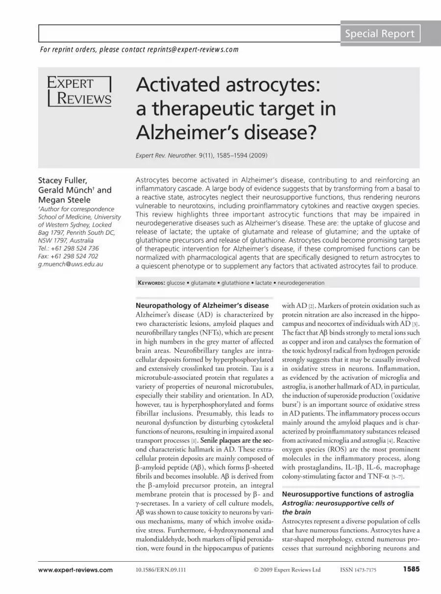
Metabolic support for neurons by provision of lactate Although glucose is generally considered the primary basal energy source in the mammalian brain, there is some evidence that lactate may become the preferred energy substrate of neurons during synap-tic activity [11]. Glucose is transported across the BBB by endothelial cells and astroglial foot processes. Astroglia take up glucose via the glucose transporter (GLUT)1 [12]. Glucose is then metabolized by glycolysis or stored as glycogen [13]. Astroglial glycolysis results in the production of lactate, owing to the conversion of pyruvate to lactate by the action of the lactate dehydrogenase (LDH) isoen-zyme LDH5. Lactate released by astroglia via H+-coupled mono-carboxylate transporters 1 and 4 is subsequently taken up by neu-rons via H+-coupled monocarboxylate transporter 2. Neurons can
convert lactate to pyruvate by the action of LDH1, enabling pyruvate to serve as a sub-strate for oxidative metabolism, partially substituting for glucose [12]. This putative relationship between astrocytes and neu-rons has been named the astrocyte–neuron lactate shuttle (Figure 1) [14]. Evidence for the astrocyte–neuron–lactate shuttle is based on in vitro demonstration that astrocytes pro-duce lactate in an activity-dependent, glu-tamate-mediated manner, and that neurons can take up this lactate as a substrate for oxi-dative meta bolism [14]. Cytological evidence supporting this theory includes differences in enzyme subtypes and lactate transporter affinities between astrocytes and neurons. For example, astrocytes primarily express LDH5, which catalyses pyruvate to lactate, and neurons primarily express LDH1, which catalyses lactate to pyruvate [15]. Additionally, use of the [H3]2-deoxyglucose autoradio-graphic method developed by Sokoloff and colleagues has revealed not only that glucose accumulation occurs exclusively in glial cells, but that energy metabolism is tightly coupled to neuronal activity [16]. Furthermore, the importance of astroglial glucose uptake by GLUT1 is demonstrated by GLUT1 defi-
ciency, a human genetic syndrome in which a decrease of 30% in GLUT1 protein expression causes severe neurological deficits and a global decrease in cortical glucose uptake as measured by PET [17]. Moreover, it is becoming increasingly accepted that synaptic activ-ity is coupled to energy utilization through an astroglial–neuronal interactive mechanism (probably involving lactate exchange from astrocytes to neurons) owing to increasing evidence from cell culture and organotypic brain slice experiments, as well as in vivo methods such as MRI and PET [18–22].
Regulation of synaptic levels of glutamateGlutamate, released into the synaptic cleft during neuronal activity, is rapidly removed by surrounding astrocytes [23]. The uptake of synaptic glutamate by astroglia is the major mechanism preventing accumulation of glutamate in the synaptic space and thus protects neurons from excessive activation and excitotoxic cell death [12].
Astrocytes, but not neurons, contain the enzyme glutamine synthetase, which amidates glutamate to glutamine. Astrocytes release glutamine into the interstitial space for uptake by neu-rons, which deamidate it via phosphate-activated glutaminase to complete the glutamate–glutamine cycle (Figure 2) [24,25]. The uptake of glutamate into astrocytes is an energetically expensive process, since each molecule of glutamate is cotransported with three sodium ions. These ions then have to be pumped from the astrocytes by exchanging with extracellular potassium, through the action of Na+/K+ ATPase. In addition, the amidation of gluta-mate to glutamine is ATP-dependent. All in all, each turn of the
Figure 1. Energy metabolism in astroglia and neurons. Glutamate uptake into the astrocyte triggers increased glucose uptake from capillaries via activation of Na+/K+-ATPase. Glucose is then processed via glycolysis, which results in lactate production. Lactate is then exchanged between astrocytes and neurons by the H+-coupled monocarboxylate transporters. Within the neuron, lactate is converted to pyruvate, which is then used in oxidative metabolism. CAC: Citric acid cycle; LDH: Lactate dehydrogenase; MCT: Monocarboxylate transporter. Adapted from [20].

www.expert-reviews.com 1587
Special Report
CapillaryAstrocyte
Lactate
Pyruvate
MCT 1 & 2
Glucose Glucose
Pyruvate
Lactate
Glutamate
Glycolysis
Glutamine
Glutamate
Glutamine
GlutaminaseGlutaminesynthetase
Neuron
NH3
ATP
ADP
Na+ Na+
ATP
ADPNa+ K+
ATPase
EAAT
K+K+
Activated astrocytes: a therapeutic target in Alzheimer’s disease?
glutamate–glutamine cycle costs astrocytes a total of four ATP, thereby tightly coupling glucose utilization in astrocytes to neu-ronal activity [24,26]. This increased energy demand in astrocytes naturally enhances glucose uptake and glycolytic flow [23]. This has important metabolic consequences as it serves as a signal that couples neuronal (glutaminergic) synaptic activity with astroglial glucose consumption [12].
Synthesis of glutathione Oxidative stress is defined as an imbalance between free oxygen species production in cells and their detoxification by enzymatic and nonenzymatic detoxification processes [9]. Glutathione (GSH), the main antioxidant thiol in mammalian cells, plays a central role in the detoxification of ROS and neutralization of peroxides in the brain [27]. GSH, a tripeptide consisting of glutamate, cysteine and glycine, is synthesized by two successive, ATP-consuming reac-tions (Figure 3). The rate-limiting step in GSH synthesis is the gen-eration of g-glutamylcysteine (g-GluCys) from glutamate and cys-teine by the action of g-GluCys synthetase. Glycine is then added to the C-terminal of g-GluCys via GSH synthetase. Glutamate and glycine are readily available in the extracellular f luid; however, cysteine is rapidly oxidised to cystine. Astrocytes take up glutamate via GLAST and GLT1, cys-tine via the X-c transporter, cysteine mainly via the Na+-dependent neutral amino acid transport system and glycine via glycine transporter 1 [28]. Neurons on the other hand can take up cysteine, but not cys-tine [29]. Astroglia release GSH, which is subsequently processed by the astrocytic ectoenzyme g-glutamyltranspeptidase to generate CysGly and g-Glu. Neurons are able to use astroglial produced CysGly as a precursor for GSH synthesis by a mecha-nism involving hydrolysis of CysGly by the ecto peptidase aminopeptidase N and subsequent uptake of cysteine and glycine [30]. The limited capacity of neurons to syn-thesise GSH leads to an increased vulner-ability to oxidative stress compared with astrocytes, and it is therefore believed that neurons rely on astroglial released GSH precursors in order to efficiently protect themselves from oxidadant and carbonyl stress [31,32].
Role of astroglia in ADActivated astroglia: a histochemical marker of inflammation in AD?Glial fibrillary acidic protein (GFAP) is an intermediate filament protein that is often used as a specific marker for activated astrocytes in the CNS. GFAP is involved
in many cellular functioning processes, such as cell structure and movement, cell communication, and the functioning of the BBB, and its upregulation accompanies the reactive response to CNS injury. GFAP upregulation is mediated by cytokines, such as TGF-b and IL-1b [33]. Other markers of astroglial activation are vimentin and S100 protein [34]. Unless other markers of astro-glial activation are mentioned, astrocytes will be referred to as ‘activated’, if they express these specific aformentioned markers.
In the AD brain, activated astrocytes have been found to be present in high concentrations surrounding neuritic plaques, with smaller numbers surrounding diffuse and dense core, non-neuritic plaques. Astrocytes form a halo around the perimeter of neuritic plaques with their processes covering the neurite layer and deeply interweaving themselves within the plaque [35]. Simpson et al. found that in some areas of AD brains astrogliosis colocal-ized with amyloid plaques, supporting the view that astrocytes react to amyloid deposits in AD. Owing to the appearance of astrogliosis in nondemented ageing brains, however, it is thought
Figure 2. Metabolism of glutamate in astroglia. The uptake of synaptic glutamate via EAATs in the astrocyte prevents accumulation of glutamate in the synaptic space. Glutamate is transported into the astrocyte together with Na+ and intracellular accumulation of Na+ activates the Na+ K+ ATPase. This leads to an increase in the ADP:ATP ratio and activation of glycolysis in the astrocyte. Glucose is transported by the endothelial cells and astrocytic end-foot processes, and in the astrocyte, it is used for synthesis of glycogen and glycolysis with production of lactate. Astrocytes transport lactate via MCT1, which is taken up by neurons via MCT2 and is converted to pyruvate, thus serving as a fuel to support neuronal metabolism. In the astrocyte, glutamate serves both as a metabolic fuel and as a precursor of glutamine and glutathione. Glutamine is synthesized from glutamate and ammonia (NH
3) by action of the glutamine synthetase
and is transported to the neurons, where it is transformed back to glutamate by action of the enzyme glutaminase. EAATs: Excitatory amino acid transporters; MCT: Monocarboxylate transporter. Adapted from [12].

Expert Rev. Neurother. 9(11), (2009)1588
Special Report
NeuronAstrocyte
Glu Cys
γGluCys
γGluCysGly
GSH
γGluCyssynthetase
GSH synthetase
GSH
γGT
GSH
CysGly
Cys
Gly
Cys
Gly
Glu
GSH
+ ATP- ADP
+ ATP- ADP
+ ATP-ADP
+ ATP- ADP
N-a
Fuller, Münch & Steele
that astrocytes normally respond to other pathological factors during aging, thus suggesting a complex interaction between astrocyte pathology and the development of AD lesions occurs during disease progression [36]. In AD, inflammation and astro-glial activation is primarily triggered by amyloid deposits in the extracellular space consisting primarily of aggregated Ab post-translationally modified by crosslinks including those formed by dityrosine and advanced glycation end products (AGEs) [37,38]. Furthermore, neurons carrying NFTs are associated with acti-vated astroglia. In one study, Sheng et al. assessed the role of IL-1a+ microglia and S100b+ astrocytes in the pathogenesis of NFTs [39]. Four distinct stages of NFT formation were identi-fied: neurons with granular perikaryal tau immunoreactivity (stage 0); fibrillar neuronal inclusions (stage 1); dense, neuronal soma-filling inclusions (stage 2); and acellular, fibrillar deposits (stage 3, ‘ghost tangles’). The numbers of tangles in randomly selected fields of parahippocampal cortex correlated with both the numbers of IL-1a+ microglia and the numbers of S100b+ astrocytes in these fields. There were progressive increases in frequency of association between tangle stages and both IL-1a+ microglia and S100b+ astrocytes: 48, 56, 67 and 92% of stage 0–3 tangles, respectively, had associated IL-1a+ microglia; and 21, 37, 55, and 91% of stage 0–3 tangles had associated S100b+ astrocytes c. In addition, ghost tangles in long-term AD patients showed paired helical filaments (PHF), suggesting that astro-cytes in ghost tangles possess the capacity to produce PHF or
that PHF are incorporated into astrocytes by endocytosis [40]. Furthermore, astroglia could be activated by signals emanating from injured and dying neurons including the ‘alarmins’ [41].
The functional role of astroglia in AD In the CNS, microglia cells are mainly responsible for innate immunity and response to inflammatory signals. They can express pattern recognition receptors and, through some of them, micro glia can also sense endogenous warning signals that alert them to cell damage. However, more and more evidence is mounting that implicates astrocytes as innate immune-competent cells that are also involved in the recognition and modulation of immune and inflammatory processes in the CNS in chronic neuro-degerative disorders such as AD. For instance, they can express pat-tern recognition receptors such as the Toll-like receptors (TLR)2, TLR3 and TLR4 [42,43], leading to the expression of poten-tially cytotoxic proinflammatory cytokines. By contrast, the stimulation of TLRs in astrocytes can be beneficial by activating pathways that lead to neuroprotection and tissue repair [42]. Furthermore, prolifera-
tion of astrocytes can lead to the formation of the so-called glial scar, preventing axonal growth within the lesion [44]. Astrocytes have also been alleged to be antigen-presenting cells [45,46]. They can express MHC I and II molecules but often only in a low percentage of cells (e.g., less than 0.1% of the astrocytes in the experimental autoimmune encephalomyelitis rat) [47] and at lower levels than in microglia or endothelial cells [48,49]. Even though their capability to express costimulatory molecules such as B7 and CD40 has been a matter of discussion [50], astrocytes are sug-gested to be involved in signaling to T cells by way of a variety of cytokines and chemokines [51]. The role played by astrocytes in the inflammatory cascade accompanying AD has been well described [35,52]. When astrocytes become stimulated by cytokines such as IL-1 and -6, they exacerbate the inflammatory process through the secretion of a wide range of cytokines, chemokines and com-plement proteins, as well as nitric oxide (NO) [53]. Moreover, astrocytes have been shown to upregulate the expression of the serine protease inhibitor a1-antichymotrypsin, which is found in high concentrations in amyloid plaques and blood vessels of AD brains [54]. Other phenotypic changes associated with astroglia in the AD brain include the upregulation of GFAP (associated with astroglial activation) [55] and the neurotrophic factor S100B [56]. Furthermore, increased levels of S100B and GFAP measured in cerebrospinal fluid of AD patients compared with age-matched controls suggests a correlation between increased levels of these proteins and disease severity [36].
Figure 3. GSH metabolism. gGluCys synthetase uses glutamate and cysteine as substrates for the dipeptide gGluCys, which is combined with glycine by GSH synthetase to generate GSH. GSH is then exported from the astrocyte. Extracellular GSH serves as a substrate for the astroglial ectoenzyme gGT, producing the dipeptide CysGly, which is an important exogenous precursor of neuronal GSH. Cys: Cysteine; gGT: g-glutamyl transpeptidase; Glu: Glutamate; Gly: Glycine; GSH: Glutathione; N-a: N-aminopeptidase. Adapted from [31].

www.expert-reviews.com 1589
Special ReportActivated astrocytes: a therapeutic target in Alzheimer’s disease?
In AD, protein aggregates of Ab and AGE-modified proteins can activate astrocytes directly, most likely via signaling through the receptor for glycation end products (RAGE), causing the production of a variety of proinflammatory factors by astrocytes [57,58]. Reactive astrocytes have been shown to release TGF-b1, TGF-b3 and IL-10 as well as proinflammatory mediators such as monocyte chemotactic protein (MCP)-1, regulated on activation, normal T-cell expressed and secreted (RANTES), TNF-a and IL-1 [59]. Thus, under pathological conditions, such as those seen in chronic inflammation and neuro degeneration, we propose that astroglia switch from metabolic support cells to immunological cells capable of inducing inflammation via the production of a variety of proinflammatory factors.
Interaction of activated microglia & astroglia in the pathogenesis of ADA large number of studies indicate that activation of microglia, with subsequent production of proinflammatory and potentially cytotoxic mediators, is suggested to play an important role in the indirect activation of astroglia. For example, the microglia-derived cytokine IL-1 activates astrocytes and induces expression of the astrocyte-derived cytokine, S100b, and increases intra-cellular free calcium levels. Self-propagation may be facilitated by means of several reinforcing feedback loops. Ab directly activates microglia, thus inducing further IL-1 production, and activates the complement system, which also leads to microglial activation with IL-1 expression. Self-propagation also could result when S100b-induced increases in intraneuronal free calcium levels lead-ing to neuronal injury and death, with consequent microglial activation [60].
Inflammation & (nitro)oxidative stress in AD The activation of microglia and astroglia is an important source of oxidative stress in AD patients [61]. ROS, originating from microglia superoxide production (‘oxidative burst’) and NO, derived from astroglia, are the most prominent molecules in the inflammatory process [62]. Growing evidence suggests oxidative stress may play an important role in neuronal degeneration in diseases such as AD [63]. Various products of oxidation reactions and mediators of oxidative stress are found in association with the histopathological AD hallmarks, including malondialdehyde, AGEs, carbonyls, nitrotyrosine, superoxide dismutase and heme oxygenase. Generally, all cellular macromolecules can be found in an oxidized form in AD tissue. While the oxidation of proteins and lipids can have immediate effects with respect to enzyme activities or membrane integrity, the oxidation of DNA may also lead to long-term mutagenic effects and, therefore, to dramatic alterations of the genetic programs of the neurons [64]. Many studies have provided evidence for the deleterious consequences of oxidative stress products on cellular targets in AD. Oxidation of mitochondrial and nuclear DNA can be observed in the pari-etal cortex of AD patients. Also observed in AD brains is protein oxidation and lipid peroxidation [65]. A number of oxidatively modified brain proteins have been identified using redox pro-teomics in AD and mild cognitive impairment (MCI). These
proteins are involved in a number of biochemical and cellular pro-cesses such as energy metabolism, protein degradation, synaptic function, neuritic growth and cellular defense systems. Among those oxidatively modified proteins are creatine kinase BB [66], b-tubulin and b-actin [67], glutamine synthase and ubiquitin carboxy-terminal hydrolase L-1 [68], dihydropyrimidinase-related protein 2, a-enolase and heat shock cognate 71[69].
Among other indices, nitroxidative damage in a cell can be indexed by measuring the levels of tyrosine nitration. Tyrosine nitration is one specific form of protein oxidation that is associated with AD [3,70–72]. NO reacting with the superoxide anion (O2-) forms the product, peroxynitrite (ONOO-), known to lead to nitra-tion of tyrosine (3-NT) residues [71,73]. Nitration of proteins results in the inactivation of several important mammalian proteins such as Mn superoxide dismutase, Cu/Zn superoxide dismutase, actin and tyrosine hydroxylase, and probably interferes with tyrosine phosphorylation-mediated cell signaling due to steric effects [74]. Studies have also demonstrated that the expression of NO synthase (NOS), the synthesis of NO, peroxynitrite production and protein tyrosine nitration are activated over the entire chronic course of AD [75,76]. Studies have also indicated that both neuronal and glial NOS may play a role in the pathogenesis of AD and peroxynitrite forma-tion. Increased expression of neuronal NOS is seen in neurons with NFTs, as well as in reactive astrocytes near amyloid plaques. The presence of neuritic plaques has also been associated with increased expression of inducible NOS (iNOS) and epithelial NOS in astro-cytes [77]. It has been shown that nitrotyrosine is present in neurons containing NFTs in AD brains, but not in control brains lacking the pathological lesion. This clearly implicates NO expression in the pathogenesis of cell death in AD [78]. It has also been dem-onstrated that iNOS is expressed in AD brains in tangle-bearing neurons, and that this high output pathway of NO production may contribute to the pathogenesis of AD [79].
Loss of astroglial neurosupportive functions: a contributing factor to the progresson of AD? Hypothesis We propose that transition of astrocytes from a basal to reactive state – which could lead to a breakdown between astroglial–neuro-nal interactions – plays a key role in the pathogenesis and progres-sion of AD. In fact, signs of astroglial dysfunction relating to the three neurosupportive functions of astroglia discussed here have been observed in numerous postmortem and in vivo studies of AD.
Change in astrocytic energy metabolism in ADEvidence from PET studies show that brain glucose uptake is impaired in AD patients, suggesting that compromised energy metabolism in the brain may be a contributing factor to neuro-degeneration [80]. The extent of the decrease in the glucose signal is often more than 10%, and since astrocytes constitute over 50% of the cell mass in the brain compared with approximately 10% for neurons, it has been suggested that the loss of signal must be, at least partially, caused by a loss of astroglial glucose uptake [81]. Furthermore, it has been demonstrated that disrupted glucose metabolism in the brain of Alzheimer’s patients occurs before the

Expert Rev. Neurother. 9(11), (2009)1590
Special Report Fuller, Münch & Steele
appearance of patho logical hallmarks, such as amyloid plaques and NFTs, thus implicating disrupted energy metabolism early in disease progression [82].
Change in astrocytic glutamate recycling in AD In addition to changes in energy metabolism, it appears that astro-cytic uptake of glutamate and release of glutamine may also be affected in AD. AD has been associated with a loss of excitatory amino acid transporter (EAAT)2 and defective glutamate trans-port [83,84]. Recent findings by Simpson and colleagues show a trend to reduced expression of EAAT2 (the main regulator of extracellular glutamate levels which is primarily expressed by astrocytes; also called GLT1) associated with increasing AD pathology, but no change in expression of EAAT1 (GLAST), pos-sibly reflecting the loss of specific astrocyte function. Based on the finding that levels of EAAT2 and gliosis in the ageing brain were inversely related, Simpson et al. hypothesised that hypertrophic astrocytes at some point lose EAAT2 expression, with resultant loss of physiological function [36].
In another study of EAAT1 expression in the cerebral cortex of control, AD and non-AD cases, it was found that EAAT1 was strongly expressed in a subset of neurofibrillary patho logy-possessing (or tau stained) cortical pyramidal neurons in dementia cases showing AD-type pathology. Owing to the fact that EAAT1 is not normally expressed in neurons, and to the observation that a few cells that stained for EAAT1 did not stain for tau, whereas all tau-positive cells were also EAAT1-positive, Scott et al. suggest that EAAT1 changes are related to NFT formation in AD, thus impli-cating aberrant glutamate transporter expression as a mechanism involved in neuro degeneration in AD [85]. Although this study did not relate directly to astroglial–neuronal interactions, one of the proposed explanations for neuronal expression of EAAT1 was as a mechanism to prevent excitotoxic damage from increased extracel-lular glutamate in addition to normal glial cell function or to replace glial cell function that may be impaired [85]. In both these and other studies of impaired glutamate transport in AD, a shared conclusion seems to be that astrocytes may lose their neuroprotective ability of successfully monitoring extracellular glutamate concentration, thus contributing to neuronal injury in AD [83,84]. In addition, it appears that the conversion of glutamate to glutamine may also be affected in AD. For example, the activity of the astrocytic enzyme glutamine synthetase is decreased in the AD brain, which may have relevance to mechanisms of chronic excitotoxicity [86]. Furthermore, it has been shown that synthetic Ab can interact with glutamine synthetase and induce inactivation of this enzyme. This might be caused through oxidation involving a structural perturbation of the tertiary structure of the enzyme [68,74,87].
Change in astrocytic glutathione supply in AD The third neuroprotective function of astrocytes discussed here – provision of GSH and GSH precursors to neurons – also appears to be affected in AD. Calabrese and colleagues have reported that plasma GSH was decreased in AD patients, and there was a sig-nificant increase in oxidative stress markers (glutathione disul-fide [GSSG], hydroxynonenal, protein carbonyl content and
nitrotyrosine) [88]. Since brains from patients with MCI showed increased protein oxidation and lipid peroxidation relative to aged-matched controls [89,90], and because MCI is believed to be a transi-tion state between normal cognition and dementia, it is suggested that oxidative stress is fundamental to the progression of AD, and not simply a consequence of the disease [91].
In post-mortem studies of AD and age-matched control brains, Karleson and colleagues found that lipid peroxidation (measure-ment of diene conjugates and lipid peroxides) was increased in the temporal and frontal inferior cortex, while antioxidant capacity was decreased [92]. It was also shown that GSSG/GSH (indicator of oxidative stress) was greatly increased in the temporal inferior cortex. It was concluded that severe oxidative damage in post-mor-tem AD brains closely relates to the appearance of AD morpho-logical abnormalities, and supposed that the susceptibility of the cerebrocortical regions to oxidative damage is greatly influenced by age-related regional peculiarities in antioxidant protection [91].
Furthermore, it has previously been shown that there is an increase in oxidized proteins in the AD brain compared with age-matched control brains [67], and that oxidatively modified proteins demon-strate impaired protein function [93]. Newman et al. have further shown that there is an increase in S-glutathionylated proteins in AD brains, and that specific proteins are sensitive to S-glutathionylation, most probably owing to their sensitivity to cysteine oxidation initi-ated by an increase in oxidative stress [94]. S-glutathionylation occurs in a number of physiologically relevant situations, where it can pro-duce discrete modulatory effects on protein function. The increas-ing evidence of functional changes resulting from this modification and the growing number of proteins shown to be S-glutathionylated both in vitro and in vivo support this contention [95,96].
While the role of S-glutathionylation is not completely understood, it is known that, generally, S-glutathionylated pro-teins (protein-GSH: PSSG) have reduced activities; however, S-glutathionylation is reversible when reduced GSH is available. This suggests that S-glutathionylation of proteins could be a pro-tective mechanism to prevent irreversible oxidization of proteins. Interestingly, the specific targets of S-glutathionylation that were identified included glyceraldehyde phosphate dehydrogenase and a-enolase, both of which catalyse reactions of the glycolytic path-way. It is plausible that increased S-glutathionylation of these glycolytic enzymes could result in reduced glucose breakdown (as is observed in PET studies in the AD brain) and a subsequent decrease in energy production. Since reduced GSH is required for S-glutahionylation reversal, it is apparent that the availability of reduced GSH is especially important to maintain normal brain functioning in oxidative conditions [94].
Do astroglia offer opportunities as drug targets for the development of specific antidementia drugs? Inhibitors of astroglial activation To summarize, astroglia in AD brains are present in increased numbers and commonly show a hypertrophic phenotype, are often found surrounding and invaginating amyloid plaques, release a wide array of mediators of inflammation (cytokines, chemokines and NO) and oxidative stress (ROS and reactive nitrogen species), as

www.expert-reviews.com 1591
Special ReportActivated astrocytes: a therapeutic target in Alzheimer’s disease?
well as other factors such as a1-antichymotrypsin and S100B, and appear to have altered energy metabolism, decreased ability to take up glutamate and a reduced oxidative defense system. It therefore seems plausible that astrocyte pathology is important in AD pro-gression, through both the production of pathogenic substances and the loss of normal function, specifically their alleged neuro protective role of recycling glutamate and providing neurons with lactate and GSH precursors. If this is found to be the case, novel therapies that support astroglial function and, thus, protect neurons may be useful in the treatment of AD. In addition, most of the approaches we suggest will not only normalize astroglial function, but might also improve neuronal function and decrease microglial activation.
There are various types of drugs that could be explored as poten-tial inhibitors of astroglial activation. Anti-inflammatory type drugs may prove to be effective in the inhibition of astroglia activation. Astroglia express receptors for various neuroactive compounds, including IL-1 and TNF-a, which lead to astroglial activation [51]. Inhibition of these receptors or their signal transduction pathways may be an effective mechanism for prevention of astroglial acti-vation. Antioxidative-type drugs may also prove to be successful in the inhibition of astroglial activation by their ability to inhibit proinflammatory redox signaling. It has been accepted that certain ROS and reactive nitrogen species can act as signaling molecules [97], and antioxidants that inhibit the production of, or can directly scavenge, these signaling molecules may prove to be an effective means to downregulate astroglial activation by free radicals. Other mechanisms by that astroglial activation could be inhibited include treatment with inhibitors of NADPH oxidase and iNOS. NADPH oxidase is a plasma membrane-associated enzyme which catalyses the production of superoxide [98]. NADPH oxidase has a high molec-ular complexity, and as a result there are a large number of inhibitors that may be able to interact with the associated signal transduc-tion pathways that lead to the activation of the enzyme [99]. iNOS is responsible for the inducible synthesis of NO in glial cells and is of particular importance in the pathology of inflammatory dis-eases. It is clear that activated glial cells in the CNS produce NO in response to induction of iNOS by cytokines such as IL-1b, TNF-a and IFN-g [100]. As these enzymes produce free radicals, inhibi-tion may prove to be effective in the downregulation of free radical production in astroglia, and therefore further astroglial activation.
Substitution of factors depleted by astroglial activationFollowing astroglial activation, many substances become depleted. To overcome this factor it may be possible to replace these com-pounds. It has been shown that astroglial activation results in the increased release of GSH [9]. Therefore, the content of GSH within the astrocyte may become depleted of GSH. To overcome this, it may be possible to increase cysteine uptake by various compounds. For example, cysteamine (2-mercaptoethylamine), N-acetylcysteine (NAC) or chemoprotective aminothiols such as S-2-(3-aminopropylamino)ethylphosphorothioic acid (WR-2721) have been shown to increase the uptake of cystine from cell culture medium [101,102]. There is some evidence from the clinic to support GSH as a promising drug for the treatment of AD. The antioxi-dant NAC or placebo was administered in a double-blind fashion
to probable AD patients. Testing for efficacy occurred after 3 and 6 months of treatment. Comparison of interval change favored NAC treatment on nearly every outcome measure, although significant differences were obtained only for a subset of cognitive tasks [103].
Drugs increasing astroglial glutamate metabolism Impaired glutamate transport (GT) into the cell and conver-sion to glutamine is another target for astroglia directed therapy in AD. Possible GT alterations in fibroblast cultures obtained from AD patients versus controls patients, and the effects of the lipoperoxidation product 4-hydroxynonenal (4-HNE) and anti-oxidants were analyzed. Basal GT was decreased by 60% in fibro-blasts from patients with AD versus control patients. Exposure to HNE did not affect GT in control patients, but it reduced GT by another 50% in patients with AD. GSH and N-acetylcysteine completely blocked 4-HNE effects and also increased basal uptake in AD cells. This study supports the hypothesis of a sys-temic impairment of GT in AD, possibly linked to oxidative stress and to reduced antioxidant defenses, which may be partially reversed by antioxidant treatment [104].
Five-year view While ‘healthy’ astroglia perform many vital neurosupportive functions, activated astrocytes become active participants in the neurodegenerative events that contribute to the pathogenesis of AD. It will become increasingly evident that astroglial activation leads to the production of free radicals and proinflammatory fac-tors, which exacerbate inflammation and drive the progression of neuronal dysfunction and cell death in Alzheimer’s disease. Furthermore, the loss of neurosupportive function such as glu-tamate removal, GSH precursor and lactate release will make neurons more susceptible to stressors and neurotoxins.
In the next few years, pharmacologically relevant mechanisms to reduce the impact of astroglial activation will be investigated in more detail both in animal models of brain inflammation, including transgenic mice overexpressing amyloid precursor protein. Drugs that interfere with astroglial activation, and therefore decrease the level of inflammation and neurodegeneration, should then enter clinical trials. There is, however, one major obstacle to overcome. Many of the compounds are off-patent drugs or food additives and are therefore of low commercial value for the pharmaceutical indus-try. In this case, it would be necessary for government agencies or philanthropic organizations to cover this gap in funding.
Key issues
• Astroglia are neurosupportive players in the brain.
• Astroglia take up glucose and release lactate to neurons.
• Astroglia recycle glutamate to glutamine for neuronal reuptake.
• Astroglia provide glutathione to neurons.
• Activated astroglia are characteristic for Alzheimer’s disease.
• Astroglial nutritive support for neurons is compromised when astroglia are activated in the brain.
• Astroglia may provide novel therapeutic targets for the treatment of Alzheimer’s disease.

Expert Rev. Neurother. 9(11), (2009)1592
Special Report Fuller, Münch & Steele
Financial & competing interests disclosureMegan Steele is supported by a PhD fellowship from Alzheimer’s Australia.The authors have no other relevant affiliations or financial involvement with any organization or entity with a financial interest in or financial
conflict with the subject matter or materials discussed in the manuscript apart from those disclosed.
No writing assistance was utilized in the production of this manuscript.
References 1 Braak H, Braak E. Staging of Alzheimer’s
disease-related neurofibrillary changes. Neurobiol. Aging 16(3), 271–278 (1995).
2 Butterfield DA, Castegna A, Lauderback CM, Drake J. Evidence that amyloid b-peptide-induced lipid peroxidation and its sequelae in Alzheimer’s disease brain contribute to neuronal death. Neurobiol. Aging 23(5), 655–664 (2002).
3 Smith M, Richey Harris P, Sayre L, Beckman J, Perry G. Widespread peroxynitrite-mediated damage in Alzheimer’s disease. J. Neurosci. 17(8), 2653–2657 (1997).
4 Halliday G, Robinson S, Shepherd C, Kril J. Alzheimer’s disease and inflammation: a review of cellular and therapeutic mechanisms. Clin. Exp. Pharmacol. Physiol. 27(1–2), 1–8 (2000).
5 Griffin WS, Sheng JG, Roberts GW, Mrak RE. Interleukin-1 expression in different plaque types in Alzheimer’s disease: significance in plaque evolution. J. Neuropathol. Exp. Neurol. 54(2), 276–281 (1995).
6 Meda L, Cassatella MA, Szendrei GI et al. Activation of microglial cells by b-amyloid protein and interferon-a. Nature 374(6523), 647–650 (1995).
7 Pachter JS. Inflammatory mechanisms in Alzheimer disease: the role of b-amyloid/glial interactions. Mol. Psychiatry 2(2), 91–95 (1997).
8 Wang D, Bordey A. The astrocyte odyssey. Prog. Neurobiol. 86(4), 342–367 (2008).
9 Gavillet M, Allaman I, Magistretti PJ. Modulation of astrocytic metabolic phenotype by proinflammatory cytokines. Glia 56(9), 975–989 (2008).
10 Frederickson R. Astroglia in Alzheimer’s disease. Neurobiol. Aging 13(2), 239–253 (1992).
11 Bouzier-Sore AK, Serres S, Canioni P, Merle M. Lactate involvement in neuron–glia metabolic interaction: 13C-NMR spectroscopy contribution. Biochimie 85(9), 841–848 (2003).
12 Benarroch E. Neuron–astrocyte interactions: partnership for normal function and disease in the central nervous system. Mayo Clin. Proc. 80, 1326–1338 (2005).
13 Oz G, Seaquist ER, Kumar A et al. Human brain glycogen content and metabolism: implications on its role in brain energy metabolism. Am. J. Physiol. Endocrinal. Metab. 292(3), 946–951 (2006).
14 Pellerin L, Magistretti P. Glutamate uptake into astrocytes stimulates aerobic glycolysis: a mechanism coupling neuronal activity to glucose utilization. Proc. Natl Acad. Sci. USA 91(22), 10625–10629 (1994).
15 Bittar PG, Charnay Y, Pellerin L, Bouras C, Magistretti PJ. Selective distribution of lactate dehydrogenase isoenzymes in neurons and astrocytes of human brain. J. Cereb. Blood Flow Metab. 16(6), 1079–1089 (1996).
16 Sokoloff L. Measurement of local cerebral glucose utilization and its relation to local functional activity in the brain. Adv. Exp. Med. Biol. 291, 21–42 (1991).
17 Pascual JM, van Heertum RL, Wang D, Engelstald K, De Vivo DC. Imaging the metabolic footprint of Glut1 deficiency on the brain. Ann. Neurol. 52(4), 458–464 (2002).
18 Aubert A, Costalat R. Interaction between astrocytes and neurons studied using a mathematical model of compartmentalized energy metabolism. J. Cereb. Blood Flow Metabol. 25(11), 1476–1490 (2005).
19 Magistretti PJ. Cellular bases of functional brain imaging: insights from neuron–glia metabolic coupling. Brain Res. 886(1–2), 108–112 (2000).
20 Magistretti P, Pellerin L. Cellular mechanisms of brain energy metabolism and their relevance to functional brain imaging. Phil. Trans. Biol. Sci. 354, 1155–1163 (1999).
21 Magistretti P, Pellerin L, Rothman D, Shulman R. Energy on demand. Science 283(5401), 496–497 (1999).
22 Aubert A, Costalat R, Magistretti PJ, Pellerin L. Brain lactate kinetics: modeling evidence for neuronal lactate uptake upon activation. Proc. Natl Acad. Sci. USA 102(45), 16448–16453 (2005).
23 Bernardinelli Y, Magistretti P, Chatton J. Astrocytes generate Na+-mediated metabolic waves. Proc. Natl Acad. Sci. USA 101(41), 14937–14942 (2004).
24 Hertz L, Dringen R, Schousboe A, Robinson SR. Astrocytes: glutamate producers for neurons. J. Neurosci. Res. 57(4), 417–428 (1999).
25 Anderson C, Swanson R. Astrocyte glutamate transport: review of properties, regulation, and physiological functions. Glia 32(1), 1–14 (2000).
26 Pellerin L, Pellegri G, Bittar P et al. Evidence supporting the existence of an activity-dependent astrocyte–neuron lactate shuttle. Dev. Neurosci. 20, 291–299 (1998).
27 Kirchhoff F, Dringen R, Giaume C. Pathways of neuron–astrocyte interactions and their possible role in neuroprotection. Eur. Arch. Psychiatry Clin. Neurosci. 251(4), 159–169 (2001).
28 Aschner M. Neuron–astrocyte interactions: implications for cellulalr energetics and antioxidant levels. Neurotoxicology 21(6), 1101–1107 (2000).
29 Sagara JI, Miura K, Bannai S. Maintenance of neuronal glutathione by glial cells. J. Neurochem. 61(5), 1672–1676 (1993).
30 Dringen R, Gutterer JM, Gros C, Hirrlinger J. Aminopeptidase N mediates the utilization of the GSH precursor CysGly by cultured neurons. J. Neurosci. Res. 66(5), 1003–1008 (2001).
31 Dringen R. Metabolism and functions of glutathione in brain. Prog. Neurobiol. 62(6), 649–671 (2000).
32 Deuther-Conrad W, Loske C, Schinzel R, Dringen R, Riederer P, Münch G. Advanced glycation endproducts change glutathione redox status in SH–SY5Y human neuroblastoma cells by a hydrogen peroxide dependent mechanism. Neurosci. Lett. 312(1), 29–32 (2001).
33 Reilly JF, Maher PA, Kumari VG. Regulation of astrocyte GFAP expression by TGF-b1 and FGF-2. Glia 22(2), 202–210 (1998).
34 Ridet JL, Malhotra SK, Privat A, Gage FH. Reactive astrocytes: cellular and molecular cues to biological function. Trends Neurosci. 20(12), 570–577 (1997).
35 Akiyama H, Barger S, Barnum S et al. Inflammation and Alzheimer’s disease. Neurobiol. Aging 21(3), 383–421 (2000).
36 Simpson J, Ince P, Lace G et al. Astrocyte phenotype in relation to Alzheimer-type pathology in the ageing brain. Neurobiol.

www.expert-reviews.com 1593
Special ReportActivated astrocytes: a therapeutic target in Alzheimer’s disease?
Aging DOI: 10.1016/j.neurobiolaging.2008.05.015 (2008) (Epub ahead of print).
37 Hensley K, Maidt ML, Yu Z, Sang H, Markesbery WR, Floyd RA. Electrochemical analysis of protein nitrotyrosine and dityrosine in the Alzheimer brain indicates region-specific accumulation. J. Neurosci. 18(20), 8126–8132 (1998).
38 Paris D, Townsend KP, Obregon DF, Humphrey J, Mullan M. Pro-inflammatory effect of freshly solubilized b-amyloid peptides in the brain. Prostaglandins Other Lipid Mediat. 70(1–2), 1–12 (2002).
39 Sheng JG, Mrak RE, Griffin WS. Glial-neuronal interactions in Alzheimer disease: progressive association of IL-1a+ microglia and S100b+ astrocytes with neurofibrillary tangle stages. J. Neuropathol. Exp. Neurol. 56(3), 285–290 (1997).
40 Ikeda K, Haga C, Akiyama H, Kase K, Iritani S. Coexistence of paired helical filaments and glial filaments in astrocytic processes within ghost tangles. Neurosci. Lett. 148(1–2), 126–128 (1992).
41 Oppenheim JJ, Yang D. Alarmins: chemotactic activators of immune responses. Curr. Opin. Immunol. 17(4), 359–365 (2005).
42 Bsibsi M, Persoon-Deen C, Verwer RW, Meeuwsen S, Ravid R, Van Noort JM. Toll-like receptor 3 on adult human astrocytes triggers production of neuroprotective mediators. Glia 53(7), 688–695 (2006).
43 Bowman CC, Rasley A, Tranguch SL, Marriott I. Cultured astrocytes express Toll-like receptors for bacterial products. Glia 43(3), 281–291 (2003).
44 Wanner IB, Deik A, Torres M et al. A new in vitro model of the glial scar inhibits axon growth. Glia 56(15), 1691–1709 (2008).
45 Pulver M, Carrel S, Mach JP, de Tribolet N. Cultured human fetal astrocytes can be induced by interferon-g to express HLA-DR. J. Neuroimmunol. 14(2), 123–133 (1987).
46 Skias DD, Kim DK, Reder AT, Antel JP, Lancki DW, Fitch FW. Susceptibility of astrocytes to class I MHC antigen-specific cytotoxicity. J. Immunol. 138(10), 3254–3258 (1987).
47 Hickey WF, Osborn JP, Kirby WM. Expression of Ia molecules by astrocytes during acute experimental allergic encephalomyelitis in the Lewis rat. Cell. Immunol. 91(2), 528–535 (1985).
48 Male DK, Pryce G, Hughes CC. Antigen presentation in brain: MHC induction on
brain endothelium and astrocytes compared. Immunology 60(3), 453–459 (1987).
49 Grenier Y, Ruijs TC, Robitaille Y, Olivier A, Antel JP. Immunohistochemical studies of adult human glial cells. J. Neuroimmunol. 21(2–3), 103–115 (1989).
50 Dong Y, Benveniste EN. Immune function of astrocytes. Glia 36(2), 180–190 (2001).
51 Benveniste E. Cytokine actions in the central nervous system. Cytokine Growth Factor Rev. 9(3–4), 259–275 (1998).
52 Tuppo E, Arias H. The role of inflammation in Alzheimer’s disease. Int. J. Biochem. Cell Biol. 37(2), 289–305 (2005).
53 Dong Y, Benveniste E. Immune function of astrocytes. Glia 36(2), 180–190 (2001).
54 Pasternack J, Abraham C, Van Dyke B, Potter H, Younkin S. Astrocytes in Alzheimer’s disease gray matter express a 1-antichymotrypsin mRNA. Am. J. Pathol. 135(5), 827–834 (1989).
55 Beach T, McGeer E. Lamina-specific arrangement of astrocytic gliosis and senile plaques in Alzheimer’s disease visual cortex. Brain Res. 463(2), 357–361 (1988).
56 Marshak DR, Pesce SA, Stanley LC, Griffin W. Increased S100 b neurotrophic activity in Alzheimer’s disease temporal lobe. Neurobiol. Aging 13(1), 1–7 (1992).
57 Yan SD, Bierhaus A, Nawroth PP, Stern DM. RAGE and Alzheimer’s disease: a progression factor for amyloid-b-induced cellular perturbation? J. Alzheimers Dis. 16(4), 833–843 (2009).
58 Wang Z, Li DD, Liang YY, Wang DS, Cai NS. Activation of astrocytes by advanced glycation end products: cytokines induction and nitric oxide release. Acta Pharmacol. Sin. 23(11), 974–980 (2002).
59 Blasko I, Stampfer-Kountchev M, Robatscher P, Veerhuis R, Eikelenboom P, Grubeck-Loebenstein B. How chronic inflammation can affect the brain and support the development of Alzheimer’s disease in old age: the role of microglia and astrocytes. Aging Cell 3(4), 169–176 (2004).
60 Mrak RE, Sheng JG, Griffin WS. Glial cytokines in Alzheimer’s disease: review and pathogenic implications. Hum. Pathol. 26(8), 816–823 (1995).
61 Retz W, Gsell W, Münch G, Rosler M, Riederer P. Free radicals in Alzheimer’s disease. J. Neural Transm. Suppl. 54, 221–236 (1998).
62 McGeer PL, McGeer EG. Inflammation and the degenerative diseases of aging. Ann. NY Acad. Sci. 1035, 104–116 (2004).
63 Simonian N, Coyle J. Oxidative stress in neurodegenerative diseases. Annu. Rev. Pharmacol. Toxicol. 36(1), 83–106 (1996).
64 Behl C. Alzheimer’s disease and oxidative stress: implications for novel therapeutic approaches. Prog. Neurobiol. 57(3), 301–323 (1999).
65 Christen Y. Oxidative stress and Alzheimer disease. Am. J. Clin. Nutr. 71(2), S621–S629 (2000).
66 Aksenov M, Aksenova M, Butterfield DA, Markesbery WR. Oxidative modification of creatine kinase BB in Alzheimer’s disease brain. J. Neurochem. 74(6), 2520–2527 (2000).
67 Aksenov MY, Aksenova MV, Butterfield DA, Geddes JW, Markesbery WR. Protein oxidation in the brain in Alzheimer’s disease. Neuroscience 103(2), 373–383 (2001).
68 Castegna A, Aksenov M, Aksenova M et al. Proteomic identification of oxidatively modified proteins in Alzheimer’s disease brain. Part I: creatine kinase BB, glutamine synthase, and ubiquitin carboxy-terminal hydrolase L-1. Free Radic. Biol. Med. 33(4), 562–571 (2002).
69 Castegna A, Aksenov M, Thongboonkerd V et al. Proteomic identification of oxidatively modified proteins in Alzheimer’s disease brain. Part II: dihydropyrimidinase-related protein 2, a-enolase and heat shock cognate 71. J. Neurochem. 82(6), 1524–1532 (2002).
70 Castegna A, Thongboonkerd V, Klein J, Lynn B, Markesbery W, Butterfield D. Proteomic identification of nitrated proteins in Alzheimer’s disease brain. J. Neurochem. 85(6), 1394 (2003).
71 Gow A, Duran D, Malcolm S, Ischiropoulos H. Effects of peroxynitrite-induced protein modifications on tyrosine phosphorylation and degradation. FEBS Lett. 385(1–2), 63–66 (1996).
72 Sultana R, Poon H, Cai J et al. Identification of nitrated proteins in Alzheimer’s disease brain using a redox proteomics approach. Neurobiol. Dis. 22(1), 76–87 (2006).
73 Butterfield D, Kanski J. Brain protein oxidation in age-related neurodegenerative disorders that are associated with aggregated proteins. Mech. Ageing Dev. 122(9), 945–962 (2001).

Expert Rev. Neurother. 9(11), (2009)1594
Special Report Fuller, Münch & Steele
74 Butterfield D, Stadtman E. Protein oxidation processes in aging brain. Adv. Cell Aging Gerontol. 2, 161–191 (1997).
75 Lüth HJ, Münch G, Arendt T. Aberrant expression of NOS isoforms in Alzheimer’s disease is structurally related to nitrotyrosine formation. Brain Res. 953(1–2), 135–143 (2002).
76 Fernandez-Vizarra P, Fernandez A, Castro-Blanco S et al. Expression of nitric oxide system in clinically evaluated cases of Alzheimer’s disease. Neurobiol. Dis. 15(2), 287–305 (2004).
77 Pacher P, Beckman J, Liaudet L. Nitric oxide and peroxynitrite in health and disease. Physiol. Rev. 87(1), 315 (2007).
78 Good P, Werner P, Hsu A, Olanow C, Perl D. Evidence of neuronal oxidative damage in Alzheimer’s disease. Am. J. Pathol. 149(1), 21–28 (1996).
79 Vodovotz Y, Lucia M, Flanders K et al. Inducible nitric oxide synthase in tangle-bearing neurons of patients with Alzheimer’s disease. J. Exp. Med. 184(4), 1425–1433 (1996).
80 Freemantle E, Vandal M, Tremblay-Mercier J et al. Omega-3 fatty acids, energy substrates, and brain function during aging. Prostaglandins Leukot. Essent. Fatty Acids 75(3), 213–220 (2006).
81 Alexander G, Chen K, Pietrini P, Rapopor S, Reiman E. Longitudinal PET evaluation of cerebral metabolic decline in dementia: a potential outcome measure in Alzheimer’s disease treatment studies. Am. J Psychiatry 159(5), 738–745 (2002).
82 Small GW, Ercoli LM, Silverman DH et al. Cerebral metabolic and cognitive decline in persons at genetic risk for Alzheimer’s disease. Proc. Natl Acad. Sci. USA 97(11), 6037–6042 (2000).
83 Li S, Mallory M, Alford M, Tanaka S, Masliah E. Glutamate transporter alterations in Alzheimer disease are possibly associated with abnormal APP expression. J. Neuropathol. Exp. Neurol. 56(8), 901–911 (1997).
84 Masliah E, Alford M, DeTeresa R, Mallory M, Hansen L. Deficient glutamate transport is associated with neurodegeneration in Alzheimer’s disease. Ann. Neurol. 40(5), 759–766 (1996).
85 Scott H, Pow D, Tannenberg A, Dodd P. Aberrant expression of the glutamate transporter excitatory amino acid transporter 1 (EAAT1) in Alzheimer’s disease. J. Neurosci. 22(3), RC206 (2002).
86 Smith GM, Miller RH. Immature type-1 astrocytes suppress glial scar formation, are
motile and interact with blood vessels. Brain Res. 543(1), 111–122 (1991).
87 Butterfield DA, Hensley K, Cole P et al. Oxidatively induced structural alteration of glutamine synthetase assessed by analysis of spin label incorporation kinetics: relevance to Alzheimer’s disease. J. Neurochem. 68(6), 2451–2457 (1997).
88 Calabrese V, Sultana R, Scapagnini G et al. Nitrosative stress, cellular stress response, and thiol homeostasis in patients with Alzheimer’s disease. Antioxid. Redox Signal. 8, 1975–1986 (2006).
89 Reed T, Perluigi M, Sultana R et al. Redox proteomic identification of 4-hydroxy-2-nonenal-modified brain proteins in amnestic mild cognitive impairment: insight into the role of lipid peroxidation in the progression and pathogenesis of Alzheimer’s disease. Neurobiol. Dis. 30(1), 107–120 (2008).
90 Butterfield DA, Lauderback CM. Lipid peroxidation and protein oxidation in Alzheimer’s disease brain: potential causes and consequences involving amyloid b-peptide-associated free radical oxidative stress. Free Radic. Biol. Med. 32(11), 1050–1060 (2002).
91 Butterfield DA, Sultana R. Redox proteomics identification of oxidatively modified brain proteins in Alzheimer’s disease and mild cognitive impairment: insights into the progression of this dementing disorder. J. Alzheimers Dis. 12(1), 61–72 (2007).
92 Karelson E, Bogdanovic N, Garlind A et al. The cerebrocortical areas in normal brain aging and in Alzheimer’s disease: noticeable differences in the lipid peroxidation level and in antioxidant defense. Neurochem. Res. 26(4), 353–361 (2001).
93 Butterfield DA, Boyd-Kimball D. Proteomics analysis in Alzheimer’s disease: new insights into mechanisms of neurodegeneration. Int. Rev. Neurobiol. 61, 159–188 (2004).
94 Newman S, Sultana R, Perluigi M et al. An increase in S-glutathionylated proteins in the Alzheimer’s disease inferior parietal lobule, a proteomics approach. J. Neurosci. Res. 85(7), 1506–1514 (2007).
95 Giustarini D, Rossi R, Milzani A, Colombo R, Dalle-Donne I. S-glutathionylation: from redox regulation of protein functions to human diseases. J. Cell. Mol. Med. 8(2), 201–212 (2004).
96 Dalle-Donne I, Milzani A, Gagliano N, Colombo R, Giustarini D, Rossi R. Molecular mechanisms and potential clinical significance of S-glutathionylation. Antioxid.
Redox Signal. 10(3), 445–473 (2008).
97 Hensley K, Robinson K, Gabbita S, Salsman S, Floyd R. Reactive oxygen species, cell signaling, and cell injury. Free Radic. Biol. Med. 28(10), 1456–1462 (2000).
98 Babior B. NADPH oxidase: an update. Blood, 93(5), 1464–1476 (1999).
99 Holland J, O’Donnell R, Chang M, Johnson D, Ziegler L. Endothelial cell oxidant production: effect of NADPH oxidase inhibitors. Endothelium 7(2), 109–119 (2000).
100 Pahan K, Sheikh F, Namboodiri A, Singh I. Inhibitors of protein phosphatase 1 and 2A differentially regulate the expression of inducible nitric-oxide synthase in rat astrocytes and macrophages. J. Biol. Chem. 273(20), 12219–12226 (1998).
101 Issels RD, Nagele A, Eckert KG, Wilmanns W. Promotion of cystine uptake and its utilization for glutathione biosynthesis induced by cysteamine and N-acetylcysteine. Biochem. Pharmacol. 37(5), 881–888 (1988).
102 Issels RD, Nagele A. Promotion of cystine uptake, increase of glutathione biosynthesis, and modulation of glutathione status by S-2-(3-aminopropylamino)ethyl phosphorothioic acid (WR-2721) in Chinese hamster cells. Cancer Res. 49(8), 2082–2086 (1989).
103 Adair JC, Knoefel JE, Morgan N. Controlled trial of N-acetylcysteine for patients with probable Alzheimer’s disease. Neurology 57(8), 1515–1517 (2001).
104 Begni B, Brighina L, Sirtori E et al. Oxidative stress impairs glutamate uptake in fibroblasts from patients with Alzheimer’s disease. Free Radic. Biol. Med. 37(6), 892–901 (2004).
Affiliations• Stacey Fuller
Department of Pharmacology, School of Medicine, University of Western Sydney, Campbelltown, NSW 1797, Australia [email protected]
• Gerald Münch Professor, School of Medicine, University of Western Sydney, Locked Bag 1797, Penrith South DC, NSW 1797, Australia Tel.: +61 298 524 736 Fax: +61 298 524 702 [email protected]
• Megan Steele Department of Pharmacology, School of Medicine, University of Western Sydney, Campbelltown, NSW, 1797, Australia [email protected]