nuclear factor kappa-b mediates selective induction of neuronal nitric oxide synthase in astrocytes...
TRANSCRIPT
Nuclear Factor Kappa-B Mediates Selective Induction of NeuronalNitric Oxide Synthase in Astrocytes During Low-LevelInflammatory Stimulation with MPTP
David L. Carbone1,2, Julie A. Moreno1,2, and Ronald B. Tjalkens1,2*
1Program in Molecular, Cellular, and Integrative Neuroscience
2Department of Environmental and Radiological Health Sciences, Colorado State University, Fort Collins,Colorado 80523
AbstractRecent advances in understanding the progression of Parkinson’s disease (PD) implicateperturbations in astrocyte function and induction of constitutively expressed neuronal nitric oxidesynthase (NOS1) in both human PD and in the MPTP model of the disease. Transcriptional regulationof NOS1 is complex but recent data suggest that nuclear factor kappa B (NF-κB) is an importanttranscription factor involved in inducible expression of the gene. The data presented here demonstratethat mild activation of primary astrocytes with low or ‘sub-optimal’ concentrations of MPTP (1 µM)and the inflammatory cytokines tumor necrosis factor alpha (10 pg/ml) and interferon gamma (1 ng/ml) results in selective induction of Nos1 mRNA and protein, increased production of nitric oxide(NO), and a significant elevation in global protein nitration. This mild inflammatory stimulus alsoresulted in activation and recruitment of p65 to a putative NF-κB response element located in theNos1 promoter region flanking exon 1. A role for NF-κB in MPTP-dependent induction of NOS1was confirmed through overexpression of a mutant IκBα super repressor of NF-κB that preventedinduction of NOS1. The data presented here thus demonstrate a role for NF-κB in selective inductionof NOS1 during early inflammatory activation of astrocytes stimulated by low-dose MPTP andinflammatory cytokines.
KeywordsParkinson’s disease; neurodegeneration; astrocyte; nitric oxide; nuclear factor-kappa B
1. IntroductionParkinson’s disease (PD) is a progressive and incurable neurodegenerative disordercharacterized by irreversible loss of dopaminergic neurons in the substantia nigra parscompacta (DeLong, 2000). While the causes of PD remain unclear, recent evidence stronglysupports a role for perturbations in astrocyte physiology and altered expression of the nitricoxide synthase (NOS; E.C. 1.14.13.39) isoforms in progression of the disease (Hirsch et al.,
*Address correspondence to: Ronald B. Tjalkens, Associate Professor, Department of Environmental and Radiological Health Sciences,Colorado State University, Campus Delivery 1680, Ft. Collins, CO 80523, Tel. (970) 491-2825, Fax. (970) 491-7569, E-mail:[email protected]'s Disclaimer: This is a PDF file of an unedited manuscript that has been accepted for publication. As a service to our customerswe are providing this early version of the manuscript. The manuscript will undergo copyediting, typesetting, and review of the resultingproof before it is published in its final citable form. Please note that during the production process errors may be discovered which couldaffect the content, and all legal disclaimers that apply to the journal pertain.
NIH Public AccessAuthor ManuscriptBrain Res. Author manuscript; available in PMC 2009 June 27.
Published in final edited form as:Brain Res. 2008 June 27; 1217: 1–9. doi:10.1016/j.brainres.2008.03.093.
NIH
-PA Author Manuscript
NIH
-PA Author Manuscript
NIH
-PA Author Manuscript
2003; Pekny and Nilsson, 2005; Teismann and Schulz, 2004). Studies exploringpathophysiologic expression of NOS have typically focused on the inducible isoform (NOS2)but induction of the constitutively expressed neuronal nitric oxide synthase (NOS1) hasemerged as an underlying component of a diverse array of neurologic disorders, includingischemic cerebral injury (e.g. stroke) (Moro et al., 2004), substance-induced neurologicaldysfunction including alcohol and methamphetamine abuse (Imam et al., 2001), as well asidiopathic neurodegenerative disorders including amyotrophic lateral sclerosis, Alzheimer’sdisease, and PD (Catania et al., 2001; Chabrier et al., 1999; Eve et al., 1998). Underscoring arole for this enzyme in the progression of PD is the observation that ablation of the Nos1 genein the 1-methyl-MPTP model of parkinsonism completely prevents neurotoxicity (Przedborskiet al., 1996), as well as additional studies demonstrating protection of dopaminergic neuronsin MPTP-treated mice by the NOS1-specific inhibitor 7-nitroindazole (7-NI) (Watanabe et al.,2004). However, despite the suggested role for NOS1 in the progression of PD, the mechanismbehind induction of this enzyme during states of glial inflammation remains poorly understood.
Because NOS1 is thought to play a crucial role in the progression of multiple neurologicalpathologies, suppression of this protein represents a potential therapeutic target for halting theprogression of disorders such as PD. Unfortunately, the complexity of the Nos1 gene hasimpeded our understanding of the transcriptional mechanisms by which it is regulated. TheNF-κB signaling pathway has recently been linked to induction of human Nos1 in studiesreporting a putative κB-like response element in the 5’ promoter region flanking the first exon(Hall et al., 1994). This report was followed by a study documenting an association betweenp65, the active nuclear component of NF-κB, with the promoter region of human Nos1 inhuman neuroblastoma cells (Li et al., 2007). Although the report by Li et al. (2007) documenteda role for NF-κB in the induction of Nos1, the study was conducted in an immoratalized humanneuronal cell line rather than primary neurons and did not consider potential differences inregulatory mechanisms that could exist between neurons and astrocytes.
The work presented here describes the selective induction of NOS1 in primary astrocytestreated with low concentrations of MPTP and the inflammatory cytokines TNF-α and IFN-γ,and tests the hypothesis that this induction occurs primarily through an NF-κB-mediatedmechanism. These data represent, to our knowledge, the first report of a functional role forNF-κB in MPTP-induced expression of NOS1 in astrocytes and provide insight into signalingmechanisms that may mediate the deleterious effects of this enzyme during degeneration ofdopaminergic neurons.
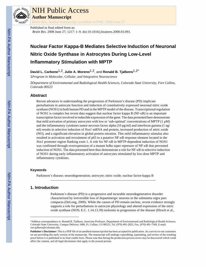
2. ResultsThe effect of low-level inflammatory stimulation by TNF-α and IFN-γ with or without themitochondrial toxin MPTP on induction of Nos1, Nos2, and Nos3 was measured in astrocytesusing RT-PCR (Figure 1A). Only induction only of Nos1 was observed (Lane 3). NeitherNos2 nor Nos3 mRNA was detected under control conditions or following exposure ofastrocytes to the combination of MPTP and TNF-α/IFN-γ. Either whole brain (Nos1 andNos3) or LPS-activated astrocyte (Nos2) RNA subject to the RT reaction in the absence (-rt;Lane 4) or presence (wb; Lane 5) of reverse transcriptase were included as negative or positivePCR controls, respectively, to confirm PCR efficacy. Because Nos1 was induced by thecytokine and MPTP insult, semi-quantitative real-time RT-PCR was employed to furtherexplore the induction of this gene relative to saline-treated control astrocytes in the presenceor absence of cytokines and/or MPTP (Figure 1B). Although treatment of the astrocytes withcytokines alone resulted in significant induction of Nos1 (4-fold over control), treatment withonly MPTP had no significant effect on levels of mRNA. However, MPTP strongly potentiatedinduction of Nos1 (7-fold over control) when used in combination with TNF-α and IFN-γ. Tofurther examine the specificity of NOS1 isoform expression during low-dose exposure to
Carbone et al. Page 2
Brain Res. Author manuscript; available in PMC 2009 June 27.
NIH
-PA Author Manuscript
NIH
-PA Author Manuscript
NIH
-PA Author Manuscript
MPTP and cytokines, expression of Nos2 was determined by semi-quantitative real time PCR(Figure 1C). No significant induction of Nos2 was observed at the levels of MPTP andcytokines that were effective in stimulating inducible expression of NOS1.
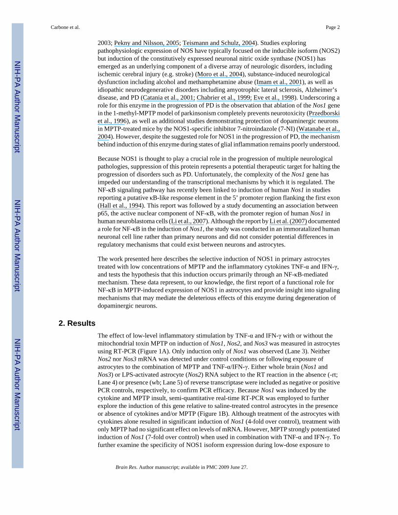
The induction of Nos1 mRNA observed in Figure 1 was mirrored by immunoblot experiments(Figure 2A) demonstrating selective expression of NOS1, but not NOS2 or NOS3, in astrocytesexposed to MPTP and TNF-α/IFN-γ (Lane 3). In all cases, membranes were stripped ofantibody and re-probed against β-Actin as a loading control. Although all three NOS isoformswere probed simultaneously, independent experiments measuring only NOS2 or NOS3 wereperformed to confirm selective induction of NOS1 (Figure 2A, lower panels). Potentiation ofNOS1 induction was demonstrated through densitometric analysis of band intensities (Figure2B). To confirm that induction of NOS1 correlated with expression of functionally activeprotein, steady state production of NO was determined by live-cell fluorescence imaging usingthe cell-permeable NO-sensitive dye, DAF-FM. Co-treatment of astrocytes with MPTP andTNF-α/IFN-γ resulted in an increase in NO production (Figure 2C). To confirm theinvolvement of NOS1 in elevated NO production, astrocytes were also treated with MPTP andTNF-α/IFN-γ in the presence of the selective NOS1 inhibitor, 7-nitroindazole (7-NI), resultingin complete suppression of NO production in both control and treated astrocytes (Figure 2C).Interestingly, 7-NI reduced NO to levels less than those observed in untreated control cells.DMSO, used as a vehicle control for 7-NI, resulted in no measurable suppression of NOproduction. Finally, to rule out any interference from induction of NOS2, astrocytes were co-treated the cytokine and MPTP combination as well as the specific NOS2 inhibitor AMT, whichdid not suppress NO production, thus confirming that NOS2 was not responsible for elevatedintracellular NO concentrations (Figure 2D).
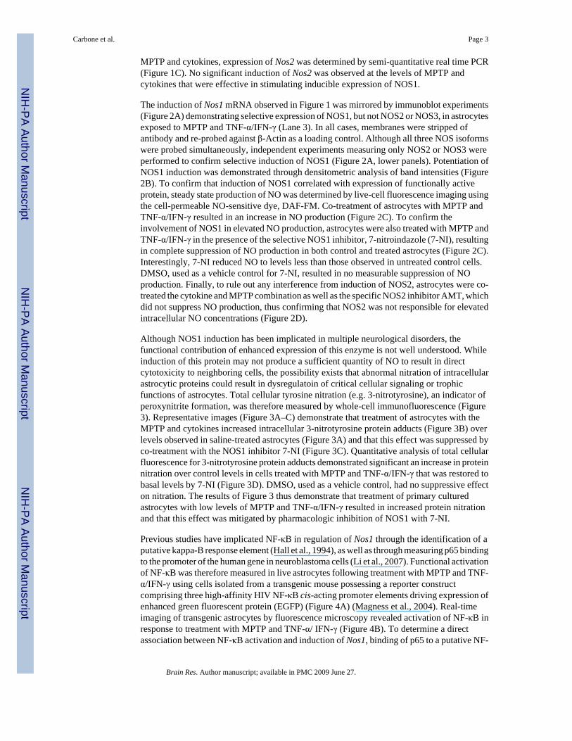
Although NOS1 induction has been implicated in multiple neurological disorders, thefunctional contribution of enhanced expression of this enzyme is not well understood. Whileinduction of this protein may not produce a sufficient quantity of NO to result in directcytotoxicity to neighboring cells, the possibility exists that abnormal nitration of intracellularastrocytic proteins could result in dysregulatoin of critical cellular signaling or trophicfunctions of astrocytes. Total cellular tyrosine nitration (e.g. 3-nitrotyrosine), an indicator ofperoxynitrite formation, was therefore measured by whole-cell immunofluorescence (Figure3). Representative images (Figure 3A–C) demonstrate that treatment of astrocytes with theMPTP and cytokines increased intracellular 3-nitrotyrosine protein adducts (Figure 3B) overlevels observed in saline-treated astrocytes (Figure 3A) and that this effect was suppressed byco-treatment with the NOS1 inhibitor 7-NI (Figure 3C). Quantitative analysis of total cellularfluorescence for 3-nitrotyrosine protein adducts demonstrated significant an increase in proteinnitration over control levels in cells treated with MPTP and TNF-α/IFN-γ that was restored tobasal levels by 7-NI (Figure 3D). DMSO, used as a vehicle control, had no suppressive effecton nitration. The results of Figure 3 thus demonstrate that treatment of primary culturedastrocytes with low levels of MPTP and TNF-α/IFN-γ resulted in increased protein nitrationand that this effect was mitigated by pharmacologic inhibition of NOS1 with 7-NI.
Previous studies have implicated NF-κB in regulation of Nos1 through the identification of aputative kappa-B response element (Hall et al., 1994), as well as through measuring p65 bindingto the promoter of the human gene in neuroblastoma cells (Li et al., 2007). Functional activationof NF-κB was therefore measured in live astrocytes following treatment with MPTP and TNF-α/IFN-γ using cells isolated from a transgenic mouse possessing a reporter constructcomprising three high-affinity HIV NF-κB cis-acting promoter elements driving expression ofenhanced green fluorescent protein (EGFP) (Figure 4A) (Magness et al., 2004). Real-timeimaging of transgenic astrocytes by fluorescence microscopy revealed activation of NF-κB inresponse to treatment with MPTP and TNF-α/ IFN-γ (Figure 4B). To determine a directassociation between NF-κB activation and induction of Nos1, binding of p65 to a putative NF-
Carbone et al. Page 3
Brain Res. Author manuscript; available in PMC 2009 June 27.
NIH
-PA Author Manuscript
NIH
-PA Author Manuscript
NIH
-PA Author Manuscript
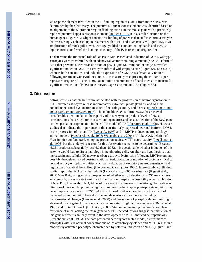
κB response element identified in the 5’-flanking region of exon 1 from mouse Nos1 wasdetermined by the ChIP assay. The putative NF-κB response element was identified based onan alignment of the 5’ promoter region flanking exon 1 in the mouse gene with a previouslyreported putative kappa-B response element (Hall et al., 1994) in a similar location on thehuman gene (Figure 4C). Slight constitutive binding of p65 was detected in control astrocytesthat was strongly enhanced upon treatment with MPTP and TNF-α/IFN-γ (Figure 4D). PCRamplification of mock pull-downs with IgG yielded no contaminating bands and 10% ChIPinput controls confirmed the loading efficiency of the PCR reactions (Figure 4D).
To determine the functional role of NF-κB in MPTP-mediated induction of NOS1, wildtypeastrocytes were transfected with an adenoviral vector containing a mutant (S32-36A) form ofIκBα that prevents nuclear translocation of p65 (Figure 5). Immunoblot analysis revealedsignificant induction NOS1 in astrocytes infected with empty vector (Figure 5A, Lanes 2–5),whereas both constitutive and inducible expression of NOS1 was substantially reducedfollowing treatment with cytokines and MPTP in astrocytes expressing the NF-κB “super-repressor” (Figure 5A, Lanes 6–9). Quantitative determination of band intensities indicated asignificant reduction of NOS1 in astrocytes expressing mutant IκBα (Figure 5B).
3. DiscussionAstrogliosis is a pathologic feature associated with the progression of neurodegeneration inPD. Activated astrocytes release inflammatory cytokines, prostaglandins, and NO thatpotentiate neuronal dysfunction in states of neurologic injury and disease (Hirsch and Hunot,2000; McGeer and McGeer, 1998). The inducible NOS isoform, NOS2, has receivedconsiderable attention due to the capacity of this enzyme to produce levels of NO atconcentrations that are cytotoxic to surrounding neurons and because deletion of the Nos2 geneconfers partial neuroprotection in the MPTP model of PD (Liberatore et al., 1999). However,studies also indicate the importance of the constitutively expressed neuronal isoform, NOS1,in the progression of human PD (Eve et al., 1998) and in MPTP-induced neuropathology inanimal models (Przedborski et al., 1996; Watanabe et al., 2004). Unlike Nos2, deletion ofNos1 in mice confers nearly complete protection against MPTP neurotoxicity (Przedborski etal., 1996) but the underlying reason for this observation remains to be determined. BecauseNOS1 produces substantially less NO than NOS2, it is questionable whether induction of thisenzyme would lead to direct pathology in neighboring cells. An alternate hypothesis is thatincreases in intracellular NO may exacerbate astrocyte dysfunction following MPTP treatment,possibly through enhanced post-translational S-nitrosylation or nitration of proteins critical tonormal astrocyte trophic activities, such as modulation of excitatory neurotransmission andregulation of cerebral blood flow (Haydon and Carmignoto, 2006). Interestingly, conflictingstudies report that NO can either inhibit (Levrand et al., 2005) or stimulate (Riganti et al.,2007) NF-κB signaling, raising the question of whether early induction of NOS1 may representan attempt by the astrocyte to mitigate inflammation. Despite the possibility of early inhibitionof NF-κB by low levels of NO, 24 hrs of low-level inflammatory stimulation globally elevatednitration of intracellular proteins (Figure 3), suggesting that inappropriate protein nitration maybe an important sequela of NOS1 induction. Indeed, studies characterizing the effects ofincreased protein nitration have documented deleterious consequences includingconformational changes (Cassina et al., 2000) and prevention of phosphorylation resulting inabnormal loss or gain of function, such as that reported for glutamine synthetase (Berlett et al.,1996) and protein kinase C (Hink et al., 2003). Studies documenting the nearly completeresistance of mice lacking the Nos1 gene to MPTP-induced lesions suggest that induction ofthis gene represents an early event in the development of MPTP-induced neuropathology(Przedborski et al., 1996). The data presented here support such a model, as treatment ofastrocytes with sub-optimal concentrations of inflammatory cytokines and MPTP results in amoderately activated phenotype characterized by selective induction of NOS1 (Figure 1 and
Carbone et al. Page 4
Brain Res. Author manuscript; available in PMC 2009 June 27.
NIH
-PA Author Manuscript
NIH
-PA Author Manuscript
NIH
-PA Author Manuscript
Figure 2), an increase in steady-state production of NO (Figure 2), and increased proteinnitration (Figure 3).
Despite a recognized association between Nos1 and the progression of multiple neurologicaldisorders, the regulation of this gene remains incompletely characterized due to its inherentcomplexity. Indeed, human Nos1 is distributed over 160 kb among 29 exons and 28 introns,with nine recognized splice variants in the neuronal tissue cluster (Nos1a-i) regulated byalternate promoter usage in the 5’ untranslated region flanking the first exon (Wang et al.,1999; Xie et al., 1995). Of these, Nos1f and Nos1g are predominantly expressed in brain (Wanget al., 1999). Although similar in both size and organization, mouse Nos1 has been much lessstudied than the human gene and far less is known regarding regulation of inducible expressionof different splice variants. Specifically, only three splice variants (Nos1a-c) of the Nos1 clusterexpressed in neurons have been characterized in mice, of which Nos1a and Nos1b havedocumented homology with human variants Nos1f and Nos1g respectively, whereas mouseNos1c is not homologous to any known human splice variant (Sasaki et al., 2000). Furthermore,three splice variants (e.g. Nos1α, β, γ) involving the second exon in both the human and mouseNos1 result in deletion of the second exon in variants β and γ, which are reportedly induced inastrocytes in human ALS (Catania et al., 2001), and of which only the β variant retains catalyticactivity (Huang et al., 1993; Kavya et al., 2006).
To date, few cis-activing transcription factors have been characterized that mediate inducibleexpression of mouse Nos1. Depolarization-induced Ca2+ influx in cortical neurons through L-type voltage-sensitive calcium channels is reported to induce Nos1 through a CREB responsivepathway at the second exon (Sasaki et al., 2000) and steroidogenic factor 1 (SF-1) is alsoreported to mediate inducible expression of mouse Nos1 (Wei et al., 2002). Involvement ofNF-κB in regulation of mouse Nos1 is suggested by a separate report characterizing theorganization of human Nos1 in which the presence of an NF-κB response element wasdocumented in the promoter region flanking the first exon (Hall et al., 1994). This site waslater demonstrated to interact with the active p65 NF-κB subunit in neuroblastoma cells (Li etal., 2007). Although interaction with the putative Nos1 NF-κB binding site in this latter studywas shown by electrophoretic mobility shift and ChIP assays, neither gene silencing nordominant-negative approaches were performed to confirm a direct role for NF-κB in inducibleexpression of the protein. Given these previous data, we performed an alignment of the humanand mouse Nos1 5’ UTR flanking the first exon and found the presence of a similar site (Figure4C). While transactivation of NF-κB responsive genes typically occurs in response to p65/p50heterodimer translocation to the nuclear response element, binding of the p50/p50 homodimerhas been reported to actually suppress induction (Ghosh et al., 1998). Therefore, binding ofp65 to the Nos1 promoter in the region identified as a potential NF-κB response elementfollowing alignment with the human was measured through ChIP, demonstrating binding ofthis transcription factor to the predicted response element following treatment of primaryastrocytes with MPTP and TNF-α/INF-γ (Figure 4D). Together with findings that NF-κB wasactivated following this mild inflammatory insult in transgenic reporter astrocytes (Figure 4B)and that overexpression of an NF-κB “super repressor” prevented MPTP- and cytokine-inducedexpression of Nos1 (Figure 5), these data demonstrate direct involvement of NF-κB in theinduction of Nos1 during MPTP-dependent potentiation of inflammatory signaling.
Collectively, the data presented here demonstrate that NOS1 is selectively induced in primaryastrocytes during mild inflammatory stimulation with cytokines and MPTP. The capacity oflow levels of MPTP to potentiate the effects of inflammatory cytokines on inducible expressionof NOS1 may provide insight into the consequences of mitochondrial dysfunction evident inPD on the inflammatory phenotype of astroglia. Selective induction of NOS1 was confirmedby the absence of detectable mRNA for Nos2 or Nos3. Although a faint immunoreactive bandwas inconsistently detected at a slightly smaller molecular weight than NOS1, this likely
Carbone et al. Page 5
Brain Res. Author manuscript; available in PMC 2009 June 27.
NIH
-PA Author Manuscript
NIH
-PA Author Manuscript
NIH
-PA Author Manuscript
represents a NOS1 splice variant, because immunoblots for NOS2 and NOS3 did not detectthe presence of these isoforms (Figure 2A, lower panels). Additional studies using the NOS2inhibitor AMT further demonstrated selective induction of functional NOS1, as this inhibitorhad no effect on intracellular NO concentrations following low-dose exposure to MPTP andTNF-α/IFN-γ (Figure 2D). The absence of NOS2 induction supports the conclusion that thesub-optimal concentrations of inflammatory cytokines and MPTP used in these studies resultin a phenotype that might be observed in the early stages of PD, before a full-scale inflammatoryresponse and ensuing astrogliosis has occurred. Furthermore, the identification of NF-κB asthe predominant pathway by which Nos1 is induced in this model reveals a potential target forsuppressing the induction of this gene. Together, the results presented here provide a basis forfurther dissection of the signaling pathways regulating inducible expression of Nos1 inastrocytes.
4. Experimental Procedures4.1. Materials
Unless otherwise stated, all reagents were purchased from Sigma-Aldrich Chemical Co. (St.Louis, MO). The NOS2 inhibitor 2-amino-5,6-dihydro-6-methyl-4H-1,3-thiazine (AMT) waspurchased from Calbiochem (San Diego, CA). Cell culture media, antibiotics, and fluorescentantibodies and dyes were purchased from Invitrogen (Carlsbad, CA). Monoclonal antibodiesagainst NOS1-3 were purchased from BD biosciences (San Jose, CA). Horseradish peroxidaseconjugated goat anti-mouse and goat anti-rabbit secondary antibodies were purchased fromSanta Cruz Biotechnology (Santa Cruz, CA) and Cell Signaling Technology (Boston, MA).For immunofluoresence studies, antibodies against GFAP and nitrosylated tyrosine werepurchased from Sigma Chemical Co. (St. Louis, MO) and Chemicon (Temecula, CA),respectively.
4.2. Astrocyte IsolationCortical astrocytes were isolated from day-1 old C57Bl/6 or transgenic mouse pups accordingto procedures described previously (Aschner and Kimelberg, 1991), and purity confirmedthrough immunofluorescent staining using antibodies against GFAP (astrocyte; approximately98% cells per field) and Iba1 (microglia; approximately 1% per field). Briefly, pups wereeuthanized by decapitation, cortices rapidly dissected out, and meninges removed. Tissue wassubject to digestion with Dispase (1.5 U/ml), and selection of astrocytes was performed bychanging media 24 hrs. after plating to remove non-adherent microglial cells. Astrocytecultures were maintained at 37°C and 5% CO2 in minimum essential media supplemented with10% heat-inactivated fetal bovine serum and a penicillin (0.001 mg/ml), streptomycin (0.002mg/ml), and neomycin (0.001) antibiotic cocktail. Cell media was changed 24 hrs prior to alltreatments to minimize interference from serum shock. All animal procedures were approvedby the Colorado State University Institutional Animal Care and Use Committee, and wereconducted in accordance with published NIH guidelines.
4.3. RT and Real-Time RT-PCRAstrocytes were treated with a sub-maximal pathologic insult (TNF-α, 10 pg/ml; IFN-γ, 1 ng/ml; MPTP, 1 µM). These doses of inflammatory cytokines were based upon previous studiesfrom our laboratory demonstrating little to no induction of Nos2 at the concentrations used(Liu et al., 2005). RNA was isolated using an RNEasy Mini kit (Qiagen, Valencia, CA), andpurity and concentration determined using a Nanodrop ND-1000 spectrophotometer(NanoDrop Technologies, Wilmington, DE). Following purification, 1 ug of RNA was usedas template for reverse transcriptase (RT) reactions using the iScript RT kit (BioRad, HerculesCA). The resulting cDNA was immediately profiled for expression of Nos using primersspecific for Nos1 (forward: 5’-TGCTACAACCTCGCTACTATT-3’; reverse: 5’-
Carbone et al. Page 6
Brain Res. Author manuscript; available in PMC 2009 June 27.
NIH
-PA Author Manuscript
NIH
-PA Author Manuscript
NIH
-PA Author Manuscript
ACATCGTCAGCCTGTATTCG-3’), Nos2 (forward: 5’-TCACGCTTGGGTCTTGTT-3’;reverse: 5’-CAGGTCACTTTGGTAGGATTT-3’), or Nos3 (forward: 5’-GGCATCACCAGGAAGAAGAC-3’; reverse: 5’-GGGACACCACATCATACTCAT- 3’).Real-time RT-PCR was used to measure relative Nos1 or Nos2 expression in response tocytokine and/or MPTP exposure using β-Actin as a housekeeping gene (forward: 5’-GCTGTGCTATGTTGCTCTAG-3’; reverse: 5’-CGCTCGTTGCCAATACTG-3’) accordingto the 2−ΔΔCT method (Livak and Schmittgen, 2001).
4.4. Western BlottingAstrocytes were treated with the cytokines TNF-α and IFN-γ (10 pg/ml and 1 ng/ml,respectively) and or MPTP (1 µM) for a period of 24 hrs prior to harvesting. Followingtreatment, cells were lysed using standard RIPA buffer supplemented with Complete™protease inhibitor (Roche, Indianapolis IN). Protein was quantified using the BCA assay(Pierce, Rockford IL), and 20 µg (NOS1) or 50 µg (NOS2 and NOS3) of protein were separatedby SDS-PAGE using a 6% slab gel (BioRad, Hercules CA) followed by semi-dry transfer topolyvinylidene fluoride (PVDF) membrane (Pall Corp., Penscola, FL). All blocking andantibody incubations were performed in 5% non-fat dry milk in tris-buffered saline containing0.2% Tween-20. Positive controls consisting of 0.5 µg activated macrophage lysate (NOS2;BD Biosciences, San Jose, CA) or 10 µg mouse brain homogenate (NOS1 and NOS3) wereincluded to confirm results. Protein was visualized on film using enhanced chemiluminescence(Pierce, Rockford, IL), and densitometry was performed using ImageQuant v5.3 (GEHealthcare, Sunnyvale, CA).
4.5. Quantitation of Intracellular Nitric OxideAstrocytes were grown to confluence in 4-chamber glass-bottom slides (Nalge Nunc,Rochester, NY), and treated for 24 hrs with saline or the cytokine and MPTP combination. Co-treatment or treatment with the NOS1 inhibitor 7-NI (10 µM) or the NOS2 inhibitor AMT (25nM) were included to demonstrate selective induction of NOS1. Following treatment, thefluorescent NO indictor 4-amino-5-methylamino-2’,7’-difluorofluorescein diacetate (DAF-FM) was added at 5 µM and intracellular de-esterification allowed to proceed for 10 min at37°C. Cell media was washed twice with serum and phenol-red-free MEM supplemented with10 mM HEPES, and DAF-FM intensity recorded via fluorescence microscopy as describedabove at 2 min. intervals over the course of 30 min. using a Zeiss Axiovert 200M microscope(Thornwood, NY) equipped, 20× air PlanApochromat objective and ORCA-ER cooled,interline charge-coupled device camera (Hammamatsu Photonics, Hamamatsu City, Japan).Intracellular NO is reported as final over initial fluorescence (F/F0) at at 490 nmex/520nmem. Images were acquired and analyzed using Slidebook software (v4.1, Intelligent ImagingInnovations, Inc., Denver, CO). A minimum of 500 – 800 cells were imaged per treatmentgroup for studies of NO over no less than 3 independent experiments.
4.6. ImmunofluorescencePrimary astrocytes were grown to confluence on 20 mm serum-coated glass coverslips, andtreated with saline or the TNF-α, IFN-γ, and MPTP combination in the presence or absence ofthe NOS1 inhibitor 7-NI (10 µM) for 24 h. Blocking and antibody hybridization was conductedin 1% BSA (w/v) in PBS, while all washes were conducted in PBS. Images were acquiredusing a Zeiss 20X air PlanApochromat objective and 6 – 8 microscopic fields were examinedper treatment group over no less than three independent experiments Fluorescent secondaryantibodies were used to detect GFAP (488 nmex/519 nmem) and nitrosylated protein (647nmex/668 nmem), respectively, while DAPI (360 nmex/460 nmem) mounting medium was usedto stain nuclei.
Carbone et al. Page 7
Brain Res. Author manuscript; available in PMC 2009 June 27.
NIH
-PA Author Manuscript
NIH
-PA Author Manuscript
NIH
-PA Author Manuscript
4.7. NF-κB reporter assays in cis-NF-κBEGFP transgenic astrocytes and expression of mutantIκBα
To measure activation of NF-κB in live cells, astrocytes were isolated from a unique transgenicmouse expressing a reporter construct consisting of three HIV NF-κB consensus elementsinserted 5’ to a minimal c-fos promoter that drives expression of enhanced green fluorescenctprotein (EGFP) (Magness et al., 2004) (Provided by Dr. Christian Jobin, University of NorthCarolina at Chapel Hill). NF-κB activity was determined by live-cell imaging and reported aspercent activated cells following treatments. A phosphorylation-deficient mutant of IκBα,IκBα-(S32,36A)-HA, was overexpressed in primary astrocytes using an adenoviral vector,delivered for 24 hrs at 2×10⁶ viral particles per ml of culture medium, with a multiplicity ofinfection of 1×10³ virions per cell shown previously by us to result in expression of the mutantprotein by over 99% of the astrocytes (Barhoumi et al., 2004). Parallel control experimentsutilized the same adenoviral construct lacking the insert. Following incubation with the mutantIκBα construct for 24 hrs, astrocytes were washed with PBS to remove viral particles andcultured in fresh medium for 24 hrs prior to gene expression studies or co-incubation withPC12 neurons. Images were acquired using a Zeiss 20X air PlanApochromat objective and 6– 8 microscopic fields were examined per treatment group over three independent experiments.
4.8. Chromatin Immunoprecipitation (ChIP) AssayChIP procedures were adapted from a previously published report (Weinmann and Farnham,2002). Astrocytes were grown to confluence in 60 mm plates (Approx. 1.2 × 10⁶ cells) andwere treated for 4 h with saline or the cytokine and MPTP combination. DNA was sheared into500 bp fragments by three ten-second pulses using a Tekmar Sonic Disrupter (Tekmar Co.,Cincinnati, OH) set at 30% output, and lysate was precleared for one hour at 4° C using salmonsperm-blocked protein-A agarose (Upstate Cell Signaling Solutions, Temecula, CA). Thebeads were then removed by centrifugation, 10% input controls were collected, and 2 µgprecipitating antibody (p65, Santa Cruz Biotechnologies) or control IgG (Calbiochem, SanDiego, CA) was added to the remaining lysate. Immune complexes were allowed to formovernight at 4° C with gentle agitation, followed by the addition of salmon sperm-blockedprotein-A agarose for an additional 60 min. Immunopurified DNA was isolated via phenol/chloroform extraction and subject to PCR using primers designed around a potential NF-κBbinding region in the mouse Nos1 promoter which was located following Clustal v1.8(Thompson et al., 1994) alignment of the mouse and human Nos1 promoter regions (forward:5’-ATC AGG CAT CCT TTC CAG AAC GTC-3’; reverse: 5’-ACT GGA TGT TAG TGACCA CAG CGG-3’). Amplicons were separated by agarose gel electrophoresis and stainedwith ethidium bromide.
4.9. Statistical AnalysisComparison of two means was performed by Student’s t-test, while comparison of more thanthree means was performed using one-way ANOVA followed by the Tukey-Kramer multiplecomparison post-hoc test using Prism software (v4.0c, Graphpad Software, Inc., San Diego,CA). For all experiments, p < 0.05 was considered significant, although the level of significancewas often much greater. Statistically different groups are identified in the figures by theassignment of a unique letter (e.g. a, b, c).
AcknowledgementsThe authors thank Dr. Christian Jobin at the University of North Carolina for providing NF-κB reporter mice. Thiswork was supported by R01ES012941 (RBT), NS055632 (DLC), and an individual research grant from the AmericanParkinson Disease Association (RBT).
Carbone et al. Page 8
Brain Res. Author manuscript; available in PMC 2009 June 27.
NIH
-PA Author Manuscript
NIH
-PA Author Manuscript
NIH
-PA Author Manuscript
ReferencesAschner M, Kimelberg HK. The use of astrocytes in culture as model systems for evaluating neurotoxic-
induced-injury. Neurotoxicology 1991;12:505–517. [PubMed: 1684034]Barhoumi R, Faske J, Liu X, Tjalkens RB. Manganese potentiates lipopolysaccharide-induced expression
of NOS2 in C6 glioma cells through mitochondrial-dependent activation of nuclear factor kappaB.Brain Res Mol Brain Res 2004;122:167–179. [PubMed: 15010209]
Berlett BS, Friguet B, Yim MB, Chock PB, Stadtman ER. Peroxynitrite-mediated nitration of tyrosineresidues in Escherichia coli glutamine synthetase mimics adenylylation relevance to signaltransduction. Proc Natl Acad Sci U S A 1996;93:1776–1780. [PubMed: 8700834]
Cassina AM, Hodara R, Souza JM, Thomson L, Castro L, Ischiropoulos H, Freeman BA, Radi R.Cytochrome c nitration by peroxynitrite. J Biol Chem 2000;275:21409–21415. [PubMed: 10770952]
Catania MV, Aronica E, Yankaya B, Troost D. Increased expression of neuronal nitric oxide synthasespliced variants in reactive astrocytes of amyotrophic lateral sclerosis human spinal cord. J Neurosci2001;21:RC148. [PubMed: 11344254]
Chabrier PE, Demerle-Pallardy C, Auguet M. Nitric oxide synthases: targets for therapeutic strategies inneurological diseases. Cell Mol Life Sci 1999;55:1029–1035. [PubMed: 10442086]
Eve DJ, Nisbet AP, Kingsbury AE, Hewson EL, Daniel SE, Lees AJ, Marsden CD, Foster OJ. Basalganglia neuronal nitric oxide synthase mRNA expression in Parkinson's disease. Brain Res Mol BrainRes 1998;63:62–71. [PubMed: 9838046]
Ghosh S, May MJ, Kopp EB. NF-kappa B and Rel proteins: evolutionarily conserved mediators ofimmune responses. Annu Rev Immunol 1998;16:225–260. [PubMed: 9597130]
Hall AV, Antoniou H, Wang Y, Cheung AH, Arbus AM, Olson SL, Lu WC, Kau CL, Marsden PA.Structural organization of the human neuronal nitric oxide synthase gene (NOS1). J Biol Chem1994;269:33082–33090. [PubMed: 7528745]
Haydon PG, Carmignoto G. Astrocyte control of synaptic transmission and neurovascular coupling.Physiol Rev 2006;86:1009–1031. [PubMed: 16816144]
Hink U, Oelze M, Kolb P, Bachschmid M, Zou MH, Daiber A, Mollnau H, August M, Baldus S,Tsilimingas N, Walter U, Ullrich V, Munzel T. Role for peroxynitrite in the inhibition of prostacyclinsynthase in nitrate tolerance. J Am Coll Cardiol 2003;42:1826–1834. [PubMed: 14642695]
Hirsch EC, Hunot S. Nitric oxide, glial cells and neuronal degeneration in parkinsonism. TrendsPharmacol Sci 2000;21:163–165. [PubMed: 10785644]
Hirsch EC, Breidert T, Rousselet E, Hunot S, Hartmann A, Michel PP. The role of glial reaction andinflammation in Parkinson's disease. Ann N Y Acad Sci 2003;991:214–228. [PubMed: 12846989]
Huang PL, Dawson TM, Bredt DS, Snyder SH, Fishman MC. Targeted disruption of the neuronal nitricoxide synthase gene. Cell 1993;75:1273–1286. [PubMed: 7505721]
Imam SZ, Newport GD, Itzhak Y, Cadet JL, Islam F, Slikker W Jr, Ali SF. Peroxynitrite plays a role inmethamphetamine-induced dopaminergic neurotoxicity: evidence from mice lacking neuronal nitricoxide synthase gene or overexpressing copper-zinc superoxide dismutase. J Neurochem2001;76:745–749. [PubMed: 11158245]
Kavya R, Saluja R, Singh S, Dikshit M. Nitric oxide synthase regulation and diversity: implications inParkinson's disease. Nitric Oxide 2006;15:280–294. [PubMed: 16934505]
Levrand S, Pesse B, Feihl F, Waeber B, Pacher P, Rolli J, Schaller MD, Liaudet L. Peroxynitrite is apotent inhibitor of NF-{kappa}B activation triggered by inflammatory stimuli in cardiac andendothelial cell lines. J Biol Chem 2005;280:34878–34887. [PubMed: 16079150]
Li Y, Zhao Y, Li G, Wang J, Li T, Li W, Lu J. Regulation of neuronal nitric oxide synthase exon 1f geneexpression by nuclear factor-kappaB acetylation in human neuroblastoma cells. J Neurochem2007;101:1194–1204. [PubMed: 17250678]
Liberatore GT, Jackson-Lewis V, Vukosavic S, Mandir AS, Vila M, McAuliffe WG, Dawson VL,Dawson TM, Przedborski S. Inducible nitric oxide synthase stimulates dopaminergicneurodegeneration in the MPTP model of Parkinson disease. Nat Med 1999;5:1403–1409. [PubMed:10581083]
Carbone et al. Page 9
Brain Res. Author manuscript; available in PMC 2009 June 27.
NIH
-PA Author Manuscript
NIH
-PA Author Manuscript
NIH
-PA Author Manuscript
Liu X, Buffington JA, Tjalkens RB. NF-kappaB-dependent production of nitric oxide by astrocytesmediates apoptosis in differentiated PC12 neurons following exposure to manganese and cytokines.Brain Res Mol Brain Res 2005;141:39–47. [PubMed: 16168523]
Livak KJ, Schmittgen TD. Analysis of relative gene expression data using real-time quantitative PCRand the 2(-Delta Delta C(T)) Method. Methods 2001;25:402–408. [PubMed: 11846609]
Magness ST, Jijon H, Van Houten Fisher N, Sharpless NE, Brenner DA, Jobin C. In vivo pattern oflipopolysaccharide and anti-CD3-induced NF-kappa B activation using a novel gene-targetedenhanced GFP reporter gene mouse. J Immunol 2004;173:1561–1570. [PubMed: 15265883]
McGeer PL, McGeer EG. Glial cell reactions in neurodegenerative diseases: pathophysiology andtherapeutic interventions. Alzheimer Dis Assoc Disord 1998;12:S1–S6. [PubMed: 9769023]
Moro MA, Cardenas A, Hurtado O, Leza JC, Lizasoain I. Role of nitric oxide after brain ischaemia. CellCalcium 2004;36:265–275. [PubMed: 15261482]
Pekny M, Nilsson M. Astrocyte activation and reactive gliosis. Glia 2005;50:427–434. [PubMed:15846805]
Przedborski S, Jackson-Lewis V, Yokoyama R, Shibata T, Dawson VL, Dawson TM. Role of neuronalnitric oxide in 1-methyl-4-phenyl-1,2,3,6-tetrahydropyridine (MPTP)-induced dopaminergicneurotoxicity. Proc Natl Acad Sci U S A 1996;93:4565–4571. [PubMed: 8643444]
Riganti C, Costamagna C, Doublier S, Miraglia E, Polimeni M, Bosia A, Ghigo D. The NADPH oxidaseinhibitor apocynin induces nitric oxide synthesis via oxidative stress. Toxicol Sci. 2007(epub aheadof print)
Sasaki M, Gonzalez-Zulueta M, Huang H, Herring WJ, Ahn S, Ginty DD, Dawson VL, Dawson TM.Dynamic regulation of neuronal NO synthase transcription by calcium influx through a CREB familytranscription factor-dependent mechanism. Proc Natl Acad Sci U S A 2000;97:8617–8622. [PubMed:10900019]
Teismann P, Schulz JB. Cellular pathology of Parkinson's disease: astrocytes, microglia andinflammation. Cell Tissue Res 2004;318:149–161. [PubMed: 15338271]
Thompson JD, Higgins DG, Gibson TJ. CLUSTAL W: improving the sensitivity of progressive multiplesequence alignment through sequence weighting, position-specific gap penalties and weight matrixchoice. Nucleic Acids Res 1994;22:4673–4680. [PubMed: 7984417]
Wang Y, Newton DC, Robb GB, Kau CL, Miller TL, Cheung AH, Hall AV, VanDamme S, Wilcox JN,Marsden PA. RNA diversity has profound effects on the translation of neuronal nitric oxide synthase.Proc Natl Acad Sci U S A 1999;96:12150–12155. [PubMed: 10518591]
Watanabe H, Muramatsu Y, Kurosaki R, Michimata M, Matsubara M, Imai Y, Araki T. Protective effectsof neuronal nitric oxide synthase inhibitor in mouse brain against MPTP neurotoxicity: animmunohistological study. Eur Neuropsychopharmacol 2004;14:93–104. [PubMed: 15013024]
Wei X, Sasaki M, Huang H, Dawson VL, Dawson TM. The orphan nuclear receptor, steroidogenic factor1, regulates neuronal nitric oxide synthase gene expression in pituitary gonadotropes. Mol Endocrinol2002;16:2828–2839. [PubMed: 12456803]
Weinmann AS, Farnham PJ. Identification of unknown target genes of human transcription factors usingchromatin immunoprecipitation. Methods 2002;26:37–47. [PubMed: 12054903]
Xie J, Roddy P, Rife TK, Murad F, Young AP. Two closely linked but separable promoters for humanneuronal nitric oxide synthase gene transcription. Proc Natl Acad Sci U S A 1995;92:1242–1246.[PubMed: 7532307]
AbbreviationsNO, nitric oxide; NOS, nitric oxide synthase; TNF-α, tumor necrosis factor alpha; IFN-γ,interferon gamma; NF-κB, nuclear factor-kappa B..
Carbone et al. Page 10
Brain Res. Author manuscript; available in PMC 2009 June 27.
NIH
-PA Author Manuscript
NIH
-PA Author Manuscript
NIH
-PA Author Manuscript
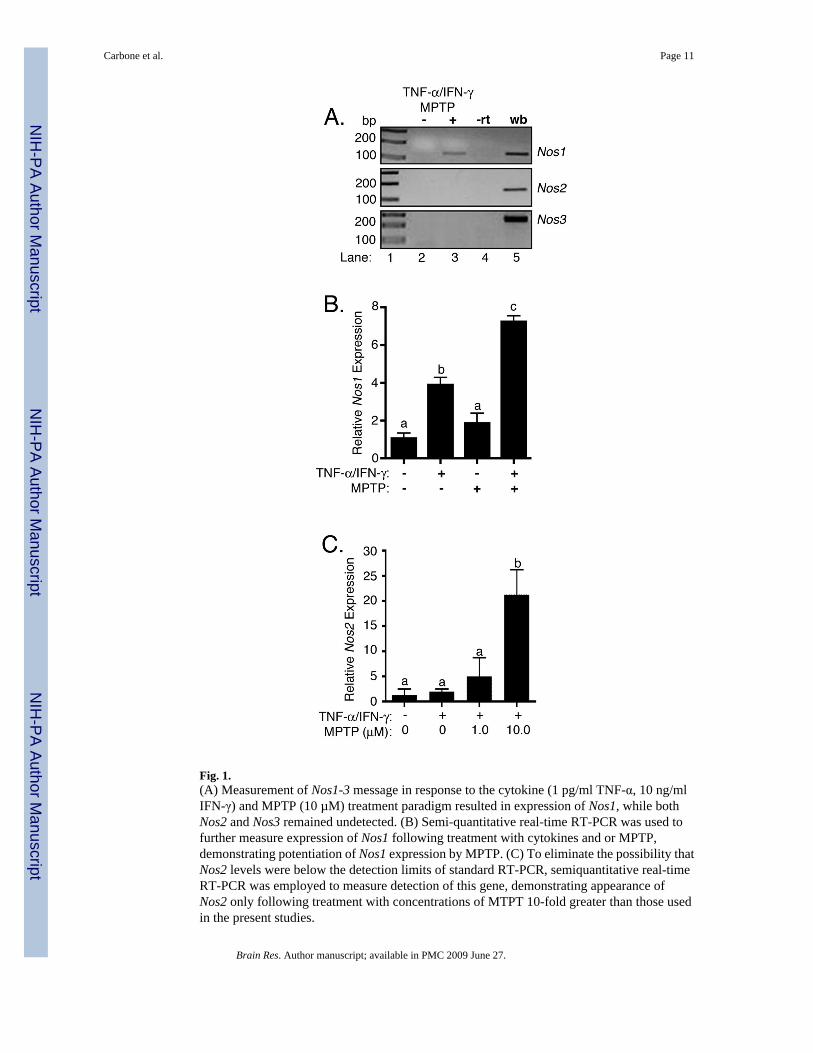
Fig. 1.(A) Measurement of Nos1-3 message in response to the cytokine (1 pg/ml TNF-α, 10 ng/mlIFN-γ) and MPTP (10 µM) treatment paradigm resulted in expression of Nos1, while bothNos2 and Nos3 remained undetected. (B) Semi-quantitative real-time RT-PCR was used tofurther measure expression of Nos1 following treatment with cytokines and or MPTP,demonstrating potentiation of Nos1 expression by MPTP. (C) To eliminate the possibility thatNos2 levels were below the detection limits of standard RT-PCR, semiquantitative real-timeRT-PCR was employed to measure detection of this gene, demonstrating appearance ofNos2 only following treatment with concentrations of MTPT 10-fold greater than those usedin the present studies.
Carbone et al. Page 11
Brain Res. Author manuscript; available in PMC 2009 June 27.
NIH
-PA Author Manuscript
NIH
-PA Author Manuscript
NIH
-PA Author Manuscript
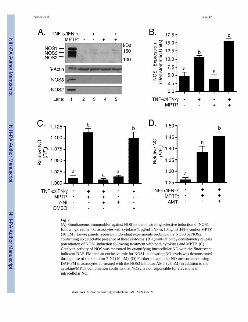
Fig. 2.(A) Simultaneous immunoblot against NOS1-3 demonstrating selective induction of NOS1following treatment of astrocytes with cytokine (1 pg/ml TNF-α, 10 ng/ml IFN-γ) and/or MPTP(10 µM). Lower panels represent individual experiments probing only NOS3 or NOS2,confirming no detectable presence of these isoforms. (B) Quantitation by densitometry revealspotentiation of NOS1 induction following treatment with both cytokines and MPTP. (C)Catalytic activity of NOS was measured by quantifying intracellular NO with the fluorescentindicator DAF-FM, and an exclusive role for NOS1 in elevating NO levels was demonstratedthrough use of the inhibitor 7-NI (10 µM). (D) Further intracellular NO measurement usingDAF-FM in astrocytes co-treated with the NOS2 inhibitor AMT (25 nM) in addition to thecytokine/MPTP combination confirms that NOS2 is not responsible for elevations inintracellular NO.
Carbone et al. Page 12
Brain Res. Author manuscript; available in PMC 2009 June 27.
NIH
-PA Author Manuscript
NIH
-PA Author Manuscript
NIH
-PA Author Manuscript
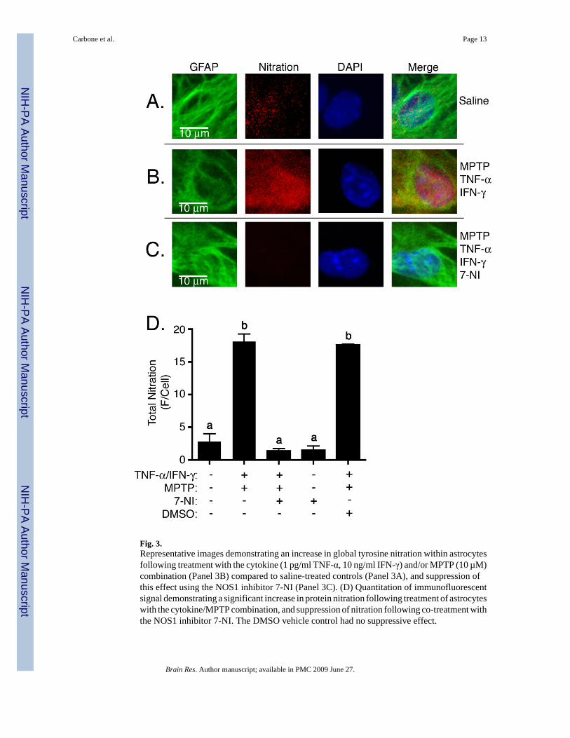
Fig. 3.Representative images demonstrating an increase in global tyrosine nitration within astrocytesfollowing treatment with the cytokine (1 pg/ml TNF-α, 10 ng/ml IFN-γ) and/or MPTP (10 µM)combination (Panel 3B) compared to saline-treated controls (Panel 3A), and suppression ofthis effect using the NOS1 inhibitor 7-NI (Panel 3C). (D) Quantitation of immunofluorescentsignal demonstrating a significant increase in protein nitration following treatment of astrocyteswith the cytokine/MPTP combination, and suppression of nitration following co-treatment withthe NOS1 inhibitor 7-NI. The DMSO vehicle control had no suppressive effect.
Carbone et al. Page 13
Brain Res. Author manuscript; available in PMC 2009 June 27.
NIH
-PA Author Manuscript
NIH
-PA Author Manuscript
NIH
-PA Author Manuscript
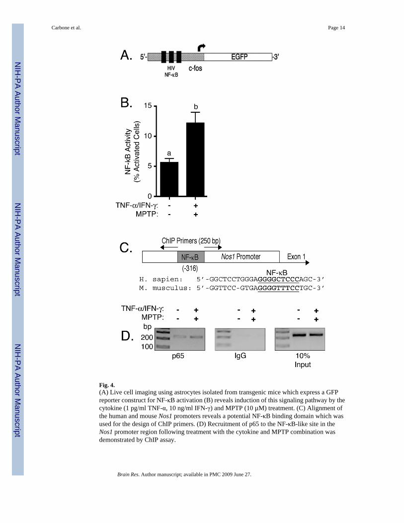
Fig. 4.(A) Live cell imaging using astrocytes isolated from transgenic mice which express a GFPreporter construct for NF-κB activation (B) reveals induction of this signaling pathway by thecytokine (1 pg/ml TNF-α, 10 ng/ml IFN-γ) and MPTP (10 µM) treatment. (C) Alignment ofthe human and mouse Nos1 promoters reveals a potential NF-κB binding domain which wasused for the design of ChIP primers. (D) Recruitment of p65 to the NF-κB-like site in theNos1 promoter region following treatment with the cytokine and MPTP combination wasdemonstrated by ChIP assay.
Carbone et al. Page 14
Brain Res. Author manuscript; available in PMC 2009 June 27.
NIH
-PA Author Manuscript
NIH
-PA Author Manuscript
NIH
-PA Author Manuscript
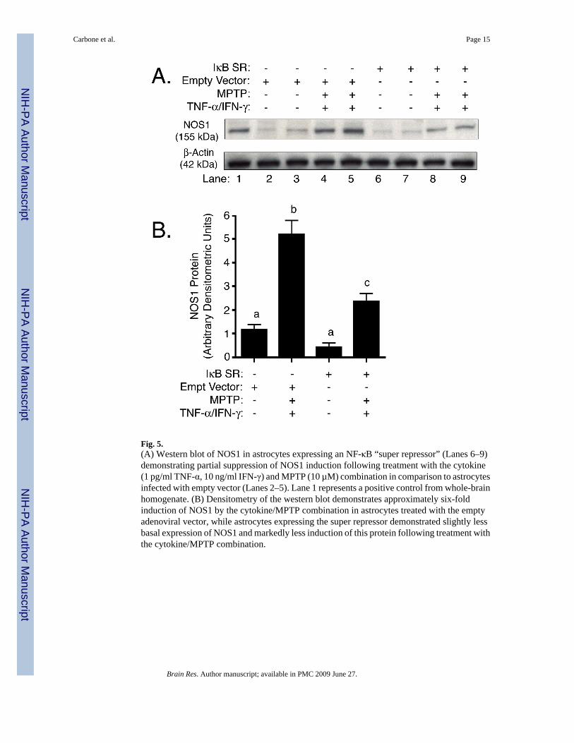
Fig. 5.(A) Western blot of NOS1 in astrocytes expressing an NF-κB “super repressor” (Lanes 6–9)demonstrating partial suppression of NOS1 induction following treatment with the cytokine(1 pg/ml TNF-α, 10 ng/ml IFN-γ) and MPTP (10 µM) combination in comparison to astrocytesinfected with empty vector (Lanes 2–5). Lane 1 represents a positive control from whole-brainhomogenate. (B) Densitometry of the western blot demonstrates approximately six-foldinduction of NOS1 by the cytokine/MPTP combination in astrocytes treated with the emptyadenoviral vector, while astrocytes expressing the super repressor demonstrated slightly lessbasal expression of NOS1 and markedly less induction of this protein following treatment withthe cytokine/MPTP combination.
Carbone et al. Page 15
Brain Res. Author manuscript; available in PMC 2009 June 27.
NIH
-PA Author Manuscript
NIH
-PA Author Manuscript
NIH
-PA Author Manuscript