mechanosensation and endothelin in astrocytes—hypothetical roles in cns pathophysiology
TRANSCRIPT

Review
Mechanosensation and endothelin in astrocytes—hypothetical roles
in CNS pathophysiology
Lyle W. Ostrow, Frederick Sachs*
Department of Physiology and Biophysics, S.U.N.Y. at Buffalo, School of Medicine and Biomedical Sciences, Buffalo, NY 14214, USA
Accepted 9 September 2004
Available online 28 October 2004
Brain Research Reviews 48 (2005) 488–508
www.elsevier.com/locate/brainresrev
Abstract
Endothelin (ET) is a potent autocrine mitogen produced by reactive and neoplastic astrocytes. ET has been implicated in the
induction of astrocyte proliferation and other transformations engendered by brain pathology, and in promoting the malignant behavior
of astrocytomas. Reactive astrocytes containing ET are found in the periphery/penumbra of a wide array of CNS pathologies.
Virtually all brain pathology deforms the surrounding parenchyma, either by direct mass effect or edema. Mechanical stress is a well
established stimulus for ET production and release by other cell types, but has not been well studied in the brain. However,
numerous studies have illustrated that astrocytes can sense mechanical stress and translate it into chemical messages. Furthermore, the
ubiquitous reticular meshwork formed by interconnected astrocytes provides an ideal morphology for sensing and responding to
mechanical disturbances. We have recently demonstrated stretch-induced ET production by astrocytes in vitro. Inspired by this finding,
the purpose of this article is to review the literature on (1) astrocyte mechanosensation, and (2) the endothelin system in astrocytes,
and to consider the hypothesis that mechanical induction of the ET system may influence astrocyte functioning in CNS
pathophysiology. We conclude by discussing evidence supporting future investigations to determine whether specific inhibition of
stretch-activated ion channels may represent a novel strategy for treating or preventing CNS disturbances, as well as the relevance to
astrocyte-derived tumors.
D 2004 Elsevier B.V. All rights reserved.
Theme: Development and regeneration
Theme: Glia and other non-neuronal cells
Keywords: Stretch; Calcium; Proliferation; Glioma; Channel blocker; Cytoskeleton; GFAP
Contents
1. The temperament of astrocytes: structural support for neurons or active role in CNS functioning? . . . . . . . . . . . . . . . 489
2. Astrocyte responses to CNS disturbances . . . . . . . . . . . . . . . . . . . . . . . . . . . . . . . . . . . . . . . . . . . . 489
3. Brain deformation and pathology . . . . . . . . . . . . . . . . . . . . . . . . . . . . . . . . . . . . . . . . . . . . . . . . 490
4. Astrocyte mechanosensation . . . . . . . . . . . . . . . . . . . . . . . . . . . . . . . . . . . . . . . . . . . . . . . . . . . 490
4.1. Stretch-induced Ca2+ waves . . . . . . . . . . . . . . . . . . . . . . . . . . . . . . . . . . . . . . . . . . . . . . . 490
4.2. Mechanosensitive ion channels (MSCs) . . . . . . . . . . . . . . . . . . . . . . . . . . . . . . . . . . . . . . . . . 493
4.3. Cytoskeletal remodeling . . . . . . . . . . . . . . . . . . . . . . . . . . . . . . . . . . . . . . . . . . . . . . . . . 493
4.4. Mechanosensation is dynamic . . . . . . . . . . . . . . . . . . . . . . . . . . . . . . . . . . . . . . . . . . . . . . 494
0165-0173/$ - s
doi:10.1016/j.br
* Correspon
USA. Fax: +1 7
E-mail addr
URL: http://
ee front matter D 2004 Elsevier B.V. All rights reserved.
ainresrev.2004.09.005
ding author. Hughes Center for Single Molecule Biophysics, Physiology and Biophysical Sciences, 320 Cary Hall, SUNY, Buffalo, NY 14214,
16 829 2569.
ess: [email protected] (F. Sachs).
www.sachslab.buffalo.edu.

L.W. Ostrow, F. Sachs / Brain Research Reviews 48 (2005) 488–508 489
5. Endothelins . . . . . . . . . . . . . . . . . . . . . . . . . . . . . . . . . . . . . . . . . . . . . . . . . . . . . . . . . . . . 495
5.1. Endothelin in astrocytes. . . . . . . . . . . . . . . . . . . . . . . . . . . . . . . . . . . . . . . . . . . . . . . . . . 495
5.2. Endothelin regulation . . . . . . . . . . . . . . . . . . . . . . . . . . . . . . . . . . . . . . . . . . . . . . . . . . . 496
6. Potential therapeutic targets . . . . . . . . . . . . . . . . . . . . . . . . . . . . . . . . . . . . . . . . . . . . . . . . . . . 497
6.1. ET-receptor antagonists as treatments for brain injury . . . . . . . . . . . . . . . . . . . . . . . . . . . . . . . . . . 497
6.2. Inhibitors of stretch-activated ion channels (SACs) . . . . . . . . . . . . . . . . . . . . . . . . . . . . . . . . . . . . 498
7. Mechanical deformation and endothelin in astrocyte-derived tumors. . . . . . . . . . . . . . . . . . . . . . . . . . . . . . . 499
8. Conclusions. . . . . . . . . . . . . . . . . . . . . . . . . . . . . . . . . . . . . . . . . . . . . . . . . . . . . . . . . . . . 500
Acknowledgments . . . . . . . . . . . . . . . . . . . . . . . . . . . . . . . . . . . . . . . . . . . . . . . . . . . . . . . . . . . 501
References . . . . . . . . . . . . . . . . . . . . . . . . . . . . . . . . . . . . . . . . . . . . . . . . . . . . . . . . . . . . . . . 501
1. The temperament of astrocytes: structural support for
neurons or active role in CNS functioning?
Astrocytes, or astroglia, along with micro- and oligoden-
dro-glia, make up the major non-neuronal cellular compo-
nents of the central nervous system (CNS). Since they are
not neurons, they were historically considered to play
mainly a structural role. In fact, the word glia comes from
the Greek for bglue.Q The notion of a purely physical role forastrocytes evolved as a result of their distinctive topology—
impressively stellate cells forming an omnipresent mesh-
work—and a lack of information on physiological function.
As recently as the early 1990s, one of the most prominent
neuroscience textbooks maintained that glial cells serve
mainly as bsupporting elements, providing firmness and
structure to the brain,Q and that they are bprobably not
essential for processing informationQ [145].In the last decade, we have learned much about glia.
Astrocytes, the most abundant cells in the CNS, secrete
many autocrine factors, as well as paracrine substances
which act on other types of glia, endothelial cells, and
neurons [10,24,47,179]. Astrocytes play a pivotal role in
the induction and maintenance of the blood–brain barrier
[24,123,139,160,248], and they communicate intracellular
Ca2+ signals bidirectionally with neurons [60,199,256].
They show robust responses to neurotransmitters such as
glutamate and norepinephrine [74,100], and signal back to
neurons by secreting neurotransmitters such as glutamate
themselves [47,219]. They also exhibit rapid electrical
activity in response to the firing of adjacent neurons
[195].
In an experimental model of long-term potentiation
(LTP), Wenzel et al. [280] observed that high frequency
stimulation of the perforant pathway caused astrocytes to
tightly wrap around the potentiated synapses, suggesting
that astrocytes may contribute to the cellular mechanisms of
learning. Anderson et al. [6] found that when rats were
taught new motor skills, the ratio of glia to Purkinje cells
increased, paralleling an increase in the number of synapses
per neuron.
Researchers have demonstrated that astrocytic Ca2+
signaling and calcium-induced transmitter release can
directly influence neuronal synaptogenesis and synaptic
transmission, providing further evidence of an astrocytic
contribution to neuronal plasticity and learning [9,15].
Araque et al. [10] pointed out that the ratio of glia to
neurons increases as one rises through the phylogenetic tree,
and the human brain has the highest ratio of glia to neurons
(N10:1).
2. Astrocyte responses to CNS disturbances
The astrocytic contribution to brain signaling is just
beginning to be investigated [10,30,47,60,87,124]. How-
ever, the astrocyte response to CNS injury and disease has
been the subject of innumberable investigations
[83,172,206]. Astrocytes proliferate during CNS develop-
ment, and then become quiescent. In regions adjacent to
CNS trauma or other pathology, astrocytes undergo
dramatic structural, biochemical, and functional trans-
formations collectively referred to as reactive gliosis.
These changes have long been recognized as some of
the earliest and most profound responses to CNS dis-
turbances [72,81].
Reactive gliosis is most commonly characterized by
changes in cell morphology and expression of GFAP, a
fibrillary intermediate filament that is specific for astrocytes
and rapidly upregulated in response to pathology
[72,76,77,81,182,194,207,236,281]. Numerous investiga-
tions have characterized GFAP expression in CNS injury
and disease since its discovery by Lawrence Eng and his
colleagues over thirty years ago (for review, see Ref. [81]).
The upregulation of GFAP is so stereotypical of the
astrocyte response to CNS pathology that the term breactiveastrocyteQ is often used simply to denote astrocytes staining
intensely for GFAP. Despite the homotypic upregulation of
GFAP, the time-course, extent, and specific details of the
astrocyte response can vary in different anatomical locations
and in specific disorders. A review by Fitch and Silver
documents several studies suggesting heterogeneity in the
glial reaction to different stimuli, especially regarding the
production of inhibitors of neuronal regeneration [92]. Other
components of the astrocyte response to CNS disturbances
include cytoskeletal remodeling, hypertrophy, proliferation,
and the secretion of growth factors, extracellular matrix, and
L.W. Ostrow, F. Sachs / Brain Research Reviews 48 (2005) 488–508490
hormonal factors involved in recruiting inflammatory and
phagocytic cells, and further directing the response to injury
[77,164,203,206,281]. The magnitude and identity of these
responses can vary in certain circumstances, with the
balance determining the specific outcome. Therefore, using
the terms breactive astrocyteQ and bgliosisQ to encompass
these diverse responses can generate confusion. For the
purposes of this review, we will refrain from using the term
bgliosis,Q and instead refer to bastrocyte activationQ as any
changes in astrocyte functioning in response to CNS
pathology. We recognize that this encompasses many
diverse responses. Indeed, we hope to convey that the
dynamic nature of astrocytic mechanosensory mechanisms
may contribute to the generation of responses uniquely
tailored to the magnitude and type of insult.
Several investigations have focused on the stimuli
responsible for activating quiescent astrocytes. Since astro-
cytes become reactive at the periphery of insults, often
without any discernible direct connections to central
pathologic processes, researchers inferred that diffusible
factors must be important. Many different chemicals are
capable of activating quiescent astrocytes. These include
endothelin, interleukins, thrombin, neurotrophic factors, and
tumor necrosis factor alpha (TNFa) [92,176]. For a given
disturbance, multiple response pathways may interact,
accounting for some variability. Certain factors may be
specific for particular types of pathology, such as proteins
produced by tumor cells, or blood borne compounds that
can reach the brain through a compromised blood–brain
barrier. Other stimuli, such as interleukins and neurotrophic
factors, are released by neighboring neurons and microglia
[4,154,161,180].
Astrocytes are apparently able to modulate their function
in response to very specific environmental variables, and
thus are gaining notoriety for their phenotypic plasticity.
Primary astrocyte cultures exhibit remarkable heterogeneity
in properties such as receptor expression and Ca2+ signaling,
even among individual cells in the same culture [273,274].
For example, Shao et al. [251] showed that two astrocytes
derived from a single parent cell by a single mitosis can
exhibit different responses to neuroligands. Although the
specific details of the astrocyte response can vary, their
ability to respond to virtually any CNS disturbance with
both stereotyped changes (such as GFAP upregulation) and
an adaptable repertoire of other components suggests that
astrocytes may possess certain all-purpose sensors to
monitor changes in their local environment. This point
was recently acknowledged by Little and O’Callaghan, who
surmised that, b. . . the diversity of insults that engender
astrogliosis and the brain-wide nature of the astrocytic
response suggest that common injury factors serve as the
trigger of this cellular reaction.Q [169] The authors point outthat although numerous factors appear capable of influenc-
ing the course of gliosis, the bdamage factorsQ responsiblefor induction—which may be common to a variety of
disorders, remain uncharacterized.
3. Brain deformation and pathology
The name astro-cyte means bstar-shapedQ cell, referringto the numerous long processes that make multiple
connections with neurons, blood vessels, and other
astrocytes. Astrocytes form a three-dimensional meshwork
that extends throughout the brain—an ideal morphology
for sensing mechanical disturbances in the parenchyma. In
the early 1980s, prior to the demonstration of a mechano-
sensory apparatus in astrocytes, or even the ability of these
cells to communicate, Mathewson and Berry hypothesized
that,
b. . .astrocytes become reactive in response to architectural
disruption. . . the response is an attempt to stabilize the
morphological integrity of the damaged brain.Q [182]
The authors based this hypothesis on studies of cerebral
stab wounds in adult rats, from which they surmised that the
qualitative and temporal evolution of the astrocytic response
correlated with the distribution of mechanical stress result-
ing from the injury; i.e., the mechanical stresses appeared to
shape the brain’s response.
The symptoms of tumors, traumatic injuries, or other
CNS disturbances are often due to a mass effect. Indeed,
distortion of the surrounding brain is one of the basic
observations used in radiographic characterization of brain
lesions. Besides the direct deformation caused by trauma
or enlarging pathologic processes, the edema secondary to,
for example, cerebral infarction or injury creates signifi-
cant stress on the surrounding tissue. Additionally,
osmotic swelling of the astrocytes themselves, such as
demonstrated by Bullock et al. [41] within three hours of
head injury in patients undergoing surgery, generates
stress on the individual cells’ membranes and associated
structures.
Since nearly all brain pathology results in some degree of
deformation of the surrounding parenchyma, the ability of
astrocytes to respond to mechanical stress could provide a
general mechanism by which a variety of insults can be
managed by a single mechanism. The translation of
mechanical deformation into intracellular chemical signals
could account for the correlation between the spatio-
temporal characteristics of the response and the distribution
of stress, without requiring an extracellular diffusible
messenger. In this sense, mechanical deformation may
function as a primary stimulus that induces astrocytes to
alter their functioning.
4. Astrocyte mechanosensation
4.1. Stretch-induced Ca2+ waves
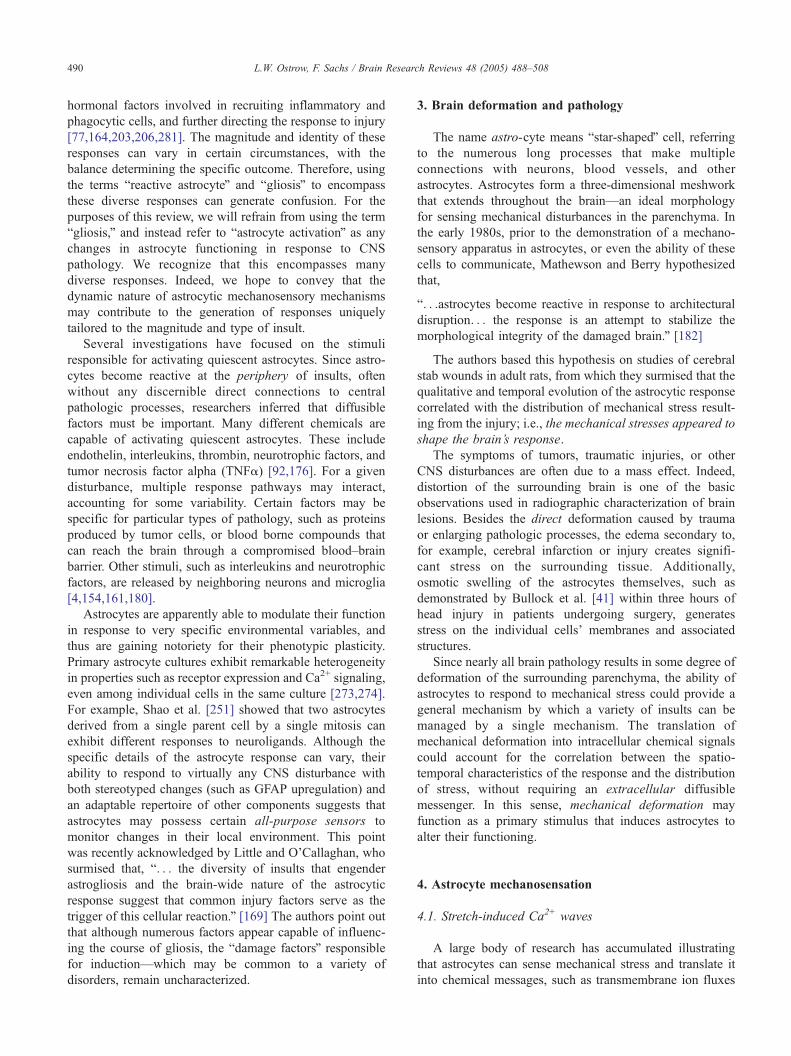
A large body of research has accumulated illustrating
that astrocytes can sense mechanical stress and translate it
into chemical messages, such as transmembrane ion fluxes

Fig. 1. Fluorescent imaging of mechanically induced calcium waves (from Ref. [214]). Note the shadow of the pipette tip in the top-left frame. Approximately
12 s have elapsed in these four frames. The illustration at the bottom shows deformation of a single cell in a confluent monolayer with the side of a pipette.
Approximate tip diameter=5 Am. Note the ~308 bend in the tip of the pipette, to prevent puncturing the cell.
L.W. Ostrow, F. Sachs / Brain Research Reviews 48 (2005) 488–508 491
and intercellular calcium (Ca2+) waves. As shown in Fig.
1, lightly pressing on a single adult rat astrocyte in a
monolayer using a micropipette tip, elicits a rapid increase
in intracellular Ca2+ in the stimulated cell (visualized by
loading the cells with a Ca2+-sensitive fluorescent dye).
Following a short delay at the cell borders, the Ca2+ wave
reappears in neighboring cells and continues to propagate
through the monolayer, often spreading throughout the
entire coverslip in confluent cultures [214]. Without
further stimulation, intracellular Ca2+ slowly decreases
over 5–10 min, after which the response is repeatable.
Similar propagated Ca2+ signals were first demonstrated in
neonatal astrocytes by Cornell-Bell et al. [63] in response
to glutamate. These waves have been widely studied,
contributing to the demise of the opinion that astrocytes
serve a merely passive structural role in the CNS. Several
researchers have shown that propagated Ca2+ signaling
can be elicited in fetal/neonatal astrocyte cultures by
many different stimuli, including direct mechanical stress
[33,52,272], endothelins [32,57,78,272], hypoxia/ischemia
[75,85], ATP [25,57], glutamate [25,52,57,62,63,74,89,
109,151,272], histamine [25,130], GABA [25], norepi-
nephrine [25,74], serotonin [25], angiotensin II [25,97],
bradykinin [25,129], substance P [25], carbachol [48,272],
brain-derived neurotrophic factor [58], gp120 HIV-1
envelope glycoprotein [59], phenylephrine [74], lysophos-
pholipids [94], and kainate [109].
In 1965, Bullock and Horridge presented a definition of a
nervous system as follows,
ban organized constellation of cells specialized for the
repeated conduction of an excited state from receptor sites
or other neurons to effectors or to other neurons.Q [40]
The most primitive nervous systems respond to stim-
ulation by propagating a diffuse signal, exemplified by the
bnerve netsQ of coelenterates. The astrocyte network is not

L.W. Ostrow, F. Sachs / Brain Research Reviews 48 (2005) 488–508492
unlike a primitive nervous system: it is composed of
interconnected cells that can respond to stimuli by
propagating a diffuse signal, in the form of an intercellular
Ca2+ wave that spreads from the point of stimulus to other
astrocytes and neurons. As suggested by M.J. Sanderson et
al., these Ca2+ signaling phenomena could serve to
organize cooperative cellular responses to local stimuli in
vivo [244].
The dependence and interdependence of a multitude of
diverse regulatory mechanisms on cell Ca2+ is difficult to
fathom. Cells seem to employ what Michael J. Berridge et
al., have referred to as a bcalcium toolkitQ in which enzymes,
ion channels, intracellular stores, inositol phosphates,
endogenous buffers, pumps, and exchangers combine to
produce an array of tightly controlled Ca2+ phenomena that
probably depend upon spatial localization of the compo-
nents [27,28]. In glial cells, intracellular Ca2+ affects cell
structure, gene expression, development, and proliferation
(for review, see Ref. [90]). Disturbances in glial Ca2+
accompany many neuropathological conditions, including
astrocytoma, trauma, stroke, and epilepsy [3,53,71,90,
105,121]. The astrocyte response to a specific disturbance
would be shaped by the unique milieu of local stimuli
(including mechanical stress), the distribution of their
sensors/receptors, the degree and mode of intercellular
communications, and the time course of the disturbances.
Specific downstream signaling events may require specific
Fig. 2. Acute puncture of a single cell in the absence of extracellular Ca2+. (A) Not
view (insert shows magnified and enhanced image of circled area). (B) The white
fluorescence, evident in panel A, is no longer present—probably due to leakag
surrounding the punctured astrocyte show an increase in Ca2+. (C) DIC image of
pipette tip has been pulled away, leaving a hole in the punctured cell (see magnifie
the video camera and used to help position the pipette tip.
dynamic changes in cytoplasmic Ca2+ levels. For example,
Berridge points out that stimulation of cell proliferation
appears to require sustained signaling. Since calcium stores
are finite, this suggests a necessity for Ca2+ influx from
extracellular sources [26,27]. If Ca2+ influx is enhanced,
then a new equilibrium between this influx, storage, and
efflux is established, resulting in a new basal intracellular
Ca2+ concentration.
When an individual astrocyte in a monolayer is deformed
by a micropipette, the initial increase in intracellular Ca2+ in
the directly stimulated cell involves both Ca2+ influx and
intracellular release [244,272]. There is conflicting evidence
concerning the dependence of intercellular wave initiation
on an influx of extracellular Ca2+. Charles et al. [52] found
that neonatal rat astrocyte cultures were capable of
propagating intercellular Ca2+ waves in the absence of
extracellular Ca2+. Only the response in the stimulated cell
was lost. However, in a similar culture system, Venance et
al. reported that initiation of the waves depended on an
influx of Ca2+ into the stimulated cell [272]. In our
experiments with adult rat astrocytes, when external Ca2+
was removed from the extracellular solution, the mechanical
stimulus did not elicit a Ca2+ wave [213]. If the pipette tip
punctured the cell, the cell became dark presumably by
leakage of dye. However, after a short delay, Ca2+ increased
in the cells surrounding the punctured astrocyte (see Fig. 2).
This propagated Ca2+ signal was limited to cells in the
e the shadow of the pipette tip overlying the cell in the middle of the field of
asterisk shows the location of the punctured cell. Note that the background
e of dye out of the punctured cell. Also note that only cells immediately
the field of view, showing the pipette tip in contact with the cell. (D) The
d insert). The black line beneath the cell in panels C and D is generated by

L.W. Ostrow, F. Sachs / Brain Research Reviews 48 (2005) 488–508 493
immediate vicinity of the damaged cell and spread in a
concentric fashion suggesting involvement of a diffusible
extracellular messenger (such as ATP) released from the
damaged cell.
The conflicting results reported by different labs regard-
ing the requirement for external Ca2+ raise the possibility
that Ca2+ waves are easily influenced by details of the
culture system, but they do occur in situ as shown in
hippocampal slice preparations, isolated intact retinas, and
acutely isolated cells [51,70,118,201]. Despite some con-
flicting data in the literature, there are agreed upon
characteristics of mechanosensitive Ca2+ signaling in
astrocytes:
(1) In response to mechanical stimulation, intracellular
Ca2+ increases.
(2) This calcium signal is propagated from cell-to-cell in a
bregenerativeQ fashion, forming an inter-cellular wave.
(3) The wave involves both an influx component as well
as release from intracellular stores.
4.2. Mechanosensitive ion channels (MSCs)
One way for cells to produce a Ca2+ influx in response to
mechanical stress is via specialized mechanosensitive ion
channels (MSCs). Currently MSCs are the only known
primary cellular mechanical transducers. Originally discov-
ered in the early 1980’s [108], MSCs are found ubiqui-
tously, from Escherichia coli to mammals [192,237–239].
Mechanical forces govern the gating of these channels, just
as voltage determines gating of voltage-gated channels. The
most common type of MSC is the stretch-activated channel
(SAC), although there are also examples of stretch-
inactivated channels (SICs) that are active at rest and
closed by tension [211,212]. SACs have been demonstrated
in neonatal astrocytes [37,134], glioma cells [36,187] and
adult astrocytes derived from striatal gelfoam implants
[261]. The astrocyte SACs can be activated by direct
pressure applied to membrane patches isolated in micro-
pipettes, and by osmotic swelling (which stresses the cell
membrane). Most SACs in animal cells, including those
found in astrocytes, are selective for cations including Na+,
K+ and Ca2+. Interestingly, one of the SACs characterized in
neonatal astrocytes is curvature sensitive [37]. This channel
is selectively opened only by an inward deformation of the
cell membrane—an ideal situation for responding to
mechanical stress imparted by the mass effects of nearby
pathology.
Since both acutely isolated astrocytes and slice prepara-
tions exhibit voltage- and ligand-gated Ca2+ channels
[73,74,152], multiple Ca2+ influx pathways probably also
exist in vivo. The non-mechanical Ca2+ influx pathways
may be indirectly activated by stretch. For example, voltage-
activated Ca2+ channels can open when stretch-channels
cause depolarization. Such a mechanism has been proposed
by O’Connor and Kimelberg to account for the regulated
volume decrease (RVD) observed in astrocytes following
osmotic swelling [209]: hypotonic media causes water
influx that swells the cells. This stretches the membrane
and opens SACs, which cause depolarization and lead to
Ca2+ influx via voltage gated channels. The increased
intracellular Ca2+ would then activate Ca2+-dependent K+
and Cl� efflux to reduce the swelling.
4.3. Cytoskeletal remodeling
Besides SACs, there may be other stress-activated
signaling pathways, since the structural elements of the
cytoskeleton are themselves mechanically sensitive poly-
mers. For example, altered GFAP expression constitutes one
of the earliest astrocyte responses to CNS disturbances:
! Dietrich et al. [72] found increased levels of GFAP
mRNA within 30 min of traumatic brain injury in rats.
! Amaducci et al. [5] similarly demonstrated morphology
changes and intense GFAP staining in the periphery of
parietal cryogenic lesions in rat brains 30 min after the
injury, spreading throughout the white matter by 48 h.
! Mucke et al. [194] demonstrated that focal mechanical
brain injury results in induction of an astrocyte-specific
GFAP-lacZ transgene within one hour.
! Cancilla et al. [45] found that GFAP mRNA was
increased following cerebral freeze-injury in mice by 6
h, and the pattern of enhanced expression in the two
weeks post-injury followed the distribution of edema.
! Liu et al. [171] similarly detected increased GFAP
mRNA within 6-h in the peri-infarcted area induced by
middle cerebral artery occlusion in rats.
Several studies have demonstrated that GFAP plays a
role in maintaining normal astrocyte cell shape and
cytoskeletal structure [102,234–236]. In a review of the
potential roles of GFAP, James Rutka et al. pointed out that
this astrocyte-specific intermediate filament has long been
considered to function as a mechanical integrator of
cellular space [236]. The authors also highlighted studies
suggesting that GFAP plays a pivotal role in forming an
interconnected dynamic cell-signaling apparatus, in con-
junction with other components of the cytoskeleton,
extracellular matrix, and associated kinases and second
messengers.
In addition to the dramatic increase in GFAP expression
that characterizes astrocyte activation, there is a transient
upregulation of vimentin (another intermediate filament),
increased expression of bactin-anchoring proteinsQ such as
vinculin, talin, and paxillin (which help attach the actin
cytoskeleton to focal adhesions on the cell membrane), and
a remodeling of the actin cytoskeleton [2,127,144,164,
225,266]. Rapid cytoskeletal remodeling in response to
mechanical stress has been demonstrated in other cell types,
suggesting that this is prototypical. Pender and McCulloch,
for example, showed that the actin stress fibers of gingival

L.W. Ostrow, F. Sachs / Brain Research Reviews 48 (2005) 488–508494
fibroblasts are disassembled within ten seconds of a stretch,
but recover completely within 1 min [221].
The actin cytoskeleton is also involved in astrocyte
signal transduction. Abd-el-Basset and Federoff demon-
strated that fetal astrocytes possess actin stress fibers
containing all of the elements traditionally considered to
make up a bcontractile unitQ—F-actin, myosin, tropomyo-
sin, and caldesmon, as well as the enzymes myosin light-
chain kinase (MLCK) and calmodulin [1]. Although
neonatal astrocytes exhibit extensive gap junctional cou-
pling shortly after subculture, Cotrina et al. [65] demon-
strated that they cannot propagate Ca2+ waves until this
actin cytoskeleton develops. The authors further demon-
strated that the radius of the propagated Ca2+ wave was
directly proportional to the fraction of cells with organized
stress fibers, and disruption of the actin cytoskeleton with
cytochalasin D or inhibition of myosin light chain kinase
attenuated the Ca2+ waves. Astrocyte stress-fibers in vitro
organize into parallel bundles that can span many cells
[1,65]. This arrangement allows the astrocyte network to
exert long range forces on the extracellular matrix and
produce coordinated changes in the structural scaffolding
of the surrounding brain.
4.4. Mechanosensation is dynamic
The stress-dependent reorganization of both intermediate
filaments (e.g. GFAP and vimentin) and the actin cytoske-
leton enable astrocytes to regulate the transduction of
mechanical forces in the brain, providing a time-dependence
to stress-induced stimulation. For example, a progressively
Fig. 3. Effects of Cyclic Stretch (1 Hz, 0–20%) on Inositol Triphosphate Productio
and exposed to cyclic deformation regimens using a Flexcell FX-3000 Strain Unit
bloadedQ into the membrane store of inositol (PIP2), and then is transferred to c
measurement of radioactivity in the cytoplasmic fraction reflects cumulative IP3 p
cytoplasmic IP3 concentration. Ca2+-free=1 h incubation with EGTA prior to stretc
bars=FSE.
increasing mechanical stress, such as a rapidly expanding
tumor mass, might continually stimulate a mechanosensory
element, whereas the response to an acute event, such as a
traumatic injury, may be attenuated as the mechanosensory
apparatus is remodeled.
The dynamic response of mechanical signaling systems
is exemplified by the intrinsic adaptation and inactivation of
the stretch-activated ion channels in different tissues
[114,205,261]. In what is presumably a more downstream
view of transduction, the mechanically induced turnover of
inositol phosphates in adult astrocyte cultures is also
dynamic [215]. Fig. 3 shows that a 1-Hz stretching regimen
applied to astrocytes increased the turnover of inositol
phosphates within 15 s, reaching a plateau within 2 min. In
other words, 2-min of periodic stretching was sufficient to
activate and then deactivate the mechanical signaling
machinery of the astrocytes. Remarkably, cessation of the
stretching was seen as a new stimulus, i.e. a change in the
mechanical environment, resulting in further IP3 production.
The correlation of this phenomenon to in situ stimuli is
unknown, but the normal mechanical environment for
astrocytes is pulsatile. Indeed, if a portion of the skull is
resected, the bbeatingQ of the brain is easily appreciated. As
with all sensory processes, there is a tonic and a phasic
response. Since the primary mechanotransducers are modu-
lated by the mechanics of their surroundings, any change in
the mechanical environment can serve as a stimulus. The
combination of cytoskeletal remodeling, dynamic second
messenger signaling, and the adaptation and inactivation of
the SACs themselves provide a complex multi-tiered time-
dependence to stress-activated signaling.
n (from Ref. [214].) Adult astrocyte cultures were grown on flexible plates
(Flexcell International, USA). Prior to stretching, tritiated myoinositol was
ytoplasmic IP3 when PLC-mediated turnover is stimulated. A subsequent
roduction. DPM=disintegrations per minute, which is proportional to mean
hing. N=3 wells for each data point (~9.6 cm2 growth area per well). Error

L.W. Ostrow, F. Sachs / Brain Research Reviews 48 (2005) 488–508 495
5. Endothelins
Endothelin (ET) is a 21-amino-acid bicyclic peptide (see
Fig. 4). It was originally identified by Yanagisawa et al.
[286] in the conditioned medium of cultured endothelial
cells, by virtue of its actions as a potent and long-lasting
vasoconstrictor. Researchers have determined that there are
at least three peptides in the endothelin family—ET-1, ET-2,
and ET-3, and at least two main types of receptors—ETAand ETB [104,188]. The ET peptides and receptors are
expressed in a tissue-specific manner. Their properties have
been characterized in numerous reviews [104,106,126,
148,210,278]. ET-1 appears to be the predominant form in
humans, and for the remainder of this review, bET-1Q andbendothelinQ are used interchangeably. However, it is
important to note that researchers have demonstrated the
existence of both ET-1 and ET-3 in the human brain
[79,80,174].
5.1. Endothelin in astrocytes
Although not found in non-proliferating quiescent
astrocytes, immunohistochemical staining has demonstrated
that reactive astrocytes contain endothelin [142,173,174,
202,292–295]. ET is found in reactive astrocytes in the
periphery of a remarkably diverse collection of CNS
pathologies, including traumatic injuries [112,227], hema-
togenous metastases [292,293], plaques of Alzheimer’s
Disease [202,294,295], experimentally induced edema
[112,113], infarcts and lacunae [202,294], hereditary
multi-infarct disease [294], lesions of viral infections
[173,202] , and Binswanger’s encephalopathy [294]. Siren
et al. [255] reported a direct correlation between astrocyte
reactivity (quantified by GFAP levels and morphology
changes) and ET-1 expression in rat brains following
cryogenic neurotrauma. Koyama et al. [158] found that a
continuous infusion of a selective ETB receptor antagonists
into the cerebral ventricles of rats attenuated the increase in
GFAP- and vimentin-positive cells after a cerebral stab
wound. Tsang et al. [269] have recently shown that the
expression of ET-1 mRNA is increased following cerebral
hypoxia/ischemia.
When ET was first discovered in reactive astrocytes,
some researchers hypothesized that it had diffused to the
astrocytes from damaged blood vessels. Indeed, in rats
subjected to acute brain trauma by cold injury, Yamada et al.
[284] demonstrated that radiolabeled ET-1 injected into the
Fig. 4. Endothelin-1 molecule. Endothelin-1 is a 21 am
left cardiac ventricle was later detected in the brain at the
injury site. However, numerous studies have demonstrated
that activated astrocytes also produce, store, and secrete
endothelins themselves [79,80,174,202,269]. Astrocytes
(and glioma cells) possess endothelin receptors, and
endothelin binding can potentiate the production and
secretion of more endothelin in a positive feedback loop
[79,80,177,178].
ET receptors appear to be broadly distributed in
astrocytes. For example, Venance et al. [273] found that
all striatal astrocytes in rat brain express binding sites for
ET-1, whereas a1-adrenergic and muscarinic binding sites
are more heterogeneously distributed. Although astrocyte
ET-receptors are distributed throughout the brain, the
relative density of ETA and ETB receptors varies in
different regions, depends on the state of differentiation,
and is altered in response to certain diseases and trauma
[98,104,111,112,196,218,227]. ETA and ETB receptors are
distinguished by their affinities for the ET-isopeptides
[210]. ETA receptors have a higher affinity for ET-1 and
ET-2 than ET-3, with Kd’s of ~20–60 pM vs. ~6.5 nM,
respectively. ETB receptors bind all three isopeptides with
equivalent affinities (Kd ~15 pM). Several researchers have
characterized ET binding as birreversibleQ—owing to their
inability to wash off bound radiolabeled ET from its
receptors [56,181,282,283]. This high affinity binding is
further demonstrated by comparing the short half-life of
ET-1 injected into the blood stream (a few minutes) with
the resultant long-lasting vasoconstriction (hours)
[253,286]. Evidence exists for additional atypical ET-
receptors, including an astrocyte-specific receptor that
may have multiple binding sites [141]. There is also ET-
binding in a human hybrid neuroblastoma/glioma cell line
that has a higher affinity than ETA or ETB receptors [7].
In addition to acting as a vasoconstrictor, numerous
studies have revealed that ET stimulates the proliferation
of particular cell types, including astrocytes. ET-1
immediately transforms differentiated, non-proliferating,
astrocyte cultures into proliferating cells, with a saturating
effect at nanomolar concentrations [49,111–113,147,165,
174,258,263]. Hama et al. [112] demonstrated that
astrocytes change from a quiescent to an immature reactive
state immediately after experimentally induced acute brain
injury in vivo, and the level of endothelin is significantly
increased at the site of injury after only 1 day. ET is also
mitogenic for numerous other cell types, including glioma
cells [13,68,249,250], brain capillary endothelial cells
ino acid, bicyclic peptide, weighing 2491.9 kD.

L.W. Ostrow, F. Sachs / Brain Research Reviews 48 (2005) 488–508496
[275], glomerular mesangial cells [155,247,254,290],
smooth muscle cells [14,175,252] (especially vascular
smooth muscle [14,132,138,156]), normal prostate epithe-
lium [133], prostate cancer cells [200], and ovarian
carcinoma cells [16].
Another component of the brain’s reaction to pathology
is infiltration of the site of injury by leukocytes [19], and
implantation of activated leukocytes caused behavioral
improvement in a rat model of Parkinson’s Disease [82].
Although not well studied in the brain, endothelin has pro-
inflammatory actions in the lungs, intestinal mucosa, blood
vessels, and cutaneous sites, which include stimulating
leukocyte recruitment [241–243], enhancement of leukocyte
adhesion and infiltration [35,107], macrophage activation
[186,189], and enhanced edema, microvascular leakage, and
nociception [101,107,222]. ET also stimulates the produc-
tion of other inflammatory cytokines including TNFa,
interleukins, and interferon gamma [91,230,257]. These
inflammatory actions are best characterized in the lungs,
where ET appears to play a major role in the pathogenesis of
asthma and immune-complex-mediated acute lung injury
[22,50,88,91,101,257]. Therefore, it is possible that ET
contributes to the pathophysiology of leukocyte recruitment
observed in the brain as well.
5.2. Endothelin regulation
It is generally recognized that there is an intimate
relationship between mechanical forces and endothelin.
Indeed, ET was initially discovered because of its most
dfamousT role as a potent and long-lasting vasoconstrictor.
Conversely, mechanical stretch is a major stimulus for ET-1
production and release. Alterations in hemodynamic shear
stress, such as might result from increased blood pressure or
heart rate, can modify ET-1 production and secretion by the
endothelium [190,191,277,291]. Endothelin then acts on
the vascular smooth muscle to regulate vessel tone.
Mechanical stress has been directly implicated in increasing
endothelin levels in several pathological conditions, includ-
ing pulmonary hypertension [270], cardiac hypertrophy
[135,136,285], atherosclerosis [120], neointimal hyperpla-
sia following coronary angioplasty [167,184], and excess
flow through the adrenal gland [44]. Additionally, gross
alterations of the mechanical environment characterize
numerous other pathologies associated with elevated
endothelin, where the role of direct cell deformation has
yet to be investigated. Some examples include persistent
pulmonary hypertension of the neonate [146,229], primary
glaucoma (i.e. raised intraocular pressure) [110,143,
208,223], mesangial proliferative nephritis [290], solid
tumor growth (i.e. mass effect) in gliomas, ovarian
carcinomas, and advanced prostate cancer [13,16,17,116,
200,260], acute and chronic renal failure [148,228],
numerous obstructive, hypertensive, and inflammatory
pulmonary diseases [101,122,226], neurotrauma [21,
246,255], bladder outlet obstruction [149,150], and pre-
eclampsia [42,84,276]. The pathogenesis of many of these
disease processes involves both the vasoconstricting and the
mitogenic actions of endothelin.
Although the expression of the ET isopeptides, convert-
ing enzymes, receptors, and other components of the
astrocytic endothelin system have been identified, consid-
erably less is understood about the regulation of this system.
As characterized in an editorial by Hannelore Ehrenreich,
bthe players within the astrocytic endothelin (ET) system are
known, but the rules of the game have been obscure. . .Q[78]. Compared to the work on astrocytes, regulation of the
ET system in other cells has been examined more
extensively. Besides stretch, several non-mechanical stimuli
can enhance ET expression, including thrombin, TGF-h,angiotensin II, arginine vasopressin, calcium ionophores,
and a host of other growth factors and cytokines (for
reviews, see Refs. [104,181,268]). Regardless of the
stimulus, alterations in cell Ca2+ play a central role in
regulating the endothelin system in all cell types
[11,38,39,61,181,191,268,287]. Abnormal levels of ET are
usually caused by increased transcription and/or secretion
(frequently upregulated in parallel), which are controlled by
intracellular Ca2+ [38,39,268]. This regulation has histor-
ically been considered to rely on a Ca2+-dependent
induction of mRNA transcription, involving the proto-
oncogene products c-fos, c-myc, and c-jun, followed by
secretion via a bconstitutiveQ pathway without appreciable
intracellular storage [104,181,286,287]. The unique gene
structure of ET-1, which contains several bacute phase
reactionQ motifs and nuclear binding factors, allows for the
rapid induction of gene transcription [131]. Intracellular
Ca2+ homeostasis has also been implicated in regulating the
unstimulated basal secretion of ET-1 by endothelial cells
[38,39], and in stimulating secretion by activating myosin
light chain kinase (MLCK), which facilitates the transfer of
ET-1 to the cell surface [153].
Even in early experiments in the vasculature, it was
apparent that certain stimuli elicited increased plasma ET-1
concentrations much faster than would be expected from de
novo synthesis. For example, Fyhrquist et al. [96] showed
that rapidly submerging a subject’s forearm into ice cold
water induced an approximately seven-fold increase in
plasma ET-1 in the arm within 2 min. Researchers have
demonstrated a second bregulatedQ secretion pathway in
endothelial cells, involving storage of mature ET-1 in
secretory vesicles and endothelial-cell-specific storage
granules called Wiebel-Palade bodies, and subsequent rapid
release induced by chemical or mechanical stimuli that
trigger a Ca2+ influx [119,231–233]. Similarly, Nakamura et
al. [197] found ET-1 co-localized with vasopressin and
oxytocin in neurosecretory granules of the rat posterior
pituitary. Thus, Ca2+ signaling controls both the rapid and
transient secretion of preformed stores of ET via the
bregulatedQ pathway, and the somewhat slower regulation
of ET-mRNA transcription, followed by secretion via the
bconstitutiveQ pathway.

L.W. Ostrow, F. Sachs / Brain Research Reviews 48 (2005) 488–508 497
In experiments with endothelial cells, Chen et al. [55]
used a bmagnetic twisting stimulatorQ to directly apply
mechanical stress to integrin receptors on endothelial cells
via ligand-coated ferromagnetic beads bound to the cell
surface. This resulted in dramatically enhanced expression
levels of ET-1 mRNA. The authors further showed that this
ET-1 induction could be modulated by cytoskeletal dis-
ruption, and was abolished by blocking stretch-activated ion
channels with Gd3+, or by removing extracellular Ca2+. This
work demonstrates:
(1) The importance of cytoskeletal components in modu-
lating cellular mechanosensitivity.
(2) The critical role of SACs as proximal mechanotrans-
ducers.
(3) The dependence of this mechanical signaling mecha-
nism on an influx of Ca2+, presumably via SACs.
ET-1 exerts its mitogenic effects in most cell types,
including astrocytes and glioma cells, by regulating intra-
cellular Ca2+ homeostasis [128,137,177,178,263,264],
potentially explaining how endothelin potentiates its own
production and secretion. Although the specifics of this
signaling cascade may vary among cell types, the mitogenic
actions of ET appear to converge on activation of the
mitogen-activated protein kinase (MAPK) pathway through
Ca2+, the induction of phospholipase C (PLC), and protein
kinase C (PKC) [104]. PLC mediates the turnover and
intracellular release of inositol triphosphate (IP3), which
diffuses through gap junctions and causes release of
intracellular Ca2+ stores in neighboring cells. This produces
a propagated intercellular Ca2+ wave. ET-1 is an extremely
potent stimulus of astrocytic IP3 production. In neonatal
astrocyte cultures, ET-1 administration caused an upregula-
tion of PLC (and subsequent increase in cytoplasmic IP3)
greater than that induced by glutamate, carbachol, or
methoxamine [272]. Additionally, Laurent Venance, Chris-
tian Giaume et al. [272,273] found that Ca2+ waves elicited
by either local application of ET-1 to a single cell or
mechanical stimulation elicited robust Ca2+ waves that
propagated throughout the astrocyte network whereas other
stimuli produced considerably smaller responses.
The capacity for ET to enhance its own expression
necessitates feedback inhibition. The existence of such
regulatory mechanisms is demonstrated by experiments
showing that under certain conditions, increased concen-
trations of ET-1 or ET-3 can inhibit gap junctional
permeability and the propagation of intercellular Ca2+
waves in neonatal astrocytes [32,265]. Additionally, Corder
et al. found that prolonged exposure to calcium ionophores
actually inhibited ET secretion by bovine aortic endothelial
cells [61].
The dramatic cytoskeletal remodeling that characterizes
the astrocyte response to pathology may favor specific
regulatory pathways by altering connections between
cellular components, or varying the relative densities of
individual cytoskeletal elements. For example, Ozawa et al.
[218] found that disruption of microtubules attenuated both
the expression of ETB receptor mRNA and the number of
ET binding sites on neonatal rat astrocytes, while micro-
filament disrupting agents had no effect. The potent self-
stimulating capacity of the ET system, combined with its
apparently multifactorial feedback regulation, suggests that
prolonged alterations or disruptions at any of several points
along these signaling cascades could result in dramatically
abnormal expression levels.
The relationship between mechanical stretch and ET
production in other cell types, combined with the observa-
tion that ET-1-positive reactive astrocytes appear in the
mechanically deformed periphery of CNS pathology, led us
to hypothesize that mechanical stress may regulate ET in
astrocytes. In our experiments with adult astrocytes grown
on flexible culture dishes, a cyclic stretching regimen
caused a dramatic increase in ET-1 production and secretion
[213,214]. Since virtually all brain pathology results in
mechanical deformation of the surrounding parenchyma,
and ET induces the proliferation of astrocytes (and glioma
cells), the possibility arises that mechanical induction of the
ET system represents a general pathway for activating or
augmenting astroglial proliferation.
Alterations in the astrocyte production of ET may affect
other cells as well. Although astrocytes are considerably
more sensitive than neurons to the mitogenic actions of ET-
1, the sensitivity of both cell types exceeds that of most
peripheral tissues [262]. Additionally, several studies have
implicated ET-1 in aspects of central autonomic control (for
review, see Kuwaki et al. [162]), and the cerebral
vasculature is particularly sensitive to the vasoconstricting
actions of ET-1 [12,95,240,267]. ET concentrations as low
as 1–100 pM affect the proliferation of astrocytes, glioma
cells and neurons, induce the contraction of cerebral and
basilar arteries, alter extracellular glutamate and dopamine
levels, and prevent cytochalasin B-induced astrocyte mor-
phology changes (see Table 1).
6. Potential therapeutic targets
6.1. ET-receptor antagonists as treatments for brain injury
Currently, experimental treatments for diseases related to
the overproduction of ET primarily focus on blocking the
effects of the peptide using receptor antagonists. Another
developing class of drugs blocks ET-converting enzymes
(ECEs), preventing the cleavage of a precursor form into
mature endothelin [140]. Endothelin-receptor antagonists
inhibit the endothelin-induced proliferation of glioma cells
in vitro [249,250]. They also reduce the degree of edema,
infarct volume, and neurological deficits in rats following an
ischemic event [20,21,183,220]. Similarly, they attenuate
neurological deficits and edema following traumatic closed-
head injury [21], control cerebral vasospasm [64], and

Table 1
ET-1 Effects in the brain at concentrationsV100 pM
Cell/Tissue Type ET-1 Conc. Effect Reference
Rat astrocytes in organotypic
cerebellar slices
EC50=8F5 pM Induced c-fos expression (correlates with mitogenicity). Sullivan and Morton,
1996 [262]
Rat neonatal cerebellar
astrocytes in mixed
astro/neuron cultures
(neuron enriched)
10–100 pM Threshold concentration for eliciting increases in intracellular
calcium. Maximal response elicited at ~10 nM ET-1.
Morton and Davenport,
1992 [193]
Primary rat astrocytes EC50=50 pM Inhibited cytochalasin B-induced stellation. Koyama and Baba,
1994 [157]
100 pM Stimulated mitogen-activated protein kinase (MAPK) activity. Kasuya et al., 1994 [147]
100 pM–100 nM Dose-dependent inhibition of glutamate uptake. Leonova et al., 2001
[166]
EC50=100 pM Stimulated proliferation (i.e. increased DNA synthesis
measured by [3H]thymidine incorporation).
MacCumber et al., 1990
[174]
100 pM Stimulated proliferation (i.e. increased DNA synthesis
measured by [3H]thymidine incorporation).
Stanimirovic et al., 1995
[258]
Rat neurons (granule cells) in
organotypic cerebellar slices
10–1000 pM
(EC50=57F9 pM)
Induced c-fos expression (correlates with mitogenicity). Sullivan and Morton,
1996 [262]
Human cerebral cortex Kd=25F6 pM [125I]ET-1 binding studies in membrane preps. Fernandezdurango et al.,
1994 [86]
Ventral striatum of
anaesthetized rats (in vivo)
10 pmol injection 8 AM increase in extracellular dopamine concentration
within 5 min of injection.
Webber et al., 1998
[279]
C6 glioma cells EC50=50 pM Stimulated proliferation (i.e. increased DNA synthesis
measured by [3H] thymidine incorporation).
MacCumber et al.,
1990 [174]
Canine basilar arteries
(in vivo)
10 pmol intracisternal
injection
Decrease in artery diameter (measured by angiography);
remained statistically significant 3 days after injection.
Asano et al., 1993 [12]
Canine cerebral arteries
(endothelium denuded)
10 pM–100 nM Dose-dependent stimulation of contraction. Contractile effects
between 10 and 300 pM were significantly greater than for
mesenteric arteries.
Tanoi et al., 1991 [267]
Porcine cerebral arteries
(ring segments)
100–300 pM Threshold concentration for ET-1 induced contractions. Fukuda et al., 1991 [95]
Examples of mitogenic and vasoconstricting actions induced by administration of endothelin.
L.W. Ostrow, F. Sachs / Brain Research Reviews 48 (2005) 488–508498
reduce both the size of lesions resulting from cold-injury
[103] and the degree of acute neuronal damage following
mild cortical trauma in rats [246]. These drugs are also
being investigated as possible treatments for a variety of
other cerebrovascular and neoplastic pathologies [46,116,
117].
Although ET-antagonists have shown promise in treating
certain conditions, they do not address the underlying
pathophysiology. Since ET plays numerous roles in normal
cell physiology throughout the body, blocking the receptors
can lead to multiple side effects. The high sensitivity of the
cerebral vasculature to ET [240], coupled with the delicate
regulation of blood flow in the brain and the role of ET in
maintaining basal vascular tone [125,126], suggests that the
potent vasoactive properties of ET-related peptides may
limit their usefulness in treating CNS pathology. By
characterizing the upstream cell-signaling pathways
involved in regulating ET production, treatments may be
designed to target the causes of ET overproduction, rather
than using receptor antagonists to block the effects.
6.2. Inhibitors of stretch-activated ion channels (SACs)
Since mechanical stress is a well characterized stimulus
for the ET system throughout the body (and this appears to
involve a stretch-induced Ca2+ influx), and pathological
processes cause mechanical perturbations, SACs represent a
logical target for inhibiting this system. The bgold-standardQtechnique for implicating an individual type of ion channel
or class of channels in a particular process is to determine
whether a specific inhibitor alters the results. Although
patch-clamping techniques have enabled the identification
and kinetic analysis of SACs in numerous cell types, the
lack of specific pharmacological agents has hindered
elucidation of their physiological roles. The lanthanide
gadolinium (Gd3+) is an effective blocker for many SACs,
and therefore is often used to implicate these channels in
cellular phenomena [115,288]. For example, Gd3+ inhibits
the stretch-induced Ca2+ influx and proliferation of fetal rat
lung cells, while Ca2+ channel blockers such as verapamil,
nifedipine, and Ni2+ have no effect [170]. However Gd3+
also blocks voltage dependent Ca2+-channels [31,163], and
is not a suitable reagent for experiments in most culture
media, as it precipitates in the presence of proteins,
phosphates, or bicarbonate [34,43,238]. Certain amino-
glycoside antibiotics (e.g. streptomycin) are also capable
of blocking SACs as well as other cation channels. These
positively charged molecules appear to function as blockers
by bpluggingQ the negatively charged pore of the channel
[115,159].

L.W. Ostrow, F. Sachs / Brain Research Reviews 48 (2005) 488–508 499
Vaz et al. [271] showed that several of these somewhat
non-specific SAC inhibitors, including gadolinium, amilor-
ide, and gentamicin, inhibit edema in vivo following head
trauma. The effects were statistically significant, even when
the SAC blockers were administered 15–30 min after the
trauma. Although it is not possible to distinguish effects on
individual cell types in their model, the authors hypothe-
sized that the reduced edema was due to inhibition of SACs
activated by endothelial cell swelling. Recently, this same
group has demonstrated that both gadolinium and amiloride
prevent alterations in brain mitochondrial function (strongly
affected by cell Ca2+) in their trauma model [245]. These
results are, at best, suggestive and need to be repeated under
more controlled conditions, since gadolinium was adminis-
tered i.v. and would be bound by proteins and small anions
[34,43,238].
In the mid-1990s, venom from the Grammastola
spatulata spider was shown to be capable of blocking
SACs in several systems, without affecting Ca2+ or other
voltage dependent channels [55,198,204]. More recently, we
have isolated and characterized a peptide from this venom
as the first specific blocker of stretch-activated cation
currents [261]. This 34-amino-acid peptide, named GsMtx-
4, belongs to the inhibitor cystine knot (ICK) family—in
which an embedded ring is formed by two disulfide bonds
and their connecting backbone, and penetrated by a third
disulfide bond (see Fig. 5). The disulfide bonds stabilize the
native conformation, and because of the stability of the
structure, these peptides are appealing candidates for drug
design [69,224].
In adult rat astrocytes, GsMtx-4 completely blocks the
stretch-activated channels, while exhibiting no effect on
voltage-sensitive currents [261]. GsMtx-4 also inhibits
swelling-activated currents in the adult astrocytes [261].
Preliminary data additionally shows that the stretch-induced
secretion of ET-1 is inhibited by GsMtx-4 [215]. GsMtx-4
Fig. 5. Ribbon Structure of GsMtx-4, illustrating binhibitor cystine knotQ(ICK) motif. A ring is formed by two disulfide bonds and their connecting
backbone (blue), and penetrated by a third disulfide bond (yellow). The
inset is an illustration of the bcystine knotQ structure from Craik et al. [69].
did not alter the basal ET-1 secretion in the absence of
stretch, illustrating the specific nature of its actions.
Streptomycin, which inhibits stretch-activated currents in
several systems, also attenuated this response, as did
reducing extracellular Ca2+. These data suggest that
mechanically induced ET-1 production by astrocytes is
dependent on an influx of Ca2+ [213]—similar to the
situation in endothelial cells [54].
The cDNA sequence encoding the GsMtx-4 peptide has
been identified, and both a synthetic and a recombinantly
cloned form have been successfully created [216]. Addi-
tionally, the solution structure has been determined using
two-dimensional NMR spectroscopy [217]. The synthetic
form is now available from The Peptide Institute, (Osaka,
Japan). Mutagenesis studies on the recombinant peptide to
determine which amino acids are critical for binding and
activity are underway. It is our hope that these studies may
result in an entirely new class of drugs for the treatment of
CNS pathologies.
7. Mechanical deformation and endothelin in
astrocyte-derived tumors
Gliomas of astrocytic origin are the most common type
of human brain tumors and are almost invariably fatal. For
the most malignant variety, glioblastoma multiforme, there
has been no significant improvement in life-expectancy
during the past 15–20 years. Li et al. [168] suggest that the
relative frequency of these tumors reflects the sensitivity of
glial cells to proliferative stimuli. As the authors point out,
the vast number of proliferatively quiescent astrocytes in the
CNS, combined with their ability to re-enter the cell cycle
upon stimulation necessitates tight control over cell pro-
liferation. This apparently fails in neoplasia. Glioma cell
proliferation is particularly sensitive to changes in Ca2+
[23], and it has been suggested that this is due to
overexpression of a Ca2+-dependent isoform of PKC
[18,29]. PKC activity directly correlates with the prolifer-
ation rates of astrocytes and gliomas [66,67,289]. Addition-
ally, both human and rat glioma cell lines have high levels
of PKC compared to non-malignant astrocytes [67].
The mass effect of enlarging gliomas is so characteristic
of their behavior that distortion of the surrounding
parenchyma provides the basis for their radiographic
detection, and is used to gauge malignant behavior. Like
non-malignant astrocytes, glioma cells possess SACs and
generate intracellular Ca2+ signals in response to mechanical
stimulation [37,53,93,187]. Several studies have demon-
strated a correlation between increasing astrocyte malig-
nancy and decreasing GFAP expression (for review, see
Ref. [236]). Since GFAP has long been considered to play a
role in stabilizing the cytoskeleton and astrocyte cell shape,
it appears that there may be a correlation between the loss of
normal structural integrity (a situation that would alter
mechanosensation), and the development of neoplasia.

L.W. Ostrow, F. Sachs / Brain Research Reviews 48 (2005) 488–508500
Additionally, if astrocytoma cell lines are transfected with
GFAP, there is an inverse relationship between the resulting
mRNA expression and cell proliferation in vitro [234].
Like non-malignant reactive astrocytes, glioma cells
produce and secrete endothelin which is mitogenic for
glioma cells in vitro [13,68,116,249,250] and in vivo
[116,259,260]. Endothelin antagonists can inhibit glioma
cell proliferation in vitro [249,250]. Stiles et al. [260]
demonstrated a strong correlation between ET-immunor-
eactivity in surgical tissue sections from human gliomas and
tumor malignancy. Further supporting the role of ET in
neoplasia, Stiles et al. showed that if a human glioma line
that is a low endothelin producer is transfected with a gene
to increase ET production, the malignancy of the tumors
resulting from xenografts into the brains of immunodeficient
rats is increased. Studies have also demonstrated significant
differences in the degree of ET-receptor expression, receptor
classes, capacities for ET clearance, and sensitivities to ET-
1-induced Ca2+/IP3 signals and mitogenesis, between
glioma strains of different malignancies and normal
astrocytes [7,8,174,282,283].
The expression of ET-1 in human astrocytomas correlates
with the vascularity of the tumors [260]. Because ET is a
potent mitogen for both capillary endothelium and vascular
smooth muscle, it has been implicated in angiogenesis
[99,260,294,296], the process whereby tumors develop their
own vasculature. Angiogenesis is necessary in order for a
solid tumor to grow larger than a few millimeters. In
glioblastoma multiforme, the most malignant variety of
astrocyte-derived gliomas, the vascular proliferation is so
striking that it is used as a diagnostic criterion to distinguish
the tumors from their less malignant counterparts [185].
Glioblastomas are essentially inoperable and invariably
fatal, primarily because of their diffusely infiltrative nature.
However, partial resection, termed debulking, often pro-
longs survival [3]. The most obvious benefit of this
procedure is a reduction of the tumor cell load; however
debulking would also lessen the pressure tending to deform
the surrounding brain.
The less malignant low-grade astrocytomas grow much
more slowly and are relatively avascular, until transforming
into more malignant tumors. A hypothetical explanation for
the behavior of these tumors might be that during their
initial slow growth, the tissue is being gradually deformed.
At some point, a threshold of mechanical deformation is
reached, sharply increasing endothelin production. The
endothelin then stimulates angiogenesis, and the resulting
vascular proliferation enables the more rapid, malignant
growth of the tumor. This would produce greater mechanical
strain, perpetuating the cycle.
Fig. 6. Regulation of the Endothelin System in Astrocytes. See text for
references and discussion.
8. Conclusions
Hypotheses that historically relegated astrocytes to a
structural and supportive role were inspired by the reticular
meshwork formed by these stellate cells. Rather than
precluding an active role in cell-signaling, this arrangement
places the astrocytes in an ideal situation to monitor,
measure, and integrate the effects of mechanical stress.
Astrocytes possess complex mechanosensory machinery,
which includes
(1) dynamically adapting cytoskeletal components that
regulate force transduction,
(2) stretch-activated ion channels, which directly sense the
mechanical stress and translate it into ion fluxes,
(3) mechanically induced second-messenger signaling
phenomena, such as intercellular Ca2+ and IP3 waves,
and
(4) the stretch-induced production and secretion of endo-
thelin, a potent astrocyte mitogen (with additional
affects on neurons, brain capillary endothelium, and
glioma cells).
Fig. 6 summarizes the pathways involved in regulating
endothelin in astrocytes. Mechanically induced Ca2+ and IP3waves are well-established astrocyte phenomena. Both ET-1
and increased intracellular Ca2+ enhance astrocyte prolifer-
ation [49,90,112,147,165,174,258,263]. Additionally, we
have shown that mechanical deformation directly stimulates
the production and secretion of ET-1 by adult astrocytes
[214]. Since virtually all brain pathology deforms the
surrounding parenchyma, astrocyte mechanosensation can
facilitate the observed stereotypical reactive response to a
diversity of insults. The existence of mechanosensory
phenomena does not preclude contributions of chemical
and electrical signaling cascades in astrocyte function.
Additionally, it is likely that the other cells of the brain

L.W. Ostrow, F. Sachs / Brain Research Reviews 48 (2005) 488–508 501
possess stress-signaling mechanisms themselves. However,
the unique morphology and dynamic stress-sensing and
modulating abilities of astrocytes make them ideal for
monitoring and responding to mechanical disturbances.
CNS pathology necessarily causes mechanical stress on
the surrounding tissue. Astrocytes possess mechanisms to
sense this stress, translate it into chemical signals, and
regulate its transduction in an adaptable time-dependent
manner. The upregulation of the astrocyte ET system
observed in a wide range of pathologies, coupled with its
potent effects on these cells, suggests a prominent role in
this response. Therefore, we believe that mechanical
induction of the astrocytic endothelin system represents a
largely unexplored, and potentially significant target for
therapeutic intervention.
Acknowledgments
We would like to thank JST and NIH for support.
References
[1] E.M. Abd-el-Basset, S. Fedoroff, Contractile units in stress fibers of
fetal human astroglia in tissue culture, J. Chem. Neuroanat. 7 (1994)
113–122.
[2] E.M. Abd-el-Basset, S. Fedoroff, Upregulation of F-actin and alpha-
actinin in reactive astrocytes, J. Neurosci. Res. 49 (1997) 608–616.
[3] R.D. Adams, M. Victor, A.H. Ropper, Intracranial neoplasms and
paraneoplastic disorders, Principles of Neurology, McGraw-Hill
Companies, New York, 1997, pp. 642–694.
[4] H. Aldskogius, E.N. Kozlova, Central neuron-glial and glial-glial
interactions following axon injury, Prog. Neurobiol. 55 (1998) 1–26.
[5] L. Amaducci, K.I. Forno, L.F. Eng, Glial fibrillary acidic protein in
cryogenic lesions of the rat brain, Neurosci. Lett. 21 (1981) 27–32.
[6] B.J. Anderson, X. Li, A.A. Alcantara, K.R. Isaacs, J.E. Black, W.T.
Greenough, Glial hypertrophy is associated with synaptogenesis
following motor-skill learning, but not with angiogenesis following
exercise, Glia 11 (1994) 73–80.
[7] K. Angelova, A. Ergul, P. Narayan, D. Puett, Endothelin binding to
NG108-15 cells: evidence for conventional ETA and ETB receptor
subtypes and super-high affinity binding components, Cell. Mol.
Biol. 42 (1996) 1243–1257.
[8] K. Angelova, A. Ergul, K.C. Peng, D. Puett, Metabolism of
endothelin-1 by neuroblastoma x glioma hybrid (NG108-15) cells,
Neurosci. Lett. 225 (1997) 1–4.
[9] A. Araque, R.P. Sanzgiri, V. Parpura, P.G. Haydon, Astrocyte-
induced modulation of synaptic transmission, Can. J. Physiol.
Pharm. 77 (1999) 699–706.
[10] A. Araque, G. Carmignoto, P.G. Haydon, Dynamic signaling
between astrocytes and neurons, Annu. Rev. Physiol. 63 (2001)
795–813.
[11] S. Asada, Y. Kasuya, T. Sakurai, T. Masaki, K. Goto, Endothelin-1-
induced down-regulation of Et(B) receptor messenger-Rna—partic-
ipation of camp, J. Cardiovasc. Pharmacol. 26 (1995) S272–S275.
[12] T. Asano, I. Ikegaki, Y. Suzuki, S. Satoh, M. Shibuya, Endothelin
and the production of cerebral vasospasm in dogs, Biochem.
Biophys. Res. Commun. 159 (1989) 1345–1351.
[13] T. Asano, M. Aoyagi, K. Hirakawa, Y. Ikawa, Effect of endothelin-1
as growth factor on a human glioma cell line; its characteristic
promotion of DNA synthesis, J. Neuro-Oncol. 18 (1993) 1–7.
[14] J.W. Assender, E. Irenius, B.B. Fredholm, Endothelin-1 causes a
prolonged protein kinase C activation and acts as a co-mitogen in
vascular smooth muscle cells, Acta Physiol. Scand. 157 (1996)
451–460.
[15] A. Bacci, C. Verderio, E. Pravettoni, M. Matteoli, The role of glial
cells in synaptic function, Philos. Trans. R. Soc. Lond., B Biol. Sci.
354 (1999) 403–409.
[16] A. Bagnato, R. Tecce, C. Moretti, D. Di Castro, V. Spergel, K.J. Catt,
Autocrine actions of endothelin-1 as a growth factor in human
ovarian carcinoma cells, Clin. Cancer Res. 1 (1995) 1059–1066.
[17] A. Bagnato, R. Tecce, V. di Castro, K.J. Catt, Activation of
mitogenic signaling by endothelin 1 in ovarian carcinoma cells,
Cancer Res. 57 (1997) 1306–1311.
[18] G.H. Baltuch, N.P. Dooley, K.M. Rostworowski, J.G. Villemure,
V.W. Yong, Protein-kinase-C isoform A overexpression in C6
glioma-cells and its role in cell-proliferation, J. Neuro-Oncol. 24
(1995) 241–250.
[19] K.S. Bankiewicz, R.J. Plunkett, I.J. Kophin, D.M. Jacobowitz,
W.T. London, E.H. Oldfield, Transient behavioral recovery in
hemiparkinsonian primates after adrenal medullary allografts, Prog.
Brain Res. 78 (1988) 543–549.
[20] F.C. Barone, R.F. White, J.D. Elliott, G.Z. Feuerstein, E.H. Ohlstein,
The endothelin receptor antagonist SB 217242 reduces cerebral focal
ischemic brain injury, J. Cardiovasc. Pharmacol. 26 (Suppl 3) (1995)
S404–S407.
[21] F.C. Barone, E.H. Ohlstein, A.J. Hunter, C.A. Campbell,
S.H. Hadingham, A.A. Parsons, Y. Yang, E. Shohami, Selective
antagonism of endothelin-A-receptors improves outcome in both
head trauma and focal stroke in rat, J. Cardiovasc. Pharmacol. 36
(2000) S357–S361.
[22] B. Battistini, A.A. Steil, S. Jancar, P. Sirois, Roles of endothelins and
their receptors in immune complex-induced/polymorphonuclear-
mediated lung injury (reversed passive arthus reaction) in CD-1
mice, Pulm. Pharmacol. Ther. 11 (1998) 165–172.
[23] M. Beljanski, S. Crochet, Differential effects of ferritin, calcium,
zinc, and gallic acid on in vitro proliferation of human glioblastoma
cells and normal astrocytes, J. Lab. Clin. Med. 123 (1994) 547–555.
[24] N. Bernoud, L. Fenart, C. Benistant, J.F. Pageaux, M.P. Dehouck,
P. Moliere, M. Lagarde, R. Cecchelli, J. Lecerf, Astrocytes are
mainly responsible for the polyunsaturated fatty acid enrichment in
blood–brain barrier endothelial cells in vitro, J. Lipid Res. 39
(1998) 1816–1824.
[25] M. Bernstein, S.A. Lyons, T. Moller, H. Kettenmann, Receptor-
mediated calcium signalling in glial cells from mouse corpus
callosum slices, J. Neurosci. Res. 46 (1996) 152–163.
[26] M.J. Berridge, Calcium signalling and cell proliferation, BioEssays
17 (1995) 491–500.
[27] M.J. Berridge, M.D. Bootman, P. Lipp, Calcium—a life and death
signal, Nature 395 (1998) 645–648.
[28] M.J. Berridge, P. Lipp, M.D. Bootman, The versatility and
universality of calcium signalling, Nat. Rev., Mol. Cell Biol. 1
(2000) 11–21.
[29] A. Besson, V.W. Yong, Involvement of p21(Waf1/Cip1) in protein
kinase C alpha-induced cell cycle progression, Mol. Cell. Biol. 20
(2000) 4580–4590.
[30] P. Bezzi, M. Domercq, S. Vesce, A. Volterra, Neuron-astrocyte cross-
talk during synaptic transmission: physiological and neuropatholog-
ical implications, Prog. Brain Res. 132 (2001) 255–265.
[31] B.A. Biagi, J.J. Enyeart, Gadolinium blocks low- and high-threshold
calcium currents in pituitary cells, Am. J. Physiol. 259 (1990)
C515–C520.
[32] F. Blomstrand, C. Giaume, E. Hansson, L. Ronnback, Distinct phar-
macological properties of ET-1 and ET-3 on astroglial gap junctions
and Ca(2+) signaling, Am. J. Physiol. 277 (1999) C616–C627.
[33] F. Blomstrand, S. Khatibi, H. Muyderman, E. Hansson, T. Olsson,
L. Ronnback, 5-hydroxytryptamine and glutamate modulate
velocity and extent of intercellular calcium signalling in hippo-

L.W. Ostrow, F. Sachs / Brain Research Reviews 48 (2005) 488–508502
campal astroglial cells in primary cultures, Neuroscience 88 (1999)
1241–1253.
[34] L.M. Boland, T.A. Brown, R. Dingledine, Gadolinium block of
calcium channels: influence of bicarbonate, Brain Res. 563 (1991)
142–150.
[35] M. Boros, S. Massberg, L. Baranyi, H. Okada, K. Messmer,
Endothelin 1 induces leukocyte adhesion in submucosal venules of
the rat small intestine, Gastroenterology 114 (1998) 103–114.
[36] C.L. Bowman, J.W. Lohr, Mechanotransducing ion channels in C6
glioma cells, Glia (1996).
[37] C.L. Bowman, J. Ding, F. Sachs, M. Sokabe, Mechanotransducing
ion channels in astrocytes, Brain Res. 584 (1992) 272–286.
[38] F. Brunner, Dependence of endothelin-1 secretion on Ca2+,
Biochem. Pharmacol. 49 (1995) 1785–1791.
[39] F. Brunner, H. Stessel, S. Simecek, W. Graier, W.R. Kukovetz, Effect
of intracellular Ca2+ concentration on endothelin-1 secretion, FEBS
Lett. 350 (1994) 33–36.
[40] T.H. Bullock, G.A. Horridge, Structure and Function in the Nervous
Systems of Invertebrates, W.H. Freeman, San Francisco, 1965.
[41] R. Bullock, W.L. Maxwell, D.I. Graham, G.M. Teasdale,
J.H. Adams, Glial swelling following human cerebral contusion:
an ultrastructural study, J. Neurol. Neurosurg. Psychiatry 54 (1991)
427–434.
[42] S. Bussen, M. Sutterlin, T. Steck, Plasma endothelin and big
endothelin levels in women with severe preeclampsia or HELLP-
syndrome, Arch. Gynecol. Obstet. 262 (1999) 113–119.
[43] R.A. Caldwell, H.F. Clemo, C.M. Baumgarten, Using gadolinium to
identify stretch-activated channels: technical considerations, Am. J.
Physiol., Cell Physiol. 44 (1998) C619–C621.
[44] L. Cameron, S. Kapas, J.P. Hinson, Endothelin-1 release from the
isolated perfused rat adrenal gland is elevated acutely in response to
increasing flow rates and ACTH(1–24), Biochem. Biophys. Res.
Commun. 202 (1994) 873–879.
[45] P.A. Cancilla, J. Bready, J. Berliner, H. Sharifi-Nia, A.W. Toga,
E.M. Santori, S. Scully, J. deVellis, Expression of mRNA for glial
fibrillary acidic protein after experimental cerebral injury, J. Neuro-
pathol. Exp. Neurol. 51 (1992) 560–565.
[46] L.O. Cardell, R. Uddman, L. Edvinsson, Endothelins: a role in
cerebrovascular disease? Cephalalgia 14 (1994) 259–265 [Review].
[47] G. Carmignoto, Reciprocal communication systems between astro-
cytes and neurones, Prog. Neurobiol. 62 (2000) 561–581.
[48] M.C. Catlin, T.J. Kavanagh, L.G. Costa, Muscarinic receptor-induced
calcium responses in astroglia, Cytometry 41 (2000) 123–132.
[49] S. Cazaubon, P.J. Parker, A.D. Strosberg, P.O. Couraud, Endothe-
lins stimulate tyrosine phosphorylation and activity of p42/mitogen-
activated protein kinase in astrocytes, Biochem. J. 293 (1993)
381–386.
[50] P. Chanez, A.M. Vignola, B. Albat, D.R. Springall, J.M. Polak,
P. Godard, J. Bousquet, Involvement of endothelin in mononuclear
phagocyte inflammation in asthma, J. Allergy Clin. Immunol. 98
(1996) 412–420.
[51] A.C. Charles, Intercellular calcium waves in glia, Glia 24 (1998)
39–49.
[52] A.C. Charles, J.E. Merrill, E.R. Dirksen, M.J. Sanderson, Intercel-
lular signaling in glial cells: calcium waves and oscillations in
response to mechanical stimulation and glutamate, Neuron 6 (1991)
983–992.
[53] A.C. Charles, C.C. Naus, D. Zhu, G.M. Kidder, E.R. Dirksen,
M.J. Sanderson, Intercellular calcium signaling via gap junctions in
glioma cells, J. Cell Biol. 118 (1992) 195–201.
[54] Y. Chen, S.M. Simasko, J. Niggel, W.J. Sigurdson, F. Sachs, Ca2+
uptake in GH3 cells during hypotonic swelling: the sensory role
of stretch-activated ion channels, Am. J. Physiol. 270 (1996)
C1790–C1798.
[55] J. Chen, B. Fabry, E.L. Schiffrin, N. Wang, Twisting integrin
receptors increases endothelin-1 gene expression in endothelial cells,
Am. J. Physiol., Cell Physiol. 280 (2001) C1475–C1484.
[56] W.J. Chiou, J.L. Wessale, T. von Geldern, T.J. Opgenorth,
J.R. Wu-Wong, dIrreversibleT endothelin-1 binding does not prohibit
ABT-627 from reversing endothelin-1-induced effects, J. Cardio-
vasc. Pharmacol. 36 (2000) S48–S52.
[57] Y.J. Cho, S.Y. Lee, Y.S. Kim, E.J. Lee, M.S. Seo, G. Yeon, K.H. Lee,
K.J. Lee, Y.K. Jo, H.K. Rha, Adenosine triphosphate-induced
heterologous desensitization of endothelin-1- and glutamate-evoked
calcium increases in cultured rat cortical astrocytes, Neurosci. Lett.
286 (2000) 33–36.
[58] E. Climent, M. Sancho-Tello, R. Minana, D. Barettino, C. Guerri,
Astrocytes in culture express the full-length Trk-B receptor and
respond to brain derived neurotrophic factor by changing intra-
cellular calcium levels: effect of ethanol exposure in rats, Neurosci.
Lett. 288 (2000) 53–56.
[59] F. Codazzi, G. Racchetti, F. Grohovaz, J. Meldolesi, Transduction
signals induced in rat brain cortex astrocytes by the HIV-1 gp120
glycoprotein, FEBS Lett. 384 (1996) 135–137.
[60] M.S. Cooper, Intercellular signaling in neuronal-glial networks,
Biosystems 34 (1995) 65–85.
[61] R. Corder, N. Khan, E.E. Anggard, J.R. Vane, Calcium ionophores
inhibit the release of endothelin-1 from endothelial cells,
J. Cardiovasc. Pharmacol. 22 (Suppl 8) (1993) S42–S45.
[62] A.H. Cornell-Bell, S.M. Finkbeiner, Ca2+ waves in astrocytes, Cell
Calcium 12 (1991) 185–204.
[63] A.H. Cornell-Bell, S.M. Finkbeiner, M.S. Cooper, S.J. Smith,
Glutamate induces calcium waves in cultured astrocytes: long-range
glial signaling, Science 247 (1990) 470–473.
[64] F. Cosentino, Z.S. Katusic, Does endothelin-1 play a role in the
pathogenesis of cerebral vasospasm? Stroke 25 (1994) 904–908
[Review].
[65] M.L. Cotrina, J.H. Lin, M. Nedergaard, Cytoskeletal assembly and
ATP release regulate astrocytic calcium signaling, J. Neurosci. 18
(1998) 8794–8804.
[66] W.T. Couldwell, J.H. Uhm, J.P. Antel, V.W. Yong, Enhanced protein-
kinase-C activity correlates with the growth-rate of malignant
gliomas invitro, Neurosurgery 29 (1991) 880–887.
[67] W.T. Couldwell, J.P. Antel, V.W. Yong, P.L. Kornblith, C.M. Loftus,
Protein-kinase-C activity correlates with the growth-rate of malig-
nant gliomas: 2. Effects of glioma mitogens and modulators of
protein-kinase-C, Neurosurgery 31 (1992) 717–724.
[68] P.O. Couraud, O. Durieu-Trautmann, D.L. Nguyen, P. Marin,
F. Glibert, A.D. Strosberg, Functional endothelin-1 receptors in rat
astrocytoma C6, Eur. J. Pharmacol. 206 (1991) 191–198.
[69] D.J. Craik, N.L. Daly, C. Waine, The cystine knot motif in toxins and
implications for drug design, Toxicon 39 (2001) 43–60.
[70] J.W. Dani, A. Chernjavsky, S.J. Smith, Neuronal activity triggers
calcium waves in hippocampal astrocyte networks, Neuron 8 (1992)
429–440.
[71] M.A. DeCoster, Calcium dynamics in the central nervous system,
Adv. Neuroimmunol. 5 (1995) 233–239 [Review].
[72] W.D. Dietrich, J. Truettner, W.Z. Zhao, O.F. Alonso, R. Busto,
M.D. Ginsberg, Sequential changes in glial fibrillary acidic protein
and gene expression following parasagittal fluid-percussion brain
injury in rats, J. Neurotrauma 16 (1999) 567–581.
[73] S. Duffy, B.A. Macvicar, Potassium-dependent calcium influx in
acutely isolated hippocampal astrocytes, Neuroscience 61 (1994)
51–61.
[74] S. Duffy, B.A. Macvicar, Adrenergic calcium signaling in astrocyte
networks within the hippocampal slice, J. Neurosci. 15 (1995)
5535–5550.
[75] S. Duffy, B.A. Macvicar, In vitro ischemia promotes calcium
influx and intracellular calcium release in hippocampal astrocytes,
J. Neurosci. 16 (1996) 71–81.
[76] A.A. DunnMeynell, B.E. Levin, Histological markers of neuronal,
axonal and astrocytic changes after lateral rigid impact traumatic
brain injury, Brain Res. 761 (1997) 25–41.
[77] M. Eddleston, L. Mucke, Molecular profile of reactive astrocytes—

L.W. Ostrow, F. Sachs / Brain Research Reviews 48 (2005) 488–508 503
implications for their role in neurologic disease, Neuroscience 54
(1993) 15–36.
[78] H. Ehrenreich, The astrocytic endothelin system: toward solving a
mystery focus on bdistinct pharmacological properties of ET-1 and
ET-3 on astroglial gap junctions and Ca(2+) signalingQ, Am. J.
Physiol. 277 (1999) C614–C615.
[79] H. Ehrenreich, R.W. Anderson, Y. Ogino, P. Rieckmann, T. Costa,
G.P.C.J. Wood, J.H. Kehrl, A.S. Fauci, Selective autoregulation of
endothelins in primary astrocyte cultures: endothelin receptor-
mediated potentiation of endothelin-1 secretion, New Biol. 3
(1991) 135–141.
[80] H. Ehrenreich, J.H. Kehrl, R.W. Anderson, P. Rieckmann,
L. Vitkovic, J.E. Coligan, A.S. Fauci, A vasoactive peptide,
endothelin-3, is produced by and specifically binds to primary
astrocytes, Brain Res. 538 (1991) 54–58.
[81] L.F. Eng, R.S. Ghirnikar, Y.L. Lee, Glial fibrillary acidic protein:
GFAP-thirty-one years (1969–2000), Neurochem. Res. 25 (2000)
1439–1451.
[82] S.E. Ewing, R.J. Weber, A. Zauner, R.J. Plunkett, Recovery in
hemiparkinsonian rats following intrastriatal implantation of acti-
vated leukocytes, Brain Res. 576 (1992) 42–48.
[83] J.W. Fawcett, R.A. Asher, The glial scar and central nervous system
repair, Brain Res. Bull. 49 (1999) 377–391 [Review].
[84] M. Faxen, J. Nasiell, N.O. Lunell, A. Blanck, Differences in mRNA
expression of endothelin-1, c-fos and c-jun in placentas from normal
pregnancies and pregnancies complicated with preeclampsia and/or
intrauterine growth retardation, Gynecol. Obstet. Investig. 44 (1997)
93–96.
[85] R. Fern, Intracellular calcium and cell death during ischemia in
neonatal rat white matter astrocytes in situ, J. Neurosci. 18 (1998)
7232–7243.
[86] R. Fernandezdurango, J.A. Dejuan, H. Zimman, F.J. Moya,
M.G. Delacoba, A. Fernandezcruz, Identification of endothelin
receptor subtype (Et(B)) in human cerebral-cortex using subtype-
selective ligands, J. Neurochem. 62 (1994) 1482–1488.
[87] R.D. Fields, The other half of the brain, Sci. Am. 290 (2004) 54–61.
[88] J.G. Filep, A. Fournier, E. Foldes-Filep, Acute pro-inflammatory
actions of endothelin-1 in the guinea-pig lung: involvement of ETA
and ETB receptors, Br. J. Pharmacol. 115 (1995) 227–236.
[89] S. Finkbeiner, Calcium waves in astrocytes-filling in the gaps,
Neuron 8 (1992) 1101–1108.
[90] S.M. Finkbeiner, Glial calcium, Glia 9 (1993) 83–104 [Review].
[91] F. Finsnes, T. Lyberg, G. Christensen, O.H. Skjonsberg, Effect of
endothelin antagonism on the production of cytokines in eosinophilic
airway inflammation, Am. J. Physiol., Lung Cell. Mol. Physiol. 280
(2001) L659–L665.
[92] M.T. Fitch, J. Silver, Beyond the glial scar: cellular and molecular
mechanisms by which glial cells contribute to CNS regenerative
failure, in: M.H. Tusgynski, J.A. Kordower (Eds.), CNS Regener-
ation: Basic Science and Clinical Advances, Academic Press, 1999,
pp. 55–88.
[93] T. Fry, J.H. Evans, M.J. Sanderson, Propagation of intercellular
calcium waves in C6 glioma cells transfected with connexins 43 or
32, Microsc. Res. Tech. 52 (2001) 289–300.
[94] E. Fuentes, A. Nadal, P.A. McNaughton, Lysophospholipids trigger
calcium signals but not DNA synthesis in cortical astrocytes, Glia 28
(1999) 272–276.
[95] S. Fukuda, K. Taga, K. Shimoji, High-sensitivity of porcine cerebral-
arteries to endothelin, Experientia 47 (1991) 475–477.
[96] F. Fyhrquist, O. Saijonmaa, K. Metsarinne, I. Tikkanen, K. Rosenlof,
T. Tikkanen, Raised plasma endothelin-I concentration following
cold pressor test, Biochem. Biophys. Res. Commun. 169 (1990)
217–221.
[97] E. Gebke, A.R. Muller, M. Jurzak, R. Gerstberger, Angiotensin II-
induced calcium signalling in neurons and astrocytes of rat circum-
ventricular organs, Neuroscience 85 (1998) 509–520.
[98] E. Gebke, A.R. Muller, U. Pehl, R. Gerstberger, Astrocytes in
sensory circumventricular organs of the rat brain express functional
binding sites for endothelin, Neuroscience 97 (2000) 371–381.
[99] A. Giaid, D. Saleh, M. Yanagisawa, R.D. Forbes, Endothelin-1
immunoreactivity and mRNA in the transplanted human heart,
Transplantation 59 (1995) 1308–1313.
[100] J. Glowinski, P. Marin, M. Tence, N. Stella, C. Giaume,
J. Premont, Glial receptors and their intervention in astrocyto-
astrocytic and astrocyto-neuronal interactions, Glia 11 (1994)
201–208 [Review].
[101] R.G. Goldie, L.B. Fernandes, A possible mediator role for
endothelin-1 in respiratory disease, Monaldi Arch. Chest Dis. 55
(2000) 162–167.
[102] J.E. Goldman, F.C. Chiu, Growth kinetics, cell shape, and the
cytoskeleton of primary astrocyte cultures, J. Neurochem. 42 (1984)
175–184.
[103] C. Gorlach, T. Hortobagyi, S. Hortobagyi, Z. Benyo, M. Wahl,
Inhibition of endothelin-1 by the competitive ETA receptor
antagonist Ro 61-1790 reduces lesion volume after cold injury in
the rat, Pflugers Arch. - Eur. J. Physiol. 441 (2001) 844–849.
[104] K. Goto, H. Hama, Y. Kasuya, Molecular pharmacology and
pathophysiological significance of endothelin, Jpn. J. Pharmacol.
72 (1996) 261–290.
[105] J.B. Gramsbergen, K.J. van den Berg, Regional and temporal profiles
of calcium accumulation and glial fibrillary acidic protein levels in
rat brain after systemic injection of kainic acid, Brain Res. 667
(1994) 216–228.
[106] G.A. Gray, D.J. Webb, The endothelin system and its potential as a
therapeutic target in cardiovascular disease, Pharmacol. Ther. 72
(1996) 109–148.
[107] D.E. Griswold, S.A. Douglas, L.D. Martin, T.G. Davis, L. Davis, Z.
Ao, M.A. Luttmann, M. Pullen, P. Nambi, D.W. Hay, E.H. Ohlstein,
Targeted disruption of the endothelin-B-receptor gene attenuates
inflammatory nociception and cutaneous inflammation in mice,
J. Cardiovasc. Pharmacol. 36 (2000) S78–S81.
[108] F. Guharay, F. Sachs, Stretch-activated single ion channel currents in
tissue-cultured embryonic chick skeletal muscle, J. Physiol. 352
(1984) 685–701.
[109] L.L. Haak, H.C. Heller, A.N. van den Pol, Metabotropic glutamate
receptor activation modulates kainate and serotonin calcium response
in astrocytes, J. Neurosci. 17 (1997) 1825–1837.
[110] I.O. Haefliger, E. Dettmann, R. Liu, P. Meyer, C. Prunte, J. Messerli,
J. Flammer, Potential role of nitric oxide and endothelin in the
pathogenesis of glaucoma, Surv. Ophthalmol. 43 (Suppl 1) (1999)
S51–S58.
[111] H. Hama, T. Sakurai, Y. Kasuya, M. Fujiki, T. Masaki, K.
Goto, Action of endothelin-1 on rat astrocytes through the
ETB receptor, Biochem. Biophys. Res. Commun. 186 (1992)
355–362.
[112] H. Hama, Y. Kasuya, T. Sakurai, G. Yamada, N. Suzuki, T. Masaki,
K. Goto, Role of endothelin-1 in astrocyte responses after acute brain
damage, J. Neurosci. Res. 47 (1997) 590–602.
[113] H. Hama, M. Uesugi, Y. Kasuya, K. Goto, [Astrocytes and
endothelins: possibilities for tissue-repair in damaged central nervous
system], Nippon Yakurigaku Zasshi-Folia Pharm. Jpn. 109 (1997)
129–143 [Review] [Japanese, English Abstract].
[114] O.P. Hamill, D.W. McBride Jr., Rapid adaptation of single
mechanosensitive channels in xenopus oocytes, Proc. Natl. Acad.
Sci. U. S. A. 89 (1992) 7462–7466.
[115] O.P. Hamill, D.W. McBride Jr., The pharmacology of mechanogated
membrane ion channels, Pharmacol. Rev. 48 (1996) 231–252
[Review].
[116] S.P. Harland, R.E. Kuc, J.D. Pickard, A.P. Davenport, Character-
ization of endothelin receptors in human brain cortex, gliomas, and
meningiomas, J. Cardiovasc. Pharmacol. 26 (Suppl 3) (1995)
S408–S411.
[117] S.P. Harland, R.E. Kuc, J.D. Pickard, A.P. Davenport, Expression of
endothelin(A) receptors in human gliomas and meningiomas, with

L.W. Ostrow, F. Sachs / Brain Research Reviews 48 (2005) 488–508504
high affinity for the selective antagonist PD156707, Neurosurgery 43
(1998) 890–898.
[118] V.J. Harrison, K. Barnes, A.J. Turner, E. Wood, R. Corder, J.R. Vane,
Identification of endothelin-1 and big endothelin-1 in secretory
vesicles isolated from bovine aortic endothelial-cells, Proc. Natl.
Acad. Sci. U. S. A. 92 (1995) 6344–6348.
[119] M.E. Harris-White, S.A. Zanotti, S.A. Frautschy, A.C. Charles,
Spiral intercellular calcium waves in hippocampal slice cultures,
J. Neurophysiol. 79 (1998) 1045–1052.
[120] D. Hasdai, D.R. Holmes, K.N. Garratt, W.D. Edwards, A. Lerman,
Mechanical pressure and stretch release endothelin-1 from human
atherosclerotic coronary arteries in vivo, Circulation 95 (1997)
357–362.
[121] S.E. Haun, E.J. Murphy, C.M. Bates, L.A. Horrocks, Extracellular
calcium is a mediator of astroglial injury during combined glucose-
oxygen deprivation, Brain Res. 593 (1992) 45–50.
[122] D.W.P. Hay, R.G. Goldie, Potential pathophysiological role of
endothelin-1 in asthma, Drug News and Perspect. 11 (1998)
411–418.
[123] Y. Hayashi, M. Nomura, S. Yamagishi, S. Harada, J. Yamashita,
H. Yamamoto, Induction of various blood–brain barrier properties
in non-neural endothelial cells by close apposition to co-cultured
astrocytes, Glia 19 (1997) 13–26.
[124] P.G. Haydon, GLIA: listening and talking to the synapse, Nat. Rev.,
Neurosci. 2 (2001) 185–193.
[125] W.G. Haynes, D.J. Webb, Contribution of endogenous generation of
endothelin-1 to basal vascular tone, Lancet 344 (1994) 852–854.
[126] W.G. Haynes, D.J. Webb, Endothelin as a regulator of cardiovascular
function in health and disease, J. Hypertens. 16 (1998) 1081–1098.
[127] S.J. Hill, E. Barbarese, T.K. McIntosh, Regional heterogeneity in the
response of astrocytes following traumatic brain injury in the adult
rat, J. Neuropathol. Exp. Neurol. 55 (1996) 1221–1229.
[128] J.A. Holzwarth, S.R. Glaum, R.J. Miller, Activation of endothelin
receptors by sarafotoxin regulates Ca2+ homeostasis in cerebellar
astrocytes, Glia 5 (1992) 239–250.
[129] Y. Ikeda, A. Ueno, H. Naraba, N. Matsuki, S. Oh-Ishi, Intracellular
Ca2+ increase in neuro-2A cells and rat astrocytes following
stimulation of bradykinin B2 receptor, Jpn. J. Pharmacol. 84
(2000) 140–145.
[130] N. Inagaki, H. Fukui, S. Ito, A. Yamatodani, H. Wada, Single type-2
astrocytes show multiple independent sites of Ca2+ signaling in
response to histamine, Proc. Natl. Acad. Sci. U. S. A. 88 (1991)
4215–4219.
[131] A. Inoue, M. Yanagisawa, Y. Takuwa, Y. Mitsui, M. Kobayashi,
T. Masaki, The human preproendothelin-1 gene. Complete
nucleotide sequence and regulation of expression, J. Biol. Chem.
264 (1989) 14954–14959.
[132] C.E. Irons, M.A. Flynn, L.M. Mok, E.E. Reynolds, Endothelin
and PDGF enhance arachidonic acid release and DNA synthesis
in vascular smooth muscle cells, Am. J. Physiol. 270 (1996)
C1642–C1646.
[133] K. Ishizaka, H. Azuma, O. Matsubara, S. Kitahara, H. Oshima,
Production of endothelin by canine prostatic epithelial cells and its
stimulatory effects on their growth, J. Androl. 20 (1999) 529–536.
[134] L. Islas, H. Pasantes-Morales, J.A. Sanchez, Characterization of
stretch-activated ion channels in cultured astrocytes, Glia 8 (1993)
87–96.
[135] H. Ito, Endothelins and cardiac hypertrophy, Life Sci. 61 (1997)
585–593 [Review].
[136] H. Ito, M. Hiroe, Y. Hirata, H. Fujisaki, S. Adachi, H. Akimoto,
Y. Ohta, F. Marumo, Endothelin ETA receptor antagonist blocks
cardiac hypertrophy provoked by hemodynamic overload, Circulation
89 (1994) 2198–2203.
[137] D. Jacques, S. Sader, S. Choufani, P. D’Orleans-Juste, D. Charest,
Endothelin-1 regulates cytosolic and nuclear Ca2+ in human
endocardial endothelium, J. Cardiovasc. Pharmacol. 36 (2000)
S397–S400.
[138] K. Janakidevi, M.A. Fisher, P.J. Del Vecchio, C. Tiruppathi, J. Figge,
A.B. Malik, Endothelin-1 stimulates DNA synthesis and prolifer-
ation of pulmonary artery smooth muscle cells, Am. J. Physiol. 263
(1992) C1295–C1301.
[139] R.C. Janzer, M.C. Raff, Astrocytes induce blood–brain barrier
properties in endothelial cells, Nature 325 (1987) 253–257.
[140] A.Y. Jeng, S. DeLombaert, Endothelin converting enzyme inhibitors,
Curr. Pharm. Des. 3 (1997) 597–614.
[141] N. Jensen, M. Hasselblatt, A.L. Siren, L. Schilling, M. Schmidt,
H. Ehrenreich, ETA and ETB specific ligands synergistically
antagonize endothelin-1 binding to an atypical endothelin receptor
in primary rat astrocytes, J. Neurochem. 70 (1998) 473–482.
[142] M.H. Jiang, A. Hoog, K.C. Ma, X.J. Nie, Y. Olsson, W.W. Zhang,
Endothelin-1-like immunoreactivity is expressed in human reactive
astrocytes, NeuroReport 4 (1993) 935–937.
[143] M.E. Kallberg, D.E. Brooks, G.A. Garcia-Sanchez, A.M. Komar-
omy, N.J. Szabo, L.T. PhD, Endothelin 1 levels in the aqueous
humor of dogs with glaucoma, J. Glaucoma 11 (2002) 105–109.
[144] M. Kalman, A. Szabo, Immunohistochemical investigation of actin-
anchoring proteins vinculin, talin and paxillin in rat brain following
lesion: a moderate reaction, confined to the astroglia of brain tracts,
Exp. Brain Res. 139 (2001) 426–434.
[145] E.R. Kandel, Nerve Cells and Behavior, in: E.R. Kandel,
J.H. Schwartz, T.M. Jessel (Eds.), Principles of Neural Science,
Appleton and Lange, Norwalk, 1991, pp. 18–32.
[146] H.L. Karamanoukian, D.T. Wilcox, P.L. Glick, Endothelin-1 in
persistent pulmonary hypertension of the neonate, J. Pediatr. 124
(1994) 489–490 [letter; comment].
[147] Y. Kasuya, Y. Abe, H. Hama, T. Sakurai, S. Asada, T. Masaki,
K. Goto, Endothelin-1 activates mitogen-activated protein kinases
through two independent signalling pathways in rat astrocytes,
Biochem. Biophys. Res. Commun. 204 (1994) 1325–1333.
[148] R.M. Kedzierski, M. Yanagisawa, Endothelin system: the double-
edged sword in health and disease, Annu. Rev. Pharmacol. Toxicol.
41 (2001) 851–876.
[149] M.A. Khan, M.R. Dashwood, C.S. Thompson, F.H. Mumtaz,
D.P. Mikhailidis, R.J. Morgan, Up-regulation of endothelin (ET(A)
and ET(B)) receptors and down-regulation of nitric oxide synthase in
the detrusor of a rabbit model of partial bladder outlet obstruction,
Urol. Res. 27 (1999) 445–453.
[150] M.A. Khan, N. Shukla, C.S. Thompson, F.H. Mumtaz,
D.P. Mikhailidis, R.J. Morgan, Endothelin-1 and urinary
bladder hyperplasia following partial bladder outlet obstruction,
J. Cardiovasc. Pharmacol. 36 (2000) S262–S263.
[151] W.T. Kim, M.G. Rioult, A.H. Cornell-Bell, Glutamate-induced
calcium signaling in astrocytes, Glia 11 (1994) 173–184
[Review].
[152] H.K. Kimelberg, Z. Cai, P. Rastogi, C.J. Charniga, S. Goderie,
V. Dave, T.O. Jalonen, Transmitter-induced calcium responses differ
in astrocytes acutely isolated from rat brain and in culture,
J. Neurochem. 68 (1997) 1088–1098.
[153] K. Kitazumi, K. Tasaka, Thrombin-stimulated phosphorylation of
myosin light chain and its possible involvement in endothelin-1
secretion from porcine aortic endothelial-cells, Biochem. Pharmacol.
43 (1992) 1701–1709.
[154] M.A. Klein, J.C. Moller, L.L. Jones, H. Bluethmann,
G.W. Kreutzberg, G. Raivich, Impaired neuroglial activation in
interleukin-6 deficient mice, Glia 19 (1997) 227–233.
[155] M. Kohno, T. Horio, K. Yokokawa, K. Yasunari, N. Kurihara,
T. Takeda, Endothelin modulates the mitogenic effect of PDGF
on glomerular mesangial cells, Am. J. Physiol. 266 (1994)
F894–F900.
[156] I. Komuro, H. Kurihara, T. Sugiyama, M. Yoshizumi, F. Takaku,
Y. Yazaki, Endothelin stimulates c-fos and c-myc expression and
proliferation of vascular smooth muscle cells, FEBS Lett. 238
(1988) 249–252 [published erratum appears in FEBS Lett 1989
Feb 27;244(2):509].

L.W. Ostrow, F. Sachs / Brain Research Reviews 48 (2005) 488–508 505
[157] Y. Koyama, A. Baba, Endothelins are extracellular signals modulat-
ing cytoskeletal actin organization in rat cultured astrocytes, Neuro-
science 61 (1994) 1007–1016.
[158] Y. Koyama, M. Takemura, K. Fujiki, N. Ishikawa, Y. Shigenaga,
A. Baba, BQ788, an endothelin ET(B) receptor antagonist, attenuates
stab wound injury-induced reactive astrocytes in rat brain, Glia 26
(1999) 268–271.
[159] A.B. Kroese, A. Das, A.J. Hudspeth, Blockage of the transduction
channels of hair cells in the bullfrog’s sacculus by aminoglycoside
antibiotics, Hear. Res. 37 (1989) 203–217.
[160] S.Kuchler-Bopp, J.P.Delaunoy, J.C.Artault,M.Zaepfel, J.B.Dietrich,
Astrocytes induce several blood–brain barrier properties in non-neural
endothelial cells, NeuroReport 10 (1999) 1347–1353.
[161] C.J. Kuhlow, J.K. Krady, A. Basu, S.W. Levison, Astrocytic
ceruloplasmin expression, which is induced by IL-1beta and by
traumatic brain injury, increases in the absence of the IL-1 type 1
receptor, Glia 44 (2003) 76–84.
[162] T. Kuwaki, H. Kurihara, W.H. Cao, Y. Kurihara, M. Unekawa,
Y. Yazaki, M. Kumada, Physiological role of brain endothelin in the
central autonomic control: from neuron to knockout mouse, Prog.
Neurobiol. 51 (1997) 545–579.
[163] A. Lacampagne, F. Gannier, J. Argibay, D. Garnier, J.Y. Le Guennec,
The stretch-activated ion channel blocker gadolinium also blocks
L-type calcium channels in isolated ventricular myocytes of the
guinea-pig, Biochim. Biophys. Acta 1191 (1994) 205–208.
[164] T.J. Langan, R.J. Plunkett, H. Asada, K. Kelly, P. Kaseloo, Long-
term production of neurotrophic factors by astrocyte cultures from
hemiparkinsonian rat brain, Glia 14 (1995) 174–184.
[165] F. Lazarini, A.D. Strosberg, P.O. Couraud, S.M. Cazaubon, Coupling
of ETB endothelin receptor to mitogen-activated protein kinase
stimulation and DNA synthesis in primary cultures of rat astrocytes,
J. Neurochem. 66 (1996) 459–465.
[166] J. Leonova, T. Thorlin, N.D. Aberg, P.S. Eriksson, L. Ronnback,
E. Hansson, Endothelin-1 decreases glutamate uptake in primary
cultured rat astrocytes, Am. J. Physiol., Cell Physiol. 281 (2001)
C1495–C1503.
[167] A. Lerman, D.R. Holmes, K.N. Garratt, G.S. Reeder, M.R. Bell, J.C.
Burnett, Local release of endothelin in human coronary-arteries in
response to balloon angioplasty, J. Am. Coll. Cardiol. (1994) A124.
[168] V. Li, K. Kelly, R. Schrot, T.J. Langan, Cell cycle kinetics and
commitment in newborn, adult, and tumoral astrocytes, Brain Res.,
Dev. Brain Res. 96 (1996) 138–147.
[169] A.R. Little, J.P. O’Callagha, Astrogliosis in the adult and developing
CNS: is there a role for proinflammatory cytokines? Neurotoxicol-
ogy 22 (2001) 607–618.
[170] M. Liu, J. Xu, A.K. Tanswell,M. Post, Inhibition ofmechanical strain-
induced fetal rat lung cell proliferation by gadolinium, a stretch-
activated channel blocker, J. Cell. Physiol. 161 (1994) 501–507.
[171] D. Liu, C.L. Smith, F.C. Barone, J.A. Ellison, P.G. Lysko, K. Li, I.A.
Simpson, Astrocytic demise precedes delayed neuronal death in focal
ischemic rat brain, Brain Res. Mol. Brain Res. 68 (1999) 29–41.
[172] F.J. Liuzzi, R.J. Lasek, Astrocytes block axonal regeneration in
mammals by activating the physiological stop pathway, Science 237
(1987) 642–645.
[173] K.C. Ma, X.J. Nie, A. Hoog, Y. Olsson, W.W. Zhang, Reactive
astrocytes in viral infections of the human brain express endothelin-
like immunoreactivity, J. Neurol. Sci. 126 (1994) 184–192.
[174] M.W. MacCumber, C.A. Ross, S.H. Snyder, Endothelin in brain:
receptors, mitogenesis, and biosynthesis in glial cells, Proc. Natl.
Acad. Sci. U. S. A. 87 (1990) 2359–2363.
[175] K. Malarkey, E.R. Chilvers, M.F. Lawson, R. Plevin, Stimulation by
endothelin-1 of mitogen-activated protein kinases and DNA syn-
thesis in bovine tracheal smooth muscle cells, Br. J. Pharmacol. 116
(1995) 2267–2273.
[176] J.W. Mandell, N.C. Gocan, S.R. Vandenberg, Mechanical trauma
induces rapid astroglial activation of ERK/MAP kinase: evidence for
a paracrine signal, Glia 34 (2001) 283–295.
[177] P. Marin, J.C. Delumeau, O. Durieu-Trautmann, D. Le Nguyen,
J.S.A. Premont, P.O. Couraud, Are several G proteins involved in the
different effects of endothelin-1 in mouse striatal astrocytes?
J. Neurochem. 56 (1991) 1270–1275.
[178] R. Marsault, P. Vigne, J.P. Breittmayer, C. Frelin, Astrocytes are
target cells for endothelins and sarafotoxin, J. Neurochem. 54 (1990)
2142–2144.
[179] D.L. Martin, Synthesis and release of neuroactive substances by glial
cells, Glia 5 (1992) 81–94.
[180] A. Martin, H.D. Hofmann, M. Kirsch, Glial reactivity in ciliary
neurotrophic factor-deficient mice after optic nerve lesion,
J. Neurosci. 23 (2003) 5416–5424.
[181] T. Masaki, S. Kimura, M. Yanagisawa, K. Goto, Molecular and
cellular mechanism of endothelin regulation. Implications for
vascular function, Circulation 84 (1991) 1457–1468 [Review].
[182] A.J. Mathewson, M. Berry, Observations on the astrocyte
response to a cerebral stab wound in adult-rats, Brain Res. 327
(1985) 61–69.
[183] Y. Matsuo, S. Mihara, M. Ninomiya, M. Fujimoto, Protective effect
of endothelin type A receptor antagonist on brain edema and injury
after transient middle cerebral artery occlusion in rats, Stroke 32
(2001) 2143–2148.
[184] C.J. McKenna, S.E. Burke, T.J. Opgenorth, R.J. Padley, L.J.
Camrud, A.R. Camrud, J. Johnson, P.J. Carlson, A. Lerman, D.R.
Holmes, R.S. Schwartz, Selective ETA receptor antagonism reduces
neointimal hyperplasia in a porcine coronary stent model, Circu-
lation 97 (1998) 2551–2556.
[185] R.E. McLendon, D.S. Enterline, R.D. Tien, W.L. Thorstad, J.M.
Bruner, Tumors of central neuroepithelial origin, in: D.D. Bigner,
R.E. McLendon, J.M. Bruner (Eds.), Russel and Rubinstein’s
Pathology of Tumors of the Nervous System, Oxford University
Press, New York, 1998, pp. 307–571.
[186] M.A. McMillen, M. Huribal, R. Kumar, B.E. Sumpio, Endothelin-
stimulated human monocytes produce prostaglandin E2 but not
leukotriene B4, J. Surg. Res. 54 (1993) 331–335.
[187] S. Medrano, E. Gruenstein, Mechanisms of regulatory volume
decrease in UC-11MG human astrocytoma cells, Am. J. Physiol.
264 (1993) C1201–C1209.
[188] R.C. Miller, J.T. Pelton, J.P. Huggins, Endothelins—from receptors
to medicine, Trends Pharmacol. Sci. 14 (1993) 54–60.
[189] V. Millul, V. Lagente, O. Gillardeaux, E. Boichot, B. Dugas, J.M.
Mencia-Huerta, G. Bereziat, P. Braquet, J. Masliah, Activation of
guinea pig alveolar macrophages by endothelin-1, J. Cardiovasc.
Pharmacol. 17 (Suppl 7) (1991) S233–S235.
[190] T. Morita, H. Kurihara, K. Maemura, M. Yoshizumi, Y. Yazaki,
Disruption of cytoskeletal structures mediates shear stress-induced
endothelin-1 gene expression in cultured porcine aortic endothelial
cells, J. Clin. Invest. 92 (1993) 1706–1712.
[191] T. Morita, H. Kurihara, K. Maemura, M. Yoshizumi, R. Nagai, Y.
Yazaki, Role of Ca2+ and protein kinase C in shear stress-induced
actin depolymerization and endothelin 1 gene expression, Circ. Res.
75 (1994) 630–636.
[192] C.E. Morris, Stretch-sensitive ion channels, in: N. Sperlakis (Ed.),
Principles of Cell Physiology and Biophysics, Academic Press, New
York, 1995, pp. 483–489.
[193] A.J. Morton, A.P. Davenport, Cerebellar neurons and glia respond
differentially to endothelins and sarafotoxin-S6B, Brain Res. 581
(1992) 299–306.
[194] L. Mucke, M.B.A. Oldstone, J.C. Morris, M.I. Nerenberg, Rapid
activation of astrocyte-specific expression of GFAP-LacZ transgene
by focal injury, New Biol. 3 (1991) 465–474.
[195] T.H. Murphy, L.A. Blatter, W.G. Wier, J.M. Baraban, Rapid
communication between neurons and astrocytes in primary cortical
cultures, J. Neurosci. 13 (1993) 2672–2679.
[196] S. Nakagomi, S. Kiryu-Seo, H. Kiyama, Endothelin-converting
enzymes and endothelin receptor B messenger RNAs are expressed
in different neural cell species and these messenger rnas are

L.W. Ostrow, F. Sachs / Brain Research Reviews 48 (2005) 488–508506
coordinately induced in neurons and astrocytes respectively follow-
ing nerve injury, Neuroscience 101 (2000) 441–449.
[197] S. Nakamura, M. Naruse, K. Naruse, S. Shioda, Y. Nakai, H.
Uemura, Colocalization of immunoreactive endothelin-1 and neuro-
hypophysial hormones in the axons of the neural lobe of the rat
pituitary, Endocrinology 132 (1993) 530–533.
[198] S.A. Nazir, D.J. Dick, F. Sachs, M.J. Lab, Effects of G. spatulata
venom, a novel stretch-activated channel blocker, in a model of
stretch-induced ventricular fibrillation in the isolated heart, Circu-
lation 92 (1995) 3076.
[199] M. Nedergaard, Direct signaling from astrocytes to neurons in
cultures of mammalian brain cells, Science 263 (1994) 1768–1771.
[200] J.B. Nelson, K. Chantack, S.P. Hedican, S.R. Magnuson, T.J.
Opgenorth, G.S. Bova, J.W. Simons, Endothelin-1 production and
decreased endothelin B receptor expression in advanced prostate
cancer, Cancer Res. 56 (1996) 663–668.
[201] E.A. Newman, K.R. Zahs, Calcium waves in retinal glial cells,
Science 275 (1997) 844–847.
[202] X.J. Nie, Y. Olsson, Endothelin peptides in brain diseases, Rev.
Neurosci. 7 (1996) 177–186 [Review].
[203] M. Nieto-Sampedro, R.P. Saneto, J. de Vellis, C.W. Cotman, The
control of glial populations in brain: changes in astrocyte mitogenic
and morphogenic factors in response to injury, Brain Res. 343 (1985)
320–328.
[204] J. Niggel, H. Hu, W. Sigurdson, C.L. Bowman, F. Sachs,
Grammostola spatulata venom blocks mechanical transduction in
GH3 neurons, zenopus oocytes, and chick heart cells, Biophys. J. 70
(1996) A347 (Ref Type: Abtract).
[205] W.Z. Niu, F. Sachs, Dynamic properties of stretch-activated K+
channels in adult rat atrial myocytes, Prog. Biophys. Mol. Biol. 82
(2003) 121–135.
[206] M.D. Norenberg, Astrocyte responses to CNS injury, J. Neuropathol.
Exp. Neurol. 53 (1994) 213–220 [Review].
[207] W.T. Norton, D.A. Aquino, I. Hozumi, F.C. Chiu, C.F. Brosnan,
Quantitative aspects of reactive gliosis: a review, Neurochem. Res.
17 (1992) 877–885.
[208] W. Noske, J. Hensen, M. Wiederholt, Endothelin-like immunor-
eactivity in aqueous humor of patients with primary open-angle
glaucoma and cataract, Graefe Arch. Clin. Exp. Ophthalmol. 235
(1997) 551–552.
[209] E.R. O’Connor, H.K. Kimelberg, Role of calcium in astrocyte
volume regulation and in the release of ions and amino acids,
J. Neurosci. 13 (1993) 2638–2650.
[210] E.H. Ohlstein, J.D. Elliott, G.Z. Feuerstein, R.R. Ruffolo, Endothelin
receptors: receptor classification, novel receptor antagonists, and
potential therapeutic targets, Med. Res. Rev. 16 (1996) 365–390.
[211] S.H. Oliet, C.W. Bourque, Mechanosensitive channels transduce
osmosensitivity in supraoptic neurons, Nature 364 (1993) 341–343.
[212] S.H. Oliet, C.W. Bourque, Gadolinium uncouples mechanical
detection and osmoreceptor potential in supraoptic neurons, Neuron
16 (1996) 175–181.
[213] L.W. Ostrow, Mechanosensation and Endothelin in Astrocytes
[thesis/dissertation]. 2005. State University of New York at
Buffalo, School of Medicine and Biomedical Sciences, Depart-
ment of Physiology and Biophysics. Dissertation defended on
8-9-02.
[214] L.W. Ostrow, T.J. Langan, F. Sachs, Stretch-induced endothelin-1
production by astrocytes, J. Cardiovasc. Pharmacol. 36 (2000)
S274–S277.
[215] L.W. Ostrow, T.M. Suchyna, F. Sachs, Mechanical induction of
endothelin-1 is inhibited by GsMtx-4, the first specific blocker of
stretch-activated ion channels, Programme of the Seventh Interna-
tional Conference on Endothelin, 2001, Ref Type: Abstract.
[216] K.L. Ostrow, A. Mammoser, T. Suchyna, F. Sachs, R. Oswald, S.
Kubo, N. Chino, P.A. Gottlieb, cDNA sequence and in vitro folding
of GsMTx4, a specific peptide inhibitor of mechanosensitive
channels, Toxicon 42 (2003) 263–274.
[217] R.E. Oswald, T.M. Suchyna, R. McFeeters, P. Gottlieb, F. Sachs,
Solution structure of peptide toxins that block mechanosensitive ion
channels, J. Biol. Chem. 277 (2002) 34443–34450.
[218] F. Ozawa, Y. Kasuya, H. Hama, S. Asada, T. Inada, K. Goto,
Microtubule dynamics regulates the level of endothelin-B receptor in
rat cultured astrocytes, J. Neurochem. 69 (1997) 562–569.
[219] V. Parpura, T.A. Basarsky, F. Liu, K. Jeftinija, S. Jeftinija, P.G.
Haydon, Glutamate-mediated astrocyte-neuron signalling, Nature
369 (1994) 744–747.
[220] T.R. Patel, S.L. Galbraith, M.A. Mcauley, A.M. Doherty, D.I.
Graham, McCulloch, Therapeutic potential of endothelin receptor
antagonists in experimental stroke, J. Cardiovasc. Pharmacol. 26
(Suppl 3) (1995) S412–S415.
[221] N. Pender, C.A. McCulloch, Quantitation of actin polymerization in
two human fibroblast sub- types responding to mechanical stretch-
ing, J. Cell Sci. 100 (1991) 187–193.
[222] A.P. Piovezan, P. D’Orleans-Juste, G.E. Souza, G.A. Rae, Endothe-
lin-1-induced ET(A) receptor-mediated nociception, hyperalgesia
and oedema in the mouse hind-paw: modulation by simultaneous
ET(B) receptor activation, Br. J. Pharmacol. 129 (2000) 961–968.
[223] G. Prasanna, R. Krishnamoorthy, A.F. Clark, R.J. Wordinger, T.
Yorio, Human optic nerve head astrocytes as a target for endothelin-
1, Invest. Ophthalmol. Visual Sci. 43 (2002) 2704–2713.
[224] M. Price-Carter, M.S. Hull, D.P. Goldenberg, Roles of individual
disulfide bonds in the stability and folding of an omega-conotoxin,
Biochemistry 37 (1998) 9851–9861.
[225] V.L. Raghavendra Rao, V.K. Dhodda, G. Song, K.K. Bowen, R.J.
Dempsey, Traumatic brain injury-induced acute gene expression
changes in rat cerebral cortex identified by GeneChip analysis,
J. Neurosci. Res. 71 (2003) 208–219.
[226] F. Reichenberger, J. Schauer, K. Kellner, U. Sack, P. Stiehl, J. Winkler,
Different expression of endothelin in the bronchoalveolar lavage in
patients with pulmonary diseases, Lung 179 (2001) 163–174.
[227] S.D. Rogers, E. Demaster, M. Catton, J.R. Ghilardi, L.A. Levin, J.E.
Maggio, P.W. Mantyh, Expression of endothelin-B receptors by glia
in vivo is increased after CNS injury in rats, rabbits, and humans,
Exp. Neurol. 145 (1997) 180–195.
[228] P. Rohmeiss, R. Birck, C. Braun, M. Kirchengast, F.J. van der
Woude, Targets for endothelin in the diseased kidney: clues for
therapeutic intervention, Exp. Nephrol. 7 (1999) 1–10.
[229] A.A. Rosenberg, J. Kennaugh, S.L. Koppenhafer, M. Loomis, B.A.
Chatfield, S.H. Abman, Elevated immunoreactive endothelin-1
levels in newborn infants with persistent pulmonary hypertension,
J. Pediatr. 123 (1993) 109–114.
[230] H. Ruetten, C. Thiemermann, Endothelin-1 stimulates the biosyn-
thesis of tumour necrosis factor in macrophages: ET-receptors, signal
transduction and inhibition by dexamethasone, J. Physiol. Pharma-
col. 48 (1997) 675–688.
[231] F.D. Russell, A.P. Davenport, Secretory pathways in endothelin
synthesis, Br. J. Pharmacol. 126 (1999) 391–398.
[232] F.D. Russell, J.N. Skepper, A.P. Davenport, Evidence using
immunoelectron microscopy for regulated and constitutive pathways
in the transport and release of endothelin, J. Cardiovasc. Pharmacol.
31 (1998) 424–430.
[233] F.D. Russell, J.N. Skepper, A.P. Davenport, Human endothelial cell
storage granules: a novel intracellular site for isoforms of the
endothelin-converting enzyme, Circ. Res. 83 (1998) 314–321.
[234] J.T. Rutka, S.L. Smith, Transfection of human astrocytoma cells with
glial fibrillary acidic protein complementary DNA: analysis of
expression, proliferation, and tumorigenicity, Cancer Res. 53
(1993) 3624–3631.
[235] J.T. Rutka, S.L. Hubbard, K. Fukuyama, K. Matsuzawa, P.B. Dirks,
L.E. Becker, Effects of antisense glial fibrillary acidic protein
complementary DNA on the growth, invasion, and adhesion of
human astrocytoma cells, Cancer Res. 54 (1994) 3267–3272.
[236] J.T. Rutka, M. Murakami, P.B. Dirks, S.L. Hubbard, L.E. Becker, K.
Fukuyama, S. Jung, A. Tsugu, K. Matsuzawa, Role of glial filaments

L.W. Ostrow, F. Sachs / Brain Research Reviews 48 (2005) 488–508 507
in cells and tumors of glial origin: a review, J. Neurosurg. 87 (1997)
420–430.
[237] F. Sachs, Mechanical transduction in biological systems, Crit. Rev.
Biomed. Eng. 16 (1988) 141–169 [Review].
[238] F. Sachs, C.E. Morris, Mechanosensitive ion channels in non-
specialized cells, Rev. Physiol., Biochem. Pharmacol. 132 (1998)
1–77.
[239] H. Sackin, Mechanosensitive channels, Annu. Rev. Physiol. 57
(1995) 333–353 [Review].
[240] J.B. Salom, G. Torregrosa, E. Alborch, Endothelins and the
cerebral-circulation, Cerebrovasc. Brain Metab. Rev. 7 (1995)
131–152.
[241] A.L. Sampaio, G.A. Rae, M.G. Henriques, Participation of endog-
enous endothelins in delayed eosinophil and neutrophil recruitment
in mouse pleurisy, Inflamm. Res. 49 (2000) 170–176.
[242] A.L. Sampaio, G.A. Rae, M.M. Henriques, Role of endothelins on
lymphocyte accumulation in allergic pleurisy, J. Leukoc. Biol. 67
(2000) 189–195.
[243] A.L.F. Sampaio, G.A. Rae, M.G. Henriques, Endothelin induces
lymphocyte and neutrophil accumulation and contribute for
leukocyte recruitment in inflammation, ET-7 Programme: Seventh
International Conference on Endothelins, vol. O33, 2001, Ref Type:
Abstract.
[244] M.J. Sanderson, A.C. Charles, S. Boitano, E.R. Dirksen, Mecha-
nisms and function of intercellular calcium signaling, Mol. Cell.
Endocrinol. 98 (1994) 173–187 [Review].
[245] A. Santos, A. Cerejo, N. Borges, I. Azevedo, A. Sarmento, Changes
in brain mitochondrial function after head trauma: effect of
mechanogated membrane ion channel blockers, Br. J. Pharmacol.
135 (2002) 105P.
[246] M. Sato, L.J. Noble, Involvement of the endothelin receptor subtype
A in neuronal pathogenesis after traumatic brain injury, Brain Res.
809 (1998) 39–49.
[247] H. Schramek, A. Sorokin, R.D. Watson, M.J. Dunn, ET-1 and PDGF
BB induce MEK mRNA and protein expression in mesangial cells,
J. Cardiovasc. Pharmacol. 26 (Suppl 3) (1995) S95–S99.
[248] M.L. Schroeter, K. Mertsch, H. Giese, S. Muller, A. Sporbert, B.
Hickel, I.E. Blasig, Astrocytes enhance radical defence in capillary
endothelial cells constituting the blood–brain barrier, FEBS Lett. 449
(1999) 241–244.
[249] Sedo, P. Provero, R.P. Revoltella, P. Beffy, J. Mizrahi, BQ-123
inhibits both endothelin 1 and endothelin 3 mediated C6 rat glioma
cell proliferation suggesting an atypical endothelin receptor, J. Biol.
Regul. Homeost. Agents 7 (1993) 95–98.
[250] A. Sedo, S. Pegoraro, P. Rovero, R.P. Revoltella, A new endothelin
C-terminal analogue IBDP 064 antagonizes endothelin-3-induced
cell proliferation, Folia Biol. 41 (1995) 97–105.
[251] Y. Shao, J.T. Porter, K.D. McCarthy, Neuroligand receptor
heterogeneity among astroglia, Perspect. Dev. Neurobiol. 2 (1994)
205–215 [Review].
[252] P.S. Shapiro, J.N. Evans, R.J. Davis, J.A. Posada, The seven-
transmembrane-spanning receptors for endothelin and thrombin
cause proliferation of airway smooth muscle cells and activation of
the extracellular regulated kinase and c-jun NH2-terminal kinase
groups of mitogen-activated protein kinases, J. Biol. Chem. 271
(1996) 5750–5754.
[253] R. Shiba, M. Yanagisawa, T. Miyauchi, Y. Ishii, S. Kimura, Y.
Uchiyama, T. Masaki, K. Goto, Elimination of intravenously injected
endothelin-1 from the circulation of the rat, J. Cardiovasc.
Pharmacol. 13 (1989) S98–S102.
[254] M.S. Simonson, Y. Wang, M.J. Dunn, Cellular signaling by
endothelin peptides: pathways to the nucleus, J. Am. Soc. Nephrol.
2 (1992) S116–S125 [Review].
[255] A.L. Siren, F. Knerlich, L. Schilling, H. Kamrowski-Kruck, A. Hahn,
H. Ehrenreich, Differential glial and vascular expression of
endothelins and their receptors in rat brain after neurotrauma,
Neurochem. Res. 25 (2000) 957–969.
[256] S.J. Smith, Neural signalling. Neuromodulatory astrocytes, Curr.
Biol. 4 (1994) 807–810 [Review].
[257] L. Speciale, K. Roda, M. Saresella, D. Taramelli, P. Ferrante,
Different endothelins stimulate cytokine production by peritoneal
macrophages and microglial cell line, Immunology 93 (1998)
109–114.
[258] D.B. Stanimirovic, R. Ball, G. Mealing, P. Morley, J.P. Durkin, The
role of intracellular calcium and protein kinase C in endothelin-
stimulated proliferation of rat type I astrocytes, Glia 15 (1995)
119–130.
[259] J.D. Stiles, Growth Factors in Human Glioma, Department of
Pathology, State University of New York, Buffalo, 1996, Ref Type:
Thesis/Dissertation.
[260] J.D. Stiles, P.T. Ostrow, L.L. Balos, S.J. Greenberg, R. Plunkett, W.
Grand, R.R. Heffner Jr., Correlation of endothelin-1 and trans-
forming growth factor beta 1 with malignancy and vascularity in
human gliomas, J. Neuropathol. Exp. Neurol. 56 (1997) 435–439.
[261] T.M. Suchyna, J.H. Johnson, K. Hamer, J.F. Leykam, D.A. Gage,
H.F. Clemo, C.M. Baumgarten, F. Sachs, Identification of a peptide
toxin from grammostola spatulata spider venom that blocks cation-
selective stretch-activated channels, J. Gen. Physiol. 115 (2000)
583–598.
[262] A.M. Sullivan, A.J. Morton, Endothelins induce Fos expression in
neurons and glia in organotypic cultures of rat cerebellum,
J. Neurochem. 67 (1996) 1409–1418.
[263] S. Supattapone, A.W. Simpson, C.C. Ashley, Free calcium rise and
mitogenesis in glial cells caused by endothelin, Biochem. Biophys.
Res. Commun. 165 (1989) 1115–1122.
[264] S. Supattapone, A.W. Simpson, C.C. Ashley, Endothelin causes a
free calcium rise in rat astrocytes, Adv. Second Messenger
Phosphoprot. Res. 24 (1990) 429–433.
[265] A. Tabernero, C. Giaume, J.M. Medina, Endoethelin-1 regulates
glucose utilization in cultured astrocytes by controlling intercellular
communication through gap junctions, Glia 16 (1996) 187–195.
[266] Y. Takamiya, S. Kohsaka, S. Toya, M. Otani, Y. Tsukada,
Immunohistochemical studies on the proliferation of reactive
astrocytes and the expression of cytoskeletal proteins following
brain injury in rats, Dev. Brain Res. 38 (1988) 201–210.
[267] C. Tanoi, Y. Suzuki, M. Shibuya, K. Sugita, K. Masuzawa, M.
Asano, Mechanism of the enhanced vasoconstrictor responses to
endothelin-1 in canine cerebral-arteries, J. Cereb. Blood Flow Metab.
11 (1991) 371–379.
[268] K. Tasaka, K. Kitazumi, The control of endothelin-1 secretion, Gen.
Pharmacol. 25 (1994) 1059–1069 [Review].
[269] M.C.S. Tsang, A.C.Y. Lo, P.T. Cheung, S.S.M. Chung, S.K. Chung,
Perinatal hypoxia-/ischemia-induced endothelin-I mRNA in astro-
cyte-like and endothelial cells, NeuroReport 12 (2001) 2265–2270.
[270] M. Ueno, T. Miyauchi, S. Sakai, T. Kobayashi, K. Goto, I.
Yamaguchi, Effects of physiological or pathological pressure load
in vivo on myocardial expression of ET-1 and receptors, Am. J.
Physiol. 277 (1999) R1321–R1330.
[271] R. Vaz, A. Sarmento, N. Borges, C. Cruz, I. Azevedo, Effect of
mechanogated membrane ion channel blockers on experimental
traumatic brain oedema, Acta Neurochir. 140 (1998) 371–375.
[272] L. Venance, N. Stella, J. Glowinski, C. Giaume, Mechanism
involved in initiation and propagation of receptor-induced intercel-
lular calcium signaling in cultured rat astrocytes, J. Neurosci. 17
(1997) 1981–1992.
[273] L. Venance, J. Premont, J. Glowinski, C. Giaume, Gap junctional
communication and pharmacological heterogeneity in astrocytes
cultured from the rat striatum, J. Physiol. 510 (Pt 2) (1998) 429–440.
[274] A. Verkhratsky, R.K. Orkand, H. Kettenmann, Glial calcium:
homeostasis and signaling function, Physiol. Rev. 78 (1998)
99–141.
[275] P. Vigne, R. Marsault, J.P. Breittmayer, C. Frelin, Endothelin
stimulates phosphatidylinositol hydrolysis and DNA synthesis in
brain capillary endothelial cells, Biochem. J. 266 (1990) 415–420.

L.W. Ostrow, F. Sachs / Brain Research Reviews 48 (2005) 488–508508
[276] P. Vural, Nitric oxide/endothelin-1 in preeclampsia, Clin. Chim. Acta
317 (2002) 65–70.
[277] D.L. Wang, B.S. Wung, Y.C. Peng, J.J. Wang, Mechanical strain
increases endothelin-1 gene expression via protein kinase C pathway
in human endothelial cells, J. Cell. Physiol. 163 (1995) 400–406.
[278] M.L. Webb, T.D. Meek, Inhibitors of endothelin, Med. Res. Rev. 17
(1997) 17–67.
[279] K.M. Webber, J.N. Pennefather, G.A. Head, M. Van den Buuse,
Endothelin induces dopamine release from rat striatum via endothe-
lin-B receptors, Neuroscience 86 (1998) 1173–1180.
[280] J. Wenzel, G. Lammert, U. Meyer, M. Krug, The influence of long-
term potentiation on the spatial relationship between astrocyte
processes and potentiated synapses in the dentate gyrus neuropil of
rat brain, Brain Res. 560 (1991) 122–131.
[281] V.W. Wu, J.P. Schwartz, Cell culture models for reactive gliosis: new
perspectives, J. Neurosci. Res. 51 (1998) 675–681.
[282] J.R. Wuwong, W.J. Chiou, S.R. Magnuson, T.J. Opgenorth,
Endothelin receptor in human astrocytoma U373Mg cells—binding,
dissociation, receptor internalization, J. Pharmacol. Exp. Ther. 274
(1995) 499–507.
[283] J.R. Wuwong, W. Chiou, S.R. Magnuson, B.R. Bianchi, C.W. Lin,
Human astrocytoma U138MG cells express predominantly type-A
endothelin receptor, Biochim. Biophys. Acta, Mol. Cell Res. 1311
(1996) 155–163.
[284] G. Yamada, H. Hama, Y. Kasuya, T. Masaki, K. Goto, Possible
sources of endothelin-1 in damaged rat brain, J. Cardiovasc.
Pharmacol. 26 (Suppl 3) (1995) S486–S490.
[285] T. Yamazaki, I. Komuro, S. Kudoh, Y. Zou, I. Shiojima, Y. Hiroi, T.
Mizuno, K. Maemura, H. Kurihara, R. Aikawa, H. Takano, Y.
Yazaki, Endothelin-1 is involved in mechanical stress-induced
cardiomyocyte hypertrophy, J. Biol. Chem. 271 (1996) 3221–3228.
[286] M. Yanagisawa, H. Kurihara, S. Kimura, Y. Tomobe, M. Kobayashi,
Y. Mitsui, Y. Yazaki, K. Goto, T. Masaki, A novel potent
vasoconstrictor peptide produced by vascular endothelial cells,
Nature 332 (1988) 411–415 [see comments].
[287] M. Yanagisawa, A. Inoue, Y. Takuwa, Y. Mitsui, M. Kobayashi, T.
Masaki, The human preproendothelin-1 gene: possible regulation by
endothelial phosphoinositide turnover signaling, J. Cardiovasc.
Pharmacol. 13 (Suppl 5) (1989) S13–S17.
[288] X.C. Yang, F. Sachs, Block of stretch-activated ion channels in
xenopus oocytes by gadolinium and calcium ions, Science 243
(1989) 1068–1071.
[289] V.W. Yong, Proliferation of human and mouse astrocytes in vitro—
signaling through the protein-kinase-C pathway, J. Neurol. Sci. 111
(1992) 92–103.
[290] A. Yoshimura, S. Iwasaki, K. Inui, T. Ideura, S. Koshikawa, M.M.T.
Yanagisawa, Endothelin-1 and endothelin B type receptor are
induced in mesangial proliferative nephritis in the rat, Kidney Int.
48 (1995) 1290–1297.
[291] M. Yoshizumi, H. Kurihara, T. Sugiyama, F. Takaku, M. Yanagi-
sawa, T. Masaki, Y. Yazaki, Hemodynamic shear stress stimulates
endothelin production by cultured endothelial cells, Biochem.
Biophys. Res. Commun. 161 (1989) 859–864.
[292] M. Zhang, Y. Olsson, Reactions of astrocytes and microglial cells
around hematogenous metastases of the human brain—expression of
endothelin-like immunoreactivity in reactive astrocytes and activa-
tion of microglial cells, J. Neurol. Sci. 134 (1995) 26–32.
[293] M. Zhang, Y. Olsson, Hematogenous metastases of the human brain -
characteristics of peritumoral brain changes—a review, J. Neuro-
Oncol. 35 (1997) 81–89 [Review].
[294] W.W. Zhang, T. Badonic, A. Hoog, M.H. Jiang, K.C. Ma, J.X. Nie,
Y. Olsson, P. Sourander, Structural and vasoactive factors influencing
intracerebral arterioles in cases of vascular dementia and other
cerebrovascular disease: a review. immunohistochemical studies on
expression of collagens, basal lamina components and endothelin-1,
Dementia 5 (1994) 153–162 [Review].
[295] W.W. Zhang, T. Badonic, A. Hoog, M.H. Jiang, K.C. Ma, X.J. Nie,
Y. Olsson, Astrocytes in Alzheimer’s disease express immunoreac-
tivity to the vaso-constrictor endothelin-1, J. Neurol. Sci. 122 (1994)
90–96.
[296] Y.D. Zhao, D.R. Springall, Q. Hamid, M. Levene, J.M. Polak,
Localization and characterization of endothelin-1 receptor binding in
the blood vessels of human pulmonary tumors, J. Cardiovasc.
Pharmacol. 26 (Suppl 3) (1995) S341–S345.