mr imaging techniques for nano-pathophysiology and theranostics
TRANSCRIPT
Advanced Drug Delivery Reviews 74 (2014) 75–94
Contents lists available at ScienceDirect
Advanced Drug Delivery Reviews
j ourna l homepage: www.e lsev ie r .com/ locate /addr
MR imaging techniques for nano-pathophysiology and theranostics☆
Kevin M. Bennett a, Jun-ichiro Jo b, Horacio Cabral c, Rumiana Bakalova b, Ichio Aoki b,⁎a The University of Hawaii at Manoa, Department of Biology, College of Natural Sciences, 2540 Campus Rd., Honolulu, HI 96822 USAb Molecular Imaging Center, National Institute of Radiological Sciences, Anagawa 4-9-1, Inage, Chiba City, Chiba 263-8555, Japanc Department of Bioengineering, Graduate School of Engineering, The University of Tokyo, 7-3-1 Hongo, Bunkyo-ku, Tokyo 113-8656, Japan
Abbreviations:3D, three dimensional; C60, fullerene; Cacid sequence; CT, computed tomography; DACHPt, (1,2-DTPA, diethylentriaminepentaacetic acid; EFTEM, energy-(Fluorescence resonance energy transfer); Gd-DOTA, gadGd-DTPA-PE, Gd-DTPA-phosphatidylethanolamine; HIFUnear infrared; NODA, 2,2′-(7-(4-((2-aminoethyl)amino)-1PDT, photodynamic therapy; PEG, polyethylene glycol;aminoethyl] aspartamide]; PET, positron emission tomogroxygen species; scFv, single chain variable fragments; SDsingle-walled carbon nanotube; T1, longitudinal relaxation☆ This review is part of the Advanced Drug Delivery Revi⁎ Corresponding author. Tel.: +81 43 206 3272.
E-mail address: [email protected] (I. Aoki).
http://dx.doi.org/10.1016/j.addr.2014.04.0070169-409X/© 2014 The Authors. Published by Elsevier B.V
a b s t r a c t
a r t i c l e i n f oAvailable online 27 April 2014
Keywords:DDSMRIIn vivo imagingNanoparticlesCancerTheranosticsMultimodalActivatable
The advent of nanoparticle DDSs (drug delivery systems, nano-DDSs) is opening newpathways to understandingphysiology and pathophysiology at the nanometer scale. A nano-DDS can be used to deliver higher local concen-trations of drugs to a target region andmagnify therapeutic effects. However, interstitial cells or fibrosis in intrac-table tumors, as occurs in pancreatic or scirrhous stomach cancer, tend to impede nanoparticle delivery. Thus, it iscritical to optimize the type and size of nanoparticles to reach the target. High-resolution 3D imaging provides ameans of “seeing” the nanoparticle distribution and therapeutic effects.We introduce the concept of “nano-path-ophysiological imaging” as a strategy for theranostics. The strategy consists of selecting an appropriate nano-DDSand rapidly evaluating drug effects in vivo to guide the next roundof therapy. In this articlewe classify nano-DDSsby component carrier materials and present an overview of the significance of nano-pathophysiological MRI.
© 2014 The Authors. Published by Elsevier B.V. This is an open access article under the CC BY-NC-ND license(http://creativecommons.org/licenses/by-nc-nd/3.0/).
Contents
1. Introduction . . . . . . . . . . . . . . . . . . . . . . . . . . . . . . . . . . . . . . . . . . . . . . . . . . . . . . . . . . . . . . . 762. MRI contrast agents . . . . . . . . . . . . . . . . . . . . . . . . . . . . . . . . . . . . . . . . . . . . . . . . . . . . . . . . . . . 78
2.1. Overview of MRI contrast agents . . . . . . . . . . . . . . . . . . . . . . . . . . . . . . . . . . . . . . . . . . . . . . . . . . 782.2. Hyperpolarized MRI contrast agents . . . . . . . . . . . . . . . . . . . . . . . . . . . . . . . . . . . . . . . . . . . . . . . . . 78
3. Polymer-based contrast agents . . . . . . . . . . . . . . . . . . . . . . . . . . . . . . . . . . . . . . . . . . . . . . . . . . . . . . 783.1. Overview . . . . . . . . . . . . . . . . . . . . . . . . . . . . . . . . . . . . . . . . . . . . . . . . . . . . . . . . . . . . . 783.2. Natural polymer-based contrast agents . . . . . . . . . . . . . . . . . . . . . . . . . . . . . . . . . . . . . . . . . . . . . . . 783.3. Synthetic polymer-based contrast agents . . . . . . . . . . . . . . . . . . . . . . . . . . . . . . . . . . . . . . . . . . . . . . 793.4. Targeted polymer-based contrast agents . . . . . . . . . . . . . . . . . . . . . . . . . . . . . . . . . . . . . . . . . . . . . . . 793.5. Polymer-based multimodal imaging . . . . . . . . . . . . . . . . . . . . . . . . . . . . . . . . . . . . . . . . . . . . . . . . 80
EST, chemical exchange saturation transfer; CNT, carbon nanotube; cRGD, cyclic peptide containing an arginine–glycine–asparticdiaminocyclohexane)platinum(II); DDS, drug delivery system; DOTA, 1,4,7,10-tetraazacyclododecane-1,4,7,10-tetraacetic acid;filtering transmission electron microscopy; EPR, enhanced permeability and retention; FRET, Förster resonance energy transferolinium 1,4,7,10-tetraazacyclododecane-1,4,7,10-tetraacetic acid; Gd-DTPA, gadolinium diethylene triamine pentaacetic acid;, high-intensity focused ultrasound; MRI, magnetic resonance imaging; Nano-DDS, nanoparticle drug delivery system; NIR,-carboxy-4-oxobutyl)-1,4,7-triazonane-1,4-diyl)diacetic acid; PARACEST, paramagnetic chemical exchange saturation transfer;PEG-b-P(Asp), PEG-b-poly(α,β-aspartic acid); PEG-b-PAsp(DET, poly(ethylene glycol)-b-poly [N-[N′-(2-aminoethyl)-2-aphy; PICsome, polyion complex vesicles; PLGA, poly(lactide-co-glycolide); PLL, poly-L-lysine; QDs, quantumdots; ROS, reactiveT, sonodynamic therapy; SPECT, single photon emission computed tomography; SPIO, superparamagnetic iron oxide; SWCNT,time; T2 and T2*, transverse relaxation time.
ews theme issue on “Nano-pathophysiology: a novel integrated approach to disease through application of nanotechnology.”
. This is an open access article under the CC BY-NC-ND license (http://creativecommons.org/licenses/by-nc-nd/3.0/).
76 K.M. Bennett et al. / Advanced Drug Delivery Reviews 74 (2014) 75–94
4. Metal particle-based contrast agent . . . . . . . . . . . . . . . . . . . . . . . . . . . . . . . . . . . . . . . . . . . . . . . . . . . . 804.1. Overview . . . . . . . . . . . . . . . . . . . . . . . . . . . . . . . . . . . . . . . . . . . . . . . . . . . . . . . . . . . . . 804.2. Superparamagnetic iron oxide-based nanoparticles . . . . . . . . . . . . . . . . . . . . . . . . . . . . . . . . . . . . . . . . . . 80
5. Quantum dot-based contrast agents . . . . . . . . . . . . . . . . . . . . . . . . . . . . . . . . . . . . . . . . . . . . . . . . . . . . 805.1. Overview . . . . . . . . . . . . . . . . . . . . . . . . . . . . . . . . . . . . . . . . . . . . . . . . . . . . . . . . . . . . . 805.2. QD-based contrast agents . . . . . . . . . . . . . . . . . . . . . . . . . . . . . . . . . . . . . . . . . . . . . . . . . . . . . 81
6. Vesicle-based contrast agent . . . . . . . . . . . . . . . . . . . . . . . . . . . . . . . . . . . . . . . . . . . . . . . . . . . . . . . . 816.1. Overview . . . . . . . . . . . . . . . . . . . . . . . . . . . . . . . . . . . . . . . . . . . . . . . . . . . . . . . . . . . . . 816.2. Basic liposome-based contrast agents . . . . . . . . . . . . . . . . . . . . . . . . . . . . . . . . . . . . . . . . . . . . . . . . 826.3. Targeted liposome-based contrast agent . . . . . . . . . . . . . . . . . . . . . . . . . . . . . . . . . . . . . . . . . . . . . . . 82
7. Polymeric micelles-based contrast agent . . . . . . . . . . . . . . . . . . . . . . . . . . . . . . . . . . . . . . . . . . . . . . . . . . 827.1. Overview . . . . . . . . . . . . . . . . . . . . . . . . . . . . . . . . . . . . . . . . . . . . . . . . . . . . . . . . . . . . . 827.2. Polymeric micelle-based contrast agents . . . . . . . . . . . . . . . . . . . . . . . . . . . . . . . . . . . . . . . . . . . . . . . 837.3. Micelles for multimodal imaging . . . . . . . . . . . . . . . . . . . . . . . . . . . . . . . . . . . . . . . . . . . . . . . . . . 84
8. Contrast agents based on other nanoparticles . . . . . . . . . . . . . . . . . . . . . . . . . . . . . . . . . . . . . . . . . . . . . . . . 858.1. PLGA-based contrast agents . . . . . . . . . . . . . . . . . . . . . . . . . . . . . . . . . . . . . . . . . . . . . . . . . . . . . 858.2. Silica-based contrast agents . . . . . . . . . . . . . . . . . . . . . . . . . . . . . . . . . . . . . . . . . . . . . . . . . . . . . 85
9. Responsive (activatable) contrast agents . . . . . . . . . . . . . . . . . . . . . . . . . . . . . . . . . . . . . . . . . . . . . . . . . . 859.1. Polymer-based responsive and biodegradable contrast agents . . . . . . . . . . . . . . . . . . . . . . . . . . . . . . . . . . . . . 859.2. Nanoparticle-based responsive contrast agents . . . . . . . . . . . . . . . . . . . . . . . . . . . . . . . . . . . . . . . . . . . . 85
9.2.1. Functional metal particle-based responsive contrast agents . . . . . . . . . . . . . . . . . . . . . . . . . . . . . . . . . . 859.2.2. Functional QD-based responsive contrast agents . . . . . . . . . . . . . . . . . . . . . . . . . . . . . . . . . . . . . . . 859.2.3. Functional liposome-based responsive contrast agents . . . . . . . . . . . . . . . . . . . . . . . . . . . . . . . . . . . . 86
10. Theranostic applications in tumor imaging and therapy . . . . . . . . . . . . . . . . . . . . . . . . . . . . . . . . . . . . . . . . . . . 8610.1. Polymer-based theranostic applications in tumors . . . . . . . . . . . . . . . . . . . . . . . . . . . . . . . . . . . . . . . . . . 8610.2. QD-based theranostic applications in tumors . . . . . . . . . . . . . . . . . . . . . . . . . . . . . . . . . . . . . . . . . . . . . 8610.3. Liposome-based theranostic applications in tumors . . . . . . . . . . . . . . . . . . . . . . . . . . . . . . . . . . . . . . . . . . 8610.4. Theranostic micelles . . . . . . . . . . . . . . . . . . . . . . . . . . . . . . . . . . . . . . . . . . . . . . . . . . . . . . . . 8610.5. Carbon-based theranostic contrast agents . . . . . . . . . . . . . . . . . . . . . . . . . . . . . . . . . . . . . . . . . . . . . . 87
10.5.1. Fullerene-based contrast agents . . . . . . . . . . . . . . . . . . . . . . . . . . . . . . . . . . . . . . . . . . . . . . 8710.5.2. Carbon nanotube- and graphene-based contrast agent . . . . . . . . . . . . . . . . . . . . . . . . . . . . . . . . . . . . 88
11. Conclusion . . . . . . . . . . . . . . . . . . . . . . . . . . . . . . . . . . . . . . . . . . . . . . . . . . . . . . . . . . . . . . . . 88Acknowledgments . . . . . . . . . . . . . . . . . . . . . . . . . . . . . . . . . . . . . . . . . . . . . . . . . . . . . . . . . . . . . . . 88References . . . . . . . . . . . . . . . . . . . . . . . . . . . . . . . . . . . . . . . . . . . . . . . . . . . . . . . . . . . . . . . . . . . 89
1. Introduction
As noted in previous chapters, the advent of nanoparticle drug deliv-ery systems (DDSs) is openingnewpathways to understanding physiol-ogy andpathophysiology at the nanometer scale. One of these pathwayslies in themerger of DDSs with high-resolution, 3Dmagnetic resonanceimaging (MRI). In this chapter, we review the pioneering concepts andresearch that have emerged from thismerger, and predict the changes itmay bring to medicine.
MRI is based on the resonance between radio waves of a specific fre-quency and themagneticmoments of protons inwatermolecules in liv-ing tissue. This resonance is observed in the presence of a large, staticmagnetic field. MRI is widely used in clinical diagnosis. Unlike X-raycomputed tomography (CT), it involves no exposure to ionizing radia-tion and provides high contrast in soft tissue. Moreover, unlike nuclearimagingmethods such as positron emission tomography (PET) or singlephoton emission computed tomography (SPECT),MRI enablesmorpho-logical imaging in 3D with high spatial resolution and can be used tomeasure blood flow, water diffusion, andmany other functional param-eters, co-registered with high-resolution anatomical images. In recentyears the development of high-sensitivity receiving coils, particularlycryogenically cooled coils, and the use of higher magnetic fieldstrengths, have enabled practical micro-imaging in spatial resolutionsof 20–50 μm for small animals, bringing the resolution of MRI into thesame range as that of low-magnification light microscopy in brain [1]or in tumors (Fig. 1).
A nanoparticle-based DDS (Nano-DDS) is defined as an agent to de-liver drugs to organs, tissues or cells using nanoparticles. However,mostnanoparticles accumulate in the liver following intravenous injectionand do not accumulate in the target tissue in sufficient concentrations
for diagnostic or therapeutic purposes. To avoid accumulation in theliver and thereby prolong their residence time in circulation, nanoparti-cles are often covalently attached to polyethylene glycol (PEG) polymerchains on the surface through “PEGylation”. PEGylation increases theopportunity for nanoparticles to accumulate at the target. In “passivetargeting” of tumors, for example, PEGylated nanoparticles of approxi-mately 30–150 nmcan accumulate in the tumor through enhanced per-meability and retention (EPR) due to increased tumor vasculaturepermeability, prolonging retention of the nanoparticles in the tumor[2]. In “active targeting”, nanoparticles with antibody, peptide, or pro-tein coatings can bind specifically to the surfaces of tumor cells or toneovascular endothelial cells, despite having a lower blood half-life.
With targeted drugs, a nano-DDS can be used to deliver higher localconcentrations in the target region thanwith small-molecule drugs, andnano-DDSs hold promise for delivery that magnifies therapeutic effectsand reduces side effects of the delivered drug. However, certain chal-lenges remain, as shown by the limited performance of the first clinical-ly approved PEGylated liposome (Doxil™) [3]. In immunodeficientanimal models the liposome exhibited marked antitumor effects. How-ever, in clinical applications the liposomes exhibited efficacy againstonly a limited number of tumors, such as Kaposi's sarcoma. This canbe attributed to the complexity of tumormorphology during the succes-sive stages of inflammation, fibrillization, hemorrhage, and repair thatoccur repeatedly in the process of tumor formation and growth inhumans. In intractable tumors, such as those of pancreatic or scirrhousstomach cancer, interstitial cells tend to proliferate and impede nano-particle delivery. Nanoparticle delivery can be improved in these casesby the concurrent use of TGF-β blockers [4]. In pancreatic cancermodels, only polymeric nano-micelles with a diameter of 30 nm, butnot with a diameter of 100 nm, can be delivered [5]. This illustrates
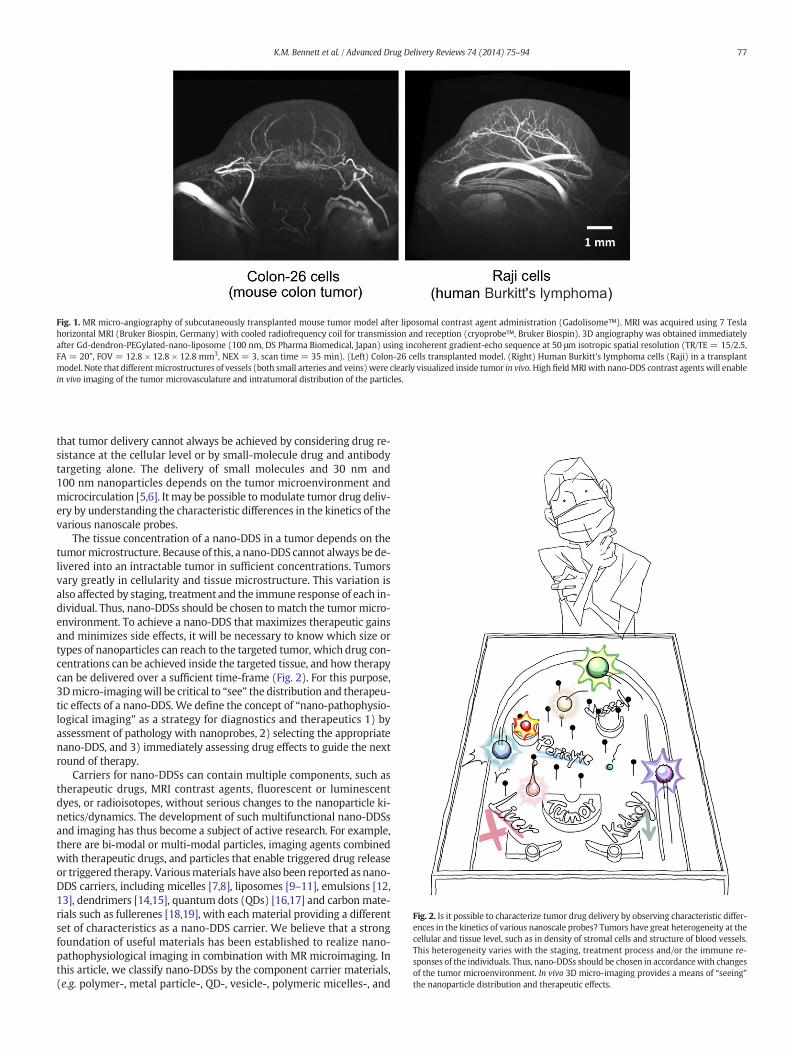
Fig. 1. MR micro-angiography of subcutaneously transplanted mouse tumor model after liposomal contrast agent administration (Gadolisome™). MRI was acquired using 7 Teslahorizontal MRI (Bruker Biospin, Germany) with cooled radiofrequency coil for transmission and reception (cryoprobe™, Bruker Biospin). 3D angiography was obtained immediatelyafter Gd-dendron-PEGylated-nano-liposome (100 nm, DS Pharma Biomedical, Japan) using incoherent gradient-echo sequence at 50 μm isotropic spatial resolution (TR/TE = 15/2.5,FA = 20°, FOV = 12.8 × 12.8 × 12.8 mm3, NEX = 3, scan time = 35 min). (Left) Colon-26 cells transplanted model. (Right) Human Burkitt's lymphoma cells (Raji) in a transplantmodel. Note that different microstructures of vessels (both small arteries and veins) were clearly visualized inside tumor in vivo. High fieldMRI with nano-DDS contrast agents will enablein vivo imaging of the tumor microvasculature and intratumoral distribution of the particles.
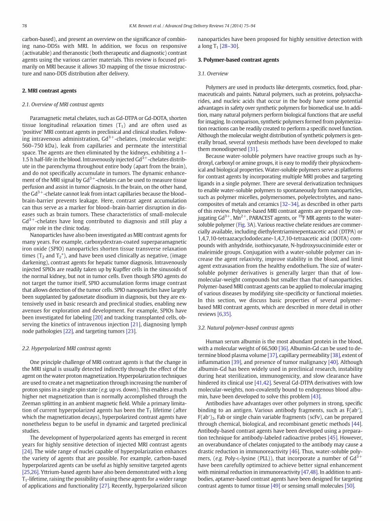
Fig. 2. Is it possible to characterize tumor drug delivery by observing characteristic differ-ences in the kinetics of various nanoscale probes? Tumors have great heterogeneity at thecellular and tissue level, such as in density of stromal cells and structure of blood vessels.This heterogeneity varies with the staging, treatment process and/or the immune re-sponses of the individuals. Thus, nano-DDSs should be chosen in accordancewith changesof the tumor microenvironment. In vivo 3D micro-imaging provides a means of “seeing”the nanoparticle distribution and therapeutic effects.
77K.M. Bennett et al. / Advanced Drug Delivery Reviews 74 (2014) 75–94
that tumor delivery cannot always be achieved by considering drug re-sistance at the cellular level or by small-molecule drug and antibodytargeting alone. The delivery of small molecules and 30 nm and100 nm nanoparticles depends on the tumor microenvironment andmicrocirculation [5,6]. It may be possible tomodulate tumor drug deliv-ery by understanding the characteristic differences in the kinetics of thevarious nanoscale probes.
The tissue concentration of a nano-DDS in a tumor depends on thetumormicrostructure. Because of this, a nano-DDS cannot always be de-livered into an intractable tumor in sufficient concentrations. Tumorsvary greatly in cellularity and tissue microstructure. This variation isalso affected by staging, treatment and the immune response of each in-dividual. Thus, nano-DDSs should be chosen to match the tumor micro-environment. To achieve a nano-DDS that maximizes therapeutic gainsand minimizes side effects, it will be necessary to know which size ortypes of nanoparticles can reach to the targeted tumor, which drug con-centrations can be achieved inside the targeted tissue, and how therapycan be delivered over a sufficient time-frame (Fig. 2). For this purpose,3Dmicro-imagingwill be critical to “see” the distribution and therapeu-tic effects of a nano-DDS. We define the concept of “nano-pathophysio-logical imaging” as a strategy for diagnostics and therapeutics 1) byassessment of pathology with nanoprobes, 2) selecting the appropriatenano-DDS, and 3) immediately assessing drug effects to guide the nextround of therapy.
Carriers for nano-DDSs can contain multiple components, such astherapeutic drugs, MRI contrast agents, fluorescent or luminescentdyes, or radioisotopes, without serious changes to the nanoparticle ki-netics/dynamics. The development of such multifunctional nano-DDSsand imaging has thus become a subject of active research. For example,there are bi-modal or multi-modal particles, imaging agents combinedwith therapeutic drugs, and particles that enable triggered drug releaseor triggered therapy. Variousmaterials have also been reported as nano-DDS carriers, including micelles [7,8], liposomes [9–11], emulsions [12,13], dendrimers [14,15], quantum dots (QDs) [16,17] and carbon mate-rials such as fullerenes [18,19], with each material providing a differentset of characteristics as a nano-DDS carrier. We believe that a strongfoundation of useful materials has been established to realize nano-pathophysiological imaging in combination with MR microimaging. Inthis article, we classify nano-DDSs by the component carrier materials,(e.g. polymer-, metal particle-, QD-, vesicle-, polymeric micelles-, and
78 K.M. Bennett et al. / Advanced Drug Delivery Reviews 74 (2014) 75–94
carbon-based), and present an overview on the significance of combin-ing nano-DDSs with MRI. In addition, we focus on responsive(activatable) and theranostic (both therapeutic and diagnostic) contrastagents using the various carrier materials. This review is focused pri-marily on MRI because it allows 3D mapping of the tissue microstruc-ture and nano-DDS distribution after delivery.
2. MRI contrast agents
2.1. Overview of MRI contrast agents
Paramagnetic metal chelates, such as Gd-DTPA or Gd-DOTA, shortentissue longitudinal relaxation times (T1) and are often used as‘positive’MRI contrast agents in preclinical and clinical studies. Follow-ing intravenous administration, Gd3+-chelates, (molecular weight:560–750 kDa), leak from capillaries and permeate the interstitialspace. The agents are then eliminated by the kidneys, exhibiting a 1–1.5 h half-life in the blood. Intravenously injectedGd3+-chelates distrib-ute in the parenchyma throughout entire body (apart from the brain),and do not specifically accumulate in tumors. The dynamic enhance-ment of the MRI signal by Gd3+-chelates can be used to measure tissueperfusion and assist in tumor diagnosis. In the brain, on the other hand,the Gd3+-chelate cannot leak from intact capillaries because the blood–brain-barrier prevents leakage. Here, contrast agent accumulationcan thus serve as a marker for blood–brain-barrier disruption in dis-eases such as brain tumors. These characteristics of small-moleculeGd3+-chelates have long contributed to diagnosis and still play amajor role in the clinic today.
Nanoparticles have also been investigated asMRI contrast agents formany years. For example, carboxydextran-coated superparamagneticiron oxide (SPIO) nanoparticles shorten tissue transverse relaxationtimes (T2 and T2*), and have been used clinically as negative, (imagedarkening), contrast agents for hepatic tumor diagnosis. Intravenouslyinjected SPIOs are readily taken up by Kupffer cells in the sinusoids ofthe normal kidney, but not in tumor cells. Even though SPIO agents donot target the tumor itself, SPIO accumulation forms image contrastthat allows detection of the tumor cells. SPIO nanoparticles have largelybeen supplanted by gadoxetate disodium in diagnosis, but they are ex-tensively used in basic research and preclinical studies, enabling newavenues for exploration and development. For example, SPIOs havebeen investigated for labeling [20] and tracking transplanted cells, ob-serving the kinetics of intravenous injection [21], diagnosing lymphnode pathologies [22], and targeting tumors [23].
2.2. Hyperpolarized MRI contrast agents
One principle challenge of MRI contrast agents is that the change inthe MRI signal is usually detected indirectly through the effect of theagent on thewater protonmagnetization.Hyperpolarization techniquesare used to create a netmagnetization through increasing the number ofproton spins in a single spin state (e.g. up vs. down). This enables amuchhigher net magnetization than is normally accomplished through theZeeman splitting in an ambient magnetic field. While a primary limita-tion of current hyperpolarized agents has been the T1 lifetime (afterwhich the magnetization decays), hyperpolarized contrast agents havenonetheless begun to be useful in dynamic and targeted preclinicalstudies.
The development of hyperpolarized agents has emerged in recentyears for highly sensitive detection of injected MRI contrast agents[24]. The wide range of nuclei capable of hyperpolarization enhancesthe variety of agents that are possible. For example, carbon-basedhyperpolarized agents can be useful as highly sensitive targeted agents[25,26]. Yttrium-based agents have also been demonstrated with a longT1-lifetime, raising the possibility of using these agents for awider rangeof applications and functionality [27]. Recently, hyperpolarized silicon
nanoparticles have been proposed for highly sensitive detection witha long T1 [28–30].
3. Polymer-based contrast agents
3.1. Overview
Polymers are used in products like detergents, cosmetics, food, phar-maceuticals and paints. Natural polymers, such as proteins, polysaccha-rides, and nucleic acids that occur in the body have some potentialadvantages in safety over synthetic polymers for biomedical use. In addi-tion, many natural polymers perform biological functions that are usefulfor imaging. In comparison, synthetic polymers formed frompolymeriza-tion reactions can be readily created to perform a specific novel function.Although themolecularweight distribution of synthetic polymers is gen-erally broad, several synthesis methods have been developed to makethemmonodispersed [31].
Because water-soluble polymers have reactive groups such as hy-droxyl, carboxyl or amine groups, it is easy tomodify their physicochem-ical and biological properties.Water-soluble polymers serve as platformsfor contrast agents by incorporating multiple MRI probes and targetingligands in a single polymer. There are several derivatization techniquesto enable water-soluble polymers to spontaneously form nanoparticles,such as polymer micelles, polymersomes, polyelectrolytes, and nano-composites of metals and ceramics [32–34], as described in other partsof this review. Polymer-based MRI contrast agents are prepared by con-jugating Gd3+, Mn2+, PARACEST agents, or 19F MR agents to the water-soluble polymer (Fig. 3A). Various reactive chelate residues are commer-cially available, including diethylentriaminepentaacetic acid (DTPA) or1,4,7,10-tetraazacyclododecane-1,4,7,10-tetraacetic acid (DOTA) com-pounds with anhydride, isothiocyanate, N-hydroxysuccinimide ester ormaleimide groups. Conjugation with a water-soluble polymer can in-crease the agent relaxivity, improve stability in the blood, and limitagent extravasation from the healthy endothelium. The size of water-soluble polymer derivatives is generally larger than that of low-molecular-weight compounds but smaller than that of nanoparticles.Polymer-basedMRI contrast agents can be applied tomolecular imagingof various diseases by modifying site-specificity or functional moieties.In this section, we discuss basic properties of several polymer-based MRI contrast agents, which are described in more detail in otherreviews [6,35].
3.2. Natural polymer-based contrast agents
Human serum albumin is the most abundant protein in the blood,with a molecular weight of 66,500 [36]. Albumin-Gd can be used to de-termine blood plasma volume [37], capillary permeability [38], extent ofinflammation [39], and presence of tumor malignancy [40]. Althoughalbumin-Gd has been widely used in preclinical research, instabilityduring heat sterilization, immunogenicity, and slow clearance havehindered its clinical use [41,42]. Several Gd-DTPA derivatives with lowmolecular-weights, non-covalently bound to endogenous blood albu-min, have been developed to solve this problem [43].
Antibodies have advantages over other polymers in strong, specificbinding to an antigen. Various antibody fragments, such as F(ab′),F(ab′)2, Fab or single chain variable fragments (scFv), can be preparedthrough chemical, biological, and recombinant genetic methods [44].Antibody-based contrast agents have been developed using a prepara-tion technique for antibody-labeled radioactive probes [45]. However,an overabundance of chelates conjugated to the antibody may cause adrastic reduction in immunoreactivity [46]. Thus, water-soluble poly-mers, (e.g. Poly-L-lysine (PLL)), that incorporate a number of Gd3+
have been carefully optimized to achieve better signal enhancementwithminimal reduction in immunoreactivity [47,48]. In addition to anti-bodies, aptamer-based contrast agents have been designed for targetingcontrast agents to tumor tissue [49] or sensing small molecules [50].
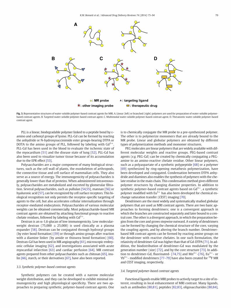
Fig. 3. Representative structures of water-soluble polymer-based contrast agents for MRI. A. Linear (left) or branched (right) polymers are used for preparation of water-soluble polymer-based contrast agents. B. Targeted water-soluble polymer-based contrast agent. C. Multimodal water-soluble polymer-based contrast agent. D. Theranostic water-soluble polymer-basedcontrast agent.
79K.M. Bennett et al. / Advanced Drug Delivery Reviews 74 (2014) 75–94
PLL is a linear, biodegradable polymer linked to a peptide bond by ε-amino and carboxyl groups of lysine. PLL-Gd can be formed by reactingthe anhydride or N-hydroxysuccinimide ester groups-bearing DTPA orDOTA to the amino groups of PLL, followed by labeling with Gd3+.PLL-Gd has been used in the blood to evaluate the ischemic state ofthe myocardium [51] and the disease state of lung [52]. PLL-Gd hasalso been used to visualize tumor tissue because of its accumulationdue to the EPR effect [53].
Polysaccharides are a major component of many biological struc-tures, such as the cell wall of plants, the exoskeleton of arthropods,the connective tissue and cell surface of mammalian cells. They alsoserve as a source of energy. The immunogenicity of polysaccharides isgenerally lower than that of proteins. When administered intravenous-ly, polysaccharides are metabolized and excreted by glomerular filtra-tion. Several polysaccharides, such as pullulan [54,55], mannan [56] orhyaluronic acid [57], can be recognized by cell surface receptors. This bi-ological recognition not only permits the receptor-specific targeting ofagents to the cell, but also accelerates cellular internalization throughreceptor-mediated endocytosis. Polysaccharides of various molecularweights can be obtained commercially. Most polysaccharide-based MRcontrast agents are obtained by attaching functional groups to reactivechelate residues, followed by labeling with Gd3+.
Dextran is an α-1,6-glucan of low immunogenicity. Lowmolecular-weight dextran (75,000 ± 25,000) is used clinically as a plasmaexpander [58]. Dextran can be conjugated through hydroxyl groups(by ester bond formation) [59] or through amino groups after reactionwith a diamine linker (by amide or thiourea bond formation) [60].Dextran-Gd has been used inMR angiography [61], microscopic embry-onic cellular imaging [62], and investigations associated with acutemyocardial infarction [63] and cardiac perfusion [64]. MR contrastagents prepared from other polysaccharides such as chitosan [65], inu-lin [66], starch, or their derivatives [67], have also been reported.
3.3. Synthetic polymer-based contrast agents
Synthetic polymers can be created with a narrow molecularweight distribution, and they can be developed to exhibit minimal im-munogenicity and high physiological specificity. There are two ap-proaches to preparing synthetic, polymer-based contrast agents. One
is to chemically conjugate the MR probe to a pre-synthesized polymer.The other is to polymerize monomers that are already bound to theMR probe. Linear and globular polymers are obtained by differenttypes of polymerization methods and monomer structures.
PEGmolecules are linear polymers that arewidely availablewith dif-ferent molecular weights and reactive groups. PEG-based contrastagents (e.g. PEG-Gd) can be created by chemically conjugating a PEG-amine to an amino-reactive chelate residue. Other linear polymers,such as a polyaspartate of a synthetic polypeptide [68] or a polymer[69] synthesized by ring-opening metathesis polymerization, havebeen developed and conjugated. Condensation between DTPA anhy-dride and diamines also enables the synthesis of polymerswith the che-late residue in themain chain. This condensationmethod gives differentpolymer structures by changing diamine properties. In addition tosynthetic polymer-based contrast agents based on Gd3+, a syntheticpolymer modified with Eu3+ has also been developed for chemical ex-change saturation transfer (CEST) imaging [70].
Dendrimers are themostwidely and systematically studied globularpolymers that are used as MRI contrast agents. There are two basic ap-proaches to forming dendrimers; one is a convergent approach inwhich the branches are constructed separately and later bound to a cen-tral core. The other is a divergent approach, inwhich thepreparation be-gins from the core and grows stepwise outward. A variety of dendrimerscan be formed by changing the chemical structure of the core, varyingthe coupling agents, and by altering the branch number. Dendrimer-based MR contrast agents can be formed by reacting amine groups onthe dendrimer with reactive chelates. In one such formulation, therelaxivity of dendrimer-Gdwas higher than that of Gd-DTPA [71]. In ad-dition, the biodistribution of dendrimer-Gd was modulated by thegeneration number (size) [72], and by the core structure [73]. In addi-tion to dendrimer-Gd, fluorinated- [74,75] and Mn2+-[76], Eu3+- orYb3+- modified dendrimers [77–79] have also been created for 19F MRand CEST imaging, respectively.
3.4. Targeted polymer-based contrast agents
Functional ligands enableMRI probes to actively target to a site of in-terest, resulting in local enhancement of MRI contrast. Many ligands,such as antibodies [80,81], peptides [82,83], oligosaccharides [80,84],
80 K.M. Bennett et al. / Advanced Drug Delivery Reviews 74 (2014) 75–94
and other bioactive substances [49,85], have been used as ligands forwater-soluble polymers (Fig. 3B). Targeted, polymer-based MRI con-trast agents can be used to image targets such as tumor, liver [84], andcartilage [86]. We have developed a polymer-based MRI contrastagent to image tissue regeneration after therapy [87]. Therapeutic an-giogenesis is one of the most important technologies to treat ischemicdiseases and to support cell transplantation. We have created apolysaccharide-based MRI contrast agent for the MRI-based evaluationof therapeutic angiogenesis by chemical conjugation of dextran-Gdwith a cyclic peptide containing an arginine–glycine–aspartic acid se-quence (cRGD) with an inherent affinity for the αvβ3 integrin (cRGD-dextran-Gd). The cRGD-dextran-Gd had an affinity for cells expressingthe αvβ3 integrin and showed a high longitudinal relaxivity comparedto DTPA-Gd. The cRGD-dextran-Gd could be used after intravenous in-jection to detect the ischemic–angiogenic region in mice with hindlimbischemia.
3.5. Polymer-based multimodal imaging
Multimodal contrast agents (Fig. 3C) can be created by conjugationof other imaging probes with water-soluble polymer contrastagents. Several groups have reported water-soluble polymer agentsfor MRI and fluorescence imaging [81,94]. It is possible to prepare mul-timodal contrast agents by encapsulating polymer-based contrastagents into other imaging probes. Shi et al. showed that gold nanoparti-cles containing denderimer-Gd could be visualized using MRI andCT [95].
4. Metal particle-based contrast agent
4.1. Overview
Magnetic nanoparticles have become important tools for moleculardetection and drug delivery. “Theranostic” agents combine these twofunctions to deliver targeted therapy to a specific location. [98,99].These agents can be combined a multifunctional or targeted nanoparti-cles for a range of applications [98,100]. For MRI, the major use of mag-netic nanoparticles is to shorten local water T2 or T1 through aninteraction between the electronic magnetic moments of the compo-nent atoms of the nanoparticle and the nuclear magnetic moment ofthe surrounding water. The strength of this interaction and the subse-quent impact on the MRI signal is determined by many factors, includ-ing the strength and localization of the magnetic moments of thenanoparticle crystal and the access of water to the crystal core. Mostparamagnetic agents shorten both T1 and T2. However, agentswith strong coupling between component atoms tend to be detectedas T2-shortening agents, and paramagnetic atoms or chelates are typi-cally detected as T1-shortening agents in normal concentrations. A com-plicating factor is the fact that T1-shortening agents are often morereadily detected in the body because T1 of the tissue is longer, whileT2 agents tend to be more powerful (i.e. have higher relaxivity). Thebasic mechanisms of MR relaxation in magnetic nanoparticles havebeen extensively reviewed [101,102], though knowledge of nanoscalemagnetism is still incomplete.
Magnetic particle synthesis lies at the interface between chemistryand physics. The chemistry involved can range from organic and bio-chemical to inorganic, and it is crucial to understand how magnetismis affected in these particles during synthesis. Some of the most recentadvances in magnetic nanoparticle synthesis have focused on tailoringthe properties of the nanoparticles to a specific application. This in-volves control over both the physicochemical properties and the inter-face between these properties and the instruments to detect oractivate the nanoparticles once they are at the target.
Superparamagnetic nanoparticles can be synthesized in a varietyof ways, ranging from physical pulverization of a large crystal [103], totraditional chemical synthesis, to microwave synthesis. Chemical
techniques inmetal crystal formation involve, broadly, either controlledionic bonding or oxidation around a small nucleated metal oxide, andthe formation can be performed in either gas- or aqueous phase. Oneof the simplest, most widely-used techniques involves co-precipitationof component metal salts. Thermal decomposition is also used, inwhich metals are mixed into a heated surfactant to control particle ox-idation, size, and shape. Recently microwave synthesis has also beenused to synthesize highly homogeneous, reproducible magnetic nano-particles. Nanoparticle synthesis has been reviewed extensively in therecent literature [104–106].
4.2. Superparamagnetic iron oxide-based nanoparticles
Some of the most common magnetic nanoparticles used in mo-lecular and cellular MRI are formed from SPIOs and oxides of othertransition metals [30,107–115]. Because of the simplicity of synthe-sis and good biocompatibility, many of the most first commercialiron oxides were dextran-coated, with an amorphous coating thatencompasses more than one particle. In recent years there havebeen a number of new particles developed, both as passive andtargeted contrast agents.
Magnetic nanoparticles have been formed from other transitionmetals, including manganese oxides and adsorbed composites ofMn2+. Mn2+ has a higher average magnetic moment than does iron,and is also readily oxidized to form a superparamagnetic crystal in ananoparticle. Several groups have developed manganese oxide nano-particles, either as high-relaxivity T2-shortening contrast agents or asagents that can dissociate upon intracellular release from an endosome.In one proposed agent, Mn2+ dissociated from the crystal due to reduc-tion in the low endosomal pH, allowing the change in MRI contrast toreport on intracellular delivery through a shift from T2- to T1-relatedcontrast [116].
Natural magnetic nanoparticles have been synthesized frombiologically-derived macromolecules [117]. These nanoparticles can becreated to incorporate many of the properties of completely syntheticnanoparticles, and the magnetism of these natural nanoparticles canbe readily controlled. Some examples of natural and endogenous mag-netic nanoparticles are those formed from the cowpea virus [118],magA [119], and from the iron storage protein ferritin [120–123]. Natu-ral nanoparticles have some proposed advantages over other nanoparti-cles in biocompatibility and ease of functionalization. Natural magneticnanoparticles may also prove to be useful “reporter” proteins formedthrough endogenous native or recombinant expression and detectionwith MRI. Thus, the development of natural nanoparticle contrastagents is an active area of investigation.
One major goal of current research in MRI-detectable nano-DDS and SPIO development is to increase the biocompatibility andfunctionalization of these agents. Biocompatibility is important to bothpreclinical and clinical translational work, and functionalization is im-portant for agent targeting and stability after delivery. There has beena great deal of recent work to develop nanoparticles with nontoxic orbiocompatible coatings [107,124–126]. Regulatory and practical guide-lines for these types of studies are being identified as more researchersaim to commercialize new agents or bring them to the clinic [127].
5. Quantum dot-based contrast agents
5.1. Overview
Over the last ten years, semiconductor QDs have been recognized asa new type of optical imaging contrast agent because of their uniquespectral properties. These novel properties can be used to optimizethe signal-to-noise ratio, to improve the sensitivity of fluorescence de-tection, and to increase the quality of fluorescent cellular andmolecularimaging. Moreover, the size-tuneable fluorescence emission and thebroad excitation spectra of QDs make it possible to use them in
81K.M. Bennett et al. / Advanced Drug Delivery Reviews 74 (2014) 75–94
multiplexed fluorescent analyses. Single QDs can be observed andtracked for up to a few hours with fluorescence confocal microscopy[128], total internal reflection microscopy [129], basic wide-fieldepifluorescence microscopy [130], and single-molecule microscopy[131]. QDs are also excellent probes for two-photon confocal microsco-py [132] because they are characterized by a large absorption crosssection.
Since water-soluble QDs have many functional groups on their sur-face, it is easy to conjugate them with biomolecules, (e.g. proteins,DNAs, RNAs, small ligands, etc.), and other chemicals, (e.g., drugs)(Fig. 4). The resulting conjugates combine the spectral characteristicsof the nanocrystal and the biomolecular functions of the attached bio-molecules. The multi-functionality of QDs is one of the major advan-tages of QDs over organic fluorophores, for which conjugation isusually restricted to one biocompatible molecule per dye molecule.The large number (10 to 100) of potential surface attachment groupscan be used to confer different functionalities to individual QDs. For in-stance, in addition to a recognition moiety, QDs can be equipped with amembrane-crossing or cell-internalization capability, and/or an enzy-matic function [133–135]. QDs are therefore appropriate matrices forthe development of novel drug carriers.
5.2. QD-based contrast agents
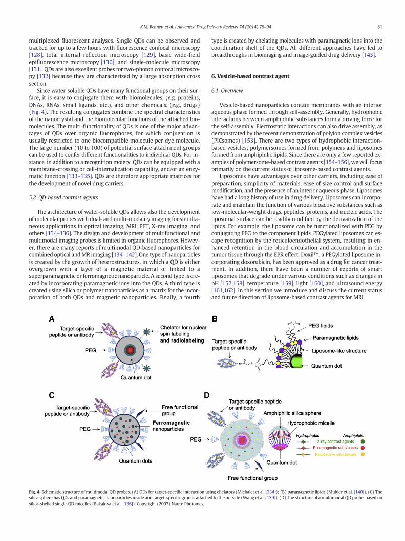
The architecture of water-soluble QDs allows also the developmentofmolecular probeswith dual- andmulti-modality imaging for simulta-neous applications in optical imaging, MRI, PET, X-ray imaging, andothers [134–136]. The design and development of multifunctional andmultimodal imaging probes is limited in organic fluorophores. Howev-er, there are many reports of multimodal QD-based nanoparticles forcombined optical andMR imaging [134–142]. One type of nanoparticlesis created by the growth of heterostructures, in which a QD is eitherovergrown with a layer of a magnetic material or linked to asuperparamagnetic or ferromagnetic nanoparticle. A second type is cre-ated by incorporating paramagnetic ions into the QDs. A third type iscreated using silica or polymer nanoparticles as a matrix for the incor-poration of both QDs and magnetic nanoparticles. Finally, a fourth
Fig. 4. Schematic structure of multimodal QD probes. (A) QDs for target-specific interaction ussilica sphere has QDs and paramagnetic nanoparticles inside and target-specific groups attachesilica-shelled single-QD micelles (Bakalova et al. [136]). Copyright (2007) Naure Photonics.
type is created by chelating molecules with paramagnetic ions into thecoordination shell of the QDs. All different approaches have led tobreakthroughs in bioimaging and image-guided drug delivery [143].
6. Vesicle-based contrast agent
6.1. Overview
Vesicle-based nanoparticles contain membranes with an interioraqueous phase formed through self-assembly. Generally, hydrophobicinteractions between amphiphilic substances form a driving force forthe self-assembly. Electrostatic interactions can also drive assembly, asdemonstrated by the recent demonstration of polyion complex vesicles(PICsomes) [153]. There are two types of hydrophobic interaction-based vesicles; polymersomes formed from polymers and liposomesformed from amphiphilic lipids. Since there are only a few reported ex-amples of polymersome-based contrast agents [154–156], wewill focusprimarily on the current status of liposome-based contrast agents.
Liposomes have advantages over other carriers, including ease ofpreparation, simplicity of materials, ease of size control and surfacemodification, and the presence of an interior aqueous phase. Liposomeshave had a long history of use in drug delivery. Liposomes can incorpo-rate and maintain the function of various bioactive substances such aslow-molecular-weight drugs, peptides, proteins, and nucleic acids. Theliposomal surface can be readily modified by the derivatization of thelipids. For example, the liposome can be functionalized with PEG byconjugating PEG to the component lipids. PEGylated liposomes can es-cape recognition by the reticuloendothelial system, resulting in en-hanced retention in the blood circulation and accumulation in thetumor tissue through the EPR effect. Doxil™, a PEGylated liposome in-corporating doxorubicin, has been approved as a drug for cancer treat-ment. In addition, there have been a number of reports of smartliposomes that degrade under various conditions such as changes inpH [157,158], temperature [159], light [160], and ultrasound energy[161,162]. In this section we introduce and discuss the current statusand future direction of liposome-based contrast agents for MRI.
ing chelators (Michalet et al. [234]); (B) paramagnetic lipids (Mulder et al. [140]). (C) Thed to the outside (Wang et al. [139]). (D) The structure of a multimodal QD probe, based on
82 K.M. Bennett et al. / Advanced Drug Delivery Reviews 74 (2014) 75–94
6.2. Basic liposome-based contrast agents
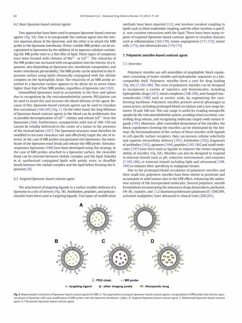
Two approaches have been used to prepare liposome-based contrastagents (Fig. 5A). One is to encapsulate the contrast agent into the inte-rior aqueous phase of the liposome, and the other is to attach the MRIprobe to the liposomemembrane.Water-solubleMRI probes can be en-capsulated in liposomes by the addition of an aqueous solution contain-ing the MR probe onto to a thin film of lipid. These types of complexeshave been formed with chelates of Mn2+ or Gd3+. The relaxivity oftheMR probes has increasedwith encapsulation into the interior of a li-posome, also depending on liposome size, membrane composition, andwater-membrane permeability. TheMRI probe can be attached to the li-posome surface using lipids chemically conjugated with the chelatecomplex on the hydrophilic head. The relaxivity of an MRI probe at-tached to a liposome surface appears to be about six to seven timeshigher than that of free MRI probes, regardless of liposome size [163].
Unmodified liposomes tend to accumulate in the liver and spleendue to recognition by the reticuloendothelial system. PEGylation canbe used to avoid this and increase the blood lifetime of the agent. Be-cause of this, liposome-based contrast agents can be used to visualizefine vasculature [164,165] (Fig. 1). On the other hand, the long lifetimesof liposome-based contrast agents in the blood can be problematic dueto possible decomplexation of Gd3+-chelate and release Gd3+ from theliposomes [166]. Furthermore, nanoparticles with size of 100–150 nmcannot be reliably delivered to the center of a tumor in the presenceof the stromal barrier [167]. The liposomal structure must therefore bemodified to increase clearance rate and effectively target the site of in-terest. In the case of MR probes encapsulated into liposomes, the mem-brane of the liposomemust break and release the MRI probe. Stimulus-responsive liposomes [168] have been developed using this strategy. Inthe case of MRI probes attached to a liposome surface, the cleavablebond can be inserted between chelate complex and the lipid. Kabalkaet al. synthesized conjugated lipids with amide, ester, or disulfidebonds between the chelate complex and the lipid before forming the li-posomes [9].
6.3. Targeted liposome-based contrast agent
The attachment of targeting ligands to a surface enables delivery of aliposome to a site of interest (Fig. 5B). Antibodies, peptides, andpolysac-charides have been used as targeting ligands. Two types of modification
Fig. 5. Representative structures of liposome-based contrast agents forMRI. A. Two approaches tous phase of liposome (left) and modification of MRI probes onto the liposome membrane (ragent. D. Theranostic liposome-based contrast agent.
methods have been reported [12]; one involves covalent coupling tolipids such as thiol-maleimide coupling, and the other involves a specif-ic, non-covalent interaction with the lipid. There have been many re-ports of targeted liposome-based contrast agents to visualize diseasessuch as inflammation [169,170], tumor angiogenesis [171,172], tumorcells [173], and atherosclerosis [174,175].
7. Polymeric micelles-based contrast agent
7.1. Overview
Polymeric micelles are self-assemblies of amphiphilic block copoly-mers consisting of water-soluble and hydrophobic segments in a bio-compatible shell. Polymeric micelles form a core for drug loading(Fig. 6A) [7,185,186]. The cores of polymeric micelles can be designedto incorporate a variety of reporters and biomolecules, includinghydrophobic drugs [187],metal complexes [188,189], and chargedmac-romolecules [190] such as nucleic acids, by engineering the core-forming backbone. Polymeric micelles present several advantages asnanocarriers, including prolonged blood circulation and a size range be-tween 10 and 100 nm. This size range is useful for evading nonspecificuptake by the reticuloendothelial system, avoiding renal excretion, con-trolling drug release, and recognizing molecular targets with surface li-gands [191]. Moreover, after controlled dissociation of the micelles, theblock copolymers forming the micelles can be eliminated by the kid-neys. By functionalization of the surface of these micelles with ligandsto cell-specific surface receptors, they can possess cellular selectivityand superior intracellular delivery [191]. Antibodies [192], fragmentsof antibodies [193], aptamers [194], peptides [195,196] and small mole-cules [197] have been used as ligands to improve the tumor targetingability of micelles (Fig. 6A). Micelles can also be designed to respondto internal stimuli such as pH, reductive environment, and enzymes[7,185,186], or external stimuli including light and ultrasound [198,199] to enhance their specificity to malignant tissues.
Due to the prolonged blood circulation of polymeric micelles andtheir small size, polymeric micelles have been shown to penetrate andaccumulate in solid tumors due to the EPR effect, enhancing the antitu-mor activity of the incorporated molecules. Several polymeric micelleformulations incorporating the anticancer drugs doxorubicin, paclitaxel,SN-38, cisplatin, and (1,2-diaminocyclohexane)platinum(II) (DACHPt,activated oxaliplatin) have advanced to clinical trials [200,201].
o prepare liposome-based contrast agents; encapsulation ofMRI probes into interior aque-ight). B. Targeted liposome-based contrast agent. C. Multimodal liposome-based contrast
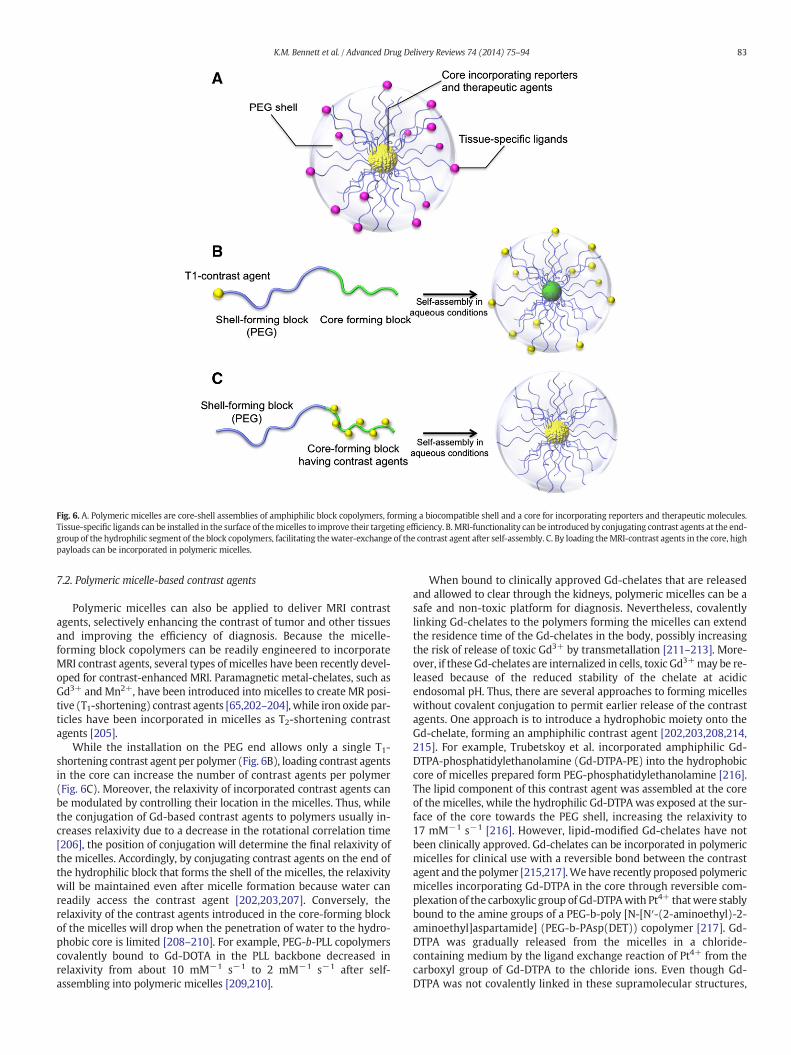
Fig. 6. A. Polymeric micelles are core-shell assemblies of amphiphilic block copolymers, forming a biocompatible shell and a core for incorporating reporters and therapeutic molecules.Tissue-specific ligands can be installed in the surface of themicelles to improve their targeting efficiency. B.MRI-functionality can be introduced by conjugating contrast agents at the end-group of the hydrophilic segment of the block copolymers, facilitating thewater-exchange of the contrast agent after self-assembly. C. By loading theMRI-contrast agents in the core, highpayloads can be incorporated in polymeric micelles.
83K.M. Bennett et al. / Advanced Drug Delivery Reviews 74 (2014) 75–94
7.2. Polymeric micelle-based contrast agents
Polymeric micelles can also be applied to deliver MRI contrastagents, selectively enhancing the contrast of tumor and other tissuesand improving the efficiency of diagnosis. Because the micelle-forming block copolymers can be readily engineered to incorporateMRI contrast agents, several types of micelles have been recently devel-oped for contrast-enhanced MRI. Paramagnetic metal-chelates, such asGd3+ and Mn2+, have been introduced into micelles to create MR posi-tive (T1-shortening) contrast agents [65,202–204], while iron oxide par-ticles have been incorporated in micelles as T2-shortening contrastagents [205].
While the installation on the PEG end allows only a single T1-shortening contrast agent per polymer (Fig. 6B), loading contrast agentsin the core can increase the number of contrast agents per polymer(Fig. 6C). Moreover, the relaxivity of incorporated contrast agents canbe modulated by controlling their location in the micelles. Thus, whilethe conjugation of Gd-based contrast agents to polymers usually in-creases relaxivity due to a decrease in the rotational correlation time[206], the position of conjugation will determine the final relaxivity ofthe micelles. Accordingly, by conjugating contrast agents on the end ofthe hydrophilic block that forms the shell of the micelles, the relaxivitywill be maintained even after micelle formation because water canreadily access the contrast agent [202,203,207]. Conversely, therelaxivity of the contrast agents introduced in the core-forming blockof the micelles will drop when the penetration of water to the hydro-phobic core is limited [208–210]. For example, PEG-b-PLL copolymerscovalently bound to Gd-DOTA in the PLL backbone decreased inrelaxivity from about 10 mM−1 s−1 to 2 mM−1 s−1 after self-assembling into polymeric micelles [209,210].
When bound to clinically approved Gd-chelates that are releasedand allowed to clear through the kidneys, polymeric micelles can be asafe and non-toxic platform for diagnosis. Nevertheless, covalentlylinking Gd-chelates to the polymers forming the micelles can extendthe residence time of the Gd-chelates in the body, possibly increasingthe risk of release of toxic Gd3+ by transmetallation [211–213]. More-over, if these Gd-chelates are internalized in cells, toxic Gd3+may be re-leased because of the reduced stability of the chelate at acidicendosomal pH. Thus, there are several approaches to forming micelleswithout covalent conjugation to permit earlier release of the contrastagents. One approach is to introduce a hydrophobic moiety onto theGd-chelate, forming an amphiphilic contrast agent [202,203,208,214,215]. For example, Trubetskoy et al. incorporated amphiphilic Gd-DTPA-phosphatidylethanolamine (Gd-DTPA-PE) into the hydrophobiccore of micelles prepared form PEG-phosphatidylethanolamine [216].The lipid component of this contrast agent was assembled at the coreof the micelles, while the hydrophilic Gd-DTPA was exposed at the sur-face of the core towards the PEG shell, increasing the relaxivity to17 mM−1 s−1 [216]. However, lipid-modified Gd-chelates have notbeen clinically approved. Gd-chelates can be incorporated in polymericmicelles for clinical use with a reversible bond between the contrastagent and the polymer [215,217].Wehave recently proposed polymericmicelles incorporating Gd-DTPA in the core through reversible com-plexation of the carboxylic group of Gd-DTPAwith Pt4+ thatwere stablybound to the amine groups of a PEG-b-poly [N-[N′-(2-aminoethyl)-2-aminoethyl]aspartamide] (PEG-b-PAsp(DET)) copolymer [217]. Gd-DTPA was gradually released from the micelles in a chloride-containing medium by the ligand exchange reaction of Pt4+ from thecarboxyl group of Gd-DTPA to the chloride ions. Even though Gd-DTPA was not covalently linked in these supramolecular structures,
84 K.M. Bennett et al. / Advanced Drug Delivery Reviews 74 (2014) 75–94
confining Gd-DTPA in the core of thesemicelles increased the relaxivityup to 48 mM−1 s−1.
Manganese (Mn) has also been considered a safe alternative to Gd3+
in constructing T1-shortening polymeric micelles [218]. Polymeric mi-celles can be created by incorporating manganese oxide in the core ofthe micelles for Mn-enhanced MRI [107]. These micelles create pH-sensitive MRI contrast because the manganese oxide nanoparticles re-lease free Mn2+ at the acidic pH of ~5 in the endosome. Relaxivity isalso increased whenMn2+ is complexedwith a protein [107]. In anoth-er approach, Jang et al. have developed polymeric micelles by self-assembly of amphiphilic PEG-Mn(III)-porphyrin polymers [219]. Theseagents are potentially useful as T1-shortening contrast agents.
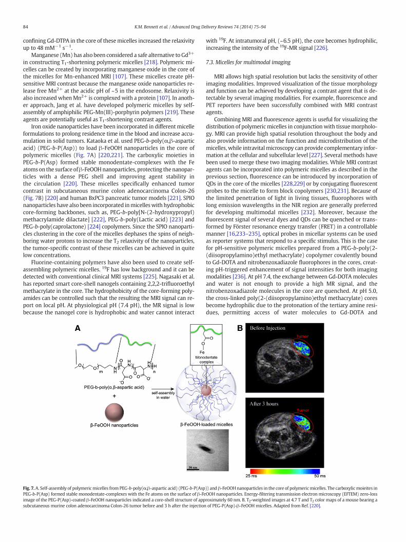
Iron oxide nanoparticles have been incorporated in different micelleformulations to prolong residence time in the blood and increase accu-mulation in solid tumors. Kataoka et al. used PEG-b-poly(α,β-asparticacid) (PEG-b-P(Asp)) to load β-FeOOH nanoparticles in the core ofpolymeric micelles (Fig. 7A) [220,221]. The carboxylic moieties inPEG-b-P(Asp) formed stable monodentate-complexes with the Featomson the surface ofβ-FeOOHnanoparticles, protecting thenanopar-ticles with a dense PEG shell and improving agent stability inthe circulation [220]. These micelles specifically enhanced tumorcontrast in subcutaneous murine colon adenocarcinoma Colon-26(Fig. 7B) [220] and human BxPC3 pancreatic tumor models [221]. SPIOnanoparticles have also been incorporated inmicelleswith hydrophobiccore-forming backbones, such as, PEG-b-poly[N-(2-hydroxypropyl)methacrylamide dilactate] [222], PEG-b-poly(Lactic acid) [223] andPEG-b-poly(caprolactone) [224] copolymers. Since the SPIO nanoparti-cles clustering in the core of the micelles dephases the spins of neigh-boring water protons to increase the T2 relaxivity of the nanoparticles,the tumor-specific contrast of these micelles can be achieved in quitelow concentrations.
Fluorine-containing polymers have also been used to create self-assembling polymeric micelles. 19F has low background and it can bedetected with conventional clinical MRI systems [225]. Nagasaki et al.has reported smart core-shell nanogels containing 2,2,2-trifluoroethylmethacrylate in the core. The hydrophobicity of the core-forming poly-amides can be controlled such that the resulting the MRI signal can re-port on local pH. At physiological pH (7.4 pH), the MR signal is lowbecause the nanogel core is hydrophobic and water cannot interact
Fig. 7.A. Self-assembly of polymericmicelles from PEG-b-poly(α,β-aspartic acid) (PEG-b-P(AspPEG-b-P(Asp) formed stable monodentate-complexes with the Fe atoms on the surface of β-Feimage of the PEG-P(Asp)-coated β-FeOOH nanoparticles indicated a core-shell structure of appsubcutaneous murine colon adenocarcinoma Colon-26 tumor before and 3 h after the injection
with 19F. At intratumoral pH, (~6.5 pH), the core becomes hydrophilic,increasing the intensity of the 19F-MR signal [226].
7.3. Micelles for multimodal imaging
MRI allows high spatial resolution but lacks the sensitivity of otherimaging modalities. Improved visualization of the tissue morphologyand function can be achieved by developing a contrast agent that is de-tectable by several imaging modalities. For example, fluorescence andPET reporters have been successfully combined with MRI contrastagents.
Combining MRI and fluorescence agents is useful for visualizing thedistribution of polymericmicelles in conjunctionwith tissuemorpholo-gy. MRI can provide high spatial resolution throughout the body andalso provide information on the function and microdistribution of themicelles, while intravitalmicroscopy can provide complementary infor-mation at the cellular and subcellular level [227]. Several methods havebeen used to merge these two imaging modalities. While MRI contrastagents can be incorporated into polymeric micelles as described in theprevious section, fluorescence can be introduced by incorporation ofQDs in the core of the micelles [228,229] or by conjugating fluorescentprobes to the micelle to form block copolymers [230,231]. Because ofthe limited penetration of light in living tissues, fluorophores withlong emission wavelengths in the NIR region are generally preferredfor developing multimodal micelles [232]. Moreover, because thefluorescent signal of several dyes and QDs can be quenched or trans-formed by Förster resonance energy transfer (FRET) in a controllablemanner [16,233–235], optical probes in micellar systems can be usedas reporter systems that respond to a specific stimulus. This is the casefor pH-sensitive polymeric micelles prepared from a PEG-b-poly(2-(diisopropylamino)ethyl methacrylate) copolymer covalently boundto Gd-DOTA and nitrobenzoxadiazole fluorophores in the cores, creat-ing pH-triggered enhancement of signal intensities for both imagingmodalities [236]. At pH 7.4, the exchange between Gd-DOTAmoleculesand water is not enough to provide a high MR signal, and thenitrobenzoxadiazole molecules in the core are quenched. At pH 5.0,the cross-linked poly(2-(diisopropylamino)ethyl methacrylate) coresbecome hydrophilic due to the protonation of the tertiary amine resi-dues, permitting access of water molecules to Gd-DOTA and
)) and β-FeOOHnanoparticles in the core of polymericmicelles. The carboxylicmoieites inOOH nanoparticles. Energy-filtering transmission electron microscopy (EFTEM) zero-lossroximately 60 nm. B. T2-weighted images at 4.7 T and T2 color maps of a mouse bearing aof PEG-P(Asp)-β-FeOOH micelles. Adapted from Ref. [220].
85K.M. Bennett et al. / Advanced Drug Delivery Reviews 74 (2014) 75–94
dequenching the nitrobenzoxadiazole moieties. pH-sensitive, T2-shortening polymeric micelles were also prepared from PEG-b-poly(β-amino ester) copolymers with ionizable tertiary amine groups havingSulforhodamine 101 bound to the poly(β-amino ester) backbone[237]. The polymeric micelles encapsulated SPIO nanoparticles at phys-iological pH 7.4, enhancing the relaxivity of the nanoparticles to morethan 250 mM−1 s−1. At pH 7.4, the fluorescence of Sulforhodamine101 was quenched by energy transfer. At pH 6.8–7.0, the fluorescentsignal was recovered, suggesting the potential use of these micelles aspH-triggered reporters of acidic biological environments.
Polymeric micelles can also be used to combine the high anatomicresolution of MRI with the highly sensitive imaging of PET. Due to thedifference in sensitivity between these imaging modalities, the detect-able concentrations of MRI contrast agents should be much higherthan those of PET probes. Thus, polymericmicelles offer unique featuresfor combining high-loads of MRI contrast agents in their cores and pre-cise amounts of PET probes on the surface of the PEG shell. For example,SPIO nanoparticles coated with PEG-phospholipids, having a DOTA che-lator at the PEG-end, were labeled with 64Cu [238].68Ga has also beencombined with SPIO nanoparticles within the same polymeric micelle.These micelles were prepared by self-assembly of PEG-b-poly(lactic-co-glycolic acid), with SPIO nanoparticles incorporated in the hydro-phobic core of the micelles and 68Ga installed on surface of the PEGshell of the micelles after chelation with 2,2′-(7-(4-((2-aminoethyl)amino)-1-carboxy-4-oxobutyl)-1,4,7-triazonane-1,4-diyl)diacetic acid(NODA) [239]. Both 64Cu and 68Ga allowed the precise study of thebiodistribution of the micelles by PET.
8. Contrast agents based on other nanoparticles
8.1. PLGA-based contrast agents
Nanoparticles prepared from water-insoluble polymers, such aspoly(lactide-co-glycolide) (PLGA), can be used as contrast agents.Both hydrophobic and hydrophilic drugs can be incorporated into thepolymeric nanoparticles by forming oil-in-water and water-in-oil-in-water emulsions. Several types of contrast agents with activatable[107], multimodal [13,251] or theranostic [252] capabilities have beenprepared by using these containers. In addition, chemical conjugationof amphiphilic substances has been used to stabilize polymeric nano-particles and provide surface modification [253].
8.2. Silica-based contrast agents
Silica-based contrast agents have been also extensively investigated.Almost all silica-based contrast agents are prepared from silica nanopar-ticles. There are two main types of silica-based nanoparticles [254],namely solid silica and mesoporous silica. Solid silica nanoparticlescan be prepared by the hydrolysis of a silicon alkoxide precursor,(through a sol–gel process), in ethanol and water (Stöber method), orinside the water phase of an emulsion in oil (through the reversemicroemulsion method). Mesoporous silica nanoparticles can be ob-tained by a surfactant-templated sol–gel reaction. Advantages of silicananoparticles include ease of preparation, biocompatibility, and evendispersion in water. Since silica nanoparticles neither absorb light norperturb a magnetic field, silica-based contrast agents can be combinedwith probes through the well-established siloxane chemistry. Silica-based T1-shortening MRI contrast agents have been prepared by bind-ing Gd-chelated trialkoxysilane derivatives to the surface of solid [255]or mesoporous silica nanoparticles [256]. Silica-based, T2-shorteningMRI contrast agents have been prepared by the encapsulating the ironoxide nanoparticle in the silica matrix [257]. Other structural types ofsilica-based MRI contrast agents based on a polyhedral oligomericsilsesquioxane (POSS)-core dendrimer has been developed by Tanakaet al. [258]. The high relaxivity of a POSS-based MRI positive contrast
agent was achieved by the addition of Gd-chelated derivatives on therigid cubic structure.
9. Responsive (activatable) contrast agents
Several types of small-molecule, chelate-based responsive agentshave been reported [259], such as enzyme-activated (beta-galactosi-dase) [260], Ca2+ concentration-activated [261] and pH-activated[262,263] agents. In this section, we highlight several polymer- ornanoparticle-based responsive or activatable contrast agents for MRI.
9.1. Polymer-based responsive and biodegradable contrast agents
Polymer-based MRI contrast agents have been modified forimproved biodegradability and rapid excretion. It is known thatdisulfide bonds are broken down by thiol-bearing compounds presentin the body [88]. Therefore, polymers containing disulfide bonds(polydisulfides) enable MR probes to be rapidly excreted due to poly-mer degradation, resulting in enhanced contrastwhere the probes accu-mulate. Several polydisulfides of linear polymers [89] or dendrimers[90,91] have been reported.
There are a few reports on activatable MRI probes prepared bywater-soluble polymers. Sherry et al. reported that the relaxivity changeof a pH-responsiveMRI probewas enhanced by attachment to a dendri-mer [92]. Lu et al. developed a Gd3+-DNA strand conjugated to a DNAaptamer for adenosine [50]. When bound to adenosine, the conjugatedissociates to release the Gd3+ containing-DNA strand, which de-creased MRI contrast due to a reduction in relaxivity. Kikuchi et al. de-veloped activatable MRI probes using pH-responsive polymers [93]. Adecrease in pH made the morphology of the polymer change from ex-panded to shrunken, resulting in increased relaxivity.
9.2. Nanoparticle-based responsive contrast agents
9.2.1. Functional metal particle-based responsive contrast agentsSeveral groups have investigated new approaches to chemical SPIO
synthesis to impart specific functions to the nanoparticles in situ. Oneexample of this is the development of a “switchable”magnetic nanopar-ticle, activated by light. [264]. Another example is the use of chemicaldoping to tune MRI contrast (r1 vs. r2) in a nanoparticle of a uniformsize [265]. Finally, nanoparticles (~100 s of nm in size) have been creat-ed through stereolithography with defined shapes that enable them tobe distinguished from each other [266].
Magnetic particle imaging has also emerged as a potentially impor-tant imaging modality, based on the direct detection of magnetic parti-cles in three dimensions [267]. Magnetic particles can be used astheranostic agents through combinedMRI-based detection and local tis-sue heating with radiofrequency energy [268]. While it can be difficultto directly functionalize metals in a crystal, many magnetic particlesare readily encapsulated in either synthetic or natural coatings.
9.2.2. Functional QD-based responsive contrast agentsQDs are also potential photosensitizers. QDs are energy donors,
based on triplet resonance energy transfer and/or FRET [141]. Energytransfer between QDs and other intracellular molecules such as tripletoxygen, reducing equivalents, and pigments, could induce the genera-tion of reactive oxygen species and reactive nitrogen species, invokingapoptosis and necrosis in cells. Several recent papers suggest that cyto-toxicity of QDs, mediated by light irradiation, might be used to kill can-cer cells [144–146]. The light-mediated cytotoxicity of QDs, combinedwith their capacity as energy donors, has enabled the applicationof QDs as novel photosensitizers or cofactors of conventionalphotosensitizing agents used in photodynamic therapy (PDT). This ap-plication was first described in 2004 [147]. Since then, many groupshave published in this area [148–152]. A significant benefit of QDs isthat they can be precisely tuned by changing their size and composition.
86 K.M. Bennett et al. / Advanced Drug Delivery Reviews 74 (2014) 75–94
The optical emission of QDs results from quantum-confinement effectsand can be tuned to emit from the ultraviolet to the near infrared(NIR) spectral regions, in contrast to the largely visible emission ofmost conventional photosensitizers. Because there isminimal light scat-tering and absorption in the NIR region of the spectrum, light of low in-tensity can be used to penetrate tissue to depths of several centimeters,allowing access to deep-seated tumors. Furthermore, because of theirlarge transition dipole moment, QDs are strong optical absorbers, mak-ing them potential drugs for photodynamic therapy.
9.2.3. Functional liposome-based responsive contrast agentsActivatable liposome-based contrast agents can be created based on
the structural change of the MRI probe in the liposome. Aime et al.prepared a pH-sensitive amphiphilic Gd3+ complex and loaded into li-posomes [176]. The Gd3+ complex was reversibly changed from hydro-philic to hydrophobic based on pH, which affected the structure andintraliposomal distribution of the complex. As a result, the relaxivitywas altered with pH due to the difference of complex status in theliposome. It is also possible to change relaxivity by a change in themolecular weight of an MRI probe. This can be used to form anactivatable liposome-based MRI contrast agent sensitive to the reduc-tive environment [177]. Skurtveit et al. incorporated Gadofosveset, alow-molecular-weight Gd-chelate with high affinity for albumin, intopH-responsive liposome [178]. The relaxivity of the liposome agent in-creased at low pH in blood due to the increase in apparent molecularweight of Gadofosveset that was released and bound to albumin.
Liposome-based contrast agents can incorporate many other imag-ing agents. Since liposomes have an interior aqueous phase and reactivemembrane, various types of multimodal liposome-based contrastagents have been reported [12,179] (Fig. 5C). Tabor et al. created amul-timodal liposome-based contrast agent composed of lipid conjugatedwith chelate complex of Gd3+, 64Cu2+ or 111In3+ and fluorescent dye,modified with PEG. This multimodal liposome was visualized by MRI,SPECT, and optical imaging, and the biodistribution and blood half-lifewere similar to those of an unmodified PEGylated-liposome [180]. Amultimodal liposome containing iohexol and gadoteridol was also re-ported for both MRI and CT [181].
10. Theranostic applications in tumor imaging and therapy
Theranostics is a portmanteau of therapeutics and diagnostics.Nano-DDSwith contrast agents can easily and closely link to theranosticapplications. In addition, because nano-carbon is excellent theranosticmaterials based on the ROS generation capability, nano carbon-basedcontrast agents are also discussed here.
10.1. Polymer-based theranostic applications in tumors
Theranostic agents (Fig. 3D) can be created by attaching therapeuticmolecules andMRI probes to water-soluble polymers. Ghandehari et al.formed a theranostic agent by conjugatingGd-DOTA and doxorubicin toN-(2-hydroxypropyl)methacrylamide copolymer [96]. In the sameway,cisplatin and gadolinium chelate were conjugated to gelatin for tumortheranostics [269]. Reineke et al. developed polymer beacons for lumi-nescence imaging and MRI of DNA delivery by conjugating chelate res-idue with polycation, which enables them not only to bind Eu3+ andGd3+ but also to complexwith DNA through an electrostatic interaction[97]. They confirmed dual imaging and effective DNA delivery into cul-tured cells. Lu et al. prepared PEGylated poly-(L-glutamic acid) conju-gates containing mesochlorin e6, a photosensitizer, and Gd-DOTA, anddemonstrated photodynamic therapy based on contrast-enhancedMRI [270]. In addition, gel-based nanoparticles (nanogel) are potential-ly applicable as biocompatible theranostic agents for PDT [271–273].
10.2. QD-based theranostic applications in tumors
It is difficult to develop QDs as both diagnostic and therapeuticagents because of the fundamental difference in the structure of thenanoparticles needed to ensure high image contrast and the photody-namic effect. For imaging diagnostics, the core nanocrystal has to becoated by a solid organic or inorganic coat and conjugated by target-specific ligands and inert polymers. For PDT, the core nanocrystal hasto be naked to ensure energy transfer to other intracellular organicmol-ecules and light-mediated cytotoxicity. Currently, theranostic applica-tions of QDs are limited because of the risk of heavy metal-inducedtoxicity. However, the development of graphene QDs may make themmore readily translated in the near future [274].
10.3. Liposome-based theranostic applications in tumors
Liposomes can be formed to simultaneously encapsulate varioustypes of compounds, such as low-molecular weight drugs, protein,and nucleic acids to form theranostic agents (Fig. 5D). Bell et al. de-signed liposome containing lipids conjugated with PEG, a fluorescentdye, surface Gd3+, and interior small interfering RNA. The liposomewas detected by MRI after accumulation in a tumor and reducedtumor growth compared to the control liposome [275]. Bao et al. devel-oped a multimodal and theranostic liposome with Gd3+, 64Cu2+, and aNIR fluorescent dye on the surface and 99mTc complex and doxorubicinin the interior. The distribution of the liposome could be visualized byMRI, NIR fluorescent, PET and SPECT imaging after intratumoral admin-istration [182].
Theranostic liposomes can also be made functional. Kono et al. pre-pared a theranostic, thermo-sensitive liposome composed of athermosensitive polymer. The liposome incorporated lipids, conjugatedwith PEG and a dendron-chelate-Gd complex on the surface and doxo-rubicin in the interior. Accumulation of the liposomes in a tumor wasconfirmed by MRI after intravenous injection. Heating caused the re-lease of the doxorubicin in the tumor tissue, resulting in therapeutic en-hancement [11]. A thermosensitive PEGylated liposome encapsulatingMn2+ for MRI contrast and doxorubicin was used to visualize tumor ac-cumulation and temperature-triggered drug release [183,184].
10.4. Theranostic micelles
MRI can be used to detect the distribution of polymeric, therapeuticmicelles in specific tissues, and to follow the response to treatment inreal time [5]. In the clinic, theranostic micelles can be used to assessthe accuracy of therapy and provide early feedback on therapeuticefficacy.
Due to the low sensitivity of MRI, relatively high concentrationsof contrast agents can be required in theranostic micelles in clinicaluse. By incorporating Gd-chelates into polymeric micelles, T1 relaxivitycan increase, reducing the concentration of Gd-chelates needed fortheranostic imaging. Taking advantage of the reversible complexationof platinum and Gd-DTPA presented above, we have constructedtheranostic polymeric micelles that incorporate the parent complex ofthe platinum anticancer drug, oxaliplatin, i.e. DACHPt, and Gd-DTPA tofollow the accumulation and efficacy of the micelles in an orthotopicmodel of intractable human pancreatic tumor (Fig. 8A) [8]. ConfiningGd-DTPA in the core of these polymericmicelles increased the relaxivityto 80.5 mM−1 s−1, which improved detection of solid tumors. Thesetheranostic micelles facilitatedmonitoring the biodistribution and ther-apeutic effects of these micelles after accumulation in the tumor(Fig. 8B) [8].
Theranostic self-assemblies have also been used in combinationwith Mn-enhanced MRI. Examples include lipid micelles containingmanganese oxide nanoparticles and the anticancer drug doxorubicinand/or plasmid DNA in their core [240], and self-assembled polymerictoroids containing Mn-protoporphyrin complexes and anticancer
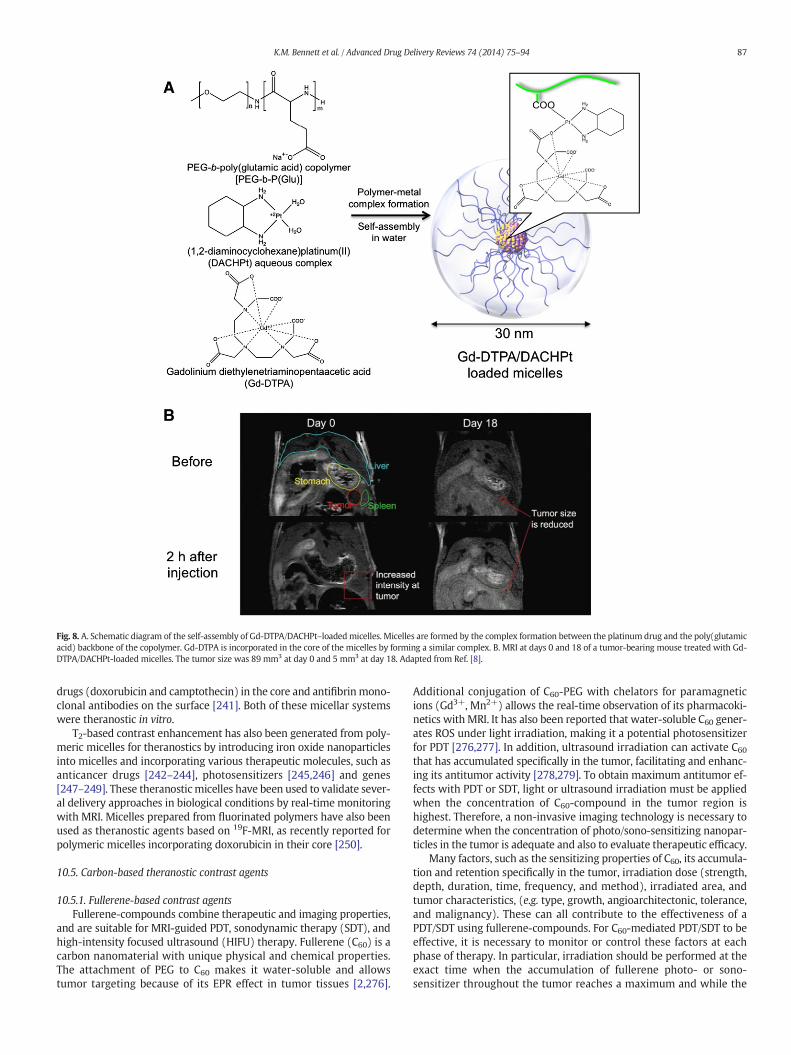
Fig. 8. A. Schematic diagram of the self-assembly of Gd-DTPA/DACHPt–loadedmicelles. Micelles are formed by the complex formation between the platinum drug and the poly(glutamicacid) backbone of the copolymer. Gd-DTPA is incorporated in the core of the micelles by forming a similar complex. B. MRI at days 0 and 18 of a tumor-bearing mouse treated with Gd-DTPA/DACHPt-loaded micelles. The tumor size was 89 mm3 at day 0 and 5 mm3 at day 18. Adapted from Ref. [8].
87K.M. Bennett et al. / Advanced Drug Delivery Reviews 74 (2014) 75–94
drugs (doxorubicin and camptothecin) in the core and antifibrinmono-clonal antibodies on the surface [241]. Both of these micellar systemswere theranostic in vitro.
T2-based contrast enhancement has also been generated from poly-meric micelles for theranostics by introducing iron oxide nanoparticlesinto micelles and incorporating various therapeutic molecules, such asanticancer drugs [242–244], photosensitizers [245,246] and genes[247–249]. These theranosticmicelles have been used to validate sever-al delivery approaches in biological conditions by real-time monitoringwith MRI. Micelles prepared from fluorinated polymers have also beenused as theranostic agents based on 19F-MRI, as recently reported forpolymeric micelles incorporating doxorubicin in their core [250].
10.5. Carbon-based theranostic contrast agents
10.5.1. Fullerene-based contrast agentsFullerene-compounds combine therapeutic and imaging properties,
and are suitable for MRI-guided PDT, sonodynamic therapy (SDT), andhigh-intensity focused ultrasound (HIFU) therapy. Fullerene (C60) is acarbon nanomaterial with unique physical and chemical properties.The attachment of PEG to C60 makes it water-soluble and allowstumor targeting because of its EPR effect in tumor tissues [2,276].
Additional conjugation of C60-PEG with chelators for paramagneticions (Gd3+, Mn2+) allows the real-time observation of its pharmacoki-netics with MRI. It has also been reported that water-soluble C60 gener-ates ROS under light irradiation, making it a potential photosensitizerfor PDT [276,277]. In addition, ultrasound irradiation can activate C60that has accumulated specifically in the tumor, facilitating and enhanc-ing its antitumor activity [278,279]. To obtain maximum antitumor ef-fects with PDT or SDT, light or ultrasound irradiation must be appliedwhen the concentration of C60-compound in the tumor region ishighest. Therefore, a non-invasive imaging technology is necessary todetermine when the concentration of photo/sono-sensitizing nanopar-ticles in the tumor is adequate and also to evaluate therapeutic efficacy.
Many factors, such as the sensitizing properties of C60, its accumula-tion and retention specifically in the tumor, irradiation dose (strength,depth, duration, time, frequency, and method), irradiated area, andtumor characteristics, (e.g. type, growth, angioarchitectonic, tolerance,and malignancy). These can all contribute to the effectiveness of aPDT/SDT using fullerene-compounds. For C60-mediated PDT/SDT to beeffective, it is necessary to monitor or control these factors at eachphase of therapy. In particular, irradiation should be performed at theexact time when the accumulation of fullerene photo- or sono-sensitizer throughout the tumor reaches a maximum and while the
88 K.M. Bennett et al. / Advanced Drug Delivery Reviews 74 (2014) 75–94
concentration in surrounding normal tissue is minimized. In publishedPDT/SDT reports where image guidance was not available, the durationof light or ultrasound irradiation has been arbitrarily decided [276,277,280]. In image-guided studies, it is possible to select the duration ofthe irradiation and to apply it when the concentration of photo- orsono-sensitizer in the target tissue is maximal. For example, C60-Mn-PEG and C60-Gd-PEG nanoclusters have enabled image-guided PDT orSDT with non-invasive real-time monitoring of its accumulation in atumor and surrounding normal tissue [19,276]. They have also allowedoptimization of the irradiation duration and area. Dynamic T2- anddiffusion-weighted MRI have also provided real-time informationabout edematous tissue damage, which is crucial to optimize the radia-tion dose. Dynamic image-guided PDT and SDT are not only useful formaximizing the therapeutic effects, but also for the development ofnew photo- and sono-sensitizers that are highly efficient generators ofROS.
To obtain better contrast enhancement in MRI, fullerene derivativesbeen developed with paramagnetic ions in the interior space of the ful-lerene [18,281–284]. However, there is a tradeoff in these structures be-tween the high relaxivity of the paramagnetic fullerene and its ability togenerate ROS. This depends on the synthesis procedure. It is importantto regulate the number of ligands and their orientation on the fullerenesurface. Surface modification of the fullerene could result in a stericscreen of the carbon atoms, which can suppress its ability to be excitedby ultrasound (via a sonoluminescence-dependentmechanism) or laserlight.
Despite these limitations, fullerene-based multifunctional contrastagents have many advantages over the widely used organic sono-sensitizers such as porphyrin derivatives [19,285]. The porphyrins fluo-rescence possesses relatively low quantum yield. The pharmacodynam-ics of porphyrins can be detected only by fluorescent imaging, which israther limited for non-invasive deep-tissue visualization. It is thereforeextremely difficult to optimize porphyrin tumor accumulation, antitu-mor effects, and side-effects in vivo [286]. The photo- and sono-sensitizers based on paramagnetic C60-compounds allow visualizationof their pharmacodynamics and optimization of their tumor accumula-tion at the exact moment of light irradiation or ultrasonication. More-over, T2- and diffusion-weighted MRI can provide information aboutthe status of the tumor tissue before and after PDT or SDT with C60-compounds.
10.5.2. Carbon nanotube- and graphene-based contrast agentCarbon nanotube (CNT)- and graphene-based compounds have also
attracted interest because of their possible application in image-guideddrug delivery using optical imaging, MRI, ultrasonography, PET, etc.CNTs and graphene are composed of pure carbon, (similar to fuller-enes), with atoms arranged cylindrically or in a regular linear hexagonalpattern, respectively. These structures possess unique electrical, optical,and mechanical properties because of their extremely high aspect ratio(length/diameter ratio). They are excellent scaffolds for the develop-ment of multimodal contrast agents with therapeutic potential.
The excellent electrical properties of CNT and graphene also allowthem to be extensively used in a wide range of biosensing platforms[287,288]. Single-walled carbon nanotubes, (SWCNTs; with a typicaldiameter of 1 nm and length of 100 to 400 nm), and graphene with allcarbon atoms exposed on their surfaces, exhibit ultra-high surfacearea available for efficient drug loading and bioconjugation fortheranostics [289–294]. Moreover, CNTs and graphene can penetratecell membranes without the toxic effects [295] seen with QDs.
CNTs have been used as optical imaging contrast agents as wellas PDT transducers for thermal ablation of cancer cells due to their effi-cient optical absorption and fluorescence in the NIR spectrum[296–298]. CNTs have also been used as T2-shortening MRI contrastagents [299]. CNTs can be filled or coated with other contrast agents,(e.g., fluorophores and paramagnetic ions), to increase optical and MRIcontrast [298–300]. Thus, CNTs may serve as multimodal contrast
probes for simultaneous use in fluorescence imaging, photothermaloptical coherent tomography, and MRI. CNTs also show promise foruse in ultrasonography. The size of the majority microbubble-basedultrasound contrast agents (1–8 μm)makes them primarily intravascu-lar flow tracers. In contrast, the chemico-physical characteristics ofCNTs (e.g.100–400 nm in length),make thempotentially able to extrav-asate and reach a target area through vascular pore sizes up to 780 nm[289,301].
Ultrasound contrast agents are classified as long half-life agentswhen the signal persists for more than 5 min after an intravenousbolus injection [302]. Most ultrasound contrast agents are eliminatedwithin a few minutes through the lungs. The blood clearance of func-tionalized CNTs in mice is about 3 h post-injection [303]. This suggeststhe possibility of eventually using CNTs in the long ultrasonographyinvestigations associated with therapy. CNTs can also contain therapeu-tic agents [304,305]. A few studies have proposed a combinationof microbubble ultrasound contrast agents with a therapeuticload or with targeting compounds [306,307]. Research focusing onmicrobubbles as DDS and target-specific compounds is at an earlystage of development as compared to research on using CNTs astarget-specific DDS for image-guided therapy [289,308,309].
SWCNTs also exhibit high-resonance Raman scattering because oftheir sharp electronic densities of state. This characteristic has beenexploited for in vitro and ex vivo imaging [310]. CNTs have also beenused as multicolor contrast agents for multiplexed Raman imaging[311]. SWCNTs conjugated with targeting peptides may also be usedin photoacoustic imaging [312,313]. Photoacoustic imaging is currentlyamajor focus of scientific research, but it has also begun to be applied inthe clinic [314].
Finally, CNTs and graphene are relatively safe in composition ascompared to other inorganic nanomaterials such as QDs, which usuallycontain heavy metals, CNTs' safety and circulation dynamics have beendemonstrated for in vivo animal studies, and they may be more readilytranslated than other inorganic nanoparticles [315,316].
11. Conclusion
Non-invasive, high-resolution in vivo imaging of tumors plays a keyrole in the development of techniques for nano-pathophysiological im-aging and theranostics in vivo. The incorporation of nano-DDSs bearingMR contrast agents can enable visualization of a drug's distribution andits therapeutic effects. The imaging of nano-DDS will provide informa-tion for alternative options in caseswhere the delivered drug concentra-tion or the therapeutic effect is found to be insufficient. Even in the sametype of cells, diseases such as tumors have a heterogeneous microenvi-ronment that changes depending on staging, treatment, and immuneresponse. Thus, nano-DDSs should be tailored to the specific diseasemi-croenvironment. Fortunately, nano-DDSs form a wide range of mate-rials, sizes, and functional properties. For example, nanomicelles aresuitable for delivering a wide range of markers and therapeutics agents.Several therapeutic nanomicelles incorporating hydrophobic drugs areunder clinical trials. Moreover, in addition to FDA-approved PEGylatedliposomes, several types of biocompatible nano-materials, such asnano-gels [317,318], are showing promise for clinical applications. Inconclusion, Nano-DDSs is a flexible, growing technology that will bebroadly important for clinical diagnostics and therapy in the future.
Acknowledgments
The authors would like to thankMs. Aiko Sekita, Ms. Sayaka Shibata,Mr. Nobuhiro Nitta, and Mr. Yoshikazu Ozawa (National Institute of Ra-diological Sciences, Japan; NIRS) for imaging, illustration and editing.The authors also would like to thank Dr. Tsuneo Saga (NIRS), Dr. KenjiKono (Osaka Prefecture University), Dr. Akihiro Kishimura (KyushuUniversity), Akihiro Tomita (Nagoya University) and Mitsunobu RKano (OkayamaUniversity) for nice discussion and providingmaterials.
89K.M. Bennett et al. / Advanced Drug Delivery Reviews 74 (2014) 75–94
This work was partially supported by Grants-in-Aid for Scientific Re-search (Kakenhi 24300167, JSPS), grant for World-Leading InnovativeR&D on Science and Technology (FIRST Program, Cabinet Office) andthe Center of Innovation Program (JST).
References
[1] G.A. Johnson, E. Calabrese, A. Badea, G. Paxinos, C. Watson, A multidimensionalmagnetic resonance histology atlas of the Wistar rat brain, Neuroimage 62(2012) 1848–1856.
[2] Y. Matsumura, H. Maeda, A new concept for macromolecular therapeutics in can-cer chemotherapy: mechanism of tumoritropic accumulation of proteins and theantitumor agent smancs, Cancer Res. 46 (1986) 6387–6392.
[3] D. Papahadjopoulos, T.M. Allen, A. Gabizon, E. Mayhew, K. Matthay, S.K. Huang, K.D.Lee, M.C. Woodle, D.D. Lasic, C. Redemann, et al., Sterically stabilized liposomes: im-provements in pharmacokinetics and antitumor therapeutic efficacy, Proc. Natl.Acad. Sci. U. S. A. 88 (1991) 11460–11464.
[4] M.R. Kano, Y. Bae, C. Iwata, Y. Morishita, M. Yashiro, M. Oka, T. Fujii, A. Komuro, K.Kiyono, M. Kaminishi, K. Hirakawa, Y. Ouchi, N. Nishiyama, K. Kataoka, K.Miyazono, Improvement of cancer-targeting therapy, using nanocarriers for intrac-table solid tumors by inhibition of TGF-beta signaling, Proc. Natl. Acad. Sci. U. S. A.104 (2007) 3460–3465.
[5] H. Cabral, N. Nishiyama, K. Kataoka, Supramolecular nanodevices: fromdesign validation to theranostic nanomedicine, Acc. Chem. Res. 44 (2011)999–1008.
[6] A. Bumb, M.W. Brechbiel, P. Choyke, Macromolecular and dendrimer-based mag-netic resonance contrast agents, Acta Radiol. 51 (2010) 751–767.
[7] K. Kataoka, A. Harada, Y. Nagasaki, Block copolymer micelles for drug delivery:design, characterization and biological significance, Adv. Drug Deliv. Rev. 47(2001) 113–131.
[8] S. Kaida, H. Cabral, M. Kumagai, A. Kishimura, Y. Terada, M. Sekino, I. Aoki, N.Nishiyama, T. Tani, K. Kataoka, Visible drug delivery by supramolecularnanocarriers directing to single-platformed diagnosis and therapy of pancreatictumor model, Cancer Res. 70 (2010) 7031–7041.
[9] G.W. Kabalka, M.A. Davis, T.H. Moss, E. Buonocore, K. Hubner, E. Holmberg, K.Maruyama, L. Huang, Gadolinium-labeled liposomes containing various amphi-philic Gd-DTPA derivatives: targeted MRI contrast enhancement agents for theliver, Magn. Reson. Med. 19 (1991) 406–415.
[10] A.M. Ponce, B.L. Viglianti, D. Yu, P.S. Yarmolenko, C.R. Michelich, J. Woo, M.B. Bally,M.W. Dewhirst, Magnetic resonance imaging of temperature-sensitive liposomerelease: drug dose painting and antitumor effects, J. Natl. Cancer Inst. 99 (2007)53–63.
[11] K. Kono, S. Nakashima, D. Kokuryo, I. Aoki, H. Shimomoto, S. Aoshima, K.Maruyama, E. Yuba, C. Kojima, A. Harada, Y. Ishizaka, Multi-functional liposomeshaving temperature-triggered release and magnetic resonance imaging fortumor-specific chemotherapy, Biomaterials 32 (2011) 1387–1395.
[12] W.J. Mulder, G.J. Strijkers, G.A. van Tilborg, A.W. Griffioen, K. Nicolay, Lipid-basednanoparticles for contrast-enhanced MRI and molecular imaging, NMR Biomed.19 (2006) 142–164.
[13] A.L. Doiron, K.A. Homan, S. Emelianov, L. Brannon-Peppas, Poly(lactic-co-glycolic)acid as a carrier for imaging contrast agents, Pharm. Res. 26 (2009) 674–682.
[14] L.D. Margerum, B.K. Campion, M. Koo, N. Shargill, J.J. Lai, A. Marumoto, P.C.Sontum, Gadolinium(III) DO3A macrocycles and polyethylene glycol coupledto dendrimers—effect of molecular weight on physical and biological proper-ties of macromolecular magnetic resonance imaging contrast agents, J. AlloysCompd. 249 (1997) 185–190.
[15] H. Kobayashi, S. Kawamoto, T. Saga, N. Sato, A. Hiraga, T. Ishimori, Y. Akita, M.H.Mamede, J. Konishi, K. Togashi, M.W. Brechbiel, Novel liver macromolecular MRcontrast agent with a polypropylenimine diaminobutyl dendrimer core: compari-son to the vascular MR contrast agent with the polyamidoamine dendrimer core,Magn. Reson. Med. 46 (2001) 795–802.
[16] I.L. Medintz, A.R. Clapp, H. Mattoussi, E.R. Goldman, B. Fisher, J.M. Mauro, Self-assembled nanoscale biosensors based on quantum dot FRET donors, Nat. Mater.2 (2003) 630–638.
[17] R. Bakalova, Z. Zhelev, I. Aoki, H. Ohba, Y. Imai, I. Kanno, Silica-shelled single quan-tum dot micelles as imaging probes with dual or multimodality, Anal. Chem. 78(2006) 5925–5932.
[18] M. Mikawa, H. Kato, M. Okumura, M. Narazaki, Y. Kanazawa, N. Miwa, H.Shinohara, Paramagnetic water-soluble metallofullerenes having the highestrelaxivity for MRI contrast agents, Bioconjug. Chem. 12 (2001) 510–514.
[19] J. Liu, S. Ohta, A. Sonoda, M. Yamada, M. Yamamoto, N. Nitta, K. Murata, Y. Tabata,Preparation of PEG-conjugated fullerene containing Gd3+ ions for photodynamictherapy, J. Control. Release 117 (2007) 104–110.
[20] J.W. Bulte, Y. Hoekstra, R.L. Kamman, R.L. Magin, A.G. Webb, R.W. Briggs, K.G.Go, C.E. Hulstaert, S. Miltenyi, T.H. The, et al., Specific MR imaging of humanlymphocytes by monoclonal antibody-guided dextran-magnetite particles,Magn. Reson. Med. 25 (1992) 148–157.
[21] T.C. Yeh, W. Zhang, S.T. Ildstad, C. Ho, In vivo dynamic MRI tracking of rat T-cellslabeled with superparamagnetic iron-oxide particles, Magn. Reson. Med. 33(1995) 200–208.
[22] R. Weissleder, G. Elizondo, L. Josephson, C.C. Compton, C.J. Fretz, D.D. Stark, J.T.Ferrucci, Experimental lymph node metastases: enhanced detection with MR lym-phography, Radiology 171 (1989) 835–839.
[23] P. Reimer, R.Weissleder, A.S. Lee, J.Wittenberg, T.J. Brady, Receptor imaging: appli-cation to MR imaging of liver cancer, Radiology 177 (1990) 729–734.
[24] A. Viale, F. Reineri, D. Santelia, E. Cerutti, S. Ellena, R. Gobetto, S. Aime,Hyperpolarized agents for advancedMRI investigations, Q. J. Nucl. Med. Mol. Imag-ing 53 (2009) 604–617.
[25] F.A. Gallagher, M.I. Kettunen, S.E. Day, D.E. Hu, J.H. Ardenkjaer-Larsen, R. Zandt, P.R.Jensen, M. Karlsson, K. Golman, M.H. Lerche, K.M. Brindle, Magnetic resonance im-aging of pH in vivo using hyperpolarized 13C-labelled bicarbonate, Nature 453(2008) 940–943.
[26] M.E. Merritt, C. Harrison, C. Storey, F.M. Jeffrey, A.D. Sherry, C.R. Malloy,Hyperpolarized 13C allows a direct measure of flux through a singleenzyme-catalyzed step by NMR, Proc. Natl. Acad. Sci. U. S. A. 104 (2007)19773–19777.
[27] M.E. Merritt, C. Harrison, Z. Kovacs, P. Kshirsagar, C.R. Malloy, A.D. Sherry,Hyperpolarized (89)Y offers the potential of direct imaging of metal ions inbiological systems by magnetic resonance, J. Am. Chem. Soc. 129 (2007)12942–12943.
[28] T.M. Atkins, M.C. Cassidy, M. Lee, S. Ganguly, C.M. Marcus, S.M. Kauzlarich, Synthe-sis of long T(1) silicon nanoparticles for hyperpolarized (2)(9)Si magnetic reso-nance imaging, ACS Nano 7 (2013) 1609–1617.
[29] M.C. Cassidy, H.R. Chan, B.D. Ross, P.K. Bhattacharya, C.M. Marcus, In vivo magneticresonance imaging of hyperpolarized silicon particles, Nat. Nanotechnol. 8 (2013)363–368.
[30] T. Kim, E. Momin, J. Choi, K. Yuan, H. Zaidi, J. Kim, M. Park, N. Lee, M.T. McMahon, A.Quinones-Hinojosa, J.W. Bulte, T. Hyeon, A.A. Gilad, Mesoporous silica-coated hol-low manganese oxide nanoparticles as positive T1 contrast agents for labeling andMRI tracking of adipose-derived mesenchymal stem cells, J. Am. Chem. Soc. 133(2011) 2955–2961.
[31] W.A. Braunecker, K. Matyjaszewski, Controlled/living radical polymerization: fea-tures, developments, and perspectives, Prog. Polym. Sci. 32 (2007) 93–146.
[32] E. Battistini, E. Gianolio, R. Gref, P. Couvreur, S. Fuzerova, M. Othman, S. Aime, B.Badet, P. Durand, High-relaxivity magnetic resonance imaging (MRI) contrastagent based on supramolecular assembly between a gadolinium chelate, amodified dextran, and poly-beta-cyclodextrin, Chemistry 14 (2008)4551–4561.
[33] M. Mikawa, N. Miwa, M. Brautigam, T. Akaike, A. Maruyama, Gd(3+)-loadedpolyion complex for pH depiction with magnetic resonance imaging, J. Biomed.Mater. Res. 49 (2000) 390–395.
[34] K. Luo, J. Tian, G. Liu, J. Sun, C. Xia, H. Tang, L. Lin, T. Miao, X. Zhao, F. Gao, Q. Gong, B.Song, X. Shuai, H. Ai, Z. Gu, Self-assembly of SiO2/Gd-DTPA-polyethyleniminenanocomposites as magnetic resonance imaging probes, J. Nanosci. Nanotechnol.10 (2010) 540–548.
[35] A.J. Villaraza, A. Bumb, M.W. Brechbiel, Macromolecules, dendrimers, andnanomaterials in magnetic resonance imaging: the interplay between size, func-tion, and pharmacokinetics, Chem. Rev. 110 (2010) 2921–2959.
[36] T. Peters Jr., Serum albumin, Adv. Clin. Chem. 13 (1970) 37–111.[37] M.E. Moseley, W.M. Chew, D.L. White, J. Kucharczyk, L. Litt, N. Derugin, J.
Dupon, R.C. Brasch, D. Norman, Hypercarbia-induced changes in cerebralblood volume in the cat: a 1H MRI and intravascular contrast agent study,Magn. Reson. Med. 23 (1992) 21–30.
[38] M.G.Wikstrom, M.E. Moseley, D.L. White, J.W. Dupon, J.L. Winkelhake, J. Kopplin, R.C.Brasch, Contrast-enhanced MRI of tumors. Comparison of Gd-DTPA and a macromo-lecular agent, Invest. Radiol. 24 (1989) 609–615.
[39] C.F. van Dijke, B.A. Kirk, C.G. Peterfy, H.K. Genant, R.C. Brasch, S. Kapila, Arthritictemporomandibular joint: correlation of macromolecular contrast-enhancedMR imaging parameters and histopathologic findings, Radiology 204 (1997)825–832.
[40] H. Daldrup, D.M. Shames, M.Wendland, Y. Okuhata, T.M. Link, W. Rosenau, Y. Lu, R.C.Brasch, Correlation of dynamic contrast-enhanced MR imaging with histologic tumorgrade: comparison ofmacromolecular and small-molecular contrastmedia, AJR Am. J.Roentgenol. 171 (1998) 941–949.
[41] Z. Zhang, S.A. Nair, T.J. McMurry, Gadoliniummeets medicinal chemistry: MRI con-trast agent development, Curr. Med. Chem. 12 (2005) 751–778.
[42] A.B. Baxter, S.C. Lazarus, R.C. Brasch, In vitro histamine release induced bymagneticresonance imaging and iodinated contrast media, Invest. Radiol. 28 (1993)308–312.
[43] R.N. Muller, B. Raduchel, S. Laurent, J. Platzek, C. Pierart, P. Mareski, L. Vander Elst,Physicochemical characterization of MS-325, a new gadolinium complex, by mul-tinuclear relaxometry, Eur. J. Inorg. Chem. (1999) 1949–1955.
[44] G.T. Hermanson, Bioconjugate Techniques, Academic Press, 1996.[45] D.J. Hnatowich, W.W. Layne, R.L. Childs, D. Lanteigne, M.A. Davis, T.W. Griffin, P.W.
Doherty, Radioactive labeling of antibody: a simple and efficient method, Science220 (1983) 613–615.
[46] P. Shreve, A.M. Aisen, Monoclonal antibodies labeled with polymeric paramagneticion chelates, Magn. Reson. Med. 3 (1986) 336–340.
[47] M.A. Slinkin, C. Curtet, A. Faivre-Chauvet, C. Sai-Maurel, J.F. Gestin, V.P. Torchilin, J.F.Chatal, Biodistribution of anti-CEA F(ab′)2 fragments conjugated with chelating poly-mers: influence of conjugate electron charge on tumor uptake and blood clearance,Nucl. Med. Biol. 20 (1993) 443–452.
[48] Y. Manabe, C. Longley, P. Furmanski, High-level conjugation of chelatingagents onto immunoglobulins: use of an intermediary poly(L-lysine)-diethylenetriaminepentaacetic acid carrier, Biochim. Biophys. Acta 883(1986) 460–467.
[49] E.D. Bernard, M.A. Beking, K. Rajamanickam, E.C. Tsai, M.C. Derosa, Target bindingimproves relaxivity in aptamer–gadolinium conjugates, J. Biol. Inorg. Chem. 17(2012) 1159–1175.
90 K.M. Bennett et al. / Advanced Drug Delivery Reviews 74 (2014) 75–94
[50] W. Xu, Y. Lu, A smart magnetic resonance imaging contrast agent responsive toadenosine based on a DNA aptamer-conjugated gadolinium complex, Chem.Commun. (Camb.) 47 (2011) 4998–5000.
[51] T.H. Lim, D.H. Lee, Y.H. Kim, S.W. Park, P.H. Park, D.M. Seo, S.T. Kim, T.K. Lee, C.W.Mun, Occlusive and reperfused myocardial infarction: detection by usingMR imaging with gadolinium polylysine enhancement, Radiology 189(1993) 765–768.
[52] Y. Berthezene, V. Vexler, R. Kuwatsuru, W. Rosenau, A. Muhler, O. Clement, D.C.Price, R.C. Brasch, Differentiation of alveolitis and pulmonary fibrosis with a mac-romolecular MR imaging contrast agent, Radiology 185 (1992) 97–103.
[53] L.R. Opsahl, E.E. Uzgiris, D.R. Vera, Tumor imaging with a macromolecular para-magnetic contrast agent: gadopentetate dimeglumine–polylysine, Acad. Radiol. 2(1995) 762–767.
[54] T. Yamaoka, Y. Tabata, Y. Ikada, Body distribution profile of polysaccharide after in-travenous administration, Drug Deliv. 1 (1993).
[55] Y. Kaneo, T. Tanaka, T. Nakano, Y. Yamaguchi, Evidence for receptor-mediated he-patic uptake of pullulan in rats, J. Control. Release 70 (2001) 365–373.
[56] C.K. Tang, J. Lodding, G. Minigo, D.S. Pouniotis, M. Plebanski, A. Scholzen, I.F.McKenzie, G.A. Pietersz, V. Apostolopoulos, Mannan-mediated gene delivery forcancer immunotherapy, Immunology 120 (2007) 325–335.
[57] A.K. Yadav, P. Mishra, G.P. Agrawal, An insight on hyaluronic acid in drug targetingand drug delivery, J. Drug Target. 16 (2008) 91–107.
[58] U. Klotz, H. Kroemer, Clinical pharmacokinetic considerations in the use of plasmaexpanders, Clin. Pharmacokinet. 12 (1987) 123–135.
[59] W.A. Gibby, A. Bogdan, T.W. Ovitt, Cross-linked DTPA polysaccharides for magneticresonance imaging. Synthesis and relaxation properties, Invest. Radiol. 24 (1989)302–309.
[60] P. Rongved, J. Klaveness, Water-soluble polysaccharides as carriers of paramagnet-ic contrast agents for magnetic resonance imaging: synthesis and relaxation prop-erties, Carbohydr. Res. 214 (1991) 315–323.
[61] P. Loubeyre, E. Canet, S. Zhao, S. Benderbous, M. Amiel, D. Revel, Carboxymethyl-dextran-gadolinium-DTPA as a blood-pool contrast agent for magnetic resonanceangiography. Experimental study in rabbits, Invest. Radiol. 31 (1996) 288–293.
[62] R.E. Jacobs, S.E. Fraser, Magnetic resonance microscopy of embryonic cell lineagesand movements, Science 263 (1994) 681–684.
[63] M. Wikstrom, H.J. Martinussen, G. Wikstrom, A. Ericsson, R. Nyman, A.Waldenstrom, A. Hemmingsson, MR imaging of acute myocardial infarction inpigs using Gd-DTPA-labeled dextran, Acta Radiol. 33 (1992) 301–308.
[64] C. Casali, M. Janier, E. Canet, J.F. Obadia, S. Benderbous, C. Corot, D. Revel, Evaluationof Gd-DOTA-labeled dextran polymer as an intravascular MR contrast agent formyocardial perfusion, Acad. Radiol. 5 (Suppl. 1) (1998) S214–S218.
[65] Y. Liu, N. Zhang, Gadolinium loaded nanoparticles in theranostic magnetic reso-nance imaging, Biomaterials 33 (2012) 5363–5375.
[66] P. Lebduskova, J. Kotek, P. Hermann, L. Vander Elst, R.N. Muller, I. Lukes, J.A. Peters,A gadolinium(III) complex of a carboxylic-phosphorus acid derivative ofdiethylenetriamine covalently bound to inulin, a potential macromolecular MRIcontrast agent, Bioconjug. Chem. 15 (2004) 881–889.
[67] T.H. Helbich, A. Gossman, P.A. Mareski, B. Raduchel, T.P. Roberts, D.M. Shames, M.Muhler, K. Turetschek, R.C. Brasch, A new polysaccharide macromolecular contrastagent for MR imaging: biodistribution and imaging characteristics, J. Magn. Reson.Imaging 11 (2000) 694–701.
[68] F. Cavagna, C. Luchinat, A. Scozzafava, Z. Xia, Polymetallic macromolecules are po-tential contrast agents of improved efficiency, Magn. Reson. Med. 31 (1994) 58–60.
[69] M.J. Allen, R.T. Raines, L.L. Kiessling, Contrast agents for magnetic resonance imag-ing synthesized with ring-opening metathesis polymerization, J. Am. Chem. Soc.128 (2006) 6534–6535.
[70] Y. Wu, Y. Zhou, O. Ouari, M. Woods, P. Zhao, T.C. Soesbe, G.E. Kiefer, A.D. Sherry,Polymeric PARACEST agents for enhancing MRI contrast sensitivity, J. Am. Chem.Soc. 130 (2008) 13854–13855.
[71] S. Langereis, Q.G. de Lussanet, M.H.P. van Genderen,W.H. Backes, E.W. Meijer, Mul-tivalent contrast agents based on gadolinium-diethylenetriaminepentaacetic acid-terminated poly(propylene imine) dendrimers for magnetic resonance imaging,Macromolecules 37 (2004) 3084–3091.
[72] N. Sato, H. Kobayashi, A. Hiraga, T. Saga, K. Togashi, J. Konishi, M.W. Brechbiel, Phar-macokinetics and enhancement patterns of macromolecular MR contrast agentswith various sizes of polyamidoamine dendrimer cores, Magn. Reson. Med. 46(2001) 1169–1173.
[73] H. Kobayashi, N. Sato, S. Kawamoto, T. Saga, A. Hiraga, T.L. Haque, T. Ishimori, J.Konishi, K. Togashi, M.W. Brechbiel, Comparison of the macromolecular MR con-trast agents with ethylenediamine-core versus ammonia-core generation-6polyamidoamine dendrimer, Bioconjug. Chem. 12 (2001) 100–107.
[74] Z. Huang, R.S. Sengar, A. Nigam, M.C. Abadjian, D.M. Potter, D.B. Grotjahn, E.C.Wiener, A fluorinated dendrimer-based nanotechnology platform: new contrastagents for high field imaging, Invest. Radiol. 45 (2010) 641–654.
[75] K.J. Thurecht, I. Blakey, H. Peng, O. Squires, S. Hsu, C. Alexander, A.K. Whittaker,Functional hyperbranched polymers: toward targeted in vivo 19F magnetic reso-nance imaging using designed macromolecules, J. Am. Chem. Soc. 132 (2010)5336–5337.
[76] M. Tan, Z. Ye, E.K. Jeong, X. Wu, D.L. Parker, Z.R. Lu, Synthesis and evaluation ofnanoglobular macrocyclic Mn(II) chelate conjugates as non-gadolinium(III) MRIcontrast agents, Bioconjug. Chem. 22 (2011) 931–937.
[77] Y. Wu, M. Evbuomwan, M. Melendez, A. Opina, A.D. Sherry, Advantages of macro-molecular to nanosized chemical-exchange saturation transfer agents for MRI ap-plications, Future Med. Chem. 2 (2010) 351–366.
[78] J.A. Pikkemaat, R.T. Wegh, R. Lamerichs, R.A. van de Molengraaf, S. Langereis, D.Burdinski, A.Y. Raymond, H.M. Janssen, B.F. de Waal, N.P. Willard, E.W. Meijer, H.
Grull, Dendritic PARACEST contrast agents for magnetic resonance imaging, Con-trast Media Mol. Imaging 2 (2007) 229–239.
[79] M.M. Ali, B. Yoo, M.D. Pagel, Tracking the relative in vivo pharmacokinetics ofnanoparticles with PARACEST MRI, Mol. Pharm. 6 (2009) 1409–1416.
[80] B. Zarabi, A. Nan, J. Zhuo, R. Gullapalli, H. Ghandehari, Macrophage targeted N-(2-hydroxypropyl)methacrylamide conjugates for magnetic resonance imaging, Mol.Pharm. 3 (2006) 550–557.
[81] C.A. Boswell, P.K. Eck, C.A. Regino, M. Bernardo, K.J.Wong, D.E. Milenic, P.L. Choyke,M.W. Brechbiel, Synthesis, characterization, and biological evaluation of integrinalphavbeta3-targeted PAMAM dendrimers, Mol. Pharm. 5 (2008) 527–539.
[82] D.B. Pike, H. Ghandehari, HPMA copolymer-cyclic RGD conjugates for tumortargeting, Adv. Drug Deliv. Rev. 62 (2010) 167–183.
[83] L. Han, J. Li, S. Huang, R. Huang, S. Liu, X. Hu, P. Yi, D. Shan, X. Wang, H. Lei, C. Jiang,Peptide-conjugated polyamidoamine dendrimer as a nanoscale tumor-targeted T1magnetic resonance imaging contrast agent, Biomaterials 32 (2011) 2989–2998.
[84] K. Luo, G. Liu, B. He, Y. Wu, Q. Gong, B. Song, H. Ai, Z. Gu, Multifunctionalgadolinium-based dendritic macromolecules as liver targeting imaging probes,Biomaterials 32 (2011) 2575–2585.
[85] I. Pashkunova-Martic, C. Kremser, M. Galanski, P. Schluga, V. Arion, P. Debbage, W.Jaschke, B. Keppler, Lectin conjugates as biospecific contrast agents for MRI. Cou-pling of Lycopersicon esculentum agglutinin to linear water-soluble DTPA-loadedoligomers, Mol. Imaging Biol. 13 (2011) 432–442.
[86] C.S. Winalski, S. Shortkroff, E. Schneider, H. Yoshioka, R.V. Mulkern, G.M. Rosen,Targeted dendrimer-based contrast agents for articular cartilage assessment byMR imaging, Osteoarthritis Cartilage 16 (2008) 815–822.
[87] J. Jo, X. Lin, T. Nakahara, I. Aoki, T. Saga, Y. Tabata, Preparation of polymer-basedmagnetic resonance imaging contrast agent to visualize therapeutic angiogenesis,Tissue Eng. Part A 19 (2013) 30–39.
[88] A.M. Mohs, X. Wang, K.C. Goodrich, Y. Zong, D.L. Parker, Z.R. Lu, PEG-g-poly(GdDTPA-co-L-cystine): a biodegradable macromolecular blood pool contrastagent for MR imaging, Bioconjug. Chem. 15 (2004) 1424–1430.
[89] Z.R. Lu, X. Wang, D.L. Parker, K.C. Goodrich, H.R. Buswell, Poly(l-glutamic acid)Gd(III)-DOTA conjugate with a degradable spacer for magnetic resonance imaging,Bioconjug. Chem. 14 (2003) 715–719.
[90] C.H. Huang, K. Nwe, A. Al Zaki, M.W. Brechbiel, A. Tsourkas, Biodegradablepolydisulfide dendrimer nanoclusters as MRI contrast agents, ACS Nano 6 (2012)9416–9424.
[91] M. Ye, Y. Qian, J. Tang, H. Hu, M. Sui, Y. Shen, Targeted biodegradable dendritic MRIcontrast agent for enhanced tumor imaging, J. Control. Release 169 (2013) 239–245.
[92] M.M. Ali, M. Woods, P. Caravan, A.C. Opina, M. Spiller, J.C. Fettinger, A.D. Sherry,Synthesis and relaxometric studies of a dendrimer-based pH-responsive MRI con-trast agent, Chemistry 14 (2008) 7250–7258.
[93] S. Okada, S. Mizukami, K. Kikuchi, Switchable MRI contrast agents based on mor-phological changes of pH-responsive polymers, Bioorg. Med. Chem. 20 (2012)769–774.
[94] S. Ju, Y. Qiu, C. Li, G.J. Teng, Y. Ni, Multimodality imaging of endothelial progenitorcells with a novel multifunctional probe featuring positive magnetic resonancecontrast and near-infrared fluorescence, Mol. Imaging 10 (2011) 359–369.
[95] S. Wen, K. Li, H. Cai, Q. Chen, M. Shen, Y. Huang, C. Peng, W. Hou, M. Zhu, G. Zhang, X.Shi, Multifunctional dendrimer-entrapped gold nanoparticles for dual mode CT/MRimaging applications, Biomaterials 34 (2013) 1570–1580.
[96] B. Zarabi, A. Nan, J. Zhuo, R. Gullapalli, H. Ghandehari, HPMA copolymer–doxorubi-cin–gadolinium conjugates: synthesis, characterization, and in vitro evaluation,Macromol. Biosci. 8 (2008) 741–748.
[97] J.M. Bryson, K.M. Fichter, W.J. Chu, J.H. Lee, J. Li, L.A. Madsen, P.M. McLendon, T.M.Reineke, Polymer beacons for luminescence and magnetic resonance imaging ofDNA delivery, Proc. Natl. Acad. Sci. U. S. A. 106 (2009) 16913–16918.
[98] A. Gianella, P.A. Jarzyna, V. Mani, S. Ramachandran, C. Calcagno, J. Tang, B. Kann, W.J.Dijk, V.L. Thijssen, A.W. Griffioen, G. Storm, Z.A. Fayad, W.J. Mulder, Multifunctionalnanoemulsion platform for imaging guided therapy evaluated in experimental can-cer, ACS Nano 5 (2011) 4422–4433.
[99] D. Yoo, J.H. Lee, T.H. Shin, J. Cheon, Theranostic magnetic nanoparticles, Acc. Chem.Res. 44 (2011) 863–874.
[100] D.P. Cormode, T. Skajaa, M.M. van Schooneveld, R. Koole, P. Jarzyna, M.E. Lobatto, C.Calcagno, A. Barazza, R.E. Gordon, P. Zanzonico, E.A. Fisher, Z.A. Fayad, W.J. Mulder,Nanocrystal core high-density lipoproteins: a multimodality contrast agent plat-form, Nano Lett. 8 (2008) 3715–3723.
[101] V.C. Jordan, V.D. Kodibagkar, S.C. Beeman, B.D. Hann, K.M. Bennett, Principles andemerging applications of nanomagnetic materials in medicine, Wiley Interdiscip.Rev. Nanomed. Nanobiotechnol. 4 (2012) 345–365.
[102] K.M. Krishnan, Biomedical nanomagnetics: a spin through possibilities in imaging,Diagn. Ther. IEEE Trans. Magn. 46 (2010) 2523–2558.
[103] C. Lam, Y.F. Zhang, Y.H. Tang, C.S. Lee, I. Bello, S.T. Lee, Large-scale synthesis of ul-trafine Si nanoparticles by ball milling, J. Cryst. Growth 220 (2000) 466–470.
[104] T. Hyeon, Chemical synthesis of magnetic nanoparticles, Chem. Commun. (Camb.)(2003) 927–934.
[105] C. Lang, D. Schuler, D. Faivre, Synthesis of magnetite nanoparticles for bio- andnanotechnology: genetic engineering and biomimetics of bacterial magnetosomes,Macromol. Biosci. 7 (2007) 144–151.
[106] X.M. Lin, A.C.S. Samia, Synthesis, assembly and physical properties of magneticnanoparticles, J. Magn. Magn. Mater. 305 (2006) 100–109.
[107] M.F. Bennewitz, T.L. Lobo, M.K. Nkansah, G. Ulas, G.W. Brudvig, E.M. Shapiro, Bio-compatible and pH-sensitive PLGA encapsulated MnO nanocrystals for molecularand cellular MRI, ACS Nano 5 (2011) 3438–3446.
[108] R.D. Engberink, S.M. van der Pol, P. Walczak, A. van der Toorn, M.A. Viergever, C.D.Dijkstra, J.W. Bulte, H.E. de Vries, E.L. Blezer, Magnetic resonance imaging of
91K.M. Bennett et al. / Advanced Drug Delivery Reviews 74 (2014) 75–94
monocytes labeledwith ultrasmall superparamagnetic particles of iron oxide usingmagnetoelectroporation in an animal model of multiple sclerosis, Mol. Imaging 9(2010) 268–277.
[109] A.A. Gilad, P. Walczak, M.T. McMahon, H.B. Na, J.H. Lee, K. An, T. Hyeon, P.C. van Zijl,J.W. Bulte, MR tracking of transplanted cells with “positive contrast” using manga-nese oxide nanoparticles, Magn. Reson. Med. 60 (2008) 1–7.
[110] G. Huang, H. Chen, Y. Dong, X. Luo, H. Yu, Z. Moore, E.A. Bey, D.A. Boothman, J. Gao,Superparamagnetic iron oxide nanoparticles: amplifying ROS stress to improve an-ticancer drug efficacy, Theranostics 3 (2013) 116–126.
[111] S. Laurent, J.L. Bridot, L.V. Elst, R.N. Muller, Magnetic iron oxide nanoparticles forbiomedical applications, Future Med. Chem. 2 (2010) 427–449.
[112] E.M. Shapiro, S. Skrtic, K. Sharer, J.M. Hill, C.E. Dunbar, A.P. Koretsky, MRI detectionof single particles for cellular imaging, Proc. Natl. Acad. Sci. U. S. A. 101 (2004)10901–10906.
[113] U.I. Tromsdorf, N.C. Bigall, M.G. Kaul, O.T. Bruns, M.S. Nikolic, B. Mollwitz, R.A.Sperling, R. Reimer, H. Hohenberg, W.J. Parak, S. Forster, U. Beisiegel, G. Adam, H.Weller, Size and surface effects on the MRI relaxivity of manganese ferrite nano-particle contrast agents, Nano Lett. 7 (2007) 2422–2427.
[114] M.V. Yigit, A. Moore, Z. Medarova, Magnetic nanoparticles for cancer diagnosis andtherapy, Pharm. Res. 29 (2012) 1180–1188.
[115] C. von zur Muhlen, D. von Elverfeldt, J.A. Moeller, R.P. Choudhury, D. Paul, C.E.Hagemeyer, M. Olschewski, A. Becker, I. Neudorfer, N. Bassler, M. Schwarz, C.Bode, K. Peter, Magnetic resonance imaging contrast agent targeted toward acti-vated platelets allows in vivo detection of thrombosis andmonitoring of thrombol-ysis, Circulation 118 (2008) 258–267.
[116] D. Granot, E.M. Shapiro, Release activation of iron oxide nanoparticles: (REACTION)a novel environmentally sensitive MRI paradigm, Magn. Reson. Med. 65 (2011)1253–1259.
[117] D.P. Cormode, P.A. Jarzyna, W.J. Mulder, Z.A. Fayad, Modified natural nanopar-ticles as contrast agents for medical imaging, Adv. Drug Deliv. Rev. 62 (2010)329–338.
[118] C.R. Kaiser, M.L. Flenniken, E. Gillitzer, A.L. Harmsen, A.G. Harmsen, M.A. Jutila, T.Douglas, M.J. Young, Biodistribution studies of protein cage nanoparticles demon-strate broad tissue distribution and rapid clearance in vivo, Int. J. Nanomedicine 2(2007) 715–733.
[119] O. Zurkiya, A.W. Chan, X. Hu, MagA is sufficient for producing magnetic nanoparti-cles in mammalian cells, making it an MRI reporter, Magn. Reson. Med. 59 (2008)1225–1231.
[120] J.W. Bulte, T. Douglas, S. Mann, R.B. Frankel, B.M. Moskowitz, R.A. Brooks, C.D.Baumgarner, J. Vymazal, M.P. Strub, J.A. Frank, Magnetoferritin: characterizationof a novel superparamagnetic MR contrast agent, J. Magn. Reson. Imaging 4(1994) 497–505.
[121] B. Cohen, H. Dafni, G. Meir, A. Harmelin, M. Neeman, Ferritin as an endogenousMRIreporter for noninvasive imaging of gene expression in C6 glioma tumors, Neopla-sia 7 (2005) 109–117.
[122] B. Iordanova, C.S. Robison, E.T. Ahrens, Design and characterization of a chimericferritin with enhanced iron loading and transverse NMR relaxation rate, J. Biol.Inorg. Chem. 15 (2010) 957–965.
[123] V.C. Jordan, M.R. Caplan, K.M. Bennett, Simplified synthesis and relaxometry ofmagnetoferritin for magnetic resonance imaging, Magn. Reson. Med. 64 (2010)1260–1266.
[124] A.L. Doiron, K. Chu, A. Ali, L. Brannon-Peppas, Preparation and initial characteriza-tion of biodegradable particles containing gadolinium-DTPA contrast agent for en-hanced MRI, Proc. Natl. Acad. Sci. U. S. A. 105 (2008) 17232–17237.
[125] D. Granot, M.K. Nkansah, M.F. Bennewitz, K.S. Tang, E.A. Markakis, E.M. Shapiro,Clinically viable magnetic poly(lactide-co-glycolide) particles for MRI-based celltracking, Magn. Reson. Med. 71 (2014) 1238–1250.
[126] M.K. Nkansah, D. Thakral, E.M. Shapiro, Magnetic poly(lactide-co-glycolide) andcellulose particles for MRI-based cell tracking, Magn. Reson. Med. 65 (2011)1776–1785.
[127] M. Modo, J. Kolosnjaj-Tabi, F. Nicholls, W. Ling, C. Wilhelm, O. Debarge, F. Gazeau,O. Clement, Considerations for the clinical use of contrast agents for cellular MRI inregenerative medicine, Contrast Media Mol. Imaging 8 (2013) 439–455.
[128] F. Pinaud, X. Michalet, L.A. Bentolila, J.M. Tsay, S. Doose, J.J. Li, G. Iyer, S. Weiss,Advances in fluorescence imaging with quantum dot bio-probes, Biomaterials 27(2006) 1679–1687.
[129] D. Jiang, L. Wang, W. Jiang, Quantitative detection of antibody based on single-molecule counting by total internal reflection fluorescence microscopy with quan-tum dot labeling, Anal. Chim. Acta. 634 (2009) 83–88.
[130] M. Barth, R. Schuster, A. Gruber, F. Cichos, Imaging single quantum dots in three-dimensional photonic crystals, Phys. Rev. Lett. 96 (2006) 243902.
[131] D.S. Liu, W.S. Phipps, K.H. Loh, M. Howarth, A.Y. Ting, Quantum dot targeting withlipoic acid ligase and HaloTag for single-molecule imaging on living cells, ACS Nano6 (2012) 11080–11087.
[132] R. Bakalova, Z. Zhelev, D. Kokuryo, L. Spasov, I. Aoki, T. Saga, Chemical nature andstructure of organic coating of quantum dots is crucial for their application in im-aging diagnostics, Int. J. Nanomedicine 6 (2011) 1719–1732.
[133] E. Cassette, M. Helle, L. Bezdetnaya, F. Marchal, B. Dubertret, T. Pons, Design of newquantum dot materials for deep tissue infrared imaging, Adv. Drug Deliv. Rev. 65(2013) 719–731.
[134] M. De, S.S. Chou, H.M. Joshi, V.P. Dravid, Hybridmagnetic nanostructures (MNS) formagnetic resonance imaging applications, Adv. Drug Deliv. Rev. 63 (2011)1282–1299.
[135] R. Koole, W.J. Mulder, M.M. van Schooneveld, G.J. Strijkers, A. Meijerink, K. Nicolay,Magnetic quantum dots for multimodal imaging, Wiley Interdiscip. Rev. Nanomed.Nanobiotechnol. 1 (2009) 475–491.
[136] R. Bakalova, Z. Zhelev, I. Aoki, I. Kanno, Designing quantum dot probes, Nat. Pho-tonics 1 (2007) 487–489.
[137] G.J. Stasiuk, S. Tamang, D. Imbert, C. Gateau, P. Reiss, P. Fries, M. Mazzanti, Optimiz-ing the relaxivity of Gd(iii) complexes appended to InP/ZnS quantum dots by link-er tuning, Dalton Trans. 42 (2013) 8197–8200.
[138] D.P. Cormode, B.L. Sanchez-Gaytan, A.J. Mieszawska, Z.A. Fayad, W.J. Mulder, Inor-ganic nanocrystals as contrast agents inMRI: synthesis, coating and introduction ofmultifunctionality, NMR Biomed. 26 (2013) 766–780.
[139] G. Wang, X. Su, The synthesis and bio-applications of magnetic and fluorescent bi-functional composite nanoparticles, Analyst 136 (2011) 1783–1798.
[140] W.J. Mulder, G.J. Strijkers, K. Nicolay, A.W. Griffioen, Quantum dots for multimodalmolecular imaging of angiogenesis, Angiogenesis 13 (2010) 131–134.
[141] I.L. Medintz, H. Mattoussi, A.R. Clapp, Potential clinical applications of quantumdots, Int. J. Nanomedicine 3 (2008) 151–167.
[142] S.P. Singh, Multifunctional magnetic quantum dots for cancer theranostics, J.Biomed. Nanotechnol. 7 (2011) 95–97.
[143] H. Ding, F. Wu, Image guided biodistribution of drugs and drug delivery,Theranostics 2 (2012) 1037–1039.
[144] E. Frohlich, The role of surface charge in cellular uptake and cytotoxicity of medicalnanoparticles, Int. J. Nanomedicine 7 (2012) 5577–5591.
[145] N. Chen, Y. He, Y. Su, X. Li, Q. Huang, H. Wang, X. Zhang, R. Tai, C. Fan, The cytotox-icity of cadmium-based quantum dots, Biomaterials 33 (2012) 1238–1244.
[146] F. Zhao, Y. Zhao, Y. Liu, X. Chang, C. Chen, Y. Zhao, Cellular uptake, intracellular traf-ficking, and cytotoxicity of nanomaterials, Small 7 (2011) 1322–1337.
[147] R. Bakalova, H. Ohba, Z. Zhelev, M. Ishikawa, Y. Baba, Quantum dots as photosensi-tizers? Nat. Biotechnol. 22 (2004) 1360–1361.
[148] G. Luo, J. Long, B. Zhang, C. Liu, S. Ji, J. Xu, X. Yu, Q. Ni, Quantum dots in cancer ther-apy, Expert Opin. Drug Deliv. 9 (2012) 47–58.
[149] E. Yaghini, A.M. Seifalian, A.J. MacRobert, Quantum dots and their potentialbiomedical applications in photosensitization for photodynamic therapy,Nanomedicine (Lond) 4 (2009) 353–363.
[150] D.K. Chatterjee, L.S. Fong, Y. Zhang, Nanoparticles in photodynamic therapy: anemerging paradigm, Adv. Drug Deliv. Rev. 60 (2008) 1627–1637.
[151] P. Juzenas, W. Chen, Y.P. Sun, M.A. Coelho, R. Generalov, N. Generalova, I.L.Christensen, Quantum dots and nanoparticles for photodynamic and radiationtherapies of cancer, Adv. Drug Deliv. Rev. 60 (2008) 1600–1614.
[152] A.M. Iga, J.H. Robertson, M.C. Winslet, A.M. Seifalian, Clinical potential of quantumdots, J. Biomed. Biotechnol. 2007 (2007) 76087.
[153] A. Koide, A. Kishimura, K. Osada, W.D. Jang, Y. Yamasaki, K. Kataoka, Semipermeablepolymer vesicle (PICsome) self-assembled in aqueous medium from a pair of oppo-sitely charged block copolymers: physiologically stable micro-/nanocontainers ofwater-soluble macromolecules, J. Am. Chem. Soc. 128 (2006) 5988–5989.
[154] S.J. Lee, H.J. Lee, M.J. Moon, H. Vu-Quang, M. Muthiah, H.L. Che, S.U. Heo, H.J. Jeong,Y.Y. Jeong, I.K. Park, Superparamagnetic iron oxide nanoparticles-loadedpolymersome-mediated gene delivery guided by enhanced magnetic resonancesignal, J. Nanosci. Nanotechnol. 11 (2011) 7057–7060.
[155] Z.L. Cheng, D.L.J. Thorek, A. Tsourkas, Porous polymersomes with encapsulated Gd-labeled dendrimers as highly efficient MRI contrast agents, Adv. Funct. Mater. 19(2009) 3753–3759.
[156] D. Kokuryo, Y. Anraku, A. Kishimura, S. Tanaka, M.R. Kano, J. Kershaw, N. Nishiyama,T. Saga, I. Aoki, K. Kataoka, SPIO-PICsome: development of a highly sensitive andstealth-capable MRI nano-agent for tumor detection using SPIO-loaded unilamellarpolyion complex vesicles (PICsomes), J. Control. Release 169 (2013) 220–227.
[157] A.E. Felber, M.H. Dufresne, J.C. Leroux, pH-sensitive vesicles, polymeric micelles,and nanospheres prepared with polycarboxylates, Adv. Drug Deliv. Rev. 64(2012) 979–992.
[158] H. Karanth, R.S. Murthy, pH-sensitive liposomes—principle and application in can-cer therapy, J. Pharm. Pharmacol. 59 (2007) 469–483.
[159] K. Kono, Thermosensitive polymer-modified liposomes, Adv. Drug Deliv. Rev. 53(2001) 307–319.
[160] J.Y. Wang, Q.F. Wu, J.P. Li, Q.S. Ren, Y.L. Wang, X.M. Liu, Photo-sensitive liposomes:chemistry and application in drug delivery, Mini Rev. Med. Chem. 10 (2010)172–181.
[161] A. Schroeder, J. Kost, Y. Barenholz, Ultrasound, liposomes, and drug delivery: prin-ciples for using ultrasound to control the release of drugs from liposomes, Chem.Phys. Lipids 162 (2009) 1–16.
[162] S.L. Huang, Liposomes in ultrasonic drug and gene delivery, Adv. Drug Deliv. Rev.60 (2008) 1167–1176.
[163] C. Tilcock, Q.F. Ahkong, S.H. Koenig, R.D. Brown, M. Davis, G. Kabalka, The design ofliposomal paramagnetic Mr agents—effect of vesicle size upon the relaxivity ofsurface-incorporated lipophilic chelates, Magn. Reson. Med. 27 (1992) 44–51.
[164] I. Bertini, F. Bianchini, L. Calorini, S. Colagrande, M. Fragai, A. Franchi, O. Gallo, C.Gavazzi, C. Luchinat, Persistent contrast enhancement by sterically stabilized para-magnetic liposomes in murine melanoma, Magn. Reson. Med. 52 (2004) 669–672.
[165] A.L. Ayyagari, X.D. Zhang, K.B. Ghaghada, A. Annapragada, X.P. Hu, R.V.Bellamkonda, Long-circulating liposomal contrast agents for magnetic resonanceimaging, Magn. Reson. Med. 55 (2006) 1023–1029.
[166] E.C. Unger, T.A. Fritz, C. Tilcock, T.E. New, Clearance of liposomal gadolinium:in vivo decomplexation, J. Magn. Reson. Imaging 1 (1991) 689–693.
[167] R.K. Jain, T. Stylianopoulos, Delivering nanomedicine to solid tumors, Nat. Rev. Clin.Oncol. 7 (2010) 653–664.
[168] V. Torchilin, Multifunctional and stimuli-sensitive pharmaceutical nanocarriers,Eur. J. Pharm. Biopharm. 71 (2009) 431–444.
[169] W.J. Mulder, G.J. Strijkers, A.W. Griffioen, L. van Bloois, G. Molema, G. Storm, G.A.Koning, K. Nicolay, A liposomal system for contrast-enhanced magnetic resonanceimaging of molecular targets, Bioconjug. Chem. 15 (2004) 799–806.
92 K.M. Bennett et al. / Advanced Drug Delivery Reviews 74 (2014) 75–94
[170] J. Barkhausen, W. Ebert, C. Heyer, J.F. Debatin, H.J. Weinmann, Detection of athero-sclerotic plaque with Gadofluorine-enhanced magnetic resonance imaging, Circu-lation 108 (2003) 605–609.
[171] D. Zhang, X.Y. Feng, T.D. Henning, L. Wen, W.Y. Lu, H. Pan, X. Wu, L.G. Zou, MR im-aging of tumor angiogenesis using sterically stabilized Gd-DTPA liposomestargeted to CD105, Eur. J. Radiol. 70 (2009) 180–189.
[172] E. Kluza, S.Y. Yeo, S. Schmid, D.W. van der Schaft, R.W. Boekhoven, R.M. Schiffelers,G. Storm, G.J. Strijkers, K. Nicolay, Anti-tumor activity of liposomal glucocorticoids:the relevance of liposome-mediated drug delivery, intratumoral localization andsystemic activity, J. Control. Release 151 (2011) 10–17.
[173] S. Erdogan, Z.O. Medarova, A. Roby, A. Moore, V.P. Torchilin, Enhanced tumor MRimaging with gadolinium-loaded polychelating polymer-containing tumor-targeted liposomes, J. Magn. Reson. Imaging 27 (2008) 574–580.
[174] A. Maiseyeu, G. Mihai, T. Kampfrath, O.P. Simonetti, C.K. Sen, S. Roy, S. Rajagopalan,S. Parthasarathy, Gadolinium-containing phosphatidylserine liposomes for molec-ular imaging of atherosclerosis, J. Lipid Res. 50 (2009) 2157–2163.
[175] L.E. Paulis, I. Jacobs, N.M. van den Akker, T. Geelen, D.G. Molin, L.W. Starmans, K.Nicolay, G.J. Strijkers, Targeting of ICAM-1 on vascular endothelium under staticand shear stress conditions using a liposomal Gd-based MRI contrast agent, J.Nanobiotechnol. 10 (2012) 25.
[176] E. Gianolio, S. Porto, R. Napolitano, S. Baroni, G.B. Giovenzana, S. Aime,Relaxometric investigations and MRI evaluation of a liposome-loaded pH-responsive gadolinium(III) complex, Inorg. Chem. 51 (2012) 7210–7217.
[177] C. Glogard, G. Stensrud, S. Aime, Novel radical-responsiveMRI contrast agent basedon paramagnetic liposomes, Magn. Reson. Chem. 41 (2003) 585–588.
[178] K.E. Lokling, S.L. Fossheim, J. Klaveness, R. Skurtveit, Biodistribution of pH-responsive liposomes for MRI and a novel approach to improve the pH-responsiveness, J. Control. Release 98 (2004) 87–95.
[179] F. Perche, V.P. Torchilin, Recent trends in multifunctional liposomal nanocarriersfor enhanced tumor targeting, J. Drug Deliv. 2013 (2013) 705265.
[180] N. Mitchell, T.L. Kalber, M.S. Cooper, K. Sunassee, S.L. Chalker, K.P. Shaw, K.L.Ordidge, A. Badar, S.M. Janes, P.J. Blower, M.F. Lythgoe, H.C. Hailes, A.B.. Tabor, In-corporation of paramagnetic, fluorescent and PET/SPECT contrast agents into lipo-somes for multimodal imaging, Biomaterials 34 (2013) 1179–1192.
[181] J. Zheng, J. Liu, M. Dunne, D.A. Jaffray, C. Allen, In vivo performance of a liposomalvascular contrast agent for CT and MR-based image guidance applications, Pharm.Res. 24 (2007) 1193–1201.
[182] S. Li, B. Goins, L. Zhang, A. Bao, Novelmultifunctional theranostic liposome drug de-livery system: construction, characterization, and multimodality MR, near-infraredfluorescent, and nuclear imaging, Bioconjug. Chem. 23 (2012) 1322–1332.
[183] A.H. Negussie, P.S. Yarmolenko, A. Partanen, A. Ranjan, G. Jacobs, D. Woods, H.Bryant, D. Thomasson, M.W. Dewhirst, B.J. Wood, M.R. Dreher, Formulation andcharacterisation of magnetic resonance imageable thermally sensitive liposomesfor use with magnetic resonance-guided high intensity focused ultrasound, Int. J.Hyperthermia 27 (2011) 140–155.
[184] A. Ranjan, G.C. Jacobs, D.L. Woods, A.H. Negussie, A. Partanen, P.S. Yarmolenko, C.E.Gacchina, K.V. Sharma, V. Frenkel, B.J. Wood, M.R. Dreher, Image-guided drug de-liverywithmagnetic resonance guided high intensity focused ultrasound and tem-perature sensitive liposomes in a rabbit Vx2 tumor model, J. Control. Release 158(2012) 487–494.
[185] N. Nishiyama, K. Kataoka, Current state, achievements, and future prospects ofpolymeric micelles as nanocarriers for drug and gene delivery, Pharmacol. Ther.112 (2006) 630–648.
[186] K. Osada, R.J. Christie, K. Kataoka, Polymeric micelles from poly(ethylene glycol)-poly(amino acid) block copolymer for drug and gene delivery, J. R. Soc. Interface6 (Suppl. 3) (2009) S325–S339.
[187] N. Nishiyama, K. Kataoka, Polymeric micelle drug carrier systems: PEG-PAsp(Dox)and second generation of micellar drugs, Adv. Exp. Med. Biol. 519 (2003) 155–177.
[188] N. Nishiyama, S. Okazaki, H. Cabral, M. Miyamoto, Y. Kato, Y. Sugiyama, K. Nishio, Y.Matsumura, K. Kataoka, Novel cisplatin-incorporated polymeric micelles can erad-icate solid tumors in mice, Cancer Res. 63 (2003) 8977–8983.
[189] H. Cabral, N. Nishiyama, S. Okazaki, H. Koyama, K. Kataoka, Preparation and biolog-ical properties of dichloro(1,2-diaminocyclohexane)platinum(II) (DACHPt)-loadedpolymeric micelles, J. Control. Release 101 (2005) 223–232.
[190] K. Itaka, K. Kataoka, Progress and prospects of polyplex nanomicelles for plasmidDNA delivery, Curr. Gene Ther. 11 (2011) 457–465.
[191] T.M. Allen, Ligand-targeted therapeutics in anticancer therapy, Nat. Rev. Cancer 2(2002) 750–763.
[192] V.P. Torchilin, A.N. Lukyanov, Z. Gao, B. Papahadjopoulos-Sternberg,Immunomicelles: targeted pharmaceutical carriers for poorly soluble drugs, Proc.Natl. Acad. Sci. U. S. A. 100 (2003) 6039–6044.
[193] C. Jin, N. Qian, W. Zhao, W. Yang, L. Bai, H. Wu, M. Wang, W. Song, K. Dou, Im-proved therapeutic effect of DOX-PLGA-PEGmicelles decorated with bivalent frag-ment HAb18 F(ab′)(2) for hepatocellular carcinoma, Biomacromolecules 11(2010) 2422–2431.
[194] O.C. Farokhzad, J. Cheng, B.A. Teply, I. Sherifi, S. Jon, P.W. Kantoff, J.P. Richie, R.Langer, Targeted nanoparticle–aptamer bioconjugates for cancer chemotherapyin vivo, Proc. Natl. Acad. Sci. U. S. A. 103 (2006) 6315–6320.
[195] X.X. Zhang, H.S. Eden, X. Chen, Peptides in cancer nanomedicine: drug carriers,targeting ligands and protease substrates, J. Control. Release 159 (2012) 2–13.
[196] M. Oba, Y. Vachutinsky, K. Miyata, M.R. Kano, S. Ikeda, N. Nishiyama, K. Itaka, K.Miyazono, H. Koyama, K. Kataoka, Antiangiogenic gene therapy of solid tumor bysystemic injection of polyplex micelles loading plasmid DNA encoding solubleflt-1, Mol. Pharm. 7 (2010) 501–509.
[197] Y. Bae, W.D. Jang, N. Nishiyama, S. Fukushima, K. Kataoka, Multifunctional poly-meric micelles with folate-mediated cancer cell targeting and pH-triggered drug
releasing properties for active intracellular drug delivery, Mol. Biosyst. 1 (2005)242–250.
[198] J.F. Gohy, Y. Zhao, Photo-responsive block copolymer micelles: design and behav-ior, Chem. Soc. Rev. 42 (2013) 7117–7129.
[199] N. Rapoport, Ultrasound-mediated micellar drug delivery, Int. J. Hyperthermia 28(2012) 374–385.
[200] Y. Matsumura, K. Kataoka, Preclinical and clinical studies of anticancer agent-incorporating polymer micelles, Cancer Sci. 100 (2009) 572–579.
[201] R. Plummer, R.H. Wilson, H. Calvert, A.V. Boddy, M. Griffin, J. Sludden, M.J. Tilby, M.Eatock, D.G. Pearson, C.J. Ottley, Y. Matsumura, K. Kataoka, T. Nishiya, A Phase Iclinical study of cisplatin-incorporated polymeric micelles (NC-6004) in patientswith solid tumours, Br. J. Cancer 104 (2011) 593–598.
[202] V.S. Trubetskoy, Polymeric micelles as carriers of diagnostic agents, Adv. DrugDeliv. Rev. 37 (1999) 81–88.
[203] V.P. Torchilin, PEG-based micelles as carriers of contrast agents for different imag-ing modalities, Adv. Drug Deliv. Rev. 54 (2002) 235–252.
[204] H.Y. Lee, H.W. Jee, S.M. Seo, B.K. Kwak, G. Khang, S.H. Cho,Diethylenetriaminepentaacetic acid-gadolinium (DTPA-Gd)-conjugated polysuc-cinimide derivatives as magnetic resonance imaging contrast agents, Bioconjug.Chem. 17 (2006) 700–706.
[205] H. Mok, M. Zhang, Superparamagnetic iron oxide nanoparticle-based delivery sys-tems for biotherapeutics, Expert Opin. Drug Deliv. 10 (2013) 73–87.
[206] J. Bryson, J.W. Reineke, T.M. Reineke, Macromolecular imaging agents containinglanthanides: can conceptual promise lead to clinical potential? Macromolecules45 (2012) 8939–8952.
[207] G. Zhang, R. Zhang, X. Wen, L. Li, C. Li, Micelles based on biodegradable poly(L-glutamic acid)-b-polylactide with paramagnetic Gd ions chelated to the shelllayer as a potential nanoscale MRI-visible delivery system, Biomacromolecules 9(2008) 36–42.
[208] A. Antonella Accardo, D. Tesauro, G. Morelli, E. Gianolio, S. Aime, M. Vaccaro, G.Mangiapia, L. Paduano, K. Schillén, Supramolecular aggregates containing lipophil-ic Gd(III) complexes as contrast agents in MRI, Coord. Chem. Rev. 253 (2009)2193–2213.
[209] E. Nakamura, K. Makino, T. Okano, T. Yamamoto, M. Yokoyama, A polymeric micelleMRI contrast agentwith changeable relaxivity, J. Control. Release 114 (2006) 325–333.
[210] K. Shiraishi, K. Kawano, Y. Maitani, M. Yokoyama, Polyion complex micelle MRIcontrast agents from poly(ethylene glycol)-b-poly(l-lysine) block copolymers hav-ing Gd-DOTA; preparations and their control of T(1)-relaxivities and blood circula-tion characteristics, J. Control. Release 148 (2010) 160–167.
[211] J.M. Idee, M. Port, I. Raynal, M. Schaefer, S. Le Greneur, C. Corot, Clinical and biolog-ical consequences of transmetallation induced by contrast agents for magnetic res-onance imaging: a review, Fundam. Clin. Pharmacol. 20 (2006) 563–576.
[212] J. Kimura, T. Ishiguchi, J. Matsuda, R. Ohno, A. Nakamura, S. Kamei, K. Ohno, T.Kawamura, K. Murata, Human comparative study of zinc and copper excretionvia urine after administration of magnetic resonance imaging contrast agents,Radiat. Med. 23 (2005) 322–326.
[213] N.R. Puttagunta, W.A. Gibby, G.T. Smith, Human in vivo comparative study of zincand copper transmetallation after administration of magnetic resonance imagingcontrast agents, Invest. Radiol. 31 (1996) 739–742.
[214] P.L. Anelli, L. Lattuada, V. Lorusso, M. Schneider, H. Tournier, F. Uggeri, Mixed mi-celles containing lipophilic gadolinium complexes as MRA contrast agents,MAGMA 12 (2001) 114–120.
[215] H. Tournier, R. Hyacinthe, M. Schneider, Gadolinium-containingmixedmicelle for-mulations: a new class of blood pool MRI/MRA contrast agents, Acad. Radiol. 9(Suppl. 1) (2002) S20–S28.
[216] V.S. Trubetskoy, M.D. Frank-Kamenetsky, K.R. Whiteman, G.L. Wolf, V.P. Torchilin,Stable polymeric micelles: lymphangiographic contrast media for gamma scintig-raphy and magnetic resonance imaging, Acad. Radiol. 3 (1996) 232–238.
[217] P. Mi, H. Cabral, D. Kokuryo, M. Rafi, Y. Terada, I. Aoki, T. Saga, I. Takehiko, N.Nishiyama, K. Kataoka, Gd-DTPA-loaded polymer–metal complex micelles withhigh relaxivity for MR cancer imaging, Biomaterials 34 (2013) 492–500.
[218] A.C. Silva, J.H. Lee, I. Aoki, A.P. Koretsky, Manganese-enhancedmagnetic resonanceimaging (MEMRI): methodological and practical considerations, NMR Biomed. 17(2004) 532–543.
[219] K. Choi, D.H. Lee, W.D. Jang, Supramolecular micelle from amphiphilic Mn(III)-por-phyrin derivatives as a potential MRI contrast agent, Bull. Korean Chem. Soc. 31(2010) 639–644.
[220] M. Kumagai, Y. Imai, T. Nakamura, Y. Yamasaki, M. Sekino, S. Ueno, K. Hanaoka, K.Kikuchi, T. Nagano, E. Kaneko, K. Shimokado, K. Kataoka, Iron hydroxide nanopar-ticles coated with poly(ethylene glycol)-poly(aspartic acid) block copolymer asnovel magnetic resonance contrast agents for in vivo cancer imaging, ColloidsSurf. B: Biointerfaces 56 (2007) 174–181.
[221] M. Kumagai, M.R. Kano, Y. Morishita, M. Ota, Y. Imai, N. Nishiyama, M. Sekino, S.Ueno, K. Miyazono, K. Kataoka, Enhanced magnetic resonance imaging of experi-mental pancreatic tumor in vivo by block copolymer-coated magnetite nanoparti-cles with TGF-beta inhibitor, J. Control. Release 140 (2009) 306–311.
[222] M. Talelli, C.J. Rijcken, T. Lammers, P.R. Seevinck, G. Storm, C.F. van Nostrum, W.E.Hennink, Superparamagnetic iron oxide nanoparticles encapsulated in biodegrad-able thermosensitive polymericmicelles: toward a targeted nanomedicine suitablefor image-guided drug delivery, Langmuir 25 (2009) 2060–2067.
[223] A. Bakandritsos, G. Mattheolabakis, R. Zboril, N. Bouropoulos, J. Tucek, D.G.Fatouros, K. Avgoustakis, Preparation, stability and cytocompatibility ofmagnetic/PLA-PEG hybrids, Nanoscale 2 (2010) 564–572.
[224] D.H. Kim, E.A. Vitol, J. Liu, S. Balasubramanian, D.J. Gosztola, E.E. Cohen, V. Novosad,E.A. Rozhkova, Stimuli-responsive magnetic nanomicelles as multifunctional heatand cargo delivery vehicles, Langmuir 29 (2013) 7425–7432.
93K.M. Bennett et al. / Advanced Drug Delivery Reviews 74 (2014) 75–94
[225] H. Peng, I. Blakey, B. Dargaville, F. Rasoul, S. Rose, A.K. Whittaker, Synthesis andevaluation of partly fluorinated block copolymers as MRI imaging agents,Biomacromolecules 10 (2009) 374–381.
[226] M. Oishi, S. Sumitani, Y. Nagasaki, On-off regulation of 19F magnetic resonance sig-nals based on pH-sensitive PEGylated nanogels for potential tumor-specific smart19F MRI probes, Bioconjug. Chem. 18 (2007) 1379–1382.
[227] Y. Matsumoto, T. Nomoto, H. Cabral, Y. Matsumoto, S. Watanabe, R.J. Christie, K.Miyata, M. Oba, T. Ogura, Y. Yamasaki, N. Nishiyama, T. Yamasoba, K. Kataoka, Di-rect and instantaneous observation of intravenously injected substances using in-travital confocal micro-videography, Biomed. Opt. Express 1 (2010) 1209–1216.
[228] W.J. Mulder, R. Koole, R.J. Brandwijk, G. Storm, P.T. Chin, G.J. Strijkers, C. de MelloDonega, K. Nicolay, A.W. Griffioen, Quantum dots with a paramagnetic coating asa bimodal molecular imaging probe, Nano Lett. 6 (2006) 1–6.
[229] L. Liu, W.C. Law, K.T. Yong, I. Roy, H. Ding, F. Erogbogbo, X. Zhang, P.N. Prasad,Multimodal imaging probes based on Gd-DOTA conjugated quantum dotnanomicelles, Analyst 136 (2011) 1881–1886.
[230] P. Howes, M. Green, A. Bowers, D. Parker, G. Varma, M. Kallumadil, M. Hughes, A.Warley, A. Brain, R. Botnar, Magnetic conjugated polymer nanoparticles as bimodalimaging agents, J. Am. Chem. Soc. 132 (2010) 9833–9842.
[231] E.J. Cha, E.S. Jang, I.C. Sun, I.J. Lee, J.H. Ko, Y.I. Kim, I.C. Kwon, K. Kim, C.H. Ahn, De-velopment of MRI/NIRF ‘activatable’ multimodal imaging probe based on ironoxide nanoparticles, J. Control. Release 155 (2011) 152–158.
[232] X. He, K.Wang, Z. Cheng, In vivo near-infrared fluorescence imaging of cancer withnanoparticle-based probes, Wiley Interdiscip. Rev. Nanomed. Nanobiotechnol. 2(2010) 349–366.
[233] J. Rao, A. Dragulescu-Andrasi, H. Yao, Fluorescence imaging in vivo: recent ad-vances, Curr. Opin. Biotechnol. 18 (2007) 17–25.
[234] X. Michalet, F.F. Pinaud, L.A. Bentolila, J.M. Tsay, S. Doose, J.J. Li, G. Sundaresan, A.M.Wu, S.S. Gambhir, S. Weiss, Quantum dots for live cells, in vivo imaging, and diag-nostics, Science 307 (2005) 538–544.
[235] X. Ji, G. Palui, T. Avellini, H.B. Na, C. Yi, K.L. Knappenberger Jr., H. Mattoussi, On thepH-dependent quenching of quantum dot photoluminescence by redox active do-pamine, J. Am. Chem. Soc. 134 (2012) 6006–6017.
[236] J. Hu, T. Liu, G. Zhang, F. Jin, S. Liu, Synergistically enhance magnetic resonance/fluorescence imaging performance of responsive polymeric nanoparticles undermildly acidic biological milieu, Macromol. Rapid Commun. 34 (2013) 749–758.
[237] G.H. Gao, H. Heo, J.H. Lee, D.S. Lee, An acidic pH-triggered polymeric micelle fordual-modality MR and optical imaging, J. Mater. Chem. 20 (2010) 5454–5461.
[238] C. Glaus, R. Rossin,M.J.Welch, G. Bao, In vivo evaluation of (64)Cu-labeledmagnet-ic nanoparticles as a dual-modality PET/MR imaging agent, Bioconjug. Chem. 21(2010) 715–722.
[239] E. Locatelli, L. Gil, L.L. Israel, L. Passoni, M. Naddaka, A. Pucci, T. Reese, V. Gomez-Vallejo, P. Milani, M. Matteoli, J. Llop, J.P. Lellouche, M.C. Franchini, Biocompatiblenanocomposite for PET/MRI hybrid imaging, Int. J. Nanomedicine 7 (2012)6021–6033.
[240] M. Howell, J. Mallela, C. Wang, S. Ravi, S. Dixit, U. Garapati, S. Mohapatra,Manganese-loaded lipid-micellar theranostics for simultaneous drug and gene de-livery to lungs, J. Control. Release 167 (2013) 210–218.
[241] D. Pan, S.D. Caruthers, G. Hu, A. Senpan, M.J. Scott, P.J. Gaffney, S.A. Wickline, G.M.Lanza, Ligand-directed nanobialys as theranostic agent for drug delivery andmanganese-based magnetic resonance imaging of vascular targets, J. Am. Chem.Soc. 130 (2008) 9186–9187.
[242] M.K. Yu, Y.Y. Jeong, J. Park, S. Park, J.W. Kim, J.J. Min, K. Kim, S. Jon, Drug-loadedsuperparamagnetic iron oxide nanoparticles for combined cancer imaging andtherapy in vivo, Angew. Chem. 47 (2008) 5362–5365.
[243] N. Nasongkla, E. Bey, J. Ren, H. Ai, C. Khemtong, J.S. Guthi, S.F. Chin, A.D. Sherry, D.A.Boothman, J. Gao, Multifunctional polymeric micelles as cancer-targeted, MRI-ultrasensitive drug delivery systems, Nano Lett. 6 (2006) 2427–2430.
[244] P. Zou, Y. Yu, Y.A. Wang, Y. Zhong, A. Welton, C. Galban, S. Wang, D. Sun,Superparamagnetic iron oxide nanotheranostics for targeted cancer cell imagingand pH-dependent intracellular drug release, Mol. Pharm. 7 (2010) 1974–1984.
[245] C. Sun, O. Veiseh, J. Gunn, C. Fang, S. Hansen, D. Lee, R. Sze, R.G. Ellenbogen, J. Olson,M. Zhang, In vivo MRI detection of gliomas by chlorotoxin-conjugatedsuperparamagnetic nanoprobes, Small 4 (2008) 372–379.
[246] H. Koo, H. Lee, S. Lee, K.H. Min, M.S. Kim, D.S. Lee, Y. Choi, I.C. Kwon, K. Kim, S.Y.Jeong, In vivo tumor diagnosis and photodynamic therapy via tumoral pH-responsive polymeric micelles, Chem. Commun. (Camb.) 46 (2010) 5668–5670.
[247] C. Wang, S. Ravi, G.V. Martinez, V. Chinnasamy, P. Raulji, M. Howell, Y. Davis, J.Mallela, M.S. Seehra, S. Mohapatra, Dual-purpose magnetic micelles for MRI andgene delivery, J. Control. Release 163 (2012) 82–92.
[248] Y. Chen, G. Lian, C. Liao, W. Wang, L. Zeng, C. Qian, K. Huang, X. Shuai, Characteri-zation of polyethylene glycol-grafted polyethylenimine and superparamagneticiron oxide nanoparticles (PEG-g-PEI-SPION) as an MRI-visible vector for siRNA de-livery in gastric cancer in vitro and in vivo, J. Gastroenterol. 48 (2013) 809–821.
[249] Y. Chen, W. Wang, G. Lian, C. Qian, L. Wang, L. Zeng, C. Liao, B. Liang, B. Huang, K.Huang, X. Shuai, Development of an MRI-visible nonviral vector for siRNA deliverytargeting gastric cancer, Int. J. Nanomedicine 7 (2012) 359–368.
[250] C. Porsch, Y. Zhang, A. Ostlund, P. Damberg, C. Ducani, E. Malmström, A.M.Nyström, In vitro evaluation of non-protein adsorbing breast cancer theranosticsbased on 19F-polymer containing nanoparticles, Part. Part. Syst. Charact. 30(2013) 381–390.
[251] F.Y. Cheng, S.P. Wang, C.H. Su, T.L. Tsai, P.C. Wu, D.B. Shieh, J.H. Chen, P.C. Hsieh, C.S.Yeh, Stabilizer-free poly(lactide-co-glycolide) nanoparticles for multimodal bio-medical probes, Biomaterials 29 (2008) 2104–2112.
[252] C. Niu, Z. Wang, G. Lu, T.M. Krupka, Y. Sun, Y. You, W. Song, H. Ran, P. Li, Y. Zheng,Doxorubicin loaded superparamagnetic PLGA-iron oxide multifunctional
microbubbles for dual-mode US/MR imaging and therapy of metastasis in lymphnodes, Biomaterials 34 (2013) 2307–2317.
[253] G. Ratzinger, P. Agrawal, W. Korner, J. Lonkai, H.M. Sanders, E. Terreno, M.Wirth, G.J.Strijkers, K. Nicolay, F. Gabor, Surface modification of PLGA nanospheres with Gd-DTPA and Gd-DOTA for high-relaxivity MRI contrast agents, Biomaterials 31(2010) 8716–8723.
[254] J.L. Vivero-Escoto, R.C. Huxford-Phillips, W. Lin, Silica-based nanoprobes forbiomedical imaging and theranostic applications, Chem. Soc. Rev. 41 (2012)2673–2685.
[255] W.J. Rieter, J.S. Kim, K.M. Taylor, H. An,W. Lin, T. Tarrant,W. Lin, Hybrid silica nano-particles for multimodal imaging, Angew. Chem. 46 (2007) 3680–3682.
[256] K.M. Taylor, J.S. Kim, W.J. Rieter, H. An, W. Lin, W. Lin, Mesoporous silica nano-spheres as highly efficient MRI contrast agents, J. Am. Chem. Soc. 130 (2008)2154–2155.
[257] J. Kim, H.S. Kim, N. Lee, T. Kim, H. Kim, T. Yu, I.C. Song, W.K. Moon, T. Hyeon, Mul-tifunctional uniform nanoparticles composed of a magnetite nanocrystal core anda mesoporous silica shell for magnetic resonance and fluorescence imaging and fordrug delivery, Angew. Chem. 47 (2008) 8438–8441.
[258] K. Tanaka, N. Kitamura, K. Naka, M. Morita, T. Inubushi, M. Chujo, M. Nagao, Y.Chujo, Improving proton relaxivity of dendritic MRI contrast agents by rigidsilsesquioxane core, Polym. J. 41 (2009) 287–292.
[259] T.J. Meade, A.K. Taylor, S.R. Bull, New magnetic resonance contrast agents as bio-chemical reporters, Curr. Opin. Neurobiol. 13 (2003) 597–602.
[260] A.Y. Louie, M.M. Huber, E.T. Ahrens, U. Rothbacher, R. Moats, R.E. Jacobs, S.E. Fraser,T.J. Meade, In vivo visualization of gene expression using magnetic resonance im-aging, Nat. Biotechnol. 18 (2000) 321–325.
[261] W.H. Li, G. Parigi, M. Fragai, C. Luchinat, T.J. Meade, Mechanistic studies of acalcium-dependent MRI contrast agent, Inorg. Chem. 41 (2002) 4018–4024.
[262] S. Zhang, K. Wu, A.D. Sherry, A novel pH-sensitive MRI contrast agent, Angew.Chem. 38 (1999) 3192–3194.
[263] S. Aime, D. Delli Castelli, E. Terreno, Novel pH-reporter MRI contrast agents,Angew. Chem. 41 (2002) 4334–4336.
[264] E.A. Osborne, B.R. Jarrett, C. Tu, A.Y. Louie, Modulation of T2 relaxation time bylight-induced, reversible aggregation of magnetic nanoparticles, J. Am. Chem.Soc. 132 (2010) 5934–5935.
[265] V. Clavijo Jordan, E. Baldelomar, S.C. Beeman, K.M. Bennett, Disruptive chemicaldoping in a ferritin-based iron oxide nanoparticle to decrease r2 and enhance de-tection with T1-weighted MRI, Contrast Media Mol. Imaging (2014) (in press).
[266] G. Zabow, S. Dodd, J. Moreland, A. Koretsky, Micro-engineered local field controlfor high-sensitivity multispectral MRI, Nature 453 (2008) 1058–1063.
[267] B. Gleich, J. Weizenecker, Tomographic imaging using the nonlinear response ofmagnetic particles, Nature 435 (2005) 1214–1217.
[268] M.M. Yallapu, S.F. Othman, E.T. Curtis, B.K. Gupta, M. Jaggi, S.C. Chauhan, Multi-functional magnetic nanoparticles for magnetic resonance imaging and cancertherapy, Biomaterials 32 (2011) 1890–1905.
[269] A. Sonoda, N. Nitta, S. Ohta, A. Seko, J. Jo, S. Morikawa, Y. Tabata, M. Takahashi, K.Murata, Development of a conjugated gadolinium and cisplatin-gelatin possessingproperties as an intravascular contrast agent for MR imaging, Eur. J. Radiol. 71(2009) 570–575.
[270] A. Vaidya, Y. Sun, Y. Feng, L. Emerson, E.K. Jeong, Z.R. Lu, Contrast-enhanced MRI-guided photodynamic cancer therapy with a pegylated bifunctional polymer con-jugate, Pharm. Res. 25 (2008) 2002–2011.
[271] K. Akiyoshi, S. Kobayashi, S. Shichibe, D. Mix, M. Baudys, S.W. Kim, J. Sunamoto,Self-assembled hydrogel nanoparticle of cholesterol-bearing pullulan as a carrierof protein drugs: complexation and stabilization of insulin, J. Control. Release 54(1998) 313–320.
[272] S. Murayama, B. Su, K. Okabe, A. Kishimura, K. Osada, M. Miura, T. Funatsu, K.Kataoka, M. Kato, NanoPARCEL: a method for controlling cellular behaviorwith ex-ternal light, Chem. Commun. (Camb.) 48 (2012) 8380–8382.
[273] S. Murayama, J.-i. Jo, Y. Shibata, K. Liang, T. Santa, T. Saga, I. Aoki, M. Kato, The sim-ple preparation of polyethylene glycol-based soft nanoparticles containing dualimaging probes, J. Mater. Chem. B 1 (2013) 4932–4938.
[274] J. Lee, K. Kim, W.I. Park, B.H. Kim, J.H. Park, T.H. Kim, S. Bong, C.H. Kim, G. Chae, M.Jun, Y. Hwang, Y.S. Jung, S. Jeon, Uniform graphene quantum dots patterned fromself-assembled silica nanodots, Nano Lett. 12 (2012) 6078–6083.
[275] G.D. Kenny, N. Kamaly, T.L. Kalber, L.P. Brody, M. Sahuri, E. Shamsaei, A.D. Miller,J.D. Bell, Novel multifunctional nanoparticlemediates siRNA tumour delivery, visu-alisation and therapeutic tumour reduction in vivo, J. Control. Release 149 (2011)111–116.
[276] Y. Tabata, Y. Murakami, Y. Ikada, Photodynamic effect of polyethylene glycol-modified fullerene on tumor, Jpn. J. Cancer Res. 88 (1997) 1108–1116.
[277] J. Arbogast, A. Darmanyan, C. Foote, Y. Rubin, F. Diederich, M. Alvarez, S. Anz, R.Whetten, Photophysical properties of sixty atom carbon molecule (C60), J. Phys.Chem. 95 (1991) 11–12.
[278] N. Yumita, R. Nishigaki, K. Umemura, S. Umemura, Hematoporphyrin as a sensitiz-er of cell-damaging effect of ultrasound, Jpn. J. Cancer Res. 80 (1989) 219–222.
[279] L. Feril, T. Kondo, S. Umemura, K. Tachibana, A. Manalo, P. Riesz, Sound waves andantineoplastic drugs: the possibility of an enhanced combined anticancer therapy,J. Med. Ultrason. 29 (2002) 173–187.
[280] G. Bhave, J.B. Lewis, S.S. Chang, Association of gadolinium based magnetic reso-nance imaging contrast agents and nephrogenic systemic fibrosis, J. Urol. 180(2008) 830–835 (discussion 835).
[281] P.P. Fatouros, F.D. Corwin, Z.J. Chen, W.C. Broaddus, J.L. Tatum, B. Kettenmann, Z.Ge, H.W. Gibson, J.L. Russ, A.P. Leonard, J.C. Duchamp, H.C. Dorn, In vitro andin vivo imaging studies of a new endohedral metallofullerene nanoparticle, Radiol-ogy 240 (2006) 756–764.
94 K.M. Bennett et al. / Advanced Drug Delivery Reviews 74 (2014) 75–94
[282] R.D. Bolskar, Gadofullerene MRI contrast agents, Nanomedicine (Lond) 3 (2008)201–213.
[283] S.A. Anderson, K.K. Lee, J.A. Frank, Gadolinium-fullerenol as a paramagnetic con-trast agent for cellular imaging, Invest. Radiol. 41 (2006) 332–338.
[284] E. Toth, R.D. Bolskar, A. Borel, G. Gonzalez, L. Helm, A.E. Merbach, B. Sitharaman, L.J.Wilson, Water-soluble gadofullerenes: toward high-relaxivity, pH-responsive MRIcontrast agents, J. Am. Chem. Soc. 127 (2005) 799–805.
[285] N. Sugita, K. Kawabata, K. Sasaki, I. Sakata, S. Umemura, Synthesis of amphiphilicderivatives of rose bengal and their tumor accumulation, Bioconjug. Chem. 18(2007) 866–873.
[286] N. Razum, O.J. Balchum, A.E. Profio, F. Carstens, Skin photosensitivity: duration andintensity following intravenous hematoporphyrin derivates, HpD and DHE,Photochem. Photobiol. 46 (1987) 925–928.
[287] D. Kato, O. Niwa, Carbon-based electrode materials for DNA electroanalysis, Anal.Sci. 29 (2013) 385–392.
[288] R.S. Dey, R.K. Bera, C.R. Raj, Nanomaterial-based functional scaffolds for ampero-metric sensing of bioanalytes, Anal. Bioanal. Chem. 405 (2013) 3431–3448.
[289] L.G. Delogu, G. Vidili, E. Venturelli, C. Menard-Moyon, M.A. Zoroddu, G. Pilo, P.Nicolussi, C. Ligios, D. Bedognetti, F. Sgarrella, R. Manetti, A. Bianco, Functionalizedmultiwalled carbon nanotubes as ultrasound contrast agents, Proc. Natl. Acad. Sci.U. S. A. 109 (2012) 16612–16617.
[290] Z. Liu, X.J. Liang, Nano-carbons as theranostics, Theranostics 2 (2012) 235–237.[291] X. Wang, C. Wang, L. Cheng, S.T. Lee, Z. Liu, Noble metal coated single-walled car-
bon nanotubes for applications in surface enhanced Raman scattering imaging andphotothermal therapy, J. Am. Chem. Soc. 134 (2012) 7414–7422.
[292] K.Y. Choi, G. Liu, S. Lee, X. Chen, Theranostic nanoplatforms for simultaneous can-cer imaging and therapy: current approaches and future perspectives, Nanoscale 4(2012) 330–342.
[293] R. Chouikrat, A. Seve, R. Vanderesse, H. Benachour, M. Barberi-Heyob, S. Richeter, L.Raehm, J.O. Durand, M. Verelst, C. Frochot, Non polymeric nanoparticles for photo-dynamic therapy applications: recent developments, Curr. Med. Chem. 19 (2012)781–792.
[294] A. Fernandez-Fernandez, R. Manchanda, A.J. McGoron, Theranostic applications ofnanomaterials in cancer: drug delivery, image-guided therapy, andmultifunctionalplatforms, Appl. Biochem. Biotechnol. 165 (2011) 1628–1651.
[295] S.T. Yang, J. Luo, Q. Zhou, H. Wang, Pharmacokinetics, metabolism and toxic-ity of carbon nanotubes for biomedical purposes, Theranostics 2 (2012)271–282.
[296] K. Yang, J. Wan, S. Zhang, B. Tian, Y. Zhang, Z. Liu, The influence of surface chemis-try and size of nanoscale graphene oxide on photothermal therapy of cancer usingultra-low laser power, Biomaterials 33 (2012) 2206–2214.
[297] K. Yang, S. Zhang, G. Zhang, X. Sun, S.T. Lee, Z. Liu, Graphene in mice: ultrahighin vivo tumor uptake and efficient photothermal therapy, Nano Lett. 10 (2010)3318–3323.
[298] X. Liu, H. Tao, K. Yang, S. Zhang, S.T. Lee, Z. Liu, Optimization of surface chemistryon single-walled carbon nanotubes for in vivo photothermal ablation of tumors,Biomaterials 32 (2011) 144–151.
[299] C. Richard, B.T. Doan, J.C. Beloeil, M. Bessodes, E. Toth, D. Scherman, Noncovalentfunctionalization of carbon nanotubes with amphiphilic Gd3+ chelates: towardpowerful T1 and T2 MRI contrast agents, Nano Lett. 8 (2008) 232–236.
[300] G. Lamanna, A. Garofalo, G. Popa, C. Wilhelm, S. Begin-Colin, D. Felder-Flesch, A.Bianco, F. Gazeau, C. Menard-Moyon, Endowing carbon nanotubes withsuperparamagnetic properties: applications for cell labeling, MRI cell trackingand magnetic manipulations, Nanoscale 5 (2013) 4412–4421.
[301] S.K. Hobbs, W.L. Monsky, F. Yuan, W.G. Roberts, L. Griffith, V.P. Torchilin, R.K. Jain,Regulation of transport pathways in tumor vessels: role of tumor type and micro-environment, Proc. Natl. Acad. Sci. U. S. A. 95 (1998) 4607–4612.
[302] J.M. Correas, L. Bridal, A. Lesavre, A. Mejean, M. Claudon, O. Helenon, Ultrasoundcontrast agents: properties, principles of action, tolerance, and artifacts, Eur. Radiol.11 (2001) 1316–1328.
[303] R. Singh, D. Pantarotto, L. Lacerda, G. Pastorin, C. Klumpp, M. Prato, A. Bianco, K.Kostarelos, Tissue biodistribution and blood clearance rates of intravenously ad-ministered carbon nanotube radiotracers, Proc. Natl. Acad. Sci. U. S. A. 103(2006) 3357–3362.
[304] K. Kostarelos, A. Bianco, M. Prato, Promises, facts and challenges for carbon nano-tubes in imaging and therapeutics, Nat. Nanotechnol. 4 (2009) 627–633.
[305] Z. Liu, S. Tabakman, K.Welsher, H. Dai, Carbon nanotubes in biology andmedicine:in vitro and in vivo detection, Imaging Drug Deliv. Nano Res. 2 (2009) 85–120.
[306] N. Rapoport, Z. Gao, A. Kennedy,Multifunctional nanoparticles for combining ultra-sonic tumor imaging and targeted chemotherapy, J. Natl. Cancer Inst. 99 (2007)1095–1106.
[307] V. Sanna, G. Pintus, P. Bandiera, R. Anedda, S. Punzoni, B. Sanna, V. Migaleddu, S.Uzzau, M. Sechi, Development of polymeric microbubbles targeted to prostate-specific membrane antigen as prototype of novel ultrasound contrast agents,Mol. Pharm. 8 (2011) 748–757.
[308] M.A. Herrero, F.M. Toma, K.T. Al-Jamal, K. Kostarelos, A. Bianco, T. Da Ros, F. Bano, L.Casalis, G. Scoles, M. Prato, Synthesis and characterization of a carbon nanotube-dendron series for efficient siRNA delivery, J. Am. Chem. Soc. 131 (2009)9843–9848.
[309] Z. Liu, K. Chen, C. Davis, S. Sherlock, Q. Cao, X. Chen, H. Dai, Drug delivery with car-bon nanotubes for in vivo cancer treatment, Cancer Res. 68 (2008) 6652–6660.
[310] Z. Liu, C. Davis, W. Cai, L. He, X. Chen, H. Dai, Circulation and long-term fate of func-tionalized, biocompatible single-walled carbon nanotubes in mice probed byRaman spectroscopy, Proc. Natl. Acad. Sci. U. S. A. 105 (2008) 1410–1415.
[311] H. Hong, T. Gao, W. Cai, Molecular imaging with single-walled carbon nanotubes,Nano Today 4 (2009) 252–261.
[312] A. de la Zerda, Z. Liu, S. Bodapati, R. Teed, S. Vaithilingam, B.T. Khuri-Yakub, X. Chen,H. Dai, S.S. Gambhir, Ultrahigh sensitivity carbon nanotube agents for photoacous-tic molecular imaging in living mice, Nano Lett. 10 (2010) 2168–2172.
[313] A. De la Zerda, C. Zavaleta, S. Keren, S. Vaithilingam, S. Bodapati, Z. Liu, J. Levi, B.R.Smith, T.J. Ma, O. Oralkan, Z. Cheng, X. Chen, H. Dai, B.T. Khuri-Yakub, S.S. Gambhir,Carbon nanotubes as photoacoustic molecular imaging agents in living mice, Nat.Nanotechnol. 3 (2008) 557–562.
[314] L.V. Wang, S. Hu, Photoacoustic tomography: in vivo imaging from organelles toorgans, Science 335 (2012) 1458–1462.
[315] H.K. Moon, S.H. Lee, H.C. Choi, In vivo near-infraredmediated tumor destruction byphotothermal effect of carbon nanotubes, ACS Nano 3 (2009) 3707–3713.
[316] Z. Liu, W. Cai, L. He, N. Nakayama, K. Chen, X. Sun, X. Chen, H. Dai, In vivobiodistribution and highly efficient tumour targeting of carbon nanotubes inmice, Nat. Nanotechnol. 2 (2007) 47–52.
[317] S. Kageyama, S. Kitano, M. Hirayama, Y. Nagata, H. Imai, T. Shiraishi, K. Akiyoshi,A.M. Scott, R. Murphy, E.W. Hoffman, L.J. Old, N. Katayama, H. Shiku, Humoral im-mune responses in patients vaccinated with 1–146 HER2 protein complexed withcholesteryl pullulan nanogel, Cancer Sci. 99 (2008) 601–607.
[318] T. Nochi, Y. Yuki, H. Takahashi, S. Sawada, M. Mejima, T. Kohda, N. Harada, I.G.Kong, A. Sato, N. Kataoka, D. Tokuhara, S. Kurokawa, Y. Takahashi, H. Tsukada, S.Kozaki, K. Akiyoshi, H. Kiyono, Nanogel antigenic protein-delivery system foradjuvant-free intranasal vaccines, Nat. Mater. 9 (2010) 572–578.