pathophysiology of external b
TRANSCRIPT
1
STATE HIGHER EDUCATIONAL INSTITUTION
«UZHHOROD NATIONAL UNIVERSITY»
MEDICAL FACULTY N 2
Department of fundamental medical disciplines
METHODOLOGICAL INSTRUCTIONS
for self-working students training for practical classes
from section:
PATHOPHYSIOLOGY OF EXTERNAL
BREATHING AND CIRULATORY
SYSTEM
Discipline Pathophysiology
Module 2 Pathophysiology of organs and systems
Submodule 5 Pathophysiology of external breathing and
circulatory system
Course 3
Faculty Medical N 2
Uzhhorod 2020
2
CONTENTS
Methodological instructions for practical classes on pathological physiology for
students of the Faculty of Medicine from the section "Pathophysiology of circulatory
system and external breathing" have been prepared in accordance with the
requirements of the program on pathological physiology for students of the medical
faculty of higher medical educational institutions of the III-IV levels of accreditation.
Criteria for assessing current progress on practical classes
MCQs Oral answer Clinical case Total mark
Topic 22 3 2 - 5
Topic 23 3 2 - 5
Topic 24 3 2 - 5
Topic 25 3 2 - 5
Topic 26 3 1 1 5
Submodule 5 13
Topic 23 Pathophysiology of the external breathing. Respiratory failure…………
3
Topic 24 Pathophysiology of systemic circulation. Pathophysiology of heart.
Insufficiency of heart. Coronary heart disease…………………………..
15
Topic 25 Pathophysiology of blood vessels……………………………………….
25
Topic 26 Heart arrhythmias. Pathophysiologic basics of ECG analysis ………….
35
Topic 27 Submodule 5……………………………………………………............. 42
3
Methodological instruction to practical lesson № 23
Module 2. Pathophysiology of organs and systems
Theme: PATHOPHYSIOLOGY OF THE EXTERNAL BREATHING. RESPIRATORY
FAILURE. INTERPRETATION OF SPIROGRAM
Student should know:
• Classifications, causes and pathogenesis of respiratory failure
• Reasons of origin and pathogenesis of asphyxia
Student should be able to: • Analyze the role of disorders of ventilation, diffusion of gases through an alveolo-capillary
membrane, perfusion in the lung circulation in development of respiratory failure. • Explain reasons and mechanisms of development of restrictive and obstructive disorders of
alveolar ventilation.
LIST OF CONTROL QUESTIONS
1. Determination of concept of failure of the external breathing, criteria, principles of
classification. 2. Extra pulmonary and pulmonary disorders of alveolar ventilation: central, nerval-muscular,
thoracodiaphragmatic, diminishing of communicating of airways, elastic properties of pulmonary tissue, amount of functioning alveoli.
3. Mechanisms of disorder of alveolar ventilation: dysregulatory, restrictive, obstructive. 4. Reasons and mechanisms of disorders of diffusion of gases in lungs. 5. Disorder of pulmonary circulation of blood. Disorder of general and regional ventilation-
perfusion relations in lungs. 6. Disorders of non-respiratory functions of lungs, their influence on hemodynamics and
hemostasis. Pathological breathing. Types of the periodic and terminal breathing. 7. Changes of indexes of gas composition of blood and acid-basic state at the different types of
respiratory failure, their role for an organism. 8. Pathogenesis of basic clinical signs of failure of the external breathing. Shortness of breath:
types, reasons, mechanisms of origin and development. 9. Asphyxia, reasons of origin and mechanisms of development.
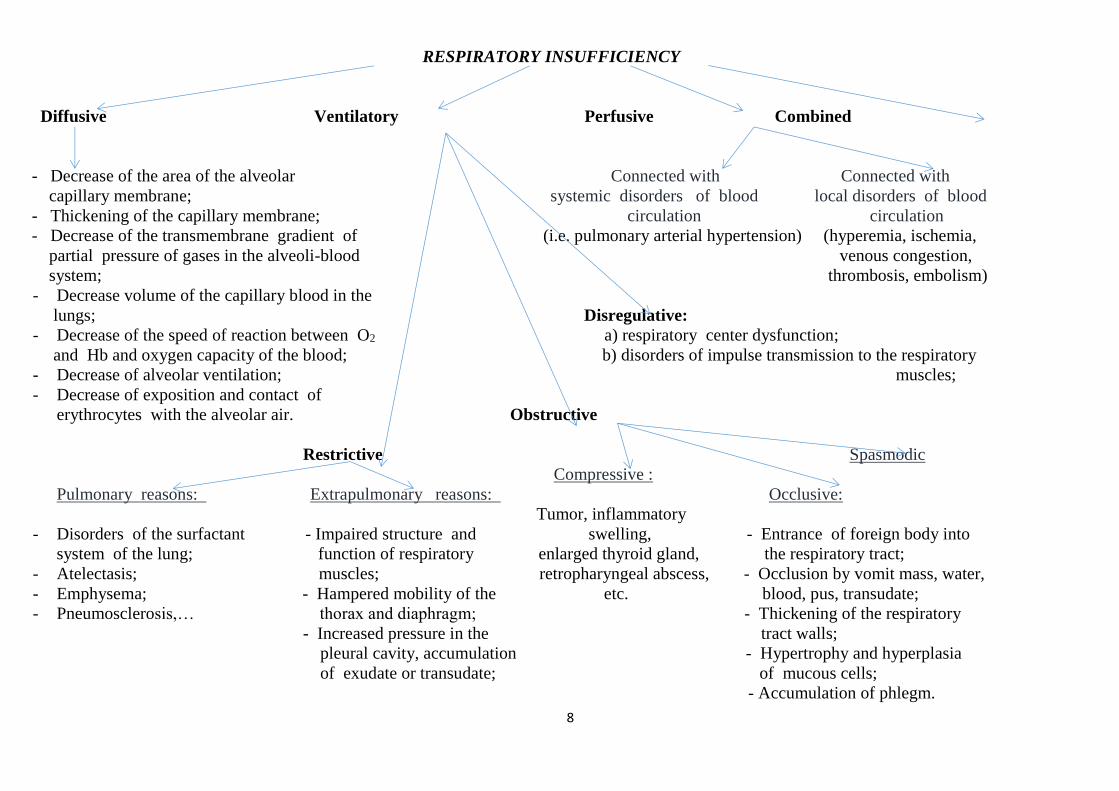
Respiratory insufficiency is a pathologic process, which develops as a result of external
respiration disorder, when the gas content of the blood does not correspond to organism
requirements at rest and under physical load.
Classification
Pathology of the respiratory system may be classified according to different principles.
Etiological classification divides it into acquired and hereditary (congenital), as well as infectious
and noninfectious (including environmental pathology caused by the pathogenic effect of
environmental factors).
Topographic classification divides it into extrapulmonary and pulmonary according to
localization of the initial cause.
Pathogenetic classification divides it into primary (pathology begins in the respiratory system)
and secondary (complication of other diseases — arterial hypertension, heart insufficiency, cardiac
asthma, etc.) as well as total and partial (disorder of all or one physiological process in the lungs).
The latter in its turn is subdivided into ventilative, diffusive, perfusive and combined. In its turn,
ventilative pathology is subdivided into disregulative, obstructive and restrictive.
4
Depending on the kind of a typical pathological process it may be divided into inflammatory,
allergic, tumorous, and vascular. Clinical classification divides it into acute and chronic.
Physical factors are mechanical traumas, foreign bodies obstructing the lumen of the
respiratory tract, barotrauma leading to increased or decreased solubility of gases in the blood,
electrotrauma (if electrical current passes through the respiratory center).
Chemical factors are poisons (muscarine), which lead to respiratory arrest or lung edema,
narcotics, side effects of drugs, smoking, harmful industrial factors leading to environmental diseases,
chemical warfare agents (phosgene), which cause pulmonary edema when inhaled.
Biological factors are infectious with tropism to the organs of the respiratory (pneumococcus,
tubercle bacillus, adenoviruses) and exogenous immune factors (vaccines, heterogenous immune
serum).
Endogenous factors are autoimmune and genetic.
Alteration of the ventilation of the alveoli
Increasing ventilation may be at increased its needs as physiological (muscle work) and
pathological (metabolic acidosis), as well as hyperactivity of the respiratory center neurons. Reducing
ventilation is not only a decrease in its needs, but if damaged neurons of the respiratory center, in
violation of the nervous and neuromuscular transmission, respiratory muscles, decreased mobility of
the chest, increasing the pleural space and restrictive and obstructive lung diseases.
There are extrapulmonary and pulmonary causes of ventilatory insufficiency of external
respiration.
Extrapulmonary include:
1) dysfunction of the respiratory center;
2) violation of the function of spinal cord motor neurons;
3) dysfunction of the neuromuscular system;
4) violation of the mobility of the chest;
5) alteration of the integrity of the chest and pleural cavity
Pulmonary causes include:
1) conduction abnormalities of the airway;
2) alteration of the elastic properties of lung tissue;
3) reducing the number of functioning alveoli.
The mechanisms of disorders of alveolar ventilation can be divided into 3 groups:
1) dysregulation;
2) restrictive, and
3) obstructive.
Dysregulation is the mechanism of ventilation observed during such processes, for which there
is a malfunction of the respiratory center.
Restrictive mechanism disorders of pulmonary ventilation called the anatomical or functional
loss of gas exchange area of the lungs. Anatomical loss occurs as a result of removal (resection) or
replacement of lung tissue (tumor). Atelectasis (alveoli collapse) can also lead to a reduction of the
diffusion surface.
Obstructive mechanism is characterized by increased resistance to air flow. Narrow airway
lumen may be narrowed by bronchial secretion, contraction or hypertrophy of bronchial muscles, loss
of elastic properties of lung tissue that supports the bronchioles in the open state or compression from
the outside.
Disorder of diffusion exists when the ratio of diffusion capacity to pulmonary perfusion
(cardiac output) is reduced. Diffusion capacity decreases with increasing diffusion distances. In the
event of pulmonary edema fluid plasma goes into the interstitial space or into the alveoli and thus the
diffusion distance increases. Inflammation also increases the diffusion distance, because in the space
between the alveoli and capillaries increases because of the swelling and the formation of connective
5
tissue. Interstitial pulmonary fibrosis and connective tissue separates the alveoli and capillaries. Since
diffusion distance - the distance between hemoglobin and alveolar gas, the anemia diffusion capacity
is also reduced. Reduced diffusion capacity can also be caused by reduction of the diffusion area, for
example, after unilateral pulmonary resection, a decrease in the number of alveolar septa
(emphysema), a decrease in the number of alveoli (pneumonia, tuberculosis), pulmonary fibrosis. The
diffusion area is reduced by alveoli collapse, intraalveolar pulmonary edema and myocardial lungs.
Violation of diffusion is observed with an increase in cardiac output (such as during exercise), blood
flows rapidly through the lungs and the contact time of blood and the alveoli decreases.
Disorder of perfusion of single alveoli in relation to its ventilation occurs in the occlusion of
blood vessels, such as pulmonary embolism. In addition, the capillaries can be separated from alveolar
by proliferating connective tissue in the case of pulmonary fibrosis. Finally, contact with the capillary
can be lost if the alveolar septa are destroyed, for example, in the case of emphysema. Alteration of
the ventilated alveoli perfusion improves the functional dead space, because the air in the alveoli of
such does not participate in gas exchange. This condition can be compensated by deep breathing. If
a large volume of the alveoli are not supplied with blood, a decrease in the area of diffusion and
perfusion to reduce the compensation can not be guaranteed an increase in depth of breathing.
Clinical manifestations of respiratory failure
Asphyxia - inability to make breathing movements. It is one of the extreme conditions and
always develops acutely. During asphyxia, there are three periods:
In the first period of asphyxia is an activation of the respiratory center, which manifests a
rapid increase in respiratory rate and depth with a predominance of the inspiratory phase of the phase
of expiration. Disorder of oxygen supply of vital centers of the brain is in the first period of the
development of anxiety, fear, general excitation, euphoria, different motor responses (before
seizures). Developing stress increases the tone of the sympathetic nervous system, which ensures the
development of mydriasis, tachycardia, hypertension.
In the second period the respiratory rate gradually decreases with continued maximum
amplitude of respiratory movements, increased expiratory phase. This is due to inhibition of the
respiratory center under the influence of cerebral hypoxia and severe hypercapnia narcotic effect.
Sympathetic hypertonicity followed by parasympathetic.
In the third period of asphyxia, a decrease of the amplitude of respiration, its frequency and
finally stopped breathing. Blood pressure is greatly reduced. After a brief cessation of breathing
usually occurs a few rare convulsive respiratory (gasping breath), followed by paralysis of respiration.
Pain in lesions of the respiratory system occurs only in those cases where the process involves
the pleura, which are a large number of pain receptors. Lung tissue does not have pain receptors.
Cough — a reflex that protects the lungs from the accumulation of secretions and penetration
of irritating and damaging agents. Cough is initiated by irritant receptors located in the walls of the
tracheobronchial tree, they are extremely sensitive to irritants, and excessive accumulation of
secretions. Afferent impulses are transmitted via the vagus nerve in the center of the medulla
oblongata responsible for the formation of the cough response. Cough reflex is a protective reaction
of the body, weakening it leads to increased likelihood of damage to lung tissue pathogens and
exacerbate already developed disease. A prolonged and unproductive cough frequent exhausts the
patient and has a negative impact on hemodynamics and respiratory function.
The pathophysiology of the some syndromes and diseases of respiratory systems
6
Pulmonary edema In the pulmonary capillary filtration pressure is determined by the
effective filtration (difference between the gradients of hydrostatic and oncotic pressures). Increase
the effective filtration pressure in the pulmonary vessels leads to pulmonary stasis, water filtration of
plasma in the interstitial space leads to interstitial pulmonary edema, and water penetration of plasma
into the alveoli is the alveolar pulmonary edema. Increased hydrostatic pressure in the pulmonary
capillaries is observed at an inadequate pumping function of the left ventricle. The reason for this
may be a decrease in myocardial force of contraction or excessive demand in contraction (heart
failure), mitral valve stenosis or regurgitation. Increased pressure in the left ventricle is transmitted
to the pulmonary vessels. Increased permeability of pulmonary capillaries may lead to pulmonary
edema. Increased permeability of the pulmonary capillary wall to plasma proteins reduces the oncotic
pressure gradient and thus increases effective filtration pressure. Permeability of pulmonary
capillaries is increased by inhaled corrosive gases (ammonia, chlorine compounds, formaldehyde),
prolonged inhalation of pure oxygen. The effect of congestion in the pulmonary circulation is to
reduce pulmonary perfusion, and reduction of oxygen absorption. Expansion of stagnant pulmonary
vessels prevents alveolar distention and reduces the elasticity of lung tissue, but this advanced
congestive capillaries compressing the bronchial tubes, leading to increased resistance to breathing.
Bronchial asthma Bronchial asthma (BA) is the most common chronic lung disease in
children. Bronchial asthma is characterized by episodic reversible episodes of bronchospasm,
resulting in excess bronchoconstrictor response to various stimuli. Since asthma is a heterogeneous
disease and its cause may be a variety of factors, one universal classification does not exist.
Nevertheless, we can distinguish two major categories of asthma:
Allergic asthma, in which episodes of bronchospasm is induced with hypersensitivity
reactions of type I in response to contact with the lung tissue of endogenous and exogenous allergens.
One type of asthma is acquired, atopic asthma, which occurs predominantly during the first two
decades of life and is associated with other allergic manifestations in the patient. In allergic asthma
serum IgE levels increased, there is eosinophilia.
Non-allergic asthma in which the mechanisms that trigger bronchospasm are not immune. In
this form of asthma attack is caused by the influence of a small number of factors that a normal person
does not cause bronchospasm. These factors include aspirin, viral respiratory infections, cold, mental
stress, exercise, inhaled irritants such as ozone, SO2, NO2. In these patients, plasma levels of IgE is
normal. Lower airway obstruction may be caused by a number of changes, including acute
bronchospasm, swelling of the airway wall, chronic mucus plugging of the lumen and reorganization
of the respiratory tract.
Acute bronchospasm is the result of IgE-dependent release of mediators under the influence
of aeroallergens, and is the primary component of early asthmatic response. Bronchospasm also may
be the result of the elimination PGE2 in blocking cyclooxygenase (aspirin asthma). Swelling of the
walls of the alveoli occurs 6-24 hours after exposure to the allergen and belongs to the late asthmatic
response. Formation of chronic airway obstruction caused by mucus exudation of plasma proteins
and the accumulation of fragments of necrotic tissue. Resolution of blockage of the lumen can take
weeks. Reorganization of the respiratory tract is associated with a change in its structure as a result
of prolonged inflammation and may damage the reversibility of airway obstruction.
Pneumonia The term "pneumonia" is described by the inflammation of lung parenchyma
(alveoli and bronchioles). The etiology of pneumonia involves the action of both infectious (bacteria,
mycoplasma, viruses, protozoa), and non-infectious agents (aspiration of gastric contents, inhalation
of irritating gases and mists). A simple classification of pneumonia divides them into two groups:
typical and atypical pneumonia.
Typical pneumonia is the result of infection by bacteria (streptococci, staphylococci,
Legionella), which multiply in the extracellular space and causing inflammation and exudation of
fluid into the cavity of the respiratory alveoli. Clinic of typical pneumonia severe, the disease is
accompanied by a massive intoxication, high fever, profuse discharge of purulent sputum.
7
The cause of atypical pneumonia are largely intracellular bacteria (mycoplasma, viruses,
chlamydia), which cause inflammatory changes mainly in the interstitial space of the lung
parenchyma. Lack of full contact with the body's immune system pathogen causes poverty and
persistent symptoms for this one.
Pathology of the pleura Inflammation of the pleura (pleuritis) is a common complication of
respiratory infections and pneumonia. The main symptom of pleurisy is pain that occurs acutely. Pain
limits the movement of the chest, coughing and deep breathing becomes impossible. Breathing
becomes shallow and frequent. There is a restrictive type of respiratory failure. The accumulation of
fluid in the pleural cavity (pleural effusion) significantly reduces respiratory function. Normally, the
visceral and parietal pleural layers are separated with a thin layer of serous fluid. As in any other
transcellular space, pleural effusion occurs in the case where the formation of liquid exceeds its
absorption.
Five mechanisms associated with abnormal accumulation of fluid in the pleural cavity: 1)
increasing the capillary pressure (heart failure),
2) increased capillary permeability (inflammatory changes of the pleura),
3) reduction of colloid osmotic pressure (hypoalbuminemia in the pathology of the lungs and kidneys)
4) increase in negative intrapleural pressure (atelectasis in),
5) disorders of the lymph drainage of pleural space (mediastinal carcinoma).
The accumulation of serous transudate in the pleural cavity is called hydrothorax. The most
common cause is heart failure hydrothorax. Among other reasons, is isolated renal failure, nephrosis,
liver failure and cancer. Exudate pleural fluid is called with a relative density greater than 1.020 and
contains inflammatory cells. States in which the exudate is formed: infection, pulmonary infarction,
tumors, rheumatoid arthritis and systemic lupus erythematosus.
Empyema — pus-filled pleural cavity occurs by direct infection of the pleural space. Found
in bacterial pneumonia, lung abscess rupture into the pleural cavity, infiltration of subdiaphragmatic
disease outbreaks or when infected as a result of trauma.
Chylothorax — effusion of lymph in the pleural cavity. Chylothorax is the result of injury,
inflammation or tumor infiltration, lymph transport of violating the thoracic duct into the central
circulation. It can also occur as a complication of intrathoracic surgical procedures and the use of
large veins for parenteral nutrition and monitoring of central hemodynamics.
Hemothorax — blood in the pleural cavity. Bleeding may result from trauma, surgery on the
organs of the chest, swelling or rupture of large vessels, such as aortic aneurysm.
Pleural effusion acts as a stopper for lungs movement, it causes a decrease in their unfolding.
Effusion can cause the displacement of mediastinal structures to the opposite side of the chest.
Compression of the lungs reduces their ventilation (restrictive type).
Pneumothorax. Normally, the air in the pleural cavity is absent. The flow of air into the pleural
cavity is called pneumothorax. Pneumothorax causes partial or complete atelectasis on the affected
side. Pneumothorax may occur without apparent cause (spontaneous pneumothorax) or as a result of
direct damage to chest or upper respiratory tract infections (traumatic pneumothorax). Tension
pneumothorax occurs when the pressure in the pleural cavity above atmospheric pressure. This is a
life-threatening condition is the result of trauma chest, at which air can enter but can not leave the
pleural space. In this case the trachea and the mediastinum deviates, it causes massive afferent
impulses from the nerve trunks of the mediastinum and the development of cardiopulmonary shock.
8
RESPIRATORY INSUFFICIENCY
Diffusive Ventilatory Perfusive Combined
- Decrease of the area of the alveolar Connected with Connected with
capillary membrane; systemic disorders of blood local disorders of blood
- Thickening of the capillary membrane; circulation circulation
- Decrease of the transmembrane gradient of (i.e. pulmonary arterial hypertension) (hyperemia, ischemia,
partial pressure of gases in the alveoli-blood venous congestion,
system; thrombosis, embolism)
- Decrease volume of the capillary blood in the
lungs; Disregulative:
- Decrease of the speed of reaction between O2 a) respiratory center dysfunction;
and Hb and oxygen capacity of the blood; b) disorders of impulse transmission to the respiratory
- Decrease of alveolar ventilation; muscles;
- Decrease of exposition and contact of
erythrocytes with the alveolar air. Obstructive
Restrictive Spasmodic
Compressive :
Pulmonary reasons: Extrapulmonary reasons: Occlusive:
Tumor, inflammatory
- Disorders of the surfactant - Impaired structure and swelling, - Entrance of foreign body into
system of the lung; function of respiratory enlarged thyroid gland, the respiratory tract;
- Atelectasis; muscles; retropharyngeal abscess, - Occlusion by vomit mass, water,
- Emphysema; - Hampered mobility of the etc. blood, pus, transudate;
- Pneumosclerosis,… thorax and diaphragm; - Thickening of the respiratory
- Increased pressure in the tract walls;
pleural cavity, accumulation - Hypertrophy and hyperplasia
of exudate or transudate; of mucous cells;
- Accumulation of phlegm.
9
KROK 1 mcqs_A is correct answer
1.A 62-year-old patient was admitted to the
neurological department due to cerebral
haemorrage. Condition is grave. There is
observed progression of deepness and
frequency of breath that turnes into reduction
to apnoea, and the cycle repeates. What
respiration type has developed in the patient?
A Cheyne-Stockes respiration
B Kussmaul respiration
C Biot's respiration
D Gasping respiration
E Apneustic respiration
2.A group of mountain climbers went
through the blood analysis at the height of
3000 m. It revealed decrease of HCO3 to 15
micromole/l (standard is 22-26
micromole/l). What is the mechanism of
HCO3 decrease?
A Hyperventilation
B Intensification of acidogenesis
C Hypoventilation
Decrease of ammoniogenesis
E Decrease of bicarbonate reabsorption in
kidneys
3.A 62-year-old patient was admitted to the
neurological department due to cerebral
haemorrage. His conditionis grave. There is
evident progression of deep and frequent
breath that turns into reduction to apnoea and
the cycle repeats. What respiration type has
developed in the patient?
A Cheyne-Stockes respiration
B Kussmaul respiration
C Biot’s respiration
D Gasping respiration
E Apneustic respiration
4.While having the dinner the child choked
and aspirated the food. Meavy cough has
started, skin and mucose are cyanotic, pulse
is rapid, respiration is infrequent, expiration
is prolonged. What disorder of the external
respiration has the child?
A Stage of expiratory dyspnea on asphyxia
B Stage of inspiratory dyspnea on asphyxia
C Stenotic respiration
D Alternating respiration
E Biot’s respiration
5.A 23-year-old patient has been admitted to
a hospital with a cranio-cerebral injury. The
patient is in a grave condition. Respiration is
characterized by prolonged convulsive
inspiration followed by a short expiration.
What kind of respiration is it typical for?
A Apneustic
B Gasping breath
C Kussmaul’s
D Cheyne-Stokes
E Biot’s
6.A child was born asphyxiated. What drug
must be administered to the newborn to
stimulate breathing?
A Aethimizolum
B Lobeline
C Prazosin
D Atropine
E Proserine
7.A female patient, having visited the
factory premises with lots of dust in the air
for the first time, has got cough and burning
pain in the throat. What respiratory
receptors, when irritated, cause this kind of
reaction?
A Irritant receptors
B Juxtacapillary (J) receptors
C Stretch receptors of lungs
D Proprioceptors of respiratory muscles
E Thermoreceptors
8.While having the dinner the child choked
and aspirated the food. Meavy cough has
started, skin and mucose are cyanotic, rapid
pulse, rear breathing, expiration is
prolonged. What disorder of the external
breathing developed in the child?
A Stage of expiratory dyspnea on asphyxia
B Stage of inspiratory dyspnea on asphyxia
C Stenotibreathing
D Alternating breathing
E Biot's breathing
9.A 12 y.o. boy who suffers from bronchial
asthma has an acute attack of asthma:
evident expiratory dyspnea, skin pallor.
What type of alveolar ventilation
disturbance is it?
A Obstructive
B Restrictive
C Throraco-diaphragmatic
D Central
E Neuromuscular
10.A patient staying in the pulmonological
department was diagnosed with pulmonary
emphysema accompanied by reduced
10
elasticity of pulmonary tissue. What type of
respiration is observed?
A Expiratory dyspnea
B Inspiratory dyspnea
C Superficial respiration
D Infrequent respiration
E Periodic respiration
11.An unconscious young man with signs of
morphine poisoning entered admission
office. His respiration is shallow and
infrequent which is caused by inhibition of
respiratory centre. What type of respiratory
failure is it?
A Ventilative dysregulatory
B Ventilative obstructive
C Ventilative restrictive
D Perfusive
E Diffusive
12.The alveolar ventilation of the patient is
5 L/min, the breath frequency is 10per/min,
and the tidal volume is 700 ml. What is the
patient’s dead space ventilation?
A 2,0 L/min
B 0,7 L/min
C 1,0 L/min
D 4,3 L/min
E –
13.X-ray examination discovered lungs
emphysema in the patient. What is the
reason of short breath development in this
case?
A Decreased lungs elasticity
B Increased lungs elasticity
C Inhibition of respiratory center
D Excitation of respiratory center
E Decreasing of alveoli receptors sensitivity
14.A patient after pathological process has a
thickened alveolar membrane. The direct
consequence of the process will be the
reduction of:
A Diffuse lung capacity
B Oxygen capacity of blood
C Minute respiratory capacity
D Alveolar lung ventilation
E Reserve expiratiory capacity
15.A patient has got a spasm of smooth
muscles of bronchi. Activators of what
membrane cytoreceptors are physiologically
reasoned to stop an attack?
A β-adrenoreceptors
B α-аdrenoreceptors
C α- and β-аdrenoreceptors
D Н-cholinoreceptors
E М-cholinoreceptors
16.A12 y.o. boy who suffers from bronchial
asthma has an acute attack of asthma:
evident expiratory dyspnea, skin pallor.
What type of alveolar ventilation
disturbance is it?
A Obstructive
B Restrictive
C Throracodiaphragmatic
D Central
E Neuromuscular
17.Examination of a miner revealed
pulmonary fibrosis accompanied by
disturbance of alveolar ventilation. What is
the main mechanism of this disturbance?
A Limitation of respiratory surface of lungs
B Constriction of superior respiratory tracts
C Disturbance of neural respiration control
D Limitation of breast mobility
E Bronchi spasm
18.A man took a quiet expiration. Name an
air volume that is mean while contained in
his lungs:
A Functional residual capacity
B Residual volume
C Expiratory reserve volume
D Respiratory volume
E Vital lung capacity
19.A man’s intrapleural pressure is being
measured. In what phase did the man hold
his breath, if his pressure is 7,5 cm Hg?
A Quiet inspiration
B Quiet expiration
C Forced inspiration
D Forceexpiration
E –
20.If a man has an attack of bronchiospasm
it is necessary to reduce the effect of vagus
on smooth muscles of bronchi. What
membrane cytoreceptors should be blocked
for this purpose?
A M-cholinoreceptors
B N-cholinoreceptors
C α-adrenoreceptors
D β-adrenoreceptors
E α- and β-adrenoreceptors
21.Vagi of an experimental animal were cut
on both sides. What respiration changes will
be observed?
A It will become deep and infrequent
11
B It will become shallow and frequent
C It will become deep and frequent
D It will become shallow an infrequent
E No changes will be observed
22.A patient with bronchial asthma has
developed acute respiratory failure. What
kind of respiratory failure occurs in this
case?
A Obstructive disturbance of alveolar
ventilation
B Restrictive ventilatory defect
C Perfusion
D Diffusion
E Dysregulation of alveolar ventilation
23.To assess the effectiveness of breathing
in patients, the indicator of functional
residual capacity is used. It includes the
following volumes:
A Expiratory reserve volume and residual
volume
B Inspiratory reserve volume and residual
volume
C Inspiratory reserve volume, tidal volume,
residual volume
D Expiratory reserve volume and tidal
volume
E Inspiratory reserve volume and tidal
volume
24.A 26-year-old female patient with
bronchitis has been administered a broad
spectrum antibiotic as a causal treatment
drug. Specify this drug:
A Doxycycline
B Interferon
C BCG vaccine
D Ambroxol
E Dexamethasone
25.A 12-year-old child has a viral infection
complicated by obstructive bronchitis.
Bronchospasm can be eliminated by
inhalations of a drug from the following
pharmacological group:
A β2-agonists
B M-anticholinergics
C N-cholinomimetics
D β2-adrenergic blockers
E Analeptics
26.Analysis of the experimental spirogram
of a 55-year-old person revealed a decrease
in tidal volume and respiratory amplitude
compared to the situation of ten years ago.
The change in these indicators is caused by:
A Decreased force of respiratory muscle
contraction
B Gas composition of the air
C Physical build of a person
D Height of a person
E Body mass of a person
27.A patient has increased thickness of
alveolar-capillary membrane caused by a
pathologic process. The direct consequence
will be reduction of the following value:
A Diffusing lung capacity
B Oxygen capacity of blood
C Respiratory minute volume
D Alveolar ventilation of lungs
E Expiratory reserve volume
28.A patient has a traumatic injury of
sternocleidomastoid muscle. This has
resulted in a decrease in the following value:
A Inspiratory reserve volume
B Expiratory reserve volume
C Respiratory volume
D Residual volume
E Functional residual lung capacity
29.When studying the signs of pulmonary
ventilation, reduction of forced expiratory
volume has been detected. What is the likely
cause of this phenomenon?
A Obstructive pulmonary disease
B Increase of respiratory volume
C Increase of inspiratory reserve volume
D Increase of pulmonary residual volume
E Increase of functional residual lung
сapacity
30.A 26-year-old female patient with
bronchitis has been administered a broad
spectrum antibiotic as a causal treatment
drug. Specify this drug:
A Doxycycline
B Interferon
C BCG vaccine
D Ambroxol
E Dexamethasone
31.A 28-year-old patient undergoing
treatment in the pulmonological department
has been diagnosed with pulmonary
emphysema caused by splitting of alveolar
septum by tissular tripsin. The disease is
cased by the congenital deficiency of the
following protein:
A α1-proteinase inhibitor
B α2-macroglobulin
C Cryoglobulin
12
D Haptoglobin
E Transferrin
32.14 days after quinsy a 15-year-old child
presented with morning facial swelling, high
blood pressure, "meat slops"urine.
Immunohistological study of a renal biopsy
sample revealed deposition of immune
complexes on the basement membranes of
the capillaries and in the glomerular
mesangium. What disease developed in the
patient?
A Acute glomerulonephritis
B Acute interstitial nephritis
C Lipoid nephrosis
D Acute pyelonephritis
E Necrotizing nephrosis
33.Urine analysis has shown high levels of
protein and erythrocytes in urine. This can
be caused by the following:
A Renal filter permeability
B Effective filter pressure
C Hydrostatiblood pressure in glomerular
capillaries
D Hydrostatic primary urine pressure in
capsule
E Oncotic pressure of blood plasma
Intrapleural pressure of an individual is
being measured.
34.In what phase did he hold his breath if the
pressure is - 25 cmH2O?
A Forced inspiration
B Quiet expiration
C Quiet inspiration
D Forceexpiration
E –
35.A female patient suffering from bronchial
asthma had got a viral infection that
provoked status asthmaticus with fatal
outcome. Histological examination of lungs
revealed spasm and edema of bronchioles,
apparent infiltration of their walls with
lymphocytes, eosinophils and other
leukocytes; labrocyte degranulation. What
mechanism of hypersensitivity underlies the
described alterations?
A Reagin reaction
B Immune cytolysis
C Inflammatory
D Autoimmune
E Immune complex
36.Lungs of a preterm infant have areas of
atelectasis (pulmonary collapse). The main
cause is:
A Surfactant deficiency
B Increased viscous resistance
C Underdeveloped inspiration muscles
D Diminisheforce of surface tension of lungs
E Surfactant excess
37.A patient with marked pneumofibrosis
that developed after infiltrating pulmonary
tuberculosis has been diagnosed with
respiratory failure. What is its pathogenetic
type?
A Restrictive
B Obstructive
C Dysregulatory
D Reflex
E Apneistic
38. When studying the signs of pulmonary
ventilation, reduction of forced expiratory
volume has been detected. What is the likely
cause of this phenomenon?
A. Obstructive pulmonary disease
B. Increase of respiratory volume
C. Increase of inspiratory reserve volume
D. Increase of pulmonary residual volume
E. Increase of functional residual lung
capacity
39. A 30-year-old man has sustained an
injury to his thorax in a traffic incident,
which caused disruption of his external
respiration. What type of ventilatory
difficulty can be observed in the given case?
A. Restrictive extrapulmonary ventilator
impairment
B. Restrictive pulmonary ventilatory
impairment
C. Obstructive ventilatory impairment
D. Impaired ventilation regulation
dysfunction
E. Cardiovascular collapse
Tests for Self-Control
1.A 23-year-old patient was hospitalized
with a craniocerebral trauma in a serious
condition. Respiration is characterized by
prolonged convulsive inspiration and
13
short expiration. What type of respiration
is it?
A. Kussmaul's.
B. Gasping.
C. Apneustic.
D. Cheyne-Stokes'.
E. Biot's.
2. Cutting of both vagus nerves was
reproduced under experimental
conditions. What type of respiration will
the experimental animal develop?
A. Frequent and shallow.
B. Frequent and deep.
C. Infrequent and shallow.
D. Infrequent and deep.
E. Periodic.
3. 5 ml of air was injected into the pleural
cavity of a rat. What type of respiration
failure develops in this case?
A. Restrictive disorder of alveolar
ventilation.
B. Obstructive disorder of alveolar
ventilation.
C. Perfusive.
D. Diffusive.
E. Disregulatory impairment of alveolar
ventilation.
4. A patient has been delivered to a
hospital in diabetic coma. Respiration is
noisy and frequent. Forced expiration
follows deep inspiration. What type of
respiration is it?
A. Apneustic.
B. Cheyne-Stokes'.
C. Gasping.
D. Stenotic.
E. Kussmaul's.
5. A diphtheria patient developed edema
of the larynx. In addition, infrequent and
deep respiration with labored inspiration
is observed. What is this respiration
called?
A. Apneustic.
B. Kussmaul's.
C. Cheyne-Stokes'.
D. Stenotic.
E. Gasping.
6. A 30-year-old man complains of
dyspnea, sensation of heaviness in the
right part of the chest, general weakness.
Body temperature is 38.9°C. Objectively:
during respiration the right part of the
thorax is behind the left one. Exudate has
been found by means of pleurocentesis in
the right part of the chest. What is the
main cause of exudation?
A. Decreased resorption of the pleural
fluid.
B. Increase of blood pressure.
C. Hyperproteinemia.
D. Aggregation of erythrocytes.
E. Increase of vessel wall permeability.
7. A 12-year-old teenager has developed
a serious attack of bronchial asthma with
such symptoms: pronounced expiratory
dyspnea, pale skin. What kind of alveolar
ventilation disorder is taking place?
A. Neuromuscular.
B. Restrictive.
C. Perfusive.
D. Central.
E. Obstructive.
8. A patient was hospitalized to the
otolaryngologic department with a
foreign body in the upper respiratory
tract. What kind of pathological
respiration is observed in this case?
A. Frequent, shallow.
B. Frequent, deep.
C. Infrequent.
D. Kussmaul's.
E. Periodic.
9. An X-ray examination revealed diffuse
atelectasis in a newborn. What is the most
possible cause of this condition?
A. Bronchopneumonia.
B. Bronchial asthma.
C. Occlusion of the pulmonary artery.
D. Surfactant deficiency.
E. Pulmonary tuberculosis.
Recommended literature:
Basic
1. Simeonova N.K. Pathophysiology/ N.Simeonova.// Kyiv, Ukraine. – 2010. – 391-407 pp. 2. Victor N. Jelski, Svetlana V. Kolesnikova. Handbook Of Pathophysiology Part 2:
Pathophysiology of organs and systems. - Donetsk, Ukraine. – 2011. – 36-50 pp.
14
3. Krishtal N.V. Pathophysiology: textbook/ N.Krishtal et al.// Kyiv: AUS Medicine Publishing,
2017. - 440-460 pp.
Additional
4. Porth, Carol. Essentials of pathophysiology: concepts of altered health states /Carol Mattson
Porth ; consultants, Kathryn J. Gaspard, Kim A. Noble. —3rd ed. 2011 Wolters Kluwer Health |
Lippincott Williams & Wilkins. – 2011. – 1282 p.
5. Robbins Pathology basis of disease / Cotran R.S., Kumar V., Robbins S.L. - 2000.
15
Methodological instruction to practical lesson № 24
Module 2. Pathophysiology of organs and systems
Theme: PATHOPHYSIOLOGY OF SYSTEMIC CIRCULATION. PATHOPHYSIOLOGY
OF HEART. INSUFFICIENCY OF HEART. CORONARY HEART DISEASE.
Student should know:
• Typical pathological states and disorder in the systemic circulation: heart failure; heart failure,
arrhythmias of heart; arterial hypertension, arterial hypotension; arteriosclerosis, atherosclerosis.
• Classifications of typical disorders in the systemic circulation.
Student should be able to:
• Analyze the changes of basic parameters of cardіо- and hemodynamics at insufficiency of
heart (frequency and force of heart-contractions, minute and systolic volumes of blood, systolic,
diastolic, mean and pulse arterial blood pressure, venous blood pressure).
• Analyze causative-consequence relationships, to be able to separate pathological and
adaptive-compensatory changes, local and systematic processes in pathogenesis of insufficiency of
blood circulation, heart failure, myocardial infarction, shock states (cardiogenic shock).
LIST OF CONTROL QUESTIONS
1. Determination of concept of heart failure, principles of its classification, characteristic of
disorders of cardio- and hemodynamics. A concept of acute and chronic ("stagnant",
congestive) heart failure. Etiology, pathogenesis, stages of chronic heart failure. Mechanisms
of development of basic clinical signs of chronic heart failure of blood (shortness of breath,
cyanosis, edema).
2. Acute heart failure: etiology, pathogenesis, changes, pathological and adaptive-
compensatory. Collapse, shock as variants of acute heart failure .
3. Determinations of concept of insufficiency of heart, principles of classification.
4. Heart failures a result of overload. Reasons of overload of heart by volume and by resistance.
Mechanisms of immediate and long duration adaptation of heart to the surplus loading:
tachycardia, hyperfunction (heterо-, homeometric), hypertrophy of myocardium.
Hypertrophy of heart: types, reasons, mechanisms of development, stage (by Meerson).
Features of the hypertrophied myocardium, reasons and mechanisms of its decompensation.
Myocardial form of cardiac insufficiency.
5. Coronarogenic damages of myocardium. Insufficiency of coronary circulation (relative and
absolute; acute and chronic), mechanisms of development. A concept of "critical stenosis".
Consequences of ischemia of myocardium: depression of retractive activity, electric instability,
damage/necrosis of cardiomyocytes, additional damage at reperfusion. Ischemic illness of heart
as example of coronary heart disease, its varieties. Clinical-laboratory criteria, signs and
complications of myocardial infarction. Pathogenesis of cardiogenic shock. Principles of
prophylaxis and treatment of ischemic heart disease.
6. Etiology and pathogenesis of noncoronarogenic damages of myocardium, Cardiomyopathies.
Classification. Characteristic of reasons and mechanisms of origin, clinical signs.
7. Extramyocardial insufficiency of heart. Disorders of pericardium. Acute tamponade of
heart. Principles of cardioprotection and treatment of insufficiency of heart/ blood
circulation.
Cardiac insufficiency is a pathological process, which is characterized by impairment of
heart functioning as a pump to move the blood through the vessels at a proper speed and
inability to supply tissues with the necessary amount of blood at rest and under physical load.
16
There are three adaptive mechanisms, which provide the maintenance of the heart minute volume
and a proper speed of blood flow:
• Enforcement of heart contractions.
• Acceleration of heart contractions (tachycardia).
• Enlargement of heart (hypertrophy).
Under increased heart load the disorders are divided into two stages.
Stage of compensation takes place, when in spite of the harmful effect of the etiological factor
the heart minute volume and blood flow speed are kept at the normal level.
Stage of decompensation develops, when the heart minute volume decreases, congestion in the
circulatory system develops.
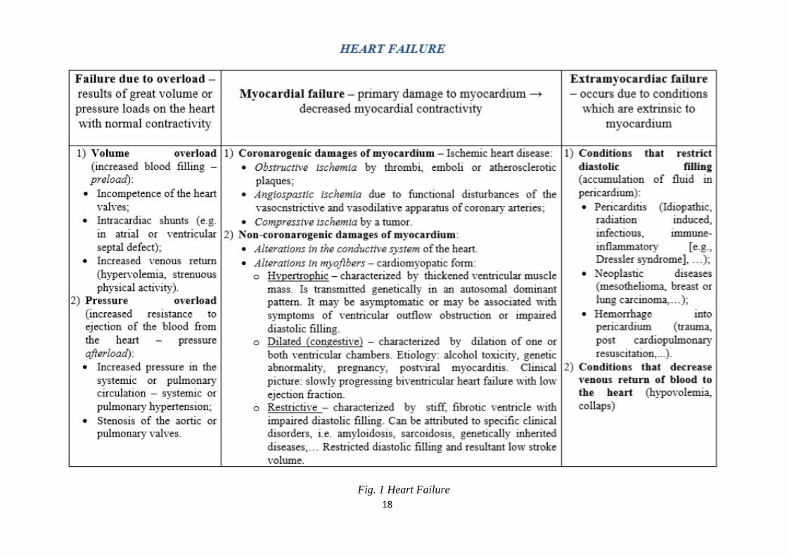
Etiology of heart failure
Heart failure may be caused by a variety of conditions including:
- conditions that impair the contractility of the heart owing to primary damage to myocardium
(e.g., cardiomyopathies );
- conditions that produce a volume overload on the heart (e.g., hypervolemia, valvular
insufficiency);
- conditions that generate a pressure overload on the heart (e.g., hypertension, valvular stenosis);
- conditions that restrict diastolic filling of the heart (e.g., mitral stenosis, cardiac tamponade);
- conditions that reduce chamber size (e.g., myocardial hypertrophy)
-conditions that decrease venous return of blood to the heart (hypovolemia, collapse).
Classification
The pathophysiological classification of cardiac insufficiency (according to the mechanisms of
development) is of special attention. There are three pathophysiological types of cardiac failure.
1. Cardiac insufficiency due to overload of the heart occurs, when a healthy heart performs hard
work for a long time.
2. Myocardial type of cardiac insufficiency results from primary myocardium pathology. It may
be caused by disorders of coronary circulation, autoimmune aggression against the heart, infection,
intoxication, hypoxia, avitaminosis, systemic hormonal and electrolyte imbalance, some hereditary
disturbances of metabolism.
3. Mixed type of cardiac insufficiency develops, when damage of the myocardium is combined
with heart overload (for example, in rheumatism, when inflammatory lesion of the heart is combined
with disorders of the valve apparatus).
Depending on the clinical course, cardiac disturbances are divided into:
a) acute and chronic;
b) right-side and left-side;
c) primary and secondary (as an associated symptom of other diseases — fever, anemia,
hyperthyroidism, etc.).
Heart insufficiency manifestations are divided into acute and chronic. Further manifestations are
divided into local (in the myocardium) and systemic (in the whole organism). Local manifestations
are subdivided into biochemical, morphological and functional changes in the myocardium.
Changes in the Myocardium
Biochemical changes in the myocardium are the following:
damage of the enzyme composition; reduction of oxygen intake (hypoxia);
disorder of oxidative phosphorylation;
reduction in the synthesis of macroergic compounds (ATP);
17
loss of glycogen; disturbance of protein synthesis; electrolytic imbalance:
•• accumulation of sodium and calcium ions in cells;
•• loss of potassium from cells;
•• hampered reverse transport of Ca2+ ions from mitochodria into the sarcoplasmic reticulum;
acidosis in the intracellular media.
Morphological changes in the myocardium include:
• destructive changes in the mitochondria;
• swelling or pyknosis of the nuclei;
• disappearance of transverse striation in the muscle fibers;
• disorders of the nervous apparatus of cells;
• dilatation of the heart cavities;
• substitution of cardiomyocytes by connective tissue (cardiosclerosis);
• cardiomyocyte death.
Functional disturbances of the myocardium are the following:
• cardiac rhythm disorders resulting from disturbance of automatism, excitability,
conductivity, contractility;
• impairment of the process of contraction and relaxation of the cardiac muscle fibers;
• reduction of the force and speed of cardiac muscle contraction;
• local contractions of separate cardiomyocytes;
• reduction of the systolic heart volume;
• increase of the residual systolic volume and diastolic pressure;
• changes in ECG.
Systemic Changes in the Organism
Acute Cardiac Insufficiency
If increased heart load is excessive, compensatory mechanisms fail to manage the overload,
and acute cardiac insufficiency develops. It is accompanied by significant changes in blood
circulation:
• acute decrease of arterial blood pressure;
• increase of venous pressure;
• significant reduction of the minute blood volume;
• circulatory and tissue hypoxia and systemic metabolic acidosis connected with it;
• acute pain;
• disorders of breathing;
• ischemia of the brain, loss of consciousness, convulsions;
• severe changes resembling shock.
Together with metabolic there may be structural changes in the cardiac muscle, so that even
under further load reduction heart activity may not be normalized. Acute cardiac insufficiency can
develop in myocardial infarction, myocarditis, ventricular fibrillation, paroxysmal tachycardia, heart
tamponade, thrombosis and embolism of the pulmonary artery.
Chronic Cardiac Insufficiency
Some manifestations of chronic cardiac insufficiency have been described in the previous
chapters, and the pathogenesis has been discussed in details. They are edema, dyspnea, venous
congestion, chronic circulatory hypoxia, cyanosis, basal metabolism increase. Chronic or congestive
cardiac insufficiency develops due to metabolic disturbances in the myocardium in prolonged
hyperfunction of the heart or different kinds of myocardium pathology.
18
Fig. 1 Heart Failure
19
KROK 1 mcqs_ A is correct answer
1.Dystrophic changes of the heart muscle are
accompanied with cardiac cavity enlargement,
decrease of the strength of heart contraction,
increased amount of blood, which remains in the
heart during systolic phase, overfilled veins. For
what state of heart is it characteristic?
A Myogenic dilatation
B Tonogenic dilatation
C Emergency stage of hyperfunction and
hypertrophy
D Cardiosclerosis
E Tamponage of the heart
2. Transmural myocardial infarction in the
patient was complicated with progressive acute
left ventricle insufficiency. What is the most
typical for this state?
A Edema of the lungs
B Edema of the extremities
C Cyanosis
D Ascites
E Arterial hypertension
3.After a serious psycho-emotional stress a 45-
year-old patient suddenly felt constricting heart
pain irradiating to the left arm, neck and left
scapula. His face turned pale, the cold sweat
stood out on it. The pain attack was stopped with
nitroglycerine. What process has developed in
this patient?
A Stenocardia
B Myocardial infarction
C Stroke
D Psychogenic shock
E Stomach ulcer perforation
5.An animal with aortic valve insufficiency got
hypertrophy of its left heart ventricle. Some of its
parts have local contractures. What substance
accumulated in the myocardiocytes caused these
contractures?
A Calcium
B Potassium
C Lactic acid
D Carbon dioxide
E Sodium
6.A 59 year old patient is a plant manager. After
the tax inspection of his plant he felt intense pain
behind his breastbone irradiating to his left arm.
15 minutes later his condition came to normal.
Which of the possible mechanisms of stenocardia
development is the leading in this case?
A High catecholamine concentration in blood
B Coronary atherosclerosis
C Intravascular aggregation of blood corpuscles
D Coronary thrombosis
E Functional heart overload
7.The patient with acute miocardial infarction
was given intravenously different solutions
during 8 hours with medical dropper 1500ml and
oxygen intranasally. He died because of
pulmonary edema. What caused the pulmonary
edema?
A Volume overload of the left ventricular
B Decreased oncotic pressure due to
hemodilution
C Allergic reaction
D Neurogenic reaction
E Inhalation of the oxygen
8.A patient who suffers from acute myocarditis
has clinical signs of cardiogenic shock. What of
the under-mentioned pathogenetic mechanisms
plays the main part in shock development?
A Disturbance of pumping ability of heart
B Depositing of blood in organs
C Reduction of diastolic flow to the heart
D Decrease of vascular tone
E Increase of peripheral vascular resistance
9.A 45 year old patient was admitted to the
cardiological department. ECG data: negative P
wave overlaps QRS complex, diastolic interval is
prolonged after extrasystole. What type of
extrasystole is it?
A Atrioventricular
B Sinus
C Atrial
D Ventricular
E Bundle-branch
10.A patient suffering from stenocardia was
taking nitroglycerine which caused restoration of
blood supply of myocardium and relieved pain in
the cardiac area. What intracellular mechanism
provides restoration of energy supply of insulted
cells?
A Intensification of ATP resynthesis
B Reduction of ATP resynthesis
C Increased permeability of membranes
D Intensification of oxygen transporting into the
cell
E Intensification of RNA generation
11.In course of a preventive examination of a
miner a doctor revealed changes of
cardiovascular fitness which was indicative of
cardiac insufficiency at the compensation stage.
What is the main proof of cardiac compensation?
20
A Myocardium hypertrophy
B Tachycardia
C Rise of arterial pressure
D Dyspnea
E Cyanosis
12.A patient ill with essential arterial
hypertension had a hypertensic crisis that
resulted in an attack of cardiac asthma. What is
the leading mechanism of cardiac insufficiency
in this case?
A Heart overload caused by high pressure
B Heart overload caused by increased blood
volume
C Absolute coronary insufficiency
D Myocardium damage
E Blood supply disturbance
13.A 60-year-old patient with a long history of
stenocardia takes coronarodilator agents. He has
also been administered acetylsalicylic acid to
reduce platelet aggregation. What is the
mechanism of antiplatelet action of
acetylsalicylic acid?
A It reduces the activity of cyclooxygenase
B It reduces the activity of phosphodiesterase
C It enhances the activity of platelet adenylate
cyclase
D It enhances the synthesis of prostacyclin
E It has membrane stabilizing effect 6 hours after
the myocardial infarction
14.A patient was found to have elevated level of
lactate dehydrogenase in blood. What isoenzyme
should be expected in this case?
A LDH1
B LDH2
C LDH3
D LDH4
E LDH5
15.A patient with extensive myocardial
infarction has developed heart failure. What
pathogenetic mechanism contributed to the
development of heart failure in the patient?
A Reduction in the mass of functioning
myocardiocytes
B Pressure overload
C Volume overload
D Acute cardiac tamponade
E Myocardial reperfusion injury
16.Autopsy of the dead patient who died from
pulmonary edema revealed a large yellowgrey
nidus in the myocardium, and a fresh thrombus
in the coronary artery. What is the most likely
diagnosis?
A Myocardial infarction
B Cardiosclerosis
C Myocarditis
D Amyloidosis
E Cardiomyopathy
17.Experimental stimulation of the sympathetic
nerve branches that innervate the heart caused an
increase in force of heart contractions because
the membrane of typical cardiomyocytes
permitted an increase in: A Calcium ion entry
B Calcium ion exit
C Potassium ion exit
D Potassium ion entry
E Calcium and potassium ion exit
18.For biochemical diagnostics of myocardial
infarction it is necessary to measure activity of a
number of enzymes and their isoenzymes. What
enzymatic test is considered to be the best to
prove or disprove the diagnosis of infarction in
the early period after the chest pain is detected?
A Creatine kinase isoenzyme CK-MB
B Creatine kinase isoenzyme CK-MM
C LDH1 lactate dehydrogenase isoenzyme
D LDH2 lactate dehydrogenase isoenzyme
E Aspartate aminotransferase cytoplasmic
isoenzyme
19.A patient in three weeks after acute
myocardial infarction has pain in the heart and
joints and pneumonia. What is the main
mechanism of development of postinfarction
Dressler’s syndrome?
A Autoimmune inflammation
B Ischemia of myocardium
C Resorption of enzymes from necrotized area of
myocardium
D Secondary infection
E Vessels ’ thrombosis
20.The high level of Lactate Dehydrogenase
(LDH) isozymes concentration showed the
increase ofLDH-1 and LDH-2 in a patient’s
blood plasma. Point out the most probable
diagnosis:
A Myocardial infarction
B Skeletal muscle dystrophy
C Diabetes mellitus
D Viral hepatitis
E Acute pancreatitis
21.Marked increase of activity of МВ-forms of
CPK (creatinephosphokinase) and LDH-1 was
revealed by examination of the patient’s blood.
What is the most probable pathology?
A Miocardial infarction
B Hepatitis
C Rheumatism
21
D Pancreatitis
E Cholecystitis
22.The calcium canals of cardiomyocytes have
been blocked on an isolated rabbit’s heart. What
changes in the heart’s activity can happen as a
result?
A Decreased rate and force of heart beat
B Decreased heart beat rate
C Decreased force of the contraction
D Heart stops in systole
E Heart stops in diastole
23.Examination of a person revealed that minute
volume of heart is 3500 mL ,systolic volume is
50 mL. What is the frequency of cardiac
contraction?
A 70 bpm
B 60 bpm
C 50 bpm
D 80 bpm
E 90 bpm
24.Dystrophic alterations of heart are
accompanied with dilation of heart cavities,
decreased force of heart contractions, increased
blood volume that remains during systole in the
heart cavity, vein overfill. What heart condition
is it typical for?
A Myogenic dilatation
B Tonogenic dilatation
C Emergency stage of hyperfunction and
hypertrophy
D Cardiosclerosis
E Cardiac tamponade
25.While preparing a patient to the operation the
heart chambers’ pressure was measured. In one
of them the pressure changed during one heart
cycle from 0 to 120 mm Hg. What chamber of
heart was it?
A Left ventricle
B Right ventricle
C Right atrium
D Left atrium
E –
26.A patient presents high activity of LDH1,2,
aspartate aminotransferase, creatine
phosphokinase. In what organ(organs) is the
development of a pathological process the most
probable?
A In the heart muscle (initial stage of
myocardium infarction)
B In skeletal muscles (dystrophy, atrophy)
C In kidneys and adrenals
D In connective tissue
E In liver and kidneys
27.12 hours after an acute attack of retrosternal
pain a patient presented a jump of aspartate
aminotransferase activity in blood serum. What
pathology is this deviation typical for?
A Myocardium infarction
B Viral hepatitis
C Collagenosis
D Diabetes mellitus
E Diabetes insipidus
28.Blood minute volume of a 30 year old woman
at rest is 5 l/m. What blood volume is pumped
through the pulmonary vessels per minute?
A 5 l
B 3,75 l
C 2,5 l
D 2,0 l
E 1,5 l
29.A 38 year old patient suffers from rheumatism
in its active phase. What laboratory characteristic
of blood serum is of diagnostic importance in
case of this pathology?
A C-reactive protein
B Uric acid
C Urea
D Creatinine
E Transferrin
30.An animal with aortic valve insufficiency got
hypertrophy of its left heart ventricle. Some of its
parts havelocal contractures. What substance
accumulated in the myocardiocytes caused these
contractures?
A Calcium
B Potassium
C Lactic acid
D Carbon dioxide
E Sodium
31.A 59 year old patient is a plant manager. After
the tax inspection of his plant he felt intense pain
behind his breastbone irradiating to his left
arm.15 minutes later his condition came to
normal. Which of the possible mechanisms of
stenocardia development is the leading in this
case?
A High catecholamine concentration in blood
B Coronary atherosclerosis
C Intravascular aggregation of blood corpuscles
D Coronary thrombosis
E Functional heart overload
32.In course of a preventive examination of a
miner a doctor revealed changes of
cardiovascular fitness which was indicative of
cardiac insufficiency at the compensation stage.
What is the main proof of cardiac compensation?
22
A Myocardium hypertrophy
B Tachycardia
C Rise of arterial pressure
D Dyspnea
E Cyanosis
33.ECG of a 44-year-old patient shows signs of
hypertrophy of both ventricles and the right
atrium. The patient was diagnosed with the
tricuspid valve insufficiency. What pathogenetic
variant of cardiac dysfunction is usually
observed in case of such insufficiency?
A Heart overload by volume
B Coronary insufficiency
C Cardiac tamponade
D Heart overload by resistance
E Primary myocardial insufficiency
34.A 56 year old patient suffering from cardiac
insufficiency has edema of feet and shins,
edematous skin is pale and cold. What is the
leading mechanism of edema pathogenesis?
A Rise of hydrostatic pressure in venules
B Drop of oncotic pressure in capillaries
C Increase of capillary permeability
D Disorder of lymph outflow
E Positive water balance
35.ECG of a 44-year-old patient shows signs of
hypertrophy of both ventricles and the right
atrium. The patient was diagnosed with the
tricuspid valve insufficiency. What pathogenetic
variant of cardiac dysfunction is usually
observed in case of such insufficiency?
A Heart overload by volume
B Heart overload by resistance
C Primary myocardial insufficiency
D Coronary insufficiency
E Cardiac tamponade
36.A 50 year old patient suffers from essential
hypertension. After a physical stress he
experienced muscle weakness, breathlessness,
cyanosis of lips, skin and face. Respiration was
accompanied by distinctly heard bubbling rales.
What mechanism underlies the development of
this syndrome?
A Acute left-ventricular failure
B Chronic right-ventricular failure
C Chronileft-ventricular failure
D Collapse
E Cardiac tamponade
37.After a serious psychoemotional stress a 48
year old patient suddenly developed acute heart
ache irradiating to the left arm. Nitroglycerine
relieved pain after 10 minutes. What is the
leading pathogenetic mechanism of this process
development?
A Spasm of coronary arteries
B Dilatation of peripheral vessels
C Obstruction of coronary vessels
D Compression of coronary vessels
E Increase in myocardial oxygen consumption
38.A 49 year old woman spent a lot of time
standing. As a result of it she got leg edema.
What is the most likely cause of the edema?
A Increase in hydrostatic pressure of blood in
veins
B Decrease in hydrostatic pressure of blood in
veins
C Decrease in hydrostatic pressure of blood in
arteries
D Increase in oncotic pressure of blood plasma
E Increase in systemic arterial pressure .
39. Patient’s systolic blood pressure is 90 mmHg,
diastolic-70mmHg.Such blood pressure is
caused by decrease of the following factor:
A. Pumping ability of the left heart
B. Pumping ability of the right heart
C. Aortic compliance
D. Total peripheral resistance
E. Vascular tone
Tests For Self-Control:
1.Acute failure of the mitral valve was
experimentally reproduced in an animal. The
heart adapted by activation of the heterometric
mechanism. What is the essence of this
mechanism?
A. Compensatory hypertrophy of the
myocardium.
B. The law of Frank—Starling.
C. Decreased formation of calcium-troponin
complexes.
D. Intensification of protein biosynthesis.
E. Intensification of conductivity.
2. A 41-year-old patient with signs of pulmonary
edema and left ventricular heart failure was given
a diagnosis of aortic stenosis. What is the cause
of heart failure development?
A. Increased volume of the vascular bed.
B. Damage of the myocardium.
C. Decreased volume of the circulating blood.
23
D. Cardiac overload due to increased blood
volume.
E. Cardiac overload due to increased blood
outflow resistance.
3. A 37-year-old man who had suffered from
mitral valve failure for many years developed
acute cardiac decompensation. What
pathophysiological variant of cardiac failure is
observed in this case?
A. Neurogenic heart damage.
B. Hypoxic heart damage.
C. Coronary heart damage.
D. Cardiac volume overload.
E. Cardiac resistance overload.
4. A patient has mitral valve regurgitation. As a
result, cardiac overload by blood volume
developed. What is the main mechanism of
immediate compensation?
A. Effect of catecholamines.
B. Homeometric.
C. Intensification of protein biosynthesis.
D. Heterometric.
E. Hypertrophy of the myocardium.
5. A woman has suffered from arterial
hypertension for 15 years. Now dyspnea and
palpitation appeared; systolic pressure decreased
a little. What is the basic mechanism of heart
failure in this case?
A. Disturbance of conductivity.
B. Cardiac overload with increased blood
volume.
C. Damage of the myocardium.
D. Cardiac overload due to increased blood
outflow resistance.
E. Disturbance of cardiac activity regulation.
6. A patient demonstrates abrupt arterial pressure
increase due to changes of the vascular tone.
What compensatory mechanism provides an
increased force of myocardial contraction in this
case?
A. Renin-angiotensin system activation.
B. Influence of the sympathetic nervous system
on the heart.
C. Influence of the parasympathetic nervous
system on the heart.
D. Homeometric.
E. Heterometric.
7. A patient has arterial hypertension. As a
consequence of hypertensic crisis, acute heart
failure developed. What is the main mechanism
of heart failure onset in this case?
A. Absolute coronary failure.
B. Cardiac volume overload.
C. Damage of the myocardium.
D. Cardiac resistance overload.
E. Relative coronary failure.
8. A 51-year-old patient complains of dyspnea,
palpitation, pain in the right hypochondrium,
edema on the legs. ECG shows hypertrophy of
both ventricles and the right atrium.
Regurgitation of the tricuspid valve is diagnosed.
What pathogenetic variety of heart failure is it?
A. Arrhythmic.
B. Cardiac resistance overload.
C. Initial myocardial failure.
D. Cardiac volume overload.
E. Extramyocardial.
9. In an 18-year-old man mitral valve
insufficiency without circulation disturbance is
revealed. What type of adaptive reaction takes
place?
A. Homeometric.
B. Heterometric.
C. Myogenic dilatation.
D. Hypertrophy of the heart.
E. Intensification of conductivity.
24
Recommended literature:
Basic
1. Simeonova N.K. Pathophysiology/ N.Simeonova.// Kyiv, Ukraine. – 2010. – 338-354 pp. 2. Victor N. Jelski, Svetlana V. Kolesnikova. Handbook Of Pathophysiology Part 2:
Pathophysiology of organs and systems. - Donetsk, Ukraine. – 2011. – 68-80 pp.
3. Krishtal N.V. Pathophysiology: textbook/ N.Krishtal et al.// Kyiv: AUS Medicine
Publishing, 2017. - 367-382 pp.
Additional
4. Porth, Carol. Essentials of pathophysiology: concepts of altered health states /Carol Mattson
Porth ; consultants, Kathryn J. Gaspard, Kim A. Noble. —3rd ed. 2011 Wolters Kluwer
Health | Lippincott Williams & Wilkins. – 2011. – 1282 p.
5. Robbins Pathology basis of disease / Cotran R.S., Kumar V., Robbins S.L. - 2000.
25
Methodological instruction to practical lesson № 25
Module 2. Pathophysiology of organs and systems
Theme: PATHOPHYSIOLOGY OF BLOOD VESSELS
Student should know:
• Modern criteria for diagnostics of arterial hypertension.
• Classifications of arterial hypertension.
Student should be able to:
• Characterize the features of different forms of arteriosclerosis, explain the modern theories of
pathogenesis of atherosclerosis.
• Interpret a primary arterial hypertension as multifactor disease. • Differentiate the role of volume changes and peripheral resistance of blood flow in
development of different haemodynamic variants of arterial hypertension. • Genetic defects as basis of pathogenesis of primary arterial hypertension. • Explain the role of kidneys in pathogenesis of primary and secondary arterial hypertension. • Apply knowledge about the experimental models of typical disorders in the system of blood
circulation (coronary heart disease, arteriosclerosis, arterial hypertension) for the analysis of their pathogenesis.
• Explain reasons and mechanisms of development of arterial hypotension.
LIST OF CONTROL QUESTIONS
1. A concept of vascular insufficiency. Types, reasons and mechanisms of its development.
Arteriosclerosis: determination of concept, classification. Basic forms of arteriosclerosis: atherosclerosis, mediacalcinosis, аrteriolosclerosis, their general characteristics (typical localization, signs, complications).
2. Atherosclerosis. Factors of risk of atherosclerosis. Experimental models. Modern and historical theories of atherogenesis. A role of damage of endothelium, inflammation, inherited and acquired disorders of receptor-mediated transport of lipoproteins (LP) (disorder of receptors of LP, defects of molecules of LP, modification of LP) in atherogenesis. Disorders of transport of lipids in blood. Hyper-, hypо-, dyslipoproteinemias. Dependence of development of dyslipoproteinemias on the factors of environment (diet), heredity and concomitant diseases. Modern classifications of dyslipoproteinemias (primary and secondary; according to the phenotype of LP; with the high or low risk of atherosclerosis), criteria of hypercholesterolemia, hypertryglyceridemia, low level of LPHD.
3. Etiology, pathogenesis of primary (inherited, familial) and secondary (at disorders of feeding, obesity, diabetes mellitus, illnesses of kidneys, hypothyreosis, cirrhosis of liver, influence of medicinal drugs), dyslipoproteinemias. Consequences/complications of dyslipoproteinemias. Principles and aims of renewal of normal lipid composition of blood.
4. Arterial hypertension (AH), determination of concept, principles of classification. Hemodynamic variants of AH. A role of disorders of pressor and depressor systems in development of AH.
5. Primary and secondary arterial hypertension. Etiology, pathogenesis. Experimental models. 6. Primary AH as multifactor disease: a role of factors of heredity and external factors in
development of primary AH. Theories of pathogenesis of primary AH (dysregulatory, membrane etc).
7. Mechanisms of development of primary and secondary hypertension of small circle of circulation of blood.
8. Arterial hypotension: determinations of concept, criteria. Etiology and pathogenesis of acute and chronic arterial hypotensions. Collapse. Reasons and mechanisms of development, signs.
26
Vascular insufficiency is a disorder of blood circulation and blood supply of organs
resulting in impairment of substance and oxygen exchange between the blood and tissues.
Atherosclerosis is a pathological process, which is characterized by infiltrative-proliferative
changes of the inner layer of elastic-type arteries with deposition of lipids, fibrin and calcium
accompanied by elasticity impairment and vessel lumen narrowing.
Endogenous factors play a more decisive role in atherosclerosis development:
• pathological heredity (enzymopathy);
• elderly age (hyperlipoproteinemia and hypercholesterolemia are more frequently observed
in elderly people);
• hormonal insufficiency (hypothyroidism, DM, hypogonadism, Cushing's syndrome);
• arterial blood hypertension («the higher the blood pressure, the greater the risk»);
• diastolic hypertension is a more important correlate;
• disorders of metabolism (obesity, gout, xanthomatosis, liver pathology).
Risk factors are:
• hypodynamia;
• overeating, alcohol abuse, smoking;
• stress, which may lead to vessel wall trophicity disorder.
Pathogenesis. Some concepts of atherosclerosis pathogenesis have been proposed:
1. Concept of primary systemic disorders of lipid metabolism and secondary damage of the vascular
wall.
2. Concept of primary damage of the vascular wall and its secondary lipoidosis («reaction to injury»
hypothesis).
From the point of view of the main link of atherosclerosis pathogenesis there are three
concepts distinguished:
1. Hypercholesterolemic.
2. Thrombogenic.
3. Genetic.
Morphological changes in vessels and the dynamics of their atherosclerotic damage proceed
in some stages with the following order of events.
1. Infiltration of the vessel intima by native or modified lipoproteins of the blood plasma.
Lipid deposition is an early event in atherogenesis. Lesions occur primarily within the tunica intima.
Excessive capture of lipids by macrophages and infiltration of the arterial wall with macrophages
containing low-density lipoproteins. Transformation of macrophages into foam cells, which are the
base of lipid stain formation. It leads to endothelial injury. Lipid capture by smooth muscle cells.
Lipid-filled smooth muscle cells lose contractility.
2. Proliferation is local irritation and multiplication of histiocytes, fibroblasts and smooth
muscle cells of vessels, which capture lipids. Connective tissue excrescence. Consolidation of the
connective fibers. Thickening of the subendothelium, deformation of the elastic tissue. Formation of
atherosclerotic fibrous plaques on the endothelium, which consists of lipid-laden smooth muscle cells
surrounded by a fibrous matrix. If a lesion is in progress, it occludes the arterial lumen.
3. Degeneration and destruction of the intima and vascular wall. Destruction of foam cells,
their lysis, fragmentation of fibrous structures. Formation of lipid stains. Formation of ulcers, which
can perforate. Progressing atherosclerotic plaques. The core of fibrous plaques consists of lipids and
debris of cells necrotized as a result of insufficient blood supply.
4. Sclerotization (calcification) of vessels. The lumen of the atherosclerotically changed
vessels narrows as a result of atherosclerotic plaque formation. When the altered complex structure
becomes rigid, it causes vascular occlusion.
Atherosclerotic changes in vessels predispose to thrombogenesis. Blood clots are formed in
the intima layer. Ischemia (infarction) develops in the region of the damaged vessels. Fibrous plaques
are altered by hemorrhage. Functional disturbances of vessels consist in:
• disorders of vessel elasticity;
• vessel incapability of dilatation;
• tendency of the damaged vessels toward spasm.
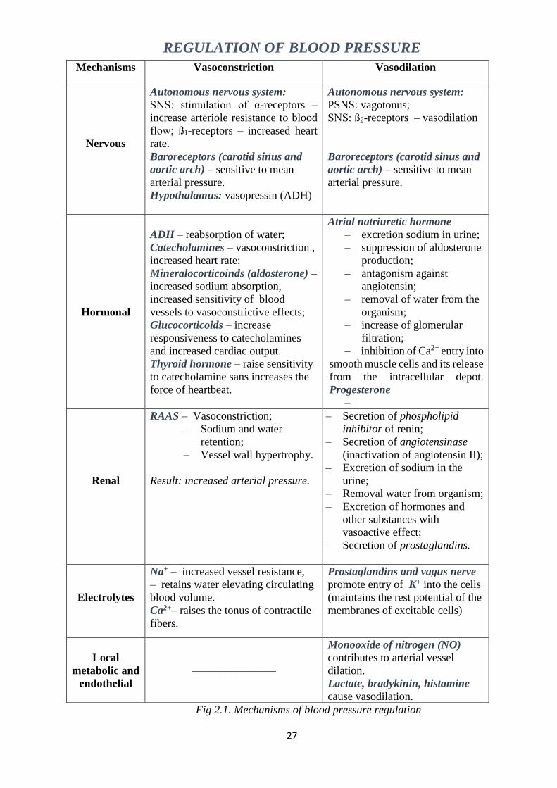
27
REGULATION OF BLOOD PRESSURE
Fig 2.1. Mechanisms of blood pressure regulation
Mechanisms Vasoconstriction Vasodilation
Nervous
Autonomous nervous system:
SNS: stimulation of α-receptors –
increase arteriole resistance to blood
flow; ß1-receptors – increased heart
rate.
Baroreceptors (carotid sinus and
aortic arch) – sensitive to mean
arterial pressure.
Hypothalamus: vasopressin (ADH)
Autonomous nervous system:
PSNS: vagotonus;
SNS: ß2-receptors – vasodilation
Baroreceptors (carotid sinus and
aortic arch) – sensitive to mean
arterial pressure.
Hormonal
ADH – reabsorption of water;
Catecholamines – vasoconstriction ,
increased heart rate;
Mineralocorticoinds (aldosterone) –
increased sodium absorption,
increased sensitivity of blood
vessels to vasoconstrictive effects;
Glucocorticoids – increase
responsiveness to catecholamines
and increased cardiac output.
Thyroid hormone – raise sensitivity
to catecholamine sans increases the
force of heartbeat.
Atrial natriuretic hormone
– excretion sodium in urine;
– suppression of aldosterone
production;
– antagonism against
angiotensin;
– removal of water from the
organism;
– increase of glomerular
filtration;
– inhibition of Ca2+ entry into
smooth muscle cells and its release
from the intracellular depot.
Progesterone
–
Renal
RAAS – Vasoconstriction;
– Sodium and water
retention;
– Vessel wall hypertrophy.
Result: increased arterial pressure.
– Secretion of phospholipid
inhibitor of renin;
– Secretion of angiotensinase
(inactivation of angiotensin II);
– Excretion of sodium in the
urine;
– Removal water from organism;
– Excretion of hormones and
other substances with
vasoactive effect;
– Secretion of prostaglandins.
Electrolytes
Na+ – increased vessel resistance,
– retains water elevating circulating
blood volume.
Ca2+– raises the tonus of contractile
fibers.
Prostaglandins and vagus nerve
promote entry of K+ into the cells
(maintains the rest potential of the
membranes of excitable cells)
Local
metabolic and
endothelial
–––––––––––––––
Monooxide of nitrogen (NO)
contributes to arterial vessel
dilation.
Lactate, bradykinin, histamine
cause vasodilation.
28
Hypertension is a sustained elevation of the systemic arterial pressure.
Primary and secondary hypertensions are distinguished.
Primary hypertension is a sustained elevation of the arterial BP which is not connected with
certain diseases or pathological processes in body‘s organs. The cause of primary hypertension is
unknown, hence essential hypertension (EH). In essential hypertension increased BP is main and
often the only sign of disease.
Secondary hypertension is a sustained elevation of the arterial BP which results from
pathological processes in body‘s organs, hence symptomatic hypertension. Secondary hypertension
occurs at the following conditions:
-renal diseases (glomerulonephritis, pyelonephritis, polikistosis of kidneys);
-endocrine diseases (mainly at adrenal tumors: pheochromocytoma, Cushing‘s syndrome,
Conn‘s disease (primary hyperaldosteronism));
-diseases of the heart and vessels (some valve diseases, coarctation of the aorta,
atherosclerosis);
- the nervous system diseases (encephalitis, trauma, hemorrhage in the brain).
In all symptomatic hypertensions the etiology is known. Cessation of the cause leads to
normalization of the arterial BP.
Risk factors of essential hypertension:
-age – EH is more common in adults than in adolescents and children;
-sex – men are affected more often than women;
-race – EH is more common among blacks than among whites;
-heredity – in some families EH develops several times more often than in remaining
population; -high-sodium diet – EH is widely spread in countries (Japan, Chine) where people have
a high salt intake;
-obesity;
-physical inactivity;
-hyperinsulinemia;
-oral contraceptive drugs- prolonged treatment with oral contraceptives containing estrogens
produces significant hypertension in some women( due to an increase in circulating levels of
angiotensinogen);
-stress.
At present there are two concepts in pathogenesis of essential hypertension: the dysregulation
theory and the membrane theory.
The dysregulation theory explains an origin of EH by disturbances of the mechanisms which
control blood pressure. The membrane theory explains an origin of EH by primary disturbances in
smooth-muscle cells of arterioles.
The dysregulation theory According to this theory there are three stages in pathogenesis of
essential hypertension including:
1. The first stage is due to the sympathetic system overactivation caused by frequent and
prolonged stresses. Persistent negative emotions cause overstrains of the higher parts of central
nervous system, especially the cerebral cortex. It results in development of a stable focus of excitation
(―pathological dominant‖) in the vasomotor center of the subcortical region. In its turn, the latter
causes an increase in excitability of the sympathetic division of the nervous system. It leads to a
release a lot of catecholamines into the blood. Arterial blood pressure increases due to the following
effects of catecholamines:
-an increase in the cardiac output, -an increase in the peripheral resistance,
-a spasm of afferent arterioles of the kidneys with a resultant release of renin by the
juxtaglomerular cells
2. The second stage is due to an activation of the renin-angiotensin system. Renin cleaves
angiotensinogen, a circulating α2-globulin produced in the liver, to form angiotensin I which is
further modified by converting enzyme to the angiotensin II. Role of angiotensin II in an increase in
arterial blood pressure :
-angiotensin II is a potent vasoconstrictor,
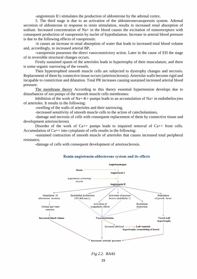
29
-angiotensin II i stimulates the production of aldosterone by the adrenal cortex.
3. The third stage is due to an activation of the aldosteronevasopressin system. Adrenal
secretion of aldosterone in response to renin stimulation, results in increased renal absorption of
sodium. Increased concentration of Na+ in the blood causes the excitation of osmoreceptors with
consequent production of vasopressin by nuclei of hypothalamus. Increase in arterial blood pressure
is due to the following effects of vasopressin:
-it causes an increase in renal absorption of water that leads to increased total blood volume
and, accordingly, to increased arterial BP,
-vasopressin possesses the direct vasoconstrictory action. Later in the cause of EH the stage
of in reversible structural changes occurs.
Firstly sustained spasm of the arterioles leads to hypertrophy of their musculature, and there
is some organic narrowing of the vessels.
Then hypertrophied smooth muscle cells are subjected to dystrophy changes and necrosis.
Replacement of them by connective tissue occurs (arteriosclerosis). Arteriolar walls become rigid and
incapable to constriction and dilatation. Total PR increases causing sustained increased arterial blood
pressure.
The membrane theory According to this theory essential hypertension develops due to
disturbances of ion pumps of the smooth muscle cells membranes:
Inhibition of the work of Na+-K+ pumps leads to an accumulation of Na+ in endotheliocytes
of arterioles. It results in the following:
-swelling of the walls of arterioles and their narrowing,
-increased sensitivity of smooth muscle cells to the action of catecholamines,
-damage and necrosis of cells with consequent replacement of them by connective tissue and
development arteriosclerosis
Disorder of the work of Ca++ pumps leads to impaired removal of Ca++ from cells.
Accumulation of Ca++ into cytoplasm of cells results in the following:
-sustained contraction of smooth muscle of arterioles that causes increased total peripheral
resistance,
-damage of cells with consequent development of arteriosclerosis.
Fig 2.2. RAAS
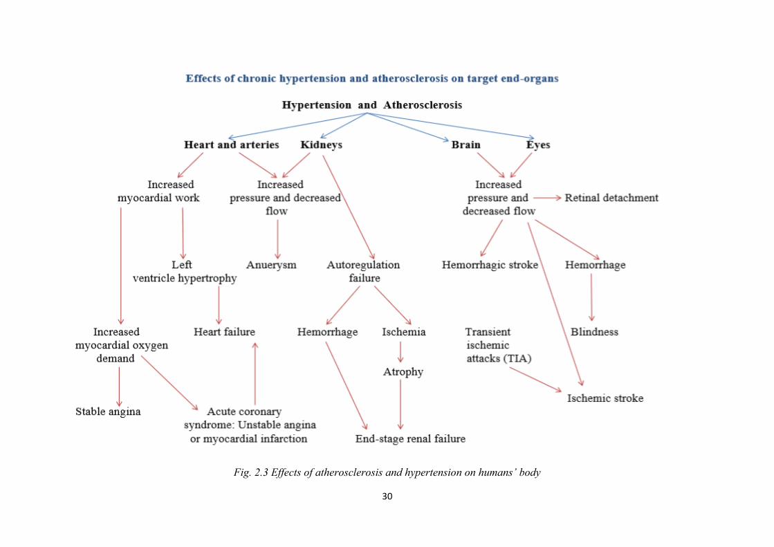
30
Fig. 2.3 Effects of atherosclerosis and hypertension on humans’ body
31
KROK 1 mcqs_A is correct answer
1.Arterial hypertention is caused by the
stenosis of the renal arteries in the patient.
Activation of what system is the main link in
the pathogenesis of this form of hypertension?
A Renin-angiotensin
B Sympathoadrenal
C Parasympathetic
D Kallikrein-kinin
E Hypothalamic-pituitary
2.Prophylactic medical examination of a 36
year old driver revealed that his AP was 150/90
mm Hg. At the end of working day he usually
hears ear noise, feels slight indisposition that
passes after some rest. He was diagnosed with
essential hypertension. What is the leading
pathogenetic mechanism in this case?
A Neurogenetic
B Nephric
C Humoral
D Endocrinal
E Reflexogenic
3.Arterial pressure of a surgeon who
performed a long operation rised up to 140/110
mm Hg. What changes of humoral regulation
could have caused the rise of arterial pressure
in this case?
A Activation of sympathoadrenal system
B Activation of formation and excretion of
aldosterone
C Activation of renin angiotensive system
D Activation of kallikrein kinin system
E Inhibition of sympathoadrenal system
4.A driver who got a trauma in a road accident
and is shocked has reduction of daily urinary
output down to 300 ml. What is the main
pathogenetic factor of such diuresis change?
A Drop of arterial pressure
B Drop of oncotic blood pressure
C Increased vascular permeability
D Decreased number of functioning
glomerules
E Secondary hyperaldosteronism
5.A 70 year old man is ill with vascular
atherosclerosis of lower extremities and
coronary heart disease. Examination revealed
disturbance of lipidic blood composition. The
main factor of atherosclerosis pathogenesis is
the excess of the following lipoproteins:
A Low-density lipoproteins
B Cholesterol
C High-density lipoproteins
D Intermediate density lipoproteins
E Chylomicrons
6.A patient has arterial hypertension. What
long-acting drug from the group of calcium
channel blockers should be prescribed?
A Amlodipine
B Octadine
C Pyrroxanum
D Atenolol
E Reserpine
7.A patient with constant headaches, pain in
the occipital region, tinnitus, dizziness has
been admitted to the cardiology department.
Objectively: AP- 180/110 mm Hg, heart rate -
95/min. Radiographically, there is a stenosis of
one of the renal arteries. Hypertensive
condition in this patient has been caused by the
activation of the following system:
A Renin-angiotensin
B Hemostatic
C Sympathoadrenal
D Kinin
E Immune
8.A 43-year-old-patient has arterial
hypertension caused by an increase in cardiac
output and general peripheral resistance.
Specify the variant of hemodynamic
development of arterial hypertension in the
given case:
A Eukinetic
B Hyperkinetic
C Hypokinetic
D Combined
E ----
9.A 16-year-old female patient has fainted
after quickly changing her body position from
horizontal to vertical one. Which process from
the ones listed below has caused the loss of
consciousness in the first place?
A Decreasing venous return
B Increasing venous return
C Increasing central venous pressure
D Decreasing oncotic pressure of blood plasma
E Increasing arterial pressure
10.A patient has insufficient blood supply to
the kidneys, which has caused the development
of pressor effect due to the constriction of
arterial resistance vessels. This is the result of
the vessels being greately affected by the
following substance:
A Angiotensin II
32
B Angiotensinogen
C Renin
D Catecholamines
E Norepinephrine
11.A 40-year-old patient complains of
intensive heartbeats, sweating, nausea, visual
impairment, arm tremor, hypertension. From
his anamnesis: 2 years ago he was diagnosed
with pheochromocytoma. Hyperproduction of
what hormones causes the given pathology?
A Catecholamines
B Aldosterone
C Glucocorticoids
D ACTH
E Thyroidal hormones
12.Arterial pressure of a surgeon who
performed a long operation rised up
to140/110mm Hg. What changes of humoral
regulation could have caused the rise of arterial
pressure in this case?
A Activation of sympathoadrenal system
B Activation of formation and excretionof
aldosterone
C Activation of renin angiotensive system
D Activation of kallikrein kinin system
E Inhibition of sympathoadrenal system
13.An aged man had raise of arterial pressure
under a stress. It was caused by activation of:
A. Sympathoadrenal system
B Parasympathetic nucleus of vagus
C Functions of thyroid gland
D Functions of adrenal cortex
E Hypophysis function
14.Prophylactic medical examination of a 36
year old driver revealed that his AP was 150/90
mm Hg. At the end of working day he usually
hears ear noise, feels slight indisposition that
passes after some rest. He was diagnosed with
essential hypertension. What is the leading
pathogenetic mechanism in this case?
A Neurogenetic
B Nephric
C Humoral
D Endocrinal
E Reflexogenic
15.Systemic arterial pressure of an adult
dropped from 120/70 to 90/50 mm Hg that led
to reflectory vasoconstriction. The
vasoconstriction will be maximal in the
following organ:
A Bowels
B Heart
C Brain
D Kidneys
E Adrenals
16.A patient ill with essential arterial
hypertension had a hypertensic crisis that
resulted in an attack of cardiac asthma. What is
the leading mechanism of cardiac insufficiency
in this case?
A Heart overload caused by high pressure
B Heart overload caused by increased blood
volume
C Absolute coronary insufficiency
D Myocardium damage
E Blood supply disturbance
17.In response to a change in body position
from horizontal to vertical blood circulation
system develops reflectory pressor reaction.
Which of the following is its compulsory
component?
A Systemic constriction of the venous vessels
B Systemic dilatation of the arterial resistive
vessels
C Decrease in the circulating blood volume
D Increase in the heart rate
E Weakening of the pumping ability of Heart
18. A patient with hypertensic crisis has
increased content of angiotensin II in blood.
Angiotensin pressor effect is based on:
A. Contraction of arteriole muscles
B. Activation of biogenic amine synthesis
C. Prostaglandin hyperproduction
D. Vasopressin production stimulation
E. Activation of kinin–kallikrein system
19. A 43-year-old-patient has arterial
hypertension caused by increase in cardiac
output and general peripheral resistance.
Specify the variant of hemodynamic
development of arterial hypertension in the
given case:
A. Eukinetic
B. Hyperkinetic
C. Hypokinetic
D. Combined
E. –
20. A patient has insufficient blood supply to
the kidneys, which has caused the development
of pressor effect due to constriction of arterial
resistance vessels. This condition results from
the vessels being strongly affected by the
following substance:
A. Angiotensin II
B. Angiotensinogen
C. Renin
D. Catecholamines
33
E. Norepinephrine
21. Patient’s systolic blood pressure is 90
mmHg, diastolic-70mmHg.Such blood
pressure is caused by decrease of the following
factor:
A. Pumping ability of the left heart
B. Pumping ability of the right heart
C. Aortic compliance
D. Total peripheral resistance
E. Vascular tone
Tests for Self-Control
1. A 60-year-old man has blood vessel
atherosclerosis. Excess of what substances
plays a leading role in the pathogenesis of this
disease?
A. Histic lipoprotein lipase.
B. High-density lipoproteins.
C. Chylomicrons.
D. Low-density lipoproteins.
E. Fatty acids.
2. Rabbits were fed with addition of
cholesterol. In 5 months atherosclerotic
changes in the aorta were found. What is the
main cause of atherogenesis in this case?
A. Endogenic hypercholesterinemia.
B. Overeating.
C. Inactivation of receptors to low-density
lipoproteins.
D. Hypodynamia.
E. Exogenous hypercholesterinemia.
3. In a patient with persistent arterial
hypertension angiography showed
atherosclerotic lesion of both renal arteries.
What is the initial mechanism of arterial
hypertension development?
A. Increased secretion of vasopressin.
B. Increased level of catecholamines.
C. Increased level of hydrocortisone.
D. Increased cardiac outflow.
E. Increased production of renin.
4. A patient suffers from hypertensic crisis. An
increased level of angiotensin II was found in
the blood. What is the pressure effect of
angiotensin associated with?
A. Activation of biogenic amine synthesis.
B. Contraction of the arteriole muscles.
C. Hyperproduction of prostaglandins.
D. Stimulation of vasopressin formation.
E. Activation of the kallikrein-kinin system.
5. A patient has arterial hypertension caused by
renal artery stenosis. Activation of what
system is the main mechanism of this form of
hypertension?
A. Sympatho-adrenal.
B. Renin-angiotensin.
C. Parasympathetic.
D. Kallikrein-kinin.
E. Hypothalamo-pituitary.
6. A patient with idiopathic hypertension
revealed increased concentration of
vasopressin in the blood. The function of what
organ does this hormone influence directly?
A. Liver.
B. Kidneys.
C. Heart.
D. Lungs.
E. Epinephral glands.
7. A 43-year-old patient suffers from arterial
hypertension caused by a moderate increase of
cardiac outflow and general peripheral
resistance. What hemodynamic variant of
arterial hypertension has developed in this
case?
A. Eukinetic.
B. Hyperkinetic.
C. Hypokinetic.
D. Akinetic.
E. Combined.
8. A 65-year-old man has been suffering from
arterial hypertension for 15 years. Now
systolic pressure is decreasing and diastolic
pressure remains increased. What
hemodynamic type of arterial hypertension is
it?
A. Hyperkinetic.
B. Normokinetic.
C. Hypokinetic.
D. Eukinetic.
E. Dyskinetic.
9. Arterial hypertension was reproduced by
narrowing the renal arteries in a dog. Activity
of the renin-angiotensin-aldosterone system
increased. What component of this system has
the strongest pressor effect?
A. Renin.
B. Angiotensin II.
C. Angiotensin I.
34
D. Kinins.
E. Aldosterone.
10. A patient with alcoholic cirrhosis of the
liver dyspnea. Decrease of arterial pressure,
ascites, dominal veins, and splenomegaly have
been hemodynamics is observed?
A. Failure of the left ventricle.
B. Failure of the right ventricle.
C. Total heart failure.
D. Collapse
E. Portal hypertension.
Recommended literature:
Basic
1. Simeonova N.K. Pathophysiology/ N.Simeonova.// Kyiv, Ukraine. – 2010. – 364-387 pp. 2. Victor N. Jelski, Svetlana V. Kolesnikova. Handbook Of Pathophysiology Part 2:
Pathophysiology of organs and systems. - Donetsk, Ukraine. – 2011. – 59-67 pp.
3. Krishtal N.V. Pathophysiology: textbook/ N.Krishtal et al.// Kyiv: AUS Medicine
Publishing, 2017. – 390-416 pp.
Additional
4. Porth, Carol. Essentials of pathophysiology: concepts of altered health states /Carol Mattson
Porth ; consultants, Kathryn J. Gaspard, Kim A. Noble. —3rd ed. 2011 Wolters Kluwer
Health | Lippincott Williams & Wilkins. – 2011. – 1282 p.
5. Robbins Pathology basis of disease / Cotran R.S., Kumar V., Robbins S.L. - 2000.
35
Methodological instruction to practical lesson № 26
Module 2. Pathophysiology of organs and systems
Theme: HEART ARRHYTHMIAS. PATHOPHYSIOLOGIC BASICS OF ECG ANALYSIS
Student should know:
• Mechanisms of development of arrhythmias. Student should be able to:
• Apply knowledge about typical disorders of heart rhythm (disorders of automatism, excitability, conductivity and combined) for the analysis of electrocardiogram.
LIST OF CONTROL QUESTIONS
1. Arrhythmias of heart: classification, reasons, mechanisms, typical electrocardiographic
signs.
2. A role of additional conduction ways of heart in development of arrhythmias.
3. Reasons and mechanisms of origin of ectopic foci of excitation in myocardium,
mechanisms of the repeated entrance and recirculation of excitation.
4. Fibrillation and defibrillation of heart.
5. Disorders in conductive system of a heart.
6. ECG signs of ischemic heart disease.
Disorders of cardiac rhythm — arrhythmias are the most often heart disease. It can varied
from non-dangerous altered heart rhythm till dramatically drop of blood pressure and death. In general
arrhythmia can arise from disorder of impulse formation or impulse conduction.
Formation of heart rhythm is based on automaticity — ability of pacemakers to generate
impulse itself. Disorders of automaticity are classified:
1. Increase of automaticity
a. Increase of automaticity of sinus node
b. Increase of automaticity of latent pacemakers
c. Ectopic sources of automaticity
2. Trigger activity
3. Decrease of automaticity
Under normal conditions heart rate in nomotopic pacemaker (sinus node) and in latent
pacemakers (sinoatrial node, Purkinje cells) is controlled by neuro-endocrine factors —
acetylcholine, norepinephrine, epinephrine. So in case of emotional stress, physical activity or other
stress reactions release of catecholamines leads to increase of heart rate (nomotopic tachycardia) and
play important role in adaptation and compensatory reactions. The same effect can be in case of action
of catcholamine mimetics or acetylcholine blockers. These factors not only maintain increase of
automaticity of nomotopic pacemaker and but also they can cause activity of latent pacemakers. If a
rate of depolarization of latent pacemaker more than a rate of depolarization of nomotopic pacemaker
ectopic impulse (extrasystole) or ectopic rhythm (paroxysmal tachycardia) occurs. Sometime in
damage of myocardium (hypoxia, acidosis, toxins) some cardiomyocytes which are not belonged to
conductive heart system can get pathological ability for spontaneous depolarization of membrane and
so they become sources of ectopic rhythm.
Trigger activity is the term which explain occurrence of tachycardia in some cases when
normal action potential causes additional depolarization and new action potentials (depolarizing after-
potentials). There are two types of these depolarizing after-potentials — early depolarizing
afterpotentials and late depolarizing after-potentials. The early depolarizing after-potentials occur in
case of prolonged action potential (in bradycardia, hypokaliemia, use of antiarrhythmic drugs). The
36
late depolarizing afterpotentials are generated after post-hyperpolarization and caused by tachycardia,
digitalis intoxication, and hypercalcaemia.
Increase of activity of parasympathetic system and release of acetylcholine lead to decrease
of heart rate. A lot of toxins and drugs can mime or block action of the acetylcholine so they can
decrease heart rhythm too.
Other cause of arrhythmia — disorders of impulse conductivity. They are classified:
1. Block of impulse conductance
2. Re-entry
3. Additional conducting pathway
Impulse conductivity is blocks when impulse achieves part of myocardium which is unable
for electric excitation by its nature (scar) or functional state (refractory period). Block can be
temporary or constant, one-sided or double-sided. Most common causes of heart block are myocardial
ischemia, myocardial sclerosis, myocardial trauma, drugs. Conduction block leads to
bradyarrhythmia and appearance of rhythm from latent pacemaker or ectopic rhythm.
Re-entry is the universal mechanism for the most of tachyarrhythmias. It is circulation of
electrical circuit in part of myocardium which cases periodical depolarization of cardiomyocytes. The
basis of this disorder is a change of local conductance when the impulse from one point of myocardial
net is distributed with unequal velocity so the impulse can ―return‖ to the initial point and cause new
wave of depolarization before a wave of depolarization from nomotopic pacemaker. Local ischemia
or drugs can change features of some part of myocardial net (result in local block or slowing of
impulse propagation) so are most common causes of this mechanism.
Additional conducting pathway (like Kent bundle) is the anatomic abnormality in some
peoples. Abnormal location of these pathways causes too fast distribution of impulse between atrium
and ventricles with no atrioventricular pause. It looks like giant re-entry mechanism where the role
of slow part of conductive net is played by normal conductivity system. The results of this disorder
are tachyarrhythmia and deficit of ventricle filling.
Clinically arrhythmias are divided into two groups: bradyarrhythmias and tachyarrhytmias.
Bradyarrythmia or bradycardia is decrease of heart rate less then 55 beats per minute. The
decrease of heart rate lead to decrease of cardiac output, so it leads to decrease of blood pressure and
as a result — circulatory hypoxia. Clinical signs are maintained by last one and include picture of
brain hypoxia, activation of sympatho-adrenal system mostly.
Types of bradyarrhytmia:
1. Atrial bradycardia
a. Respiratory sinus arrhythmia
b. Sinus bradycardia
c. Sick sinus syndrome
2. Atrioventricular nodal bradycardia
a. Junctional rhythm
b. Sinoatrial block
3. Ventricular bradycardia
Respiratory sinus arrhythmia — occurs during respiration. Heart rate increases during
inhalation and decreases during exhalation. Caused by changes in the vagal tone during respiration.
Sinus bradycardia — can be normal in athletes, caused by increase tone of parasympathetic
nervous system, damage of sinus nod by ischemia, drugs, trauma.
Sick sinus syndrome — variability of sinus rhythm rate connected with malfunction of sinus
node often due to ageing.
37
Junctional rhythm — rhythm from AV node in case of total damage of nomotopic pacemaker.
Sinoatrial block — most common cause of bradycardia, usual complication of ischemic heart
disease. In the basis of this disorder — block of impulse propagation in AV node.
First degree AV block — on ECG PR interval greater than 0.20sec
Second degree AV block —
Type Mobitz I — on ECG there is progressive prolongation of PR interval (Wenckebach
periods) with disappearance of QRS complex.
Type Mobitz II — on ECG there is prolonged PR interval and disappearance of QRS complex
occurs suddenly without prolongation of PR interval. Disappearance of QRS can be regular (drop of
every 4th QRS complex) and clustered (drop of 2 or 3 QRS complexes in series).
Third degree AV block — on ECG there is connection between P waves and QRS complexes.
QRS complexes very deformed and are manifestation of ventricular rhythm.
Ventricular rhythm — heart rate less than 45 beats per minute, source of impulses lies below
Purkinje cells, occurs in inhibition of nomotopic pacemaker, third degree AV block. On ECG this
one is characterized by deformed QRS complexes.
Tachyarrythmia or tachycardia is increase of heart rate more then 100 beats per minute (varies
according to age and sex). The progressive high increase of heart rate lead to decrease of stroke
volume due to shortening of ventricles filling time, so it leads to decrease of cardiac output, drop of
blood pressure and as a result — circulatory hypoxia. In addition increase of heart rate increases
oxygen demand of myocardium and make period of filling of coronary arteries shorter (they are filled
during diastole). Both these phenomena lead to progressive heart ischemia and make worse
circulatory hypoxia. The last one makes clinical picture as it makes in case of bradyarrhythmia.
Types of tachyarrhytmia:
1. Supraventricular tachyarrhythmia
a. Sinus tachycardia
b. Atrial tachycardia
c. Atrial flutter
d. Atrial fibrillation
e. Paroxysmal supraventricular tachycardia (AV nodal reentrant tachycardia)
f. Wolff-Parkinson-White syndrome
2. Ventricular tachyarrhythmia
a. Ventricular tachycardia
b. Torsades de pointes
c. Ventricular fibrillation
Sinus tachycardia — heart rate 100-180 bpm. Normal P wave and QRS complexes. Usually
this is result of increase of sympathetic tone (stress, physical exertion, fever, hypethyreosis,
hypoxemia).
Atrial tachycardia — heart rate 100-180 bpm. P wave is deformed.
Atrial flutter — on ECG P waves look like saw teeth their rate 250-300 bpm. High rate (>280)
of impulses from sinus node doesn‘t allow AV node to pass excitation to ventricles, so ventricles has
their own rhythm. Decrease of atrial rhythm to 200 bpm leads to propagation of this rhythm to
ventricles and as a result heart rate increases.
Atrial fibrillation — on ECG P waves looks chaotic and often unrecognizable. It is potential
life-threatening state because of decrease of stroke volume. Also in atrium in this condition there is
high risk of thrombus formation and further arterial embolisation due to disorder of normal
hemodynamics (local congestion of blood). Most common cause of atrial fibrillation is dilatation of
left or right atrium (in mitral stenosis or insufficiency).

38
Paroxysmal supraventricular tachycardia — abrupt attack of tachycardia 140-250 bpm with
the same abrupt cessation of this one. Reentry — the main mechanism of this type of arrhythmia.
Wolff-Parkinson-White syndrome — result of presence of additional conducting pathway
(most often bundle of Kent). On ECG PR interval shorter than normal, ascending part of R wave is
deformed (delta-wave), QRS complex wide.
Ventricular tachycardia — sequence of ventricle extrasystoles with rate 100-200 bpm. On
EKG QRS complexes wide and deformed, P waves absent. It can be result of increase of automaticity
of ectopic pacemakers or due to re-entry mechanism. The most common cause of this type of
arrhythmia is myocardial infarction with scarring. Often this one transforms to ventricular fibrillation.
Ventricle fibrillation — the most dangerous arrhythmia which demand emergency treatment.
It is characterized by chaotic excitement of myocardial cells with non-effective contraction. It leads
to crucial decrease of stroke volume and drop of blood pressure and cardiogenic shock. Ventricle
fibrillation is the main cause of death in myocardial infarction, cardiomyopathies, cardial valve
disease, congenital heart diseases, hyperkalemia, hypomagnesemia.
KROK 1 mcqs_A is correct answer
1.Person has stable HR, not more than 40 bpm.
What is the pacemaker of the heart rhythm in
this person?
A Atrioventricular node
B Sinoatrial node
C His' bundle
D Branches of His' bundle
E Purkinye' fibers
2.ECG of a 44-year-old patient shows signs of
hypertrophy of both ventricles and the right
atrium. The patient was diagnosed with the
tricuspid valve insufficiency. What
pathogenetic variant of cardiac dysfunction is
usually observed in case of such insufficiency?
A Heart overload by volume
B Heart overload by resistance
C Primary myocardial insufficiency
D Coronary insufficiency
E Cardiac tamponade
3.In a 45-year-old patient on ECG it was
revealed: sinus rhythm, the number of
auricular complexes exceeds number of
ventricular complexes; progressing extension
of the P-Q interval from complex to complex;
fallout of some ventricular complexes; Р
waves and QRST complexes are without
changes. Name the type of heart rhythm
disfunction.
A Atrioventricular block of the II degree
B Synoauricular block
C Atrioventricular blockade of the I degree
D Intraatrial block
E Complete atrioventricular block
4.Processes of repolarisation are disturbed in
ventricular myocardium in examined person. It
will cause amplitude abnormalities of
configuration and duration of the wave:
A Т
B Q
C R
D S
E P
5.ECG of a patient shows such alterations: P-
wave is normal, P-Q-interval is short,
ventricular QRST complex is wide, R-wave is
double-peak or two-phase. What form of
arrhythmia is it?
A WPW syndrome (Wolff-Parkinson-White)
B Frederick's syndrome (atrial flutter)
C Atrioventricular block
D Ventricular fibrillation
E Ciliary arrhythmia
6.A 49 y.o. woman consulted a doctor about
heightened fatigue and dyspnea during
physical activity. ECG: heart rate is 50/min,
PQ is extended, QRS is unchanged, P wave
quanity exceeds quantity of QRS complexes.
What type of arrhythmia does the patient have?
A Atrio-ventricular block
B Extrasystole
C Sinus bradycardia
D Ciliary arhythmia
E Sinoatrial block
7.A patient has extrasystole. ECG shows no P
wave, QRS complex is deformed, there is a full
compensatory pause. What extrasystoles are
these?
A Ventricular
B Atrial
C Atrio-ventricular
39
D Sinus
E –
8.Electrocardiogram of a 45-year-oldman
showed absence of P-wave in all the leads.
What part of the conducting system is blocked?
A Sino-atrial node
B Atrio-ventricular node
C Common branch of the bundle of His
D Branches of the bundle of His
E Purkinje’s fibres
9.A person has steady HR not exceeding40
bpm. What is the pacemaker of the heart
rhythm in this person?
A Atrioventricular node
B Sinoatrial node
C His’ bundle
D Branches of His’ bundle
E Purkinye’ fibers
10.After the trauma, the patient’s right n.vagus
was damaged. Which violation of the cardiac
activity is possible in this case?
A Violation of the automatism of Kiss-Fleck
node
B Violation of the automatism of a
atrioventricular node
C Violation of a conductivity in the right
auricle
D Block of a conductivity in the
atrioventricular node
E Arrhythmia
11.An isolated cell of human heart
automatically generates excitation impulses
with frequency 60 times pro minute. What
structure does this cell belong to?
A Sino-atrial node
B Atrium
C Ventricle
D Atrioventricular node
E His’ bundle
12.Examination of an isolated cardiomyocyte
revealed that it didn’t generate excitation
impulses automatically. This cardiomyocyte
was obtained from:
A Ventricles
B Sinoatrial node
C Atrioventricular node
D His’ bundle
E Purkinje’s fibers
13.A 49 y.o. woman consulted a doctor about
heightened fatigue and dyspnea during
physical activity. ECG: heart rate is 50/min,
PQ is extended, QRS is unchanged, P wave
quanity exceeds quantity of QRS complexes.
What type of arrhythmia does the patient have?
A Atrioventricular block
B Extrasystole
C Sinus bradycardia
D Ciliary arrhythmia
E Sinoatrial block
14.A patient has extrasystole .ECG shows no P
wave, QRS complex is deformed, there is a full
compensatory pause. What extrasystoles are
these?
A Ventricular
B Atrial
C Atrioventricular
D Sinus
E –
15.ECG of a patient with hyperfunction of
thyroid gland showed heart hurry. It is
indicated by depression of the following ECG
element:
A R − R interval
B P − Q segment
C P − Q interval
D P − T interval
E QRS complex
16.Vagus branches that innervate heart are
being stimulated in course of an experiment.
As a result of it the excitement conduction
from atria to the ventricles was brought to a
stop. It is caused by electrophysical changes in
the following structures:
A Atrioventricular node
B His’ bundle
C Sinoatrial node
D Ventricles
E Atria
17.A cardiac electric stimulator was implanted
to a 75 year old man with heart rate of 40 bpm.
Thereafter the heart rate rose up to 70 bpm. The
electric stimulator has undertaken the function
of the following heart part:
A Sinoatrial node
B Atrioventricular node
C His’ bundle branches
D His’ bundle fibers
E Purkinje’s fibers
18.Analysis of the ECG revealed the missing
of several PQRST cycles. The remaining
waves and complexes are not changed. Specify
the type of arrhythmia:
A Sinoatrial block
B Atrial fibrillation
C Atrioventricular block
40
D Atrial premature beat
E Intra-atrial block
19.ECG of a patient displays an abnormally
long R wave (up to 0,18 s). This is caused by a
decrease in the conduction velocity of the
following heart structures:
A Ventricles
B Atria
C Atrio-ventricular node
D Right ventricle
E Left ventricle
20.Since a patient has had myocardial
infarction ( type of block)
A Complete atrio-ventricular
B Partial atrio-ventricular
C Sino-atrial
D Intra-atrial
E Intraventricular
21.Since a patient has had myocardial
infarction, his atria and ventricles contract
independently from each other with a
frequency of 60-70 and 3540 per minute.
Specify the type of heart block in this case:
A Complete atrio-ventricular
B Partial atrio-ventricular
C Sino-atrial
D Intra-atrial
E Intraventricular
22.A patient complains of palpitation after
stress. The pulse is 104 bpm, P-Q=0,12
seconds, there are no changes of QRS
complex. What type of arrhythmia does the
patient have?
A Sinus tachycardia
B Sinus bradycardia
C Sinus arrhythmia
D Ciliary arrhythmia
E Extrasystole
23.ECG of a patient shows such alterations: P-
wave is normal, P-Q-interval is short,
ventricular QRST complex is wide, R-wave is
double-peak or two-phas e. What form of
arrhythmia is it?
A WPW syndrome (Wolff-Parkinson-White)
B Fredericks syndrome (atrial flutter)
C Ventricular fibrillation
D Atrioventricular block
E Ciliary arrhythmia
24.A 45 year old patient was admitted to the
cardiological department. ECG data: negative
P wave overlaps QRS complex, diastolic
interval is prolonged after extrasystole. What
type of extrasystole is it?
A Atrioventricular
B Sinus
C Atrial
D Ventricular
E Bundle-branch
25.A patient has delayed conduction of
excitement through the atrioventricular node.
What changes of ECG will be observed?
A Prolongation of P − Q interval
B Prolongation of Q − S interval
C Negative T wave
D S − T-segment displacement
E Prolongation of Q − T interval
26. A patient complains of palpitations after
stress. Pulse is104/min. ,P-Q=0,12 seconds,
there are no changes in QRS complex. What
type of arrhythmia does the patient have?
A. Sinus tachycardia
B. Sinus bradycardia
C. Sinus arrhythmia
D. Ciliary arrhythmia
E. Extrasystole
27. Electrocardiogramanalysis demonstrates
that cardiac cycle of a human equals 1 second.
It means that heart rate per minute equals:
A. 60
B. 50
C. 70
D. 80
E. 100
Recommended literature:
Basic
1. Simeonova N.K. Pathophysiology/ N.Simeonova.// Kyiv, Ukraine. – 2010. – 348-350
pp. 2. Victor N. Jelski, Svetlana V. Kolesnikova. Handbook Of Pathophysiology Part 2:
Pathophysiology of organs and systems. - Donetsk, Ukraine. – 2011. – 51-58 pp.
3. Krishtal N.V. Pathophysiology: textbook/ N.Krishtal et al.// Kyiv: AUS Medicine
Publishing, 2017. - 383-389 pp.
Additional
4. Porth, Carol. Essentials of pathophysiology: concepts of altered health states /Carol

41
Mattson Porth ; consultants, Kathryn J. Gaspard, Kim A. Noble. —3rd ed. 2011 Wolters
Kluwer Health | Lippincott Williams & Wilkins. – 2011. – 1282 p.
5. Robbins Pathology basis of disease / Cotran R.S., Kumar V., Robbins S.L. - 2000.
42
Methodological instruction to practical lesson № 27
Module 2. Pathophysiology of organs and systems
Theme: PRACTICAL SKILLS WITH THE THEME "THE PATHOPHYSIOLOGY OF CIRCULATORY SYSTEM AND EXTERNAL BREATHING"
SEMANTIC MODULE 5.
Student should be able to perform analysis of : • Cause and effect (changes in local and general, pathological and adaptive-compensatory, specific and non-specific, leading and supporting units) in the pathogenesis of common disorders in the circulatory and respiratory systems; • understand patterns of disorders of electrical phenomena in the heart in various types of diseases and interpret pathological processes in bronchopulmonary tissue in different types of damage
Abilities and practical skills: • Solving situational problems with the definition of common disorders in the circulatory and respiratory system (arterial hyper- and hypotension, circulatory and respiratory failure, coronary artery disease, arrhythmia), their main types (through the application of knowledge of the principles of classification), causes and mechanisms. • Based on the results of laboratory and instrumental investigations (blood tests, ECG, spirometry, pneumatic) identify the main pathological process in the lungs and heart. • Evaluate automaticity, conduction, excitability of the heart on an electrocardiogram