osteomyelitis in paediatric age group, pathophysiology and
TRANSCRIPT
Citation: Yadav M and Singh A. Osteomyelitis in Paediatric Age Group, Pathophysiology and Adaptive Responses of Causing Microbes. Austin J Orthopade & Rheumatol. 2017; 4(2): 1053.
Austin J Orthopade & Rheumatol - Volume 4 Issue 2 - 2017ISSN: 2472-369X | www.austinpublishinggroup.com Singh et al. © All rights are reserved
Austin Journal of Orthopedics & Rheumatology
Open Access
Abstract
Osteomyelitis (OM) or inflammation of the bone, is usually caused by bacterial infection. Bone infections in children are primarily hematogenous in origin, although cases secondary to penetrating trauma, surgery, or infection in a contiguous site are also reported. OM is a biofilm-based infection of bone which is the characteristic growth pattern for most bacteria and are now understood to consist of interactive communities with the ability to alter their gene expression in order to ensure survival, where the majority of causative microorganisms are sessile in nature and render to develop adaptive responses against antibiotics, host implant, and other environmental effects. In other side the osteoclastic and osteoblastic cells play a central role in the immune response of bone has resulted in a better understanding of osteo-immunology. This review focuses on pathogenic advancement at genetic level due several adaptations, so it will open a new window in terms of development of novel treatment strategies in the management of osteomyelitis and periprosthetic infections.
Keywords: Osteomyelitis; Bacterial adaptation; Bone Infection; Osteolysis; Antibiotic resistance
IntroductionOsteomyelitis is a progressive infection of bone that results in
inflammatory destruction followed by new bone formation [1], Osteomyelitis usually begins as an acute infection, but it may evolve into a chronic condition [2]. Osteomyelitis is strictly defined as any form of inflammation involving bone and/or bone marrow, but it is almost exclusively the result of infection. The definition of OM is broad, and encompasses a wide variety of conditions. Traditionally, the length of time the infection has been present and whether there is pus formation or sclerosis (increased density of bone) is used to arbitrarily classify OM.
Neonates are more prone to osteomyelitis. Because of a less developed immune system, osteomyelitis can be caused by less virulent agents and tends to present fewer clinical signs. The combination of unclear symptoms in neonates and the presence of transphysial vessels can lead to indolent infections that are often discovered at a late stage [3,4]. In children, compared to adults, the periostium is loosely attached to the bone, and in case of infection it can easily be lifted, thereby creating a space for pus collection.
The reported annual incidence of childhood osteomyelitis is 3 to 20 per 100,000. For acute osteomyelitis the incidence is 8 per 100,000 and for sub-acute osteomyelitis 5 per 100,000 [2,3,5,6]. The incidence is higher in children below 3 years of age, with a peak incidence in children below 1 year of age. Approximately 50% of cases of osteomyelitis occur in the first 5 years of life. Boys are more likely than girls to be affected. The long bones of the lower extremities are most often involved, although any bone may be affected [7,8]. But the most common site of infection is the long bones, especially the femur and tibia. Most infections are monoostotic, but polyostotic involvement of up to 6.8 % is reported in infants, and even 22 % in
Review Article
Osteomyelitis in Paediatric Age Group, Pathophysiology and Adaptive Responses of Causing MicrobesYadav M and Singh A*Department of Orthopaedic Surgery, King George’s Medical University, India
*Corresponding author: Singh Ajai, Department of Orthopaedic Surgery, King George’s Medical University, Lucknow, India
Received: March 17, 2017; Accepted: April 18, 2017; Published: April 25, 2017
neonates [7]. As well asrisk factors for osteomyelitis include trauma, sickle cell disease, immunodeficiency, sepsis, minor trauma in combination with bacteraemia, an indwelling vascular catheter and chronic vascular lines [4,7].
Normal bone is highly resistant to infection (but in case of general predisposing factors such as diabetes and peripheral vascular disease tip the balance in favor of the bacterium). Thus, osteomyelitis arises only when there is a large organism inoculation, trauma leading to bone damage, or the presence of foreign material. The pathogenesis of osteomyelitis is multifactorial. Some important factors include virulence of the infecting organisms, underlying immune status of the host, and the type, location, and vascularity of the bone. Among the causing pathogens Staphylococcus aureus is the most common cause of osteomyelitis, has been extensively used as a model to study pathogenesis [9-11].
As well as the role of genetics in the pathogenesis of osteomyelitis is a field of growing research interest. Multiple genetic differences have been identified between patients with osteomyelitis and control subjects, indicating possible hereditary susceptibilities (Table 1). A recent study identified polymorphisms resulting in upregulation of MMPs (Matrix Matelloproteinases) with significantly higher frequency in patients with osteomyelitis than in healthy controls. The mutation may cause an increase in osteoblast MMP1 production, which has been linked to osteo-destructive activity in metastasis [12] and inflammatory arthropathy [13]. The IL- 1α (-889 TT) genotype has also been found to be more common in patients with osteomyelitis [14]. Mutations in the G(-248)A polymorphism at the promoter region of the bax gene was observed significantly more frequently in osteomyelitis patients [15].
In summary, targeting the pathogenesis of inflammatory bone
Austin J Orthopade & Rheumatol 4(2): id1053 (2017) - Page - 02
Singh A Austin Publishing Group
Submit your Manuscript | www.austinpublishinggroup.com
disorders might be decisive to enable effective treatments based on a causal approach, understanding the interactions between the pathogens on the one side and the host’s innate and acquired immune system as well as the cellular components of the bone tissue on the other side. Although the term ‘osteomyelitis’ suggests simplicity by translation into its original meaning, inflammatory bone disorders still remain a condition with an overwhelming complexity of depending factors and will be a challenge for future research projects to resolve.
Pathophysiology of bone infection Normal bone is highly resistant to infection (but in case of general
predisposing factors such as diabetes and peripheral vascular disease tip the balance in favour of the bacterium). Thus, osteomyelitis arises only when there is a large organism inoculation, trauma leading to bone damage, or the presence of foreign material. The pathogenesis of osteomyelitis is multifactorial. Some important factors include virulence of the infecting organisms, underlying immune status of the host, and the type, location, and vascularity of the bone. Among the causing pathogens Staphylococcus aureus is the most common cause of osteomyelitis, has been extensively used as a model to study pathogenesis [16].
The Pathogen: The type of infecting organism depends on the age of the child and underlying medical problem (Table 2). S. aureus is the most common cause of osteomyelitis in all age groups, accounting for 70% to 90% of infections. Infection caused by microorganism Staphylococcus aureus is becoming an increasingly common problem [17].
Organisms other than S. aureus causing infection in older children include Streptococcus pyogenes, Streptococcus pneumoniae, and Kingellakingae [18]. S. pyogenes causes approximately 10% of
cases of acute hematogenous osteomyelitis with a peak incidence of disease in preschool-age and early school–age children [19]. Children with S. pyogenes osteomyelitis often have a recent history of varicella infection and present with higher fever and White Blood Cell (WBC) counts compared with children infected with S. aureus. Children with osteomyelitis caused by S. pneumoniae are younger than children infected with S. aureus and S. pyogenes. They are more likely to have joint involvement [20].
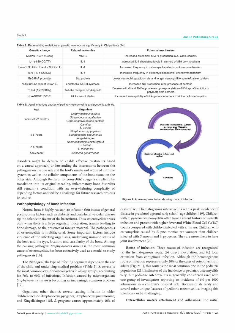
Route of infection: Three routes of infection are recognized: (a) the hematogenous route, (b) direct inoculation, and (c) local extension from contiguous infection. Although the hematogenous route of infection represents only 20% of the cases of osteomyelitis in adults (Figure 1), this route is the most common one in the pediatric population [21]. Estimates of the incidence of pediatric osteomyelitis vary, but pediatric osteomyelitis is generally considered rare, with one group of investigators reporting an incidence of 6.0 per 1000 admissions in a children’s hospital [22]. Because of its rarity and several other unique features of pediatric osteomyelitis, imaging this infection can be challenging.
Extracellular matrix attachment and adhesions: The initial
Genetic change Related molecules Potential mechanism
MMP1(- 1607 1G/2G) MMP1 Increased osteoblast MMP1 production in2G allele carriers
IL-1 (-889 CC/TT) IL-1 Increased IL-1 circulating levels in carriers of-889 polymorphism
IL-4 (-1098 GG/TT and -590CC/TT) IL-4 Increased frequency in osteomyelitispatients; unknownmechanism
IL-6 (-174 GG/CC) IL-6 Increased frequency in osteomyelitispatients; unknownmechanism
G(-248)A promoter Bax protein Lower neutrophil apoptosisrate and longer neutrophillife spaninA allele carriers
NOS3(27-bp repeat, intron 4) endothelial NOS3 synthase Increased NO production inthe presence of bacteria
TLR4 (Asp299Gly) Toll-like receptor, NF-kappa B DecreasedIL-6 and TNF-alpha levels; phosphorylation ofNF-kappaB inhibitor in polymorphism carriers
HLA-DRB1*100101 HLA class II alleles Increased susceptibility of HLA genotypecarriers to sickle cell osteomyelitis
Table 1: Representing mutations at genetic level occurs significantly in OM patients [14].
Age Organism
Infants 0 –2 months
Staphylococcus aureus Streptococcus agalactiae
Gram-negative enteric bacteria Candida
≤ 5 Years
S. aureus Streptococcus pyogenes
Streptococcus pneumoniaeKingellakingae
Haemophilusinfluenzae type b
> 5 Years S. aureus S. pyogenes
Adolescent Neisseria gonorrhoeae
Table 2: Usual infectious causes of pediatric osteomyelitis and pyogenic arthritis.
Figure 1: Above representation showing route of infection.
Austin J Orthopade & Rheumatol 4(2): id1053 (2017) - Page - 03
Singh A Austin Publishing Group
Submit your Manuscript | www.austinpublishinggroup.com
event in the localization of infection appears to be adhesion of the bacteria to the Extracellular Matrix (ECM). The ECM is a biologically active layer composed of a complex mixture of macromolecules, such as fibronectin, fibrinogen, albumin, vitronectin, and collagen. Host cell adhesion, migration, proliferation, and differentiation are all influenced by the composition and structural organization of the surrounding ECM. Interaction between host cells and the ECM is known to be mediated by specific receptors such as integrins, which are composed of α and β units and link many ECM proteins to the eukaryotic cellular cytoskeleton [23]. The ECM not only serves as a substrate for host cells, but also for colonizing bacteria. If an infection is to develop, pathogenic bacteria must cling to the tissue in order to overcome removal by physical forces. As well as using non-specific hydrophobic and electrostatic forces to interact with their hosts.
Bacteria have surface proteins with specific affinity for components of the ECM and for plasma proteins. These proteins are often called ECM-Binding Proteins (ECMBPs) or MSCRAMMs (Microbial Surface Components Recognizing Adhesive Matrix Molecules). The S. aureus proteins responsible for binding to fibronectin (fibronectin binding protein; fnbp), collagen (collagen binding protein; cna) and fibrinogen (clumping factor; cifA and cifB) are the best-studied ECMBPs [24]. Peacock et al. showed that seven putative virulence genes in S. aureus, including the adhesin genes fnbA and cna, the toxin genes sej, eta, hlg, and icaA, which are involved in biofilm production, were found to be associated with invasive isolates [25].
Attachment to biomaterial surfaces: S. aureus is a common cause of metal-biomaterial, bone-joint, and soft-tissue infections [26], while S. epidermidis is more common with polymer-associated implant [27]. It has been shown that both fibrinogen [28] and fibronectin [29] deposited in vivo onto the implant surface mediate bacterial adherence.
Role of osteoblasts in pathophysiology: Osteoblasts are responsible for the deposition of bone matrix; they are found on bone surfaces and are derived from mesenchymalosteoprogenitor cells. These cells secrete osteoid, a mixture of bone matrix proteins primarily made up of type I collagen (over 90%), proteoglycans such as decorin and biglycan, glycoproteins such as fibronectin, osteonectin and tenascin-C, osteopontin, osteocalcin and bone sialoprotein [30]. The opposing action of bone matrix removal is performed by osteoclasts, multinucleate cells that are derived from the macrophage-monocyte lineage. These cells express large quantities of a vacuolar-type H(+)-ATPase on their cell surface, along with chloride channel 7 (ClC 7) enabling localized hydrochloric acid secretion into a closed compartment, known as the resorption lacuna, and subsequent solubilization of bone mineral [31]. The balance of activity between these two cell types is crucial to maintaining the proper homeostasis of bone turnover, and any shift in the relative levels of osteoblast and osteoclast activity can result in bone pathology [32].
Infection with a pathogen such as S. aureus is capable of stimulating such a shift, mediated in part by induction of an inflammatory response. There is an intimate interaction between the two cell types, with osteoblasts interpreting the majority of extracellular signals and subsequently modulating osteoclast differentiation and function [32,33]. Interaction between the RANK (Receptor Activator for Nuclear Factor κB) receptor, expressed by osteoclast precursors, and
its cognate ligand, RANKL, expressed by osteoblasts is essential for osteoclastogenesis [33]. Osteoprotegrin (OPG) is an endogenous inhibitor of RANKL signaling, functioning as a decoy receptor that binds to RANKL and prevents its association with RANK [34].
The bacterial uptake is promoted by fibronectin binding proteins that capture fibronectin and use it as a bridge between bacteria and the a5b1 integrin [35]. Integrin clustering results in signaling that leads to bacterial uptake into phagocytic vesicles. The mechanism of invasion differs between S. aureus and S. epidermidis and the latter does not gain entry via the fibronectin-integrin α5β1 mechanism [36]. The level of expression of the alternative sigma factor, σB, affects fnbA expression and the fibronectin binding ability of S. aureus strains correlates with the level of internalization of bacteria by osteoblasts suggesting that σB-mediated up-regulation of FnBP expression may facilitate invasion [37]. Once internalized bacteria can escape the phagosome and cause necrosis [38]. Slow growing variants (called small colony variants) often emerge allowing the bacteria and the infection to persist [27].
Protein A (SpA) is an important virulence factor of S. aureus. It binds to a variety of ligands including the Fc region of IgG, Willebrand factor, tumour necrosis factor receptor-1 (TNFR-1) [39]. By binding the Fc portion of SpA ligand TNFa has been implicated in a wide spectrum of bone diseases including osteoporosis and rheumatoid arthritis. Several reports have demonstrated that S. aureus can induce apoptosis in osteoblasts. Osteoblasts express high levels of TNFR-1. S. aureus SpA binds to osteoblasts, possibly through an interaction with the death receptor TNFR-1 which induces host cell expression of Tumour Necrosis factor Apoptosis Inducing Ligand (TRAIL) produced by S. aureus-infected osteoblasts induces caspase-8 activation [40].
TRAIL can induce apoptosis in human osteoclasts via TRAIL receptor 2, and also inhibits osteoclast differentiation. Therefore it is possible that apoptosis of bone cells infected with S. aureus, and potentially of neighbouring uninfected cells may contribute to bone loss in osteomyelitis. S. aureus infection of osteoblasts led to a significant increase in RANKL expression in their membrane [41]. The RANKL displayed on the membrane of osteoblasts stimulates differentiation in osteoclasts and is a key induction molecule
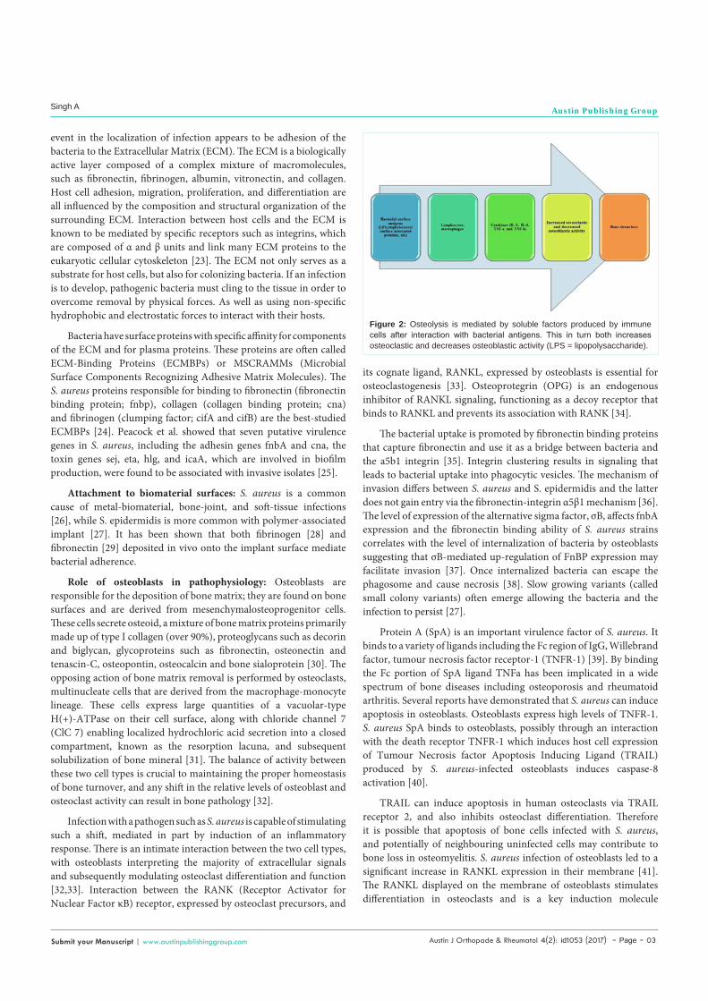
Figure 2: Osteolysis is mediated by soluble factors produced by immune cells after interaction with bacterial antigens. This in turn both increases osteoclastic and decreases osteoblastic activity (LPS = lipopolysaccharide).
Austin J Orthopade & Rheumatol 4(2): id1053 (2017) - Page - 04
Singh A Austin Publishing Group
Submit your Manuscript | www.austinpublishinggroup.com
involved in bone resorption leading to bone destruction. In essence binding of major S. aureus virulence protein, SpA with osteoblasts results in the generation of multiple signals leading to inhibition of osteoblast proliferation, induction of osteoblast apoptosis, inhibition of mineralization and release of mediators capable of inducing bone resorption via osteoclast activation [42]. So finally the observation was that the S. aureus can be internalized by osteoblasts may be relevant to this clinical problem (Figure 2).
Bacterial biofilm formation: A biofilm is defined as a microbially derived sessile community, typified by cells that are attached to a substratum, are embedded in a matrix of extracellular polymeric substance, and exhibit an altered phenotype with regard to growth, gene expression, and protein production, as well as by adopting the sessile mode of life, biofilm-embedded microorganisms benefit from a number of advantages over their planktonic counterparts (Table 3). Biofilm depth can vary, from a single cell layer to a thick community of cells surrounded by a thick polymeric milieu. Structural analyses have shown that these thick biofilms possess a complex architecture in which microcolonies can exist in distinct pillar or mushroom-shaped structures [43], through which an intricate channel network runs.
Formation of biofilm is a two-stage process in which bacteria first attach to a substrate (e.g., bone) and then attach to each other as the biofilm grows and matures. The two-stage process, the initial attachment appears to be dependent on the production of one or more protein adhesins, whereas the subsequent aggregation of bacteria into a biofilm is dependent on the production of exopolysaccharide adhesins [44]. It is known that once a biofilm has formed, the bacteria within the biofilm are protected from phagocytosis and antibiotics, and a mouse bacteraemia model found that the biofilm enhanced S. aureus virulence factors, such as the α-toxin [45]. A final detachment (or dispersal) phase involves the detachment of single cells or cell clusters and is believed to cause new infection sites in the human body.
Staphylococcus spp. can produce a multilayered biofilm embedded within a glycocalyx, or slime layer. Early studies described the solid component of the glycocalyx as primarily composed of teichoic acids (80%) and staphylococcal and host proteins. In recent years, the polysaccharide intercellular adhesin (PIA) has been found in many S. aureus strains, and is required for biofilm formation and bacterium-bacterium adhesion. The genes and products of the ica locus [icaR (regulatory) and icaADBC (biosynthetic) genes] have been demonstrated to be necessary for biofilm formation and virulence, and are up-regulated in response to anaerobic growth. Another important component of the staphylococcal biofilm is extracellular DNA (eDNA). The discovery that this substance is an important
component of biofilms was recently made in P. aeruginosa [46].
Immune response to bacterial biofilmIt is well known that the planktonic bacteria with immunoglobulin
and complement lead to activation of Polymorphonuclear Neutrophil (PMN) inducing phagocytosis and generation of reactive oxygen species. Bacterial biofilms are also attacked by PMNs but the immunological efficiency remains dependent on its maturation state. Owing to a comparably lower biofilm clearance, young biofilm structures are easier to target for PMNs [47].
Except for PMNs, the presence of a biofilm leads also to an activation of T-cells and monocytes and resulting altogether in a local increase of proinflammatory cytokines.It is assumed that the continuous release of inflammatory mediators is conducting osteolytic and tissue damaging processes [48]. A significantly higher amount of CD28−/CD4+ cells at a high level of activation and low proliferation level were found in samples of infected bone when compared with the healthy bone, indicating an increased cytotoxicity by higher expression of CD11b and secretion of perforin compared with CD28+ cells. As this characteristic expression it was presumed that this cellular pattern might have an impact on osteoclast activity and therefore on bone resorption processes. An increased expression of macrophage inflammatory proteins (MIP1[alpha], CCL3) and MIP2[alpha] (CXCL2) was found in bone samples of patients suffering from infection of orthopaedic prostheses with further close correlation of CD14 as a marker for macrophages and monocytes, respectively. As well as osteoblasts were also capable to produce macrophage inflammatory proteins in vitro when stimulated by bacteria [49].
Impact of bacterial invasion on bone tissue componentsThe presence of bacteria also directly influences the cellular
components of the bone tissue. In an invitro study, it was shown that an infection with S. aureus leads to an increase in expression of Tolllike receptor 2 (TLR2) as a part of the innate immune system, which is known to be upregulated in the process of microbial invasion. Apoptotic cell death was further induced and
Mitogen activated protein kinase pathways were activated in osteoblasts. The invasion expression of TLR2 and the activity level of Jun N-terminal kinases (JNK) were directly correlated to have a direct impact on osteoblast apoptosis and ostegenic differentiation after bacterial invasion [50]. It has recently been shown that bacterial endotoxins especially lipopolysaccharide, which localized in the outer membrane of Gramnegative bacteria, was also able to promote apoptosis and inhibit differentiation of osteoblasts by JNK pathway activation. These findings were confirmed in another invitro study, suggesting that methicillinresistant S. aureus biofilms excrete soluble
S. No. Advantages to Bacteria References
1. The capability of the extracellular matrix to seize and concentrate a number of environmental nutrients, such as carbon, nitrogen, and phosphate [44]
2.The facilitation of resistance to a number of removal tactics, such as elimination by antimicrobial agents, shear stress, host phagocytic clearance, and host oxygen radical and protease defences. This innate resistance to antimicrobial factors is mediated through very low metabolic levels and radically down-regulated rates of cell division of the deeply entrenched micro-organisms.
[44]
3.
The potential for dispersion via detachment. Microcolonies may detach under the direction of mechanical fluid shear or through a genetically programmed response that mediates the detachment process. Under the direction of fluid flow, this microcolony travels to other regions of the host system to attach and seeding of virgin surfaces may be accomplished by the migration of single, motile cells from the cores of attached microcolonies.
[40]
Table 3: By adopting the sessile mode of life, biofilm-embedded microorganisms benefit from a number of advantages over their planktonic counterparts.
Austin J Orthopade & Rheumatol 4(2): id1053 (2017) - Page - 05
Singh A Austin Publishing Group
Submit your Manuscript | www.austinpublishinggroup.com
molecules, which directly impacted the osteoblasts by a decrease in viability and osteogenic potential and indirectly by an increase of the expression of the receptor activator of nuclear factor kappa B (NFkB) ligand (RANKL) by osteoblasts, which is also known to promote osteoclast activity [49].
Adaptations of causing agentsThere are so many causing microorganisms but Staphylococcus
aureusis a major human pathogen and an important cause of death and morbidity worldwide. Of major concern are the continuous emergence and spread of environmental resistance, drugs and implant resistance as well as antibiotic resistant strains, such as Methicillin-Resistant S. aureus (MRSA), which have limited treatment options [51].
Antibiotic resistance: The increase in antibiotic resistance has been attributed to a combination of microbial characteristics, the selective pressure of antibiotic use and social and technical changes that enhance the transmission of resistant organisms. The growing threat from resistant organisms calls for concerted action to prevent the emergence of new resistant strains and the spread of existing ones [52]. Many procedures, use and misuse of antibiotics in man have resulted in antibiotic-resistant bacteria. The nutritive and therapeutic antibiotic treatment of farm animals amounts to a half of the world’s antibiotic output and has also resulted in antibiotic-resistant bacteria. Evidence is accumulating to support the hypothesis that antibiotic-resistant bacteria from poultry, pigs and cattle enter the food supply, can be found in human food [53], colonize human digestive tract and transfer resistance genes to human commensals.
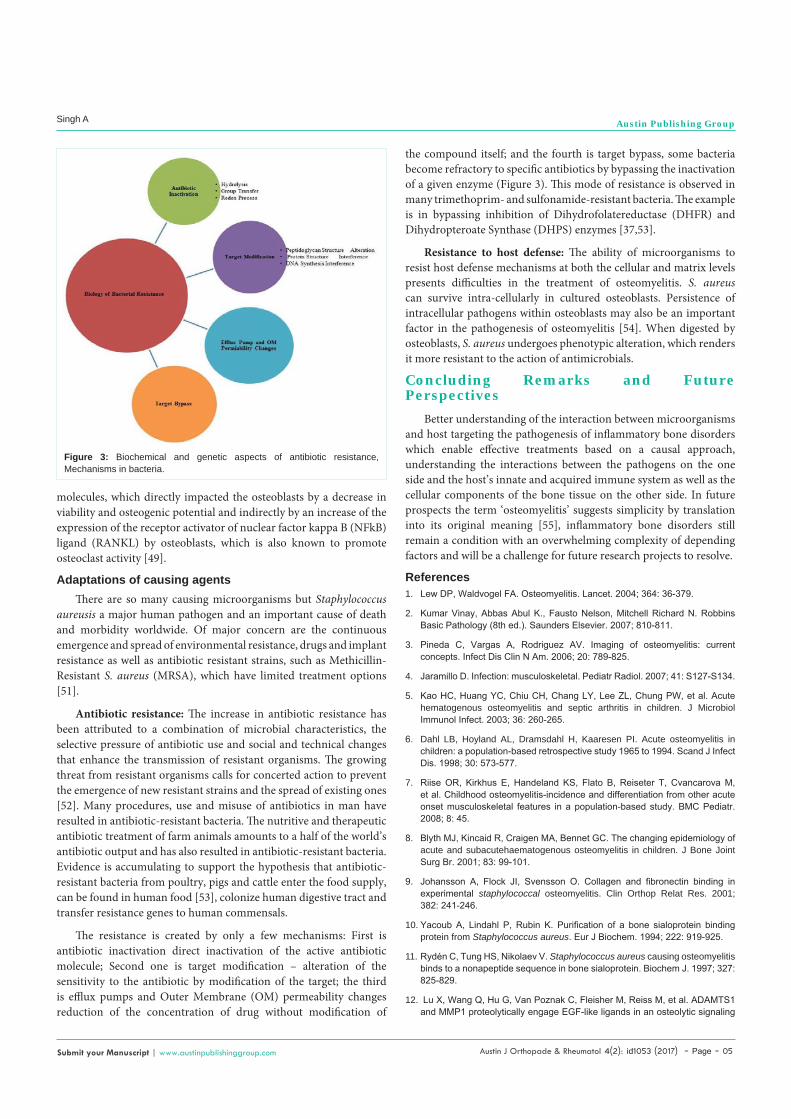
The resistance is created by only a few mechanisms: First is antibiotic inactivation direct inactivation of the active antibiotic molecule; Second one is target modification – alteration of the sensitivity to the antibiotic by modification of the target; the third is efflux pumps and Outer Membrane (OM) permeability changes reduction of the concentration of drug without modification of
the compound itself; and the fourth is target bypass, some bacteria become refractory to specific antibiotics by bypassing the inactivation of a given enzyme (Figure 3). This mode of resistance is observed in many trimethoprim- and sulfonamide-resistant bacteria. The example is in bypassing inhibition of Dihydrofolatereductase (DHFR) and Dihydropteroate Synthase (DHPS) enzymes [37,53].
Resistance to host defense: The ability of microorganisms to resist host defense mechanisms at both the cellular and matrix levels presents difficulties in the treatment of osteomyelitis. S. aureus can survive intra-cellularly in cultured osteoblasts. Persistence of intracellular pathogens within osteoblasts may also be an important factor in the pathogenesis of osteomyelitis [54]. When digested by osteoblasts, S. aureus undergoes phenotypic alteration, which renders it more resistant to the action of antimicrobials.
Concluding Remarks and Future Perspectives
Better understanding of the interaction between microorganisms and host targeting the pathogenesis of inflammatory bone disorders which enable effective treatments based on a causal approach, understanding the interactions between the pathogens on the one side and the host’s innate and acquired immune system as well as the cellular components of the bone tissue on the other side. In future prospects the term ‘osteomyelitis’ suggests simplicity by translation into its original meaning [55], inflammatory bone disorders still remain a condition with an overwhelming complexity of depending factors and will be a challenge for future research projects to resolve.
References1. Lew DP, Waldvogel FA. Osteomyelitis. Lancet. 2004; 364: 36-379.
2. Kumar Vinay, Abbas Abul K., Fausto Nelson, Mitchell Richard N. Robbins Basic Pathology (8th ed.). Saunders Elsevier. 2007; 810-811.
3. Pineda C, Vargas A, Rodriguez AV. Imaging of osteomyelitis: current concepts. Infect Dis Clin N Am. 2006; 20: 789-825.
4. Jaramillo D. Infection: musculoskeletal. Pediatr Radiol. 2007; 41: S127-S134.
5. Kao HC, Huang YC, Chiu CH, Chang LY, Lee ZL, Chung PW, et al. Acute hematogenous osteomyelitis and septic arthritis in children. J Microbiol Immunol Infect. 2003; 36: 260-265.
6. Dahl LB, Hoyland AL, Dramsdahl H, Kaaresen PI. Acute osteomyelitis in children: a population-based retrospective study 1965 to 1994. Scand J Infect Dis. 1998; 30: 573-577.
7. Riise OR, Kirkhus E, Handeland KS, Flato B, Reiseter T, Cvancarova M, et al. Childhood osteomyelitis-incidence and differentiation from other acute onset musculoskeletal features in a population-based study. BMC Pediatr. 2008; 8: 45.
8. Blyth MJ, Kincaid R, Craigen MA, Bennet GC. The changing epidemiology of acute and subacutehaematogenous osteomyelitis in children. J Bone Joint Surg Br. 2001; 83: 99-101.
9. Johansson A, Flock JI, Svensson O. Collagen and fibronectin binding in experimental staphylococcal osteomyelitis. Clin Orthop Relat Res. 2001; 382: 241-246.
10. Yacoub A, Lindahl P, Rubin K. Purification of a bone sialoprotein binding protein from Staphylococcus aureus. Eur J Biochem. 1994; 222: 919-925.
11. Rydén C, Tung HS, Nikolaev V. Staphylococcus aureus causing osteomyelitis binds to a nonapeptide sequence in bone sialoprotein. Biochem J. 1997; 327: 825-829.
12. Lu X, Wang Q, Hu G, Van Poznak C, Fleisher M, Reiss M, et al. ADAMTS1 and MMP1 proteolytically engage EGF-like ligands in an osteolytic signaling
Figure 3: Biochemical and genetic aspects of antibiotic resistance, Mechanisms in bacteria.
Austin J Orthopade & Rheumatol 4(2): id1053 (2017) - Page - 06
Singh A Austin Publishing Group
Submit your Manuscript | www.austinpublishinggroup.com
cascade for bone metastasis. 2009; 23: 1882-1894.
13. Neidhart M, Baraliakos X, Seemayer C, Zelder C, Gay R E, Michel B A, et al. Expression of cathepsin K and matrix metalloproteinase 1 indicate persistent osteodestructive activity in long-standing ankylosing spondylitis. Ann Rheum Dis. 2009; 68: 1334-1339.
14. Mayank Roy, Jeremy S. Somerson, Kevin G. Kerr, Jonathan L. Pathophysiology and Pathogenesis of Osteomyelitis. Osteomyelitis. 1-19.
15. Ocaña M G, Valle-Garay E, Montes A H, Meana A, Cartón J A, Fierer J, et al. Bax gene G(-248)A promoter polymorphism is associated with increased lifespan of the neutrophils of patients with osteomyelitis. Genet Med. 2007; 9: 249-255.
16. Ciampolini J, Harding KG. Pathophysiology of chronic bacterial osteomyelitis. Why do antibiotics fail so often? Postgrad Med J. 2000; 76: 479-483.
17. Martínez-Aguilar G1, Avalos-Mishaan A, Hulten K, Hammerman W, Mason EO Jr, Kaplan SL. Community-acquired, methicillinresistant and methicillin-susceptible Staphylococcus aureus musculoskeletal infections in children. Pediatr Infect Dis J. 2004; 23: 701-706.
18. Yagupsky P. Kingellakingae: from medical rarity to an emerging paediatric pathogen. Lancet Infect Dis 2004; 4: 358-367.
19. Ibia EO, Imoisili M, Pikis A. Group A beta-hemolytic streptococcal osteomyelitis in children. Pediatrics. 2003; 112: 22-26.
20. Tan TQ, Mason EO Jr, Barson WJ, Wald ER, Schutze GE, Bradley JS, et al. Clinical characteristics and outcome of children with pneumonia attributable to penicillin-susceptible and penicillin-nonsusceptible Streptococcus pneumoniae. Pediatrics. 1998; 102: 1369 -1375.
21. Mader JT, Shirtliff M, Calhoun JH. The host and the skeletal infection: classification and pathogenesis of acute bacterial bone and joint sepsis. Baillieres Best Pract Res Clin Rheumatol. 1999; 13: 1-20.
22. Arnold SR, Elias D, Buckingham SC, Thomas ED, Novais E, Arkader A, et al. Changing patterns of acute hematogenous osteomyelitis and septic arthritis: emergence of community-asso¬ciated methicillin-resistant Staphylococcus aureus. J Pediatr Orthop. 2006; 26: 703-708.
23. Ruoslahti E. Integrins. J Clin Invest, 1991; 87: 1-5.
24. Flock JI. Extracellular-matrix-binding proteins as targets for the prevention of Staphylococcus aureus infections. Mol Med Today, 1999; 5: 532-537.
25. Peacock SJ, Moore CE, Justice A, Kantzanou M, Story L, Mackie K, et al. Virulent combinations of adhesin and toxin genes in natural populations of Staphylococcus aureus. Infect Immun. 2002; 70: 4987-4996.
26. Petty W, Spanier S, Shuster JJ, Silverthorne C. The influence of skeletal implants on incidence of infection. Experiments in a canine model. J Bone Joint Surg Am. 1985; 67: 1236-1244.
27. Von Eiff C, Bettin D, Proctor RA, Rolauffs B, Lindner N, Winkelmann, W, et al. Recovery of small colony variants of Staphylococcus aureus following gentamicin bead placement for osteomyelitis. Clin Infect Dis. 1997; 25:1250-1251.
28. Brokke P, Dankert J, Carballo J, Feijen J. Adherence of coagulase-negative staphylococci onto polyethylene catheters in vitro and in vivo: a study on the influence of various plasma proteins. J Biomater Appl, 1991; 5: 204-226.
29. Fischer B, Vaudaux P, Magnin M, el Mestikawy Y, Proctor R A, Lew D P, et al. Novel animal model for studying the molecular mechanisms of bacterial adhesion to bone-implanted metallic devices: role of fibronectin in Staphylococcus aureus adhesion. J Orthop Res. 1996; 14: 914-920.
30. Mackie EJ. Osteoblasts: novel roles in orchestration of skeletal architecture. Int J Biochem Cell Biol. 2003; 35: 1301-1305.
31. Blair HC, Teitelbaum SL, Ghiselli R, Gluck S. Osteoclastic bone resorption by a polarized vacuolar proton pump. Science. 1989; 245: 855-857.
32. Henderson B, Nair SP. Hard labour: bacterial infection of the skeleton. Trends Microbiol. 2003; 11; 570-577.
33. Matsuo K, Irie N. Osteoclast-osteoblast communication. Arch Biochem
Biophys. 2008; 473:201-209.
34. Wada T, Nakashima T, Hiroshi N, Penninger, JM. RANKL-RANK signaling in osteoclastogenesis and bone disease. Trends Mol Med. 2006; 12: 17-25.
35. Sinha B, François PP, Nüsse O, Foti M, Hartford OM, Vaudaux P, et al. Fibronectin-binding protein acts as Staphylococcus aureus invasin via fibronectin bridging to integrin alpha5beta1. Cell Microbiol. 1999; 1: 101-117.
36. Khalil H, Williams RJ, Stenbeck G, Henderson B, Meghji S, Nair SP. Invasion of bone cells by Staphylococcus epidermidis. Microbes Infect. 2007; 9: 460-465.
37. Nair SP, Bischoff M, Senn MM, Berger-Bachi B. The sigma B regulon influences internalization of Staphylococcus aureus by osteoblasts. Infect Immun. 2003; 71: 4167-4170.
38. Wright JA, Nair SP. Interaction of staphylococci with bone. Int J Med Microbiol. 2010; 300: 193-204.
39. Gomez MI, OSeaghdha M, Magargee M, Foster TJ, Prince AS. Staphylococcus aureus protein A activates TNFR1 signaling through conserved IgG binding domains. J Biol Chem. 2006; 281: 20190-20196.
40. Alexander EH, Rivera FA, Marriott I, Anguita J, Bost KL, Hudson MC. Staphylococcus aureus - induced tumor necrosis factor - related apoptosis - inducing ligand expression mediates apoptosis and caspase-8 activation in infected osteoblasts. BMC Microbiol. 2003; 3: 1471-2180.
41. Somayaji SN, Ritchie S, Sahraei M, Marriott I, Hudson MC. Staphylococcus aureus induces expression of receptor activator of NF-kappaB ligand and prostaglandin E2 in infected murine osteoblasts. Infect Immun. 2008; 76: 5120-5126.
42. Claro T, Widaa A, OSeaghdha M, Miajlovic H, Foster TJ, OBrien FJ, et al. Staphylococcus aureus protein A binds to osteoblasts and triggers signals that weaken bone in osteomyelitis. PLoS One. 2011; 6: e18748.
43. Costerton JW, Lewandowski Z, Caldwell DE, Korber DR, Lappin-Scott HM. Microbial biofilms. Annu Rev Microbiol. 1995; 49: 711-745.
44. Heilmann C, Schweitzer O, Gerke C, Vanittanakom N, Mack D, Gotz F. Molecular basis of intercellular adhesion in the biofilm-forming Staphylococcus epidermidis. Mol Microbiol. 1996; 20: 1083-1091.
45. Thakker M, Park JS, Carey V, Lee JC. Staphylococcus aureus serotype 5 capsular polysaccharide is antiphagocytic and enhances bacterial virulence in a murine bacteremia model. Infect Immun, 1998; 66: 5183-5189.
46. Whitchurch CB, Tolker-Nielsen T, Ragas PC, Mattick, JS. Extracellular DNA required for bacterial biofilm formation. Science. 2002; 295: 1487.
47. Günther F, Wabnitz GH, Stroh P, Prior B, Obst U, Samstag Y, et al. Host defence against Staphylococcus aureus biofilms infection: phagocytosis of biofilms by polymorphonuclear neutrophils (PMN). Mol Immunol. 2009; 46:1805-1813.
48. Wagner C, Obst U, Hansch GM. Implant associated posttraumatic osteomyelitis: collateral damage by local host defense? Int J Artif Organs. 2005; 28: 1172-1180.
49. Beck-Broichsitter BE, Smeets R, Heiland M. Current Concepts in Pathogenesis of Acute and Chronic Osteomyelitis. Curr Opin Infect Dis. 2015; 28: 240-245.
50. Chen Q, Hou T, Luo F, Wu X, Xie Z, Xu J. Involvement of toll-like receptor 2 and proapoptotic signaling pathways in bone remodeling in osteomyelitis. Cell Physiol Biochem. 2014; 34: 1890-1900.
51. Grundmann H, Aires-de-Sousa M, Boyce J, Tiemersma E. Emergence and resurgence of methicillin-resistant Staphylococcus aureus as a public-health threat. Lancet. 2006; 368: 874-885.
52. Okeke IN, Klugman KP, Bhutta ZA, Duse AG, Jenkins P, O’Brien TF, et al. Antimicrobial resistance in developing countries. Part II: Strategies for containment. Lancet Infect Dis. 2005; 5: 568-580.
53. SenkaD`idi, Jagoda, Bla`enka Kos. Antibiotic Resistance Mechanisms in Bacteria: Biochemical and Genetic Aspects. Food Technol. Biotechnol. 2007; 46: 11-21.
Austin J Orthopade & Rheumatol 4(2): id1053 (2017) - Page - 07
Singh A Austin Publishing Group
Submit your Manuscript | www.austinpublishinggroup.com
54. Lambert PA Bacterial resistance to antibiotics: Modified target sites. Adv Drug Deliv Rev. 2005; 57: 1471-1485.
55. Szafranska AK, Oxley AP, Chaves-Moreno D, Horst SA, Roßlenbroich S,
Peters G, et al. High-Resolution Transcriptomic Analysis of the Adaptive Response of Staphylococcus aureus during Acute and Chronic Phases of Osteomyelitis. mbio.asm.org. 2014; 5: e01775-14.
Citation: Yadav M and Singh A. Osteomyelitis in Paediatric Age Group, Pathophysiology and Adaptive Responses of Causing Microbes. Austin J Orthopade & Rheumatol. 2017; 4(2): 1053.
Austin J Orthopade & Rheumatol - Volume 4 Issue 2 - 2017ISSN: 2472-369X | www.austinpublishinggroup.com Singh et al. © All rights are reserved