riluzole elevates glt-1 activity and levels in striatal astrocytes
TRANSCRIPT
Riluzole elevates GLT-1 activity and levels in striatal astrocytes
Marica Carboneb, Susan Dutyb, and Marcus Rattraya,b
aUniversity of Reading, Reading School of Pharmacy, 204 Hopkins Building, Whiteknights,Reading RG6 6UB, U.K.bKing’s College London, Wolfson Centre for Age-Related Diseases, Guy’s Campus, London SE11UL, U.K
AbstractDrugs which upregulate astrocyte glutamate transport may be useful neuroprotective compoundsby preventing excitotoxicity. We set up a new system to identify potential neuroprotective drugswhich act through GLT-1. Primary mouse striatal astrocytes grown in the presence of the growth-factor supplement G5 express high levels of the functional glutamate transporter, GLT-1 (alsoknown as EAAT2) as assessed by Western blotting and 3H-glutamate uptake assay, and levelsdecline following growth factor withdrawal. The GLT-1 transcriptional enhancer dexamethasone(0.1 or 1 μM) was able to prevent loss of GLT-1 levels and activity following growth factorwithdrawal. In contrast, ceftriaxone, a compound previously reported to enhance GLT-1expression, failed to regulate GLT-1 in this system. The neuroprotective compound riluzole (100μM) upregulated GLT-1 levels and activity, through a mechanism that was not dependent onblockade of voltage-sensitive ion channels, since zonasimide (1 mM) did not regulate GLT-1.Finally, CDP-choline (10 μM – 1 mM), a compound which promotes association of GLT-1/EAAT2 with lipid rafts was unable to prevent GLT-1 loss under these conditions. This observationextends the known pharmacological actions of riluzole, and suggests that this compound mayexert its neuroprotective effects through an astrocyte-dependent mechanism.
KeywordsEAAT2; neuroprotection; citicholine; Parkinson’s Disease; glutamate uptake; glutamatetransporters
1. IntroductionAstrocytes are considered to have a key role in regulating neurodegenerative diseaseprogression. One potentially important way of regulating glutamate levels, is the astrocyteglutamate transport system. Astrocytic glutamate transporters remove glutamate from thesynaptic cleft and thus control the duration and magnitude of glutamate’s actions (Beart andO’Shea, 2007; Danbolt, 2001). Down-regulation of the major transporter EAAT2 (known inrodents as GLT-1) is considered to be an important contributor to neurodegeneration and/ordisease symptoms (Beart and O’Shea, 2007; Rattray and Bendotti, 2006).
In the last few years, a number of groups have identified clinically-useful drugs which areable to elevate GLT-1 levels in vitro (Boston-Howes et al., 2008; Colton et al., 2010; Ganelet al., 2006; Li et al., 2010; Rothstein et al., 2005). In the first study of this kind, Rothstein’s
Address correspondence and reprint requests to Marcus Rattray, University of Reading, Reading School of Pharmacy, 204 HopkinsBuilding, Whiteknights, Reading RG6 6UB, U.K. [email protected] tel: +44(0)118 7892. Fax: +44(0)118 378 4703.
Europe PMC Funders GroupAuthor ManuscriptNeurochem Int. Author manuscript; available in PMC 2012 August 29.
Published in final edited form as:Neurochem Int. 2012 January ; 60(1): 31–38. doi:10.1016/j.neuint.2011.10.017.
Europe PM
C Funders A
uthor Manuscripts
Europe PM
C Funders A
uthor Manuscripts
group reported that an orally-active beta-lactam antibiotic, ceftriaxone, elevates GLT-1levels in vitro, and also has efficacy as a neuroprotectant in a transgenic mouse model ofmotor neurone disease (Rothstein et al., 2005).
We wish to identify drugs which act on astrocytes to elevate glutamate transporter levelsthat might afford symptomatic relief and neuroprotection in Parkinson’s disease by reducingextracellular glutamate levels. As a first step towards this goal, we used striatal astrocytes inculture to test a number of orally active compounds in clinical use for their ability to up-regulate GLT-1 levels and activity. Compounds of different classes and mechanisms werechosen that have been previously suggested to increase astrocyte glutamate transporterfunction or attenuate neurodegeneration in other cellular systems or in vivo, namelyceftriaxone (Lee et al., 2008; Miller et al., 2008; Rothstein et al., 2005), riluzole (Azbill etal., 2000; Dunlop et al., 2003; Frizzo et al., 2004; Fumagalli et al., 2008), dexamethasone(Zschocke et al., 2005), zonasimide (Asanuma et al., 2010; Ueda et al., 2003) and CDP-choline (Hurtado et al., 2005; Hurtado et al., 2008).
2. Materials and Methods2.1 Materials
Dexamethasone and riluzole hydrocloride were purchased from Tocris (Avonmouth, UK).Ceftriaxone sodium, citicoline sodium and zonisamide sodium salt and all other chemicals,unless specified otherwise were purchased from Sigma (Poole, UK). All cell culturereagents were obtained from Invitrogen (Paisley, UK).
2.2 Primary Culture of Mouse AstrocytesPrimary cultures of mouse striatal astrocytes were prepared from E15/E16 Swiss mouseembryos (NIH, Harlan, UK), a slight modification of previously reported methods (Bahia etal., 2008). Striata were dissected and gently dissociated by mechanical repetitive pipetting inphosphate-buffered saline (PBS, Ca2+- and Mg2+-free) supplemented with glucose (33 mM).Cells were plated into 6-well or 24-well Nunc multiwell plates that had been coatedpreviously overnight with 1.5 μg/mL poly L-ornithine (molecular weight 30,000-70,000)and then sequentially washed in water and PBS before coating with culture mediumsupplemented with 10% heat inactivated foetal bovine serum. Following removal of the finalcoating solution, cells were seeded (9.5 × 10⁴/cm2) in culture media a medium composed ofa mixture of Dulbecco’s modified Eagle’s medium and F-12 nutrient (1:1 v/v) supplementedwith 2 mM glutamine, 5 mM HEPES buffer (pH 7.4) and 10% heat inactivated fetal bovineserum. Cells were cultured at 37°C in a humidified atmosphere of 95% air and 5%. After 7days in vitro (DIV) the culture medium was supplemented with the defined culturesupplement G5 (insulin 5 μg/ml, transferrin 10 μg/ml, selenite 5.2 ng/ml, biotin 10 ng/ml,hydrocortisone 3.6 ng/ml, FGF2 5.2 ng/ml, EGF 10 ng/ml). After 4 DIV with G5supplemented medium, cells were used for 3-day drug treatment in the G5 withdrawngrowing medium when the astrocytes were confluent and no neurons could be found in thecultures by light microscopy. Cultures were also washed twice with PBS/glucose at 4 and 6DIV to remove neuronal cells. For some experiments, cerebral cortical astrocytes were alsoprepared for comparative studies.
2.3 Immunocytochemistry of primary astrocytesCells grown on poly-L-ornithine coated 12mm round glass coverslips were fixed with 4%paraformaldehyde for 10 min. The cells were rinsed in PBS and then blocked andpermeabilised with 0.2% Triton X-100/1% normal goat serum (NGS) in PBS and incubatedovernight in an antibody cocktail containing 1% NGS and a mouse monoclonal GFAPantibody (1:400, Sigma) and rabbit anti GLT-1 antibody (Ab12 1:1000, gift of Dr D. Pow,
Carbone et al. Page 2
Neurochem Int. Author manuscript; available in PMC 2012 August 29.
Europe PM
C Funders A
uthor Manuscripts
Europe PM
C Funders A
uthor Manuscripts
Brisbane, Australia) (Williams et al., 2005). The cells were rinsed in PBS and thenincubated 1 h at RT in PBS/ 1% NGS containing secondary antibodies anti-mouse Alexa488 (1:1000) and anti-rabbit Alexa 568 (1:1000) (Invitrogen, Paisley, UK). Nuclei werestained during 30 min with the nuclear dye Hoechst 33342 (1:1000) (Invitrogen, Paisley,UK). The coverslips were rinsed in PBS and mounted in an aqueous mounting media(Mowiol medium: 0.2 M Tris, pH 8.5, 1.6% Mowiol 40-88, 0.2% DABCO), 5% glycerol (v/v), 0.02% sodium azide). Control incubations leaving out the primary or secondaryantibodies were performed for each antibody.
2.4 Western Blot AnalysisCells grown in 6-well plates were rinsed with ice-cold PBS, pH 7.4, and scraped with lysisbuffer (50 mM Tris, 1% Triton X-100, 150 mM NaCl, 2 mM EDTA, 2 mM EGTA, 0.5 mMPMSF, 10 μg/ml leupeptin, 10 μg/ml antipain, 2 μg/ml pepstatin A, 1 μg/ml chymostatin, 5mM sodium pyrophosphate, 1 mM Na3VO4 and 50 mM NaF). Lysates were collected andcentrifuged at 2000 × g (5 min, 4 °C), after which protein concentration was determinedusing a Bradford protein assay (BioRad, Hemel Hempstead, UK). Protein samples (30 μg)were separated by 9% polyacrylamide gel electrophoresis and proteins transferred tonitrocellulose membranes (Hybond-ECL, GE Healthcare, Little Chalfont, U.K.) by semi-dryelectroblotting. Membranes were blocked in TBS (20 mM Tris, pH 7.5, 0.5 M NaCl)containing 4% skimmed milk for 1 h. After washes with TBS containing 0.05% Tween-20(TTBS), the membranes were incubated with the following antibodies in TTBS containing1% skimmed milk powder: rabbit anti-GLT-1 (1/4000, gift of D. Pow, Brisbane), rabbit anti-GLAST (Anti A522 1/5000, gift of Dr N. Danbolt, Oslo) (Lehre et al., 1995), and rabbitanti-GAPDH (1/2000, Calbiochem International, Merck Chemicals Ltd, Nottingham, UK).Subsequently, the antigen-antibody complex was detected with a horseradish peroxidase-conjugated goat anti-rabbit IgG (1/1000; Sigma, Poole, UK). Immunoreactive proteins weredetected using ECL Western blotting detection reagents and autoradiography (GEHealthcare, Little Chalfont, U.K.). Bands were analysed using ImageJ (NIH, Bethesda, MD,USA). GLT-1 quantification was obtained by obtaining densities for both the 70 KDa lower(monomeric) band and the higher molecular mass ca. 200 KDa(multimeric) bands, whichwere then combined, as described previously (Suchak et al., 2003). In all cases, GLT-1 andGLAST band densities were normalised to the housekeeping protein control beforecombintation for analysis. Data were analysed by One-Way ANOVA followed byBonferroni’s multiple comparison tests, where appropriate (Prism, GraphPad, La Jolla,USA). None of the drug treatments altered the levels of GAPDH protein.
2.5 Measurement of L-[3H]-glutamate transportUptake was carried out according to previously reported methods (Peacey et al., 2009).Astrocyte cultures in 24-well plates were incubated with radiolabeled glutamate (L-3[H]Glutamic acid, 20 nM, Amersham Biosciences) and unlabeled glutamate (Sigma) mixed toobtain a final concentration of 100μM in uptake buffer (5mM Tris, 140 mM NaCl, 2.5 mMKCl, 1.2 mM CaCl2, 1.2mM MgCl2, 1.2 mM K2HPO4, 10 mM Glucose and 10 mMHEPES, pH 7.4). In each experiment, Na+-dependent transport was estimated by subtractingthe data obtained by replacing sodium chloride with choline chloride. Uptake wasdetermined after 5 min by removing the radioactive solution and rinsing with ice-coldcholine chloride-containing Tris buffer (pH7.4). Cells were lysed in 0.1M NaOH, and theamount of the incorporated glutamate was determined by liquid scintillation counting of thecell lysate. When required, cells were incubated with the non-selective glutamate transporterinhibitor Threo-beta-benzyloxyaspartate (TBOA, Tocris Cookson, Avonmouth, UK. 1 mM)or the GLT-1 inhibitor WAY-213613 (gift of Dr. J. Dunlop, Wyeth, Princeton USA, 10 μM)at 37°C for 15 min before adding the substrate and during the glutamate uptake. Data wereanalysed by One-Way ANOVA followed by Bonferroni’s multiple comparison tests, where
Carbone et al. Page 3
Neurochem Int. Author manuscript; available in PMC 2012 August 29.
Europe PM
C Funders A
uthor Manuscripts
Europe PM
C Funders A
uthor Manuscripts
appropriate (Prism, GraphPad, La Jolla, USA). All experiments were carried out at least intriplicate, each experiment contained replicates from 2-3 culture wells.
3. Results3.1 Striatal astrocytes in vitro possess GLT-1 levels and activity
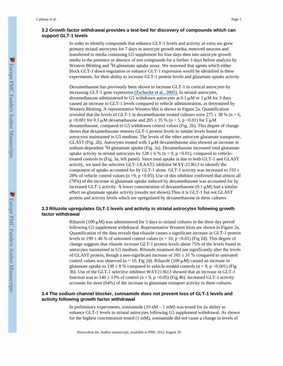
First we established cell culture conditions to support glutamate transporter expression instriatal astrocytes. E15/16 mouse striatal astrocytes that are grown in astrocyte growth media(DMEM/Hams F12 containing 10% FCS) have little expression of the GLT-1 glutamatetransporter after 10-12 days in culture (Fig 1a). Since GLT-1 expression in astrocytes isknown to be highly dependent on growth factors (Figiel et al., 2003; Gegelashvili et al.,1997; Vermeiren et al., 2005), we therefore developed a protocol based on the observationsof Vermeiren et al. (2005). After 7 days in vitro (DIV) in astrocyte growth medium, neuronsare removed by dislocation and the culture medium is replaced with G5-supplementedastrocyte growth medium. Fig. 1 shows that after 7 DIV in astrocyte growth mediumfollowed by 7 DIV in G5 supplemented astrocyte growth medium (G5 supplemented), theGLT-1 expression is detectable, and much higher than found in striatal astrocytes that aregrown for 14 DIV in unsupplemented astrocyte growth medium (i.e. without G5; Fig. 1a,1b), although not as high as typically found in cortical astrocytes (Fig 1c).
Western blotting confirms that there is a much higher level of GLT-1 expression in G5supplemented astrocytes compared to astrocytes grown in unsupplemented growth medium(Fig. 1d, compare first two lanes), with levels of expression in striatal astrocytes were lowerthan found in cortical astrocytes (Fig 1d). Fig 1e shows quantification of GLT-1 banddensities derived from Western blots from cortical or striatal astrocytes. For both types ofastrocytes, very little GLT-1 was detectable on Western Blots when grown for 14 days inunsupplemented astrocyte growth medium. When cortical or striatal astrocytes weresupplemented with G5, there was robust GLT-1 expression, G5 supplemented striatalastrocytes had approximately 45% lower GLT-1 levels than G5 supplemented corticalastrocytes (n = 2). If astrocytes were grown for 7 DIV in astrocyte growth medium followedby 4 DIV in G5 supplemented astrocyte growth medium, followed by 3 DIV in astrocytegrowth medium alone (G5 withdrawal), approximately two-thirds of GLT-1 protein waslost, with the loss similar for cortical and striatal astrocyte cultures), The GLT-1 expressionin cultures grown with G5 supplementation was, on average 264± 3.5% (n =6) compared toG5 withdrawal cultures. We interpret the reduction in GLT-1 between G5 supplemented andG5 withdrawn cultures as a loss of GLT-1 protein rather than an incomplete inductionGLT-1 gene expression, since in pilot experiments using cortical astrocytes, we found thatthe GLT-1 levels found after 7 days supplementation with G5 were no higher than foundafter 4 days supplementation with G5 (data not shown).
Pharmacological analysis (Fig. 1f) shows that the 3H-glutamate uptake in G5 supplementedstriatal astrocytes is completely blocked by replacing sodium with choline (inhibition = 99.3± 0.3%, n =3), and mostly blocked by the non-specific glutamate transporter inhibitor DL-TBOA (inhibition of 88.4 ± 2.7%, n =4), indicating that the bulk of glutamate uptake isthrough sodium-dependent glutamate transporters of the EAAT family. The GLT-1/EAAT2selective inhibitor WAY-213613 blocks about 40% of 3H-glutamate uptake (38.7 ± 14.3%,n =4), and about half of the uptake is blocked by the GLAST/EAAT1 preferring inhibitorSerine-o-sulphate (54.4 ± 4.3%, n =4) showing that both GLT-1 and GLAST contribute toglutamate uptake, as expected from studies of cortical astrocyte cultures grown under similarconditions e.g. Tortarolo et al. (2004). We note, however, that the relative proportion ofuptake due to GLT-1 varied somewhat between cultures. Consistent with Western blot data,G5 supplemented striatal astrocytes supported a greater level of glutamate uptake activitycompared to G5 withdrawal cultures (Fig. 1g).
Carbone et al. Page 4
Neurochem Int. Author manuscript; available in PMC 2012 August 29.
Europe PM
C Funders A
uthor Manuscripts
Europe PM
C Funders A
uthor Manuscripts
3.2 Growth factor withdrawal provides a test-bed for discovery of compounds which cansupport GLT-1 levels
In order to identify compounds that enhance GLT-1 levels and activity in vitro, we grewprimary striatal astrocytes for 7 days in astrocyte growth media, removed neurons andtransferred to media containing G5 supplement for four days then into astrocyte growthmedia in the presence or absence of test compounds for a further 3 days before analysis byWestern Blotting and 3H-glutamate uptake assay. We reasoned that agents which eitherblock GLT-1 down-regulation or enhance GLT-1 expression would be identified in theseexperiments, for their ability to increase GLT-1 protein levels and glutamate uptake activity.
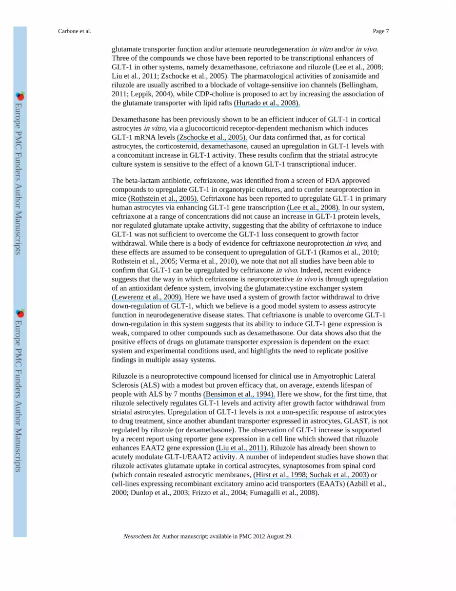
Dexamethasone has previously been shown to increase GLT-1 in cortical astrocytes byincreasing GLT-1 gene expression (Zschocke et al., 2005). In striatal astrocytes,dexamethasone administered to G5 withdrawn astrocytes at 0.1 μM or 1 μM for 3 dayscaused an increase in GLT-1 levels compared to vehicle administration, as determined byWestern Blotting. A representative Western blot is shown in Figure 2a. Quantificationrevealed that the levels of GLT-1 in dexamethasone treated cultures were 275 ± 38 % (n = 6,p <0.001 for 0.1 μM dexamathasone and 205 ± 35 % (n = 5, p <0.01) for 1 μMdexamethasone, compared to G5 withdrawn control values (Fig. 2b). This degree of changeshows that dexamethasone restores GLT-1 protein levels to similar levels found inastrocytes maintained in G5 medium. The levels of the other astrocyte glutamate transporter,GLAST (Fig. 2b). Astrocytes treated with 1 μM dexamethasone also showed an increase insodium-dependent 3H-glutamate uptake (Fig. 3a). Dexamethasone increased total glutamateuptake activity in striatal astrocytes by 128 ± 6 % (n = 9, p <0.01), compared to vehicle-treated controls to (Fig. 3a, left panel). Since total uptake is due to both GLT-1 and GLASTactivity, we used the selective GLT-1/EAAT2 inhibitor WAY-213613 to identify thecomponent of uptake accounted for by GLT-1 alone. GLT-1 activity was increased to 193 ±28% of vehicle control values (n =9, p <0.05). Use of this inhibitor confirmed that almost all(78%) of the increase in glutamate uptake induced by dexamethasone was accounted for byincreased GLT-1 activity. A lower concentration of dexamethasone (0.1 μM) had a similareffect on glutamate uptake activity (results not shown).Thus it is GLT-1 but not GLASTprotein and activity levels which are upregulated by dexamethasone in these cultures.
3.3 Riluzole upregulates GLT-1 levels and activity in striatal astrocytes following growthfactor withdrawal
Riluzole (100 μM) was administered for 3 days to striatal cultures in the three day periodfollowing G5 supplement withdrawal. Representative Western blots are shown in Figure 2a.Quantification of the data reveals that riluzole causes a significant increase in GLT-1 proteinlevels to 199 ± 46 % of untreated control values (n = 10, p <0.01) (Fig 2d). This degree ofchange suggests that riluzole increase GLT-1 protein levels about 75% of the levels found inastrocytes maintained in G5 medium. Riluzole treatment did not significantly alter the levelsof GLAST protein, though a non-significant increase of 165 ± 31 % compared to untreatedcontrol values was observed (n = 10, Fig 2d). Riluzole (100 μM) caused an increase inglutamate uptake to 138 ± 8 % compared to vehicle-treated controls (n = 9, p <0.001) (Fig3b). Use of the GLT-1 selective inhibitor WAY213613 showed that an increase in GLT-1function was to 140 ± 13% of control (n = 9, p <0.05) (Fig 4b). Increased GLT-1 activityaccounts for most (64%) of the increase in glutamate transport activity in these cultures.
3.4 The sodium channel blocker, zonisamide does not prevent loss of GLT-1 levels andactivity following growth factor withdrawal
In preliminary experiments, zonisamide (10 nM – 1 mM) was tested for its ability toenhance GLT-1 levels in striatal astrocytes following G5 supplement withdrawal. As shownfor the highest concentration tested (1 mM), zonisamide did not cause a change in levels of
Carbone et al. Page 5
Neurochem Int. Author manuscript; available in PMC 2012 August 29.
Europe PM
C Funders A
uthor Manuscripts
Europe PM
C Funders A
uthor Manuscripts
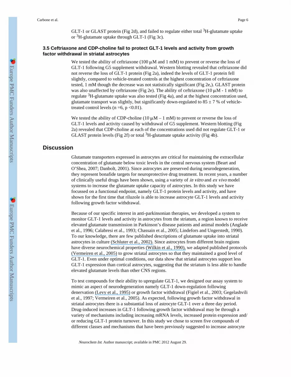
GLT-1 or GLAST protein (Fig 2d), and failed to regulate either total 3H-glutamate uptakeor 3H-glutamate uptake through GLT-1 (Fig 3c).
3.5 Ceftriaxone and CDP-choline fail to protect GLT-1 levels and activity from growthfactor withdrawal in striatal astrocytes
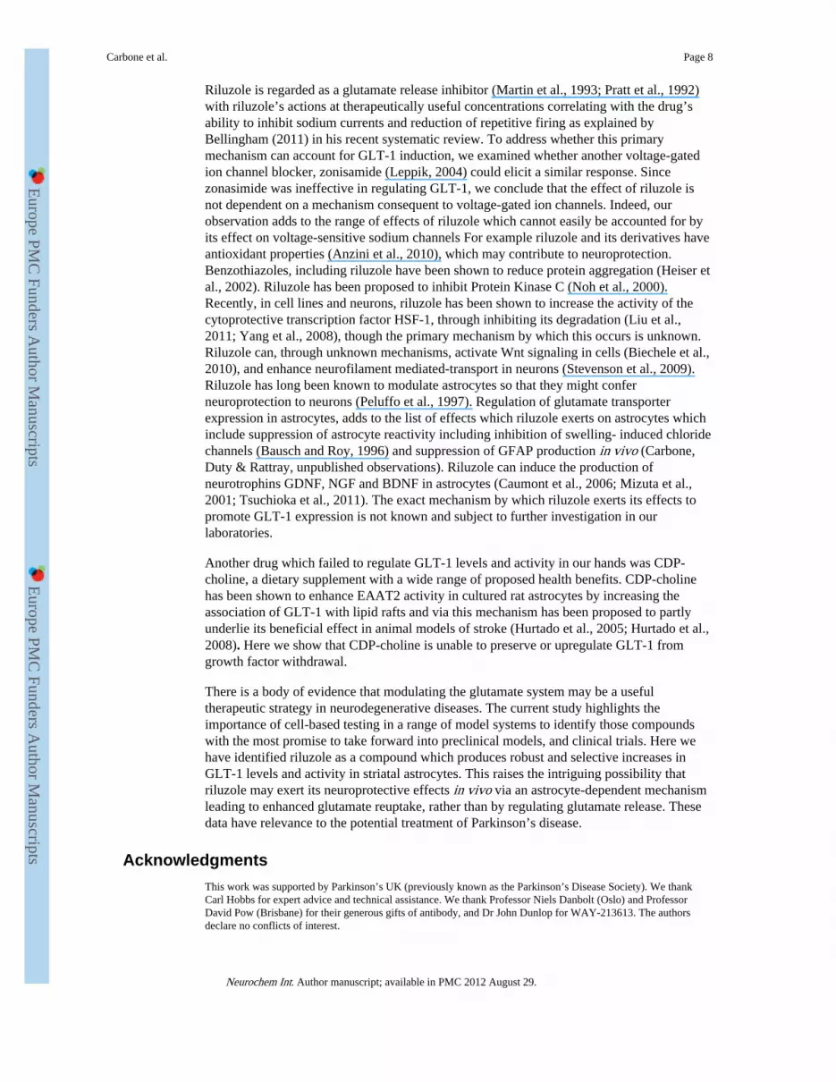
We tested the ability of ceftriaxone (100 μM and 1 mM) to prevent or reverse the loss ofGLT-1 following G5 supplement withdrawal. Western blotting revealed that ceftriaxone didnot reverse the loss of GLT-1 protein (Fig 2a), indeed the levels of GLT-1 protein fellslightly, compared to vehicle-treated controls at the highest concentration of ceftriaxonetested, 1 mM though the decrease was not statistically significant (Fig 2e,). GLAST proteinwas also unaffected by ceftriaxone (Fig 2e). The ability of ceftriaxone (10 μM - 1 mM) toregulate 3H-glutamate uptake was also tested (Fig 4a), and at the highest concentration used,glutamate transport was slightly, but significantly down-regulated to 85 ± 7 % of vehicle-treated control levels (n =6, p <0.01).
We tested the ability of CDP-choline (10 μM – 1 mM) to prevent or reverse the loss ofGLT-1 levels and activity caused by withdrawal of G5 supplement. Western blotting (Fig2a) revealed that CDP-choline at each of the concentrations used did not regulate GLT-1 orGLAST protein levels (Fig 2f) or total 3H-glutamate uptake activity (Fig 4b).
DiscussionGlutamate transporters expressed in astrocytes are critical for maintaining the extracellularconcentration of glutamate below toxic levels in the central nervous system (Beart andO’Shea, 2007; Danbolt, 2001). Since astrocytes are preserved during neurodegeneration,they represent bonafide targets for neuroprotective drug treatment. In recent years, a numberof clinically useful drugs have been shown, using a variety of in vitro and ex vivo modelsystems to increase the glutamate uptake capacity of astrocytes. In this study we havefocussed on a functional endpoint, namely GLT-1 protein levels and activity, and haveshown for the first time that riluzole is able to increase astrocyte GLT-1 levels and activityfollowing growth factor withdrawal.
Because of our specific interest in anti-parkinsonian therapies, we developed a system tomonitor GLT-1 levels and activity in astrocytes from the striatum, a region known to receiveelevated glutamate transmission in Parkinson’s disease patients and animal models (Angladeet al., 1996; Calabresi et al., 1993; Chassain et al., 2005; Lindefors and Ungerstedt, 1990).To our knowledge, there are few published descriptions of glutamate uptake into striatalastrocytes in culture (Schluter et al., 2002). Since astrocytes from different brain regionshave diverse neurochemical properties (Wilkin et al., 1990), we adapted published protocols(Vermeiren et al., 2005) to grow striatal astrocytes so that they maintained a good level ofGLT-1. Even under optimal conditions, our data show that striatal astrocytes support lessGLT-1 expression than cortical astrocytes, suggesting that the striatum is less able to handleelevated glutamate levels than other CNS regions.
To test compounds for their ability to upregulate GLT-1, we designed our assay system tomimic an aspect of neurodegeneration namely GLT-1 down-regulation followingdenervation (Levy et al., 1995) or growth factor withdrawal (Figiel et al., 2003; Gegelashviliet al., 1997; Vermeiren et al., 2005). As expected, following growth factor withdrawal instriatal astrocytes there is a substantial loss of astrocyte GLT-1 over a three day period.Drug-induced increases in GLT-1 following growth factor withdrawal may be through avariety of mechanisms including increasing mRNA levels, increased protein expression and/or reducing GLT-1 protein turnover. In this study we chose to screen five compounds ofdifferent classes and mechanisms that have been previously suggested to increase astrocyte
Carbone et al. Page 6
Neurochem Int. Author manuscript; available in PMC 2012 August 29.
Europe PM
C Funders A
uthor Manuscripts
Europe PM
C Funders A
uthor Manuscripts
glutamate transporter function and/or attenuate neurodegeneration in vitro and/or in vivo.Three of the compounds we chose have been reported to be transcriptional enhancers ofGLT-1 in other systems, namely dexamethasone, ceftriaxone and riluzole (Lee et al., 2008;Liu et al., 2011; Zschocke et al., 2005). The pharmacological activities of zonisamide andriluzole are usually ascribed to a blockade of voltage-sensitive ion channels (Bellingham,2011; Leppik, 2004), while CDP-choline is proposed to act by increasing the association ofthe glutamate transporter with lipid rafts (Hurtado et al., 2008).
Dexamethasone has been previously shown to be an efficient inducer of GLT-1 in corticalastrocytes in vitro, via a glucocorticoid receptor-dependent mechanism which inducesGLT-1 mRNA levels (Zschocke et al., 2005). Our data confirmed that, as for corticalastrocytes, the corticosteroid, dexamethasone, caused an upregulation in GLT-1 levels witha concomitant increase in GLT-1 activity. These results confirm that the striatal astrocyteculture system is sensitive to the effect of a known GLT-1 transcriptional inducer.
The beta-lactam antibiotic, ceftriaxone, was identified from a screen of FDA approvedcompounds to upregulate GLT-1 in organotypic cultures, and to confer neuroprotection inmice (Rothstein et al., 2005). Ceftriaxone has been reported to upregulate GLT-1 in primaryhuman astrocytes via enhancing GLT-1 gene transcription (Lee et al., 2008). In our system,ceftriaxone at a range of concentrations did not cause an increase in GLT-1 protein levels,nor regulated glutamate uptake activity, suggesting that the ability of ceftriaxone to induceGLT-1 was not sufficient to overcome the GLT-1 loss consequent to growth factorwithdrawal. While there is a body of evidence for ceftriaxone neuroprotection in vivo, andthese effects are assumed to be consequent to upregulation of GLT-1 (Ramos et al., 2010;Rothstein et al., 2005; Verma et al., 2010), we note that not all studies have been able toconfirm that GLT-1 can be upregulated by ceftriaxone in vivo. Indeed, recent evidencesuggests that the way in which ceftriaxone is neuroprotective in vivo is through upregulationof an antioxidant defence system, involving the glutamate:cystine exchanger system(Lewerenz et al., 2009). Here we have used a system of growth factor withdrawal to drivedown-regulation of GLT-1, which we believe is a good model system to assess astrocytefunction in neurodegenerative disease states. That ceftriaxone is unable to overcome GLT-1down-regulation in this system suggests that its ability to induce GLT-1 gene expression isweak, compared to other compounds such as dexamethasone. Our data shows also that thepositive effects of drugs on glutamate transporter expression is dependent on the exactsystem and experimental conditions used, and highlights the need to replicate positivefindings in multiple assay systems.
Riluzole is a neuroprotective compound licensed for clinical use in Amyotrophic LateralSclerosis (ALS) with a modest but proven efficacy that, on average, extends lifespan ofpeople with ALS by 7 months (Bensimon et al., 1994). Here we show, for the first time, thatriluzole selectively regulates GLT-1 levels and activity after growth factor withdrawal fromstriatal astrocytes. Upregulation of GLT-1 levels is not a non-specific response of astrocytesto drug treatment, since another abundant transporter expressed in astrocytes, GLAST, is notregulated by riluzole (or dexamethasone). The observation of GLT-1 increase is supportedby a recent report using reporter gene expression in a cell line which showed that riluzoleenhances EAAT2 gene expression (Liu et al., 2011). Riluzole has already been shown toacutely modulate GLT-1/EAAT2 activity. A number of independent studies have shown thatriluzole activates glutamate uptake in cortical astrocytes, synaptosomes from spinal cord(which contain resealed astrocytic membranes, (Hirst et al., 1998; Suchak et al., 2003) orcell-lines expressing recombinant excitatory amino acid transporters (EAATs) (Azbill et al.,2000; Dunlop et al., 2003; Frizzo et al., 2004; Fumagalli et al., 2008).
Carbone et al. Page 7
Neurochem Int. Author manuscript; available in PMC 2012 August 29.
Europe PM
C Funders A
uthor Manuscripts
Europe PM
C Funders A
uthor Manuscripts
Riluzole is regarded as a glutamate release inhibitor (Martin et al., 1993; Pratt et al., 1992)with riluzole’s actions at therapeutically useful concentrations correlating with the drug’sability to inhibit sodium currents and reduction of repetitive firing as explained byBellingham (2011) in his recent systematic review. To address whether this primarymechanism can account for GLT-1 induction, we examined whether another voltage-gatedion channel blocker, zonisamide (Leppik, 2004) could elicit a similar response. Sincezonasimide was ineffective in regulating GLT-1, we conclude that the effect of riluzole isnot dependent on a mechanism consequent to voltage-gated ion channels. Indeed, ourobservation adds to the range of effects of riluzole which cannot easily be accounted for byits effect on voltage-sensitive sodium channels For example riluzole and its derivatives haveantioxidant properties (Anzini et al., 2010), which may contribute to neuroprotection.Benzothiazoles, including riluzole have been shown to reduce protein aggregation (Heiser etal., 2002). Riluzole has been proposed to inhibit Protein Kinase C (Noh et al., 2000).Recently, in cell lines and neurons, riluzole has been shown to increase the activity of thecytoprotective transcription factor HSF-1, through inhibiting its degradation (Liu et al.,2011; Yang et al., 2008), though the primary mechanism by which this occurs is unknown.Riluzole can, through unknown mechanisms, activate Wnt signaling in cells (Biechele et al.,2010), and enhance neurofilament mediated-transport in neurons (Stevenson et al., 2009).Riluzole has long been known to modulate astrocytes so that they might conferneuroprotection to neurons (Peluffo et al., 1997). Regulation of glutamate transporterexpression in astrocytes, adds to the list of effects which riluzole exerts on astrocytes whichinclude suppression of astrocyte reactivity including inhibition of swelling- induced chloridechannels (Bausch and Roy, 1996) and suppression of GFAP production in vivo (Carbone,Duty & Rattray, unpublished observations). Riluzole can induce the production ofneurotrophins GDNF, NGF and BDNF in astrocytes (Caumont et al., 2006; Mizuta et al.,2001; Tsuchioka et al., 2011). The exact mechanism by which riluzole exerts its effects topromote GLT-1 expression is not known and subject to further investigation in ourlaboratories.
Another drug which failed to regulate GLT-1 levels and activity in our hands was CDP-choline, a dietary supplement with a wide range of proposed health benefits. CDP-cholinehas been shown to enhance EAAT2 activity in cultured rat astrocytes by increasing theassociation of GLT-1 with lipid rafts and via this mechanism has been proposed to partlyunderlie its beneficial effect in animal models of stroke (Hurtado et al., 2005; Hurtado et al.,2008). Here we show that CDP-choline is unable to preserve or upregulate GLT-1 fromgrowth factor withdrawal.
There is a body of evidence that modulating the glutamate system may be a usefultherapeutic strategy in neurodegenerative diseases. The current study highlights theimportance of cell-based testing in a range of model systems to identify those compoundswith the most promise to take forward into preclinical models, and clinical trials. Here wehave identified riluzole as a compound which produces robust and selective increases inGLT-1 levels and activity in striatal astrocytes. This raises the intriguing possibility thatriluzole may exert its neuroprotective effects in vivo via an astrocyte-dependent mechanismleading to enhanced glutamate reuptake, rather than by regulating glutamate release. Thesedata have relevance to the potential treatment of Parkinson’s disease.
AcknowledgmentsThis work was supported by Parkinson’s UK (previously known as the Parkinson’s Disease Society). We thankCarl Hobbs for expert advice and technical assistance. We thank Professor Niels Danbolt (Oslo) and ProfessorDavid Pow (Brisbane) for their generous gifts of antibody, and Dr John Dunlop for WAY-213613. The authorsdeclare no conflicts of interest.
Carbone et al. Page 8
Neurochem Int. Author manuscript; available in PMC 2012 August 29.
Europe PM
C Funders A
uthor Manuscripts
Europe PM
C Funders A
uthor Manuscripts
Abbreviations used
ALS amyotrophic lateral sclerosis
BDNF brain derived neurotrophic factor
CDP choline cytidine 5’diphosphocholine
DIV days in vitro
EAAT2 Excitatory amino acid transporter 2
EGF Epidermal Growth Factor
FGF2 Fibroblast Growth Factor 2
GAPDH glyceraldehyde-3-phosphate dehydrogenase
GDNF glial derived neurotrophic factor
GFAP glial fibrillary acidic protein
GLAST glutamate and aspartate transporter
GLT-1 glutamate transporter 1
NGF nerve growth factor
NGS normal goat serum
TBOA DL-Threo-beta-benzyloxyaspartate
ReferencesAnglade P, Mouatt-Prigent A, Agid Y, Hirsch E. Synaptic plasticity in the caudate nucleus of patients
with Parkinson’s disease. Neurodegeneration. 1996; 5:121–128. [PubMed: 8819132]
Anzini M, Chelini A, Mancini A, Cappelli A, Frosini M, Ricci L, Valoti M, Magistretti J, Castelli L,Giordani A, Makovec F, Vomero S. Synthesis and biological evaluation of amidine, guanidine, andthiourea derivatives of 2-amino(6-trifluoromethoxy)benzothiazole as neuroprotective agentspotentially useful in brain diseases. J Med Chem. 2010; 53:734–744. [PubMed: 19950903]
Asanuma M, Miyazaki I, Diaz-Corrales FJ, Kimoto N, Kikkawa Y, Takeshima M, Miyoshi K, MurataM. Neuroprotective effects of zonisamide target astrocyte. Annals of Neurology. 2010; 67:239–249.[PubMed: 20225289]
Azbill RD, Mu X, Springer JE. Riluzole increases high-affinity glutamate uptake in rat spinal cordsynaptosomes. Brain Res. 2000; 871:175–180. [PubMed: 10899284]
Bahia PK, Rattray M, Williams RJ. Dietary flavonoid (−)epicatechin stimulates phosphatidylinositol3-kinase-dependent anti-oxidant response element activity and up-regulates glutathione in corticalastrocytes. J Neurochem. 2008; 106:2194–2204. [PubMed: 18624917]
Bausch AR, Roy G. Volume-sensitive chloride channels blocked by neuroprotective drugs in humanglial cells (U-138MG). Glia. 1996; 18:73–77. [PubMed: 8891694]
Beart PM, O’Shea RD. Transporters for L-glutamate: an update on their molecular pharmacology andpathological involvement. Br J Pharmacol. 2007; 150:5–17. [PubMed: 17088867]
Bellingham MC. A review of the neural mechanisms of action and clinical efficiency of riluzole intreating amyotrophic lateral sclerosis: what have we learned in the last decade? CNS Neurosci Ther.2011; 17:4–31. [PubMed: 20236142]
Bensimon G, Lacomblez L, Meininger V, ALS/Riluzole Study Group. A controlled trial of riluzole inamyotrophic lateral sclerosis. N Engl J Med. 1994; 330:585–591. [PubMed: 8302340]
Biechele TL, Camp ND, Fass DM, Kulikauskas RM, Robin NC, White BD, Taraska CM, Moore EC,Muster J, Karmacharya R, Haggarty SJ, Chien AJ, Moon RT. Chemical-genetic screen identifiesriluzole as an enhancer of Wnt/beta-catenin signaling in melanoma. Chem Biol. 2010; 17:1177–1182. [PubMed: 21095567]
Carbone et al. Page 9
Neurochem Int. Author manuscript; available in PMC 2012 August 29.
Europe PM
C Funders A
uthor Manuscripts
Europe PM
C Funders A
uthor Manuscripts
Boston-Howes W, Williams EO, Bogush A, Scolere M, Pasinelli P, Trotti D. Nordihydroguaiareticacid increases glutamate uptake in vitro and in vivo: Therapeutic implications for amyotrophiclateral sclerosis. Experimental Neurology. 2008; 213:229–237. [PubMed: 18625223]
Calabresi P, Mercuri NB, Sancesario G, Bernardi G. Electrophysiology of dopamine-denervatedstriatal neurons. Implications for Parkinson’s disease. Brain. 1993; 116(Pt 2):433–452. [PubMed:8096420]
Caumont AS, Octave JN, Hermans E. Specific regulation of rat glial cell line-derived neurotrophicfactor gene expression by riluzole in C6 glioma cells. J Neurochem. 2006; 97:128–139. [PubMed:16524382]
Chassain C, Bielicki G, Donnat JP, Renou JP, Eschalier A, Durif F. Cerebral glutamate metabolism inParkinson’s disease: an in vivo dynamic (13)C NMS study in the rat. Exp Neurol. 2005; 191:276–284. [PubMed: 15649482]
Colton CK, Kong Q, Lai L, Zhu MX, Seyb KI, Cuny GD, Xian J, Glicksman MA, Lin CL.Identification of translational activators of glial glutamate transporter EAAT2 through cell-basedhigh-throughput screening: an approach to prevent excitotoxicity. J Biomol Screen. 2010; 15:653–662. [PubMed: 20508255]
Danbolt NC. Glutamate uptake. Progress in Neurobiology. 2001; 65:1–105. [PubMed: 11369436]
Dunlop J, Beal McIlvain H, She Y, Howland DS. Impaired Spinal Cord Glutamate Transport Capacityand Reduced Sensitivity to Riluzole in a Transgenic Superoxide Dismutase Mutant Rat Model ofAmyotrophic Lateral Sclerosis. J. Neurosci. 2003; 23:1688–1696. [PubMed: 12629173]
Figiel M, Maucher T, Rozyczka J, Bayatti N, Engele J. Regulation of glial glutamate transporterexpression by growth factors. Exp Neurol. 2003; 183:124–135. [PubMed: 12957496]
Frizzo, M.E.d.S.; Dall’Onder, LP.; Dalcin, KB.; Souza, DO. Riluzole Enhances Glutamate Uptake inRat Astrocyte Cultures. Cellular and Molecular Neurobiology. 2004; 24:123–128. [PubMed:15049516]
Fumagalli E, Funicello M, Rauen T, Gobbi M, Mennini T. Riluzole enhances the activity of glutamatetransporters GLAST, GLT1 and EAAC1. European Journal of Pharmacology. 2008; 578:171–176.[PubMed: 18036519]
Ganel R, Ho T, Maragakis NJ, Jackson M, Steiner JP, Rothstein JD. Selective up-regulation of theglial Na+-dependent glutamate transporter GLT1 by a neuroimmunophilin ligand results inneuroprotection. Neurobiology of Disease. 2006; 21:556–567. [PubMed: 16274998]
Gegelashvili G, Danbolt NC, Schousboe A. Neuronal soluble factors differentially regulate theexpression of the GLT1 and GLAST glutamate transporters in cultured astroglia. J Neurochem.1997; 69:2612–2615. [PubMed: 9375696]
Heiser V, Engemann S, Brocker W, Dunkel I, Boeddrich A, Waelter S, Nordhoff E, Lurz R, SchugardtN, Rautenberg S, Herhaus C, Barnickel G, Bottcher H, Lehrach H, Wanker EE. Identification ofbenzothiazoles as potential polyglutamine aggregation inhibitors of Huntington’s disease by usingan automated filter retardation assay. Proc Natl Acad Sci U S A. 2002; 99(Suppl 4):16400–16406.[PubMed: 12200548]
Hirst WD, Price GW, Rattray M, Wilkin GP. Serotonin transporters in adult rat brain astrocytesrevealed by [3H]5-HT uptake into glial plasmalemmal vesicles. Neurochem Int. 1998; 33:11–22.[PubMed: 9694037]
Hurtado O, Moro MA, Cardenas A, Sanchez V, Fernandez-Tome P, Leza JC, Lorenzo P, Secades JJ,Lozano R, Davalos A, Castillo J, Lizasoain I. Neuroprotection afforded by prior citicolineadministration in experimental brain ischemia: effects on glutamate transport. Neurobiol Dis.2005; 18:336–345. [PubMed: 15686962]
Hurtado O, Pradillo JM, Fernandez-Lopez D, Morales JR, Sobrino T, Castillo J, Alborch E, Moro MA,Lizasoain I. Delayed post-ischemic administration of CDP-choline increases EAAT2 associationto lipid rafts and affords neuroprotection in experimental stroke. Neurobiol Dis. 2008; 29:123–131. [PubMed: 17884513]
Lee S-G, Su Z-Z, Emdad L, Gupta P, Sarkar D, Borjabad A, Volsky DJ, Fisher PB. Mechanism ofCeftriaxone Induction of Excitatory Amino Acid Transporter-2 Expression and Glutamate Uptakein Primary Human Astrocytes. J. Biol. Chem. 2008; 283:13116–13123. [PubMed: 18326497]
Carbone et al. Page 10
Neurochem Int. Author manuscript; available in PMC 2012 August 29.
Europe PM
C Funders A
uthor Manuscripts
Europe PM
C Funders A
uthor Manuscripts
Lehre KP, Levy LM, Ottersen OP, Storm-Mathisen J, Danbolt NC. Differential expression of two glialglutamate transporters in the rat brain: quantitative and immunocytochemical observations. JNeurosci. 1995; 15:1835–1853. [PubMed: 7891138]
Leppik IE. Zonisamide: chemistry, mechanism of action, and pharmacokinetics. Seizure. 2004;13(Suppl 1):S5–9. discussion S10. [PubMed: 15511691]
Levy LM, Lehre KP, Walaas SI, Storm-Mathisen J, Danbolt NC. Down-regulation of glial glutamatetransporters after glutamatergic denervation in the rat brain. Eur J Neurosci. 1995; 7:2036–2041.[PubMed: 8542061]
Lewerenz J, Albrecht P, Tien ML, Henke N, Karumbayaram S, Kornblum HI, Wiedau-Pazos M,Schubert D, Maher P, Methner A. Induction of Nrf2 and xCT are involved in the action of theneuroprotective antibiotic ceftriaxone in vitro. J Neurochem. 2009; 111:332–343. [PubMed:19694903]
Li Y, Sattler R, Yang EJ, Nunes A, Ayukawa Y, Akhtar S, Ji G, Zhang PW, Rothstein JD. Harmine, anatural beta-carboline alkaloid, upregulates astroglial glutamate transporter expression.Neuropharmacology. 2010
Lindefors N, Ungerstedt U. Bilateral regulation of glutamate tissue and extracellular levels in caudate-putamen by midbrain dopamine neurons. Neurosci Lett. 1990; 115:248–252. [PubMed: 1978265]
Liu AY, Mathur R, Mei N, Langhammer CG, Babiarz B, Firestein BL. Neuroprotective drug riluzoleamplifies the heat shock factor 1 (HSF1)- and glutamate transporter 1 (GLT1)-dependentcytoprotective mechanisms for neuronal survival. J Biol Chem. 2011; 286:2785–2794. [PubMed:21098017]
Martin D, Thompson MA, Nadler JV. The neuroprotective agent riluzole inhibits release of glutamateand aspartate from slices of hippocampal area CA1. Eur J Pharmacol. 1993; 250:473–476.[PubMed: 8112408]
Miller BR, Dorner JL, Shou M, Sari Y, Barton SJ, Sengelaub DR, Kennedy RT, Rebec GV. Up-regulation of GLT1 expression increases glutamate uptake and attenuates the Huntington’s diseasephenotype in the R6/2 mouse. Neuroscience. 2008; 153:329–337. [PubMed: 18353560]
Mizuta I, Ohta M, Ohta K, Nishimura M, Mizuta E, Kuno S. Riluzole stimulates nerve growth factor,brain-derived neurotrophic factor and glial cell line-derived neurotrophic factor synthesis incultured mouse astrocytes. Neurosci Lett. 2001; 310:117–120. [PubMed: 11585581]
Noh KM, Hwang JY, Shin HC, Koh JY. A novel neuroprotective mechanism of riluzole: directinhibition of protein kinase C. Neurobiol Dis. 2000; 7:375–383. [PubMed: 10964608]
Peacey E, Miller CC, Dunlop J, Rattray M. The four major N- and C-terminal splice variants of theexcitatory amino acid transporter GLT-1 form cell surface homomeric and heteromeric assemblies.Mol Pharmacol. 2009; 75:1062–1073. [PubMed: 19201818]
Peluffo H, Estevez A, Barbeito L, Stutzmann JM. Riluzole promotes survival of rat motoneurons invitro by stimulating trophic activity produced by spinal astrocyte monolayers. Neurosci Lett. 1997;228:207–211. [PubMed: 9218644]
Pratt J, Rataud J, Bardot F, Roux M, Blanchard JC, Laduron PM, Stutzmann JM. Neuroprotectiveactions of riluzole in rodent models of global and focal cerebral ischaemia. Neurosci Lett. 1992;140:225–230. [PubMed: 1501783]
Ramos KM, Lewis MT, Morgan KN, Crysdale NY, Kroll JL, Taylor FR, Harrison JA, Sloane EM,Maier SF, Watkins LR. Spinal upregulation of glutamate transporter GLT-1 by ceftriaxone:therapeutic efficacy in a range of experimental nervous system disorders. Neuroscience. 2010;169:1888–1900. [PubMed: 20547213]
Rattray M, Bendotti C. Does excitotoxic cell death of motor neurons in ALS arise from glutamatetransporter and glutamate receptor abnormalities? Experimental Neurology. 2006; 201:15–23.[PubMed: 16806177]
Rothstein JD, Patel S, Regan MR, Haenggeli C, Huang YH, Bergles DE, Jin L, Dykes Hoberg M,Vidensky S, Chung DS, Toan SV, Bruijn LI, Su Z.-z. Gupta P, Fisher PB. [beta]-Lactamantibiotics offer neuroprotection by increasing glutamate transporter expression. Nature. 2005;433:73–77. [PubMed: 15635412]
Schluter K, Figiel M, Rozyczka J, Engele J. CNS region-specific regulation of glial glutamatetransporter expression. Eur J Neurosci. 2002; 16:836–842. [PubMed: 12372019]
Carbone et al. Page 11
Neurochem Int. Author manuscript; available in PMC 2012 August 29.
Europe PM
C Funders A
uthor Manuscripts
Europe PM
C Funders A
uthor Manuscripts
Stevenson A, Yates DM, Manser C, De Vos KJ, Vagnoni A, Leigh PN, McLoughlin DM, Miller CCJ.Riluzole protects against glutamate-induced slowing of neurofilament axonal transport.Neuroscience Letters. 2009; 454:161–164. [PubMed: 19429076]
Suchak SK, Baloyianni NV, Perkinton MS, Williams RJ, Meldrum BS, Rattray M. The ‘glial’glutamate transporter, EAAT2 (Glt-1) accounts for high affinity glutamate uptake into adult rodentnerve endings. J Neurochem. 2003; 84:522–532. [PubMed: 12558972]
Tortarolo M, Crossthwaite AJ, Conforti L, Spencer JP, Williams RJ, Bendotti C, Rattray M.Expression of SOD1 G93A or wild-type SOD1 in primary cultures of astrocytes down-regulatesthe glutamate transporter GLT-1: lack of involvement of oxidative stress. J Neurochem. 2004;88:481–493. [PubMed: 14690536]
Tsuchioka M, Hisaoka K, Yano R, Shibasaki C, Kajiatani N, Takebayashi M. Riluzole-induced glialcell line-derived neurotrophic factor production is regulated through fibroblast growth factorreceptor signaling in rat C6 glioma cells. Brain Res. 2011
Ueda Y, Doi T, Tokumaru J, Willmore LJ. Effect of zonisamide on molecular regulation of glutamateand GABA transporter proteins during epileptogenesis in rats with hippocampal seizures. BrainRes Mol Brain Res. 2003; 116:1–6. [PubMed: 12941455]
Verma R, Mishra V, Sasmal D, Raghubir R. Pharmacological evaluation of glutamate transporter 1(GLT-1) mediated neuroprotection following cerebral ischemia/reperfusion injury. Eur JPharmacol. 2010; 638:65–71. [PubMed: 20423712]
Vermeiren C, Najimi M, Maloteaux JM, Hermans E. Molecular and functional characterisation ofglutamate transporters in rat cortical astrocytes exposed to a defined combination of growth factorsduring in vitro differentiation. Neurochem Int. 2005; 46:137–147. [PubMed: 15627514]
Wilkin GP, Marriott DR, Cholewinski AJ. Astrocyte heterogeneity. Trends Neurosci. 1990; 13:43–46.[PubMed: 1690928]
Williams SM, Sullivan RK, Scott HL, Finkelstein DI, Colditz PB, Lingwood BE, Dodd PR, Pow DV.Glial glutamate transporter expression patterns in brains from multiple mammalian species. Glia.2005; 49:520–541. [PubMed: 15578656]
Yang J, Bridges K, Chen KY, Liu AY. Riluzole increases the amount of latent HSF1 for an amplifiedheat shock response and cytoprotection. PLoS ONE. 2008; 3:e2864. [PubMed: 18682744]
Zschocke J, Bayatti N, Clement AM, Witan H, Figiel M, Engele J, Behl C. Differential Promotion ofGlutamate Transporter Expression and Function by Glucocorticoids in Astrocytes from VariousBrain Regions. J. Biol. Chem. 2005; 280:34924–34932. [PubMed: 16079146]
Carbone et al. Page 12
Neurochem Int. Author manuscript; available in PMC 2012 August 29.
Europe PM
C Funders A
uthor Manuscripts
Europe PM
C Funders A
uthor Manuscripts
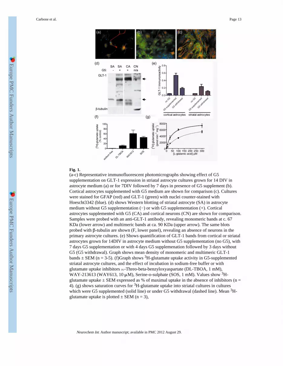
Fig. 1.(a-c) Representative immunofluorescent photomicrographs showing effect of G5supplementation on GLT-1 expression in striatal astrocyte cultures grown for 14 DIV inastrocyte medium (a) or for 7DIV followed by 7 days in presence of G5 supplement (b).Cortical astrocytes supplemented with G5 medium are shown for comparison (c). Cultureswere stained for GFAP (red) and GLT-1 (green) with nuclei counter-stained withHoescht3342 (blue). (d) shows Western blotting of striatal astrocyte (SA) in astrocytemedium without G5 supplementation (−) or with G5 supplementation (+). Corticalastrocytes supplemented with G5 (CA) and cortical neurons (CN) are shown for comparison.Samples were probed with an anti-GLT-1 antibody, revealing monomeric bands at c. 67KDa (lower arrow) and multimeric bands at ca. 90 KDa (upper arrow). The same blotsprobed with β-tubulin are shown (F, lower panel), revealing an absence of neurons in theprimary astrocyte cultures. (e) Shows quantification of GLT-1 bands from cortical or striatalastrocytes grown for 14DIV in astrocyte medium without G5 supplementation (no G5), with7 days G5 supplementation or with 4 days G5 supplemenation followed by 3 days withoutG5 (G5 withdrawal). Graph shows mean density of monomeric and multimeric GLT-1bands ± SEM (n = 3-5). (f)Graph shows 3H-glutamate uptake activity in G5-supplementedstriatal astrocyte cultures, and the effect of incubation in sodium-free buffer or withglutamate uptake inhibitors DL-Threo-beta-benzyloxyaspartate (DL-TBOA, 1 mM),WAY-213613 (WAY613, 10 μM), Serine-o-sulphate (SOS, 1 mM). Values show 3H-glutamate uptake ± SEM expressed as % of maximal uptake in the absence of inhibitors (n =4). (g) shows saturation curves for 3H-glutamate uptake into striatal cultures in cultureswhich were G5 supplemented (solid line) or under G5 withdrawal (dashed line). Mean 3H-glutamate uptake is plotted ± SEM (n = 3),
Carbone et al. Page 13
Neurochem Int. Author manuscript; available in PMC 2012 August 29.
Europe PM
C Funders A
uthor Manuscripts
Europe PM
C Funders A
uthor Manuscripts
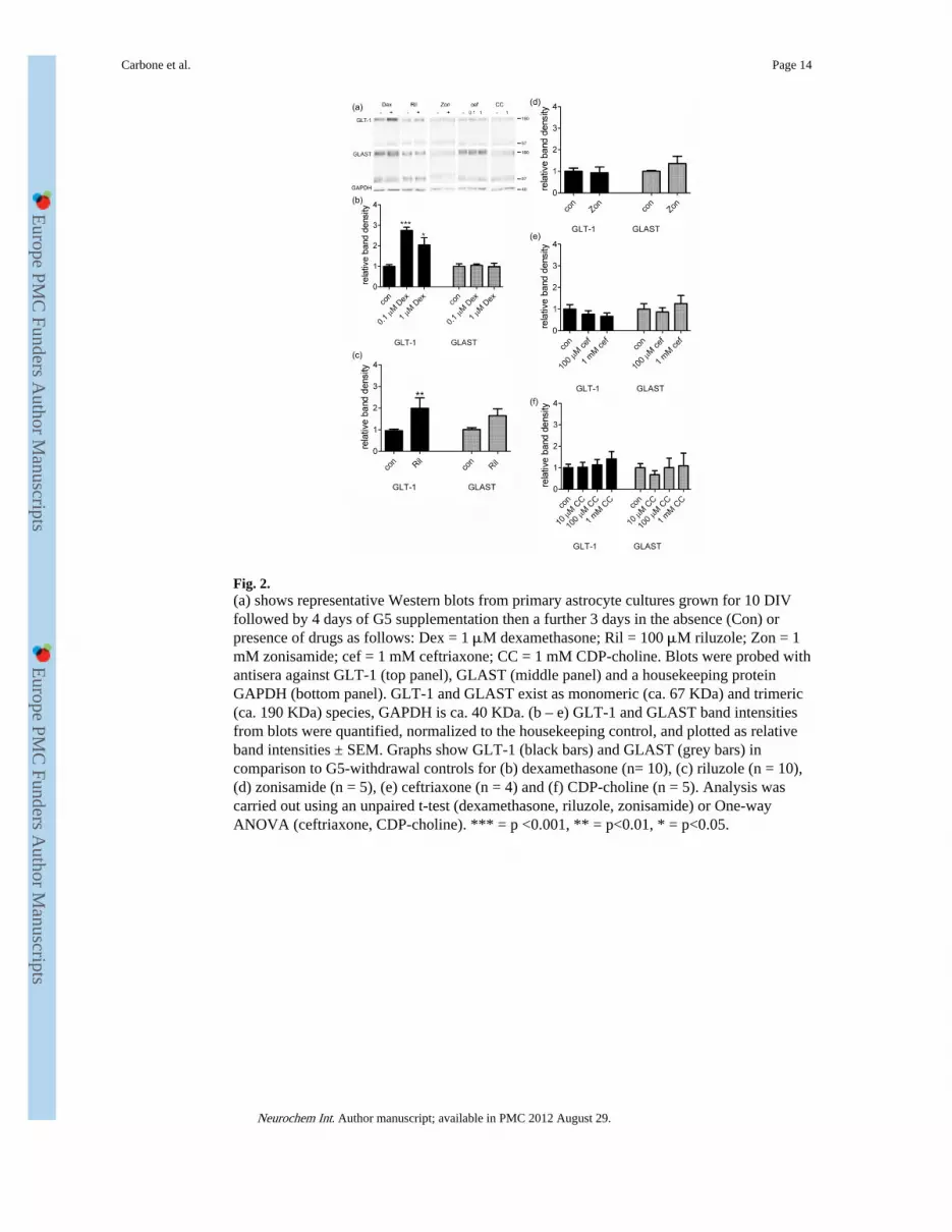
Fig. 2.(a) shows representative Western blots from primary astrocyte cultures grown for 10 DIVfollowed by 4 days of G5 supplementation then a further 3 days in the absence (Con) orpresence of drugs as follows: Dex = 1 μM dexamethasone; Ril = 100 μM riluzole; Zon = 1mM zonisamide; cef = 1 mM ceftriaxone; CC = 1 mM CDP-choline. Blots were probed withantisera against GLT-1 (top panel), GLAST (middle panel) and a housekeeping proteinGAPDH (bottom panel). GLT-1 and GLAST exist as monomeric (ca. 67 KDa) and trimeric(ca. 190 KDa) species, GAPDH is ca. 40 KDa. (b – e) GLT-1 and GLAST band intensitiesfrom blots were quantified, normalized to the housekeeping control, and plotted as relativeband intensities ± SEM. Graphs show GLT-1 (black bars) and GLAST (grey bars) incomparison to G5-withdrawal controls for (b) dexamethasone (n= 10), (c) riluzole (n = 10),(d) zonisamide (n = 5), (e) ceftriaxone (n = 4) and (f) CDP-choline (n = 5). Analysis wascarried out using an unpaired t-test (dexamethasone, riluzole, zonisamide) or One-wayANOVA (ceftriaxone, CDP-choline). *** = p <0.001, ** = p<0.01, * = p<0.05.
Carbone et al. Page 14
Neurochem Int. Author manuscript; available in PMC 2012 August 29.
Europe PM
C Funders A
uthor Manuscripts
Europe PM
C Funders A
uthor Manuscripts
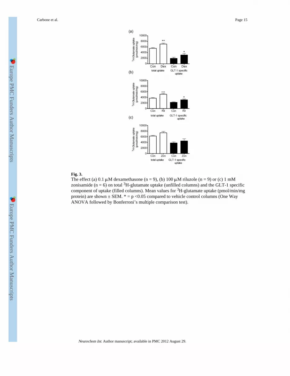
Fig. 3.The effect (a) 0.1 μM dexamethasone (n = 9), (b) 100 μM riluzole (n = 9) or (c) 1 mMzonisamide (n = 6) on total 3H-glutamate uptake (unfilled columns) and the GLT-1 specificcomponent of uptake (filled columns). Mean values for 3H-glutamate uptake (pmol/min/mgprotein) are shown ± SEM. * = p <0.05 compared to vehicle control columns (One WayANOVA followed by Bonferroni’s multiple comparison test).
Carbone et al. Page 15
Neurochem Int. Author manuscript; available in PMC 2012 August 29.
Europe PM
C Funders A
uthor Manuscripts
Europe PM
C Funders A
uthor Manuscripts
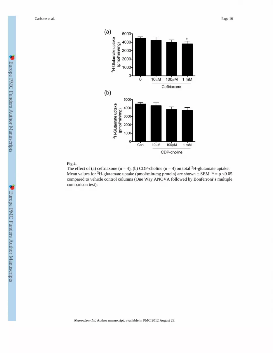
Fig 4.The effect of (a) ceftriaxone (n = 4), (b) CDP-choline (n = 4) on total 3H-glutamate uptake.Mean values for 3H-glutamate uptake (pmol/min/mg protein) are shown ± SEM. * = p <0.05compared to vehicle control columns (One Way ANOVA followed by Bonferroni’s multiplecomparison test).
Carbone et al. Page 16
Neurochem Int. Author manuscript; available in PMC 2012 August 29.
Europe PM
C Funders A
uthor Manuscripts
Europe PM
C Funders A
uthor Manuscripts