intracellular signaling mechanisms in photodynamic therapy
TRANSCRIPT
www.bba-direct.com
Biochimica et Biophysica Acta 1704 (2004) 59–86
Review
Intracellular signaling mechanisms in photodynamic therapy
Ramiro D. Almeida, Bruno J. Manadas, Arselio P. Carvalho, Carlos B. Duarte*
Center for Neuroscience and Cell Biology, Department of Zoology, University of Coimbra, Coimbra, 3004-517 Portugal
Received 14 January 2004; received in revised form 26 May 2004; accepted 28 May 2004
Available online 21 June 2004
Abstract
In photodynamic therapy (PDT) a sensitizer, light and oxygen are used to induce death of tumor cells and in the treatment of certain
noncancerous conditions. Cell death in PDT may occur by apoptosis or by necrosis, depending on the sensitizer, on the PDT dose and on
the cell genotype. Some sensitizers that have been used in PDT are accumulated in the mitochondria, and this may explain their
efficiency in inducing apoptotic cell death, both in vitro and in vivo. In this review we will focus on the events that characterize
apoptotic death in PDT and on the intracellular signaling events that are set in motion in photosensitized cells. Activation of
phospholipases, changes in ceramide metabolism, a rise in the cytosolic free Ca2 + concentration, stimulation of nitric oxide synthase
(NOS), changes in protein phosphorylation and alterations in the activity of transcription factors and on gene expression have all been
observed in PDT-treated cells. Although many of these metabolic reactions contribute to the demise process, some of them may
antagonize cell death. Understanding the signaling mechanisms in PDT may provide means to modulate the PDT effects at the molecular
level and potentiate its antitumor effectiveness.
D 2004 Elsevier B.V. All rights reserved.
Keywords: Photodynamic therapy; Apoptosis; Necrosis; Calcium; Phosphorylation
0304-419X/$ - see front matter D 2004 Elsevier B.V. All rights reserved.
doi:10.1016/j.bbcan.2004.05.003
Abbreviations: AIF, apoptosis-inducing factor; ALA, 5-aminolevulinate; AlPc, chloroaluminum phthalocyanine; AlPcS2, bisulfonated aluminum
phthalocyanine; AlPcS4, tetrasulfonated aluminum phthalocyanine; ANT, adenine nucleotide translocator; AP-1, transcription factor activator protein-1;
Apaf-1, apoptotic protease activating factor-1; APP, aminopyropheophorbide; ATX-S10, 13,17-bis(1-carboxypropionyl)carbamoylethyl-8-ethenyl-2-hydroxy-
3-hydroxyiminoethylidene-2,7,12,18-tetramethylporphyrin; 2-BA-2-DMHA, 2-butylamino-2-demethoxy-hypocrellin A; Bid, BH3-interacting domain death
agonist; BPD-MA, benzoporphyrin derivative monoacid ring A (verteporfin); CARD, caspase-recruitment domain; CDK, cyclin-dependent protein kinase;
COX, cyclooxygenase; CPO, 9-capronyloxy-tetrakis (methoxyethyl) porphycene; CrmA, viral cytokine response modifier A; DIABLO, direct inhibitor of
apoptosis-binding protein with low pI; DNA-PKCS, DNA-dependent protein kinase (catalytic subunit); E2F, E2F family of transcription factors; EGF,
epidermal growth factor; EGFR, EGF receptor; ER, endoplasmic reticulum; ERK, extracellular signal-regulated kinase; Etk/Bmx, epithelial and endothelial
tyrosine kinase or bone marrow tyrosine kinase gene in chromosome X; FADD, Fas-associated protein with a death domain; FAK, focal adhesion kinase;
FasL, Fas ligand; GFP, green fluorescent protein; GRP, glucose-regulated proteins; HPD, haematoporphyrin derivative; HPMA, N-(2-hydroxypropyl)-
methacrylamide; HPPH, 2-[1-hexyloxyethyl]-2-devinyl pyropheophorbide-a; HS1, haematopoietic lineage cell-specific protein 1; HSE, heat-shock elements;
HSF, heat-shock factors; HSP, heat-shock protein; IAP, inhibitor of apoptosis; ICAD, inhibitor of caspase activated DNAse; IL, interleukin; Ins(1,4,5)P3,
inositol 1,4,5-trisphosphate; JAK, Janus kinase; JNK/SAPK, c-Jun N-terminal kinase/stress activated protein kinase; LIF, leukemia inhibitory factor; LIFR,
LIF receptor; LFS, Li–Fraumeni syndrome; MAPK, mitogen-activated protein kinase; Mce6, mesochlorine e6 monoethylenediamine; MIP-2, macrophage
inflammatory protein-2; MKP-1, mitogen-activated protein kinase phosphatase 1; MMP, matrix metalloproteinases; MPTP, mitochondrial permeability
transition pore; NADPH-d, nicotinamide adenine dinucleotide hydrogen phosphate-diaphorase; mTHPC, meso-tetrahydroxyphenyl chlorin (FoscanR); NOS,nitric oxide synthase; NF-nB, nuclear factor kappa B; Npe6, mono-L-aspartyl chlorin e6; OSMRh, oncostatin receptor-h; PARP, poly(ADP-ribose)
polymerase; Pc 4, silicon phthalocyanine 4; PDT, photodynamic therapy; PF, proflavine; PG, prostaglandin; PI3-K, phosphatidylinositol-3-kinase; PI-PLC,
phosphatidylinositol-4,5-bisphosphate-specific phospholipase C; PKB, protein kinase B; PKC, protein kinase C; PPME, pyropheophorbide-a methyl ester;
cm, mitochondrial membrane potential; Rb, retinoblastoma; SERCA, sarco/endoplasmic reticulum Ca2+ ATPase; Smac, second mitochondria-derived
activator of caspase; SOD, superoxide dismutase; STAT, signal transducers and activators of transcription; TB, thromboxane; tBid, truncated form of Bid;
TBR, 2,4,5,7-tetrabromorhodamine 123 bromide; TNF, tumor necrosis factor; TPA, 12-O-tetradecanoylphorbol-13-acetate; VDAC, voltage-dependent anion
channel; VEGF, the vascular endothelial growth factor
* Corresponding author. Tel.: +351-239-480209; fax: +351-239-480208.
E-mail address: [email protected] (C.B. Duarte).
1. Introduction. . . . . . . . . . . . . . . . . . . . . . . . . . . . . . . . . . . . . . . . . . . . . 60
2. Mechanisms of cell death in PDT . . . . . . . . . . . . . . . . . . . . . . . . . . . . . . . . . 61
2.1. Apoptotic cell death in PDT . . . . . . . . . . . . . . . . . . . . . . . . . . . . . . . . 61
2.1.1. Death receptor-mediated apoptosis in PDT . . . . . . . . . . . . . . . . . . . . 61
2.1.2. Mitochondria-mediated apoptosis in PDT . . . . . . . . . . . . . . . . . . . . . 62
3. Effect of PDT on the [Ca2+]i homeostasis . . . . . . . . . . . . . . . . . . . . . . . . . . . . . 68
4. Activation of lipid metabolism by PDT . . . . . . . . . . . . . . . . . . . . . . . . . . . . . . 70
4.1. Phosphatidylinositol-specific phospholipase C (PI-PLC) . . . . . . . . . . . . . . . . . . 70
4.2. Phopholipase A2 and arachidonic acid metabolites . . . . . . . . . . . . . . . . . . . . . 70
4.3. Ceramide . . . . . . . . . . . . . . . . . . . . . . . . . . . . . . . . . . . . . . . . . . 70
5. Role of cyclic nucleotides in PDT . . . . . . . . . . . . . . . . . . . . . . . . . . . . . . . . . 72
5.1. cAMP . . . . . . . . . . . . . . . . . . . . . . . . . . . . . . . . . . . . . . . . . . . . 72
5.2. Nitric oxide and cGMP . . . . . . . . . . . . . . . . . . . . . . . . . . . . . . . . . . . 72
6. Role of MAPKs and CDKs in PDT . . . . . . . . . . . . . . . . . . . . . . . . . . . . . . . . 72
7. Differential effect of PDT on tyrosine kinases . . . . . . . . . . . . . . . . . . . . . . . . . . . 73
8. Regulation of transcription factors in PDT . . . . . . . . . . . . . . . . . . . . . . . . . . . . . 74
8.1. Transcription factor activator protein-1 (AP-1) . . . . . . . . . . . . . . . . . . . . . . . 74
8.2. Transcription nuclear factor kappa B (NF-nB) . . . . . . . . . . . . . . . . . . . . . . . 75
8.3. Retinoblastoma (Rb) and E2F family of transcription factors . . . . . . . . . . . . . . . . 75
9. Changes in protein levels in response to PDT . . . . . . . . . . . . . . . . . . . . . . . . . . . 76
9.1. Heme oxygenase . . . . . . . . . . . . . . . . . . . . . . . . . . . . . . . . . . . . . . 76
9.2. Glucose-regulated proteins . . . . . . . . . . . . . . . . . . . . . . . . . . . . . . . . . 76
9.3. Heat-shock proteins . . . . . . . . . . . . . . . . . . . . . . . . . . . . . . . . . . . . . 76
9.4. Anti-oxidant enzymes and apoptosis regulatory proteins . . . . . . . . . . . . . . . . . . 77
9.5. Clusterin . . . . . . . . . . . . . . . . . . . . . . . . . . . . . . . . . . . . . . . . . . 77
9.6. p53 . . . . . . . . . . . . . . . . . . . . . . . . . . . . . . . . . . . . . . . . . . . . . 77
9.7. Interleukins . . . . . . . . . . . . . . . . . . . . . . . . . . . . . . . . . . . . . . . . . 78
9.8. Regulators of angiogenesis . . . . . . . . . . . . . . . . . . . . . . . . . . . . . . . . . 78
Acknowledgements . . . . . . . . . . . . . . . . . . . . . . . . . . . . . . . . . . . . . . . . . . . 79
References . . . . . . . . . . . . . . . . . . . . . . . . . . . . . . . . . . . . . . . . . . . . . . . . 79
Contents
R.D. Almeida et al. / Biochimica et Biophysica Acta 1704 (2004) 59–8660
1. Introduction tumor tissue retention, rapid clearance from surrounding
Photodynamic therapy (PDT) of cancer is based on the
tumor-specific accumulation of a photosensitizer, often a
porphyrin derivative, followed by irradiation with visible
light, which induces cell death and tumor ablation. Photo-
frinR was the first photosensitizer approved for human use,
and has been successfully employed in the preatment of
many types of tumor, including lung, esophageal, cervical,
bladder and gastric tumors [2–4]. To enhance the potential
of PDT and its clinical applications, a second generation of
photosensitizers was produced, with properties comparable
or superior to PhotofrinR, including chemical purity, in-
creased photon absorption at longer wavelengths, improved
normal tissue, high quantum yield of reactive oxygen spe-
cies, and minimal toxicity in the dark [3,5–7]. The devel-
opment of new sensitizers, the acquired knowledge on the
conditions related with selective photosensitizer accumula-
tion in target tissues and the improvement in the systems
used for light delivery led to the development of PDT for use
against ophthalmological, cardiovascular and immune-me-
diated conditions [3,4,8,9]. In addition to the development of
new photosensitizers, the improvement of clinical PDT will
rely on the translation of information generated from studies
concerning the basic mechanisms involved in the demise
process in photosensitized cells. This review focuses on
recent advances in understanding the role of the intracellular
R.D. Almeida et al. / Biochimica et Biophysica Acta 1704 (2004) 59–86 61
signaling machinery in apoptotic death in PDT. Understand-
ing the signaling mechanisms in PDT may provide means to
modulate the PDT effects at the molecular level.
2. Mechanisms of cell death in PDT
The anti-cancer effects of PDT are thought to occur at
two different levels: (i) direct lethal effects on tumor cells,
and (ii) vascular impairment which limit blood supply to the
region [2]. The interaction of light with the photosensitizer
molecule raises its energy state and in the presence of
molecular oxygen leads to the formation of reactive oxygen
species, primarily singlet oxygen (1O2), which can react
with electron rich regions of many biomolecules, giving rise
to oxidized species [2,10]. Since singlet oxygen has a very
short lifetime in cells, its intracellular targets are located
close to the sites where the sensitizer is located [11].
Therefore, it is not surprising that the type of response
triggered by activation of the photosensitizers depends on
their intracellular localization [12–17]. The cell genotype
and the PDT dose were also found to determine whether cell
death occurs by apoptosis or necrosis [17–28]. Indeed,
apoptosis was the predominant mode of cell death when
murine leukemia P388 cells were photosensitized with
chloroaluminum phthalocyanine (AlPc), using low light
doses, whereas necrosis was observed for higher light doses
[18]. Similar results were obtained in studies where human
bladder carcinoma HT1197 cells were subjected to 5-amino-
levulinate (ALA)-induced PDT [24] and in CNE2 cells,
TWO-1 cells (human nasopharyngeal carcinoma cells) and
AY-27 cells (chemically-induced rat bladder carcinoma
cells) sensitized with hypericin [23,25]. These findings
indicate that the type of cell death switches from apoptosis
to necrosis with the increase of the intensity of the insult, as
previously reported for other apoptotic stimuli [29].
2.1. Apoptotic cell death in PDT
Two major apoptotic pathways have been characterized:
the death receptor-mediated, or extrinsic pathway, and the
mitochondria-mediated apoptosis, or intrinsic pathway [30–
32]. In the first pathway, cell surface receptors from the
tumor necrosis factor (TNF) gene family are stimulated,
activating the initiator caspase-8 via adaptor and scaffolding
proteins [33]. The second process is triggered by disruption
of mitochondrial function, which causes the release of
cytochrome C to the cytosol [34]. Released cytochrome C
binds Apaf-1 and induces its oligomerization, in the pres-
ence of dATP. This complex, termed apoptosome, recruits
and activates the initiator caspase-9 [35–37].
In both pathways, the activation of initiator caspases
(caspase-8 or caspase-9) leads to the activation of effector
caspases (caspase-3, -6 and -7) (Fig. 1). During apoptosis
the cells shrink, the nuclear chromatin becomes pyknotic
and condenses against the nuclear membrane, and eventu-
ally the cytoplasm and the nucleus break up into apoptotic
bodies. Although the cytoplasmic organelles remain intact,
DNA is digested at internucleosomal sites, giving rise to
fragments that are multiples of 180–200 bp [30–32].
Apoptotic cells and apoptotic bodies are engulfed and
cleared by phagocytes [38].
2.1.1. Death receptor-mediated apoptosis in PDT
Photosensitization of human epidermoid carcinoma
A431 cells with silicon phthalocyanine 4 (Pc 4) transiently
increased the protein levels of the surface death receptor Fas
(also known as APO-1 or CD95) and of its ligand FasL,
under conditions where cell death occurred by apoptosis
[39]. Under the same conditions, a multimerization of the
Fas protein and interaction of the receptor with the adaptor
molecule FADD (Fas-associated protein with a death do-
main) were also observed [39]. The Fas–FADD complex is
known to bind procaspase-8, and on this molecular scaffold,
procaspase-8 activates itself proteolytically and triggers the
death pathway [30–33]. Accordingly, caspase-8 cleavage
was observed in the human epidermoid carcinoma A431
cells sensitized with Pc 4, and pre-incubation of the cells
with rhFas:Fc fusion protein partially inhibited cell death
[39]. Antibodies against Fas or FasL also reduced apoptotic
death of poorly differentiated CNE2 and of moderately
differentiated TWO-1 human nasopharyngeal cells sub-
jected to PDT with Hypocrellin A and Hypocrellin B [40].
In in vivo studies, in which NR-S1 tumor-bearing mice were
treated by PDT with PhotofrinR, Fas-positive tumor cells
were observed in the same area where many TUNEL-
positive tumor cells were found, and expression of Fas
and FasL was also observed in the tumor cells surrounding
TUNEL-positive cells [41]. The role of the Fas/FasL system
in PDT may explain the cooperativity observed in MDCK II
(2–8 Madison–Darby canine kidney cells) and V79 (Chi-
nese hamster lung fibroblasts) cell death after photosensiti-
zation with PhotofrinR [42]. Accordingly, MDCK cells
were found to express Fas and FasL (e.g. see Ref. [43]).
Activation of caspase-8 was found to mediate apoptotic
death of human promyelocytic HL-60 cells photosensitized
with Rose Bengal [21,44]. In this model, inhibition of
caspase-8 also prevented cytochrome C release from mito-
chondria and cleavage of procaspase-3 [21], suggesting that
caspase-8 acts upstream of these events. Caspase-8 is known
to cleave Bid (BH3-interacting domain death agonist),
generating a C-terminal fragment of the protein, tBid, that
can activate the mitochondrial apoptotic pathway [45–47].
Bid seems to promote death by activating the pro-apoptotic
proteins Bax and Bak, and it may also inactivate pro-
survival proteins [45–47], thereby inducing the release of
cytochrome C (see below).
PDT with Rose Bengal increased p38 MAPK (mitogen-
activated protein kinase) activity, and inhibition of the kinase
prevented Bid cleavage, the decrease in mitochondrial mem-
brane potential and the release of cytochrome C from the
mitochondria [44]. Since cleavage of procaspase-8 was not
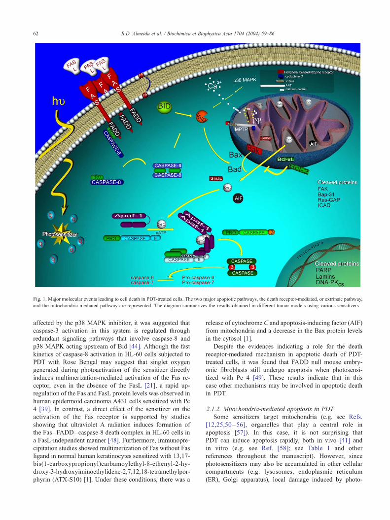
Fig. 1. Major molecular events leading to cell death in PDT-treated cells. The two major apoptotic pathways, the death receptor-mediated, or extrinsic pathway,
and the mitochondria-mediated-pathway are represented. The diagram summarizes the results obtained in different tumor models using various sensitizers.
R.D. Almeida et al. / Biochimica et Biophysica Acta 1704 (2004) 59–8662
affected by the p38 MAPK inhibitor, it was suggested that
caspase-3 activation in this system is regulated through
redundant signaling pathways that involve caspase-8 and
p38 MAPK acting upstream of Bid [44]. Although the fast
kinetics of caspase-8 activation in HL-60 cells subjected to
PDT with Rose Bengal may suggest that singlet oxygen
generated during photoactivation of the sensitizer directly
induces multimerization-mediated activation of the Fas re-
ceptor, even in the absence of the FasL [21], a rapid up-
regulation of the Fas and FasL protein levels was observed in
human epidermoid carcinoma A431 cells sensitized with Pc
4 [39]. In contrast, a direct effect of the sensitizer on the
activation of the Fas receptor is supported by studies
showing that ultraviolet A radiation induces formation of
the Fas–FADD–caspase-8 death complex in HL-60 cells in
a FasL-independent manner [48]. Furthermore, immunopre-
cipitation studies showed multimerization of Fas without Fas
ligand in normal human keratinocytes sensitized with 13,17-
bis(1-carboxypropionyl)carbamoylethyl-8-ethenyl-2-hy-
droxy-3-hydroxyiminoethylidene-2,7,12,18-tetramethylpor-
phyrin (ATX-S10) [1]. Under these conditions, there was a
release of cytochrome C and apoptosis-inducing factor (AIF)
from mitochondria and a decrease in the Bax protein levels
in the cytosol [1].
Despite the evidences indicating a role for the death
receptor-mediated mechanism in apoptotic death of PDT-
treated cells, it was found that FADD null mouse embry-
onic fibroblasts still undergo apoptosis when photosensi-
tized with Pc 4 [49]. These results indicate that in this
case other mechanisms may be involved in apoptotic death
in PDT.
2.1.2. Mitochondria-mediated apoptosis in PDT
Some sensitizers target mitochondria (e.g. see Refs.
[12,25,50–56], organelles that play a central role in
apoptosis [57]). In this case, it is not surprising that
PDT can induce apoptosis rapidly, both in vivo [41] and
in vitro (e.g. see Ref. [58]; see Table 1 and other
references throughout the manuscript). However, since
photosensitizers may also be accumulated in other cellular
compartments (e.g. lysosomes, endoplasmic reticulum
(ER), Golgi apparatus), local damage induced by photo-

Table 1
Major molecular events during apoptotic death induced by PDT
Cellular event Sensitizer Cell line References
Cytochrome C release Aluminum phthalocyanine Human breast epithelial cell line MCF10A [94]
from mitochondria 5-Aminolevulinate (ALA) Human promyelocytic leukemia HL60 cell line [78]
N-aspartyl chlorin e6 Mouse leukemia L1210 cells [278]
Murine hepatoma Hepa 1c1c7 [60]
ATX-S10 Normal human keratinocytes [1]
Benzoporphyrin derivative monoacid Human adenocarcinoma HeLa cells [71,73]
ring A (BPD-MA, verteporfin) Human umbilical venous endothelial cells [72]
9-capronyloxy-tetrakis (methoxyethyl)
porphycene (CPO)
Mouse leukemia L1210 cells [59]
Hypericin Human adenocarcinoma HeLa cells [20]
Rat/mouse T cell hybridoma [68]
Meta-tetra(hydroxyphenyl)-chlorin
(mTHPC; FoscanR)Human breast adenocarcinoma MCF-7 cells [61]
Silicon phthalocyanine 4 (Pc 4) Human epidermoid carcinoma A431 cells [54]
L5178Y-R mouse lymphoma [64,70]
Jurkat human lymphoma T cells [85]
Human breast adenocarcinoma MCF-7c3 cells [88,99,100]
Pheophorbide a Chinese hamster V79 cells [63]
Porphycene monomer (PcM) Murine leukemia P388 cells [67]
Rose Bengal Human promyelocytic leukemia HL60 cell line [44]
Loss of mitochondrial Aluminum phthalocyanine Human breast epithelial cell line MCF10A [94]
membrane potential 5-Aminolevulinate (ALA) Human promyelocytic leukemia HL60 cell line [78]
Human breast cancer MCF-7 cells and
adriamycin-resistant MCF-7/ADR cells
[28]
N-aspartyl chlorin e6 Murine hepatoma Hepa 1c1c7 [60]
9-Capronyloxy-tetrakis
(methoxyethyl) porphycene (CPO)
Mouse leukemia L1210 cells [59]
13,15-N-cycloimide derivatives of 13,
15-[N-(3-hydroxypropyl)]cycloimide
chlorin p6
Human lung adenocarcinoma A549 cells [17]
5,10-di[4-(N-trimethylaminophenyl)-15,
20-diphenylporphyrin (DADP-a)
Mouse leukemia L1210 cells [16]
Hematoporphyrin Human chronic myelogenous leukemia K562 cells [65]
Hypericin J Jhan cells (derived from human
lymphoblastoid Jurkat cells)
[69]
Human glioblastoma SNB-19 cell line [256]
Rat/mouse T cell hybridoma (PC60R1R2) [68]
Meta-tetra(hydroxyphenyl)-chlorin
(mTHPC; FoscanR)Human breast adenocarcinoma MCF-7 cells [61]
Monoglucosylporphyrin Human chronic myelogenous leukemia K562 cells [65]
Silicon phthalocyanine 4 (Pc 4) Human epidermoid carcinoma A431 cells [54]
Jurkat human lymphoma T cells [161]
Porphycene monomer (PcM) Murine leukemia P388 cells [67]
Rose Bengal Human promyelocytic leukemia HL60 cell line [44]
Tetrasulfonated aluminum (III)
phthalocanine (AlPcS4)
Human epidermoid carcinoma A431 cells [66]
Cleavage of pro-caspase-9 5-Aminolevulinate (ALA) Human promyelocytic leukemia HL60 cell line [78]
/caspase-9 formation N-aspartyl chlorin e6 Murine hepatoma Hepa 1c1c7 [60]
Benzoporphyrin derivative monoacid Human adenocarcinoma HeLa cells [73]
ring A (BPD-MA, verteporfin) Human promyelocytic leukemia HL60 cell line [79]
Human umbilical venous endothelial cells [72]
Bisulfonated aluminum
phthalocyanine (AlPcS2)
Human promyelocytic leukemia HL60 cell line [183]
Silicon phthalocyanine 4 (Pc 4) SW480 colon cancer xenographs grown in
athymic nude mice
[231]
Human breast adenocarcinoma MCF-7c3 cells [88]
Cleavage of pro-caspase-3 5-Aminolevulinate (ALA) Human promyelocytic leukemia HL60 cell line [78]
/caspase-3 formation Benzoporphyrin derivative monoacid Human promyelocytic leukemia HL60 cell line [75,76,79]
ring A (BPD-MA, verteporfin) Human umbilical venous endothelial cells [72]
Human promyelocytic leukemia HL60 cell line [77,79]
Human adenocarcinoma HeLa cells [71,73]
(continued on next page)
R.D. Almeida et al. / Biochimica et Biophysica Acta 1704 (2004) 59–86 63
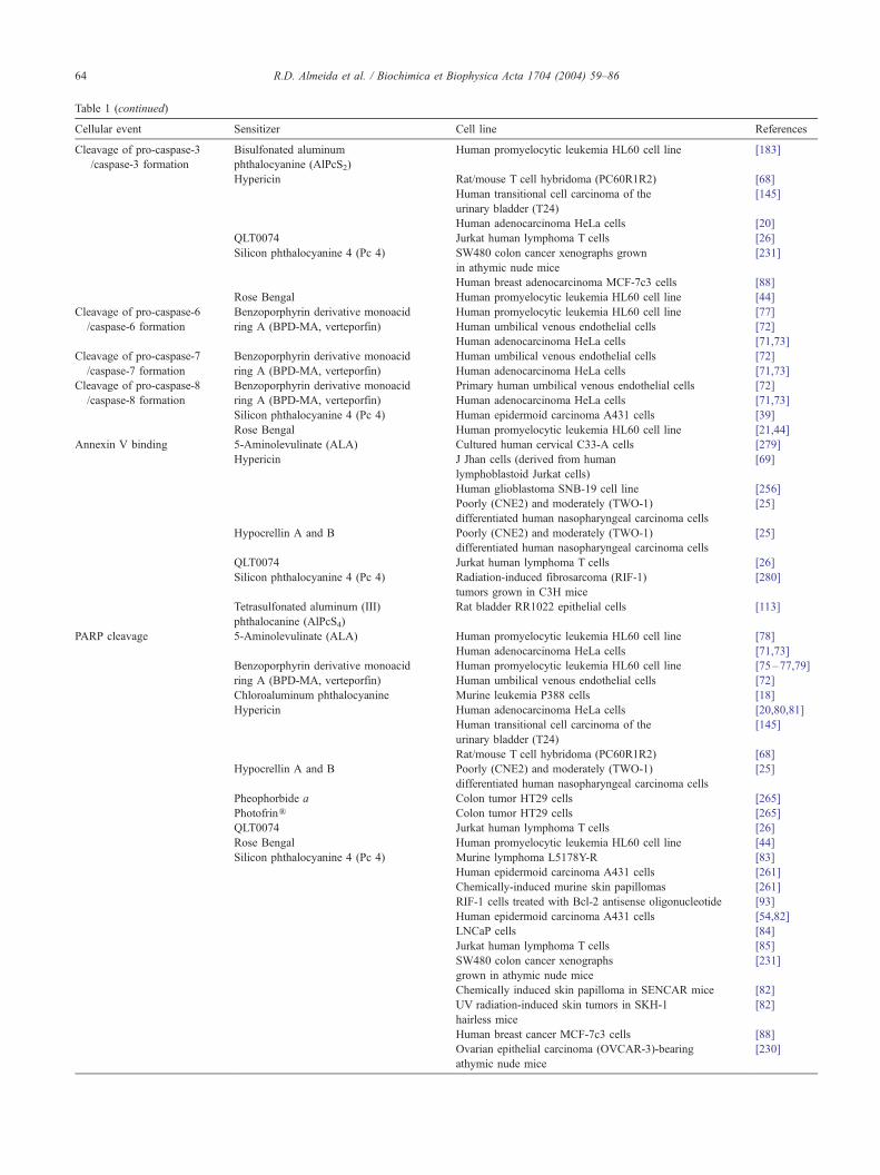
Table 1 (continued)
Cellular event Sensitizer Cell line References
Cleavage of pro-caspase-3
/caspase-3 formation
Bisulfonated aluminum
phthalocyanine (AlPcS2)
Human promyelocytic leukemia HL60 cell line [183]
Hypericin Rat/mouse T cell hybridoma (PC60R1R2) [68]
Human transitional cell carcinoma of the
urinary bladder (T24)
[145]
Human adenocarcinoma HeLa cells [20]
QLT0074 Jurkat human lymphoma T cells [26]
Silicon phthalocyanine 4 (Pc 4) SW480 colon cancer xenographs grown
in athymic nude mice
[231]
Human breast adenocarcinoma MCF-7c3 cells [88]
Rose Bengal Human promyelocytic leukemia HL60 cell line [44]
Cleavage of pro-caspase-6 Benzoporphyrin derivative monoacid Human promyelocytic leukemia HL60 cell line [77]
/caspase-6 formation ring A (BPD-MA, verteporfin) Human umbilical venous endothelial cells [72]
Human adenocarcinoma HeLa cells [71,73]
Cleavage of pro-caspase-7 Benzoporphyrin derivative monoacid Human umbilical venous endothelial cells [72]
/caspase-7 formation ring A (BPD-MA, verteporfin) Human adenocarcinoma HeLa cells [71,73]
Cleavage of pro-caspase-8 Benzoporphyrin derivative monoacid Primary human umbilical venous endothelial cells [72]
/caspase-8 formation ring A (BPD-MA, verteporfin) Human adenocarcinoma HeLa cells [71,73]
Silicon phthalocyanine 4 (Pc 4) Human epidermoid carcinoma A431 cells [39]
Rose Bengal Human promyelocytic leukemia HL60 cell line [21,44]
Annexin V binding 5-Aminolevulinate (ALA) Cultured human cervical C33-A cells [279]
Hypericin J Jhan cells (derived from human
lymphoblastoid Jurkat cells)
[69]
Human glioblastoma SNB-19 cell line [256]
Poorly (CNE2) and moderately (TWO-1)
differentiated human nasopharyngeal carcinoma cells
[25]
Hypocrellin A and B Poorly (CNE2) and moderately (TWO-1)
differentiated human nasopharyngeal carcinoma cells
[25]
QLT0074 Jurkat human lymphoma T cells [26]
Silicon phthalocyanine 4 (Pc 4) Radiation-induced fibrosarcoma (RIF-1)
tumors grown in C3H mice
[280]
Tetrasulfonated aluminum (III)
phthalocanine (AlPcS4)
Rat bladder RR1022 epithelial cells [113]
PARP cleavage 5-Aminolevulinate (ALA) Human promyelocytic leukemia HL60 cell line [78]
Human adenocarcinoma HeLa cells [71,73]
Benzoporphyrin derivative monoacid Human promyelocytic leukemia HL60 cell line [75–77,79]
ring A (BPD-MA, verteporfin) Human umbilical venous endothelial cells [72]
Chloroaluminum phthalocyanine Murine leukemia P388 cells [18]
Hypericin Human adenocarcinoma HeLa cells [20,80,81]
Human transitional cell carcinoma of the
urinary bladder (T24)
[145]
Rat/mouse T cell hybridoma (PC60R1R2) [68]
Hypocrellin A and B Poorly (CNE2) and moderately (TWO-1)
differentiated human nasopharyngeal carcinoma cells
[25]
Pheophorbide a Colon tumor HT29 cells [265]
PhotofrinR Colon tumor HT29 cells [265]
QLT0074 Jurkat human lymphoma T cells [26]
Rose Bengal Human promyelocytic leukemia HL60 cell line [44]
Silicon phthalocyanine 4 (Pc 4) Murine lymphoma L5178Y-R [83]
Human epidermoid carcinoma A431 cells [261]
Chemically-induced murine skin papillomas [261]
RIF-1 cells treated with Bcl-2 antisense oligonucleotide [93]
Human epidermoid carcinoma A431 cells [54,82]
LNCaP cells [84]
Jurkat human lymphoma T cells [85]
SW480 colon cancer xenographs
grown in athymic nude mice
[231]
Chemically induced skin papilloma in SENCAR mice [82]
UV radiation-induced skin tumors in SKH-1
hairless mice
[82]
Human breast cancer MCF-7c3 cells [88]
Ovarian epithelial carcinoma (OVCAR-3)-bearing
athymic nude mice
[230]
R.D. Almeida et al. / Biochimica et Biophysica Acta 1704 (2004) 59–8664

Table 1 (continued)
Cellular event Sensitizer Cell line References
DNA fragmentation 5-Aminolevulinate (ALA) Human promyelocytic leukemia HL60 cell line [78]
V79 Chinese hamster lung fibroblast cell line [281]
CDF1 mice bearing the colon-26 tumor [282]
Intracranially-implanted VX2 carcinoma [283]
N-aspartyl chlorin e6 Mouse leukemia L1210 cells [278]
Benzoporphyrin derivative monoacid Murine P815 mastocytoma cells [198]
ring A (BPD-MA, verteporfin) Human promyelocytic leukemia HL60 cell line [76]
Bisulfonated aluminum
phthalocyanine (AlPcS2)
Human promyelocytic leukemia HL60 cell line [183]
2-Butylamino-2-demethoxyhypocrellin
A (2-BA-2-DMHA)
Human gastric adenocarcinoma MGC803 cells [92]
Chloroaluminum phthalocyanine Mouse lymphoma L5178Y cells [284]
Chloroaluminum phthalocyanine
tetrasulfonate
RIF-1 tumor-bearing C3H/HeN mice [285]
DO15 (methylene blue analog) Mouse leukemia P388 cells [286]
Hematoporphyrin Human chronic myelogenous
leukemia K562 cells
[65]
Hematoporphyrin derivative (HPD) Human liver adenocarcinoma SKHEP-1 cells [287]
Murine fibroblasts, clone L929 [109]
Hypericin Human adenocarcinoma HeLa cells [20]
Lutetium texaphyrin (PCI-0123) EMT6 sarcoma-bearing BALB/c mice [288]
Merocyanine 540 Murine myeloid leukemia JCS cell line [289]
Meta-tetra(hydroxyphenyl)-chlorin Murine myeloid leukemia JCS cell line [289]
(mTHPC; FoscanR) Intracranially implanted VX2 carcinoma [283]
Murine myeloid leukemia M1 cell line [289]
Mesochlorin e6 monoethylenediamine
(Mce6), and
N-(2-hydroxypropyl)methacrylamide
(HPMA) copolymer-Mce conjugates
Human ovarian carcinoma A2780 cells [15]
Monoglucosylporphyrin Human chronic myelogenous
leukemia K562 cells
[65]
Pheophorbide a Chinese hamster V79 cells [63]
Human pancreatic tumor cell line (HA-hpc2) [22]
Colon tumor HT29 cells [265]
PhotofrinR HL-60 derived cell line [291]
Human pancreatic carcinoma
transplanted into nude mice
[292]
V79 Chinese hamster lung fibroblasts [14]
Colon tumor HT29 cells [265]
Intracranially implanted VX2 carcinoma [283]
Photofrin II Human prostate carcinoma PC3 cells [293]
Human non-small cell lung carcinoma (H322a) [293]
Rat mammary carcinoma MTF7 cells [293]
RIF-1 tumor-bearing C3H/HeN mice [285]
Human adenocarcinoma HeLa cells [255]
Human cervical cancer CaSki cell lines [255]
Porphycene dimmer (PcD) Mouse leukemia P388 cells [13]
Rose Bengal Human promyelocytic leukemia HL60 cell line [44,294]
Silicon phthalocyanine 4 (Pc 4) Chinese hamster ovary (CHO)
cell-derived cell line
[91]
RIF-1 cells treated with Bcl-2
antisense oligonucleotide
[93]
Human epidermoid carcinoma A431 cells [229]
Human breast adenocarcinoma MCF-7c3 cells [88]
Chinese hamster ovary (CHO) cells [155,193]
Murine lymphoma L5178Y-R cells [83,120,154,
155,193,197]
Human prostate tumor LNCaP cells [84]
Human leukemia U937 cells [155]
Promonocytic U1 cells [19]
SENCAR mice bearing chemically
induced squamous papillomas
[290]
RIF-1 tumor-bearing C3H/HeN mice [285]
(continued on next page)
R.D. Almeida et al. / Biochimica et Biophysica Acta 1704 (2004) 59–86 65
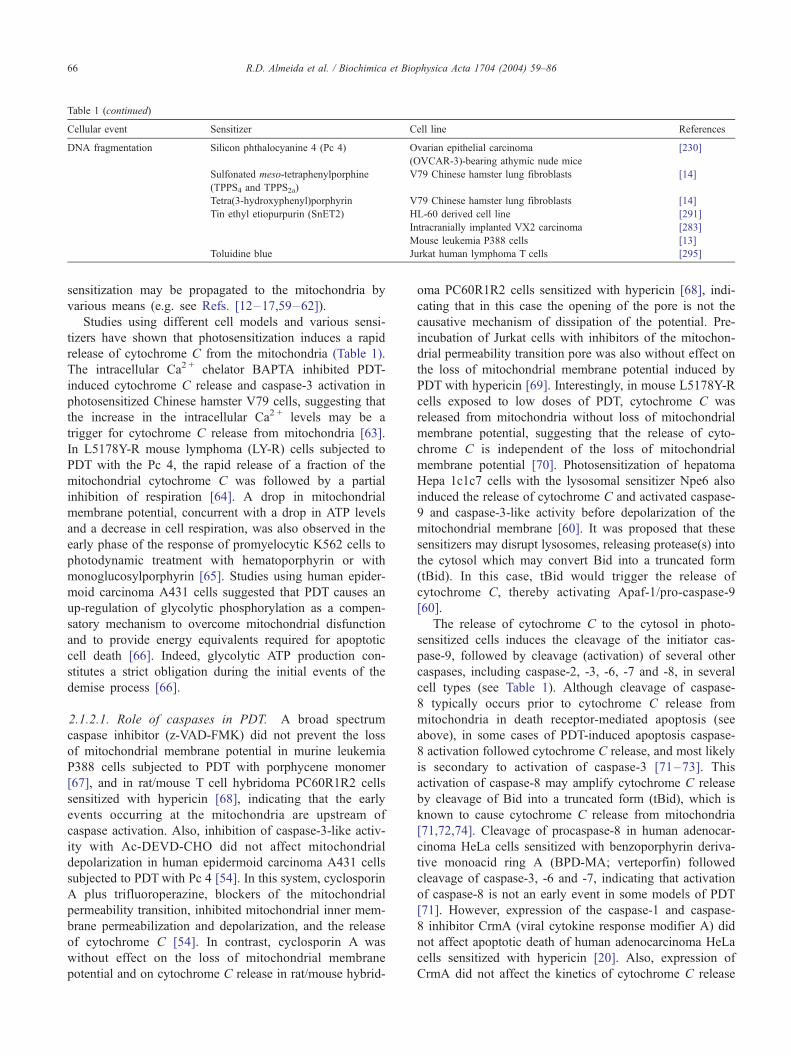
Table 1 (continued)
Cellular event Sensitizer Cell line References
DNA fragmentation Silicon phthalocyanine 4 (Pc 4) Ovarian epithelial carcinoma
(OVCAR-3)-bearing athymic nude mice
[230]
Sulfonated meso-tetraphenylporphine
(TPPS4 and TPPS2a)
V79 Chinese hamster lung fibroblasts [14]
Tetra(3-hydroxyphenyl)porphyrin V79 Chinese hamster lung fibroblasts [14]
Tin ethyl etiopurpurin (SnET2) HL-60 derived cell line [291]
Intracranially implanted VX2 carcinoma [283]
Mouse leukemia P388 cells [13]
Toluidine blue Jurkat human lymphoma T cells [295]
R.D. Almeida et al. / Biochimica et Biophysica Acta 1704 (2004) 59–8666
sensitization may be propagated to the mitochondria by
various means (e.g. see Refs. [12–17,59–62]).
Studies using different cell models and various sensi-
tizers have shown that photosensitization induces a rapid
release of cytochrome C from the mitochondria (Table 1).
The intracellular Ca2 + chelator BAPTA inhibited PDT-
induced cytochrome C release and caspase-3 activation in
photosensitized Chinese hamster V79 cells, suggesting that
the increase in the intracellular Ca2 + levels may be a
trigger for cytochrome C release from mitochondria [63].
In L5178Y-R mouse lymphoma (LY-R) cells subjected to
PDT with the Pc 4, the rapid release of a fraction of the
mitochondrial cytochrome C was followed by a partial
inhibition of respiration [64]. A drop in mitochondrial
membrane potential, concurrent with a drop in ATP levels
and a decrease in cell respiration, was also observed in the
early phase of the response of promyelocytic K562 cells to
photodynamic treatment with hematoporphyrin or with
monoglucosylporphyrin [65]. Studies using human epider-
moid carcinoma A431 cells suggested that PDT causes an
up-regulation of glycolytic phosphorylation as a compen-
satory mechanism to overcome mitochondrial disfunction
and to provide energy equivalents required for apoptotic
cell death [66]. Indeed, glycolytic ATP production con-
stitutes a strict obligation during the initial events of the
demise process [66].
2.1.2.1. Role of caspases in PDT. A broad spectrum
caspase inhibitor (z-VAD-FMK) did not prevent the loss
of mitochondrial membrane potential in murine leukemia
P388 cells subjected to PDT with porphycene monomer
[67], and in rat/mouse T cell hybridoma PC60R1R2 cells
sensitized with hypericin [68], indicating that the early
events occurring at the mitochondria are upstream of
caspase activation. Also, inhibition of caspase-3-like activ-
ity with Ac-DEVD-CHO did not affect mitochondrial
depolarization in human epidermoid carcinoma A431 cells
subjected to PDT with Pc 4 [54]. In this system, cyclosporin
A plus trifluoroperazine, blockers of the mitochondrial
permeability transition, inhibited mitochondrial inner mem-
brane permeabilization and depolarization, and the release
of cytochrome C [54]. In contrast, cyclosporin A was
without effect on the loss of mitochondrial membrane
potential and on cytochrome C release in rat/mouse hybrid-
oma PC60R1R2 cells sensitized with hypericin [68], indi-
cating that in this case the opening of the pore is not the
causative mechanism of dissipation of the potential. Pre-
incubation of Jurkat cells with inhibitors of the mitochon-
drial permeability transition pore was also without effect on
the loss of mitochondrial membrane potential induced by
PDT with hypericin [69]. Interestingly, in mouse L5178Y-R
cells exposed to low doses of PDT, cytochrome C was
released from mitochondria without loss of mitochondrial
membrane potential, suggesting that the release of cyto-
chrome C is independent of the loss of mitochondrial
membrane potential [70]. Photosensitization of hepatoma
Hepa 1c1c7 cells with the lysosomal sensitizer Npe6 also
induced the release of cytochrome C and activated caspase-
9 and caspase-3-like activity before depolarization of the
mitochondrial membrane [60]. It was proposed that these
sensitizers may disrupt lysosomes, releasing protease(s) into
the cytosol which may convert Bid into a truncated form
(tBid). In this case, tBid would trigger the release of
cytochrome C, thereby activating Apaf-1/pro-caspase-9
[60].
The release of cytochrome C to the cytosol in photo-
sensitized cells induces the cleavage of the initiator cas-
pase-9, followed by cleavage (activation) of several other
caspases, including caspase-2, -3, -6, -7 and -8, in several
cell types (see Table 1). Although cleavage of caspase-
8 typically occurs prior to cytochrome C release from
mitochondria in death receptor-mediated apoptosis (see
above), in some cases of PDT-induced apoptosis caspase-
8 activation followed cytochrome C release, and most likely
is secondary to activation of caspase-3 [71–73]. This
activation of caspase-8 may amplify cytochrome C release
by cleavage of Bid into a truncated form (tBid), which is
known to cause cytochrome C release from mitochondria
[71,72,74]. Cleavage of procaspase-8 in human adenocar-
cinoma HeLa cells sensitized with benzoporphyrin deriva-
tive monoacid ring A (BPD-MA; verteporfin) followed
cleavage of caspase-3, -6 and -7, indicating that activation
of caspase-8 is not an early event in some models of PDT
[71]. However, expression of the caspase-1 and caspase-
8 inhibitor CrmA (viral cytokine response modifier A) did
not affect apoptotic death of human adenocarcinoma HeLa
cells sensitized with hypericin [20]. Also, expression of
CrmA did not affect the kinetics of cytochrome C release
R.D. Almeida et al. / Biochimica et Biophysica Acta 1704 (2004) 59–86 67
and procaspase-3 cleavage in a rat/mouse T cell hybridoma
sensitized with hypericin [68], suggesting that caspase-
8 does not play a major role in the demise process in this
model.
The activation of caspases in photosensitized cells leads
to the cleavage of a number of other cell proteins, including
Bap-31 (shuttle protein between the ER and the intermediate
compartment and/or Golgi complex [71]), DNA-dependent
protein kinase (catalytic subunit) (DNA-PKCS [75]), ICAD
(inhibitor of caspase activated DNAse); prevents DNA
fragmentation via binding to caspase-activated deoxyribo-
nuclease [76]), focal adhesion kinase (FAK, a kinase in-
volved in the regulation of cell adhesion [73]), lamins
(structural components of the nuclear envelope [73]), PARP
(poly(ADP-ribose) polymerase, a DNA repair enzyme
[18,20,44,54,68,71,72,75,77–85]) and Ras GTPase-activat-
ing protein (Ras-GAP, a negative regulator of the Ras
signaling pathway [71]). DNA fragmentation in segments
that are multiples of 180–200 bp, another hallmark of
apoptotic cell death [86], was also observed in PDT, using
different cell types and sensitizers (Table 1).
In contrast to the numerous reports showing the role of
caspase-3-like enzymes in apoptotic death in photosensi-
tized cells, a recent study where human cervix carcinoma
HeLa cells were photosensitized with 2,4,5,7-tetrabromo-
rhodamine 123 bromide (TBR), which is incorporated into
the Golgi apparatus, showed that cell death occurred by a
Ca2 +-dependent and caspase-3-independent mechanism
[87]. Despite the fact that no cleavage of procaspase-3
was observed, the morphology of sensitized cells showed
condensed nuclear chromatin and apoptotic bodies, charac-
teristic of cell death by apoptosis [87].
The role of caspase-3 in PDT was further investigated by
comparing the response of caspase-3 deficient cell line
MCF-7 with MCF-7c3 cells, which express a stably trans-
fected caspase-3 gene. Although MCF-7 cells were less
sensitive to photosensitization with Pc 4 when compared
with MCF-7c3 cells, as assayed by reduction of a tetrazo-
lium salt, the two cell lines were equally sensitive to Pc 4-
PDT when evaluated by the clonogenic assay [88]. These
results indicate that the critical step in Pc 4-PDT induced
cell death is independent of caspase-3. Although cyto-
chrome C release following photosensitization was similar
in both cell lines, there was a marked reduction in caspase-9
activation in MCF-7 cells, suggesting that caspase-3 may be
important for the amplification of the pro-caspase-9 pro-
cessing [88]. In vivo studies, in which xenographs of
caspase-3-expressing (MCF-7c3) or deficient (MCF-7 and
MCF-7v) cells were generated in athymic nude mice,
showed that the final outcome of the Pc 4-PDT treatment
was independent of the presence of the caspase [89].
2.1.2.2. Role of the Bcl-2 family of proteins in PDT. The
Bcl-2 family of proteins can be subdivided in two groups
based on their role in apoptotic cell death: Bcl-2, Bcl-xL,
Bcl-w, Mcl-1 and A1 possess anti-apoptotic activity, where-
as Bax, Bak, Bok, Bid, Bim, Bik/Nbk, Bad, Bcl-xS, Bmf,
Hrk, Noxa and Puma are pro-apoptotic (for reviews see
Refs. [45–47]; see also Ref. [90]). Although the role of
some of these proteins in PDT remains to be investigated,
conflicting data have been obtained concerning the influ-
ence of Bcl-2 on the demise process in photosensitized cells.
Overexpression of Bcl-2 in the Chinese hamster ovary
cell line CHO inhibited DNA fragmentation and apoptosis,
and partly protected the cells from cell death, as determined
by the clonogenic assay, in experiments where Pc 4 was
used as a sensitizer [91]. Similar results were obtained using
human acute myelogenous leukaemia HL-60 cells, with
benzoporphyrin derivative monoacid ring A (BPD-MA) as
a sensitizer and at low PDT doses [77]. In this case, Bcl-2
overexpression prevented pro-caspase-3 and -6 cleavage,
proteolysis of PARP and formation of hypodiploid DNA
[77]. Overexpression of Bcl-xL in HL-60 cells also pre-
vented PDT-induced cleavage of pro-caspase-3 and DNA
fragmentation [76]. Enforced expression of Bcl-2 also
delayed PDT-induced apoptotic death in a rat/mouse T cell
hybridoma (PC60R1R2) sensitized with hypericin, by
delaying cytochrome C release, pro-caspase-3 activation
and PARP cleavage [68], and similar findings were reported
in Jurkat cells photosensitized with hypericin [69]. This
delay in apoptosis may be due to phosphorylation of Bcl-2,
through a cyclin-dependent protein kinase 1 (CDK1)-de-
pendent mechanism, which produces a delay of the G2/M
phase, as proposed for human adenocarcinoma HeLa cells
subjected to PDT with hypericin [81]. Overexpression of
Bcl-2 and Bcl-xL also delayed morphological changes,
depressed caspase activation, and limited substrate degra-
dation in HeLa cells subjected to PDT with verteporfin, but
no protection against loss of viability was observed [73]. In
this case, overexpressing Bcl-2 or Bcl-xL did not affect
cytochrome C release from mitochondria following photo-
sensitization, indicating that these proteins confer resistance
to caspase-3 activation, even after the appearance of cyto-
chrome C in the cytosol [73].
The regulatory role of Bcl-2 in PDT has also been
investigated by transfecting an antisense Bcl-2 sequence in
a retrovirus vector into a human gastric adenocarcinoma
MGC803 cell line, followed by PDT with 2-BA-2-DMHA
(2-butylamino-2-demethoxy-hypocrellin A [92]). Reduction
of the Bcl-2 protein levels increased phototoxicity, as
determined by the MTT assay and by the generation of
oligonucleosomal DNA fragments [92]. Similarly, incuba-
tion of the PDT-resistant cell line RIF-1 (radiation-induced
fibrosarcoma cells) with Bcl-2 antisense oligonucleotide
resulted in sensitization to PDT-mediated apoptotic death,
when the Pc 4 photosensitizer was used [93].
In contrast with the reports indicating that Bcl-2 confers
a certain degree of resistance to apoptotic death after PDT,
studies using the MCF10A human breast epithelial cell
line and the photosensitizer AlPc showed that overexpres-
sion of Bcl-2 increased PDT-induced apoptotic cell death
[94]. This increase in the susceptibility to PDT was
R.D. Almeida et al. / Biochimica et Biophysica Acta 1704 (2004) 59–8668
attributed to the observed up-regulation of Bax protein
levels in transfected cells and to the selective degradation
of Bcl-2 in photosensitized cells [94]. A down-regulation
of Bcl-2 protein levels in PDT-treated cells may have freed
Bax to exert it pro-apoptotic effects, since the amount of
this protein in the cell was not changed [94]. Bax is a
cytosolic monomeric protein in healthy cells, but it
changes conformation during apoptosis, integrates into
the outer mitochondrial membrane and oligomerizes. These
oligomers are thought to induce or contribute to the
permeabilization of the outer mitochondrial membrane,
allowing the efflux of apoptogenic proteins [45–47].
However, a recent study showed that the disappearance
of Bcl-2 from Western Blots prepared from DU-145 cells
photosensitized with the Pc 4 was instead due cross-link
formation [55]. In this case Bax was not cross-linked by
Pc 4-PDT [55], suggesting that the increase in sensitivity
to PDT observed in Bcl-2 overexpressing cells may be due
to a change in the balance of functional pro- and anti-
apoptotic members of the Bcl-2 family of proteins. It
remains to be determined whether photodamaged Bcl-2
retains its anti-apoptotic function.
Assuming that the function of Bcl-2 is lost when the high
molecular weight complexes are formed, a change in the
balance between the activity of pro- and anti-apoptotic
proteins of the Bcl-2 family may also explain the enhanced
sensitivity to PDT observed in human epidermoid carcino-
ma (A431) cells overexpressing Bcl-2 [93]. Photosensitiza-
tion of these cells with Pc 4 reduced the protein levels of
anti-apoptotic Bcl-2 and Bcl-xL, as determined by Western
Blot, and up-regulated protein expression of the pro-apo-
ptotic Bcl-xS, Bak and Bad, but not of Bid [93,95]. PDT of
RIF-1 cells treated with a Bcl-2 antisense oligonucleotide
also up-regulated the protein levels of Bcl-xS, Bak and Bad,
in addition to Bid [93]. Photosensitization of murine leuke-
mia L1210 cells with various sensitizers reduced Bcl-2
protein levels, without affecting Bax [59,96], as detected
by Western Blot, and similar findings were reported for
human breast cancer MCF-7 cells and DU-145 cells (human
prostate cancer) sensitized with Pc 4 [55,95,97]. In the latter
study, the protein levels of Bad were unchanged by PDT
[97].
Taken together, the available evidences indicate that a
shift in the balance between the activity of pro- and anti-
apoptotic members of the Bcl-2 family of proteins may
decide the susceptibility of cells to PDT-mediated apoptotic
death. However, this model contrasts with the lack of effect
of PDT on Bcl-2 protein levels in studies using a rat/mouse
T cell hybridoma (PC60R1R2) or human adenocarcinoma
HeLa cells and the sensitizer hypericin [68,81].
In human umbilical venous endothelial cells subjected to
PDT with verteporfin there was a reduction in the cytosolic
levels of Bax, although the total amount of the protein
present in the cells did not change [72], and similar results
were reported for murine leukemia L1210 cells sensitized
with 9-capronyloxy-tetrakis (methoxyethyl) porphycene
(CPO) [59]. These results are in agreement with the de-
scribed translocation of Bax to the mitochondria during
apoptosis, where it oligomerizes and contributes to the
efflux of apoptogenic proteins [45–47]. Overexpression of
Bcl-2 in MCF 7c3 (human breast cancer cell line stably
transfected with human procaspase-3 cDNA) inhibited PDT
induced activation-associated conformational change of
Bax, and higher doses of the sensitizer Pc 4 were required
to activate Bax in cells overexpressing Bcl-2 [98]. This may
explain the protection conferred by the increased expression
of Bcl-2 in MCF 7c3 cells [98]. However, no changes were
observed in the amount of cytosolic Bax in human adeno-
carcinoma HeLa cells subjected to PDT with verteporfin
[73], suggesting that, in this case, the protein may not play a
role in the demise process.
The role of Bax in cytochrome C release from mito-
chondria was investigated using Bax antisense oligonu-
cleotides and by comparing the photodynamic effects of
the Pc 4 photosensitizer in MCF 7c3 cells (human breast
cancer cell line stably transfected with human procaspase-3
cDNA) and in DU-145 cells, a human prostate cancer cell
line that lacks detectable amounts of Bax [99]. PDT-treated
MCF-7c3 cells showed cytochrome C release from mito-
chondria and apoptotic nuclei, whereas the latter cell type
was not affected [99,100]. Also, treatment of MCF-7c3
cells with Bax antisense oligonucleotides reduced Pc 4-
PDT induced apoptotic death and restoration of Bax
expression in DU-145 cells restored apoptosis, indicating
that the lack of Bax is responsible for the resistance of
these cells to PDT-induced apoptosis [100]. However, the
fact that Bax-negative DU-145 cells are as sensitive to Pc
4-PDT as MCF-7c3 cells, as determined by the clonogenic
assay, indicates that the commitment to cell death occurs at
a step prior to Bax activation. In order to further investi-
gate the role of Bax in cell death by PDT, MCF-7c3 and
DU-145 cells were transfected with second mitochondria-
derived activator of caspase (Smac)/direct inhibitor of
apoptosis-binding protein with low pI (DIABLO) or
Smac/DIABLO tagged with green fluorescent protein
(GFP) at its C-terminus (Smac/DIABLO-GFP [99]).
Smac/DIABLO is a protein of the mitochondrial inter-
membrane space, which is released together with cyto-
chrome C from mitochondria in response to apoptotic
stimuli, and promotes caspase activation by inactivating
the inhibition of apoptosis (IAP) [101,102]. PDT with Pc 4
induced the release of Smac/DIABLO-GFP in MCF-7c3
cells but not in DU-145 cells, and expression of the protein
increased apoptotic death in the former cell type [99]. This
suggests that Smac/DIABLO promotes cell death in Pc 4-
PDT in a Bax-dependent manner.
3. Effect of PDT on the [Ca2+]i homeostasis
Photodynamic treatment of various cell types has been
shown to raise the [Ca2 +]i [103–117], which may even-
R.D. Almeida et al. / Biochimica et Biophysica Acta 1704 (2004) 59–86 69
tually lead to cell death [63,109,114,118]. The calcium
chelator BAPTA inhibited the release of cytochrome C,
caspase-3 activation and apoptotic death of Chinese ham-
ster V79 cells photosensitized with pheophorbide, indicat-
ing that Ca2 + indeed plays a role in PDT-induced
apoptosis [63]. Chelation of intracellular Ca2 + with
BAPTA also reduced apoptotic death of HeLa cells (hu-
man cervix carcinoma cell line) photosensitized with TBR,
which is incorporated into the Golgi apparatus [87].
However, in some cell types it was observed that elevation
of the [Ca2 +]i following photosensitization promotes cell
survival and buffering of the intracellular Ca2 + enhanced
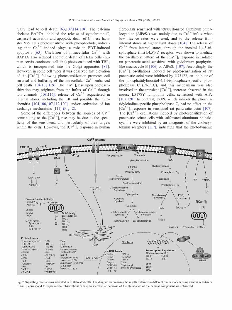
cell death [104,108,119]. The [Ca2 +]i rise upon photosen-
sitization may originate from the influx of Ca2 + through
ion channels [108,116], release of Ca2 + sequestered in
internal stores, including the ER and possibly the mito-
chondria [104,106,107,112,120], and/or activation of ion
exchange mechanisms [111] (Fig. 2).
Some of the differences between the sources of Ca2 +
contributing to the [Ca2 +]i rise may be due to the speci-
ficity of the sensitizers, and particularly of their targets
within the cells. However, the [Ca2 +]i response in human
Fig. 2. Signalling mechanisms activated in PDT-treated cells. The diagram summar
z and # correspond to experimental observations where an increase or decrease
fibroblasts sensitized with tetrasulfonated aluminum phtha-
locyanine (AlPcS4) was mainly due to Ca2 + influx when
low fluence rates were used, and to the release from
internal stores at higher light doses [104]. The release of
Ca2 + from internal stores, through the inositol 1,4,5-tri-
sphosphate (Ins(1,4,5)P3) receptor, was shown to mediate
the oscillatory pattern of the [Ca2 +]i response in isolated
rat pancreatic acini sensitized with gadolinium porphyrin-
like macrocycle B [106] or AlPcS4 [107]. Accordingly, the
[Ca2 +]i oscillations induced by photosensitization of rat
pancreatic acini were inhibited by U73122, an inhibitor of
the phosphatidylinositol-4,5-bisphosphate-specific phos-
pholipase C (PI-PLC), and this mechanism was also
involved in the transient [Ca2 +]i increase observed in the
mouse L5178Y lymphoma cells, sensitized with AlPc
[107,120]. In contrast, D609, which inhibits the phospha-
tidylcholine-specific phospholipase C, had no effect on the
[Ca2 +]i response in sensitized rat pancreatic acini [107].
The [Ca2 +]i oscillations induced by photosensitization of
pancreatic acinar cells with sulfonated aluminum phthalo-
cyanine were inhibited by an antagonist of the cholecys-
tokinin receptors [117], indicating that the photodynamic
izes the results obtained in different tumor models using various sensitizers.
of the abundance of the cellular component was observed.
R.D. Almeida et al. / Biochimica et Biophysica Acta 1704 (2004) 59–8670
effects on the [Ca2 +]i may be secondary to the activation
of plasma membrane-associated receptors.
In human adenocarcinoma HeLa cells photosensitized
with verteporfin the release of Ca2 + from internal stores,
mainly from the ER, was associated with a rapid caspase-
independent depletion of sarco/endoplasmic reticulum Ca2 +
ATPase (SERCA2) [112]. Also, in vitro studies using
subcellular fractions isolated from the rat liver showed that
microsomal Ca2 +-uptake mechanisms were more sensitive
to photosensitization with haematoporphyrin or protopor-
phyrin than the influx of Ca2 + into isolated mitochondria
[121]. The impairment of the Ca2 +-uptake system in the ER
may therefore contribute to the rise in the [Ca2 +]i, due to a
change in the equilibrium rate of Ca2 + cycling across the
store membrane, favoring the Ca2 + leak mechanism that
contributes to the depletion of ER Ca2 + store [122]. Ac-
cordingly, selective SERCA inhibitors (thapsigargin, cyclo-
piazonic acid and 2,5-di-(t-butyl)-1,4-benzohydroquinone)
increase the [Ca2 +]i (e.g. see Refs. [123–125]) by permit-
ting Ca2 + to leak out of the ER, and thapsigargin causes
apoptotic cell death by a mechanism involving the activa-
tion of the ER associated caspase-12 in fibroblasts
[126,127]. This caspase may be activated by caspase-7
and/or by calpains [128,129], but whether calpains and
caspase-12 play a role in PDT remains to be investigated.
This pathway may be particularly significant in PDT-in-
duced cell death using ER-localizing photosensitizers.
The plasma membrane Ca2 +-ATPase has a high Ca2 +
affinity, and it was recently shown to be inactivated in cells
undergoing apoptosis due to cleavage by caspases [130].
Since the effector caspases are rapidly activated in PDT, the
inactivation of the plasma membrane Ca2 +-ATPases may
also contribute to the rise in the [Ca2 +]i, particularly in the
regions close to the plasma membrane.
4. Activation of lipid metabolism by PDT
4.1. Phosphatidylinositol-specific phospholipase C
(PI-PLC)
The rise in the [Ca2 +]i upon photosensitization may
activate a number of intracellular events that shape cell
demise, by interfering with subroutines of the death pro-
gram. Photosensitization of the mouse lymphoma L5178Y
cells with AlPc leads to a rapid accumulation of Ins(1,4,5)P3and increases the [Ca2 +]i, mainly due to release from
internal stores, and both events are significantly reduced
by the phosphatidylinositol-specific phospholipase C inhib-
itor U73122 [120]. This phospholipase C inhibitor also
inhibits [Ca2 +]i spicking in rat pancreatic acini sensitized
with AlPcS4 [107]. The mechanism of PI-PLC activation in
PDT remains to be determined, but it may be secondary to a
small rise in the [Ca2 +]i since the enzyme is activated by
Ca2 + [131]. This phospholipase cleaves phosphatidylinosi-
tol 4,5-bisphosphate, to yield Ins(1,4,5)P3 and diacylgly-
cerol [132]. Ins(1,4,5)P3 releases Ca2 + from internal stores
contributing to the amplification of the [Ca2 +]i signal and,
accordingly, the Ins(1,4,5)P3 receptor inhibitor 2-amino-
ethoxydiphenylborate (2-APB) abolished the [Ca2 +]i spik-
ing in rat pancreatic acini sensitized with AlPcS4 [107].
Diacylglycerol activates protein kinase C, which may con-
tribute or prevent cell death depending on the isoforms
involved [133,134]. Inhibition of phospholipase C with
U73122 was found to prevent the fragmentation of DNA
resulting from the photosensitization of mouse lymphoma
L5178Y cells with AlPc [120], suggesting that the release of
Ca2 + from the internal stores and/or protein kinase C (PKC)
activation plays an important role in PDT-induced apoptotic
death. A role for PKC in PDT-induced cell was proposed
based on the results showing that inhibition of PKC or
down-regulation of the kinase activity by prolonged incu-
bation with 12-O-tetradecanoylphorbol-13-acetate (TPA)
prevented death of Chinese hamster ovary (CHO) cells
following photosensitization with aluminum phthalocya-
nine, as determined using the clonogenic assay [135].
4.2. Phopholipase A2 and arachidonic acid metabolites
Photosensitization of different cell types was also shown
to stimulate rapidly the release of arachidonic acid and of its
metabolites [119,120,136–139]. The formation of arachi-
donic acid may result from the activation of phospholipase
A2, an enzyme activated by Ca2 + (e.g. see Refs. [140, 141])
and, accordingly, inhibition of this enzyme with p-bromo-
phenacyl bromide prevented DNA fragmentation induced by
photosensitization of mouse L5178Y lymphoma cells with
AlPc [120]. Although arachidonic acid may also arise from
the sequential stimulation of phospholipase C (see above)
and diacylglycerol lipase [142], the role of this pathway in
PDT-induced cell death has never been investigated.
In T24 human bladder tumor cells photosensitized with
the haematoporphyrin derivative (HPD), there was a Ca2 +-
dependent and indomethacin-sensitive transient release of
prostaglandin (PG)E2 and thromboxane (TB)B2, the prod-
ucts of the metabolism of PGH2 generated by prostaglandin
E isomerase and thromboxane A2 synthase (thromboxane
A2 gives rise to thromboxane B2), respectively. In this case
indomethacin enhanced photosensitivity, and pre-incubation
of the cells with PGE2 prevented cell death, as determined
using the clonogenic assay, indicating that the cyclooxyge-
nase pathway activated during PDT protects cells from
photodynamically inflicted damage in vitro [119]. However,
the role of cyclooxygenases in PDT may actually depend on
the cell genotype and/or the PDT dose, since pre-incubation
of C6 glioma cells with indomethacin increased the number
of cells surviving to PDT with HPD, whereas the survival
rate for endothelial cells was decreased in the presence of
the inhibitor when higher HPD concentrations were used
[143].
Other studies have also shown that the arachidonic acid
metabolism is changed in tumor cells subjected to PDT.
R.D. Almeida et al. / Biochimica et Biophysica Acta 1704 (2004) 59–86 71
Accordingly, PGE2 was transiently released by pancreatic
acini photosensitized with AlPc sulfonate [138], and by
photodynamic treatment of mouse RIF tumor cells and
peritoneal macrophages (but not murine L929 fibroblasts)
with Photofrin II [136]. Recent studies showed an enhanced
cyclooxygenase-2 (COX-2) transcription in RIF cells sub-
jected to PDT with Photofrin porfimer, and a long-term up-
regulation of the COX-2 protein levels was also observed
[144]. In vitro studies with BA (mouse mammary carcinoma)
and LLC (Lewis lung carcinoma) cells, using Photofrin
porfimer as a sensitizer, also showed an up-regulation of
COX-2 protein levels in photosensitized cells, and similar
results were obtained in PDT-treated RIF tumors growing in
C3Hmice [144]. The up-regulation of COX-2 was associated
with an increase in the production of PGE2 in vivo and in
vitro, and inhibition of the enzyme enhanced PDT respon-
siveness in RIF tumors [144]. In contrast, although PDTwith
hypericin also up-regulated COX-2 mRNA and protein
levels, and stimulated the release of PGE2, in the T24 (human
transitional cell carcinoma of the urinary bladder) and HeLa
(human cervix carcinoma cells) cell lines, inhibition of the
enzyme or exogenously added PGE2 was without effect on
cell death [145]. Similarly, the fragmentation of DNA in
mouse L5178Y lymphoma cells sensitized with AlPc was not
affected by the COX inhibitor indomethacin, suggesting that
the arachidonic acid metabolite important for inducing apo-
ptosis may not be produced through the cyclooxygenase-
dependent pathway [120]. These results do not rule out a
possible role for prostaglandins in in vitro PDT since these
eicosanoids may also be produced by a noncyclooxygenase
mechanism, catalyzed by free radicals produced as a result of
photosensitization [146].
Administration of cyclooxygenase inhibitors to rats prior
to PDT with Photofrin II prevented vessel constriction and
changes in permeability, suggesting that metabolites pro-
duced by the cyclooxygenase-dependent pathway play a
role in these events [147–149]. Thromboxane appears to
play a major role in these effects and on tumor response,
since inhibition of thromboxane synthase or of thromboxane
receptors significantly reduced vessel constriction, inhibited
vessel permeability and reduced tumor cure [150]. However,
in vivo studies using four different zinc phthalocyanines
showed that the release of eicosanoids and the role of the
cyclooxygenases in PDT depend on the photosensitizer used
[139].
4.3. Ceramide
Ceramide is a stress-induced second messenger that may
be generated by sphingomyelinases, which cleave sphingo-
myelin to yield ceramide and phosphorylcholine, or by de
novo synthesis by a synthase [151]. The initial step in the
latter pathway is catalyzed by serine palmitoyltransferase,
which gives rise to 3-ketosphinganine from L-serine and
palmitoyl CoA. 3-Ketosphinganine is then reduced to
sphinganine, and acylation follows to produce ceramide
[85]. Depending on the target cell, ceramide may induce
diverse biological responses, including apoptosis, cell cycle
arrest, differentiation and proliferation [151,152]. Ceramide
is converted into sphingomyelin and glucosylceramide in
reactions catalyzed by phosphatidylcholine:ceramide phos-
phocoline transferase (sphingomyelin synthase) and UDP-
glucose:ceramide glucosyl transferase (glucosylceramide
synthase), respectively [153].
Ceramide accumulation was observed in response to
photodynamic treatment of L5178Y mouse lymphoma cells,
human leukemia (U937) cells, Jurkat human lymphoma
cells, CHO cells, A431 human epidermoid carcinoma cells,
normal human lymphoblasts and mouse embryo fibroblasts
with Pc 4 [154–159]. The studies performed in CHO and
Jurkat cells showed that PDT inactivates sphingomyelin
synthase and glucosylceramide synthase, with no up-regu-
lation in serine palmitoyltransferase activity, indicating that
de novo accumulation of ceramide is due to inhibition of its
conversion to complex sphingolipids rather than to an
increase in the mechanisms of synthesis [159]. In fact, in
CHO cells subjected to Pc 4-PDT, there was a reduction in
LCB1 protein levels, one of the subunits of serine palmi-
toyltransferase [159]. Furthermore, the activity of acid
sphingomyelinase was inhibited in human epidermoid car-
cinoma A431 cells upon photosensitization with the same
sensitizer [158].
Sensitization of the colon cell line HCT-116 with
pyropheophorbide-a methyl ester (PPME) leads to the
accumulation of ceramide, through the activity of the acid
sphingomyelinase [160]. Accordingly, Niemann–Pick dis-
ease lymphoblasts, which lack acid sphingomyelinase
activity, failed to respond to PDT with ceramide accumu-
lation and apoptotic death [156]. These results suggest that
ceramide generation by sphingomyelinase may be the
cause of apoptotic death in PDT. Although the role of
the plasma membrane-associated neutral sphingomyelinase
in the demise process has not been investigated in detail,
studies in the colon cancer HCT-116 cell line sensitized
with PPME showed no activation of the enzyme [160].
Photosensitization of Jurkat cells with Pc 4 induced
intracellular accumulation of sphinganine and ceramide,
and apoptotic death was reduced by the serine palmitoyl-
transferase inhibitor ISP-1 (Myriocin), an inhibitor of de
novo sphingolipid synthesis [85,159]. In addition, PDT-
induced sphinganine accumulation and apoptotic death were
also inhibited by ISP-1 in A431 human epidermoid and
HT29 human carcinoma cells [85]. ISP-1 also inhibited
PDT-induced caspase-3-like activity and PARP cleavage in
PDT-treated Jurkat cells, but it was without effect on the
collapse of the mitochondrial membrane potential and on
cytochrome C release from the mitochondria [85,161],
suggesting that de novo accumulated sphinganine and/or
ceramide initiates apoptosis downstream of cytochrome C
release from mitochondria. Similar results were obtained in
a study using CHO cells photosensitized with Pc 4 [159].
However, the role of de novo ceramide synthesis in the

R.D. Almeida et al. / Biochimica et Biophysica Acta 1704 (2004) 59–8672
demise process in photosensitized cells may depend on the
cell type since fumonisin B1, an inhibitor of ceramide
synthase, had no effect on apoptosis in embryonic fibro-
blasts sensitized with Pc 4 [49]. A number of targets of
ceramide have been identified [151,152], including the
stress-activated protein kinase cascade [162,163], which is
activated in PDT (see below).
5. Role of cyclic nucleotides in PDT
5.1. cAMP
In vitro photosensitization of human bladder transitional
carcinoma cells (clone T24) with HPD stimulated rapidly
and transiently the intracellular accumulation of cAMP. This
effect was suppressed by indomethacin, a cyclooxygenase
inhibitor, which enhanced the photodynamically induced
loss of clonogenicity [164]. These results indicate that the
cytoprotective effect of PGE2 in these cells is mediated by
an increase in the intracellular cAMP content. In Chinese
hamster V79 cells photosensitized with pheophorbide a, the
protective effect of cAMP against apoptotic death was found
to occur at a level downstream of cytochrome C release
from the mitochondria, and upstream of caspase-3 activation
[63].
5.2. Nitric oxide and cGMP
Nitric oxide (NO) plays an important role in tumor cell
biology since it increases blood flow in the tumor, thereby
promoting tumor growth [165] and facilitating metastasis
[166]. Activation of macrophages by photosensitization with
mTHPC stimulated the release of nitric oxide [167], and
nasopharyngeal carcinoma CNE2 cells sensitized with
hypericin showed an increase in the activity of nicotinamide
adenine dinucleotide hydrogen phosphate-diaphorase
(NADPH-d, a potential marker of NO synthase activity)
and in the immunoreactivity of the nitric oxide synthase
(NOS) I and II [168]. Using an electrochemical sensor for on-
line measurement of NO changes, Dalbasti et al. [169]
detected a transient rise in NO production in the cerebellum
as a result of ALA-mediated PDT. This was probably due to
the rapid activation of the neuronal isoform of NOS, which is
constitutively expressed and is activated by Ca2 + and cal-
modulin [170]. Taken together, these evidences indicate that
PDT may affect NO production in the photosensitized
tissues.
NO is expected to play a complex role in PDT, since
several of the tissue changes following photosensitization,
including (i) vascular responses, (ii) the massive neutrophil
recruitment and (iii) cell death (Refs. [149,171–174]; see
also references above), have been shown to be modulated by
NO in other systems [175–177]. Subcutaneous tumors
generating low levels of NO were found to be more
sensitive to PDT with PhotofrinR than those containing
high levels of NO, and the administration of the NOS
inhibitor NG-nitro-L-arginine, together with PDT treatment,
enhanced tumor regression [178,179]. Since in these studies
the NOS inhibitor reduced the blood flow in the tumor, this
may explain the increase in PDT efficiency [179]. In
addition, the possibility that NO generated by tumor cells
and by endothelial cells of the tumor microvasculature may
directly affect tumor cell death cannot be excluded, since
NO has been shown to protect cells from different apoptotic
stimuli [180]. This hypothesis was recently evaluated using
in vitro photosensitization systems [181–183]. Pre-incuba-
tion of human lymphoblastoid CCRF-CEM cells with NO
donors or L-arginine, the NOS substrate, decreased cell
death by apoptosis induced by photosensitization with
bisulfonated aluminum phthalocyanine (AlPcS2), by a
mechanism independent of S-nitrosylation of caspase-3-like
enzymes. The protective mechanism induced by NO was
mediated by the activation of protein kinase G, and occurred
at a level upstream of caspase-9 processing [183]. Although
the protective effect of NO against apoptotic death in several
models has been attributed to an increase in the expression
of heme oxygenase-1, heat shock protein 70 or Bcl-2 [184–
186], this was not the case in the protection of CCRF-CEM
cells from photosensitization by pre-exposure to NO [183].
The presence of NO during the photosensitization period
intercepted lipid-derived radicals and protected COH-BR1
cells (human breast tumor line) from necrotic cell death, in
experiments where sensitization was induced via de metab-
olism of added ALA to PpIX [182,187].
6. Role of MAPKs and CDKs in PDT
The mitogen activated protein kinases (MAPKs) are a
group of serine/threonine protein kinases, activated by dual
phosphorylation on a tyrosine and a threonine residue. The
three members of this family of kinases include the extra-
cellular signal regulated kinases (ERKs), the c-Jun N-
terminal kinases/stress activated protein kinases (JNK/
SAPKs), and the p38 MAPK. Each of these enzymes
participates in closely related signaling cascades that ulti-
mately contribute to the regulation of gene expression in
response to a variety of stimuli. Although activation of
ERK1/2 is generally associated with cell survival, and p38
MAPK and JNK linked to induction of apoptosis, the role of
each MAPK in cell death is dependent on the cell type and
on the physiological context [188,189].
Activation of JNK and p38 MAPK, but not of ERK1 and
ERK2, was observed for murine Pas 212 keratinocytes in
response to oxidative stress produced by photoactivation of
benzoporphyrin derivative, at near cytotoxic levels [190].
Similar findings were reported for human HaCaT keratino-
cytes and for the hypopharyngeal carcinoma FaDu subjected
to ALA-PDT [191,192], and in human adenocarcinomaHeLa
cells treated with low hypericin-PDT doses [81]. When this
mode of sensitization was used, an increase in the phosphor-
R.D. Almeida et al. / Biochimica et Biophysica Acta 1704 (2004) 59–86 73
ylation (activity) of p38 MAPK was also observed in the
human melanoma cell lines Bro and SkMel-23 [191]. Photo-
activation of Pc 4 also increased the activity of JNK inmurine
LY-R leukemic lymphoblasts and CHO cells, whereas the
activity of p38 MAPK was stimulated only in CHO cells.
Also, under the same conditions a reduction in the phosphor-
ylation of ERK2 was observed in CHO cells, but not in LY-R
leukemic lymphoblasts [193]. Taken together the results
suggest that the role of MAPKs in PDT may depend on the
cell type, the photosensitizer used and/or the light dose used.
The photosensitizer that is best characterized in terms of
its effects on MAPK activity and the role of these kinases in
cell death is hypericin. Photoactivated hypericin stimulated
JNK activity and produced an irreversible inhibition of the
activity of ERK2 in A431 (human epidermoid carcinoma
cells), human HaCaT keratinocytes, murine L929 fibroblasts
and human adenocarcinoma HeLa cells [80]. In agreement
with the results with the other sensitizers, PDT with hyper-
icin also produced a sustained activation of p38 MAPK in
HeLa cells [80]. The effect of photosensitization with
hypericin on the activity of stress-activated kinases in this
cell line was not affected by pretreatment with the antiox-
idants N-acetylcysteine or butylated hydroxyanisole [80], in
contrast with the role of reactive oxygen species in the
activation of JNK1 and p38 MAPK in photosensitized
murine keratinocytes [190]. T24 cells (human transitional
cell carcinoma of the urinary bladder) bear an oncogenic H-
ras mutation, and the p38 MAPK and the Raf-ERK cas-
cades are constitutively active. When this cell line was
subjected to hypericin-PDT ERK activity was also partly
inhibited, as observed for the other cell lines (see above),
but the phosphorylation level of p38 MAPK dropped
drastically to undetectable levels, followed by a de novo
activation of the kinase [145]. In these cells, p38 MAPK
activity may account for the observed stabilization of COX-
2 mRNA levels and for the up-regulation of its protein
levels following photosensitization with hypericin [145].
Although caspase inhibitors prevented death of HeLa cells
sensitized with hypericin, they were without effect on the
JNK1 and p38MAPK activation [80], indicating that caspase
activation is either unrelated or lies downstream from these
MAPKs. Furthermore, inhibition of the JNK and p38 MAPK
pathways enhanced PDT induced apoptotic death, suggesting
that these MAPKs play an important role in counteracting the
effects of photosensitization with hypericin.
Although mitochondria-associated Bcl-2 was phosphor-
ylated in human adenocarcinoma HeLa cells subjected to
sub-lethal PDT doses, in relation to G2/M cell cycle arrest,
preceding apoptosis, this effect could not be attributed to
MAPKs [81]. By contrast, a pathway involving CDK1 was
found to be responsible for the phosphorylation of mito-
chondria-associated Bcl-2, on Ser70, and this may constitute
a signal to delay apoptosis in G2/M-phase arrested HeLa
cells [81]. Increasing the photodynamic stress using higher
doses of hypericin promptly caused apoptosis in HeLa cells,
during which Bcl-2 was not phosphorylated, probably
because the photodamage was pushed over a threshold level
which results in a rapid activation of the cell death machin-
ery [81]. In contrast to the role of the p38 MAPK in PDT
based on the studies using hypericin (see above), inhibition
of the kinase with SB202190 protected murine LY-R leu-
kemic lymphoblasts and CHO cells from apoptotic death
induced by photosensitization with Pc 4 [193]. Similar
results were obtained in the murine fibroblast cell line
NIH 3T3 and in the human leukemia HL60 cells, using
Rose Bengal as a sensitizer [44,194]. The effect of p38
MAPK in LY-R leukemic lymphoblasts occurred at a level
upstream of caspase-9 and caspase-3 activation, and inhibi-
tion of p38 MAPK also prevented caspase-3 activation in
HL60 cells subjected to PDT with Rose Bengal.
Taken together the available evidence suggests that the
effect of PDT on the activity of MAPKs depend on the cell
line and/or photosensitizer used. Tong et al. [195,196]
addressed this question by comparing the effect of Photo-
frinR-mediated PDT on the activation of MAPKs in im-
mortalized Li–Fraumeni syndrome (LFS087) cells and in a
normal human fibroblast cell line (GM38A), using equiva-
lent cellular levels of the sensitizer. The former cell line is
considerably more resistant to PhotofrinR-mediated PDT
when compared with normal human fibroblasts and showed
a sustained activation of JNK1 and phosphorylation (acti-
vation) of p38 MAPK and of ERK1/2 [196]. In contrast,
photosensitization of the normal human fibroblast cell line
GM38A induced a transient activation of the three MAPKs,
and the reduction in ERK1/2 phosphorylation after the
initial peak was correlated with an up-regulation in the
expression of mitogen-activated protein kinase phosphatase
1 (MKP-1), which dephosphorylates ERK1/2 [195]. The
sustained activation of ERK1/2 in the LFS087 cell line
might result from the low levels of expression of MKP-1,
which may not reach the threshold required for dephosphor-
ylation of the kinase. Although Raf-1 is normally involved
in the regulation of ERK1/2, this is probably not the case in
photosensitized LFS087 and in GM38A cells, since Photo-
frinR-mediated PDT significantly reduced Raf-1 protein
levels [195]. Inhibition of ERK1/2 phosphorylation with
PD098,059 increased the sensitivity of LFS087 cells to
PhotofrinR-mediated PDT, suggesting that this pathway
protects the cells from cell death [195]. In contrast, blocking
the p38 MAPK in LFS087 cells, by transient infection with
a recombinant adenovirus expressing a dominant negative
mutant of p38 MAPK, or in human adenocarcinoma HeLa
cells, by stable transfection with a dominant negative mutant
of p38 MAPK, had no effect on cell survival following
PhotofrinR-mediated PDT [196].
7. Differential effect of PDT on tyrosine kinases
Protein tyrosine phosphorylation increased rapidly in the
mouse lymphoma L5178Y-R cells upon photosensitization
with Pc 4 [197], and the immunoreactivity with an antibody
R.D. Almeida et al. / Biochimica et Biophysica Acta 1704 (2004) 59–8674
that specifically recognizes phosphotyrosine also increased
in extracts prepared from murine mastocytoma P815 cells
subjected to PDT with benzoporphyrin derivative monoacid
ring A [198]. Although the intracellular events responsible
for the up-regulation of protein tyrosine phosphorylation in
photosensitized cells have not been determined, they may
be, at least in part, secondary to a rise in the [Ca2 +]i (e.g. see
Ref. [199]). Tyrosine phosphorylation of proteins in
L5178Y-R cells photosensitized with Pc 4 was amplified
by pre-incubating the cells with the tyrosine phosphatase
inhibitor sodium orthovanadate [197], indicating that the
effect of PDT on tyrosine phosphorylation is rapidly coun-
teracted by tyrosine phosphatases. Although the lack of
effect of sodium orthovanadate on PDT with Pc 4 [197]
suggests that the demise pathways do not rely on a broad
increase in tyrosine phosphorylation, further studies will be
required in order to establish the general role of tyrosine
kinases in cell death upon photosensitization.
Stimulation of tyrosine kinase activity and phosphoryla-
tion of the 80-kDa c-Src substrate HS1 (haematopoietic
lineage cell-specific protein 1) occurred rapidly in murine
L5178Y-R leukemic lymphoblasts following photoactiva-
tion of Pc 4 [197], under conditions where cell death
occurred by apoptosis. HS1 is a protein found in the
cytoplasm and to a lesser extent in the nucleus of essentially
all haematopoietic cells [200], and it was found to play a
role in antigen-receptor-induced apoptosis in the murine B-
lymphoma WEHI-231 cells [201]. This protein also plays an
important role in the signaling from surface IgM leading to
apoptosis in peritoneal B cells [202], but the role of HS1 in
PDT is still not known.
Photosensitization of membrane fractions isolated from
human epidermoid carcinoma A431 cells and 3T3 HIR
mouse fibroblast cells with hypericin inhibited irreversibly
the protein tyrosine kinase activity of the epidermal growth
factor (EGF) and insulin receptors, but it was without effect
on the activity of the cytosolic protein tyrosine kinases Lyn,
Fgr, TPK-IIB and c-Src kinase [203]. Treatment of FaDu
cells (hypopharyngeal carcinoma) with ALA-PDT caused an
extensive reduction in the amount of EGF receptor (EGFR)
protein levels, and of the phosphorylation of ERK following
stimulation of the receptors [192]. Studies using the human
epidermoid carcinoma A431 cells, which express high
levels of the EGFR, also showed that PDT with Pc 4 result
in a rapid time-dependent reduction in the EGFR protein
levels and in receptor phosphorylation on tyrosine, under
conditions where cell death occurred by apoptosis. A similar
reduction in the levels of Shc (52- and 46-kDa isoforms), a
protein that belongs to the signaling machinery of the
receptor, was also observed, in addition to a decrease in
its phosphorylation on tyrosine, further indicating that PDT
decreases the signaling activity of these receptors [82].
Similar results were obtained in in vivo experiments, where
chemically as well as ultraviolet B radiation-induced squa-
mous papillomas, in SENCAR and SKH-1 hairless mice,
respectively, were subjected to Pc 4-PDT [82]. The EGFR is
considered to enhance cell proliferation, motility, adhesion,
invasion and angiogenesis [204] and, therefore, the rapid
effect of photosensitization on the EGFR protein levels, and
on its signaling machinery, may be a cause rather than a
consequence of cell death in PDT.
Akt/PKB is a serine/threonine kinase that plays an impor-
tant role in cell survival, being activated downstream of the
phosphatidylinositol-3-kinase (PI3-K) upon stimulation of
numerous receptors for growth factors [205–208]. Photo-
sensitization of the murine fibroblast cell line NIH 3T3 with
Rose Bengal increased the phosphorylation of Akt on serine
473, which is taken as a sign of kinase activation [194]. This
effect was mediated by activation of PI3-K, but was inde-
pendent of activation of growth factor receptors and of FAK,
a tyrosine kinase. Indeed, photosensitization with Rose
Bengal decreased FAK phosphorylation (activity), which
may explain the reduction in cell adhesion [194]. However,
it remains to be determined whether Akt/PKB activation
modulates the demise process in photosensitized cells.
The non-receptor tyrosine kinase Etk/Bmx (epithelial and
endothelial tyrosine kinase or bone marrow tyrosine kinase
gene in chromosome X), which showed activity in resting
LNCaP prostate cancer cells, downstream of PI3-K, provid-
ed some protection to these cells from apoptotic death
induced by PDT with Pc 4 [84]. However, the mechanism
of action of Etk/Bmx in photosensitized cells is not known.
The response of hypopharyngeal carcinoma FaDu cells
and of cultured human pulmonary fibroblasts to IL-6-type
cytokines (IL-6, LIF [leukemia inhibitory factor] and oncos-
tatin M), namely STAT (signal transducers and activators of
transcription) phosphorylation on tyrosine, was also signif-
icantly impaired after photosensitization with ALA or Photo-
frinR. Under the same conditions it was observed a reduction
of the fully processed plasma membrane form of the leuke-
mia inhibitory factor receptor-a (LIFRa), but no significant
effect was observed on the proteins oncostatin receptor-h(OSMRh) and gp130 [192]. The reduction in LIFRa could
account for the loss of responsiveness to LIF, whereas the
lack of activity of OSM in photosensitized cells may be due
to an impairment of the functionality of those receptor
subunits and/or of the signaling pathway downstream of
the receptors [192]. Accordingly, JAK1 (Janus kinase 1)
protein levels were decreased in cervical adenocarcinoma
HeLa cells subjected to PDT with ALA [192]. Signaling by
IL-6-type cytokines has been associated with the control of
function of differentiated cells [209–211] and with suppres-
sion of proliferation of various epithelial cells [212, 213].
8. Regulation of transcription factors in PDT
8.1. Transcription factor activator protein-1 (AP-1)
AP-1 is a homo- or heterodimeric protein complex,
composed of Jun (c-Jun, Jun-B, Jun-D), Fos (c-Fos, Fos-B,
Fra-1, Fra-2), Maf (c-Maf, MafB, MafA, MafG/F/K and Nrl)
R.D. Almeida et al. / Biochimica et Biophysica Acta 1704 (2004) 59–86 75
and ATF (ATF2, LRF1/ATF3, B-ATF, JDP1, JDP2) -family
proteins. This family of transcription factors is activated by a
variety of physical and chemical stresses and recognize
either cAMP response elements (CRE, TGACGTCA) or
TPA response elements (TGAG/CTCA). AP-1 transcription
factors have been related to both induction and prevention of
apoptosis, depending on the tissue and on its developmental
stage (reviewed in [214,215]).
Photosensitization of murine RIF-1 cells with Photofrin,
Npe6 (mono-L-aspartyl chlorin e6) or with Rose Bengal
increased c-fos mRNA levels. PDT with PhotofrinR also
increased transiently c-jun, c-myc and egr-1 mRNA in the
same cells [216], and c-fos and c-jun mRNA in human
adenocarcinoma HeLa cells [217]. Furthermore, mRNA
stability experiments showed an increased half-life of c-
fos and c-jun transcripts in HeLa cells sensitized with
PhotofrinR, and a concomitant increase in AP-1-DNA
binding activity was also observed [217,218]. An increase
in AP-1 DNA binding activity was also observed in Photo-
frin-PDT using mouse leukemia L1210 cells [219] and the
human keratinocyte PAM 212 cell line. Inhibition of protein
kinases, with staurosporin, or of phospholipase A2, with
quinacrine, blocked the induction of c-fos mRNA in RIF-1
cells sensitized with PhotofrinR [216], suggesting that these
pathways may participate in the signaling mechanisms
leading to activation of early response genes. This is in
agreement with the results showing that AP-1 is activated by
p38 MAPK and JNK [214], two protein kinases that are
stimulated in photosensitized cells (see Section 5). Howev-
er, the signaling cascade responsible for AP-1 activation in
PDT remains to be determined.
8.2. Transcription nuclear factor kappa B (NF-jB)
Under resting conditions the transcription factor NF-nBis present in the cytoplasm as homo- or heterodimers,
formed by association of subunits belonging to a protein
family that includes p50, p52, p65 (RelA), RelB and c-
Rel. These complexes are sequestered in the cytoplasm by
proteins belonging to the inhibitor of NF-nB (InB) family,
including InBa, InBh, InBq, p100 and p105. Stimuli
leading to NF-nB activation typically initiate a specific
signal transduction cascade leading to phosphorylation of
InBs. Once phosphorylated, InB is selectively ubiquiti-
nated and degraded by the 26S proteosome, releasing NF-
nB that is then translocated to the nucleus where it
participates in transcriptional activation (reviewed in Refs.
[220,221]). NF-nB has been shown to either promote or
inhibit apoptosis, depending on the cell type and the type
of inducer [222–224].
PDT has been shown to induce NF-nB activity in
various cell models and using different sensitizers, but
limited information is available concerning the role of the
transcription factor in the demise process. Activation of
NF-nB upon photosensitization was first shown in studies
using mouse leukemia L1210 cells and Photofrin II as a
sensitizer [219]. Photosensitization of the lymphocytic
ACH-2 cells with proflavine (PF) or with methylene blue
led to the degradation of InBa and increased NF-nB (p50
and p65) DNA-binding activity [225,226]. Similar results
were obtained in the promyelocytic leukemia HL-60 cells
sensitized with the benzoporphyrin derivative monoacid
ring A (verteporfin), in which InBh levels were maintained
unchanged [79]. Furthermore, HeLa cells (human cervix
carcinoma cells) and T24 cells (human transitional cell
carcinoma of the urinary bladder) photosensitized with
hypericin also showed a reduction in the cytosolic levels
of InBa, which correlated in time with binding of nuclear
proteins to the NF-nB site of the cox-2 promoter [145].
The activation of NF-nB and its role in cell death upon
photosensitization was investigated using the sensitizer
PPME [160,227] and its derivative aminopyropheophor-
bide (APP) [228], which results from the addition of a
positively charged group that increases cellular uptake.
PPME-mediated photosensitization of HCT-116 human
colon carcinoma cells activated NF-nB by triggering the
signaling pathway mediated by the IL-1 receptor. This
involved degradation of the cytoplasmic InBa pool, but
not of InBh, p105 or p100, and supershift assays using
antibodies against the NF-nB proteins showed that p50 and
RelA contributed to the increase in NF-nB DNA binding
activity in photosensitized cells [160]. Concomitant with
NF-nB activation in human ECV 304 and HMEC-1
endothelial cells photosensitized by PPME, InBa was also
degraded, and a slight modification in the InBh, InBq,p100 and p105 levels was observed [227]. Interestingly, in
this case the activation of NF-nB was proposed to occur
by a mechanism independent of the activation of InBkinases and require the activity of a tyrosine kinase
[227]. Expression of a dominant negative mutant of InBaincreased apoptotic death of HCT-116 cells sensitized with
APP [228], suggesting that NF-nB regulates the expression
of gene(s) encoding proteins with an anti-apoptotic activ-
ity. However, it was not determined whether cells pre-
vented from undergoing apoptosis in the presence of the
dominant negative mutant of InBa died by a non-apoptotic
mechanism.
8.3. Retinoblastoma (Rb) and E2F family of transcription
factors
Photosensitization of A431 human epidermoid carcino-
ma cells with Pc 4 resulted in inhibition of cell growth
and G0–G1 phase arrest of the cell cycle. This effect was
associated with a time-dependent inhibition of the expres-
sion and activity of CDK2 and CDK6, two critical kinases
operative in the G1 phase of the cell cycle, and of their
regulatory partners cyclin E and cyclin D1 [229]. In the
same study it was found that PDT up-regulates the
expression of the cyclin kinase inhibitor WAF1/CIP1/
p21, thereby increasing its binding towards cyclin D1
and CDK6. A decrease in the binding of cyclin D1 toward
R.D. Almeida et al. / Biochimica et Bio76
CDK2 and CDK6 was also observed [229]. An up-
regulation in the WAF1/CIP1/p21 protein levels was also
observed upon photosensitization of human ovarian carci-
noma (OVCAR-3)-bearing athymic nude mice with Pc 4
[230], but not in SW480 colon cancer xenographs sub-
jected to PDT with the same sensitizer [231]. These
observations suggest that induction of cell death in these
tumors may be triggered by a WAF1/CIP1/p21-indepen-
dent mechanism.
The cyclin D/CDK4/6, cyclin A/CDK2 and cyclin E/
CDK2 complexes are involved in the phosphorylation of Rb
during the cell cycle (reviewed in Ref. [232]), and the
observed changes in CDKs in photosensitized cells may
account for the observed reduction in Rb phosphorylation
[233]. A reduction in Rb phosphorylation in PDT may be
responsible for the induction of WAF1/CIP1/p21 (when
observed), since the predominance of hypo-phosphorylated
Rb was previously associated with the induction of the
protein [234]. Hypo-phosphorylated Rb is also capable of
associating and thereby inhibiting the activity of the E2F
family of transcription factors (E2F [235,236]), which are
involved in the regulation of cell cycle [232]. More recently
evidences were provided for an active role of Rb in gene
repression through the Rb-E2F complex binding to a pro-
moter, thereby contributing to growth arrest [237]. The
photodynamic response in human epidermoid carcinoma
A431 cells sensitized with Pc 4 was accompanied by a
down-regulation of five members of the E2F (1–5) tran-
scription factor family, and of their heterodimeric partners
DP1 and DP2 [233]. Since Rb and E2F regulate the G1! S
transition in the cell cycle (reviewed in Refs. [232,237]), the
reduction in Rb phosphorylation and in the E2F and DP1
and 2 protein levels may be responsible for imposing an
artificial checkpoint at this phase, leading to a G0/G1 arrest
and a subsequent apoptotic death.
9. Changes in protein levels in response to PDT
9.1. Heme oxygenase
The oxidative stress caused by PDT has been shown to
induce the expression of several proteins typically associ-
ated with cellular responses to stress conditions. Photosen-
sitization of Chinese hamster fibroblast cells (V-79) with
Photofrin II or with Rose Bengal increased heme oxygenase
protein levels [238]. Heme oxygenase catalyzes the oxida-
tive degradation of heme to biliverdin, which is then
converted into bilirubin by biliverdin reductase [239,240].
Bilirubin and biliverdin are efficient anti-oxidants [241]
(reviewed in Ref. [242]). The heme oxygenase gene con-
tains binding sites for several transcription factors, includ-
ing an AP-1 consensus sequence [243] (reviewed in Ref.
[242]) that may contribute to an up-regulation of gene
expression since this transcription factor may be activated
in PDT (see above).
9.2. Glucose-regulated proteins
PDT with Photofrin II increased the mRNA encoding
glucose-regulated proteins (GRP-78 and GRP-94) in mouse
RIF-1 cells [244]. In vivo studies also revealed a transient
elevation of GRP-78 mRNA in transplanted mouse mam-
mary carcinomas photosensitized with Photofrin II [244].
Investigation of the mechanism of cell death in human
ovarian carcinoma A2780 cells treated with structurally
diverse N-(2-hydroxypropyl)-methacrylamide (HPMA) co-
polymer (P)-mesochlorin e6 monoethylenediamine (Mce6)
conjugates that possess differential subcellular accumulation
or covalent attachments of photosensitizers (or both)
showed that a conjugate that targets the nucleus selectively
abolished GRP-94 expression [15]. In this study, A2780
cells photosensitized with a conjugate possessing Mce6linkage via a lysosomally degradable tetrapeptide spacer
(HPMA copolymer-Mce6 conjugate containing Mce6 bound
via the glycylphenylalanylleucylglycine (GFLG) linker)
selectively increased mRNA levels for HSP-70 in a transient
manner [15].
9.3. Heat-shock proteins
In vivo and in vitro studies using a chlorin (Npe6)- or
purpurin (tin etio-purpurin)-based sensitizer also showed an
increased in HSP-70 mRNA and protein levels in murine
RIF-1 cells subjected to PDT [245]. Interestingly, a similar
increase in HSP-70 was observed in in vivo but not in vitro
studies using Photofrin II [245]. Metabolic studies aiming at
determining changes in protein synthesis caused by in vitro
and in vivo PDT with benzoporphyrin derivative revealed
that the pattern of expression was similar in both cases.
Using two-dimensional gel electrophoresis and by compar-
ison with published gel patterns, the proteins whose expres-
sion changed upon PDT were identified as HSP-110, HSP-
90, HSP-73, HSP-72, HSP-47, GRP-100 and GRP-80 [246].
The transcription of genes belonging to the heat shock
family is regulated by a mechanism involving the binding
of heat-shock factors (HSF) to specific heat-shock elements
(HSE). In nonstress conditions the transcription factor HSF
is found in the cytoplasm, in a monomeric form, associated
with HSP-70 [247]. During cellular stress HSP-70 binds to
denatured proteins, freeing HSF that trimerizes and migrates
to the nucleus, where it binds to HSE [247]. Accordingly, in
vitro photosensitization of the RIF tumor cells with Npe6
induced HSF-1 binding to HSE, and transiently induced the
expression of a reporter gene containing an inducible hsp70
promoter [248]. This was not observed when Photofrin was
used as a sensitizer, possibly due to differences in the
subcellular targets of the molecules [245]. Interestingly,
HSF binding to HSE and reporter gene expression was
observed for Npe6- and PhotofrinR-mediated in vivo PDT
studies, when RIF cells were grown as solid tumors in mice
[248]. Also, in vivo and in vitro studies, using a mouse
mammary EMT6 cell line stably transfected with a plasmid
physica Acta 1704 (2004) 59–86

R.D. Almeida et al. / Biochimica et Biophysica Acta 1704 (2004) 59–86 77
consisting of the gene for GFP under the control of an hsp70
promoter, showed that sublethal doses of meso-tetra(hydrox-
y)phenyl chlorin (mTHPC; FoscanR)-PDT induce hsp70-
driven GFP expression [249]. The up-regulation in HSP-70
[250] and HSP-90 protein levels may protect the cells from
apoptotic death induced by PDT since both proteins interact
with the apoptotic machinery thereby suppressing apoptosis.
Indeed, HSP-70 binds to the caspase-recruitment domain
(CARD) of Apaf-1 and prevents the recruitment of pro-
caspase-9 to the apoptosome complex [251,252]. HSP-90 is
also a negative regulator of the mitochondrial apoptotic
pathway, which acts by forming a cytosolic complex with
Apaf-1, which inhibits the formation of the active apopto-
some complex [253].
9.4. Anti-oxidant enzymes and apoptosis regulatory proteins
Recent studies using murine colon-26 (C26) cells, a
poorly differentiated colon adenocarcinoma cell line,
showed that PDT with Photofrin increases the protein levels
of manganese superoxide dismutase (MnSOD), but not of
Cu,Zn-SOD [254]. Previous studies using human adenocar-
cinoma HeLa cells also showed induction of MnSOD
mRNA following photosensitization with Photofrin II
[255]. Transient transfection of the T24 bladder cancer cell
line with the MnSOD gene, but not with the Cu,Zn-SOD
gene, or pretreatment of C26 and T24 cells with a cell
permeable SOD mimetic, resulted in a considerable decrease
in the effectiveness of PDT with PhotofrinR [254]. These
results suggest that inhibition of SOD activity may be
effective in potentiating the antitumor effectiveness of
PDT. An up-regulation of mRNA expression of gammaglu-
tamyl cysteine synthetase, which catalyzes the rate-limiting
step in overall glutathione biosynthesis, and is subject to
feedback regulation by glutathione, was also observed in the
glioblastoma SNB-19 cell line sensitized with hypericin,
under conditions where cell death occurred by apoptosis
[256].
The effect of ALA-based PDT on the proteome was
also investigated in the HL-60 promyelocytic cell line,
under conditions where cell death occurred mainly by
apoptosis [257]. From a total of more than 1350 protein
spots obtained after separation in 2D gels, at an early
stage of the demise process, the cellular content in seven
proteins was increased and 17 were suppressed following
sensitization. The latter group included the calreticulin
precursor, p58 microsomal protein (Erp57) and protein
disulfide isomerase (p55), which are all chaperones in-
volved in calcium homeostasis. Under the same conditions
Western blotting experiments showed a decrease in the
amount of Erp72 and an up-regulation of calmodulin and
HSP-60 [78]. Furthermore, ALA-mediated PDT in HL-60
cells decreased the expression of the anti-apoptotic pro-
teins of the Bcl-2 family, Bcl-2 and Bcl-xL, and of the
pro-apoptotic protein of the same family Bax [78] (see
Section 2.1.2.2 for other references). In contrast, RT-PCR
analysis showed that bcl-2 is up-regulated in human
ovarian carcinoma A2780 cells upon photoactivation of
Mce6 (mesochlorin e6 monoethylenediamine), P-GFLG-
Mce6 [HPMA copolymer-Mce6 conjugate containing
Mce6 bound via glycylphenylalanylleucylglycine (GFLG)
linker] and NLS(FITC)GFLGMce6 [HPMA copolymer-
Mce6 conjugate containing Mce6 bound via glycylpheny-
lalanylleucylglycine (GFLG) spacer and containing
NLS(FITC) bound via a thioether linkage] [15]. Apoptotic
death of A431 human epidermoid carcinoma cells sensi-
tized with Pc 4 followed an increase in the protein levels
of Fas and Fas ligand [39], proteins involved in the
initiation of the death receptor-mediated pathway of apo-
ptotic cell death (see Section 2.1.1).
PDT has been shown to alter the expression of TNF and
IL-6. Accordingly, TNF-a gene transcription increased in
keratinocytes sensitized with Pc 4 [258], and thioglycolate-
elicited murine macrophages released TNF when subjected
to PDT with Photofrin II [259]. Photodynamic activation of
the monocyte cell line U937 differentiated into macrophages
(U937A cells) with mTHPC (FoscanR) also induced a dose-dependent production of TNF-a [167]. However, although
anti-TNF-a antibodies and pentoxifylline, an inhibitor of
cytokine transcription, prevented cutaneous photosensitivity
in adult C3H/HeN mice injected with Pc 4, none of these
agents affected Pc 4 PDT-induced tumor (RIF-1 radiation-
induced fibrosarcoma) regression [258].
9.5. Clusterin
Clusterin is a glycoprotein with a nearly ubiquitous tissue
distribution, but its functions remain to be understood [260].
In vitro studies showed an up-regulation of clusterin protein
levels in human epidermoid carcinoma A431 cells treated
with Pc 4-PDT, under conditions where cell death occurs by
apoptosis, but no effect was observed in apoptosis-resistant
RIF-1 cells [261]. Similarly, an increase in clusterin protein
levels was observed in PDT-treated chemically induced
murine skin papillomas [261], but the significance of these
findings remains to be determined.
9.6. p53
The p53 protein is present in low amounts in healthy
cells, becoming up-regulated in response to various types of
stress. This causes the cell to delay its entry into S phase
until an eventual damage to DNA has been repaired.
Furthermore, p53 is also involved in triggering apoptosis
under circumstances in which damage is too severe to be
repaired. The p53 tumor suppressor gene is mutated in over
50% of human cancers, and an important feature of the
oncogenicity of these mutants is their ability to interfere
with p53-dependent apoptosis [262]. The role of p53 in cell
death in PDT has been addressed in two ways: (i) comparing
responses to PDT of closely related human tumor cells,
expressing wild-type or lacking the protein (e.g. Ref. [263]);

R.D. Almeida et al. / Biochimica et Biophysica Acta 1704 (2004) 59–8678
(ii) comparing the clonogenical survival of normal human
fibroblasts that express wild-type p53 and immortalized Li–
Fraumeni syndrome (LFS) cells that express only mutant
p53, and the LFS cells expressing wild-type p53 introduced
using an adenovirus vector [264]. PDT-treated cells with
various sensitizers up-regulated the expression of p53 [263–
265], but PDT-induced cell death or apoptosis did not show
a significant dependence on p53.
9.7. Interleukins
PDT of tumors causes a rapid induction of an inflamma-
tory response, characterized by the infiltration of leucocytes,
mainly neutrophils, into the sensitized tumor [9,174,266–
268]. This response is thought to be important for the
activation of antitumor immunity, and in vivo studies with
mammary EMT6 tumors sensitized with 2-[1-hexylox-
yethyl]-2-devinyl pyropheophorbide-a (HPPH) showed that
the influx of neutrophils is induced by the local up-regulation
of the attractant chemokine MIP-2 (macrophage inflamma-
tory protein, in rodents, or IL-8 in humans). Also, the up-
regulation in E-selectin in the tumor microvasculature in-
creased the adhesion of EMT6 microvessels. Although there
was also an increase in the neutrophil attractant chemokine
KC, it had no effect on the recruitment of neutrophils [266].
Once at the tumor site, neutrophils may cause direct tumor
cell destruction, may recruit leucocytes and lymphocytes by
secreting chemokines/cytokines and may be implicated in
tumor rejection in T-cell-dependent reactions [269].
Photofrin-mediated photosensitization of human adeno-
carcinoma HeLa cells also induced rapidly the production of
IL-6 mRNA, followed by secretion of the interleukin [218].
Under the same conditions AP-1 DNA binding was ob-
served, at the distal AP-1 site of the IL-6 promoter, but not
at the proximal site [218]. The mRNA for IL-6 was also
changed by PDT with Photofrin in EMT6 mammary tumor
cells in culture and in vivo, and a concomitant up-regulation
in the interleukin protein levels was also observed in the
latter model [174]. An enhanced expression of IL-6 in
EMT6 tumors and an increase in the circulatory levels of
IL-6 were also observed in in vivo studies, using the
sensitizer HPPH [266]. The up-regulation in the IL-6 levels
contrasts with the observed impairment of the response of
hypopharyngeal carcinoma FaDu cells and of cultured
human pulmonary fibroblasts to IL-6-type cytokines (IL-6,
LIF and oncostatin M), observed after photosensitization
with ALA or Photofrin [192] (see above). This may account
for the lack of effect of IL-6 on neutrophil infiltration of
EMT6 tumors, when HPPH was used as a sensitizer [266],
but contrasts with the role of the cytokine on the induction
of systemic neutrophilia in Photofrin-PDT [267,270].
9.8. Regulators of angiogenesis
Damage to the microvasculature induced by PDT pro-
duces hypoxia due to the reduction in blood flow [2,3].
PDT with PhotofrinR induced the expression of the
hypoxia-inducible factor-1a (HIF-1a) subunit of the HIF-
1 transcription factor complex in BA mammary carcinoma
tumors growing in C3H mice [271]. HIF-1a associates
with HIF-1h to form HIF-1, but whereas the latter subunit
is constitutively expressed, HIF-1a is rapidly degraded
under normoxic conditions [272,273]. Hypoxia stabilizes
HIF-1a, allowing the formation of the transcriptionally
active HIF-1 complex, which binds to the hypoxia re-
sponse element found in the promotor region of specific
genes, including the vascular endothelial growth factor
(VEGF) gene [272,273]. Accordingly, an increase in
VEGF protein levels was found in BA mammary carcino-
ma tumors growing in C3H mice subjected to PDT with
PhotofrinR under conditions where cell death occurred by
necrosis [271]. Oxidative stress may also lead to the
overexpression of VEGF since PDT-treatment of cultured
BA tumor cells also caused a slight increase in VEGF
[271]. Since VEGF has a strong mitogenic activity, pri-
marily in endothelial cells [274], studies were performed
using the antiangiogenic compounds EMAP-II (single
chain polypeptide) and IM862 (L-glutamyl-L-tryptophan
dipeptide) to determine whether they could enhance the
tumorocidal action of PDT. Both peptides decreased VEGF
expression and improved tumoricidal responses in PDT-
treated BA mammary carcinoma tumors growing in C3H
mice [271]. In this model, a synthetic inhibitor of matrix
metalloproteinases (MMP), Prinomastat, also increased the
percentage of long-term cures after photosensitization with
Photofrin [275]. Interestingly, PDT increased the expres-
sion and activation of MMP-1, -3, -8 and -9 in BA tumors
subjected to PDT with Photofrin, and also changed the
expression of MMP regulators. In particular, there was an
up-regulation in EMMPRIN levels, a protein that efficient-
ly stimulates the production of MMPs [276], and a
decrease of TIMP-1 expression, a protein that regulates
the metabolism of the extracellular matrix [275,277].
Taken together, these findings indicate that a combination
therapy approach, using PDT together with MMP inhib-
itors and/or antiangiogenic compounds, can increase the
therapeutic efficacy.
10. Conclusions
The experimental evidences discussed throughout the
manuscript show a large heterogeneity in the mechanisms
leading to cell death in PDT. Although the demise process is
triggered mainly by reactive oxygen species, primarily by
singlet oxygen (1O2), it is shaped by the cell genotype, the
PDT dose and the intracellular location of the sensitizer. In
addition to the activation of the molecular machinery
leading to cell death, PDT may also set in motion metabolic
reactions that protect cells from the oxidative damage.
Inhibition of the latter events is likely to potentiate the
antitumor effectiveness of PDT.

R.D. Almeida et al. / Biochimica et Biophysica Acta 1704 (2004) 59–86 79
Acknowledgements
The work in the authors laboratory was supported by
Fundac�ao para a Ciencia e a Tecnologia, Portugal.
References
[1] H. Takahashi, Y. Itoh, Y. Miyauchi, S. Nakajima, I. Sakata, A.
Ishida-Yamamoto, H. Iizuka, Activation of two caspase cascades,
caspase 8/3/6 and caspase 9/3/6, during photodynamic therapy using
a novel photosensitizer, ATX-S10(Na), in normal human keratino-
cytes, Arch. Dermatol. Res. 295 (2003) 242–248.
[2] T.J. Dougherty, C.J. Gomer, B.W. Henderson, G. Jori, D. Kessel, M.
Korbelik, J. Moan, Q. Peng, Photodynamic therapy, J. Natl. Cancer
Inst. 90 (1998) 889–905.
[3] R. Ackroyd, C. Kelty, N. Brown, M. Reed, The history of photo-
detection and photodynamic therapy, Photochem. Photobiol. 74
(2001) 656–669.
[4] T.J. Dougherty, An update on photodynamic therapy applications,
J. Clin. Laser Med. Surg. 20 (2002) 3–7.
[5] C.J. Gomer, Preclinical examination of first and second generation
photosensitizers used in photodynamic therapy, Photochem. Photo-
biol. 54 (1991) 1093–1107.
[6] G. Jori, Tumour photosensitizers: approaches to enhance the selec-
tivity and efficiency of photodynamic therapy, J. Photochem. Photo-
biol., B 36 (1996) 87–93.
[7] I.J. MacDonald, T.J. Dougherty, Basic principles of photodynamic
therapy, J. Porphyr. Phthalocyanines 5 (2001) 105–129.
[8] T. Reynolds, Photodynamic therapy expands its horizons, J. Natl.
Cancer Inst. 89 (1997) 112–114.
[9] D.W. Hunt, A.H. Chan, Influence of photodynamic therapy on im-
munological aspects of disease—an update, Expert Opin. Investig.
Drugs 9 (2000) 807–817.
[10] I. Rosenthal, E. Ben-Hur, Role of oxygen in the phototoxicity of
phthalocyanines, Int. J. Radiat. Biol. 67 (1995) 85–91.
[11] J. Moan, K. Berg, The photodegradation of porphyrins in cells can
be used to estimate the lifetime of singlet oxygen, Photochem. Pho-
tobiol. 53 (1991) 549–553.
[12] Q. Peng, J. Moan, J.M. Nesland, Correlation of subcellular and
intratumoral photosensitizer localization with ultrastructural fea-
tures after photodynamic therapy, Ultrastruct. Pathol. 20 (1996)
109–129.
[13] Y. Luo, C.K. Chang, D. Kessel, Rapid initiation of apoptosis by
photodynamic therapy, Photochem. Photobiol. 63 (1996) 528–534.
[14] B.B. Noodt, K. Berg, T. Stokke, Q. Peng, J.M. Nesland, Different
apoptotic pathways are induced from various intracellular sites
by tetraphenylporphyrins and light, Br. J. Cancer 79 (1999)
72–81.
[15] M. Tijerina, P. Kopeckova, J. Kopecek, Mechanisms of cytotox-
icity in human ovarian carcinoma cells exposed to free Mce6 or
HPMA copolymer-Mce6 conjugates, Photochem. Photobiol. 77
(2003) 645–652.
[16] D. Kessel, R. Luguya, M.G. Vicente, Localization and photody-
namic efficacy of two cationic porphyrins varying in charge distri-
bution, Photochem. Photobiol. 78 (2003) 431–435.
[17] A. Feofanov, G. Sharonov, A. Grichine, T. Karmakova, A.
Pljutinskaya, V. Lebedeva, R. Ruziyev, R. Yakubovskaya, A.
Mironov, M. Refregier, J.C. Maurizot, P. Vigny, Comparative
study of photodynamic properties of 13,15-N-cycloimide deriva-
tives of chlorin p6, Photochem. Photobiol. 79 (2004) 172–188.
[18] Y. Luo, D. Kessel, Initiation of apoptosis versus necrosis by photo-
dynamic therapy with chloroaluminum phthalocyanine, Photochem.
Photobiol. 66 (1997) 479–483.
[19] E. Ben-Hur, J. Oetjen, B. Horowitz, Silicon phthalocyanine Pc 4 and
red light causes apoptosis in HIV-infected cells, Photochem. Photo-
biol. 65 (1997) 456–460.
[20] A. Vantieghem, Z. Assefa, P. Vandenabeele, W. Declercq, S. Courtois,
J.R. Vandenheede, W. Merlevede, P. de Witte, P. Agostinis, Hyper-
icin-induced photosensitization of HeLa cells leads to apoptosis or
necrosis. Involvement of cytochrome c and procaspase-3 activation
in the mechanism of apoptosis, FEBS Lett. 440 (1998) 19–24.
[21] S. Zhuang, M.C. Lynch, I.E. Kochevar, Caspase-8 mediates caspase-
3 activation and cytochrome c release during singlet oxygen-induced
apoptosis of HL-60 cells, Exp. Cell Res. 250 (1999) 203–212.
[22] A. Hajri, S. Coffy, F. Vallat, S. Evrard, J. Marescaux, M. Apraha-
mian, Human pancreatic carcinoma cells are sensitive to photody-
namic therapy in vitro and in vivo, Br. J. Surg. 86 (1999) 899–906.
[23] A.R. Kamuhabwa, P.M. Agostinis, M.A. D’Hallewin, L. Baert,
P.A. de Witte, Cellular photodestruction induced by hypericin in
AY-27 rat bladder carcinoma cells, Photochem. Photobiol. 74
(2001) 126–132.
[24] L. Wyld, M.W. Reed, N.J. Brown, Differential cell death response to
photodynamic therapy is dependent on dose and cell type, Br. J.
Cancer 84 (2001) 1384–1386.
[25] S.M. Ali, M. Olivo, Bio-distribution and subcellular localization of
Hypericin and its role in PDT induced apoptosis in cancer cells, Int.
J. Oncol. 21 (2002) 531–540.
[26] H. Jiang, D.J. Granville, J.R. North, A.M. Richter, D.W. Hunt, Se-
lective action of the photosensitizer QLT0074 on activated human T
lymphocytes, Photochem. Photobiol. 76 (2002) 224–231.
[27] K. Plaetzer, T. Kiesslich, B. Krammer, P. Hammerl, Characterization
of the cell death modes and the associated changes in cellular energy
supply in response to AlPcS4-PDT, Photochem. Photobiol. Sci. 1
(2002) 172–177.
[28] T. Tsai, R.L. Hong, J.C. Tsai, P.J. Lou, I.F. Ling, C.T. Chen, Effect of
5-aminolevulinic acid-mediated photodynamic therapy on MCF-7
and MCF-7/ADR cells, Lasers Surg. Med. 34 (2004) 62–72.
[29] M. Leist, P. Nicotera, The shape of cell death, Biochem. Biophys.
Res. Commun. 236 (1997) 1–9.
[30] M.O. Hengartner, The biochemistry of apoptosis, Nature 407 (2000)
770–776.
[31] D.R. Green, G.I. Evan, A matter of life and death, Cancer Cell 1
(2002) 19–30.
[32] A. Lawen, Apoptosis—an introduction, Bioessays 25 (2003)
888–896.
[33] D. Wallach, E.E. Varfolomeev, N.L. Malinin, Y.V. Goltsev, A.V.
Kovalenko, M.P. Boldin, Tumor necrosis factor receptor and Fas sig-
naling mechanisms, Annu. Rev. Immunol. 17 (1999) 331–367.
[34] X. Liu, C.N. Kim, J. Yang, R. Jemmerson, X. Wang, Induction of
apoptotic program in cell-free extracts: requirement for dATP and
cytochrome c, Cell 86 (1996) 147–157.
[35] H. Zou, Y. Li, X. Liu, X. Wang, An APAF-1�cytochrome c multi-
meric complex is a functional apoptosome that activates procaspase-
9, J. Biol. Chem. 274 (1999) 11549–11556.
[36] K. Lauber, H.A. Appel, S.F. Schlosser, M. Gregor, K. Schulze-
Osthoff, S. Wesselborg, The adapter protein apoptotic protease-
activating factor-1 (Apaf-1) is proteolytically processed during
apoptosis, J. Biol. Chem. 276 (2001) 29772–29781.
[37] J.M. Adams, S. Cory, Apoptosomes: engines for caspase activation,
Curr. Opin. Cell Biol. 14 (2002) 715–720.
[38] R.J. Krieser, K. White, Engulfment mechanism of apoptotic cells,
Curr. Opin. Cell Biol. 14 (2002) 734–738.
[39] N. Ahmad, S. Gupta, D.K. Feyes, H. Mukhtar, Involvement of Fas
(APO-1/CD-95) during photodynamic-therapy-mediated apoptosis
in human epidermoid carcinoma A431cells, J. Invest. Dermatol.
115 (2000) 1041–1046.
[40] S.M. Ali, S.K. Chee, G.Y. Yuen, M. Olivo, Photodynamic therapy
induced Fas-mediated apoptosis in human carcinoma cells, Int. J.
Mol. Med. 9 (2002) 257–270.
[41] T. Yokota, H. Ikeda, T. Inokuchi, K. Sano, T. Koji, Enhanced cell
death in NR-S1 tumor by photodynamic therapy: possible involve-
R.D. Almeida et al. / Biochimica et Biophysica Acta 1704 (2004) 59–8680
ment of Fas and Fas ligand system, Lasers Surg. Med. 26 (2000)
449–460.
[42] J. Dahle, O. Kaalhus, J. Moan, H.B. Steen, Cooperative effects of
photodynamic treatment of cells in microcolonies, Proc. Natl. Acad.
Sci. U. S. A. 94 (1997) 1773–1778.
[43] K.H. Tan, W. Hunziker, Compartmentalization of Fas and Fas ligand
may prevent auto-or paracrine apoptosis in epithelial cells, Exp. Cell
Res. 284 (2003) 283–290.
[44] S. Zhuang, J.T. Demirs, I.E. Kochevar, p38 Mitogen-activated
protein kinase mediates Bid cleavage, mitochondrial dysfunction,
and caspase-3 activation during apoptosis induced by singlet oxy-
gen but not by hydrogen peroxide, J. Biol. Chem. 275 (2000)
25939–25948.
[45] B. Antonsson, J.C. Martinou, The Bcl-2 protein family, Exp. Cell
Res. 256 (2000) 50–57.
[46] M.H. Harris, C.B. Thompson, The role of the Bcl-2 family in the
regulation of outer mitochondrial membrane permeability, Cell
Death Differ. 7 (2000) 1182–1191.
[47] S. Cory, J.M. Adams, The Bcl2 family: regulators of the cellular life-
or-death switch, Nat. Rev. Cancer 2 (2002) 647–656.
[48] S. Zhuang, I.E. Kochevar, Ultraviolet A radiation induces rapid
apoptosis of human leukemia cells by Fas ligand-independent acti-
vation of the Fas death pathways, Photochem. Photobiol. 78 (2003)
61–67.
[49] B. Nagy, W.C. Yeh, T.W. Mak, S.M. Chiu, D. Separovic, FADD null
mouse embryonic fibroblasts undergo apoptosis after photosensiti-
zation with the silicon phthalocyanine Pc 4, Arch. Biochem. Bio-
phys. 385 (2001) 194–202.
[50] R.S. Murant, S.L. Gibson, R. Hilf, Photosensitizing effects of Photo-
frin II on the site selected mitochondrial enzymes adenylate kinase
and monoamine oxidase, Cancer Res. 47 (1987) 4323–4328.
[51] K.W. Woodburn, N.J. Vardaxis, J.S. Hill, A.H. Kaye, D.R. Phillips,
Subcellular localization of porphyrins using confocal laser scanning
microscopy, Photochem. Photobiol. 54 (1991) 725–732.
[52] F. Ricchelli, S. Gobbo, G. Jori, C. Salet, G. Moreno, Temperature-
induced changes in fluorescence properties as a probe of porphyrin
microenvironment in lipid membranes: 2. The partition of hemato-
porphyrin and protoporphyrin in mitochondria, Eur. J. Biochem. 233
(1995) 165–170.
[53] R.M. Szeimies, S. Karrer, C. Abels, P. Steinbach, S. Fickweiler,
H. Messmann, W. Baumler, M. Landthaler, 9-Acetoxy-2,7,12,17-
tetrakis-(beta-methoxyethyl)-porphycene (ATMPn), a novel photo-
sensitizer for photodynamic therapy: uptake kinetics and intracel-
lular localization, J. Photochem. Photobiol., B. 34 (1996) 67–72.
[54] M. Lam, N.L. Oleinick, A.L. Nieminen, Photodynamic therapy-in-
duced apoptosis in epidermoid carcinoma cells. Reactive oxygen
species and mitochondrial inner membrane permeabilization, J. Biol.
Chem. 276 (2001) 47379–47386.
[55] J. Usuda, S.M. Chiu, E.S. Murphy, M. Lam, A.L. Nieminen, N.L.
Oleinick, Domain dependent photodamage to Bcl-2. A membrane
anchorage region is needed to form the target of phthalocyanine
photosensitization, J. Biol. Chem. 278 (2003) 2021–2029.
[56] A. Graham, G. Li, Y. Chen, J. Morgan, A. Oseroff, T.J. Dougherty,
R.K. Pandey, Structure–activity relationship of new octaethylpor-
phyrin-based benzochlorins as photosensitizers for photodynamic
therapy, Photochem. Photobiol. 77 (2003) 561–566.
[57] D.D. Newmeyer, S. Ferguson-Miller, Mitochondria: releasing power
for life and unleashing the machineries of death, Cell 112 (2003)
481–490.
[58] D. Kessel, Y. Luo, Y. Deng, C.K. Chang, The role of subcellular
localization in initiation of apoptosis by photodynamic therapy, Pho-
tochem. Photobiol. 65 (1997) 422–426.
[59] D. Kessel, M. Castelli, Evidence that bcl-2 is the target of three
photosensitizers that induce a rapid apoptotic response, Photochem.
Photobiol. 74 (2001) 318–322.
[60] J.J. Reiners Jr., J.A. Caruso, P. Mathieu, B. Chelladurai, X.M. Yin,
D. Kessel, Release of cytochrome c and activation of pro-caspase-9
following lysosomal photo damage involves Bid cleavage, Cell
Death Differ. 9 (2002) 934–944.
[61] M.H. Teiten, S. Marchal, M.A. D’Hallewin, F. Guillemin, L.
Bezdetnaya, Primary photodamage sites and mitochondrial
events after Foscan photosensitization of MCF-7 human breast
cancer cells, Photochem. Photobiol. 78 (2003) 9–14.
[62] M.H. Teiten, L. Bezdetnaya, P. Morliere, R. Santus, F. Guillemin,
Endoplasmic reticulum and Golgi apparatus are the preferential sites
of Foscan localisation in cultured tumour cells, Br. J. Cancer 88
(2003) 146–152.
[63] O. Inanami, A. Yoshito, K. Takahashi, W. Hiraoka, M. Kuwabara,
Effects of BAPTA-AM and forskolin on apoptosis and cytochrome c
release in photosensitized Chinese hamster V79 cells, Photochem.
Photobiol. 70 (1999) 650–655.
[64] M.E. Varnes, S.M. Chiu, L.Y. Xue, N.L. Oleinick, Photodynamic
therapy-induced apoptosis in lymphoma cells: translocation of cyto-
chrome c causes inhibition of respiration as well as caspase activa-
tion, Biochem. Biophys. Res. Commun. 255 (1999) 673–679.
[65] V. Carre, C. Jayat, R. Granet, P. Krausz, M. Guilloton, Chronology
of the apoptotic events induced in the K562 cell line by photody-
namic treatment with hematoporphyrin and monoglucosylporphyrin,
Photochem. Photobiol. 69 (1999) 55–60.
[66] C.B. Oberdanner, T. Kiesslich, B. Krammer, K. Plaetzer, Glucose is
required to maintain high ATP-levels for the energy-utilizing steps
during PDT-induced apoptosis, Photochem. Photobiol. 76 (2002)
695–703.
[67] D. Kessel, Y. Luo, Photodynamic therapy: a mitochondrial inducer
of apoptosis, Cell Death Differ. 6 (1999) 28–35.
[68] A. Vantieghem, Y. Xu, W. Declercq, P. Vandenabeele, G. Denecker,
J.R. Vandenheede, W. Merlevede, P.A. de Witte, P. Agostinis, Dif-
ferent pathways mediate cytochrome c release after photodynamic
therapy with hypericin, Photochem. Photobiol. 74 (2001) 133–142.
[69] R. Chaloupka, P.X. Petit, N. Israel, F. Sureau, Over-expression of
Bcl-2 does not protect cells from hypericin photo-induced mitochon-
drial membrane depolarization, but delays subsequent events in the
apoptotic pathway, FEBS Lett. 462 (1999) 295–301.
[70] S. Chiu, H.H. Evans, M. Lam, A. Nieminen, N.L. Oleinick, Phtha-
locyanine 4 photodynamic therapy-induced apoptosis of mouse
L5178Y-R cells results from a delayed but extensive release of cy-
tochrome c from mitochondria, Cancer Lett. 165 (2001) 51–58.
[71] D.J. Granville, C.M. Carthy, H. Jiang, G.C. Shore, B.M. McManus,
D.W. Hunt, Rapid cytochrome c release, activation of caspases 3, 6,
7 and 8 followed by Bap31 cleavage in HeLa cells treated with
photodynamic therapy, FEBS Lett. 437 (1998) 5–10.
[72] D.J. Granville, J.R. Shaw, S. Leong, C.M. Carthy, P. Margaron, D.W.
Hunt, B.M. McManus, Release of cytochrome c, Bax migration, Bid
cleavage, and activation of caspases 2, 3, 6, 7, 8, and 9 during endo-
thelial cell apoptosis, Am. J. Pathol. 155 (1999) 1021–1025.
[73] C.M. Carthy, D.J. Granville, H. Jiang, J.G. Levy, C.M. Rudin, C.B.
Thompson, B.M. McManus, D.W. Hunt, Early release of mitochon-
drial cytochrome c and expression of mitochondrial epitope 7A6
with a porphyrin-derived photosensitizer: Bcl-2 and Bcl-xL overex-
pression do not prevent early mitochondrial events but still depress
caspase activity, Lab. Invest. 79 (1999) 953–965.
[74] E.A. Slee, S.A. Keogh, S.J. Martin, Cleavage of BID during cyto-
toxic drug and UV radiation-induced apoptosis occurs downstream
of the point of Bcl-2 action and is catalysed by caspase-3: a potential
feedback loop for amplification of apoptosis-associated mitochon-
drial cytochrome c release, Cell Death Differ. 7 (2000) 556–565.
[75] D.J. Granville, J.G. Levy, D.W. Hunt, Photodynamic therapy indu-
ces caspase-3 activation in HL-60 cells, Cell Death Differ. 4 (1997)
623–628.
[76] D.J. Granville, H. Jiang, M.T. An, J.G. Levy, B.M. McManus, D.W.
Hunt, Overexpression of Bcl-X(L) prevents caspase-3-mediated ac-
tivation of DNA fragmentation factor (DFF) produced by treatment
with the photochemotherapeutic agent BPD-MA, FEBS Lett. 422
(1998) 151–154.

R.D. Almeida et al. / Biochimica et Biophysica Acta 1704 (2004) 59–86 81
[77] D.J. Granville, H. Jiang, M.T. An, J.G. Levy, B.M. McManus, D.W.
Hunt, Bcl-2 overexpression blocks caspase activation and down-
stream apoptotic events instigated by photodynamic therapy, Br. J.
Cancer 79 (1999) 95–100.
[78] D. Grebenova, K. Kuzelova, K. Smetana, M. Pluskalova, H.
Cajthamlova, I. Marinov, O. Fuchs, J. Soucek, P. Jarolim, Z. Hrkal,
Mitochondrial and endoplasmic reticulum stress-induced apoptotic
pathways are activated by 5-aminolevulinic acid-based photodynamic
therapy in HL60 leukemia cells, J. Photochem. Photobiol., B 69
(2003) 71–85.
[79] D.J. Granville, C.M. Carthy, H. Jiang, J.G. Levy, B.M. McManus,
J.Y. Matroule, J. Piette, D.W. Hunt, Nuclear factor-kappaB activa-
tion by the photochemotherapeutic agent verteporfin, Blood 95
(2000) 256–262.
[80] Z. Assefa, A. Vantieghem, W. Declercq, P. Vandenabeele, J.R.
Vandenheede, W. Merlevede, P. de Witte, P. Agostinis, The
activation of the c-Jun N-terminal kinase and p38 mitogen-ac-
tivated protein kinase signaling pathways protects HeLa cells
from apoptosis following photodynamic therapy with hypericin,
J. Biol. Chem. 274 (1999) 8788–8796.
[81] A. Vantieghem, Y. Xu, Z. Assefa, J. Piette, J.R. Vandenheede, W.
Merlevede, P.A.M. de Witte, P. Agostinis, Phosphorylation of
BCL-2 in G2/M phase arrested cells following photodynamic ther-
apy with hypericin involves a CDK1-mediated signal and delays
the onset of apoptosis, J. Biol. Chem. (2002) M204348200.
[82] N. Ahmad, K. Kalka, H. Mukhtar, In vitro and in vivo inhibition of
epidermal growth factor receptor-tyrosine kinase pathway by photo-
dynamic therapy, Oncogene 20 (2001) 2314–2317.
[83] J. He, C.M. Whitacre, L.Y. Xue, N.A. Berger, N.L. Oleinick, Pro-
tease activation and cleavage of poly(ADP-ribose) polymerase: an
integral part of apoptosis in response to photodynamic treatment,
Cancer Res. 58 (1998) 940–946.
[84] L.Y. Xue, Y. Qiu, J. He, H.J. Kung, N.L. Oleinick, Etk/Bmx, a PH-
domain containing tyrosine kinase, protects prostate cancer cells
from apoptosis induced by photodynamic therapy or thapsigargin,
Oncogene 18 (1999) 3391–3398.
[85] B. Wispriyono, E. Schmelz, H. Pelayo, K. Hanada, D. Separovic, A
role for the de novo sphingolipids in apoptosis of photosensitized
cells, Exp. Cell Res. 279 (2002) 153–165.
[86] M.F. Counis, A. Torriglia, DNases and apoptosis, Biochem. Cell.
Biol. 78 (2000) 405–414.
[87] M. Ogata, O. Inanami, M. Nakajima, T. Nakajima, W. Hiraoka, M.
Kuwabara, Ca2+-dependent and caspase-3-independent apoptosis
caused by damage in Golgi apparatus due to 2,4,5,7-tetrabromorhod-
amine 123 bromide-induced photodynamic effects, Photochem. Pho-
tobiol. 78 (2003) 241–247.
[88] L.Y. Xue, S.M. Chiu, N.L. Oleinick, Photodynamic therapy-induced
death of MCF-7 human breast cancer cells: a role for caspase-3 in
the late steps of apoptosis but not for the critical lethal event, Exp.
Cell Res. 263 (2001) 145–155.
[89] C.M. Whitacre, T.H. Satoh, L. Xue, N.H. Gordon, N.L. Oleinick,
Photodynamic therapy of human breast cancer xenografts lacking
caspase-3, Cancer Lett. 179 (2002) 43–49.
[90] X.M. Sun, S.B. Bratton, M. Butterworth, M. MacFarlane, G.M.
Cohen, Bcl-2 and Bcl-xL inhibit CD95-mediated apoptosis by
preventing mitochondrial release of Smac/DIABLO and subse-
quent inactivation of X-linked inhibitor-of-apoptosis protein, J.
Biol. Chem. 277 (2002) 11345–11351.
[91] J. He, M.L. Agarwal, H.E. Larkin, L.R. Friedman, L.Y. Xue, N.L.
Oleinick, The induction of partial resistance to photodynamic ther-
apy by the protooncogene BCL-2, Photochem. Photobiol. 64
(1996) 845–852.
[92] W.G. Zhang, L.P. Ma, S.W. Wang, Z.Y. Zhang, G.D. Cao, Antisense
bcl-2 retrovirus vector increases the sensitivity of a human gastric
adenocarcinoma cell line to photodynamic therapy, Photochem. Pho-
tobiol. 69 (1999) 582–586.
[93] M. Srivastava, N. Ahmad, S. Gupta, H. Mukhtar, Involvement of
Bcl-2 and Bax in photodynamic therapy-mediated apoptosis. Anti-
sense Bcl-2 oligonucleotide sensitizes RIF1 cells to photodynamic
therapy apoptosis, J. Biol. Chem. 276 (2001) 15481–15488.
[94] H.R. Kim, Y. Luo, G. Li, D. Kessel, Enhanced apoptotic response to
photodynamic therapy after bcl-2 transfection, Cancer Res. 59
(1999) 3429–3432.
[95] L.Y. Xue, S.M. Chiu, A. Fiebig, D.W. Andrews, N.L. Oleinick,
Photodamage to multiple Bcl-xL isoforms by photodynamic therapy
with the phthalocyanine photosensitizer Pc 4, Oncogene 22 (2003)
9197–9204.
[96] D. Kessel, M. Castelli, J.J. Reiners Jr., Apoptotic response to pho-
todynamic therapy versus the Bcl-2 antagonist HA14-1, Photochem.
Photobiol. 76 (2002) 314–319.
[97] L.Y. Xue, S.M. Chiu, N.L. Oleinick, Photochemical destruction of
the Bcl-2 oncoprotein during photodynamic therapy with the
phthalocyanine photosensitizer Pc 4, Oncogene 20 (2001)
3420–3427.
[98] J. Usuda, K. Azizuddin, S.M. Chiu, N.L. Oleinick, Association be-
tween the photodynamic loss of Bcl-2 and the sensitivity to apopto-
sis caused by phthalocyanine photodynamic therapy, Photochem.
Photobiol. 78 (2003) 1–8.
[99] J. Usuda, S.M. Chiu, K. Azizuddin, L.Y. Xue, M. Lam, A.L.
Nieminen, N.L. Oleinick, Promotion of photodynamic therapy-in-
duced apoptosis by the mitochondrial protein Smac/DIABLO: de-
pendence on Bax, Photochem. Photobiol. 76 (2002) 217–223.
[100] S.M. Chiu, L.Y. Xue, J. Usuda, K. Azizuddin, N.L. Oleinick, Bax
is essential for mitochondrion-mediated apoptosis but not for cell
death caused by photodynamic therapy, Br. J. Cancer 89 (2003)
1590–1597.
[101] C. Du, M. Fang, Y. Li, L. Li, X. Wang, Smac, a mitochondrial
protein that promotescytochrome c-dependent caspase activation
by eliminating IAP inhibition, Cell 102 (2000) 33–42.
[102] A.M. Verhagen, P.G. Ekert, M. Pakusch, J. Silke, L.M. Connolly,
G.E. Reid, R.L. Moritz, R.J. Simpson, D.L. Vaux, Identification of
DIABLO, a mammalian protein that promotes apoptosis by binding
to and antagonizing IAP proteins, Cell 102 (2000) 43–53.
[103] H. Tajiri, A. Hayakawa, Y. Matsumoto, I. Yokoyama, S. Yoshida,
Changes in intracellular Ca2 + concentrations related to PDT-induced
apoptosis in photosensitized human cancer cells, Cancer Lett. 128
(1998) 205–210.
[104] A. Hubmer, A. Hermann, K. Uberriegler, B. Krammer, Role of
calcium in photodynamically induced cell damage of human fibro-
blasts, Photochem. Photobiol. 64 (1996) 211–215.
[105] O.A. Gederaas, K. Thorstensen, I. Romslo, The effect of brief illu-
mination on intracellular free calcium concentration in cells with 5-
aminolevulinic acid-induced protoporphyrin IX synthesis, Scand. J.
Clin. Lab. Invest. 56 (1996) 583–589.
[106] Z.J. Cui, Y. Habara, D.Y. Wang, T. Kanno, A novel aspect of pho-
todynamic action: induction of recurrent spikes in cytosolic calcium
concentration, Photochem. Photobiol. 65 (1997) 382–386.
[107] Z.J. Cui, T. Kanno, Photodynamic triggering of calcium oscillation
in the isolated rat pancreatic acini, J. Physiol. 504 (1997) 47–55.
[108] L.C. Penning, M.H. Rasch, E. Ben-Hur, T.M. Dubbelman, A.C.
Havelaar, J. Van der Zee, J. Van Steveninck, A role for the transient
increase of cytoplasmic free calcium in cell rescue after photody-
namic treatment, Biochim. Biophys. Acta 1107 (1992) 255–260.
[109] L.C. Penning, J.W. Lagerberg, J.H. VanDierendonck, C.J. Cornelisse,
T.M. Dubbelman, J. VanSteveninck, The role of DNA damage and
inhibition of poly(ADP-ribosyl)ation in loss of clonogenicity of
murine L929 fibroblasts, caused by photodynamically induced ox-
idative stress, Cancer Res. 54 (1994) 5561–5567.
[110] E. Ben-Hur, T.M. Dubbelman, J. Van Steveninck, Phthalocyanine-
induced photodynamic changes of cytoplasmic free calcium in Chi-
nese hamster cells, Photochem. Photobiol. 54 (1991) 163–166.
[111] K.G. Specht, M.A. Rodgers, Plasma membrane depolarization and
calcium influx during cell injury by photodynamic action, Biochim.
Biophys. Acta 1070 (1991) 60–68.
R.D. Almeida et al. / Biochimica et Biophysica Acta 1704 (2004) 59–8682
[112] D.J. Granville, D.O. Ruehlmann, J.C. Choy, B.A. Cassidy, D.W.Hunt,
van Breemen, C. van Breemen, B.M. McManus, Bcl-2 increases
emptying of endoplasmic reticulum Ca2 + stores during photodynam-
ic therapy-induced apoptosis, Cell Calcium 30 (2001) 343–350.
[113] A. Ruck, K. Heckelsmiller, R. Kaufmann, N. Grossman, E. Haseroth,
N. Akgun, Light-induced apoptosis involves a defined sequence of
cytoplasmic and nuclear calcium release in AlPcS4-photosensitized
rat bladder RR 1022 epithelial cells, Photochem. Photobiol. 72 (2000)
210–216.
[114] E. Ben-Her, T.M. Dubbelman, Cytoplasmic free calcium changes as
a trigger mechanism in the response of cells to photosensitization,
Photochem. Photobiol. 58 (1993) 890–894.
[115] G. Yonuschot, Early increase in intracellular calcium during photody-
namic permeabilization, Free Radic. Biol. Med. 11 (1991) 307–317.
[116] P.G. Joshi, K. Joshi, S. Mishra, N.B. Joshi, Ca2 + influx induced by
photodynamic action in human cerebral glioma (U-87 MG) cells:
possible involvement of a calcium channel, Photochem. Photobiol.
60 (1994) 244–248.
[117] Y.P. An, R. Xiao, H. Cui, Z.J. Cui, Selective activation by photody-
namic action of cholecystokinin receptor in the freshly isolated rat
pancreatic acini, Br. J. Pharmacol. 139 (2003) 872–880.
[118] D.I. Marks, R.M. Fox, Mechanisms of photochemotherapy-induced
apoptotic cell death in lymphoid cells, Biochem. Cell. Biol. 69
(1991) 754–760.
[119] L.C. Penning, M.J. Keirse, J. VanSteveninck, T.M. Dubbelman,
Ca2+-mediated prostaglandin E2 induction reduces haematopor-
phyrin-derivative-induced cytotoxicity of T24 human bladder tran-
sitional carcinoma cells in vitro, Biochem. J. 292 (1993) 237–240.
[120] M.L. Agarwal, H.E. Larkin, S.I. Zaidi, H. Mukhtar, N.L. Oleinick,
Phospholipase activation triggers apoptosis in photosensitized
mouse lymphoma cells, Cancer Res. 53 (1993) 5897–5902.
[121] F. Ricchelli, P. Barbato, M. Milani, S. Gobbo, C. Salet, G. Moreno,
Photodynamic action of porphyrin on Ca2 + influx in endoplasmic
reticulum: a comparison with mitochondria, Biochem. J. 338 (1999)
221–227.
[122] T. Pozzan, R. Rizzuto, P. Volpe, J. Meldolesi, Molecular and cellular
physiology of intracellular calcium stores, Physiol. Rev. 74 (1994)
595–636.
[123] G.A. Moore, D.J. McConkey, G.E. Kass, P.J. O’Brien, S. Orrenius,
2,5-Di(tert-butyl)-1,4-benzohydroquinone—a novel inhibitor of liver
microsomal Ca2 + sequestration, FEBS Lett. 224 (1987) 331–336.
[124] O. Thastrup, P.J. Cullen, B.K. Drobak, M.R. Hanley, A.P. Dawson,
Thapsigargin, a tumor promoter, discharges intracellular Ca2 + stores
by specific inhibition of the endoplasmic reticulum Ca2+-ATPase,
Proc. Natl. Acad. Sci. U. S. A. 87 (1990) 2466–2470.
[125] C.B. Duarte, P.F. Santos, A.P. Carvalho, [Ca2 +]i regulation by glu-
tamate receptor agonists in cultured chick retina cells, Vis. Res. 36
(1996) 1091–1102.
[126] T. Nakagawa, H. Zhu, N. Morishima, E. Li, J. Xu, B.A. Yankner, J.
Yuan, Caspase-12 mediates endoplasmic-reticulum-specific apopto-
sis and cytotoxicity by amyloid-beta, Nature 403 (2000) 98–103.
[127] R.V. Rao, S. Castro-Obregon, H. Frankowski, M. Schuler, V. Stoka,
G. del Rio, D.E. Bredesen, H.M. Ellerby, Coupling endoplasmic
reticulum stress to the cell death program. An Apaf-1-independent
intrinsic pathway, J. Biol. Chem. 277 (2002) 21836–21842.
[128] T. Nakagawa, J. Yuan, Cross-talk between two cysteine protease
families. Activation of caspase-12 by calpain in apoptosis, J. Cell
Biol. 150 (2000) 887–894.
[129] R.V. Rao, E. Hermel, S. Castro-Obregon, G. del Rio, L.M. Ellerby,
H.M. Ellerby, D.E. Bredesen, Coupling endoplasmic reticulum stress
to the cell death program. Mechanism of caspase activation, J. Biol.
Chem. 276 (2001) 33869–33874.
[130] B.L. Schwab, D. Guerini, C. Didszun, D. Bano, E. Ferrando-May, E.
Fava, J. Tam, D. Xu, S. Xanthoudakis, D.W. Nicholson, E. Carafoli,
P. Nicotera, Cleavage of plasma membrane calcium pumps by cas-
pases: a link between apoptosis and necrosis, Cell Death Differ. 9
(2002) 818–831.
[131] D.A. Eberhard, R.W. Holz, Intracellular Ca2 + activates phospholi-
pase C, Trends Neurosci. 11 (1988) 517–520.
[132] M.J. Berridge, Inositol trisphosphate and calcium signalling, Nature
361 (1993) 315–325.
[133] G.M. Bokoch, Caspase-mediated activation of PAK2 during apopto-
sis: proteolytic kinase activation as a general mechanism of apopto-
tic signal transduction? Cell Death Differ. 5 (1998) 637–645.
[134] T.G. Cross, D. Scheel-Toellner, N.V. Henriquez, E. Deacon, M.
Salmon, J.M. Lord, Serine/threonine protein kinases and apoptosis,
Exp. Cell Res. 256 (2000) 34–41.
[135] M.H. Rasch, K. Tijssen, J.W. Lagerberg, W.E. Corver, J.
VanSteveninck, T.M. Dubbelman, The role of protein kinase C
activity in the killing of Chinese hamster ovary cells by ionizing
radiation and photodynamic treatment, Photochem. Photobiol. 66
(1997) 209–213.
[136] B.W. Henderson, J.M. Donovan, Release of prostaglandin E2 from
cells by photodynamic treatment in vitro, Cancer Res. 49 (1989)
6896–6900.
[137] B.W. Henderson, B. Owczarczak, J. Sweeney, T. Gessner, Effects of
photodynamic treatment of platelets or endothelial cells in vitro on
platelet aggregation, Photochem. Photobiol. 56 (1992) 513–521.
[138] M. al-Laith, E.K. Matthews, Z.J. Cui, Photodynamic drug action
on isolated rat pancreatic acini. Mobilization of arachidonic acid
and prostaglandin production, Biochem. Pharmacol. 46 (1993)
567–573.
[139] V.H. Fingar, T.J. Wieman, P.S. Karavolos, K.W. Doak, R. Ouellet,
J.E. van Lier, The effects of photodynamic therapy using differently
substituted zinc phthalocyanines on vessel constriction, vessel leak-
age and tumor response, Photochem. Photobiol. 58 (1993) 251–258.
[140] C.B. Duarte, P.F. Santos, J. Sanchez-Prieto, A.P. Carvalho, Gluta-
mate release evoked by glutamate receptor agonists in cultured chick
retina cells: modulation by arachidonic acid, J. Neurosci. Res. 44
(1996) 363–373.
[141] J. Balsinde, M.A. Balboa, P.A. Insel, E.A. Dennis, Regulation and
inhibition of phospholipase A2, Annu. Rev. Pharmacol. Toxicol. 39
(1999) 175–189.
[142] J.F. Dixon, L.E. Hokin, Secretogogue-stimulated phosphatidylinosi-
tol breakdown in the exocrine pancreas liberates arachidonic acid,
stearic acid, and glycerol by sequential actions of phospholipase C
and diglyceride lipase, J. Biol. Chem. 259 (1984) 14418–14425.
[143] M.T. Foultier, T. Patrice, S. Yactayo, Y. Lajat, F. Resche, Photody-
namic treatment of normal endothelial cells or glioma cells in vitro,
Surg. Neurol. 37 (1992) 83–88.
[144] A. Ferrario, K. Von Tiehl, S. Wong, M. Luna, C.J. Gomer, Cyclo-
oxygenase-2 inhibitor treatment enhances photodynamic therapy-
mediated tumor response, Cancer Res. 62 (2002) 3956–3961.
[145] N. Hendrickx, C. Volanti, U. Moens, O.M. Seternes, P. De Witte,
J.R. Vandenheede, J. Piette, P. Agostinis, Up-regulation of cyclo-
oxygenase-2 and apoptosis resistance by p38 MAPK in hypericin-
mediated photodynamic therapy of human cancer cells, J. Biol.
Chem. 278 (2003) 52231–52239.
[146] J.D. Morrow, T.M. Harris, L.J. Roberts 2nd, Noncyclooxygenase
oxidative formation of a series of novel prostaglandins: analytical
ramifications for measurement of eicosanoids, Anal. Biochem. 184
(1990) 1–10.
[147] V.H. Fingar, T.J. Wieman, K.W. Doak, Role of thromboxane and
prostacyclin release on photodynamic therapy-induced tumor de-
struction, Cancer Res. 50 (1990) 2599–2603.
[148] V.H. Fingar, T.J. Wieman, K.W. Doak, Mechanistic studies of PDT-
induced vascular damage: evidence that eicosanoids mediate this
process, Int. J. Radiat. Biol. 60 (1991) 303–309.
[149] V.H. Fingar, T.J. Wieman, S.A. Wiehle, P.B. Cerrito, The role of
microvascular damage in photodynamic therapy: the effect of treat-
ment on vessel constriction, permeability, and leukocyte adhesion,
Cancer Res. 52 (1992) 4914–4921.
[150] V.H. Fingar, K.A. Siegel, T.J. Wieman, K.W. Doak, The effects
of thromboxane inhibitors on the microvascular and tumor re-
R.D. Almeida et al. / Biochimica et Biophysica Acta 1704 (2004) 59–86 83
sponse to photodynamic therapy, Photochem. Photobiol. 58
(1993) 393–399.
[151] S. Mathias, L.A. Pena, R.N. Kolesnick, Signal transduction of stress
via ceramide, Biochem. J. 335 (1998) 465–480.
[152] N. Andrieu-Abadie, V. Gouaze, R. Salvayre, T. Levade, Ceramide in
apoptosis signaling: relationship with oxidative stress, Free Radic.
Biol. Med. 31 (2001) 717–728.
[153] Y.A. Hannun, C. Luberto, K.M. Argraves, Enzymes of sphingolipid
metabolism: from modular to integrative signaling, Biochemistry 40
(2001) 4893–4903.
[154] D. Separovic, J. He, N.L. Oleinick, Ceramide generation in response
to photodynamic treatment of L5178Y mouse lymphoma cells, Can-
cer Res. 57 (1997) 1717–1721.
[155] D. Separovic, K.J. Mann, N.L. Oleinick, Association of ceramide
accumulation with photodynamic treatment-induced cell death, Pho-
tochem. Photobiol. 68 (1998) 101–109.
[156] D. Separovic, J.J. Pink, N.A. Oleinick, M. Kester, D.A. Boothman,
M. McLoughlin, L.A. Pena, A. Haimovitz-Friedman, Niemann–
Pick human lymphoblasts are resistant to phthalocyanine 4-photo-
dynamic therapy-induced apoptosis, Biochem. Biophys. Res. Com-
mun. 258 (1999) 506–512.
[157] S.M. Chiu, T.W. Davis, M. Meyers, N. Ahmad, H. Mukhtar, D.
Separovic, Phthalocyanine 4-photodynamic therapy induces ceram-
ide generation and apoptosis in acid sphingomyelinase-deficient
mouse embryonic fibroblasts, Int. J. Oncol. 16 (2000) 423–427.
[158] B. Nagy, S.M. Chiu, D. Separovic, Fumonisin B1 does not prevent
apoptosis in A431 human epidermoid carcinoma cells after photo-
sensitization with a silicon phthalocyanine, J. Photochem. Photo-
biol., B 57 (2000) 132–141.
[159] V. Dolgachev, M.S. Farooqui, O.I. Kulaeva, M.A. Tainsky, B. Nagy,
K. Hanada, D. Separovic, De novo ceramide accumulation due to
inhibition of its conversion to complex sphingolipids in apoptotic
photosensitized cells, J. Biol. Chem. 279 (2004) 23238–23249.
[160] J.Y. Matroule, G. Bonizzi, P. Morliere, N. Paillous, R. Santus, V.
Bours, J. Piette, Pyropheophorbide-a methyl ester-mediated photo-
sensitization activates transcription factor NF-kappaB through the
interleukin-1 receptor-dependent signaling pathway, J. Biol. Chem.
274 (1999) 2988–3000.
[161] V. Dolgachev, B. Nagy, B. Taffe, K. Hanada, D. Separovic, Reac-
tive oxygen species generation is independent of de novo sphingo-
lipids in apoptotic photosensitized cells, Exp. Cell Res. 288 (2003)
425–436.
[162] E. Coroneos, Y. Wang, J.R. Panuska, D.J. Templeton, M. Kester,
Sphingolipid metabolites differentially regulate extracellular signal-
regulated kinase and stress-activated protein kinase cascades, Bio-
chem. J. 316 (1996) 13–17.
[163] M. Verheij, R. Bose, X.H. Lin, B. Yao, W.D. Jarvis, S. Grant, M.J.
Birrer, E. Szabo, L.I. Zon, J.M. Kyriakis, A. Haimovitz-Friedman,
Z. Fuks, R.N. Kolesnick, Requirement for ceramide-initiated
SAPK/JNK signalling in stress-induced apoptosis, Nature 380
(1996) 75–79.
[164] L.C. Penning, J. VanSteveninck, T.M. Dubbelman, HPD-induced
photodynamic changes in intracellular cyclic AMP levels in human
bladder transitional carcinoma cells, clone T24, Biochem. Biophys.
Res. Commun. 194 (1993) 1084–1089.
[165] L.C. Jadeski, K.O. Hum, C. Chakraborty, P.K. Lala, Nitric oxide
promotes murine mammary tumour growth and metastasis by stim-
ulating tumour cell migration, invasiveness and angiogenesis, Int. J.
Cancer 86 (2000) 30–39.
[166] G.M. Tozer, V.E. Prise, D.J. Chaplin, Inhibition of nitric oxide syn-
thase induces a selective reduction in tumor blood flow that is re-
versible with L-arginine, Cancer Res. 57 (1997) 948–955.
[167] S. Coutier, L. Bezdetnaya, S. Marchal, V. Melnikova, I. Belitchenko,
J.L. Merlin, F. Guillemin, Foscan (mTHPC) photosensitized macro-
phage activation: enhancement of phagocytosis, nitric oxide release
and tumour necrosis factor-alpha-mediated cytolytic activity, Br. J.
Cancer 81 (1999) 37–42.
[168] S.M. Ali, M. Olivo, Nitric oxide mediated photo-induced cell death
in human malignant cells, Int. J. Oncol. 22 (2003) 751–756.
[169] T. Dalbasti, S. Cagli, E. Kilinc, N. Oktar, M. Ozsoz, Online electro-
chemical monitoring of nitric oxide during photodynamic therapy,
Nitric Oxide 7 (2002) 301–305.
[170] U. Forstermann, Regulation of nitric oxide synthase expression
and activity, in: B. Mayer (Ed.), Handbook of Experimental
Pharmacology, Springer-Verlag, Berlin, 2000, pp. 71–91.
[171] W.M. Star, H.P. Marijnissen, A.E. van den Berg-Blok, J.A. Versteeg,
K.A. Franken, H.S. Reinhold, Destruction of rat mammary tumor
and normal tissue microcirculation by hematoporphyrin derivative
photoradiation observed in vivo in sandwich observation chambers,
Cancer Res. 46 (1986) 2532–2540.
[172] M.W. Reed, F.N. Miller, T.J. Wieman, M.T. Tseng, C.G. Pietsch, The
effect of photodynamic therapy on the microcirculation, J. Surg. Res.
45 (1988) 452–459.
[173] G. Krosl, M. Korbelik, G.J. Dougherty, Induction of immune cell
infiltration into murine SCCVII tumour by photofrin-based photo-
dynamic therapy, Br. J. Cancer 71 (1995) 549–555.
[174] S.O. Gollnick, X. Liu, B. Owczarczak, D.A. Musser, B.W.
Henderson, Altered expression of interleukin 6 and interleukin 10
as a result of photodynamic therapy in vivo, Cancer Res. 57 (1997)
3904–3909.
[175] E. Moilanen, P. Vuorinen, H. Kankaanranta, T. Metsa-Ketela,
H. Vapaatalo, Inhibition by nitric oxide-donors of human poly-
morphonuclear leucocyte functions, Br. J. Pharmacol. 109 (1993)
852–858.
[176] H.H. Schmidt, U. Walter, NO at work, Cell 78 (1994) 919–925.
[177] D.G. Hirst, F.W. Flitney, in: R. Bicknell, C.E. Lewis, N. Ferrara
(Eds.), Tumor Angiogenesis, Oxford Univ. Press, Oxford, 1997,
pp. 153–167.
[178] B.W. Henderson, T.M. Sitnik-Busch, L.A. Vaughan, Potentiation of
photodynamic therapy antitumor activity in mice by nitric oxide
synthase inhibition is fluence rate-dependent, Photochem. Photobiol.
70 (1999) 64–71.
[179] M. Korbelik, C.S. Parkins, H. Shibuya, I. Cecic, M.R. Stratford,
D.J. Chaplin, Nitric oxide production by tumour tissue: impact on
the response to photodynamic therapy, Br. J. Cancer 82 (2000)
1835–1843.
[180] L. Liu, J.S. Stamler, NO: an inhibitor of cell death, Cell Death Differ.
6 (1999) 937–942.
[181] E.R. Gomes, T. Cruz, C.F. Lopes, A.P. Carvalho, C.B. Duarte, Pho-
tosensitization of lymphoblastoid cells with phthalocyanines at dif-
ferent saturating incubation times, Cell Biol. Toxicol. 15 (1999)
249–260.
[182] M. Niziolek, W. Korytowski, A.W. Girotti, Chain-breaking antioxi-
dant and cytoprotective action of nitric oxide on photodynamically
stressed tumor cells, Photochem. Photobiol. 78 (2003) 262–270.
[183] E.R. Gomes, R.D. Almeida, A.P. Carvalho, C.B. Duarte, Nitric oxide
modulates tumor cell death induced by photodynamic therapy
through a cGMP-dependent mechanism, Photochem. Photobiol. 76
(2002) 423–430.
[184] Y.M. Kim, M.E. de Vera, S.C. Watkins, T.R. Billiar, Nitric oxide
protects cultured rat hepatocytes from tumor necrosis factor-alpha-
induced apoptosis by inducing heat shock protein 70 expression,
J. Biol. Chem. 272 (1997) 1402–1411.
[185] R. Foresti, R. Motterlini, The heme oxygenase pathway and its
interaction with nitric oxide in the control of cellular homeostasis,
Free Radic. Res. 31 (1999) 459–475.
[186] C.V. Suschek, V. Krischel, D. Bruch-Gerharz, D. Berendji, J.
Krutmann, K.D. Kroncke, V. Kolb-Bachofen, Nitric oxide fully
protects against UVA-induced apoptosis in tight correlation with
Bcl-2 up-regulation, J. Biol. Chem. 274 (1999) 6130–6137.
[187] M. Niziolek, W. Korytowski, A.W. Girotti, Nitric oxide inhibition
of free radical-mediated lipid peroxidation in photodynamically
treated membranes and cells, Free Radic. Biol. Med. 34 (2003)
997–1005.
R.D. Almeida et al. / Biochimica et Biophysica Acta 1704 (2004) 59–8684
[188] L. Chang, M. Karin, Mammalian MAP kinase signalling cascades,
Nature 410 (2001) 37–40.
[189] T.J. Matos, C.B. Duarte, A.P. Carvalho,M.C. Lopes, in: L. Heilmeyer,
P. Friedrich (Eds.), ProteinModules in Cellular Signalling, IOS Press,
Amsterdam, 2001, pp. 12–28.
[190] J. Tao, J.S. Sanghera, S.L. Pelech, G. Wong, J.G. Levy, Stimulation
of stress-activated protein kinase and p38 HOG1 kinase in murine
keratinocytes following photodynamic therapy with benzoporphyrin
derivative, J. Biol. Chem. 271 (1996) 27107–27115.
[191] L.O. Klotz, C. Fritsch, K. Briviba, N. Tsacmacidis, F. Schliess, H.
Sies, Activation of JNK and p38 but not ERK MAP kinases in
human skin cells by 5-aminolevulinate photodynamic therapy, Can-
cer Res. 58 (1998) 4297–4300.
[192] T.W. Wong, E. Tracy, A.R. Oseroff, H. Baumann, Photodynamic
therapy mediates immediate loss of cellular responsiveness to cyto-
kines and growth factors, Cancer Res. 63 (2003) 3812–3818.
[193] L. Xue, J. He, N.L. Oleinick, Promotion of photodynamic therapy-
induced apoptosis by stress kinases, Cell Death Differ. 6 (1999)
855–864.
[194] S. Zhuang, I.E. Kochevar, Singlet oxygen-induced activation of Akt/
protein kinase B is independent of growth factor receptors, Photo-
chem. Photobiol. 78 (2003) 361–371.
[195] Z. Tong, G. Singh, A.J. Rainbow, Sustained activation of the
extracellular signal regulated kinase pathway protects cells from
photofrin-mediated photodynamic therapy, Cancer Res. 62 (2002)
5528–5535.
[196] Z. Tong, G. Singh, K. Valerie, A.J. Rainbow, Activation of the
stress-activated JNK and p38 MAPK kinases in human cells by
Photofrin-mediated photodynamic therapy, J. Photochem. Photo-
biol., B 71 (2003) 77–85.
[197] L.Y. Xue, J. He, N.L. Oleinick, Rapid tyrosine phosphorylation of
HS1 in the response of mouse lymphoma L5178Y-R cells to photo-
dynamic treatment sensitized by the phthalocyanine Pc 4, Photo-
chem. Photobiol. 66 (1997) 105–113.
[198] D.J. Granville, J.G. Levy, D.W. Hunt, Photodynamic treatment with
benzoporphyrin derivative monoacid ring A produces protein tyro-
sine phosphorylation events and DNA fragmentation in murine P815
cells, Photochem. Photobiol. 67 (1998) 358–362.
[199] D.B. Pereira, A.P. Carvalho, C.B. Duarte, Genistein inhibits Ca2 +
influx and glutamate release from hippocampal synaptosomes: pu-
tative non-specific effects, Neurochem. Int. 42 (2003) 179–188.
[200] D. Kitamura, H. Kaneko, Y. Miyagoe, T. Ariyasu, T. Watanabe,
Isolation and characterization of a novel human gene expressed
specifically in the cells of hematopoietic lineage, Nucleic Acids
Res. 17 (1989) 9367–9379.
[201] T. Fukuda, D. Kitamura, I. Taniuchi, Y. Maekawa, L.E. Benhamou,
P. Sarthou, T. Watanabe, Restoration of surface IgM-mediated apo-
ptosis in an anti-IgM-resistant variant of WEHI-231 lymphoma cells
by HS1, a protein-tyrosine kinase substrate, Proc. Natl. Acad. Sci. U.
S. A. 92 (1995) 7302–7306.
[202] I. Taniuchi, D. Kitamura, Y. Maekawa, T. Fukuda, H. Kishi, T.
Watanabe, Antigen receptor-induced clonal expansion and deletion
of lymphocytes are impaired in mice lacking HS1 protein, a sub-
strate of the antigen-receptor-coupled tyrosine kinases, EMBO J. 14
(1995) 3664–3678.
[203] P. Agostinis, A. Vandenbogaerde, A. Donella-Deana, L.A. Pinna,
K.T. Lee, J. Goris, W. Merlevede, J.R. Vandenheede, P. De Witte,
Photosensitized inhibition of growth factor regulated protein kinases
by hypericin, Biochem. Pharmacol. 49 (1995) 1615–1622.
[204] D.C. Spaulding, B.O. Spaulding, Epidermal growth factor receptor
expression and measurement in solid tumors, Semin. Oncol. 29
(2002) 45–54.
[205] G. Nunez, L. del Peso, Linking extracellular survival signals and the
apoptotic machinery, Curr. Opin. Neurobiol. 8 (1998) 613–618.
[206] J. Downward, Mechanisms and consequences of activation of pro-
tein kinase B/Akt, Curr. Opin. Cell Biol. 10 (1998) 262–267.
[207] E.S. Kandel, N. Hay, The regulation and activities of the multifunc-
tional serine/threonine kinase Akt/PKB, Exp. Cell Res. 253 (1999)
210–229.
[208] J. Schlessinger, Cell signaling by receptor tyrosine kinases, Cell 103
(2000) 211–225.
[209] T. Fukada, Y. Yoshida, K. Nishida, T. Ohtani, T. Shirogane, M. Hibi,
T. Hirano, Signaling through Gp130: toward a general scenario of
cytokine action, Growth Factors 17 (1999) 81–91.
[210] T. Hirano, K. Ishihara, M. Hibi, Roles of STAT3 in mediating the
cell growth, differentiation and survival signals relayed through
the IL-6 family of cytokine receptors, Oncogene 19 (2000)
2548–2556.
[211] T. Ishibashi, H. Kimura, T. Uchida, S. Kariyone, P. Friese, S.A.
Burstein, Human interleukin 6 is a direct promoter of maturation
of megakaryocytes in vitro, Proc. Natl. Acad. Sci. U. S. A. 86
(1989) 5953–5957.
[212] S.L. Grant, C.G. Begley, The oncostatin M signalling pathway: re-
versing the neoplastic phenotype? Mol. Med. Today 5 (1999)
406–412.
[213] P. Klausen, L. Pedersen, J. Jurlander, H. Baumann, Oncostatin
M and interleukin 6 inhibit cell cycle progression by prevention
of p27kip1 degradation in HepG2 cells, Oncogene 19 (2000)
3675–3683.
[214] E. Shaulian, M. Karin, AP-1 as a regulator of cell life and death, Nat.
Cell Biol. 4 (2002) E131–E136.
[215] M. Ameyar, M. Wisniewska, J.B. Weitzman, A role for AP-1 in
apoptosis: the case for and against, Biochimie 85 (2003) 747–752.
[216] M.C. Luna, S. Wong, C.J. Gomer, Photodynamic therapy mediat-
ed induction of early response genes, Cancer Res. 54 (1994)
1374–1380.
[217] G. Kick, G. Messer, G. Plewig, P. Kind, A.E. Goetz, Strong and
prolonged induction of c-jun and c-fos proto-oncogenes by photo-
dynamic therapy, Br. J. Cancer 74 (1996) 30–36.
[218] G. Kick, G. Messer, A. Goetz, G. Plewig, P. Kind, Photodynamic
therapy induces expression of interleukin 6 by activation of AP-1 but
not NF-kappa B DNA binding, Cancer Res. 55 (1995) 2373–2379.
[219] S.W. Ryter, C.J. Gomer, Nuclear factor kappa B binding activity in
mouse L1210 cells following photofrin II-mediated photosensitiza-
tion, Photochem. Photobiol. 58 (1993) 753–756.
[220] S. Ghosh, M.J. May, E.B. Kopp, NF-kappa B and Rel proteins:
evolutionarily conserved mediators of immune responses, Annu.
Rev. Immunol. 16 (1998) 225–260.
[221] H.L. Pahl, Activators and target genes of Rel/NF-kappaB transcrip-
tion factors, Oncogene 18 (1999) 6853–6866.
[222] M. Barkett, T.D. Gilmore, Control of apoptosis by Rel/NF-kappaB
transcription factors, Oncogene 18 (1999) 6910–6924.
[223] B. Kaltschmidt, C. Kaltschmidt, T.G. Hofmann, S.P. Hehner, W.
Droge, M.L. Schmitz, The pro- or anti-apoptotic function of NF-
kappaB is determined by the nature of the apoptotic stimulus, Eur.
J. Biochem. 267 (2000) 3828–3835.
[224] J. Kucharczak, M.J. Simmons, Y. Fan, C. Gelinas, To be, or not to
be: NF-kappaB is the answer-role of Rel/NF-kappaB in the regula-
tion of apoptosis, Oncogene 22 (2003) 8961–8982.
[225] B. Piret, S. Legrand-Poels, C. Sappey, J. Piette, NF-kappa B tran-
scription factor and human immunodeficiency virus type 1 (HIV-1)
activation by methylene blue photosensitization, Eur. J. Biochem.
228 (1995) 447–455.
[226] S. Legrand-Poels, V. Bours, B. Piret, M. Pflaum, B. Epe, B. Rentier,
J. Piette, Transcription factor NF-kappa B is activated by photosen-
sitization generating oxidative DNA damages, J. Biol. Chem. 270
(1995) 6925–6934.
[227] C. Volanti, J.Y. Matroule, J. Piette, Involvement of oxidative stress in
NF-kappaB activation in endothelial cells treated by photodynamic
therapy, Photochem. Photobiol. 75 (2002) 36–45.
[228] J.Y. Matroule, A.C. Hellin, P. Morliere, A.S. Fabiano, R. Santus,
M.P. Merville, J. Piette, Role of nuclear factor-kappa B in colon
cancer cell apoptosis mediated by aminopyropheophorbide photo-
sensitization, Photochem. Photobiol. 70 (1999) 540–548.
R.D. Almeida et al. / Biochimica et Biophysica Acta 1704 (2004) 59–86 85
[229] N. Ahmad, D.K. Feyes, R. Agarwal, H. Mukhtar, Photodynamic
therapy results in induction of WAF1/CIP1/P21 leading to cell
cycle arrest and apoptosis, Proc. Natl. Acad. Sci. U. S. A. 95
(1998) 6977–6982.
[230] V.C. Colussi, D.K. Feyes, J.W. Mulvihill, Y.S. Li, M.E. Kenney,
C.A. Elmets, N.L. Oleinick, H. Mukhtar, Phthalocyanine 4 (Pc 4)
photodynamic therapy of human OVCAR-3 tumor xenografts, Pho-
tochem. Photobiol. 69 (1999) 236–241.
[231] C.M. Whitacre, D.K. Feyes, T. Satoh, J. Grossmann, J.W. Mulvihill,
H. Mukhtar, N.L. Oleinick, Photodynamic therapy with the phtha-
locyanine photosensitizer Pc 4 of SW480 human colon cancer xen-
ografts in athymic mice, Clin. Cancer Res. 6 (2000) 2021–2027.
[232] J.M. Trimarchi, J.A. Lees, Sibling rivalry in the E2F family, Nat.
Rev. Mol. Cell Biol. 3 (2002) 11–20.
[233] N. Ahmad, S. Gupta, H. Mukhtar, Involvement of retinoblastoma
(Rb) and E2F transcription factors during photodynamic therapy of
human epidermoid carcinoma cells A431, Oncogene 18 (1999)
1891–1896.
[234] C. Bowen, S. Spiegel, E.P. Gelmann, Radiation-induced apopto-
sis mediated by retinoblastoma protein, Cancer Res. 58 (1998)
3275–3281.
[235] E.W. Lam, R.J. Watson, An E2F-binding site mediates cell-cycle
regulated repression of mouse B-myb transcription, EMBO J. 12
(1993) 2705–2713.
[236] S.J. Weintraub, K.N. Chow, R.X. Luo, S.H. Zhang, S. He, D.C.
Dean, Mechanism of active transcriptional repression by the retino-
blastoma protein, Nature 375 (1995) 812–815.
[237] J.W. Harbour, D.C. Dean, Rb function in cell-cycle regulation and
apoptosis, Nat. Cell Biol. 2 (2000) E65–E67.
[238] C.J. Gomer, M. Luna, A. Ferrario, N. Rucker, Increased transcription
and translation of heme oxygenase in Chinese hamster fibroblasts
following photodynamic stress or Photofrin II incubation, Photo-
chem. Photobiol. 53 (1991) 275–279.
[239] M.D. Maines, Heme oxygenase: function, multiplicity, regulatory
mechanisms, and clinical applications, FASEB J. 2 (1988)
2557–2568.
[240] B.A. Schacter, Heme catabolism by heme oxygenase: physiology,
regulation, and mechanism of action, Semin. Hematol. 25 (1988)
349–369.
[241] R. Stocker, Y. Yamamoto, A.F. McDonagh, A.N. Glazer, B.N.
Ames, Bilirubin is an antioxidant of possible physiological impor-
tance, Science 235 (1987) 1043–1046.
[242] K.K. Elbirt, H.L. Bonkovsky, Heme oxygenase: recent advances in
understanding its regulation and role, Proc. Assoc. Am. Physicians
111 (1999) 438–447.
[243] J. Alam, Z. Den, Distal AP-1 binding sites mediate basal level
enhancement and TPA induction of the mouse heme oxygenase-1
gene, J. Biol. Chem. 267 (1992) 21894–21900.
[244] C.J. Gomer, A. Ferrario, N. Rucker, S. Wong, A.S. Lee, Glucose
regulated protein induction and cellular resistance to oxidative stress
mediated by porphyrin photosensitization, Cancer Res. 51 (1991)
6574–6579.
[245] C.J. Gomer, S.W. Ryter, A. Ferrario, N. Rucker, S. Wong, A.M.
Fisher, Photodynamic therapy-mediated oxidative stress can in-
duce expression of heat shock proteins, Cancer Res. 56 (1996)
2355–2360.
[246] P.M. Curry, J.G. Levy, Stress protein expression in murine tumor
cells following photodynamic therapy with benzoporphyrin deriva-
tive, Photochem. Photobiol. 58 (1993) 374–379.
[247] R.I. Morimoto, Cells in stress: transcriptional activation of heat
shock genes, Science 259 (1993) 1409–1410.
[248] M.C. Luna, A. Ferrario, S. Wong, A.M. Fisher, C.J. Gomer, Photo-
dynamic therapy mediated oxidative stress as a molecular switch for
the temporal expression of genes ligated to the human heat shock
promoter, Cancer Res. 60 (2000) 1637–1644.
[249] S. Mitra, E.M. Goren, J.G. Frelinger, T.H. Foster, Activation of heat
shock protein 70 promoter with mTHPC-photodynamic therapy
reported by green fluorescent protein in vitro and in vivo, Photo-
chem. Photobiol. 78 (2003) 615–622.
[250] M. Nonaka, H. Ikeda, T. Inokuchi, Inhibitory effect of heat shock
protein 70 on apoptosis induced by photodynamic therapy in vitro,
Photochem. Photobiol. 79 (2004) 94–98.
[251] A. Saleh, S.M. Srinivasula, L. Balkir, P.D. Robbins, E.S. Alnemri,
Negative regulation of the Apaf-1 apoptosome by Hsp70, Nat. Cell
Biol. 2 (2000) 476–483.
[252] H.M. Beere, B.B. Wolf, K. Cain, D.D. Mosser, A. Mahboubi, T.
Kuwana, P. Tailor, R.I. Morimoto, G.M. Cohen, D.R. Green, Heat-
shock protein 70 inhibits apoptosis by preventing recruitment of
procaspase-9 to the Apaf-1 apoptosome, Nat. Cell Biol. 2 (2000)
469–475.
[253] P. Pandey, A. Nakazawa, Y. Ito, R. Datta, S. Kharbanda, D. Kufe,
Requirement for caspase activation in monocytic differentiation of
myeloid leukemia cells, Oncogene 19 (2000) 3941–3947.
[254] J. Golab, D. Nowis, M. Skrzycki, H. Czeczot, A. Baranczyk-Kuzma,
G.M. Wilczynski, M. Makowski, P. Mroz, K. Kozar, R. Kaminski,
A. Jalili, M. Kopec, T. Grzela, M. Jakobisiak, Antitumor effects of
photodynamic therapy are potentiated by 2-methoxyestradiol. A su-
peroxide dismutase inhibitor, J. Biol. Chem. 278 (2003) 407–414.
[255] H. Das, T. Koizumi, T. Sugimoto, S. Yamaguchi, K. Hasegawa, Y.
Tenjin, R. Nishimura, Induction of apoptosis and manganese super-
oxide dismutase gene by photodynamic therapy in cervical carcino-
ma cell lines, Int. J. Clin. Oncol. 5 (2000) 97–103.
[256] L. Miccoli, A. Beurdeley-Thomas, G. De Pinieux, F. Sureau, S.
Oudard, B. Dutrillaux, M.F. Poupon, Light-induced photoactiva-
tion of hypericin affects the energy metabolism of human glioma
cells by inhibiting hexokinase bound to mitochondria, Cancer
Res. 58 (1998) 5777–5786.
[257] D. Grebenova, P. Halada, J. Stulik, V. Havlicek, Z. Hrkal, Protein
changes in HL60 leukemia cells associated with 5-aminolevulinic
acid-based photodynamic therapy. Early effects on endoplasmic re-
ticulum chaperones, Photochem. Photobiol. 72 (2000) 16–22.
[258] C. Anderson, S. Hrabovsky, Y. McKinley, K. Tubesing, H.P. Tang,
R. Dunbar, H. Mukhtar, C.A. Elmets, Phthalocyanine photodynamic
therapy: disparate effects of pharmacologic inhibitors on cutaneous
photosensitivity and on tumor regression, Photochem. Photobiol. 65
(1997) 895–901.
[259] S. Evans, W. Matthews, R. Perry, D. Fraker, J. Norton, H.I. Pass,
Effect of photodynamic therapy on tumor necrosis factor produc-
tion by murine macrophages, J. Natl. Cancer Inst. 82 (1990)
34–39.
[260] S.E. Jones, C. Jomary, Clusterin, Int. J. Biochem. Cell Biol. 34
(2002) 427–431.
[261] K. Kalka, N. Ahmad, T. Criswell, D. Boothman, H. Mukhtar, Up-
regulation of clusterin during phthalocyanine 4 photodynamic ther-
apy-mediated apoptosis of tumor cells and ablation of mouse skin
tumors, Cancer Res. 60 (2000) 5984–5987.
[262] M. Oren, Decision making by p53: life, death and cancer, Cell Death
Differ. 10 (2003) 431–442.
[263] A.M. Fisher, A. Ferrario, N. Rucker, S. Zhang, C.J. Gomer, Photo-
dynamic therapy sensitivity is not altered in human tumor cells after
abrogation of p53 function, Cancer Res. 59 (1999) 331–335.
[264] Z. Tong, G. Singh, A.J. Rainbow, The role of the p53 tumor sup-
pressor in the response of human cells to photofrin-mediated photo-
dynamic therapy, Photochem. Photobiol. 71 (2000) 201–210.
[265] A. Hajri, S. Wack, C. Meyer, M.K. Smith, C. Leberquier, M.
Kedinger, M. Aprahamian, In vitro and in vivo efficacy of photo-
frin and pheophorbide a, a bacteriochlorin, in photodynamic ther-
apy of colonic cancer cells, Photochem. Photobiol. 75 (2002)
140–148.
[266] S.O. Gollnick, S.S. Evans, H. Baumann, B. Owczarczak, P. Maier,
L. Vaughan, W.C. Wang, E. Unger, B.W. Henderson, Role of cyto-
kines in photodynamic therapy-induced local and systemic inflam-
mation, Br. J. Cancer 88 (2003) 1772–1779.
[267] J. Sun, I. Cecic, C.S. Parkins, M. Korbelik, Neutrophils as inflam-
R.D. Almeida et al. / Biochimica et Biophysica Acta 1704 (2004) 59–8686
matory and immune effectors in photodynamic therapy-treated mouse
SCCVII tumours, Photochem. Photobiol. Sci. 1 (2002) 690–695.
[268] M. Korbelik, G. Krosl, J. Krosl, G.J. Dougherty, The role of host
lymphoid populations in the response of mouse EMT6 tumor to
photodynamic therapy, Cancer Res. 56 (1996) 5647–5652.
[269] E. Di Carlo, G. Forni, P. Lollini, M.P. Colombo, A. Modesti, P.
Musiani, The intriguing role of polymorphonuclear neutrophils in
antitumor reactions, Blood 97 (2001) 339–345.
[270] I. Cecic, M. Korbelik, Mediators of peripheral blood neutrophilia
induced by photodynamic therapy of solid tumors, Cancer Lett. 183
(2002) 43–51.
[271] A. Ferrario, K.F. von Tiehl, N. Rucker, M.A. Schwarz, P.S. Gill, C.J.
Gomer, Antiangiogenic treatment enhances photodynamic therapy
responsiveness in a mouse mammary carcinoma, Cancer Res. 60
(2000) 4066–4069.
[272] J.A. Forsythe, B.H. Jiang, N.V. Iyer, F. Agani, S.W. Leung, R.D.
Koos, G.L. Semenza, Activation of vascular endothelial growth
factor gene transcription by hypoxia-inducible factor 1, Mol. Cell
Biol. 16 (1996) 4604–4613.
[273] C.J. Schofield, P.J. Ratcliffe, Oxygen sensing by HIF hydroxylases,
Nat. Rev. Mol. Cell Biol. 5 (2004) 343–354.
[274] D. Shweiki, A. Itin, D. Soffer, E. Keshet, Vascular endothelial
growth factor induced by hypoxia may mediate hypoxia-initiated
angiogenesis, Nature 359 (1992) 843–845.
[275] A. Ferrario, C.F. Chantrain, K. von Tiehl, S. Buckley, N. Rucker,
D.R. Shalinsky, H. Shimada, Y.A. DeClerck, C.J. Gomer, The matrix
metalloproteinase inhibitor prinomastat enhances photodynamic
therapy responsiveness in a mouse tumor model, Cancer Res. 64
(2004) 2328–2332.
[276] H. Guo, S. Zucker, M.K. Gordon, B.P. Toole, C. Biswas, Stimulation
of matrix metalloproteinase production by recombinant extracellular
matrix metalloproteinase inducer from transfected Chinese hamster
ovary cells, J. Biol. Chem. 272 (1997) 24–27.
[277] S. Wojtowicz-Praga, Clinical potential of matrix metalloprotease
inhibitors, Drugs R&D 1 (1999) 117–129.
[278] D. Kessel, Y. Luo, P. Mathieu, J.J. Reiners Jr., Determinants of the
apoptotic response to lysosomal photodamage, Photochem. Photo-
biol. 71 (2000) 196–200.
[279] W. Xiang, H. Weingandt, F. Liessmann, S. Klein, H. Stepp, R.
Baumgartner, P. Hillemanns, Photodynamic effects induced by ami-
nolevulinic acid esters on human cervical carcinoma cells in cul-
ture, Photochem. Photobiol. 74 (2001) 617–623.
[280] M. Subbarayan, U.O. Hafeli, D.K. Feyes, J. Unnithan, S.N.
Emancipator, H. Mukhtar, A simplified method for preparation of
99mTc-annexin V and its biologic evaluation for in vivo imaging
of apoptosis after photodynamic therapy, J. Nucl. Med. 44 (2003)
650–656.
[281] B.B. Noodt, K. Berg, T. Stokke, Q. Peng, J.M. Nesland, Apoptosis
and necrosis induced with light and 5-aminolaevulinic acid-derived
protoporphyrin IX, Br. J. Cancer 74 (1996) 22–29.
[282] J. Webber, Y. Luo, R. Crilly, D. Fromm, D. Kessel, An apoptotic
response to photodynamic therapy with endogenous protoporphyrin
in vivo, J. Photochem. Photobiol., B 35 (1996) 209–211.
[283] L. Lilge, M. Portnoy, B.C. Wilson, Apoptosis induced in vivo by
photodynamic therapy in normal brain and intracranial tumour tis-
sue, Br. J. Cancer 83 (2000) 1110–1117.
[284] M.L. Agarwal, M.E. Clay, E.J. Harvey, H.H. Evans, A.R. Antunez,
N.L. Oleinick, Photodynamic therapy induces rapid cell death by
apoptosis in L5178Y mouse lymphoma cells, Cancer Res. 51
(1991) 5993–5996.
[285] S.I. Zaidi, N.L. Oleinick, M.T. Zaim, H. Mukhtar, Apoptosis during
photodynamic therapy-induced ablation of RIF-1 tumors in C3H
mice: electron microscopic, histopathologic and biochemical evi-
dence, Photochem. Photobiol. 58 (1993) 771–776.
[286] D.J. Ball, Y. Luo, D. Kessel, J. Griffiths, S.B. Brown, D.I.
Vernon, The induction of apoptosis by a positively charged meth-
ylene blue derivative, J. Photochem. Photobiol., B 42 (1998)
159–163.
[287] M.A. Laukka, K.K. Wang, J.A. Bonner, Apoptosis occurs in lym-
phoma cells but not in hepatoma cells following ionizing radiation
and photodynamic therapy, Dig. Dis. Sci. 39 (1994) 2467–2475.
[288] K.W. Woodburn, Q. Fan, D.R. Miles, D. Kessel, Y. Luo, S.W.
Young, Localization and efficacy analysis of the phototherapeutic
lutetium texaphyrin (PCI-0123) in the murine EMT6 sarcoma
model, Photochem. Photobiol. 65 (1997) 410–415.
[289] J.Y. Chen, N.K. Mak, J.M. Wen, W.N. Leung, S.C. Chen, M.C.
Fung, N.H. Cheung, A comparison of the photodynamic effects of
temoporfin (mTHPC) and MC540 on leukemia cells: efficacy and
apoptosis, Photochem. Photobiol. 68 (1998) 545–554.
[290] R. Agarwal, N.J. Korman, R.R. Mohan, D.K. Feyes, S. Jawed, M.T.
Zaim, H. Mukhtar, Apoptosis is an early event during phthalocya-
nine photodynamic therapy-induced ablation of chemically induced
squamous papillomas in mouse skin, Photochem. Photobiol. 63
(1996) 547–552.
[291] A.M. Fisher, K. Danenberg, D. Banerjee, J.R. Bertino, P. Danenberg,
C.J. Gomer, Increased photosensitivity in HL60 cells expressing
wild-type p53, Photochem. Photobiol. 66 (1997) 265–270.
[292] H. Tajiri, N. Shinomiya, A. Hayakawa, Y. Matsumoto, S. Yoshida,
Photodynamic therapy-induced rapid cell death by apoptosis in hu-
man pancreatic carcinoma transplanted-into nude mice, J. Clin. Bio-
chem. Nutr. 21 (1996) 29–37.
[293] X.Y. He, R.A. Sikes, S. Thomsen, L.W. Chung, S.L. Jacques,
Photodynamic therapy with photofrin II induces programmed cell
death in carcinoma cell lines, Photochem. Photobiol. 59 (1994)
468–473.
[294] I.E. Kochevar, M.C. Lynch, S. Zhuang, C.R. Lambert, Singlet oxy-
gen, but not oxidizing radicals, induces apoptosis in HL-60 cells,
Photochem. Photobiol. 72 (2000) 548–553.
[295] J.F. Tremblay, S. Dussault, G. Viau, F. Gad, M. Boushira, R. Bis-
sonnette, Photodynamic therapy with toluidine blue in Jurkat cells:
cytotoxicity, subcellular localization and apoptosis induction, Photo-
chem. Photobiol. Sci. 1 (2002) 852–856.