f508del-cftr increases intracellular ca2+ signaling that causes enhanced calcium-dependent cl−...
TRANSCRIPT

1
2
3Q1
4
5Q2678
9
10111213141516171819Q320212223242526272829
43
44
45
46
47
48
49
50
51
52
53
54
55
56
57
58
59
60
Biochimica et Biophysica Acta xxx (2011) xxx–xxx
BBADIS-63338; No. of pages: 8; 4C:
Contents lists available at SciVerse ScienceDirect
Biochimica et Biophysica Acta
j ourna l homepage: www.e lsev ie r .com/ locate /bbad is
OO
F
F508del-CFTR increases intracellular Ca2+ signaling that causes enhancedcalcium-dependent Cl− conductance in cystic fibrosis☆
Joana Raquel Martins a,b, Patthara Kongsuphol a, Eva Sammels d, Shehrazade Daimène b, Fadi AlDehni a,Luka Clarke b, Rainer Schreiber a, Humbert de Smedt d, Margarida D. Amaral b,c, Karl Kunzelmann a,⁎a Institut für Physiologie, Universität Regensburg, Universitätsstraße 31, D-93053 Regensburg, Germanyb University of Lisboa, Faculty of Sciences, BioFIG, Centre for Biodiversity, Functional and Integrative Genomics, Portugalc Centre of Human Genetics, National Institute of Health, Lisboa, Portugald Department Molecular Cell Biology, K.U. Leuven, Leuven, Belgium
☆ This work has not been supported by the tobacco in⁎ Corresponding author. Tel.: +49 941 943 4302; fax
E-mail address: [email protected]
0925-4439/$ – see front matter © 2011 Published by Eldoi:10.1016/j.bbadis.2011.08.008
Please cite this article as: J.R. Martins, et al.,conductance in cystic fibrosis, Biochim. Bio
Ra b s t r a c t
a r t i c l e i n f oC
30
31
32
33
34
35
36
37
38
39
40
Article history:Received 8 May 2011Received in revised form 9 August 2011Accepted 23 August 2011Available online xxxx
Keywords:TMEM16ABestrophin 1Ca2+ activated Cl− currentCaCC cystic fibrosisCF inflammationPurinergic receptorLPSEndoplasmic reticulumCounter-ion channelCl− channel
TED PIn many cells, increase in intracellular calcium ([Ca2+]i) activates a Ca2+-dependent chloride (Cl−) conductance
(CaCC). CaCC is enhanced in cystic fibrosis (CF) epithelial cells lacking Cl− transport by the CF transmembraneconductance regulator (CFTR). Here, we show that in freshly isolated nasal epithelial cells of F508del-homozygous CF patients, expression of TMEM16A and bestrophin 1was unchanged. However, calcium signalingwas strongly enhanced after induction of expression of F508del-CFTR, which is unable to exit the endoplasmicreticulum (ER). Since receptor-mediated [Ca2+]i increase is Cl− dependent, we suggested that F508del-CFTRmay function as an ER chloride counter-ion channel for Ca2+. This was confirmed by expression of the doublemutant F508del/G551D-CFTR, which remained in the ER but had no effects on [Ca2+]i. Moreover, F508del-CFTR could serve as a scavenger for inositol-1,4,5-trisphosphate [IP3] receptor binding protein released withIP3 (IRBIT). Our data may explain how ER-localized F508del-CFTR controls intracellular Ca2+ signaling.
dustry.: +49 941 943 4315.g.de (K. Kunzelmann).
sevier B.V.
F508del-CFTR increases intracellular Ca2+ signphys. Acta (2011), doi:10.1016/j.bbadis.2011.
© 2011 Published by Elsevier B.V.
4142
E
R61
62
63
64
65
66
67
68
69
70
71
72
73
74
75
76
77
UNCO
R1. Introduction
There is a well described relationship between the cystic fibrosistransmembrane conductance regulator (CFTR) and Ca2+-activatedCl− channels (CaCCs). Enhanced Ca2+ activated Cl− conductancehas been detected in the airways of cystic fibrosis (CF) patients andin cultured airway cells [1–4]. Other studies demonstrated an inhibi-tory effect of CFTR on CaCCs [5,6]. Yet, the reasons for enhanced CaCCin CF cells remained unclear, although polarized primary cultures,exhibit an expansion of the endoplasmic reticulum (ER) compart-ment, which correlated with enhanced Ca2+-dependent activationof luminal Cl− channels [7].
Pro-inflammatory pathways which appear to be constitutivelyactive in CF airways, may be the cause for the observed Ca2+i mobi-lization from ER Ca2+ stores and the augmented Ca2+-activated Cl−
conductance. However, it is currently under debate whether upregu-lation of pro-inflammatory pathways in CF airway epithelial cells
78
79
80
81
82
leading to those ER/Ca2+ effects, is due to exogenous infection or ifthis is an intrinsic property of CF cells, caused by misfolded F508del-CFTR, unfolded protein response (UPR) and ER-stress [8]. IncreasedERmass and ER-derived Ca2+i signals revert to normal in primary cul-tures of F508del-CF bronchial epithelia maintained in the absence ofluminal infection, while primary cultures of non-CF cells exhibitthe same ER/Ca2+ effects as CF cells, when exposed to supernatantpurulent material (SMM) collected from lungs of CF patients [9].Moreover, patients with other non-F508del mutants not retained inthe ER may also have enhanced CaCCs [2]. Thus, the current viewis that the ER expansion observed in CF cells is, in fact, an acquiredepithelial response to chronic airway infection/inflammation. Still,the ER/Ca2+ effects in CF cells may be accentuated by the absenceof wtCFTR expression in epithelial cells [7,10–13]. However, howCFTR plays a role in such events leading to enhanced Ca2+ signalingremains unclear.
Two Cl− channels have been shown to contribute to Ca2+-dependent Cl− conductance in the airways [14]. Bestrophin 1 (Best-1)facilitates Ca2+ activated Cl− conductance, as airway epithelialcells from Best-1 knockoutmice have reduced ATP-dependent Cl− con-ductance [15]. Recent data suggest that ER-localized Best-1 facilitatesreceptor-mediated intracellular Ca2+ signaling, probably by serving as
aling that causes enhanced calcium-dependent Cl−
08.008
T
83
84
85
86
87
88
89
90
91
92
93
94
95
96
97
98
99
100
101
102
103
104
105
106
107
108
109
110
111
112
113
114
115
116
117
118
119
120
121
122
123
124
125
126
127
128
129
130
131
132
133
134
135
136
137
138
139
140
141
142
143
144
145
146
147
148
149
150
151
152
153
154
155
156
157
158
159
160
161
162
163
164
165
166
167
168
169
170
171
172
173
174
175
176
177
178
179
180
181
182
183
184
185
186
187
188
189
190
191
192
193
194
195
196
2 J.R. Martins et al. / Biochimica et Biophysica Acta xxx (2011) xxx–xxx
UNCO
RREC
a Cl− counter-ion channel in the ER [16]. In contrast TMEM16A wasidentified as the luminal membrane localized Ca2+-activated Cl− chan-nel [14,17–19]. By analyzing Cl− transport in TMEM16A null mice,we demonstrated that TMEM16A is essential for Ca2+-dependent Cl−
secretion as well as mucociliary clearance of mouse airways [20,21].Therefore we further investigated whether expression of Best-1and TMEM16A is different in CF and non-CF epithelial cells, andalso whether expression of these two CaCCs changes upon exposureto bacterial lipopolysaccharide (LPS). We were unable to detect sig-nificant differences in expression of TMEM16A and Best-1 and pro-pose a mechanism for increased receptor-mediated Ca2+ signalingin F508del-CFTR expressing cells.
2. Methods
2.1. Cell culture
CFBE cells stably expressing wtCFTR or F508del-CFTR [22]were a generous gift from Dr. J.P. Clancy (University of Alabama atBirmingham, Birmingham, Alabama). Baby hamster kidney (BHK)cells were transfected with wtCFTR or F508del-CFTR. A549 cellswere grown on glass cover slips and expression of wtCFTR orF508del-CFTR was induced with 1 μg/ml doxycycline (Sigma-Aldrich,Taufkirchen, Germany). Calu3 cells were grown on permeablesupports under air–liquid interface (ALI) conditions. For detection ofIL-8 secretion, media from CFBE cells were collected following exposureto LPS. Interleukin (IL)-8 was measured by ELISA (R&D Systems)according to manufacturer instructions.
2.2. cDNAs, siRNAs and transfection
cDNA for humanTMEM16A was inserted with a FLAG tag intopcDNA3.1V5-His (Invitrogen, Karlsruhe, Germany). The pRK5 vectorcarrying cDNA for human bestrophin-1 (hbest1, NM_004183)was kindly provided by Dr. Hugh Cahill, (John Hopkins University,USA). The N-glycosylation (Asn-Ala-Thr) site was inserted into theN-terminus of hBest1. Mouse IRBIT (NM_145542) was cloned intoPEXPR-IBA103.
2.3. Participant selection and nasal cell collection
Following approval by the Ethical Review Board of the University ofLisbon and written consent by patients, samples were collected fromF508del-homozygous CF through nasal scraping, and RNA was isolatedas described earlier [23]. Real-time PCR was used to quantify theamount of a target sequence in a cDNA sample using an ABI 7000Sequence detection System (Applied Biosystems) and primers:hTMEM16A (NM_018043.4) 5′-CCTCACGGGCTTTGAAGAG-3′, 5′-CTCC-AAGACTCTGGCTTCGT-3′; hBest1 (NM_004183) 5′-TCTTCACGTTCCTG-CAGTTCT-3′, 5′ TCCTCTCCAAAGGGGTTGAT 3′ and β-actin (NM_001101)5′-CAACGGCTCCGGCATGTG-3′, 5′-CTTGCTCTGGGCCTCGTC-3′.
2.4. Western blotting, co-immunoprecipitation, antibodies, N-glycosylationassay, immunofluorescence
Expression of hbest1-NG was analyzed by Western blotting asdescribed earlier [24,25]. Co-immunoprecipitation experiments wereperformed in A549 cells overexpressing wtCFTR or F508del-CFTR andin baby hamster kidney (BHK) cells transiently expressing wtCFTR orF508del-CFTR according to Refs. [24,25]. For immunofluorescence BHKcells were transfected with hbest1-FLAG or TMEM16A- FLAG-His andfixed with 4% (v/v) formaldehyde. Permeabilized cells were treatedwith Triton X-100 (0.25% w/v) for 20 min and incubated with theANTI-FLAG® M2 antibody for 45 min. Cells were observed using anAxioskop fluorescence microscope (Zeiss, Jena, Germany).
Please cite this article as: J.R. Martins, et al., F508del-CFTR increases intraconductance in cystic fibrosis, Biochim. Biophys. Acta (2011), doi:10.10
ED P
RO
OF
2.5. Iodide quenching, Ca2+ measurement
Quenching of intracellular fluorescence generated by the iodidesensitive YFP (EYFPI152L) was used to measure anion conductanceas described earlier [26]. For measurement of intracellular Ca2+ con-centrations, cells were loaded with 2 μM Fura-2/AM (MolecularProbes, Eugene, OR) and Ca2+ measurements were performed asdescribed earlier [16]. For measurements under polarized conditions,CFBE cells were grown on 12-mm Snapwell coated permeable supports(300,000 cells/well, Costar). The media from the apical compartmentwas removed to enable an air–liquid interface (ALI). Measurementswere performed after 3 days.
2.6. Double electrode voltage clamp (DEVC)
cRNAs encoding IRBIT, P2Y2-receptors or F508del-CFTR wereinjected and oocytes from Xenopus laevis female frogs were measuredin DEVC as described previously [16].
3. Results
3.1. Cellular localization of TMEM16A and Best-1
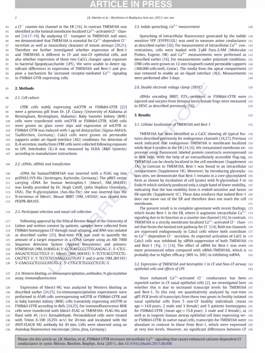
TMEM16A has been identified as a CaCC showing all typical fea-tures described previously for endogenous channels [18,27]. Previouswork indicated that endogenous TMEM16A is membrane localizedwhile Best-1 resides in the ER [14,16]. We reexamined membrane ex-pression using fluorescent labeled protein constructs overexpressedin BHK cells. With the help of an extracellularly accessible Flag-tag,TMEM16A can be clearly localized in the cell membrane (Supplement1A). In contrast to TMEM16A, Best-1 was found in an intracellularcompartment (Supplement 1B). Moreover, by introducing glycosyla-tion sites, we demonstrate that Best-1 remains in a core-glycosylatedform as shown by incubation of cell lysates with N-glycosidase F orEndo Hwhich similarly produced only a single band of lower mobility,indicating that the low-mobility form is endoH-sensitive and henceER-specific (Supplement 1C). These data reinforce that indeed Best-1does not move out of the ER and therefore does not reach the cellmembrane.
The present result is in complete agreement with recent findings,which locate Best-1 in the ER, where it augments intracellular Ca2+
signaling due to its function as a counter-ion channel [16]. In contrast,TMEM16A is a strictly membrane localized Ca2+-activated Cl− chan-nel that forms the luminal exit pathway for Cl−[14]. Both ion channelsare expressed endogenously in Calu3 cells where both contributeto Ca2+-dependent Cl− secretion. As expected activation of CaCC inCalu3 cells was inhibited by siRNA-suppression of both TMEM16Aand Best-1 (Fig. 1) [14]. The effect of siRNA for Best-1 was evenmore pronounced when compared with siRNA-TMEM16A, which isprobably due to higher efficacy (80% vs. 50%) in inhibiting mRNA.
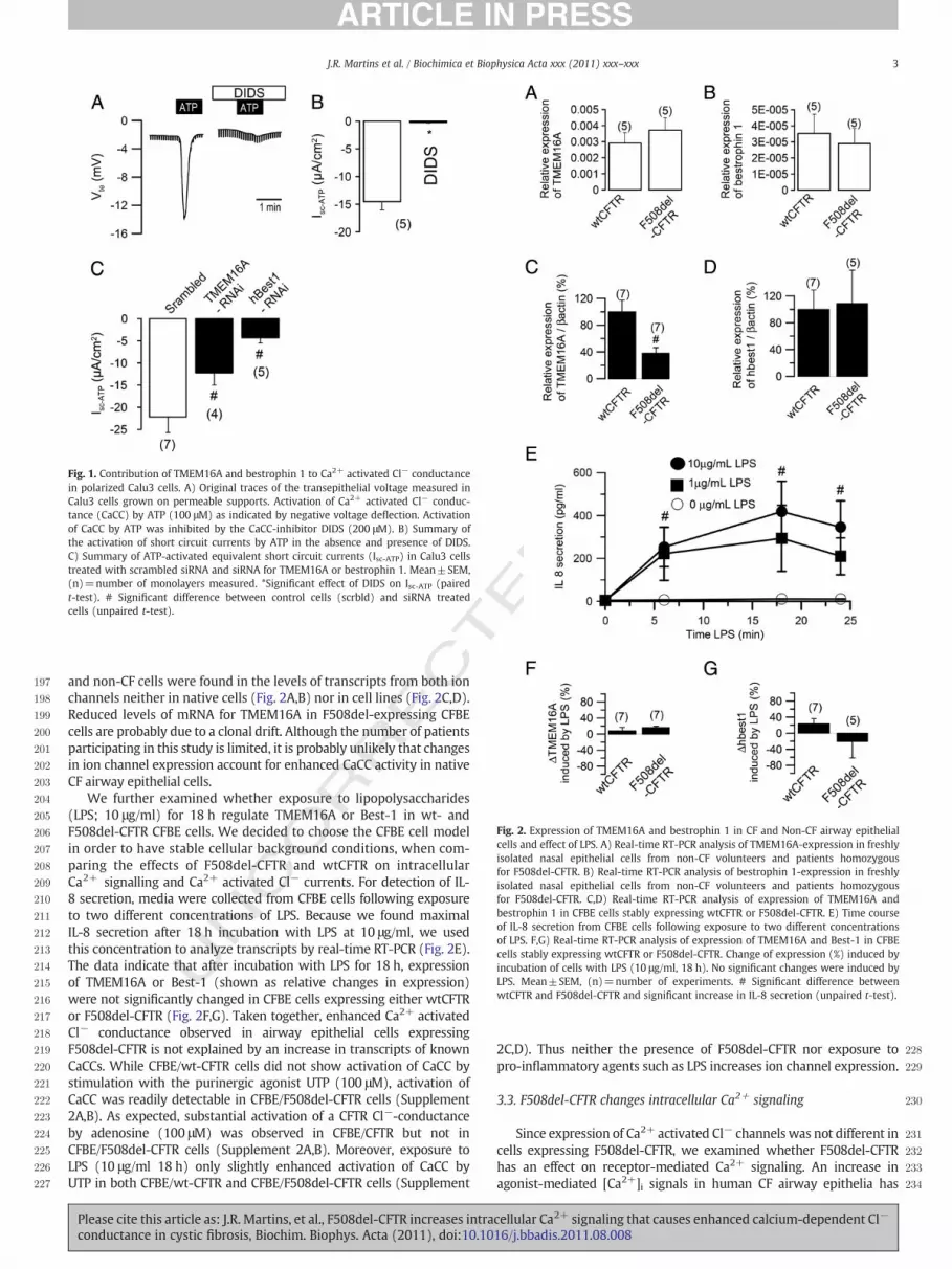
3.2. Expression of TMEM16A and bestrophin 1 in CF and Non-CF airwayepithelial cells and effects of LPS
Since enhanced Ca2+-activated Cl− conductance has beenreported earlier in CF nasal epithelial cells [2], we investigated herewhether this is due to increased transcript levels for TMEM16Aand Best-1. To this end, we quantitatively analyzed by real-timeqRT-PCR levels of transcripts from these two genes in freshly isolatednasal epithelial cells from 5 non-CF healthy individuals (meanage=14.8 years, 2 male and 3 female) and 5 patients homozygousfor F508del-CFTR (mean age=15.8 years; 3 male and 2 female), aswell as in isogenic human airway epithelial cell lines expressing wt-or F508del-CFTR. In native nasal cells, transcripts for TMEM16A wereabundant in contrast to those from Best-1, which were expressedat very low levels. However, no significant differences between CF
cellular Ca2+ signaling that causes enhanced calcium-dependent Cl−
16/j.bbadis.2011.08.008
TED P
RO
OF
197
198
199
200
201
202
203
204
205
206
207
208
209
210
211
212
213
214
215
216
217
218
219
220
221
222
223
224
225
226
227
228
229
230
231
232
233
234
Fig. 1. Contribution of TMEM16A and bestrophin 1 to Ca2+ activated Cl− conductancein polarized Calu3 cells. A) Original traces of the transepithelial voltage measured inCalu3 cells grown on permeable supports. Activation of Ca2+ activated Cl− conduc-tance (CaCC) by ATP (100 μM) as indicated by negative voltage deflection. Activationof CaCC by ATP was inhibited by the CaCC-inhibitor DIDS (200 μM). B) Summary ofthe activation of short circuit currents by ATP in the absence and presence of DIDS.C) Summary of ATP-activated equivalent short circuit currents (Isc-ATP) in Calu3 cellstreated with scrambled siRNA and siRNA for TMEM16A or bestrophin 1. Mean±SEM,(n)=number of monolayers measured. *Significant effect of DIDS on Isc-ATP (pairedt-test). # Significant difference between control cells (scrbld) and siRNA treatedcells (unpaired t-test).
Fig. 2. Expression of TMEM16A and bestrophin 1 in CF and Non-CF airway epithelialcells and effect of LPS. A) Real-time RT-PCR analysis of TMEM16A-expression in freshlyisolated nasal epithelial cells from non-CF volunteers and patients homozygousfor F508del-CFTR. B) Real-time RT-PCR analysis of bestrophin 1-expression in freshlyisolated nasal epithelial cells from non-CF volunteers and patients homozygousfor F508del-CFTR. C,D) Real-time RT-PCR analysis of expression of TMEM16A andbestrophin 1 in CFBE cells stably expressing wtCFTR or F508del-CFTR. E) Time courseof IL-8 secretion from CFBE cells following exposure to two different concentrationsof LPS. F,G) Real-time RT-PCR analysis of expression of TMEM16A and Best-1 in CFBEcells stably expressing wtCFTR or F508del-CFTR. Change of expression (%) induced byincubation of cells with LPS (10 μg/ml, 18 h). No significant changes were induced byLPS. Mean±SEM, (n)=number of experiments. # Significant difference betweenwtCFTR and F508del-CFTR and significant increase in IL-8 secretion (unpaired t-test).
3J.R. Martins et al. / Biochimica et Biophysica Acta xxx (2011) xxx–xxx
UNCO
RRECand non-CF cells were found in the levels of transcripts from both ion
channels neither in native cells (Fig. 2A,B) nor in cell lines (Fig. 2C,D).Reduced levels of mRNA for TMEM16A in F508del-expressing CFBEcells are probably due to a clonal drift. Although the number of patientsparticipating in this study is limited, it is probably unlikely that changesin ion channel expression account for enhanced CaCC activity in nativeCF airway epithelial cells.
We further examined whether exposure to lipopolysaccharides(LPS; 10 μg/ml) for 18 h regulate TMEM16A or Best-1 in wt- andF508del-CFTR CFBE cells. We decided to choose the CFBE cell modelin order to have stable cellular background conditions, when com-paring the effects of F508del-CFTR and wtCFTR on intracellularCa2+ signalling and Ca2+ activated Cl− currents. For detection of IL-8 secretion, media were collected from CFBE cells following exposureto two different concentrations of LPS. Because we found maximalIL-8 secretion after 18 h incubation with LPS at 10 μg/ml, we usedthis concentration to analyze transcripts by real-time RT-PCR (Fig. 2E).The data indicate that after incubation with LPS for 18 h, expressionof TMEM16A or Best-1 (shown as relative changes in expression)were not significantly changed in CFBE cells expressing either wtCFTRor F508del-CFTR (Fig. 2F,G). Taken together, enhanced Ca2+ activatedCl− conductance observed in airway epithelial cells expressingF508del-CFTR is not explained by an increase in transcripts of knownCaCCs. While CFBE/wt-CFTR cells did not show activation of CaCC bystimulation with the purinergic agonist UTP (100 μM), activation ofCaCC was readily detectable in CFBE/F508del-CFTR cells (Supplement2A,B). As expected, substantial activation of a CFTR Cl−-conductanceby adenosine (100 μM) was observed in CFBE/CFTR but not inCFBE/F508del-CFTR cells (Supplement 2A,B). Moreover, exposure toLPS (10 μg/ml 18 h) only slightly enhanced activation of CaCC byUTP in both CFBE/wt-CFTR and CFBE/F508del-CFTR cells (Supplement
Please cite this article as: J.R. Martins, et al., F508del-CFTR increases intraconductance in cystic fibrosis, Biochim. Biophys. Acta (2011), doi:10.10
2C,D). Thus neither the presence of F508del-CFTR nor exposure topro-inflammatory agents such as LPS increases ion channel expression.
3.3. F508del-CFTR changes intracellular Ca2+ signaling
Since expression of Ca2+ activated Cl− channels was not different incells expressing F508del-CFTR, we examined whether F508del-CFTRhas an effect on receptor-mediated Ca2+ signaling. An increase inagonist-mediated [Ca2+]i signals in human CF airway epithelia has
cellular Ca2+ signaling that causes enhanced calcium-dependent Cl−
16/j.bbadis.2011.08.008

235
236
237
238
239
240
241
242
243
244
245
246
247
248
249
250
251
252
253
254
255
256
257
258
259
260
261
262
263
264
265
266
267
268
269
270
271
272
273
274
275
4 J.R. Martins et al. / Biochimica et Biophysica Acta xxx (2011) xxx–xxx
been already reported earlier [7]. In the present study, intracellularCa2+ was enhanced by UTP stimulation (100 μM) in both CFBE/wt-CFTR and CFBE/F508del-CFTR cells (Fig. 3). We found that UTP-induced increases in both peak and plateau [Ca2+]i were significantlyenhanced in CFBE/F508del-CFTR cells, while baseline [Ca2+]i was iden-tical in both CFBE/wt-CFTR and CFBE/F508del-CFTR cells (Fig. 3A–C).We also examined whether exposure to LPS (10 μg/ml, 18 h) changedintracellular Ca2+ signals induced by UTP (100 μM) in CFBE/wt-CFTRand CFBE/F508del-CFTR cells. The summary of these experiments indi-cates a 10% increase of the UTP-induced peak [Ca2+]i but a 40% decreaseof the Ca2+ plateau after exposure to LPS, which was however onlyobserved in F508del-CFTR expressing cells (Fig. 3D). We proposethat the small increase in peak Ca2+ is due to slightly augment Ca2+
release from the ER-store, while reuptake of Ca2+ into the ER-storemay be enhanced under LPS treatment, thus reducing plateau Ca2+.It remains currently unclear why these effects were only observed inF508del-CFTR cells. Finally, Ca2+ signals were also measured in CFBEcells grown under polarized (ALI) conditions, which fully confirmresults obtained in cells grown on glass cover slips (Fig. 3E,F).
To further examine how F508del-CFTR changes intracellular Ca2+
signaling, and to demonstrate that this was not unique to CFBE cells,
UNCO
RRECT
276
277
278
279
280
281
Fig. 3. Ca2+ signaling in CFBE/wt-CFTR and CFBE/F508del-CFTR and effect of LPS.A,C) Summary recordings of increase in intracellular Ca2+ ([Ca2+]i) in CFBE/wt-CFTR(A) and CFBE/F508del-CFTR (C) cells activated by stimulation with UTP (100 μM). B)Summary of baseline [Ca2+]i and UTP-induced increase peak and plateau [Ca2+]i afterstimulation with UTP of CFBE/wt-CFTR and CFBE/F508del-CFTR (D) cells. D) Change(%) of UTP-induced peak and plateau [Ca2+]i after incubation of CFBE/wt-CFTR andCFBE/F508del-CFTR cells with LPS (10 μg/ml, 18 h). E) Summary recordings of [Ca2+]i in CFBE/wt-CFTR (left) and CFBE/F508del-CFTR (right) cells. F) Summary of Ca2+
levels (right) obtained in polarized grown CFBE cells. Mean±SEM, (n)=number ofcells measured (corresponding to the number of experiments). *Significant effectsof UTP (paired t-test). #Significant difference between wtCFTR and F508del-CFTR orcontrol and LPS-treatment (unpaired t-test).
Please cite this article as: J.R. Martins, et al., F508del-CFTR increases intraconductance in cystic fibrosis, Biochim. Biophys. Acta (2011), doi:10.10
PRO
OF
we created inducible airway epithelial (A549) cell lines, which stablyexpress wtCFTR or F508del-CFTR under an inducible (Tet-ON) pro-moter (Figs. 4A, 5A). Both cell lines were seeded at exactly the samedensity and bothwtCFTR and F508del-CFTR expressing cells weremea-sured as confluent monolayers. Moreover, for the measurementswe discarded cells with excessive CFTR-expression or cells showing ex-cessive loading with Fura2, in order to avoid cell stress and unfoldedprotein response (UPR). We measured UTP (100 μM) activated Ca2+
transients in wtCFTR A549 cells under non-induced (no expression)and induced (expression of wtCFTR) conditions, and found no increasein Ca2+i but rather a significant decrease in both peak and plateau(Fig. 4C). This is due to the depolarization of the membrane voltage byCFTR, which reduces the driving force for Ca2+ release and influx [28].Notably, CFTR is also activated by stimulationwith UTP [29]. In contrast,induction of expression of F508del-CFTR significantly increased Ca2+
signals elicited by stimulation with UTP (Fig. 5B,C). Thus, expressionof ER-localized F508del-CFTR clearly augments intracellular Ca2+
signals elicited by stimulation of GTP-coupled membrane receptors.
3.4. ER-trapped F508del-CFTR facilitates Ca2+ movements by acting as aCl− counter-ion channel
Next, we examined the mechanism by which ER-localized F508del-CFTR possibly augments intracellular Ca2+ signals. We hypothesizedthat F508del-CFTR trapped in the ER could affect receptor-mediatedCa2+ release from ER Ca2+ stores. One possibility is that F508del-CFTRfunctions as a Cl− channel in the ER membrane, thereby allowing Cl−
fluxes in parallel to Ca2+ movements. Such a counter-ion channel
ED
Fig. 4. Induction of expression of wtCFTR does not augment Ca2+ signaling. A) Expressionof wtCFTR in non-induced (left upper) and induced (right upper) A549 cells. B) Originalrecordings of intracellular Ca2+ [Ca2+]i signals elicited by UTP (100 μM) in non-induced(no wtCFTR, left) and induced (wtCFTR, right) A549 cells. C) Summary of baseline[Ca2+]i (con) and peak and plateau [Ca2+]i after stimulation with UTP. *Significant effectsof UTP (paired t-test). #Significant difference between +/− wtCFTR and F508del-CFTR(unpaired t-test).
cellular Ca2+ signaling that causes enhanced calcium-dependent Cl−
16/j.bbadis.2011.08.008

RECT
282
283
284
285
286
287
288
289
290
291
292
293
294
295
296
297
298
299
300
301
302
303
304
305
306
307
308
309
310
311
312
313
314
315
316
317
318
319
320
321
322
323
324
325
326
327
328
329
330
331
332
333
334
335
336
337
338
339
340
341
342
343
344
345
346
347
348
349
350
351
352
353
354
355
356
357
358
359
360
361
362
363
364
Fig. 5. Induction of expression of F508del-CFTR augments Ca2+ signaling. A) Expressionof F508del-CFTR in non-induced (left upper) and induced (right upper) A549 cells.B) Original recordings of intracellular Ca2+ ([Ca2+]i signals elicited by UTP (100 μM)in non-induced (no F508del-CFTR, left) and induced (F508del-CFTR, right) A549 cells.C) Summary of baseline [Ca2+]i (con) and peak and plateau [Ca2+]i after stimulationwith UTP. Bar=20 μm. Mean±SEM, (n)=number of cells measured. *Significanteffects of UTP (paired t-test). #Significant difference between wtCFTR andF508del-CFTR (unpaired t-test).
5J.R. Martins et al. / Biochimica et Biophysica Acta xxx (2011) xxx–xxx
UNCO
R
function has been discussed for a long time and was recently proposedto be the Ca2+-activated Cl− channel Best-1 [16]. We therefore, testedwhether UTP-induced Ca2+ signaling in A549 cells was Cl− dependent.To that end, we measured Ca2+i transients in induced A549/wtCFTRand A549/F508del-CFTR cells in Cl−-depleted cells (0 mM extracellularCl−; Fig. 6A). UTP-induced increase in [Ca2+i] was largely reduced inthe absence of extracellular Cl− and when compared to Ca2+ increasein physiological Ringer (145 mM extracellular Cl−) solution (Fig. 3B).Notably, attenuation of Ca2+ transients in Cl− free solution wasmore pronounced in F508del-CFTR expressing cells (Figs. 3B, 6A)and increased Ca2+ signals in F508del-CFTR expressing cells were nolonger observed under Cl− free conditions (Fig. 6A).
In order to further examine whether the ability of F508del-CFTRto produce a Cl− conductance in the ER membrane may influence theCa2+ signal, we expressed the double mutant F508del/G551D-CFTRthat i) does not traffic to the cell membrane but remains in the ER(caused by the F508del mutation; Fig. 6C) and ii) does not operateas a Cl− channel, due to defective channel gating (caused by theG551D mutation; data not shown). The experiments were performedon confluent monolayers. In fact, A549 cells expressing the double mu-tant F508del/G551D-CFTR cells did no longer produce enhanced Ca2+
signals (Fig. 6B). These results suggest that the presence of F508del-
Please cite this article as: J.R. Martins, et al., F508del-CFTR increases intraconductance in cystic fibrosis, Biochim. Biophys. Acta (2011), doi:10.10
ED P
RO
OF
CFTR in the ER, which has been shown earlier to be functional whenretained in endoplasmic reticulum [30], may affect intracellular Ca2+
signaling by acting as a Cl− counter-ion channel, thereby facilitatingCa2+ movement over the ER membrane.
3.5. Expression of IRBIT antagonizes enhanced Ca2+ signals inXenopus oocytes
It was recently reported, that the protein inositol-1,4,5-trisphosphate[IP3] receptor binding protein released with IP3 (IRBIT) binds to CFTRwhen released from the IP3-receptor upon binding of inositol-1,4,5-tri-sphosphate [IP3] [25]. Because F508del-CFTR accumulates in the ERbeing thus in close proximity to the IP3-receptor, we speculated thatF508del-CF may compete with IP3receptors for binding to IRBIT [31].Thus reduced binding of IRBIT to the IP3receptor would enhanceagonist-induced Ca2+ release from the ER. To test this hypothesis, wemade use of the expression system in Xenopus oocytes since it allowsparallel expression of several proteins. Although oocytes express atlow temperatures (18 °C), the amount of mature F508-CFTR in the cellmembrane is probably low as whole cell conductance in these cellswere only 12±2.1 μS (n=12), which is low compared to wtCFTR-expressing oocytes (123±10.7 μS; n=14). When P2Y2-receptorswere expressed in oocytes, stimulation by UTP (100 μM) activatedendogenous Ca2+-dependent TMEM16A channels and produced atransient and outwardly rectifying whole cell Cl− current (Fig. 7A).Notably, co-expression of wtCFTR with P2Y2-receptors slightly, butsignificantly augmented the Ca2+-activated Cl− current, while co-expression of F508del-CFTR induced a significantly larger Ca2+ acti-vated Cl− current (Fig. 7B–D). Additional co-expression of IRBIThad little effects on Ca2+ activated currents in wtCFTR-expressingcells, but dramatically inhibited UTP-induced currents in F508del-CFTR co-expressing cells (Fig. 7D). These results would be in agree-ment with the idea that F508del-CFTR accumulating in the ER maybind IRBIT thereby facilitating agonist-induced IP3 binding to theIP3 receptor and further increase in intracellular Ca2+, or it couldsimply be a non-specific antagonistic effect of IRBIT. To examinebinding of IRBIT to CFTR we performed co-immunoprecipitation ex-periments in two different cell lines: A549 cells stably expressing ei-ther wtCFTR or F508del-CFTR, and baby hamster kidney (BHK) cellstransiently expressing wtCFTR or F508del-CFTR. Moreover,two different co-immunoprecipitation protocols [24,25] were usedand either IRBIT or CFTR were immunoprecipitated. Under no condi-tions we could observe a co-immunoprecipitation, as observed in[25], and we have therefore no evidence that IRBIT binds directlyto CFTR when expressed in these two different cell lines (Supple-ment 3). Taken together, the present results suggest that enhancedCa2+-activated Cl− conductance in CF epithelial cells is due toaugmented intracellular Ca2+ signaling, caused by ER-localizedF508del-CFTR. F508del-CFTR may probably function as an ER-locatedcounter-ion channel which facilitates Ca2+ release from ER stores.In this regard it is probably interesting to note that in an earlierstudy we were unable to detect enhanced Ca2+ activated Cl− transportin epithelial tissues of transgenic G551D mice [32].
4. Discussion
4.1. Inflammation in CF
Although CFTR is known for more than 20 years, it remains enig-matic how abnormalities in CFTR can cause chronic and persistentpulmonary inflammation. There is agreement that loss of functionalCFTR results in activation of neutrophils that produce large amountsof proteases and reactive oxygen species (ROS). These changesare associated with reduced mucociliary clearance of bacteria,and induction of hyperinflammatory responses in CF airways. TheNF-κB pathway and Ca2+ mobilization in airway epithelial cells are
cellular Ca2+ signaling that causes enhanced calcium-dependent Cl−
16/j.bbadis.2011.08.008

UNCO
RRECT
D P
RO
OF
365
366
367
368
369
370
371
372
373
374
375
376
377
378
379
380
381
382
383
384
385
386
387
388
389
390
391
392
393
394
395
396
397
Fig. 6. Intracellular Ca2+signaling is Cl− dependent. A) Summary of intracellular Ca2+ [Ca2+]i signals elicited by UTP (100 μM) stimulation of A549 cells expressing wt-CFTR or F508del-CFTR in Cl− depleted cells (0 mM extracellular Cl− concentration). Peak and plateau Ca2+ increase were largely attenuated in both A549/wt-CFTR and A549/F508del-CFTR cells in theabsence of extracellular Cl−. B) Summary of [Ca2+]i increase induced by UTP (100 μM) in A549 cells expressing F508del-CFTR or the doublemutant F508del/G551D-CFTR in the presenceof 145 mM extracellular Cl− concentration. C) Expression of F508del/G551D-CFTR (left panel) and pcDNA3 control plasmid (right panel) in A549 cells. Mean±SEM, (n)=number ofcells measured. *Significant effects of UTP (paired t-test). #Significant difference between wtCFTR and F508del-CFTR or F508del-CFTR and the double mutant (unpaired t-test).
Fig. 7. Potential role of IRBIT for enhanced Ca2+ activated Cl− conductance in F508del-CFTR expressing oocytes. A) Original recording of whole cell Cl− currents activated byUTP (100 μM) in P2Y2-receptor expressing Xenopus oocytes. B) Original recording ofwhole cell Cl− currents activated by UTP in P2Y2-receptor and wtCFTR co-expressingXenopus oocytes. C) Original recording of whole cell Cl− currents activated by UTPin P2Y2-receptor and F505del-CFTR co-expressing Xenopus oocytes. D) Summary of calcu-lated relative whole cell conductances activated by UTP in the absence or presence ofcoexpressed IRBIT. Mean±SEM, (n)=number of cells measured. *Significant effectsof UTP (paired t-test). #Significant difference between different batches (unpaired t-test).
6 J.R. Martins et al. / Biochimica et Biophysica Acta xxx (2011) xxx–xxx
Please cite this article as: J.R. Martins, et al., F508del-CFTR increases intraconductance in cystic fibrosis, Biochim. Biophys. Acta (2011), doi:10.10
Ebelieved to be of key importance for lung inflammation, through releaseof mediators such as interleukin-8 [33]. Evidence suggests that CFTRmutations, most importantly F508del-CFTR itself can produce apro-inflammatory milieu in the airways that precedes infection.Thus F508del-CFTR has been demonstrated to accumulate in theER and to trigger a stress response that leads to NFκB activationand IL8 production [12,13,34]. Another study found that inhibitionof CFTR in airway epithelial cells mimicked the CF-typical inflam-matory profile with increase in nuclear NFχB and IL-8 secretion[11], while others showed that expression of functional CFTR onthe cell surface negatively regulates NFκB mediated innate immuneresponse [35].
Other studies demonstrated increased secretion of pro-inflammatory cytokines by CF airway cells only after exposure topathogenic bacteria [7,10,36]. Thus Ribeiro and collaborators demon-strated expansion of an ER compartment close to the luminal mem-brane of chronically infected and inflamed airway epithelia, like thoseaffected by cystic fibrosis [7]. Large luminal ER pools lead to enhancedapical Ca2+ signaling, which explains the augmented Ca2+ activatedCl− secretion observed in cystic fibrosis airways. In contrast to thesestudies, we were not able to detect any significant effects of themajor bacterial component LPS. Moreover, in preliminary experimentswe incubated airway cells with purulent sputum from CF patients(kindly provided by Dr. Carla Ribeiro, UNC, Chapel Hill, USA) in 1:200and 1:1 dilutions, 16 h) but did not see a change in expression ofBest-1 or TMEM16A (data not shown).
4.2. Enhanced Ca2+-dependent activation of TMEM16A
Interestingly, pro-inflammatory interleukins have been shown tostimulate both Ca2+-activated Cl− and SK4 K+ channels [37]. Galiettaand colleagues actually identified TMEM16A as a Ca2+ activated Cl−
channel because Ca2+ activated Cl− secretion in bronchial epithelialcells was upregulated by IL4 [18,37]. Although Best-1 was found to beupregulated during renal inflammation [16,27] it was only minimally
cellular Ca2+ signaling that causes enhanced calcium-dependent Cl−
16/j.bbadis.2011.08.008

T
398
399
400
401
402
403
404
405
406
407
408
409
410
411
412
413
414
415
416
417
418
419
420
421
422
423
424
425
426
427
428
429
430
431
432
433
434
435
436
437
438
439
440
441
442
443
444
445
446
447
448
449
450
451
452
453
454
455
456
457
458
459
460
461462463464465466467468469470471472473474475476477478479480481482483484485486487488489490491492493494495496497498499500501502503504505506507508509510511512513514515516517518519520521522523524525526527528529530531532533534535536537538539540541542543544
7J.R. Martins et al. / Biochimica et Biophysica Acta xxx (2011) xxx–xxx
UNCO
RREC
enhanced in F508del-CFTR expressing cells upon exposure of thecells to bacterial LPS. The fact that retention of misfolded F508del-CFTR leads to imbalances in Ca2+ homeostasis is a well recognizedfact, however, the reasons for the Ca2+ increase are poorly understood[13]. Our present data suggest a direct link between F508del-CFTRand intracellular Ca2+ signaling, thereby connecting F508del-CFTR toenhanced Ca2+-dependent Cl− secretion in CF. Notably; we alsofound recently that baseline Cl− currents mediated by the Cl− channelSLC26A9 are enhanced in wtCFTR-expressing cells when compared toairway cells expressing F508del-CFTR [38].
Our data would explain why conversely Ca2+ signaling is affectedby F508del-CFTR, as this defective, but still partially active CFTRchannel may provide a Cl− conductance in the ER membrane andthus facilitate Ca2+ movement by allowing transport of the counter-ion Cl−[30]. The concept of a counter-ion channel in the ER to balancenegative charges occurring through Ca2+ release and reuptake intothe ER-store has long been proposed [39]. In the sarcoplasmic reticu-lum (SR), Cl− channels play an essential role in excitation–contractioncoupling, by balancing charge movement during calcium release andreuptake [40,41]. This is also known from airway smooth musclecells. SR-localized Cl− channels in the SRmembrane allow for neutral-ization of electrostatic charges that would otherwise build up duringCa2+ movement [42]. Blockage of these Cl− channels in airwaysmooth muscle cells might be an effective way in inhibiting airwaysmooth muscle hyperresponsiveness observed in asthma [43]. Alsofor Best-1, a function as a counter-ion channel in the ER of epithelialcells has been proposed recently [16].
4.3. Enhanced Ca2+ signaling, proliferation and the role of IRBIT
Our present data also provide an explanation for the enhancedproliferation observed for CF epithelial cells. It has been shown earlierthat cell proliferation in bronchial epithelium and submucosal glandsof cystic fibrosis patients is much increased when compared to non-CF cells [44]. The high proliferation rate of CF airway epithelial cellshas been explained by the chronic inflammatory process that takesplace in CF airways. However, it is also observed under in vitro condi-tions and in the absence of exogenous pro-inflammatory factors [45].A change in intracellular Ca2+ signaling in cells expressing F508del-CFTR could explain enhanced proliferative activity and delayed cellu-lar differentiation. Finally, the present experiments also demonstratea functional interference of CFTR with the IP3-receptor binding pro-tein IRBIT. IRBIT has been shown to suppress the activity of IP3 recep-tors by competing with IP3 for a common binding site [31]. A recentreport showed that IRBIT coordinates epithelial fluid and HCO3-secretion due to stimulation of the Na+/HCO3
− co-transporter andCFTR [25]. However, in contrast to this study we were unable tocoimmunoprecipitate CFTR and IRBIT in the present study (Supplement3) and therefore propose that F508del-CFTR affects Ca2+ signaling inan IRBIT and Cl− dependent manner, due to its ability to operate as anER-trapped ion channel.
Supplementary materials related to this article can be foundonline at doi:10.1016/j.bbadis.2011.08.008.
Acknowledgements
The work was supported by DFGSFB699A7, DFGKU 756/8-2, andTargetScreen2 (EU-FP6-2005-LH-037365) and pluriannual fundingof CIGMH (FCT, Portugal). JR Martins is a recipient of doctoral fel-lowship SFRH/BD/28663/2006 (from FCT, Portugal). The authorswould like to thank Dr. Celeste Barreto (Hospital de Sta Maria, Lisboa,Portugal), CF patients and their families for access to nasal cellsand Carla Ribeiro (UNC, Chapel Hill, NC, USA) for SMM lavage fluid.E.S. obtained a doctoral grant from the “Excellentiefinanciering”, (EFKULeuven, 2005–2009). We thank KirstenWelkenhuyzen for excellenttechnical assistance.
Please cite this article as: J.R. Martins, et al., F508del-CFTR increases intraconductance in cystic fibrosis, Biochim. Biophys. Acta (2011), doi:10.10
ED P
RO
OF
References
[1] M.R. Knowles, L.L. Clarke, R.C. Boucher, Activation by extracellular nucleotides ofchloride secretion in the airway epithelia of patients with cystic fibrosis, N. Engl. J.Med. 325 (1991) 533–538.
[2] M. Mall, T. Gonska, J. Thomas, R. Schreiber, H.H. Seydewitz, J. Kuehr, M. Brandis, K.Kunzelmann, Modulation of Ca2+ activated Cl− secretion by basolateral K+channels in human normal and cystic fibrosis airway epithelia, Pediatr. Res.53 (2003) 608–618.
[3] B.R. Grubb, R.N. Vick, R.C. Boucher, Hyperabsorption of Na+ and raised Ca2+ me-diated Cl− secretion in nasal epithelia of CF mice, Am. J. Physiol. 266 (1994)C1478–C1483.
[4] A.M. Paradiso, C.M. Ribeiro, R.C. Boucher, Polarized signaling via purinoceptors innormal and cystic fibrosis airway epithelia, J. Gen. Physiol. 117 (2001) 53–68.
[5] K. Kunzelmann, M. Mall, M. Briel, A. Hipper, R. Nitschke, S. Ricken, R. Greger, Thecystic fibrosis transmembrane conductance regulator attenuates the endogenousCa2+ activated Cl− conductance in Xenopus ooyctes, Pflügers Arch. 434 (1997)178–181.
[6] L. Wei, A. Vankeerberghen, H. Cuppens, J. Eggermont, J.J. Cassiman, G. Droogmans,B. Nilius, Interaction between calcium-activated chloride channels and thecystic fibrosis transmembrane conductance regulator, Pflugers Arch. 438 (1999)635–641.
[7] C.M. Ribeiro, A.M. Paradiso, U. Schwab, J. Perez-Vilar, L. Jones, W. O'neal, R.C.Boucher, Chronic airway infection/inflammation induces a Ca2+i-dependenthyperinflammatory response in human cystic fibrosis airway epithelia, J. Biol.Chem. 280 (2005) 17798–17806.
[8] R. Bartoszewski, A. Rab, A. Jurkuvenaite, M. Mazur, J. Wakefield, J.F. Collawn, Z.Bebok, Activation of the unfolded protein response by deltaF508 CFTR, Am. J.Respir. Cell Mol. Biol. 39 (2008) 448–457.
[9] C.M. Ribeiro, The role of intracellular calcium signals in inflammatory responsesof polarised cystic fibrosis human airway epithelia, Drugs R.D. 7 (2006) 17–31.
[10] K. Hybiske, Z. Fu, C. Schwarzer, J. Tseng, J. Do, N. Huang, T.E. Machen, Effectsof cystic fibrosis transmembrane conductance regulator (CFTR) and delta F508-CFTR on inflammatory response, ER stress and Ca2+ of airway epithelia, Am. J.Physiol. Lung Cell. Mol. Physiol. 293 (2007) L1250–L1260.
[11] A. Perez, A.C. Issler, C.U. Cotton, T.J. Kelley, A.S. Verkman, P.B. Davis, CFTR inhibi-tion mimics the cystic fibrosis inflammatory profile, Am. J. Physiol. Lung Cell. Mol.Physiol. 292 (2007) L383–L395.
[12] F. Antigny, C. Norez, F. Becq, C. Vandebrouck, Calcium homeostasis is abnormal incystic fibrosis airway epithelial cells but is normalized after rescue of F508del-CFTR, Cell Calcium 43 (2008) 175–183.
[13] M. Rottner, C. Kunzelmann, M. Mergey, J.M. Freyssinet, M.C. Martinez, Exaggeratedapoptosis and NF-{kappa}B activation in pancreatic and tracheal cystic fibrosiscells, FASEB J. 21 (2007) 2939–2948.
[14] K. Kunzelmann, P. Kongsuphol, F. AlDehni, Y. Tian, J. Ousingsawat, R. Warth, R.Schreiber, Bestrophin and TMEM16 - Ca2+ activated Cl− channels with differentfunctions, Cell Calcium 46 (2009) 233–241.
[15] R. Barro Soria, R. Schreiber, K. Kunzelmann, Bestrophin 1 and 2 are componentsof the Ca2+ activated Cl− conductance in mouse airways, BBA 1783 (2008)1993–2000.
[16] R. Barro Soria, F. AlDehni, J. Almaca, R. Witzgall, R. Schreiber, K. Kunzelmann, ERlocalized bestrophin1 acts as a counter-ion channel to activate Ca2+ dependention channels TMEM16A and SK4, Pflügers Arch. 459 (2009) 485–497.
[17] Y.D. Yang, H. Cho, J.Y. Koo, M.H. Tak, Y. Cho, W.S. Shim, S.P. Park, J. Lee, B. Lee, B.M.Kim, R. Raouf, Y.K. Shin, U. Oh, TMEM16A confers receptor-activated calcium-dependent chloride conductance, Nature 455 (2008) 1210–1215.
[18] A. Caputo, E. Caci, L. Ferrera, N. Pedemonte, C. Barsanti, E. Sondo, U. Pfeffer, R.Ravazzolo, O. Zegarra-Moran, L.J. Galietta, TMEM16A, a membrane protein associatedwith calcium-dependent chloride channel activity, Science 322 (2008) 590–594.
[19] L. Ferrera, A. Caputo, I. Ubby, E. Bussani, O. Zegarra-Moran, R. Ravazzolo, F. Pagani,L.J. Galietta, Regulation of TMEM16A chloride channel properties by alternativesplicing, J. Biol. Chem. 284 (2009) 33360–33368.
[20] J. Ousingsawat, J.R. Martins, R. Schreiber, J.R. Rock, B.D. Harfe, K. Kunzelmann, Lossof TMEM16A causes a defect in epithelial Ca2+ dependent chloride transport,J. Biol. Chem. 284 (2009) 28698–28703.
[21] J.R. Rock, W.K. O'Neal, S.E. Gabriel, S.H. Randell, B.D. Harfe, R.C. Boucher, B.R.Grubb, Transmembrane protein 16A (TMEM16A) is a Ca2+regulated Cl−-secretorychannel in mouse airways, J. Biol. Chem. 284 (2009) 14875–14880.
[22] Z. Bebok, J.F. Collawn, J. Wakefield, W. Parker, Y. Li, K. Varga, E.J. Sorscher, J.P.Clancy, Failure of cAMP agonists to activate rescued deltaF508 CFTR inCFBE41o-airway epithelial monolayers, J. Physiol. 569 (2005) 601–615.
[23] S. Beck, D. Penque, S. Garcia, A. Gomes, C. Farinha, L. Mata, S. Gulbenkian, K.Gil-Ferreira, A. Duarte, P. Pacheco, C. Barreto, B. Lopes, J. Cavaco, J. Lavinha, M.D.Amaral, Cystic fibrosis patients with the 3272–26A–NGmutation have mild disease,leaky alternative mRNA splicing, and CFTR protein at the cell membrane, Hum.Mutat. 14 (1999) 133–144.
[24] E. Sammels, B. Devogelaere, D. Mekahli, G. Bultynck, L. Missiaen, J.B. Parys, Y. Cai, S.Somlo, H. De Smedt, Polycystin-2 activation by inositol 1,4,5-trisphosphate-inducedCa2+ release requires its direct association with the inositol 1,4,5-trisphosphatereceptor in a signaling microdomain, J. Biol. Chem. 285 (2010) 18794–18805.
[25] D. Yang, N. Shcheynikov, W. Zeng, E. Ohana, I. So, H. Ando, A. Mizutani, K.Mikoshiba, S. Muallem, IRBIT coordinates epithelial fluid and HCO3-secretion bystimulating the transporters pNBC1 and CFTR in the murine pancreatic duct,J. Clin. Invest. 119 (2009) 193–202.
[26] L.J. Galietta, P.M. Haggie, A.S. Verkman, Green fluorescent protein-based halide indi-cators with improved chloride and iodide affinities, FEBS Lett. 499 (2001) 220–224.
cellular Ca2+ signaling that causes enhanced calcium-dependent Cl−
16/j.bbadis.2011.08.008

545546547548549550551552553554555556557558559560561562563Q4564565566567568569570
571572573574575576577578579580581582583584585586587588589590591592593594595596
598
8 J.R. Martins et al. / Biochimica et Biophysica Acta xxx (2011) xxx–xxx
[27] F. AlDehni, M. Spitzner, J.R. Martins, R. Barro Soria, R. Schreiber, K. Kunzelmann,Role of bestrophin for proliferation and in epithelial to mesenchymal transition,J. Am. Soc. Nephrol. 20 (2009) 1556–1564.
[28] K.G. Fischer, J. Leipziger, P. Rubini-Illes, R. Nitschke, R. Greger, Attenuation ofstimulated Ca2+ influx in colonic epithelial (HT29) cells by cAMP, Pflugers Arch.432 (1996) 735–740.
[29] D. Faria, R. Schreiber, K. Kunzelmann, CFTR is activated through stimulation ofpurinergic P2Y2 receptors, Pflügers Arch. 457 (2009) 1373–1380.
[30] E.A. Pasyk, J.K. Foskett, Mutant (delta F508) cystic fibrosis transmembraneconductance regulator Cl− channel is functional when retained in endoplasmicreticulum of mammalian cells, J. Biol. Chem. 270 (1995) 12347–12350.
[31] H. Ando, A. Mizutani, H. Kiefer, D. Tsuzurugi, T. Michikawa, K. Mikoshiba, IRBITsuppresses IP3 receptor activity by competing with IP3 for the common bindingsite on the IP3 receptor, Mol. Cell 22 (2006) 795–806.
[32] D. Oceandy, B.J. McMorran, R. Schreiber, B. Wainwright, K. Kunzelmann, GFP-tagged CFTR transgene is functional in the G551D Cystic fibrosis mouse colon,J. Membr. Biol. 192 (2003) 159–167.
[33] J. Jacquot, O. Tabary, P. Le Rouzic, A. Clement, , Airway epithelial cell inflammatorysignalling in cystic fibrosis, Int. J. Biochem. Cell Biol. (2008).
[34] D.R. Koehler, G.P. Downey, N.B. Sweezey, A.K. Tanswell, J. Hu, Lung inflammation as atherapeutic target in cystic fibrosis, Am. J. Respir. Cell Mol. Biol. 31 (2004) 377–381.
[35] N. Vij, S. Mazur, P.L. Zeitlin, CFTR is a negative regulator of NFkappaB mediatedinnate immune response, PLoS One 4 (2009) e4664.
[36] M.N. Becker, M.S. Sauer, M.S. Muhlebach, A.J. Hirsh, Q. Wu, M.W. Verghese, S.H.Randell, Cytokine secretion by cystic fibrosis airway epithelial cells, Am. J. Respir.Crit. Care Med. 169 (2004) 645–653.
UNCO
RRECT
597
Please cite this article as: J.R. Martins, et al., F508del-CFTR increases intraconductance in cystic fibrosis, Biochim. Biophys. Acta (2011), doi:10.10
OF
[37] L.J. Galietta, P. Pagesy, C. Folli, E. Caci, L. Romio, B. Costes, E. Nicolis, G. Cabrini,M. Goossens, R. Ravazzolo, O. Zegarra-Moran, IL-4 is a potent modulator of iontransport in the human bronchial epithelium in vitro, J. Immunol. 168 (2002)839–845.
[38] J. Ousingsawat, R. Schreiber, K. Kunzelmann, Differential contribution of SLC26A9to Cl(−) conductance in polarized and non-polarized epithelial cells, J. Cell. Physiol.(2011) 10.
[39] M.J. Berridge, The endoplasmic reticulum: a multifunctional signaling organelle,Cell Calcium 32 (2002) 235–249.
[40] J.I. Kourie, ATP-sensitive voltage- and calcium-dependent chloride channelsin sarcoplasmic reticulum vesicles from rabbit skeletal muscle, J. Membr. Biol.157 (1997) 39–51.
[41] J.R. Coonan, G.D. Lamb, Effect of chloride on Ca2+ release from the sarcoplasmicreticulum of mechanically skinned skeletal muscle fibres, Pflugers Arch. 435 (1998)720–730.
[42] S. Hirota, N. Trimble, E. Pertens, L.J. Janssen, Intracellular Cl− fluxes play a novelrole in Ca2+ handling in airway smooth muscle, Am. J. Physiol. Lung Cell. Mol.Physiol. 290 (2006) L1146–L1153.
[43] L.J. Janssen, Asthma therapy: how far have we come, why did we fail and whereshould we go next? Eur. Respir. J. 33 (2009) 11–20.
[44] M.W. Leigh, J.E. Kylander, J.R. Yankaskas, R.C. Boucher, Cell proliferation in bron-chial epithelium and submucosal glands of cystic fibrosis patients, Am. J. Respir.Cell Mol. Biol. 12 (1995) 605–612.
[45] R. Hajj, P. Lesimple, B. Nawrocki-Raby, P. Birembaut, E. Puchelle, C. Coraux,Human airway surface epithelial regeneration is delayed and abnormal in cysticfibrosis, J. Pathol. 211 (2007) 340–350.
OED P
R
cellular Ca2+ signaling that causes enhanced calcium-dependent Cl−
16/j.bbadis.2011.08.008