treatment and control of leishmaniasis using photodynamic therapy
TRANSCRIPT
Chapter 17
Treatment and Control of Leishmaniasis UsingPhotodynamic Therapy
Debora P. Aureliano, Martha S. Ribeiro,José Angelo Lauletta Lindoso, Fabio C. Pogliani,Fábio P. Sellera, Dennis Song andMauricio S. Baptista
Additional information is available at the end of the chapter
http://dx.doi.org/10.5772/57456
1. Introduction
Leishmaniasis is a chronic disease affecting the skin, mucosal and/or internal organs, causedby flagellate protozoa Leishmania of the Trypanosomatidae family. [1] It is among the six mostimportant disease in terms of its impact in public health. The world incidence of leishmaniasisis very large with about half a million new cases per year. About 12 million people are infectedwith Leishmania ssp parasites worldwide. New treatment alternatives are highly needed. Ourgoal here is to critically revise the literature in order to show the potential of PhotodynamicTherapy in the treatment and comprehensive control of this disease. We have separated thischapter in nine sections, besides this brief introduction, which are: Leishmaniasis: Backgroundand treatment strategies; Mechanisms in Photodynamic Therapy; Treatment of animalsinfected with leishmaniasis using PDT; Vector control using PDT; PDT alternatives for Bloodpurification; PDT on the treatment of Old World Tegumentary Leishmaniasis; PDT - In vitrotests in species that cause Tegumentary Leishmaniasis; Conclusions; References.
2. Leishmaniasis — Background and treatment strategies
There are two main forms of leishmaniasis, visceral (VL) and tegumentary (TL) leishmaniasis,which are also respectively called Kala Azar and Bauru ulcer. The later, received its namebecause of the original high prevalence in Bauru, a city in the countryside of the State of São
© 2014 The Author(s). Licensee InTech. This chapter is distributed under the terms of the Creative CommonsAttribution License (http://creativecommons.org/licenses/by/3.0), which permits unrestricted use,distribution, and reproduction in any medium, provided the original work is properly cited.
Paulo, in Brazil. The tegumentary leishmaniasis is characterized by skin lesions (cutaneous-CL) and mucocutaneous lesions (such as, nasal and mouth regions) [2].
Leishmaniasis is a common zoonosis, with domestic (dogs and cats) and wild (rodents,marsupials, edentulous and wild canids) reservoirs. It is transmitted to humans by sand flies,which comprise the genus Lutzomyia and Phlebotomus. Details of the etiology and patho‐physiology of the disease are out of the scope of this chapter and we suggest that the readerconsult reviews that focus on these subjects [3].
The current scenario of leishmaniasis treatment is not promising. Therapeutic approachesinclude systemic administrations of anti-parasitic medications, which often present seriousside effects. Few drugs are available in the clinic, mainly antimonials and amphotericin, andthe frequency of resistance development is rising. Therefore, there is an urgent need to establishnew and more effective treatments for both VL and TL. The treatment of TL (the focus of thischapter) urges new drugs and new therapeutic forms, that allows for more effective andconveniently administered treatments [4].
One of the promising approaches, and the one discussed in here, is photodynamic therapy(PDT). The main expectation of this approach is that it treats lesions in a localized manner,without damaging healthy tissues [5]. The few reports that are available in the literature havevalidated this hypothesis. In addition, no sign of systemic toxicity is reported in PDT, elimi‐nating one of the major health issues related to existing TL treatments.[6] These points will befurther discussed in this chapter.
The use of light as a therapeutic modality has gained strong impulse recently due to thedevelopment of efficient and affordable light sources. Consequently, photo-activated drugs(PhotoSensitizers-PS) play key roles in the present clinical portfolio, and more importantly,are the major lead in the development of new drugs to treat a variety of diseases such as cancer,microbial infections and tropical diseases. However, increasing the efficiency of PDT photo‐sensitizers remains challenging [7-9].
The use of PDT in veterinary is much less common even considering the benefits that suchstrategies could bring in the treatment of high-value reproducing animals, as well as, in thetreatment of animals that are reservoirs of human diseases [10].
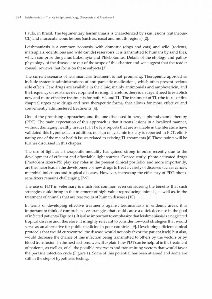
In terms of developing effective treatments against leishmaniasis in endemic areas, it isimportant to think of comprehensive strategies that could cause a quick decrease in the poolof infected patients (Figure 1). It is also important to emphasize that leishmaniasis is a neglectedtropical disease and, therefore, it is highly relevant to consider low-cost strategies that wouldserve as an alternative for public medicine in poor countries [9]. Developing efficient clinicalprotocols that would cure/control the disease would not only favor the patient itself, but also,would decrease the chance of this infection being transmitted to others by the vectors or byblood transfusion. In the next sections, we will explain how PDT can be helpful in the treatmentof patients, as well as, of all the possible reservoirs and transmitting vectors that would favorthe parasite infection cycle (Figure 1). Some of this potential has been attained and some arestill in the step of hypothesis testing.
Leishmaniasis - Trends in Epidemiology, Diagnosis and Treatment394
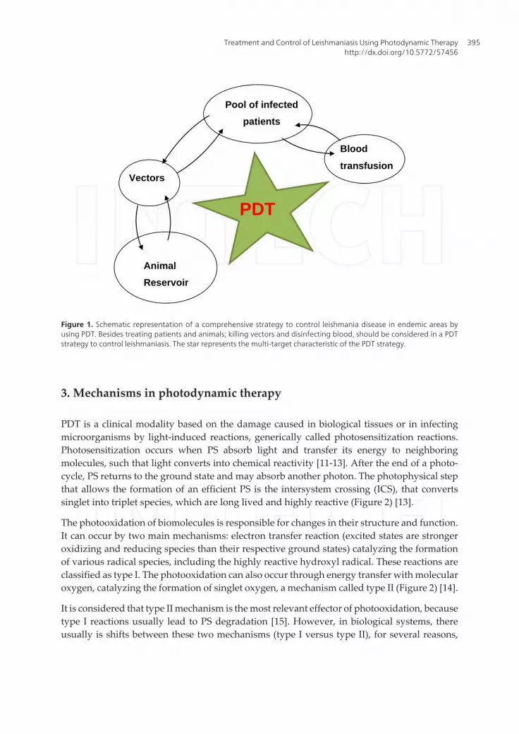
Figure 1: Schematic representation of a comprehensive strategy to control leishmania disease in endemic areas by using PDT. Besides treating patients and animals; killing vectors and disinfecting blood, should be considered in a PDT strategy to control leishmaniasis. The star represents the multi-target characteristic of the PDT strategy.
3. Mechanisms in Photodynamic Therapy
PDT is a clinical modality based on the damage caused in biological tissues or in
infecting microorganisms by light-induced reactions, generically called photosensitization
reactions. Photosensitization occurs when PS absorb light and transfer its energy to
neighboring molecules, such that light converts into chemical reactivity[11-13] After the end
of a photo-cycle, PS returns to the ground state and may absorb another photon. The
photophysical step that allows the formation of an efficient PS is the intersystem crossing
(ICS), that converts singlet into triplet species, which are long lived and highly reactive
(Figure 2)[13].
The Photooxidation of biomolecules is responsible for changes in their structure and
function. It can occur by two main mechanisms: electron transfer reaction (excited states are
stronger oxidizing and reducing species than their respective ground states) catalyzing the
formation of various radical species, including the highly reactive hydroxyl radical. These
reactions are classified as type I. The photooxidation can also occur through energy transfer
with molecular oxygen, catalyzing the formation of singlet oxygen, a mechanism called type
II (Figure 2) [14].
Pool of infected
patients
Vectors
Blood
transfusion
Animal
Reservoir
PDT
Figure 1. Schematic representation of a comprehensive strategy to control leishmania disease in endemic areas byusing PDT. Besides treating patients and animals; killing vectors and disinfecting blood, should be considered in a PDTstrategy to control leishmaniasis. The star represents the multi-target characteristic of the PDT strategy.
3. Mechanisms in photodynamic therapy
PDT is a clinical modality based on the damage caused in biological tissues or in infectingmicroorganisms by light-induced reactions, generically called photosensitization reactions.Photosensitization occurs when PS absorb light and transfer its energy to neighboringmolecules, such that light converts into chemical reactivity [11-13]. After the end of a photo-cycle, PS returns to the ground state and may absorb another photon. The photophysical stepthat allows the formation of an efficient PS is the intersystem crossing (ICS), that convertssinglet into triplet species, which are long lived and highly reactive (Figure 2) [13].
The photooxidation of biomolecules is responsible for changes in their structure and function.It can occur by two main mechanisms: electron transfer reaction (excited states are strongeroxidizing and reducing species than their respective ground states) catalyzing the formationof various radical species, including the highly reactive hydroxyl radical. These reactions areclassified as type I. The photooxidation can also occur through energy transfer with molecularoxygen, catalyzing the formation of singlet oxygen, a mechanism called type II (Figure 2) [14].
It is considered that type II mechanism is the most relevant effector of photooxidation, becausetype I reactions usually lead to PS degradation [15]. However, in biological systems, thereusually is shifts between these two mechanisms (type I versus type II), for several reasons,
Treatment and Control of Leishmaniasis Using Photodynamic Therapyhttp://dx.doi.org/10.5772/57456
395
including local concentrations of oxygen and of reducing species, interaction of PS with otherbiomolecules and PS aggregation [17-21].
Free radicals and singlet oxygen have different reactivity towards biological targets, but bothcan react with them [14,22]. Singlet oxygen mainly reacts by addition to double bonds (Figure2). The efficiency of photo-induced cell killing seems to depend more on the amount of PS thatis located in the intracellular environment and on the specific intracellular location than on thein-vitro photophysical efficiency of the PS [23-28].
It is considered that type II mechanism is the most relevant effector of photooxidation,
because type I reactions usually lead to PS degradation [15]. However, in biological systems,
there usually is shifts between these two mechanisms (type I versus type II), for several
reasons, including local concentrations of oxygen and of reducing species, interaction of PS
with other biomolecules and PS aggregation [17-21].
Free radicals and singlet oxygen have different reactivity towards biological targets,
but both can react with them [14,22]. Singlet oxygen mainly reacts by addition to double
bonds (Figure 2). The efficiency of photo-induced cell killing seems to depend more on the
amount of PS that is located in the intracellular environment and on the specific intracellular
location than on the in-vitro photophysical efficiency of the PS [23-28].
Figure 2: Top scheme. Main mechanisms of photooxidation. PS, 1PS, 3PS: photosensitizer ground state, singlet and triplet species, respectively. O2 and 1O2 correspond to oxygen in the ground state and the singlet excited state, respectively. hv represents light absorption at a specific wavelength and ICS is intersystem crossing between the singlet and the triplet states. Bottom scheme: Reaction of singlet oxygen with a double bond forming a hydroperoxide, which is the main reaction of singlet oxygen with lipid double bonds. PDT combines three components to kill cells (eukaryotic and prokaryotic) and non-
cellular organisms such as virus: PS, light and oxygen. PS is applied either topically or
systemically and it must incorporate in the biological tissue to be treated, which is exposed to
light in the presence of oxygen. The PS needs to absorb efficiently the incident light and form
triplet species [14]. There are hundreds of PS molecules that have been synthesized and
tested. In Figure 3 we present the chemical structures of few that are worth commenting in
this chapter, because they either have been involved on treatments of leishmania or have the
potential to be. Methylene Blue (MB) and Crystal Violet (CV) are positively charged and
low-cost photosensitizers that enter cells and react mainly by type II and type I mechanisms,
Type I FREE RADICALS
1O2
O2
ICS
hν
PS
1PS 3PS
e-
Type II
Figure 2. Top scheme. Main mechanisms of photooxidation. PS, 1PS, 3PS: photosensitizer ground state, singlet andtriplet species, respectively. O2 and 1O2 correspond to oxygen in the ground state and the singlet excited state, respec‐tively. hv represents light absorption at a specific wavelength and ICS is intersystem crossing between the singlet andthe triplet states. Bottom scheme: Reaction of singlet oxygen with a double bond forming a hydroperoxide, which isthe main reaction of singlet oxygen with lipid double bonds.
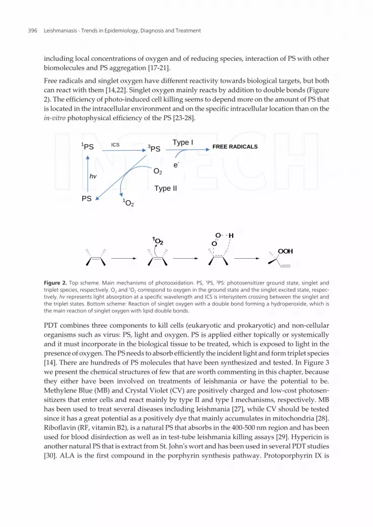
PDT combines three components to kill cells (eukaryotic and prokaryotic) and non-cellularorganisms such as virus: PS, light and oxygen. PS is applied either topically or systemicallyand it must incorporate in the biological tissue to be treated, which is exposed to light in thepresence of oxygen. The PS needs to absorb efficiently the incident light and form triplet species[14]. There are hundreds of PS molecules that have been synthesized and tested. In Figure 3we present the chemical structures of few that are worth commenting in this chapter, becausethey either have been involved on treatments of leishmania or have the potential to be.Methylene Blue (MB) and Crystal Violet (CV) are positively charged and low-cost photosen‐sitizers that enter cells and react mainly by type II and type I mechanisms, respectively. MBhas been used to treat several diseases including leishmania [27], while CV should be testedsince it has a great potential as a positively dye that mainly accumulates in mitochondria [28].Riboflavin (RF, vitamin B2), is a natural PS that absorbs in the 400-500 nm region and has beenused for blood disinfection as well as in test-tube leishmania killing assays [29]. Hypericin isanother natural PS that is extract from St. John's wort and has been used in several PDT studies[30]. ALA is the first compound in the porphyrin synthesis pathway. Protoporphyrin IX is
Leishmaniasis - Trends in Epidemiology, Diagnosis and Treatment396
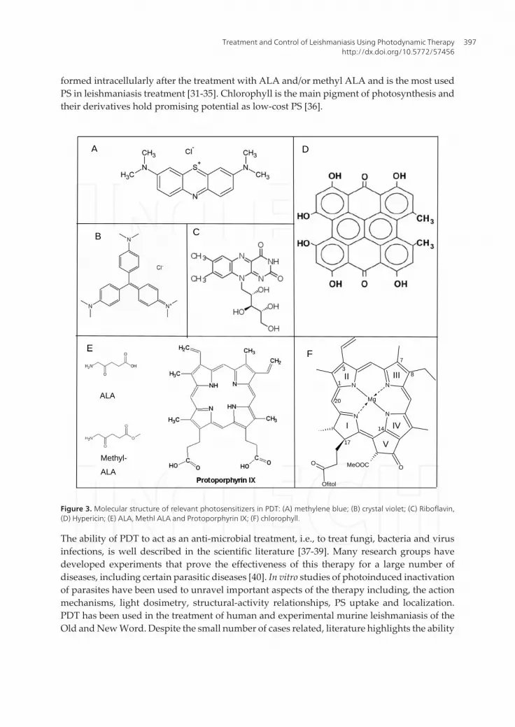
formed intracellularly after the treatment with ALA and/or methyl ALA and is the most usedPS in leishmaniasis treatment [31-35]. Chlorophyll is the main pigment of photosynthesis andtheir derivatives hold promising potential as low-cost PS [36].
respectively. MB has been used to treat several diseases including leishmania[27], while CV
should be tested since it has a great potential as a positively dye that mainly accumulates in
mitochondria[28]. Riboflavin (RF, vitamin B2), is a natural PS that absorbs in the 400-500
nm region and has been used for blood disinfection as well as in test-tube leishmania killing
assays [29]. Hypericin is another natural PS that is extract from St. John's wort and has been
used in several PDT studies [30]. ALA is the first compound in the porphyrin synthesis
pathway. Protoporphyrin IX is formed intracellularly after the treatment with ALA and/or
methyl ALA and is the most used PS in leishmaniasis treatment [31-35]. Chlorophyll is the
main pigment of photosynthesis and their derivatives hold promising potential as low-cost PS
[36].
Figure 3. Molecular structure of relevant photosensitizers in PDT: (A) methylene blue; (B) crystal violet; (C) Riboflavin, (D) Hypericin; (E) ALA, Methl ALA and Protoporphyrin IX; (F) chlorophyll.
C
N N
N N
O
Ofitol
O MeOOC
Mg
1
3
7
8
14
17
20
I
II III
IV
V
A
B
E
D
F
ALA
Methyl-
ALA
Figure 3. Molecular structure of relevant photosensitizers in PDT: (A) methylene blue; (B) crystal violet; (C) Riboflavin,(D) Hypericin; (E) ALA, Methl ALA and Protoporphyrin IX; (F) chlorophyll.
The ability of PDT to act as an anti-microbial treatment, i.e., to treat fungi, bacteria and virusinfections, is well described in the scientific literature [37-39]. Many research groups havedeveloped experiments that prove the effectiveness of this therapy for a large number ofdiseases, including certain parasitic diseases [40]. In vitro studies of photoinduced inactivationof parasites have been used to unravel important aspects of the therapy including, the actionmechanisms, light dosimetry, structural-activity relationships, PS uptake and localization.PDT has been used in the treatment of human and experimental murine leishmaniasis of theOld and New Word. Despite the small number of cases related, literature highlights the ability
Treatment and Control of Leishmaniasis Using Photodynamic Therapyhttp://dx.doi.org/10.5772/57456
397
of PDT to deliver better results compared to traditional treatments, emphasizing its bettereffectiveness in leading to amastigote-free lesions in a shorter time periods, in addition to itsexcellent esthetic results.
4. Treatment of animals infected with leishmaniasis using PDT
PDT has emerged in the treatment of cutaneous diseases among human and different animalspecies [41]. Researchers have shown that PDT offers an effective alternative in the treatmentof CL indicating that it also has a great clinical potential in the treatment of this disease withinVeterinary Medicine [27]. The initial studies using PDT to treat leishmaniasis were performedin humans and are further described on section 7 [31-35]. Although some animals, especiallymammals, constitute important reservoirs of the parasites, leishmaniasis also has clinicalimportance because some species can develop injuries, become sick and die due to the diseaseand its complications. Therefore, from this point of view, Veterinary Medicine has specialinterest, not only to control the disease epidemiology, but also to treat infected and sickanimals.
The main vertebrate hosts (domestic and wild) described and classified as hosts of theseprotozoan through natural and/or experimental infections, are: foxes, opossums, armadillos,anteaters, sloths, rodents, cat, dog, goat, sheep, buffaloes, horses and primates [42-47]. Whilethe treatment of infected animals provides possibilities for partial or total removal of cutaneouslesions, it is still not possible to guarantee the elimination of the infectious agents from thecarrier animal, remaining the possibility that it remains as a host reservoir. Therefore, there isa great need to further investigate the treatment of domestic and wild animals with leishma‐niasis, by using PDT.
Among all involved animals, the domestic dog and some rodents are the main sources ofhuman infection in America and in the Middle East, respectively; therefore, being the majorsurban reservoir hosts of leishmaniasis [44,48]. The proximity of this animal to humanscomplicates the disease control. The lack of identification of infected animals becomes achallenge, mainly due to the numerous generic clinical manifestations, and sometimes theabsence of pathognomonic lesions in the dogs [49]. The skin disorders are quite common inanimals, and include opaque hair coat, alopecia, depigmentation, hyperkeratosis of nasal planand digital cushions, mucocutaneous ulcers, intradermal nodules, onychogryphosis andexcessive flaking [50,51] but the most common presentation of the cutaneous disease is asymmetrical alopecia accompanied by intense flaking with silvery appearance that often startson the head and spreads to other parts of the body [52]. However, these symptoms aresometimes not correlated with leishmaniasis. Regarding the condition of the dogs as reservoirhosts in the epidemiology of the disease, clinical treatment is not recommended so far [51],making euthanasia of the infected animals mandatory in many countries [50] and keeping thecontroversial discussion among public health authorities, animal protectors and veterinarians[53,54]. Despite the importance of dogs in the epidemiology of the disease, the most usedanimal model and the one that has shown success in the treatment of the cutaneous diseaseare rodents, mainly mice and hamsters.
Leishmaniasis - Trends in Epidemiology, Diagnosis and Treatment398
Several studies demonstrated the possibility of using PDT in animal models, especially onmurines. In 2007 Akilov et al. reported an evaluation of the use of ALA (precursor of PpIX) inTL caused by Old World species in ears of Balb/c mice [55]. Akilov et al. also highlighted theaction of ALA-PDT in murine with leishmaniasis compared to a control group treated withALA [56]. The results showed a significant reduction of 24.5 folds in the parasite load comparedwith the control group. Nevertheless, they observed vascular damage in ears of the PDT-ALAgroup probably caused by PDT. According to the authors, a wide inflammatory and immu‐nologic response was noted in Balb/c ears of ALA-PDT group, which correlated with theexpressive decrease of parasite load and with the healing of the tissue.
Despite ALA, other classes of photosensitizers already widely used in PDT began to be tested.The phenothiazine 3,7-bis(di-n-butylamino)phenothiazin-5-ium bromide (PPA904) was testedby Akilov et al. in mice [57]. Ears of female Balb/c were infected with metacyclic parasites ofLeishmania sp. Following infection, mice were treated with PPA904 cream and irradiated witha broad band light source. They tested the PS concentration, time of uptake and absorptionsite in the ear. The results showed that PPA904 applied during at least 90 min in consecutivesessions of PDT decreased parasite load around 5.2 log compared to the controls groups.However, PPA904 application also lead to skin irritation. Another study was carried out withfemale Balb/c infected with L. major parasites expressing green fluorescent protein (GFP) tomonitor the parasitic load and the efficacy of PDT [58]. PPA904 was applied in the ears of themice and the parasitic load was compared with control group (only infected). The fluorescenceof GFP parasite in the ear of mice after the PPA-PDT decrease significantly, about 80%,compared to control group. The authors emphasized that this result was obtained after morethan one PDT session.
Peloi et al.chosen a different murine, which is also considered an appropriate model to developleishmaniasis caused by some New World Leishmania spp. Hamsters were used to investigatethe effectiveness of PDT with methylene blue (MB) photosensitizer [59]. A light-emitting diode(LED) was chosen as light source. The footpads of hamsters were infected with Leishmania sp.The control presented an increase in thickness throughout the treatment. An opposite reactionoccurred in the group A and B treated with oil/water lotion MB+LED and aqueous solutionMB+LED, respectively. Statistically significant reductions on the thickness of the footpad andparasitic load were observed.
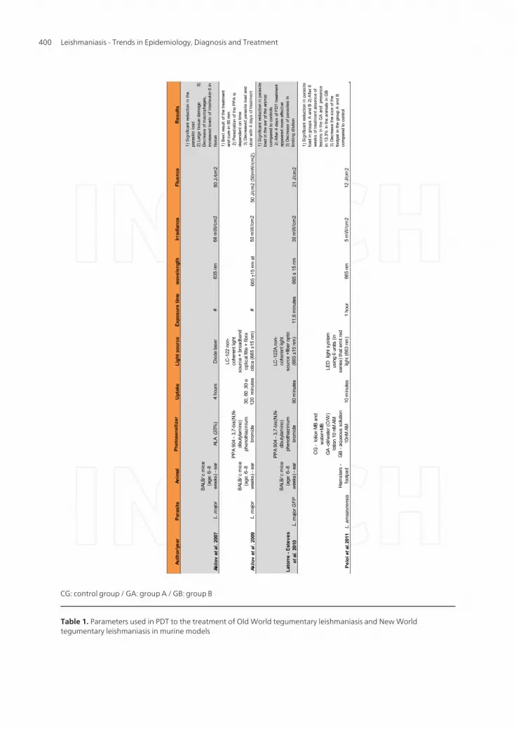
The scientific reports in PDT-treated animal models mentioned in this chapter show similarresults to those reported in humans. In other words, PDT is capable to treat infected woundsreducing the parasitic load. In some cases, the complete disappearance of the parasite fromtissue is achieved. Other aspect to highlight is its ability to inactivate both Old and New worldLeishmania spp. Details of parameters from scientific studies using PDT on Old World and NewWorld TL in murine models are described in table 1. However, treatment conditions of infectedanimals out of experimental controlled environment have not been described. Therefore, PDThas to become a more common procedure to be used in the clinical practice of VeterinaryMedicine. It certainly has the unfulfilled potential to become a therapeutic alternative inveterinary medicine, and to help controlling the parasitic cycle in humans.
Treatment and Control of Leishmaniasis Using Photodynamic Therapyhttp://dx.doi.org/10.5772/57456
399
CG: control group / GA: group A / GB: group B
Table 1. Parameters used in PDT to the treatment of Old World tegumentary leishmaniasis and New Worldtegumentary leishmaniasis in murine models
Leishmaniasis - Trends in Epidemiology, Diagnosis and Treatment400
5. Vector control using PDT
The field of insect photo-killing by administration of photosensitizer molecules and lightexposition (usually sun light) is one of the areas of possible PDT application that has receivedsmall attention of the scientific community [60-63]. The few studies, which were mainly reportedby Jori and co-authors, sustain that there is indeed great potential on this area. There are reportsshowing that the PS activity is a function of its log POW value and of its amphiphilic character[62,63]. PDT was also shown to be efficient for Larva control of dengue vector Aedes aegypti [60].However, there is no scientific report on the use of PDT to control the vector (Phlebotomus sandflies) and its larva, which are responsible for the transmission the leishmania parasites. It is alsoimportant to emphasize that the amount of information available concerning larva develop‐ment of phlebotomine sand flies is much less than what is known for the mosquitoes whosecontrol have been studied by PDT. Nevertheless, for the matter of bringing new ideas to thefield of Leishmania treatment, the concentration of photosensitizers that are needed to neutral‐ize larva and to kill those mosquitoes is several orders of magnitude smaller than the concentra‐tions of chemical insecticides, which are currently used for vectors control, causing greatdisturbance in the whole ecosystem. Therefore, it is up to our community to develop and teststrategies to control vectors of Leishmania parasites using PDT.
6. Blood purification
The purification of blood products is critical to avoid disease transmittance through bloodtransfusion. Although this is not the main route of transmission of leishmaniasis, it is a possibleone, and cases have been reported in the literature [64]. The focus of the disinfection strategyis to kill microorganisms without harming the cellular and plasma components. PDT offersgreat potential to be successful in blood disinfection, because it is a multi–target strategy, i.e,the reactive species that are formed (after light absorption and photosensitization reaction) areeffective against viruses, bacteria, fungi, and parasites [37-40]. This strategy has even beenproved effective to promote pathogen inactivation in the presence of fragile blood components,such as stem cells from blood of embryo’s cord [65-68]. It is better than UV treatments, becauseit does not cause direct damage to blood components. Several PS have been used for blooddisinfection including MB, CV and RF (Figure 2). Molecules that have intracellular targets suchas MB and CV can be used to treat plasma derivatives but not whole cell blood, because theywill cause extensive hemolysis. RF, however, is an aqueous based photosensitiser, which donot enter cells and can be used to disinfect whole blood derivatives. RF reacts either by type Ior by type II mechanisms and is already in use. Several companies commercialize kits for bloodand plasma decontamination, like Macopharma, whose technology for plasma decontamina‐tion is based on MB photosensitization (http://www.macopharmausa.com/). In the case ofleishmaniasis, parasites remain mostly in the intracellular environment, except when they arein transit from a lysed cell to infect a macrophage or other phagocytic cell. We could think ofusing PDT to remove parasites in the plasma or to develop strategies to target PSs to destroyonly infected cells of contaminated blood.
Treatment and Control of Leishmaniasis Using Photodynamic Therapyhttp://dx.doi.org/10.5772/57456
401
7. Photodynamic therapy on the treatment of old world tegumentaryLeishmaniasis
There are several reports on the literature dealing with the treatment of leishmaniasis by PDT[5,6,33,34]. The first report was conducted by Enk’s group in 2003 [5,6]. Both studies reportedthe use ALA and MAL, combined with red light. These authors performed the treatment of 32TL lesions from 11 Israeli patients. The diagnostic was accomplished by verifying the amasti‐gote presence in direct smear from the lesions [5]. This work showed that about 96% of thelesions healed, leaving some mild scars and pigment in place of the old lesions. Just one lesionpresented amastigotes forms after PDT. Gardlo et al. published the case of a patient, aged 34,with CL confirmed by histology. According to the authors, the patient developed resistanceto the treatment with sodium stibogluconate and presented 10 lesions, which were treated fivetimes with PDT and five times with paromomycin sulfate ointment [6]. The result obtained issimilar to the previous work and showed that the five ulcers treated with PDT healed withoutsigns of amastigotes, while two ulcers treated with paromomycin partially responded to thedrug, one of them did not respond and two lesions were shown to have no amastigotes. Theulcers that did not responded to paromomycin ointment were subsequently treated with PDTsuccessfully.
Asilian and Davami developed a placebo-controlled, randomized clinical trial that provideddefinitive evidence of the efficacy of PDT in the treatment of CL [34]. 60 patients with confirmedCL by clinical and parasitological diagnosis were separated in 3 groups with differenttreatments. Group 1 was treated with PDT once a week, group 2 received twice daily para‐momycin plus methylbenzethonium chloride ointment and in group 3 was used a paraffin-based ointment without active ingredients with same application time of the group 2. Duringfour weeks, the groups received the treatments described above. At the end of the study healingwas present in 93.5% of the patients of group 1, 41.2% of group 2 and 13.3% of group 3. At thesame time, 100%, 64.7% and 20% of the lesions had parasitological cure in group 1, 2 and 3,respectively.
Other studies accomplished in Iran and German corroborated with the results described above.According to the authors, PDT showed to have the capacity to treat wounds caused by OldWord Leishmania species. We emphasize that most of the reports claim that this therapeuticalmodality can achieve results above 90% healing of wounds, however, a caveat must be heldsince some of these studies indicate that not all healed wounds become free of parasite [35,56].The mechanism of ALA PDT in the case of leishmaniasis was shown to be due to the killingof infected host-cell killing (macrophages) instead of direct parasite killing (see furtherdiscussion about this issue on section 8).
One CL case of the New World leishmaniasis is described in the literature. Song et al. reportedthe case of a Brazilian patient presenting cutaneous leishmaniasis confirmed by smear stainedby Giemsa. PDT was carried out using MB. In this specific case because of ethical concerns ofpossible development of evolution to mucocutaneou disease, the patient received at the sametime a low dose of pentavalent antimony and PDT. The patient had two ulcers. One receivePDT and the other was only being treated with the low-dose pentavalent [27]. The treatment
Leishmaniasis - Trends in Epidemiology, Diagnosis and Treatment402
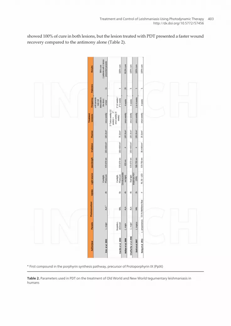
showed 100% of cure in both lesions, but the lesion treated with PDT presented a faster woundrecovery compared to the antimony alone (Table 2).
13
TAB
LE 2
: Par
amet
ers
used
in P
DT
on th
e tre
atm
ent o
f Old
Wor
ld a
nd N
ew W
orld
tegu
men
tary
leis
hman
iasi
s in
hum
ans
*
Firs
t com
poun
d in
the
porp
hyrin
syn
thes
is p
athw
ay, p
recu
rsor
of P
roto
porp
hyrin
IX (P
pIX
)
Auth
or/ye
arPa
rasi
tePh
otos
ensi
taze
rUp
take
Ligh
t sou
rce
wave
leng
thIrr
adia
nce
Flue
nce
Trea
tmen
t se
ssio
nsFr
eque
ncy
Patie
ntes
Re
sults
Enk
et a
l. 20
03L.
maj
orAL
A4h
Cur
elig
ht,
Phot
ocur
e57
0-67
0 nm
150
mW
/cm
²10
0 J/
cm²
once
wee
kly
until
para
site
w
as n
ot
dete
ctab
le in
th
e di
rect
sm
ear
11
96%
cur
e
1 pa
tient
e w
ith 1
lesi
on
pres
ente
d pa
rasi
te
Gar
dlo
et a
l. 20
03Su
spei
ta L
. do
nova
niM
AL5h
Cur
elig
ht,
Phot
ocur
e57
0-67
0 nm
150
mW
/cm
²75
J/c
m²
1º T
wic
e w
eekly
(12
wee
ks) +
2 º
once
wee
kly (4
w
eeks
)1º
12
wee
ks /
2º 4
wee
ks1
100%
cur
e
Asili
an e
t al.
2006
L. m
ajor
*AL
A4h
Om
nilu
x(vis
ible
re
d lig
ht)
633
nm#
100
J/cm
²on
ce w
eekly
4 w
eeks
2093
,5%
cur
e
Gha
ffarif
ar e
t al. 2
006
L. m
ajor
AL
A4h
Red
light
57
0-67
0 nm
150
mW
/cm
²10
0 J/
cm²
once
wee
kly4
wee
ks5
100%
cur
e
Sohl
et a
l. 200
7L.
trop
ica
MAL
3hW
aldm
an P
DT
1200
L58
0-70
0 nm
#10
0 J/
cm²
once
wee
kly1
to 4
wee
ks1
100%
cur
e
Song
et a
l. 20
11L.
am
azon
ensi
s0.
5 %
Met
hile
ne B
lue
#R
L 50
- LE
D57
0-75
0 nm
35 m
W/c
m²
20 J
/cm
²on
ce w
eekly
4 w
eeks
110
0% c
ure
*
Phot
osen
sitiz
er
* First compound in the porphyrin synthesis pathway, precursor of Protoporphyrin IX (PpIX)
Table 2. Parameters used in PDT on the treatment of Old World and New World tegumentary leishmaniasis inhumans
Treatment and Control of Leishmaniasis Using Photodynamic Therapyhttp://dx.doi.org/10.5772/57456
403
This brief account of the use of PDT for the treatment of CL demonstrates the ability ofthis therapeutic modality and encourages its use. It also stimulates research in the pur‐suit of new protocols with new PS, which could ensure not only healing but also clinicaland parasitological cure of these patients.
Details of parameters from scientific studies using PDT on the treatment of Old World andNew World tegumentary leishmaniasis in humans are described in Table 2.
8. Photodynamic therapy — In vitro tests in species which causeTegumentary Leishmaniasis
The effectiveness of PDT on CL treatment was first conducted in humans and in animal models.In vitro tests began less than ten years ago to allow testing of PDT parameters like the efficiencyof different types of photosensitizers, their respective uptakes and concentrations andaccumulation sites.
Sujoy Dutta et al. began in vitro studies with the New World specie, L. amazonensis in 2005 [69].The first part of that work evaluated Leishmania transfectants expressing GFPs. The PS testedwas aluminum phthalocyanine chloride (AlPhCl) in different concentrations. The principalfactor tested was the light-mediated cytolysis when cells were in the presence or pre-incubatedwith the AlPhCl. In the dark there was no phototoxicity for both promastigote and amastigoteforms of the parasite. The opposite effect occurred when the photosensitizer received red lightillumination, showing that promastigotes appear to be more sensible than amastigote forms.In addition, the loss of fluorescence of the GFP parasites indicated cell death. On the secondpart of the study, J774 cells (cell line immortalized murine Balb⁄ c monocyte ⁄ macrophage)were tested at the same conditions reported above. The authors observed that they were 10-20fold more resistant than promatigotes. According to the authors, the photosensitized Leishma‐nia cells are susceptible to cytolysis, probably due to the generation of reactive oxidative speciesafter illumination, an indicative of inefficiency of their antioxidant mechanisms. ALA did notinduce protoporphyrin IX (PpIX) production in the Leishmania cells, because of a deficiency inthe heme biosynthetic pathway in this parasite [57, 70].
Tests with other phthalocyanines were developed by Pinto et al. using species of Old and Newworld Leishmanias, L. major and L. braziliensis. The parasites were incubated with aluminumphthalocyanine tetrasulfonate (AlPcS4) at different concentrations and irradiated with aGaAlAs diode laser (λ= 659 nm, 40 mW). The experiments indicated a significant reduction ofviable parasites in both species compared to controls, however L. braziliensis demonstratedhigher mortality than L. major [71].
In Brazil, Song et al. performed tests to understand mechanism of action of PDT using MB ina case report. Promastigotes of L. amazonensis were incubated with different concentrations ofMB, washed with PBS and illuminated using a home-built LED light source with a wavelengthof maximum emission at λ= 650 nm. After the irradiation, cell survival was determined withMTT assay that detected cell toxicity after irradiation of light in the presence MB. There was
Leishmaniasis - Trends in Epidemiology, Diagnosis and Treatment404
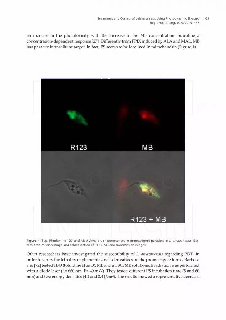
an increase in the phototoxicity with the increase in the MB concentration indicating aconcentration-dependent response [27]. Differently from PPIX induced by ALA and MAL, MBhas parasite intracellular target. In fact, PS seems to be localized in mitochondria (Figure 4).
Figure 4. Top: Rhodamine 123 and Methylene blue fluorescences in promastigote parasites of L. amazonensis. Bot‐tom: transmission image and colocalization of R123, MB and transmission images.
Other researchers have investigated the susceptibility of L. amazonensis regarding PDT. Inorder to verify the lethality of phenothiazine’s derivatives on the promastigote forms, Barbosaet al [72] tested TBO (toluidine blue O), MB and a TBO/MB solutions. Irradiation was performedwith a diode laser (λ= 660 nm, P= 40 mW). They tested different PS incubation time (5 and 60min) and two energy densities (4.2 and 8.4 J/cm2). The results showed a representative decrease
Treatment and Control of Leishmaniasis Using Photodynamic Therapyhttp://dx.doi.org/10.5772/57456
405
on the viability of L.amazonensis promastigotes for all treated groups in comparison to theircontrols. The authors did not find statistical differences between the dyes, but reported thatthe best result was observed with TBO.
Dutta et al. published an article that described the use of a combination of photosensitizers.Uroporphyrin (URO1) and aluminum phthalocyanine chloride (AlPhCl) were used inuroporphyrinogenic mutants of L. amazonensis (RAT/ BA/ 74 /LV78) 12-1 clone, transfectedwith pX-alad and p6,5-PBGD [73]. This transfected Leishmania is able to absorb ALA and turnsit into URO 1. The authors evaluated the combination of both drugs into promastigotes withand without irradiation of red light. Results showed photolysis of the irradiated parasites withboth photosensitizers whereas non-irradiated parasites showed no damage.
Hernández et al. published another study that compared encapsulated chloroaluminumphthalocyanine (CLAlPc) in liposomes (UDL-CLAlPc) and free in solution. The experimentswere conducted with two species of New World Leishmania in promastigote and amastigoteforms and in THP1 cells. The experiments tried to verify the ability of the photosensitizer inreaching the Leishmania inside THP1 host cell. According to the authors, the UDL-ClAlPcphotosensitizer was almost 10 times more photoactive than free ClAlPc on THP-1 cells as wellas on promastigotes and with intracellular amastigotes of L. chagasi and L. panamensis [74].
9. Conclusions
• First reports of cutaneous leishmaniasis using PDT were performed in humans;
• Treatments using porphyrin precursors, ALA and MAL, showed positive results on the cureof patients with CL;
• The low-cost phenothiazine methylene blue and red light can be used to treat patients withCL;
• More than one PDT session is necessary to achieve wound healing.
• Both New and Old World Leishmania can be treated with PDT.
• Murine models of infection such as Balb/c and hamster show to be appropriate for PDTstudies of CL treatment.
• In vitro tests demonstrate that Old and New world Leishmania species can be used to testnew photosensitizers and to establish structure/activity relationships.
• PDT also has the potential to control leishmaniasis transmission by the treatment of vectorsand infected animal reservoirs, although the development of these potentials will needfurther investigation.
Leishmaniasis - Trends in Epidemiology, Diagnosis and Treatment406
Author details
Debora P. Aureliano1,2, Martha S. Ribeiro1, José Angelo Lauletta Lindoso2,3, Fabio C. Pogliani3,Fábio P. Sellera4, Dennis Song3,5 and Mauricio S. Baptista5
1 Centro de Lasers e Aplicações (CLA)/ IPEN- CNEN, Brazil
2 Laboratório de Soroepidemiologia (LIM38-HC-FMUSP) e Instituto de Medicina Tropicalde São Paulo da Universidade de São Paulo, Brazil
3 Instituto de Infectologia Emilio Ribas-SES-SP, Brazil
4 Departamento de Clínica Médica, Faculdade de Medicina Veterinária e Zootecnia da Uni‐versidade de São Paulo, Brazil
5 Departamento de Bioquímica, Instituto de Química, Universidade de São Paulo, Brazil
References
[1] Farrell JD, World Class Parasites-Volume 4: Leishmania. Kluwer Academic Publish‐er, Boston, 2002.
[2] Peacock CS, Berriman M. Comparative genomic analysis of three Leishmania speciesthat cause diverse human disease. Nature Genetics 2007, 39, 839.
[3] de Almeida MC, Vilhena V, Barral A, Barral-Netto M. Leishmanial infection: analysisof its first steps. A review. Mem Inst Oswaldo Cruz 2003, 98, 861.
[4] Santos DO, Coutinho CER, Madeira MF, Bottino CG, Vieira RT, Nascimento SB, Ber‐nardino A, Bourguignon SC, Corte-Real S, Pinho RT, Rodrigues CR, Castro, HC.Leishmaniasis treatment—a challenge that remains: a review. Parasitol Res 2008, 103,1.
[5] Enk CD, Fritsch C, Jonas F, Nasereddin A, Ingber A, Jaffe CL, Ruzicka T. Treatmentof cutaneous leishmaniasis with photodynamic therapy. Arch Dermatol AmericanMedical Association; 2003;139(4), 432.
[6] Gardlo K, Horska Z, Enk CD, Rauch L, Megahed M, Ruzicka T, Fritsch C. Treatmentof cutaneous leishmaniasis by photodynamic therapy. J Am Acad Dermatol. 2003,48(6), 893.
[7] Van der Snoek EM, Robinson DJ, van Hellemond JJ, Neumann HAM. A review ofphotodynamic therapy in cutaneous leishmaniasis. J Eur Acad Dermatol Venereol 2008,22(8), 918.
Treatment and Control of Leishmaniasis Using Photodynamic Therapyhttp://dx.doi.org/10.5772/57456
407
[8] Demidova TN, Hamblin MR. Photodynamic therapy targeted to pathogens. Int J Im‐munopathol Pharmacol. 2011,17(3), 245.
[9] Tardivo JP, Del Giglio A, Oliveira CS, Gabrielli DS, Junqueira HC, Tada DB, SeverinoD, Turchiello R, Baptista MS. Methylene Blue in Photodynamic Therapy: From BasicMechanisms to Clinical Applications. Photodyag Photodyn Ther 2005, 2/3, 175.
[10] Tesh RB. Control of zoonotic visceral leishmaniasis: is it time to change strategies?Am J Trop Med Hyg. 1995, 52(3), 287.
[11] Brown SB, Brown EA, Walker I. The present and future role of photodynamic thera‐py in cancer treatment. The Lancet Oncology 2004, 5, 497.
[12] Allison RR, Downie GH, Cuenca R, Hu X-H, Childs CJH, Sibata CH. Photodiagn Pho‐todyn Ther 2004, 1, 27.
[13] Wilkinson F, Helman WP, Ross AB. Rate Constants for the Decay and Reactions ofthe Lowest Electronically Excited Singlet State of Molecular Oxygen in Solution. AnExpanded and Revised Compilation J Phys Chem 1993, 22, 113.
[14] Foote CS. Mechanisms of photosensitized oxidations. Science 1968, 162, 963.
[15] Dougherty TJ. Photochemistry in the Treatment of Cancer. Adv Photochem 1992, 17,275.
[16] Castano AP, Mroz P, Hamblin MR. Photodynamic therapy and anti-tumour immuni‐ty Nature Rev 2006, 6, 535.
[17] Junqueira HC, Severino D, Dias LG, Gugliotti M, Baptista MS. Modulation of theMethylene Blue Photochemical Properties Based on the Adsorption at Aqueous Mi‐celle Interfaces. Phys Chem Chem Phys 2002, 4, 2320.
[18] Severino D, Junqueira HC, Gabrielli DS, Gugliotti M, Baptista MS. Influence of Nega‐tively Charged Interfaces on the Ground and Excited State Properties of MethyleneBlue Photochem Photobiol 2003, 77, 459.
[19] Gabrieli D, Belisle E, Severino D, Kowaltowski AJ, Baptista MS Binding, aggregationand photochemical properties of methylene blue in mitochondrial suspensions Photo‐chem Photobiol 2004, 79, 227.
[20] Baptista MS, Indig GL. Effect of BSA Binding on Photophysical Photochemical Prop‐erties of Triarylmethane Dyes. J Phys Chem B 1998, 102, 4678.
[21] Baptista MS, Indig GL. Mechanism of Photobleaching of Ethyl Violet Non-CovalentlyBound to Bovine serum Albumin Chem Comm 1997, 18, 1791.
[22] David R. Kearns. Physical and chemical properties of singlet molecular oxygen, ChemRev 1971, 71 (4), 395.
Leishmaniasis - Trends in Epidemiology, Diagnosis and Treatment408
[23] Pavani C, Uchoa AF, Oliveira CS, Iamamoto Y, Baptista MS. Effect of zinc insertionand hydrophobicity on the membrane interactions and PDT activity of porphyrinphotosensitizers Photochem Photobiol Sci 2009, 8, 233.
[24] Pavani C, IamamotoY, Baptista MS. Mechanism and Efficiency of Cell Death of TypeII Photosensitizers: Effect of Zinc Chelation, Photochem Photobiol 2012, 88, 774.
[25] Garcez AS, Núñez SC, Baptista MS, Daghastanli NA, Itri R, Hamblin MR, RibeiroMS. Antimicrobial mechanisms behind photodynamic effect in the presence of hy‐drogen peroxide. Photochem Photobiol Sci 2011, 10, 483.
[26] Uchoa AF, Oliveira CS, Baptista MS. Relationship between structure and photoactivi‐ty of porphyrins derived from protoporphyrin IX. J Porphyr Phthaloc 2010, 14, 832.
[27] Song D, Lindoso JA, Oyafuso LK, Hatsumi E, Kanashiro Y, Cardoso JL, Uchoa AF,Tardivo JP, Baptista MS. Photodynamic therapy using methylene blue to treat cuta‐neous leishmaniasis. Photomed Laser Surg 2011, 29, 711.
[28] Oliveira C, Turchiello R, Kowaltowski AJ, Indig GL, Baptista MS. Major determi‐nants of photoinduced cell kill: subcellular localization versus photosensitization effi‐ciency Free Radic Biol Med 2011, 51, 824.
[29] Silva AV, López-Sánchez A, Rivas L, Baptista MS; Orellana G. Molecular Engineeringof Riboflavin Derivatives for Enhanced Photodynamic Activity against Leishmania.Tetrahedron, submitted.
[30] Tardivo JP, Baptista MS. Treatment of Osteomyelitis in the Feet of Diabetic Patientsby Photodynamic Antimicrobial Chemotherapy Photomed Laser Surg 2009, 27, 145.
[31] Gondim RMF, Vieira VCC, Veras MV, Ferreira MA, Caldini ETEG, Muñoz DR, Bap‐tista MS. Protoporphyrin fluorescence induced by methyl–ALA in skin healing, Pho‐todiagn Photodyn Ther in press.
[32] Kosaka S, Akilov OE, O'Riordan K, Hasan T. A Mechanistic Study of delta-Aminole‐vulinic Acid-Based Photodynamic Therapy for Cutaneous Leishmaniasis. J Inv Der‐matol 2007, 127, 1546.
[33] Bristow C-A, Hudson R, Paget TA, Boyle RW. Potential of cationic porphyrins forphotodynamic treatment of cutaneous Leishmaniasis. Photodiagn Photodyn Ther 2006,3, 162.
[34] Asilian A, Davami M. Comparison between the efficacy of photodynamic therapyand topical paromomycin in the treatment of Old World cutaneous leishmaniasis: aplacebo-controlled, randomized clinical trial. Clin Exp Dermatol 2006, 31(5), 634.
[35] Ghaffarifar F, Jorjani O, Mirshams M, Miranbaygi MH, Hosseini ZK.Photodynamictherapy as a new treatment of cutaneous leishmaniasis. East Mediterr Health J 2006,12(6), 902.
Treatment and Control of Leishmaniasis Using Photodynamic Therapyhttp://dx.doi.org/10.5772/57456
409
[36] Bonnett R, Benzie R, Grahn MF, Salgado A, Valles MA. Photodynamic therapy pho‐tosensitizers derived from chlorophyll a. Proc. SPIE 1994, 2078, 171.
[37] Jori G, Fabris C, Soncin M, Ferro S, Coppellotti O, Dei D, Fantetti L, Chiti G, RoncucciG. Photodynamic therapy in the treatment of microbial infections: basic principlesand perspective applications. Lasers Surg Med 2001, 34(1), 18.
[38] Wainwright M. Photodynamic antimicrobial chemotherapy (PACT) J Antimicrob Che‐mother, 1998, 42,13.
[39] Hamblin MR, Hasan T. Photodynamic therapy: a new antimicrobial approach to in‐fectious disease? Photochem Photobiol Sci 2004, 3, 436.
[40] Baptista MS, Wainwright M. Photodynamic antimicrobial chemotherapy (PACT) forthe treatment of malaria, leishmaniasis and trypanosomiasis. Braz J Med Biol Res 2011,44, 1.
[41] Peplow PV, Chung T-Y, Baxter GD. Photodynamic Modulation of Wound Healing: AReview of Human and Animal Studies. Photomed Laser Surg 2012, 30, 118.
[42] Anjili CO, Ngichabe CK, Mbati PA, Lugalia RM, Wamwayi HM, Githure JI. Experi‐mental infection of domestic sheep with culture-derived Leishmania donovani promas‐tigotes. Veter Parasitol, 1998, 74, 315.
[43] Rey L. Principais grupos de protozoários e metazoários, parasitos do homem e seusvetores. In: Parasitologia. 3 ed. Rio de Janeiro: Guanabara Koogan, 2001. cap.9, p.123.
[44] Lainson R, Ishikawa EAY, Silveira FT. American visceral leishmaniasis: wild animalhosts. Trans Royal Soc Trop Med Hyg 2002, 96, 630.
[45] Garg RD, Ravendra A. Animal models for vaccine studies for visceral leishmaniasis.Indian J Med Res 2006, 123, 439.
[46] Vedovello FD, Jorge FA, Lonardoni MVC, Teodoro U, Silveira TGV. American cuta‐neous leishmaniasis in horses from endemic areas in the North-Central Mesoregionof Parana State, Brazil. Zoonoses and Public Health 2008, 55, 149.
[47] Bhattarai NR, Van Der Auwera G, Rijal S, Picado A, Speybroeck N, Khanal B, DeDoncker S, DAS ML, Ostyn B, Davies C, Coosemans M., Berkvens D, Boelaert, M,Dujardin JC. Domestic animals and epidemiology of visceral leishmaniasis, Nepal.Emerg Infect Dis 2010, 16(2), 231.
[48] Faiman R, Abbasi I, Jaffe C, Motro Y, Nasereddin A, Schnur LF, Torem M, PratlongF, Dedet JP, Warburg A. A newly emerged cutaneous leishmaniasis focus in northernIsrael and two new reservoir hosts of Leishmania major. PLoS Negl Trop Dis. 2013,7(2), 1.
[49] Singh S, Sivakumar R. Recent advances in the diagnosis of leishmaniasis. J PostgradMed 2003, 49, 55.
Leishmaniasis - Trends in Epidemiology, Diagnosis and Treatment410
[50] Ferrer L. Leishmaniasis. In: Kirk RW, Bonagura JD. Kirk’s Current Veterinary Thera‐py XI. Philadelphia: W. B. Saunders, 1992, 266.
[51] Kontos VJ, Koutinas AF. Old world canine leishmaniasis. Compendium on ContinuingEducation for the Practing Veterinarian 1993, 15, 949.
[52] Noli C. Canine leishmaniasis. Waltham Focus, 1999, 9, 16.
[53] Oliveira CI, Teixeira MJ, Gomes R, Barral A, Brodskyn C. Animal models for infec‐tious diseases caused by parasites: Leishmaniasis. Drug Discovery Today: Disease Mod‐els 2004, 1, 1.
[54] Esteves EL, Akilov OE, Rai1 P, Beverley SM, Hasan T. Monitoring the Efficacy of An‐timicrobial Photodynamic Therapy in a Murine Model of Cutaneous Leishmaniasisusing L. major expressing GFP. J Biophotonics 2010, 3, 328.
[55] kilov OE, Kosaka S, O’Riordan K, Hasan T. Parasiticidal effect of delta-aminolevulin‐ic acid-based photodynamic therapy for cutaneous leishmaniasis is indirect andmediated through the killing of the host cells. Exp Dermatol 2007, 16, 651.
[56] Sohl S, Kauer F, Paasch U, Simon JC. Photodynamic treatment of cutaneous leishma‐niasis. J Dtsch Dermatol Ges 2007, 5(2), 128.
[57] Akilov OE, Yousaf W, Lukjan SX, Verma S, Hasan T. Optimization of topical photo‐dynamic therapy with 3,7-bis(di-n-butylamino)phenothiazin-5-ium bromide for cuta‐neous leishmaniasis. Lasers Surg Med 2009, 41(5), 358.
[58] Latorre-Esteves E, Akilov OE, Rai P, Beverley SM, Hasan T. Monitoring the efficacyof antimicrobial photodynamic therapy in a murine model of cutaneous leishmania‐sis using L. major expressing GFP. J Biophotonics 2010, 3(5-6), 328.
[59] Peloi LS, Biondo CEG, Kimura E, Politi MJ, Lonardoni MVC, Aristides SMA, et al.Photodynamic therapy for American cutaneous leishmaniasis: the efficacy of methyl‐ene blue in hamsters experimentally infected with Leishmania (Leishmania) amazo‐nensis. Exp Parasitol 2011, 128(4), 353.
[60] Lucantoni L, Magaraggia M, Lupidi G, Ouedraogo RK, Coppellotti O, Esposito F,Fabris C, Jori G, Habluetzel A. Novel, Meso -Substituted Cationic Porphyrin Mole‐cule for Photo-Mediated Larval Control of the Dengue Vector Aedes aegypti. Plos Ne‐glect Trop Disease 2011, 5, e1434.
[61] Coppellotti O, Fabris C, Soncin M, Magaraggia M, Camerin M, Jori G, Guidolin L,Porphyrin Photosensitised Processes in the Prevention and Treatment of Water- andVector-Borne Diseases. Curr Med Chem 2012, 19, 808.
[62] Ben Amor T, Jori G. Sunlight-activated insecticides: historical background and mech‐anisms of phototoxic activity. Insect Biochem Mol Biol 2000, 30, 915.
[63] Ben Amor T, Bortolotto L, Jori G. Porphyrins and related compounds as photoacti‐vatable insecticides. 3. Laboratory and field studies. Photochem Photobiol 2000, 71,124.
Treatment and Control of Leishmaniasis Using Photodynamic Therapyhttp://dx.doi.org/10.5772/57456
411
[64] Cardo LJ. Leishmania: risk to the blood supply. Transfusion 2006,46(9),1641.
[65] Wainwright M. Pathogen inactivation in blood products. Curr Med Chem 2002, 9, 127.
[66] Goodrich RP, Platz MS, Martin CB. Use of visible light to reduce of wavelengths of500 to 550 nm to reduce the number of pathogen in the blood and blood components.Patent No. US patent 7,498,156 B2. 2009.
[67] Ruane PH, Edrich R, Gampp D, Keil SD, Leonard RL, Goodrich RP. Photochemicalinactivation of selected viruses and bacteria in platelet concentrates using riboflavinand light. Transfusion 2004, 44, 877.
[68] Trannoy LL, van Hensbergen Y, Lagerberg JWM, Brand A. Photodynamic treatmentwith mono-phenyl-tri-(N-methyl-4-pyridyl)-porphyrin for pathogen inactivation incord blood stem cell products. Transfusion 2008, 48, 2629.
[69] Dutta S, Ray D, Kolli BK, Chang K-P. Photodynamic sensitization of Leishmania am‐azonensis in both extracellular and intracellular stages with aluminum phthalocya‐nine chloride for photolysis in vitro. Antimicrob Agents Chemother 2005, 49(11), 4474.
[70] Chang CS, Chang KP. Heme requirement and acquisition by extracellular and intra‐cellular stages of Leishmania mexicana amazonensis. Mol Biochem Parasitol 1985,16(3), 267.
[71] Pinto JG, Soares CP, Mittmann J. Assessment of Leishmania major and Leishmaniabraziliensis promastigote viability after photodynamic treatment with aluminumphthalocyanine tetrasulfonate (AlPcS4). J Venom Anim Toxins Incl Trop Dis 2011, 17(3),300.
[72] Barbosa AF, Sangiorgi BB, Galdino SL, Barral-Netto M, Pitta IR, Pinheiro AL. Photo‐dynamic antimicrobial chemotherapy (PACT) using phenothiazine derivatives asphotosensitizers against Leishmania braziliensis. Lasers Surg Med 2012, 44(10), 850.
[73] Dutta S, Waki K, Chang KP. Combinational sensitization of Leishmania with uropor‐phyrin and aluminum phthalocyanine synergistically enhances their photodynamicinactivation in vitro and in vivo. Photochem Photobiol 2012,88(3), 620.
[74] Hernández IP, Montanari J, Valdivieso W, Morilla MJ, Romero EL, Escobar P. In vi‐tro phototoxicity of ultradeformable liposomes containing chloroaluminum phthalo‐cyanine against New World Leishmania species. J Photochem Photobiol B 2012, 117,157.
Leishmaniasis - Trends in Epidemiology, Diagnosis and Treatment412