outcome of photodynamic therapy for early esophageal cancer
TRANSCRIPT
original article
Gut and Liver Vol 1 No 2 December 2007 pp 126-131
Correspondence to Young Koog CheonDigestive Disease Center Soon Chun Hyang University Hospital 657 Hannam-dong Yongsan-gu Seoul 140-743 KoreaTel +82-2-709-9202 Fax +82-2-749-1968 E-mail yksky001hanmailnet cykhospschackr
Received on August 14 2007 Accepted on December 10 2007
Outcome of Photodynamic Therapy for Early Esophageal Cancer
Young Koog Cheon Wan Jung Kim Joo Young Cho Joon Seong Lee Moon Sung Lee and Chan Sup ShimDepartment of Internal Medicine Institute for Digestive Research Soon Chun Hyang University College of Medicine Seoul Korea
BackgroundAims Endoscopic treatment as an alter-native to surgery has become increasingly popular for improving the quality of life Although photodynamic therapy (PDT) has been used for the endoscopic treatment of digestive cancer its curative efficacy re-mains unclear We evaluated the curative efficacy of PDT in superficial esophageal cancer in inoperable patients Methods Ten male patients with histologi-cally proven early esophageal cancer (surgery was contraindicated for age > 80 years surgery was con-traindicated Karnofsky performance status of at least 30 or refusal of surgery) were intravenously in-jected with a hematoporphyrin derivative (2 mgkg) and PDT was performed 48 h later The response to treatment was assessed by gastroscopy with biopsies Results The mean follow-up period was 276 months (range 96-587 months) Endoscopic ultrasonography revealed that all ten cases were at tumor stage T1 Complete remission (CR) to initial and subsequent PDT was observed in all patients For the CR cases the recurrence rate was 10 (110) and the time from initial PDT to recurrence was 96 months Conclusions For patients in whom surgery is risky or refused PDT may represent an acceptable alter-native treatment modality especially for superficial esophageal cancer without lymph node metastasis However a study involving long-term follow-up in a large population is needed for confirmation (Gut and Liver 20071126-131)
Key Words Photodynamic therapy Endoscopic treat-ment Early esophageal cancer Hematoporphyrin
INTRODUCTION
The conventional therapy for esophageal cancer is
esophagectomy a complex surgical procedure with a mor-tality rate of 3 to 51-3 Superficial esophageal cancer is not usually associated with locoregional lymph node in-volvement or distant metastasis45 Therefore local treat-ment of superficial esophageal cancer may reasonably con-sidered as a solution for the cure However the success of local treatment depends on accurate staging which might be difficult with currently existing technology Before the introduction of endosonography the diagnosis of an early stage cancer in the gastrointestinal tract was established by pathological examination of the resected surgical specimen High-resolution endosonography allows the diagnosis to be made in situ with a sensitivity and specificity of nearly 9067 The endosonographic in situ diagnosis of an early cancer offers local endoscopic ther-apy to inoperable patients with intention for cure after evaluating the histological classification Endoscopic treat-ments such as endoscopic mucosal resection (EMR) pho-todynamic therapy (PDT) and non-selective laser destruc-tion or electrodestruction have become increasingly popu-lar as an alternative to surgery in the hope of offering the patient a superior quality of life by avoiding a surgical procedure and its postoperative problems89
The PDT is a means of selective sensitization of pre-cancerous or malignant lesions using a systemically appli-cable photosensitizer with subsequent endoscopically controlled photochemically induced tissue ablation10 Several investigators have reported the use of PDT in ear-ly esophageal cancer with encouraging results1112 We re-port on the application of PDT with intention for cure in selected patients with superficial esophageal cancer
original article
Cheon YK et al Outcome of Photodynamic Therapy for Early Esophageal Cancer 127
Table 1 Characteristics of Ten Male Patients Evaluated in this Study985114985114985114985114985114985114985114985114985114985114985114985114985114985114985114985114985114985114985114985114985114985114985114985114985114985114985114985114985114985114985114985114985114985114985114985114985114985114985114985114985114985114985114985114985114985114985114985114985114985114985114985114985114985114985114985114985114985114985114985114985114985114985114985114985114985114985114985114985114985114985114985114985114985114985114985114985114985114985114985114985114985114985114985114985114985114985114985114985114985114985114985114985114985114985114985114985114985114985114985114985114985114985114985114985114985114985114985114985114985114985114Patient TNM stage by Age Endoscopic appearance Location Histology Comments no EUS amp CT985103985103985103985103985103985103985103985103985103985103985103985103985103985103985103985103985103985103985103985103985103985103985103985103985103985103985103985103985103985103985103985103985103985103985103985103985103985103985103985103985103985103985103985103985103985103985103985103985103985103985103985103985103985103985103985103985103985103985103985103985103985103985103985103985103985103985103985103985103985103985103985103985103985103985103985103985103985103985103985103985103985103985103985103985103985103985103985103985103985103985103985103985103985103985103985103985103985103985103985103985103985103985103985103985103985103985103985103985103985103985103
1 87 Erosiveplaque-like Distal T1N0M0 PD Adeno 2 66 Flatcongestive Middle T1N0M0 SCC 3 77 Flatcongestive Middle T1N0M0 SCC 4 68 Flatcongestive Middle T1N0M0 SCC Previous total gastrectomy for stomach cancer 5 64 Erosiveplaque-like Middle T1N0M0 SCC 6 68 Papillary Middle T1N0M0 SCC Previous total gastrectomy for stomach cancer 7 74 Erosiveplaque-like Distal T1N0M0 SCC Previous subtotal gastrectomy for stomach cancer 8 65 Flatcongestive Middle T1N0M0 SCC Colon cancer 9 68 Flatcongestive Middle T1N0M0 SCC10 81 Papillary Middle T1N0M0 SCC985103985103985103985103985103985103985103985103985103985103985103985103985103985103985103985103985103985103985103985103985103985103985103985103985103985103985103985103985103985103985103985103985103985103985103985103985103985103985103985103985103985103985103985103985103985103985103985103985103985103985103985103985103985103985103985103985103985103985103985103985103985103985103985103985103985103985103985103985103985103985103985103985103985103985103985103985103985103985103985103985103985103985103985103985103985103985103985103985103985103985103985103985103985103985103985103985103985103985103985103985103985103985103985103985103985103985103985103985103985103985103SCC squamous cell carcinoma PD Adeno poorly differentiated adenocarcinoma
MATERIALS AND METHODS
1 Patients
Between January 2001 and November 2007 ten pa-tients with superficial esophageal cancer (T1) were in-cluded in a prospective study All treated patients were either ineligible for or had refused conventional surgery All patients were hospitalized for the diagnostic work-up and PDT until at least 2 days after PDT Early esophageal cancer was categorized endoscopically as superficial ero-siveplaque-like (the cancerous area appeared as a slightly depressed lesion against a reddish background or slightly elevated with a granular or coarse knobby surface) con-gestiveflat (a flat patch of localized edema and conges-tion) or papillary type (protruded or circumscribed poly-poid lesion)13 The tumor extension was assessed using computed tomography (CT) and endoscopic ultrasono-graphy (EUS Olympus GF-UM2000 Olympus Tokyo Japan) in all patients (no extension beyond the muscular layer or metastasis to regional lymph nodes) The tumor staging was based on the depth of invasion to the diges-tive wall which was delineated as having five-layer echoic patterns The tumor was classified as uT1 when the mid-dle hyperechoic layer was intact (mucosal or submucosal invasion only) All patients were informed of this clinical study and gave written informed consent
2 Photodynamic therapy
The hematoporphyrin derivative was administered intra-venously at a dose of 2 mgkg body weight For light dis-tribution we used flexible cylindrical diffuser probes (biolitec Stirling UK) mounted on a 400-μm quartz fi-ber with an active length of 10 to 20 cm at the distal tip The light source was a diode laser system (Ceralas PDT 633 CeramOptec Bonn Germany) with a maximum
power output of 2 W and a wavelength of 633plusmn3 nm The power emitted by the diffuser tip was calibrated to 400 mWcm before PDT was conducted and was checked subsequently using an integrating sphere power meter The laser irradiation was performed 48 h after injecting the drug The mean irradiation time was 450 s (400-540 s) and the energy dose was 120-200 (mean 160) Jcm2 of the diffuser length
3 Evaluating the tumor response
A complete tumor response was defined as having nor-mal or cicatrical mucosa on endoscopic examination with negative for malignancy which was proven by biopsy at the previous tumor site Chromoendoscopy was applied during follow up to assist the identification of small re-sidual or recurrent lesions The procedure was done using Lugos iodine solution diluted 11 and sprayed over the esophageal mucosa beginning at the site proximal to the original area of early esophageal cancer and beyond it The tumor response was defined as a failure (incom-plete) if there was evidence of a residual tumor in endo-scopic biopsies (4-6) 1 month after PDT In cases with incomplete responses a second PDT was performed The response was evaluated 6 months after the first PDT ses-sion in all living patients as an overall assessment of the efficacy of the treatment
4 Follow-up
A follow-up endoscopy was performed 48 hours after PDT to determine the initial therapeutic effect Subse-quent endoscopies with biopsies (4-6) were performed 1 and 2 months after PDT to assess the evolution of tumor necrosis and the existence of residual tumor and at 3-month intervals up to 1 year and at 6-month intervals thereafter EUS and CT were performed at 3 6 and 12
128 Gut and Liver Vol 1 No 2 December 2007
Table 2 Results of Photodynamic Therapy (PDT)985114985114985114985114985114985114985114985114985114985114985114985114985114985114985114985114985114985114985114985114985114985114985114985114985114985114985114985114985114985114985114985114985114985114985114985114985114985114985114985114985114985114985114985114985114985114985114985114985114985114985114985114985114985114985114985114985114985114985114985114985114985114985114985114985114985114985114985114985114985114985114985114985114985114985114985114985114985114985114985114985114985114985114985114985114985114985114985114985114985114985114985114985114985114985114985114985114985114985114985114985114985114985114985114985114985114985114985114985114985114985114Patient Dose PDT Follow-up Additional Response Recurrence Patient status no (Jcm) sessions (time to recurrence) treatment985103985103985103985103985103985103985103985103985103985103985103985103985103985103985103985103985103985103985103985103985103985103985103985103985103985103985103985103985103985103985103985103985103985103985103985103985103985103985103985103985103985103985103985103985103985103985103985103985103985103985103985103985103985103985103985103985103985103985103985103985103985103985103985103985103985103985103985103985103985103985103985103985103985103985103985103985103985103985103985103985103985103985103985103985103985103985103985103985103985103985103985103985103985103985103985103985103985103985103985103985103985103985103985103985103985103985103985103985103985103985103
1 180 2 CR No 145 985103 Died of heart disease 2 200 1 CR No 461 985103 Alive disease free 3 180 1 CR No 587 985103 Alive disease free 4 120 1 CR No 444 985103 Died of stomach cancer 5 140 1 CR No 299 985103 Alive disease free 6 160 1 CR No 118 985103 Alive disease free 7 160 2 CR No 18 985103 Alive disease free 8 160 1 CR Yes 96 (96) RT Alive disease free 9 160 1 CR No 104 985103 Alive disease free10 180 2 CR No 286 985103 Died of an unknown cause985103985103985103985103985103985103985103985103985103985103985103985103985103985103985103985103985103985103985103985103985103985103985103985103985103985103985103985103985103985103985103985103985103985103985103985103985103985103985103985103985103985103985103985103985103985103985103985103985103985103985103985103985103985103985103985103985103985103985103985103985103985103985103985103985103985103985103985103985103985103985103985103985103985103985103985103985103985103985103985103985103985103985103985103985103985103985103985103985103985103985103985103985103985103985103985103985103985103985103985103985103985103985103985103985103985103985103985103985103985103985103CR complete response RT radiotherapy
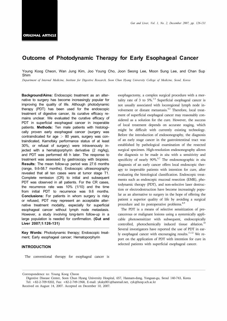
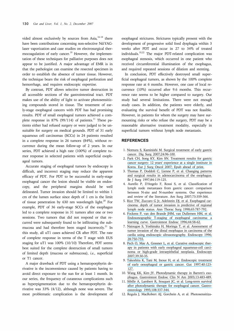
Fig 1 (Case 9) (A) Before PDT A slightly depressed lesion unstained with Lugols solution is seen on the mid-esophagus (B) Two days after PDT Endo-scopy shows coagulation necro-sis with ulcer at the PDT treated lesion (C) One month after PDT The previously PDT- induced ulcerative lesion have healed (D) Five months after PDT The scar well stained with Lugols solution is seen at the previously cancerous lesion and there is no remaining tumor in the biopsied specimens
months
RESULTS
Ten male patients with superficial esophageal tumors were included in the study (mean age 713 years range 64-82 years Table 1) In five patients surgical treatment was abandoned due to severe associated medical illnesses including cardiovascular diseases in three and chronic res-
piratory failure in two patients In other patients the choice of PDT was made due to the cancer at another sites (previous surgical treatment of three stomach can-cers and one colon cancer) age older than 80 or the pa-tients refusal of surgery Nine tumors were squamous cell carcinoma and one was poorly differentiated adeno-carcinoma Endoscopic appearance was determined as ero-siveplaque-like in cases flatcongestive in five cases and papillary-type in two cases Eight tumors were located at
Cheon YK et al Outcome of Photodynamic Therapy for Early Esophageal Cancer 129
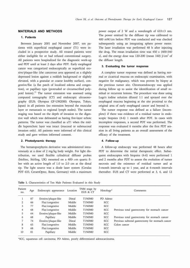
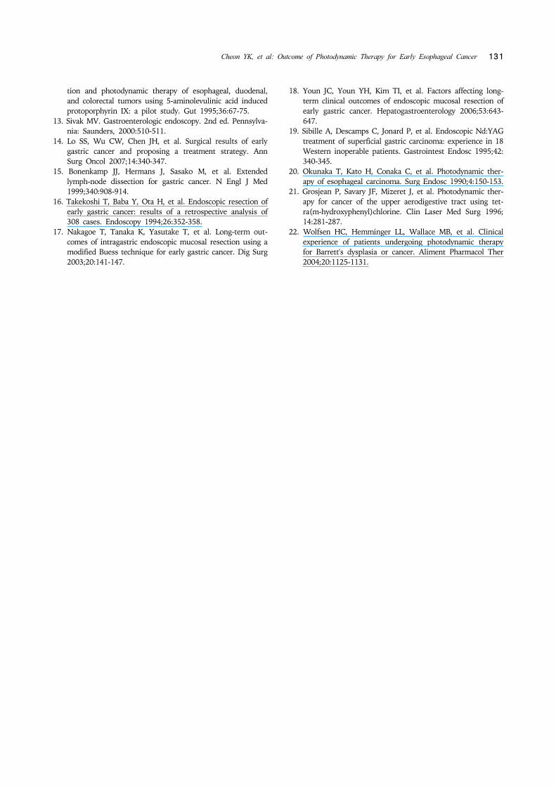
Fig 2 (Case 1) (A B) A flat reddish lesion unstained with Lugols solution is seen on the mid-esophagus (the biopsied showsspecimen squamous cell carcinoma) (C) Two days after photodynamic therapy Endoscopy shows circumferential coagulation necrosis with an ulcer at the PDT treated lesion (D) Two months after PDT Endoscopy shows luminal narrowing with fibrous scarring at the site of the PDT-treated lesion (E) Fluoroscopic image shows a metal stent at the site of the esophageal stricture(F) Endoscopy shows the improvement at the stricture site 2 months after stent removal
the mid esophagus and two were located at the distal esophagus According to the TNM classification all ten patients were diagnosed as having endosonographic uT1 (Table 2) Severe necrotic change was observed 2 days after PDT Mucosal healing and complete re-epithelialization took an average of 4 weeks (Fig 1) A complete response was ob-tained in all ten patients (100) Seven patients received a single session of the treatment and three patients re-ceived two sessions due to residual tumors determined by n endoscopic biopsies 4 weeks after the initial PDT The mean follow-up period was 276 months (96-587 months) Local recurrence occurred in one patient (10) after 96 months (case 8) During the follow-up three patients died of cause not related to the esophageal cancer Case 1 was uT1 with poorly differentiated ad-enocarcinoma and died at 145 months from a heart at-tack (he had underlying two-vessel coronary disease) Case 4 died from advanced stomach cancer Case 10 died from an unknown cause
Cutaneous photosensitization and pigmentation oc-curred in four patients but was not severe Esophageal stenosis occurred in one patient who received circum-ferential illumination of the esophagus and required re-peated dilation followed by stenting (Fig 2)
DISCUSSION
Based on numerous surgical reports a favorable post-operative outcome in terms of survival is currently ex-pected in early gastrointestinal cancer1214 In an effort to obtain a better quality of life less invasive treatment is more commonly chosen for such cancer with a low possi-bility of accompanying metastatic lymph nodes45 In larg-er studies the reported morbidity and mortality rates were 20-40 and 4-10 respectively315 Endoscopic treatment is another treatment option in gastrointestinal oncology and its therapeutic efficacy is now being eval-uated from various perspectives In addition to reports on endoscopic mucosal resection (EMR) that have been pro-
130 Gut and Liver Vol 1 No 2 December 2007
vided almost exclusively by sources from Asia16-18 there have been contributions concerning non-selective NdYAG- laser vaporization and case studies on electrosurgical ther-mocoagulation of early cancer19 However the implemen-tation of these techniques for palliative purposes does not appear to be justified A major advantage of EMR is in that the pathologist can examine the resected specimen in order to establish the absence of tumor tissue However the technique bears the risk of esophageal perforation and hemorrhage and requires endoscopic expertise By contrast PDT allows selective tumor destruction in all accessible sections of the gastrointestinal tract PDT makes use of the ability of light to activate photosensitiz-ing compounds stored in tissue The treatment of ear-ly-stage esophageal cancers with PDT has had promising results PDT of small esophageal tumors achieved a com-plete response in 87 (99114) of patients11 These pa-tients either had refused surgery or were judged to be un-suitable for surgery on medical grounds PDT of 31 early squamous cell carcinomas (SCCs) in 24 patients resulted in a complete response in 26 tumors (84) without re-currence during the mean follow-up of 2 years In our series PDT achieved a high rate (100) of complete tu-mor response in selected patients with superficial esoph-ageal tumors Accurate staging of esophageal tumors by endoscopy is difficult and incorrect staging may reduce the apparent efficacy of PDT For PDT to be successful in early-stage esophageal cancer the lesion should be visible on endos-copy and the peripheral margins should be well delineated Tumor invasion should be limited to within 1 cm of the lumen surface since depth of 1 cm is the limit of tissue penetration by 630 nm wavelength light20 For example PDT of 36 early-stage SCCs of the esophagus led to a complete response in 31 tumors after one or two sessions Two tumors that did not respond or that re-curred were subsequently found to be infiltrating the sub-mucosa and had therefore been staged incorrectly21 In this study all uT1 cases achieved CR after PDT The rate of complete response in terms of the T stage with EUS staging for uT1 was 100 (1010) Therefore PDT seems best suited for the complete destruction of small tumors of limited depth (mucosa or submucosa) ie superficial or T1 cancer A major drawback of PDT using a hematoporphyrin de-rivative is the inconvenience caused by patients having to avoid direct exposure to the sun for at least 1 month In our series the frequency of cutaneous complications such as hyperpigmentation due to the hematoporphyrin de-rivative was 33 (412) although none was severe The most problematic complication is the development of
esophageal strictures Strictures typically present with the development of progressive solid food dysphagia within 3 weeks after PDT and occur in 27 to 34 of treated individuals1022 The major PDT-related complication was esophageal stenosis which occurred in one patient who received circumferential illumination of the esophagus and required repeated sessions of dilation and stenting In conclusion PDT effectively destroyed small super-ficial esophageal tumors as shown by the 100 complete response rate at 6 months However one case of local re-currence (10) occurred after 96 months This recur-rence rate seems to be higher compared to surgery Our study had several limitations There were not enough study cases In addition the patients were elderly and evaluating the survival benefit of PDT was not feasible However in patients for whom the surgery may have sur-mounting risks or who refuse the surgery PDT may be a reasonable alternative treatment modality especially in superficial tumors without lymph node metastasis
REFERENCES
1 Nomura S Kaminishi M Surgical treatment of early gastric cancer Dig Surg 20072496-100
2 Park CH Song KY Kim SN Treatment results for gastric cancer surgery 12 years experience at a single institute in Korea Eur J Surg Oncol 2007 Epub ahead of print
3 Thomas P Doddoli C Lienne P et al Changing patterns and surgical results in adenocarcinoma of the esophagus Br J Surg 199784119-125
4 Aurello P DAngelo F Rossi S et al Classification of lymph node metastases from gastric cancer comparison between N-site and N-number systems Our experience and review of the literature Am Surg 200773359-366
5 Rice TW Zuccaro G Jr Adelstein DJ et al Esophageal car-cinoma depth of tumor invasion is predictive of regional lymph node status Ann Thorac Surg 199865787-792
6 Fockens P van den Brande JHM van Dullemen HM et al Endosonographic T-staging of esophageal carcinoma a learning curve Gastrointest Endosc 19964458-62
7 Natsugoe S Yoshinaka H Moringa T et al Assessment of tumor invasion of the distal esophagus in carcinoma of the cardia using endoscopic ultrasonography Endoscopy 1996 28750-755
8 Pech O May A Gossner L et al Curative endoscopic ther-apy in patients with early esophageal squamous-cell carci-noma or high-grade intraepithelial neoplasia Endoscopy 20073930-35
9 Takeshita K Tani M Inoue H et al Endoscopic treatment of early oesophageal or gastric cancer Gut 199740123- 127
10 Wang KK Kim JY Photodynamic therapy in Barretts eso-phagus Gastrointest Endosc Clin N Am 200313483-489
11 Sibille A Lambert R Souquet JC et al Long-term survival after photodynamic therapy for esophageal cancer Gastro-enterology 1995108337-344
12 Regula J MacRobert AJ Gorchein A et al Photosensitiza-
Cheon YK et al Outcome of Photodynamic Therapy for Early Esophageal Cancer 131
tion and photodynamic therapy of esophageal duodenal and colorectal tumors using 5-aminolevulinic acid induced protoporphyrin IX a pilot study Gut 19953667-75
13 Sivak MV Gastroenterologic endoscopy 2nd ed Pennsylva-nia Saunders 2000510-511
14 Lo SS Wu CW Chen JH et al Surgical results of early gastric cancer and proposing a treatment strategy Ann Surg Oncol 200714340-347
15 Bonenkamp JJ Hermans J Sasako M et al Extended lymph-node dissection for gastric cancer N Engl J Med 1999340908-914
16 Takekoshi T Baba Y Ota H et al Endoscopic resection of early gastric cancer results of a retrospective analysis of 308 cases Endoscopy 199426352-358
17 Nakagoe T Tanaka K Yasutake T et al Long-term out-comes of intragastric endoscopic mucosal resection using a modified Buess technique for early gastric cancer Dig Surg 200320141-147
18 Youn JC Youn YH Kim TI et al Factors affecting long- term clinical outcomes of endoscopic mucosal resection of early gastric cancer Hepatogastroenterology 200653643- 647
19 Sibille A Descamps C Jonard P et al Endoscopic NdYAG treatment of superficial gastric carcinoma experience in 18 Western inoperable patients Gastrointest Endosc 199542 340-345
20 Okunaka T Kato H Conaka C et al Photodynamic ther-apy of esophageal carcinoma Surg Endosc 19904150-153
21 Grosjean P Savary JF Mizeret J et al Photodynamic ther-apy for cancer of the upper aerodigestive tract using tet-ra(m-hydroxyphenyl)chlorine Clin Laser Med Surg 1996 14281-287
22 Wolfsen HC Hemminger LL Wallace MB et al Clinical experience of patients undergoing photodynamic therapy for Barretts dysplasia or cancer Aliment Pharmacol Ther 2004201125-1131
Cheon YK et al Outcome of Photodynamic Therapy for Early Esophageal Cancer 127
Table 1 Characteristics of Ten Male Patients Evaluated in this Study985114985114985114985114985114985114985114985114985114985114985114985114985114985114985114985114985114985114985114985114985114985114985114985114985114985114985114985114985114985114985114985114985114985114985114985114985114985114985114985114985114985114985114985114985114985114985114985114985114985114985114985114985114985114985114985114985114985114985114985114985114985114985114985114985114985114985114985114985114985114985114985114985114985114985114985114985114985114985114985114985114985114985114985114985114985114985114985114985114985114985114985114985114985114985114985114985114985114985114985114985114985114985114985114985114985114985114985114985114985114985114Patient TNM stage by Age Endoscopic appearance Location Histology Comments no EUS amp CT985103985103985103985103985103985103985103985103985103985103985103985103985103985103985103985103985103985103985103985103985103985103985103985103985103985103985103985103985103985103985103985103985103985103985103985103985103985103985103985103985103985103985103985103985103985103985103985103985103985103985103985103985103985103985103985103985103985103985103985103985103985103985103985103985103985103985103985103985103985103985103985103985103985103985103985103985103985103985103985103985103985103985103985103985103985103985103985103985103985103985103985103985103985103985103985103985103985103985103985103985103985103985103985103985103985103985103985103985103985103985103
1 87 Erosiveplaque-like Distal T1N0M0 PD Adeno 2 66 Flatcongestive Middle T1N0M0 SCC 3 77 Flatcongestive Middle T1N0M0 SCC 4 68 Flatcongestive Middle T1N0M0 SCC Previous total gastrectomy for stomach cancer 5 64 Erosiveplaque-like Middle T1N0M0 SCC 6 68 Papillary Middle T1N0M0 SCC Previous total gastrectomy for stomach cancer 7 74 Erosiveplaque-like Distal T1N0M0 SCC Previous subtotal gastrectomy for stomach cancer 8 65 Flatcongestive Middle T1N0M0 SCC Colon cancer 9 68 Flatcongestive Middle T1N0M0 SCC10 81 Papillary Middle T1N0M0 SCC985103985103985103985103985103985103985103985103985103985103985103985103985103985103985103985103985103985103985103985103985103985103985103985103985103985103985103985103985103985103985103985103985103985103985103985103985103985103985103985103985103985103985103985103985103985103985103985103985103985103985103985103985103985103985103985103985103985103985103985103985103985103985103985103985103985103985103985103985103985103985103985103985103985103985103985103985103985103985103985103985103985103985103985103985103985103985103985103985103985103985103985103985103985103985103985103985103985103985103985103985103985103985103985103985103985103985103985103985103985103985103SCC squamous cell carcinoma PD Adeno poorly differentiated adenocarcinoma
MATERIALS AND METHODS
1 Patients
Between January 2001 and November 2007 ten pa-tients with superficial esophageal cancer (T1) were in-cluded in a prospective study All treated patients were either ineligible for or had refused conventional surgery All patients were hospitalized for the diagnostic work-up and PDT until at least 2 days after PDT Early esophageal cancer was categorized endoscopically as superficial ero-siveplaque-like (the cancerous area appeared as a slightly depressed lesion against a reddish background or slightly elevated with a granular or coarse knobby surface) con-gestiveflat (a flat patch of localized edema and conges-tion) or papillary type (protruded or circumscribed poly-poid lesion)13 The tumor extension was assessed using computed tomography (CT) and endoscopic ultrasono-graphy (EUS Olympus GF-UM2000 Olympus Tokyo Japan) in all patients (no extension beyond the muscular layer or metastasis to regional lymph nodes) The tumor staging was based on the depth of invasion to the diges-tive wall which was delineated as having five-layer echoic patterns The tumor was classified as uT1 when the mid-dle hyperechoic layer was intact (mucosal or submucosal invasion only) All patients were informed of this clinical study and gave written informed consent
2 Photodynamic therapy
The hematoporphyrin derivative was administered intra-venously at a dose of 2 mgkg body weight For light dis-tribution we used flexible cylindrical diffuser probes (biolitec Stirling UK) mounted on a 400-μm quartz fi-ber with an active length of 10 to 20 cm at the distal tip The light source was a diode laser system (Ceralas PDT 633 CeramOptec Bonn Germany) with a maximum
power output of 2 W and a wavelength of 633plusmn3 nm The power emitted by the diffuser tip was calibrated to 400 mWcm before PDT was conducted and was checked subsequently using an integrating sphere power meter The laser irradiation was performed 48 h after injecting the drug The mean irradiation time was 450 s (400-540 s) and the energy dose was 120-200 (mean 160) Jcm2 of the diffuser length
3 Evaluating the tumor response
A complete tumor response was defined as having nor-mal or cicatrical mucosa on endoscopic examination with negative for malignancy which was proven by biopsy at the previous tumor site Chromoendoscopy was applied during follow up to assist the identification of small re-sidual or recurrent lesions The procedure was done using Lugos iodine solution diluted 11 and sprayed over the esophageal mucosa beginning at the site proximal to the original area of early esophageal cancer and beyond it The tumor response was defined as a failure (incom-plete) if there was evidence of a residual tumor in endo-scopic biopsies (4-6) 1 month after PDT In cases with incomplete responses a second PDT was performed The response was evaluated 6 months after the first PDT ses-sion in all living patients as an overall assessment of the efficacy of the treatment
4 Follow-up
A follow-up endoscopy was performed 48 hours after PDT to determine the initial therapeutic effect Subse-quent endoscopies with biopsies (4-6) were performed 1 and 2 months after PDT to assess the evolution of tumor necrosis and the existence of residual tumor and at 3-month intervals up to 1 year and at 6-month intervals thereafter EUS and CT were performed at 3 6 and 12
128 Gut and Liver Vol 1 No 2 December 2007
Table 2 Results of Photodynamic Therapy (PDT)985114985114985114985114985114985114985114985114985114985114985114985114985114985114985114985114985114985114985114985114985114985114985114985114985114985114985114985114985114985114985114985114985114985114985114985114985114985114985114985114985114985114985114985114985114985114985114985114985114985114985114985114985114985114985114985114985114985114985114985114985114985114985114985114985114985114985114985114985114985114985114985114985114985114985114985114985114985114985114985114985114985114985114985114985114985114985114985114985114985114985114985114985114985114985114985114985114985114985114985114985114985114985114985114985114985114985114985114985114985114985114Patient Dose PDT Follow-up Additional Response Recurrence Patient status no (Jcm) sessions (time to recurrence) treatment985103985103985103985103985103985103985103985103985103985103985103985103985103985103985103985103985103985103985103985103985103985103985103985103985103985103985103985103985103985103985103985103985103985103985103985103985103985103985103985103985103985103985103985103985103985103985103985103985103985103985103985103985103985103985103985103985103985103985103985103985103985103985103985103985103985103985103985103985103985103985103985103985103985103985103985103985103985103985103985103985103985103985103985103985103985103985103985103985103985103985103985103985103985103985103985103985103985103985103985103985103985103985103985103985103985103985103985103985103985103985103
1 180 2 CR No 145 985103 Died of heart disease 2 200 1 CR No 461 985103 Alive disease free 3 180 1 CR No 587 985103 Alive disease free 4 120 1 CR No 444 985103 Died of stomach cancer 5 140 1 CR No 299 985103 Alive disease free 6 160 1 CR No 118 985103 Alive disease free 7 160 2 CR No 18 985103 Alive disease free 8 160 1 CR Yes 96 (96) RT Alive disease free 9 160 1 CR No 104 985103 Alive disease free10 180 2 CR No 286 985103 Died of an unknown cause985103985103985103985103985103985103985103985103985103985103985103985103985103985103985103985103985103985103985103985103985103985103985103985103985103985103985103985103985103985103985103985103985103985103985103985103985103985103985103985103985103985103985103985103985103985103985103985103985103985103985103985103985103985103985103985103985103985103985103985103985103985103985103985103985103985103985103985103985103985103985103985103985103985103985103985103985103985103985103985103985103985103985103985103985103985103985103985103985103985103985103985103985103985103985103985103985103985103985103985103985103985103985103985103985103985103985103985103985103985103985103CR complete response RT radiotherapy
Fig 1 (Case 9) (A) Before PDT A slightly depressed lesion unstained with Lugols solution is seen on the mid-esophagus (B) Two days after PDT Endo-scopy shows coagulation necro-sis with ulcer at the PDT treated lesion (C) One month after PDT The previously PDT- induced ulcerative lesion have healed (D) Five months after PDT The scar well stained with Lugols solution is seen at the previously cancerous lesion and there is no remaining tumor in the biopsied specimens
months
RESULTS
Ten male patients with superficial esophageal tumors were included in the study (mean age 713 years range 64-82 years Table 1) In five patients surgical treatment was abandoned due to severe associated medical illnesses including cardiovascular diseases in three and chronic res-
piratory failure in two patients In other patients the choice of PDT was made due to the cancer at another sites (previous surgical treatment of three stomach can-cers and one colon cancer) age older than 80 or the pa-tients refusal of surgery Nine tumors were squamous cell carcinoma and one was poorly differentiated adeno-carcinoma Endoscopic appearance was determined as ero-siveplaque-like in cases flatcongestive in five cases and papillary-type in two cases Eight tumors were located at
Cheon YK et al Outcome of Photodynamic Therapy for Early Esophageal Cancer 129
Fig 2 (Case 1) (A B) A flat reddish lesion unstained with Lugols solution is seen on the mid-esophagus (the biopsied showsspecimen squamous cell carcinoma) (C) Two days after photodynamic therapy Endoscopy shows circumferential coagulation necrosis with an ulcer at the PDT treated lesion (D) Two months after PDT Endoscopy shows luminal narrowing with fibrous scarring at the site of the PDT-treated lesion (E) Fluoroscopic image shows a metal stent at the site of the esophageal stricture(F) Endoscopy shows the improvement at the stricture site 2 months after stent removal
the mid esophagus and two were located at the distal esophagus According to the TNM classification all ten patients were diagnosed as having endosonographic uT1 (Table 2) Severe necrotic change was observed 2 days after PDT Mucosal healing and complete re-epithelialization took an average of 4 weeks (Fig 1) A complete response was ob-tained in all ten patients (100) Seven patients received a single session of the treatment and three patients re-ceived two sessions due to residual tumors determined by n endoscopic biopsies 4 weeks after the initial PDT The mean follow-up period was 276 months (96-587 months) Local recurrence occurred in one patient (10) after 96 months (case 8) During the follow-up three patients died of cause not related to the esophageal cancer Case 1 was uT1 with poorly differentiated ad-enocarcinoma and died at 145 months from a heart at-tack (he had underlying two-vessel coronary disease) Case 4 died from advanced stomach cancer Case 10 died from an unknown cause
Cutaneous photosensitization and pigmentation oc-curred in four patients but was not severe Esophageal stenosis occurred in one patient who received circum-ferential illumination of the esophagus and required re-peated dilation followed by stenting (Fig 2)
DISCUSSION
Based on numerous surgical reports a favorable post-operative outcome in terms of survival is currently ex-pected in early gastrointestinal cancer1214 In an effort to obtain a better quality of life less invasive treatment is more commonly chosen for such cancer with a low possi-bility of accompanying metastatic lymph nodes45 In larg-er studies the reported morbidity and mortality rates were 20-40 and 4-10 respectively315 Endoscopic treatment is another treatment option in gastrointestinal oncology and its therapeutic efficacy is now being eval-uated from various perspectives In addition to reports on endoscopic mucosal resection (EMR) that have been pro-
130 Gut and Liver Vol 1 No 2 December 2007
vided almost exclusively by sources from Asia16-18 there have been contributions concerning non-selective NdYAG- laser vaporization and case studies on electrosurgical ther-mocoagulation of early cancer19 However the implemen-tation of these techniques for palliative purposes does not appear to be justified A major advantage of EMR is in that the pathologist can examine the resected specimen in order to establish the absence of tumor tissue However the technique bears the risk of esophageal perforation and hemorrhage and requires endoscopic expertise By contrast PDT allows selective tumor destruction in all accessible sections of the gastrointestinal tract PDT makes use of the ability of light to activate photosensitiz-ing compounds stored in tissue The treatment of ear-ly-stage esophageal cancers with PDT has had promising results PDT of small esophageal tumors achieved a com-plete response in 87 (99114) of patients11 These pa-tients either had refused surgery or were judged to be un-suitable for surgery on medical grounds PDT of 31 early squamous cell carcinomas (SCCs) in 24 patients resulted in a complete response in 26 tumors (84) without re-currence during the mean follow-up of 2 years In our series PDT achieved a high rate (100) of complete tu-mor response in selected patients with superficial esoph-ageal tumors Accurate staging of esophageal tumors by endoscopy is difficult and incorrect staging may reduce the apparent efficacy of PDT For PDT to be successful in early-stage esophageal cancer the lesion should be visible on endos-copy and the peripheral margins should be well delineated Tumor invasion should be limited to within 1 cm of the lumen surface since depth of 1 cm is the limit of tissue penetration by 630 nm wavelength light20 For example PDT of 36 early-stage SCCs of the esophagus led to a complete response in 31 tumors after one or two sessions Two tumors that did not respond or that re-curred were subsequently found to be infiltrating the sub-mucosa and had therefore been staged incorrectly21 In this study all uT1 cases achieved CR after PDT The rate of complete response in terms of the T stage with EUS staging for uT1 was 100 (1010) Therefore PDT seems best suited for the complete destruction of small tumors of limited depth (mucosa or submucosa) ie superficial or T1 cancer A major drawback of PDT using a hematoporphyrin de-rivative is the inconvenience caused by patients having to avoid direct exposure to the sun for at least 1 month In our series the frequency of cutaneous complications such as hyperpigmentation due to the hematoporphyrin de-rivative was 33 (412) although none was severe The most problematic complication is the development of
esophageal strictures Strictures typically present with the development of progressive solid food dysphagia within 3 weeks after PDT and occur in 27 to 34 of treated individuals1022 The major PDT-related complication was esophageal stenosis which occurred in one patient who received circumferential illumination of the esophagus and required repeated sessions of dilation and stenting In conclusion PDT effectively destroyed small super-ficial esophageal tumors as shown by the 100 complete response rate at 6 months However one case of local re-currence (10) occurred after 96 months This recur-rence rate seems to be higher compared to surgery Our study had several limitations There were not enough study cases In addition the patients were elderly and evaluating the survival benefit of PDT was not feasible However in patients for whom the surgery may have sur-mounting risks or who refuse the surgery PDT may be a reasonable alternative treatment modality especially in superficial tumors without lymph node metastasis
REFERENCES
1 Nomura S Kaminishi M Surgical treatment of early gastric cancer Dig Surg 20072496-100
2 Park CH Song KY Kim SN Treatment results for gastric cancer surgery 12 years experience at a single institute in Korea Eur J Surg Oncol 2007 Epub ahead of print
3 Thomas P Doddoli C Lienne P et al Changing patterns and surgical results in adenocarcinoma of the esophagus Br J Surg 199784119-125
4 Aurello P DAngelo F Rossi S et al Classification of lymph node metastases from gastric cancer comparison between N-site and N-number systems Our experience and review of the literature Am Surg 200773359-366
5 Rice TW Zuccaro G Jr Adelstein DJ et al Esophageal car-cinoma depth of tumor invasion is predictive of regional lymph node status Ann Thorac Surg 199865787-792
6 Fockens P van den Brande JHM van Dullemen HM et al Endosonographic T-staging of esophageal carcinoma a learning curve Gastrointest Endosc 19964458-62
7 Natsugoe S Yoshinaka H Moringa T et al Assessment of tumor invasion of the distal esophagus in carcinoma of the cardia using endoscopic ultrasonography Endoscopy 1996 28750-755
8 Pech O May A Gossner L et al Curative endoscopic ther-apy in patients with early esophageal squamous-cell carci-noma or high-grade intraepithelial neoplasia Endoscopy 20073930-35
9 Takeshita K Tani M Inoue H et al Endoscopic treatment of early oesophageal or gastric cancer Gut 199740123- 127
10 Wang KK Kim JY Photodynamic therapy in Barretts eso-phagus Gastrointest Endosc Clin N Am 200313483-489
11 Sibille A Lambert R Souquet JC et al Long-term survival after photodynamic therapy for esophageal cancer Gastro-enterology 1995108337-344
12 Regula J MacRobert AJ Gorchein A et al Photosensitiza-
Cheon YK et al Outcome of Photodynamic Therapy for Early Esophageal Cancer 131
tion and photodynamic therapy of esophageal duodenal and colorectal tumors using 5-aminolevulinic acid induced protoporphyrin IX a pilot study Gut 19953667-75
13 Sivak MV Gastroenterologic endoscopy 2nd ed Pennsylva-nia Saunders 2000510-511
14 Lo SS Wu CW Chen JH et al Surgical results of early gastric cancer and proposing a treatment strategy Ann Surg Oncol 200714340-347
15 Bonenkamp JJ Hermans J Sasako M et al Extended lymph-node dissection for gastric cancer N Engl J Med 1999340908-914
16 Takekoshi T Baba Y Ota H et al Endoscopic resection of early gastric cancer results of a retrospective analysis of 308 cases Endoscopy 199426352-358
17 Nakagoe T Tanaka K Yasutake T et al Long-term out-comes of intragastric endoscopic mucosal resection using a modified Buess technique for early gastric cancer Dig Surg 200320141-147
18 Youn JC Youn YH Kim TI et al Factors affecting long- term clinical outcomes of endoscopic mucosal resection of early gastric cancer Hepatogastroenterology 200653643- 647
19 Sibille A Descamps C Jonard P et al Endoscopic NdYAG treatment of superficial gastric carcinoma experience in 18 Western inoperable patients Gastrointest Endosc 199542 340-345
20 Okunaka T Kato H Conaka C et al Photodynamic ther-apy of esophageal carcinoma Surg Endosc 19904150-153
21 Grosjean P Savary JF Mizeret J et al Photodynamic ther-apy for cancer of the upper aerodigestive tract using tet-ra(m-hydroxyphenyl)chlorine Clin Laser Med Surg 1996 14281-287
22 Wolfsen HC Hemminger LL Wallace MB et al Clinical experience of patients undergoing photodynamic therapy for Barretts dysplasia or cancer Aliment Pharmacol Ther 2004201125-1131