esophageal strictures and diverticula
TRANSCRIPT
Esophageal Strictures andDiverticula
C. Daniel Smith, MD
KEYWORDS
� Esophageal stricture � Esophageal diverticula � Esophageal surgery� GERD complications � Esophageal stricture management� Esophageal diverticulectomy
KEY POINTS
� Esophageal disease, and in particular, dysfunction of the lower esophageal sphincter(LES) manifesting as gastroesophageal flux disease, is the most common of all gastroin-testinal conditions impacting patients daily.
� Conditions leading impairment of esophageal outflow can be categorized into 2 broad cat-egories, esophageal stricture or narrowing, and disorders of esophageal motility and LESfunction.
� Management focuses on the diverticulum itself, and relieving the underlying sphincterdysfunction.
� Many conditions can cause esophageal luminal narrowing or stricture; the most commonare peptic, malignant, and congenital. Other causes include autoimmune, iatrogenic,medication induced, radiation induced, infectious, caustic, and idiopathic.
INTRODUCTION
The topics of this article can best be understood in the context of impairment ofesophageal outflow and its consequences. Conditions that lead to impairment ofesophageal outflow can best be categorized into 2 broad categories: esophagealstricture or narrowing and disorders of esophageal motility and lower esophagealsphincter (LES) function. The consequence of esophageal stricture most often in-volves the immediate mechanical impact of the esophageal narrowing, and treatmentfocuses on relieving the stricture and control of the underlying process, which lead tothe stricture. Esophageal diverticula are most commonly the result of pressurization ofthe esophagus above a dysfunctional sphincter that fails to open appropriately (loweresophageal and cricopharyngeal), leading to the development of a false diverticulumjust proximal to the sphincter. Management focuses on the diverticulum itself, andrelieving the underlying sphincter dysfunction. One type of diverticulum not related
Piedmont Clinic, 2795 Peachtree Road, Unit 1808, Atlanta, GA 30305, USAE-mail address: [email protected]
Surg Clin N Am 95 (2015) 669–681http://dx.doi.org/10.1016/j.suc.2015.02.017 surgical.theclinics.com0039-6109/15/$ – see front matter � 2015 Elsevier Inc. All rights reserved.
Smith670
to esophageal motor dysfunction, a midesophageal diverticulum is also discussed inthis article. In contrast with the false diverticula of the esophagus, a midesophagealdiverticulum is a true diverticulum and the result of mediastinal inflammatory pro-cesses and the resulting focal traction on the esophageal wall, and is therefore notrelated to esophageal outflow obstruction.Many conditions can cause esophageal luminal narrowing or stricture. The most
common causes are peptic, malignant, and congenital; other causes include autoim-mune, iatrogenic, medication induced, radiation induced, infectious, caustic, andidiopathic.
ESOPHAGEAL STRICTURE
The term ‘esophageal stricture’ is reserved typically for intrinsic diseases of the esoph-agus causing luminal narrowing through inflammation, fibrosis, or neoplasia. Stricturesare grouped typically into benign and malignant categories, with treatment varyingdepending on the underlying cause. Other causes of esophageal narrowing some-times considered under the category of esophageal stricture include extrinsiccompromise of the esophageal lumen by direct invasion, lymph node enlargement,or direct compression. This article focuses on the intrinsic causes of esophageal nar-rowing/stricture.
Presentation
Regardless of the nature of a stricture, the clinical presentation typically involves anyor all of the following: dysphagia, food impaction, odynophagia, chest pain, andweight loss. Of these, progressive dysphagia to solids is the most common presentingsymptom, with benign strictures following a more slow and insidious progression (eg,months to years), whereas dysphagia of a malignant stricture tends to progress morerapidly (eg, in weeks to months).The clinical history may help to determine the cause of the dysphagia, although 25%
of patient presenting with peptic strictures have no prior heartburn or other symptomsof gastroesophageal reflux disease (GERD). A known history of use of medicationsknown to cause peptic ulcers or irritation, or caustic ingestion, are other examplesof clinical history that might suggest the underlying cause.
Diagnosis
Esophagogastroduodenoscopy and contrast swallow are the mainstays of the initialworkup and diagnosis for esophageal strictures. Although a contrast swallow is ob-tained most easily, esophagogastroduodenoscopy can provide more overall informa-tion and establish not only the diagnosis of a stricture or esophageal narrowing, butalso allow visualization of the esophageal mucosa, including biopsy to establish defin-itively the underlying cause of the stricture. This becomes especially important indetermining whether a stricture is benign or malignant. Contrast swallow may beparticularly useful in defining the overall esophageal anatomy and identifying otherassociated pathology, such as an esophageal diverticulum. Esophageal pH testing,esophageal motility may be needed to confirm a diagnosis of GERD or an underlyingesophageal motor abnormality (see Esophageal Diverticula section). Finally, when astricture is determined to be malignant, or extrinsic pathology is thought to be thecause of esophageal narrowing, CT of the chest and abdomen is indicated to establishthe cause of extrinsic narrowing and/or to stage a biopsy-proven malignant stricture.Endoscopic ultrasonography has emerged as a useful diagnostic tool to characterizethe nature of a stricture and assess the stage and severity of a malignant or infiltrating
Esophageal Strictures and Diverticula 671
process. This has become the mainstay of staging malignant disease of theesophagus.
Benign Esophageal Stricture
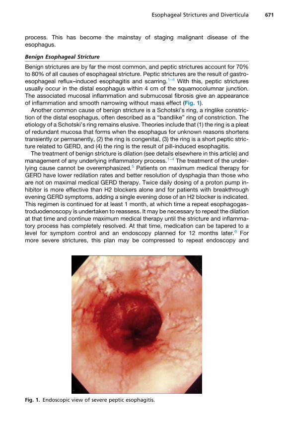
Benign strictures are by far the most common, and peptic strictures account for 70%to 80% of all causes of esophageal stricture. Peptic strictures are the result of gastro-esophageal reflux–induced esophagitis and scarring.1–4 With this, peptic stricturesusually occur in the distal esophagus within 4 cm of the squamocolumnar junction.The associated mucosal inflammation and submucosal fibrosis give an appearanceof inflammation and smooth narrowing without mass effect (Fig. 1).Another common cause of benign stricture is a Schotski’s ring, a ringlike constric-
tion of the distal esophagus, often described as a “bandlike” ring of constriction. Theetiology of a Schotski’s ring remains elusive. Theories include that (1) the ring is a pleatof redundant mucosa that forms when the esophagus for unknown reasons shortenstransiently or permanently, (2) the ring is congenital, (3) the ring is a short peptic stric-ture related to GERD, and (4) the ring is the result of pill-induced esophagitis.The treatment of benign stricture is dilation (see details elsewhere in this article) and
management of any underlying inflammatory process.1–4 The treatment of the under-lying cause cannot be overemphasized.5 Patients on maximum medical therapy forGERD have lower redilation rates and better resolution of dysphagia than those whoare not on maximal medical GERD therapy. Twice daily dosing of a proton pump in-hibitor is more effective than H2 blockers alone and for patients with breakthroughevening GERD symptoms, adding a single evening dose of an H2 blocker is indicated.This regimen is continued for at least 1 month, at which time a repeat esophagogas-troduodenoscopy is undertaken to reassess. It may be necessary to repeat the dilationat that time and continue maximum medical therapy until the stricture and inflamma-tory process has completely resolved. At that time, medication can be tapered to alevel for symptom control and an endoscopy planned for 12 months later.6 Formore severe strictures, this plan may be compressed to repeat endoscopy and
Fig. 1. Endoscopic view of severe peptic esophagitis.
Smith672
dilation within 1 to 2 weeks of an initial dilation, and more frequent reassessments. Ad-juncts such as steroid injections in and around the stricture have been used, especiallyfor more chronic fibrotic strictures. Stenting (see elsewhere in this article) has little rolein benign strictures unless the underlying issue with the stricture is anastomotic break-down and leak from a recent esophageal procedure (which is beyond the scope of thisarticle).Surgery is indicated for peptic stricture that recurs despite maximal medical ther-
apy, in which case an antireflux procedure is indicated, or for nondilatable fibroticstrictures, which typically requires resection and reconstruction to resolve.7 Oneshould be cautioned about using a segmental resection of the distal esophagus andesophagogastrostomy to manage a benign stricture because the majority of these pa-tients will have severe GERD after such a procedure, leaving the patient with ongoingissues with peptic injury to the esophagus.8 If a resection is needed, it is best to use anesophagojejunostomy to avoid severe GERD.
Malignant Esophageal Stricture
The most common cause of malignant esophageal stricture is adenocarcinoma asso-ciated with Barrett’s esophagus. This is a change from decades ago when most ma-lignant disease of the esophagus was squamous cancer associated with alcohol andtobacco use. The management of malignant stricture centers on tissue diagnosis,staging, and definitive therapy versus palliation. In contrast with benign strictures, dila-tion plays only a temporizing role, typically to facilitate placement of a stent or preparefor definitive therapy (resection). Stenting (see elsewhere in this article) is much morecommon in malignant stricture, either as permanent management for advanced dis-ease or temporary management to allow completion of neoadjuvant therapy beforeundergoing resection.
Management
DilationEsophageal dilation9 for stricture involves selection of technique of dilation, use ofadjuncts and endpoint.
TechniquesMercury-filled bougies (Maloney or Hurst dilators) are reasonable for uncomplicatedstrictures with an initial diameter of greater than 10mm. These dilators are inexpensiveand fluoroscopy is not needed. This is the technique used for self, at-home dilations.Wire-guided polyvinyl bougies (Savary-Gilliard dilators) are stiff dilators appropriate
for strictures 5 to 20 mm in diameter and are best suited for long, tight strictures. Fluo-roscopy is typically needed to assess guidewire placement and to visualize safe pas-sage of the dilator. Use usually requires sedation and is more traumatic on the larynxthan other techniques of dilation.10
Through-the-scope balloon dilators allow visualized placement and dilation.Although more expensive, balloon dilation seems to result in safe management ofmore complicated and tighter strictures with fewer sessions and a lower recurrencerate.11
AdjunctsIntralesional steroid injection and endoscopic stricturoplasty are the 2most commonlytalked about adjuncts to stricture dilation. Although few data exist to support a mech-anism of action, the first ventures to decrease the inflammatory reaction to the traumaof dilation and thereby limit the degree of restenosis after dilation. Several studies haveachieved larger final luminal diameter and lower stricture recurrence with the use of
Esophageal Strictures and Diverticula 673
intralesional steroid.12 It seems reasonable to use this in a benign stricture wheredysphagia persists despite dilations and maximal medical management of GERD.Four-quadrant stricturoplasty followed by dilation has been described for more
fibrotic strictures with limited success.13 Concern with stricturoplasty relates to perfo-ration making the fibrotic strictures most appealing for this adjunct.
Endpoint of Dilation
How much dilation can be achieved in a single session of dilation, and what luminaldiameter should be the goal remain controversial. Most would agree that gaining 1to 2 mm of luminal diameter through 3 consecutive passes of dilators of increasingsize during 1 session is a good general rule. Use of balloon dilators may allow evenmore increase in luminal diameter during a session. Obviously, perforation remainsthe concern, and balloon dilation provides real-time, direct visualization of the me-chanical effects of the dilation and may allow more aggressive, safe dilation. Most pa-tient experience complete relief of dysphagia when a luminal diameter of 40 to 54F isachieved.
Stenting
Stenting for esophageal structure is used most commonly for malignant strictures,either to provide permanent palliation for advanced disease or temporary palliationwhile a patient is treated with neoadjuvant therapy in preparation for curative resec-tion.14,15 Permanent stents are usually self-expanding metal or plastic stents, andtemporary stents have the stent itself covered so as to limit tissue ingrowth, allowingthe stent to be removed more easily. The details of stent design and placement arebeyond the scope of this article.
Surgery
Finally, surgery has a primary role for a malignant stricture where staging reveals apotentially curable cancer. In this case, esophagectomy with either high thoracic orcervical esophageal anastomosis to tubularized stomach or colon interposition ispreferred. Distal esophageal segmental resection with esophagogastrectomy shouldbe avoided owing to the severe GERD that often results with the LES gone and anintrathoracic anastomosis to stomach. If it is desirable to preserve as much esoph-agus as possible, it is better to use jejunum for reestablishing intestinal continuity.The role of surgery in benign stricture is largely limited to antireflux procedures to
manage the GERD that is etiologic in most benign strictures. For a nondilatable benignstricture, segmental resection is reasonable so long as an esophagojejunostomy isperformed rather than an esophagogastrostomy (see elsewhere in this article).
ESOPHAGEAL DIVERTICULA
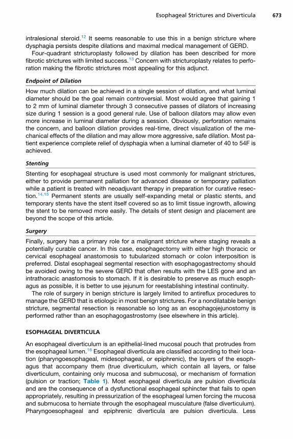
An esophageal diverticulum is an epithelial-lined mucosal pouch that protrudes fromthe esophageal lumen.16 Esophageal diverticula are classified according to their loca-tion (pharyngoesophageal, midesophageal, or epiphrenic), the layers of the esoph-agus that accompany them (true diverticulum, which contain all layers, or falsediverticulum, containing only mucosa and submucosa), or mechanism of formation(pulsion or traction; Table 1). Most esophageal diverticula are pulsion diverticulaand are the consequence of a dysfunctional esophageal sphincter that fails to openappropriately, resulting in pressurization of the esophageal lumen forcing the mucosaand submucosa to herniate through the esophageal musculature (false diverticulum).Pharyngoesophageal and epiphrenic diverticula are pulsion diverticula. Less
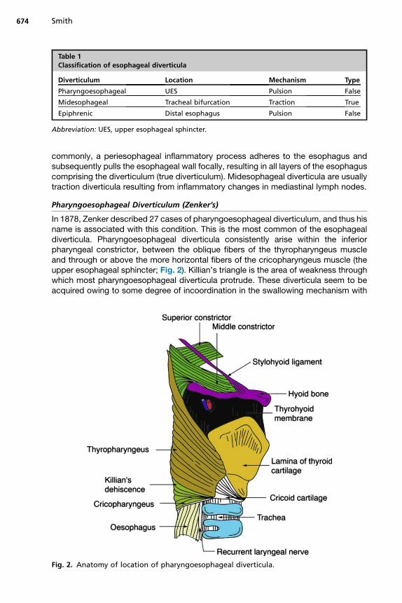
Table 1Classification of esophageal diverticula
Diverticulum Location Mechanism Type
Pharyngoesophageal UES Pulsion False
Midesophageal Tracheal bifurcation Traction True
Epiphrenic Distal esophagus Pulsion False
Abbreviation: UES, upper esophageal sphincter.
Smith674
commonly, a periesophageal inflammatory process adheres to the esophagus andsubsequently pulls the esophageal wall focally, resulting in all layers of the esophaguscomprising the diverticulum (true diverticulum). Midesophageal diverticula are usuallytraction diverticula resulting from inflammatory changes in mediastinal lymph nodes.
Pharyngoesophageal Diverticulum (Zenker’s)
In 1878, Zenker described 27 cases of pharyngoesophageal diverticulum, and thus hisname is associated with this condition. This is the most common of the esophagealdiverticula. Pharyngoesophageal diverticula consistently arise within the inferiorpharyngeal constrictor, between the oblique fibers of the thyropharyngeus muscleand through or above the more horizontal fibers of the cricopharyngeus muscle (theupper esophageal sphincter; Fig. 2). Killian’s triangle is the area of weakness throughwhich most pharyngoesophageal diverticula protrude. These diverticula seem to beacquired owing to some degree of incoordination in the swallowing mechanism with
Fig. 2. Anatomy of location of pharyngoesophageal diverticula.
Esophageal Strictures and Diverticula 675
an abnormally high intrapharyngeal pressure leading to protrusion of esophageal mu-cosa and submucosa through the esophageal wall with subsequent diverticulumformation.
Diagnosis
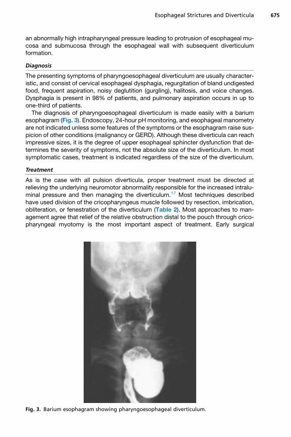
The presenting symptoms of pharyngoesophageal diverticulum are usually character-istic, and consist of cervical esophageal dysphagia, regurgitation of bland undigestedfood, frequent aspiration, noisy deglutition (gurgling), halitosis, and voice changes.Dysphagia is present in 98% of patients, and pulmonary aspiration occurs in up toone-third of patients.The diagnosis of pharyngoesophageal diverticulum is made easily with a barium
esophagram (Fig. 3). Endoscopy, 24-hour pHmonitoring, and esophageal manometryare not indicated unless some features of the symptoms or the esophagram raise sus-picion of other conditions (malignancy or GERD). Although these diverticula can reachimpressive sizes, it is the degree of upper esophageal sphincter dysfunction that de-termines the severity of symptoms, not the absolute size of the diverticulum. In mostsymptomatic cases, treatment is indicated regardless of the size of the diverticulum.
Treatment
As is the case with all pulsion diverticula, proper treatment must be directed atrelieving the underlying neuromotor abnormality responsible for the increased intralu-minal pressure and then managing the diverticulum.17 Most techniques describedhave used division of the cricopharyngeus muscle followed by resection, imbrication,obliteration, or fenestration of the diverticulum (Table 2). Most approaches to man-agement agree that relief of the relative obstruction distal to the pouch through crico-pharyngeal myotomy is the most important aspect of treatment. Early surgical
Fig. 3. Barium esophagram showing pharyngoesophageal diverticulum.
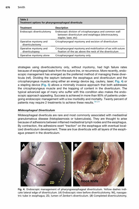
Table 2Treatment options for pharyngoesophageal diverticula
Treatment Description
Endoscopic diverticulotomy Endoscopic division of cricopharyngeus and common wallbetween diverticulum and esophagus (electrocautery,stapler, laser, etc)
Operative myotomy anddiverticulectomy
Cricopharyngeal myotomy and excision of diverticulum
Operative myotomy anddiverticulopexy
Cricopharyngeal myotomy and mobilization of sac with suturefixation of the sac above the neck of the diverticulum
Operative myotomy alone Cricopharyngeal myotomy only
Smith676
strategies using diverticulectomy only, without myotomy, had high failure ratesbecause of esophageal leaks from the suture line, or recurrence. More recently, endo-scopic management has emerged as the preferred method of managing these diver-ticula (ref). Dividing the septum between the esophagus and diverticulum and thecricopharyngeus muscle using either an energy device (eg, cautery, laser; Fig. 4) ora stapling device (Fig. 5) allows a minimally invasive approach that both addressesthe cricopharyngeus muscle and the trapping of content in the diverticulum. Thetypical advanced age of many who suffer with this condition also makes the endo-scopic approach appealing. Success is achieved in more than 90% of patients under-going endoscopic management with a low morbidity and mortality. Twenty percent ofpatients may require 2 treatments to achieve these results.18–21
Midesophageal Diverticulum
Midesophageal diverticula are rare and most commonly associated with mediastinalgranulomatous disease (histoplasmosis or tuberculosis). They are thought to arisebecause of adhesions between inflamedmediastinal lymph nodes and the esophagus.By contraction, the adhesions exert “traction” on the esophagus with eventual local-ized diverticulum development. These are true diverticula with all layers of the esoph-agus present in the diverticulum.
Fig. 4. Endoscopic management of pharyngoesophageal diverticulum. Yellow dashes indi-cate lateral edge of diverticulum. (A) Endoscopic view before diverticulotomy. NG, nasogas-tric tube in esophagus; ZD, lumen of Zenker’s diverticulum. (B) Completed diverticulotomy.
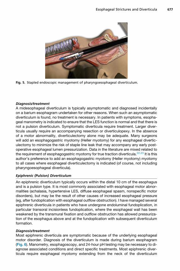
Fig. 5. Stapled endoscopic management of pharyngoesophageal diverticulum.
Esophageal Strictures and Diverticula 677
Diagnosis/treatmentA midesophageal diverticulum is typically asymptomatic and diagnosed incidentallyon a barium esophagram undertaken for other reasons. When such an asymptomaticdiverticulum is found, no treatment is necessary. In patients with symptoms, esopha-geal manometry is indicated to ensure that the LES function is normal and that there isnot a pulsion diverticulum. Symptomatic diverticula require treatment. Larger diver-ticula usually require an accompanying resection or diverticulopexy. In the absenceof a motor abnormality, diverticulectomy alone may be adequate. Many surgeonswill add an esophagogastric myotomy (Heller myotomy) for any esophageal divertic-ulectomy to minimize the risk of staple line leak that may accompany any early post-operative esophageal lumen pressurization. Data in the literature are mixed related tothe requirement of esophagogastric myotomy for true traction diverticula.22,23 It is thisauthor’s preference to add an esophagogastric myotomy (Heller myotomy) myotomyto all cases where esophageal diverticulectomy is indicated (of course, not includingpharyngoesophageal diverticula).
Epiphrenic (Pulsion) Diverticulum
An epiphrenic diverticulum typically occurs within the distal 10 cm of the esophagusand is a pulsion type. It is most commonly associated with esophageal motor abnor-malities (achalasia, hypertensive LES, diffuse esophageal spasm, nonspecific motordisorders), but may be the result of other causes of increased esophageal pressure(eg, after fundoplication with esophageal outflow obstruction). I have managed severalepiphrenic diverticula in patients who have undergone endoluminal fundoplication, inparticular transoral incisionless fundoplication, where the esophageal wall has beenweakened by the transmural fixation and outflow obstruction has allowed pressuriza-tion of the esophagus above and at the fundoplication with subsequent diverticulumformation.
Diagnosis/treatmentMost epiphrenic diverticula are symptomatic because of the underlying esophagealmotor disorder. Diagnosis of the diverticulum is made during barium esophagram(Fig. 6). Manometry, esophagoscopy, and 24-hour pH testing may be necessary to di-agnose associated conditions and direct specific treatments. Most epiphrenic diver-ticula require esophageal myotomy extending from the neck of the diverticulum
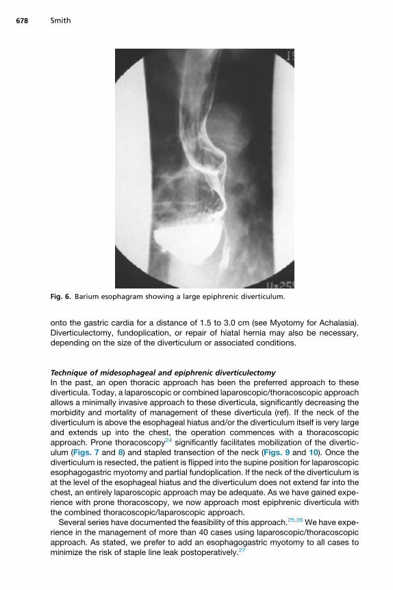
Fig. 6. Barium esophagram showing a large epiphrenic diverticulum.
Smith678
onto the gastric cardia for a distance of 1.5 to 3.0 cm (see Myotomy for Achalasia).Diverticulectomy, fundoplication, or repair of hiatal hernia may also be necessary,depending on the size of the diverticulum or associated conditions.
Technique of midesophageal and epiphrenic diverticulectomyIn the past, an open thoracic approach has been the preferred approach to thesediverticula. Today, a laparoscopic or combined laparoscopic/thoracoscopic approachallows a minimally invasive approach to these diverticula, significantly decreasing themorbidity and mortality of management of these diverticula (ref). If the neck of thediverticulum is above the esophageal hiatus and/or the diverticulum itself is very largeand extends up into the chest, the operation commences with a thoracoscopicapproach. Prone thoracoscopy24 significantly facilitates mobilization of the divertic-ulum (Figs. 7 and 8) and stapled transection of the neck (Figs. 9 and 10). Once thediverticulum is resected, the patient is flipped into the supine position for laparoscopicesophagogastric myotomy and partial fundoplication. If the neck of the diverticulum isat the level of the esophageal hiatus and the diverticulum does not extend far into thechest, an entirely laparoscopic approach may be adequate. As we have gained expe-rience with prone thoracoscopy, we now approach most epiphrenic diverticula withthe combined thoracoscopic/laparoscopic approach.Several series have documented the feasibility of this approach.25,26 We have expe-
rience in the management of more than 40 cases using laparoscopic/thoracoscopicapproach. As stated, we prefer to add an esophagogastric myotomy to all cases tominimize the risk of staple line leak postoperatively.27
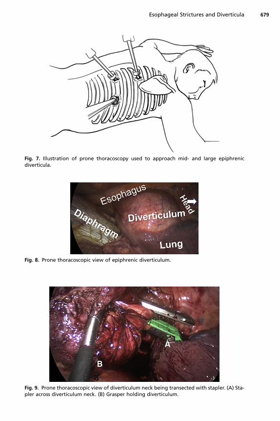
Fig. 7. Illustration of prone thoracoscopy used to approach mid- and large epiphrenicdiverticula.
Fig. 8. Prone thoracoscopic view of epiphrenic diverticulum.
Fig. 9. Prone thoracoscopic view of diverticulum neck being transected with stapler. (A) Sta-pler across diverticulum neck. (B) Grasper holding diverticulum.
Esophageal Strictures and Diverticula 679
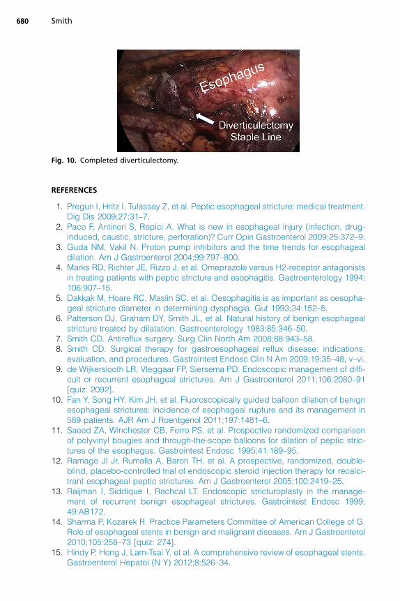
Fig. 10. Completed diverticulectomy.
Smith680
REFERENCES
1. Pregun I, Hritz I, Tulassay Z, et al. Peptic esophageal stricture: medical treatment.Dig Dis 2009;27:31–7.
2. Pace F, Antinori S, Repici A. What is new in esophageal injury (infection, drug-induced, caustic, stricture, perforation)? Curr Opin Gastroenterol 2009;25:372–9.
3. Guda NM, Vakil N. Proton pump inhibitors and the time trends for esophagealdilation. Am J Gastroenterol 2004;99:797–800.
4. Marks RD, Richter JE, Rizzo J, et al. Omeprazole versus H2-receptor antagonistsin treating patients with peptic stricture and esophagitis. Gastroenterology 1994;106:907–15.
5. Dakkak M, Hoare RC, Maslin SC, et al. Oesophagitis is as important as oesopha-geal stricture diameter in determining dysphagia. Gut 1993;34:152–5.
6. Patterson DJ, Graham DY, Smith JL, et al. Natural history of benign esophagealstricture treated by dilatation. Gastroenterology 1983;85:346–50.
7. Smith CD. Antireflux surgery. Surg Clin North Am 2008;88:943–58.8. Smith CD. Surgical therapy for gastroesophageal reflux disease: indications,
evaluation, and procedures. Gastrointest Endosc Clin N Am 2009;19:35–48, v–vi.9. de Wijkerslooth LR, Vleggaar FP, Siersema PD. Endoscopic management of diffi-
cult or recurrent esophageal strictures. Am J Gastroenterol 2011;106:2080–91[quiz: 2092].
10. Fan Y, Song HY, Kim JH, et al. Fluoroscopically guided balloon dilation of benignesophageal strictures: incidence of esophageal rupture and its management in589 patients. AJR Am J Roentgenol 2011;197:1481–6.
11. Saeed ZA, Winchester CB, Ferro PS, et al. Prospective randomized comparisonof polyvinyl bougies and through-the-scope balloons for dilation of peptic stric-tures of the esophagus. Gastrointest Endosc 1995;41:189–95.
12. Ramage JI Jr, Rumalla A, Baron TH, et al. A prospective, randomized, double-blind, placebo-controlled trial of endoscopic steroid injection therapy for recalci-trant esophageal peptic strictures. Am J Gastroenterol 2005;100:2419–25.
13. Raijman I, Siddique I, Rachcal LT. Endoscopic stricturoplasty in the manage-ment of recurrent benign esophageal strictures. Gastrointest Endosc 1999;49:AB172.
14. Sharma P, Kozarek R. Practice Parameters Committee of American College of G.Role of esophageal stents in benign and malignant diseases. Am J Gastroenterol2010;105:258–73 [quiz: 274].
15. Hindy P, Hong J, Lam-Tsai Y, et al. A comprehensive review of esophageal stents.Gastroenterol Hepatol (N Y) 2012;8:526–34.
Esophageal Strictures and Diverticula 681
16. Smith CD. Esophagus. In: Norton JA, Chang AE, Lowry SF, et al, editors. Essentialpractice of surgery basic science and clinical evidence. New York: Springer - Ver-lag; 2003. p. 167–84.
17. Zaninotto G, Narne S, Costantini M, et al. Tailored approach to Zenker’s diver-ticula. Surg Endosc 2003;17:129–33.
18. Tang SJ. Flexible endoscopic Zenker’s diverticulotomy: approach that involvesthinking outside the box (with videos). Surg Endosc 2014;28:1355–9.
19. Parker NP, Misono S. Carbon dioxide laser versus stapler-assisted endoscopicZenker’s diverticulotomy: a systematic review and meta-analysis. OtolaryngolHead Neck Surg 2014;150:750–3.
20. Law R, Baron TH. Transoral flexible endoscopic therapy of Zenker’s diverticulum.Dig Surg 2013;30:393.
21. Huberty V, El Bacha S, Blero D, et al. Endoscopic treatment for Zenker’s divertic-ulum: long-term results (with video). Gastrointest Endosc 2013;77:701–7.
22. Isaacs KE, Graham SA, Berney CR. Laparoscopic transhiatal approach for resec-tion of midesophageal diverticula. Ann Thorac Surg 2012;94:e17–9.
23. Galata CL, Bruns CJ, Pratschke S, et al. Thoracoscopic resection of a giant mid-esophageal diverticulum. Ann Thorac Surg 2012;94:293–5.
24. Goldberg RF, Bowers SP, Parker M, et al. Technical and perioperative outcomesof minimally invasive esophagectomy in the prone position. Surg Endosc 2013;27:553–7.
25. Herbella FA, Patti MG. Modern pathophysiology and treatment of esophagealdiverticula. Langenbecks Arch Surg 2012;397:29–35.
26. Soares RV, Montenovo M, Pellegrini CA, et al. Laparoscopy as the initial approachfor epiphrenic diverticula. Surg Endosc 2011;25:3740–6.
27. Melman L, Quinlan J, Robertson B, et al. Esophageal manometric characteristicsand outcomes for laparoscopic esophageal diverticulectomy, myotomy, and par-tial fundoplication for epiphrenic diverticula. Surg Endosc 2009;23:1337–41.