kai l is unchanged in metastatic and nonmetastatic esophageal and gastric cancers1
TRANSCRIPT
[CANCER RESEARCH 58. 753-758, February 15. 1998]
KAI l Is Unchanged in Metastatic and Nonmetastatic Esophageal andGastric Cancers1
Xiao-Zhong Guo, Helmut Friess,2 Christoph Maurer, Pascal Berberat, Wen-Hao Tang, Arthur Zimmermann,
Markus Naef, Hans U. Graber, Murray Korc, and Markus W. BüchlerDepartment of Visceral and Transplantation Surgery ¡X-Z.G., H. F., C. M.. P. B.. W-H. T., M. N.. H. U. C., M. W. B.¡and Institute of Pathology [A. Z.], University of Bern,Inselspital, CH-3010 Bern, Switzerland: and Division of Endocrinology, Metabolism and Diabetes, Departments of Medicine, Biological Chemistry, and Pharmacology, University
of California. Irvine. California 92697 [M. K.¡
ABSTRACT
Down-regulation of KAI I iiiRVV expression has been shown to be
associated with the formation of métastases or disease progression inpancreatic cancer. Whether Ai// possesses similar characteristics inother malignancies of the gastrointestinal tract is not known.
Here, we compared the patterns of KAI1 inKNA expression in 41esophageal cancers and 35 stomach cancers to assess whether KAI1 mightalso be of biological relevance in the metastatic ability of these tumors.
By Northern blot analysis, KAI l niKNA levels ranged widely in bothnormal and cancerous esophageal and gastric tissue samples, with nostatistical differences. No association between k VII niRNA expressionand tumor stage or tumor differentiation was seen in these tumors. Inaddition, KAI l mRNA expression was similar in primary esophageal andgastric cancer samples with or without métastases.By in situ hybridization, KAll mRNA expression was evident in the cytoplasm of mostsquamous epithelial cells in the normal esophagus and in nonmucosalepithelial cells of the normal stomach. The staining intensity in the esophageal and gastric cancer cells was similar to that in the normal controls.
This differential pattern of KAI1 mRNA expression in esophageal andgastric cancers in comparison to pancreatic cancer indicates that AU/seems to influence the potential of gastrointestinal cancer cells to metas-
tasize differently. In esophageal and gastric cancers, the formation ofmétastasesis not dependent on the reduction of Al// in the cancer cells.
INTRODUCTION
The esophagus and the stomach are major sites of human cancers(1). The high mortality associated with esophageal and gastric cancersstems, in part, from the fact that many tumors are not detected untilthe disease has progressed to an advanced stage in which lymph nodeor distant métastasesare already present. Even when the primarytumor is resectable, surgery alone is often unable to provide a cure inthese tumor stages. Therefore, the overall 5-year survival rates for
patients with esophageal cancer and gastric cancer who undergoresection are still unsatisfactory (2, 3). Standard oncological regimenssuch as chemo- and/or radiotherapy have not had a major impact on
the prognosis of these malignancies to date, and therefore, newapproaches, including early diagnosis, preventive strategies, and innovative treatment, are still sorely needed.
In the past few years, many biological studies have analyzedgenetic and molecular events occurring in cancers to identify genesthat are involved in their initiation, progression, and suppression. Thesearch for genes promoting or suppressing tumor spread has led to theidentification of several factors that are associated with métastases(4,5). nm23-Hl, which is located on the long arm of chromosome 17, hasbeen proposed as a metastasis suppressor gene. Down-regulation of itsexpression during tumor development and progression has been re-
Received 8/1/97; accepted 12/15/97.The costs of publication of this article were defrayed in part by the payment of page
charges. This article must therefore be hereby marked advertisement in accordance with18 U.S.C. Section 1734 solely to indicate this fact.
1This work was supported by Swiss National Foundation for Scientific Research.Grant 32-049494.96/1.
2 To whom requests for reprints should be addressed. Phone: 41 31 6329722; Fax:
41316329732.
ported in several in vitro and in vivo systems (6, 7). Recent molecularcloning experiments identified a new gene named KAU, which suppresses the ability of prostate cancer cells to metastasize (4). The KAI1gene belongs to a structurally distinct family of cell surface membraneglycoproteins (8-14). These glycoproteins have a similar structure,
with integral membrane proteins consisting of four transmembranedomains and one large extracellular domain, which is N-glycosylated.
Recent studies indicate that members of this gene family may influence the growth behaviors of cancer cells. However, their physiological and pathological functions are generally unknown.
Recently, we reported enhanced KAI1 mRNA expression in early-
stage primary pancreatic cancers but decreasing levels in primarypancreatic cancer samples in which lymph node or distant métastaseswere present (15). This observation, together with the findings inprostate cancer, suggests that the presence of KAU might influencethe ability of cancer cells to metastasize (4). However, it is not knownwhether KAI1 possesses similar characteristics in other gastrointestinal malignancies, such as esophageal and gastric cancers, whichwould support the hypothesis that this glycoprotein is generally important for the metastatic ability of cancer cells. Therefore, here, weanalyzed and compared the patterns of KAI1 mRNA expression inesophageal and gastric cancers to determine whether KAII might alsobe of biological relevance in the metastatic capability of these tumors.
PATIENTS AND METHODS
Patients. Tumor specimens were taken from 41 patients with non-metastatic (n = 12) and metastatic (n = 29) primary esophagealcancers (8 female and 33 male; mean age, 59.8 years; range, 42-76years) and 35 patients with nonmetastatic (n = 13) and metastatic(n = 22) primary stomach cancers (14 female and 21 male; mean age,67 years; range, 49-80 years) and were classified (Table 1) according
to the TNM classification of the International Union Against Cancer(16). For comparison, normal tissue specimens of the esophagus(n = 15) and stomach (n = 14) were obtained from 16 previously
healthy multiorgan donors (7 female and 9 male; mean age, 39 years)from whom other organs were obtained for transplantation. All studieswere approved by the Ethics Committee of the University of Bern(Bern, Switzerland).
Tissue Sampling. For RNA extraction and Northern blot analysis,normal and cancerous specimens were immediately frozen in liquidnitrogen after removal in the operating room and were stored at-80°C until use. In addition, freshly removed normal and cancerous
tissue samples were immediately fixed in buffered formaldehydesolution for 24 h and embedded in paraffin for in situ hybridization.
Northern Blot Analysis. Total RNA was isolated by the single-step guanidinium isothiocyanate method (17, 18) and size-fractionated (20 /ng/lane) on 1.2% agarose-1.8 M formaldehyde gels (17,
18). The gels were stained with ethidium bromide for verificationof RNA integrity and loading equivalency. Fractionated RNA waselectrotransferred onto nylon membranes (GeneScreen; DuPontInternational, Regensdorf, Switzerland) and cross-linked by UV
irradiation.
753
Research. on December 24, 2014. © 1998 American Association for Cancercancerres.aacrjournals.org Downloaded from
KAU IN ESOPHAGEAL AND GASTRIC CANCER
Table 1 TNM classification of the investigated tumor samples according to theInternational Union Against Cancer
Tumor stage Tumor grade
Esophagealcancer(n=41)Gastric
cancer(n= 35)I412II133III2213IV271422201631616411
The blots were prehybridized for 1-2 h at 65°Cin a buffer con
taining 50% formamide, 5X SSC (sodium chloride/sodium citratebuffer), 2% blocking reagent (Boehringer Mannheim GmbH, Mannheim, Germany), and 0.1% /V-lauroylsarcosine. After addition of theDIG'-labeled KAII antisense probe, hybridization was carried out at65°Cfor 18 h. The filters were washed for 5 min in 2X SSC-0.1%
SDS at room temperature, followed by two washes in 0.1X SSC-0.1 %SDS at 68°Cfor 15 min. Afterward, the filters were incubated in 20
ml of blocking buffer ( 1% blocking reagent in 100 mmol/liter maleicacid. 150 mmol/liter sodium chloride, and 175 mmol/liter sodiumhydroxide) containing 1 fil of anti-DIG alkaline phosphatase antibod
ies (Boehringer Mannheim) for 30 min, washed with blocking bufferfor 15 min, and incubated with 4 pd of CDP-Star (25 ITIM;BoehringerMannheim). The membranes were then exposed to X-ray films for15 s at room temperature, as described previously (17-19).
To assess equivalent RNA loading, the membranes were rehybrid-ized with the 32P-labeled 7S cDNA probe (17. 19). The blots wereprehybridized for 4-8 h at 42°Cin a buffer containing 50% form-amide. 1% SDS, 0.75 Msodium chloride, 5 mM EDTA, 5X Denhardt's
solution, 100 jug/ml denatured salmon sperm DNA, 10% dextransulfate, and 50 mM sodium phosphate buffer (pH 7.4). The hybridization was carried out at 42°Cfor 12 h with the 32P-labeled cDNAprobe (1 X IO5 cpm/ml) in the same buffer. Washing was started byrinsing the blots twice (50°C)in 2X SSC. The blots were, subsequently, washed three times at 55°Cin 0.2 X SSC and 2% SDS. Theblots were then exposed at -80°C to Fuji X-ray films with intensi
fying screens for 4-8 h. The intensity of the radiographie KAII and
7S signals was quantified in the esophageal and gastric tissue samplesby video densitometry analysis (Bio-Rad 620; Bio-Rad, New York.NY), as reported previously (17-19). The ratio between the KAII
mRNA signal and the corresponding 7S signal was calculated for eachsample and statistically analyzed.
In Situ Hybridization. In situ hybridization was performed asreported previously (15, 20). Tissue sections of normal and canceroustissue samples were always processed simultaneously. In addition, weprocessed consecutive slides, one slide was incubated with the senseprobe, and the other slide was incubated simultaneously with theantisense probe. Tissue sections (4 ju.m) were deparaffinized, rehy-
drated. and incubated in 0.2 mol/liter HC1 for 20 min. After washingwith 2X SSC, the tissues were permeabilized with proteinase K(Boehringer Mannheim) at concentrations of 30 /xg/ml in esophagealtissues and 40 fig/ml in gastric tissues for 15 min at 37°C.After
postfixation with 4% paraformaldehyde in phosphate buffer sodium (5min) and washing in 2x SSC, the sections were prehybridized for 1 h(at 53°Cfor esophageal tissue slides and at 55°Cfor gastric tissue
slides) in a buffer containing 50% (v/v) formamide, 4x SSC, 2XDenhardt's reagent, and 250 p,g/ml RNA. Hybridization was per
formed overnight at the same temperatures in 50% (v/v) formamide,4X SSC, 2X Denhardt's reagent. 500 jag/ml RNA, and 10% (v/v)
dextran sulfate. The final concentration of the DIG-labeled KAII
probes (antisense and sense) was approximately 0.5 ng/pil. After
1The abbreviation used is: DIG. digoxigenin.
hybridization, excess probe was removed by washing in 2X SSC andby RNase treatment as follows: 100 units/ml RNase Tl (BoehringerMannheim) and 0.2 /¿g/mlDNase-free RNase (Boehringer Mannheim) at 37°Cfor 30 min. Washings were performed at 55°Cforesophageal tissue slides and at 60°Cfor gastric tissue slides in 2X
SSC for 10 min and twice in 0.2 X SSC for 10 min each. Afterward,the sections were incubated with an anti-DIG antibody conjugated
with alkaline phosphatase (Boehringer Mannheim). For the followingcolor reaction, we used 5-bromo-4-chloro-3-indolyl phosphatase and
nitroblue tetrazolium (Sigma, Buchs, Switzerland).Pretreatment of the slides with RNase abolished the hybridization
signals, and sense probes corresponding to the antisense probes failedto produce a signal.
The results of in situ hybridization were semiquantitatively analyzed and scored as previously reported (15, 21). The in situ hybridization signals were evaluated by two independent pathologists whowere blinded to patient status, followed by resolution of any differences by joint review and consultation with a third observer.
Preparation of DIG-labeled KAII Probes. To prepare DIG-
labeled cRNA probes for Northern blot analysis and in situ hybridization, a 500-bp XbaVHindlll fragment of human KAII cDNA wassubcloned into the pCR-II vector (Invitrogen, San Diego, CA), whichcontains promoters for DNA-dependent SP6 and T7 RNA poly-
merases. After linearization of the plasmid with Xbal, the antisenseKAII probe was transcribed using SP6 polymerase and the RibomaxSystem (Promega Biotechnology, Madison, WI). The transcriptionresulted in a DIG-labeled antisense riboprobe that was specific for the
KAI l mRNA. To evaluate the specificity of the in situ hybridizationreaction, a DIG-labeled sense probe was generated after linearization
of the plasmid with Hindlll and in vitro transcription with T7 polymerase and the Ribomax System (Promega Biotechnology, Madison,WI; Ref. 22). For the in situ hybridization experiments, the KAIIantisense and sense probes were shortened to a length of approximately 150 bases, according to the procedure of Cox et al. (23).
Preparation of a 32P-labeled 7S cDNA Probe. To verify equiv
alent RNA loading on Northern blot membranes, all filters wererehybridized with a murine 0.19-kb 7S cDNA probe that cross-
hybridizes with human 7S RNA, as reported previously (17, 18, 20).The 7S cDNA probe was radiolabeled with [a-32P]dCTP (3000 Ci/
mmol; DuPont, Boston. MA) using a random primer labeling system(Pharmacia Biotech AG, Dubendorf, Switzerland; Refs. 17-19).
Statistical Analysis. Results were expressed as median and rangeor as mean ±SE. For statistical analysis, the Mann-Whitney U test,the Student's t test, or the x2 test was used. Significance was defined
as P < 0.05.
RESULTS
KAII mRNA Expression by Northern Blot Analysis
Northern blot analysis was performed using 20 p.g of total RNAextracted from normal and cancerous tissue samples.
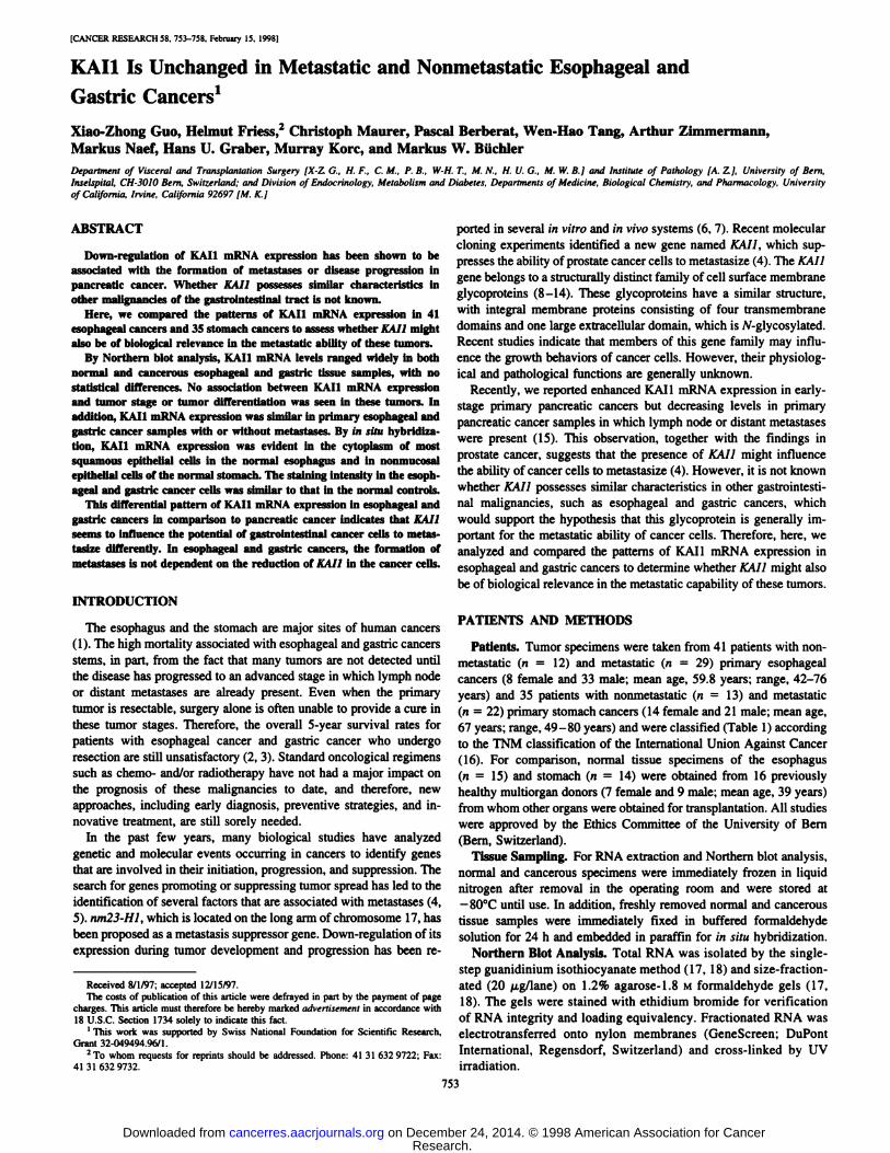
Esophagus. In the normal esophagus, moderate to strong KAIImRNA signals were detectable in 60% of the samples (Fig. 1). In theremaining samples, KAI l mRNA levels were weak or too low to bedetected by Northern blot analysis. In esophageal cancers, 49% of thesamples exhibited KAI 1 mRNA expression levels 1.6-fold higher than
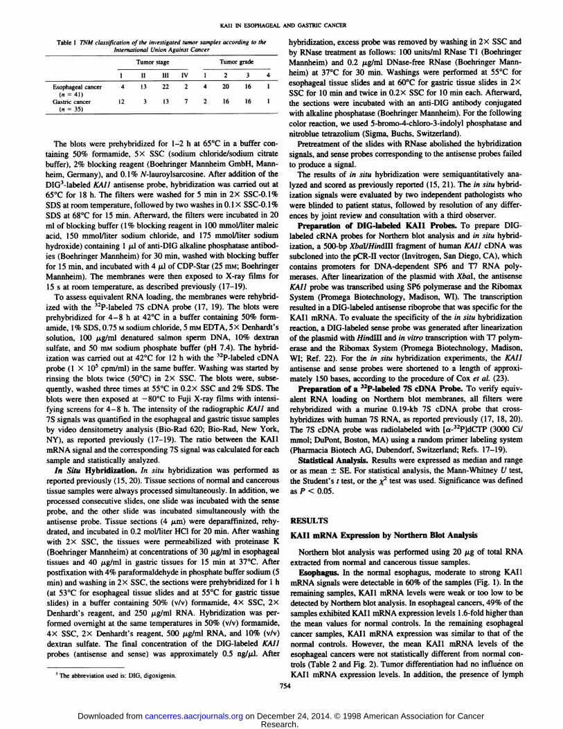
the mean values for normal controls. In the remaining esophagealcancer samples, KAII mRNA expression was similar to that of thenormal controls. However, the mean KAII mRNA levels of theesophageal cancers were not statistically different from normal controls (Table 2 and Fig. 2). Tumor differentiation had no influence onKAI l mRNA expression levels. In addition, the presence of lymph
754
Research. on December 24, 2014. © 1998 American Association for Cancercancerres.aacrjournals.org Downloaded from
KAU IN ESOPHAGEAL AND GASTRIC CANCER
Normal Esophageal Cancer
KAI1
7S
Fig. 1. Northern blot analysis of KAU mRNA expression in normal and canceroustissues of the esophagus. In esophageal cancer. KA1I mRNA expression was comparableto that of normal controls.
node or distant métastaseshad no influence on KAI l mRNA levels inthe primary esophageal tumor samples (Table 2 and Fig. 2). Primaryesophageal tumors in which lymph node (n = 29) or distant métastases (n = 2) were present at the time of resection exhibited KAI l
mRNA expression levels similar to those of primary esophagealtumors in which no lymph node métastases(n = 12) were found at the
time of tumor resection (Table 2 and Fig. 2).Stomach. An expression pattern similar to that in the esophagus
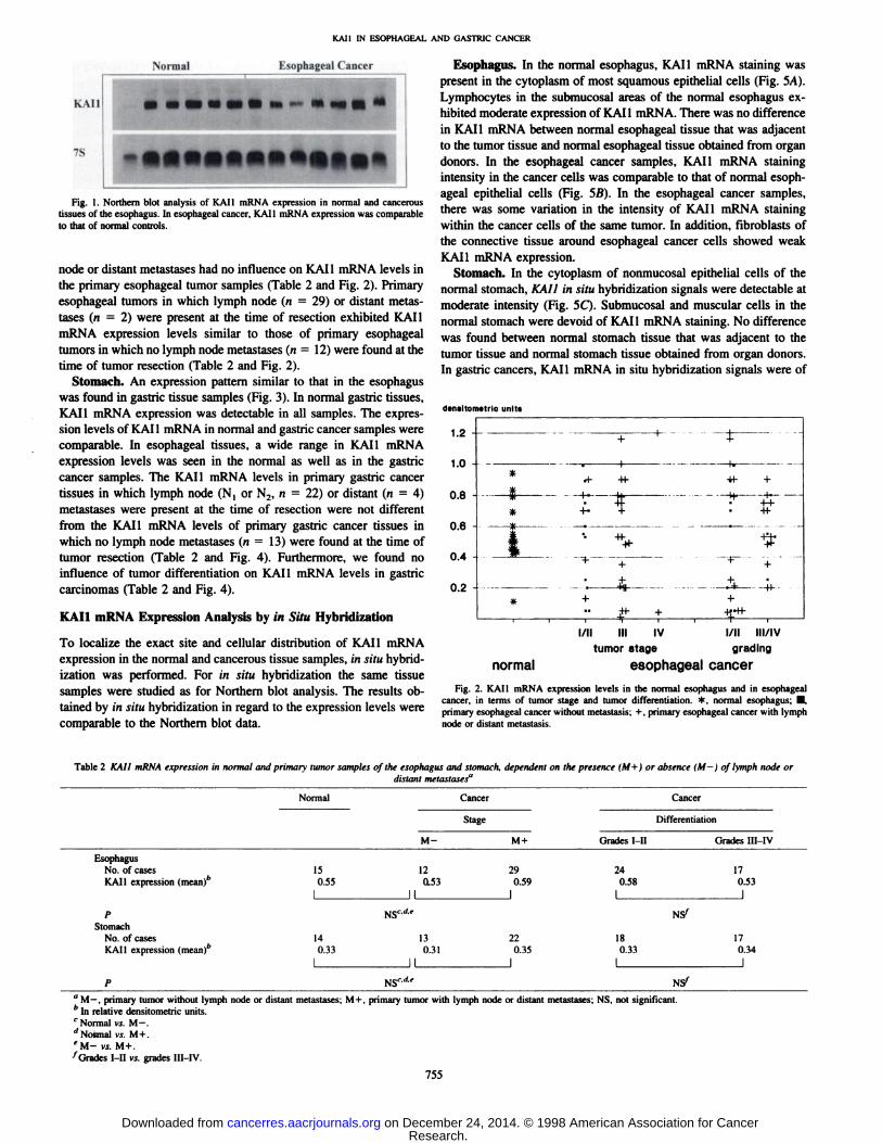
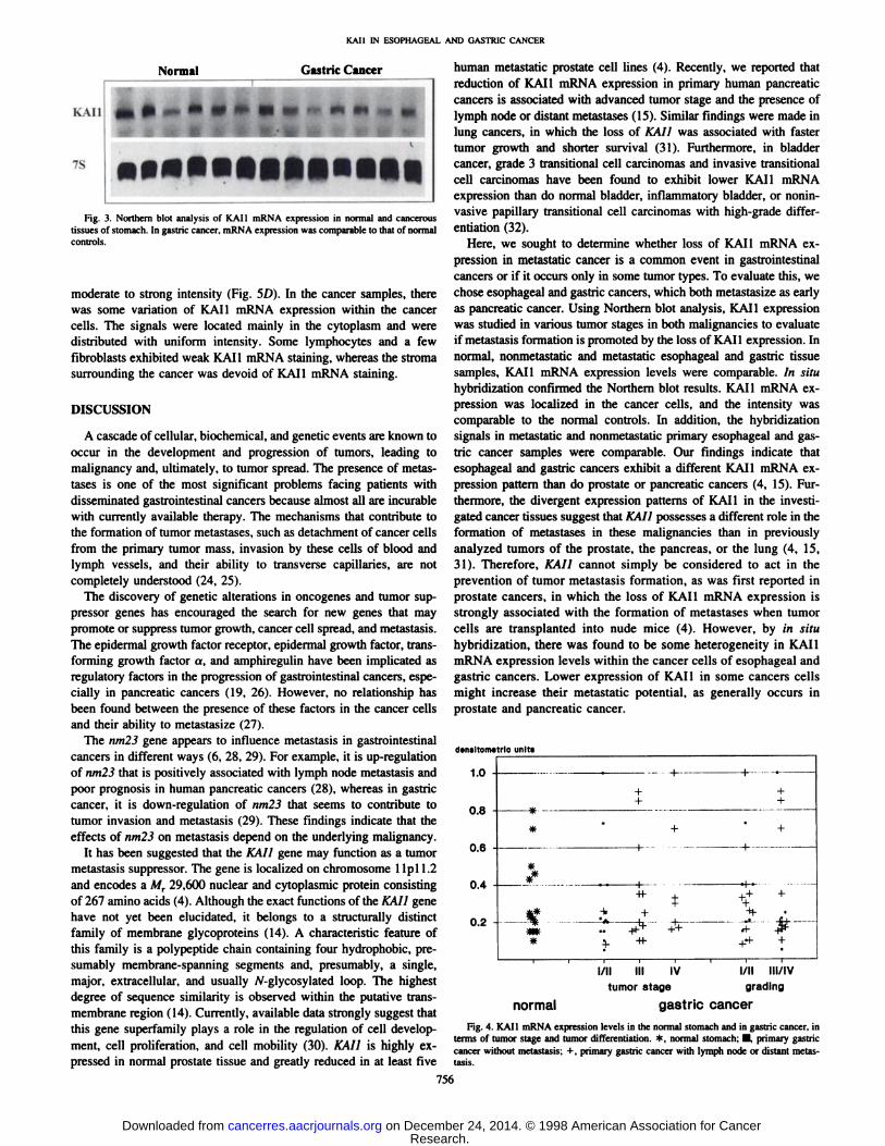
was found in gastric tissue samples (Fig. 3). In normal gastric tissues,KAI1 mRNA expression was detectable in all samples. The expression levels of KAI l mRNA in normal and gastric cancer samples werecomparable. In esophageal tissues, a wide range in KAI1 mRNAexpression levels was seen in the normal as well as in the gastriccancer samples. The KAI1 mRNA levels in primary gastric cancertissues in which lymph node (N, or N2, n = 22) or distant (n = 4)
métastaseswere present at the time of resection were not differentfrom the KAI1 mRNA levels of primary gastric cancer tissues inwhich no lymph node métastases(n = 13) were found at the time of
tumor resection (Table 2 and Fig. 4). Furthermore, we found noinfluence of tumor differentiation on KAI l mRNA levels in gastriccarcinomas (Table 2 and Fig. 4).
KAI1 mRNA Expression Analysis by in Silu Hybridization
To localize the exact site and cellular distribution of KAI1 mRNAexpression in the normal and cancerous tissue samples, in situ hybridization was performed. For in situ hybridization the same tissuesamples were studied as for Northern blot analysis. The results obtained by in situ hybridization in regard to the expression levels werecomparable to the Northern blot data.
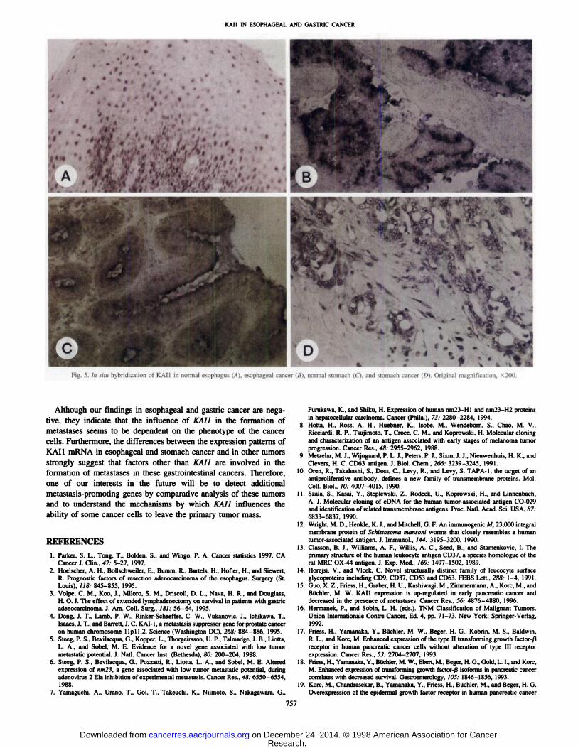
Esophagus. In the normal esophagus, KAI1 mRNA staining waspresent in the cytoplasm of most squamous epithelial cells (Fig. 5A).Lymphocytes in the submucosal areas of the normal esophagus exhibited moderate expression of KAI l mRNA. There was no differencein KAI1 mRNA between normal esophageal tissue that was adjacentto the tumor tissue and normal esophageal tissue obtained from organdonors. In the esophageal cancer samples, KAI1 mRNA stainingintensity in the cancer cells was comparable to that of normal esophageal epithelial cells (Fig. 5fi). In the esophageal cancer samples,there was some variation in the intensity of KAI1 mRNA stainingwithin the cancer cells of the same tumor. In addition, fibroblasts ofthe connective tissue around esophageal cancer cells showed weakKAI1 mRNA expression.
Stomach. In the cytoplasm of nonmucosal epithelial cells of thenormal stomach, KAII in situ hybridization signals were detectable atmoderate intensity (Fig. 5C). Submucosal and muscular cells in thenormal stomach were devoid of KAI l mRNA staining. No differencewas found between normal stomach tissue that was adjacent to thetumor tissue and normal stomach tissue obtained from organ donors.In gastric cancers, KAI1 mRNA in situ hybridization signals were of
densitometric units
1.2 -
0.2 -
normal
l/ll III IV l/ll III/IV
tumor stage grading
esophageal cancer
Fig. 2. KAI I mRNA expression levels in the normal esophagus and in esophagealcancer, in terms of tumor stage and tumor differentiation. *, normal esophagus; •,primary esophageal cancer without metastasis; +, primary esophageal cancer with lymphnode or distant metastasis.
Table 2 KAI I mRNA expression in nonna! ami primary tumor samples of the esophagus and stomach, dependent on the presence (M+ ) or absence (M-) of Ivmph node ordistant métastases"
EsophagusNo.ofcasesKAIl expression(mean)'PStomachNo.
ofcasesKAIl expression(mean)'FNormal
CancerCancerStage
DifferentiationM-
M+ Grades I-II Grades1II-IV15
12 29 24170.550.53 0.59 0.580.531
II 1 11NS"*'
NS714
13 22 18170.330.31 0.35 0.330.341
II 1 11NS''-''-'
NS7
" M —,primary tumor without lymph node or distant métastases;M + . primary tumor with lymph node or distant métastases;NS, not significant.* In relative densitometric units.' Normal vs. M —.''Normal vs. M + .'M- vs.M+.'Grades l-II vs. grades I1I-IV.
755
Research. on December 24, 2014. © 1998 American Association for Cancercancerres.aacrjournals.org Downloaded from
KAM IN ESOPHAOEAL AND GASTRIC CANCER
Normal Gastric Cancer
kAll
7S ••••••••••••lFig. 3. Northern blol analysis of KAI l mRNA expression in normal and cancerous
tissues of stomach. In gastric cancer, mRNA expression was comparable to that of normalcontrols.
moderate to strong intensity (Fig. 5D). In the cancer samples, therewas some variation of KAll mRNA expression within the cancercells. The signals were located mainly in the cytoplasm and weredistributed with uniform intensity. Some lymphocytes and a fewfibroblasts exhibited weak KAll mRNA staining, whereas the stremasurrounding the cancer was devoid of KAll mRNA staining.
DISCUSSION
A cascade of cellular, biochemical, and genetic events are known tooccur in the development and progression of tumors, leading tomalignancy and. ultimately, to tumor spread. The presence of métastases is one of the most significant problems facing patients withdisseminated gastrointestinal cancers because almost all are incurablewith currently available therapy. The mechanisms that contribute tothe formation of tumor métastases,such as detachment of cancer cellsfrom the primary tumor mass, invasion by these cells of blood andlymph vessels, and their ability to transverse capillaries, are notcompletely understood (24, 25).
The discovery of genetic alterations in oncogenes and tumor suppressor genes has encouraged the search for new genes that maypromote or suppress tumor growth, cancer cell spread, and metastasis.The epidermal growth factor receptor, epidermal growth factor, transforming growth factor a. and amphiregulin have been implicated asregulatory factors in the progression of gastrointestinal cancers, especially in pancreatic cancers (19, 26). However, no relationship hasbeen found between the presence of these factors in the cancer cellsand their ability to metastasize (27).
The nm23 gene appears to influence metastasis in gastrointestinalcancers in different ways (6, 28, 29). For example, it is up-regulation
of nm23 that is positively associated with lymph node metastasis andpoor prognosis in human pancreatic cancers (28), whereas in gastriccancer, it is down-regulation of nm23 that seems to contribute to
tumor invasion and metastasis (29). These findings indicate that theeffects of nm23 on metastasis depend on the underlying malignancy.
It has been suggested that the KAU gene may function as a tumormetastasis suppressor. The gene is localized on chromosome 1Ipl 1.2and encodes a Mr 29.600 nuclear and cytoplasmic protein consistingof 267 amino acids (4). Although the exact functions of the KAll genehave not yet been elucidated, it belongs to a structurally distinctfamily of membrane glycoproteins (14). A characteristic feature ofthis family is a polypeptide chain containing four hydrophobic, presumably membrane-spanning segments and, presumably, a single,major, extracellular, and usually /V-glycosylated loop. The highestdegree of sequence similarity is observed within the putative trans-
membrane region (14). Currently, available data strongly suggest thatthis gene superfamily plays a role in the regulation of cell development, cell proliferation, and cell mobility (30). KAll is highly expressed in normal prostate tissue and greatly reduced in at least five
human metastatic prostate cell lines (4). Recently, we reported thatreduction of KAll mRNA expression in primary human pancreaticcancers is associated with advanced tumor stage and the presence oflymph node or distant métastases(15). Similar findings were made inlung cancers, in which the loss of KAI1 was associated with fastertumor growth and shorter survival (31). Furthermore, in bladdercancer, grade 3 transitional cell carcinomas and invasive transitionalcell carcinomas have been found to exhibit lower KAll mRNAexpression than do normal bladder, inflammatory bladder, or nonin-vasive papillary transitional cell carcinomas with high-grade differ
entiation (32).Here, we sought to determine whether loss of KAll mRNA ex
pression in metastatic cancer is a common event in gastrointestinalcancers or if it occurs only in some tumor types. To evaluate this, wechose esophageal and gastric cancers, which both metastasize as earlyas pancreatic cancer. Using Northern blot analysis, KAI l expressionwas studied in various tumor stages in both malignancies to evaluateif metastasis formation is promoted by the loss of KAll expression. Innormal, nonmetastatic and metastatic esophageal and gastric tissuesamples, KAll mRNA expression levels were comparable. In situhybridization confirmed the Northern blot results. KAll mRNA expression was localized in the cancer cells, and the intensity wascomparable to the normal controls. In addition, the hybridizationsignals in metastatic and nonmetastatic primary esophageal and gastric cancer samples were comparable. Our findings indicate thatesophageal and gastric cancers exhibit a different KAll mRNA expression pattern than do prostate or pancreatic cancers (4, 15). Furthermore, the divergent expression patterns of KAll in the investigated cancer tissues suggest that KAIl possesses a different role in theformation of métastasesin these malignancies than in previouslyanalyzed tumors of the prostate, the pancreas, or the lung (4, 15,31). Therefore, KAll cannot simply be considered to act in theprevention of tumor metastasis formation, as was first reported inprostate cancers, in which the loss of KAll mRNA expression isstrongly associated with the formation of métastaseswhen tumorcells are transplanted into nude mice (4). However, by in situhybridization, there was found to be some heterogeneity in KAllmRNA expression levels within the cancer cells of esophageal andgastric cancers. Lower expression of KAll in some cancers cellsmight increase their metastatic potential, as generally occurs inprostate and pancreatic cancer.
¿enaltómetrieunits#
++*
ttff
-S- -i-t-** A ++ * W
normal
l/ll III IV l/ll III/IVtumor stage grading
gastric cancer
Fig. 4. KAll mRNA expression levels in the normal stomach and in gastric cancer, interms of tumor stage and tumor differentiation. *. normal stomach: •.primary gastriccancer without metastasis; +. primary gastric cancer with lymph node or distant metastasis.
756
Research. on December 24, 2014. © 1998 American Association for Cancercancerres.aacrjournals.org Downloaded from
KAM IN ESOPHAGEAL AND GASTRIC CANCER
•*;N
%V^ ^ --'\!'\;^ --;^¥jr».i'v» - '*J
•lFig. 5. ¡nsitu hybridization of KAU ¡nnormal esophagus (A), esophageal cancer (ß).normal stomach (C), and stomach cancer I/)). Original magnification. - Joil.
Although our findings in esophageal and gastric cancer are negative, they indicate that the influence of KAIl in the formation ofmétastasesseems to be dependent on the phenotype of the cancercells. Furthermore, the differences between the expression patterns ofKAI1 mRNA in esophageal and stomach cancer and in other tumorsstrongly suggest that factors other than KAI1 are involved in theformation of métastasesin these gastrointestinal cancers. Therefore,one of our interests in the future will be to detect additionalmetastasis-promoting genes by comparative analysis of these tumors
and to understand the mechanisms by which KAI1 influences theability of some cancer cells to leave the primary tumor mass.
REFERENCES
1. Parker, S. L., Tong, T., Bolden, S., and Wingo. P. A. Cancer statistics 1997. CACancer ¡.Clin., 47: 5-27, 1997.
2. Hoelscher, A. H., Bollschweiler, E., Bumm. R., Barteis. H., Hofler, H., and Siewert,R. Prognostic factors of resection adenocarcinoma of the esophagus. Surgery (St.Louis), 118: 845-855, 1995.
3. Volpe, C. M., Koo, ¡..Miloro, S. M., Driscoll, D. L., Nava, H. R., and Douglass,H. O. J. The effect of extended lymphadenectomy on survival in patients with gastricadenocarcinoma. 1. Am. Coll. Surg., 181: 56-64, 1995.
4. Dong, J. T., Lamb, P. W., Rinker-Schaeffer, C. W., Vukanovic, J., Ichikawa, T.,Isaacs, J. T., and Barrett. J. C. KAM, a metastasis suppressor gene for prostate canceron human chromosome llpll.2. Science (Washington DC), 268: 884-886, 1995.
5. Steeg. P. S., Bevilacqua, G.. Kopper, L., Thorgeirsson, U. P.. Talmadge, J. B., Liotta,L. A., and Sobel, M. E. Evidence for a novel gene associated with low tumormetastatic potential. J. Nati. Cancer Inst. (Bethesda), 80: 200-204, 1988.
6. Steeg, P. S.. Bevilacqua, G., Pozzatti, R., Liotta. L. A., and Sobel. M. E. Alteredexpression of nm23, a gene associated with low tumor metastatic potential, duringadenovirus 2 Eia inhibition of experimental metastasis. Cancer Res., 48: 6550-6554,
1988.7. Yamaguchi, A., Urano, T., Goi. T., Takeuchi, K., Niimoto, S., Nakagawara, G.,
Furukawa, K.. and Shiku, H. Expression of human nm23-HI and nm23-H2 proteinsin hepatocellular carcinoma. Cancer (Phila.l, 73: 2280-2284, 1994.
8. Motta. H.. Ross, A. H., Huebner, K.. Isobe, M.. Wendebom. S.. Chao. M. V.,Ricciardi. R. P., Tsujimoto, T.. Croce. C. M., and Koprowski. H. Molecular cloningand characterization of an antigen associated with early stages of melanoma tumorprogression. Cancer Res., 48: 2955-2962, 1988.
9. Metzelar, M. J.. Wijngaard, P. L. J., Peters, P. J., Sixm, J. J., Nieuwenhuis, H. K., andClevers, H. C. CD63 antigen. J. Biol. Chem., 266: 3239-3245, 1991.
10. Oren. R.. Takahashi, S., Doss. C.. Levy, R., and Levy. S. TAPA-1, the target of anantiproliferative antibody, defines a new family of transmembrane proteins. Mol.Cell. Biol.. 10: 4007-4015, 1990.
11. Szala, S., Kasai, Y., Steplewski. Z., Rodeck, U.. Koprowski, H., and Linnenbach.A. J. Molecular cloning of cDNA for the human tumor-associated antigen CO-029and identification of related transmembrane antigens. Proc. Nati. Acad. Sci. USA. 87:6833-6837. 1990.
12. Wright, M. D.. Henkle, K. J., and Mitchell, G. F. An immunogenic M, 23,000 integralmembrane protein of Schistosoma nianstmi worms that closely resembles a humantumor-associated antigen. J. Immunol., 144: 3195-3200, 1990.
13. Classon. B. J.. Williams, A. F., Willis, A. C., Seed, B., and Stamenkovic. I. Theprimary structure of the human leukocyte antigen CD37. a species homologue of therat MRC OX-44 antigen. J. Exp. Med., 169: 1497-1502. 1989.
14. Horejsi, V., and Vlcek, C. Novel structurally distinct family of leucocyte surfaceglycoproteins including CD9. CD37, CD53 and CD63. FEBS Lett., 288: 1-4. 1991.
15. Quo, X. Z.. Friess, H., Graber, H. U., Kashiwagi, M., Zimmermann, A., Köre,M.. andBüchler.M. W. KAI 1 expression is up-regulated in early pancreatic cancer anddecreased in the presence of métastases.Cancer Res., 56: 4876-4880. 1996.
16. Hermanek, P., and Sobin, L. H. (eds.). TNM Classification of Malignant Tumors.Union Internationale Contre Cancer. Ed. 4, pp. 71-73. New York: Springer-Verlag.
1992.17. Friess, H., Yamanaka. Y.. Büchler,M. W., Beger, H. G., Kobrin. M. S., Baldwin.
R. L., and Köre.M. Enhanced expression of the type H transforming growth factor-ß
receptor in human pancreatic cancer cells without alteration of type HI receptorexpression. Cancer Res.. 53: 2704-2707. 1993.
18. Friess. H.. Yamanaka, Y., BUchler. M. W., Eben, M.. Beger, H. G.. Gold. L. I., and Korc.M. Enhanced expression of transforming growth factor-ßisoforms in pancreatic cancercorrelates with decreased survival. Gastroenterology, IOS: I846-1856. 1993.
19. Korc, M.. Chandrasekar, B., Yamanaka, Y.. Friess, H.. Büchler,M., and Beger. H. G.Overexpression of the epidermal growth factor receptor in human pancreatic cancer
757
Research. on December 24, 2014. © 1998 American Association for Cancercancerres.aacrjournals.org Downloaded from
KAM IN ESOPHAOEAL AND GASTRIC CANCER
is associated with concomitant increase in levels of epidermal growth factor andtransforming growth factor alpha. J. Clin. Invest., 90: 1353-1360. 1992.
20. Friess, H., Camero, D., Graber. H.. Tang. W. H.. Guo. X. Z., Kashiwagi. M..Zimmermann. A.. Gold. L. !.. Köre,M., and Büchler,M. W. Enhanced urokinaseplasminogen activation in chronic pancreatitis suggests a role in its pathogenesis.Gastroenterology. 11.1: 904-913. 1997.
21. Gress, T. M, Mliller-Pillasch. F., Leren. M. M.. Friess, H., Büchler,M., and Adler.
G. Expression and in situ localization of genes coding for extracellular matrix proteinsand extracellular matrix degrading proteases in pancreatic cancer. Int. J. Cancer, 62:407-413, 1995.
22. Deflorin, J., Friess, H.. Schobinger, S.. Briindler, M. A., Schilling. M., Naef, M..Korc. M.. and Büchler.M. W. Overexpression of the epidermal growth factor receptorfamily in gastric cancer suggests a role in tumor pathogenesis. Dig. Surg., 14:252-259, 1997.
23. Cox, K. H., Deleon, D. V., Augerer, L. M.. and Augerer, R. C. Detection of mRNAsin sea urchin embryos by in situ hybridization using asymmetric RNA probes. Dev.Biol., 101: 485-502, 1984.
24. Liotta, L. A., Rao. C. N., and Wewer, U. M. Biochemical interactions of tumor cellswith the basement membrane. Annu. Rev. Biochem.. 5.5: 1037-1057. 1986.
25. Ochalek, T., Nordt. F. J., Tullberg, K., and Burger, M. Correlation between celldeformability and metastatic potential in B16-F1 melanoma cell variants. CancerRes., 48: 5124-5128, 1988.
26. Eben. M.. Yokoyama. M.. Kobrin. M. S.. Friess, H., Lopez. M. E.. Büchler,M. W..Johnson, G. R., and Korc, M. Induction and expression of amphiregulin in humanpancreatic cancer. Cancer Res., 54: 3959-3962. 1994.
27. Moskal, T. L., Huang, S., Ellis. L. M., Pritsche, H. A. J., and Chakrabarty, S. Serumlevels of transforming growth factor alpha in gastrointestinal cancer patients. CancerEpidemiol. Biomark. Prev., 4: 127-131, 1995.
28. Sasaki. Z., Inaji, H., Higashiyama, M.. Imaoka. S.. and Iwanaga, T. Expression ofnucleotide diphosphate kinase/nm2.i gene product in human pancreatic cancer: anassociation with lymph node metastasis and tumor invasion. Clin. Exp. Metastasis,//.- 151-158, 1993.
29. Kodera. Y., Isobe, K. I., Yamauchi, M., Kondoh. K., Kimura, N., Akiyama, S., Itoh,K., Nakashima, I., and Takagi. H. Expression of nm23 H-l RNA levels in humangastric cancer tissues. Cancer (Phila.), 73: 259-265, 1994.
30. Wright. M. D., and Tomlinson. M. G. The ins and outs of the transmembrane 4superfamily. Immunol. Today, /5: 588-595, 1994.
31. Adachi, M.. Taki, T.. leki, Y., Huang, C. L., Higashiyama, M., and Miyaké,M.Correlation of KA11/CDK2 gene expression with good prognosis in patients withnon-small cell lung cancer. Cancer Res., 56: 1751-1755. 1996.
32. Yu, Y., Yang, J-L., Markovic, B., Jackson. P., Yardley, G., Barrett. J., and Russell,P. J. Loss of KAI1 messenger RNA expression in both high-grade and invasivehuman bladder cancers. Clin. Cancer Res.. 3: 1045-1049, 1997.
758
Research. on December 24, 2014. © 1998 American Association for Cancercancerres.aacrjournals.org Downloaded from
1998;58:753-758. Cancer Res Xiao-Zhong Guo, Helmut Friess, Christoph Maurer, et al. and Gastric CancersKAI1 Is Unchanged in Metastatic and Nonmetastatic Esophageal
Updated version
http://cancerres.aacrjournals.org/content/58/4/753
Access the most recent version of this article at:
E-mail alerts related to this article or journal.Sign up to receive free email-alerts
Subscriptions
Reprints and
To order reprints of this article or to subscribe to the journal, contact the AACR Publications
Permissions
To request permission to re-use all or part of this article, contact the AACR Publications
Research. on December 24, 2014. © 1998 American Association for Cancercancerres.aacrjournals.org Downloaded from