ratio between positive lymph nodes and total excised axillary lymph nodes as an independent...
TRANSCRIPT
ORIGINAL ARTICLE – BREAST ONCOLOGY
Ratio Between Positive Lymph Nodes and Total Excised AxillaryLymph Nodes as an Independent Prognostic Factor for OverallSurvival in Patients with Nonmetastatic Lymph Node-PositiveBreast Cancer
Hassan A. Hatoum, MD1, Faek R. Jamali, MD2, Nagi S. El-Saghir, MD1, Khaled M. Musallam, MD1,
Muhieddine Seoud, MD3, Hani Dimassi, PhD4, Jaber Abbas, MD2, Mohamad Khalife, MD2, Fouad I. Boulos, MD5,
Ayman N. Tawil, MD5, Fadi B. Geara, MD6, Ziad Salem, MD1, Achraf A. Shamseddine, BSc1, Karine Al-Feghali,
BSc1, and Ali I. Shamseddine, MD1
1Department of Internal Medicine, Division of Hematology/Oncology, American University of Beirut Medical Center,
Beirut, Lebanon; 2Department of Surgery, Division of General Surgery, American University of Beirut Medical Center,
Beirut, Lebanon; 3Department of Obstetrics and Gynecology, American University of Beirut Medical Center, Beirut,
Lebanon; 4School of Pharmacy, Lebanese American University, Byblos, Lebanon; 5Department of Pathology and
Laboratory Medicine, American University of Beirut Medical Center, Beirut, Lebanon; 6Department of Radiation
Oncology, American University of Beirut Medical Center, Beirut, Lebanon
ABSTRACT
Background. The status of the axillary lymph nodes in
nonmetastatic lymph node-positive breast cancer (BC)
patients remains the single most important determinant of
overall survival (OS). Although the absolute number of
nodes involved with cancer is important for prognosis, the
role of the total number of excised nodes has received less
emphasis. Thus, several studies have focused on the utility
of the axillary lymph node ratio (ALNR) as an independent
prognostic indicator of OS. However, most studies suffered
from shortcomings, such as including patients who
received neoadjuvant therapy or failing to consider the use
of adjuvant therapy and tumor receptor status in their
analysis.
Methods. We conducted a single-center retrospective
review of 669 patients with nonmetastatic lymph node-
positive BC. Data collected included patient demographics;
breast cancer risk factors; tumor size, histopathological,
receptor, and lymph node status; and treatment modalities
used. Patients were subdivided into four groups according
to ALNR value (\.25, .25–.49, .50–.74, .75–1.00). Study
parameters were compared at the univariate and multivar-
iate levels for their effect on OS.
Results. On univariate analysis, both the absolute number
of positive lymph nodes and the ALNR were significant
predictors of OS. On multivariate analysis, only the ALNR
remained an independent predictor of OS, with a 2.5-fold
increased risk of dying at an ALNR of C.25.
Conclusions. Our study demonstrates that ALNR is a
stronger factor in predicting OS than the absolute number
of positive axillary lymph nodes.
Breast cancer (BC) is the most common cancer in
women and the second most common cause of female
cancer death, with an estimated 182,460 new cases diag-
nosed and 40,480 deaths in the United States in 2008.1
Despite the advent of sentinel node biopsy techniques and
the dawning of the molecular era of BC staging, the status
of the axillary lymph nodes (ALN) remains the single most
important determinant of overall survival (OS).2–6
For patients with pathologically proven ALN involve-
ment, the number of positive axillary lymph nodes (pALN)
Presented in part at the American Society of Clinical Oncology
(ASCO) Breast Cancer Symposium 2007 (abstract 70); the 2007
ASCO annual meeting (abstract 21072); and the 6th European Breast
Cancer Conference 2008 (abstract 476).
�
A. I. Shamseddine, MD
e-mail: [email protected]
This article was originally published in Annals of Surgical Oncology
DOI 10.1245/s10434-009-0653-83.
,
DOI 10.1 /s13 -0 -0
Indian J Surg Oncol
193 11 0007 62-x
Indian Association of Surgical Oncology 2011
(October December 201 ) 1(4):305 –
Published Online: 202 March 119
0– 312
correlates with the incidence of distant metastasis and OS,
and more than three pALN is associated with a 13% to 24%
locoregional recurrence rate.7,8 The relationship between
number of pALN and survival seems to be linear, with each
additional pALN detected leading to a worsening of the
prognosis.9,10 The American Joint Committee on Cancer
staging system was recently revised, grouping patients by
the absolute number of pALN. This classification improved
stratification in OS, but the confounding effect that the
number of excised nodes may have on the yield of positive
nodes and its effect on BC-specific survival prognostic
accuracy and management decisions are problems that
remain unresolved.6,11 Thus, the ratio between the number
of pALN and the total number of excised nodes, or the
axillary lymph node ratio (ALNR), may be a more com-
prehensive approach to estimate prognosis because it takes
into account the number of excised nodes and may
accordingly adjust for differences in nodal staging.
Since 1999, several reports described the significance of
ALNR as an independent prognostic factor for OS in
nonmetastatic lymph node-positive BC.12–18 Some studies
included a heterogeneous group of patients recruited from
different centers, which implies that patients were treated
by different surgeons and different medical oncology
teams.14,18 Moreover, in many of these series, additional
factors affecting OS, such as tumor receptor status
(including HER2) and use of neoadjuvant or adjuvant
treatment regimens, were not considered.12–18 We thus
report a single institutional experience with the prognostic
significance of ALNR in nonmetastatic lymph node-posi-
tive BC patients treated by the same medical and surgical
oncology team while attempting to address many of the
shortcomings evident in some of the previous trials.
METHODS
We conducted a retrospective review of 1450 BC
patients treated at the American University of Beirut
Medical Center between the years 1983 and 2004. Data
were retrieved from the medical records, tumor registry
database, and the outpatient clinic charts of each patient.
Of the 1450 patients, 1092 patients were diagnosed with
stage I, II, or III (nonmetastatic) disease and were con-
sidered for further selection. Inclusion criteria included
histologically proven invasive breast carcinoma with evi-
dence of lymph node involvement at pathological staging;
exclusion criteria included evidence of neoadjuvant che-
motherapy. The tumor, node, and metastasis system of the
American Joint Committee on Cancer (AJCC), 6th edition,
was used for staging.19
Data collected included patients’ demographics, medical
history, history of benign breast disease, family history of
breast malignancies, age at menarche, childbearing, use of
oral contraceptive pills, menopause status, and use of
hormone replacement therapy. Other retrieved data inclu-
ded age at diagnosis, type of surgery (partial vs. total
mastectomy), ALN involvement, use of adjuvant chemo-
therapy, hormone therapy, and postoperative radiotherapy.
Indications for postmastectomy radiotherapy included all
T3 and T4 tumors, positive microscopic surgical margins,
and/or any T stage with three or more ALN involved.
Patients who underwent partial mastectomy were treated to
the supraclavicular region including the axillary apex if
they had C3 ALN involved.
Evaluated tumor characteristics included tumor size,
histological type and grade, hormone receptor status, and
HER2 overexpression studies. HER2 positivity was defined
either immunohistochemically, where tumors show strong
and complete circumferential membranous staining in at
least 30% of cells, or by fluorescent in situ hybridization,
where the currently used test does not include centromeric
staining for chromosome 17, and the cutoff for HER2
positivity is an average of 6-fold amplification of the HER2
gene in the assessed (at least 20) tumor cells. In the 669
patients, ALNR (number of pALN divided by the total
number of excised ALN) was calculated. Patients were
subdivided into four groups according to ALNR value
(\.25, .25–.49, .50–.74, .75–1.00); these mathematical
quartiles were used to allow a fair chance for each quartile
to represent itself and to try to delineate a practical cutoff
for the clinical setting. The primary endpoint was OS,
which was calculated as the length of time from diagnosis
until death, irrespective of the cause. The institutional
review board at our center approved this study.
Statistical Analysis
Abstracted data were coded and entered into SPSS
version 16 statistical software (SPSS, Chicago, IL).
Patients’ general characteristics, tumor characteristics, and
ALN description were summarized by frequencies and
percentages. Five- and 10-year survival rates were carried
out at the univariate level by Kaplan-Meier analysis, and P
values from the log rank test were reported. Furthermore,
variables that showed significance at the univariate level
(tumor size, hormone receptor status, ALNR, number of
pALN) were tested at the multivariate level by the Cox
proportionate hazard model, with the exception of tumor
stage, which was totally defined by tumor size and number
of pALN, both of which were entered into the model. In
this model, ALNR was used as a categorical variable to
help compare different categories of ALNR; all the possi-
ble numbers of pALN were used as a continuous variable,
allowing for maximal differentiation. Coefficients pro-
duced by the models were exponentiated, producing hazard
ratios, and their respective standard errors were used to
306 Indian J Surg Oncol (October December 201 ) 1(4):305 – 0– 312
calculate the 95% confidence intervals. All analyses were
carried out at the .05 level.
RESULTS
Patient Demographics and Disease Characteristics
Over the 21-year period, 1450 patients were diagnosed
with BC. Among these, 669 patients had nonmetastatic
node positive BC and were included in the analysis. The
median age at diagnosis of the sample was 49 years (range,
24–86 years). Data on patient demographics, medical his-
tory, and BC risk factors are summarized in Table 1.
Among 660 patients with available data on type of surgery,
559 (84.7%) had total mastectomy, while 101 (15.3%) had
breast-conserving surgery. Adjuvant chemotherapy, hor-
mone therapy, and radiotherapy were used in 81.4%,
29.6%, and 76% of patients, respectively. Chemotherapy
regimens were mostly anthracycline based (56%), while
hormone therapy mainly consisted of tamoxifen (95%). All
patients were treated before 2005 so none of the patients
received adjuvant trastuzumab.
Determinants of OS
The median follow-up for the entire cohort was
3.4 years (range, .08–17.42 years), and the median follow-
up for all patients alive was 3.25 years (range, .08–
17.42 years). In univariate analysis, tumor size (B2 cm,
2.1–5, [ 5 cm; P = .037), estrogen-progesterone receptor
status (P \ .0001), tumor stage (P \ .0001), number of
pALN (1–3, 4–9, C10; P \ .0001), and ALNR (\.25, .25–
.49, .50–.74, .75–1.00; P \ .0001) were the only significant
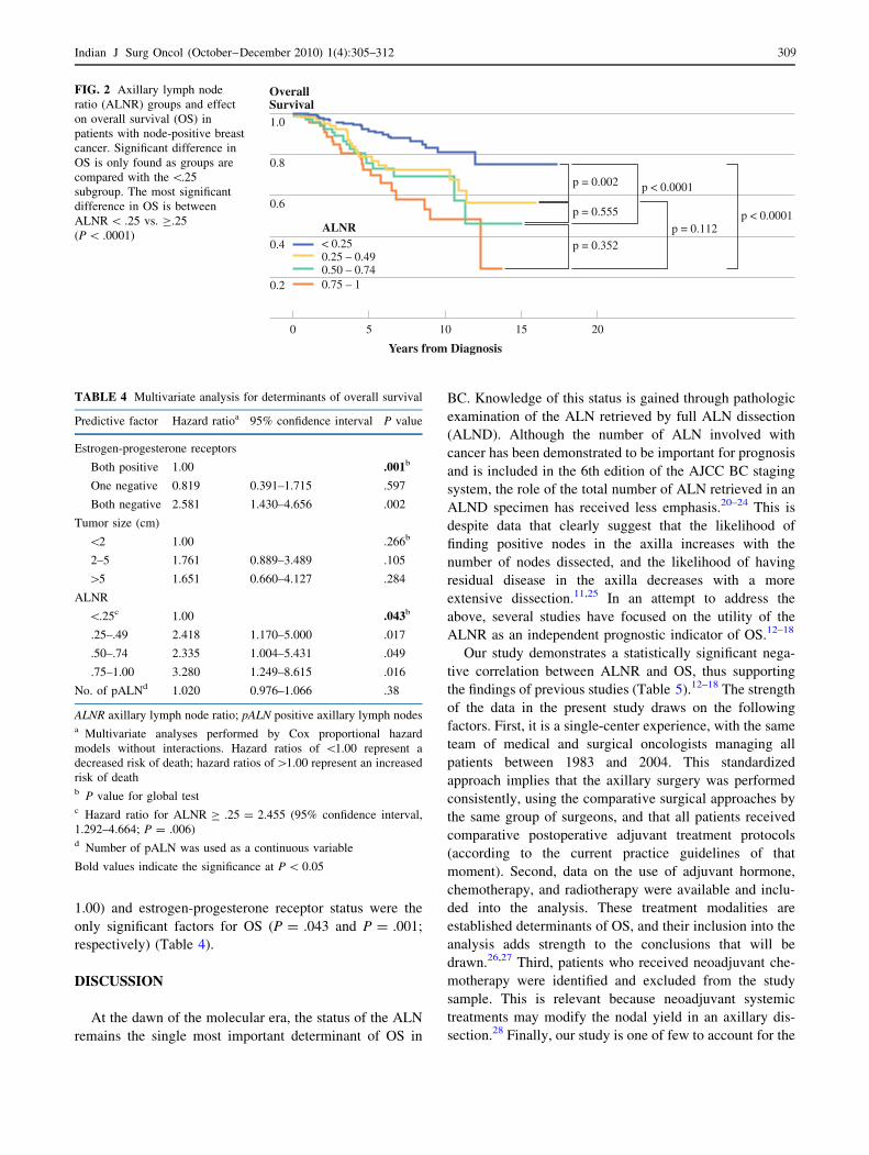
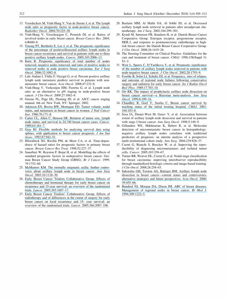
predictors of OS (Tables 2 and 3). Figures 1 and 2 display
categorical survival for number of pALN and ALNR,
respectively.
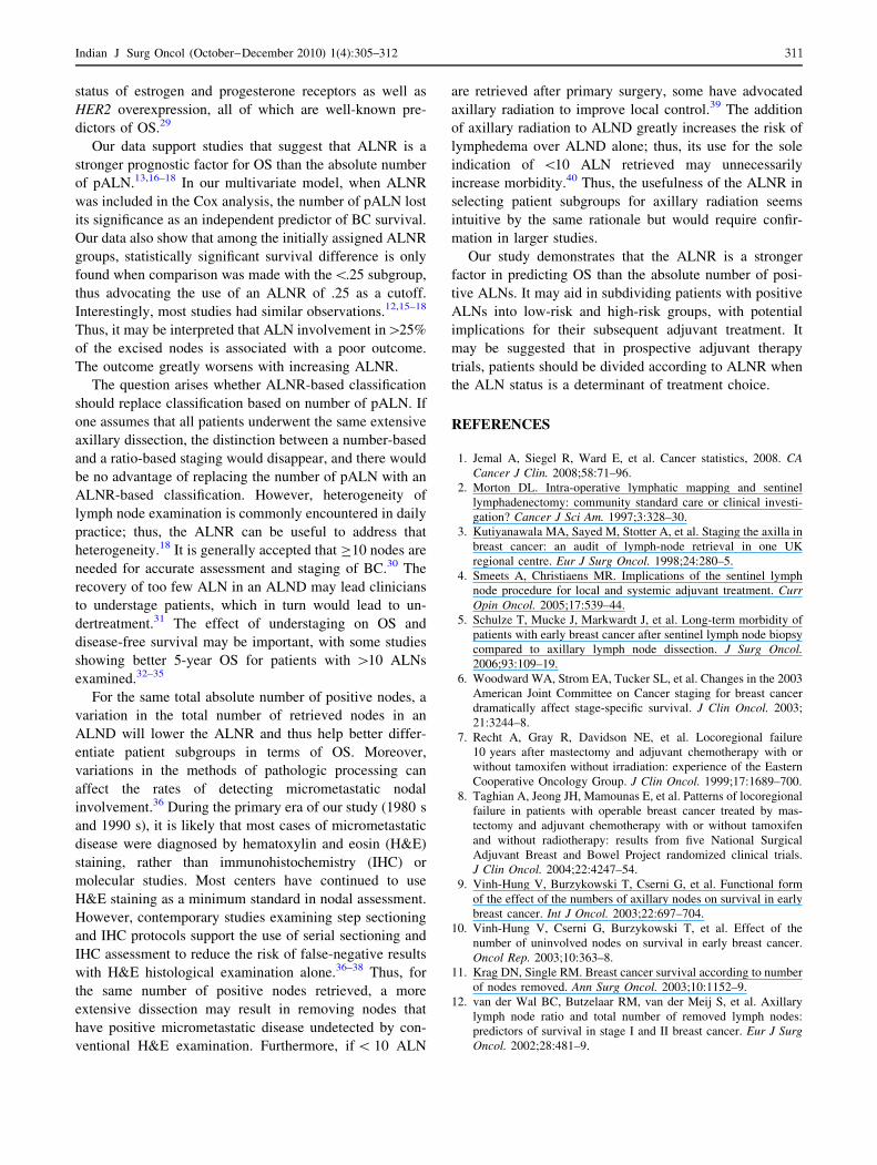
A multivariate model was obtained in which ALNR
subgroups and all prognostic factors with univariate
P \ .05 (except tumor stage) were entered into the model.
All factors were treated as simple categorical variables
except number of pALN, which was used as a continuous
variable. ALNR categories (\.25, .25–.49, .50–.74, .75–
TABLE 1 Patient demographics, medical history, and breast cancer
risk-factor history for 699 patients
Parameter n (%)
Age at diagnosis (y)
\51 365 (54.2)
51–70 266 (39.8)
[70 38 (6)
Hypertension
Yes 93 (14.2)
No 562 (85.8)
Missing 14
Diabetes
Yes 49 (7.5)
No 606 (92.5)
Missing 14
Cardiac disease
Yes 31 (4.7)
No 624 (95.3)
Missing 14
Family history of breast cancer
Present 123 (18.7)
Absent 538 (81.3)
Previous malignancy
Yes 29 (4.4)
No 628 (95.6)
Missing 12
Oral contraceptive pills
Yes 92 (22.1)
No 325 (77.9)
Missing 252
Hormone replacement therapy
Yes 23 (13.9)
No 146 (86.1)
Missing 500
Benign breast disease
Yes 25 (3.8)
No 627 (96.2)
Missing 17
Smoking
Yes 187 (36.1)
No 331 (63.9)
Missing 151
Pregnancy
No previous pregnancy 105 (17)
Previous pregnancy 513 (83)
Missing 51
Menopause
Post 356 (54.4)
Pre 299 (45.6)
Missing 14
TABLE 1 continued
Parameter n (%)
Age at menarche (y)
B11 48 (9.4)
12–13 189 (37.1)
C14 272 (53.7)
Missing 160
307Indian J Surg Oncol (October December 201 ) 1(4):305 – 0– 312
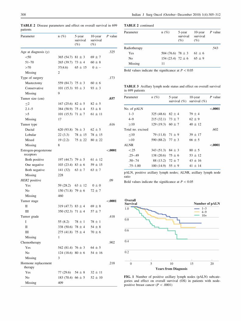
TABLE 2 Disease parameters and effect on overall survival in 699
patients
Parameter n (%) 5-year
survival
(%)
10-year
survival
(%)
P value
Age at diagnosis (y) .325
\50 365 (54.7) 81 ± 3 69 ± 7
51–70 265 (39.7) 73 ± 4 60 ± 8
[70 37(4.6) 65 ± 15 0 ± –
Missing 2
Type of surgery .173
Mastectomy 559 (84.7) 75 ± 3 60 ± 6
Conservative 101 (15.3) 93 ± 3 93 ± 3
Missing 9
Tumor size (cm) .037
B2 167 (25.6) 82 ± 5 82 ± 5
2.1–5 384 (58.9) 75 ± 4 53 ± 8
[5 101 (15.5) 71 ± 7 61 ± 11
Missing 17
Tumor type .616
Ductal 620 (93.8) 76 ± 3 62 ± 5
Lobular 22 (3.3) 78 ± 15 78 ± 15
Mixed 19 (2.2) 75 ± 22 80 ± 22
Missing 8
Estrogen-progesterone
receptors
\.0001
Both positive 197 (44.7) 79 ± 5 61 ± 12
One negative 103 (23.4) 83 ± 6 59 ± 15
Both negative 141 (32) 63 ± 7 63 ± 7
Missing 228
HER2 positive .09
Yes 59 (28.2) 63 ± 12 0 ± 0
No 150 (71.8) 79 ± 6 72 ± 7
Missing 460
Tumor stage \.0001
II 319 (47.7) 83 ± 4 69 ± 8
III 350 (52.3) 71 ± 4 57 ± 7
Tumor grade .418
I 55 (8.2) 78 ± 1 78 ± 1
II 338 (50.6) 78 ± 4 54 ± 8
III 275 (41.8) 75 ± 4 70 ± 6
Missing 1
Chemotherapy .902
Yes 542 (81.4) 76 ± 3 64 ± 5
No 124 (18.6) 80 ± 6 54 ± 16
Missing 3
Hormone replacement
therapy
.218
Yes 77 (29.6) 54 ± 8 32 ± 11
No 183 (70.4) 66 ± 5 52 ± 10
Missing 409
TABLE 2 continued
Parameter n (%) 5-year
survival
(%)
10-year
survival
(%)
P value
Radiotherapy .543
Yes 504 (76.6) 78 ± 3 61 ± 6
No 154 (23.4) 72 ± 6 65 ± 9
Missing 11
Bold values indicate the significance at P \ 0.05
TABLE 3 Axillary lymph node status and effect on overall survival
in 699 patients
Parameter n (%) 5-year
survival (%)
10-year
survival (%)
P value
No. of pALN \.0001
1–3 325 (48.6) 82 ± 4 79 ± 4
4–9 215 (32.1) 73 ± 7 62 ± 9
C10 129 (19.3) 60 ± 7 40 ± 12
Total no. excised .602
B10 79 (11.8) 71 ± 9 39 ± 17
[10 590 (88.2) 77 ± 3 66 ± 5
ALNR \.0001
\.25 343 (51.3) 84 ± 3 80 ± 5
.25–.49 138 (20.6) 75 ± 6 53 ± 12
.50–.74 88 (13.2) 72 ± 7 43 ± 16
.75–1.00 100 (14.9) 55 ± 9 41 ± 14
pALN, positive axillary lymph nodes; ALNR, axillary lymph node
ratio
Bold values indicate the significance at P \ 0.05
1.0
0.8
0.6
0.4
0.2
0 20
Years from Diagnosis
155 10
OverallSurvival
1–34–910+
Number of pALN
FIG. 1 Number of positive axillary lymph nodes (pALN) subcate-
gories and effect on overall survival (OS) in patients with node-
positive breast cancer (P \ .0001)
308 Indian J Surg Oncol (October December 201 ) 1(4):305 – 0– 312
1.00) and estrogen-progesterone receptor status were the
only significant factors for OS (P = .043 and P = .001;
respectively) (Table 4).
DISCUSSION
At the dawn of the molecular era, the status of the ALN
remains the single most important determinant of OS in
BC. Knowledge of this status is gained through pathologic
examination of the ALN retrieved by full ALN dissection
(ALND). Although the number of ALN involved with
cancer has been demonstrated to be important for prognosis
and is included in the 6th edition of the AJCC BC staging
system, the role of the total number of ALN retrieved in an
ALND specimen has received less emphasis.20–24 This is
despite data that clearly suggest that the likelihood of
finding positive nodes in the axilla increases with the
number of nodes dissected, and the likelihood of having
residual disease in the axilla decreases with a more
extensive dissection.11,25 In an attempt to address the
above, several studies have focused on the utility of the
ALNR as an independent prognostic indicator of OS.12–18
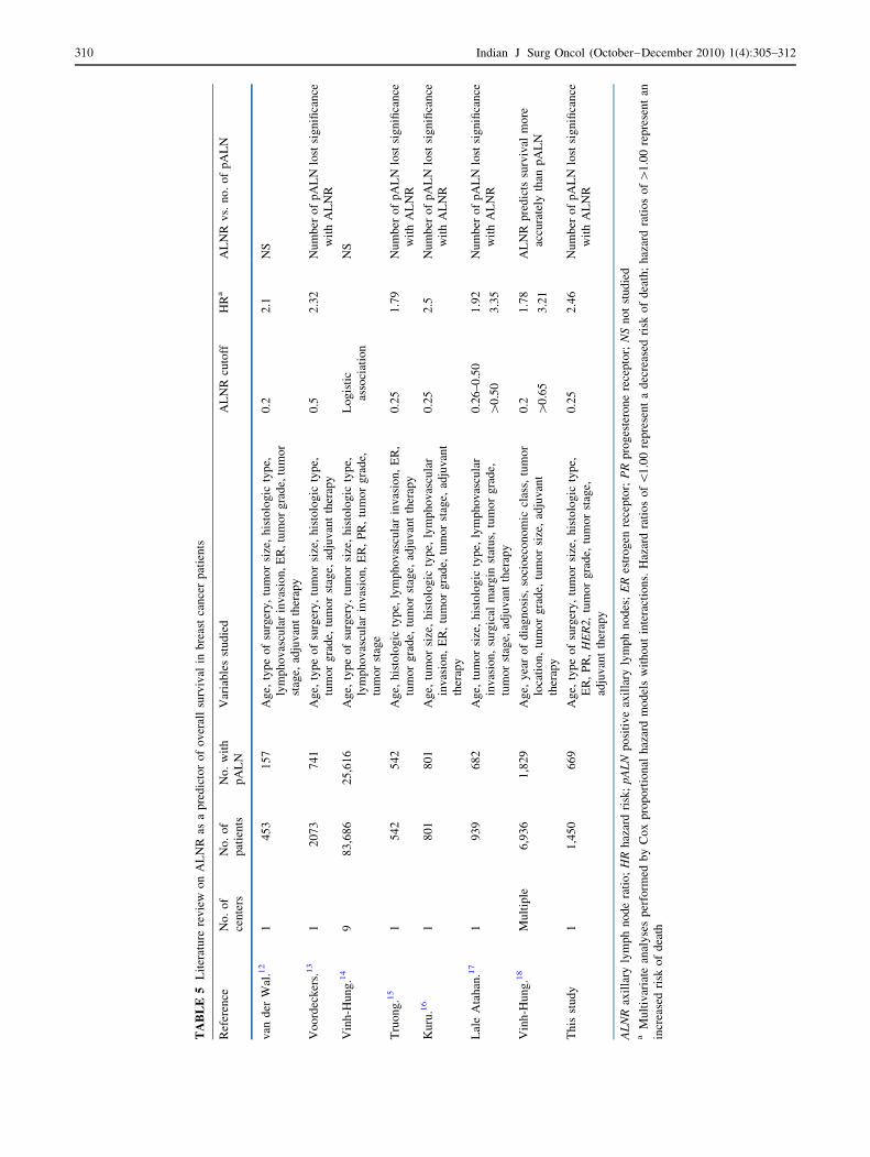
Our study demonstrates a statistically significant nega-
tive correlation between ALNR and OS, thus supporting
the findings of previous studies (Table 5).12–18 The strength
of the data in the present study draws on the following
factors. First, it is a single-center experience, with the same
team of medical and surgical oncologists managing all
patients between 1983 and 2004. This standardized
approach implies that the axillary surgery was performed
consistently, using the comparative surgical approaches by
the same group of surgeons, and that all patients received
comparative postoperative adjuvant treatment protocols
(according to the current practice guidelines of that
moment). Second, data on the use of adjuvant hormone,
chemotherapy, and radiotherapy were available and inclu-
ded into the analysis. These treatment modalities are
established determinants of OS, and their inclusion into the
analysis adds strength to the conclusions that will be
drawn.26,27 Third, patients who received neoadjuvant che-
motherapy were identified and excluded from the study
sample. This is relevant because neoadjuvant systemic
treatments may modify the nodal yield in an axillary dis-
section.28 Finally, our study is one of few to account for the
1.0
0.8
0.6
0.4
0.2
0 20
Years from Diagnosis
155 10
OverallSurvival
< 0.250.25 – 0.490.50 – 0.740.75 – 1
p < 0.0001p = 0.112ALNR
p < 0.0001
p = 0.352
p = 0.555
p = 0.002
FIG. 2 Axillary lymph node
ratio (ALNR) groups and effect
on overall survival (OS) in
patients with node-positive breast
cancer. Significant difference in
OS is only found as groups are
compared with the \.25
subgroup. The most significant
difference in OS is between
ALNR \ .25 vs. C.25
(P \ .0001)
TABLE 4 Multivariate analysis for determinants of overall survival
Predictive factor Hazard ratioa 95% confidence interval P value
Estrogen-progesterone receptors
Both positive 1.00 .001b
One negative 0.819 0.391–1.715 .597
Both negative 2.581 1.430–4.656 .002
Tumor size (cm)
\2 1.00 .266b
2–5 1.761 0.889–3.489 .105
[5 1.651 0.660–4.127 .284
ALNR
\.25c 1.00 .043b
.25–.49 2.418 1.170–5.000 .017
.50–.74 2.335 1.004–5.431 .049
.75–1.00 3.280 1.249–8.615 .016
No. of pALNd 1.020 0.976–1.066 .38
ALNR axillary lymph node ratio; pALN positive axillary lymph nodesa Multivariate analyses performed by Cox proportional hazard
models without interactions. Hazard ratios of \1.00 represent a
decreased risk of death; hazard ratios of[1.00 represent an increased
risk of deathb P value for global testc Hazard ratio for ALNR C .25 = 2.455 (95% confidence interval,
1.292–4.664; P = .006)d Number of pALN was used as a continuous variable
Bold values indicate the significance at P \ 0.05
309 Indian J Surg Oncol (October December 201 ) 1(4):305 – 0– 312
TA
BL
E5
Lit
erat
ure
rev
iew
on
AL
NR
asa
pre
dic
tor
of
ov
eral
lsu
rviv
alin
bre
ast
can
cer
pat
ien
ts
Ref
eren
ceN
o.
of
cen
ters
No
.o
f
pat
ien
ts
No
.w
ith
pA
LN
Var
iab
les
stu
die
dA
LN
Rcu
toff
HR
aA
LN
Rv
s.n
o.
of
pA
LN
van
der
Wal
.12
14
53
15
7A
ge,
typ
eo
fsu
rger
y,
tum
or
size
,h
isto
log
icty
pe,
lym
ph
ov
ascu
lar
inv
asio
n,
ER
,tu
mo
rg
rad
e,tu
mo
r
stag
e,ad
juv
ant
ther
apy
0.2
2.1
NS
Vo
ord
eck
ers.
13
12
07
37
41
Ag
e,ty
pe
of
surg
ery
,tu
mo
rsi
ze,
his
tolo
gic
typ
e,
tum
or
gra
de,
tum
or
stag
e,ad
juv
ant
ther
apy
0.5
2.3
2N
um
ber
of
pA
LN
lost
sig
nifi
can
ce
wit
hA
LN
R
Vin
h-H
un
g.1
49
83
,68
62
5,6
16
Ag
e,ty
pe
of
surg
ery
,tu
mo
rsi
ze,
his
tolo
gic
typ
e,
lym
ph
ov
ascu
lar
inv
asio
n,
ER
,P
R,
tum
or
gra
de,
tum
or
stag
e
Lo
gis
tic
asso
ciat
ion
NS
Tru
on
g.1
51
54
25
42
Ag
e,h
isto
log
icty
pe,
lym
ph
ov
ascu
lar
inv
asio
n,
ER
,
tum
or
gra
de,
tum
or
stag
e,ad
juv
ant
ther
apy
0.2
51
.79
Nu
mb
ero
fp
AL
Nlo
stsi
gn
ifica
nce
wit
hA
LN
R
Ku
ru.1
61
80
18
01
Ag
e,tu
mo
rsi
ze,
his
tolo
gic
typ
e,ly
mp
ho
vas
cula
r
inv
asio
n,
ER
,tu
mo
rg
rad
e,tu
mo
rst
age,
adju
van
t
ther
apy
0.2
52
.5N
um
ber
of
pA
LN
lost
sig
nifi
can
ce
wit
hA
LN
R
Lal
eA
tah
an.1
71
93
96
82
Ag
e,tu
mo
rsi
ze,
his
tolo
gic
typ
e,ly
mp
ho
vas
cula
r
inv
asio
n,
surg
ical
mar
gin
stat
us,
tum
or
gra
de,
tum
or
stag
e,ad
juv
ant
ther
apy
0.2
6–
0.5
01
.92
Nu
mb
ero
fp
AL
Nlo
stsi
gn
ifica
nce
wit
hA
LN
R[
0.5
03
.35
Vin
h-H
un
g.1
8M
ult
iple
6,9
36
1,8
29
Ag
e,y
ear
of
dia
gn
osi
s,so
cio
eco
no
mic
clas
s,tu
mo
r
loca
tio
n,
tum
or
gra
de,
tum
or
size
,ad
juv
ant
ther
apy
0.2
1.7
8A
LN
Rp
red
icts
surv
ival
mo
re
accu
rate
lyth
anp
AL
N[
0.6
53
.21
Th
isst
ud
y1
1,4
50
66
9A
ge,
typ
eo
fsu
rger
y,
tum
or
size
,h
isto
log
icty
pe,
ER
,P
R,
HE
R2
,tu
mo
rg
rad
e,tu
mo
rst
age,
adju
van
tth
erap
y
0.2
52
.46
Nu
mb
ero
fp
AL
Nlo
stsi
gn
ifica
nce
wit
hA
LN
R
AL
NR
axil
lary
lym
ph
no
de
rati
o;
HR
haz
ard
risk
;p
AL
Np
osi
tiv
eax
illa
ryly
mp
hn
od
es;
ER
estr
og
enre
cep
tor;
PR
pro
ges
tero
ne
rece
pto
r;N
Sn
ot
stu
die
da
Mu
ltiv
aria
tean
aly
ses
per
form
edb
yC
ox
pro
po
rtio
nal
haz
ard
mo
del
sw
ith
ou
tin
tera
ctio
ns.
Haz
ard
rati
os
of\
1.0
0re
pre
sen
ta
dec
reas
edri
sko
fd
eath
;h
azar
dra
tio
so
f[
1.0
0re
pre
sen
tan
incr
ease
dri
sko
fd
eath
310 Indian J Surg Oncol (October December 201 ) 1(4):305 – 0– 312
status of estrogen and progesterone receptors as well as
HER2 overexpression, all of which are well-known pre-
dictors of OS.29
Our data support studies that suggest that ALNR is a
stronger prognostic factor for OS than the absolute number
of pALN.13,16–18 In our multivariate model, when ALNR
was included in the Cox analysis, the number of pALN lost
its significance as an independent predictor of BC survival.
Our data also show that among the initially assigned ALNR
groups, statistically significant survival difference is only
found when comparison was made with the\.25 subgroup,
thus advocating the use of an ALNR of .25 as a cutoff.
Interestingly, most studies had similar observations.12,15–18
Thus, it may be interpreted that ALN involvement in[25%
of the excised nodes is associated with a poor outcome.
The outcome greatly worsens with increasing ALNR.
The question arises whether ALNR-based classification
should replace classification based on number of pALN. If
one assumes that all patients underwent the same extensive
axillary dissection, the distinction between a number-based
and a ratio-based staging would disappear, and there would
be no advantage of replacing the number of pALN with an
ALNR-based classification. However, heterogeneity of
lymph node examination is commonly encountered in daily
practice; thus, the ALNR can be useful to address that
heterogeneity.18 It is generally accepted that C10 nodes are
needed for accurate assessment and staging of BC.30 The
recovery of too few ALN in an ALND may lead clinicians
to understage patients, which in turn would lead to un-
dertreatment.31 The effect of understaging on OS and
disease-free survival may be important, with some studies
showing better 5-year OS for patients with [10 ALNs
examined.32–35
For the same total absolute number of positive nodes, a
variation in the total number of retrieved nodes in an
ALND will lower the ALNR and thus help better differ-
entiate patient subgroups in terms of OS. Moreover,
variations in the methods of pathologic processing can
affect the rates of detecting micrometastatic nodal
involvement.36 During the primary era of our study (1980 s
and 1990 s), it is likely that most cases of micrometastatic
disease were diagnosed by hematoxylin and eosin (H&E)
staining, rather than immunohistochemistry (IHC) or
molecular studies. Most centers have continued to use
H&E staining as a minimum standard in nodal assessment.
However, contemporary studies examining step sectioning
and IHC protocols support the use of serial sectioning and
IHC assessment to reduce the risk of false-negative results
with H&E histological examination alone.36–38 Thus, for
the same number of positive nodes retrieved, a more
extensive dissection may result in removing nodes that
have positive micrometastatic disease undetected by con-
ventional H&E examination. Furthermore, if \ 10 ALN
are retrieved after primary surgery, some have advocated
axillary radiation to improve local control.39 The addition
of axillary radiation to ALND greatly increases the risk of
lymphedema over ALND alone; thus, its use for the sole
indication of \10 ALN retrieved may unnecessarily
increase morbidity.40 Thus, the usefulness of the ALNR in
selecting patient subgroups for axillary radiation seems
intuitive by the same rationale but would require confir-
mation in larger studies.
Our study demonstrates that the ALNR is a stronger
factor in predicting OS than the absolute number of posi-
tive ALNs. It may aid in subdividing patients with positive
ALNs into low-risk and high-risk groups, with potential
implications for their subsequent adjuvant treatment. It
may be suggested that in prospective adjuvant therapy
trials, patients should be divided according to ALNR when
the ALN status is a determinant of treatment choice.
REFERENCES
1. Jemal A, Siegel R, Ward E, et al. Cancer statistics, 2008. CACancer J Clin. 2008;58:71–96.
2. Morton DL. Intra-operative lymphatic mapping and sentinel
lymphadenectomy: community standard care or clinical investi-
gation? Cancer J Sci Am. 1997;3:328–30.
3. Kutiyanawala MA, Sayed M, Stotter A, et al. Staging the axilla in
breast cancer: an audit of lymph-node retrieval in one UK
regional centre. Eur J Surg Oncol. 1998;24:280–5.
4. Smeets A, Christiaens MR. Implications of the sentinel lymph
node procedure for local and systemic adjuvant treatment. CurrOpin Oncol. 2005;17:539–44.
5. Schulze T, Mucke J, Markwardt J, et al. Long-term morbidity of
patients with early breast cancer after sentinel lymph node biopsy
compared to axillary lymph node dissection. J Surg Oncol.2006;93:109–19.
6. Woodward WA, Strom EA, Tucker SL, et al. Changes in the 2003
American Joint Committee on Cancer staging for breast cancer
dramatically affect stage-specific survival. J Clin Oncol. 2003;
21:3244–8.
7. Recht A, Gray R, Davidson NE, et al. Locoregional failure
10 years after mastectomy and adjuvant chemotherapy with or
without tamoxifen without irradiation: experience of the Eastern
Cooperative Oncology Group. J Clin Oncol. 1999;17:1689–700.
8. Taghian A, Jeong JH, Mamounas E, et al. Patterns of locoregional
failure in patients with operable breast cancer treated by mas-
tectomy and adjuvant chemotherapy with or without tamoxifen
and without radiotherapy: results from five National Surgical
Adjuvant Breast and Bowel Project randomized clinical trials.
J Clin Oncol. 2004;22:4247–54.
9. Vinh-Hung V, Burzykowski T, Cserni G, et al. Functional form
of the effect of the numbers of axillary nodes on survival in early
breast cancer. Int J Oncol. 2003;22:697–704.
10. Vinh-Hung V, Cserni G, Burzykowski T, et al. Effect of the
number of uninvolved nodes on survival in early breast cancer.
Oncol Rep. 2003;10:363–8.
11. Krag DN, Single RM. Breast cancer survival according to number
of nodes removed. Ann Surg Oncol. 2003;10:1152–9.
12. van der Wal BC, Butzelaar RM, van der Meij S, et al. Axillary
lymph node ratio and total number of removed lymph nodes:
predictors of survival in stage I and II breast cancer. Eur J SurgOncol. 2002;28:481–9.
311Indian J Surg Oncol (October December 201 ) 1(4):305 – 0– 312
13. Voordeckers M, Vinh-Hung V, Van de Steene J, et al. The lymph
node ratio as prognostic factor in node-positive breast cancer.
Radiother Oncol. 2004;70:225–30.
14. Vinh-Hung V, Verschraegen C, Promish DI, et al. Ratios of
involved nodes in early breast cancer. Breast Cancer Res. 2004;
6:R680–8.
15. Truong PT, Berthelet E, Lee J, et al. The prognostic significance
of the percentage of positive/dissected axillary lymph nodes in
breast cancer recurrence and survival in patients with one to three
positive axillary lymph nodes. Cancer. 2005;103:2006–13.
16. Kuru B. Prognostic significance of total number of nodes
removed, negative nodes removed, and ratio of positive nodes to
removed nodes in node positive breast carcinoma. Eur J SurgOncol. 2006;32:1082–8.
17. Lale Atahan I, Yildiz F, Ozyigit G, et al. Percent positive axillary
lymph node metastasis predicts survival in patients with non-
metastatic breast cancer. Acta Oncol. 2008;47:232–8.
18. Vinh-Hung V, Verkooijen HM, Fioretta G, et al. Lymph node
ratio as an alternative to pN staging in node-positive breast
cancer. J Clin Oncol. 2009;27:1062–8.
19. Greene FL, Page DL, Fleming ID, et al. AJCC cancer staging
manual. 6th ed. New York, NY: Springer; 2002.
20. Atkinson EN, Brown BW, Montague ED. Tumor volume, nodal
status, and metastasis in breast cancer in women. J Natl CancerInst. 1986;76:171–8.
21. Carter CL, Allen C, Henson DE. Relation of tumor size, lymph
node status, and survival in 24,740 breast cancer cases. Cancer.1989;63:181–7.
22. Gray RJ. Flexible methods for analyzing survival data using
splines, with application to breast cancer prognosis. J Am StatAssoc. 1992;87:942–51.
23. Hilsenbeck SG, Ravdin PM, de Moor CA, et al. Time-depen-
dence of hazard ratios for prognostic factors in primary breast
cancer. Breast Cancer Res Treat. 1998;52:227–37.
24. Sauerbrei W, Royston P, Bojar H, et al. Modelling the effects of
standard prognostic factors in nodepositive breast cancer: Ger-
man Breast Cancer Study Group (GBSG). Br J Cancer. 1999;
79:1752–60.
25. McMasters KM. The eternally enigmatic axilla: further contro-
versy about axillary lymph node in breast cancer. Ann SurgOncol. 2003;10:1128–30.
26. Early Breast Cancer Trialists Collaborative Group. Effects of
chemotherapy and hormonal therapy for early breast cancer on
recurrence and 15-year survival: an overview of the randomised
trials. Lancet. 2005;365:1687–17.
27. Early Breast Cancer Trialists’ Collaborative Group. Effects of
radiotherapy and of differences in the extent of surgery for early
breast cancer on local recurrence and 15- year survival: an
overview of the randomised trials. Lancet. 2005;366:2087–106.
28. Baslaim MM, Al Malik OA, Al Sobhi SS, et al. Decreased
axillary lymph node retrieval in patients after neoadjuvant che-
motherapy. Am J Surg. 2002;184:299–301.
29. Kyndi M, Sørensen FB, Knudsen H, et al. Danish Breast Cancer
Cooperative Group. Estrogen receptor, progesterone receptor,
HER-2, and response to postmastectomy radiotherapy in high-
risk breast cancer: the Danish Breast Cancer Cooperative Group.
J Clin Oncol. 2008;26:1419–26.
30. The Steering Committee on Clinical Practice. Guidelines for the
care and treatment of breast cancer. CMAJ. 1998;158(Suppl 3):
S1–2.
31. Weir L, Speers C, D’Yachkova Y, et al. Prognostic significance
of the number of axillary lymph nodes removed in patients with
node-negative breast cancer. J Clin Oncol. 2002;20:1793–9.
32. Fowble B, Solin LJ, Schultz DJ, et al. Frequency, sites of relapse,
and outcome of regional node failures following conservative
surgery and radiation for early breast cancer. Int J Radiat OncolBiol Phys. 1989;17:703–10.
33. Orr RK. The impact of prophylactic axillary node dissection on
breast cancer survival—a Bayesian meta-analysis. Ann SurgOncol. 1999;6:109–16.
34. Chaudhry R, Goel V, Sawka C. Breast cancer survival by
teaching status of the initial treating hospital. CMAJ. 2001;
164:183–8.
35. Sosa JA, Diener-West M, Gusev Y, et al. Association between
extent of axillary lymph node dissection and survival in patients
with stage I breast cancer. Ann Surg Oncol. 1998;5:140–9.
36. Gillanders WE, Mikhitarian K, Hebert R, et al. Molecular
detection of micrometastatic breast cancer in histopathology-
negative axillary lymph nodes correlates with traditional
predictors of prognosis: an interim analysis of a prospective
multi-institutional cohort study. Ann Surg. 2004;239:828–37.
37. Cserni G, Bianchi S, Boecker W, et al. Improving the repro-
ducibility of diagnosing micrometastases and isolated tumor
cells. Cancer. 2005;103:358–67.
38. Turner RR, Weaver DL, Cserni G, et al. Nodal stage classification
for breast carcinoma: improving interobserver reproducibility
through standardized histologic criteria and image-based training.
J Clin Oncol. 2008;26:258–63.
39. Sakorafas GH, Tsiotou AG, Balsiger BM. Axillary lymph node
dissection in breast cancer—current status and controversies,
alternative strategies and future perspectives. Acta Oncol. 2000;
39:455–66.
40. Bundred NJ, Morgan DA, Dixon JM. ABC of breast diseases.
Management of regional nodes in breast cancer. Br Med J.1994;309:1222–5.
312 Indian J Surg Oncol (October December 201 ) 1(4):305 – 0– 312