mast cell types and cell-to-cell interactions in lymph nodes of the opossum didelphis albiventris
TRANSCRIPT

Abstract Previous light-microscopic studies haveshown a unique population of mast cells in lymphatic si-nuses of lymph nodes located in the head, neck, axillaryfossa and inguinal region of the opossum. In the presentwork, scanning and transmission electron-microscopicstudies in the opossum mandibular and superficial axil-lary lymph nodes have strengthened the differences be-tween connective-tissue mast cells (CTMC) and the lym-phatic-sinus mast cells (LSMC). Further, close apposi-tions of mast cells to other cells were described. At thenodal capsule, CTMC contacted fibroblast and granu-locytes. In the lymphatic sinuses a few CTMC contactedLSMC, macrophages and reticular cells. The LSMC con-tacted macrophages, reticular cells and other LSMC. Afew LSMC could be located in the medullary cord inclose contact with plasma cells or other lymphoid cells,keeping the same ultrastructural features of those foundin the lymphatic sinuses. An important new finding wasprovided by light-microscopic studies in nine abdominallymph nodes. Most of them (para-aortic, common iliac,cardial, cecocolic and those of the body and tail of thepancreas) displayed numerous LSMC with the same dis-tribution and histological features described herein.However, the mesenteric, pyloric and head-of-pancreaslymph nodes were virtually devoid of LSMC. Instead,their mast cells occurred mainly at the medullary cordsand were very similar to the CTMC. Ultrastructural stud-ies at the mesenteric lymph nodes confirmed the CTMCcharacter of the mast cells located at both medullarycords and sinuses, and disclosed interactions with macro-phages and lymphoid cells.
Key words Macrophages · Neutrophils · Lymphocytes ·Mast cell subtypes · Marsupials ·
Introduction
Several studies developed by Kitamura and co-workers(reviewed by Kitamura 1989) in mice revealed that mastcells originate from bone marrow precursors. Circulatingprecursors occur in mouse blood as nongranulated cellsthat migrate to different tissues in which proliferationand differentiation to granulated mast cells take placeunder the influence of several growth factors. The mastcell heterogeneity could result from the microenviron-ment peculiarities (reviewed by Galli 1990; Metcalfe etal. 1997; Lin and Befus 1999). In rodents two popula-tions of mast cells have been clearly distinguished bymorphological, biochemical and physiological criteria:the connective-tissue mast cells (CTMC) and the mucos-al mast cells (MMC) as described by Enerback (1986)and reviewed by Galli (1990). In the opossum, CTMCand MMC could also be distinguished by light- and elec-tron-microscopic studies, the latter being found in the in-testinal mucosa and submucosa (Santos and Machado1994). Unexpectedly, light-microscopic studies in oppo-sum lymph nodes located in the head, neck and limbsshowed numerous mast cells, clearly different fromCTMC and MMC, restricted to the lymphatic sinuses.The opossum lymphatic-sinus mast cells (LSMC) dif-fered from the opossum CTMC by their larger size andenlarged cytoplasmic granules that were also more het-erogeneous in shape and staining properties (Chiarini-Garcia and Machado 1992). The presence of a uniquepopulation of mast cells in the opposum lymph nodes isan interesting finding since no dissimilarity with CTMChas been reported for mast cells of eutherian lymphnodes (Sainte-Marie and Peng 1990; Lozzi et al. 1996).The abundance of LSMC in opossum lymph nodes con-trasts with the usually smaller proportion of mast cells inlymph nodes of eutherian mammals. However, in ro-dents, their number increases with age (Sainte-Mairie
H. Chiarini-Garcia · C.R.S. Machado (✉)Department of Morphology, Institute of Biological Sciences,Federal University of Minas Gerais (UFMG), P.O. Box 486,30161-970 Belo Horizonte, MG, Brazile-mail: [email protected].: 55-31-4992796, Fax: 55-31-4992771 or 55-31-4992810
A.A.D. SantosDepartment of Morphology, Federal University of Uberlândia,P.O. Box 593, 38400-066 Überlândia, MG, Brazil
Anat Embryol (2000) 201:197–206 © Springer-Verlag 2000
O R I G I N A L A RT I C L E
Hélio Chiarini-Garcia · Ana Alice D. SantosConceição R.S. Machado
Mast cell types and cell-to-cell interactions in lymph nodesof the opossum Didelphis albiventris
Accepted: 8 September 1999

and Peng 1990) and after both antigenic and non-anti-genic stimulation, probably by a process of drainingfrom the stimulation site (Sainte-Marie and Peng 1990;Lozzi et al.1996 and references therein).
The present study aims at establishing the ultrastruc-tural features of mast cells of opossum lymph nodes lo-cated in the cervical and axillary regions, emphasizingthe interaction with other cells. By studying both parietaland visceral lymph nodes located in the abdominal cavi-ty of the opossum, we also intend to verify the extent ofthe occurrence and distribution of LSMC.
Materials and methods
Eight South American opossums, Didelphis albiventris (Marsupia-lia, Didelphidae) were captured in Belo Horizonte, Brazil, under apermit provided by the Brazilian Institute for the Environment(IBAMA-MG). The animals looked healthy and were maintainedin individual cages for less than 24 h with water ad libitum. Theyweighed 450–1100 g on the day of sacrifice and were free of cuta-neous wounds. Care of the animals and euthanasia were in accor-dance with the guidelines for laboratory animals established by theNational Institute of Health, USA.
Transmission electron microscopy (TEM)
Three adult animals (two females and one male) under Nembutalanesthesia (30 mg/kg of body weight, i.p.) were perfused from theleft ventricle to the right atrium with Ringer solution followed by amodified Karnovsky’s fixative (2.5% glutaraldehye-2% paraformal-dehyde in 0.1 M cacodylate buffer at pH 7.2). The perfusion pres-sure for both solutions ranged from 70 to 80 mm Hg and the vol-umes depended on the animal weight. In two other opossums (onemale and one female), the right mandibular lymph nodes were re-moved before the perfusion procedure, in which the controlled pres-sure ranged from 110 to 120 mm Hg. After perfusion, sagittal slicesof superior lips, and fragments of mandibular, superficial axillary,and mesenteric lymph nodes were immersed in the fixative for 4–6 h,then post-fixed in reduced osmium (Russell and Burguet 1978) for2 h, dehydrated in graded series of ethanol and embedded in Epon812. After staining with uranyl acetate and lead citrate, the sectionswere examined in a Zeiss EM10 electron microscope.
The average diameter (sum of two perpendicular axes dividedby two), of the mast-cell cytoplasmic granules was measured inelectron micrographs at a final magnification of ×7000 to ×23 000.For statistical analysis the Student’s t-test was used at the 95%confidence level.
Scanning electron microscopy (SEM)
The same animals used for TEM provided tissues for analysis inthe SEM. After the intracardiac perfusion, fragments of the man-dibular and superficial axillary lymph nodes remained in the Kar-novsky’s fixative for 24 h, then post-fixed in sequential baths ofosmium, tannic acid, thiosemicarbazide and osmium, according toMurakami and Jones (1980). The tissues were then dehydrated inethanol, cryofractured in liquid nitrogen, critical point-dried in aCO2 dryer (CPD-020, Balzers) mounted on SEM stubs and subse-quently sputter-coated (BSV-203 unit of the BAF-300 equipment,Balzers). The gold-coated fragments were examined in a ZeissDSM950 scanning electron microscope.
Light microscopy
The following parietal and visceral abdominal lymph nodes wereremoved from three adult male opossums under ether anesthesia:
anterior para-aortic, common iliac, cardial, pyloric, mesenteric,cecocolic, colic, and those related to the head, body and tail of thepancreas according to the anatomical description of Azzali andDiDio (1965). All abdominal lymph nodes or their fragments werefixed in Carnoy’s solution (ethanol, chloroform and acetic acid,6:3:1 by volume) for 24 h at 4° C and processed for embedding inglycol methacrylate (Tecknovit 7100, Kulser). For staining, 0.5- or3-µm-thick sections were treated with 0.5% toluidine blue with1% sodium borate.
Results
Connective-tissue mast cells
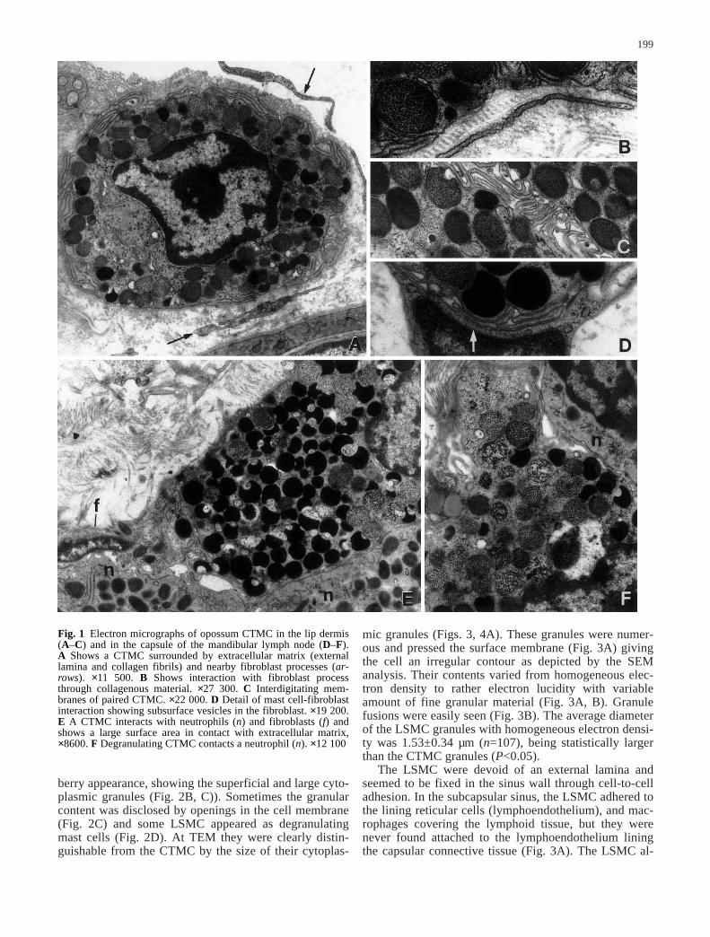
At TEM, dermal CTMC presented narrow surface pro-cesses curved toward the cell surface and displayed adiscontinuous granular coat (external lamina) in whichcollagen fibrils stuck in (Fig. 1A). However, direct con-tact with fibrillar collagen was also apparent (Fig. 1B).Although dermal CTMC could lie near fibroblasts ortheir long processes, no close contact between mast cellsand any type of connective-tissue cells were observed.However, paired mast cells were frequent in the dermis.This mast cell-mast cell interaction involved membraneinterdigitations (Fig. 1C).
In all animals, the capsule of the mandibular and su-perficial cervical lymph nodes exhibited several CTMCand some mononuclear leukocytes and neutrophilssparsely distributed among the numerous fibroblasts andcollagen fibrils. Several CTMC were less buried in colla-gen fibrils, exhibiting large surface areas in direct con-tact with granulocytes and fibroblasts (Fig. 1E). In thelatter, pinocytic vesicles could accumulate in the contact-ing fibroblast membrane (Fig. 1D). In the nodal capsule,degranulating CTMC seemed more frequent than in thelip dermis and some of them were in close apposition toneutrophils (Fig. 1F). Mast cells with bizarre forms ordisplaying surface folds ending far from the cell surfaceor even lamelliform extensions were also found. By mea-suring 109 homogeneous-appearing cytoplasmic granulesof nodal CTMC, their average diameter was estimated at0.49±0.07 µm.
In SEM, the CTMC could be identified with certainlyonly after fracture that showed up their cytoplasmicgranules. Probably the difficulty in recognizing non-frac-tured CTMC was due to their smooth surfaces, besidesthe surrounding collagen fibrils (Fig. 2A).
Lymphatic-sinus mast cells (LSMC) in the mandibularand axillary lymph nodes
The mandibular and superficial axillary lymph nodes ex-hibited LSMC in all lymphatic sinuses. They were rathernumerous in the medullary sinuses (Figs. 2B, 3A). How-ever, the LSMC were observed only when a pressure of70–80 mm Hg was maintained throughout the intracardi-ac perfusion with the fixative. A perfusing pressure of110–120 mm Hg washed out virtually all LSMC. AtSEM, the LSMC were easily identified by their black-
198
berry appearance, showing the superficial and large cyto-plasmic granules (Fig. 2B, C)). Sometimes the granularcontent was disclosed by openings in the cell membrane(Fig. 2C) and some LSMC appeared as degranulatingmast cells (Fig. 2D). At TEM they were clearly distin-guishable from the CTMC by the size of their cytoplas-
mic granules (Figs. 3, 4A). These granules were numer-ous and pressed the surface membrane (Fig. 3A) givingthe cell an irregular contour as depicted by the SEManalysis. Their contents varied from homogeneous elec-tron density to rather electron lucidity with variableamount of fine granular material (Fig. 3A, B). Granulefusions were easily seen (Fig. 3B). The average diameterof the LSMC granules with homogeneous electron densi-ty was 1.53±0.34 µm (n=107), being statistically largerthan the CTMC granules (P<0.05).
The LSMC were devoid of an external lamina andseemed to be fixed in the sinus wall through cell-to-celladhesion. In the subcapsular sinus, the LSMC adhered tothe lining reticular cells (lymphoendothelium), and mac-rophages covering the lymphoid tissue, but they werenever found attached to the lymphoendothelium liningthe capsular connective tissue (Fig. 3A). The LSMC al-
199
Fig. 1 Electron micrographs of opossum CTMC in the lip dermis(A–C) and in the capsule of the mandibular lymph node (D–F). A Shows a CTMC surrounded by extracellular matrix (externallamina and collagen fibrils) and nearby fibroblast processes (ar-rows). ×11 500. B Shows interaction with fibroblast processthrough collagenous material. ×27 300. C Interdigitating mem-branes of paired CTMC. ×22 000. D Detail of mast cell-fibroblastinteraction showing subsurface vesicles in the fibroblast. ×19 200.E A CTMC interacts with neutrophils (n) and fibroblasts (f) andshows a large surface area in contact with extracellular matrix,×8600. F Degranulating CTMC contacts a neutrophil (n). ×12 100
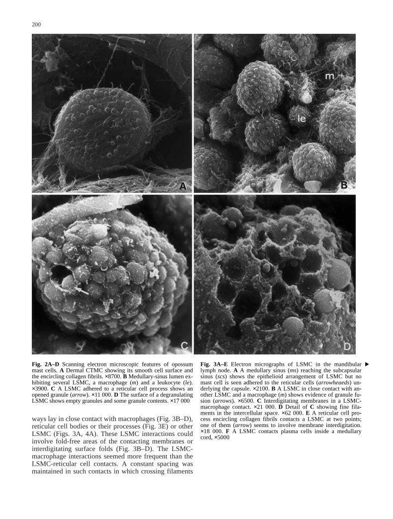
ways lay in close contact with macrophages (Fig. 3B–D),reticular cell bodies or their processes (Fig. 3E) or otherLSMC (Figs. 3A, 4A). These LSMC interactions couldinvolve fold-free areas of the contacting membranes orinterdigitating surface folds (Fig. 3B–D). The LSMC-macrophage interactions seemed more frequent than theLSMC-reticular cell contacts. A constant spacing wasmaintained in such contacts in which crossing filaments
200
Fig. 2A–D Scanning electron microscopic features of opossummast cells. A Dermal CTMC showing its smooth cell surface andthe encircling collagen fibrils. ×8700. B Medullary-sinus lumen ex-hibiting several LSMC, a macrophage (m) and a leukocyte (le).×3900. C A LSMC adhered to a reticular cell process shows anopened granule (arrow). ×11 000. D The surface of a degranulatingLSMC shows empty granules and some granule contents. ×17 000
Fig. 3A–E Electron micrographs of LSMC in the mandibularlymph node. A A medullary sinus (ms) reaching the subcapsularsinus (scs) shows the epithelioid arrangement of LSMC but nomast cell is seen adhered to the reticular cells (arrowheaeds) un-derlying the capsule. ×2100. B A LSMC in close contact with an-other LSMC and a macrophage (m) shows evidence of granule fu-sion (arrows). ×6500. C Interdigitating membranes in a LSMC-macrophage contact. ×21 000. D Detail of C showing fine fila-ments in the intercellular space. ×62 000. E A reticular cell pro-cess encircling collagen fibrils contacts a LSMC at two points;one of them (arrow) seems to involve membrane interdigitation.×18 000. F A LSMC contacts plasma cells inside a medullarycord, ×5000
▲

201
could be clearly seen (Fig. 3D). Frequently, the macro-phage cytoplasm close to the interaction with LSMC ex-hibited large phagosomes, besides numerous vesicles andrough endoplasmic reticulum profiles. The LSMC-LSMC contact could not be completely resolved by our
methodological approach and the possibility of otherforms of cell junction, such as gap junctions, can not bediscarded. A few LSMC were seen apparently inside themedullary cords in apposition to plasma cells and otherlymphoid cells (Fig. 3F).
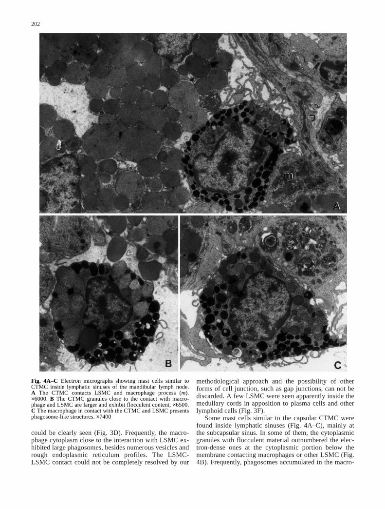
Some mast cells similar to the capsular CTMC werefound inside lymphatic sinuses (Fig. 4A–C), mainly atthe subcapsular sinus. In some of them, the cytoplasmicgranules with flocculent material outnumbered the elec-tron-dense ones at the cytoplasmic portion below themembrane contacting macrophages or other LSMC (Fig.4B). Frequently, phagosomes accumulated in the macro-
202
Fig. 4A–C Electron micrographs showing mast cells similar toCTMC inside lymphatic sinuses of the mandibular lymph node. A The CTMC contacts LSMC and macrophage process (m).×6000. B The CTMC granules close to the contact with macro-phage and LSMC are larger and exhibit flocculent content, ×6500.C The macrophage in contact with the CTMC and LSMC presentsphagosome-like structures. ×7400
phage cytoplasm facing the CTMC-like cells (Fig. 4C)as described for the LSMC-macrophage interactions.
Mast cells of the abdominal lymph nodes
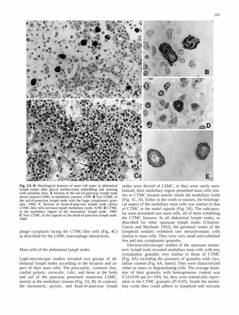
Light-microscopic studies revealed two groups of ab-dominal lymph nodes according to the location and as-pect of their mast cells. The para-aortic, common iliac,cardial pyloric, cecocolic, colic, and those at the bodyand tail of the pancreas presented numerous LSMC,mainly at the medullary sinuses (Fig. 5A, B). In contrast,the mesenteric, pyloric, and head-of-pancreas lymph
nodes were devoid of LSMC, or they were rarely seen.Instead, their medullary region presented mast cells sim-ilar to CTMC located mainly inside the medullary cords(Fig. 5C, D). Either in the cords or sinuses, the histologi-cal aspect of the medullary mast cells was similar to thatof CTMC at the nodal capsule (Fig. 5E). The subcapsu-lar sinus presented rare mast cells, all of them exhibitingthe CTMC features. In all abdominal lymph nodes, asdescribed for other opossum lymph nodes (Chiarini-Garcia and Machado 1992), the germinal center of thelymphoid nodules exhibited rare metachromatic cellssimilar to mast cells. They were very small and exhibitedfew and tiny cytoplasmic granules.
Electron-microscopic studies of the opossum mesen-teric lymph node revealed medullary mast cells with tinycytoplasmic granules very similar to those of CTMC(Fig. 6A) including the presence of granules with crys-talline content (Fig. 6A, insert). They were characterizedeither as intact or degranulating cells. The average diam-eter of their granules with homogeneous content was0.53±0.09 µm (n=100). So, they were statistically equiv-alent to the CTMC granules (P>0.05). Inside the medul-lary cords they could adhere to lymphoid and reticular
203
Fig. 5A–D Histological features of mast cell types in abdominallymph nodes after glycol methacrylate embedding and stainingwith toluidine blue. A Section of the tail-of-pancreas lymph nodeshows typical LSMC in medullary sinuses. ×290. B Two LSMC ofthe tail-of-pancreas lymph node with the large cytoplasmic gran-ules. ×900. C Section of head-of-pancreas lymph node showsCTMC-like cells (arrows) inside medullary cords. ×290. D CTMCin the medullary region of the mesenteric lymph node. ×900. E Two CTMC in the capsule of the head-of-pancreas lymph node.×900

cells or to the extracellular matrix surrounding bloodvessels (Fig. 6A). In the medullary sinuses, the mastcells contacted macrophages (Fig. 6B) and reticular cellsas described for the LSMC. In these close contacts, theintercellular space exhibited filamentous material linkingthe LSMC and macrophage membranes (Fig. 6C).
Discussion
CTMC have already been described in the opossum earwhere their external lamina was impressive (Santos andMachado 1994). Our studies in the lip dermis showedCTMC with a more delicate and discontinuous externallamina and some of them seem to adhere directly to fi-brillar collagen. In the nodal capsule, the external laminacould even be absent, and CTMC could contact fibro-blast and granulocytes. Adhesion of murine mast cell tofibroblast is well studied in vitro and seems to involve c-kit and its ligand, the stem factor that has been implicat-ed in mast cell proliferation and differentiation (Adachiet al. 1992, 1995; Tsai et al. 1991). Besides, there arestrong arguments favoring reciprocal influences betweenmast cells and fibroblasts (Davidson et al. 1983; Takedaet al. 1989; Levi-Schaffer and Rubinchik 1994). Our datashowing concentration of pinocytic vesicles in the fibro-blast membrane in close apposition to mast cells suggesta functional interaction between these two opossumcells, as evidenced in the human lung (Heard et al.1992). The interaction of the opossum CTMC withgranulocytes is a new finding that deserves future study.In eutherian mammals, studies on mast cell-leukocyte in-teractions are basically restricted to lymphocytes.
The present paper reinforces the unique character ofthe LSMC in both transmission and scanning electronmicroscopy, their cytoplasmic granules being three timeslarger than those of CTMC. Mast cells exhibiting largecytoplasmic granules are also very numerous in the lym-phatic sinuses of cervical lymph nodes of Sminthopsiscrassicaudata, an Australian marsupial (Haynes 1991)and in the mandibular and axillary lymph nodes of fourother Brazilian Didelphidae (Chiarini-Garcia and Pereira1999).
Our ultrastructural studies showed that LSMC adhereto the sinus wall through cell-to-cell contacts with mac-rophages, reticular cells and other LSMC. The LSMC-LSMC interactions explain the epithelioid arrangementof these cells in the medullary sinuses. The LSMC-mac-rophage contacts seem to be more frequent than the
204
Fig. 6A–C Electron micrographs of mast cells similar to CTMCin medullary cords and sinuses of the opossum mesenteric lymphnode. A Mast cell inside a medullary cord close to a blood vesselcontacts a lymphocyte (li). ×9800. Insert shows a cytoplasmicgranule with crystalline content from a resting mast cell. ×31 700.B Mast cell in a medullary sinus contacts a macrophage (m)through membrane interdigitations. ×10 000. C Detail of the mastcell-macrophage close apposition to show the interdigitating mem-branes with constant intercellular space crossed by a filamentousstructure. ×45 000

LSMC-reticular cell apposition, probably because mac-rophages practically surrounded the medullary cordsthrough interaction with reticular cells. In these LSMCclose contacts, the crossing filamentous structures in theintercellular space could represent adhesion molecules.Despite these LSMC adherences, high pressure duringthe intracardiac perfusion washed then out.
In contrast to contacts with reticular cells, those withmacrophages could involve an accumulation of organ-elles in the macrophage cytoplasm abutting the interac-tion with LSMC. Also, the medullary-sinus CTMC inclose contact with macrophages exhibited ultrastructuralchanges of the cytoplasmic granules close to the contact-ing membranes. Altogether these findings indicate afunctional interaction between macrophages and mastcells in the medullary sinuses. In rat lymph nodes macro-phages and reticular cells are able to take up granule ma-trix released by degranulating mast cells (Miyata and Ta-kara 1985). Preliminary studies indicate that macro-phages in the opossum lymph nodes exert a similar roleafter treatment with compound 48/80 (Ghiarini-Garcia etal. 1997).
Our studies of abdominal lymph nodes disclosed anintriguing finding. The mesenteric, pyloric and the head-of-pancreas lymph nodes were derived of LSMC. Inthese three lymph nodes, the medullary-region mast cellswere few, similar to CTMC and located mainly insidethe cords. These three lymph nodes drain the lymph fromthe small intestine, gall bladder, and distal portion of thestomach and head of the pancreas (Azzali and DiDio1965). The mesenteric lymph node is larger than the twoothers and drains exclusively from the jejunum and ile-um. The other six abdominal lymph nodes drain fromdifferent abdominal ogans and regions, but none receiveslymph from the small intestine. Therefore, the origin ofthe afferent lymphatic vessels might be implicated in theabsence of LSMC. Mast cells or their precursors are sup-posed to reach lymph nodes via the afferent lymphaticvessels. So, small intestine-originated mast cells couldbe insensitive to local factors able to induce the LSMCphenotype. Alternatively, the LSMC-free lymph nodescould be devoid of such factors. The few CTMC-likecells located in the medullary sinuses of the mesentericlymph node contacted macrophages as the LSMC did inother opossum nodes. This makes it difficult to relate theLSMC differentiation simply to contact with macro-phages. Another point is the preferential location of themedullary mast cells in the cords. If they came via theafferent lymph, they had to be attracted to the interior ofthe medullary cords. Developmental studies aiming atcontributing to the elucidation of these questions are inprogress.
The medullary-cord CTMC could be classed as restingor degranulating mast cells according to criteria estab-lished for eutherian CTMC (Friedman and Kaliner 1988).Frequently, the degranulating ones were in close apposi-tion to lymphocytes. This finding may constitute ultra-structural evidence favoring a participation of cell-to-cellcontact in regulating mast cell degranulation. Activated
lymphocytes produce soluble factors involved in the pro-liferation and differentiation of mast cells. However,there is strong evidence showing that activated lympho-cytes may affect mast cell biology through cell-to-cellcontact (Oh and Metcalfe 1996; Bhattacharyya et al.1998). Further, degranulation of mast cells can be mediat-ed by such contacts in vitro (Inamura et al. 1998). On theother hand, rodent mast cells are able to secrete numerouscytokines known to affect leukocytes, including the mastcell itself (reviewed by Lin and Befus 1999). The regula-tory potential of mast cells through the secretion of cyto-kines remains to be studied in marsupials. Anyway, thelarge number of different types of mast cells in the opos-sum lymph nodes and their numerous cell-to-cell contactsmake this animal suitable for studies on the significanceof mast cell adherence to other cells in vivo.
Acknowledgements This work was supported by Fundação deAmparo a Pesquisa do Estado de Minas Gerais, FAPEMIG(1095/92), Conselho Nacional de Desenvolvimento Científico eTecnológico-CNPq (522037-96) and Pro-Reitoria de Pesquisa-UFMG (049153/93-61 and 050359/94-15). The ultrastructuralstudies were developed at the Electron Microscope Center (CE-MEL) of the Institute of Biological Sciences at UFMG. A.A.D.S.was supported at UFMG through a CAPES fellowship.
References
Adachi S, Ebi Y, Nishikawa S, Haysahi S, Yamazaki M, Kasugai T,Yamamura T, Nomura S, Kitamura Y (1992) Necessity of ex-tracellular domain of W (c-kit) receptor for attachment of mu-rine cultured mast cells to fibroblasts. Blood 179:650–656
Adachi S, Tsujimura T, Jippo T, Morimoto M, Isozaki K, Kasugai T,Nomura S, Kitamura Y (1995) Inhibition of attachment be-tween cultured mast cells and fibroblasts by phorbol 12-myri-state 13-acetate and stem cell factor. Exp Hematol 23:58–65
Azzali G, DiDio LJA (1965) The lymphatic system of Didelphisazarae and Didelphis marsupialis. Am J Anat 116:449–470
Bhattacharyya SP, Drucker I, Kiershenbaum AS, Metcalfe DD,Mekori YA (1998) Activated A lymphocytes induce degranu-lation and cytokine production by human mast cells followingcell-to-cell contact. J Leukoc Biol 63:337–341
Chiarini-Garcia H, Machado CRS (1992) Mast cell types and theirdistribution in the lymph nodes of the opossum Didelphis albi-ventris (Marsupialia, Didelphidae). Cell Tissue Res 268:571–574
Chiarini-Garcia H, Pereira FM (1999) A comparative study oflymph node mast cell populations in five marsupial species.Tissue Cell 31:318–326
Chiarini-Garcia H, Santos AAD, Machado CRS (1997) Morpho-logical changes induced by the compound 48/80 in mast cellsof the opossum Didelphis albiventris. Acta Microsc 6 [SupplB]:750–751
Davidson S, Mansour A, Gallily R, Smolarski M, Rofolovitch M,Gisburg H (1983) Mast cell differentiation depends on T cellsand granule synthesis on fibroblast. Immunology 48:439–452
Enerback L (1986) Mast cell heterogeneity. The evolution of theconcept of a specific mucosal mast cell. In: Befus AD,Bienenstock J, Denburg JA (eds) Mast cell differentiation andheterogeneity. Raven Press, New York, pp 1–26
Friedman MM, Kaliner M (1988) Ultrastructural changes in hu-man skin mast cells during antigen-induced degranulation invivo. J Allergy Clin Immunol 82:998–1005
Galli SJ (1990) New insights into ”the riddle of the mast cells”:Microenvironmental regulation of mast cell development andphenotypic heterogeneity. Lab Invest 62:5–33
Haynes JI (1991) Cervical lymph nodes and mast cells in the mar-supial Sminthopsis crassicaudata. Anat Rec 231:7–13
205

Heard BE, Dewar A, Corrin B (1992) Apposition of fibroblast tomast cells and lymphocytes in normal human lung and in cryp-togenic fibrosing alveolitis. Ultrastructure and cell perimetermeasurements. J Pathol 166:303–310
Inamura N, Mekori YA, Bhattacharyya SP, Bianchini PJ, MetcalfeDD (1998) Induction and enhancement of Fc(epsilon)RI-dependent mast cell degranulation following coculture withactivated T cells: dependency on ICAM-1 and leukocyte func-tion-associated antigen (LFA)-1-mediated heterotypic aggre-gation. J Immunol 160:4026–4033
Kitamura Y (1989) Heterogeneity of mast cells and phenotypicchange between subpopulations. Annu Rev Immunol 7:59–76
Levi-Schaffer F, Rubinchik E (1994) Mast cell/fibroblast interac-tions. Clin Exp Allergy 24:1016–1021
Lin T-J, Befus AD (1999) Mast cells and eosinophils in mucosaldefenses and pathogenesis. In: Ogra PL, Mestecky J, Lamm ME,Strober W, Bienenstock J, McGhee JR (eds) Mucosal Immu-nology, 2nd edn, Academic Press, San Diego, pp 469–481
Lozzi SP, Machado CRS, Gerken SE, Mota-Santos TA (1996) In-volvement of regional lymph nodes after penetration of Schis-tosoma mansoni cercariae in naive and infected mice. MemInst Oswaldo Cruz 91:491–498
Metcalfe DD, Baram D, Mekori YA (1997) Mast cells. PhysiolRev 77:1033–1079
Miyata K, Takaya K (1985) Acid-phosphatase activity of reticularcells and macrophages in the lymph node of the rat after inges-tion of mast cell granules. Histochemistry 83:201–205
Murakami T, Jones AL (1980) Conductive staining of biologicalspecimens for non-coated scanning electron microscopy: dou-ble staining by tannin-osmium and osmium-thiocarbohydr-azide-osmium methods. In: Johari O (ed) Scanning electronmicroscopy. Part I. AFM O’Hare, Chicago, pp 221–226
Oh CK, Metcalfe DD (1996) Activated lymphocytes inducedpromoter activity of the TCA3 gene in mast cells followingcell-to-cell contact. Biochem Biophys Res Commun 221:510–514
Russell LD, Burguet S (1978) Ultrastructural of Leydig cells asrevealed by secondary tissue treatment with a ferrocyanide:osmium mixture. Tissue Cell 9:99–112
Sainte-Marie G, Peng FS (1990) Mast cells and fibrosis in com-partments of lymph nodes of normal, gnotobiotic, and athymicrats. Cell Tissue Res 261:1–15
Santos AAD, Machado CRS (1994) Histochemical and ultra-structural studies of mast cells in the intestinal mucosa andskin of the oposum Didelphis albiventris. Histochem J 26:233–238
Takeda K, Hatamochi A, Ueki H (1989) Increased number of themast cells accompany enhanced collagen synthesis in linearlocalized scleroderma. Arch Dermatol Res 281:288–290
Tsai M, Takeishi T, Thompson H, Langley KE, Zsebo KM,Metcalfe DD, Geissler EN, Galli S (1991) Induction of mastcell proliferation, maturation, and heparin synthesis by the ratc-kit ligand, stem cell factor. Proc Natl Acad Sci USA88:6382-6386
206