esophageal and small bowel obstruction by occupational bezoar: report of a case
TRANSCRIPT
1
Occupational exposure to wood dust. Esophageal and Small Bowel Obstruction by
Occupational Bezoar: Report of a Case
M. PITIAKOUDIS1, A. TSAROUCHA1, T.C. CONSTANTINIDIS3, K. MIMIDIS2,
E. EFSTATHIOU1, G.A. STATHOPOULOS3, C. SIMOPOULOS1
1 2nd Department of Surgery 2 1st Department of Internal Medicine, Endoscopy Unit 3 Laboratory of Hygiene and Environmental Protection
Medical School, Democritus University of Thrace
68100 Alexandroupolis, Greece
Abstract
A 70-year old male is presented suffering from esophageal and small bowel obstruction,
caused by an occupational bezoar. The patient has worked as a carpenter for 35 years.
Ten years before the incident he underwent pyloroplasty and vagotomy. The part of the
bezoar, which caused the esophageal obstruction was removed during endoscopy, while
the part of the small bowel was treated surgically. The patient recovered well and was
discharged on the 8th postoperative day. The study presents a rare case of intestinal
obstruction (esophageal and small bowel) caused by an occupational bezoar in a patient
with a previous gastric surgery.
Introduction
2
Phytobezoar causing bowel obstruction in patients with previous gastric surgery is a well
known late complication, although very rare. It is a concretion of food fibers (fruit and
vegetable fibers) or foreign bodies in the stomach.1 The stomach is the most common
place of bezoar formation. In a normal stomach vegetable fibers can not pass through the
pylorus; they undergo hydrolysis within the stomach, which softens them enough to go
through the small bowel. After gastric surgery, because the gastric motility is disturbed,
the gastric acidity is decreased, and the stomach may rapidly empty, there is an increased
possibility for bezoar formation, causing acute abdomen due to small bowel obstruction.
Reports of bezoars causing obstruction of the gastrointestinal tract exist since the
late 18th century.2 Mir and Mir3 reported on 22 cases of bezoar found in the literature
from 1966 to 1973. Most case reports refer to bezoars of food with fibers or foreign
bodies. Moseley4 reported the first case of a phytobezoar (citrus, onions, mushrooms)
following pyloroplasty and vagotomy.
There have not been incidents of occupational reason. The reverse migration of the
bezoar to the esophagus is also very rare, and may occur after persistent vomiting and
bezoar fragmentation.5,6 In this paper, the case of a patient with a phytobezoar of
occupational origin is presented. The patient suffered from both esophageal and small
bowel obstruction.
Case report
A 70-year old male carpenter, who complained about dysphagia for several days, and
epigastric pain, nausea and vomiting for 24 hours, was admitted to the Department of
Internal Medicine. He had worked as a carpenter for 35 years, only occasionally using
3
personal protective equipment. Ten years before the incident, he had undergone
cholecystectomy, pyloroplasty and vagotomy.
Plain x-ray and ultrasonography of the abdomen was performed after admission with
no diagnosis. A chest x-ray and pulmonary function tests were normal. Vomiting
continued and dysphagia got worse. The patient then underwent upper gastrointestinal
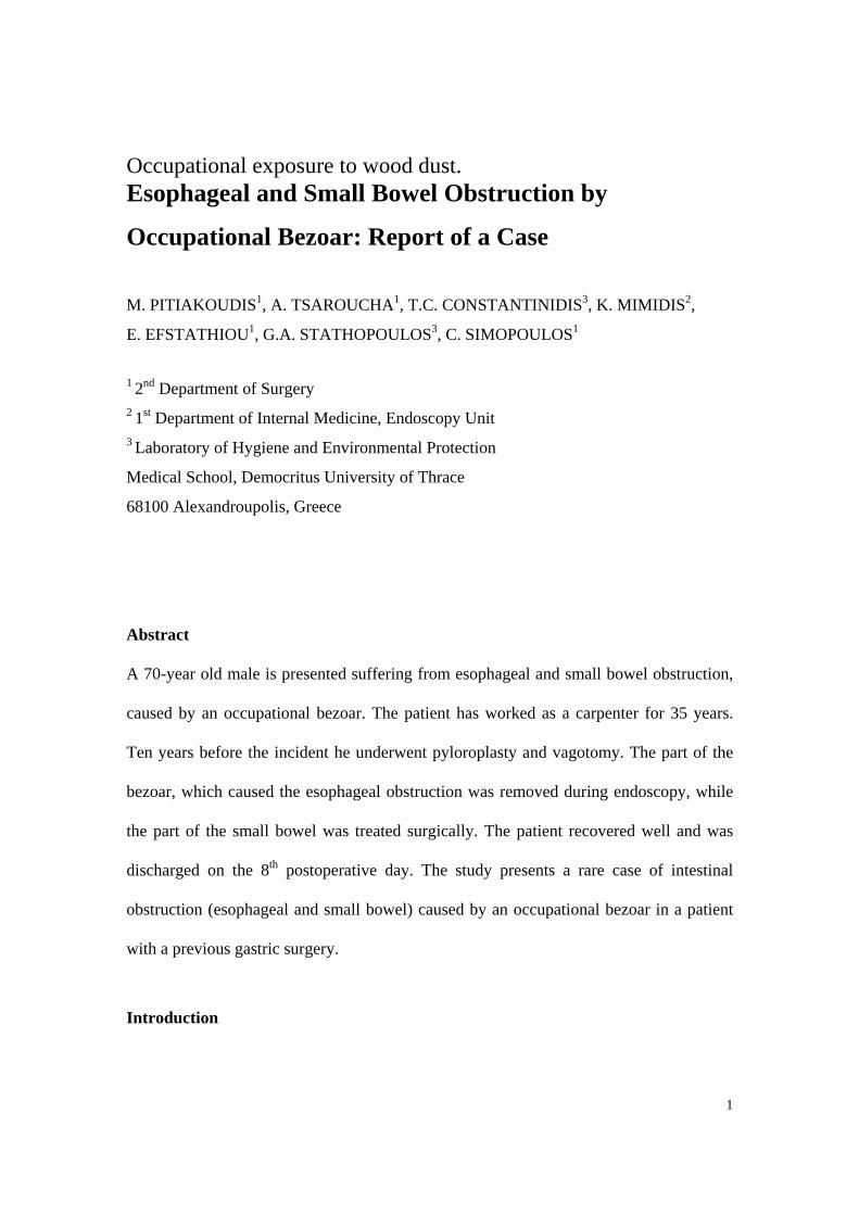
endoscopy as an alternative for diagnosis. The endoscopy (Olympus GIF-V2, Olympus
Optical Co. Ltd., Tokyo, Japan) of the esophagus showed a foreign body (dimensions:
1.5cm long, 2.8cm in diameter), which was endoscopically removed using a dormia
basket (Fig. 1).
Figure 1. Endocopy: (a) obstruction of the lower third of the esophagus due to a
bezoar; (b) removal of the esophageal bezoar with a Dormia-basket.
4
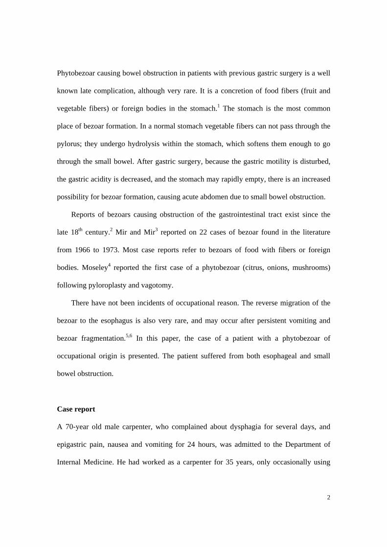
Further endoscopic examination to the second part of the duodenum revealed no
pathological findings. Pathological examination reported that the removed foreign body
consisted of wood dust (Fig. 2a).
Figure 2. The entire bezoar: (a) esophageal; (b) intestinal.
The patient’s condition improved only partially after endoscopy, so he was kept in
the hospital for further examination. Few days later he felt abdominal pain and a new x-
ray of the abdomen showed dilated gas-filled loops of the small bowel with no air in the
colon, suggesting small bowel obstruction. He was then transferred to the surgical clinic
for further treatment. His condition worsened the following days and the bowel
obstruction could not be managed with conservative treatment, so the patient underwent
exploratory laparotomy. The laparotomy revealed a swollen small bowel all the way
almost till the end of the ileum. A solid palpable mass (dimension 9.0cm long, 2.9cm in
5
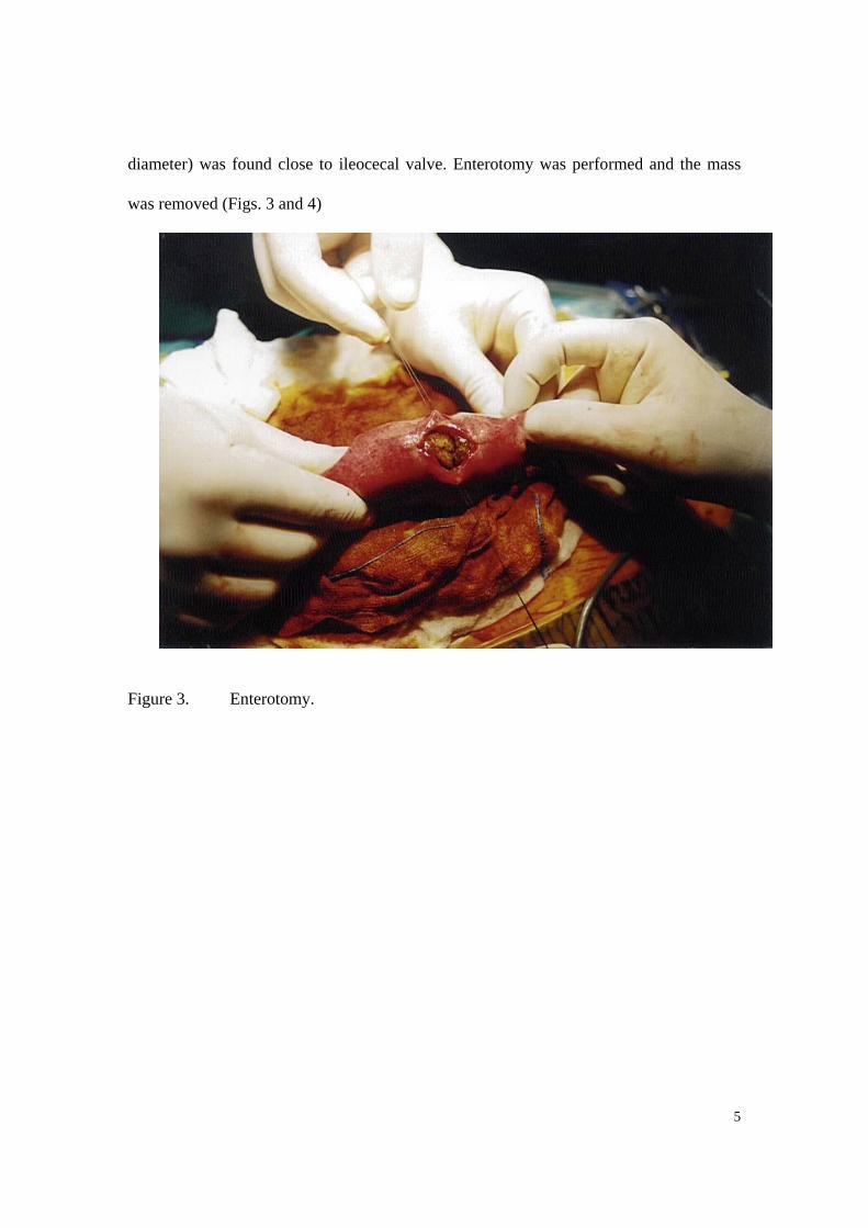
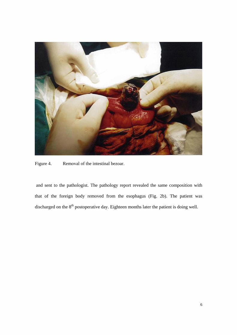
diameter) was found close to ileocecal valve. Enterotomy was performed and the mass
was removed (Figs. 3 and 4)
Figure 3. Enterotomy.
6
Figure 4. Removal of the intestinal bezoar.
and sent to the pathologist. The pathology report revealed the same composition with
that of the foreign body removed from the esophagus (Fig. 2b). The patient was
discharged on the 8th postoperative day. Eighteen months later the patient is doing well.
7
Discussion
Intestinal obstruction due to phytobezoars (food, vegetable fibers, and/or foreign bodies)
is rarely encountered in adults with a normal intestinal tract. Phytobezoar usually is a
cause of intestinal obstruction in patients with previous vagotomy and drainage or gastric
resection.1,3,4 Most of the bezoars that have been presented in the literature are
concretions of poorly digested food, which are usually formed initially in the stomach; a
fragment of them may migrate in the bowel and cause obstruction.1,2,4 The two usual sites
of the obstruction are in the ileum and the jejunum; it is unusual the reverse migration of
the bezoar to the esophagus, as it occurred in our patient.5,6 Some factors seem to be
responsible for bezoar formation. The risk factors that explain the etiology and
pathogenic mechanisms in patients with pyloroplasty - vagotomy include: (1) decreased
secretion of hydrochloric acid and reduced peptic activity; (2) decreased gastric motility
(atonic stomach); and (3) occupational exposure.1,2,4
In cases the bezoar is located in the esophagus and the stomach, there should be an
attempt to first treat it conservatively.3 If the bezoar is causing acute intestinal
obstruction there is indication for surgical treatment.1,3,4 In our patient the cause of the
bezoar was the accumulated wood dust (occupational bezoar) in the stomach and the
previous gastric surgery. The bezoar had two different final locations, one in the
esophagus and the other in the small bowel. The initial mass was probably in the
stomach, and due to the continuing vomiting, the bezoar was fragmented. The fragments
migrated causing obstruction in two different locations in the intestinal tract. The tail of
the bezoar moved to the esophagus, while the rest from the stomach through the pylorus
(because of the previous pyloroplasty and vagotomy) moved to the small bowel. Two
8
different therapies were used to treat this occupational phytobezoar, namely endoscopy
and surgery.
This is a rare case of bezoar causing two obstructions, nearly simultaneously in the
same patient. To our knowledge this is the first case of an occupational bezoar consisting
of wood dust. The patient only occasionally used personal protective equipment in his
work, and during the month before the incident, he had worked around twelve hours per
day. The use of personal protective equipment is imperative to avoid serious health
problems to the worker.
In conclusion, since occupational bezoars may be a cause of intestinal obstruction
(esophageal and/or small bowel), patients who have undergone a previous gastric surgery
should avoid certain kinds of fiber in their diet, and also avoid similar occupational
exposures.
9
References
1. Rubin M, Shimonov M, Grief F, Rotestein Z, Lelkuk S. Phytobezoar: A rare cause of
intestinal obstruction. Dig Surg 1998; 15(1):52-4.
2. Madura MJ, Naughton BJ, Craig RM. Duodenal bezoar: a report case and review of
the literature. Gastrointest Endosc 1982; 28:26-8.
3. Mir AM, Mir MA. Phytobezoar after vagotomy with drainage or resection. Brit J
Surg 1973; 60(11):846-7.
4. Moseley RV. Pyloric obstruction by a phytobezoar following pyloroplasty and
vagotomy. Arch Surg 1967; 94:290-1.
5. Hermoso JC, Rosado R, Ramirez D, Ruiz JJ, Medina P, Bonetti A. A esophageal
impaction of a bezoar after gastric surgery. Rev Esp Enf Digest 1991; 79:139-41.
6. Goel AL, Seenu V, Srikrishna NV, Goyal S, Thakur KK, Shukla NK. Esophageal
bezoar: a rare but distinct clinical entity. Trop Gastroenterol 1995; 16:43.
10
Απόφραξη οισοφάγου και λεπτού εντέρου σε ξυλουργό οφειλόµενη σε φυτοπίληµα επαγγελµατικής αιτιολογίας Μ. Πιτιακούδης1, Α. Τσαρούχα1, Θ. Κωνσταντινίδης3, Κ. Μιµίδης2, Ε. Ευσταθίου1, Γ.Α. Σταθόπουλος3, Κ. Σιµόπουλος1
1 2η Χειρουργική λινική Τµήµατος Ιατρικής ∆ηµοκρίτειου Πανεπιστηµίου Θράκης 2 1η Παθολογική λινική Τµήµατος Ιατρικής ∆ηµοκρίτειου Πανεπιστηµίου Θράκης 3 Εργαστήριο Υγιεινής και Προστασίας Περιβάλλοντος Τµήµατος Ιατρικής ∆.Π.Θ . Φυτοπιλήµατα προκαλούντα αποφράξεις της γαστρεντερικής οδού αναφέρονται από τον 18ο αιώνα. Τα περισσότερα περιστατικά που αναφέρονται στη σύγχρονη βιβλιογραφία δηµιουργούνται από τρόφιµα πλούσια σε φυτικές ίνες καθώς και ξένα σώµατα. Εβδοµηντάχρονος άνδρας προσήλθε στο τµήµα επειγόντων περιστατικών του Γενικού Περιφερειακού Νοσοκοµείου Αλεξανδρούπολης παραπονούµενος γιά επιγαστρικό πόνο, ναυτία, έµετους και δυσκαταποσία. Γιά τη συµπτωµατολογία αυτήν εισήχθη στην Παθολογική λινική. Απλή ακτινογραφία κοιλίας και υπερηχογράφηµα δεν βοήθησαν στη διάγνωση. Η κλινική εικόνα παρουσίασε σταδιακά επιδείνωση και ο ασθενής µεταφέρθηκε στη Χειρουργική κλινική. Από τα στοιχεία του ιστορικού προέκυψε ότι ο ασθενής εργαζόταν ως ξυλουργός γιά 35 έτη, χωρίς να λαµβάνει ατοµικά µέτρα προστασίας κατά την εργασία του, παρά µόνο περιστασιακά. Ο ασθενής προ δεκαετίας είχε υποβληθεί σε χολοκυστεκτοµή, πυλωροπλαστική και βαγοτοµή. Κατά τη διάρκεια διαγνωστικής ενδοσκόπησης, εντοπίσθηκε ξένο σώµα Το τµήµα του φυτοπιλήµατος που προκαλούσε την οισοφαγική απόφραξη εξαιρέθηκε, ενώ το υπόλοιπο τµήµα του φυτοπιλήµατος στο λεπτό έντερο αφαιρέθηκε χειρουργικά. Η παθολοανατοµική µελέτη έδειξε ότι και στις δύο περιπτώσεις τα φυτοπιλήµατα αποτελούνταν από σκόνη ξύλου. Ο ασθενής επανέκαµψε και έλαβε εξιτήριο την 8η µετεγχειρητική ηµέρα. ∆εκαοκτώ µήνες µετά η κατάσταση του ασθενούς παραµένει καλή. Στη βιβλιογραφία δεν αναφέρονται φυτοπιλήµατα επαγγελµατικής αιτιολογίας, γι’αυτό και θεωρείται ότι το περιστατικό που αναφέρεται στην ανακοίνωση αυτήν είναι εξαιρετικά σπάνιο. Επίσης είναι εξαιρετικά σπάνιο από φυτοπίληµα να προκληθούν δύο αποφράξεις του γαστρεντερικού συστήµατος στον ίδιο ασθενή. Η αναφορά του περιστατικού αυτού, επιβεβαιώνει την αναγκαιότητα της λήψης ενδελεχούς ιστορικού και τη σηµασία του επαγγελµατικού ιστορικού. Επίσης, µε τη λήψη προληπτικών µέτρων προστασίας κατά την εργασία είναι δυνατό να αποφευχθούν περιστατικά, όπως το παραπάνω, που απαιτούσε χειρουργικές δράσεις.