hedgehog signaling is activated in subsets of esophageal cancers
TRANSCRIPT
Hedgehog signaling is activated in subsets of esophageal cancers
Xiaoli Ma1, Tao Sheng
2, Yuanxin Zhang
1, Xiaoli Zhang
2, Jing He
2, Shuhong Huang
1, Kai Chen
2, Josh Sultz
2,
Patrick A. Adegboyega2, Hongwei Zhang1* and Jingwu Xie2*
1Institute of Developmental Biology, School of Life Sciences, Shandong University, Jinan, China2Sealy Center for Cancer Cell Biology, Departments of Pharmacology, Toxicology and Pathology,University of Texas Medical Branch, Galveston, TX, USA
The hedgehog pathway plays a critical role in the development ofthe foregut. However, the role of the hedgehog pathway in primaryesophageal cancers is not well studied. Here, we report that elevatedexpression of hedgehog target genes occurs in 14 of 22 primaryesophageal cancers. The hedgehog signaling activation is not associ-ated with tumor subtypes, stages, or differentiation. While the sonichedgehog (Shh) transcript is localized to the tumor tissue, expres-sion of Gli1 and PTCH1 is observed both in the tumor and in thestroma. We discovered that 4 esophageal squamous cell carcinomas,which overexpress Shh, have genomic amplification of the Shh gene.Treatment of esophageal cancer cells with smoothened antagonist,KAAD-cyclopamine, or the neutralizing antibodies of Shh reducescell growth and induces apoptosis. Overexpression of Gli1 underthe CMV promoter renders these cells resistant to the treatments.Thus, our results indicate that elevated expression of Shh and itstarget genes is quite common in esophageal cancers. Our data alsoindicate that downregulation of Gli1 expression may be an impor-tant mechanism by which KAAD-cyclopamine inhibits growth andinduces apoptosis in esophageal cancer cells (supplementary mate-rial for this article can be found on the International Journal of Can-cer website at http://www.interscience.wiley.com/jpages/0020-7136/suppmat/index.html).' 2005 Wiley-Liss, Inc.
Key words: sonic hedgehog; cyclopamine; smoothened; esophagealcancer; GI cancer
The hedgehog pathway plays a critical role in embryonic devel-opment, tissue polarity and carcinogenesis.1 Secreted Hedgehogmolecules bind to the receptor patched (PTC-PTCH1, PTCH2),thereby alleviating PTC-mediated suppression of smoothened(SMO), a putative 7-transmembrane protein. SMO signaling trig-gers a cascade of intracellular events, leading to activation of thepathway through GLI-dependent transcription.2,3 Activation ofHedgehog signaling, through loss-of-function mutations ofPTCH1 or activated mutations of SMO, occurs frequently inhuman basal cell carcinomas (BCCs) and medulloblastomas.4–9
More recently, abnormal activation of the hedgehog pathway isalso reported in subsets of small cell lung cancer, pancreatic can-cer, gastric cancers, prostate cancer and several esophageal cancercell lines.10–15
Esophageal cancer is the 6th most frequent cause of cancer deathworldwide.16 In the United States, the incidence of esophagealadenocarcinoma has nearly quadrupled over the past few decadesdespite a decline in the overall incidence of cancers in esophagus.Most esophageal cancers remain clinically silent until late in the dis-ease process. Thus, these cancers are often associated with later diag-noses, poorer prognoses, significant morbidities and high mortalityrates. Although an early report indicates activation of the hedgehogpathway in several esophageal cancer cell lines, it is not clear if pri-mary tumors of esophagus have activated hedgehog signaling.12
To examine the significance of hedgehog pathway activation inesophageal cancers, we have analyzed expression of sonic hedge-hog (Shh) and its target genes in 22 primary esophageal tumorsusing in situ hybridization, real-time PCR and immunohistochem-istry. Through the assessment of the hedgehog target genes, wefind that activation of the hedgehog pathway occurs in 14 of 22esophageal cancers. We discover genomic amplification of Shh in4 squamous cell carcinomas with elevated Shh expression. Thesedata indicate that activation of the hedgehog pathway can be used
as a valuable biomarker for diagnosis and molecular classificationof esophageal cancers. Our results further suggest that targetedinhibition of the hedgehog signaling by KAAD-cyclopamine orShh neutralizing antibodies may be effective in chemopreventionand treatment of esophageal cancers.
Material and methods
Tumor sample
Specimens from 22 cases of esophageal cancers (tumors and thematched adjacent normal tissues) and 1 case of normal esophagealtissue were received as discarded materials from the Shandong QiLu Hospital, Jinan, China, or from University of Texas MedicalBranch Surgical Pathology with approval from the institutionalreview board. Pathology reports and H&E staining of each speci-men were reviewed to determine the nature of the disease and thetumor histology. Esophageal cancers were divided into 2 majorsubtypes according to the WHO guideline17 as follows: adenocar-cinoma (4 cases) and squamous cell carcinomas (18 cases).
In situ hybridization
Gli1 (X07384) was cloned into pBluescript M13 1 KS usingHindIII (50) and XbaI (30). The plasmid was digested with NruI togenerate the sense fragment (412 bp) and with NdeI to generatethe antisense fragment (682 bp). PTCH1 (U59464; cloned intoXbaI50 and ClaI30 of pRK5) was digested with DraIII to generatea small cDNA fragment (590 bp). Sense and antisense probes wereobtained by T3 and T7 in vitro transcription using a kit fromRoche (Mannheim, Germany). Tissue sections (6 lm thick) weremounted onto poly-L-lysine slides.18 Following deparaffinization,tissue sections were rehydrated in a series of dilutions of ethanol.To enhance signal and facilitate probe penetration, sections wereimmersed in 0.3% Triton X-100 solution for 15 min at room tem-perature, followed by treatment with proteinase K (20 lg/ml) for20 min at 37�C. The sections were then incubated with 4% (v/v)paraformaldehyde/PBS for 5 min at 4�C. After washing with PBSand 0.1M triethanolamine, the slides were incubated with prehy-bridization solution (50% formamide, 50% 4 3 SSC) for 2 hr at37�C. The probe was added to each tissue section at a concentrationof 1 lg/ml and hybridized overnight at 42�C. After high-stringencywashing (2 3 SSC twice, 1 3 standard saline citrate twice, 0.5 3SSC twice at 37�C), sections were incubated with an alkaline phos-phatase-conjugated sheep antidigoxigenin antibody, which cata-
Grant sponsor: The National Institutes of Health; Grant number: R01-CA94160; Grant sponsor: The Department of Defense; Grant number:DOD-PC030429; Grant sponsor: The American Cancer Society; Grantsponsor: The Sealy Foundation for Biomedical Sciences; Grant sponsor:The National Science Foundation of China; Grant number: 30228031.*Correspondence to: Jingwu Xie, Sealy Center for Cancer Cell Biol-
ogy, Departments of Pharmacology, Toxicology and Pathology, Universityof Texas Medical Branch, Galveston, TX 77555. Fax:1409-747-1938.E-mail: [email protected] or to Hongwei Zhang, Institute of Developmen-tal Biology, School of Life Sciences, Shandong University, Jinan, China250100. E-mail: [email protected]
�The first three authors contributed equally to this work.Received 7 October 2004; Accepted after revision 8 April 2005
DOI 10.1002/ijc.21295Published online 7 July 2005 in Wiley InterScience (www.interscience.
wiley.com).
Int. J. Cancer: 118, 139–148 (2006)' 2005 Wiley-Liss, Inc.
Publication of the International Union Against Cancer
lyzed a color reaction with the NBT/BCIP (nitro-blue-tetrazolium/5-bromo-4-chloro-3-indolyl phosphate) substrate (Roche). Blue in-dicated strong hybridization. As negative controls, sense probeswere used in all hybridization and no positive signals were observed.
RNA isolation, PCR and quantitative PCR
Total RNAs were extracted using an RNA extraction kit fromPromega (Madison, WI) according to the manufacturer’s instruc-tions. The exon II of the Shh gene was amplified with the forwardprimer 50-TAACGTGTCCGTCGGTGGG -30 and the reverse pri-mer 50-TGCTTTCACCGAGCAGTGG-30 using the followingcycles: 96�C for 4 min, 25 cycles of 96�C for 30 sec, 57�C for45 sec and 72�C for 45 sec, plus 72�C for 5 min (50 ng of totalgenomic DNA in 25 ll PCR cocktail). D10S222 was amplifiedusing the same condition but with 27 cycles. For real-time PCRanalyses, we detected the levels of Shh, Gli1 and PTCH1 tran-scripts using the Applied Biosystems’ assays-by-demand assaymixtures (Applied Biosystems, Foster City, CA) and predeveloped18S rRNA (VIC dye-labeled probe) TaqMan assay reagent (P/N4319413E) as an internal control. The procedure was describedpreviously.13 Rnase P was used as the internal control for detect-ing genomic amplification of the Shh gene. The sequences for pri-mers and probes are available upon request. The amount of target(22DDCT) was obtained by normalization to an endogenous refer-ence (18sRNA or Rnase P) and relative to a calibrator.
Immunohistochemistry
Representative formalin-fixed and paraffin-embedded tissuesections (6 lm thickness) were used for immunohistochemistrywith specific antibodies to human Shh and PTCH1 (catalog num-ber 9024 for Shh and 6149 for PTCH1; Santa Cruz Biotechnology,Santa Cruz, CA). All primary antibodies have been previouslytested for immunohistostaining.11,13,19 Immunohistochemistry wascarried out as previously reported.13
Cell culture, MTT assay, BrdU incorporation, flow cytometryand TUNEL assay
Cell lines (KYSE-180 and KYSE-270, purchased from GermanCollection of Microorganisms and Cell Cultures, Braunschweig,Germany; and RKO, purchased from American Type Culture
Collection, Manassas, VA) were cultured in RPMI-1640 with 10%FBS (KYSE-180), F12/RPMI-1640 (1:1) with 2% FBS (KYSE-270), or DMEM with 10% FBS (RK0), respectively. Cells (0.5%FBS) were treated with KAAD-cyclopamine (at a final concentra-tion of 2 or 5 lM) or Shh neutralizing antibodies (5E1 monoclonalantibody was purchased from the Hybridoma Bank, University ofIowa, and was used at the concentrations of 0.1 or 0.5 lg/ml). Forcolorimetric MTT assay, culture media (including KAAD-cyclop-amine) were changed every 24 hr, and the assay was performedaccording to our published protocol in the presence of 0.5%FBS.19 BrdU20,21 and flow cytometry20 was performed as previ-ously reported. Ectopic expression of Gli1, under the control ofthe CMV promoter, in KYSE-180 and KYSE-270 cells wasachieved by transient transfection with lipofectAmine 2000,19,20
and Gli1 was detected by immunofluorescent staining with theMyc tag antibody 9e10 (Sigma, St. Louis, MO).20 TUNEL assaywas performed using a kit from Roche according to the manu-facturer’s instructions.19,20
Results
Expression of hedgehog target genes in esophageal cancers
Hedgehog is a critical endodermal signal for the epithelial-mes-odermal interactions during development of the vertebrate gut. Inadult esophagus, hedgehog signaling is undetectable.22,23 To testif hedgehog signaling is activated in primary esophageal cancers,we examined expression of hedgehog target genes Gli1 andPTCH1 in 22 cases of esophageal specimens (see Table I forspecimen information). Increased levels of PTCH1 and Gli1 tran-scripts indicate activation of the hedgehog pathway.1
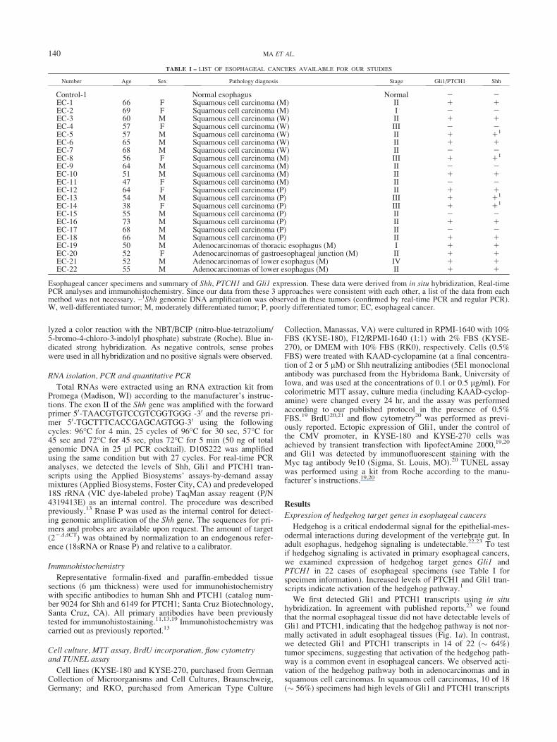
We first detected Gli1 and PTCH1 transcripts using in situhybridization. In agreement with published reports,23 we foundthat the normal esophageal tissue did not have detectable levels ofGli1 and PTCH1, indicating that the hedgehog pathway is not nor-mally activated in adult esophageal tissues (Fig. 1a). In contrast,we detected Gli1 and PTCH1 transcripts in 14 of 22 (� 64%)tumor specimens, suggesting that activation of the hedgehog path-way is a common event in esophageal cancers. We observed acti-vation of the hedgehog pathway both in adenocarcinomas and insquamous cell carcinomas. In squamous cell carcinomas, 10 of 18(� 56%) specimens had high levels of Gli1 and PTCH1 transcripts
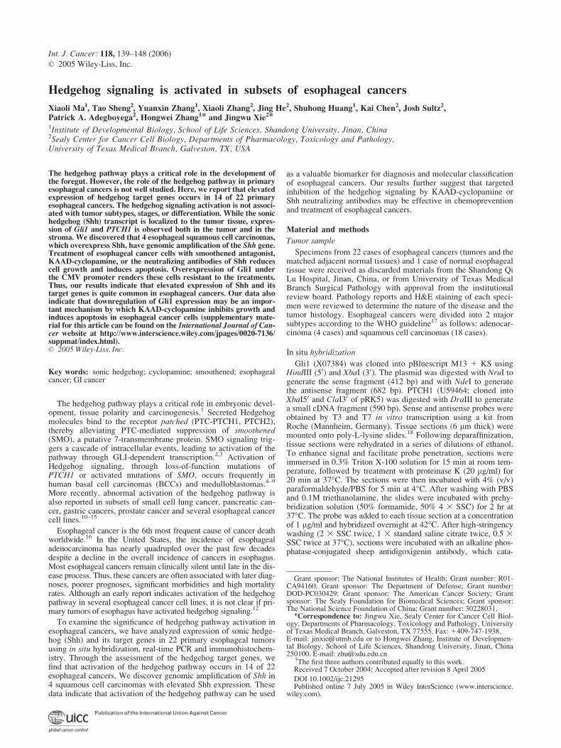
TABLE I – LIST OF ESOPHAGEAL CANCERS AVAILABLE FOR OUR STUDIES
Number Age Sex Pathology diagnosis Stage Gli1/PTCH1 Shh
Control-1 Normal esophagus Normal 2 2EC-1 66 F Squamous cell carcinoma (M) II 1 1EC-2 69 F Squamous cell carcinoma (M) I 2 2EC-3 60 M Squamous cell carcinoma (W) II 1 1EC-4 57 F Squamous cell carcinoma (W) III 2 2EC-5 57 M Squamous cell carcinoma (W) II 1 11
EC-6 65 M Squamous cell carcinoma (W) II 1 1EC-7 68 M Squamous cell carcinoma (W) II 2 2EC-8 56 F Squamous cell carcinoma (M) III 1 11
EC-9 64 M Squamous cell carcinoma (M) II 2 2EC-10 51 M Squamous cell carcinoma (M) II 1 1EC-11 47 F Squamous cell carcinoma (M) II 2 2EC-12 64 F Squamous cell carcinoma (P) II 1 1EC-13 54 M Squamous cell carcinoma (P) III 1 11
EC-14 38 F Squamous cell carcinoma (P) III 1 11
EC-15 55 M Squamous cell carcinoma (P) II 2 2EC-16 73 M Squamous cell carcinoma (P) II 1 1EC-17 68 M Squamous cell carcinoma (P) II 2 2EC-18 66 M Squamous cell carcinoma (P) II 1 1EC-19 50 M Adenocarcinomas of thoracic esophagus (M) I 1 1EC-20 52 F Adenocarcinomas of gastroesophageal junction (M) II 1 1EC-21 52 M Adenocarcinomas of lower esophagus (M) IV 1 1EC-22 55 M Adenocarcinomas of lower esophagus (M) II 1 1
Esophageal cancer specimens and summary of Shh, PTCH1 and Gli1 expression. These data were derived from in situ hybridization, Real-timePCR analyses and immunohistochemistry. Since our data from these 3 approaches were consistent with each other, a list of the data from eachmethod was not necessary. –1Shh genomic DNA amplification was observed in these tumors (confirmed by real-time PCR and regular PCR).W, well-differentiated tumor; M, moderately differentiated tumor; P, poorly differentiated tumor; EC, esophageal cancer.
140 MA ET AL.
(see Table I for details). All 4 adenocarcinomas had detectableexpression of Gli1 and PTCH1 (Table I). These data indicate thatactivation of the hedgehog pathway occurs frequently in esopha-geal cancers.
Further analyses did not reveal any association of hedgehog tar-get gene expression with the tumor stage or differentiation. In situhybridization data indicate that transcripts of Gli1 (Fig. 1b and c)and PTCH1 (not shown here) are detectable both in the tumor
(indicated by arrows) and in the adjacent stroma tissue (indicatedby arrowheads). These data suggest that hedgehog signaling mayinvolve epithelium/stroma interactions during development ofesophageal cancer.
To confirm the in situ hybridization data, we performed real-time PCR analyses to detect the levels of Gli1 and PTCH1transcripts. We found that Gli1 and PTCH1 transcripts fromesophageal tumors were several folds higher than those from
FIGURE 1 – Elevated expression of Gli1 and PTCH1 in primary esophageal tumors. Gli1 transcript (blue as positive) was detected by in situhybridization in the normal control (a) and esophageal cancers (b–d). (c0) and (c) are from the same tumor, with (c0) being derived from the Gli1sense probe. Similarly, (d0) is the sense probe control of (d). Expression of Gli1 transcript was strong in the tumor (indicated by arrows) andweak in the stroma (indicated by arrowheads). The pattern of PTCH1 transcript was similar to those of Gli1 (figures not shown), indicating acti-vation of the hedgehog pathway in esophageal cancers. The data are summarized in Table I.
141HEDGEHOG SIGNALING
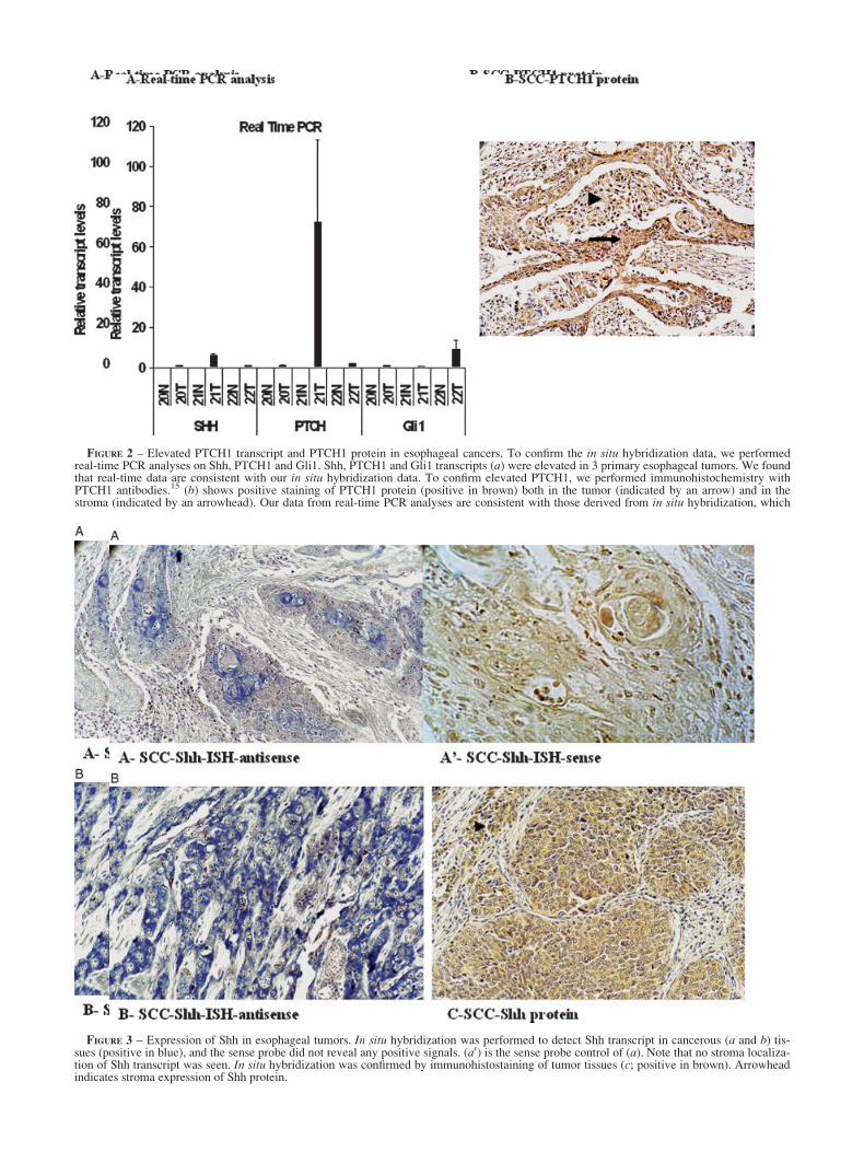
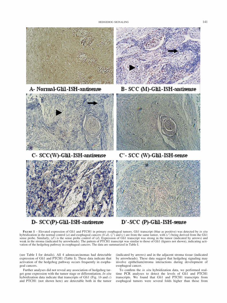
FIGURE 2 – Elevated PTCH1 transcript and PTCH1 protein in esophageal cancers. To confirm the in situ hybridization data, we performedreal-time PCR analyses on Shh, PTCH1 and Gli1. Shh, PTCH1 and Gli1 transcripts (a) were elevated in 3 primary esophageal tumors. We foundthat real-time data are consistent with our in situ hybridization data. To confirm elevated PTCH1, we performed immunohistochemistry withPTCH1 antibodies.15 (b) shows positive staining of PTCH1 protein (positive in brown) both in the tumor (indicated by an arrow) and in thestroma (indicated by an arrowhead). Our data from real-time PCR analyses are consistent with those derived from in situ hybridization, which
FIGURE 3 – Expression of Shh in esophageal tumors. In situ hybridization was performed to detect Shh transcript in cancerous (a and b) tis-sues (positive in blue), and the sense probe did not reveal any positive signals. (a0) is the sense probe control of (a). Note that no stroma localiza-tion of Shh transcript was seen. In situ hybridization was confirmed by immunohistostaining of tumor tissues (c; positive in brown). Arrowheadindicates stroma expression of Shh protein.
the matched normal tissues (Fig. 2a shows levels of Shh,PTCH1 and Gli1), confirming that expression of PTCH1 andGli1 is elevated in the tumor tissues. Expression of PTCH1 inthe tumor was further confirmed by immunohistochemistry(Fig. 2b).11,13 All tissues with detectable PTCH1 protein hadelevated PTCH1 transcript. In agreement with the in situhybridization results, PTCH1 protein was detected both in thetumor (indicated by an arrow in Fig. 2b) and in the stroma(indicated by an arrowhead in Fig. 2b). Data derived fromin situ hybridization, real-time PCR and immunohistochemistryanalyses all indicate that activation of the hedgehog pathway isa common event in esophageal cancers.
Expression of Shh in esophageal cancers
It is reported that Shh overexpression may be responsible foractivation of the hedgehog pathway in pancreatic cancer and sev-eral primary gastric cancers.11,12 To test this possibility in esopha-geal cancer, we first examined expression of Shh in esophagealspecimens by in situ hybridization. As expected, Shh expressionwas undetectable in the normal esophageal tissue (data notshown). In contrast, many of the primary tumors expressed a highlevel of Shh transcript (Figs. 2a and 3a and b, Table I). The Shhtranscript was detectable specifically in the tumor, not in thestroma (Fig. 3a and b), suggesting that the tumor cells are thesource for Shh expression. Shh expression was associated with
detectable levels of Gli1 and PTCH1 transcripts, suggesting animportant role of Shh in activating hedgehog pathway in esopha-geal cancers.
Furthermore, we detected Shh protein by immunohistochemis-try using specific antibodies.11 In agreement with the in situhybridization data, we found that tumors with Shh transcript hadhigher levels of Shh protein (Fig. 3c). As a secreted molecule,sonic hedgehog protein was also detected in the stroma (indicatedas arrowhead in Fig. 3c). We believe that the secreted sonic hedge-hog protein may be responsible for elevated expression of Gli1and PTCH1 transcripts in the tumor as well as in the stroma(Fig. 1). These results suggest a paracrine signaling mechanism ofsonic hedgehog in esophageal cancers.
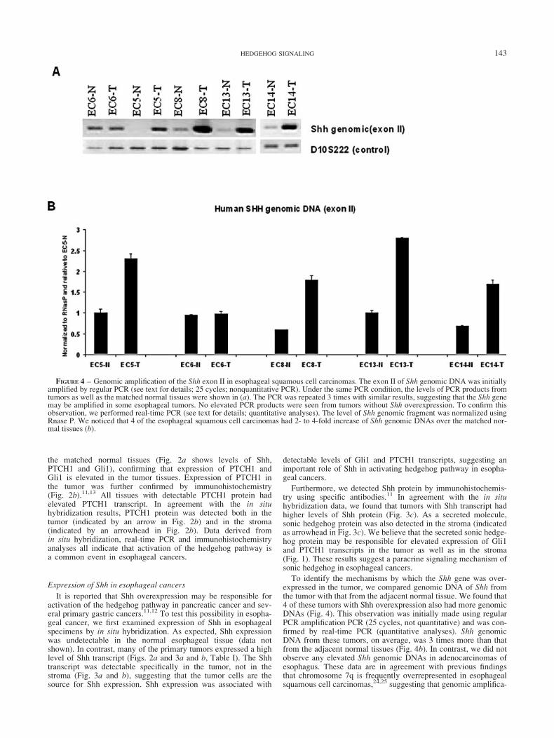
To identify the mechanisms by which the Shh gene was over-expressed in the tumor, we compared genomic DNA of Shh fromthe tumor with that from the adjacent normal tissue. We found that4 of these tumors with Shh overexpression also had more genomicDNAs (Fig. 4). This observation was initially made using regularPCR amplification PCR (25 cycles, not quantitative) and was con-firmed by real-time PCR (quantitative analyses). Shh genomicDNA from these tumors, on average, was 3 times more than thatfrom the adjacent normal tissues (Fig. 4b). In contrast, we did notobserve any elevated Shh genomic DNAs in adenocarcinomas ofesophagus. These data are in agreement with previous findingsthat chromosome 7q is frequently overrepresented in esophagealsquamous cell carcinomas,24,25 suggesting that genomic amplifica-
FIGURE 4 – Genomic amplification of the Shh exon II in esophageal squamous cell carcinomas. The exon II of Shh genomic DNA was initiallyamplified by regular PCR (see text for details; 25 cycles; nonquantitative PCR). Under the same PCR condition, the levels of PCR products fromtumors as well as the matched normal tissues were shown in (a). The PCR was repeated 3 times with similar results, suggesting that the Shh genemay be amplified in some esophageal tumors. No elevated PCR products were seen from tumors without Shh overexpression. To confirm thisobservation, we performed real-time PCR (see text for details; quantitative analyses). The level of Shh genomic fragment was normalized usingRnase P. We noticed that 4 of the esophageal squamous cell carcinomas had 2- to 4-fold increase of Shh genomic DNAs over the matched nor-mal tissues (b).
143HEDGEHOG SIGNALING
tion of the Shh gene in some of these esophageal cancers may beresponsible for Shh overexpression.
Targeted inhibition of hedgehog pathway and esophagealcancer cells
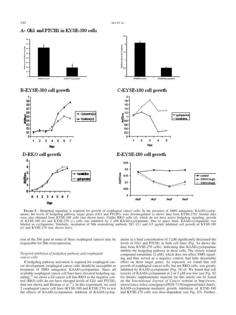
If hedgehog pathway activation is required for esophageal can-cer development, esophageal cancer cells should be susceptible totreatment of SMO antagonist, KAAD-cyclopamine. Since allavailable esophageal cancer cell lines have elevated hedgehog sig-naling,12 we chose a GI cancer cell line RKO as the negative con-trol (RKO cells do not have elevated levels of Gli1 and PTCH1;data not shown and Berman et al.12). In this experiment, we used2 esophageal cancer cell lines (KYSE-180 and KYSE-270) to testthe effects of KAAD-cyclopamine. Addition of KAAD-cyclop-
amine at a final concentration of 2 lM significantly decreased thelevels of Gli1 and PTCH1 in both cell lines (Fig. 5a shows thedata from KYSE-270 cells), indicating that KAAD-cyclopamineinhibits the hedgehog pathway in these cells. The closely relatedcompound tomatidine (2 lM), which does not affect SMO signal-ing and thus served as a negative control, had little discernibleeffect on these target genes. As expected, we found that cellgrowth of esophageal cancer cells, but not RKO cells, was greatlyinhibited by KAAD-cyclopamine (Fig. 5b–d). We found that celltoxicity of KAAD-cyclopamine at 2 or 5 lM was low (see Fig. S2for details; supplementary material for this article can be foundon the International Journal of Cancer website at http://www.interscience.wiley.com/jpages/0020-7136/suppmat/index.html).KAAD-cyclopamine-mediated growth inhibition of KYSE-180and KYSE-270 cells was dose-dependent (see Fig. S3). Further-
FIGURE 5 – Hedgehog signaling is required for growth of esophageal cancer cells. In the presence of SMO antagonist, KAAD-cyclop-amine, the levels of hedgehog pathway target genes (Gli1 and PTCH1) were downregulated (a shows data from KYSE-270). Similar datawere also obtained from KYSE-180 cells (not shown here). Unlike RKO cells (d), which do not have active hedgehog signaling, growthof KYSE-180 (b) and KYSE-270 (c) cells was inhibited by 2 lM KAAD-cyclopamine. Due to space limit, KAAD-cyclopamine waslabeled as cyclopamine. Similarly, incubation of Shh neutralizing antibody 5E1 (0.1 and 0.5 lg/ml) inhibited cell growth of KYSE-180(e) and KYSE-270 (not shown here).
144 MA ET AL.
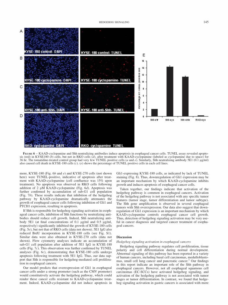
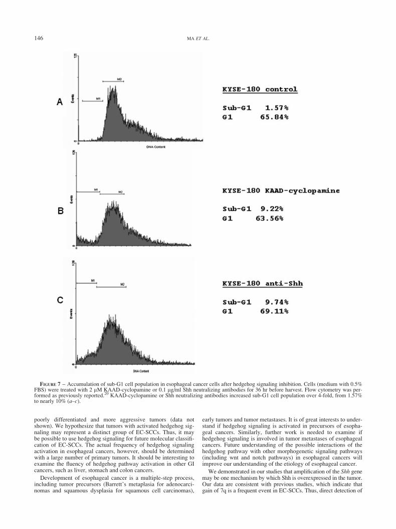
more, KYSE-180 (Fig. 6b and e) and KYSE-270 cells (not shownhere) were TUNEL-positive, indicative of apoptosis after treat-ment with KAAD-cyclopamine (cell confluence was 15% upontreatment). No apoptosis was observed in RKO cells followingaddition of 2 lM KAAD-cyclopamine (Fig. 6d). Apoptosis wasfurther confirmed by accumulation of sub-G1 cell population(Fig. 7b). These results indicate that inhibition of the hedgehogpathway by KAAD-cyclopamine dramatically attenuates thegrowth of esophageal cancer cells following inhibition of Gli1 andPTCH1 expression, resulting in apoptosis.
If Shh is responsible for hedgehog signaling activation in esoph-ageal cancer cells, inhibition of Shh functions by neutralizing anti-bodies should reduce cell growth. Indeed, Shh neutralizing anti-body 5E1 (at final concentrations of 0.1 lg/ml and 0.5 lg/ml,respectively) significantly inhibited the growth of KYSE-180 cells(Fig. 5e), but not that of RKO cells (data not shown). 5E1 IgG alsoreduced BrdU incorporation in KYSE-180 cells (see Fig. S1).Similar data were also obtained in KYSE-270 cells (data notshown). Flow cytometry analyses indicate an accumulation ofsub-G1 cell population after addition of 5E1 IgG in KYSE-180cells (Fig. 7c). This observation was further confirmed by TUNELanalyses (Fig. 6c), demonstrating that KYSE-180 cells undergoapoptosis following treatment with 5E1 IgG. Thus, our data sup-port that Shh is responsible for hedgehog-mediated cell prolifera-tion in esophageal cancers.
Our model predicts that overexpression of Gli1 in esophagealcancer cells under a strong promoter (such as the CMV promoter)would constitutively activate the hedgehog pathway, which couldrender these cancer cells resistant to KAAD-cyclopamine treat-ment. Indeed, KAAD-cyclopamine did not induce apoptosis in
Gli1-expressing KYSE-180 cells, as indicated by lack of TUNELstaining (Fig. 8). Thus, downregulation of Gli1 expression may bean important mechanism by which KAAD-cyclopamine inhibitsgrowth and induces apoptosis of esophageal cancer cells.
Taken together, our findings indicate that activation of thehedgehog pathway is common in esophageal cancers. Activationof the hedgehog pathway is not associated with any specific tumorfeatures (tumor stage, tumor differentiation and tumor subtype).The Shh gene amplification is observed in several esophagealtumors with Shh overexpression. Our data also suggest that down-regulation of Gli1 expression is an important mechanism by whichKAAD-cyclopamine controls esophageal cancer cell growth.Thus, detection of hedgehog signaling activation may be very use-ful in cancer diagnosis and targeted cancer treatment of esopha-geal cancers.
Discussion
Hedgehog signaling activation in esophageal cancers
Hedgehog signaling pathway regulates cell proliferation, tissuepolarity and cell differentiation during normal development.Abnormal signaling of this pathway has been reported in a varietyof human cancers, including basal cell carcinomas, medulloblasto-mas, small cell lung cancer and pancreatic cancer.1 Our findingsin this report indicate an important role of the Shh pathway inesophageal cancers. However, not all esophageal squamous cellcarcinomas (EC-SCCs) have activated hedgehog signaling, andactivation of the hedgehog pathway is not associated with tumorstages or tumor differentiation. In contrast, we found that hedge-hog signaling activation in gastric cancers is associated with more
FIGURE 6 – KAAD-cyclopamine and Shh neutralizing antibodies induce apoptosis in esophageal cancer cells. TUNEL assay revealed apopto-sis (red) in KYSE180 (b) cells, but not in RKO cells (d), after treatment with KAAD-cyclopamine (labeled as cyclopamine due to space) for36 hr. The tomatidine-treated control group had very few TUNEL positive cells (a and e). Similarly, Shh neutralizing antibody 5E1 (0.1 lg/ml)also caused cell death in KYSE-180 cells (c). (e) shows the percentage of TUNEL positive cells in each cell lines.
145HEDGEHOG SIGNALING
poorly differentiated and more aggressive tumors (data notshown). We hypothesize that tumors with activated hedgehog sig-naling may represent a distinct group of EC-SCCs. Thus, it maybe possible to use hedgehog signaling for future molecular classifi-cation of EC-SCCs. The actual frequency of hedgehog signalingactivation in esophageal cancers, however, should be determinedwith a large number of primary tumors. It should be interesting toexamine the fluency of hedgehog pathway activation in other GIcancers, such as liver, stomach and colon cancers.
Development of esophageal cancer is a multiple-step process,including tumor precursors (Barrett’s metaplasia for adenocarci-nomas and squamous dysplasia for squamous cell carcinomas),
early tumors and tumor metastases. It is of great interests to under-stand if hedgehog signaling is activated in precursors of esopha-geal cancers. Similarly, further work is needed to examine ifhedgehog signaling is involved in tumor metastases of esophagealcancers. Future understanding of the possible interactions of thehedgehog pathway with other morphogenetic signaling pathways(including wnt and notch pathways) in esophageal cancers willimprove our understanding of the etiology of esophageal cancer.
We demonstrated in our studies that amplification of the Shh genemay be one mechanism by which Shh is overexpressed in the tumor.Our data are consistent with previous studies, which indicate thatgain of 7q is a frequent event in EC-SCCs. Thus, direct detection of
FIGURE 7 – Accumulation of sub-G1 cell population in esophageal cancer cells after hedgehog signaling inhibition. Cells (medium with 0.5%FBS) were treated with 2 lM KAAD-cyclopamine or 0.1 lg/ml Shh neutralizing antibodies for 36 hr before harvest. Flow cytometry was per-formed as previously reported.20 KAAD-cyclopamine or Shh neutralizing antibodies increased sub-G1 cell population over 4-fold, from 1.57%to nearly 10% (a–c).
146 MA ET AL.
Shh expression in primary esophageal cancer specimens may be aneffective way for diagnosis for subsets of EC-SCC.
Therapeutic perspective of esophageal cancer through targetedinhibition of hedgehog pathway
Addition of smoothened antagonist, KAAD-cyclopamine, orShh neutralizing antibodies in culture medium of esophageal can-cer cells causes inhibition of cell growth, resulting in apoptosis.These data suggest that hedgehog signaling inhibitors may beeffective in future treatment of esophageal cancers. Our prelimi-nary data further indicate that activation of caspases-8 and -3occurs in KYSE-180 cells after treatment with Shh antibodies(data not shown), suggesting that a death receptor pathway is acti-vated. Additional understanding of apoptotic mechanisms will behelpful for future design of novel drugs for esophageal cancers.
We further demonstrated that overexpression of Gli1 preventscyclopamine-mediated apoptosis in esophageal cancer cells, fur-ther supporting the specificity of KAAD-cyclopamine. Our recentstudies indicated that chronic oral administration of KAAD-cyclopamine of Ptch11/2 mice did not affect the overall survivalof the mice,20 which provides a foundation for clinical trials ofKAAD-cyclopamine on esophageal cancers. Thus, it is quite pos-sible in the future, with availability of a specific SMO antagonist,KAAD-cyclopamine, to treat the subsets of esophageal cancer inwhich the hedgehog pathway is activated.
Acknowledgements
The authors thank Huiping Guo for technical support in real-time PCR analysis.
References
1. Pasca di Magliano M, Hebrok M. Hedgehog signalling in cancer for-mation and maintenance. Nat Rev Cancer 2003;3:903–11.
2. Taipale J, Beachy PA. The hedgehog and Wnt signalling pathways incancer. Nature 2001;411:349–54.
3. Ingham PW. Transducing hedgehog: the story so far. EMBO J 1998;17:3505–11.
4. Hahn H, Wicking C, Zaphiropoulous PG, Gailani MR, Shanley S,Chidambaram A, Vorechovsky I, Holmberg E, Unden AB, Gillies S,Negus K, Smyth I, et al. Mutations of the human homolog of Droso-phila patched in the nevoid basal cell carcinoma syndrome. Cell1996;85:841–51.
5. Johnson RL, Rothman AL, Xie J, Goodrich LV, Bare JW, BonifasJM, Quinn AG, Myers RM, Cox DR, Epstein EH Jr, Scott MP.Human homolog of patched, a candidate gene for the basal cell nevussyndrome. Science 1996;272:1668–71.
6. Xie J, Johnson RL, Zhang X, Bare JW, Waldman FM, Cogen PH,Menon AG, Warren RS, Chen LC, Scott MP, Epstein EH Jr. Muta-tions of the PATCHED gene in several types of sporadic extracutane-ous tumors. Cancer Res 1997;57:2369–72.
7. Raffel C, Jenkins RB, Frederick L, Hebrink D, Alderete B, Fults DW,James CD. Sporadic medulloblastomas contain PTCH mutations.Cancer Res 1997;57:842–5.
8. Xie J, Murone M, Luoh SM, Ryan A, Gu Q, Zhang C, Bonifas JM,Lam CW, Hynes M, Goddard A, Rosenthal A, Epstein EH Jr, et al.Activating smoothened mutations in sporadic basal-cell carcinoma.Nature 1998;391:90–2.
9. Taylor MD, Liu L, Raffel C, Hui CC, Mainprize TG, Zhang X, Aga-tep R, Chiappa S, Gao L, Lowrance A, Hao A, Goldstein AM, et al.Mutations in SUFU predispose to medulloblastoma. Nat Genet 2002;31:306–10.
10. Watkins DN, Berman DM, Burkholder SG, Wang B, Beachy PA,Baylin SB. Hedgehog signalling within airway epithelial progenitorsand in small-cell lung cancer. Nature 2003;422:313–7.
11. Thayer SP, Di Magliano MP, Heiser PW, Nielsen CM, Roberts DJ,Lauwers GY, Qi YP, Gysin S, Fernandez-Del Castillo C, Yajnik V,Antoniu B, McMahon M, et al. Hedgehog is an early and late media-tor of pancreatic cancer tumorigenesis. Nature 2003;425:851–6.
12. Berman DM, Karhadkar SS, Maitra A, Montes De Oca R,Gerstenblith MR, Briggs K, Parker AR, Shimada Y, Eshleman JR,Watkins DN, Beachy PA. Widespread requirement for Hedgehogligand stimulation in growth of digestive tract tumours. Nature 2003;425:846–51.
13. Sheng T, Li C-X, Zhang X, Chi S, He N, Chen K, McCormick F,Gatalica Z, Xie J. Activation of the hedgehog pathway in advancedprostate cancer. Mol Cancer 2004;3:29.
14. Sanchez P, Hernandez AM, Stecca B, Kahler AJ, DeGueme AM, Bar-rett A, Beyna M, Datta MW, Datta S, Ruiz i Altaba A. Inhibition ofprostate cancer proliferation by interference with SONIC HEDGE-HOG-GLI1 signaling. Proc Natl Acad Sci USA 2004;101:12561–6.
15. Karhadkar SS, Bova GS, Abdallah N, Dhara S, Gardner D, Maitra A,Isaacs JT, Berman DM, Beachy PA. Hedgehog signalling in prostateregeneration, neoplasia and metastasis. Nature 2004;431:707–12.
16. Pisani P, Parkin DM, Bray F, Ferlay J. Estimates of the worldwidemortality from 25 cancers in 1990. Int J Cancer 1999;83:18–29.
17. Sarbia M, Becker KF, Hofler H. Pathology of upper gastrointestinalmalignancies. Semin Oncol 2004;31:465–75.
18. Unden A, Zaphiropoulos PG, Toftgard R, Stahle-Backdahl M. Humanpatched (PTCH) mRNA is overexpressed consistently in tumor cellsof both familial and sporadic basal cell carcinoma. Cancer Res 1997;57:2336–40.
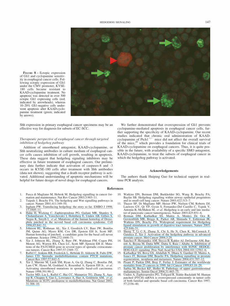
FIGURE 8 – Ectopic expressionof Gli1 and cyclopamine sensitiv-ity in esophageal cancer cells. Fol-lowing ectopic expression of Gli1under the CMV promoter, KYSE-180 cells became resistant toKAAD-cyclopamine treatment. Noapoptosis was detected in over 500ectopic Gli1 expressing cells (red;indicated by arrowheads), whereas10–20% Gli1-negative cells under-went apoptosis after KAAD-cyclo-pamine treatment (green; indicatedby arrows).
147HEDGEHOG SIGNALING

19. Li C, Chi S, He N, Zhang X, Guicherit O, Wagner R, Tyring S, Xie J.IFNalpha induces Fas expression and apoptosis in hedgehog pathwayactivated BCC cells through inhibiting Ras-Erk signaling. Oncogene2004;23:1608–17.
20. Athar M, Li C, Tang X, Chi S, Zhang X, Kim AL, Tyring SK, Kope-lovich L, Hebert J, Epstein EH Jr, Bickers DR, Xie J. Inhibition ofsmoothened signaling prevents ultraviolet B-induced basal cell carci-nomas through regulation of Fas expression and apoptosis. CancerRes 2004;64:7545–52.
21. Xie J, Aszterbaum M, Zhang X, Bonifas JM, Zachary C, Epstein E,McCormick F. A role of PDGFRalpha in basal cell carcinoma prolif-eration. Proc Natl Acad Sci USA 2001;98:9255–9.
22. van den Brink GR, Hardwick JC, Nielsen C, Xu C, ten KateFJ, Glickman J, van Deventer SJ, Roberts DJ, PeppelenboschMP. Sonic hedgehog expression correlates with fundic gland dif-
ferentiation in the adult gastrointestinal tract. Gut 2002;51:628–33.
23. Arsic D, Keenan J, Quan QB, Beasley S. Differences in the levels ofSonic hedgehog protein during early foregut development caused byexposure to adriamycin give clues to the role of the Shh gene in oeso-phageal atresia. Pediatr Surg Int 2003;19:463–6.
24. Yen CC, Chen YJ, Chen JT, Hsia JY, Chen PM, Liu JH, Fan FS, ChiouTJ, Wang WS, Lin CH. Comparative genomic hybridization of esopha-geal squamous cell carcinoma: correlations between chromosomal aber-rations and disease progression/prognosis. Cancer 2001;92:2769–77.
25. Walch AK, Zitzelsberger HF, Bruch J, Keller G, Angermeier D,Aubele MM, Mueller J, Stein H, Braselmann H, Siewert JR, Hofler H,Werner M. Chromosomal imbalances in Barrett’s adenocarcinomaand the metaplasia-dysplasia-carcinoma sequence. Am J Pathol2000;156:555–66.
148 MA ET AL.