photodynamic antimicrobial chemotherapy using zinc
TRANSCRIPT
Photodynamic antimicrobialchemotherapy using zincphthalocyanine derivatives intreatment of bacterial skin infection
Zhuo ChenYaxin ZhangDong WangLinsen LiShanyong ZhouJoy H. HuangJincan ChenPing HuMingdong Huang
Downloaded From: https://www.spiedigitallibrary.org/journals/Journal-of-Biomedical-Optics on 08 Jan 2022Terms of Use: https://www.spiedigitallibrary.org/terms-of-use
Photodynamic antimicrobial chemotherapy usingzinc phthalocyanine derivatives in treatment ofbacterial skin infection
Zhuo Chen,a,b,* Yaxin Zhang,a,b,c Dong Wang,a,b,c Linsen Li,d Shanyong Zhou,a Joy H. Huang,a Jincan Chen,aPing Hu,a and Mingdong Huanga,b,*aChinese Academy of Sciences, Fujian Institute of Research on the Structure of Matter, State Key Laboratory of Structural Chemistry,155 West Yangqiao Road, Fuzhou, Fujian 350002, ChinabGraduate University of Chinese Academy of Sciences, 19 Yuquan Road, Shijingshan District, Beijing 100049, ChinacFujian Normal University, 1 Keji Road, University Town, Fuzhou, Fujian 350117, ChinadShenyang Medical College, 146 North Huanghe Main Street, Shenyang, Liaoning 110034, China
Abstract. Photodynamic antimicrobial chemotherapy (PACT) is an effective method for killing bacterial cells inview of the increasing problem of multiantibiotic resistance. We herein reported the PACT effect on bacteriainvolved in skin infections using a zinc phthalocyanine derivative, pentalysine β-carbonylphthalocyanine zinc(ZnPc-ðLysÞ5). Compared with its anionic ZnPc counterpart, ZnPc-ðLysÞ5 showed an enhanced antibacterialefficacy in vitro and in an animal model of localized infection. Meanwhile, ZnPc-ðLysÞ5 was observed to signifi-cantly reduce the wound skin blood flow during wound healing, indicating an anti-inflammation activity. Thisstudy provides new insight on the mechanisms of PACT in bacterial skin infection. © 2016 Society of Photo-Optical
Instrumentation Engineers (SPIE) [DOI: 10.1117/1.JBO.21.1.018001]
Keywords: photodynamic antimicrobial chemotherapy; zinc phthalocyanine; bacterial infection; skin; Propionibacterium acnes;Staphylococcus aureus.
Paper 150615PRR received Sep. 16, 2015; accepted for publication Dec. 4, 2015; published online Jan. 7, 2016.
1 IntroductionThe most common bacterial infections, such as cellulitis,abscesses, and postsurgical infections, are usually caused bypathogens like Staphylococcus aureus1 and Propionibacteriumacnes2 that may lead to serious local and systemic complica-tions.3 For example, S. aureus can cause a range of illnesses,from minor skin infections to life-threatening diseases suchas pneumonia, meningitis, osteomyelitis, endocarditis, toxicshock syndrome, bacteremia, and sepsis.4 In the past 60 years,antibiotics have been critical in the fight against infectiousdisease caused by these bacteria and other microbes. However,the broad application of antibiotics leads to bacterial resistanceand becomes an increasing public health problem. Nowadays,about 70% of the bacteria that cause infections in hospitalsare resistant to at least one of the drugs most commonly usedfor treatment. The threat of bacterium S. aureus is not onlydue to its distribution and pathogenicity but also to its abilityto overcome antimicrobial agents.5
Antimicrobial peptides, also called host defense peptides, arean important component of the natural defenses of most livingorganisms against invading pathogens and have become animportant research direction in the past two decades.6,7 Thediscovery of natural antibacterial peptides, including histatins,defensins, cathelicidins, magainins, cecropins, and tachyplasins,has provided a new way to fight antibiotic-resistant microorgan-isms. The wide spectrum of antimicrobial activities reported forthese molecules suggests that they could be used in the treatmentof viral or parasitic infections. However, there are many general
obstacles to move antimicrobial peptides to clinical applica-tions,8 including the toxicity against eukaryotic cells, the stabil-ity of the peptides in vivo, the potential for cross-resistance, andthe high cost of production.
Photodynamic antimicrobial chemotherapy (PACT) is a newmethod for killing bacterial cells.9 PACT treatment utilizes vis-ible or near-infrared light at the appropriate wavelength to excitethe nontoxic photosensitizer. The excited photosensitizer under-goes intersystem crossing to long-lived triplet states and, in thepresence of oxygen, transfers its energy to molecular oxygenand generates reactive oxygen species such as singlet oxygenand hydroxyl radical, which are responsible for the killing ofmicrobial cells nearby. One of the advantages of PACT in theinactivation of microorganisms is that both antibiotic-sensitiveand -resistant strains can be successfully photoinactivated. Theother advantage is that repeated photosensitization of bacterialcells does not induce a selection of resistant strains.10 AlthoughPACT is gaining increasing acceptance for the treatment oflocally occurring infections such as psoriasis11 and sclero-derma12 in dermatology, it is not, at present, a mainstream thera-peutic option.
As a key component of PACT, an ideal photosensitizershould have high absorption coefficients in the near-infraredregion, where light has deep penetration into tissues, and highphotostability to minimize photobleaching. Phthalocyanines,a versatile class of macrocyclic compounds featured witha high fluorescence quantum yield, long triplet lifetimes, andhigh triplet quantum yields, are gathering growing interest aseffective photosensitizers in targeted photodynamic therapy and
*Address all correspondence to: Zhuo Chen, E-mail: [email protected];Mingdong Huang, E-mail: [email protected] 1083-3668/2016/$25.00 © 2016 SPIE
Journal of Biomedical Optics 018001-1 January 2016 • Vol. 21(1)
Journal of Biomedical Optics 21(1), 018001 (January 2016)
Downloaded From: https://www.spiedigitallibrary.org/journals/Journal-of-Biomedical-Optics on 08 Jan 2022Terms of Use: https://www.spiedigitallibrary.org/terms-of-use
imaging of tumors due to their longer wavelength band absorp-tion (λmax 600 to 700 nm) and higher extinction coefficients(ε ∼ 110;000). Selectivity to target cells rather than host mam-malian cells is another key property for the photosensitizer. Inaddition, photosensitizers with positive charges tend to bind tocells that carry negative charges on their surfaces.
Conjugation of antimicrobial peptides to phthalocynine pho-tosensitizer is one strategy to develop antimicrobial therapy.ZnPc-ðLysÞ5, a phthalocyanine derivative with five positivecharges, prepared as a high purity single isomer, was previouslyreported13 as an effective photosensitizer with high activity inboth cultured tumor cells and experimental animal tumors.14
To further investigate the photodynamic inactivation ofZnPc-ðLysÞ5 on microorganisms involved in skin bacterialinfections, we evaluated the antibacterial efficacy of ZnPc-ðLysÞ5 in vitro and in vivo using the key pathogenic factorsP. acnes and S. aureus. This study may provide a safe and effec-tive approach for the treatment of bacterial skin infections.
2 Experimental Techniques
2.1 Preparation of Photosensitizers
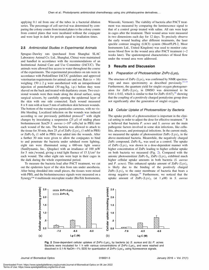
According to our early published protocols,14,15 the unsymmetri-cal ZnPc-ðLysÞ5 was prepared via the activation of the carboxylicacid group on ZnPc-COOH by 1-ethyl-3-(3-dimethylamino-propyl) carbodiimide and N-hydroxysuccinimide, and a couplingto pentalysine. The synthesized ZnPc-ðLysÞ5 was characterizedby 1H NMR [AV-400, Bruker, 400 MHz, in dimethyl sulphoxide(DMSO)], 13C NMR (75 MHz, in DMSO), FTIR (Magna-IR750, Nicolett), and high-resolution mass spectra–electrosprayionization (DECAX-30000 LCQ Deca XP massspectrometer). The UV–vis absorption spectrum of ZnPc-ðLysÞ5 in dimethylfomamide was recorded from 300 to800 nm using quartz cuvettes with 1-cm path length on aLambda-35 UV/vis spectrometer (PerkinElmer, Massachusetts)in DMSO. Analytical high-performance liquid chromatography(HPLC) was carried out on a C-18 reversed-phase HPLC system(Dalian Elite Analytical Instruments Co., Ltd., Dalian, China;Column: SinoChrom ODS-BP 250 × 4.6 mm, 5 μm), using a lin-ear gradient from 50% to 100% (v/v) of methanol:acetonitrile(1∶1) at a flow rate of 1 ml∕min. The UV–vis absorptionspectrum of photosensitizer ZnPc-ðLysÞ5 in DMSO (Fig. 1)was typical of ZnPc with the strongest absorption at678 nm (extinction coefficient of 118;380 L · mol−1 · cm−1).Furthermore, singlet oxygen (1O2) is believed to be the majorcytotoxic agent involved in photodynamic therapy. The quantumyield of singlet oxygen generation of ZnPc-ðLysÞ5 was measuredin reference to ZnPc, which has a quantum yield of0.67 in DMSO.16
Zinc phthalocyanine tetrasulfonate (ZnPc-S4) with anabsorption peak at 680 nm was kindly provided by ProfessorNaisheng Chen of Fuzhou University, China.
2.2 Microorganism and Culture Condition
As the standard bacteria used in antibacterial activity testsaccording to the National Standard of the People’s Republicof China in detection and control of pathogens (GB4789.28-2013), P. acnes (ATCC 6919) and S. aureus subsp. aureus(ATCC 6538) were purchased from Beijing ZhongyuanLtd. S. aureus were cultured in nutrient agars, and P. acneswere cultured in thioglycollate medium in anaerobic bags.
Thioglycollate medium is a semisolid nutrient medium contain-ing a low concentration of agar to prevent convection of oxygenfrom the surface. The thioglycollate medium was boiled beforeuse in order to drive off the oxygen and inoculated withoutexcessive shaking of the medium after cooling.
A luminescent strain of S. aureus Xen29 (NCTC8532),a derivative of the biofilm forming S. aureus 12600 that pos-sesses a stable copy of the modified Photorhabdus luminescensluxABCDE operon at a single integration site on the bacterialchromosome, was purchased from Caliper Life Sciences,Inc., and grown at 37°C using Luria–Bertani broth containingkanamycin (200 μg∕ml to select for resistance encoded by theplasmid) to an absorbance of 0.5 at 600 nm correspondingto 1.44 × 10⁸ organisms∕ml.
2.3 Cellular Uptake of Photosensitizer by Bacteria
Aliquots of microorganism suspension [106 colony forming unit(CFU)/ml] were incubated in 96-multiwell plates (Falcon) withphotosensitizer ZnPc-ðLysÞ5 at different concentrations (10−7,10−6.5, and 10−6 M) for 1 h at 37°C. The exponentially growingcells were then washed with sterile phosphate buffer saline(PBS) before lysis with NaOH [0.1 N, 1.0 ml with 1% sodiumdodecyl sulfate (SDS)] to give a homogeneous solution. Thefluorescence of the cell extract was measured on a microplatereader (Synergy 4, BioTek Instruments). The concentration ofcellular protein was determined using a bicinchoninic acid pro-tein assay kit (Pierce, Thermo Fisher Scientific). Standardcurves were made with cell lysates treated as above withknown added amounts of bovine serum albumin. Results areexpressed as nmol of phthalocyanine per mg cell protein.
2.4 Antimicrobial Studies in Cultured Bacteria
Bacteria suspensions (∼106 CFU∕ml) were incubated in thedark at room temperature for 1 h with ZnPc-ðLysÞ5 (at concen-trations of 10−7, 10−6.5, 10−6, 10−5.5, and 10−5 M) followed bythe light exposure using a light-emitting diode (LED) lightsource (SunDynamic, Inc., Qingdao) of 680 nm and with apower of 100 mW for 1 or 2 min (i.e., light dosages at 3 or6 J∕cm2). After illumination of the appropriate wells, cell viabil-ity was determined by incubating agar plates overnight after
Fig. 1 UV–vis absorption spectrum of photosensitizer ZnPc-ðLysÞ5 inDMSO.
Journal of Biomedical Optics 018001-2 January 2016 • Vol. 21(1)
Chen et al.: Photodynamic antimicrobial chemotherapy using zinc phthalocyanine derivatives. . .
Downloaded From: https://www.spiedigitallibrary.org/journals/Journal-of-Biomedical-Optics on 08 Jan 2022Terms of Use: https://www.spiedigitallibrary.org/terms-of-use
applying 0.1 ml from one of the tubes in a bacterial dilutionseries. The percentage of cell survival was determined by com-paring the colony counts from treated plates to the colony countsfrom control plates that were incubated without the conjugateand were kept in dark for periods equal to irradiation times.
2.5 Antimicrobial Studies in Experimental Animals
Sprague–Dawley rats (purchased from Shanghai SLACLaboratory Animal Co. Ltd., Shanghai, China) were maintainedand handled in accordance with the recommendations of anInstitutional Animal Care and Use Committee (IACUC). Theanimals were allowed free access to water throughout the courseof the experiments. The experimental procedures on rats were inaccordance with PerkinElmer IACUC guidelines and approvedveterinarian requirements for animal care and use. Rats (n ¼ 16)weighing 150� 2 g were anesthetized with an intraperitonealinjection of pentobarbital (30 mg∕kg, i.p.) before they wereshaved on the back and treated with depilatory cream. Two exci-sional wounds were then made along the dorsal surface, usingsurgical scissors, by carefully opening the epidermal layer ofthe skin with one side connected. Each wound measured8 × 8 mmwith at least 5 mm of unbroken skin between wounds.The bottom of the wound was panniculus carnosus, with no vis-ible bleeding. Localized infection on the wounds was inducedaccording to our previously published protocol13 with slightchanges by inoculating a suspension (25 μl) of midlog phasebioluminescent Xen29 S. aureus (∼108 cells∕ml in PBS) intoeach wound of the rats. The bacteria was allowed to attach tothe tissue for 30 min, then 25 μl of ZnPc-ðLysÞ5 (1 mM in PBS)or ZnPc-S4 (1 mM in PBS) was added into the wounds. Aftera further 30 min were given to allow the conjugates to bindto and penetrate the bacteria under subdued room lighting,eight rats were illuminated using a 680-nm light source(SunDynamic, Inc., Qingdao) with an irradiance of 100 mWfor 5 min ∕wound, giving a total light fluence of 15 J∕cm2 foreach wound. The other eight rats were kept in their cages inthe dark during the whole experimental period.
To measure the bacteria load after PACT treatment, we cutout the epidermis layer of the skin from rats under anesthesia.After being shredded into small pieces, the tissues were mixedwith PBS, and the bioluminescence signals were measured on aSynergy™ 4 multimode microplate reader (BioTek Instruments,
Winooski, Vermont). The viability of bacteria after PACT treat-ment was measured by comparing the luminescence signal tothat of a control group treated with saline. All rats were keptin cages after the treatment. Their wound areas were measuredin two dimensions each day for 12 days. To precisely observethe early wound healing after different treatments, the laserspeckle contrast imaging (LSCI) system (MoorFLPI-2, MoorInstruments Ltd., United Kingdom) was used to monitor cuta-neous blood flow in the wound area after PACT treatment (∼2weeks later). The spatiotemporal characteristics of blood flowunder the wound area were addressed.
3 Results and Discussion
3.1 Preparation of Photosensitizer ZnPc-(Lys)5
The structure of ZnPc-ðLysÞ5 was confirmed by NMR spectros-copy and mass spectrometry as described previously.13–15
Furthermore, the quantum yield for singlet oxygen photogener-ation for ZnPc-ðLysÞ5 in DMSO was determined to be0.64� 0.02, which is similar to that for ZnPc (0.67),16 showingthat the coupling of a positively charged pentalysine group doesnot significantly alter the generation of singlet oxygen.
3.2 Cellular Uptake of Photosensitizer by Bacteria
The uptake profile of a photosensitizer is important in the clini-cal setting in order to adjust the dose for effective treatment.17 Itis believed that bacteria P. acnes and S. aureus are the mainpathogenic factors involved in some skin infections, like cellu-litis, abscesses, and postsurgical infections. In the current study,we measured the uptake of photosensitizer ZnPc-ðLysÞ5 in theabove-mentioned bacteria. Meanwhile, the negatively chargedZnPc compound, ZnPc-S4, was used as a control. The uptakeof ZnPc-ðLysÞ5 was shown in a dose-dependent manner withhigher concentration of ZnPc leading to higher cellular uptakeon both bacteria we measured (Fig. 2). Compared with theanionic photosensitizer ZnPc-S4, ZnPc-ðLysÞ5 exhibited muchhigher cellular uptake amounts in both bacteria (S. aureusand P. acnes). This enhanced uptake amount of ZnPc-ðLysÞ5is likely due to the binding of the positively chargedZnPc-ðLysÞ5 to the outer membrane of bacteria that bears astrong negative charge.18 Furthermore, we noticed that theuptake amount of ZnPc-ðLysÞ5 (at 1 μM) in S. aureus
Fig. 2 Dose-dependent cellular uptakes of ZnPc-ðLysÞ5 by bacteria (a) S. aureus and (b) P. acnes.Bacteria were incubated for 1 h with various concentrations of ZnPc-ðLysÞ5 and were washed anddissolved in 0.1-N NaOH—1% SDS for quantization of ZnPc-ðLysÞ5 using fluorescence signals.
Journal of Biomedical Optics 018001-3 January 2016 • Vol. 21(1)
Chen et al.: Photodynamic antimicrobial chemotherapy using zinc phthalocyanine derivatives. . .
Downloaded From: https://www.spiedigitallibrary.org/journals/Journal-of-Biomedical-Optics on 08 Jan 2022Terms of Use: https://www.spiedigitallibrary.org/terms-of-use
(1.97 nmol∕mg protein) was about 2.7 times more than that inP. acnes (0.73 nmol∕mg protein).
3.3 Antimicrobial Studies in Cultured Bacteria
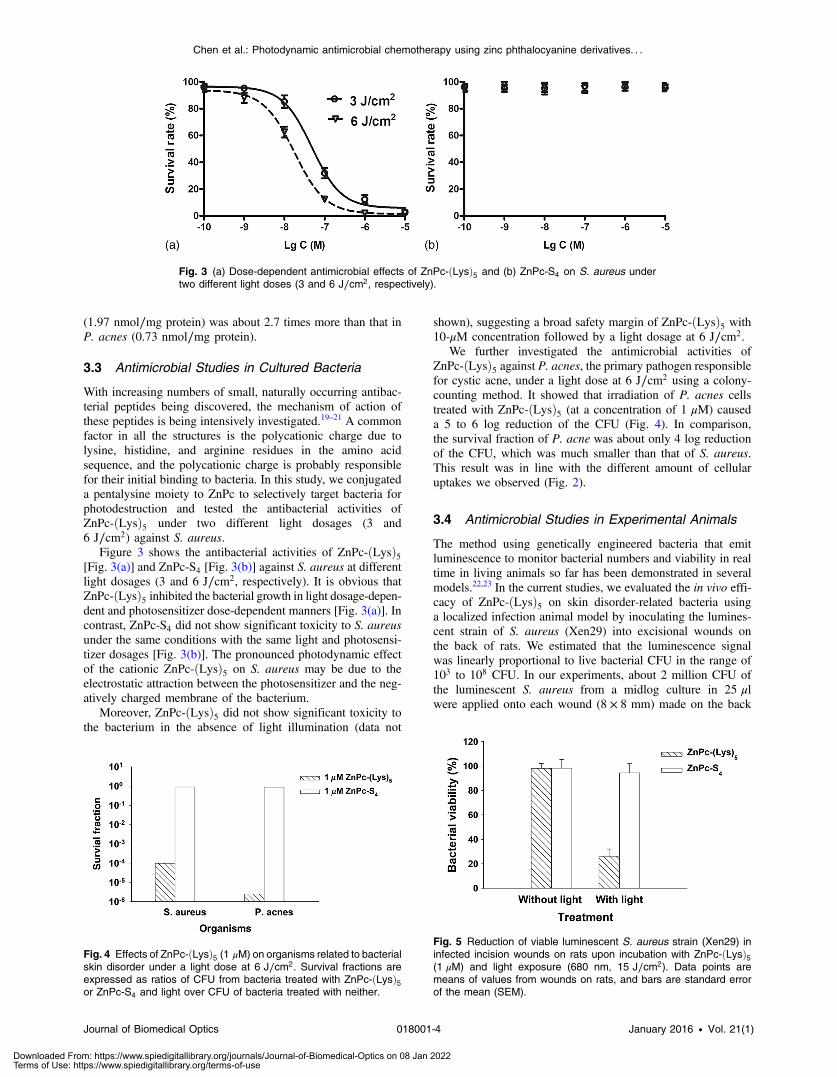
With increasing numbers of small, naturally occurring antibac-terial peptides being discovered, the mechanism of action ofthese peptides is being intensively investigated.19–21 A commonfactor in all the structures is the polycationic charge due tolysine, histidine, and arginine residues in the amino acidsequence, and the polycationic charge is probably responsiblefor their initial binding to bacteria. In this study, we conjugateda pentalysine moiety to ZnPc to selectively target bacteria forphotodestruction and tested the antibacterial activities ofZnPc-ðLysÞ5 under two different light dosages (3 and6 J∕cm2) against S. aureus.
Figure 3 shows the antibacterial activities of ZnPc-ðLysÞ5[Fig. 3(a)] and ZnPc-S4 [Fig. 3(b)] against S. aureus at differentlight dosages (3 and 6 J∕cm2, respectively). It is obvious thatZnPc-ðLysÞ5 inhibited the bacterial growth in light dosage-depen-dent and photosensitizer dose-dependent manners [Fig. 3(a)]. Incontrast, ZnPc-S4 did not show significant toxicity to S. aureusunder the same conditions with the same light and photosensi-tizer dosages [Fig. 3(b)]. The pronounced photodynamic effectof the cationic ZnPc-ðLysÞ5 on S. aureus may be due to theelectrostatic attraction between the photosensitizer and the neg-atively charged membrane of the bacterium.
Moreover, ZnPc-ðLysÞ5 did not show significant toxicity tothe bacterium in the absence of light illumination (data not
shown), suggesting a broad safety margin of ZnPc-ðLysÞ5 with10-μM concentration followed by a light dosage at 6 J∕cm2.
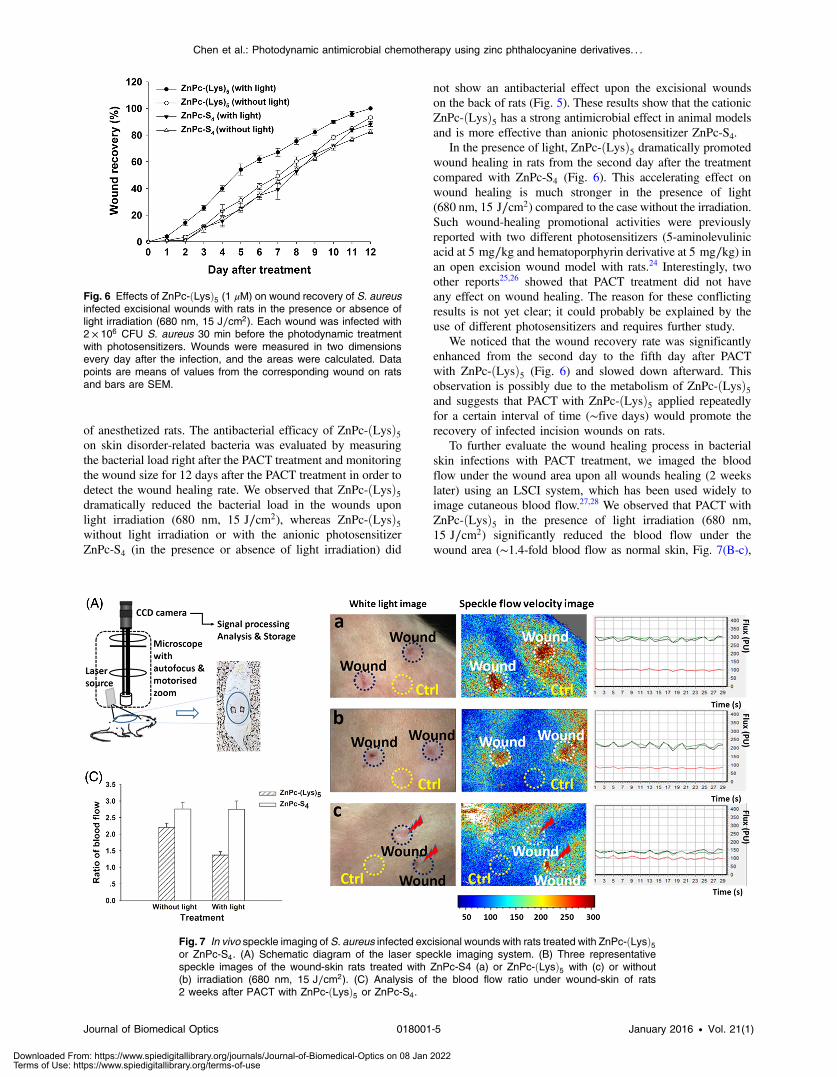
We further investigated the antimicrobial activities ofZnPc-ðLysÞ5 against P. acnes, the primary pathogen responsiblefor cystic acne, under a light dose at 6 J∕cm2 using a colony-counting method. It showed that irradiation of P. acnes cellstreated with ZnPc-ðLysÞ5 (at a concentration of 1 μM) causeda 5 to 6 log reduction of the CFU (Fig. 4). In comparison,the survival fraction of P. acne was about only 4 log reductionof the CFU, which was much smaller than that of S. aureus.This result was in line with the different amount of cellularuptakes we observed (Fig. 2).
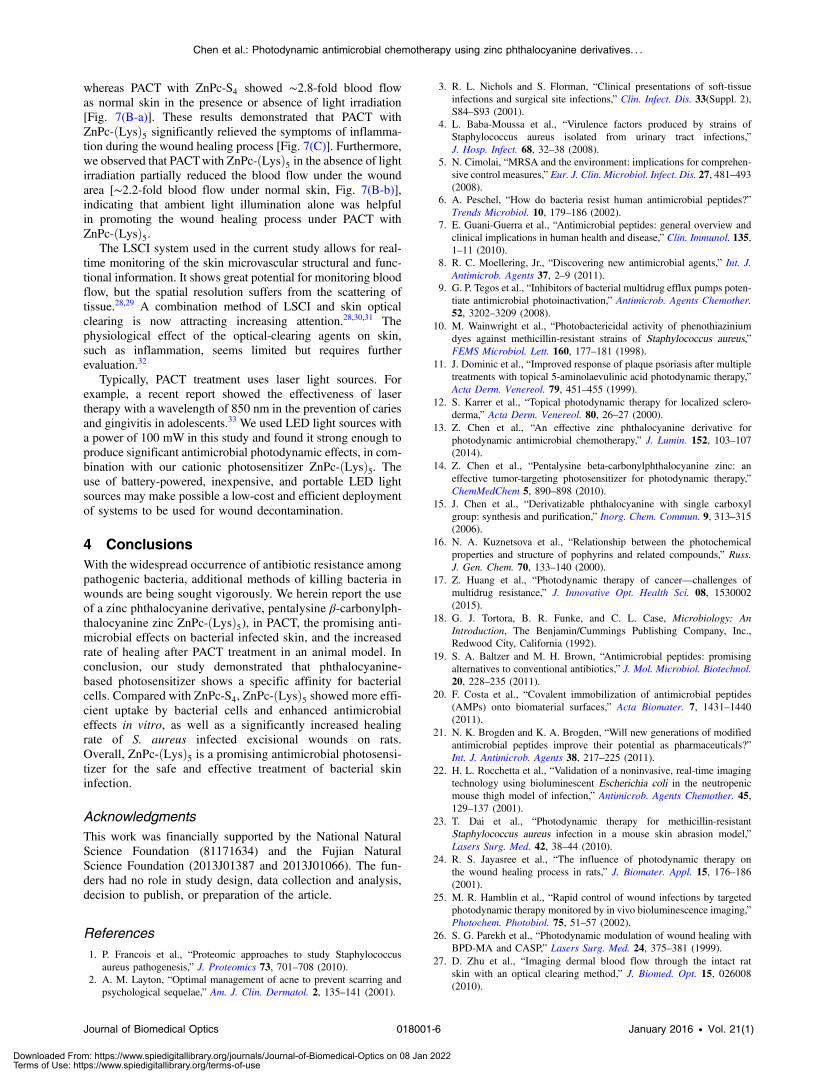
3.4 Antimicrobial Studies in Experimental Animals
The method using genetically engineered bacteria that emitluminescence to monitor bacterial numbers and viability in realtime in living animals so far has been demonstrated in severalmodels.22,23 In the current studies, we evaluated the in vivo effi-cacy of ZnPc-ðLysÞ5 on skin disorder-related bacteria usinga localized infection animal model by inoculating the lumines-cent strain of S. aureus (Xen29) into excisional wounds onthe back of rats. We estimated that the luminescence signalwas linearly proportional to live bacterial CFU in the range of103 to 108 CFU. In our experiments, about 2 million CFU ofthe luminescent S. aureus from a midlog culture in 25 μlwere applied onto each wound (8 × 8 mm) made on the back
Fig. 3 (a) Dose-dependent antimicrobial effects of ZnPc-ðLysÞ5 and (b) ZnPc-S4 on S. aureus undertwo different light doses (3 and 6 J∕cm2, respectively).
Fig. 4 Effects of ZnPc-ðLysÞ5 (1 μM) on organisms related to bacterialskin disorder under a light dose at 6 J∕cm2. Survival fractions areexpressed as ratios of CFU from bacteria treated with ZnPc-ðLysÞ5or ZnPc-S4 and light over CFU of bacteria treated with neither.
Fig. 5 Reduction of viable luminescent S. aureus strain (Xen29) ininfected incision wounds on rats upon incubation with ZnPc-ðLysÞ5(1 μM) and light exposure (680 nm, 15 J∕cm2). Data points aremeans of values from wounds on rats, and bars are standard errorof the mean (SEM).
Journal of Biomedical Optics 018001-4 January 2016 • Vol. 21(1)
Chen et al.: Photodynamic antimicrobial chemotherapy using zinc phthalocyanine derivatives. . .
Downloaded From: https://www.spiedigitallibrary.org/journals/Journal-of-Biomedical-Optics on 08 Jan 2022Terms of Use: https://www.spiedigitallibrary.org/terms-of-use
of anesthetized rats. The antibacterial efficacy of ZnPc-ðLysÞ5on skin disorder-related bacteria was evaluated by measuringthe bacterial load right after the PACT treatment and monitoringthe wound size for 12 days after the PACT treatment in order todetect the wound healing rate. We observed that ZnPc-ðLysÞ5dramatically reduced the bacterial load in the wounds uponlight irradiation (680 nm, 15 J∕cm2), whereas ZnPc-ðLysÞ5without light irradiation or with the anionic photosensitizerZnPc-S4 (in the presence or absence of light irradiation) did
not show an antibacterial effect upon the excisional woundson the back of rats (Fig. 5). These results show that the cationicZnPc-ðLysÞ5 has a strong antimicrobial effect in animal modelsand is more effective than anionic photosensitizer ZnPc-S4.
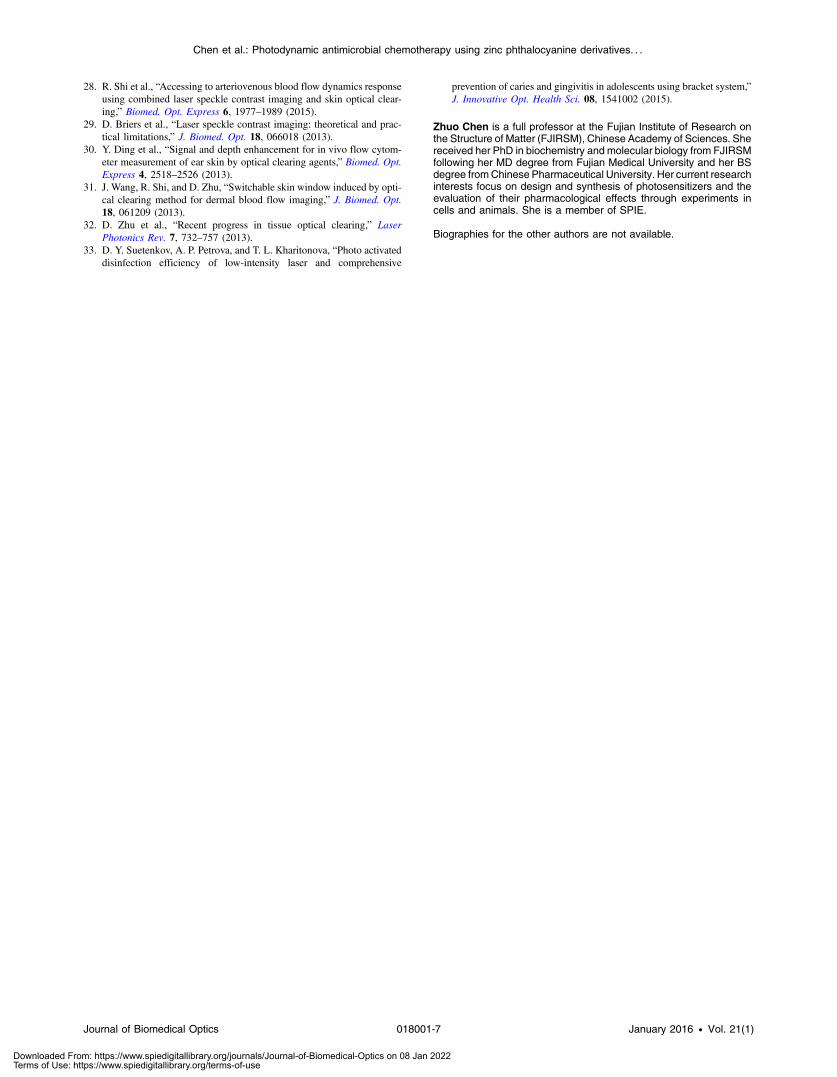
In the presence of light, ZnPc-ðLysÞ5 dramatically promotedwound healing in rats from the second day after the treatmentcompared with ZnPc-S4 (Fig. 6). This accelerating effect onwound healing is much stronger in the presence of light(680 nm, 15 J∕cm2) compared to the case without the irradiation.Such wound-healing promotional activities were previouslyreported with two different photosensitizers (5-aminolevulinicacid at 5 mg∕kg and hematoporphyrin derivative at 5 mg∕kg) inan open excision wound model with rats.24 Interestingly, twoother reports25,26 showed that PACT treatment did not haveany effect on wound healing. The reason for these conflictingresults is not yet clear; it could probably be explained by theuse of different photosensitizers and requires further study.
We noticed that the wound recovery rate was significantlyenhanced from the second day to the fifth day after PACTwith ZnPc-ðLysÞ5 (Fig. 6) and slowed down afterward. Thisobservation is possibly due to the metabolism of ZnPc-ðLysÞ5and suggests that PACT with ZnPc-ðLysÞ5 applied repeatedlyfor a certain interval of time (∼five days) would promote therecovery of infected incision wounds on rats.
To further evaluate the wound healing process in bacterialskin infections with PACT treatment, we imaged the bloodflow under the wound area upon all wounds healing (2 weekslater) using an LSCI system, which has been used widely toimage cutaneous blood flow.27,28 We observed that PACT withZnPc-ðLysÞ5 in the presence of light irradiation (680 nm,15 J∕cm2) significantly reduced the blood flow under thewound area (∼1.4-fold blood flow as normal skin, Fig. 7(B-c),
Fig. 6 Effects of ZnPc-ðLysÞ5 (1 μM) on wound recovery of S. aureusinfected excisional wounds with rats in the presence or absence oflight irradiation (680 nm, 15 J∕cm2). Each wound was infected with2 × 106 CFU S. aureus 30 min before the photodynamic treatmentwith photosensitizers. Wounds were measured in two dimensionsevery day after the infection, and the areas were calculated. Datapoints are means of values from the corresponding wound on ratsand bars are SEM.
Fig. 7 In vivo speckle imaging of S. aureus infected excisional wounds with rats treated with ZnPc-ðLysÞ5or ZnPc-S4. (A) Schematic diagram of the laser speckle imaging system. (B) Three representativespeckle images of the wound-skin rats treated with ZnPc-S4 (a) or ZnPc-ðLysÞ5 with (c) or without(b) irradiation (680 nm, 15 J∕cm2). (C) Analysis of the blood flow ratio under wound-skin of rats2 weeks after PACT with ZnPc-ðLysÞ5 or ZnPc-S4.
Journal of Biomedical Optics 018001-5 January 2016 • Vol. 21(1)
Chen et al.: Photodynamic antimicrobial chemotherapy using zinc phthalocyanine derivatives. . .
Downloaded From: https://www.spiedigitallibrary.org/journals/Journal-of-Biomedical-Optics on 08 Jan 2022Terms of Use: https://www.spiedigitallibrary.org/terms-of-use
whereas PACT with ZnPc-S4 showed ∼2.8-fold blood flowas normal skin in the presence or absence of light irradiation[Fig. 7(B-a)]. These results demonstrated that PACT withZnPc-ðLysÞ5 significantly relieved the symptoms of inflamma-tion during the wound healing process [Fig. 7(C)]. Furthermore,we observed that PACTwith ZnPc-ðLysÞ5 in the absence of lightirradiation partially reduced the blood flow under the woundarea [∼2.2-fold blood flow under normal skin, Fig. 7(B-b)],indicating that ambient light illumination alone was helpfulin promoting the wound healing process under PACT withZnPc-ðLysÞ5.
The LSCI system used in the current study allows for real-time monitoring of the skin microvascular structural and func-tional information. It shows great potential for monitoring bloodflow, but the spatial resolution suffers from the scattering oftissue.28,29 A combination method of LSCI and skin opticalclearing is now attracting increasing attention.28,30,31 Thephysiological effect of the optical-clearing agents on skin,such as inflammation, seems limited but requires furtherevaluation.32
Typically, PACT treatment uses laser light sources. Forexample, a recent report showed the effectiveness of lasertherapy with a wavelength of 850 nm in the prevention of cariesand gingivitis in adolescents.33 We used LED light sources witha power of 100 mW in this study and found it strong enough toproduce significant antimicrobial photodynamic effects, in com-bination with our cationic photosensitizer ZnPc-ðLysÞ5. Theuse of battery-powered, inexpensive, and portable LED lightsources may make possible a low-cost and efficient deploymentof systems to be used for wound decontamination.
4 ConclusionsWith the widespread occurrence of antibiotic resistance amongpathogenic bacteria, additional methods of killing bacteria inwounds are being sought vigorously. We herein report the useof a zinc phthalocyanine derivative, pentalysine β-carbonylph-thalocyanine zinc ZnPc-ðLysÞ5), in PACT, the promising anti-microbial effects on bacterial infected skin, and the increasedrate of healing after PACT treatment in an animal model. Inconclusion, our study demonstrated that phthalocyanine-based photosensitizer shows a specific affinity for bacterialcells. Compared with ZnPc-S4, ZnPc-ðLysÞ5 showed more effi-cient uptake by bacterial cells and enhanced antimicrobialeffects in vitro, as well as a significantly increased healingrate of S. aureus infected excisional wounds on rats.Overall, ZnPc-ðLysÞ5 is a promising antimicrobial photosensi-tizer for the safe and effective treatment of bacterial skininfection.
AcknowledgmentsThis work was financially supported by the National NaturalScience Foundation (81171634) and the Fujian NaturalScience Foundation (2013J01387 and 2013J01066). The fun-ders had no role in study design, data collection and analysis,decision to publish, or preparation of the article.
References1. P. Francois et al., “Proteomic approaches to study Staphylococcus
aureus pathogenesis,” J. Proteomics 73, 701–708 (2010).2. A. M. Layton, “Optimal management of acne to prevent scarring and
psychological sequelae,” Am. J. Clin. Dermatol. 2, 135–141 (2001).
3. R. L. Nichols and S. Florman, “Clinical presentations of soft-tissueinfections and surgical site infections,” Clin. Infect. Dis. 33(Suppl. 2),S84–S93 (2001).
4. L. Baba-Moussa et al., “Virulence factors produced by strains ofStaphylococcus aureus isolated from urinary tract infections,”J. Hosp. Infect. 68, 32–38 (2008).
5. N. Cimolai, “MRSA and the environment: implications for comprehen-sive control measures,” Eur. J. Clin. Microbiol. Infect. Dis. 27, 481–493(2008).
6. A. Peschel, “How do bacteria resist human antimicrobial peptides?”Trends Microbiol. 10, 179–186 (2002).
7. E. Guani-Guerra et al., “Antimicrobial peptides: general overview andclinical implications in human health and disease,” Clin. Immunol. 135,1–11 (2010).
8. R. C. Moellering, Jr., “Discovering new antimicrobial agents,” Int. J.Antimicrob. Agents 37, 2–9 (2011).
9. G. P. Tegos et al., “Inhibitors of bacterial multidrug efflux pumps poten-tiate antimicrobial photoinactivation,” Antimicrob. Agents Chemother.52, 3202–3209 (2008).
10. M. Wainwright et al., “Photobactericidal activity of phenothiaziniumdyes against methicillin-resistant strains of Staphylococcus aureus,”FEMS Microbiol. Lett. 160, 177–181 (1998).
11. J. Dominic et al., “Improved response of plaque psoriasis after multipletreatments with topical 5-aminolaevulinic acid photodynamic therapy,”Acta Derm. Venereol. 79, 451–455 (1999).
12. S. Karrer et al., “Topical photodynamic therapy for localized sclero-derma,” Acta Derm. Venereol. 80, 26–27 (2000).
13. Z. Chen et al., “An effective zinc phthalocyanine derivative forphotodynamic antimicrobial chemotherapy,” J. Lumin. 152, 103–107(2014).
14. Z. Chen et al., “Pentalysine beta-carbonylphthalocyanine zinc: aneffective tumor-targeting photosensitizer for photodynamic therapy,”ChemMedChem 5, 890–898 (2010).
15. J. Chen et al., “Derivatizable phthalocyanine with single carboxylgroup: synthesis and purification,” Inorg. Chem. Commun. 9, 313–315(2006).
16. N. A. Kuznetsova et al., “Relationship between the photochemicalproperties and structure of pophyrins and related compounds,” Russ.J. Gen. Chem. 70, 133–140 (2000).
17. Z. Huang et al., “Photodynamic therapy of cancer—challenges ofmultidrug resistance,” J. Innovative Opt. Health Sci. 08, 1530002(2015).
18. G. J. Tortora, B. R. Funke, and C. L. Case, Microbiology: AnIntroduction, The Benjamin/Cummings Publishing Company, Inc.,Redwood City, California (1992).
19. S. A. Baltzer and M. H. Brown, “Antimicrobial peptides: promisingalternatives to conventional antibiotics,” J. Mol. Microbiol. Biotechnol.20, 228–235 (2011).
20. F. Costa et al., “Covalent immobilization of antimicrobial peptides(AMPs) onto biomaterial surfaces,” Acta Biomater. 7, 1431–1440(2011).
21. N. K. Brogden and K. A. Brogden, “Will new generations of modifiedantimicrobial peptides improve their potential as pharmaceuticals?”Int. J. Antimicrob. Agents 38, 217–225 (2011).
22. H. L. Rocchetta et al., “Validation of a noninvasive, real-time imagingtechnology using bioluminescent Escherichia coli in the neutropenicmouse thigh model of infection,” Antimicrob. Agents Chemother. 45,129–137 (2001).
23. T. Dai et al., “Photodynamic therapy for methicillin-resistantStaphylococcus aureus infection in a mouse skin abrasion model,”Lasers Surg. Med. 42, 38–44 (2010).
24. R. S. Jayasree et al., “The influence of photodynamic therapy onthe wound healing process in rats,” J. Biomater. Appl. 15, 176–186(2001).
25. M. R. Hamblin et al., “Rapid control of wound infections by targetedphotodynamic therapy monitored by in vivo bioluminescence imaging,”Photochem. Photobiol. 75, 51–57 (2002).
26. S. G. Parekh et al., “Photodynamic modulation of wound healing withBPD-MA and CASP,” Lasers Surg. Med. 24, 375–381 (1999).
27. D. Zhu et al., “Imaging dermal blood flow through the intact ratskin with an optical clearing method,” J. Biomed. Opt. 15, 026008(2010).
Journal of Biomedical Optics 018001-6 January 2016 • Vol. 21(1)
Chen et al.: Photodynamic antimicrobial chemotherapy using zinc phthalocyanine derivatives. . .
Downloaded From: https://www.spiedigitallibrary.org/journals/Journal-of-Biomedical-Optics on 08 Jan 2022Terms of Use: https://www.spiedigitallibrary.org/terms-of-use
28. R. Shi et al., “Accessing to arteriovenous blood flow dynamics responseusing combined laser speckle contrast imaging and skin optical clear-ing,” Biomed. Opt. Express 6, 1977–1989 (2015).
29. D. Briers et al., “Laser speckle contrast imaging: theoretical and prac-tical limitations,” J. Biomed. Opt. 18, 066018 (2013).
30. Y. Ding et al., “Signal and depth enhancement for in vivo flow cytom-eter measurement of ear skin by optical clearing agents,” Biomed. Opt.Express 4, 2518–2526 (2013).
31. J. Wang, R. Shi, and D. Zhu, “Switchable skin window induced by opti-cal clearing method for dermal blood flow imaging,” J. Biomed. Opt.18, 061209 (2013).
32. D. Zhu et al., “Recent progress in tissue optical clearing,” LaserPhotonics Rev. 7, 732–757 (2013).
33. D. Y. Suetenkov, A. P. Petrova, and T. L. Kharitonova, “Photo activateddisinfection efficiency of low-intensity laser and comprehensive
prevention of caries and gingivitis in adolescents using bracket system,”J. Innovative Opt. Health Sci. 08, 1541002 (2015).
Zhuo Chen is a full professor at the Fujian Institute of Research onthe Structure of Matter (FJIRSM), Chinese Academy of Sciences. Shereceived her PhD in biochemistry and molecular biology from FJIRSMfollowing her MD degree from Fujian Medical University and her BSdegree fromChinese Pharmaceutical University. Her current researchinterests focus on design and synthesis of photosensitizers and theevaluation of their pharmacological effects through experiments incells and animals. She is a member of SPIE.
Biographies for the other authors are not available.
Journal of Biomedical Optics 018001-7 January 2016 • Vol. 21(1)
Chen et al.: Photodynamic antimicrobial chemotherapy using zinc phthalocyanine derivatives. . .
Downloaded From: https://www.spiedigitallibrary.org/journals/Journal-of-Biomedical-Optics on 08 Jan 2022Terms of Use: https://www.spiedigitallibrary.org/terms-of-use