plant photodynamic stress
TRANSCRIPT
Université de Limoges ED 614 - Chimie, Environnement, Géosciences, Agrosciences (CEGA) Peirene EA 7500
Thèse pour obtenir le grade de
Docteur de l’Université de Limoges Discipline/spécialité : Biosciences de l’environnement et de la santé/Sciences agronomiques et écologiques
Présentée et soutenue par
Mohammad ISSAWI
Le 6 juin 2018
JURY:
Rapporteurs M. Tim Maisch, Pr., Department of dermatology, University Hospital Regensburg, Allemagne Mme. Magali Gary-Bobo, Dr., Institut des biomolecules Max Mousseron, Université de Montpellier, France
Examinateurs M. Henri Batoko, Pr., Institute of life sciences, University of Louvain, Belgique M. Vincent Sol, Pr., Peirene EA 7500, Université de Limoges, France Mme. Stéphanie Leroy-Lhez, Dr., Peirenne EA 7500, Université de Limoges, France Mme. Catherine Riou, Dr., Peirene EA 7500, Université de Limoges, France
Plant photodynamic stress: study of molecular and cellular mechanisms in plant and plant cells upon
porphyrin treatment
Thèse de doctorat
Thèse dirigée par Catherine RIOU

I
“Properly speaking, there are in the world no such men as self-made men.”
Frederick Douglass
“My philosophy of life is that if we make up our mind what we are going to make of our
lives, then work hard toward that goal, we never lose. Somehow we win out.”
Ronald Reagan
“I am not a perfectionist, but I like to feel that things are done well. More important than that, I feel an
endless need to learn, to improve, to evolve.”
Cristiano Ronaldo

II
Droits d’auteurs
Cette création est mise à disposition selon le Contrat :
« Attribution-Pas d'Utilisation Commerciale-Pas de modification 3.0 France »
disponible en ligne : http://creativecommons.org/licenses/by-nc-nd/3.0/fr/

III
Dedication
To the memory of my father who passed away 15 years ago… Dad, you are missed...
To my mother who knew it would be a long and hard road, but encouraged and supported me
along the way…
To my brother, Ahmad…
To my sister, Mariam...
IV
Acknowledgements
Due to the lack of support to local PhD programs in science and the major challenge in seeking funding in
my country Lebanon, I have applied for a PhD in plant biology in Limoges-France. I was accepted when I
was still in Iran, doing my M2 internship (September 2014) thanks to my brother Ahmad Issawi, Dr.
Catherine Riou and Pr. Vincent Sol for their help and support.
Over the past three years of my PhD (2015-2018) I have witnessed unforgettable memories that will
remain engraved in my heart. All the work presented in the present manuscript involves many different
people, from different places, but all of them contributed in various but important ways.
This journey would not have been possible without the support of my family, mentors, and friends.
First of all, I owe a special debt of gratitude to all members of the jury starting with Pr. Tim Maisch and
Dr. Magali Gary-bobo as referees. I had the honor to meet Pr. Maisch at Coimbra and I appreciate his
discussion there and his acceptance to be a part of my thesis jury. I also thank Pr. Henri Batoko, Pr.
Vincent Sol, Dr. Stéphanie Lery-Lhez and Dr. Catherine Riou for accepting to review my manuscript.
With a deep sense of gratitude, I would like to convey thanks to Pr. Vincent Sol, our lab-head, for
accepting me to join his group. Monsieur Sol, it was my pleasure to be a member in your team, known for
your humbleness despite your prestigious status as a professor in organic chemistry and a group leader.
Scientists in Coimbra (Portugal) - where I have attended the international photodynamic association
world congress - were very interested when they have realized that I am working within Pr. Sol’s
research team.
My sincerest thanks are extended to my PhD advisor Dr. Catherine Riou. Catherine, I cannot express to
what extent I was lucky when you replied me by e-mail on September 3rd, 2014 confirming your
acceptance to start a PhD under your guidance. Therafter, I highly appreciated your patience and
support facing the complications of the Lebanese university. I have always remembered my first day in
the lab (March 3rd, 2015) when I started to measure the hypocotyl and root length of in vitro growing
tomato plantlets. I also remember our first meeting when you said that I must have three publications at
the end of my thesis. And here I am ending with five publications as first author and two others as co-
author. Over my three years I have always fascinated by your good mood and your funny jokes “à la
Bretonne”. Your strategical thinking and insightful leadership have inspired me, and I hope to work with
you more and more. The Time wents so fast! (PS: Dorénavant je vais vous tutoyer).
I would like to thank Stephanie Leroy-Lhez whose “chemical” instructions and contributions always
enlightening a lot our work. Special thanks for Céline Faugeron, I have gained a lot of experimental
knowledge from her experience concerning GC and sugar analysis. I would like to thank also Yann
Launay for his patience and his helpful contribution concerning starch granule microscopy.
Next, I am so grateful to Dr. Ahmad Kobeissi for his valuable cooperation and I am pleased that
Mohammad Muhieddine (M2 student at the Lebanese university) has been contributed to my work.

V
I will never forget that wonderful time in Coimbra with Guillaume, Florent, Tan and the greatest
Bertrand. “Come on!”.
Thanks to Robert Granet, Olivier, Frédérique, Mark-Arthur, Shihong four your help concerning organic
chemistry. Mark-Arthur, “ici c‘est Madrid, Hala Madrid! y Viva CR7”.
Thanks to Jeremy for your “phenalenonic” gifts and Cedric for sharing your knowledge about HPLC.
My next thanks are addressed to members of our lab: Nico, Amandine, CRICRI, Gaelle, Idelette, Anais,
Florian, Zineb, Soukaina, Roger, Dorothée and Michèle.
I also thank all Master 1 students who contributed to my work: Francois, David, Candice and Amy.
For Salim (my lab-mate) and Hajar, I will always remember our great moments and beautiful gatherings.
Mec! rappelles toi toujours “Hala Madrid y nada mas”. On va s’inscrire à la gym t’inquiète!
For Veronica, the dynamo of our lab and my “successor” working on plant-microbes-photosensitizers,
you have all the ingredients for success. Hang in there!
For to the one and the only one, the greatest ever Holm Mustapha Amara! Words always fail to say
something enough for you! Very and very briefly, your unique name fits you well.
I especially thank my family: Mom, Ahmad and Mariam, the true and the great supporters. Thank you for
supporting me emotionally and financially, you should know that your support and encouragement was
worth more than I can express on paper. I always knew that you believed in me and wanted the best for
me. I would not have made it this far without you.

VI
Table of content
Dedication ............................................................................................................................................................ III
Acknowledgements .............................................................................................................................................. IV
Table of content ................................................................................................................................................... VI
List of figures ...................................................................................................................................................... VIII
List of tables ......................................................................................................................................................... IX
List of abbreviations .............................................................................................................................................. X
I. Introduction ................................................................................................................................................ 1
I.1. Photosensitizers ......................................................................................................................................... 1
I.1.1. General overview ............................................................................................................................... 1 I.1.2. Mechanisms of PS activation under light .......................................................................................... 1 I.1.3. Classification of PS ............................................................................................................................. 3 I.1.4. A great category of PS: the porphyrins .............................................................................................. 9
I.1.4.1. Porphyrin overview ................................................................................................................... 9
I.1.4.2. Natural porphyrins and tetrapyrrole biosynthesis .................................................................. 12
I.2. Applications of PS .................................................................................................................................... 15
I.2.1. Photodynamic treatment in medicine ............................................................................................. 15 I.2.1.1. Photodynamic therapy against cancers .................................................................................. 15
I.2.1.2. Photodynamic therapy against non-oncological diseases ...................................................... 16
I.2.1.3. Photodynamic therapy limitations .......................................................................................... 16
I.2.2. Antimicrobial photodynamic treatment (APDT) .............................................................................. 17 I.2.2.1. APDT in medical environment ................................................................................................. 17
I.2.2.2. APDT for water sewage ........................................................................................................... 18
I.2.2.3. APDT for food decontamination .............................................................................................. 18
I.2.2.4. APDT in industrial domain ....................................................................................................... 19
I.2.2.5. APDT in agronomy ................................................................................................................... 19
I.2.3. PUBLICATION 1: Review “Plant photodynamic stress: what's new?”, Frontiers in Plant Science .. 20
II. PhD objectives .......................................................................................................................................... 30
II. 1. APDT for plants: Is it a joke? ................................................................................................................. 30
II. 2. Application of charged porphyrins on tobacco cell suspension: a help to understand how PS are
photoactivated in plant cells ......................................................................................................................... 31
II. 3. Manuscript presentation ...................................................................................................................... 32
III. Results ....................................................................................................................................................... 33
Chapter I. APDT in agronomy: Dream or reality? ............................................................................................... 33
PUBLICATION 2: “Synergistic enhancement of tolerance mechanisms in response to photoactivation of
cationic tetra (N-methylpyridyl) porphyrins in tomato plantlets”, Journal of Photochemistry and
Photobiology B: Biology. ................................................................................................................................ 34
VII
PUBLICATION 3: “Responses of an adventitious fast-growing plant to photodynamic stress: comparative
study of anionic and cationic porphyrin effect on Arabidopsis thaliana”, Physiologia Plantarum. .............. 75
Discussion and perspectives ........................................................................................................................ 104
Chapter II. TBY-2 cells: a helpful tool to understand the cellular responses to photoactivated anionic
porphyrin treatment from A to Z ...................................................................................................................... 106
PUBLICATION 4: “Unexpected features of exponentially growing Tobacco Bright Yellow-2 cell suspension
culture in relation to excreted extracellular polysaccharides and cell wall composition”, Glycoconjugate
Journal.......................................................................................................................................................... 107
PUBLICATION 5: “Characterization of pH dependent charge states and physico-chemical properties of
anionic porphyrins” (Submitted to Photochemical & Photobiological Sciences). ....................................... 122
PUBLICATION 6: “Crossing the first threshold: New insights in the influence of chemical structure of anionic
porphyrins from cell wall interactions to photodynamic cell death induction in TBY-2 suspension culture”
(in preparation for submission to The Plant Journal)......................................................................................... 139
Discussion and perspectives ................................................................................................................... 170
IV. General conclusion ................................................................................................................................. 173
V. Perspectives and outlook ....................................................................................................................... 174
References ........................................................................................................................................................ 177
Annex 1 ............................................................................................................................................................. 188
Annex 2 ............................................................................................................................................................. 189
Annex 3 ............................................................................................................................................................. 190
Annex 4 ............................................................................................................................................................. 191
Curriculum Vitae ............................................................................................................................................... 192

VIII
List of figures
Figure 1. Photodynamic reactions of PS. .............................................................................................................. 2
Figure 2. Porphyrinoids compounds. .................................................................................................................... 3
Figure 3. Pigments of life: heme and chlorophyll. .............................................................................................. 10
Figure 4. Porphyrin absorption spectrum. .......................................................................................................... 11
Figure 5. Structure of endogenous tetrapyrrolic photosensitizers. .................................................................... 13
Figure 6. Tetrapyrrole biosynthetic pathways in plants. .................................................................................... 14
Figure 7. PDT application in cancer treatment. .................................................................................................. 16
Figure 8. APDT application for food decontamination. ...................................................................................... 18
Figure 9. APDT mechanism and its possible multiple targets in agronomy........................................................ 20
Figure 10. Schematic representation of the “two-in-one” strategy based on the use of PS in agronomy. ..... 104
Figure 11. Structure of molecular dyad coupling TPPC with fluorescein via alkyne linker. .............................. 175

IX
List of tables
Table 1. PS classification according to their origin. .............................................................................................. 9
Table 2. Cell wall composition of TBY-2 cells. ................................................................................................... 170
X
List of abbreviations
ALA: 5-aminolevulinic acid
APDT: Antimicrobial Photodynamic Treatment
BODIPY: Boron-dipyrromethene
CPO: Coproporphyrinogen oxidase
FeCH: Fe-chelatase
GluTS: Glutamyl t-RNA synthetase
GlutTR: Glutamyl-tRNA reductase
GSA: Glutamate-1- semialdehyde aminotransferase
HP: Hematoporphyrin
HPD: Hematoporphyrin derivative
ISC: Intersystem Crossing
MgCH: Mg-chelatase
m-THPC: Metat-tetra (hydroxyphenyl) chlorin
PACT: Photoantimicrobial Chemotherapy
PBGD: Porphobilinogen deaminase
PDI: Photodynamic Inactivation
PDT: Photodynamic Therapy
PPO: Protoporphyrinogen oxidase
PS: Photosensitizer
ROS: Reactive Oxygen Species
S0: Ground state
S1: Excited singlet state
T1: Excited triplet state
TBY-2: Tobacco Bright Yellow-2
THPP: Tetra-hydroxyphenyl porphyrin
TMPyP: Tetra (N-methylpyridyl) porphyrin

XI
TPyP: tetra-pyridyl porphyrin
TPPC: Tetra (carboxyphenyl) porphyrin
TPPP: Tetra (phosphonatophenyl) porphyrin
TPPS: Tetra (sulfonatophenyl) porphyrin
TRX: Thioredoxin
UROD: Uroporphyrinogen decarboxylase
UROS: Uroporphyrinogen synthase
Introduction
Mohammad ISSAWI | Thèse de doctorat | Université de Limoges | 2018
Licence CC BY-NC-ND 3.0
1
I.Introduction
I.1. Photosensitizers
I.1.1. General overview
Photosensitizers (PS) are conjugated chromophores that absorb ultraviolet or visible radiations
and transfer energy to adjacent molecules (oxygen or other molecules) through photochemical
reactions. Approximately 1000 B.C., Egyptians treated the skin pigment loss disease using a plant
dried powder that contained psoralens which belong to furocoumarins, family of natural PS (Table 1,
Joshi and Saenz 2013). In the beginning of the 20th century, PS and their applications became more
and more investigated (Berg et al. 2005). The first photosensitization reaction was reported in 1900
when a German student called Oskar Raab discovered that acridine orange was toxic to the protozoan
(Paramecium caudatum) in the presence of light (Van Straten et al. 2017). Four years later, his
teacher Professor Hermann von Tappeiner demonstrated that the presence of oxygen was a
prerequisite for the occurrence of photosensitization coining the term “photodynamic reaction”.
In parallel, Hill’s studies on plant photosynthesis allowed to understand how the green
pigments called chlorophylls could be excited by sunlight (Hill 1937). Afterward, the
photosensitization mechanisms of chlorophyll were intensively studied and explored in the context of
photosynthesis knowledge (Krasnovsky and Brin 1947, Evstigneev 1965, Krieger-liszkay 2004).
There is no doubt that in the global process of photosynthesis, the photoreaction triggered by
photoreceptors or PS named chlorophylls localized in chloroplasts of land plants and algae or
cyanobacteria, has allowed life on earth with changes of atmosphere composition enriched in oxygen
(Bassham 1959, Hohmann-marriott and Blankenship 2011).
From 1970s onwards, photodynamic reaction was largely investigated as potential tool for
applications in medicine and struggle against microorganisms.
I.1.2. Mechanisms of PS activation under light
As a first step and to understand the potential applications of the PS in biological systems, the
mechanism of PS photoactivation must be briefly described. Under light (UV-Visible), PS molecule
jumps from a stable to an excited state. In presence of molecular oxygen, PS causes oxidative
damages within biological systems. Illumination of a PS leads to the absorption of a photon and
promotes the PS to its excited singlet state S1. From this unstable and typically short-lived state, the
Introduction
Mohammad ISSAWI | Thèse de doctorat | Université de Limoges | 2018
Licence CC BY-NC-ND 3.0
2
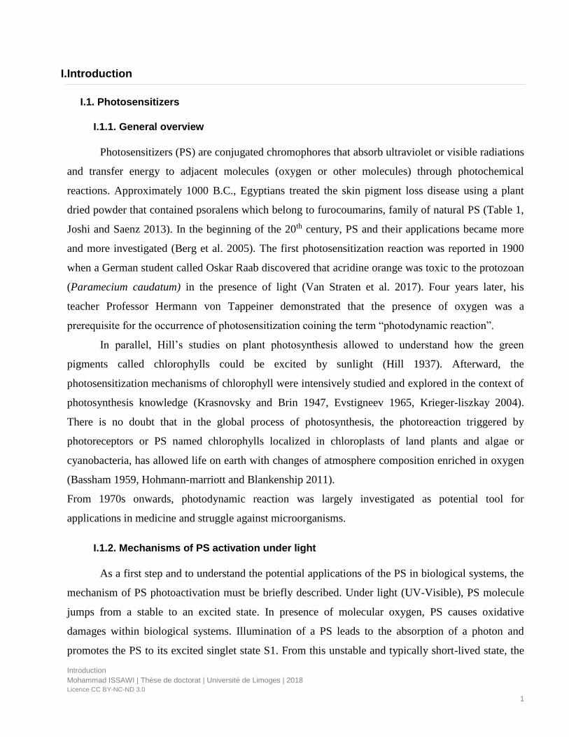
PS can return to its ground state S0 by converting its energy into heat or fluorescence, a feature which
can be used for the purposes of detection and optical monitoring. Alternatively, PS undergo
intersystem crossing yielding PS in excited triplet state T1. In this T1 state, the PS can relax through
phosphorescence or react with other molecules to create chemically reactive species via two types of
reactions (Figure 1). PS in T1 can react with biological substrates and transfer an electron to generate
radical anion or cation species. Typically, the PS reacts with an electron donating substrate to form
PS that subsequently reacts with oxygen to form superoxide anion radicals, hydroxyl radicals and
hydrogene peroxide. This is called type I reaction. In a type II reaction, excited PS reacts directly with
molecular oxygen by transfer of energy to form singlet oxygen (1O2) which is a highly reactive
oxygen species.
Singlet oxygen and hydroxyl radical can directly react with nearly all biomolecules leading to
oxidative damage. Hydrogen peroxide can be a part of a reaction that produces hydroxyl radical when
it reacts with a metal via Fenton reaction (Fe2+ + H2O2 → ⚫ OH + Fe3+ + ⁻OH). Most of PSs are
thought to act through type II reactions where singlet oxygen is the main molecule causing oxidative
cellular damage (Figure 1, Ding et al. 2012, Baptista et al. 2017).
ISC: intersystem crossing, 1O2: singlet oxygen, O2·⁻: superoxide anion, H2O2: hydrogen peroxide, ·OH: hydroxyl radical,
PS: photosensitizer in its ground state, 1PS: photosensitizer in its excited singlet state, 3PS: photosensitizer in its excited
triplet state.
Figure 1. Photodynamic reactions of PS.
Light

Introduction
Mohammad ISSAWI | Thèse de doctorat | Université de Limoges | 2018
Licence CC BY-NC-ND 3.0
3
I.1.3. Classification of PS
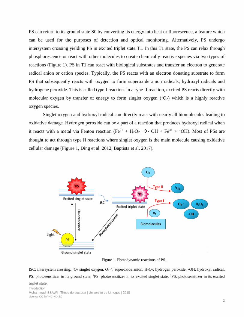
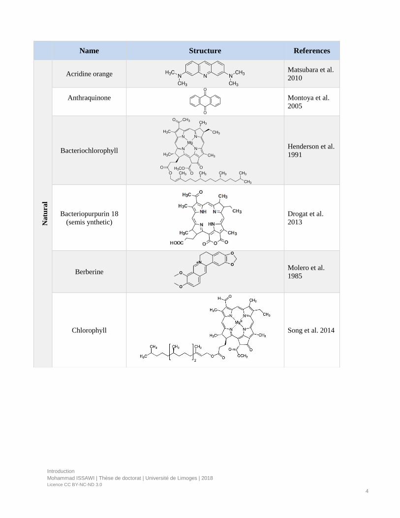
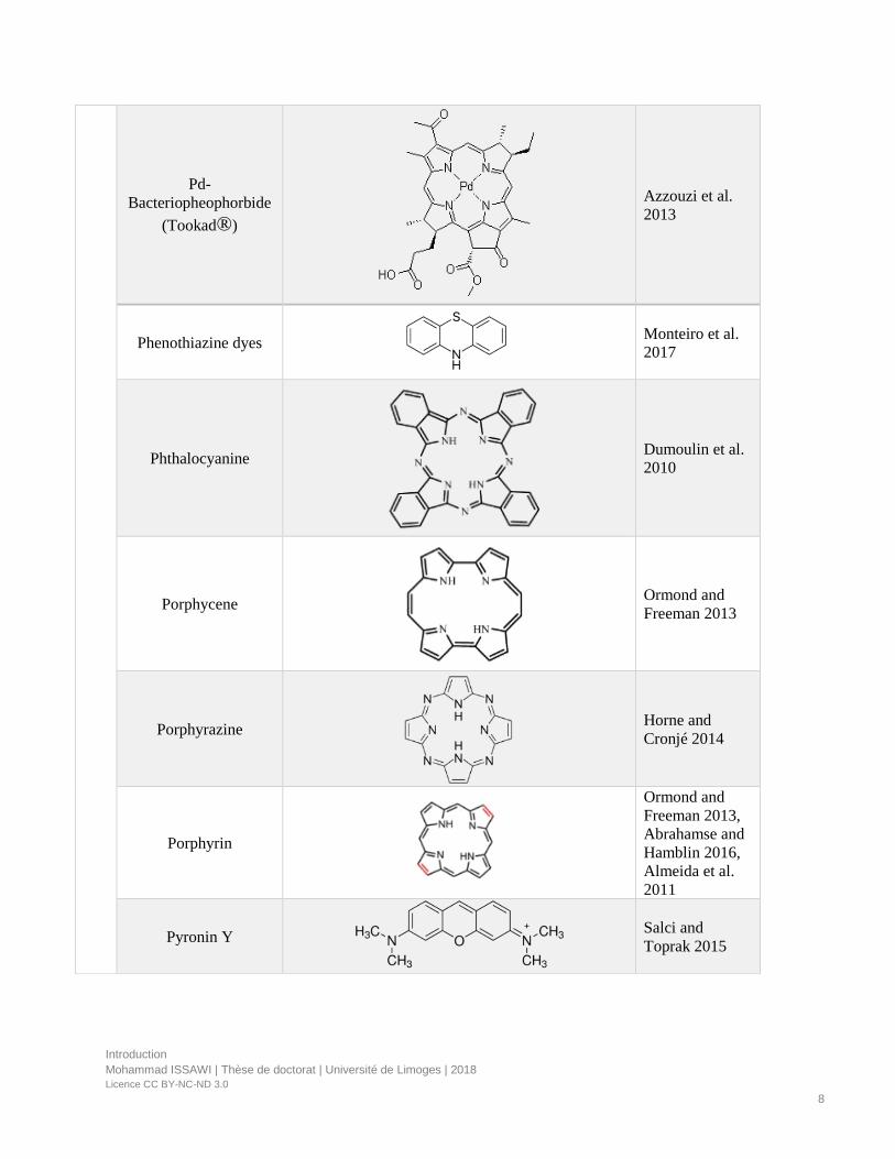
PS can be distinguished between synthetic and naturally occurring compounds. Natural
compounds are found in nature and can be extracted and modulated whereas synthetic compounds
encompass all types of molecules that can be synthetized by chemical reactions such as porphyrins
and derivatives, phtalocyanines, phenothiazines, xanthenes, corroles, squaraines and BODIPY.
PS can be classified into porphyrinoid and non-porphyrin molecules (Table 1).
• Porphyrinoid compounds have tetrapyrrolic backbone and are the most useful PS in
medicine and in the perspective of environmental applications due to their highly
conjugated structure and absorption of light in the visible region (Figure 2).
An extended family of photosensitizers bearing a tetrapyrrole macrocycle and show different structural features as N
distribution, group substitution and redox state. They have the most useful applications as photodynamic compounds.
• Non-porphyrin compounds encompass among others anthraquinone, phenothiazine, xanthene,
curcumine, and phenalenone. Although these compounds are not yet clinically approved, they
were tested in clinical trials in addition to their efficient use as antimicrobials (Table 1).
Figure 2. Porphyrinoids compounds.
Introduction
Mohammad ISSAWI | Thèse de doctorat | Université de Limoges | 2018
Licence CC BY-NC-ND 3.0
4
Name Structure References
Natu
ral
Acridine orange
Matsubara et al.
2010
Anthraquinone
Montoya et al.
2005
Bacteriochlorophyll
Henderson et al.
1991
Bacteriopurpurin 18
(semis ynthetic)
Drogat et al.
2013
Berberine
Molero et al.
1985
Chlorophyll
Song et al. 2014

Introduction
Mohammad ISSAWI | Thèse de doctorat | Université de Limoges | 2018
Licence CC BY-NC-ND 3.0
5
Chlorophyllin
(semi synthetic)
Azizullah et al.
2014, Hader et
al. 2016
Coumarin
De menezes et
al. 2014a ,
Fracarolli et al.
2016
Curcumin
Spaeth et al.
2017
Fullerene
Dagani 1992
Furocoumarin
De Menezes et
al. 2014a ,
Fracarolli et al.
2016
Harmine
Hazen and
Gutierrez-
Gonzalves 1988
Hematoporphyrin
(semi synthetic)
Hazen et al.
1987
Melanin
Liu et al. 2015

Introduction
Mohammad ISSAWI | Thèse de doctorat | Université de Limoges | 2018
Licence CC BY-NC-ND 3.0
6
Orcein
Molero and
Hazen 1988
Perylenequinone
Daub et al. 2005
Phenalenone
Nazir et al. 2015
Song et al. 2017
Muehler et al.
2017
Pheophorbide
Yoon et al. 2014
Pheophytin
Hsu et al. 2010
Protoporphyrin IX
Yoshida et al.
2017, Delcanale
et al. 2016
Purpurin 18
(semi synthetic)
Drogat et al.
2011

Introduction
Mohammad ISSAWI | Thèse de doctorat | Université de Limoges | 2018
Licence CC BY-NC-ND 3.0
7
Riboflavin
Maisch et al.
2014
Thiophene
Dicosmo et al.
1982, Ebermann
et al. 1996
Syn
thet
ic
Bacteriochlorin
Juzenienne 2009
BODIPY
Durantini et al.
2018
Chlorine
Juzenienne 2009
Corrole
Pohl et al. 2014
Merocyanine
Sieber et al.
1989
N-confused porphyrin
Thomas et al.
2012
Neutral red
Kharkawal et al.
2012
Introduction
Mohammad ISSAWI | Thèse de doctorat | Université de Limoges | 2018
Licence CC BY-NC-ND 3.0
8
Pd-
Bacteriopheophorbide
(Tookad®)
Azzouzi et al.
2013
Phenothiazine dyes
Monteiro et al.
2017
Phthalocyanine
Dumoulin et al.
2010
Porphycene
Ormond and
Freeman 2013
Porphyrazine
Horne and
Cronjé 2014
Porphyrin
Ormond and
Freeman 2013,
Abrahamse and
Hamblin 2016,
Almeida et al.
2011
Pyronin Y
Salci and
Toprak 2015
Introduction
Mohammad ISSAWI | Thèse de doctorat | Université de Limoges | 2018
Licence CC BY-NC-ND 3.0
9
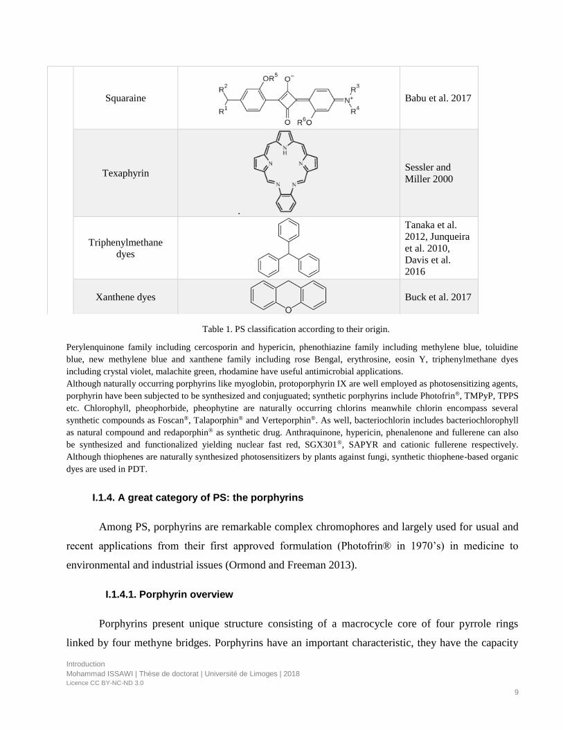
Perylenquinone family including cercosporin and hypericin, phenothiazine family including methylene blue, toluidine
blue, new methylene blue and xanthene family including rose Bengal, erythrosine, eosin Y, triphenylmethane dyes
including crystal violet, malachite green, rhodamine have useful antimicrobial applications.
Although naturally occurring porphyrins like myoglobin, protoporphyrin IX are well employed as photosensitizing agents,
porphyrin have been subjected to be synthesized and conjuguated; synthetic porphyrins include Photofrin®, TMPyP, TPPS
etc. Chlorophyll, pheophorbide, pheophytine are naturally occurring chlorins meanwhile chlorin encompass several
synthetic compounds as Foscan®, Talaporphin® and Verteporphin®. As well, bacteriochlorin includes bacteriochlorophyll
as natural compound and redaporphin® as synthetic drug. Anthraquinone, hypericin, phenalenone and fullerene can also
be synthesized and functionalized yielding nuclear fast red, SGX301®, SAPYR and cationic fullerene respectively.
Although thiophenes are naturally synthesized photosensitizers by plants against fungi, synthetic thiophene-based organic
dyes are used in PDT.
I.1.4. A great category of PS: the porphyrins
Among PS, porphyrins are remarkable complex chromophores and largely used for usual and
recent applications from their first approved formulation (Photofrin® in 1970’s) in medicine to
environmental and industrial issues (Ormond and Freeman 2013).
I.1.4.1. Porphyrin overview
Porphyrins present unique structure consisting of a macrocycle core of four pyrrole rings
linked by four methyne bridges. Porphyrins have an important characteristic, they have the capacity
Squaraine
Babu et al. 2017
Texaphyrin
.
Sessler and
Miller 2000
Triphenylmethane
dyes
Tanaka et al.
2012, Junqueira
et al. 2010,
Davis et al.
2016
Xanthene dyes
Buck et al. 2017
Table 1. PS classification according to their origin.
Introduction
Mohammad ISSAWI | Thèse de doctorat | Université de Limoges | 2018
Licence CC BY-NC-ND 3.0
10
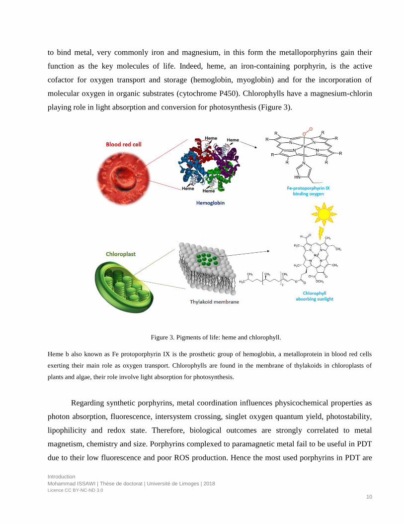
to bind metal, very commonly iron and magnesium, in this form the metalloporphyrins gain their
function as the key molecules of life. Indeed, heme, an iron-containing porphyrin, is the active
cofactor for oxygen transport and storage (hemoglobin, myoglobin) and for the incorporation of
molecular oxygen in organic substrates (cytochrome P450). Chlorophylls have a magnesium-chlorin
playing role in light absorption and conversion for photosynthesis (Figure 3).
Heme b also known as Fe protoporphyrin IX is the prosthetic group of hemoglobin, a metalloprotein in blood red cells
exerting their main role as oxygen transport. Chlorophylls are found in the membrane of thylakoids in chloroplasts of
plants and algae, their role involve light absorption for photosynthesis.
Regarding synthetic porphyrins, metal coordination influences physicochemical properties as
photon absorption, fluorescence, intersystem crossing, singlet oxygen quantum yield, photostability,
lipophilicity and redox state. Therefore, biological outcomes are strongly correlated to metal
magnetism, chemistry and size. Porphyrins complexed to paramagnetic metal fail to be useful in PDT
due to their low fluorescence and poor ROS production. Hence the most used porphyrins in PDT are
Figure 3. Pigments of life: heme and chlorophyll.

Introduction
Mohammad ISSAWI | Thèse de doctorat | Université de Limoges | 2018
Licence CC BY-NC-ND 3.0
11
Q bands
Q bands
Soret band
Zn and Pd-coordinated. Moreover, these metalloporphyrins show some differences. For example, Pd-
porphyrins are highly uptaken by biological cells comparing to Zn analogs owing to palladium small
size and the absence of axial bonding. Moreover, Pd-porphyrins are more efficient to produce
hydroxyl radical because they are better electron acceptor. In addition, subcellular localization and
DNA photocleavage efficacy were not the same when comparing metalloporphyrins to their free-base
counterparts (Guldi et al. 2000, Josefsen and Bowle 2008, Skwor et al. 2016, Dabrowski et al. 2016).
Porphyrins, either metalated or free base, exhibit a very characteristic absorption spectrum in
the visible range (from 400 nm to 800 nm) due to several electronic transitions. Indeed, their UV-
Visible spectrum shows an intense absorption band called Soret band in the near UV followed by four
other absorption bands in the visible range called Q bands for free base porphyrin or two for
metalloporphyrins (Figure 4).
Figure 4. Porphyrin absorption spectrum.
Free base porphyrins (blue) show intensive absorption in the blue range assigned to soret band and 4 bands of lower
intensity called Q bands in the visible range. Metalated porphyrins (red) show two Q bands instead of four bands.

Introduction
Mohammad ISSAWI | Thèse de doctorat | Université de Limoges | 2018
Licence CC BY-NC-ND 3.0
12
Porphyrin emission is centered in the range 550-800 nm and metalloporphyrins show
blueshifted fluorescence bands comparing to their free-base analogs (Valicsek and Horvath 2013).
Most of porphyrins, mainly metalloporphyrins, exhibit poor quantum yields (usually lower than 0.2)
relatively to other PS such as chlorins, bacteriochlorins and phtalocyanines (Barker et al. 2007,
Mandal et al. 2016).
I.1.4.2. Natural porphyrins and tetrapyrrole biosynthesis
Endogenous tetrapyrroles are macrocyclic molecules playing crucial role in the vital process
that are mandatory for diverse organism viability including light harvesting (chlorophylls), oxygen
transport, oxidative phosphorylation, oxygen storage, nitrogen fixation, ROS scavenging (heme)
(Battersby et al. 1980; Senge et al. 2015). Due to their chemical structure consisting of conjugated
double bonds, metal coordination and the variation of external side chains, they acquire the capacity
to absorb light and accept different redox state (Brzezowski et al. 2015). Their biosynthetic pathway
is well established in mammals, plants as well as in microorganisms. The source of 5-aminolevulenic
acid (5-ALA) which is the first common precursor can be either glycine plus succinyl-CoA (shemin
pathway) or glutamate (C5 pathway), depending on the organisms. The shemin pathway operates in
some bacteria, humans and yeast whereas C5 pathway exists in plants and most of bacteria.
The biosynthesis pathway presents two branching points. Firstly, serial enzymatic reactions -
including the action of UROS - that convert ALA to uroporphyrinogen III, the first cyclic
tetrapyrrole. Uroporphyrinogen III is at the first crossroad that leads to siroheme (synthesized in
bacteria, yeast, and plants) and cobalamin B12 (synthesized in bacteria and archaea) and cofactor
F430 (exists only in methanogenic bacteria) and coproporphyrinogen III through the intervention of
coproporphyrinogen oxidase (Rodionov et al. 2003, Tripathy et al. 2010, Senge et al. 2014).
Secondly, dominant reactions - including the action of protoporphyrinogen oxidase - lead to the
production of protoporphyrin IX which is the first photoactive PS at the second branch point between
iron branch leading to heme and magnesium branch leading to chlorophyll synthesis. The magnesium
branch is unique for photosynthetic organisms and involves Mg-photosensitizers as Mg-
protoporphyrin IX, Mg-protopophyrin IX methylester, Mg-protochlorophyllide. Uroporphyrinogen III
and coproporphyrinogen III become potent PS if any disturbance of that metabolic pathways takes
place. Hence, it should be of importance to note that tetrapyrrole biosynthetic pathways are tighly
Introduction
Mohammad ISSAWI | Thèse de doctorat | Université de Limoges | 2018
Licence CC BY-NC-ND 3.0
13
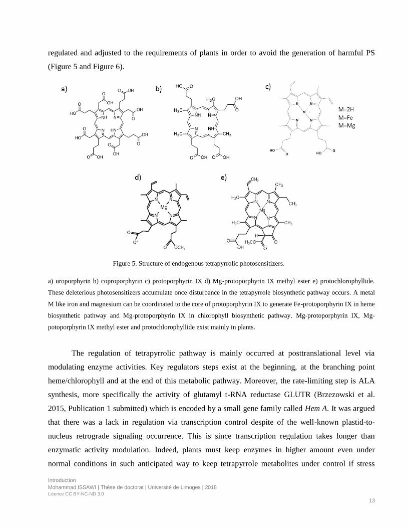
regulated and adjusted to the requirements of plants in order to avoid the generation of harmful PS
(Figure 5 and Figure 6).
a) uroporphyrin b) coproporphyrin c) protoporphyrin IX d) Mg-protoporphyrin IX methyl ester e) protochlorophyllide.
These deleterious photosensitizers accumulate once disturbance in the tetrapyrrole biosynthetic pathway occurs. A metal
M like iron and magnesium can be coordinated to the core of protoporphyrin IX to generate Fe-protoporphyrin IX in heme
biosynthetic pathway and Mg-protoporphyrin IX in chlorophyll biosynthetic pathway. Mg-protoporphyrin IX, Mg-
potoporphyrin IX methyl ester and protochlorophyllide exist mainly in plants.
The regulation of tetrapyrrolic pathway is mainly occurred at posttranslational level via
modulating enzyme activities. Key regulators steps exist at the beginning, at the branching point
heme/chlorophyll and at the end of this metabolic pathway. Moreover, the rate-limiting step is ALA
synthesis, more specifically the activity of glutamyl t-RNA reductase GLUTR (Brzezowski et al.
2015, Publication 1 submitted) which is encoded by a small gene family called Hem A. It was argued
that there was a lack in regulation via transcription control despite of the well-known plastid-to-
nucleus retrograde signaling occurrence. This is since transcription regulation takes longer than
enzymatic activity modulation. Indeed, plants must keep enzymes in higher amount even under
normal conditions in such anticipated way to keep tetrapyrrole metabolites under control if stress
Figure 5. Structure of endogenous tetrapyrrolic photosensitizers.
Introduction
Mohammad ISSAWI | Thèse de doctorat | Université de Limoges | 2018
Licence CC BY-NC-ND 3.0
14
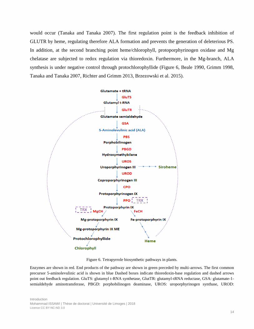
would occur (Tanaka and Tanaka 2007). The first regulation point is the feedback inhibition of
GLUTR by heme, regulating therefore ALA formation and prevents the generation of deleterious PS.
In addition, at the second branching point heme/chlorophyll, protoporphyrinogen oxidase and Mg
chelatase are subjected to redox regulation via thioredoxin. Furthermore, in the Mg-branch, ALA
synthesis is under negative control through protochlorophyllide (Figure 6, Beale 1990, Grimm 1998,
Tanaka and Tanaka 2007, Richter and Grimm 2013, Brzezowski et al. 2015).
Figure 6. Tetrapyrrole biosynthetic pathways in plants.
Enzymes are shown in red. End products of the pathway are shown in green preceded by multi-arrows. The first common
precursor 5-aminolevulinic acid is shown in blue Dashed boxes indicate thioredoxin-base regulation and dashed arrows
point out feedback regulation. GluTS: glutamyl t-RNA synthetase, GlutTR: glutamyl-tRNA reductase, GSA: glutamate-1-
semialdehyde aminotransferase, PBGD: porphobilinogen deaminase, UROS: uroporphyrinogen synthase, UROD:
Introduction
Mohammad ISSAWI | Thèse de doctorat | Université de Limoges | 2018
Licence CC BY-NC-ND 3.0
15
uroporphyrinogen decarboxylase, CPO: coproporphyrinogen oxidase, PPO: protoporphyrinogen oxidase, FeCH: Fe-
chelatase, MgCH: Mg-chelatase, TRX: thioredoxin.
I.2. Applications of PS
In the present manuscript we develop biological PS applications. Thus, energetic applications
such as solar energy conversion, electrochemistry and organic electronics will be out of context and
are not discussed (reader can refer to Zhao et al. 2013).
I.2.1. Photodynamic treatment in medicine
To date, the main application of PS is their use in photodynamic therapy (PDT) which is a
worldwide clinically approved cancer therapy relying on a photochemical reaction between three
components: PS, light, and molecular oxygen. PDT is a non-invasive procedure for the treatment of
cancers and other diseases. In this paragraph we will develop the PDT applications from cancer to
non-cancerous diseases in medical domains like dermatology, odontology, etc.
I.2.1.1. Photodynamic therapy against cancers
Forty years ago, a new treatment based on the combination effects of three harmless elements:
light-oxygen-PS was imagined and investigated to struggle against cancer cells. Thus, the most
famous and commercial PS introduced in the 1970’s was the Photofrin® that is a mixture of dimers
and oligomers of hematoporphyrin derivatives linked by ether, ester and C-C bonds (Ormond and
Freeman 2013). The drawbacks of Photofrin® as skin sensitivity and weak absorption at 630 nm lead
scientists to synthetize and design new molecules that could act as improved PS (Abrahamse and
Hamblin 2016). Within PDT context, PS were ranged into 3 generations basing on the time of
development and physico-chemical characteristics. photofrin® represents the first-generation PS,
second generation regroup all porphyrinoid PS with improved features and third generation
designates modified PS with biological conjugates as antibodies and carriers.
Researchers seek for the ideal PS that must fulfill the following requirements: it should have: no dark
toxicity, natural fluorescence which is important for diagnostics, low photobleaching and high
chemical stability, high singlet oxygen quantum yield, high absorption in “therapeutic window”
between 620 and 850 nm (Figure 7).

Introduction
Mohammad ISSAWI | Thèse de doctorat | Université de Limoges | 2018
Licence CC BY-NC-ND 3.0
16
Figure 7. PDT application in cancer treatment.
(modified from https://hope4cancer.com/our-therapies/sono-photo-dynamic-therapy/). PS is transported and released into
the tumor via blood circulation in the case of deep tumor where it exerts photochemical reactions leading to ROS
generation that bring about cancer reduction under light. ROS: Reactive oxygen species.
I.2.1.2. Photodynamic therapy against non-oncological diseases
PDT applications reached therapies for non-malignancies and include several domains such as
ophthalmology, dentistry, dermatology as well as cosmetics. It treats disease like age-related macular
degeneration, psoriasis, actinic keratosis and rheumatoid arthritis using clinically approved PS as
Photofrin®, Verteporphin®, and Levulan®-mediated protoporphyrin IX. Very recently, PDT using
methyl aminolevulinic acid was employed for the removal of non-pigmented hair in the domain of
cosmetics since laser therapies failed to remove blond or white hair due to the lack of chromophores
(Babilas and Szeimies 2005, Uebelhoer and Dover 2005, Konopka et al. 2007, Rishi and Agarwal
2015, Shin et al. 2016).
I.2.1.3. Photodynamic therapy limitations
The difficulties or the limits of PDT in cancer or other diseases remain to optimize PS that
must be photoactivated to be efficient to induce cell death. This treatment is efficient for superficial
diseases but is not uploaded for deep tissue alterations. Researchers developed second generation PS
as phtalocyanin and chlorin having stronger Q band absorption in order to the best deal with possible
wavelength for ideal tissue penetration (therapeutic window). Furthermore, optical processes were
Introduction
Mohammad ISSAWI | Thèse de doctorat | Université de Limoges | 2018
Licence CC BY-NC-ND 3.0
17
enhanced by exciting PS that have intense absorption in the blue range with two photons in the
wavelength range from 800 to 1000 nm. Thus, that methodology dealt with light tissue penetration.
Moreover, as advances have been made successfully in nanoengineering, researchers tended to target
carcinoma that are deeply buried in human body via PS integration with nanocarriers as liposomes,
quantum dots, carbon and inorganic nanomaterials (Ogawa and Kobuke 2008, Ormand and freeman
2013, Hong et al. 2016).
It is worthy to note that the promising PDT can be considered as an alternative modality to
conventional cancer therapies like surgery, chemo- and radiotherapy and should be combined with
these therapies to boost cancer control. Nevertheless, one of its current drawbacks is that PDT is
ineffective against metastatic cancers which are the most frequent cause of death.
I.2.2. Antimicrobial photodynamic treatment (APDT)
Antimicrobial photodynamic treatment is derived from PDT and describes the use of PDT to
inactivate microbial cells. Indeed, photodynamic inactivation (PDI) or Photo-Antimicrobial
chemotherapy (PACT) or APDT are similar terms or acronyms. Their applications are widely
expanded beyond medical scope to reach industrial, environmental and agricultural domains (Alves et
al. 2010).
I.2.2.1. APDT in medical environment
PS was also used in order to inactivate virus and microorganisms such as bacteria, yeast and
fungi in the present time witnessing the rise of multidrug resistant superbugs and therefore, the end of
antibiotic era (Huang et al. 2010). APDT was clinically approved mainly in the field of dermatology
and dentistry using 5-aminolevulenic acid (ALA)-mediated protoporphyrin IX and phenothiazinium
dyes. Since skin and mouth infections are localized and superficial contrarily to systemic infections,
APDT emerges as highly successful methodology in order to struggle against periodontitis, acne and
other oral and skin infections. Moreover, APDT was used efficiently to reduce hospital-acquired
infections such as nosocomial infections through PS applications on medical devices (Maisch et al.
2005, Kharkwal et al. 2011, McCoy et al. 2014, Yin et al. 2015).
Introduction
Mohammad ISSAWI | Thèse de doctorat | Université de Limoges | 2018
Licence CC BY-NC-ND 3.0
18
I.2.2.2. APDT for water sewage
Water disinfection through APDT is seen as efficient, eco-friendly and cost-effective
treatment against bacteria as Enterococcus faecalis, virus as bacteriophage (T4-like sewage
bacteriophage) and parasites as Ascaris lumbricoides (nematode). APDT for this purpose relies
mainly on the use of porphyrins, methylene blue and rose Bengal (Table 1). Its applications include
aquaculture, crop irrigation and hospital water disinfection (Costa et al. 2008, Carvalho et al. 2009,
Arrojado et al. 2011, Thandu et al. 2015, Bartolomeu et al. 2017).
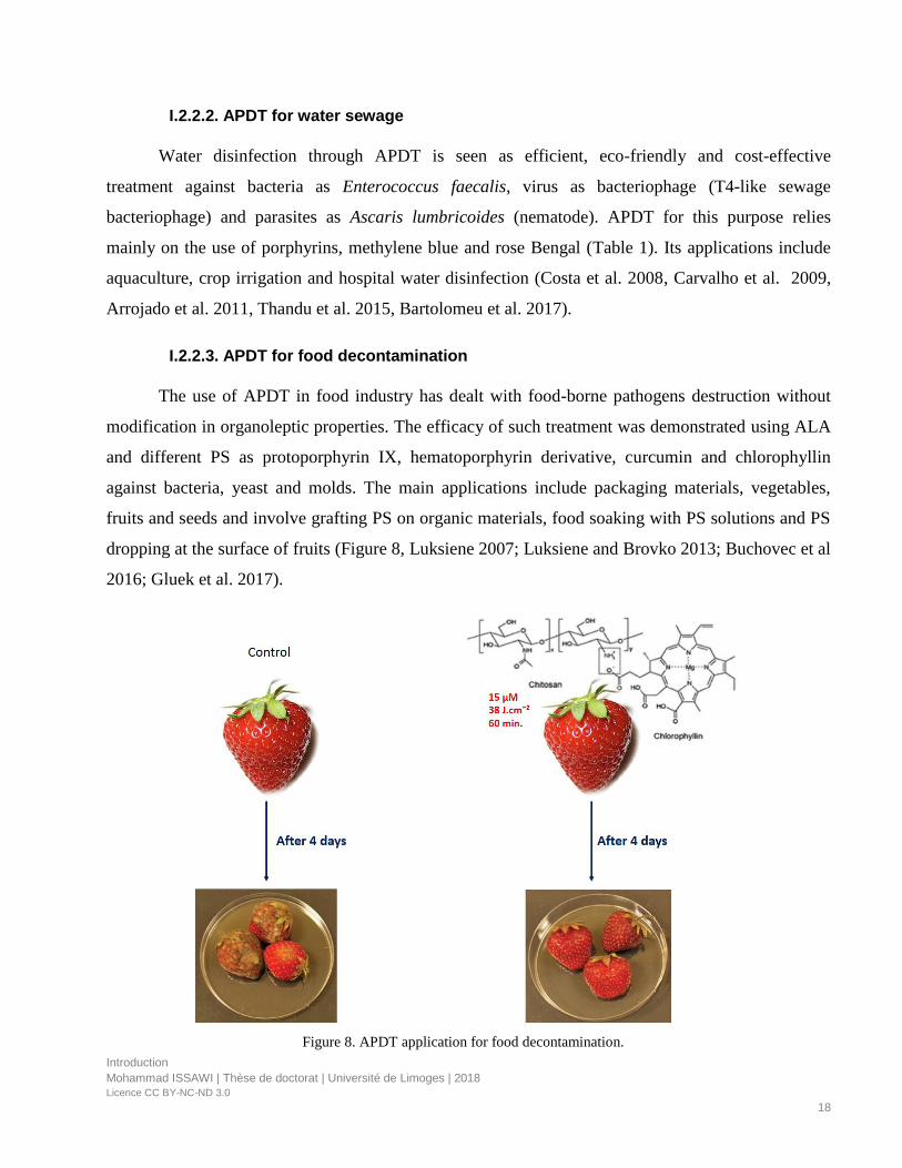
I.2.2.3. APDT for food decontamination
The use of APDT in food industry has dealt with food-borne pathogens destruction without
modification in organoleptic properties. The efficacy of such treatment was demonstrated using ALA
and different PS as protoporphyrin IX, hematoporphyrin derivative, curcumin and chlorophyllin
against bacteria, yeast and molds. The main applications include packaging materials, vegetables,
fruits and seeds and involve grafting PS on organic materials, food soaking with PS solutions and PS
dropping at the surface of fruits (Figure 8, Luksiene 2007; Luksiene and Brovko 2013; Buchovec et al
2016; Gluek et al. 2017).
Figure 8. APDT application for food decontamination.
Introduction
Mohammad ISSAWI | Thèse de doctorat | Université de Limoges | 2018
Licence CC BY-NC-ND 3.0
19
Strawberries were soaked with 0.1% chitosan (nutritional supplement) and 15 µM chlorophyllin then exposed to 405 nm
light at fluence equal to 38 J.cm-2 for 60 min. whereas strawberries that are not treated served as control. 4 days after
treatment, control strawberries were totally infected whereas treated strawberries with chlorophyllin–chitosan were not
spoiled (Buchovec et al. 2016)
I.2.2.4. APDT in industrial domain
It consists in incorporation of cationic porphyrins or protoporphyrin IX into different textile
materials to create protective clothing. Another application involves the grafting of porphyrins on
cellulose to create sanitized packaging materials as cardboard, as well as embedding them into paper,
fabrics and plastics. (Ringot et al. 2010, Feese et al. 2011; Lhotakova et al. 2012). Recently, APDT
using methylene blue and cationic corrole PS was employed for the restauration of historic
constructions that were colonized by photosynthetic biofilms (McCullagh and Robertson 2006, Pohl
et al. 2014).
I.2.2.5. APDT in agronomy
In the last four years, the use of PS acquires new potential aiming to eradicate plant pathogens
as well as unwanted vegetation without affecting crops (Figure 9). This particular aspect is largely
developed in the review (Publication 1 submitted) and will be further deeply discussed later in the
present manuscript. Briefly APDT applications reach agricultural domain as potential approach to
fight against plant pathogens. Indeed, it was recently shown that perennial plants like citrus and kiwi
trees were not affected after PS treatment whereas strawberry plants were damaged (De Menezes et
al. 2014 a,b, Fracarolli et al. 2016, De Menezes et al. 2016, Gonzales et al. 2017, Jesus et al. 2018).
Introduction
Mohammad ISSAWI | Thèse de doctorat | Université de Limoges | 2018
Licence CC BY-NC-ND 3.0
20
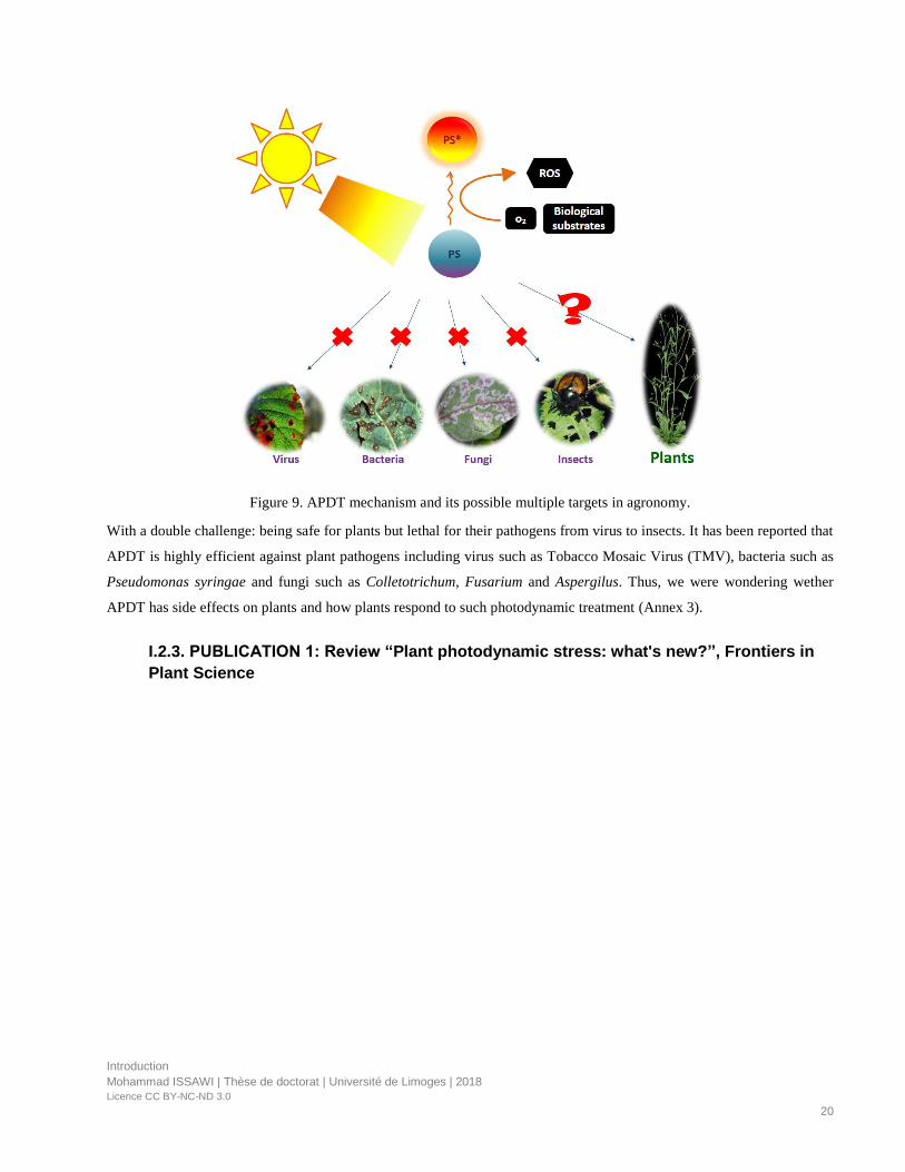
Figure 9. APDT mechanism and its possible multiple targets in agronomy.
With a double challenge: being safe for plants but lethal for their pathogens from virus to insects. It has been reported that
APDT is highly efficient against plant pathogens including virus such as Tobacco Mosaic Virus (TMV), bacteria such as
Pseudomonas syringae and fungi such as Colletotrichum, Fusarium and Aspergilus. Thus, we were wondering wether
APDT has side effects on plants and how plants respond to such photodynamic treatment (Annex 3).
I.2.3. PUBLICATION 1: Review “Plant photodynamic stress: what's new?”, Frontiers in
Plant Science
MINI REVIEWpublished: 23 May 2018
doi: 10.3389/fpls.2018.00681
Frontiers in Plant Science | www.frontiersin.org 1 May 2018 | Volume 9 | Article 681
Edited by:
Paula Casati,
Consejo Nacional de Investigaciones
Científicas y Técnicas (CONICET),
Argentina
Reviewed by:
Raquel Esteban,
University of the Basque Country
(UPV/EHU), Spain
Nejat Duzgunes,
University of the Pacific, United States
*Correspondence:
Catherine Riou
Specialty section:
This article was submitted to
Plant Abiotic Stress,
a section of the journal
Frontiers in Plant Science
Received: 13 February 2018
Accepted: 03 May 2018
Published: 23 May 2018
Citation:
Issawi M, Sol V and Riou C (2018)
Plant Photodynamic Stress: What’s
New? Front. Plant Sci. 9:681.
doi: 10.3389/fpls.2018.00681
Plant Photodynamic Stress: What’sNew?Mohammad Issawi, Vincent Sol and Catherine Riou*
Laboratoire Peirene (EA7500), Faculté des Sciences et Techniques, Université de Limoges, Limoges, France
In the 1970’s, an unconventional stressful photodynamic treatment applied to plants
was investigated in two directions. Exogenous photosensitizer treatment underlies
direct photodynamic stress while treatment mediating endogenous photosensitizer
over-accumulation pinpoints indirect photodynamic stress. For indirect photodynamic
treatment, tetrapyrrole biosynthesis pathway was deregulated by 5-aminolevulenic acid
or diphenyl ether. Overall, photodynamic stress involves the generation of high amount of
reactive oxygen species leading to plant cell death. All these investigations were mainly
performed to gain insight into new herbicide development but they were rapidly given up
or limited due to the harmfulness of diphenyl ether and the high cost of 5-aminolevulinic
acid treatment. Twenty years ago, plant photodynamic stress came back by way of crop
transgenesis where for example protoporphyrin oxidases from human or bacteria were
overexpressed. Such plants grew without dramatic effects of photodamage suggesting
that plants tolerated induced photodynamic stress. In this review, we shed light on
the occurrence of plant photodynamic stress and discuss challenging issues in the
context of agriculture focusing on direct photodynamic modality. Indeed, we highlighted
applications of exogenous PS especially porphyrins on plants, to further develop an
emerged antimicrobial photodynamic treatment that could be a new strategy to kill plant
pathogens without disturbing plant growth.
Keywords: 5-aminolevulinic acid, diphenyl ether herbicides, photosensitizers, plant photodynamic stress,
porphyrins, tetrapyrroles
INTRODUCTION
Almost all abiotic stresses induce oxidative stress underlying imbalance between reactive oxygenspecies (ROS) production and plant defense systems (Ramel et al., 2012; Müller-Xing et al., 2014;Hu et al., 2015; Loreti et al., 2016; Vian et al., 2016; Chakradhar et al., 2017; Hasan et al., 2017;Jaleel et al., 2017; Ohama et al., 2017; Rihan et al., 2017; Yang and Guo, 2017). Often, photo-oxidative and photodynamic stresses are confused whereas they bear two distinct meanings. Theformer points out a light-driven generation of ROS in chloroplasts through the photosensitizationof excited chlorophyll molecules that are embedded in antennae complex and reaction centeror via electron leakage from overloaded electron transport chain within photosystem apparatus.However, photodynamic stress involves the accumulation of exogenous or endogenous PS atvarious subcellular compartments and subsequently photochemical ROS production via two typesof photochemical reactions under light conditions. In the type I, redox state change of excitedsensitizer occurs upon reactions with biological molecules and oxygen resulting in hydrogenperoxide and free radical generation while In the Type II, energy from excited PS is transferreddirectly to oxygen leading to singlet oxygen production (Figure 1; Foyer et al., 1994; Pospíšil, 2016;Kashef et al., 2017).
Issawi et al. Plant Photodynamic Stress
In plants, occurrence of photodynamic stress correspondsto two distinct artificial situations. The first one involvesderegulations of plant tetrapyrrole biosynthetic pathway usingmolecules such as 5-aminolevulinic acid (ALA), diphenyl ether(DPE) or genetic tools. Tetrapyrroles play numerous roles fromlight harvesting, oxygen transport, oxidative phosphorylation,oxygen storage, nitrogen fixation to ROS scavenging (heme)(Figure 1A; Battersby et al., 1980; Senge et al., 2014, 2015). Undernormal conditions, this primary biosynthetic pathway is tinyregulated andmainly confined to plastidial organelles that protectcells from potential or accidental oxidative burst. Nevertheless,when this pathway is not anymore regulated by for instanceexogenous supply of ALA or DPE or genetic modifications,some intermediates such as protoporphyrin IX (PPIX) andMg-porphyrins become powerful photosensitizers that couldtrigger carbohydrates, proteins, lipids and nucleic acids damages(Rebeiz, 2014). In the second situation, photodynamic stress isinduced through plant exposure to exogenous PS which are ableto produce high amount of ROS inside cells under irradiation.Applications of exogenous PS such as phenothiazinium dyes,coumarins and furocoumarins, porphyrins were performed andsummarized in Table 1. A new application of photodynamictreatment on plants is raising up as efficient weapon to struggleagainst pathogens essentially bacteria and fungi in the contextof the so-called antimicrobial photodynamic treatment (APDT)(Figure 1B). Indeed, within APDT, plants of agronomic interestwill normally grow protecting themselves from deleteriouseffect of photoactivated PS via setting up powerful antioxidantmachinery. This review will develop photodynamic stress inplants and focus on the direct photodynamic stress regardingAPDT to gain insight in improving agronomic practiceswith high crop yield and environmental protection goals.Photodynamic strategy applied to pathogens or microorganismsis subject of numerous reviews and will not be developedhere (Ben Amor and Jori, 2000; Jori and Brown, 2004;Maisch, 2007, 2009; Donnelly et al., 2008; Almeida et al.,2011; Jori, 2011; Alves et al., 2015; Liu et al., 2015; Tim,2015; Hamblin, 2016; Wainwright et al., 2016; Kashef et al.,2017).
INDIRECT PHOTODYNAMIC STRESS
Forcing plants to accumulate excessive amount of endogenoustetrapyrrolic photosensitizers induce photodynamic stressconditions such as ALA feeding, DPE treatment as wellas by transgenesis experiments leading to growth anddevelopment impediment. In this review, we will not developthe crop transgenesis tools because they do not fit with plantphotodynamic treatment. The reader should refer to thesesreferences for more informations (Li, 2003; Lee et al., 2004; Liand Nicholl, 2005; Jung et al., 2008; Ayliffe et al., 2009; Jung,2011; Quesada et al., 2013; Yun et al., 2013; Kim et al., 2014).
5-aminolevulinic Acid (ALA) Feeding5-aminolevulinic acid is not a PS per se. Instead, it is anon-protein amino acid and the first common precursor ofthe tetrapyrrole (chlorophylls, heme, and derivatives) pathway
(Figure 1A). Its supply lead to PPIX and/or other intermediatesover-accumulation. From the 70’s, exogenous application ofALA on yeast, insects, plants and in mammal cells wasshown to induce high accumulation of tetrapyrroles (Figure 1A;Brouillet et al., 1975; Rebeiz et al., 1984, 1995; Matsumotoet al., 1994; Juzeniene et al., 2002; Fotinos et al., 2006;Xu et al., 2015). When tetrapyrroles were over-accumulatedby ALA feeding, plants could not anymore struggle againstinduced photodynamic stress and died (Rebeiz et al., 1984,1990; Matsumoto et al., 1994). When cucumber fields weresprayed with ALA, it was found that seedlings accumulatedmassive amount of endogenous porphyrins especially thepotential singlet oxygen generator “protochlorophyllide” under5,000 foot candle (Rebeiz et al., 1988). A similar resultwas obtained on duckweed (Lemna paucicostata Hegelm.)that showed rapid membrane damage after light irradiationand increase in both protochlorophyllide and PPIX contentssuggesting herbicidal effect of ALA (Matsumoto et al., 1994).In the other hand, ALA-treated plants significantly upregulatedtranscript levels of genes encoding superoxide dismutaseand serine/threonine kinase receptors but the induction ofantioxidative components lacked capacity to withstand ROSgeneration (Phung and Jung, 2014, 2015). In 2004, Jung andco-workers shed light on “photodynamic stress” as they showedthat rice plants suffered from severe oxidative damage uponthe ectopic expression of the bacterial ALA synthase genebringing about the accumulation of harmful photosensitizersPPIX and protochlorophyllide under 350 µmol.m−2.s−1 (Junget al., 2004). ALA feeding was performed in order to look for anew herbicide. However, there was no commercial formulationof ALA as field effective herbicide owing to the high amountrequired (≥5mM) and the cost-effective treatment (Sasikalaet al., 1994; Phung and Jung, 2014; Xu et al., 2015; Nguyen et al.,2016).
Diphenyl Ether (DPE) TreatmentSince 1960’s, DPE essentially oxyfluorfen and acifluorfen wereintroduced as commercial herbicides to control weeds (Yanget al., 2006). They constitute the main class of PPO-inhibitingherbicides that are widely investigated. Phung and Jung (2015)reported the different responses of photodynamically stressedrice plants undergoing ALA (5mM) and oxyfluorfen (50µM)herbicidal treatment. In term of phenotype under illumination,ALA induced bleached necrotic spots while oxyfluorfen causedbronzed necrotic spots on the leaves. This difference inphotodynamic symptoms was due to PPIX overaccumulationin cytoplasm in DPE-treated plants whereas the photodynamicdestruction of chlorophyll by Mg-porphyrins was responsibleof the white spot appearance. Beyond the phenotypical effects,the brown necrosis in DPE-treated plants exhibited a moredispersed H2O2 production and was accompanied by anincrease in H2O2-scavenging enzymes, catalase and peroxidaseactivities as well as dehydroascorbate content, a strong stressmarker compared to those of ALA-treated plants (Phungand Jung, 2015). Their mode of action was established andconsisted in the inhibition of protoporphyrinogen oxidase (PPO)the last enzyme at the branching point between heme and
Frontiers in Plant Science | www.frontiersin.org 2 May 2018 | Volume 9 | Article 681
Issawi et al. Plant Photodynamic Stress
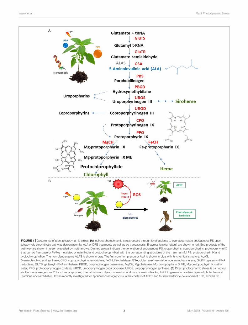
FIGURE 1 | Occurrence of plant photodynamic stress. (A) Indirect photodynamic stress occurs through forcing plants to over-accumulate endogenous PS upon
tetrapyrrole biosynthetic pathway deregulation by ALA or DPE treatments as well as by transgenesis. Enzymes (capital letters) are shown in red. End products of the
pathway are shown in green preceded by multi-arrows. Dashed arrows indicate the generation of endogenous PS (uroporphyrins, coproporphyrins, protoporphyrin IX
that can be free-base or Fe/Mg metalated or esterified and protochlorophyllide) with the corresponding structures of the main harmful PS: protoporphyrin IX and
protochlorophyllide. The non-plant enzyme ALAS is shown in gray. The first common precursor ALA is shown in blue with its chemical structure. ALAS,
5-aminolevulinic acid synthase; CPO, coproporphyrinogen oxidase; FeCH, Fe-chelatase; GSA, glutamate-1-semialdehyde aminotransferase; GlutTR, glutamyl-tRNA
reductase; GluTS, glutamyl t-RNA synthetase; PBGD, porphobilinogen deaminase; MgCH, Mg-chelatase; Mg-protoporphyrin IX ME, Mg-protoporphyrin IX methyl
ester; PPO, protoporphyrinogen oxidase; UROD, uroporphyrinogen decarboxylase; UROS, uroporphyrinogen synthase. (B) Direct photodynamic stress is carried out
via the use of exogenous PS such as porphyrins, phenothiazinium dyes, coumarins, and furocoumarins leading to ROS generation via two types of photochemical
reactions upon irradiation. It was recently investigated for applications in agronomy in the context of APDT and for new herbicide development. *PS, excited PS.
Frontiers in Plant Science | www.frontiersin.org 3 May 2018 | Volume 9 | Article 681

Issawi et al. Plant Photodynamic Stress
TABLE1|Exo
genousPSandtheircharacteristicsandapplicatio
nin
plantphotodyn
amictreatm
ent.
PS
Origin
Concentration
Lightrange
Lightintensity
Plantmaterial
Function*
References
Non-porphyrin
compounds
Procionyellow
Syn
thetic
4%
solutio
nUV
Elodealeafmeso
phyllcells
Intracellularultrastructure
staining
Goodwin,1976
Cercosp
orin
Natural
Varia
ble(0.5–1
8.7
µM)Vario
ustypesoflamps
Varia
ble
Potato,carrot,redbeet,
tobaccoleafdiscs,
maizeroots
andNT575tobacco
susp
ensioncells
Phytotoxin
MacriandVianello,1979;
Daub,1982a,b;Dauband
Brig
gs,
1983
Rose
bengal
Syn
thetic
10mM
White
100and350
µmol.m
−2.s−1
Pealeafdiscs
Membraneandnucleus
staining
KnoxandDodge,1984
Hyp
eric
inNatural
100
µM
White
400
µmol.m
−2.s−1
Pealeafdiscs
Plantdefense
KnoxandDodge,1985a
EosinY
Syn
thetic
5µM
and1mM
White
andgreen
350
µmol.m
−2.s−1
and5.26mW.cm−2
Pealeafdiscsandonionbulb
roots
Protein
staining,DNA
binding
KnoxandDodge,1985b;
Molero
andHazen,1988
Berberin
eNatural
Varia
ble(1nM
to
10
µM)
Violet
195KW.m
−2
Roots
ofonionbulbs
Antim
icrobial,heparin
staining,DNAbinding
Molero
etal.,
1985
Pyronin
YSyn
thetic
1and5
µM
Green
129.9
KW.m
−2
Roots
ofonionbulbs
DNAbinding
Arm
as-Portelaetal.,
1985
Acrid
inorange
Syn
thetic
5µM
Green
5.26mW.cm−2
Roots
ofonionbulbs
Mito
chondria
stainingand
DNAbinding
Molero
andHazen,1988
Orcein
Natural
5µM
Green
5.26mW.cm−2
Roots
ofonionbulbs
Chromoso
mestaining
Molero
andHazen,1988
Harm
ine
Natural
500nM
UV
2.5
mW.cm−2
Roots
ofonionbulbs
enzymatic
inhibitionand
DNAbinding
Hazenand
Gutierrez-Gonzalvez,
1988
Coumarin
sand
furocoumarin
s
Natural
Varia
ble
Solarradiatio
nCitrustreeleavesand
strawberryleaves
Plantdefense
deMenezesetal.,
2014a;
Fracarollietal.,
2016
Phenothiazinium
dyes
Syn
thetic
Varia
ble(5,25,and
50
µM)
Solarradiatio
nCitrustreeleaves(health
yand
combinatedwith
fungal
pathogen)
DNA/R
NAstainingand
bacteria
lstaining
deMenezesetal.,
2014b;
Gonzalesetal.,
2017
Porphyrins
HPD
Syn
thetic
25
µg.m
l−1
NearUV
9W.m
−2
Viciafabaleafprotoplasts
Usa
geinPDTas
PHOTOFRIN
Kjeldstadetal.,
1986
TPyP
andHP
Syn
thetic
100nM
Red
0.001J.m−2
Roots
ofonionbulbs
DNAbinding
Hazenetal.,
1987
TMPyP
/Zn-TMPyP
Syn
thetic
Varia
ble(10nM
to
100
µM)
Red,blue,white
and
solarradiatio
n
Varia
ble
Onionbulb
roots,TBY-2cells,
kiwileaves(health
yand
contaminated),tomato
and
Arabidopsisthalianaplantlets
DNAbinding
Villaneuva
etal.,
1986,
1989;Riouetal.,
2014;
Guillaumotetal.,
2016;
Issa
wietal.,
2018;Je
sus
etal.,
2018
TPPS/Zn-TPPS
Syn
thetic
3.5
µM
White
95and250
µmol.m
−2.s−1
TBY-2su
spensioncells,
tomato
andArabidopsis
thalianaplantlets
Riouetal.,
2014;Guillaumot
etal.,
2016;Issa
wietal.,
2018
TPyP,Tetra(4-pyridyl)porphyrin;TMPYP,Tetra(N-m
ethylpyridyl)porphyrin;TPPS,meso-tetra(4-sulfophenyl)porphyrin;HPD,Hematoporphyrinderivative;HP,Hematoporphyrin;TBY-2,TobaccoBrightYellow-2.*OtherpropertiesofPS
thatarenotrelatedtotheirphotodynamicaction.
Frontiers in Plant Science | www.frontiersin.org 4 May 2018 | Volume 9 | Article 681
Issawi et al. Plant Photodynamic Stress
chlorophyll synthesis (Figure 1A; Matringe and Scalla, 1987;Matringe et al., 1989a,b). Thus, triggering leakage of PPOsubstrate, the non-fluorescent protoporphyrinogen IX that wasconverted by unknown peroxidase to the first effective PSof this pathway PPIX. Indeed, when PPIX absorbs light, itinduces photochemical reactions and vital processes are affected(Figure 1A).
DIRECT PHOTODYNAMIC STRESS: ANOLD STORY WITH NOVEL DEVELOPMENT
Plant exposure to exogenous PS could induce tissue damage andsubsequently cell death. First studies relative to direct applicationof PS on plantmaterials were reported four decades ago (Table 1).Concerning porphyrins, the most used PS, Kjeldstad and co-workers showed the photodamage of plasma membrane of Viciafaba leaf protoplasts subjected to hematoporphyrin derivativetreatment under near UV light (Kjeldstad et al., 1986). Moreover,other studies showed mutagenic effect of porphyrins which wereable to bind DNA in root meristematic cells of onion bulbs(Table 1; Villaneuva et al., 1986, 1989; Hazen et al., 1987). Theaims of testing exogenous PS on plant materials were to studythe symplastic intracellular movement, decipher the mode ofaction of fungal toxin as well as the effects of singlet oxygen onplant cells and exploring sister chromatide exchange upon dyeDNA-intercalation in fast-rate dividing cells (Table 1; Goodwin,1976; Macri and Vianello, 1979; Daub, 1982a,b; Daub and Briggs,1983; Knox and Dodge, 1984, 1985a,b; Armas-Portela et al., 1985;Molero et al., 1985; Kjeldstad et al., 1986; Villaneuva et al., 1986,1989; Hazen et al., 1987; Hazen and Gutierrez-Gonzalvez, 1988;Molero and Hazen, 1988).
For agronomic issues, the use of exogenous PS as powerfulphotoactivated molecules was not anymore investigated becausethe undesirable effects described above. Hence, any potentialapplication of whatever PS in the aim to fight plant pathogensrequires a risk assessment on plant hosts. It was reported thatnatural photosensitizers such as coumarins and furocoumarinsor synthetic ones such as phenothiazinium and porphyrinsinactivated pathogenic agents as virus (Tobacco mosaic virus),bacteria (Pseudomonas syringae) and fungi (Collectotrchumabscissum, Colletotrichum gloeosporioides, Collectotrichumacutatum, Aspergillus nidulans, Fusarium oxysporum, Fusariummoniliforme, Fusarium solani) (Table 1). However, when spottedon orange tree and strawberry plants, or on kiwi contaminatedleaves under solar radiation, the leaves and flowers were notaffected by either natural/synthetic photosensitizers exceptedfor strawberry leaves that were damaged upon treatment with100µM phenothiazinium (Orlob, 1967; de Menezes et al.,2014a,b; Fracarolli et al., 2016; Gonzales et al., 2017; Jesus et al.,2018). In another extended context, Issawi and co-workersconceived a double target strategy that could eradicate in thesame time unwanted vegetation and plant pathogens withoutkilling plants of agronomic interest (Figure 1B). To fulfillthat purpose, they studied the effect of exogenous water-soluble cationic and anionic porphyrins on tomato, plant ofagronomic interest and on Arabidopsis thaliana, weed-like
plant. Thus, they showed that cationic porphyrins were able toeradicate Arabidopsis plantlets without killing tomato plantlets(Guillaumot et al., 2016; Issawi et al., 2018). Favorably, Riouand co-workers treated TBY-2 cells with these same porphyrinsaiming to find out new herbicides because no new herbicidemodes of action were discovered since the last 3 decades (Duke,2012; Heap, 2014; Riou et al., 2014).
DETERMINANTS OF PLANTPHOTODYNAMIC STRESS
Plants exposed to various stressors respond by involvingmechanisms of sensing and signaling (Tuteja and Sopory,2008; Mittler and Blumwald, 2010; Suzuki et al., 2012; Pandeyet al., 2016; Zhu, 2016; Mittler, 2017). Although plant stresssignaling pathways were abundantly investigated, stress sensorsremain largely unknown so it is much difficult to detectsensing systems in plants subjected to direct photodynamicstress under unconventional conditions. However, Phungand collaborators outlined the switch photodynamic/drought-tolerance in PPO-transgenic rice under drought conditionsexplaining how drought determinants reduced porphyrin levelin order to elaborate tolerance response through gene expressionmodulation upon sensing change in tetrapyrrole amount (Phunget al., 2011). Nonetheless seeking for photodynamic sensorsrepresents serious challenge. Exogenous PS exert photodynamicfunction through the production of ROS including singletoxygen and hydrogen peroxide which are well known signalingmolecules, it is worthwhile to distinguish between primarysensing that may be assigned to the PS per se and secondarysensing ascribed to ROS (Laloi et al., 2007; Niu and Liao,2016; Wang et al., 2016). ABC transporters and TSPO receptorcould play role in exogenous PS sensing since they wereidentified as endogenous tetrapyrrolic receptors (Theodoulou,2000; Guillaumot et al., 2009). Interestingly, PPIX interactedwith Toll-Like Receptor 4 (TLR-4) in mammals. Possibleinteraction with a putative TLR should be investigated in plantcells (Figueiredo et al., 2007; Tangudu and Spasic, 2017). Interm of signaling, exogenous photodynamic action is likelyto generate various secondary messengers holding signalingpotential like ROS, modified proteins, lipid peroxidation by-products. In addition, photodynamic signaling may also involvecross-talk with phytohormones as abscissic acid, jasmonicacid, salicylic acid, calcium, protein kinases, transcriptionfactors (Tuteja and Sopory, 2008; Suzuki et al., 2012; Zhu,2016). Thus, studies involving transcription profiling, proteomicand metabolomic approaches should be envisaged uponphotodynamic administration of exogenous PS in plants.
CHALLENGES AND PERSPECTIVES
In the present review, two ways to carry out photodoynamicaction on plants were emphasized (i) indirect photodynamicreaction via ALA or DPE treatments and transgenesis, (ii)direct photodynamic reaction through the use of exogenous PS.Comparing to conventional weed management methods using
Frontiers in Plant Science | www.frontiersin.org 5 May 2018 | Volume 9 | Article 681

Issawi et al. Plant Photodynamic Stress
photodynamic DPE herbicides that are commercially available,it was reported that this kind of herbicides were not only toxic toweeds but wildlife was also affected as DPE herbicides were toxicto nestling birds and fresh water polyp, also, they could be toxicto humans. In addition, DPE alone were not expected to controlweeds and need the combinational use of chemicals or mulch.Indeed, there are several reported weeds that developed DPE-resistance (Hoffman et al., 1991; Rio et al., 1997; Li and Nicholl,2005; Beckie and Tardif, 2012; Reed et al., 2018). Thus, weconclude that direct photodynamic treatment holding herbicidalpotential via exogenous PS could be a promising approachrelying on the fact that weeds cannot induce resistance againstPS because they exert a multi-targeted photodynamic actiondamaging cellular DNA, lipids, carbohydrates and proteins.Taken together, we support the application of exogenous PS asweed control alternative and further studies are required as faras we know that studies concerning that task are very few (Riouet al., 2014; Issawi et al., 2018).
In regard to antimicrobial strategies, APDT was recentlyenvisaged as promising approach to strike against plantpathogens without side effects on plant hosts and environment.In this context, we think that several aspects must be takeninto consideration such as whole study of a defined pathosystemshould be considered including biological life cycle of pathogen,growth and reproduction phenology of plants and timing of PSapplication.Moreover, PS administrationmethods as inclusion insoil aqueous phase, spotting on leaves and field spraying shouldnot be neglected although spraying is the most convenient andreasonable.
CONCLUSION
In conclusion, plant photodynamic stress is considered as abioticstress linked to ROS production as the first cause of celldeath. It is still poorly studied regarding characterization ofphotodynamic stress determinants and outcomes especially atmolecular level in plants. Besides, plant photodynamic stresshas not been exploited yet, especially as valuable exogenoustreatment for the purposes we mentioned above. Nevertheless,we are confident that in the near future, this approach basedon PS and especially porphyrins could be relevant to respondto the Directive 2009/128/EC of the European Parliament plansthat aim to reduce the use of pesticides while maintaininghigh yield as well as high quality in agricultural production. PSare photodegradable and non-toxic under dark as well as theywere used at micromolar concentrations therefore they could bepromising candidates to fulfill the task of European projects forenvironment sustainability in respect to wildlife, water sourcesand human health.
AUTHOR CONTRIBUTIONS
MI and CR prepared and wrote the present manuscript. MIdesigned the figure and the table. VS reviewed the manuscript.
ACKNOWLEDGMENTS
MI was supported by a Grant from the Municipality of Sharkieh(Lebanon).
REFERENCES
Almeida, A., Cunha, A., Faustino, M. A. F., Tome, A. C., and Neves, M. G.
P. M. S. (2011). “Porphyrins as antimicrobial photosensitizing agents” in
Photodynamic Inactivation of Microbial Pathogens: Medical and Environmental
Applications, eds M. R. Hamblin and G. Jori (Cambridge, UK: RCS Publishing),
83–160.
Alves, E., Faustino, M. A. E., Neves, M. S., Cunhaa, A., Nadaisc, H., and
Almeida, A. (2015). Potential applications of porphyrins in photodynamic
inactivation beyond the medical scope. J. Photochem. Photobiol. B Biol. 22,
34–57. doi: 10.1016/j.jphotochemrev.2014.09.003
Armas-Portela, R., Hazen, M. J., and Stockert, J. C. (1985). Increase in
sister-chromatid exchanges in BrdU-substituted chromosomes of Allium
cepa induced by the combined effect of pyronin Y and green light.
Mutat. Res. Genet. Toxicol. 158, 77–80. doi: 10.1016/0165-1218(85)
90100-4
Ayliffe, M. A., Agostino, A., Clarke, B. C., Furbank, R., Von Caemmerer,
S., and Pryor, A. J. (2009). Suppression of the barley uroporphyrinogen
III synthase gene by a Ds activation Tagging element generates
developmental photosensitivity. Plant Cell 21, 814–831. doi: 10.1105/tpc.108.
063685
Battersby, A. R., Fooks, C. J. R., Matcham, G. W. H., and Mcdonald, E. (1980).
Biosynthesis of the pigments of life: formation of the macrocycle. Nature 285,
17–21. doi: 10.1038/285017a0
Beckie, H. J., and Tardif, F. J. (2012). Herbicide cross resistance in weeds.Crop Prot.
35, 15–28. doi: 10.1016/j.cropro.2011.12.018
Ben Amor, T., and Jori, G. (2000). Sunlight-activated insecticides: historical
background and mechanisms of phototoxic activity. Insect Biochem. Mol. Biol.
30, 915–925. doi: 10.1016/S0965-1748(00)00072-2
Brouillet, N., Arselin-De Chateaubodeau, G., and Volland, C.
(1975). Studies on protoporphyrin biosynthetic pathway in
Saccharomyces cerevisiae; characterization of the tetrapyrrole
intermediates. Biochimie 57, 647–655. doi: 10.1016/S0300-9084(75)
80146-5
Chakradhar, T., Mahanty, S., Reddy, R. A., Divya, K., Reddy, P. S.,
and Reddy, M. K. (2017). “Biotechnological perspective of reactive
oxygen species (ROS)-mediated stress tolerance in plants,” in Reactive
Oxygen Species and Antioxidant Systems in Plants: Role and Regulation
under Abiotic Stress, eds M. Khan and N. Khan (Singapore: Springer),
53–87.
Daub, M. E. (1982a). Cercosporin, a photosensitizing toxin from Cercospora
species. Phytopathology 72, 370–374. doi: 10.1094/Phyto-72-370
Daub, M. E. (1982b). Peroxidation of tobacco membrane lipids by the
photosensitizing toxin, cercosporin. Plant Physiol. 69, 1361–1364.
doi: 10.1104/pp.69.6.1361
Daub, M. E., and Briggs, S. P. (1983). Changes in tobacco cell membrane
composition and structure caused by cercosporin. Plant Physiol. 71, 763–766.
doi: 10.1104/pp.71.4.763
deMenezes, H. D., Pereira, A. C., Brancini, G. T. P., Leão, H. C., Massola Júnior, N.
S., Bachmann, L., et al. (2014a). Furocoumarins and coumarins photoinactivate
Colletotrichum acutatum and Aspergillus nidulans fungi under solar radiation.
J. Photochem. Photobiol. Biol. 131, 74–83. doi: 10.1016/j.jphotobiol.2014.
01.008
de Menezes, H. D., Rodrigues, G. B., Teixeira, S. P., Massola, N. S., Bachmann, L.,
Wainwright, M., et al. (2014b). In vitro photodynamic inactivation of plant-
pathogenic fungi colletotrichum acutatum and colletotrichum gloeosporioides
with novel phenothiazinium photosensitizers. Appl. Environ. Microbiol. 80,
1623–1632. doi: 10.1128/AEM.02788-13
Frontiers in Plant Science | www.frontiersin.org 6 May 2018 | Volume 9 | Article 681

Issawi et al. Plant Photodynamic Stress
Donnelly, R. F., McCarron, P. A., and Tunney, M. M. (2008).
Antifungal photodynamic therapy. Microbiol. Res. 163, 1–12.
doi: 10.1016/j.micres.2007.08.001
Duke, S. O. (2012).Why have no new herbicide modes of action appeared in recent
years? Pest Manag. Sci. 68, 505–512. doi: 10.1002/ps.2333
Figueiredo, R. T., Fernandez, P. L., Mourao-Sa, D. S., Porto, B. N., Dutra, F. F.,
Leticia, L. S., et al. (2007). Characterization of heme as activator of toll-like
receptor 4. J. Biol. Chem. 282, 20221–20229. doi: 10.1074/jbc.M610737200
Fotinos, N., Campo, M. A., Popowycz, F., Gurny, R., and Lange, N. (2006).
5-Aminolevulinic acid derivatives in photomedicine: characteristics,
application and perspectives. J. Photochem. Photobiol. 82, 994–1015.
doi: 10.1562/2006-02-03-IR-794
Foyer, C. H., Lelandais, M., and Kunert, K. J., (1994). Photooxidative stress in
plants. Physiol. Plant 92, 696–717. doi: 10.1111/j.1399-3054.1994.tb03042.x
Fracarolli, L., Rodrigues, G. B., Pereira, A. C., Massola Júnior, N. S.,
Silva-Junior, G. J., Bachmann, L., et al. (2016). Inactivation of plant-
pathogenic fungus Colletotrichum acutatum with natural plant-produced
photosensitizers under solar radiation. J. Photochem. Photobiol. Biol. 162,
402–411. doi: 10.1016/j.jphotobiol.2016.07.009
Gonzales, J. C., Brancini, G. T. P., Rodrigues, G. B., Silva-Junior, G. J.,
Bachmann, L., Wainwright, M., et al. (2017). Photodynamic inactivation of
conidia of the fungus Colletotrichum abscissum on Citrus sinensis plants with
methylene blue under solar radiation. J. Photochem. Photobiol. Biol. 176, 54–61.
doi: 10.1016/j.jphotobiol.2017.09.008
Goodwin, P. B. (1976). “Physiological and electrophysiological evidence
for intercellular communication in plant symplasts,” in intercellular
Communication in Plants: Studies on Plasmodesmata, eds B. E. S. Gunning
and A. W. Robards (Berlin; Heidelberg; New York, NY: Springer), 121–129.
doi: 10.1007/978-3-642-66294-2_6
Guillaumot, D., Guillon, S., Morsomme, P., and Batoko, H. (2009). ABA,
porphyrins and plant TSPO-related protein. Plant Signal. Behav. 4, 1087–1090.
doi: 10.4161/psb.4.11.9796
Guillaumot, D., Issawi, M., Da Silva, A., Leroy-Lhez, S., Sol, V., and
Riou, C. (2016). Synergistic enhancement of tolerance mechanisms
in response to photoactivation of cationic tetra (N-methylpyridyl)
porphyrins in tomato plantlets. J. Photochem. Photobiol. Biol. 156, 69–78.
doi: 10.1016/j.jphotobiol.2016.01.015
Hamblin, M. R. (2016). Antimicrobial photodynamic inactivation: a bright
newtechnique to kill resistant microbes. Curr. Opin. Microbiol. 33, 67–73.
doi: 10.1016/j.mib.2016.06.008
Hasan, M. K., Cheng, Y., Kanwar, M. K., Chu, X. Y., Ahammed, G. J., and Qi, Z.
Y. (2017). Responses of plant proteins to heavy metal stress—a review. Front.
Plant Sci. 8:1492. doi: 10.3389/fpls.2017.01492
Hazen, M. J., and Gutierrez-Gonzalvez, M. G. (1988). UV-mediated toxic
bioactivity of harmine in the meristematic cells of Allium cepa. Mutagenesis 3,
333–335. doi: 10.1093/mutage/3.4.333
Hazen, M. J., Villaneuva, A., and Stockert, J. C. (1987). Induction of sister
chromatide echanges in Allium cepa meristematic cells exposed to meso-
tetra (4-pyridyl) porphine and hematoporphyrin photoradiation. J. Photochem.
Photobiol. Biol. 46, 463–467. doi: 10.1111/j.1751-1097.1987.tb04796.x
Heap, I. (2014). Global perspective of herbicide-resistant weeds. Pest Manag. Sci.
70, 1306–1315. doi: 10.1002/ps.3696
Hoffman, D. J., Spann, J. W., LeCaptain, L. J., Christine, M. B., and
Rattner, B. A. (1991). Developmental toxicity of diphenyl ether herbicides
in nestling American kestrels. J. Toxicol. Environ. Health 34, 323–336.
doi: 10.1080/15287399109531571
Hu, B., Wang, W., Deng, K., Li, H., Zhang, Z., Zhang, L., et al. (2015).
MicroRNA399 is involved in multiple nutrient starvation responses in rice.
Front. Plant Sci. 6:188. doi: 10.3389/fpls.2015.00188
Issawi, M., Guillaumot, D., Sol, V., and Riou, C. (2018). Responses of an
adventitious fast-growing plant to photodynamic stress: comparative study of
anionic and cationic porphyrin effect on Arabidopsis thaliana. Physiol. Plant
162, 379–390. doi: 10.1111/ppl.12666
Jaleel, C. A., Manivannan, P., Wahid, A., Farooq, M., Al-juburi, H. J.,
Somasundaram, R., et al. (2017). Drought stress in plants: a review on
morphological characteristics and pigments composition. Int. J. Agric. Biol. 11,
100–105.
Jesus, V., Martins, D., Branco, T., Valério, N., Neves, M. G. P. M. S., Faustino,
M. A. F., et al. (2018). An insight into the photodynamic approach versus
copper formulations in the control of Pseudomonas syringae pv. actinidiae in
kiwi plants. Photochem. Photobiol. Sci. 17, 180–191. doi: 10.1039/C7PP00300E
Jori, G. (2011). “Antimicrobial photodynamic therapy: basic principles,”
in Photodynamic Inactivation of Microbial Pathogens: Medical and
Environemental Applications, eds M. R. Hamblin and G. Gori (Cambridge, UK:
RCS Publishing), 83–160.
Jori, G., and Brown, S. (2004). Photosensitized inactivation of microorganisms.
Photochem. Photobiol. Sci. 3, 403–405. doi: 10.1039/b311904c
Jung, S. (2011). Level of protoporphyrinogen oxidase activity tightly correlates with
photodynamic and defense responses in oxyfluorfen-treated transgenic rice. J.
Pestic. Sci. 36, 16–21. doi: 10.1584/jpestics.G10-46
Jung, S., Lee, H. J., Lee, Y., Kang, K., Kim, Y. S., Grimm, B., et al. (2008).
Toxic tetrapyrrole accumulation in protoporphyrinogen IX oxidase-
overexpressing transgenic rice plants. Plant Mol. Biol. 67, 535–546.
doi: 10.1007/s11103-008-9338-0
Jung, S., Yang, K., Lee, D. E., and Back, K. (2004). Expression of
Bradyrhizobium japonicum 5-aminolevulinic acid synthase induces
severe photodynamic damage in transgenic rice. Plant Sci. 167, 789–795.
doi: 10.1016/j.plantsci.2004.05.038
Juzeniene, A., Juzenas, P., Iani, V., and Moan, J. (2002). Topical application of 5-
aminolevulinic acid and its methylester, hexylester and octylester derivatives:
considerations for dosimetry in mouse skin model. J. Photochem. Photobiol. 76,
329–334. doi: 10.1562/0031-8655(2002)076<0329:TAOAAA>2.0.CO;2
Kashef, N., Huang, Y. Y., and Hamblin, M. R. (2017). Advances in antimicrobial
photodynamic inactivation at the nanoscale. Nanophotonics 6, 853–879.
doi: 10.1515/nanoph-2016-0189
Kim, J. G., Back, K., Lee, H. Y., Lee, H. J., Phung, T. H., Grimm, B., et al.
(2014). Increased expression of Fe-chelatase leads to increased metabolic
flux into heme and confers protection against photodynamically induced
oxidative stress. Plant Mol. Biol. 6, 271–287. doi: 10.1007/s11103-014-
0228-3
Kjeldstad, B., Olsten, M. L., Christensen, T., and Johnsson, A. (1986).
Photodynamic damage to protoplasts with a hematoporphyrin derivative. Plant
Sci. 44, 139–143. doi: 10.1016/0168-9452(86)90083-X
Knox, J. P., and Dodge, A. D. (1984). Photodynamic damage to plant leaf tissue by
rose bengal. Plant Sci. Lett. 37, 3–7. doi: 10.1016/0304-4211(84)90194-9
Knox, J. P., and Dodge, A. D. (1985a). Isolation and activity of the
photodynamic pigment hypericin. Plant Cell Environ. 8, 19–25.
doi: 10.1111/j.1365-3040.1985.tb01204.x
Knox, J. P., and Dodge, A. D. (1985b). The photodynamic action of eosin, a
singlet-oxygen generator. Planta 164, 22–29. doi: 10.1007/BF00391021
Laloi, C., Stachowiak, M., Pers-Kamczyc, E., Warzych, E., Murgia, I., and Apel, K.
(2007). Cross-talk between singlet oxygen- and hydrogen peroxide-dependent
signaling of stress responses in Arabidopsis thaliana. Proc. Natl. Acad. Sci.
U.S.A. 104, 672–677. doi: 10.1073/pnas.0609063103
Lee, Y., Jung, S., and Back, K. (2004). Expression of human protoporphyrinogen
oxidase in transgenic rice induces both a photodynamic response
and oxyfluorfen resistance. Pest. Biochem. Physiol. 80, 65–74.
doi: 10.1016/j.pestbp.2004.06.008
Li, X. (2003). Development of protoporphyrinogen oxidase as an efficient selection
marker for agrobacterium tumefaciens-mediated transformation of maize.
Plant Physiol. 133, 736–747. doi: 10.1104/pp.103.026245
Li, X., andNicholl, D. (2005). Development of PPO inhibitor-resistant cultures and
crops. Pest Manag. Sci. 61, 277–285. doi: 10.1002/ps.1011
Liu, Y., Qin, R., Zaat, S., Breukink, E., and Heger, M. (2015). Antibacterial
photodynamic therapy: overview of a promising approach to fight
antibiotic-resistant bacterial infections. J. Clin. Transl. Res. 1, 140–167.
doi: 10.18053/jctres.201503.002
Loreti, E., Van veen, H., and Perata, P. (2016). Plant responses to flooding
stress. Curr. Opin. Plant Biol. 33, 64–71. doi: 10.1016/j.pbi.2016.
06.005
Macri, F., and Vianello, A. (1979). Photodynamic activity of cercosporin on plant
tissues. Plant Cell Environ. 2, 267–271. doi: 10.1111/j.1365-3040.1979.tb00078.x
Maisch, T. (2007). Anti-microbial photodynamic therapy: useful in the future?
Lasers Med. Sci. 22, 83–91. doi: 10.1007/s10103-006-0409-7
Frontiers in Plant Science | www.frontiersin.org 7 May 2018 | Volume 9 | Article 681
Issawi et al. Plant Photodynamic Stress
Maisch, T. (2009). A new strategy to destroy antibiotic resistant microorganisms:
antimicrobial photodynamic treatment. Mini Rev. Med. Chem. 9, 974–983.
doi: 10.2174/138955709788681582
Matringe, M., Camadro, J., Labbe, P., and Scalla, R. (1989a). Protoporphyrinogen
oxidase as a molecular target for diphenyl ether herbicides. Biochem. J. 260,
231–235. doi: 10.1042/bj2600231
Matringe, M., Camadro, J., Labbe, P., and Scalla, R. (1989b). Protoporphyrinogen
oxidase inhibition by three peroxidizing herbicides: oxadiazon, LS 82-556 and
M&B 39279. FEB Lett. 245, 35–38. doi: 10.1016/0014-5793(89)80186-3
Matringe, M., and Scalla, R. (1987). Photoreceptors and respiratory electron
flow involvement in the activity of acifluorfen-methyl and LS 82-556 on
non chlorophyllous soybean cells. Pest. Biochem. Physiol. 27, 267–274.
doi: 10.1016/0048-3575(87)90056-3
Matsumoto, H., Tanida, Y., and Ishizuka, K. (1994). Porphyrin intermediate
involved in herbicidal action of -Aminolevulinic acid on duckweed
(Lemna paucicostata Hegelm.). Pest. Biochem. Physiol. 48, 214–221.
doi: 10.1006/pest.1994.1022
Mittler, R. (2017). ROS are good. Trends Plant Sci. 22, 11–19.
doi: 10.1016/j.tplants.2016.08.002
Mittler, R., and Blumwald, E. (2010). Genetic engineering for modern
agriculture: challenges and perspectives. Annu. Rev. Plant Biol. 61, 443–462.
doi: 10.1146/annurev-arplant-042809-112116
Molero, M. L., and Hazen, M. J. (1988). Photodynamic effect of acridine
orange, eosin Y and orcein in a plant system in vivo measured by
the sister chromatid exchanges test. J. Plant Physiol. 132, 636–637.
doi: 10.1016/S0176-1617(88)80268-2
Molero, M. L., Hazen, M. J., and Stockert, J. C. (1985). Photodynamic effect of
berberine sulfate on the growth rate of Allium cepa roots. J. Plant Physiol. 120,
91–94. doi: 10.1016/S0176-1617(85)80126-7
Müller-Xing, R., Xing, Q., and Goodrich, J. (2014). Footprints of the sun:
memory of UV and light stress in plants. Front. Plant Sci. 5:474.
doi: 10.3389/fpls.2014.00474
Nguyen, A. H., Kim, H.-S., and Jung, S. (2016). Altered tetrapyrrole
metabolism and transcriptome during growth-promoting actions in rice
plants treated with 5-aminolevulinic acid. Plant Growth Regul. 78, 133–146.
doi: 10.1007/s10725-015-0080-8
Niu, L., and Liao, W. (2016). Hydrogen peroxide signaling in plant development
and abiotic responses: crosstalk with nitric oxide and calcium. Front. Plant Sci.
7:230. doi: 10.3389/fpls.2016.00230
Ohama, N., Sato, H., Shinozaki, K., and Yamaguchi-Shinozaki, K. (2017).
Transcriptional regulatory network of plant heat stress response. Trends Plant
Sci. 22, 53–65. doi: 10.1016/j.tplants.2016.08.015
Orlob, G. B., (1967). Inactivation of purified plant viruses and their
nucleic acids by photosensitizing dyes. Virol. J. 31, 402–413.
doi: 10.1016/0042-6822(67)90219-X
Pandey, G. K., Pandey, A., Prasad, M., and Böhmer, M. (2016). Editorial: abiotic
stress signaling in plants: functional genomic intervention. Front. Plant Sci.
7:681. doi: 10.3389/fpls.2016.00681
Phung, T. H., Jung, H. I., Park, J. H., Kim, J. G., Back, K., and Jung, S.
(2011). Porphyrin biosynthesis control under water stress: sustained porphyrin
status correlates with drought tolerance in transgenic rice. Plant Physiol. 157,
1746–1764. doi: 10.1104/pp.111.188276
Phung, T. H., and Jung, S. (2014). Perturbed porphyrin biosynthesis contributes
to differential herbicidal symptoms in photodynamically stressed rice (Oryza
sativa) treated with 5-aminolevulinic acid and oxyfluorfen. Pest. Biochem.
Physiol. 116, 103–110. doi: 10.1016/j.pestbp.2014.10.002
Phung, T. H., and Jung, S. (2015). Differential antioxidant defense and
detoxification mechanisms in photodynamically stressed rice plants
treated with the deregulators of porphyrin biosynthesis, 5-aminolevulinic
acid and oxyfluorfen. Biochem. Biophys. Res. Commun. 459, 346–351.
doi: 10.1016/j.bbrc.2015.02.125
Pospíšil, P. (2016). Production of reactive oxygen species by photosystem II
as a response to light and temperature stress. Front. Plant Sci. 7:1950.
doi: 10.3389/fpls.2016.01950
Quesada, V., Sarmiento-Manus, R., Gonzalez-Bayon, R., Hricova, A., Ponce, M. R.,
and Micol, J. L. (2013). Porphobilinogen deaminase deficiency alters vegetative
and reproductive development and causes lesions in Arabidopsis. PLoS ONE
8:53378. doi: 10.1371/journal.pone.0053378
Ramel, F., Sulmon, C., Serra, A. A., Gouesbet, G., and Couée, I. (2012).
Xenobiotic sensing and signalling in higher plants. J. Exp. Bot. 63, 3999–4014.
doi: 10.1093/jxb/ers102
Rebeiz, C. A. (2014). Chlorophyll Biosynthesis and Technological
Applications. Dordrecht; Heidelberg; New York, NY; London: Springer.
doi: 10.1007/978-94-007-7134-5
Rebeiz, C. A., Gut, L. J., Lee, K., Juvik, J. A., Rebeiz, C. C., and Bouton, C. E.
E. (1995). Photodynamics of porphyric insecticides. Crit. Rev. Plant Sci. 14,
329–366. doi: 10.1080/07352689509382363
Rebeiz, C. A., Montazer-Zouhoor, A., Hopen, H. J., and Wu, S. M. (1984).
Photodynamic herbicides: 1. concept and phenomenology. Enzyme Microb.
Technol. 6, 390–396. doi: 10.1016/0141-0229(84)90012-7
Rebeiz, C. A., Montazer-Zouhoor, A., Mayasich, J. M., Tripathy, B. C.,
Wu, S., Rebeiz, C. C., et al. (1988). Photodynamic herbicides. Recent
developments andmolecular basis of selectivity.Crit. Rev. Plant Sci. 6, 385–436.
doi: 10.1080/07352688809382256
Rebeiz, C. A., Reddy, K. N., Nandihalli, U. B., and Velu, J. (1990). Tetrapyrrole-
dependent photodynamic herbicides. Photochem. Photobiol. Sci. 52, 1099–1117.
doi: 10.1111/j.1751-1097.1990.tb08451.x
Reed, T. V., Boyd, N. S., Wilson, P. C., and Dittmar, P. J. (2018). Effect of plastic
mulch type on fomesafen dissipation in florida vegetable production systems.
Weed Sci. 66, 142–148. doi: 10.1017/wsc.2017.48
Rihan, H. Z., Al-Issawi, M., and Fuller, M. P. (2017). Advances in physiological
and molecular aspects of plant cold tolerance. J. Plant Interact. 12, 143–157.
doi: 10.1080/17429145.2017.1308568
Rio, B., Parent-Massin, D., Lautraite, S., and Hoellinger, H. (1997). Effects
of a diphenyl-ether herbicide, oxyfluorfen, on human BFU-E/CFU-E
development and haemoglobin synthesis. Hum. Exp. Toxicol. 16, 115–122.
doi: 10.1177/096032719701600207
Riou, C., Calliste, C. A., Da Silva, A., Guillaumot, D., Rezazgui, O., Sol, V.,
et al. (2014). Anionic porphyrin as a new powerful cell death inducer
of Tobacco Bright Yellow-2 cells. Photochem. Photobiol. Sci. 13, 621–625.
doi: 10.1039/c3pp50315a
Sasikala, C. H., Ramana, C. V., and Rao, P. R. (1994). 5-Aminolevulinic acid:
a potential herbicide/insecticide from microorganisms. Biotechnol. Prog. 10,
451–459. doi: 10.1021/bp00029a001
Senge, M. O., MacGowan, S. A., and O’Brien, J. M. (2015). Conformational control
of cofactors in nature – the influence of protein-induced macrocycle distortion
on the biological function of tetrapyrroles. ChemComm 51, 17031–17063.
doi: 10.1039/C5CC06254C
Senge, M., Ryan, A., Letchford, K., MacGowan, S., and Mielke, T. (2014).
Chlorophylls, symmetry, chirality, and photosynthesis. Symmetry (Basel). 6,
781–843. doi: 10.3390/sym6030781
Suzuki, N., Koussevitzky, S., Mittler, R., and Miller, G. (2012). ROS and redox
signaling in the response of plants to abiotic stress: ROS and redox signaling in
plants. Plant Cell Environ. 35, 259–270. doi: 10.1111/j.1365-3040.2011.02336.x
Tangudu, N. K., and Spasic, M. V. (2017). Heme activates macrophage
hepcidin expression via toll like receptor 4 and extracellular signal-
regulated kinases signaling pathway. J. Clin. Pharmacol. Biopharm. 6, 1–6.
doi: 10.4172/2167-065X.1000166
Theodoulou, F. L. (2000). Plant ABC transporters. Biochim. Biophys. Acta 1465,
79–103. doi: 10.1016/S0005-2736(00)00132-2
Tim, M. (2015). Strategies to optimize photosensitizers for photodynamic
inactivation of bacteria. J. Photochem. Photobiol. Biol. 150, 2–10.
doi: 10.1016/j.jphotobiol.2015.05.010
Tuteja, N., and Sopory, S. K. (2008). Chemical signaling under abiotic
stress environment in plants. Plant Signal. Behav. 3, 525–536.
doi: 10.4161/psb.3.8.6186
Vian, A., Davies, E., Gendraud, M., and Bonnet, P. (2016). Plant responses
to high frequency electromagnetic fields. Biomed Res. Int. 2016:1830262.
doi: 10.1155/2016/1830262
Villaneuva, A., Hazen, M. J., and Stockert, J. C. (1986). Photodynamic effect
of porphyrin derivative meso-tetra (4-N-methypyridyl) porphine on sister
chromatide exchanges in meristematic cells. Experientia 42, 1269–1271.
doi: 10.1007/BF01946418
Villaneuva, A., canete,M., andHazen,M. J. (1989). Uptake andDNAphotodamage
induced in plant cells in vivo by two cationic porphyrins. Mutagenesis 4,
157–159. doi: 10.1093/mutage/4.2.157
Frontiers in Plant Science | www.frontiersin.org 8 May 2018 | Volume 9 | Article 681

Issawi et al. Plant Photodynamic Stress
Wainwright, M., Maisch, T., Nonell, S., Plaetzer, K., Almeida, A., Tegos, G. P., et al.
(2016). Photoantimicrobials-are we afraid of the light? Lancet Infect. Dis. 17,
49–55. doi: 10.1016/S1473-3099(16)30268-7
Wang, L., Kim, C., Xu, X., Piscurewiscz, U., Dogra, V., Singh, S., et al. (2016).
Singlet oxygen- and EXECUTER1-mediated signaling is initiated in grana
margins and depends on the protease FtsH2. Proc. Natl. Acad. Sci. U.S.A. 113,
1–9. doi: 10.1073/pnas.1603562113
Xu, L., Zhang, W., Ali, B., Islam, F., Zhu, J., and Zhou, W. (2015). Synergism of
herbicide toxicity by 5-aminolevulinic acid is related to physiological and ultra-
structural disorders in crickweed (Malachium aquaticum L.). Pest. Biochem.
Physiol. 125, 53–61. doi: 10.1016/j.pestbp.2015.06.002
Yang, K., Jung, S., Lee, Y., and Back, K. (2006). Modifying Myxococcus
xanthus protoporphyrinogen oxidase to plant codon usage and high level of
oxyfluorfen resistance in transgenic rice. Pest. Biochem. Physiol. 86, 186–194.
doi: 10.1016/j.pestbp.2006.04.003
Yang, Y., and Guo, Y. (2017). Elucidating the molecular mechanisms mediating
plant salt-stress responses. New Phytol. 2, 523–539. doi: 10.1111/nph.14920
Yun, Y. B., Park, J. I., Choi, H. S., Jung, H., Jang, S. J., Back, K., et al.
(2013). Protoporphyrinogen oxidase–overexpressing transgenic rice is resistant
to drought stress. Crop Sci. 53, 1076–1085. doi: 10.2135/cropsci2012.
07.0452
Zhu, J. K. (2016). Abiotic stress signaling and responses in plants. Cell 167,
313–324. doi: 10.1016/j.cell.2016.08.029
Conflict of Interest Statement: The authors declare that the research was
conducted in the absence of any commercial or financial relationships that could
be construed as a potential conflict of interest.
Copyright © 2018 Issawi, Sol and Riou. This is an open-access article distributed
under the terms of the Creative Commons Attribution License (CC BY). The use,
distribution or reproduction in other forums is permitted, provided the original
author(s) and the copyright owner are credited and that the original publication
in this journal is cited, in accordance with accepted academic practice. No use,
distribution or reproduction is permitted which does not comply with these terms.
Frontiers in Plant Science | www.frontiersin.org 9 May 2018 | Volume 9 | Article 681
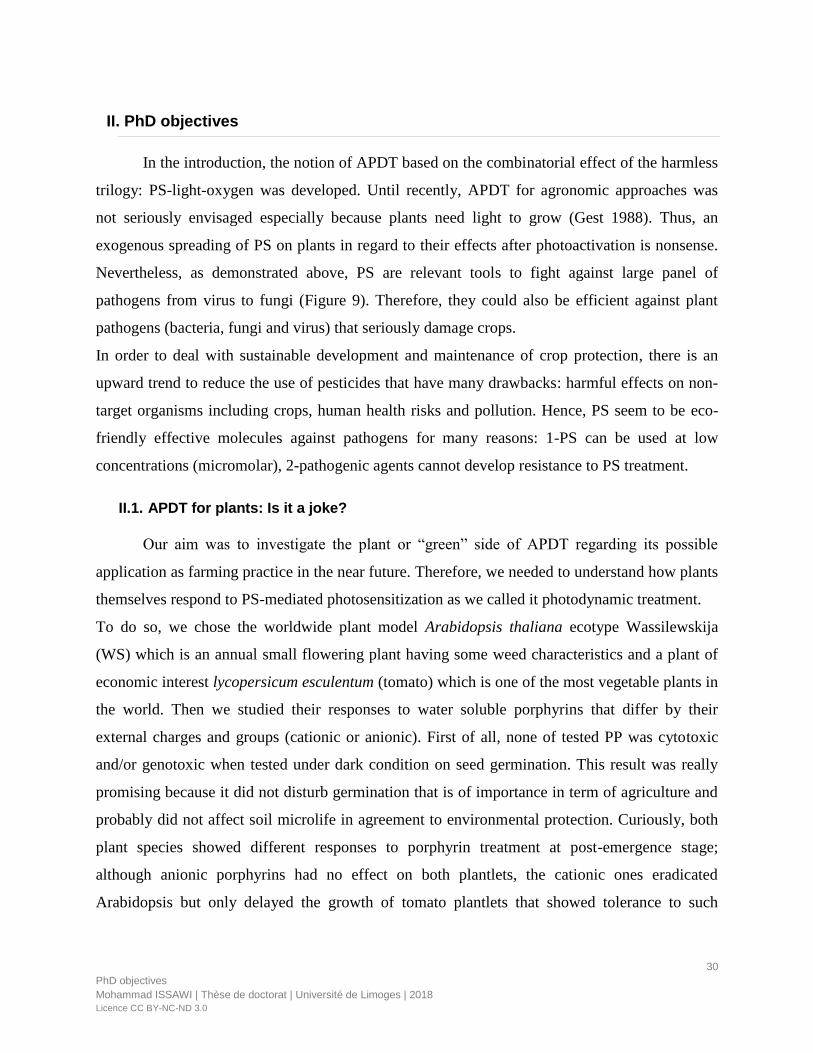
30
PhD objectives
Mohammad ISSAWI | Thèse de doctorat | Université de Limoges | 2018
Licence CC BY-NC-ND 3.0
II. PhD objectives
In the introduction, the notion of APDT based on the combinatorial effect of the harmless
trilogy: PS-light-oxygen was developed. Until recently, APDT for agronomic approaches was
not seriously envisaged especially because plants need light to grow (Gest 1988). Thus, an
exogenous spreading of PS on plants in regard to their effects after photoactivation is nonsense.
Nevertheless, as demonstrated above, PS are relevant tools to fight against large panel of
pathogens from virus to fungi (Figure 9). Therefore, they could also be efficient against plant
pathogens (bacteria, fungi and virus) that seriously damage crops.
In order to deal with sustainable development and maintenance of crop protection, there is an
upward trend to reduce the use of pesticides that have many drawbacks: harmful effects on non-
target organisms including crops, human health risks and pollution. Hence, PS seem to be eco-
friendly effective molecules against pathogens for many reasons: 1-PS can be used at low
concentrations (micromolar), 2-pathogenic agents cannot develop resistance to PS treatment.
II.1. APDT for plants: Is it a joke?
Our aim was to investigate the plant or “green” side of APDT regarding its possible
application as farming practice in the near future. Therefore, we needed to understand how plants
themselves respond to PS-mediated photosensitization as we called it photodynamic treatment.
To do so, we chose the worldwide plant model Arabidopsis thaliana ecotype Wassilewskija
(WS) which is an annual small flowering plant having some weed characteristics and a plant of
economic interest lycopersicum esculentum (tomato) which is one of the most vegetable plants in
the world. Then we studied their responses to water soluble porphyrins that differ by their
external charges and groups (cationic or anionic). First of all, none of tested PP was cytotoxic
and/or genotoxic when tested under dark condition on seed germination. This result was really
promising because it did not disturb germination that is of importance in term of agriculture and
probably did not affect soil microlife in agreement to environmental protection. Curiously, both
plant species showed different responses to porphyrin treatment at post-emergence stage;
although anionic porphyrins had no effect on both plantlets, the cationic ones eradicated
Arabidopsis but only delayed the growth of tomato plantlets that showed tolerance to such

31
PhD objectives
Mohammad ISSAWI | Thèse de doctorat | Université de Limoges | 2018
Licence CC BY-NC-ND 3.0
photodynamic treatment. All these results are largely developed and discussed in two
publications and constitute the first chapter of the present manuscript (Publications 2 and 3).
II. 2. Application of charged porphyrins on tobacco cell suspension: a help to
understand how PS are photoactivated in plant cells
These studies on plants were not suitable to reveal the fine regulations at molecular level
that drives cell and therefore plantlet to death. Indeed, plants are complex multicellular
organisms and their responses to drugs and/or PS are multifactorial depending on their genetic
background, annual or perennial, stage of development and age, organs or tissues… Thus, we
decided to use a simpler model to explore molecular mechanisms of plant photodynamic stress at
cellular level: Tobacco Bright Yellow-2 suspension culture, that is one of the most used plant
cell cultures for cellular biology research (Nagata 1992). The second reason to choose TBY-2
cells was their ability to grow under dark conditions. Therefore, it is not difficult to induce PS in
contact with TBY-2 under light to gain insight in their effects on plant cells. Before I came into
the lab, a previous study based on TBY-2 cells and charged porphyrins that are water-soluble,
showed that anionic porphyrins were much more efficient to induce cell death than cationic ones
(Riou et al., 2014). This first result was in contradiction to the results obtained in planta. Thus,
we wanted to understand why anionic porphyrins were so efficient to induce TBY-2 cell death
while it was cationic porphyrins in Arabidopsis and tomato plantlets. Young plantlet cells as well
as TBY-2 cells are surrounded by a primary cell wall. Our hypothesis was stated following cell
wall composition of TBY-2 cells. It was suspected to be different in term of pectins,
hemicellulose and cellulose contents from that of plantlet cells. In a first attempt and as
preliminary data needed to understand porphyrin interaction with plant cell, we established TBY-
2 cell wall composition and their polysaccharide secretion in the spent medium during TBY-2
cell growth phases (Publication 4).
This work was a pre-requisite for further experiments with porphyrins. Indeed, we needed
to know cell wall composition (largely negatively charged) to be able to understand potential
porphyrin-cell wall interactions. Here, we try to tackle two questions: 1-how do porphyrins
induce cell death in relation to their chemical structure and interact with the first barrier, the
plant cell wall? 2- what are the molecular mechanisms in term of ROS production and cell
responses underlying plant photodynamic treatment?

32
PhD objectives
Mohammad ISSAWI | Thèse de doctorat | Université de Limoges | 2018
Licence CC BY-NC-ND 3.0
II. 3. Manuscript presentation
We chose to present our PhD manuscript as accepted and/or submitted (or in prepration)
publications with general introduction and discussion relative to the multiple aspects of this
work. In the Chapter I, two publications deal with APDT feasibility in Plants (Guillaumot et al.,
2016; Issawi et al. 2018). In the Chapter II, one publication was accepted in Glycoconjugate
journal (Issawi et al. 2017) relative to TBY-2 characterization: cell wall and polysaccharide
secretion in spent medium. The two other publications cover separately chemical and biological
aspects that will help to answer to the fundamental question: how anionic porphyrins are able to
strongly induce TBY2 cell death? The chemical structure of anionic porphyrins and their
behavior in organic solvent or aqueous solutions were investigated in Leroy-Lhez et al.
submitted to Photochemical & photobiological science. Porphyrin and cell wall interaction and
induction of photodynamic stress in TBY-2 cells were developed and studied in a manuscript in
preparation for the Plant Journal. The end of the manuscript will be devoted to a large discussion
with open up perspectives followed by a completed bibliography and a CV of the PhD candidate.

33
Results
Mohammad ISSAWI | Thèse de doctorat | Université de Limoges | 2018
Licence CC BY-NC-ND 3.0
III.Results
Chapter I. APDT in agronomy:
Dream or reality?

34
Results
Mohammad ISSAWI | Thèse de doctorat | Université de Limoges | 2018
Licence CC BY-NC-ND 3.0
Response to photoactivated PS is linked to plant species and physicochemical properties
of PS. Thus, it was recommended to study the responses of plant to photodynamic treatment in a
case-by-case way depending on plant species. In order to fulfill that task, we focused on annual
plants that could show tolerance to photoactivated porphyrins unlikely to perennial trees that are
protected by thick cuticle preventing efficiently the PS photodamage. Firstly, we focused on
plant of agronomic interest: Lycopersicum esculentum, commonly known as tomato. Response to
cationic or anionic porphyrin was opposite. While anionic porphyrin (TPPS) tested at 50 µM did
not alter plantlet growth, cationic porphyrins (Zn-TMPyP or free-base TMPyP) significantly
reduced growth at low concentration (3.5 µM). Nevertheless, tomato plantlets were able to resist
to cationic porphyrins via induction of antioxidative systems (enzymes such as catalase,
peroxidase and superoxide dismutase and antioxidant molecules) (Publication 2). To enlarge our
expertise, we chose Arabidopsis thaliana, ecotype Wassilewskija (WS). Arabidopsis is a “lab
star” presenting special features as fast growth rate and invasiveness. Surprisingly, we showed
that these plants were roughly killed under the action of cationic porphyrins. Thus, we conceived
a dual strategy aiming to eradicate in the same way plant pathogens and weeds (Publication 3).
PUBLICATION 2: “Synergistic enhancement of tolerance mechanisms in response to
photoactivation of cationic tetra (N-methylpyridyl) porphyrins in tomato plantlets”,
Journal of Photochemistry and Photobiology B: Biology.

�������� ����� ��
Synergistic enhancement of tolerance mechanisms in response to photoactiva-tion of cationic tetra (N-methylpyridyl) porphyrins in tomato plantlets
Damien Guillaumot, Mohammad Issawi, Anne Da Silva, Stephanie Leroy-Lhez, Vincent Sol, Catherine Riou
PII: S1011-1344(15)30093-2DOI: doi: 10.1016/j.jphotobiol.2016.01.015Reference: JPB 10233
To appear in:
Received date: 13 October 2015Revised date: 15 January 2016Accepted date: 25 January 2016
Please cite this article as: Damien Guillaumot, Mohammad Issawi, Anne Da Silva,Stephanie Leroy-Lhez, Vincent Sol, Catherine Riou, Synergistic enhancement of tol-erance mechanisms in response to photoactivation of cationic tetra (N-methylpyridyl)porphyrins in tomato plantlets, (2016), doi: 10.1016/j.jphotobiol.2016.01.015
This is a PDF file of an unedited manuscript that has been accepted for publication.As a service to our customers we are providing this early version of the manuscript.The manuscript will undergo copyediting, typesetting, and review of the resulting proofbefore it is published in its final form. Please note that during the production processerrors may be discovered which could affect the content, and all legal disclaimers thatapply to the journal pertain.
ACC
EPTE
D M
ANU
SCR
IPT
ACCEPTED MANUSCRIPT
1
Synergistic enhancement of tolerance mechanisms in response to photoactivation of
cationic tetra (N-methylpyridyl) porphyrins in tomato plantlets
Damien Guillaumota, Mohammad Issawi
a, Anne Da Silva
b, Stephanie Leroy-Lhez
a, Vincent
Sola and Catherine Riou
a*
aLaboratoire de Chimie des Substances Naturelles (EA 1069), Université de Limoges, Faculté
des Sciences et Techniques, 123 avenue Albert Thomas, 87060 Limoges Cedex, France
b Unité de Génétique Moléculaire et Animale (UMR INRA 1061), Université de Limoges,
Faculté des Sciences et Techniques, 123 avenue Albert Thomas, 87060 Limoges Cedex,
France
* correspondence to: Catherine Riou, Laboratoire de Chimie des Substances Naturelles (EA
1069), Université de Limoges, Faculté des Sciences et Techniques, 123 avenue Albert
Thomas, 87060 Limoges Cedex, France E-mail : [email protected]
Keywords: Photoactivation; cationic and anionic porphyrins; ROS production; Solanum
lycopersicum.

ACC
EPTE
D M
ANU
SCR
IPT
ACCEPTED MANUSCRIPT
2
Abstract
Antimicrobial photodynamic treatment (APDT) is largely used in medical domain and could
be envisaged as a farming practice against crop pathogens such as bacteria and fungi that
generate drops in agricultural yields. Thus as a prerequisite for this potential application, we
studied the effect of water soluble anionic (TPPS and Zn-TPPS) and cationic (TMPyP and
Zn-TMPyP) porphyrins tested on tomato (Solanum lycopersicum) plantlets grown in vitro
under a 16h photoperiod. First of all, under dark conditions, none of the four porphyrins
inhibited germination and induced cyctotoxic effects on tomato plantlets as etiolated
development was not altered. The consequences of porphyrin long term photoactivation (14
days) were thus studied on in vitro-grown tomato plantlets at phenotypic and molecular
levels. Cationic porphyrins especially Zn-TMPyP were the most efficient photosensitizers and
dramatically altered growth without killing plantlets. Indeed, tomato plantlets were rescued
after cationic porphyrins treatment. To gain insight the different molecular ways implied in
the plantlet tolerance to photoactivated Zn-TMPyP, lipid peroxidation, antioxidative
molecules (total thiols, proline, ascorbate) and ROS detoxification enzymes were evaluated.
In parallel to an increase in lipid peroxidation and hydrogen peroxide production,
antioxidative molecules and enzymes (guaiacol peroxidase, catalase and superoxide
dismutase) were up-regulated in root apparatus in response to photoactivated Zn-TMPyP.
This study showed that tomato plantlets could overcome the pressure triggered by
photoactivated cationic porphyrin by activating antioxidative molecule and enzyme arsenal
and confining Zn-TMPyP into cell wall and/or apoplasm, suggesting that APDT directed
against tomato pathogens could be envisaged in the future.
ACC
EPTE
D M
ANU
SCR
IPT
ACCEPTED MANUSCRIPT
3
1. Introduction
Molecules such as porphyrins, naturally present in all living kingdom, are
photoexcitable by sunlight and able to produce reactive oxygen species (ROS) such as
superoxide anion radical, hydrogen peroxide, hydroxyl radical and singlet oxygen which, in
turn, lead to cell damage and ultimately cell death1,2
. Under dark, porphyrins do not exhibit
significant cyto or genotoxicity.1,4
Two studies performed on root meristematic cells of Allium
cepa and tobacco Bright Yellow cell suspensions showed opposite results with photoactivated
porphyrins 4,5
. Study performed on Allium cepa root apex demonstrated that cationic
porphyrins, either free or Zn-metallated, induce DNA photodamage whereas in tobacco cell
suspension, anionic and cationic porphyrins were not cytotoxic and anionic porphyrin was the
most efficient death inducer compared to cationic one upon illumination. According to
literature, this last result was quite surprising taking into account that cationic or neutral
porphyrins are always the most efficient to induce cell death from bacteria to mammal cells
and/or tissues. Previous work showed that derivatives of cationic porphyrin were more
efficient than neutral and anionic porphyrins on Gram-negative bacteria (Escherichia coli)
and that these effects were less pronounced on Gram-positive bacteria such as Enterococcus
hirae or Staphylococcus aureus,2.6-10
. Likewise, exogenous supply of cationic porphyrin in
cultures of cyanobacteria and green microalgae showed that the latter were more
photosensitive than the former.11
Upon illumination, protoporphyrin IX and other derivatives
were proved to be strongly cytotoxic to the yeasts Saccharomyces cerevisiae and Candida
albicans. 1,12-14
Furthermore, tested on mosquito or fly larvae, photoactivated cationic
porphyrins also possess an insecticidal power.15-17
Finally, strategies known as photodynamic
therapy including antimicrobial photodynamic treatment are based on the use of
photoactivated porphyrins in a large variety of medical domains such as cancerology,
ACC
EPTE
D M
ANU
SCR
IPT
ACCEPTED MANUSCRIPT
4
dermatology, ophtamology or odontology 1-3, 18.
This strategy is also envisaged to struggle
plant-pathogens such as Colletrichum acutatum and Aspergillus nidulans fungi19
All these studies were focused on an optimization of a fatal issue such as death linked to
photoactivation of porphyrins. Furthermore they were essentially performed on isolated cells
or tissues from prokayotic to eukaryotic cells, excepted on mosquito larvae. Nevertheless it
should be of interest to understand the consequence of photoactivation of exogenous
porphyrins in whole organisms especially in plants. Indeed as antimicrobial photodynamic
treatment (APDT) could be considered as a potential agronomic strategy against plant
pathogens (fungi and bacteria)19
, it would be of interest to study the response of plants to
photoactivated porphyrins. In that respect, our aim was to study the effect of two categories
of water-soluble synthetic porphyrins: cationic and anionic porphyrins, either free base
(TMPyP and TPPS, respectively) or zinc-metallated (Zn-TMPyP and Zn-TPPS) on tomato
plantlets grown in vitro. Previous studies on plants were performed to set up modifications of
endogenous porphyrins biosynthesis by transgenesis.20-24
Authors showed that an
accumulation of protoporphyrin IX, common precursor of heme and chlorophylls, triggered
lethal phenotypic alterations (growth inhibition, browning leaves, desiccation) after
illumination.22,23
Based on these studies and because porphyrins could be used as antimicrobial agents in
agronomic practices, we studied the consequences of sunlight photoactivation of exogenous
porphyrins on tomato plantlets at phenotypic and molecular levels. We supposed that defense
mechanisms such as antioxidative machinery would be activated to allow plantlet tolerance to
the presence of exogenous photosensitizer. We firstly showed that none of these porphyrins
was able to inhibit germination and was cytotoxic under dark conditions. Thus, we compared
the consequences of their photoactivation on phenotype of 14 day-old in vitro plantlets.
Cationic porphyrins, especially Zn-TMPyP, induced the strongest altered phenotype.
ACC
EPTE
D M
ANU
SCR
IPT
ACCEPTED MANUSCRIPT
5
Nevertheless, tomato plantlets could resist to high amounts of cationic porphyrins and also be
completely rescued afterwards. Thus, taking into account these first results, we focused our
study on the cellular and molecular mechanisms triggered in particularly by Zn-TMPyP in
plantlets roots, trying to understand how tomato plantlets were able to resist to photoactivated
Zn-TMPyP. Indeed, endogenous levels of total thiol, proline and ascorbate, considered as
antioxidant molecules, and enzyme activities such as peroxidases and superoxide dismutase
were significantly up-regulated in tomato roots. In conclusion we suggested that, in response
to Zn-TMPyP photoactivation tomato plantlets were able to produce appropriate amounts of
total thiol, proline and ascorbate as well as to sufficiently activate ROS detoxification
enzymes to resist to photoactivated Zn-TMPyP.
2. Materials and methods
2.1 Porphyrins
All porphyrins are soluble in water and their chemical structures have been reported in Fig 1.
Anionic porphyrin corresponds to TPPS and was purchased from Sigma-Aldrich (St Louis,
MO, USA). Zn-TPPS was obtained by metallation of TPPS with zinc acetate.25
Cationic
porphyrins: Tetra (N-methylpyridyl) porphyrin tetrachloride or TMPyP and Zn-TMPyP were
purchased from Frontier Scientific (Carnforth, UK).
To evaluate porphyrin photostability in the plant culture medium, they were added to the
medium at the wanted concentration. For all experiments, plates were maintained for 15 days
in a controlled growth chamber at 22 °C under a 16h photoperiod and a photon flux density of
250 µmol.m-2
.s-1
. Photostability was evaluated by Uv-Vis absorption method, monitoring
decrease of the Soret Band at 437 and 414 for Zn-TMPyP and TPPS respectively. For each
measurement, a square (1cm²) of medium with porphyrin was taken, suspended in 10 mL
ACC
EPTE
D M
ANU
SCR
IPT
ACCEPTED MANUSCRIPT
6
water under vigorous agitation. Then absorption spectra was recorded. Initial value of Soret
band absorbance was used to define 100% level.
2.2 Plant material
Seeds of tomato var Bali F1 hybrid purchased from Tézier (Portes-lès-Valence, France), were
germinated in vitro on a Gamborg B5 culture medium (Duchefa Biochemie, Haarlem,
Netherlands) at pH 5.8 supplemented with 2% (w/v) sucrose and 0.8% (w/v) agar (Difco,
Dallas, USA).26
Porphyrins were added to the medium after autoclaving. For all experiments,
plants were grown on 25 mL of B5 alone or supplemented with porphyrin in a controlled
growth chamber at 22 °C under a 16h photoperiod and a photon flux density of 250 μmol. m-2
.
s-1
. Cool daylight lamps (OSRAM Lumilux 24W) were used in the growth chamber. After 14
days of growth, tomato plantlets were harvested and used for phenotypic and biochemical
analyses. Germination ratio was evaluated after 4 days of culture using a binocular
microscope. Under these conditions, 8 tomato seeds were sown on the same plate. The
experiments were performed three time independently. Pictures from the last experiment were
taken with a digital camera and/or under a Leica binocular microscope.
For cytotoxicity assessment, plates containing 8 seeds and surrounding by
alumimium foil in order to avoid any light source were maintained vertically for 4 days in
growth chamber. The experiments were performed three time independently.
Roots and shoots from tomato plantlets isolated from 14 day-old plantlets were
harvested, weighed and frozen at -20 °C until used. For the determination of the ratio dry
weight on fresh weight, tomato organs were fresh weighed (FW), dried at 42 °C for 4 days
and weighed again (DW).
ACC
EPTE
D M
ANU
SCR
IPT
ACCEPTED MANUSCRIPT
7
2.3 Confocal microscopy analysis
We incubated 14 day-old plantlets in B5 medium containing or not 3.5 µM Zn-TMPyP for 20
h in the dark and rinsed them 3 times with B5 new medium before observation under a
LSM510META Zeiss confocal microscope. For analysis, two ways of data acquisition were
performed: a spectral acquisition to check signal specificity of Zn-TMPyP and a channel
mode for calcofluor and Zn-TMPyP. For spectral acquisition, samples were excited at 458 nm
and fluorescence was monitored between 476 and 732 nm. Zn-TMPyP specific emission peak
was detected around 640 nm as its fluorescence emission maxima is at 634 nm
4. For channel
mode acquisition, we used an excitation wavelength at 405 nm and emission wavelength
between 420 and 450 nm for calcofluor and excitation wavelength at 458 nm and a long pass
filter (cut off at 560 nm) for Zn-TMPyP.
2.4 In situ DAB and NBTZ stainings
3,3-diaminobenzidine (DAB) staining on tomato roots was performed using DAB Kit (Sigma-
Aldrich, St Louis, MO, USA) following manufacturer’s instructions. Nitro blue tetrazolium
(NBTZ) staining was performed as followed: roots were dived into 10 mM phosphate buffer
pH7.8, 10 mM NaNO3 and 0.1 % (w/v) NBTZ. Roots were vacuum infiltrated for 5 min and
placed in the dark for 2 h for DAB and 30 min for NBTZ respectively. DAB or NBTZ
solutions were replaced by 70% (v/v) ethanol and stored at 4 °C until examination under a
binocular microscope.
2.5 Hydrogen peroxide (H2O2) quantification
The H2O2 measurement was performed according to 27
with some modifications. 1 mL of
sodium phosphate buffer (25 mM, pH 7) was added to the frozen samples ground with liquid
nitrogen. The homogenate was centrifuged at 10,000 × g at 4◦C for 15 min. 335 µl of 0.1%

ACC
EPTE
D M
ANU
SCR
IPT
ACCEPTED MANUSCRIPT
8
titanium III sulfate in 20% (w/v) H2SO4 was added in extracted solution. The absorbance was
measured at 415 nm and H2O2 level was expressed as nM.g-1
proteins. Proteins were
quantified by Bradford assay using BSA as standard28
.
2.6 Antioxidant molecule assays
2.6.1 Proline content
Free proline contents of tomato roots were assayed according to a previous work.29
2.6.2 Ascorbate content
Ascorbate was quantified as previously reported 30
with some minor modifications. Frozen
plant tissues (between 50 and 200 mg FW) were ground into powder using liquid nitrogen.
The resulting powder was weighed before addition of 200 µL of 6% TCA solution. Samples
were then incubated for 10 minutes on ice and centrifuged at 16 000 g for 25 minutes at 4 °C.
Supernatant was completed to 500 µL with 6% TCA and centrifuged 16 000 g for 10 minutes
at 4 °C. 100 µL of each sample were then transferred to 150 µL 0.2 M phosphate buffer pH
7.4 and supplemented with 250 µL 10 % TCA, 200 μL 42 % H3PO4, 200 μL 2,2′-dipyridyl
and 100 μL 3% FeCl3. Similarly, standards containing 0 to 70 µmoles of ascorbate were
realized. After 40 minute incubation in a 42 °C water bath, absorbance of standards and
samples were read at 525 nm.
2.6.3 Total thiol assay
Total thiol assay was performed according to 31
. Frozen samples were ground in liquid
nitrogen and then extracted into 1 mL of 0.2 N HCl. After centrifugation at 16000 x g for 4
min, 500 µL of extracted solution were neutralized with 400 µL NaOH (0.2M) and 50 µL
NaH2PO4 (0.2M). 0.7 mL of 0.12 M NaH2PO4 (pH 7.5), 6 mM EDTA, 0.1 ml of 6 mM
Dithiobis 2 nitro benzoic acid (DTNB) were added to 0.2 mL extract. For standards, extract

ACC
EPTE
D M
ANU
SCR
IPT
ACCEPTED MANUSCRIPT
9
was replaced by 0, 10, 20, 50, and 100 nmol GSH (total volume 1mL). Absorbance at 412 nm
was read 5 min after the addition of standard or extract.
2.7 Determination of malondialdehyde (MDA) content
Around 100 mg of fresh or frozen plant materials were ground in liquid nitrogen. The
resulting powder was suspended into 1.5 mL of 20% TCA. After centrifugation, supernatants
were collected and analyzed for their MDA content as described by 32
with minor
modifications. 500 µL of each sample were added to either 500 µL 20 % TCA alone (-TBA)
or 20% of TCA supplemented with 0.5% thiobarbituric acid (+TBA). Samples were then
boiled in a water bath for 30 minutes. The samples were cooled on ice and absorbances were
read at 440, 560 and 600 nm. Owing to the possible presence of porphyrin in the extract and
subsequent interference with absorbance readings at 440 nm, some modifications were
brought to the calculation formula proposed by 32
1) [(Abs 532+TBA - Abs 600+TBA) - (Abs 532-TBA - Abs600-TBA)] = A
2) {[(Abs 440+TBA - Abs 600+TBA) - (Abs 440-TBA - Abs 600-TBA)] 0.0571} = B
3) MDA equivalents (nmoL.mL-1
) = [(A-B)/157 000] .106
2.8 Determination of enzymatic activities
Frozen plant samples were ground to a fine powder in liquid nitrogen. Proteins were
solubilized into 1 mL of extraction buffer containing 50 mM phosphate buffer pH 7.8, 1 mM
EDTA, 1% (w/v) PVP and 10 % (v/v) glycerol and centrifuged at 16 100 g for 10 minutes at 4
°C. Protein concentration of the resulting supernatant was quantified according to Bradford
(1976)28
.
ACC
EPTE
D M
ANU
SCR
IPT
ACCEPTED MANUSCRIPT
10
2.8.1 Catalase
Catalase activity was measured according to Aebi’s protocol.33
20 µg of total soluble proteins
were diluted into a 50 mM phosphate potassium buffer (pH 6.5). The reaction was initiated by
addition of H2O2 to a final concentration of 20 mM and for a final volume of 1 mL.
Absorbance decrease at 240 nm was monitored during 3 minutes and H2O2 disappearance was
calculated on the slope of the linear portion of the resulting curve using a molar extinction
coefficient of 43.6 M−1
cm−1
.
2.8.2 Guaiacol peroxidase
Guaiacol peroxidase activity was monitored at 436 nm due to the formation of tetraguaiacol
and calculated using a molar extinction coefficient of 25.5 mM−1
.cm−1
. 10 µg of total soluble
proteins were diluted into 50 mM phosphate potassium buffer pH 6.5 and supplemented with
0.25% guaiacol (5 % stock solution in 95 % ethanol w/v). Reaction was initiated by addition
of H2O2 to a final concentration of 2.5 mM and absorbance was immediately monitored
during 2 minutes.
2.8.3 Ascorbate peroxidase
Ascorbate peroxidase activity was analyzed by adapting the microplate protocol to tubes.34
20
µg of soluble protein extracts of tomato roots were diluted into 50 mM phosphate buffer pH
7.0 supplemented with 0.25 mM ascorbic acid. The reaction started by addition of H2O2 up to
5 mM in a final reaction volume of 1 mL. The decrease in absorbance at 290 nm
corresponding to the formation of ascorbate was then monitored during 3 minutes. Activity
was calculated on the slope of the linear section of the resulting curve using a molar
extinction coefficient of 2.8 mM−1
.cm−1
.
2.8.4 Superoxide dismutase
Superoxide dismutase (SOD) activity protocol was performed in absence of bathocuproine
sulfonate.35
Standard curve was set so that the increase in absorbance at 560 nm was between
ACC
EPTE
D M
ANU
SCR
IPT
ACCEPTED MANUSCRIPT
11
0.02 and 0.03 absorbance unit per minute and was monitored for 5 minutes. One unit of SOD
was defined as the amount of enzyme required for a 50% inhibition of NBT reduction. 5 µL
of enzyme extract were used. SOD specific enzyme activity was determined according to the
protein concentration of each sample.
2.9 Statistical analysis
Germination percentage was assessed according to generalized linear models with
quasibinomial proportional distribution of errors to correct overdispersed data. All results
were expressed as mean ± SEM (Standard Error of the mean). Data were analyzed by t-
student test and one-way ANOVA followed by the Dunnett multiple comparison test36
using
the Control group as reference, while p-value < 0.05 was considered statistically significant.
All tests were performed in R statistical software (R Developmental Core Team 2012).

ACC
EPTE
D M
ANU
SCR
IPT
ACCEPTED MANUSCRIPT
12
3. RESULTS
3.1 Seed germination in presence of 80 µM anionic and cationic porphyrins
Germination of tomato seeds sown on media containing each tested porphyrin at high
concentration was examined (Table 1). As none of the photosensitizers significantly inhibited
tomato seed germination, anionic or cationic porphyrins could definitely not be considered as
cytotoxic at pre-emergence state.
3.2 Phenotype analyses of tomato plantlets in the dark and under illumination.
3.2.1 Porphyrins did not altered tomato seedling development under dark
To gain insight porphyrins cytotoxicity during post-germination event, seedling growth was
tested and observed in the dark in presence of 80 µM porphyrins for both species (Fig. 2).
Tomato seedlings grown in the dark did not show any significative growth alteration after 4
days treatment (Fig. 1) suggesting that the four photosensitizers were neither cytotoxic nor
genotoxic for tomato seedlings.
3.2.2 Phenotype alterations under 16h photoperiod
As porphyrins are photoexcitable by visible light, light exposure effects of porphyrins, tomato
seeds were allowed to germinate on each compounds for 14 days (Fig. 3A). Although
phenotypic alterations were not really obvious in contact of 50 µM TPPS or Zn-TPPS
compared to control condition, plantlets showed significant reduction in hypocotyl and root
length while no new leave did appeared in presence of 3.5 µM TMPyP or Zn-TMPyP. We
determined the ratio of dry to fresh weight; this ratio significantly increased in all the tested
conditions in comparison to the controls. Taking together, these results suggested that cationic
porphyrin especially the metalled form induced a strong abiotic stress (Fig. 3B). Intriguingly
Zn-TPPS seemed to be the less stressful photoactivated porphyrins in regard to its effect on
ACC
EPTE
D M
ANU
SCR
IPT
ACCEPTED MANUSCRIPT
13
plant phenotype of plantlets (Fig. 3A). To gain insight into the selection of the most efficient
photosensitizers with the aim of a future APDT application, we gave up the anionic
porphyrins (Zn-TPPS and TPPS) that are not enough efficient to kill fungi and bacteria. In a
other way, we showed that they did not alter tomato plantlet growth.
3.3 Plantlet rescue after treatment with photoactivated cationic porphyrins TMPyP and
Zn-TMPyP
As high concentration of cationic photosensitizers induced important alterations of
developmental trait of tomato plantlets under light but did not seem to be cytotoxic in the
dark, we checked whether tomato plantlets could be rescued after 14 days on 80 µM cationic
porphyrins. Such tomato plantlets were thus transferred on medium without porphyrins for
one week and then allowed to grow in soil for 6 more weeks (Fig. 4). In light of our results,
we could assume that tomato plants were rescued since they had recovered a growth similar to
control plant, meaning that 14 days contact with cationic porphyrins did not fatally disturb
growth and development of tomato plantlets to prevent further normal development, which
was quite surprising after 80 µM Zn-TMPyP treatment. Thus in order to understand the
reason behind such tolerance, we decided to further analyze the defense mechanisms in
presence of photoactivated TMPyP and Zn-TMPyP. We decided to work with a very lower
concentration of cationic porphyrins corresponding to 3.5 µM which was able to sufficiently
stress plants.
3.4 Zn-TMpyP localization in root apparatus
As under illumination, Zn-TMPyP clearly induced phenotypic effects on plantlets; it was of
importance to check Zn-TMPyP localization in root apparatus although it was already shown
that cationic porphyrins (free metal or not) were located in meristematic root cells of Allium
ACC
EPTE
D M
ANU
SCR
IPT
ACCEPTED MANUSCRIPT
14
cepa and more precisely into nucleus19
. As photoactivated Zn-TMPyP treatment strongly
affected plants tissues, root incubation was performed in the dark in order to study its
localization in tomato healthy roots. In tomato thick sections performed in elongation zone,
Zn-TMPyP seemed to be confined to cell wall and/or apoplast as shown by co-localization
with calcofluor used as cell wall marker (Fig. 5). Contrary to previous work5, our result
strongly suggested that Zn-TMPyP stayed outside of cell cytoplasm of tomato root epidermis.
This result could be also well correlated to the non cytotoxicity or genotoxicity described
above.
3.5 Lipid peroxidation and ROS production as a consequence of cationic porphyrin
photoactivation
Because photoactivated porphyrins generate reactive oxygen species (ROS), cell membranes
containing high amounts of polyunsaturated lipids represent a target for these powerful
reactive molecules leading to lipid peroxidation, degradation, and further malondialdehyde
production (MDA). Thus we checked MDA level in roots and shoots of tomato plantlets. A
significant increase in MDA content was found in roots and shoots of cationic porphyrins
treated plantlets compared to control (Fig. 6). This was probably linked to Zn-TMPyP
localization in root cells. As shown by confocal microscopy, Zn-TMPyP preferentially
accumulates into cell wall rather than in apoplasm, thus preventing its lethal effect under
illumination. Nevertheless DAB and NBTZ stainings showed that ROS such as hydrogen
peroxide (H2O2) and in a lesser extent anion superoxide (O2-) were produced in root apparatus
in particularly root apex of treated plantlets (Fig. 7 A and B). This was also confirmed by the
detection of a high amount of H2O2 in roots by comparison in shoots (Fig. 6C). As H2O2 is
more stable than the other ROS and diffuses into cells, we supposed that photoactivation of
Zn-TMPyP probably led to type I rather than type II reactions in root cells.
ACC
EPTE
D M
ANU
SCR
IPT
ACCEPTED MANUSCRIPT
15
3.6 Antioxidant molecule productions in treated tomato plantlets
To gain insight into ROS generated pathway upon porphyrins illumination, we determined
ascorbate, proline and total thiol contents in plantlets considering them as antioxidant
molecules. The abiotic stress triggered by photoactivated Zn-TMPyP induced, in tomato
roots, a huge increase in proline (around 40 fold) and to a lesser extent in ascorbate and total
thiol (around twice) contents (Fig. 8). These results could explain why tomato plantlets were
able to better resist to photoactivated Zn-TMPyP by generating a large amount of proline and
to a lesser extent thiol and ascorbate in order to offset ROS production triggered by its
photoactivation.
3.7 Pattern of antioxidative stress enzymes in response to photoactivated Zn-cationic
porphyrin
Finally, the question of enzymes involved in superoxide radicals and hydrogen peroxide take
off was considered as another way to fight against ROS production induced by photoactivated
Zn-TMPyP. Thus we determined guaiacol and ascorbate peroxidases, catalase and superoxide
dismutase activities in tomato roots growing in presence of 3.5 µM Zn-TMPyP. Guaiacol
peroxidase and catalase activities were clearly up-regulated, reaching, respectively, three-fold
and around two-fold enhancement compared to the control (Fig. 9). Strikingly, ascorbate
peroxidase did not show any increase although intracellular ascorbate content was shown to
increase (Fig. 8 and 9). Superoxide dismutase activity also increased, suggesting its
implication in transformation of superoxide radicals overproduced by photoactivated Zn-
TMPyP treated roots into hydrogen peroxide that could be essentially transformed by catalase
and not by ascorbate peroxidase that, in turn, did not change (Fig. 9).

ACC
EPTE
D M
ANU
SCR
IPT
ACCEPTED MANUSCRIPT
16
4 DISCUSSION
In this work, we studied the effect of two categories of synthetic porphyrins on tomato
plantlets (Solanum lycopersicum var. Bali). The choice of tomato plants as experimental
model was triggered by two main reasons: this plant represents a wide cultured species over
the world and it is mainly attacked by pathogens such as bacteria (Xanthomonas campestris
and Pseudomonas syringae) and fungi (Phytophtora infectans).37
These pathogens could be
eradicated by using the PACT strategy 1-3,7,8,19
. From that perspective, it should be of interest
to understand how plants could resist to such a future potential treatment. We are confident
that these preliminary data performed in vitro reflected what could happen in green houses or
soil. To our knowledge this is the first study that address this type of issue in regard to the use
of photosensitizers as pesticides. Thus our aim was not to describe a new herbicide but to try
to understand how plants are able to fight against a photoactivated antimicrobial agent
potentially used in agriculture. Previous study and experiments in our laboratory were
monitored to test the effect of anionic and cationic porphyrins on Pseudomonas aeruginosa
and showed that cationic porphyrins were also the most efficient (data not shown)37
.
Currently, the phototoxicity property of porphyrins and their herbicidal effect were
indirectly described in plants. The first approach was to feed cultures or spread plants with
precursor of tetrapyrrole compounds biosynthesis such as 5-aminolevulenic acid (ALA).39-41
Although presenting a true herbicide property, ALA treatment required high concentrations
(around 20 mM) that was very much higher to 3.5 µM cationic porphyrins described in this
study.40,41
The other approach using transgenesis targeted to porphyrin oxidase activity,
showed that modifications in porphyrins metabolism in rice and maize plants led to fatal
issue.20-24
Culture on exogenous cationic porphyrins also induced dramatic effects on tomato
plantlets similar to the described phenotype of porphyrin oxidase overproducer. Nevertheless
ACC
EPTE
D M
ANU
SCR
IPT
ACCEPTED MANUSCRIPT
17
in regard to plant rescue when the porphyrin pressure was removed and their rapid
photobleaching (Table 2), cationic porphyrins could not be considered as potential post-
emergence herbicide.
Under dark conditions porphyrins did not lead to phenotypic alterations of tomato
plantlets. This result confirmed the no-cytotocity and genotoxicity effect of porphyrins as
already shown for tobacco plant cells.4
Upon illumination, porphyrins generate radicals such as superoxide anion (O2•-
) and
hydroxyl radical (OH•) by electron transfer (type I process) or singlet oxygen (
1O2) by energy
transfer to dioxygen (type II process). As singlet oxygen lifetime is very short (order of the
nanosecond), its scope is limited to the formation site.1,42
Nevertheless it remains still very
difficult to detect this species in vivo. Generation of free radicals as superoxide anion radical
(O2
•-) or hydroxyl radical (OH
•) could also lead to the formation of hydrogen peroxide (H2O2)
and other reactive oxygen species. The significant increase in H2O2 detected in tomato roots
treated with Zn-TMPyP could at least correspond to a pool coming from type I reaction and
anion superoxide and from the transformation of singlet oxygen into H2O2, the most stable
ROS. All these ROS damage a large number of cellular constituents such as nucleic acids,
proteins and lipids, leading to cell death by necrosis and more often by apoptosis.43-45
As a
consequence of porphyrin photoactivation, cell death was shown in animal cells, fungi,
bacteria and recently in tobacco BY-2 cells. 1,4,6-17
One of the consequences of Zn-TMPyP photoactivation was supposed to be superoxide
anion radical and hydroxyl radical productions. Thus because lipid peroxidation is thought to
be mainly triggered by these ROS, we performed a MDA quantification on tomato roots.
Tomato roots and especially shoot cells showed an increase in MDA, suggesting that cell
membranes were damaged. Nevertheless as Zn-TMPyP seemed to be only accumulated in cell
ACC
EPTE
D M
ANU
SCR
IPT
ACCEPTED MANUSCRIPT
18
wall and/or apoplasm and not in the nucleus as shown in Allium root cells5, we suggested that
Zn-TMPyP were able to produce ROS close to plasma membrane under illumination.
Furthermore the Zn-TMPyP localization could explain its no-genotoxicity in tomato plantlets
although DNA damages could also be triggered by MDA; this molecule has been shown to be
highly mutagenic for bacteria and mammal cells as well.45
Another way for the plantlets to tolerate to photoactivated Zn-TMPyP and to protect
themselves against ROS, is to produce antioxidant molecules that could neutralize ROS. Thus
we determined proline and ascorbate contents in tomato roots. Clearly, tomato plantlets were
able to co-activate the production of total thiol, proline and ascorbate. In plants, the role of
proline as free radical scavenger in response to a large range of abiotic and biotic stresses was
proposed.47,48
Proline can act as singlet oxygen quencher and as scavenger of hydroxyl
radicals47
. We can suppose that proline (40 fold increase) assumes these two roles in tomato
roots in response to photoactivated Zn-TMPyP. Additionally, ascorbate can rapidly react with
singlet oxygen resulting in hydrogen peroxide production and has therefore been suggested to
be a good sink of this ROS.49
Both amounts of these ROS scavengers and/or quenchers were
significantly overproduced in Zn-TMPyP treated tomato roots to counteract its
photoactivation. It should be also interesting to quantify other scavengers such as α tocopherol
to refine the different ROS triggered by Zn-TMPyP under illumination in roots.
As for ascorbate and proline, a significant increase in total thiol content was detected in
treated roots in response to Zn-TMPyP. Total thiol quantification was envisaged as a link
between its potential role as scavenger and ROS enzymes especially peroxidases. Previous
work showed that protoporphyrin IX was able to be degraded by plant peroxidases associated
to thiol substrates such as glutathione and cysteine50
. Furthermore this reaction was inhibited
by ascorbate50
. This could explain why we did not detected any change in ascorbate
peroxidase activity in tomato roots but around a two fold increase in ascorbate content. Thus

ACC
EPTE
D M
ANU
SCR
IPT
ACCEPTED MANUSCRIPT
19
we envisage to address this issue by measuring glutathione content and glutathione relative
activities. In the other hand, we might suppose that Zn-TMPyP could also be degraded by this
way which should be also relevant to its non fatal effect after a 14-day treatment.
In parallel, we investigated ROS detoxification enzyme activities such as peroxidases,
catalases and superoxide dismutases. In plantlets, these enzymes with exception of ascorbate
peroxidases were clearly up regulated to counter balance ROS production induced by Zn-
TMPyP photoactivation. GPX and catalase could scavenge H2O2 produced in one way by
SOD and in other way by Zn-TMPyP photoactivation (type I reaction). Furthermore, GPX
activity is known to be regulated by biotic and abiotic stresses that corresponded to our
situation.27,53
A possible explanation could arise from Zn-TMPyP subcellular localization in
apoplast and/or cell wall of tomato root cells. Indeed GPX that are class III peroxidases are
mainly located into cytoplasm and apoplast while class I peroxidases such as ascorbate
peroxidases are essentially intracellular51,52
. Thus, it seems relevant that GPX would
preferentially be activated in order to detoxify the cells from the oxidative stress generated by
photoactivated Zn-TMPyP. Additionally the effect of GPX in preventing membrane lipid
peroxidation has also been reported and is consistent with the lower amount of MDA found in
Zn-TMPyP treated tomato plantlets53
. Our study based on tomato plantlets grown in vitro in
contact to Zn-TMPyP revealed that tomato plantlets were able to protect themselves from this
abiotic stress using three different and concomitant ways: Zn-TMPyP accumulation in cell
wall and potentially its degradation, antioxidant molecules production and activation of ROS
detoxification enzymes.
Although photoactivated Zn-TMPyP was efficient to dramatically slow down plantlet
growth, plantlets did not die. We supposed that while the enzymatic activities and anti-
oxidative molecules significantly increased, it was not sufficient to assume a normal growth
of the plantlets grown in contact to photoactivated Zn-TMPyP. One hypothesis could be that a

ACC
EPTE
D M
ANU
SCR
IPT
ACCEPTED MANUSCRIPT
20
proportion of H2O2 amount stay into cell and interfered with root growth. Another hypothesis
should be that primary metabolism and anti-oxidative molecules synthesis activated in
response to Zn-TPMyP affected the other metabolisms linked to normal root growth. Indeed,
root apparatus was really and deeply altered showing a typical reduction of main root growth
and multiple lateral root formation linked to an escape strategy. Such behaviour was also
described in resistance to abiotic stress. Plant growth and development suffered from the fact
that it focused on coping with such oxidative stress and efficiently activating its defense
mechanisms. Considering the ability of photoactivated cationic porphyrins to rapidly kill
bacteria at low concentration7 (decrease in bacterial population by a 10
5 factor in 30 min) and
their inefficiency to kill tomato plants after a longer and higher treatment, we think that
cationic porphyrin and especially Zn-TMPyP, might be promising candidates as antimicrobial
agents in agronomic practices.
ACC
EPTE
D M
ANU
SCR
IPT
ACCEPTED MANUSCRIPT
21
ACKNOWLEDGEMENTS
Authors thank the région Limousin for financial supports of Dr Guillaumot as post doctoral
fellowship and the French National Research Agency (ANR Porphy-Plant), Dr F. Brevier for
providing Zn-TPPS, Dr C. Carrion for confocal microscopy analysis and Dr. Michel Guilloton
for help in manuscript editing.
Conflict of interest the authors declare that they have no conflict of interest.
Author contribution statement
Damien Guillaumot, Mohammad Issawi and Catherine Riou designed research and conducted
all plant experiments. Stephanie Leroy-Lhez performed the photostability measurements and
Anne Da Silva all statistical tests. Vincent Sol helped in the reflection and writing of the
manuscript. All authors read and approved the manuscript.

ACC
EPTE
D M
ANU
SCR
IPT
ACCEPTED MANUSCRIPT
22
REFERENCES
[1] Donnelly RF, McCarron PA, Tunney M, Antifungal photodynamic therapy. Microbiol.
Res. 163 (2008) 1-12. DOI: 10.1016/j.micres.2007.08.001
[2] Benov L, Photodynamic therapy: Current status and future directions. Med. Princ. Pract.
24 (2015) 14-28, DOI: 10.1159/000362416.
[3] Maisch T, Resistance in antimicrobial photodynamic inactivation of bacteria.
Photochem. Photobiol. Sci. (2015) DOI: 10.1039/c5pp00037h.
[4] Riou C, Calliste CA, Da Silva A, Guillaumot D, Rezazgui O, Sol V, Leroy-Lhez S,
Anionic porphyrin as a new powerful cell death inducer of Tobacco Bright Yellow-2
cells. Photochem Photobiol. Sci. (2014) DOI: 10.1039/c3pp50315a.
[5] Villanueva A, Canete M, Hazen MJ, Uptake and DNA photodammage induced in plant
cells in vivo by two cationic porphyrins. Mutagenesis 4 (1989) 157-159.
[6] Jori G, Brown SB, Photosensitized inactivation of microorganisms. Photochem.
Photobiol. Sci 3 (2004) 403-405. DOI:10.1039/b311904c
[7] Merchat M, Bertoloni G, Giacomoni P, Villanueva A, Jori G, Meso-substituted cationic
porphyrins as efficient photosensitizers of gram-positive and gram-negative bacteria. J.
Photochem. Photobiol 32 (1996) 153-157.
[8] Gábor F, Szocs K, Maillard P, Csík G, Photobiological activity of exogenous and
endogenous porphyrin derivatives in Escherichia coli and Enterococcus hirae cells
Radiat Environ Biophys 40 (2001) 145–151.
[9] Ringot C, Sol V, Barrière M, Saad N, Bressollier P, Granet R, Couleaud P, Frochot C,
Krausz P, Triazinyl porphyrin-based photoactive cotton fabrics: preparation,
characterization and antibacterial activity. BioMacromol 12 (2012) 1716-1723.
dx.doi.org/10.1021/bm200082d
[10] Mbakidi JP, Herke K, Alvès S, Chaleix V, Granet R, Krausz P, Leroy-Lhez S, Ouk T,
Sol V, Synthesis and photobiocidal properties of cationic porphyrin-grafted paper.
Carbohydate Polymers 91 (2013) 333-338.
http://dx.doi.org/10.1016/j.carbpol.2012.08.013
[11] Drabkova M, Marsalek B, Admiraal W, Photodynamic therapy against cyanobacteria.
Environ. Toxicol. 22 (2007) 112-117. DOI10.1002/tox.20240
ACC
EPTE
D M
ANU
SCR
IPT
ACCEPTED MANUSCRIPT
23
[12] Carré V, Gaud O, Sylvain I, Bourdon O, Spiro M, Blais J, Granet R, Krausz P,
Guilloton M, Fungicidal properties of meso-arylglycosylporphyrins : influence of sugar
substituents on photoinduced damage in the yeast Saccharomyces cerevisiae. J.
Photochem. Photobiol. 48 (1999) 57-62.
[13] Cormick MP, Alvarez MG, Rovera M, Durantini E, Photodynamic inactivation of
Candida albicans sensitized by tri and tetra-cationic porphyrin derivatives. Eur. J. Med.
Chem. 44 (2009) 1592-1599. DOI:10.1016/j.ejmech.2008.07.026
[14] Funes MD, Caminos DA, Alvarez MG, Fungo F, Otero LA, Durantini EN,
Photodynamic properties and photoantimicrobial action of electrochemically generated
porphyrin polymeric films Environ. Sci. Technol 43 (2009) 902-908. DOI:
10.1021/es802450b
[15] Ben Amor T, Jori G, Sunlight-activated insecticides: Historical background and
mechanisms of phototoxic activity. Insect Biochem. Mol. Biol. 30 (2000) 915-925.
[16] Dondji B, Duchon S, Diabate A, Hervé J-P, Corbel V, Hougard J-M, Santus R, Schrevel
J, Assessment of Laboratory and Field Assays of Sunlight-Induced Killing of Mosquito
Larvae by Photosensitizers. J. Med. Entomol. 42 (2005) 652-656.
[17] Lucantoni L, Magaraggia M, Lupidi G, Ouedraogo R, Coppelloti O, Esposito F, Fabris
C, Jori G, Habluetzel A, Novel Meso-substituted cationic porphyrin molecule for photo
mediated larval control of the dengue vector Aedes aegypti. Plos Neglected Tropical
Diseases 5 (2011) 1434-1445. DOI:10.1371/journal.pntd0001434
[18] Robertson CA, Hawkins Evans D, Abrahamse H, Photodynamic therapy (PDT): a short
review on cellular mechanisms and cancer research applications for PDT. J. Photochem.
Photobiol 96 (2009) 1-8. DOI:10.1016/j.photobiol.2009.04.001
[19] Menezes HD, Pereira AC, Brancini GTP, de Leão HC, Massola Junior NS, Bachmann L,
Wainwright M, Bastos JK, Braga GUL, Furocoumarins and coumarins photoinactivate
Colletrichum acutatum and Aspergillus nidulans fungi under solar radiation. J.
Photochem. Photobiol. B: Biol.131 (2014) 74-83. DOI
10.1016/j.jphotobiol.2014.01.008
[20] Kuk Y, Lee H, Chung J, Kim K., Lee S, Ha S, Back K, Guh J, Expression of a Bacillus
subtilis protoporphyrinogen oxidase gene in rice plants reduces sensitivity to
peroxidizing herbicides. Biol. Plantarum 49 (2005) 557-583. DOI 10.1007/s10535-005-
0052-3
ACC
EPTE
D M
ANU
SCR
IPT
ACCEPTED MANUSCRIPT
24
[21] Li X, Nicholl D, Development of PPO inhibitor-resistant cultures and crops. Pest Manag
Sci. 61 (2005) 277-285. DOI:10.1002/ps.1011
[22] Jung S, Lee H, Lee Y, Kang K, Kim YS, Grimm B, Back K, Toxic tetrapyrrole
accumulation in protoporphyrinogen IX oxidase-overexpressing transgenic rice plants.
Plant Mol Biol. 67(2008) 535-546.
[23] Phung T-A, Jung H, Park J-H, Kim J-G, Back K, Jung S, Porphyrin biosynthesis control
under water stress: sustained prophyrin status correlates with drought tolerance in
transgenic rice. Plant Physiol. 157 (2011) 1746-1764. DOI:101104/pp.111.188276
[24] Yun YB, Park J-I, Choi HS, Jung H-I, Jang SJ, Back K, Kuk YI, Protoporphyrinogen
oxidase-overexpressing transgenic rice is resistance to drought stress. Crop Sci. 53
(2013) 1076-1085. doi:10.2135/cropsci2012.07.0452
[25] Barbat A, Gloaguen V, Sol V, Krausz P, Aqueous extraction of glucuronoxylans from
chestnut wood: new strategy for lignin oxidation using phthalocyanine or
porphyrin/H2O2 system. Bioresource Tech. 101 (2010) 6538-6543.
doi:10.1016/j.biortech.2010.03.054
[26] Gamborg OL, Miller RA, Ojima K, Nutrient requirements of suspension cultures of
soybean root cells. Exp. Cell Res. 50 (1968) 151-158.
[27] Pandey VP, Singh S, Jaiswal N, Awasthi M, Pandey B, Dwivedi UN, Papaya fruit
ripening: ROS metabolism, gene cloning, characterization and molecular docking of
peroxidase. J. Mol. Catal. B: Enzymatic 98 (2013) 98-105.
http://dx.doi.org/10.1016/j.molcatb.2013.10.005
[28] Bradford MM, A rapid and sensitive method for the quantitation of microgram quantities
of protein utilizing the principle of protein-dye binding. Anal biochem 72 (1976) 248-
254.
[29] Moudouma CFM, Gloaguen V, Riou C, Forestier L, Saladin G, High concentration of
cadmium induces AtPCS2 gene expression in Arabidopsis thaliana (l.) heynh ecotype
Wassilewskija seedlings. Acta Physiol. Plantarum 34 (2012) 1083-1091. DOI
10.1007/s11738-011-0905-7
[30] Kampfenkel K, Van Montagu M, Inze D, Effects of Iron Excess on Nicotiana
plumbaginifolia Plants (Implications to Oxidative Stress). Plant Physiol. 107 (1995)
725-735.

ACC
EPTE
D M
ANU
SCR
IPT
ACCEPTED MANUSCRIPT
25
[31] Queval G, Noctor G, A plate reader method for the measurement of NAD, NADP,
glutathione, and ascorbate in tissue extracts: Application to redox profiling during
Arabidopsis rosette development. Anal. Biochem. 363 (2007) 58–69.
[32] Hodges DM, DeLong JM, Forney CF, Prange RK, Improving the thiobarbituric acid-
reactive-substances assay for estimating lipid peroxidation in plant tissues containing
anthocyanin and other interfering compounds. Planta 207 (1999) 604-607.
[33] Aebi H, Catalase in vitro. Methods Enzymol 105 (1884) 121-126.
[34] Murshed R, Lopez-Lauri F, Sallanon H, Microplate quantification of enzymes of the
plant ascorbate–glutathione cycle. Anal. biochem. 383 (2008) 320-322.
doi:10.1016/j.ab.2008.07.020
[35] Weydert CJ, Cullen JJ, Measurement of superoxide dismutase, catalase and glutathione
peroxidase in cultured cells and tissue. Nature protocols 5 (2010) 51-66.
[36] Dunnett CW, A multiple comparisons procedure for comparing several treatments with a
control. J. Am Stat. Ass. 50 (1955) 1096-1121.
[37] Bergougnoux V, The history of tomato: from domestication to biopharming. Biotech.
Adv 32 (2014) 170-189. doi:10.1016/j.biotechadv.2013.11.003
[38] Merchan M, Ouk TS, Kubat P, Lang K, Coelho C, Verbey V, Commereuc S, Leroux F,
Sol V, Taviot-Gueho C, Photostability and photobactericidal properties of porphyrin-
layered double hydroxide-polyurethane composite films. J. Mater. Chem. B 8 (2013)
2139-2146. DOI:10.1039/c3tb20070a
[39] Duke SO, Rebeiz CA, Porphyrin biosynthesis as a tool in pest management: An
overview. In: Duke SO, Rebeiz CA (eds) Porphyric Pesticides: Chemistry, Toxicology
and pharmaceutical Applications, ACS Symp Ser 559. American Chemical Society,
Washington DC, pp 1–17 (1994).
[40] Chakraborty N, Tripathy BC. Involvement of Singlet Oxygen in 5-Aminolevulinic Acid-
Induced Photodynamic Damage of Cucumber (Cucumis sativus L.) Chloroplasts. Plant
Physiol. 98(1) (1992) 7-11.
[41] Sasikala C, Ramana CV, Rao PR, 5-Aminolevulinic acid: a potential
herbicide/insecticide from microorganisms. Biotechnol Progress 10 (1994) 451–459.
[42] Moan J, On the diffusion length of singlet oxygen in cells and tissues. J. Photochem.
Photobiol. 6 (1990) 343-344.
[43] Gil SS, Tuteja N, Reactive oxygen species and antioxidant machinery in abiotic stress
ACC
EPTE
D M
ANU
SCR
IPT
ACCEPTED MANUSCRIPT
26
tolerance in crop plants. Plant Physiol. Biochem. 48 (2010) 909-930.
doi:10.1016/j.plaphy.2010.08.016
[44] Avery SV, Molecular targets of oxidative stress. Biochem J. 434 (2011) 201-210.
[45] Marnett LJ, Oxy radicals, lipid peroxidation and DNA damage. Toxicol. 181-182 (2002)
219-222.
[46] Beckie HJ, Tardif FJ, Herbicide cross resistance in weeds. Crop Protect. 35 (2012) 15-28.
doi:10.1016/j.cropro.2011.12.018
[47] Szabados L, Savoure A, Proline: a multifunctional amino acid, Trends Plant Sci. 15
(2010) 89-97. DOI:10.1016/j.tplants.2009.11.009
[48] Rejeb KB, Abdelly C, Savoure A, How reactive oxygen species and proline face stress
together, Plant Physiol. Biochem. 80 (2014) 278-284.
http://dx.doi.org/10.1016/j.plaphy.2014.04.007
[49] Kramarenko GG, Hummel SG, Martin SM, Buettner GR, Ascorbate reacts with singlet
oxygen to produce hydrogen peroxide, Photochem Photobiol 82 (2006) 1634-1637.
[50] Dayan FE, Rimando AM, Duke SO, Jacobs NJ, Thiol-dependent degradation of
protoporphyrin IX by plant peroxidases. FEBS 444 (1999) 227-230.
[51] Kärkönen A, Kuchitsu K, Reactive oxygen species in cell wall metabolism and
development in plants, Phytochem. 112 (2015) 22-32.
http://dx.doi.org/10.1016/j.phytochem.2014.09.016
[52] Mathé C, Barre A, Jourda C, Dunand C. Evolution and expression of class III
peroxidases. Arch Biochem Biophys. 500(1) (2010) 58-65.
doi:10.1016/j.abb.2010.04.007
[53] Tayefi-Nasrabadi H, Dehghan G, Daeihassani B, Movafegi A, Samadi A,
Some biochemical properties of guaiacol peroxidases as modified by salt
stress in leaves of salt-tolerant and salt-sensitive safflower (Carthamus
tinctorius L.cv.) cultivars, African J. Biotech. 10(5) (2011) 751–763.
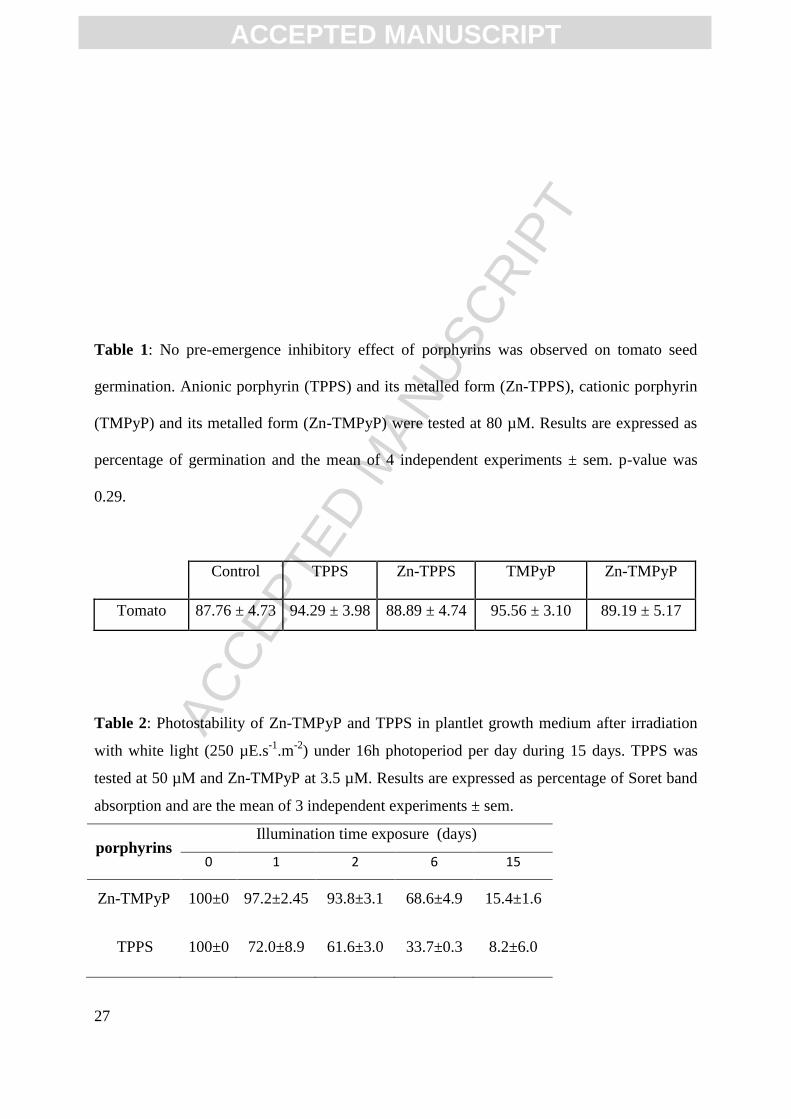
ACC
EPTE
D M
ANU
SCR
IPT
ACCEPTED MANUSCRIPT
27
Table 1: No pre-emergence inhibitory effect of porphyrins was observed on tomato seed
germination. Anionic porphyrin (TPPS) and its metalled form (Zn-TPPS), cationic porphyrin
(TMPyP) and its metalled form (Zn-TMPyP) were tested at 80 µM. Results are expressed as
percentage of germination and the mean of 4 independent experiments ± sem. p-value was
0.29.
Control TPPS Zn-TPPS TMPyP Zn-TMPyP
Tomato 87.76 ± 4.73 94.29 ± 3.98 88.89 ± 4.74 95.56 ± 3.10 89.19 ± 5.17
Table 2: Photostability of Zn-TMPyP and TPPS in plantlet growth medium after irradiation
with white light (250 µE.s-1
.m-2
) under 16h photoperiod per day during 15 days. TPPS was
tested at 50 µM and Zn-TMPyP at 3.5 µM. Results are expressed as percentage of Soret band
absorption and are the mean of 3 independent experiments ± sem.
porphyrins Illumination time exposure (days)
0 1 2 6 15
Zn-TMPyP 100±0 97.2±2.45 93.8±3.1 68.6±4.9 15.4±1.6
TPPS 100±0 72.0±8.9 61.6±3.0 33.7±0.3 8.2±6.0
ACC
EPTE
D M
ANU
SCR
IPT
ACCEPTED MANUSCRIPT
28
FIGURES LEGEND
Figure 1: Structures of different porphyrins tested in this study
Figure 2: Porphyrins tested at 80 µM are not cytotoxic for tomato plantlets grown in the dark.
Tomato seed germination took place for 4 days on media supplemented with 80 µM
porphyrins. B5 represented the control without porphyrins. Experiments were repeated at least
3 times. Representative pictures of one of these biological repeats were reported. Scale bar
represents 1 cm.
Figure 3: Cationic porphyrins, especially Zn-TMPyP, altered growth and development. A)
Tomato seeds germinated on different media supplemented with 50 µM of TPPS/Zn-TPPS
and 3.5 µM TMPyP/Zn-TMPyP for 14 days under 16 h photoperiod. B5 corresponded to the
control medium without porphyrins. Experiments were repeated at least 3 times and
representative pictures were taken. Scale bar represents 1 cm for tomato. B) Dry to fresh
weight ratio (DW/FW) determined in 14 day-old tomato plantlets (cf mat and meth). Tomato
seeds germinated on different media supplemented with 50 µM TPPS/Zn-TPPS and 3.5 µM
TMPyP/Zn-TMPyP for 14 days under 16 h photoperiod. Results are the mean of 4
independent experiments ± sem. ***: P < 0.001
Figure 4: Tomato plantlets placed on high concentrations of TMPyP/Zn-TMPyP for 14 days
could be rescued and recovered a phenotype similar to control plants. Tomato seed
germination took place on media supplemented with 80 µM PP for 14 days (D14). Plantlets
ACC
EPTE
D M
ANU
SCR
IPT
ACCEPTED MANUSCRIPT
29
were placed for one week on B5 medium (D21) and then transferred to soil and green house
for 6 weeks (D63). C represented the control without cationic porphyrins. Scale bar for D14
and D21 represents 1 cm, scale bar for D63 represent 3 cm.
Figure 5: Zn-TMPyP localization in tomato root sections. Pictures were taken from spectral
mode acquisition. Left panel: calcofluor staining and Right panel: Zn-TMPyP treated roots.
Labelling and detection are described in mat and meth. Scale bar represents 50 µm.
Figure 6: Photoactivated cationic porphyrins induce lipid peroxidation in 14 day-old tomato
roots and shoots. Black box corresponded to control parts of plantlets, 3.5 µM TMPyP and
Zn-TMPyP to grey and dark grey respectively. Results are the mean of 4 independent
experiments ± sem (ns: not significant; *** P<0.001).
Figure 7: H2O2 production in whole 14 day-old tomato plantlets. A) Control root apparatus
was stained by NBTZ (left) or root apex by DAB (right) (cf Mat and meth). B) Zn-TMPyP
treated roots stained by NBTZ or DAB. Zn-TMPyP was tested at 3.5 µM. Scale bar represents
1 mm. C) H2O2 quantification was performed on isolated roots and shoots of no treated (B5)
or treated plantlets. Results are the mean of 5 independent experiments ± sem (*** P<0.001; *
0.01<P<0.05).
Figure 8: Antioxidant molecule contents in 14 day-old tomato roots. Black boxes
corresponded to control roots and grey boxes to 3.5 µM Zn-TMPyP. Results are the mean of 4
independent experiments ± sem (*** P<0.001; **P<0.01).
Figure 9: Effect of photoactivated Zn-TMPyP on antioxidant enzyme specific activities in 14
day-old tomato roots. Activities were determined as described in mat and meth. GPX:
guaiacol peroxidase, APX: ascorbate peroxidase, CAT: catalase, SOD: superoxide dismutase.
Results are the mean of 4 independent biological experiments ± sem (ns: not significant; a:
0.05<P<0.1; *: 0.01<P<0.05; ***: P<0.001)
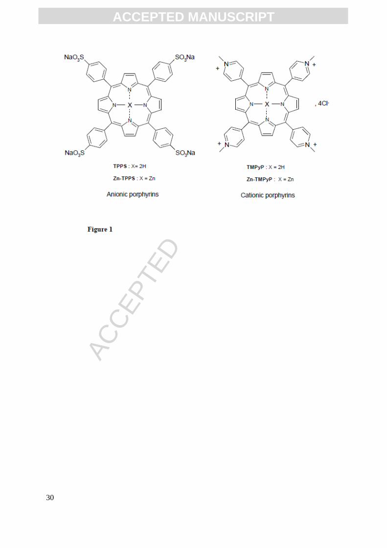
ACC
EPTE
D M
ANU
SCR
IPT
ACCEPTED MANUSCRIPT
30

ACC
EPTE
D M
ANU
SCR
IPT
ACCEPTED MANUSCRIPT
31
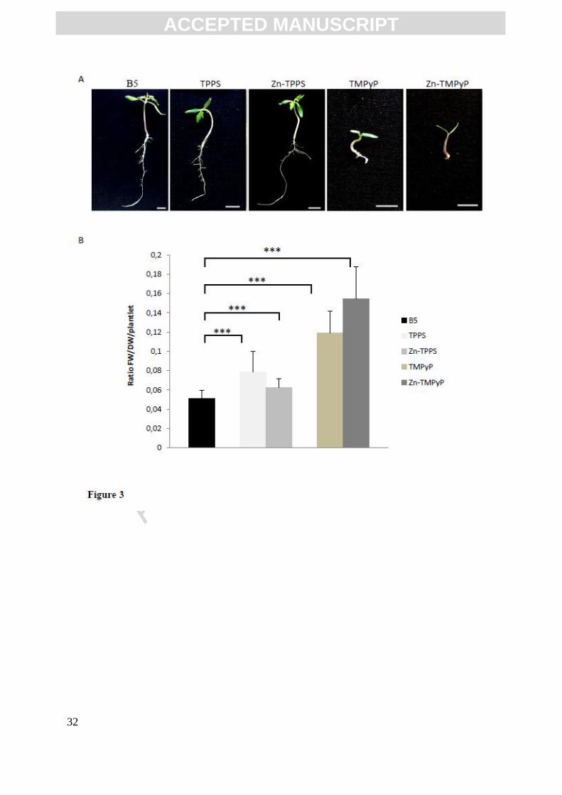
ACC
EPTE
D M
ANU
SCR
IPT
ACCEPTED MANUSCRIPT
32

ACC
EPTE
D M
ANU
SCR
IPT
ACCEPTED MANUSCRIPT
33
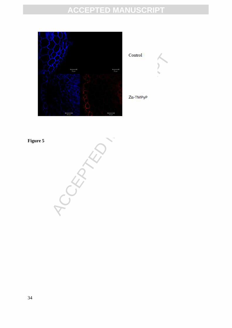
ACC
EPTE
D M
ANU
SCR
IPT
ACCEPTED MANUSCRIPT
34
Figure 5
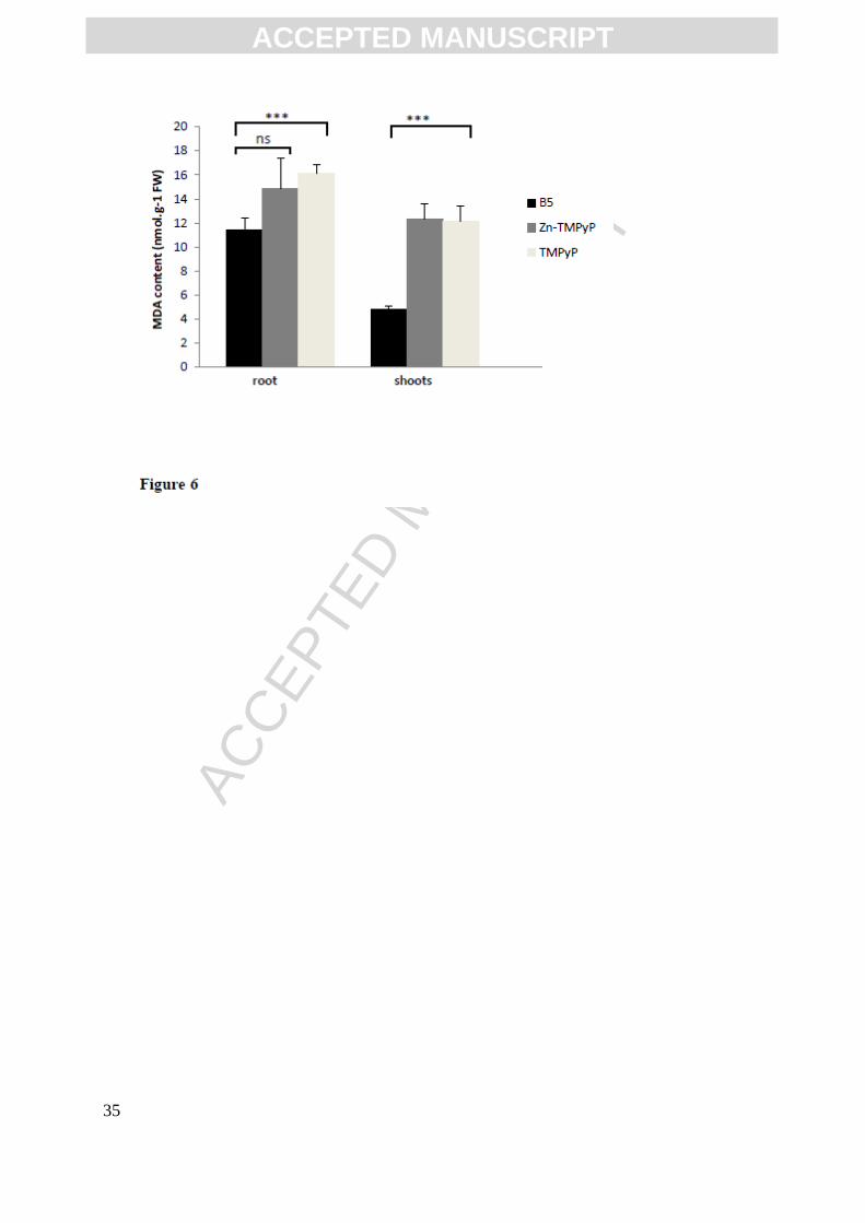
ACC
EPTE
D M
ANU
SCR
IPT
ACCEPTED MANUSCRIPT
35
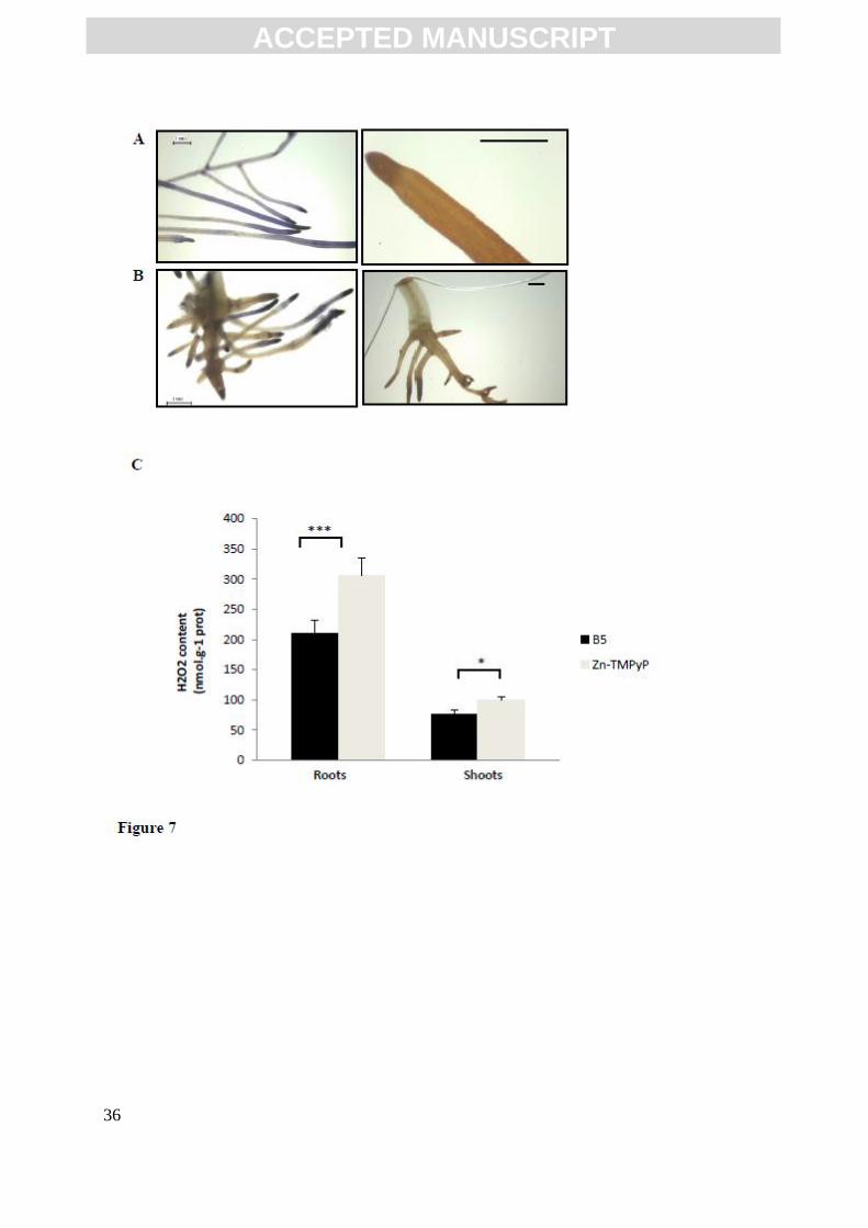
ACC
EPTE
D M
ANU
SCR
IPT
ACCEPTED MANUSCRIPT
36

ACC
EPTE
D M
ANU
SCR
IPT
ACCEPTED MANUSCRIPT
37
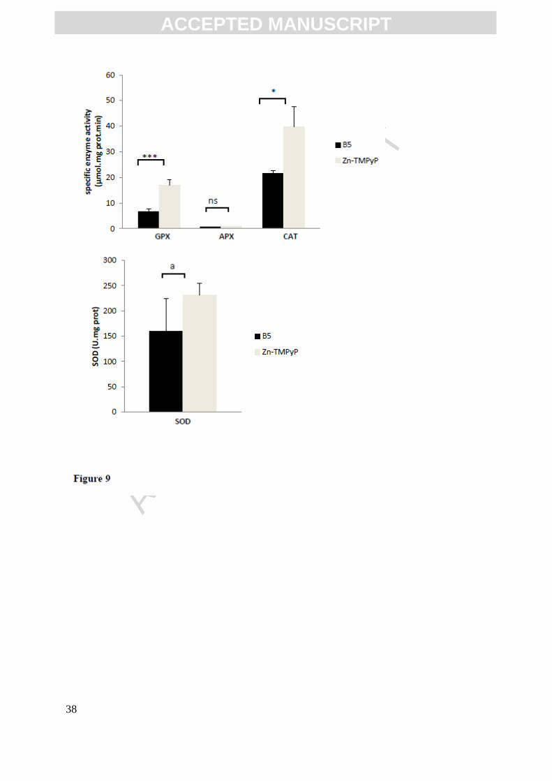
ACC
EPTE
D M
ANU
SCR
IPT
ACCEPTED MANUSCRIPT
38

ACC
EPTE
D M
ANU
SCR
IPT
ACCEPTED MANUSCRIPT
39
Highlights
* Zn-TMPyP altered vegetative development of tomato plantlets without killing them.
* Anti-oxidative molecules and enzymes involved in ROS detoxification increased
* Zn-TMPyP is proposed as a valuable APDT molecule with no-killer effect on plants.

75
Results
Mohammad ISSAWI | Thèse de doctorat | Université de Limoges | 2018
Licence CC BY-NC-ND 3.0
PUBLICATION 3: “Responses of an adventitious fast-growing plant to photodynamic
stress: comparative study of anionic and cationic porphyrin effect on Arabidopsis
thaliana”, Physiologia Plantarum.
Acc
epte
d A
rticl
eResponses of an adventitious fast-growing plant to photodynamic stress: comparative study of
anionic and cationic porphyrin effect on Arabidopsis thaliana
Mohammad Issawi, Damien Guillaumot, Vincent Sol and Catherine Riou*
Laboratoire de Chimie des Substances Naturelles (EA 1069), Université de Limoges, Faculté des
Sciences et Techniques, 123 avenue Albert Thomas, 87060 Limoges Cedex, France
Correspondence
* corresponding author,
e-mail : [email protected]
Abbreviations – APDT, antimicrobial photodynamic treatment; AP, Meso-Tetra (4-sulfophenyl)
Porphyrin; CP, Tetra (N-methylpyridyl) porphyrin tetrachloride.
Antimicrobial Photodynamic Treatment based on the use of a photosensitizer to produce reactive
oxygen species that induce cell death could be envisaged to fight against plant pathogens. For setting
this strategy, we want to study how plants themselves respond to photodynamic treatment. In a
previous work we showed that tomato plantlets were able to resist to photoactivated tetra (N-
methylpyridyl) porphyrin (CP) or Zinc metalated form (CP-Zn). To enlarge our plant expertise related
to exogenous porphyrins treatment and to further defend this approach, we studied how an adventice
like Arabidopsis thaliana responded to exogenous supply of anionic and cationic porphyrins. Both
types of photosensitizers had no negative effect on seed germination and did not hamper Arabidopsis
etiolated plantlet development under dark conditions. Thus, post-emergence effects of porphyrin
photoactivation on 14 day-old in vitro Arabidopsis plantlet development under light were observed.
CP-Zn was the most efficient photosensitizer to kill Arabidopsis plantlets while anionic tetra (4-
sulfonatophenyl) porphyrin only delayed their growth and development. Indeed only 7 % of plantlets
could be rescued after CP-Zn treatment. Furthermore, non-enzymatic and enzymatic defense
components involved in detoxification of ROS generated by CP-Zn under illumination were down-
regulated or stable with the exception of seven fold increase in proline content. As previously
demonstrated in the literature for microbial agents and in the present work for Arabidopsis, CP-Zn was
efficient enough to eradicate in the same time unwanted vegetation and plant pathogens without killing
plants of agronomic interest such as tomato plantlets.
This article is protected by copyright. All rights reserved.
This article has been accepted for publication and undergone full peer review but has not been through the copyediting, typesetting, pagination and proofreading process, which may lead to differences between this version and the Version of Record. Please cite this article as doi: 10.1111/ppl.12666

Acc
epte
d A
rticl
e
Introduction
The light-based approach antimicrobial photodynamic treatment (APDT) that is used in
several domains such as medicine, food and water decontamination could be used beyond (Alves et al
2015). It could be envisaged as a farming practice in order to eradicate plant pathogens as bacteria and
fungi that cause serious threats by drastically reducing crops yield. Studies focusing on the side effects
of APDT directly on plants are rare (De Menezes et al 2014a,b, Fracarolli et al 2016, Guillaumot et al
2016). However, such studies are required to evaluate the efficiency of APDT. Recently, it was
reported that natural photosensitizers such as coumarins and fucocoumarins or synthetic such as
phenothiazinium inactivated pathogenic fungi (Colletotrichum, Aspergillus and Fusarium). When
spotted on orange tree or strawberry plants, orange tree leaves were not affected by either
natural/synthesic photosensitizers whereas they damaged strawberry leaves when tested at 100 µM (De
Menezes et al 2014a,b, Fracarolli et al 2016). Our aim is to explore, the phenotypic and molecular
responses of plantlets growing in vitro under photodynamic stress induced by photoactivated
porphyrins included in growth medium. In the present work we want to know how Arabidopsis
thaliana, an adventitious fast-growing plants respond to such photodynamic treatment.
Molecules such as porphyrins, naturally present in all living kingdom, seem to be good
candidates for APDT. Indeed, these systems, photo-excitable by sunlight, are able to induce cell death
by the mean of reactive oxygen species (ROS) such as superoxide anion radical, hydrogen peroxide,
hydroxyl radical and singlet oxygen. Under dark conditions, porphyrins do not exhibit significant
cyto- or genotoxicity (Donnelly et al 2008). Until now applications for porphyrins were essentially
performed on cancer cells for photodynamic therapy or on microorganisms as antimicrobial agents
(Plaetzer et al 2013, Maish 2015, Pérez-Lagunal et al 2017, Zampini et al 2017). For example, upon
illumination, protoporphyrin IX and other derivatives induced a strong cytotoxic effect in yeasts
Saccharomyces cerevisiae and Candida albicans (Donnelly et al 2008, Carré et al 1999, Cormick et al
2009, Funes et al 2009, Quiroga et al 2015). Another study showed that the derivatives of cationic
porphyrin were more efficient than neutral and anionic porphyrins on Gram-negative bacteria
(Escherichia coli) than on Gram-positive bacteria such as Enterococcus hirae or Staphylococcus
This article is protected by copyright. All rights reserved.
Acc
epte
d A
rticl
eaureus (Merchat et al 1996, Gabor et al 2001, Ringot et al 2012, Fayyaz 2015). As well, the
exogenous supply of cationic porphyrin on cyanobacteria and green microalgae cultures showed that
microalgae were more sensitive to the effect of the photosensitizer than cyanobacteria (Drabkova et al
2007). Furthermore, tested on mosquito or fly larvae, it would appear that photoactivated porphyrins
also have an insecticidal effect (Ben Amor and Jori 2000, Dondji et al 2005, Lucantoni et al 2011).
Over the last three decades, it was of interest to modify the endogenous porphyrin pathway in order to
overproduce proptoporphyrin IX, the first natural photosensitizer, by supplying 5-aminolevulinic acid
on plants (5-ALA). 5-ALA induced herbicidal and negative effect on wildlife when tested at high
concentration around 5 mM (Rebeiz et al, 1984, Chakraborty and Tripathy 1992, Matsumoto et al
1994, Sasikala et al 1994). To our knowledge, only two studies were conducted on plants. On Allium
cepa root meristematic cells it was reported that zinc metalated/free base cationic porphyrins were able
to be up-taken by cells and induce DNA photodamage (Villanueva et al 1989). More recently, study
on tomato plantlets showed that they were able to withstand the damaging effects of photoactivated
cationic porphyrins (Guillaumot et al. 2016). As pointed by these studies, we supposed that exogenous
photoactivated porphyrins could seriously alter plant development leading to preferentially death of
fast growing species such as Arabidopsis thaliana. Arabidopsis is a cosmopolitan small flowering
plant that is widely used as a model organism in plant biology research. It is the “darling” of plant
science but it is neither grown for food nor to feed animals and it is not of major agronomic
significance. In the contrary, Arabidopsis plants have some weedy traits and may be considered as
unwanted vegetation in a farm field. Responses of such plants to photodynamic treatment is required
in order to cover all aspects of APDT as a future commercial tool in the field. (Meyerowitz 1989,
Provart and Meyerowitz 2016). Moreover, as discussed by De Menezes (2014a), this type of treatment
required a case-by-case analysis because its outcome depends on plant species.
For this work, we tested two categories of water-soluble porphyrins: cationic and anionic
synthetic porphyrins respectively that were free-base or zinc-metalated. We showed that none of the
porphyrins was able to produce a pre-emergence lethal phenotype of Arabidopsis. Thus, we compared
the consequences of their photoactivation on phenotypes of 14 day-old Arabidopsis plantlets and
showed that cationic porphyrin especially CP-Zn tested at 3.5 µM induced the strongest phenotypic
This article is protected by copyright. All rights reserved.
Acc
epte
d A
rticl
ealterations. Thus, taking into account these first results, we focused our study on cellular and
molecular mechanisms triggered by CP-Zn in plantlets roots, trying to understand why tomato
plantlets were less sensitive than Arabidopsis. Indeed, Arabidopsis presented very high H2O2 and
MDA productions leading to biomembrane damages. Moreover, endogenous levels of proline,
ascorbate and total thiols considered as antioxidant molecules and enzyme activities such as
peroxidases, catalase and superoxide dismutase were clearly down-regulated in Arabidopsis plantlets.
In conclusion, we suggested that Arabidopsis plantlets were photodynamically damaged by
photoactivated cationic porphyrins that impair enzymatic defense machinery and rendering them
unable to produce sufficient amount of antioxidative molecules such as ascorbate and total thiols to
withstand this stressful conditions.
Materials and methods
Plant material
Seeds of Arabidopsis thaliana ecotype Wassilewskija (Ws-4) were germinated in vitro on a Gamborg
B5 culture medium (Duchefa Biochemie, Haarlem, Netherlands) at pH 5.8 supplemented with 2%
(w/v) sucrose and 0.8% (w/v) agar (Difco, Dallas, TX (Gamborg et al 1968). For all experiments,
plates were maintained for 14 days in a controlled growth chamber at 22°C under a 16h photoperiod
and a photon flux density of 250 µmol m-2 s-1. Germination ratio was evaluated after 4 days of culture
using binocular microscope. Under these conditions, around 100 Arabidopsis seeds were tested on the
same square plate. Pictures were taken under binocular microscope (Leica EZ4D stereo microscope
W/ digital camera, Jena, Germany) and Leica LAS EZ software. For cytotoxicity, plates were
maintained vertically under same conditions without illumination for 7 days. After 14 day culture,
plantlets were harvested, weight and frozen at -20°C until used. For determination of dry weight,
plantlets were placed in an oven at 42°C for 4 days and weight.
Porphyrins
This article is protected by copyright. All rights reserved.
Acc
epte
d A
rticl
eAll porphyrins were soluble in water and their chemical structures were reported (Riou et al 2014).
Briefly, anionic porphyrin (AP and AP-Zn) correspond to the free zinc sulfonated
tetraphenylporphyrin (H2TPPS) and Zinc-TPPS, respectively. AP was purchased from Sigma-Aldrich
(St Louis, MO) and AP-Zn from Porphychem (Dijon, France). Cationic porphyrins: Tetra (N-
methylpyridyl) porphyrin tetrachloride or H2TMPyP (CP) and Zinc TMPyP (CP-Zn) were purchased
from Frontier Scientific (Carnforth, United Kingdom).
Confocal microscopy analysis
We incubated 14 day-old plantlets in B5 medium with or without 3.5 µM CP-Zn overnight
under dark conditions and rinsed them 3 times before observations under a Zeiss confocal microscope
(LSM 510-META, Jena, Germany). For analysis, two ways of acquisition were performed: a spectral
acquisition to verify signal specificity of CP-Zn and a channel mode for root apex stained by
calcofluor (Sigma-aldrich, St Louis, MO). For spectral acquisition, samples were excited at 458 nm
and fluorescence was followed between 476 and 732 nm. CP-Zn emission peak was detected around
640 nm (Guillaumot et al 2016). For channel mode acquisition and calcofluor, we used an excitation
wavelength at 405 nm and emission wavelength between 420 and 450 nm.
In situ DAB staining
3,3-diaminobenzidine (DAB) staining on Arabidopsis whole plantlets was performed using DAB Kit
(Sigma-Aldrich, St Louis, MO) following manufacturer’s instructions. Plant material was vacuum-
infiltrated for 5 min and placed under dark conditions for 2 h. DAB solution was replaced by 70%
(v/v) ethanol and conserved at 4°C until observations under binocular microscope.
Antioxidant molecule assays
The proline content was analyzed as follow. Frozen samples of plantlets were ground in liquid
nitrogen then homogenized in 1 ml of 5% (w/v) 5-sulfosalicylic acid. After centrifugation for 15 min
at 13 000 g and 4°C, 250 µl of supernatants were mixed with 1ml of 1% (w/v) ninhydrin in 60% (v/v)
acetic acid. The samples were incubated at 100°C for 20 min and cooled at room temperature before
This article is protected by copyright. All rights reserved.

Acc
epte
d A
rticl
eaddition of 1.25 ml toluene. After 4 h incubation at room temperature, the upper phase was collected
and absorbance was read at 520 nm. A standard curve was performed with proline and the content was
expressed in µmol g⁻¹ FW.
Total thiol assay was performed according to (Guillaumot et al 2016). Frozen samples were ground in
liquid nitrogen and then extracted into 1 ml of 0.2 N HCl. After centrifugation at 16 000 g for 4 min,
500 μl of extracted solution were neutralized with 400 μl NaOH (0.2M) and 50 μl NaH2PO4 (0.2 M).
0.7 ml of 0.12 M NaH2PO4 (pH 7.5), 6 mM EDTA, 0.1 ml of 6 mM Dithiobis 2-nitro benzoic acid
(DTNB) were added to 0.2 ml extract. For standards, extract was replaced by 0, 10, 20, 50, and 100
nmol GSH (total volume 1 ml). Absorbance at 412 nm was read 5 min after the addition of standards
or extract.
Ascorbate was quantified as reported by Kampfenkel et al (1995). Briefly, frozen plant tissues (around
100 mg fresh weight) were ground in liquid nitrogen with a mortar and pestle. The resulting powder
was weighted before addition of 200 µl of 6 % TCA solution. Samples were then incubated for 10 min
on ice and centrifuged at 16 000 g for 25 min at 4°C. Supernatant was completed to 500 µl with 6%
TCA and centrifuged 16 000 g for 10 min. at 4°C. 100 µl of each sample were then transferred to 150
µL 0.2 M phosphate buffer pH 7.4 and supplemented with 250 µl 10 % TCA, 200 μl 42% H3PO4, 200
μl 2,2′-dipyridyl and 100 μl 3% FeCl3. Standards ranged from 0 to 70 nmoles of ascorbate were run.
After 40 min incubation at 42°C, absorbance of standards and samples was read at 525 nm.
Determination of MDA content
MDA content was determined by the thiobarbituric acid (TBA) reaction as described by Hodges
(1999), with minor modifications. Around 100 mg of fresh or frozen plant materials were ground with
liquid nitrogen. The resulting powder was suspended into 1.5 ml of 20% TCA. After centrifugation,
supernatants were collected and analyzed for their MDA content. 500µl of each sample were added to
either 500 µl 20% TCA alone (-TBA) or 20% of TCA supplemented with 0.5% thiobarbituric acid
(+TBA). Samples were then boiled in a water bath for 30 min. The samples were cooled on ice and
absorbances were read at 440, 560 and 600 nm. Corrections for interfering compounds were made and
the MDA content was calculated as follow:
This article is protected by copyright. All rights reserved.

Acc
epte
d A
rticl
e(1) [(Abs 532+TBA - Abs 600+TBA) - (Abs 532-TBA - Abs600-TBA)] = A
(2) {[(Abs 440+TBA - Abs 600+TBA) - (Abs 440-TBA - Abs 600-TBA)] 0.0571} = B
(3) MDA equivalents (nmoL.mL-1) = [(A-B)/157 000] .106
Enzymatic activities determination
Frozen plant samples were ground to a fine powder in liquid nitrogen using a mortar and pestle.
Proteins were solubilized into 1 ml of extraction buffer containing 50 mM phosphate buffer pH 7.8, 1
mM EDTA, 1% (w/v) PVP and 10% (v/v) glycerol and centrifuged at 16 000 g for 10 min at 4°C. The
resulting supernatant was used for protein quantification according to Bradford (Bradford 1976) and
then for antioxidant enzyme analysis.
The activity of catalase (CAT) was measured by monitoring H₂O₂ decomposition at 240 nm during 3
min according to Aebi’s protocol (Aebi 1984). 50 or 20 µg of total soluble proteins from each sample
were diluted in 50 mM phosphate potassium buffer (pH 6.5). The reaction was initiated by the addition
of H2O2 to a final concentration of 20 mM. Amount of H2O2 was calculated based on a molar
extinction coefficient of 43.6 M−1 cm−1.
Guaiacol peroxidase activity was quantified following the increase in absorbance at 436 nm during 2
min due to the formation of tetraguaiacol and calculated with a molar extinction coefficient of
25.5 mM−1 cm−1. 10 µg of total soluble proteins were diluted into 50 mM phosphate potassium buffer
pH 6.5 and supplemented with 0.25% of guaiacol (5% stock solution in 95% ethanol w/v). Reaction
started by addition of H2O2 to a final concentration of 2.5 mM.
Ascorbate peroxidase activity was analyzed by adapting the microplate protocol to tubes by estimating
the rate of ascorbate oxidation (extinction coefficient: 2.8 mM⁻¹·cm⁻¹) (Murshed et al. 2008). 50 µg of
soluble protein extracts were diluted in 50 mM phosphate buffer pH 7.0 supplemented with 0.25 mM
ascorbic acid. The reaction started by addition of H2O2 to the final concentration of 5 mM. The
decrease in absorbance at 290 nm was then measured during 3 min.
The activity of superoxide dismutase (SOD) was assayed by measuring its ability to inhibit the
photochemical reduction of nitro blue tetrazolium (NBT) according to (Weydert and Cullen 2010).
Standard curve was set so that the increase in absorbance at 560 nm was between 0.02 and 0.03
This article is protected by copyright. All rights reserved.
Acc
epte
d A
rticl
eabsorbance unit per minutes and was monitored for 5 min. One unit of SOD was defined as the
amount of enzyme required for an inhibition of 50% of NBT reduction. 10 µl of enzyme extract were
used and further converted to an amount of proteins according to their respective concentration.
Statistical analysis
All biological experiments were performed at least three times independently. Results were expressed
as mean ± SD (Standard Deviation). Data were analyzed by t-student test and one-way ANOVA using
the PAST free software.
Results
We tested two categories of porphyrins: cationic and anionic because of their solubility in
water and culture medium. Furthermore, anionic or cationic porphyrins were free base or zinc
metalated. Our aim was to describe the effect of low concentration of photoactivated porphyrins at pre
and post emergence levels.
High concentration of anionic or cationic porphyrins does not affect seed germination
Arabidopsis seeds were placed in darkness for 7 days on media containing each porphyrin tested at
high concentration. Photosensitizer concentrations were 50 µM for anionic compounds and 5 µM for
cationic species. As none of these tested concentrations inhibited Arabidopsis seed germination (Table
1), anionic or cationic porphyrins could definitively not be considered as a pre-emergent inhibitor.
Moreover, it seemed that 5 µM CP promoted seed germination (Table 1). Thus, we studied subsequent
effects of porphyrins at the post-germination level.
Photoactivated cationic porphyrins efficiently affect Arabidopsis growth
In regard to potential cytotoxicity of PP described in mammal cells (Magnieto et al 2014), post-
germination events under dark conditions were assayed. Thus we tested the same concentrations for
AP/AP-Zn and CP/CP-Zn that tested in Table 1 and monitored etiolated plantlet development during 7
days (Fig. 1). Indeed, 7 day-old etiolated plantlets seemed very similar to control ones in term of root
This article is protected by copyright. All rights reserved.
Acc
epte
d A
rticl
eand hypocotyls growth (Fig. 1). This result showed that under dark conditions, the four porphyrins did
not lead to significant cytotoxicity and/or genotoxicity which is a great advantage for further
agronomic investigations. Thus, under darkness prevailing conditions in the soil, porphyrins would not
be cytotoxic for living systems as seedlings and soil microorganisms.
Using large spectrum illumination that mimic summer sunlight for 16h per day, we then tested
porphyrin effect on seedlings grown at light. In a first attempt, 14 day-old Arabidopsis whole plantlets
grown in vitro were transferred for a short time light exposure (72h experiment corresponds to 48h
light) into a new liquid culture medium containing porphyrins (Fig. 2). In contact to AP/AP-Zn,
Arabidopsis plantlets did not show leaf alterations compared to control plants whereas on 3.5 µM
CP/CP-Zn, yellowish leaves and nearly dead plantlets were observed (Fig. 2). This first result
suggested a greater efficiency of cationic porphyrins to induce cell and plantlet death.
To gain insight to the effects of long time exposure to porphyrins, Arabidopsis seedlings were grown
in contact to each porphyrin for 14 days under 16 h photoperiod. Although phenotypic alterations were
not obviously observed in contact to 50 µM AP or AP-Zn compared to control condition, plantlets
placed on 3.5 µM CP or CP-Zn showed significant reduction of root length, asymmetry in cotyledon
expansion, that could become white, and no formation of new leaves (Fig. 3A). We determined dry on
fresh weight ratio in control and treated plantlets. A significant increase in this ratio was observed for
all tested conditions (Fig 3B). It was probably linked to plantlet dehydration that could be also
correlated to the 7-fold proline increase in CP-Zn treated plantlets (Fig. 3B, Table 2). Nevertheless,
those results confirm that anionic porphyrins (free or zinc metaled forms) had almost no effect on
Arabidopsis plantlets (Fig. 3). Since previous studies on microbial agents showed higher efficiency of
cationic porphyrins and Arabidopsis plantlets treated with cationic porphyrins were prone to
pronounced photodynamic stress, we did not pursue with anionic porphyrins that did not effectively
inhibit Arabidopsis vegetative growth while tested at nearly 15 times more concentrated than cationic
porphyrins.
Few plantlets could be rescued after photoactivation of CP-Zn treatment
This article is protected by copyright. All rights reserved.

Acc
epte
d A
rticl
eAnother focus for agronomic traits was to rescue plantlets that grew on photoactivated porphyrins.
Only few green plantlets obtained on CP-Zn were rescued whereas the white plantlets could not be
rescued (Fig 4). Tested at the same concentration (5 µM) it was also shown that photoactivated CP was
less efficient than CP-Zn to induce the death of Arabidopsis plantlets (Fig. 4). Nevertheless, CP-Zn
worked at dose dependent manner and we tested a range of CP-Zn concentrations from 1 to 10 µM on
Arabidopsis seedling development (Fig. 4C) as 3.5 µM CP-Zn already induced lethal phenotype for
60% of Arabidopsis plantlets. Whereas on 1 µM CP-Zn, nearly 100% plantlets were green and could
undergo further development, on 10 µM CP-Zn this proportion fall to less than 5% of surviving
plantlets. According to our results, we decided to continue with an intermediary concentration of CP-
Zn corresponding to 3.5 µM that was an effective concentration for killing Arabidopsis seedlings. .
H2O2 production and lipid peroxidation strongly increase in treated Arabidopsis plantlets
Because photoactivated porphyrins generate reactive oxygen species, we monitored H2O2 production
in plantlets in situ by DAB staining and biochemical assay. In parallel we determined membrane lipid
peroxidation by MDA assay in treated plantlets compared to control ones (Fig. 5). A significant
increase in both H₂O₂ and MDA contents was determined suggesting that H2O2 over-production and
lipid peroxidation were together responsible for plantlet death (Fig. 5). Our hypothesis was that CP-Zn
was probably able to accumulate in the cell wall and/or cytoplasm of Arabidopsis root, thereby
inducing ROS over-production under illumination.
Antioxidant arsenal is overwhelmed in treated Arabidopsis plantlets
As antioxidant molecules, total thiol, proline and ascorbate contents in CP-Zn treated plantlets were
assayed. The photodynamic stress triggered by photoactivated CP-Zn induced a 7-fold increase in
proline content in Arabidopsis whole plantlets while ascorbate amount did not significantly change
and total thiol content fall down in response to CP-Zn (Table 2). These results could explain why
plantlets were not able to fight against ROS production triggered by CP-Zn photoactivation.
Furthermore, while ascorbate and guaiacol peroxidases were not activated, superoxide dismutase and
This article is protected by copyright. All rights reserved.

Acc
epte
d A
rticl
ecatalase were down-regulated (Fig 6). Taken together, it could explain why DAB staining was so
intense in the treated plantlets (Fig.5A).
CP-Zn is localized in Arabidopsis root apex
As CP-Zn seemed to be a good weed killer candidate, it was also of importance to check CP-Zn
localization in Arabidopsis root apparatus. Previous study already showed that cationic porphyrin was
able to get in Alliun cepa root meristematic cells (Villanueva et al., 1989). Confocal microscopy
analysis, using the natural fluorescence of CP-Zn and calcofluor as a contrast marker of the cell wall,
showed that CP-Zn was strongly localized in the first external layers of root (epidermis and
parenchyma cells) in Arabidopsis root apex (Fig. 7). Furthermore, it seemed that CP-Zn was in cell
wall and/or cytoplasm of these cells but further analysis are required to distinguish between the two
localizations. Nevertheless this result suggested that CP-Zn could cross cell wall, get in the cytoplasm
where it could be photoactivated and induced a strong oxidative burst leading to plantlet death.
Discussion
To our knowledge, this approach of supplying exogenous porphyrin on Arabidopsis plantlets
was the first although it was extensively studied on bacteria, fungi, insects and mammal cells
(Donnelly et al 2008, Plaetzer et al 2013, Maish 2015, Pérez-Lagunal et al 2017). Furthermore,
cationic porphyrins were able to reach root meristematic cell nucleus and induced DNA photodamage
(Villanueva et al 1989). Recently and in the present work, we showed that cationic porphyrins at 3.5
µM were able to inhibit Arabidopsis early vegetative development whereas tomato plantlets treated
with 80 µM CP/CP-Zn were also affected but could be rescued (Guillaumot et al 2016). This result
was in agreement to the necessity of a case-by-case study because photosensitizer effects depend on
the ability to be uptaken by plants and to enter into cells. Indeed, observation of CP-Zn localization in
Arabidopsis treated roots showed an intense labelling from epidermis cells to vessels while in tomato
roots labelling was confined to external layers suggesting a differential root interaction and uptake of
the photosensitizer between the two distant species (Guillaumot et al., 2016). This also was observed
This article is protected by copyright. All rights reserved.
Acc
epte
d A
rticl
ebetween citrus and strawberry leaves in contact to coumarins and fucocoumarins where citrus leaf
cuticle prevents photosensitizer to cross epidermis whereas it could diffuse through strawberry
epidermis and induce oxidative damages (De Menezes et al 2014a). Thus, the future use of APDT in
environment must consider plant species and their anatomical traits such as thickness and/or
interaction between epidermis cells and photosensitizers. Moreover, the effects of photosensitizer will
be also more or less important depending on the way of its administration, e.g. soil feeding and
spraying. Soil feeding supposes that photosensitizer should be up-taken by roots and translocated to
the leaves where they become photoactivated. In the present study, we also showed that translocation
occurred in 14 day-old CP-Zn treated plantlets through H2O2 cotyledon accumulation linked to CP-Zn
photoactivation. This result was also in agreement to previous work with Mg-ProtoIX feeding in
Arabidopsis (Kindgren et al., 2012; Larkin, 2016). The spraying method is probably less harmful for
plants than the constant photosensitizer inclusion in culture medium as we did, explaining why a low
CP-Zn (3.5 µM) was so efficient to kill Arabidopsis plantlets in vitro. We supposed that in the field
such concentration will have no effect on plants treated by spraying. Nevertheless, studies on plant
pathogens as fungi showed that a concentration up to 50 µM of natural and synthetic photosensitizers
was required to eradicate fungi (De Menezes et al 2014a,b, Fracarolli et al 2016). At this threshold
concentration of cationic porphyrins, Arabidopsis and pathogens will be most likely eradicated
whereas this concentration does not kill tomato plantlets (Guillaumot et al 2016). Further study on
plants in greenhouse alone and against pathogens under porphyrin spreading are now required to test
this hypothesis.
In the other hand, we showed that under dark condition, photosensitizers did not affect germination
and post-germinative development of Arabidopsis and tomato plantlets (Guillaumot et al 2016). Thus
it does not affect soil micro-wildlife where darkness takes over.
Under light conditions, post-emergence effect of porphyrins was investigated and we showed
that cationic porphyrins were able to inhibit Arabidopsis early vegetative development. When
illuminated, photosensitizers, especially porphyrins, generate radicals such as superoxide anion (O2-)
and hydroxyl radical (OH) by electron transfer (type I process) or singlet oxygen (1O2) by energy
transfer to dioxygen (type II process). All these ROS damage cellular constituents such as nucleic
This article is protected by copyright. All rights reserved.

Acc
epte
d A
rticl
eacids, proteins, lipids leading to cell death by necrosis and more often by apoptosis (Gill and Tuteja
2010, Avery 2011). As a consequence of porphyrin photoactivation, cell death was shown in animal
cells, fungi, bacteria and recently in tobacco BY-2 cells (Donnelly et al 2008, Ringot et al 2012,
Plaetzer et al 2013, Riou et al 2014, Maish 2015). Because lipid peroxidation is thought to be mainly
triggered by these ROS, we performed MDA quantification on Arabidopsis whole plantlets (Gill and
Tuteja 2010, Avery 2011). A significant increase in MDA was detected suggesting that cell
membranes were seriously damaged, presumably leading to cell death and ultimately to plantlet death.
Our result strongly suggested that CP-Zn was able to get inside root cells (Villanueva et al 1989) and
produce ROS close to the different membrane systems (endoplasmic reticulum, mitochondria) under
illumination. Nevertheless, photoactivation of CP-Zn and CP was also shown to induce DNA
photodamage (Villanueva et al 1989). Altogether, lipid peroxidation and DNA damage are seen as
consequences of CP-Zn photoactivation in root cells and led to a fatal issue in Arabidopsis. Moreover,
DNA damages could also be induced by MDA itself which was shown to be highly mutagenic in
bacteria and mammal cells (Marnett 2002). Further investigations should be performed to check
whether DNA photodamage also occurs in Arabidopsis.
Another way for the plantlets to resist to photoactivated CP-Zn and to protect themselves
against ROS, is to produce antioxidant molecules that could scavenge ROS. Thus, we determined
proline, total thiol and ascorbate contents and ROS detoxification enzyme activities such peroxidases,
catalase and superoxide dismutase. Arabidopsis plantlets did not show any increase in these enzyme
activities but only a 7-fold increase in proline content that is mainly considered as a stress marker.
Moreover, it was argued that proline accumulation is a consequence to tissue dehydration under
abiotic stress injuries more than being an induced radical scavenger or singlet oxygen quencher (De
Lacerda et al 2003). Thus, we considered that the high amount of proline in treated plantlets was not
sufficient enough to overcome photoactivated CP-Zn damages in plantlet cells. To efficiently cope
with the photoactivated porphyrins all the molecular defenses must be activated as shown in tomato
plantlets (Guillaumot et al 2016). In fact, we suggest that 3.5 µM CP-Zn was already too high,
preventing Arabidopsis plantlets from triggering defense mechanisms. Indeed, at 1 µM CP-Zn,
Arabidopsis plantlets survived and were probably able to activate these pathways. Further
This article is protected by copyright. All rights reserved.
Acc
epte
d A
rticl
einvestigations are needed at molecular level to confirm this hypothesis. Nevertheless, in the aim to
find an efficient weed killer, we proposed to use CP-Zn at 3.5 µM that is able to kill approximatively
90 % of plantlets.
Finally, we pointed out difference between CP and CP-Zn response to Arabidopsis. It was also
shown that CP-Zn was a stronger photosensitizer than CP on DNA photodamage (Villanueva et al
1989) because of its binding mode that must be different to free Zn complex form. Until now it is quite
difficult to understand why the metalation of cationic porphyrin lead to a fatal issue for Arabidopsis
compared to CP without Zn metalation. We supposed that it could be linked to CP-Zn greater stability
than CP. Study on photobleaching and photostability of CP-Zn compared to CP would be soon
engaged.
In conclusion, we demonstrated that adventitious Arabidopsis plantlets were irreversibly
damaged by photoactivated cationic porphyrins that inhibit enzymatic defense machinery and
rendering them unable to produce sufficient amount of antioxidative molecules to withstand this
photodynamic stressful conditions. This data should be kept in mind for further investigation related to
APDT use in the farming practical approach that aims to eradicate plant pathogens without deleterious
effects on plants of agronomic interest. Additionally, we can imagine a double target strategy that
could eradicate in the same time unwanted vegetation and plant pathogens without killing plants of
agronomic interest such as tomato plantlets under sunlight.
Author contributions
MI, DG and CR designed research and conducted experiments. VS helped in the reflection and writing
of the manuscript. All authors read and approved the manuscript.
Acknowledgments –
Mohammad Issawi was supported by a Grant from the municipality of Sharkieh (Lebanon). Authors
thank Dr C. Carrion for confocal microscopy analysis.
This article is protected by copyright. All rights reserved.

Acc
epte
d A
rticl
e
References
Aebi H (1984) Catalase in vitro. Methods Enzymol 105: 121-126
Alves E, Faustino MAE, Neves MS, Cunhaa A, Nadaisc H, Almeida A (2015) Potential applications
of porphyrins in photodynamic inactivation beyond the medical scope. J Photochem Photobiol
B Biol 22: 34-57
Avery S (2011) Molecular targets of oxidative stress. Biochem J 434: 201–210
Ben Amor TB, Jori G (2000) Sunlight-activated insecticides: historical background and mechanisms
of phototoxic activity. Insect Biochem.Mol Biol 30: 915–925
Bradford MM (1976) A rapid and sensitive method for the quantitation of microgram quantities of
protein utilizing the principle of protein-dye binding. Anal.Biochem 72: 248–254
Carré V, Gaud O, Sylvain I, Bourdon O, Spiro M, Biais J, Granet R, Krausz P, Guilloton M (1999)
Fungicidal properties of meso-arylglycosylporphyrins: influence of sugar substituents on
photoinduced damage in the yeast Saccharomyces cerevisiae. J Photochem Photobiol B Biol
48: 57–62
Chakraborty N, Tripathy BC (1992) Involvement of Singlet Oxygen in 5-Aminolevulinic Acid-
induced Photodynamic Damage of Cucumber (Cucumis sativus L.) Chloroplasts. Plant Physiol
98: 7-11
Chu ES, Wu RW, Yow CM, Wong TK, Chen JY (2006) The cytotoxic and genotoxic potential of 5-
aminolevulinic acid on lymphocytes: a comet assay study, Cancer Chemother Pharmacol 58:
408–414
Cormick MP, Alvarez MG, Rovera M, Durantini EN (2009) Photodynamic inactivation of Candida
albicans sensitized by tri- and tetra-cationic porphyrin derivatives. Eur J.Med Chem 44: 1592–
1599
De Lacerda CF, Cambraia J, Oliva MA, Ruiz HA, Prisco JT (2003) Solute accumulation
anddistribution during shoot and leaf development in two sorghum genotypes under salt stress.
Environ Exp Bot 49: 107-120
De Menezes HD, Tonani L, Bachmann L, Wainwright M, Braga UL G, von Zeska Kress MR (2014a)
Furocoumarins and coumarins photoinactivate Colletotrichum acutatum and Aspergillus
nidulans fungi under solar radiation. J Photochem Photobiol B Biol 131: 74-83
De Menezes HD, Rodrigues GB, Teixeira SDP, Massola NS, Bachmann L, Wainwright M, Bragaa
ULG (2014b) In Vitro Photodynamic Inactivation of Plant-Pathogenic Fungi Colletotrichum
acutatum and Colletotrichum gloeosporioides with Novel Phenothiazinium Photosensitizers.
Appl Environ Microb 80: 1623-1632
De Menezes HD, Tonani L, Bachmann L, Wainwright M, Gilberto Braga ULG, Von Zeska Kress MR
(2016) Photodynamic treatment with phenothiazinium photosensitizers kills both
This article is protected by copyright. All rights reserved.
Acc
epte
d A
rticl
eungerminated and germinated microconidia of the pathogenic fungi Fusarium oxysporum,
Fusarium moniliforme and Fusarium solani. J photochem Photobiol 164:1-12
Dondji B, Duchon S, Diabate A, Herve JP, Corbel V, Hougard JM, Santus R, Schrevel J (2005)
Assessment of laboratory and field assays of sunlight-induced killing of mosquito larvae by
photosensitizers. J Med Entomol 42: 652–656
Donnelly RF, McCarron PA, Tunney MM (2008) Antifungal photodynamic therapy. Microbiol Res
163: 1–12
Drabkova M, Marsalek B, Admiraal W (2007) Photodynamic therapy against cyanobacteria. Environ
Toxicol 22: 112–115
Fayyaz F, Rahimi R, Rassa M, Yaghoobi RZ (2015) Photodynamic Antimicrobial Chemotherapy, A
Pathway for Photo-Inactivation of Bacteria by Porphyrin Compounds., 1st International
Electronic Conference on Molecular Science Cell Signaling, Survival and Growth .
Fracarolli L, Rodrigues GB, Pereira AC, Massola Júnior NS, José Silva-Junior G, Bachmann L,
Wainwright M, Kenupp Bastos J, Braga GUL (2016) Inactivation of plant pathogenic fungus
Colletotrichum acutatum with natural plant-produced photosensitizers under solar radiation. J
Photochem Photobiol B Biol 162 : 402-411
Funes MD, Caminos DA, Alvarez MG, Fungo F, Otero LA, Durantini EN (2009) Photodynamic
Properties and Photoantimicrobial Action of Electrochemically Generated Porphyrin
Polymeric Films. Environ Sci Technol 43: 902–908
Gabor F, Szocs K, Maillard P, Csik G (2001) Photobiological activity of exogenous and endogenous
porphyrin derivatives in Escherichia coli and Enterococcus hirae cells. Radiat Environ
Biophys 40: 145–151.
Gamborg OI, Miller RA, Ojima K (1968) Nutrient requirements of suspension cultures of soybean
root cells. Exp. Cell Res 50:151-158
Gill SS, Tuteja N (2010) Reactive oxygen species and antioxidant machinery in abiotic stress
tolerance in crop plants. Plant Physiol Biochem 48: 909–930
Guillaumot D, Issawi M, Da Silva A, Leroy-Lhez S, Sol V, Riou C (2016) Synergistic enhancement of
tolerance mechanisms in response to photoactivation of cationic tetra (N-methylpyridyl)
porphyrins in tomato plantlets. J Photochem Photobiol B Biol. 156: 69-78
Heap I (2014) Global perspective of herbicide-resistant weeds. Pest Manag 70: 1306–1315
Hodges DM, DeLong JM, Forney CF, Prange RK (1999) Improving the thiobarbituric acid-reactive-
substances assay for estimating lipid peroxidation in plant tissues containing anthocyanin and
other interfering compounds. Planta 207: 604–611
Kampfenkel K, Van Montagu M, Inzé D (1995) Effects of iron excess on Nicotiana plumbaginifolia
plants (implications to oxidative stress). Plant Physiol 107: 725–735
Kindgren P, Norén L, Lopez GDB, Shaikhali J, Strand A (2012) Interplay between HEAT SHOCK
PROTEIN 90 and HY5 Controls PhANG Expression in Response to the GUN5 Plastid Signal.
This article is protected by copyright. All rights reserved.
Acc
epte
d A
rticl
eMol. Plant 5:901-913
Larkin, R.M. (2016) Tetrapyrrole signaling in plants. Front. Plant Sci. 7:1586-1603
Lucantoni L, Magaraggia M, Lupidi G, Ouedraogo K, Coppellotti O, Esposito F, Fabris C, Jori G,
Habluetzel A (2011) Novel, Meso-Substituted Cationic Porphyrin Molecule for Photo-
Mediated Larval Control of the Dengue Vector Aedes aegypti. PLoS Negl Trop Dis 5:1434-
1446
Magnieto JR, Di Meo F, Delcourt MC, Clément S, Norman P, Richete S, Linares M, Surin M (2014)
Binding modes of a core-extended metalloporphyrin to human telomeric DNA G-
quadruplexes. Org Biomol Chem 13: 2453-63
Maish T (2015) Strategies to optimize photosensitizers for photodynamic inactivation of bacteria,
Photochem Photobiol B Biol 150: 2-10
Marnett LJ (2002) Oxy radicals, lipid peroxidation and DNA damage. Toxicol 181-182: 219-222
Matsumoto H, Tanida Y, Ishizuka K (1994) Porphyrin Intermediate Involved in Herbicidal Action of
δ-Aminolevulinic Acid on Duckweed (Lemna paucicostata Hegelm.), Pestic Biochem and
Physiol 48: 214‑221.
Merchat M, Bertolini G, Giacomini P, Villaneuva A, Jori J (1996) Meso-substituted cationic
porphyrins as efficient photosensitizers of gram-positive and gram-negative bacteria. J
Photochem Photobiol B Biol 32: 153–157.
Meyerowitz EM (1989) Arabidopsis, a useful weed. Cell 56: 263–269.
Murshed R, Lopez-Lauri F, Sallanon H (2008) Microplate quantification of enzymes of the plant
ascorbate glutathione cycle. Anal Biochem 383: 320–322.
Pérez-Laguna1 V, Pérez-Artiaga L, Lampaya-Pérez V, García-Luque I, Ballesta S, Nonell S, Paz-
Cristobal MP, Gilaberte Y, Rezusta A (2017) Bactericidal Effect of Photodynamic Therapy,
Alone or in Combination with Mupirocin or Linezolid, on Staphylococcus aureus. Front
Microbiol 8: 1002
Pläetzer k, Berneburg M, Kiesslich T, Maisch T (2013) New Applications of Photodynamic Therapy
in Biomedicine and Biotechnology. Biomed Res Int. 2013:161362-161365
Provart NJ and Meyerowitz EM (2016) 50 years of Arabidopsis research: highlights and future
directions. New Phytol. 209: 921-944
Quiroga ED, Mora SJ, Alvarez MG, Durantini EN (2016) Photodynamic inactivation of Candida
albicans by a tetracationic tentacle porphyrin and its analogue without intrinsic charges in
presence of fluconazole. Photodiagnosis Photodyn Ther. 13: 334-40
Rebeiz, CA, Montazer-Zouhoor A, Hopen HJ, Wu SM (1984). Photodynamic herbicides: 1.
Concept and phenomenology. Enzyme and Microbial Technology, 6: 390‑396
Ringot C, Sol V, Barrière M, Saad N, Bressollier P, Granet R, Couleaud P, Frochot C, Krausz P
(2012). Triazinyl porphyrin-based photoactive cotton fabrics: preparation, characterization and
This article is protected by copyright. All rights reserved.
Acc
epte
d A
rticl
e antibacterial activity. BioMacromol 12: 1716-1723
Riou C, Calliste CA, Da Silva A, Guillaumot D, Rezazgui O, Sol V, Leroy-Lhez S (2014) Anionic
porphyrin as a new powerful cell death inducer of Tobacco Bright Yellow-2 cells. Photochem
Photobiol Sci 13: 621-625
Sasikala C, Ramana CV, Rao PR (1994) 5-Aminolevulinic Acid: A Potential Herbicide/Insecticide
from Microorganisms. Biotechnol Prog 10: 451-459
Villanueva A, Cañete M, Hazen MJ (1989) Uptake and DNA photodamage induced in plant cells in
vivo by two cationic porphyrins. Mutagenesis 4: 157–159
Weydert CJ, Cullen JJ (2010) Measurement of superoxide dismutase, catalase and glutathione
peroxidase in cultured cells and tissue. Nat Protoc 5: 51–66
Zampini G, Planas O, Marmottini F, Gulias O, Agut M, Nonell S, Latterini L (2017) Morphology
effects on singlet oxygen production and bacterial photoinactivation efficiency by different silica-
protoporphyrin IX nanocomposites RSC Advances 7: 14422-14229
FIGURES LEGEND
Fig. 1. Porphyrins are not cytotoxic for Arabidopsis plantlets grown under dark conditions.
Arabidopsis seeds (At) were placed on different media supplied with 50 µM anionic
porphyrin (AP/AP-Zn) and 5 µM of cationic porphyrin (CP/CP-Zn) for 7 days. C represented
the control without porphyrins. Scale bar represents 1 cm.
Fig. 2. Arabidopsis plantlet phenotypes placed on porphyrins for 3 days. 14 day-old
Arabidopsis plantlets grown on B5 medium were transferred in liquid B5 supplemented with
50 µM AP/AP-Zn or 3.5 µM CP/CP-Zn for 72 h in growth chamber with 16 h photoperiod. C
represented the control without porphyrins, T0: the beginning of experiment and T72h: 3 days
after.
This article is protected by copyright. All rights reserved.
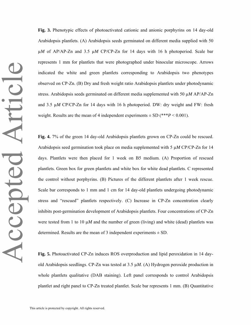
Acc
epte
d A
rticl
eFig. 3. Phenotypic effects of photoactivated cationic and anionic porphyrins on 14 day-old
Arabidopsis plantlets. (A) Arabidopsis seeds germinated on different media supplied with 50
µM of AP/AP-Zn and 3.5 µM CP/CP-Zn for 14 days with 16 h photoperiod. Scale bar
represents 1 mm for plantlets that were photographed under binocular microscope. Arrows
indicated the white and green plantlets corresponding to Arabidopsis two phenotypes
observed on CP-Zn. (B) Dry and fresh weight ratio Arabidopsis plantlets under photodynamic
stress. Arabidopsis seeds germinated on different media supplemented with 50 µM AP/AP-Zn
and 3.5 µM CP/CP-Zn for 14 days with 16 h photoperiod. DW: dry weight and FW: fresh
weight. Results are the mean of 4 independent experiments ± SD (***P < 0.001).
Fig. 4. 7% of the green 14 day-old Arabidopsis plantlets grown on CP-Zn could be rescued.
Arabidopsis seed germination took place on media supplemented with 5 µM CP/CP-Zn for 14
days. Plantlets were then placed for 1 week on B5 medium. (A) Proportion of rescued
plantlets. Green box for green plantlets and white box for white dead plantlets. C represented
the control without porphyrins. (B) Pictures of the different plantlets after 1 week rescue.
Scale bar corresponds to 1 mm and 1 cm for 14 day-old plantlets undergoing photodynamic
stress and “rescued” plantlets respectively. (C) Increase in CP-Zn concentration clearly
inhibits post-germination development of Arabidopsis plantlets. Four concentrations of CP-Zn
were tested from 1 to 10 µM and the number of green (living) and white (dead) plantlets was
determined. Results are the mean of 3 independent experiments ± SD.
Fig. 5. Photoactivated CP-Zn induces ROS overproduction and lipid peroxidation in 14 day-
old Arabidopsis seedlings. CP-Zn was tested at 3.5 µM. (A) Hydrogen peroxide production in
whole plantlets qualitative (DAB staining). Left panel corresponds to control Arabidopsis
plantlet and right panel to CP-Zn treated plantlet. Scale bar represents 1 mm. (B) Quantitative
This article is protected by copyright. All rights reserved.
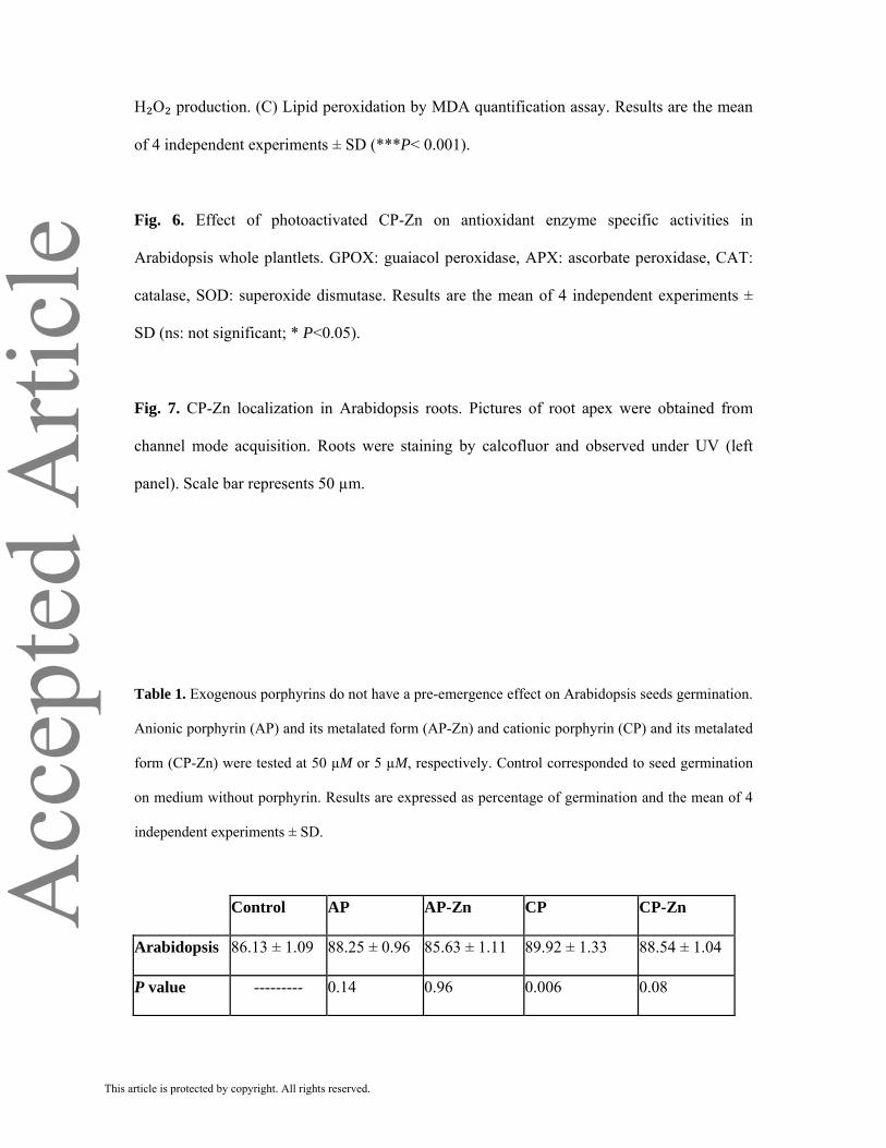
Acc
epte
d A
rticl
eH₂O₂ production. (C) Lipid peroxidation by MDA quantification assay. Results are the mean
of 4 independent experiments ± SD (***P< 0.001).
Fig. 6. Effect of photoactivated CP-Zn on antioxidant enzyme specific activities in
Arabidopsis whole plantlets. GPOX: guaiacol peroxidase, APX: ascorbate peroxidase, CAT:
catalase, SOD: superoxide dismutase. Results are the mean of 4 independent experiments ±
SD (ns: not significant; * P<0.05).
Fig. 7. CP-Zn localization in Arabidopsis roots. Pictures of root apex were obtained from
channel mode acquisition. Roots were staining by calcofluor and observed under UV (left
panel). Scale bar represents 50 µm.
Table 1. Exogenous porphyrins do not have a pre-emergence effect on Arabidopsis seeds germination.
Anionic porphyrin (AP) and its metalated form (AP-Zn) and cationic porphyrin (CP) and its metalated
form (CP-Zn) were tested at 50 µM or 5 µM, respectively. Control corresponded to seed germination
on medium without porphyrin. Results are expressed as percentage of germination and the mean of 4
independent experiments ± SD.
Control AP AP-Zn CP CP-Zn
Arabidopsis 86.13 ± 1.09 88.25 ± 0.96 85.63 ± 1.11 89.92 ± 1.33 88.54 ± 1.04
P value --------- 0.14 0.96 0.006 0.08
This article is protected by copyright. All rights reserved.

Acc
epte
d A
rticl
e
Table 2. Ratio of antioxidant molecule contents in Arabidopsis plantlets grown on medium containing
3.5 µM CP-Zn or medium without porphyrin (control) for 14 days under 16 h photoperiod. Results are
the mean of 4 independent experiments ± SD (ns: not significant, *** P <0.001)
Proline content
(µmol g⁻¹ FW)
Ascorbate content
(nmol g⁻¹ FW)
Total thiol content
(nmol mg⁻¹ FW)
Control CP-Zn 3.5 µM Control CP-Zn 3.5 µM Control CP-Zn 3.5 µM
0.72 ± 0.1 4.9 ± 1 185.2 ± 10 209 ± 27 58.67 ± 1.6 32.37 ± 2.6
*** ns ***
This article is protected by copyright. All rights reserved.
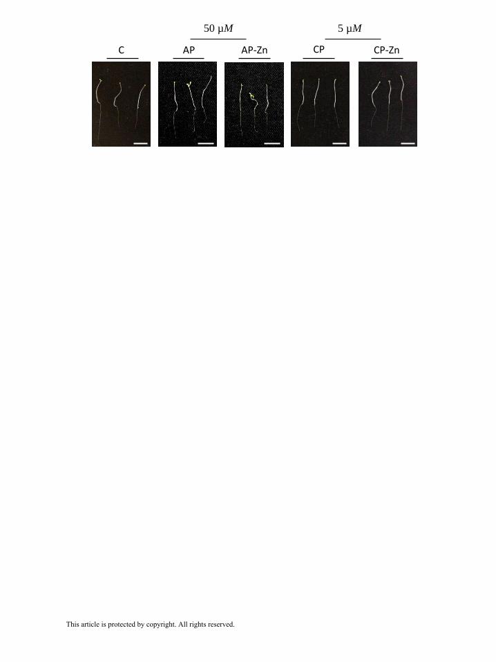
Acc
epte
d A
rticl
eC CP CP-ZnAP AP-Zn
50 µM 5 µM
This article is protected by copyright. All rights reserved.

Acc
epte
d A
rticl
e T0
T 72h
C AP AP-Zn CP CP-Zn
50 µM 3.5 µM
This article is protected by copyright. All rights reserved.

Acc
epte
d A
rticl
e
0
0,02
0,04
0,06
0,08
0,1
0,12
0,14
0,16
0,18
0,2
Arabidopsis
Rat
io D
W/F
W
C
AP 50 µM
AP-Zn 50 µM
CP 3.5 µM
CP-Zn 3.5 µM
***
***
***
***
C AP AP-Zn CP CP-ZnA
B
50 µM 3.5 µM
C
AP 50 µM
AP-Zn 50 µM
CP 3.5 µM
CP-Zn 3.5 µM
This article is protected by copyright. All rights reserved.
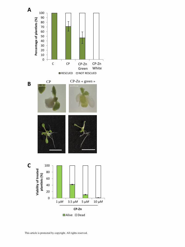
Acc
epte
d A
rticl
eA
B
C
0
10
20
30
40
50
60
70
80
90
100
C CP5 CPZ5G CPZ5W
Pe
rce
nta
ge o
f p
lan
tle
ts (
%)
C CP
RESCUED NOT RESCUED
CP CP-Zn « green »
0
20
40
60
80
100
CpZn 1.0µM
CPZn 3.5µM
CPZn 5.0µM
CPZn10.0 µM
Via
bili
ty o
f tr
eat
ed
p
lan
tle
ts (
%)
Alive Dead
CP-Zn Green
CP-Zn White
1 µM 3.5 µM 5 µM 10 µM
CP-Zn
This article is protected by copyright. All rights reserved.

Acc
epte
d A
rticl
eC CP-Zn 3.5 µM
***
A
B
C
11.2
0
5
10
15
20
25
30
1
MD
A c
on
ten
t (n
mo
l g⁻¹
FW
)
C CP-Zn 3.5 µM
0
50
100
150
200
250
300
350
1
H₂O
₂ co
nte
nt
(nm
ol g
⁻¹ p
rot)
C CP-Zn 3.5 µM
***
CP-Zn 3.5 µM
CP-Zn 3.5 µM
C CP-Zn 3.5µM
C CP-Zn 3.5µM
This article is protected by copyright. All rights reserved.

Acc
epte
d A
rticl
e 139.6
0
20
40
60
80
100
120
140
160
180
1
CA
T (µ
mo
lH₂O
₂ m
in⁻¹
mg⁻
¹ pro
t)
108.7
0
50
100
150
200
250
300
1
SOD
(U
mg⁻
¹ pro
t)
12.2
0
2
4
6
8
10
12
14
16
18
20
1
GP
OX
(µ
mo
lgu
aiac
ol m
in⁻¹
mg⁻
¹ p
rot)
0.25
0
0,1
0,2
0,3
0,4
0,5
0,6
0,7
0,8
0,9
1
1
AP
X (
µm
olH
₂O₂
min
⁻¹ m
g⁻¹ p
rot)
ns
CCP-Zn 3.5µMns
*
*
This article is protected by copyright. All rights reserved.

Acc
epte
d A
rticl
eC
CP-Zn
3.5 µM
Calcofluor Auto-fluorescence
This article is protected by copyright. All rights reserved.

104
Results
Mohammad ISSAWI | Thèse de doctorat | Université de Limoges | 2018
Licence CC BY-NC-ND 3.0
Discussion and perspectives
We showed that negatively charged porphyrins tested at 50 µM had no effects on both
plantlets. Moreover, positively charged porphyrins (free base TMPyP or Zinc-metalated) were
deleterious for Arabidopsis demonstrating an herbicide potential effect but they only delayed
tomato plantlet growth. According to literature, this result was surprising for tomato plantlets
that were also rescued after 14 days treatment. Indeed, cationic porphyrins are known as
powerful PS against various types of microorganisms including plant pathogens (Orlob 1967,
Fayyaz et al. 2015, Jesus et al. 2018). Taken together, we could imagine a double weapon
modality aiming to struggle against plant pathogens and unwanted weeds (Figure 10). We only
worked on plant side but the research team of Pr Gilberto Braga in Brazil showed the ability to
apply this strategy using a PS, more specifically at the level of plant pathogens (De Menezes et
al. 2014 a,b, Fracarolli et al. 2016, De Menezes et al. 2016, Gonzales et al. 2017). We are now
enlarging our study testing PS treatments on perennial plant: Vitis vinifera var Chardonnay
clones and a new annual species: potato explants (Veronica Ambrosini PhD work).
Figure 10. Schematic representation of the “two-in-one” strategy based on the use of PS in agronomy.
One of the most important perspective that should be taken in consideration to validate
APDT in field areas is the requirement of whole study of a defined pathosystem including

105
Results
Mohammad ISSAWI | Thèse de doctorat | Université de Limoges | 2018
Licence CC BY-NC-ND 3.0
biological life cycle of pathogen, phenology of growth and reproduction of plants and timing of
PS application. For example, Gonzales et al. 2017 reported that to fight against Colletotrichum
fungal species that cause orange crop damage in the Americas via photodynamc approach,
methylene blue PS must be applied in the wet season because rain splash disperses conidia and
citrus flowering coincides with rain events in tropical region. Other study conducted by Jesus et
al, 2018 showed that in temperate region, porphyrin like TMPyP should be applied in the period
when bacteria (Pseudomonas syringae) infect their host as kiwi plants in winter and early spring
and especially after winter pruning and bud outbreak that facilitates infection process. In
accordance with the last point, our laboratory is on the road to develop consistent pathosystem
involving the mold (Botrytis cinerea) and its two plant hosts Solanum tuberosum (potato) and
Vitis vinifera (grapevine) then trying to apply porphyrin molecular systems in agreement with
APDT (Veronica Ambrosini PhD study).
This study also pointed out the potential use of photodynamic treatment to eradicate
weeds by means of herbicide. Indeed, this could explain why developing such approach in
agriculture was not envisaged due to its large spectrum targets including plants and more broadly
wildlife (Figure 9). Nevertheless, based on our case-by-case study, we demonstrated that plant of
agronomic interest can tolerate photodynamic stress and anionic porphyrins had no effects when
tested on both plants. Moreover, orange and kiwi tree leaves and flowers were not affected by PS
treatment. In addition, the demand for new herbicide is being to increase by the agricultural
chemical industry because there are no new herbicide modes of action and up to 400 weeds
evolved resistance in the last 30 years. Currently, the World Health Organization classified some
Diphenyl ether (for more details see DPE in the review (Publication 1 submitted) as "substances
of very high concern". Thus, some commercial herbicides will be soon banned, and agriculture is
still missing some new herbicide molecules. Therefore, we think about the possibility to employ
PS especially porphyrins as herbicide due to their multi-target sites at cellular level, their
efficient application at very low (µM) concentration and micro-organisms subjected to PS
treatment did not develop resistance yet.
We are aware that our studies represent a first preliminary step to examine the usefulness
of plant photodynamic treatment at large scale level in the near future. Our studies constitute the
cornerstone for sustainable agriculture and crop protection basing on the power of light-driven
PS activation against pathogens and weeds.

106
Results
Mohammad ISSAWI | Thèse de doctorat | Université de Limoges | 2018
Licence CC BY-NC-ND 3.0
Chapter II. TBY-2 cells: a helpful
tool to understand the cellular
responses to photoactivated
anionic porphyrin treatment from
A to Z
107
Results
Mohammad ISSAWI | Thèse de doctorat | Université de Limoges | 2018
Licence CC BY-NC-ND 3.0
In order to study the cellular and molecular mechanisms underlying plant responses to
photodynamic treatment by exogenously porphyrin supply, we looked out for plant cell
suspension cultures that are intensively used in plant biology as reliable tool for the investigation
of wide range of phenomena bypassing therefore whole plant structural complexity. We chose
TBY-2 cell suspension showing high growth rate with good synchrony as well as easy and
inexpensive culture.
In a previous study published in 2014, we showed that anionic porphyrin (TPPS) was
more efficient than cationic ones (TMPyP) to induce high production of hydrogen peroxide
leading to cell death. This result was in opposition to what the literature reported. Indeed,
cationic porphyrins were more powerful than anionic forms to photodamage almost each living
entity (Tegos et al. 2005, Guillaumot et al. 2016).
To gain insight in molecular mechanisms that occurred at cellular level in response to
photoactivated three anionic porphyrins (TPPS, TPPC and TPPP), we tried to cover all aspects
from extracellular porphyrin behaviour, cell wall interaction and penetration, photodynamic
damage, cell responses to cell death. As already mentioned in introduction, a preliminary but
necessary step for this exhaustive study was to get knowledge about TBY-2 cell wall
(publication 4).
PUBLICATION 4: “Unexpected features of exponentially growing Tobacco Bright
Yellow-2 cell suspension culture in relation to excreted extracellular polysaccharides
and cell wall composition”, Glycoconjugate Journal.

Results
Mohammad ISSAWI | Thèse de doctorat | Université de Limoges | 2018
Licence CC BY-NC-ND 3.0
108
Dear Dr. Riou:
We are pleased to inform you that your manuscript, "Unexpected features of
exponentially growing Tobacco Bright Yellow-2 cell suspension culture in relation
to excreted extracellular polysaccharides and cell wall composition" has been
accepted for publication in Glycoconjugate Journal.
For queries regarding your accepted paper, please click the following link
http://www.springer.com/10719; then click on "Contacts", and then "Production
Editor", complete the query form and click "Submit".
Please remember to always include your manuscript number, #GLYC-D-17-00015R3,
whenever inquiring about your manuscript. Thank you.
Best regards,
The Editorial Office
Glycoconjugate Journal
Results
Mohammad ISSAWI | Thèse de doctorat | Université de Limoges | 2018
Licence CC BY-NC-ND 3.0
109
Unexpected features of exponentially growing Tobacco Bright Yellow-2 cell
suspension culture in relation to excreted extracellular polysaccharides and
cell wall composition
Mohammad Issawi, Mohammad Muhieddine, Celine Girard, Vincent Sol and Catherine Riou*
Université de Limoges, Laboratoire de Chimie des Substances Naturelles, EA 1069, 123 avenue Albert Thomas,
87060 Limoges, France.
*Corresponding author: [email protected]; Tel: +33 555457474; Fax: +33 555457202

Results
Mohammad ISSAWI | Thèse de doctorat | Université de Limoges | 2018
Licence CC BY-NC-ND 3.0
110
ABSTRACT
This article presents a new insight about TBY-2 cells; from extracellular polysaccharides secretion to cell wall
composition during cell suspension culture. In the medium of cells taken 2 days after dilution (end of lag phase),
a two unit pH decrease from 5.38 to 3.45 was observed and linked to a high uronic acid (UA) amount secretion
(47.8 %) while, in 4 and 7 day-old spent media, pH increased and UA amounts decreased 35.6 and 42.3 % UA,
respectively. To attain deeper knowledge of the putative link between extracellular polysaccharide excretion and
cell wall composition, we determined cell wall UA and neutral sugar composition of cells from D2 to D12
cultures. While cell walls from D2 and D3 cells contained a large amount of uronic acid (twice as more as the
other analysed cell walls), similar amounts of neutral sugar were detected in cells from lag to end of exponential
phase cells suggesting an enriched pectin network in young cultures. Indeed, monosaccharide composition
analysis lead to an estimated percentage of pectins of 56% for D3 cell wall against 45% D7 cell walls indicating
that the cells at the mid-exponential growth phase re-organized their cell wall linked to a decrease in secreted
UA that finally led to a stabilization of the spent medium pH to 5.4. In conclusion, TBY-2 cell suspension from
lag to stationary phase showed cell wall remodeling that could be of interest in drug interaction and
internalization study.
.
Keywords:
cell wall composition; extracellular polysaccharides; Tobacco BY-2 suspension culture; uronic acid.
Abbreviations:
CW: cell wall; TBY-2: tobacco bright yellow-2 cells; UA: uronic acid.
Results
Mohammad ISSAWI | Thèse de doctorat | Université de Limoges | 2018
Licence CC BY-NC-ND 3.0
111
INTRODUCTION
Amongst broad and numerous literature on plant cell suspensions, Tobacco Bright Yellow cell line
(TBY-2) suspension remains largely well documented because of its extensive use in laboratories working from
cell cycle regulation to programmed cell death or responses to biotic or abiotic stress [1-7]. TBY-2 cells mainly
form chains of 2 to 8 cells, ovoid-like micro-colonies that are easy to observe, label and count. While they are
usually cultivated in the dark in liquid medium, they could also grow on solidified medium under continuous
light or dark. Moreover, the culture of these cells does not require sophisticated material and is feasible for a
great variety of experiments from protoplast to transient DNA expression [8].
Moreover, TBY-2 cells have been also used as a model system for cell wall studies including the
purification of enzymes involved in cell wall formation or regeneration from protoplasts of transformed TBY-2
cell lines [9-12]. However, TBY-2 cell wall composition per se has not been readily investigated. Until now, two
studies were performed on BY-2 rhamnogalacturonan-II, a component of the pectin network, in response to
boron feeding and only one on whole secondary cell wall characterization [13-15]. Indeed, knowledge about
TBY-2 cell wall (CW) will help to understand potential interactions between exogenous molecules (herbicides,
elicitors, growth factors) and primary CW, their entry into cell and/or their accumulation into this compartment.
This is of interest to model plant cell interaction with exogenous molecules such as in some abiotic stresses
(drug, herbicide treatments). Furthermore, growing cells modify initial cell culture medium in term of chemical
composition [16]. Both molecules from primary and secondary metabolisms (amino and organic acids,
alkaloids), cell wall polysaccharides and enzymes and/or proteins were found in spent medium of suspension-
cultured cells of Lupinus polyphyllus, Pyrus communis and Arabidopsis thaliana making cell medium similar to
the vacuole for detoxification but also as a sink-source [16-18]. Particularly, the release of extracellular
polysaccharides which are thought to be non-cellulosic cell wall matrix from plant cells in suspension culture
into the medium has been intensively reported by several works [19-23]. As a consequence of sucrose uptake,
ammonium assimilation and organic acid release, pH of medium was more and more acidic especially in the first
days of culture [16, 24]. Since we also found a decrease in pH of culture medium during TBY-2 cell growth, just
before the exponential phase starts, we aim to investigate polysaccharide secretion in spent medium of growing
cells and try to link it to cell wall composition.
Results
Mohammad ISSAWI | Thèse de doctorat | Université de Limoges | 2018
Licence CC BY-NC-ND 3.0
112
MATERIAL and METHODS
TBY-2 cell suspension
Tobacco Bright Yellow-2 cell suspension was maintained in the dark under continuous agitation (140 rpm) at 22
°C in modified Murashige and Skoog medium, supplemented with 0.27 mg.L-1 2,4-dichlorophenoxyacetic acid;
10 mg.mL-1 thiamine-HCl; 100 mg.L-1 myo-inositol; 30 g.L-1 sucrose and 200 mg.L-1 KH2PO4, pH 5.8. The
suspension was subcultured every 7 days, by transferring 1.5 to 2 mL aliquot into 40 mL of fresh medium.
Establishment of growth curve was performed by weighing the cell pellets obtained from 40 mL of suspension
(4,500 g, 10 min); supernatants were either discarded or kept for analyses of excreted polysaccharides. Cells
isolated at the end of lag phase (2 days after dilution), at the beginning (3 or 4 days after dilution) or the mid-
exponential phase (7 or 8 days after dilution), and, at the stationary phase (9, 10 and 14 days after dilution) were
used for cell wall isolation and analysis.
Cell wall extraction
Cell wall was extracted according to [15] with minor modifications. TBY-2 cells were ground in liquid nitrogen
then homogenized in 0.5 M potassium phosphate buffer (pH 7.0) by sonication (50 pulses/min for 1 min, 4
times). Homogenates were centrifuged (4,500 g, 30 min at 4 ˚C) and pellets were washed twice with the same
buffer and subsequently 3 times with cold distilled water. To remove lipids, cell walls were extracted 3 times
with chloroform/methanol (1:1, v/v). Proteins were eliminated by three extractions with phenol/ acetic acid/water
(2:1:1, v/v/v). After centrifugation (900 g, 10 min), cell walls were rinsed with sufficient ethanol and water to
remove the solvent. Starch hydrolysis was performed by a treatment with type XII-A Bacillus α-amylase (50
U/ml) from Sigma-Aldrich (Mo, USA) for D2, D3 and D4 cell walls (1 h, 37°C). D7, D8, D9 and D12 cell walls
were incubated with XII-A Bacillus α-amylase (100 U/ml) overnight at room temperature. After starch
hydrolysis, cell wall fractions were centrifuged (4,500 g, 30 min at 4 ˚C) then freeze-dried for 24 hours.
Sugar content determination
Whole cell walls were solubilized in 10 ml distilled water. Neutral sugars and uronic acids were determined
according to [25] and [26] respectively with glucose and glucuronic acid as standards. Absorbance values were
corrected according to [27].
Monosaccharide composition
Monosaccharide composition was evaluated by gas liquid chromatography using methanolysis followed by
trimethylsilylation according to [28] modified by [29].
Main polysaccharides estimation
We estimated the amount of cell wall major polysaccharides from the monosaccharide composition according to
[30]. Pectin percentage was evaluated as following % pectin = GalA + Rha + Gal + Ara. Cellulose percentage
was estimated arbitrarily by 50% Glc (cellulose = ½ Glc), the other half was attributed to hemicellulose that
comprises in particular xyloglucans, xylans and mannans which can therefore be estimated by the sum ½ Glc +
Xyl + GlcA + Man. Monosaccharides are expressed in molar percentage as determined in [29].
Extracellular polysaccharide quantification
According to [31], TBY-2 culture medium was concentrated by rotary evaporation then subjected to
precipitation by ethanol 95% (v/v), 3 vol. at 4 ºC overnight. After centrifugation (3,500g, 30 min), pellets were

Results
Mohammad ISSAWI | Thèse de doctorat | Université de Limoges | 2018
Licence CC BY-NC-ND 3.0
113
dialyzed (MWCO: 12,000-14,000 Da) against distilled water and represented the extracellular polymer fraction.
Sugar content was evaluated according to [25] and [26] as described above.
Statistical analysis
All biological experiments were performed at least three times independently. Results were expressed as mean ±
SD (Standard Deviation). Data were analyzed by t-student test and one-way ANOVA using the PAST free
software.
RESULTS and DISCUSSION
To gain insight into TBY-2 cells growth curve, we monitored, daily, the fresh weight of the whole
culture (40 mL culture) until day 14 after dilution (Fig. 1A). Surprisingly TBY-2 cells taken 7 days after dilution
were not in stationary phase as mainly described in the literature but rather in mid-exponential phase (Fig. 1A ;
[2,7]). Nine days after dilution, the cells were still in “good health” with a high viability percentage (> 92 % data
not shown) but they progressively became elongated as shown in Fig1B for D14 cells and, while they increased
in volume, granules appeared around the nucleus (Fig. 1A). We supposed that, for these reasons, and because 9
day-old cultures also became very viscus and difficult to dilute and to work with, most laboratories choose to
work with D7 cells or even with very young cell suspensions (D3 and/or D4), as we previously did [32]. In fact,
D3 or D4 cells were in very early exponential growth phase considering that D2 cells were at the end of the lag
phase (Fig 1A). We placed the switch between lag (D2) and exponential (D3) phases not in regard to cell curve
or cell size (no significant difference in fresh weights with P=0.9081; Fig 1A) but to pH variations: a two pH-
unit decrease during the lag phase followed, after D2, by a slow return to the initial pH of the medium around 5.4
(Fig. 1B) as it had already been shown with lupinus cell suspension [16]. We noticed that this pH was reached
for D7 cells and stayed constant after that point (Fig.1A).
To better understand this pH decrease observed 2 days after dilution, we measured the amount of
extracellular polysaccharides excreted by the cells in the dried spent medium, especially uronic acids (UA), at
D2, D4 and D7. Spent medium is a complex combination of culture medium basal components and sugars and/or
proteins excreted by cells themselves. As shown in Table 1, dry weight of the extracellular fraction varied from
nearly 1 mg for D2 and D4 supernatants to more than 4 mg for D7 suggesting very high molecule secretions of
cells in mid-exponential phase. Nevertheless, determination of UA and neutral contents in D7 dry spent medium
did not reflect an important polysaccharide secretion suggesting that D7 cells were rather able to secrete protein
and/or lipid into the medium (Table 1). For D2 and D4 spent media, while UA secretion was significantly higher
(0.285 and 0.258 g.g-1 DW extracellular fraction respectively) than in D7 medium (0.196 g.g-1 DW extracellular
fraction), neutral sugar production for D4 cells was notably upregulated (Table 1). In TBY-2 cells, it was shown
that cell invertase released fructose and glucose from sucrose breakdown into the culture medium that could also
be the case for D4 cells [33]. Thus, it should be interesting to follow sucrose consummation during cell growth
and establish extracellular fraction composition especially for D4 cells.
Our aim was firstly to understand the pH decrease in D2 spent medium (Fig. 1A). Although UA contents relative
to DW extracellular fraction were similar in D2 and D4 media, percentage of UA in D2 spent medium (47.8%)
was clearly higher to D4 one (35.6%) (Table 1). Thus, we supposed that the pH decrease was mainly due to
excreted UA. Nevertheless, it could also be triggered by organic acid production such as malic and oxalic acids
which is strongly linked to high oxidative metabolism that could strongly occur in D2 [16]. Indeed, D2 cells are

Results
Mohammad ISSAWI | Thèse de doctorat | Université de Limoges | 2018
Licence CC BY-NC-ND 3.0
114
metabolically active because they doubled their fresh weight from 1.7 g to 3 g in D3. Thus, organic acid
secretion could be envisaged at the end of lag phase linked to respiratory metabolism and energy production. It
was also shown that sucrose uptake and ammonium assimilation led to acidification of culture media of Lupinus
polyphyllus and Ocotea catharinensis suspensions suggesting a similar situation for TBY-2 cells [16,24].
Moreover, as rhamnogalacturonans II could also be excreted by TBY-2 cells, this could be similar in D2
supernatant leading to potential acidification [13,14]. Further studies have to be made in order to confirm these
hypotheses.
As TBY-2 cells from the end of lag phase to mid exponential phase were able to excrete
polysaccharides especially UA, it was of interest to characterize their cell wall in term of cellulose,
hemicellulose and pectin networks as well as their sugar composition. We determined the cell wall composition
of cells from D2 to D12 (Fig. 2). Surprisingly, concerning UA content, D2 and D3 cell walls were very similar
and contained nearly 3 times less UA than D4 to D12 cell walls (Fig. 2A). On the contrary, neutral sugar
contents from lag phase (D2) to end of exponential phase (D8) cell walls were very similar around 270 µg.mg⁻¹
dried CW (Fig. 2B). In fact, D2 and D3 cell walls were enriched in UA-bearing polysaccharides such as pectins,
compared to D7 and later cell walls (Fig.2). That was confirmed by monosaccharide analysis and percentage
estimation of the three main cell wall polysaccharides from D3, D4 and D7 cell walls (Table 2). D3 and D4 were
very similar in regard to pectin content although D3 had significantly more negatively charged sugars especially
galacturonic acid than D4 (Table 2). On the opposite, D4 contained more neutral sugars as rhamnose and
arabinose that are also parts of pectin network (Table 2). Taken together, D3 and D4 cell wall polysaccharide
compositions were similar and different to D7 cell wall which is enriched in cellulose network, twice as much as
D3 and D4 cell walls. Moreover, measurement of zeta potential showed that it was similar between D3 and D4
cell wall (around -30 mV, data not shown) while it was less negative for D7 cell wall (-20 mV) that was in good
agreement with its rich neutral sugar content. For stationary phase D9 and D12 cell walls, neutral sugar content
significantly increased (Fig2B). This could be linked to a thickening of cell wall with an extra-cellulose
deposition that could be investigated by electronic microscopy.
In conclusion, TBY-2 cell suspension from D2 after dilution to stationary phase showed cell wall
remodeling. For D7 cell wall, we showed that cells at mid-exponential phase that are usually used in the
laboratories around the world, were still able to secrete UA in their spent medium. As reported by [34], UA-rich
pectic polymers which were excreted by white campion (Silene latifolia) suspension culture were able to
promote cell growth when added into new culture media. Thus, we suppose that a synergistic effect between
cells after the mid-log growth phase induced by UA-rich polysaccharides excreted through a specific secretory
pathway that could be linked to an unconventional secretory pathway [35]; [36]. This remains unclear and
warrants to be further investigated.
Acknowledgements
Mohammad Issawi was supported by a Grant from the municipality of Sharkieh (Lebanon). Authors greatly
thank Pr M. Guilloton for reading this manuscript.

Results
Mohammad ISSAWI | Thèse de doctorat | Université de Limoges | 2018
Licence CC BY-NC-ND 3.0
115
References
1. Kato, K., Matsumoto, T., Koiwai, S., Mizusaki, S., Nishida, K., Nogushi, M., Tamaki, E.: Liquid
suspension culture of tobaco cells. in Fermentation Technology Today. ed Terui G (Society of
Fermentation Technology, Osaka) 689–695 (1972).
2. Nagata, T., Nemoto, Y., Hasezawa, S.: Tobacco BY-2 cell line as the “Hela” cell in the cell biology of
higher plants. Internal. Rev. Cytol. 132, 1–30 (1992).
3. Kobayashi, I., Hakuno, H.: Actin-related defense mechanism to reject penetration attempt by a non-
pathogen is maintained in tobacco BY-2 cells. Planta 217, 340–345 (2003).
4. de Pinto, M.C., Locato, V., Sgobba, A., Romero-Puertas, M. d. C., Gadaleta, C., Delledonne, M., De Gara,
L.: S-Nitrosylation of Ascorbate Peroxidase Is Part of Programmed Cell Death Signaling in Tobacco
Bright Yellow-2 Cells. Plant Physiol. 163, 1766–1775 (2013).
5. Takahashi, S., Kojo, K.H., Kutsuna, N., Endo, M., Toki, S., Isoda, H., Hasezawa, S.: Differential
responses to high- and low-dose ultraviolet-B stress in tobacco Bright Yellow-2 cells. Front. Plant Sci. 6,
254-264 (2015).
6. Moscatiello, R., Baldan, B., Navazio, L.: Plant Cell Suspension Cultures. In: Maathuis, F.J.M. (ed.) Plant
Mineral Nutrients. pp. 77–93. Humana Press, Totowa, NJ (2013).
7. Srba, M., Černíková, A., Opatrný, Z., Fischer, L.: Practical guidelines for the characterization of tobacco
BY-2 cell lines. Biol. Plant. 60, 13–24 (2016).
8. Bratic, A., Majic, D., Samardzic, J., Kragl, M.W., Maksimovic, V.: Transient expression in tobacco Bright
Yellow 2 cells and pollen grains: A fast, efficient and reliable system for functional promoter analysis of
plant genes. Arch. Biol. Sci. 62, 57–62 (2010).
9. De Marco, A., Guzzardi, P., Jamet, É.: Isolation of tobacco isoperoxidases accumulated in cell-suspension
culture medium and characterization of activities related to cell wall metabolism. Plant Physiol. 120, 371–
382 (1999).
10. Nebenführ, A., Frohlick, J.A., Staehelin, L.A.: Redistribution of Golgi stacks and other organelles during
mitosis and cytokinesis in plant cells. Plant Physiol. 124, 135–152 (2000).
11. Hong, Z., Delauney, A.J., Verma, D.P.S.: A cell plate–specific callose synthase and its interaction with
phragmoplastin. Plant Cell 13, 755–768 (2001).
12. Yokoyama, R., Tanaka, D., Fujino, T., Itoh, T., Nishitani, K.: Cell wall dynamics in tobacco BY-2 cells.
In: Tobacco BY-2 Cells. 53, 217–230 (2004).
13. Kobayashi, M., Ohno, K., Matoh, T.: Boron nutrition of cultured tobacco BY-2 cells. II. Characterization
of the boron-polysaccharide complex. Plant Cell Physiol.. 38, 676–683 (1997).
14. Matoh, T., Takasaki, M., Kobayashi, M., Takabe, K.: Boron nutrition of cultured tobacco BY-2 cells. III.
Characterization of the boron-rhamnogalacturonan II complex in cells acclimated to low levels of boron.
Plant Cell Physiol. 41, 363–366 (2000).
15. Goué, N., Mortimer, J.C., Nakano, Y., Zhang, Z., Josserand, M., Ohtani, M., Dupree, P., Kakegawa, K.,
Demura, T.: Secondary cell wall characterization in a BY-2 inductive system. PCTOC 115, 223–232
(2013).
16. Wink, M.: The cell culture medium—a functional extracellular compartment of suspension-cultured cells.
In: Primary and Secondary Metabolism of Plants and Cell Cultures III. pp. 307–319. Springer (1994).

Results
Mohammad ISSAWI | Thèse de doctorat | Université de Limoges | 2018
Licence CC BY-NC-ND 3.0
116
17. Jamet, E., Canut, H., Boudart, G., Pont-Lezica, R.F.: Cell wall proteins: a new insight through proteomics.
Trends Plant Sci. 11, 33–39 (2006).
18. Webster, J.M., Oxley, D., Pettolino, F.A., Bacic, A.: Characterisation of secreted polysaccharides and
glycoproteins from suspension cultures of Pyrus communis. Phytochem. 69, 873–881 (2008).
19. Becker, G.E., Hui, P.A., Albersheim, P.: Synthesis of extracellular polysaccharide by suspensions of Acer
pseudoplatanus cells. Plant Physiol. 39, 913 (1964).
20. Olson, A.C., Evans, J.J., Frederick, D.P., Jansen, E.F.: Plant suspension culture media macromolecules—
pectic substances, protein, and peroxidase. Plant Physiol. 44, 1594–1600 (1969).
21. Liau, D.-F., Boll, W.G.: Extracellular polysaccharide from cell suspension cultures of bush bean
(Phaseolus vulgaris cv. Contender). Can. J. Bot. 50, 2031–2037 (1972).
22. Moore, T.S.: An extracellular macromolecular complex from the surface of soybean suspension cultures.
Plant Physiol. 51, 529–536 (1973).
23. Oliveira C.J.F., Cavalari, A.A., Carpita, N.C., Buckeridge, M.S., Braga, M.R.: Cell wall polysaccharides
from cell suspension cultures of the Atlantic Forest tree Rudgea jasminoides (Rubiaceae). Trees. 24, 713–
722 (2010).
24. Moser, J.R, Gonçalves Garcia, M., Maria Viana, A.: Establishment and growth of embryogenic suspension
cultures of Ocotea catharinensis Mez.(Lauraceae). PCTOC. 78, 37–42 (2004).
25. Dubois, M., Gilles, K.A., Hamilton,, J.K., Rebers P.A, Smith, F. : Colorimetric method for determination
of sugars and related substances. Analytical Chem. 28, 350–356 (1956).
26. Blumenkrantz, N., Asboe-Hansen, G.: New method for quantitative determination of uronic acids.
Analytical Biochem. 54, 484–489 (1973).
27. Montreuil, J., Bouquelet, S., Debray, H., Fournet, B., Spik, G., Strecker, G.: In: Chaplin MF, Kennedy JF.
(Eds), Carbohydrate analysis, a practical approach, IRL Press, Oxford, Washington D.C 143-204. (1986).
28. Kamerling, J.P., Gerwig, G.J., Vliegenthart, J.F.G., Clamp, J.R.: Characterization by gas-liquid
chromatography-mass spectrometry and proton-magnetic-resonance spectroscopy of pertrimethylsilyl
methyl glycosides obtained in the methanolysis of glycoproteins and glycopeptides. Biochem. J. 151, 491–
495 (1975).
29. Marga, F., Freyssac, V., Morvan, H.: Rapid gas liquid chromatography microanalysis of carbohydrates in
woody plant tissues. J. Trace Microprobe Techn. 13, 473–478 (1995).
30. Douchiche, O., Driouich, A., Morvan, C.: Impact of cadmium on early stages of flax fibre differentiation:
Ultrastructural aspects and pectic features of cell walls. Plant Physiol. Biochem. 49, 592–599 (2011).
31. Muschitz, A., Faugeron, C., Morvan, H.: Response of cultured tomato cells subjected to excess zinc: role
of cell wall in zinc compartmentation. Acta Physiol. Plant. 31, 1197–1204 (2009).
32. Riou, C., Calliste, C.A., Da Silva, A., Guillaumot, D., Rezazgui, O., Sol, V., Leroy-Lhez, S.: Anionic
porphyrin as a new powerful cell death inducer of Tobacco Bright Yellow-2 cells. Photochem. Photobiol.
Sci. 13, 621-625 (2014).
33. Yamanaka, A., Hashimoto, A., Matsuo, T., Kanou, M., Suehara, K.-I., Kameoka, T.: Analysis of kinetic
uptake phenomena of monosaccharide and disaccharide by suspension TBY-2 cells using an FT-IR/ATR
method. Bioproc Biosystems Eng. 30, 457–468 (2007).
34. Morvan, H., Priem, B., Verdus, M.C., Kwan, J.S., Morvan, C.: Relationship between the acidity of the

Results
Mohammad ISSAWI | Thèse de doctorat | Université de Limoges | 2018
Licence CC BY-NC-ND 3.0
117
pectic acid polymers from suspension cultured cells of white campion and their promoting effects on the
growth. Food Hydrocolloids. 1, 501–503 (1987).
35. Ding, Y., Robinson, D.G., Jiang, L.: Unconventional protein secretion (UPS) pathways in plants. Curr. Op.
Cell Biol. 29, 107–115 (2014).
36. Kim, S.-J., Brandizzi, F.: The plant secretory pathway for the trafficking of cell wall polysaccharides and
glycoproteins. Glycobiol. 26, 940–949 (2016).

Results
Mohammad ISSAWI | Thèse de doctorat | Université de Limoges | 2018
Licence CC BY-NC-ND 3.0
118
Table 1: Estimations of UA and neutral sugars content in extracellular fractions of 3 culture points: 2 days (D2),
4 days (D4) and 7 days (D7) after dilutions. D2: end of lag phase, D4: rapid exponential growth phase and D7:
mid-exponential phase. Uronic acid (UA) and neutral sugar (NS) contents were determined as described in
material and methods. Each result is the mean of three independent experiments ±SD.
Dry weight of the
extracellular
fraction (mg)
Percentage of UA
in the extracellular
fraction (%)
UA content (g.g¯¹
DW extracellular
fraction)
NS content (g.g-1
DW extracellular
fraction)
D2 0.76 ± 0.06 47.8 ± 1.6 0.285 ± 0.033 0.312 ± 0.035
D4 0.95 ± 0.04 35.6 ± 1.3 0.258 ± 0.035 0.468 ± 0.04
D7 4.3 ± 0.2 42.3 ± 1.3 0.196 ± 0.013 0.268 ± 0.049

Results
Mohammad ISSAWI | Thèse de doctorat | Université de Limoges | 2018
Licence CC BY-NC-ND 3.0
119
Table 2: Estimations of monosaccharide compositions and deduced percentage of cellulose, hemicellulose and
pectin fraction in cell wall from D3, D4 and D7 cell suspensions. Molar percentages and monosaccharide content
were determined and calculated as described in material and methods section and represented the mean of three
independent experiments ±SD. CW: cell wall; nd: not detected.
D3 CW D4 CW D7 CW
Monosaccharides
(%)
Rhamnose 9.5 ± 0.3 13 ± 1.3 15.47 ± 0.3
Arabinose 5.1 ± 0.4 8 ± 0.2 21 ± 0.7
Fucose 2.1 ± 0.1 3.2 ± 0.5 3.57 ± 0.04
Xylose 7.4 ± 0.4 8.75 ± 0.7 3.09 ± 0.07
Mannose 8.65 ± 0.15 10.3 ± 1.1 5.42 ± 0.2
Galactose 7.35 ± 0.9 7.26 ± 1 2 ± 0.02
Galacturonic acid 32.65 ± 0.2 24.1 ± 1.2 5.77 ± 0.3
Glucose 20.95 ± 1.1 19.20 ± 0.8 43 ± 0.7
Glucuronic acid 5.7 ± 0.1 4.25 ± 0.6 nd
Polysaccharides
(%)
Cellulose 10.7 10.1 21.5
Hemicellulose 33.1 34.6 31
Pectins 56.1 52.2 45

Results
Mohammad ISSAWI | Thèse de doctorat | Université de Limoges | 2018
Licence CC BY-NC-ND 3.0
120
Figure 1: TBY-2 cell suspension characterization. A) TBY-2 suspension growth curve (circles) and
pH variations of spent medium during growth (triangles). Fresh weights of independent cultures were determined
in function of culture time after dilution. D: day after dilution. Each point is the mean of results from 5
independent cultures ± SD. pH of the medium after autoclaving without cells used for cell dilution was measured
(5.38 ± 0.14). Each pH value is the mean of four independent cultures ± SD. B) Pictures of TBY-2 cells 4 days
after dilution (D4) or early exponential phase; 7 days after dilution (D7) or mid-exponential phase and finally 14
days after dilution (D14) or stationary phase.

Results
Mohammad ISSAWI | Thèse de doctorat | Université de Limoges | 2018
Licence CC BY-NC-ND 3.0
121
Figure 2: Sugar content determination of TBY-2 cell wall during growth. Isolated and
purified cell walls (CW) were analyzed at the end of lag phase (2 days after dilution), at the beginning (3 or 4
days after dilution) or the mid-exponential phase (7 or 8 days after dilution), and, at the stationary phase (9 or 12
days after dilution). Determinations of UA A) and neutral sugar B) contents in different CW. Results are the
mean ± SD of three independent experiments (NS: not significant; *: P<0.05 ; **: P<0.01 ; *** : P<0.001).

Results
Mohammad ISSAWI | Thèse de doctorat | Université de Limoges | 2018
Licence CC BY-NC-ND 3.0
122
PUBLICATION 5: “Characterization of pH dependent charge states and physico-
chemical properties of anionic porphyrins” (Submitted to Photochemical &
Photobiological Sciences).

Characterization of pH dependent charge states and
physico-chemical properties of anionic porphyrins
Journal: Photochemical & Photobiological Sciences
Manuscript ID PP-ART-04-2018-000152
Article Type: Paper
Date Submitted by the Author: 10-Apr-2018
Complete List of Authors: Leroy-Lhez, Stéphanie; PEIRENE EA7500, Faculté des Sciences et Techniques - Université de Limoges Rezazgui, Olivier; Laboratoire PEREINE Issawi, Mohammad; PEIRENE - EA 7500, Faculté des Sciences et techniques - université de Limoges Elhabiri, Mourad; CNRS, ECPM, UMR 7509 Calliste, Claude; PEIRENE EA 7500, Faculté de Pharmacie - Université de Limoges Riou, Catherine; PEIRENE EA7500, aculté des Sciences et Techniques - Université de Limoges
Photochemical & Photobiological Sciences
Journal Name
ARTICLE
This journal is © The Royal Society of Chemistry 20xx J. Name., 2013, 00, 1-3 | 1
Please do not adjust margins
Please do not adjust margins
Received 00th January 20xx,
Accepted 00th January 20xx
DOI: 10.1039/x0xx00000x
www.rsc.org/
Characterization of pH dependent charge states and physico-chemical properties of anionic porphyrins
Stéphanie Leroy-Lhez,a* Olivier Rezazgui,a Mohammad Issawi,a Mourad Elhabiri,b Claude Alain Calliste,a Catherine Riou.a
Surprisingly, we had shown in a previous report that anionic porphyrins are better cell death inducers under light irradiation
than cationic ones. Thus, always for Antimicrobial PhotoDynamic Treatment applications in the agronomic domain, we
investigated structure-activity relationship of a series of anionic porphyrins. Their charge states as a function of the pH
medium, their abilities to generate reactive oxygen species as well as their spectroscopic and photophysical properties have
been investigated. These data will be used to argue discussion on biological assays on Tobacco cells.
Introduction
In the tetrapyrrolic macrocycles’ family, porphyrins are
unquestionably the most studied.1 Naturally present in living
organisms (i.e., both in animal and plants), crucial for many
biological functions,2 easy to be chemically functionnalized,3,4
porphyrins exhibit a number of fascinating properties which
allow targeting a wide range of applications.5,6 Thus, the recent
scientific achievements in the synthesis and development of
these molecules have made possible their use to a wide range
of applications in fields as distinct as energy (solar panel7),
microelectronics8 or even in industry as catalysts9,10 or
analytical probes.11 However, it is in the medical sector that
their interest is highest, first as imaging probes12 but above all
as photosensitive drugs.13,14
During the last decades, the porphyrins’ capacity to generate
Reactive Oxygen Species (ROS) under light irradiation was
deeply explored in PhotoDynamic Therapy (PDT). This non-
invasive technique uses light concomitantly to active
compounds, the so-called photosensitizers, to treat, for
example, precancerous lesions, various cancers or skin
deseases.15 In response to new health issues that have emerged
in recent years (nosocomial infections, groundwater
contamination…), PDT’s principle was successfully transposed
to new targets.16 Always based on ROS production, porphyrins
and others photosensitizers (PS) were used as photoactivable
insecticides against devastating and invasive species,17 or
disease vectors.18–20 However the most known is the
Antimicrobial PhotoDynamic Treatment (APDT), focused on
infections brought by deleterious pathogens such as
bacteria,21,22 virus23 and fungi.24–26
Always to respond to crucial societal issues, APDT is increasingly
used for agronomic issues. Without being exhaustive, the work
of Braga et al.25-26 on plant pathogens fight can be cited. In this
domain, porphyrins27-29 can also be used directly as
photoactivable crop protection products to offer new and
cleaner solutions for agronomy or even on an endogenous
way.30–32 Whatever, to the best of our knowledge, there are still
a few studies on such applications for porphyrins although it has
been demonstrated that they were able to penetrate deep into
the cells.33,34 To develop this field, a deeper understanding of
interaction between porphyrins and plant cells is required.
Indeed, it is of upmost importance to protect plants from
further treatment and to enlarge efficiency of the PS towards
plant pathogens while plants are kept safe. A first publication
from our group in 2014 has demonstrated the potential of
charged-porphyrins to induce cell death on Tobacco cells
(Tobacco Bright Yellow-2 or TBY-2).28 These preliminary results
performed both on anionic and cationic porphyrins showed that
the anionic ones were the more efficient to lead to hydrogen
peroxide overproduction and thus to cells apoptosis.
In line with this previous investigation and in the frame of our
research topic focused on APDT applications in agronomy, the
main matter of this work was to evaluate the capacity of anionic
porphyrins to induce cell death, depending on nature and
number of anionic groups. For this purpose, we have chosen
three commercially available compounds exhibiting different
anionic functions as well as two of their zinc analogues (Scheme
1). Indeed, as their protonation and charge states intimately
rely on the pH of the medium, the possibility (for free-base
porphyrins) or not (metalloporphyrins under given conditions)
to protonated the central nitrogen is of upmost importance. In
the present study, we focused our attention on the UV-Vis
absorption properties of a homogenous series of free-base or
Zn(II) porphyrins as a function of pH to evaluate their charge
pattern and the corresponding outcomes on TBY-2 cells.
a PEIRENE – EA7500, Univ. Limoges, 123 Avenue Albert Thomas, 87060 Limoges, France. b Université de Strasbourg, Université de Haute-Alsace, CNRS, LIMA, UMR 7042, Equipe Chimie Bioorganique et Médicinale, ECPM, 25 Rue Becquerel, 67000 Strasbourg, France.
Page 4 of 29Photochemical & Photobiological Sciences
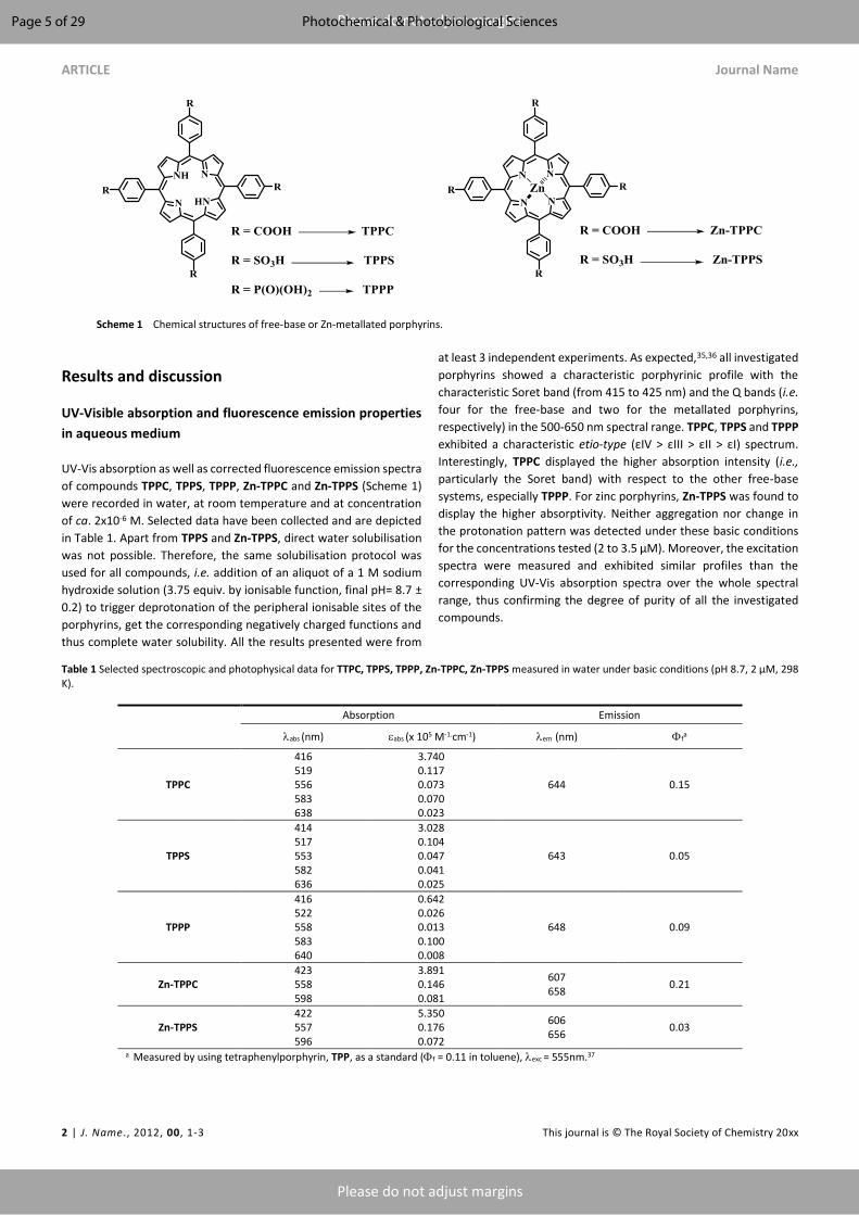
ARTICLE Journal Name
2 | J. Name., 2012, 00, 1-3 This journal is © The Royal Society of Chemistry 20xx
Please do not adjust margins
Please do not adjust margins
Results and discussion
UV-Visible absorption and fluorescence emission properties
in aqueous medium
UV-Vis absorption as well as corrected fluorescence emission spectra
of compounds TPPC, TPPS, TPPP, Zn-TPPC and Zn-TPPS (Scheme 1)
were recorded in water, at room temperature and at concentration
of ca. 2x10⁻⁶ M. Selected data have been collected and are depicted
in Table 1. Apart from TPPS and Zn-TPPS, direct water solubilisation
was not possible. Therefore, the same solubilisation protocol was
used for all compounds, i.e. addition of an aliquot of a 1 M sodium
hydroxide solution (3.75 equiv. by ionisable function, final pH= 8.7 ±
0.2) to trigger deprotonation of the peripheral ionisable sites of the
porphyrins, get the corresponding negatively charged functions and
thus complete water solubility. All the results presented were from
at least 3 independent experiments. As expected,35,36 all investigated
porphyrins showed a characteristic porphyrinic profile with the
characteristic Soret band (from 415 to 425 nm) and the Q bands (i.e.
four for the free-base and two for the metallated porphyrins,
respectively) in the 500-650 nm spectral range. TPPC, TPPS and TPPP
exhibited a characteristic etio-type (εIV > εIII > εII > εI) spectrum.
Interestingly, TPPC displayed the higher absorption intensity (i.e.,
particularly the Soret band) with respect to the other free-base
systems, especially TPPP. For zinc porphyrins, Zn-TPPS was found to
display the higher absorptivity. Neither aggregation nor change in
the protonation pattern was detected under these basic conditions
for the concentrations tested (2 to 3.5 µM). Moreover, the excitation
spectra were measured and exhibited similar profiles than the
corresponding UV-Vis absorption spectra over the whole spectral
range, thus confirming the degree of purity of all the investigated
compounds.
Table 1 Selected spectroscopic and photophysical data for TTPC, TPPS, TPPP, Zn-TPPC, Zn-TPPS measured in water under basic conditions (pH 8.7, 2 µM, 298 K).
Absorption Emission
abs (nm) abs (x 105 M-1.cm-1) em (nm) fa
TPPC
416 519 556 583 638
3.740 0.117 0.073 0.070 0.023
644 0.15
TPPS
414 517 553 582 636
3.028 0.104 0.047 0.041 0.025
643 0.05
TPPP
416 522 558 583 640
0.642 0.026 0.013 0.100 0.008
648 0.09
Zn-TPPC 423 558 598
3.891 0.146 0.081
607 658
0.21
Zn-TPPS 422 557 596
5.350 0.176 0.072
606 656
0.03
a Measured by using tetraphenylporphyrin, TPP, as a standard (f = 0.11 in toluene), exc = 555nm.37
Scheme 1 Chemical structures of free-base or Zn-metallated porphyrins.
Page 5 of 29 Photochemical & Photobiological Sciences
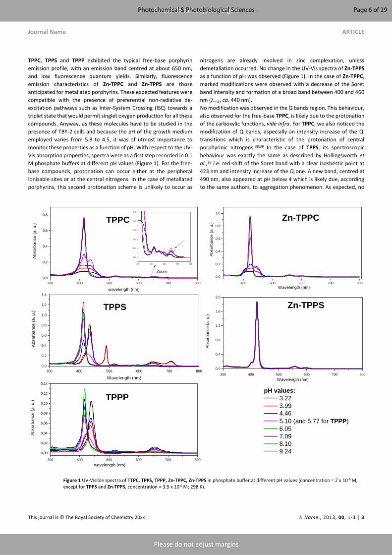
Journal Name ARTICLE
This journal is © The Royal Society of Chemistry 20xx J. Name., 2013, 00, 1-3 | 3
Please do not adjust margins
Please do not adjust margins
TPPC, TPPS and TPPP exhibited the typical free-base porphyrin
emission profile, with an emission band centred at about 650 nm,
and low fluorescence quantum yields. Similarly, fluorescence
emission characteristics of Zn-TPPC and Zn-TPPS are those
anticipated for metallated porphyrins. These expected features were
compatible with the presence of preferential non-radiative de-
excitation pathways such as Inter-System Crossing (ISC) towards a
triplet state that would permit singlet oxygen production for all these
compounds. Anyway, as these molecules have to be studied in the
presence of TBY-2 cells and because the pH of the growth medium
employed varies from 5.8 to 4.5, it was of utmost importance to
monitor these properties as a function of pH. With respect to the UV-
Vis absorption properties, spectra were as a first step recorded in 0.1
M phosphate buffers at different pH values (Figure 1). For the free-
base compounds, protonation can occur either at the peripheral
ionisable sites or at the central nitrogens. In the case of metallated
porphyrins, this second protonation scheme is unlikely to occur as
nitrogens are already involved in zinc complexation, unless
demetallation occurred. No change in the UV-Vis spectra of Zn-TPPS
as a function of pH was observed (Figure 1). In the case of Zn-TPPC,
marked modifications were observed with a decrease of the Soret
band intensity and formation of a broad band between 400 and 460
nm (max ca. 440 nm).
No modification was observed in the Q bands region. This behaviour,
also observed for the free-base TPPC, is likely due to the protonation
of the carboxylic functions, vide infra. For TPPC, we also noticed the
modification of Q bands, especially an intensity increase of the QI
transitions which is characteristic of the protonation of central
porphyrinic nitrogens.38,39 In the case of TPPS, its spectroscopic
behaviour was exactly the same as described by Hollingsworth et
al.,36 i.e. red-shift of the Soret band with a clear isosbestic point at
423 nm and intensity increase of the QI one. A new band, centred at
490 nm, also appeared at pH below 4 which is likely due, according
to the same authors, to aggregation phenomenon. As expected, no
Figure 1 UV-Visible spectra of TTPC, TPPS, TPPP, Zn-TPPC, Zn-TPPS in phosphate buffer at different pH values (concentration = 2 x 10⁻⁶ M, except for TPPS and Zn-TPPS, concentration = 3.5 x 10⁻⁶ M; 298 K).
300 400 500 600 700 8000,0
0,2
0,4
0,6
0,8
400 500 600 700 800
0,0
0,2
0,4
0,6
0,8
1,0
300 400 500 600 700 8000,0
0,2
0,4
0,6
0,8
1,0
1,2
1,4
300 400 500 600 700 8000,0
0,4
0,8
1,2
1,6
2,0
300 400 500 600 700 800
0,00
0,02
0,04
0,06
0,08
0,10
0,12
0,14
400 500 600 700 800
0,00
0,02
0,04
0,06
0,08
0,10
Abso
rban
ce (a
. u.)
wavelength (nm)
Zn-TPPSTPPS
Abso
rban
ce (a
. u.)
Wavelength (nm)
pH values: 3.22 3.99 4.46 5.10 (and 5.77 for TPPP) 6.05 7.09 8.10 9.24
Abso
rban
ce (a
. u.)
Wavelength (nm)
TPPP
Abso
rban
ce (a
. u.)
Wavelength (nm)
Zoom
Zn-TPPC
Abso
rban
ce (a
. u.)
wavelength (nm)
TPPC
Page 6 of 29Photochemical & Photobiological Sciences
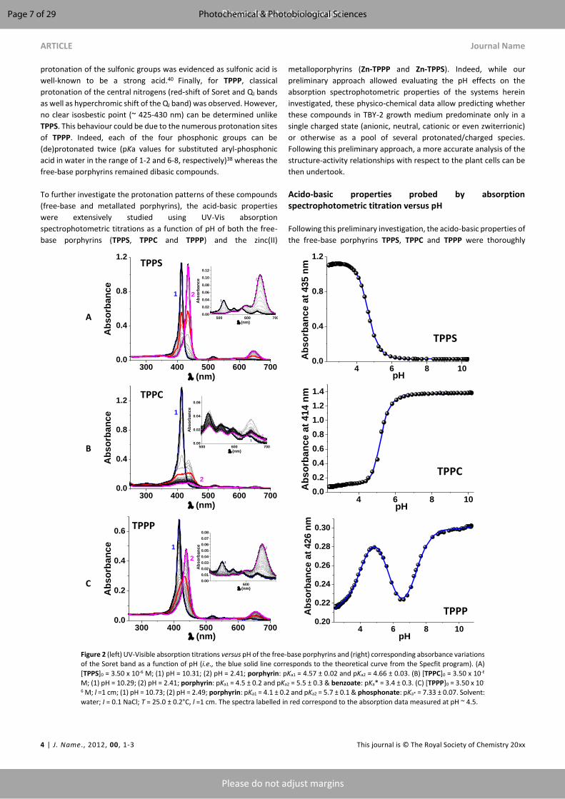
ARTICLE Journal Name
4 | J. Name., 2012, 00, 1-3 This journal is © The Royal Society of Chemistry 20xx
Please do not adjust margins
Please do not adjust margins
protonation of the sulfonic groups was evidenced as sulfonic acid is
well-known to be a strong acid.40 Finally, for TPPP, classical
protonation of the central nitrogens (red-shift of Soret and QI bands
as well as hyperchromic shift of the QI band) was observed. However,
no clear isosbestic point (~ 425-430 nm) can be determined unlike
TPPS. This behaviour could be due to the numerous protonation sites
of TPPP. Indeed, each of the four phosphonic groups can be
(de)protonated twice (pKa values for substituted aryl-phosphonic
acid in water in the range of 1-2 and 6-8, respectively)38 whereas the
free-base porphyrins remained dibasic compounds.
To further investigate the protonation patterns of these compounds
(free-base and metallated porphyrins), the acid-basic properties
were extensively studied using UV-Vis absorption
spectrophotometric titrations as a function of pH of both the free-
base porphyrins (TPPS, TPPC and TPPP) and the zinc(II)
metalloporphyrins (Zn-TPPP and Zn-TPPS). Indeed, while our
preliminary approach allowed evaluating the pH effects on the
absorption spectrophotometric properties of the systems herein
investigated, these physico-chemical data allow predicting whether
these compounds in TBY-2 growth medium predominate only in a
single charged state (anionic, neutral, cationic or even zwiterrionic)
or otherwise as a pool of several protonated/charged species.
Following this preliminary approach, a more accurate analysis of the
structure-activity relationships with respect to the plant cells can be
then undertook.
Acido-basic properties probed by absorption spectrophotometric titration versus pH
Following this preliminary investigation, the acido-basic properties of
the free-base porphyrins TPPS, TPPC and TPPP were thoroughly
A
B
C
300 400 500 600 7000.0
0.4
0.8
1.2
2
Abs
orba
nce
(nm)
1
TPPS
500 600 7000.00
0.02
0.04
0.06
0.08
0.10
0.12
2
Abs
orba
nce
(nm)
1
4 6 8 100.0
0.4
0.8
1.2
Abs
orba
nce
at 4
35 n
m
pH
TPPS
300 400 500 600 7000.0
0.4
0.8
1.2
2
Abs
orba
nce
(nm)
1
TPPC
500 600 7000.00
0.02
0.04
0.06
2
Abs
orba
nce
(nm)
1
4 6 8 100.00.20.40.60.81.01.21.4
Abs
orba
nce
at 4
14 n
m
pH
TPPC
300 400 500 600 7000.0
0.2
0.4
0.6
1
Abs
orba
nce
(nm)
2
TPPP
6000.000.010.020.030.040.050.060.070.08
1
Abs
orba
nce
(nm)
2
4 6 8 100.20
0.22
0.24
0.26
0.28
0.30
Abs
orba
nce
at 4
26 n
m
pH
TPPP
Figure 2 (left) UV-Visible absorption titrations versus pH of the free-base porphyrins and (right) corresponding absorbance variations of the Soret band as a function of pH (i.e., the blue solid line corresponds to the theoretical curve from the Specfit program). (A)
[TPPS]0 = 3.50 x 10⁻⁶ M; (1) pH = 10.31; (2) pH = 2.41; porphyrin: pKa1 = 4.57 ± 0.02 and pKa2 = 4.66 ± 0.03. (B) [TPPC]0 = 3.50 x 10⁻⁶ M; (1) pH = 10.29; (2) pH = 2.41; porphyrin: pKa1 = 4.5 ± 0.2 and pKa2 = 5.5 ± 0.3 & benzoate: pKa* = 3.4 ± 0.3. (C) [TPPP]0 = 3.50 x 10-
6 M; l =1 cm; (1) pH = 10.73; (2) pH = 2.49; porphyrin: pKa1 = 4.1 ± 0.2 and pKa2 = 5.7 ± 0.1 & phosphonate: pKa* = 7.33 ± 0.07. Solvent:
water; I = 0.1 NaCl; T = 25.0 ± 0.2°C, l =1 cm. The spectra labelled in red correspond to the absorption data measured at pH ~ 4.5.
Page 7 of 29 Photochemical & Photobiological Sciences

Journal Name ARTICLE
This journal is © The Royal Society of Chemistry 20xx J. Name., 2013, 00, 1-3 | 5
Please do not adjust margins
Please do not adjust margins
examined in water containing 0.1 M of NaCl as the supporting
electrolyte. The absorption spectrophotometric titrations versus pH
were first carried out by adding aliquots of HCl to a basic solution of
the porphyrins to ensure appropriate solubilisation of these
macrocycles in water. Figure 2A first depicts the absorption titration
versus pH of TPPS. Under basic conditions, TPPS is characterized by
a Soret band centred at 413 nm (413 = 3.23 x 10⁵ M-1 cm-1) and a set
of four weak Q bands ranging from 500 nm to 700 nm. It is
noteworthy that the spectroscopic properties measured by this
approach might slightly differ from the preliminary absorption data
(Table 1) because of the different experimental conditions and of the
fact that these data were derived from the electronic absorption
spectra of the pure species that were calculated using statistical
methods.41-46 Upon pH decrease, the Soret band gradually
underwent a bathochromic shift to max of 435 (435 = 3.28 x 10⁵ M-1
cm-1).
Concomitantly, the four Q bands were altered to two bands, the
more intense being centred at 645 nm (645 = 3.22 x 10⁴ M-1 cm-1).
This spectral variation is consistent with the successive protonations
of two nitrogens of the tetrapyrrolic moiety. It has been indeed
shown that the absorption spectrum of diprotonated porphyrin
consist of two Q-bands in addition to a red-shifted Soret band due to
the symmetry alteration induced by the protonation of the
tetrapyrrolic core. The absorption and potentiometric data have
been processed 41–46 and allowed evaluating the two corresponding
pKa values (pKa1 = 4.57 ± 0.02 and pKa2 = 4.66 ± 0.03 in 0.1 M NaCl,
Table 2) that are in agreement with previously reported data (pKa1 =
4.6 and pKa2 = 5.2 in 0.1 M NaCl). By contrast with the preliminary
spectrophometric approach (i.e., pH buffers were used, Figure 1), no
additional absorption centred at 490 nm was observed under our
conditions in agreement. This JB absorption corresponds to J-
aggregates (sharp band - denoted as JB band - that is red-shifted by
56 nm with respect to the Soret B band at 435 nm of [TPPS.2H]2-
monomer)47 that is prevented under our experimental conditions.
This process was reported to occur under acidic pH (pH < 4) following
protonations of the tetrapyrrolic core.47,48 The diprotonated species
of TPPS therefore exists in the form of a zwiterrionic compound with
the four peripheral sites being negatively charged and the
tetrapyrrolic core being positively charged.49 The absorption
electronic spectra of the protonated species of TPPS are given in ESI,
while the distribution diagrams of the TPPS protonated species are
depicted in Figure 3. At pH 4-5, the three protonated species coexist.
Following these results, we anticipate that Zn-TPPS mainly
predominates as a tetra-anionic species in a broad range of pH given
the fact that the porphyrinic nitrogens are involved in the Zn(II)
coordination.
Figure 2B illustrates then the spectral variation experienced by TPPC
upon pH decrease. Under basic conditions, the fully deprotonated
species is characterized by an intense Soret band at 414 nm (414 =
3.93 x 10⁵ M-1 cm-1) and four weakly absorptive Q bands lying
between 480 nm and 700 nm (see ESI). TPPC mainly exist as a
monomeric form (~ 80 %) under our experimental conditions (3.5
µM) as it was shown that TPPC could lead to dimer at neutral pH with
a dimerisation constant of 4.55 x 10⁴ M-1.50 Up to pH ~ 5, a weak
hypochromic shift (i.e., protonation of the carboxylate functions)
concomitant to a partial protonation of the porphyrinic core could be
observed in agreement with the classical bathochromic shift of the
Soret band. When the pH was further decreased, the Soret band
faded and a broad, split and weak absorption centred at ~ 430 nm
appeared. This feature likely suggests that further acido-basic
reactions of TPPC led to protonated species that underwent
aggregation in water. It has been indeed shown46 that for 1 < pH<
4.5, the Soret band of [TPPC.6H]2+ is split into two components at
about 444 (with a shoulder at 439) and 404 nm and this was assigned
to the formation of a dimeric species. In the same report,46 the pKas
of the carboxylic functions and of the tetrapyrrolic protonated units
were estimated to be at about 5. Due to the precipitation that
occurred below pH ~ 5, only the spectra recorded from basic pH
values to pH 5.2 were processed. The processing of the data allowed
evaluating two pKa values. The lowest deprotonation constant
determined under these conditions (pKa1 = 4.1 ± 0.4) is proposed to
be related to deprotonations of one of the nitrogens of the
tetrapyrrolic core (i.e., the first deprotonation of the tetrapyrrolic
core was not accessible due to precipitation of TPPC), while the
deprotonations of the four peripheral benzoate units are
characterized by an apparent deprotonation constant (pKa2 = 5.4 ±
0.5). The distribution diagrams of the TPPC protonated species are
given in Figure 3 and suggest that the dimerization is likely induced
by the protonation in a narrow pH window of the benzoate functions
giving to the neutral species that dimerizes. These observations are
in agreement with the preliminary absorption spectrophotometric
investigation (Figure 1) as well as published results.46,50
To go even further in this approach, we have recorded the spectral
variations of an aqueous solution of Zn-TPPC as a function of pH (see
ESI). Similarly to TPPC, the metallated porphyrin Zn-TPPC was
solubilised under basic conditions. The fully deprotonated species
(i.e., benzoate functions) is characterized by an intense Soret band at
422 nm (422 = 3.32 x 10⁵ M-1 cm-1) as well as two Q bands at 557 nm
(557 = 1.16 x 10⁴ M-1 cm-1) and 597 nm (597 = 5.93 x 10³ M-1 cm-1).
Upon acidification, the absorption bands experienced a gradual
hypochromic shift and then a significant alteration of the absorption
characteristics (at ~ pH 5) as seen previously for TPPC in water (vide
supra). This confirmed that protonation of the benzoate units led to
a species that underwent self-interaction in water (i.e., most likely
dimerization on the basis of similar spectral features). The pKa value
provided as a mere estimate was found to be at about 4.8. This is in
line with the previous observations (Figure 1).
We finally examined the acido-basic properties of TPPP in water
(Figure 2C). As for the two previous free-base porphyrins (vide
supra), the fully deprotonated species predominates under basic
conditions (Soret band at 416 nm, 416 = 1.92 x 10⁵ M-1 cm-1 and four
Q bands lying between 480 nm and 700 nm). The gradual pH
decrease first led to a hypochromic shift of the Soret band up to pH
~ 7 with no evidence of protonation of the tetrapyrrolic core. This
likely suggests that the phosphonate groups are being protonated.
From pH ~ 7 to ~ 2.5, the Soret band then experienced a significant
bathochromic shift in agreement with the protonations of two
nitrogens of the porphyrinic unit. Under these experimental
conditions (2.49 < pH < 10.73, Figure 2C), each of the phosphonate
units most likely undergo monoprotonation (i.e., RPO32- + H+ <->
RPO3H-) as suggested from the data measured on phenylphosphonic
acid (pKa1 = 1.68 and pKa2 = 7.021)51 or other derivatives such as
Page 8 of 29Photochemical & Photobiological Sciences

ARTICLE Journal Name
6 | J. Name., 2012, 00, 1-3 This journal is © The Royal Society of Chemistry 20xx
Please do not adjust margins
Please do not adjust margins
nitrilo-tris(methylenephosphonic acid) (pKa < 2 in CH3OH/H2O
80/20).50 Full protonation of the phosphonate units thus likely occurs
under very acidic conditions (pH < 1). The statistical processing of the
spectral and potentiometric data sets allowed calculating three
protonation constants. The tetrapyrrolic core is associated to two
deprotonation constants (pKa1 = 4.1 ± 0.2 and pKa2 = 5.7 ± 0.1) that
are in agreement with those determined for the other investigated
free-base porphyrinic systems (i.e., TPPS and TPPC, vide supra, Table
2). An apparent protonation constant was also evidenced for the four
phosphonate groups (pKa* = 7.33 ± 0.07). The absorption electronic
spectra of the protonated species of TPPP are available in the ESI,
while the distribution diagrams of these species are given in Figure
3. At pH 4.5, two main negatively charged species predominates,
namely [TPPP.5H]3- and [TPPP.6H]2-.
To get a deeper understanding of our homogenous series of
porphyrins, their acido-basic properties were also assessed in a
mixed solvent made of 80 % of methanol and 20 % of water (by
weight) containing 0.1 M of n-tetrabutylammonium perchlorate as
the supporting salt. It has been shown that the optimal solubilisation
of TPPS or TPPC was achieved for aqueous solutions containing 80 %
of methanol. 48 Under these conditions aggregation was prevented
and only the porphyrin monomers predominate. The UV-Vis
absorption spectra of TPPS, TPPC, TPPP, Zn-TPPS and Zn-TPPC were
recorded over a large span of pH values starting from acidic
conditions and are depicted in Figure 4.
Table 2 Physico-chemical data measured for TTPC, TPPS, TPPP, Zn-TPPC and Zn-TPPS.
pKa ± 3
Nitrogens of the
tetrapyrrolic core
Peripheral ionisable
sites Aggregation occurred?
TPPS (water)a pKa1 = 4.57 ± 0.02
pKa2 = 4.66 ± 0.03 pKa(sulfonate) << 2 No
TPPS (CH3OH/H2O)b pKa1 = 2.3 ± 0.2
pKa2 = 2.7 ± 0.1 pKa(sulfonate) << 2 No
Zn-TPPS (CH3OH/H2O)b na pKa(sulfonate) << 2 No
TPPC (water)a pKa1 < 4
pKa2 = 4.1 ± 0.4 pKa(benzoate)* = 5.4 ± 0.4 Yes (dimerization)
TPPC (CH3OH/H2O)b pKa1 = 2.1 ± 0.4
pKa2 = 2.4 ± 0.2 pKa(benzoate)* = 5.8 ± 0.2 No
Zn-TPPC (CH3OH/H2O)b na pKa(benzoate)* = 7.1 ± 0.1 No
Zn-TPPC (water)a na pKa(benzoate)* ~ 4.8 Yes (dimerization)
TPPP (water)a pKa1 = 4.1 ± 0.2
pKa2 = 5.7 ± 0.1 pKa(phosphonate)* = 7.33 ± 0.07 No
TPPP (CH3OH/H2O)b pKa2 + pKa3= 4.8 ± 0.3
pKa1 (phosphonate) << 2
pKa4 (phosphonate) = 3.8 ± 0.3
pKa5 (phosphonate) = 5.7 ± 0.2
pKa6 (phosphonate) = 9.77 ± 0.06
No
a water, 0.1 M NaCl; b CH3OH/H2O 80/20 w/w, 0.1 n-NBu4ClO4; na = not applicable. T = 25.0 ± 0.2 °C.
As mentioned above, the peripheral sulfonic groups of TPPS are
strong acids and were consequently found to be deprotonated
within the pH range considered (i.e., pKa < 1).47 Under acidic pH (pH
~ 2), the absorption spectrum of TPPS is characterized by an intense
Soret band centred at 436 nm (436 = 4.13 x 10⁵ M-1 cm-1) as well as
an intense QI band at 648 nm (648 = 4.46 x 10⁴ M-1 cm-1). These
spectrophotometric features are in agreement with those
determined in water (vide supra) and are characteristics of a
protonated TPPS species with the two nitrogens of the tetrapyrrolic
core being involved. By contrast with the preliminary approach in
water (Figure 1), no additional absorption was found at 490 nm (i.e.,
characteristic of an J-aggregation phenomenon)52 thus indicating
that this solvent system (i.e., CH3OH:H2O 80/20 w/w, I = 0.1 M n-
NBu4ClO4) prevents aggregation and ensures efficient solubilisation
of all the protonated TPPS species. The increase of the medium
acidity to pH 4 led to deprotonation of the TPPS chromophoric unit
as shown by the hypsochromic shifts of the Soret band to 416 nm
(416 = 3.14 x 10⁵ M-1 cm-1) (Figure 4). Above pH 4-5 no further
absorption spectral variation was observed (Figure 4) in agreement
with the absence of any other acido-basic equilibria. The
processing41–45 of the spectrophotometric and potentiometric data
sets led to the evaluation of two acidic pKa (pKa1 = 2.3 ± 0.2 and pKa2
= 2.7 ± 0.1) values that are related to the nitrogen atoms of the
porphyrin moiety (Table 2).
Page 9 of 29 Photochemical & Photobiological Sciences
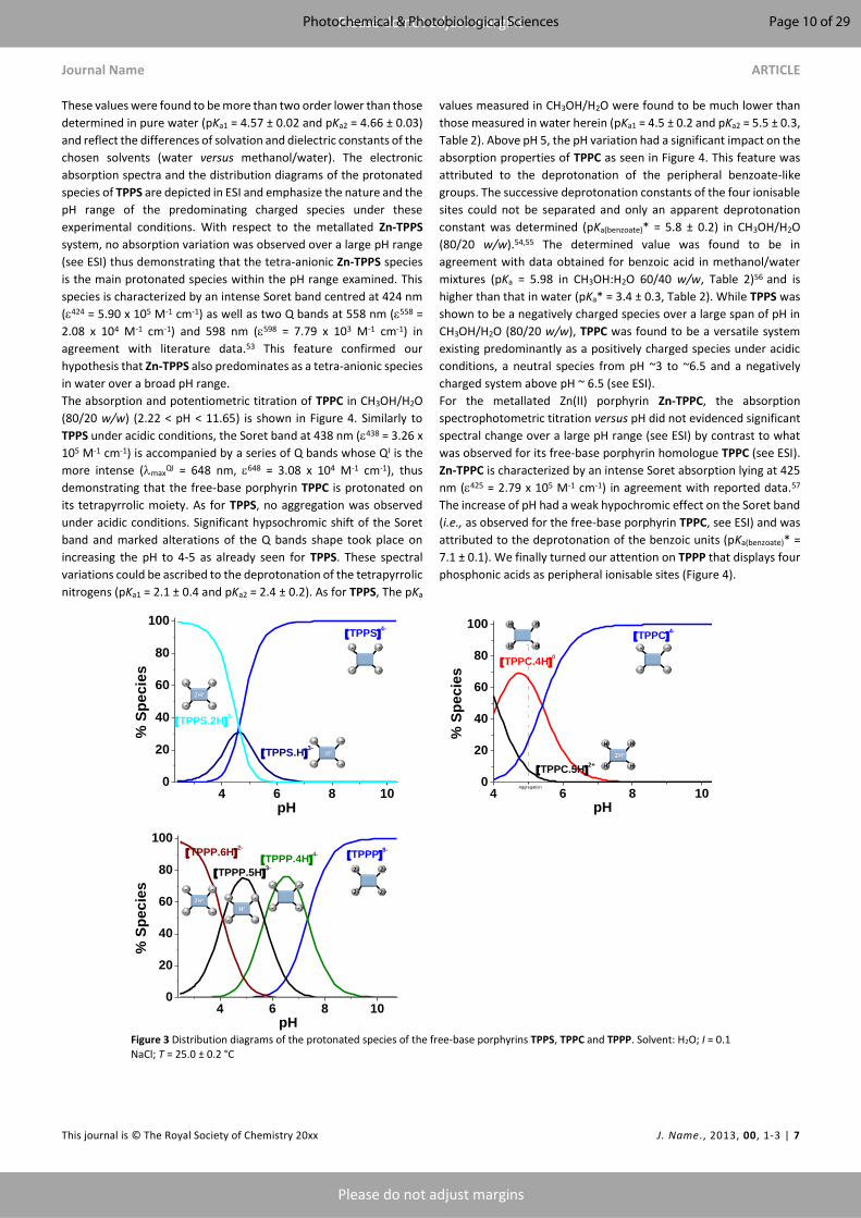
Journal Name ARTICLE
This journal is © The Royal Society of Chemistry 20xx J. Name., 2013, 00, 1-3 | 7
Please do not adjust margins
Please do not adjust margins
These values were found to be more than two order lower than those
determined in pure water (pKa1 = 4.57 ± 0.02 and pKa2 = 4.66 ± 0.03)
and reflect the differences of solvation and dielectric constants of the
chosen solvents (water versus methanol/water). The electronic
absorption spectra and the distribution diagrams of the protonated
species of TPPS are depicted in ESI and emphasize the nature and the
pH range of the predominating charged species under these
experimental conditions. With respect to the metallated Zn-TPPS
system, no absorption variation was observed over a large pH range
(see ESI) thus demonstrating that the tetra-anionic Zn-TPPS species
is the main protonated species within the pH range examined. This
species is characterized by an intense Soret band centred at 424 nm
(424 = 5.90 x 10⁵ M-1 cm-1) as well as two Q bands at 558 nm (558 =
2.08 x 10⁴ M-1 cm-1) and 598 nm (598 = 7.79 x 10³ M-1 cm-1) in
agreement with literature data.53 This feature confirmed our
hypothesis that Zn-TPPS also predominates as a tetra-anionic species
in water over a broad pH range.
The absorption and potentiometric titration of TPPC in CH3OH/H2O
(80/20 w/w) (2.22 < pH < 11.65) is shown in Figure 4. Similarly to
TPPS under acidic conditions, the Soret band at 438 nm (438 = 3.26 x
105 M-1 cm-1) is accompanied by a series of Q bands whose QI is the
more intense (maxQI = 648 nm, 648 = 3.08 x 10⁴ M-1 cm-1), thus
demonstrating that the free-base porphyrin TPPC is protonated on
its tetrapyrrolic moiety. As for TPPS, no aggregation was observed
under acidic conditions. Significant hypsochromic shift of the Soret
band and marked alterations of the Q bands shape took place on
increasing the pH to 4-5 as already seen for TPPS. These spectral
variations could be ascribed to the deprotonation of the tetrapyrrolic
nitrogens (pKa1 = 2.1 ± 0.4 and pKa2 = 2.4 ± 0.2). As for TPPS, The pKa
values measured in CH3OH/H2O were found to be much lower than
those measured in water herein (pKa1 = 4.5 ± 0.2 and pKa2 = 5.5 ± 0.3,
Table 2). Above pH 5, the pH variation had a significant impact on the
absorption properties of TPPC as seen in Figure 4. This feature was
attributed to the deprotonation of the peripheral benzoate-like
groups. The successive deprotonation constants of the four ionisable
sites could not be separated and only an apparent deprotonation
constant was determined (pKa(benzoate)* = 5.8 ± 0.2) in CH3OH/H2O
(80/20 w/w).54,55 The determined value was found to be in
agreement with data obtained for benzoic acid in methanol/water
mixtures (pKa = 5.98 in CH3OH:H2O 60/40 w/w, Table 2)56 and is
higher than that in water (pKa* = 3.4 ± 0.3, Table 2). While TPPS was
shown to be a negatively charged species over a large span of pH in
CH3OH/H2O (80/20 w/w), TPPC was found to be a versatile system
existing predominantly as a positively charged species under acidic
conditions, a neutral species from pH ~3 to ~6.5 and a negatively
charged system above pH ~ 6.5 (see ESI).
For the metallated Zn(II) porphyrin Zn-TPPC, the absorption
spectrophotometric titration versus pH did not evidenced significant
spectral change over a large pH range (see ESI) by contrast to what
was observed for its free-base porphyrin homologue TPPC (see ESI).
Zn-TPPC is characterized by an intense Soret absorption lying at 425
nm (425 = 2.79 x 10⁵ M-1 cm-1) in agreement with reported data.57
The increase of pH had a weak hypochromic effect on the Soret band
(i.e., as observed for the free-base porphyrin TPPC, see ESI) and was
attributed to the deprotonation of the benzoic units (pKa(benzoate)* =
7.1 ± 0.1). We finally turned our attention on TPPP that displays four
phosphonic acids as peripheral ionisable sites (Figure 4).
Figure 3 Distribution diagrams of the protonated species of the free-base porphyrins TPPS, TPPC and TPPP. Solvent: H2O; I = 0.1
NaCl; T = 25.0 ± 0.2 °C
4 6 8 100
20
40
60
80
100
TPPS.2H2-
TPPS.H3-
% S
peci
es
pH
TPPS4-
2H+
-
- -
-
H+
-
- -
-
-
- -
-
4 6 8 100
20
40
60
80
100TPPP.4H
4-TPPP.6H2-
TPPP.5H3-
% S
peci
es
pH
TPPP8-
2-
2- 2-
2-
-
- -
-
-
- -
-
H+-
- -
-
2H+
4 6 8 100
20
40
60
80
100
TPPC.5H2+
TPPC.4H0
% S
peci
es
pH
TPPC4-
Aggregation
2H+
H
H H
H
-
- -
-
H
H H
H
Page 10 of 29Photochemical & Photobiological Sciences

ARTICLE Journal Name
8 | J. Name., 2012, 00, 1-3 This journal is © The Royal Society of Chemistry 20xx
Please do not adjust margins
Please do not adjust margins
Under acidic conditions, TPPP is characterized by an intense Soret
band centred at 438 nm (438 = 1.0 x 10⁵ M-1 cm-1) and a set of two Q
bands (max QI = 651 nm, 651 = 1.03 x 10⁴ M-1 cm-1) that clearly
evidenced the diprotonated nature of a tetrapyrrolic core.
Deprotonation of the porphyrinic core was easily monitored by the
red-shift of the Soret band as well as the altered profile of the Q
bands (i.e., from two main absorptions to four bands upon
deprotonation). A global acidic deprotonation constant was obtained
(pKa2 + pKa3= 4.8 ± 0.3) and was found to be similar to those
measured for TPPS (pKa1 + pKa2= 5.0) and TPPC (pKa1 + pKa2= 4.5) in
CH3OH/H2O.
Further increase of the pH led to a hyperchromic shift of the Soret
band (Figure 2) and was attributed to the stepwise deprotonation of
the monoprotonated phosphonate units (i.e., RPO3H- ↔ RPO32- + H+;
pKa4 = 3.8 ± 0.3, pKa5 = 5.7 ± 0.2 and pKa6 = 9.77 ± 0.06). The first
deprotonation constant pKa1 of the monoprotonated phosphonate
units was estimated to be lower than 2 and could not be determined
with good accuracy. The pKa separation of the phosphonate groups
measured for TPPP was found to be in agreement with other
polyphosphonated derivatives.58 The distribution diagrams of the
protonated species of TPPP is depicted in ESI.
A
B
C
300 400 500 600 7000.0
0.5
1.0
1.5
2.0
2
Abs
orba
nce
(nm)
1500 550 600 650 700
0.00
0.04
0.08
0.12
0.16
0.20
2
Abs
orba
nce
(nm)
1
TPPS
2 4 6 8 100.4
0.6
0.8
1.0
1.2
1.4
Abs
orba
nce
at 4
20 n
mpH
TPPS
300 400 500 600 7000.0
0.5
1.0
1.5
2.0
1Abs
orba
nce
(nm)
2500 550 600 650 700
0.00
0.02
0.04
0.06
0.08
0.10
1
Abs
orba
nce
(nm)
2
TPPC
2 4 6 8 100.8
1.0
1.2
1.4
Abs
orba
nce
at 4
20 n
m
pH
TPPC
300 400 500 600 7000.0
0.2
0.4
0.6
0.8
1.0
1.2
1Abs
orba
nce
(nm)
2
TPPP
500 550 600 650 7000.00
0.02
0.04
0.06
0.08
0.10
1Abs
orba
nce
(nm)
2
2 4 6 8 100.4
0.6
0.8
1.0
1.2
Abs
orba
nce
at 4
20 n
m
pH
TPPP
Figure 4 (left) UV-Visible absorption titrations versus pH of the free-base and zinc(II) porphyrins and (right) corresponding
absorbance variations of the Soret band as a function of pH (i.e., the blue solid line corresponds to the theoretical curve from the
Specfit program). (A) [TPPS]0 = 7.18 x 10⁻⁶ M; (1) pH = 2.16; (2) pH = 11.43; porphyrin: pKa1 = 2.3 ± 0.2 and pKa2 = 2.7 ± 0.1. (B) [TPPC]0
= 5.33 x 10⁻⁶ M; (1) pH = 2.22; (2) pH = 11.65; porphyrin: pKa1 = 2.1 ± 0.4 and pKa2 = 2.4 ± 0.2 & benzoate: pKa* = 5.8 ± 0.2. (C) [TPPP]0
= 6.90 x 10⁻⁶ M; l =1 cm; (1) pH = 2.19; (2) pH = 11.36; porphyrin: pKa2 + pKa3= 4.8 ± 0.3 & phosphonate: pKa1 < 2, pKa4 = 3.8 ± 0.3,
pKa5 = 5.7 ± 0.2, pKa6 = 9.77 ± 0.06. Solvent: CH3OH/H2O (80/20 by weight); I = 0.1 n-NBu4ClO4; T = 25.0 ± 0.2°C, l =1 cm.
Page 11 of 29 Photochemical & Photobiological Sciences

Journal Name ARTICLE
This journal is © The Royal Society of Chemistry 20xx J. Name., 2013, 00, 1-3 | 9
Please do not adjust margins
Please do not adjust margins
ROS production To evaluate and compare the capacity of all studied porphyrins to
generate ROS under light irradiation, Electron Paramagnetic
Resonance (EPR) spectroscopy was used to detect the superoxide
anion O2•− (produced after a photo-induced electron transfer, i.e. the
so-called Type I process) and the singlet oxygen 1O2 (produced
through an energy transfer to dioxygen, i.e. the so-called Type II
process), according to the method described by Riou et al.28 As 1O2 is
non-radical and both 1O2 and O2•− are short-lived species,
experiments were performed in presence of specific scavengers (e.g.
2,2,6,6-tetramethyl-4-piperidone or TEMP, giving TEMPO in the
presence of 1O2, and 5,5-dimethyl-1-pyrroline N-oxide, DMPO, giving
DMPO-OOH in the presence of O2•−). The results are summarized in
Figure 5. No variation of the initial EPR signal was observed when the
reaction mixture was measured without light irradiation or in the
absence of a PS for both singlet oxygen and superoxide anion
production monitoring. With respect to the singlet oxygen
production, the metallated porphyrins herein investigated were
immediately degraded, probably by 1O2 itself, as depicted in Figure
5A. The same behaviour (i.e. degradation by singlet oxygen
production) was already observed in our previous study for the
metallated porphyrin investigated then (namely zinc(II) tetra-(N-
methyl-pyridinium)-porphyrin tetrachloride) even if the degradation
pattern was slower for this metalloporphyrin.28 Concerning the free-
base porphyrins, TPPC and TPPS were clearly shown to be more
efficient in production of singlet oxygen than TPPP, although
degradation was observed for TPPC after 20 minutes of accumulated
irradiation.
Regarding O2•− production (Figure 5B), all compounds exhibited
similar profiles, i.e. a rapid growth followed by a plateau (the most
efficient being TPPC), except for Zn-TPPS which followed a quasi-
linear progression. Interestingly, no correlation was noticed between
ROS production and metalation state of the porphyrins. Indeed, if
O2•− levels reached with Zn-TPPC were lower to those reached with
TPPC, the opposite behaviour was observed for the TPPS series. As
for 1O2 production, TPPP was among the less efficient system.
However, all investigated compounds can generate ROS, by both
type I and II processes for TPPC, TPPS, and TPPP and only by Type I
process for Zn-TPPC and Zn-TPPS, the most efficient system being
TPPC.
UV-Visible absorption properties in TBY-2 cells growth medium
As the growth medium for the TBY-2 cells, a modified Murashige and
Skoog medium supplemented by 0.27 mg L-1 of 2,4-
dichlorophenoxyacetic acid and 10 mg mL-1 of thiamine solutions was
used.59 Due to complexity of this medium, UV-Vis absorption spectra
of all the investigated porphyrins were recorded in spent medium
and compared to those obtained, at the same pH, in phosphate
buffer (Figures 1 and 6). The value of pH was fixed at 4.5, the pH value
of spent medium of growing cells60 which were used in biological
assay (vide infra). Hence, we believe it is the best way to mimic as
faithfully as possible experimental conditions of biological assays. As
one can see from Figure 6, no significant modification of the
absorption profile with respect to the medium was observed for Zn-
TPPS and TPPP whereas a hypochromic effect was noticed for TPPS
when phosphate buffer was replaced by the growth medium. We
supposed an effect of the complexity of the spent medium.60
Altogether, these results and those obtained from the absorption
spectrophotometric study (vide supra) evidenced that, as expected,
in growth medium, Zn-TPPS predominates as a tetra anionic species
([Zn-TPPS]4-), whereas TPPP likely exist under di-anionic or tri-anionic
forms, the phosphonate groups being all mono-protonated and one
([TPPP.5H]3-) or two nitrogens ([TPPP.6H]2-) being protonated (Figure
3). The position of the Soret band and the presence of an intense Q
band at about 650 nm strongly substantiate this hypothesis. In the
case of TPPS, the species displaying protonated nitrogens
(zwiterrionic [TPPS.H]3- and [TPPS.2H]2- species) and the fully
deprotonated tetra-anionic one ([TPPS]4-) coexist at this pH as
evidenced by the split Soret band (Figure 6). These observations are
in excellent agreement with the data collected in this report (i.e.,
these three species coexist in equivalent amounts, Figures 2 and 3).
-20
0
20
40
60
80
100
0 2 4 6 8 10 12 14 16 18 20 22 24
TEM
PO
EP
R s
ign
al in
ten
sity
x1
0-3
(a.u
.)
Irradiation Time (min.)
Evolution de la production d'oxygéne singulet des différentes porhyrines (40 µM)(TEMP (12,5 mM) - 20000 Lux - Tampon Phosphate 0,01 M, pH 7,4 - 0,51 mW
TPPC
Zn-TPPC
TPPS
Zn-TPPS
TPPP
0
20
40
60
80
100
120
140
0 2 4 6 8 10 12 14 16 18 20
DM
PO
-OO
H E
PR
sig
nal
inte
nsi
ty x
10
-3
Irradiation Time (min.)
DMPO-OOH PRODUCTION (5000 LUX - 225 mM dmpo)
TPPC
Zn-TPPC
TPPS
Zn-TPPS
TPPP
Figure 5 (A) EPR signal of TEMPO generation by irradiation of TPPC, Zn-TPPC, TPPS, Zn-TPPS and TPPP. ([TEMP] = 12.5 mM [Porphyrins] = 40 μM in 0.01 M phosphate buffer, pH 7.4); (B) EPR signal of DMPO-OOH generated by irradiation of TPPC, Zn-TPPC,
TPPS, Zn-TPPS and TPPP. ([DMPO] = 225 mMol [Porphyrins] = 50 μM in DMSO). The irradiation was performed with white light (280 μE.s−1.m−2 for experiments A and 70 μE.s−1.m−2 for experiments B). The values represent the mean ± S.D. obtained from 3 independent experiments (experiments were performed as described in ESI).
Page 12 of 29Photochemical & Photobiological Sciences
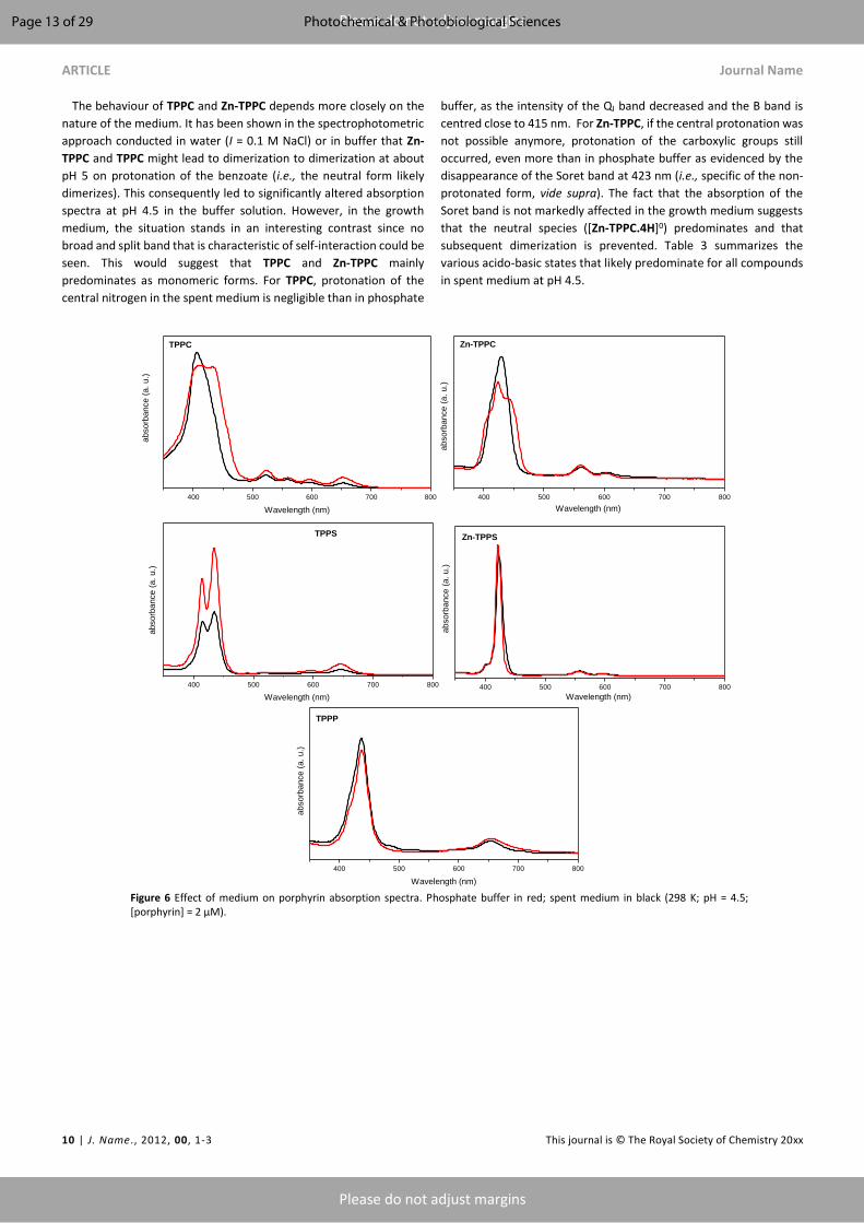
ARTICLE Journal Name
10 | J. Name., 2012, 00, 1-3 This journal is © The Royal Society of Chemistry 20xx
Please do not adjust margins
Please do not adjust margins
The behaviour of TPPC and Zn-TPPC depends more closely on the
nature of the medium. It has been shown in the spectrophotometric
approach conducted in water (I = 0.1 M NaCl) or in buffer that Zn-
TPPC and TPPC might lead to dimerization to dimerization at about
pH 5 on protonation of the benzoate (i.e., the neutral form likely
dimerizes). This consequently led to significantly altered absorption
spectra at pH 4.5 in the buffer solution. However, in the growth
medium, the situation stands in an interesting contrast since no
broad and split band that is characteristic of self-interaction could be
seen. This would suggest that TPPC and Zn-TPPC mainly
predominates as monomeric forms. For TPPC, protonation of the
central nitrogen in the spent medium is negligible than in phosphate
buffer, as the intensity of the QI band decreased and the B band is
centred close to 415 nm. For Zn-TPPC, if the central protonation was
not possible anymore, protonation of the carboxylic groups still
occurred, even more than in phosphate buffer as evidenced by the
disappearance of the Soret band at 423 nm (i.e., specific of the non-
protonated form, vide supra). The fact that the absorption of the
Soret band is not markedly affected in the growth medium suggests
that the neutral species ([Zn-TPPC.4H]0) predominates and that
subsequent dimerization is prevented. Table 3 summarizes the
various acido-basic states that likely predominate for all compounds
in spent medium at pH 4.5.
400 500 600 700 800 400 500 600 700 800
400 500 600 700 800 400 500 600 700 800
400 500 600 700 800
Wavelength (nm)
TPPC
abso
rban
ce (a
. u.)
Wavelength (nm)
abso
rban
ce (a
. u.)
Wavelength (nm)
abso
rban
ce (a
. u.)
Wavelength (nm)
abso
rban
ce (a
. u.)
Zn-TPPC
Zn-TPPSTPPS
abso
rban
ce (a
. u.)
Wavelength (nm)
TPPP
Figure 6 Effect of medium on porphyrin absorption spectra. Phosphate buffer in red; spent medium in black (298 K; pH = 4.5; [porphyrin] = 2 µM).
Page 13 of 29 Photochemical & Photobiological Sciences

Journal Name ARTICLE
This journal is © The Royal Society of Chemistry 20xx J. Name., 2013, 00, 1-3 | 11
Please do not adjust margins
Please do not adjust margins
Table 3 Main protonated forms of all compounds in the spent medium and phosphate buffer as deduced from their UV-Vis absorption profiles (298 K; pH =
4.5; value under bracket is the total charge of the compound).a
Fully anionic
Peripheral protonation
H2-Porphyrin2-
H-Porphyrin3-
H- Porphyrin+
Fully protonated
TPPC phosphate buffer X (Neutral) X (+)
growth medium X (Neutral)
Zn-TPPC phosphate buffer X (4-) X (Neutral)
growth medium X (4-) X (Neutral)
TPPS phosphate buffer X (4-) X (2-) X (3-)
growth medium X (4-) X (2-) X (3-)
Zn-TPPS phosphate buffer X (4-)
growth medium X (4-)
TPPP phosphate buffer X (2-) X (3-)
growth medium X (2-) X (3-)
a In the case of TPPP, only one protonation occurs on the phosphonate groups. TPPP still have 4 negative charges at the peripheral positions, not shown.
Photo-stability in TBY-2 cells growth medium To check the photo-stability of these different porphyrins, and their
capacity to reach the cells on the time scale of the experiment, their
absorption spectra were recorded for different irradiation times
under the same conditions (i.e. light, concentration) used for
biological assay. The intensity of the Soret band was monitored as a
function of irradiation time and allowed calculating a percentage of
the non-degraded porphyrins. Indeed, except for TPPP, no other
modification than the intensity of UV-Vis absorption spectra were
observed, thus indicating that TPPC, TPPS and their zinc counterpart
did not undergo photo-transformation but only photo-bleaching. In
the case of TPPP, the the max of the Soret band oscillated between
427 and 434 nm (see ESI). This behaviour can be explained by the
equilibrium, at this pH for two TPPP protonated species, namely the
mono- and diprotonated on the central nitrogens (Figure 3). Hence,
for this compound, the percentages were calculated for the
maximum of absorption, whatever the wavelength, assuming that it
will provide a good idea of the photo-stability of this compound. The
results are summarized in Table 4.
Table 4 Photo-stability of TTPC, TPPS, TPPP, Zn-TPPC, Zn-TPPS in the growth medium (pH = 5.4, 298 K, 95 µE. s-1.m-2).a
T0 After 3h of dark incubation 1h under light 3h under light
TPPC (2 µM) b 100 54.08 7.85 18.02 4.82 7.95 6.67
TPPS (3.5 µM) b 100 94.89 0.76 89.25 1.94 84.07 3.52
TPPP (3.5 µM) b 100 94.29 1.34 64.98 4.84 39.57 6.02
Zn-TPPC (2 µM)c 100 70.46 11.99 10.41 0.58 4.10 1.52
Zn-TPPS (3.5 µM)c 100 97.00 1.50 48.60 0.50 11.90 1.40
a Values were determined by monitoring the Soret band intensity at 416 nm for TPPC, 414 nm for TPPS, 423 nm for Zn-TPPC and 422 nm for Zn-TPPS. For TPPP,
the maximum of absorption was recorded. b The value is the mean of at least 3 independent measurements. c The value is the mean of 2 independent measurements.
After dark incubation over 3h in the growth medium, we noticed a
degradation of TPPC and its metallated counterpart (around 46 and
30 % respectively) whereas the others remained stable. As previously
observed,28 the metallated compounds are less stable than their
free-base homologues. To sum up, the more stable were TPPS and
TPPP, then TPPC, the metallated ones, Zn-TPPS and Zn-TPPC, being
the less stable species, especially Zn-TPPC, after one hour of light
irradiation.
Biologic assays
In a previous work, we have shown that the anionic porphyrin TPPS
was the most efficient to induce TBY-2 cell death compared to a
-
-
-
--
-
-
H
-
-
- H
H
H
2H+
-
-
-
- -
-
-H+
-
-
-
- -
-
-
H+
H
-
-
- H
H
H
2H+
H
-
-
- H
H
H
Page 14 of 29Photochemical & Photobiological Sciences
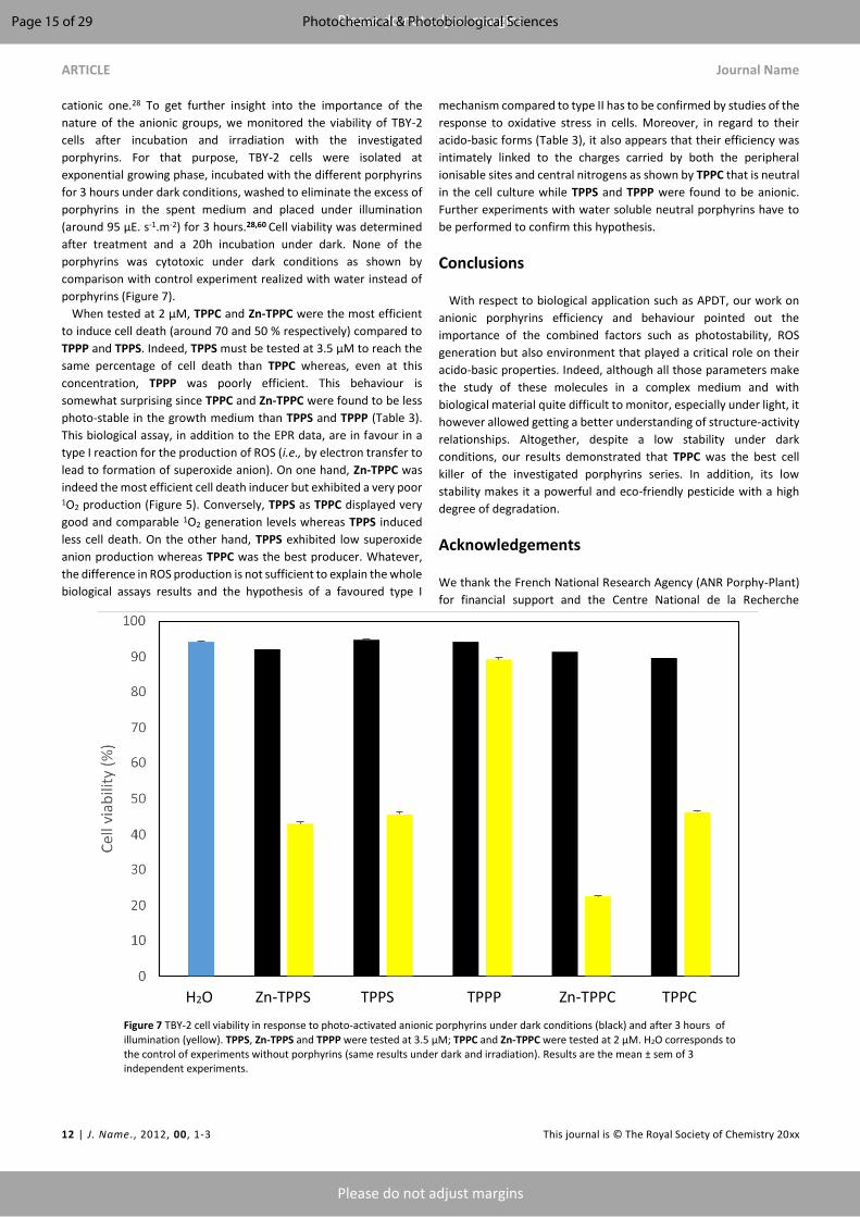
ARTICLE Journal Name
12 | J. Name., 2012, 00, 1-3 This journal is © The Royal Society of Chemistry 20xx
Please do not adjust margins
Please do not adjust margins
cationic one.28 To get further insight into the importance of the
nature of the anionic groups, we monitored the viability of TBY-2
cells after incubation and irradiation with the investigated
porphyrins. For that purpose, TBY-2 cells were isolated at
exponential growing phase, incubated with the different porphyrins
for 3 hours under dark conditions, washed to eliminate the excess of
porphyrins in the spent medium and placed under illumination
(around 95 µE. s-1.m-2) for 3 hours.28,60 Cell viability was determined
after treatment and a 20h incubation under dark. None of the
porphyrins was cytotoxic under dark conditions as shown by
comparison with control experiment realized with water instead of
porphyrins (Figure 7).
When tested at 2 µM, TPPC and Zn-TPPC were the most efficient
to induce cell death (around 70 and 50 % respectively) compared to
TPPP and TPPS. Indeed, TPPS must be tested at 3.5 µM to reach the
same percentage of cell death than TPPC whereas, even at this
concentration, TPPP was poorly efficient. This behaviour is
somewhat surprising since TPPC and Zn-TPPC were found to be less
photo-stable in the growth medium than TPPS and TPPP (Table 3).
This biological assay, in addition to the EPR data, are in favour in a
type I reaction for the production of ROS (i.e., by electron transfer to
lead to formation of superoxide anion). On one hand, Zn-TPPC was
indeed the most efficient cell death inducer but exhibited a very poor 1O2 production (Figure 5). Conversely, TPPS as TPPC displayed very
good and comparable 1O2 generation levels whereas TPPS induced
less cell death. On the other hand, TPPS exhibited low superoxide
anion production whereas TPPC was the best producer. Whatever,
the difference in ROS production is not sufficient to explain the whole
biological assays results and the hypothesis of a favoured type I
mechanism compared to type II has to be confirmed by studies of the
response to oxidative stress in cells. Moreover, in regard to their
acido-basic forms (Table 3), it also appears that their efficiency was
intimately linked to the charges carried by both the peripheral
ionisable sites and central nitrogens as shown by TPPC that is neutral
in the cell culture while TPPS and TPPP were found to be anionic.
Further experiments with water soluble neutral porphyrins have to
be performed to confirm this hypothesis.
Conclusions
With respect to biological application such as APDT, our work on
anionic porphyrins efficiency and behaviour pointed out the
importance of the combined factors such as photostability, ROS
generation but also environment that played a critical role on their
acido-basic properties. Indeed, although all those parameters make
the study of these molecules in a complex medium and with
biological material quite difficult to monitor, especially under light, it
however allowed getting a better understanding of structure-activity
relationships. Altogether, despite a low stability under dark
conditions, our results demonstrated that TPPC was the best cell
killer of the investigated porphyrins series. In addition, its low
stability makes it a powerful and eco-friendly pesticide with a high
degree of degradation.
Acknowledgements
We thank the French National Research Agency (ANR Porphy-Plant)
for financial support and the Centre National de la Recherche
Figure 7 TBY-2 cell viability in response to photo-activated anionic porphyrins under dark conditions (black) and after 3 hours of
illumination (yellow). TPPS, Zn-TPPS and TPPP were tested at 3.5 µM; TPPC and Zn-TPPC were tested at 2 µM. H2O corresponds to the control of experiments without porphyrins (same results under dark and irradiation). Results are the mean ± sem of 3 independent experiments.
H2O Zn-TPPS TPPS TPPP Zn-TPPC TPPC
Page 15 of 29 Photochemical & Photobiological Sciences

Journal Name ARTICLE
This journal is © The Royal Society of Chemistry 20xx J. Name., 2013, 00, 1-3 | 13
Please do not adjust margins
Please do not adjust margins
Scientifique (CNRS) and the University of Strasbourg (LIMA UMR
7042). We also want to warmly thank Pr. F. Fages for his advices and
for having suggested this fruitful collaboration.
Note and references
1 K. M. Kadish, The Porphyrin Handbook: Synthesis and organic
chemistry, Elsevier, 2000.
2 D. Mauzerall, in Photosynthesis I, eds. P. D. A. Trebst and P. D. M.
Avron, Springer Berlin Heidelberg, 1977, pp. 117–124.
3 M. Danquah, Review of Handbook of Porphyrin Science with
Application to Chemistry, Physics, Materials Science, Engineering,
Biology and Medicine. Volume 40: Nanoorganization of
Porphyrinoids, J. Nat. Prod., 2017, 80, 1232–1232.
4 S. Hiroto, Y. Miyake and H. Shinokubo, Synthesis and
Functionalization of Porphyrins through Organometallic
Methodologies, Chem. Rev., 2017, 117, 2910–3043.
5 M. O. Senge, Stirring the porphyrin alphabet soup—
functionalization reactions for porphyrins, Chem. Commun.,
2011, 47, 1943–1960.
6 A. R. Antonangelo, K. C. M. Westrup, L. A. Burt, C. Grazia Bezzu,
T. Malewschik, G. S. Machado, F. S. Nunes, N. B. McKeown and S.
Nakagaki, Synthesis, crystallographic characterization and
homogeneous catalytic activity of novel unsymmetric porphyrins,
RSC Adv., 2017, 7, 50610–50618.
7 Ö. Birel, S. Nadeem and H. Duman, Porphyrin-Based Dye-
Sensitized Solar Cells (DSSCs): a Review, J. Fluoresc., 2017, 27,
1075–1085.
8 M. Jurow, A. E. Schuckman, J. D. Batteas and C. M. Drain,
Porphyrins as Molecular Electronic Components of Functional
Devices, Coord. Chem. Rev., 2010, 254, 2297–2310.
9 S. Lin, C. S. Diercks, Y.-B. Zhang, N. Kornienko, E. M. Nichols, Y.
Zhao, A. R. Paris, D. Kim, P. Yang, O. M. Yaghi and C. J. Chang,
Covalent organic frameworks comprising cobalt porphyrins for
catalytic CO2 reduction in water, Science, 2015, 349, 1208–1213.
10 J. Barona-Castaño, C. Carmona-Vargas, T. Brocksom and K. de
Oliveira, Porphyrins as Catalysts in Scalable Organic Reactions,
Molecules, 2016, 21, 310.
11 W.-B. Huang, W. Gu, H.-X. Huang, J.-B. Wang, W.-X. Shen, Y.-Y. Lv
and J. Shen, A porphyrin-based fluorescent probe for optical
detection of toxic Cd2+ ion in aqueous solution and living cells,
Dyes Pigments, 2017, 143, 427–435.
12 H. Huang, W. Song, J. Rieffel and J. F. Lovell, Emerging
applications of porphyrins in photomedicine, Front. Phys., ,
DOI:10.3389/fphy.2015.00023.
13 H. Kato, Y. Kato, R. Yoneyama, R. Ishikawa, M. Kojika, K. Miyajima,
N. Takizawa and K. Furukawa, Review of PDT for lung cancer and
future, Photodiagnosis Photodyn. Ther., 2017, 17, A30–A31.
14 M. Q. Almerie, G. Gossedge, K. E. Wright and D. G. Jayne,
Photodynamic Therapy Use for Peritoneal Carcinomatosis:
Systematic Review, Photodiagnosis Photodyn. Ther.
15 S. Pei, A. C. Inamadar, K. A. Adya and M. M. Tsoukas, Light-based
therapies in acne treatment, Indian Dermatol. Online J., 2015, 6,
145–157.
16 E. Alves, M. A. F. Faustino, M. G. P. M. S. Neves, Â. Cunha, H.
Nadais and A. Almeida, Potential applications of porphyrins in
photodynamic inactivation beyond the medical scope, J.
Photochem. Photobiol. C Photochem. Rev., 2015, 22, 34–57.
17 T. Ben Amor and G. Jori, Sunlight-activated insecticides: historical
background and mechanisms of phototoxic activity, Insect
Biochem. Mol. Biol., 2000, 30, 915–925.
18 C. Fabris, M. Soncin, G. Jori, A. Habluetzel, L. Lucantoni, S.
Sawadogo, L. Guidolin and O. Coppellotti, Effects of a new
photoactivatable cationic porphyrin on ciliated protozoa and
branchiopod crustaceans, potential components of freshwater
ecosystems polluted by pathogenic agents and their vectors,
Photochem. Photobiol. Sci. Off. J. Eur. Photochem. Assoc. Eur. Soc.
Photobiol., 2012, 11, 294–301.
19 O. Coppellotti, C. Fabris, M. Soncin, M. Magaraggia, M. Camerin,
G. Jori and L. Guidolin, Porphyrin photosensitised processes in
the prevention and treatment of water-and vector-borne
diseases, Curr. Med. Chem., 2012, 19, 808–819.
20 A. Stallivieri, F. L. Guern, R. Vanderesse, E. Meledje, G. Jori, C.
Frochot and S. Acherar, Synthesis and photophysical properties
of the photoactivatable cationic porphyrin 5-(4-N-
dodecylpyridyl)-10,15,20-tri(4-N-methylpyridyl)-21H,23H-
porphyrin tetraiodide for anti-malaria PDT, Photochem.
Photobiol. Sci., 2015, 14, 1290–1295.
21 E. T. Carrera, H. B. Dias, S. C. T. Corbi, R. A. C. Marcantonio, A. C.
A. Bernardi, V. S. Bagnato, M. R. Hamblin and A. N. S. Rastelli, The
application of antimicrobial photodynamic therapy (aPDT) in
dentistry: a critical review, Laser Phys., , DOI:10.1088/1054-
660X/26/12/123001.
22 F. Le Guern, V. Sol, C. Ouk, P. Arnoux, C. Frochot and T.-S. Ouk,
Enhanced Photobactericidal and Targeting Properties of a
Cationic Porphyrin following the Attachment of Polymyxin B,
Bioconjug. Chem., 2017, 28, 2493–2506.
23 G. B. Kharkwal, S. K. Sharma, Y.-Y. Huang, T. Dai and M. R.
Hamblin, Photodynamic Therapy for Infections: Clinical
Applications, Lasers Surg. Med., 2011, 43, 755–767.
24 Y. LIANG, L.-M. LU, Y. CHEN and Y.-K. LIN, Photodynamic therapy
as an antifungal treatment, Exp. Ther. Med., 2016, 12, 23–27.
25 J. C. Gonzales, G. T. P. Brancini, G. B. Rodrigues, G. J. Silva-Junior,
L. Bachmann, M. Wainwright and G. Ú. L. Braga, Photodynamic
inactivation of conidia of the fungus Colletotrichum abscissum on
Citrus sinensis plants with methylene blue under solar radiation,
J. Photochem. Photobiol. B, 2017, 176, 54–61.
26 L. Fracarolli, G. B. Rodrigues, A. C. Pereira, N. S. Massola Júnior,
G. José Silva-Junior, L. Bachmann, M. Wainwright, J. Kenupp
Bastos, G.U.L. Braga, Inactivation of plant-pathogenic fungus
Colletotrichum acutatum with natural plant-produced
photosensitizers under solar radiation, Journal of Photochemistry
& Photobiology, B: Biology, 2016, 162, 402-411.. 27 D. Guillaumot, M. Issawi, A. Da Silva, S. Leroy-Lhez, V. Sol, C. Riou,
Synergistic enhancement of tolerance mechanisms in response
to photoactivation of cationic tetra (N-methylpyridyl) porphyrins
in tomato plantlets, Journal of Photochemistry and Photobiology.
B, Biology, 2016, 156, 69-78.
28 C. Riou, C. A. Calliste, A. Da Silva, D. Guillaumot, O. Rezazgui, V.
Sol and S. Leroy-Lhez, Anionic porphyrin as a new powerful cell
death inducer of Tobacco Bright Yellow-2 cells, Photochem.
Photobiol. Sci., 2014, 13, 621.
Page 16 of 29Photochemical & Photobiological Sciences

ARTICLE Journal Name
14 | J. Name., 2012, 00, 1-3 This journal is © The Royal Society of Chemistry 20xx
Please do not adjust margins
Please do not adjust margins
29 M. Issawi, D. Guillaumot, V. Sol, C. Riou, Responses of an
adventitious fast-growing plant to photodynamic stress:
comparative study of anionic and cationic porphyrin effect on
Arabidopsis thaliana, Physiologia Plantarum, 2018, 162, 379-390.
30 C. A. Rebeiz, A. Montazer-Zouhoor, H. J. Hopen and S. M. Wu,
Photodynamic herbicides: 1. Concept and phenomenology,
Enzyme Microb. Technol., 1984, 6, 390–396.
31 C. A. Rebeiz, K. N. Reddy, U. B. Nandihalli and J. Velu,
Tetrapyrrole-Dependent Photodynamic Herbicides, Photochem.
Photobiol., 1990, 52, 1099–1117.
32 S. O. Duke, J. Lydon, J. M. Becerril and T. D. Sherman, Weed
Science Society of America, Weed Sci., 1991, 39, 465–473.
33 A. Villanueva, M. J. Hazen and J. C. Stockert, Photodynamic effect
of the porphyrin derivative meso-tetra (4-N-methylpyridyl)
porphine on sister chromatid exchanges in meristematic cells,
Experientia, 1986, 42, 1269–1271.
34 A. Villanueva, M. Cañete and M. J. Hazen, Uptake and DNA
photodamage induced in plant cells in vivo by two cationic
porphyrins, Mutagenesis, 1989, 4, 157–159.
35 F. Odobel, E. Blart, M. Lagrée, M. Villieras, H. Boujtita, N. E. Murr,
S. Caramori and C. A. Bignozzi, Porphyrin dyes for TiO2
sensitization, J. Mater. Chem., 2003, 13, 502–510.
36 J. V. Hollingsworth, A. J. Richard, M. G. H. Vicente and P. S. Russo,
Characterization of the self-assembly of meso-tetra(4-
sulfonatophenyl)porphyrin (H(2)TPPS(4-)) in aqueous solutions,
Biomacromolecules, 2012, 13, 60–72.
37 A. B. Ormond and H. S. Freeman, Effects of substituents on the
photophysical properties of symmetrical porphyrins, Dyes
Pigments, 2013, 96, 440–448.
38 L. D. Freedman and G. O. Doak, The Preparation And Properties
Of Phosphonic Acids, Chem. Rev., 1957, 57, 479–523.
39 J. R. Weinkauf, S. W. Cooper, A. Schweiger and C. C. Wamser,
Substituent and Solvent Effects on the Hyperporphyrin Spectra of
Diprotonated Tetraphenylporphyrins, J. Phys. Chem. A, 2003,
107, 3486–3496.
40 V. S. Bhat and A. K. Srivastava, Ionic Conductivity in Binary Solvent
Mixtures. 5. Behavior of Selected 1:1 Electrolytes in Ethylene
Carbonate + Water at 25 °C, J. Chem. Eng. Data, 2001, 46, 1215–
1221.
41 R. A. Binsteadt and A. D. Zuberbülher, SPECFIT: A program for
global least squares fitting of equilibrium and kinetics systems
using factor analysis and Marquardt minimisation, Chapel Hill,
NC, USA, 1998.
42 H. Gampp, M. Maeder, C. J. Meyer and A. D. Zuberbühler,
Calculation of equilibrium constants from multiwavelength
spectroscopic data—I: Mathematical considerations, Talanta,
1985, 32, 95–101.
43 H. Gampp, M. Maeder, C. J. Meyer and A. D. Zuberbu¨hler,
Calculation of equilibrium constants from multiwavelength
spectroscopic data—II132, 95.: Specfit: two user-friendly
programs in basic and standard fortran 77, Talanta, 1985, 32,
257–264.
44 H. Gampp, M. Maeder, C. J. Meyer and A. D. Zuberbühler,
Calculation of equilibrium constants from multiwavelength
spectroscopic data—IV: Model-free least-squares refinement by
use of evolving factor analysis, Talanta, 1986, 33, 943–951.
45 D. Marquardt, An Algorithm for Least-Squares Estimation of
Nonlinear Parameters, J. Soc. Ind. Appl. Math., 1963, 11, 431–
441.
46 M. Maeder and A. D. Zuberbuehler, Nonlinear least-squares
fitting of multivariate absorption data, Anal. Chem., 1990, 62,
2220–2224.
47 R. F. Pasternack, P. R. Huber, P. Boyd, G. Engasser, L. Francesconi,
E. Gibbs, P. Fasella, G. Cerio Venturo and L. deC. Hinds,
Aggregation of meso-substituted water-soluble porphyrins, J.
Am. Chem. Soc., 1972, 94, 4511–4517.
48 J. Sobczyński, H. H. Tønnesen and S. Kristensen, Influence of
aqueous media properties on aggregation and solubility of four
structurally related meso-porphyrin photosensitizers evaluated
by spectrophotometric measurements, Pharm., 2013, 68, 100–
109.
49 M. Y. Choi, J. A. Pollard, M. A. Webb and J. L. McHale, Counterion-
Dependent Excitonic Spectra of Tetra(p-
carboxyphenyl)porphyrin Aggregates in Acidic Aqueous Solution,
J. Am. Chem. Soc., 2003, 125, 810–820.
50 B. Spiess, E. Harraka, D. Wencker and P. Laugel, Complexing
properties of nitrilotri(methylenephosphonic) acid with various
transition and heavy metals in a 10:90 ethanol—water medium,
Polyhedron, 1987, 6, 1247–1249.
51 M. Wozniak and G. Nowogrocki, Phosphonate complexes. Part 6.
Influence of steric effects, solvation, and chelation on stability, J.
Chem. Soc. Dalton Trans., 1981, 0, 2423–2428.
52 D.-M. Chen, T. He, D.-F. Cong, Y.-H. Zhang and F.-C. Liu,
Resonance Raman Spectra and Excited-State Structure of
Aggregated Tetrakis(4-sulfonatophenyl)porphyrin Diacid, J. Phys.
Chem. A, 2001, 105, 3981–3988.
53 P. Neta, One-electron transfer reactions involving zinc and cobalt
porphyrins in aqueous solutions, J. Phys. Chem., 1981, 85, 3678–
3684.
54 H. N. Po and N. M. Senozan, The Henderson-Hasselbalch
Equation: Its History and Limitations, J. Chem. Educ., 2001, 78,
1499.
55 J. Reijenga, A. van Hoof, A. van Loon and B. Teunissen,
Development of Methods for the Determination of pKa Values,
Anal. Chem. Insights, 2013, 8, 53–71.
56 J. Chatt and A. A. Williams, The nature of the co-ordinate link. Part
IX. The dissociation constants of the acids p-R3M·C6H4·CO2H (M
= C, Si, Ge, and Sn and R = Me and Et) and the relative strengths
of dπ–pπ-bonding in the M–Car bond, J. Chem. Soc. Resumed,
1954, 0, 4403–4411.
57 J. Rochford, D. Chu, A. Hagfeldt and E. Galoppini, Tetrachelate
Porphyrin Chromophores for Metal Oxide Semiconductor
Sensitization: Effect of the Spacer Length and Anchoring Group
Position, J. Am. Chem. Soc., 2007, 129, 4655–4665.
58 S. Abada, A. Lecointre, I. Déchamps-Olivier, C. Platas-Iglesias, C.
Christine, M. Elhabiri and L. Charbonniere, Highly stable acyclic
bifunctional chelator for 64Cu PET imaging, Radiochim. Acta Int.
J. Chem. Asp. Nucl. Sci. Technol., 2011, 99, 663–678.
59 T. Murashige and F. Skoog, A Revised Medium for Rapid Growth
and Bio Assays with Tobacco Tissue Cultures, Physiol. Plant.,
1962, 15, 473–497.
60 M. Issawi, M. Muhieddine, C. Girard, V. Sol and C. Riou,
Unexpected features of exponentially growing Tobacco Bright
Page 17 of 29 Photochemical & Photobiological Sciences

Journal Name ARTICLE
This journal is © The Royal Society of Chemistry 20xx J. Name., 2013, 00, 1-3 | 15
Please do not adjust margins
Please do not adjust margins
Yellow-2 cell suspension culture in relation to excreted
extracellular polysaccharides and cell wall composition,
Glycoconj. J., 2017, 34, 585–590.
Page 18 of 29Photochemical & Photobiological Sciences

139
Results
Mohammad ISSAWI | Thèse de doctorat | Université de Limoges | 2018
Licence CC BY-NC-ND 3.0
PUBLICATION 6: “Crossing the first threshold: New insights in the influence of
chemical structure of anionic porphyrins from cell wall interactions to photodynamic
cell death induction in TBY-2 suspension culture” (in preparation for submission to The
Plant Journal)
140
Crossing the first threshold: New insights in the influence of chemical structure of
anionic porphyrins from cell wall interactions to photodynamic cell death induction in
TBY-2 suspension culture.
Mohammad Issawi, Stephanie Leroy-Lhez, Vincent Sol and Catherine Riou*
Laboratoire Peirene EA7500, avenue Albert Thomas, 87060 Limoges Cedex
*Corresponding author
Abstract (233 words)
In this study, our fundamental research interest was to understand how negatively charged
porphyrins could be able to interact with plant cell wall and further act inside cells. Thus, three
anionic porphyrins differing by their anionic external groups (carboxylates, sulfonates and
phosphonates) were tested. Firstly, tobacco cell wall was isolated to monitor in vitro its
interactions with the three different anionic porphyrins. Unexpectedly, these negatively charged
molecules were able to bind to negatively charged cell wall probably by weak bounds such as H
bonds and/or electrostatic interactions when tetrapyrrolic core was protonated. Moreover, we
showed that, at the pH of spent cell culture medium (4.5), the neutrality of the carboxylated
porphyrin (TPPC) facilitated its cell wall crossing while the diffusion of the two other sulfonated
(TPPS) or phosphonated (TPPP) porphyrins that remained anionic was delayed. Once inside
TBY-2 cells, TPPC induced higher production of both H2O2 and MDA compared to TPPS after
illumination. That result was in good correlation to a strong cell death induction by
photoactivated TPPC. Furthermore, ROS scavenging enzymes such as catalase, peroxidases and
superoxide dismutase were also strongly down modulated in response to TPPC before 1 hour
illumination while these enzymes were almost unchanged in response to photoactivated TPPS.
To our knowledge, this is the first study that took in account the whole story from interactions of
porphyrins with plant cell wall to their photodynamic activity inside the cells.
Abbreviations: APDT: antimicrobial photodynamic treatment; PDT: photodynamic therapy; PS;
photosensitizer, ROS; reactive oxygen species, TPPC: tetrakis-carboxyphenyl porphyrin; TPPS:
tetrakis-sulfonatophenyl porphyrin; TPPP: tetrakis-phosphonatopheny porphyrin

141
Introduction
In the past decade, photodynamic strategy as a powerful tool to kill harmful cells or
organisms such as bacteria, fungi and cancer cells was intensively reported (Hamblin 2016).
Whole process depends on the use of key molecules called photosensitizers (PS) that could be
excited by light and subsequently interacts with other molecules such as molecular oxygen
and/or biological substrates resulting in the generation of highly toxic reactive oxygen species
(ROS) that lead to death.
Photodynamic treatment (PDT) was firstly developed against cancer and in various
medical fields such as ophthalmology, odontology and dermatology (Babilas and Szeimies 2010,
Rishi and Agarwal 2015). PDT was also used in order to inactivate microorganisms such as
bacteria, yeast and fungi, the so-called photodynamic inactivation (PDI) or APDT for
antimicrobial photodynamic treatment especially as a new tool against multidrug resistant
microorganisms (Huang et al., 2010). APDT applications were investigated from medicine
domain to agriculture, water disinfection and food decontamination (Alves et al., 2010).
Until now, cationic PS were described as the most efficient PS to induce cell death.
Nevertheless, in a lesser extent, negatively charged PS were also able to induce cell death
excepted on Gram negative bacteria (George et al., 2009). Meanwhile, it was shown that anionic
PS, when tested in combination with cell wall disrupting agents like EDTA, CaCl2, polymyxin B,
were really efficient to kill Gram negative bacteria (Le Guern et al., 2017). The resistance of
Gram negative bacteria to anionic PS was probably due to the structure of their cell wall that has
an impermeable outer membrane surrounding a peptidoglycan layer inside which is located the
plasma membrane whereas Gram positive bacteria have a thick porous permeable cell wall
(Annex 1, Annex 2, Maisch et al., 2004). Nevertheless, George et al., (2009) pointed out the
mechanism of uptake of anionic and cationic PS by Gram positive or Gram negative bacteria that
possess negatively charged cell walls owing to the presence of teichoic acid residues and
lipopolysaccharides in their cell wall, respectively. PS could cross easily the Gram positive cell
wall through electrostatic interactions and protein transporters while cationic PS are taken up by
Gram negative bacteria through self-promoted uptake . Furthermore, other factors such as PS
charge distribution and amphiphilicity must be taken in consideration in PS and bacteria cell wall
interactions (Reddi et al., 2002, Kessel et al., 2003, Lazerri et al., 2004, Demidova et al., 2005,
Simoes et al., 2005, Banfi et al., 2006).
142
Fungi cell wall that is mostly composed of glucan-chitin complex and mannoproteins
could be considered as an intermediate in term of permeability between Gram positive and Gram
negative bacteria suggesting that fungi will be difficult to photoinactivate with anionic PS
(Annex 1, Annex 2, Lipke and Ovalle 1998, Kashef et al., 2017). Nevertheless, it is still unclear
how the binding and uptake of PS in fungi occur. Several studies reported that the porphyrins
cannot be uptaken by yeast and the photodynamic inactivation of fungal cells was mediated by
unbound PS that are present in the extracellular medium (Bertolini et al., 1987, 1989, Donnelly
et al., 2008). Other study showed that cationic, anionic or neutral porphryrins are able to bind
and inactivate candida and saccharomyces yeasts (Oriel and Nitzan 2012, Moghnie et al., 2017).
Studies on PS and insect cells have been also conducted. These cells have an outermost
layer, called envelop, which is rich in neutral lipids, esterified fatty acids and proteins, and
surround cuticle harboring fibrillar chitin. Investigations showed that PS were able to
photoinactivate insects such as pest flies, anopheles, culex and aedes larvae (Ben Amor and Jori
2000, Lucantoni et al., 2011, Moussian 2013, De Souza et al., 2017). It was mediated by cationic
PS like methylene blue and di-cis N-methyl pyridyl porphyrins and anionic PS like rose Bengal
and hematoporphyrin but it was more pronounced using amphiphilic PS (Fairbrother et al., 1981,
Ben Amor and Jori 2000). Moreover, PS accumulate in the cuticle, midgut and malphigian tubes
insects (Lucantoni et al., 2011).
Only few studies focused on photodynamic inactivation of phototrophic organisms like
green algae surrounding by a wall rich in cellulose, xyloglucan and negatively charged polymeric
matrix (sulfated carbohydrates) or cyanobacteria (Synytsya et al., 2015). Both organisms were
significantly more sensitive to cationic PS like positively charged corrole-based PS, methylene
blue, than anionic ones like nuclear fast red, negatively charged corrole-based PS, sulfonated
phtalocyanine, sulfonated porphyrine, this being probably due to the unfavored interaction
between anionic PS and phototrophic cells (McCullagh and Robertson 2006, Drabkova et al.,
2007, Pohl et al., 2015). However, there is no direct investigation on interactions and binding of
PS to phototrophic microorganisms or plant cell walls.
Only few studies on leaves, root cells or cell lines with exogenous natural or synthetic PS
supply were reported (Annex 3, for review Issawi et al., 2018, submitted). Orange tree leaves
and flowers and kiwi leaves were not affected by either natural/synthetic photosensitizers
whereas strawberry leaves were significantly damaged (De Menezes et al., 2014a,b, Fracarolli et
143
al., 2016, Gonzales et al., 2017, Jesus et al., 2018, Issawi et al., 2018 submitted). In previous
work, we studied the effect of exogenous water-soluble cationic and anionic porphyrins on
tomato and on Arabidopsis thaliana plantlets. Thus, we showed that anionic porphyrins did not
inhibit growth even tested at high concentration (50 µM) (Guillaumot,et al., 2016, Issawi et al.,
2018). Intriguingly and in contrary, we demonstrated that anionic porphyrins were greatly more
efficient to induce TBY-2 plant cell death compared to cationic ones (Riou et al., 2014).
Therefore, we focused our attention on the structure-activity relationship of anionic porphyrins
that differ in the meso-substituted groups namely sulfonates (TPPS), carboxylates (TPPC) and
phosphonates (TPPP) and their ability to induce TBY-2 photodynamic cell death.
Material and methods
Porphyrins
5,10,15,20-tetrakis(carboxyphenyl) porphyrin (TPPC) was purchased from Frontier Scientific
(Carnforth, United Kingdom). 5,10,15,20-tetrakis(4-sulfonatophenyl) porphyrin TPPS as tetra
ammonium salt and 5,10,15,20-tetrakis(4-phosphonatophenyl) porphyrin TPPP were purchased
from PorphyChem (Dijon, France). Stock solutions (1mM) of porphyrins were prepared by
dissolving the necessary mass of porphyrin in 300 µL of 1 M NaOH for TPPC and directly in
water for TPPP and TPPS.
A special phosphate buffer (0.1 M, pH 4.5) was prepared by mixing K2HPO4 in deionized water
and addition of H3PO4 according to Hollingsworth et al., 2012. The pH variation as a function of
H3PO4 addition was monitoring with a Mettler Toledo Five EasyTM FE20 pH-meter.
TBY-2, suspension culture
TBY-2 cells were cultured according to Issawi et al., 2017. The spent medium corresponds to the
native medium (pH 4.5) of growing cells at the begenning of the exponential phase (3 and 4-day
old cells). The new medium corresponds to a freshly prepared medium (pH 5.4 after
autoclaving).
Cell wall interactions with porphyrins
TBY-2 cell wall was isolated as described in Issawi et al., 2017. 2.5 mg of isolated and
lyophilized cell wall was stirred 24h in 0.1M phosphate buffer at pH = 4.5. Porphyrin solution
was then added to reach a final concentration of 2 µM. Cell wall and porphyrin solution was
stirred for 3h under dark conditions and then centrifuged at 4500 rpm for 5 min at room
144
temperature. Supernatant was discarded. 5 mL of phosphate buffer (pH 4.5) was added to the
pellet and solution was centrifuged again. This was performed twice. Supernatants were used to
evaluate the quantity of free porphyrin not retained by the cell wall. Deionized water (5 mL) and
10 µL of 1 M NaOH were added to the pellet (final pH 6.4). This solution was stirred 1h under
dark and then centrifuged at 4500 rpm for 5 min at room temperature. Supernatant was
discarded. Deionized water (5 mL) and 10µL of 1 M NaOH were added to the pellet and the
solution was stirred overnight under dark conditions and then centrifuged at 4500 rpm for 5 min
at room temperature. Supernatant was removed. 5 mL of water was added to the pellet and
solution was centrifuged again. This was done twice. Supernatants were used to evaluate the
quantity of porphyrin initially retained by the wall but released at this pH value. Pellet was
suspended in 2.5 mL of water in order to check that all porphyrin was released. Concentration of
porphyrin was monitored by UV-visible absorption. UV–vis spectra were recorded on a Specord
210 (Analytik Jena) double-beam spectrophotometer equipped with a Peltier temperature-
controlled cell holder (air-cooled, Analitika Jena).
Confocal microscope analysis
Four day-old TBY-2 cells were incubated with porphyrins for 3 hours under dark, rinsed 3 times
with new TBY-2 culture medium before observations under a Zeiss confocal microscope
LSM510META. For spectral acquisition, samples were excited at 405 nm and fluorescence peak
was followed at around 640 nm for the three porphyrins. For observation and pictures, we
performed both spectral and mode acquisitions.
H2O2 quantification and lipid peroxidation assays
The H2O2 production and MDA generation that was associated to lipid peroxidation were
quantified as described in Guillaumot et al., 2016 and Issawi et al., 2018.
Determination of proline content
Proline content was assayed according to Issawi et al., 2018
Ascorbate (ASH/DHA), glutathione (GSH/GSSG) and pyridine nucleotide
(NADPH/NADP+) assays
All assays were conducted by adapting microplate to Eppendorf tube protocol according to
Noctor et al., 2007. Redox states were calculated according to Locato et al., 2015 as reduced
forms/reduced forms + oxidized forms.
Enzymatic assays
145
Guaiacol and ascorbate peroxidases (GPOX and APX), catalase (CAT) and superoxide
dismutase (SOD) enzymatic specific activities were determined as described in Guillaumot et al.,
2016 and Issawi et al., 2018. Dehydroascorbate reductase (DHAR) and glutathione reductase
(GR) activities were monitored according to Murik et al., 2013.
Determination of caspase-like (CLK) protease activities
CLK-1 and CLK-3 activities were measured according to Mlejnek and Procházka 2002 with
modifications. TBY-2 cells were ground in nitrogen liquid and homogenized in caspase buffer
containing 50 mM Hepes pH7.2, 1 mM EDTA, 0.2% Chaps, 5 mM DTT, and proteinase
inhibitor cocktail for 15 min at 4 °C. Lysates were centrifuged for 45 min, 18000 rpm and then
filtered through a nitrocellulose filter, pore size 0.2 µm. Clarified lysates were mixed with 1 ml
of caspase assay buffer pH7.2 containing 50 mM Pipes-KOH, 5 mM EGTA, 2 mM MgCl2, 5
mM DTT and reactions were initiated by adding a specific substrate (100 µM). Samples were
incubated 1 h at 30 °C before reading the fluorescence by spectrofluorimeter at appropriate
excitation and emission wavelengths (380 nm and 445 nm respectively). Acetyl-Tyr-Val-Ala-
Asp-aminomethylcoumarin (Ac-YVAD-AMC) and acetyl-Asp-Glu-Val-Asp-
aminomethylcoumarin (Ac-DEVD-AMC) served as substrates for CLK-1 and CLK-3 proteases,
respectively. A no specific residual substrate cleavage was determined in the presence of 100 µM
Ac-YVAD-CHO (for CLK-1) or 100 µM Ac-DEVD-CHO (for CLK-3) and was subtracted from
every measured value of activity. Ac-YVAD-AMC, Ac-DEVD-CHO and Ac-DEVD-AMC were
purchased from Sigma-Aldrich (France) and Ac-YVAD-CHO from VWR
DNA fragmentation
DNA isolation from 500 µl of treated or control TBY-2 cells was performed following
manufacturer’s advices (NexttecTM, Hilgertshausen, Germany). DNA was resuspended in 80 µL
of H2O and kept overnight at 4°C. 40 µL of aqueous DNA was tested on 1.8 % agarose gel (50
V, 8 h migration).
Statistical analysis
All biological experiments were performed at least three times independently. Results were
expressed as mean ± SD (Standard Deviation). Data were analyzed by t-student test and one-way
ANOVA using the PAST free software. Mean values were expressed as 100% in comparison to
dark condition as reference excepted for ascorbate, glutathione and NADP that have conjugate
species.

146
Results
I-Cytotoxicity of anionic porphyrins in TBY-2 cells
As the first step of our study and because it was also described in literature for cationic
porphyrins, we decided to test the cytotoxicity of the three porphyrins of interest at high
concentration. No significant effect on TBY-2 cell was observed for TPPC and TPPP and only a
low cytotoxic or genotoxic effect was determined for TPPS (Fig.1). Thus, we concluded that
further effect that we will be able to show after irradiation, will only depend on their ability to be
photo-activated and produce deleterious ROS in TBY-2 cells.
II-Photoactivated TPPC is the most efficient to induce TBY-2 cell death
Upon 3 h light exposure, TPPC tested at 2 µM and TPPS at 3.5 µM strongly induced TBY-2 cell
death (around 60 % cell death) whereas TPPP, even tested at 10 µM, weakly did it (around 20
%) (Fig. 2). This result was unexpected because TPPC was unstable under dark conditions and
further photo-degraded and/or photo-bleached whereas the two other porphyrins were more
stable even under light conditions (Leroy-Lhez et al., 2018, submitted). As the three porphyrins
are anionic and share the same tetrapyrrolic cycle (Fig 9), we hypothesized that their differential
power as plant cell death inducer depends on four relevant factors that could act alone or in
combination : medium composition, interaction with plant cell wall and its crossing, ROS
production (singlet oxygen and superoxide anion or H2O2). Furthermore, Leroy-Lhez (2018)
showed that, following the pH of the medium (water, methanol/water, phosphate buffer, growth
or spent TBY-2 medium), the three porphyrins presented differential charge pattern as shown in
Fig.9. Therefore, this factor (charge pattern) must be also considered but must be related to the
interaction with plant cell wall. Moreover, Issawi et al., 2017 described a complex composition
of spent medium of TBY-2 cells compared to the new one. Indeed, TPPC was even more
efficient to induce cell death in new medium than in spent medium (Fig.2). We supposed that a
putative interaction with secreted molecules such as polysaccharides could prevent TPPC
interaction with cell wall making it less efficient (Fig.2). Nevertheless, this explanation did not
work for TPPP and TPPS that did not display any difference as cell death inducer in the new
medium (Fig.2). Fortunately, our result relative to cell death induction in presence of TPPC and
TPPS were well correlated to previous study that showed that TPPC and TPPS were the most
efficient to produce ROS compared to TPPP (Leroy-Lhez et al., 2018). In addition, TPPC was
mainly observed within cell wall of TBY-2 cells with a spotty pattern whereas TPPS and TPPP

147
localizations were very diffuse and weak (Fig. 3). In situ localizations could explain the
difference as death inducer between the three porphyrins. Nevertheless, based only on this
experiment, it was quite difficult to understand the difference in cell death induction between
TPPS and TPPP. Moreover, while TPPP did not strongly trigger cell death even at 10 µM it was
shown that TPPP was also able to produce ROS (Fig.2, Leroy-Lhez et al., 2018). Thus, we
supposed that as a first step to enlarge our understanding, plant cell wall interaction with the
three different porphyrins should be a key factor to explore.
III- Relationship between chemical structure of anionic porphyrin and cell wall
To gain insight into relative efficiency of anionic porphyrins to induce TBY-2 cell death (Fig.2,
3), we monitored to work on isolated cell wall in presence of porphyrins. We showed that
porphyrins interacted differentially with TBY-2 isolated cell wall basing on their chemical
structure (Fig. 4). After 3h incubation of TBY-2 cell wall with porphyrins at pH 4.5, we observed
a strong retention of the three anionic porphyrins by the cell wall, TPPS being the most retained
(100%) whereas TPPP showed the lowest interactions (92% and 77% respectively). This result
partially supported our hypothesis suggesting that TPPP was not able to firmly interact and/or
cross cell wall as also shown in Fig. 3. In contrary TPPS tightly interacted with plant cell wall
and thus should take longer time to cross it. Moreover, in all case, the increase of the pH value
above 6 brought about the release of all trapped porphyrins. Finally, while TPPS strongly
interacted with cell wall, TPPC was spared from its trapping and cross it very rapidly (Fig. 4).
TPPC was fully uncharged at the tested pH (Fig. 9). This result suggested that neutrality assisted
the crossing of plant cell wall. The two other porphyrins took more time owing to electrostatic
interactions. According to our result and surprisingly, we showed that in a negatively charged
plant cell wall, anionic porphyrins were able to bind and cross it despite negative charges.
IV- Porphyrin Photoactivation consequences into TBY-2 cells
1) Photoactivated TPPS and TPPC induce a huge oxidative burst in TBY-2 cells
For further investigations concerning oxidative stress we gave up TPPP because its inefficiency
to induce cell death under light (Fig. 2). TPPS and TPPC at 3.5 µM and 2 µM respectively, after
photo activation into cells, induced an excessive oxidative stress. Thus, we measured the amount
of the most stable ROS, namely H2O2 and showed that its level increased significantly over 3 h
illumination (Fig 5A). In parallel, the quantity of MDA that is the final product of bio-membrane
lipid peroxidation increased markedly under 3 h illumination (Fig 5B). Proline as a stress marker
148
showed a higher amount in cells treated with both porphyrins (Fig 5C). H2O2 production, MDA
generation and proline were more pronounced in TBY-2 cells treated with TPPC than with TPPS
that was in good correlation with cell death induction (Fig.2).
2) Antioxidant enzymes activities were inhibited in TBY-2 cells subjected to
photodynamic stress
Different patterns of antioxidant enzymes inhibition were shown in TBY-2 cells treated with
photoactivated porphyrins. TPPS at 3.5 µM induce an inactivation of APX whereas GPOX and
SOD were not changed. The increase in CAT activity during 1 h illumination indicated that CAT
is possibly acting in the dismutation of the excess H₂O₂ (Fig 6A). However, TPPC at 2 µM
triggered complete inhibition of the enzymatic machinery with exception of CAT activity that
remained constant (Fig 6B).
3) The redox hub was altered in TBY-2 cells undergoing photodynamic treatment
Photodynamic stress induced fluctuations in the level of the components of ascorbate-glutathione
cycle over illumination time. TBY-2 cells treated with 3.5 µM TPPS showed decrease in ASH
and DHA contents whereas they remained nearly unchanged in cells treated with 2 µM TPPC
(Fig 7 A). In fact, over 2h illumination, a significant decrease in ascorbate redox state for TPPC
treated cells was observed due to an increase in DHA relative to ASH (from 0.71 to 0.59, data
not shown). While GSH amount was quite constant in TPPS-treated cells, it decreased
significantly in TBY-2 cells treated with TPPC (Fig. 7 B). The oxidized form of glutathione
(GSSG) content remained very low in TBY-2 cells after TPPS and TPPC photoexcitation leading
to a perfect stabilization of glutathione redox state (0.99) (Fig 7 B). Pyridine nucleotide
phosphate quantification showed a 4 fold increase in NADP+ content in cells treated with TPPC
and a lower increase for TPPS since NADPH level in response to both porphyrins slightly
decreased (Fig 7). In response to TPPS, ascorbate recycling enzyme DHAR activity was
inhibited and GR clearly up regulated after 1h light exposure (Fig. 7D). In response to TPPC and
by comparison to TPPS situation, DHAR and GR were differentially regulated leading to a
significant decrease in GR activity after 1 hour treatment (Fig. 7D). Remarkably DHAR
remained constant explaining why total ascorbate remained also constant (Fig. 7A,D). This
suggested that cells were not able to defend themselves after this point under TPPC
phototreatment in agreement to TPPC high efficiency to induce cell death (Fig.2).
149
TPPS and TPPC photoactivation induce apoptosis-like cell death in TBY-2 cells
TBY-2 cells undergoing photodynamic stress presented membrane shrinkage which is a hallmark
of apoptosis (Fig 8A). To gain insight into the type of cell death mediated by photoactivated
porphyrins in TBY-2 cells, we assayed caspase-like activities and DNA fragmentation. We
showed that CLK-1 and CLK-3 activities were significantly involved in the mediation of cell
death in TBY-2 cells treated with both porphyrins. CLK-1 and CLK-3 activities increased
around 2-fold excepted CLK-3 in TPPC-treated cells which increased three times in comparison
to control cells incubated in dark conditions (Fig 8 A and B). In addition, genomic DNA
fragmentation assay showed low-molecular-weight internucleosomal DNA degradation in
fragments of nearly 250 bp (Fig 8 C).
Discussion
In this study, we covered mechanisms that occurred in TBY2 cells upon anionic porphyrin
treatment from cell wall interactions to photodynamic responses and cell death. Basing on
previous studies that outlined the efficacy of photoactivated anionic porphyrins over cationic
ones to kill TBY-2 cells, we tested three different anionic porphyrins differing by their external
groups that bear negative charges (carboxylates, sulfonates and phosphonates) (Riou et al., 2014;
Leroy-Lhez et al, 2018, submitted). Furthermore, in Leroy-Lhez et al., (2018), we determined
and predicted accurately the global charge of these PS that interact with cells at the beginning of
exponential phase at acidic pH (4.5) of spent medium (Annex 4).
As we wanted to be exhaustive regarding cellular mechanisms that occurred from outside to
inside of TBY-2 cells upon photodynamic treatment, the first step was to decipher how such
complex molecules interact with cell wall polysaccharides and consequently are able to cross it.
TBY-2 cell line is quite a very special cell suspension with a high growth rate and only primary
cell wall. In a previous work following growth rate curve, TBY-2 cell wall composition and its
corresponding spent medium were established and showed that the spent medium (pH and
composition) varied in function to cell suspension age (Issawi et al., 2017). We monitored all our
experiments with TBY-2 cells isolated at the beginning of exponential growth phase (3 or 4 days
after dilution) when spent medium is at pH 4.5. Plant primary cell wall is an extremely complex
structure in which cellulose and hemicellulose network is embedded in a matrix of pectic
polysaccharides with a protein set. Plant primary cell wall architecture has been widely studied

150
for fundamental knowledge while studies on chemical interaction (drugs, herbicides, growth
factors) with plant cell wall, are still very difficult to access in situ (Caffal and Mohnen 2009,
Chen et al 2010, Jarvis 2011, Liu et al 2013, Martinez-Sanz et al 2015). In our case, the emission
of fluorescence properties of TPPC, TPPS and TPPP (em = ca. 640 nm), allows to follow them
with confocal microscope coupled to spectrophotometer. Nevertheless, the in situ result that we
described in this paper was not sufficient to understand their cell wall interaction and crossing.
Thus, we performed an in vitro experiment with isolated TBY-2 pectin-rich cell wall and anionic
porphyrins. Until now, two modes of porphyrin-sugar recognition were analyzed. The first
involved covalent linkage between boronic acid-conjugated porphyrins and saccharides and the
second mode was based on coordination bonding and non-covalent interactions including H
bonds, electrostatic, and hydrophobic interactions (Kralova et al 2014). Here, we pointed out that
TPPC and TPPS showed perfect binding to the cell wall whereas TPPP had weaker retention
pattern at the same concentration (2 µM). Furthermore, according to our biological results on
TBY-2 cell death in response to photoactivated porphyrins, we supposed that TPPC rapidly got
inside the cells than TPPS. According to Leroy-Lhez et al., 2018, external carboxylic acid groups
were protonated at pH 4.5 in spent medium, leading to a neutral TPPC while TPPS remained
totally anionic with one or two positive charges on the central nitrogens of porphyrin unit and 4
anions charge on the side groups (Fig 9). Therefore, three species of TPPS co-existed in spent
medium. Thus, we supposed that TPPC formed H bonds with pectin carboxyl groups whereas
TPPS established two types of chemical bounds represented by electrostatic interaction between
positively charged tetrapyrrolic core and anionic pectin-rich wall in addition to sulfonate H
acceptor groups. This could also explain why TPPC cell wall crossing was faster than TPPS that
remained longer time in cell wall. TPPP did not strongly interact with TBY-2 cell wall. For the
same reason as TPPS, it should stay longer time in cell wall compartment. Furthermore, two
species of TPPP coexisted as tri-anionic or di-anionic due to core protonation suggesting a strong
electrostatic interaction that delayed its crossing as TPPS. Moreover, as TPPP and TPPS are very
similar in term of molecular weight (around 1000 Da) greater than TPPC (around 791 Da), a
potential steric hindrance could also explain this delay. Thus, to explain the difference between
TPPP and TPPS, the strongest ROS production of TPPS remained the more valuable hypothesis.
In apoplast according to its pH comprised between 5 and 6, two acid-base species (neutral and
tetra-anionic) of TPPC should coexisted. Hence, we could imagine that the negatively charged
151
TPPC got inside the cytoplasm via bulk endocytosis as argued for TPPS that remained anionic
regardless of pH. The symmetrical and neutral form of TPPC triggered lipid peroxidation
mediated permeation supported by MDA quantification after light exposure. This mode of
diffusion via phospholipid disruption was envisaged basing on a study on the interaction in
liposomal membrane mimicking system of meso-tetrahydroxyphenyl chlorine (m-THPPC) that
shares similarities with TPPC (Mojzisova et al 2009).
Thus, according to our results, the main issue to explain the force of anionic PS to induce TBY-2
cell death is the global charge of the PS, the less anionic it is, the strongest is the effect, neutral
form being the best cell death inducer. Further investigations should be monitored to explore this
hypothesis based on the use of known neutral porphyrins such as tetrakis-hydroxyphenyl
porphyrin or glycosylated porphyrins. Nevertheless, molecular size, ROS production and
photostability should always be kept in mind.
Porphyrins were reported as cytotoxic and DNA binders depending on their tested concentration
(Villanueva et al 1989, Shi et al 2017). In agreement to these previous studies, TPPS was indeed
cytotoxic when tested at very high concentration (100 µM). Nevertheless, tested at a
concentration 30 times lower, TPPS was not anymore cytotoxic. Intriguingly and in contrast to
TPPS, TPPC and TPPP did not show any significant dark toxicity when tested at 100 µM.
According to TPPP localization and its weaker cell wall interaction, this result was not surprising
although it was for TPPC. Nevertheless, TPPC was never found in the nucleus suggesting that
the nuclear envelop plays a physical barrier that prevents TPPC entry. Indeed, TPPS was
reported to interact with DNA only in vitro. Moreover, TPPS was shown to be relocalized to the
nucleus upon photosensitization via protein translocators. (Kessel 1997, Liao 2009, Patito and
Malik 2001).
Once inside cells, porphyrins exerted their photodynamic function via the generation of
superoxide anion (O₂·−) hydrogen peroxide (H₂O₂) and hydroxyl radical (·OH) by electron
transfer (type I process) or singlet oxygen (¹O₂) by energy transfer to molecular oxygen (type II
process). All these ROS damage vital cellular constituents such as carbohydrates, nucleic acids,
proteins, lipids leading to cell death (Gill and Tuteja 2010, Demidchik 2015, Czarnocka et al,
2017). As expected after TPPS and TPPC photoactivation, an excessive amount of H₂O₂ were
produced in TBY-2 cells mainly through type I photochemical reaction. Consequently,
biomembranes should be disrupted as also shown by lipid peroxidation. Moreover, the setting up

152
of photodynamic stress was marked by the important production of proline that is well known as
stress marker (De Lacerda et al, 2013, Yaish 2015).
In the other side, TBY-2 cells and plantlets responded to such photodynamic treatment
(Guillaumot et al., 2016; Issawi et al., 2018). Plant cells are equipped with defense systems
assigned to enzymatic and molecular defense in order to keep ROS under control (Racchi 2013,
You and Chan 2015). While enzymatic activities such as APX, GPOX were rapidly and strongly
inhibited (50 % and 30 % for GPOX and APX respectively after 1h light in presence of TPPC, it
was obviously less strong in the case of TPPS even after 3h treatment (Fig. 6). The increase in
CAT activity monitored after 1 h illumination under TPPS treatment indicated that this enzyme
plays a major role in H₂O₂ dismutation ascribed as early responses to photodynamic stress. Such
response was also reported in TBY-2 cells under nickel stress (Pompeu et al 2008) (Fig. 5A).
Indeed, this could also explain why TPPS was a less powerful cell killer because cells were able
to defend themselves under TPPS action and not upon TPPC treatment. A similar situation was
reported in TBY-2 cells subjected to abiotic stress such as boron deprivation and heat shock
(Hoque 2007, Koshiba et al 2008, De Pinto et al 2013, Van Doorn and Ketsa 2014). Altogether,
the first line of defense through peroxidases GPOX and APX and CAT demonstrated that
molecular mechanisms of defense were not regulated in the same way for both porphyrins
proving that cells were more able to defend themselves against TPPS than TPPC. We
hypothesized that TPPS endocytosis that is known as a slow mechanism and
compartmentalization in endocytic vesicules delayed its photodynamic effect into cell while free
TPPC could act directly on biological targets after photoactivation
The second line of defense was relative to antioxidant molecules such as ascorbate and
glutathione implemented in Foyer-Halliwell-Asada cycle that also engages four enzymes : APX,
MDHAR, DHAR, and GR in addition to the reducing agent NADPH (Potters et al 2010). Indeed,
TPPC and TPPS showed opposite patterns regarding the evolution of ASH/DHA couple.
Concerning glutathione, the redox state of glutathione did not change suggesting that the
glutathione was maintained in its reduced form despite the rise of oxidative load. However, the
amount of NADP+ in TPPC and TPPS treated cells increased significantly suggesting the high
redox cycling of GSH pool that occurred through the ascorbate-glutathione cycle and
corresponded to the decrease in NADPH content that was converted to NADP+ in cells treated
with both porphyrins. That high amount of GSH was linked to GR activity especially in TPPS-
153
treated cells. Taken together redox homeostasis was drastically overwhelmed in
photodynamically stressed cells despite the stability of glutathione redox state as stated in TBY2
under salt stress and ophiobilin A toxicity (Hoque et al 2007, Locato et al 2015)
The ultimate outcome of porphyrin-mediated photodynamic stress was cell death. The hallmarks
of apoptosis-like in TBY-2 treated with TPPC and TPPS were obvious : membrane shrinkage,
increase in caspase-like activities and DNA fragmentation. These morphological and
biochemical features of programmed cell death were reported in TBY-2 cells exposed to
cadmium, isopentyladenosine and acrolein witnessing therefore the occurrence of apoptosis-like
cell death in plant cells (Mlejnek and Prochazka 2002, Kuthanova et al 2008, Biswas and Mano
2016).
Conclusion and perspectives
Our aim was to dissect the mechanisms that occurred from outside to inside TBY2 cells upon
anionic porphyrin phototreatment. We showed that different anionic porphyrins that are
negatively charged were able to interact and cross plant cell wall that bears anionic
polysaccharides. Thus we tried to mimic plant cell wall interactions with our molecules in
phosphate buffer with a pH closed to the spent medium but also to follow in situ their
localization. Quite unexpectedly, we showed that negatively charged molecules were able to bind
to negatively charged cell wall probably by weak bounds such as H bonds and/or electrostatic
bonds when core was protonated. Moreover, we also showed that the neutrality of the molecule
such as TPPC at the tested pH played a crucial role in the rate of cell wall crossing. These results
constituted a corpus of data that could be enlarged to water soluble charged molecules such as
pesticides in the agronomic domain relative to plants. To our knowledge, this is the first study
that took in account all the story or fate of a molecule from its interaction to its activity within
cells.
Acknowledgements
Mohammad Issawi was granted by the municipality of Sharkieh (Lebanon).
154
References
Alves, E., Faustino, M.A.F., Neves, M.G.P.M.S., Cunha, Â., Nadais, H. and Almeida, A.
(2015) Potential applications of porphyrins in photodynamic inactivation beyond the medical
scope. J. Photochem. Photobiol. C: Photochemistry Reviews, 22, 34–57.
Babilas, P., Schreml, S., Landthaler, M. and Szeimies, R.-M. (2010) Photodynamic therapy in
dermatology: state-of-the-art. Photodermatol. Photoimmunol. Photomed. 26, 118–132.
Banfi, S., Caruso, E., Buccafurni, L., Battini, V., Zazzaron, S., Barbieri, P. and Orlandi, V.
(2006) Antibacterial activity of tetraaryl-porphyrin photosensitizers: An in vitro study on Gram
negative and Gram positive bacteria. J. Photochem. Photobiol. B: Biology, 85, 28–38.
Ben Amor, T. and Jori, G. (2000) Sunlight-activated insecticides: historical background and
mechanisms of phototoxic activity. Insect Biochem Mol Biol. 30, 915–925.
Bertoloni, G., Reddi, E., Gatta, M., Burlini, C. and Jori, G. (1989) Factors influencing the
haematoporphyrin-sensitized photoinactivation of Candida albicans. Microbiology, 135, 957–
966.
Bertoloni, G., Zambotto, F., Conventi, L., Reddi, E. and Jori, G. (1987) Role of specific
cellular targets in the hematoporphyrin-sensitized photoinactivation of microbial cells. J.
Photochem. Photobiol. 46, 695–698.
Biswas, M.S. and Mano, J. (2016) Reactive Carbonyl Species Activate Caspase-3-Like
Protease to Initiate Programmed Cell Death in Plants. Plant Cell Physiol. 57, 1432-1442.
Caffall, K.H. and Mohnen, D. (2009) The structure, function, and biosynthesis of plant cell
wall pectic polysaccharides. Carbohydr. Res. 344, 1879–1900.
Chen, R., Ratnikova, T.A., Stone, M.B., Lin, S., Lard, M., Huang, G., Hudson, J.S. and Ke,
P.C. (2010) Differential Uptake of Carbon Nanoparticles by Plant and Mammalian Cells. Small,
6, 612–617.
Chen, Z. and Gallie, D.R. (2006) Dehydroascorbate Reductase Affects Leaf Growth,
Development, and Function. Plant Physiol. 142, 775–787.
Czarnocka, W. and Karpiński, S. (2018) Friend or foe ? Reactive oxygen species production,
scavenging and signaling in plant response to environmental stresses. Free Radic. Biol.
De Lacerda, C.F., Cambraia, J., Oliva, M.A., Ruiz, H.A. and Prisco, J.T. (2003) Solute
accumulation and distribution during shoot and leaf development in two sorghum genotypes
under salt stress. Environ. Exper. Bot. 49, 107–120.
De Menezes, H.D. de, Pereira, A.C., Brancini, G.T.P., Leão, H.C. de, Massola Júnior, N.S.,
Bachmann, L., Wainwright, M., Bastos, J.K. and Braga, G.U.L. (2014a) Furocoumarins and
coumarins photoinactivate Colletotrichum acutatum and Aspergillus nidulans fungi under solar
radiation. J. Photochem. Photobiol. B. 131, 74–83.
155
De Menezes, H.D., Rodrigues, G.B., Teixeira, S.P., Massola, N.S., Bachmann, L.,
Wainwright, M. and Braga, G.U.L. (2014b). In Vitro Photodynamic Inactivation of Plant-
Pathogenic Fungi Colletotrichum acutatum and Colletotrichum gloeosporioides with Novel
Phenothiazinium Photosensitizers. Appl. Environ. Microbiol. 80, 1623–1632.
Demidchik, V. (2015) Mechanisms of oxidative stress in plants: From classical chemistry to cell
biology. Environ. Exper. Bot. 109, 212–228.
Demidova, T.N. and Hamblin, M.R. (2005) Effect of Cell-Photosensitizer Binding and Cell
Density on Microbial Photoinactivation. Antimicrob. Agents Chemother. 49, 2329–2335.
De Lacerda, C.F., Cambraia, J., Oliva, M.A., Ruiz, H.A. and Prisco, J.T. (2003) Solute
accumulation and distribution during shoot and leaf development in two sorghum genotypes
under salt stress. Environ. Exper. Bot. 49, 107–120.
Donnelly, R.F., McCarron, P.A. and Tunney, M.M. (2008) Antifungal photodynamic therapy.
Microbiol. Res. 163, 1–12.
Doorn, W.G. van (2011) Classes of programmed cell death in plants, compared to those in
animals. J. Exp. Bot. 62, 4749–4761.
Drábková, M., Maršálek, B. and Admiraal, W. (2007) Photodynamic therapy against
cyanobacteria. Environ. Toxicol. 22, 112–115.
Fairbrother, T.E., Essig, H.W., Combs, R.L. and Heitz, J.R. (1981) Toxic effects of rose
bengal and erythrosin B on three life stages of the face fly, Musca autumnalis. Ecol. Entomol. 10,
506–510.
Foyer, C.H. and Noctor, G. (2011) Ascorbate and Glutathione: The Heart of the Redox Hub.
Plant Physiol. 155, 2–18.
Fracarolli, L., Rodrigues, G.B., Pereira, A.C., Massola Júnior, N.S., Silva-Junior, G.J.,
Bachmann, L., Wainwright, M., Bastos, J.K. and Braga, G.U.L. (2016) Inactivation of plant-
pathogenic fungus Colletotrichum acutatum with natural plant-produced photosensitizers under
solar radiation. J. Photochem. Photobiol. B: Biology, 162, 402–411.
Gadjev, I., Stone, J.M. and Gechev, T.S. (2008) Programmed Cell Death in Plants: new
insights into redox regulation and the role of hydrogen peroxide. Int. Rev. Cell Mol. Biol. 270,
87-144.
George, S., Hamblin, M.R. and Kishen, A. (2009) Uptake pathways of anionic and cationic
photosensitizers into bacteria. Photochem. Photobiol. Sci. 8, 788-795.
Gill, S.S. and Tuteja, N. (2010) Reactive oxygen species and antioxidant machinery in abiotic
stress tolerance in crop plants. Plant Physiol. Biochem. 48, 909–930.

156
Gonzales, J.C., Brancini, G.T.P., Rodrigues, G.B., Silva-Junior, G.J., Bachmann, L.,
Wainwright, M. and Braga, G.Ú.L. (2017) Photodynamic inactivation of conidia of the fungus
Colletotrichum abscissum on Citrus sinensis plants with methylene blue under solar radiation. J.
Photochem. Photobiol. B: Biology, 176, 54–61.
Guillaumot, D., Issawi, M., Da Silva, A., Leroy-Lhez, S., Sol, V. and Riou, C. (2016)
Synergistic enhancement of tolerance mechanisms in response to photoactivation of cationic
tetra (N-methylpyridyl) porphyrins in tomato plantlets. J. Photochem. Photobiol. B: Biology,
156, 69–78.
Hamblin, M.R. (2016) Antimicrobial photodynamic inactivation: a bright new technique to kill
resistant microbes. Curr. Opin. Microbiol. 33, 67–73.
Hollingsworth, J.V., Richard, A.J., Vicente, G.H., and Russo, P.S. (2012). Characterization
of the Self-Assembly of meso-Tetra(4-sulfonatophenyl)porphyrin (H2TPPS4−) in Aqueous
Solutions. Biomacromolecules. 13, 60−72
Hoque, M.A., Banu, M.N.A., Okuma, E., Amako, K., Nakamura, Y., Shimoishi, Y. and
Murata, Y. (2007) Exogenous proline and glycinebetaine increase NaCl-induced ascorbate–
glutathione cycle enzyme activities, and proline improves salt tolerance more than glycinebetaine
in tobacco Bright Yellow-2 suspension-cultured cells. Plant Physiol. 164, 1457–1468.
Huang, L., Huang, Y.-Y., Mroz, P., et al., (2010) Stable Synthetic Cationic Bacteriochlorins as
Selective Antimicrobial Photosensitizers. Antimicrob. Agents Chemother. 54, 3834–3841.
Issawi, M., Guillaumot, D., Sol, V. and Riou, C. (2018) Responses of an adventitious fast-
growing plant to photodynamic stress: comparative study of anionic and cationic porphyrin
effect on Arabidopsis thaliana. Physiol. Plant. 162, 379–390.
Issawi, M., Muhieddine, M., Girard, C., Sol, V. and Riou, C. (2017) Unexpected features of
exponentially growing Tobacco Bright Yellow-2 cell suspension culture in relation to excreted
extracellular polysaccharides and cell wall composition. Glycoconj J. 34, 585–590.
Issawi, M., sol, V., and Riou, C. (2018) Plant photodynamic stress: what’s new? (Submitted to
Frontiers in plant science).
Jarvis, M. (2011) Plant cell walls: Supramolecular assemblies. Food Hydrocoll. 25, 257–262.
Jesus, V., Martins, D., Branco, T., et al., (2018) An insight into the photodynamic approach
versus copper formulations in the control of Pseudomonas syringae pv. actinidiae in kiwi plants.
Photochem. Photobiol. Sci. 17, 180–191.
Kashef, N., Huang, Y.-Y. and Hamblin, M.R. (2017) Advances in antimicrobial photodynamic
inactivation at the nanoscale. Nanophotonics, 6, 853-879.
Kessel, D. (1997) Subcellular Localization of Photosensitizing Agents Introduction. Photochem.
Photobiol. 65, 387-388.
157
Kessel, D., Luguya, R. and Vicente, M.G.H. (2003) Localization and photodynamic efficacy of
two cationic porphyrins varying in charge distribution. J. Photochem. Photobiol., 78, 431–435.
Konopka, K. and Goslinski, T. (2007) Photodynamic Therapy in Dentistry. J. Dent. Res. 86,
694–707.
Koshiba, T., Kobayashi, M. and Matoh, T. (2009) Boron Nutrition of Tobacco BY-2 Cells. V.
Oxidative Damage is the Major Cause of Cell Death Induced by Boron Deprivation. Plant Cell
Physiol. 50, 26–36.
Králová, J., Kejík, Z., Bříza, T., Kaplánek, R., Záruba, K., Martásek, P., and Král, V.
(2014)
Design, Synthesis, Selective Recognition Properties and Targeted Drug Delivery Application in
Handbook of Porphyrin Science With Applications to Chemistry, Physics, Materials Science,
Engineering, Biology and Medicine. Volume 33: Applications — Part II (Kadish, K.M., Smith,
K.M., and Guilard, R. eds.), pp. 1-75.
Kuthanova, A., Fischer, L., Nick, P. and Opatrny, Z. (2008) Cell cycle phase-specific death
response of tobacco BY-2 cell line to cadmium treatment. Plant Cell Environ. 31, 1634–1643.
Larissa Marila de Souza, Natalia Mayumi Inada, Sebastião Pratavieira, Juliano José
Corbi, Cristina Kurachi and Vanderlei Salvador Bagnato (2017) Efficacy of Photogem®
(Hematoporphyrin Derivative) as a Photoactivatable Larvicide against Aedes aegypti (Diptera:
Culicidae) Larvae. Life Sci. 11, 74-81.
Lazzeri, D., Rovera, M., Pascual, L., and Durantini, E.N. (2004) Photodynamic studies and
photoinactivation of Escherichia coli using meso-substituted cationic porphyrin derivatives with
asymmetric charge distribution. Photochem. Photobiol. 80, 286-293.
Le Guern, F., Sol, V., Ouk, C., Arnoux, P., Frochot, C. and Ouk, T.-S. (2017) Enhanced
Photobactericidal and Targeting Properties of a Cationic Porphyrin following the Attachment of
Polymyxin B. Bioconjugate Chem. 28, 2493–2506.
Leroy-lhez, S., Rezazgui, O., Issawi, M., Elhabiri, M., Calliste, C.A., Riou, C. (2018)
Characterization of pH dependent charge states and physico-chemical properties of anionic
porphyrins. (Submitted to Photochemical & Photobiological Sciences).
Liao, B., He, B., Liu, R. and Huang, Y. (2009) Induced Circular Dichroism of Anionic
Porphyrin TPPS Aggregates in DNA Solutions. Polymer Journal, 41, 739–743
Lipke, P.N. and Ovalle, R. (1998) Cell wall architecture in yeast: new structure and new
challenges. J. Bacteriol. 180, 3735–3740.
Liu, Q., Zhang, X., Zhao, Y., Lin, J., Shu, C., Wang, C. and Fang, X. (2013) Fullerene-
Induced Increase of Glycosyl Residue on Living Plant Cell Wall. Environ. Sci. Technol. 47,
7490–7498.
158
Locato, V., Uzal, E.N., Cimin, S., Zonno, M.C., Evidente, A., Micera, A., Foyer, C.H. and
De Gara, L. (2015) Low concentrations of the toxin ophiobolin A lead to an arrest of the cell
cycle and alter the intracellular partitioning of glutathione between the nuclei and cytoplasm.
J.Exp.Bot. 66, 2991-3000.
Lucantoni, L., Magaraggia, M., Lupidi, G., Ouedraogo, R.K., Coppellotti, O., Esposito, F.,
Fabris, C., Jori, G. and Habluetzel, A. (2011) Novel, Meso-Substituted Cationic Porphyrin
Molecule for Photo-Mediated Larval Control of the Dengue Vector Aedes aegypti P.
Kittayapong, ed. PLoS Negl. Trop. Dis. 5, e1434.
Maisch, T., Szeimies, R.-M., Jori, G. and Abels, C. (2004) Antibacterial photodynamic
therapy in dermatology. Photochem. Photobiol. Sci. 3, 907-917.
Martínez-Sanz, M., Gidley, M.J. and Gilbert, E.P. (2015) Application of X-ray and neutron
small angle scattering techniques to study the hierarchical structure of plant cell walls: A review.
Carbohydr. Polym. 125, 120–134.
McCullagh, C. and Robertson, P.K.J. (2006) Photosensitized Destruction of Chlorella vulgaris
by Methylene Blue or Nuclear Fast Red Combined with Hydrogen Peroxide under Visible Light
Irradiation. Environ. Sci. Technol. 40, 2421–2425.
Mlejnek, P. and Procházka, S. (2002) Activation of caspase-like proteases and induction of
apoptosis by isopentenyladenosine in tobacco BY-2 cells. Planta, 215, 158–166.
Moghnie, S., Tovmasyan, A., Craik, J., Batinic-Haberle, I. and Benov, L. (2017) Cationic
amphiphilic Zn-porphyrin with high antifungal photodynamic potency. Photochem. Photobiol.
Sci. 16, 1709–1716.
Moussian, B. (2013) The Arthropod Cuticle. In A. Minelli, G. Boxshall, and G. Fusco, eds.
Arthropod Biology and Evolution. Berlin, Heidelberg: Springer Berlin Heidelberg, pp. 171–196.
Oriel, S. and Nitzan, Y. (2012) Mechanistic Aspects of Photoinactivation of Candida albicans
by Exogenous Porphyrins†. J. Photochem. Photobiol., 88, 604–612.
Patito, I.A., Rothmann, C. and Malik, Z. (2001) Nuclear transport of photosensitizers during
photosensitization and oxidative stress. Biology of the Cell, 93, 285–291.
Pohl, J., Saltsman, I., Mahammed, A., Gross, Z. and Röder, B. (2014) Inhibition of green
algae growth by corrole-based photosensitizers. J. Appl. Microbiol. 118, 305–312.
Pompeu, G.B., Gratão, P.L., Vitorello, V.A. and Azevedo, R.A. (2008) Antioxidant
isoenzyme responses to nickel-induced stress in tobacco cell suspension culture. Sci. Agric. 65,
548–552.
Potters, G., Horemans, N. and Jansen, M.A.K. (2010) The cellular redox state in plant stress
biology – A charging concept. Plant Physiol Biochem. 48, 292–300.
Racchi, M. (2013) Antioxidant Defenses in Plants with Attention to Prunus and Citrus spp.

159
Antioxidants, 2, 340–369.
Rantong, G. and Gunawardena, A.H.L.A.N. (2015) Programmed cell death: genes involved in
signaling, regulation, and execution in plants and animals. Botany, 93, 193–210.
Reddi, E., Ceccon, M., Valduga, G., Jori, G., Bommer, J., Elisei, F., Latterini, L. and
Mazzucato, U. (2002) Photophysical properties and antibacterial activity of meso-sub-stituted
cationic porphyrins, Photochem. Photobiol. 75, 462-470.
Riou, C., Calliste, C.A., Da Silva, A., Guillaumot, D., Rezazgui, O., Sol, V. and Leroy-Lhez,
S. (2014) Anionic porphyrin as a new powerful cell death inducer of Tobacco Bright Yellow-2
cells. Photochem. Photobiol. Sci. 13, 621-625.
Rishi, P., and Agarwal, V. (2015) Current Role of Photodynamic Therapy in Ophthalmic
Practice. Sci. J. Med. & Vis. Res. Foun. 33, 97-99.
Shi, L., Jiang, Y.-Y., Jiang, T., Yin, W., Yang, J.-P., Cao, M.-L., Fang, Y.-Q. and Liu, H.-Y.
(2017) Water-soluble Manganese and Iron Mesotetrakis(carboxyl)porphyrin: DNA Binding,
Oxidative Cleavage, and Cytotoxic Activities. Molecules, 22, 1084-1101.
Simões, C., Gomes, M.C., Neves, M.G.P.M.S., Cunha, Â., Tomé, J.P.C., Tomé, A.C.,
Cavaleiro, J.A.S., Almeida, A. and Faustino, M.A.F. (2016) Photodynamic inactivation of
Escherichia coli with cationic meso -tetraarylporphyrins – The charge number and charge
distribution effects. Catal. Today, 266, 197–204.
Synystsya, A., Čopíková, J., Kim, W.J. and Park, Y.I. (2015) Cell Wall Polysaccharides of
Marine Algae. In: Handbook of Marine Biotechnology (Kim, S.K., eds) Springer, Berlin,
Heidelberg, pp. 543-590.
Villaneuva, A., canete, M., and Hazen, M.J. (1989). Uptake and DNA photodamage induced
in plant cells in vivo by two cationic porphyrins. Mutagenesis 4, 157-159.
Yaish, M.W. (2015) Proline accumulation is a general response to abiotic stress in the date palm
tree (Phoenix dactylifera L.). Genet. Mol. Res. 14, 9943–9950.
You, J. and Chan, Z. (2015) ROS Regulation During Abiotic Stress Responses in Crop
Plants. Front. Plant Sci. 6, 1092-1105.

160
Figure Legends
Figure 1 Anionic porphyrins are not cyto and/or genotoxic for TBY-2 cells under dark
conditions. Cells isolated at the beginning of exponential cell phase were incubated with TPPS,
TPPC and TPPP each tested at 100 µM under dark for three hours. H2O corresponded to the
control without any porphyrin. Results are the means ± sem of 3 independent experiments (no
significant effect for TPPC and TPPP compared to control, for TPPS * p<0.05)

161
Figure 2 Photoactivated anionic porphyrins induce TBY-2 cell death. The three PS were tested
under dark (black box) and light (yellow and brown boxes) conditions as described in material
and methods. 3 hour-incubations with the tested porphyrins were performed in the spent TBY-2
cell medium (pH 4.5; yellow box) and in new TBY-2 cell medium (pH 5.38 ± 0.14; brown box),
respectively. Porphyrin concentrations were reported on the graph. Results are the means ± sem
of 4 independent experiments
0
20
40
60
80
100
120
Cel
l via
bili
ty (
%)
TPPS 3.5 µM TPPC 2 µM TPPP 10 µM
*** ns
ns

162
Figure 3 Differential localization of the three porphyrins in cell wall and cytoplasm of TBY-2
cells. 4 day-old TBY-2 cells were plasmolysed for 1h then incubated 3h with PP under dark in
plasmolysis medium, rinsed and observed under Zeiss confocal microscope. PP were tested at
3.5 µM for TPPC (A) and TPPS (B) and 5 µM for TPPP (C). Left panel corresponds to λ
emission from 640 to 680 nm (spectral detection) and right panel : bright field (channel mode).
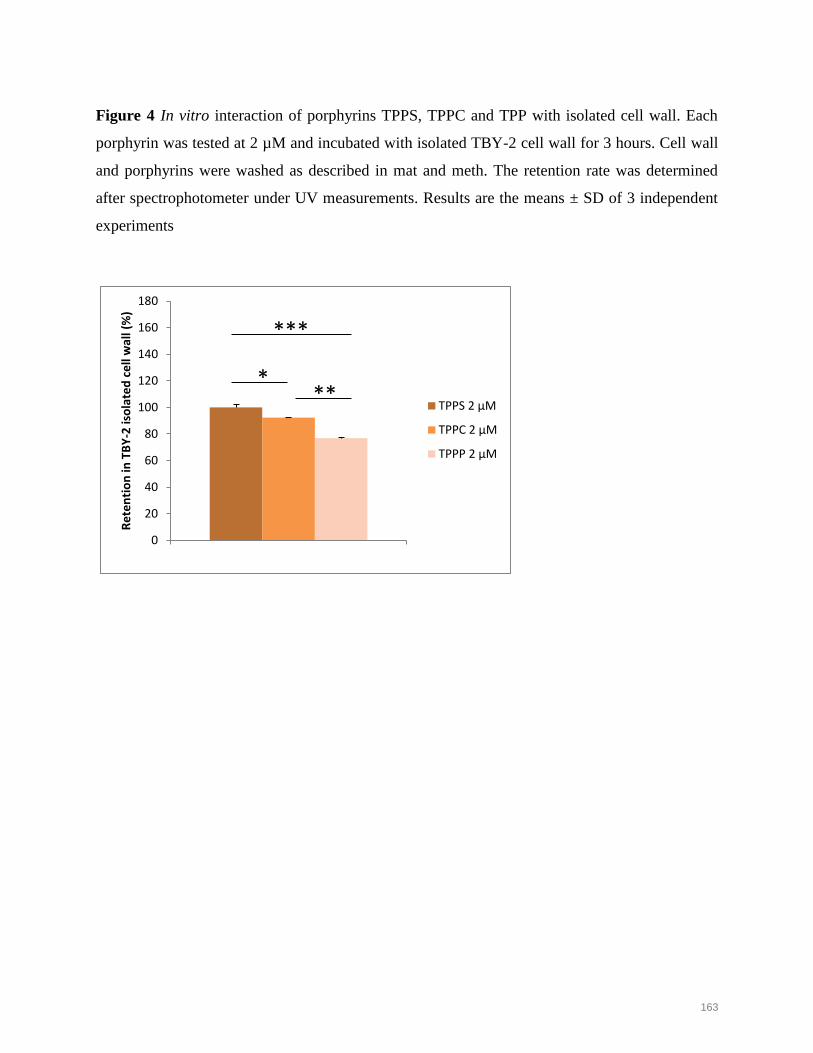
163
Figure 4 In vitro interaction of porphyrins TPPS, TPPC and TPP with isolated cell wall. Each
porphyrin was tested at 2 µM and incubated with isolated TBY-2 cell wall for 3 hours. Cell wall
and porphyrins were washed as described in mat and meth. The retention rate was determined
after spectrophotometer under UV measurements. Results are the means ± SD of 3 independent
experiments
0
20
40
60
80
100
120
140
160
180
1
Re
ten
tio
n in
TB
Y-2
iso
late
d c
ell
wal
l (%
)
TPPS 2 µM
TPPC 2 µM
TPPP 2 µM
* **
***

164
Figure 5 Responses of TBY-2 cells subjected to photo-activation of TPPS and TPPC. TPPS was
tested at 3.5 µM and TPPC at 2 µM. A) H₂O₂ production. B) lipid peroxidation assay through
MDA.quantification. C) Stress response determination via proline quantification. Ratio
calculation was determined from control not-treated cells that arbitrary corresponds to 100.
0
50
100
150
200
250
300
350
0 1 2 3
H₂O
₂ co
nte
nt
(%)
Illumination time (h)
TPPS
TPPC
0
50
100
150
200
250
300
350
0 1 2 3
MD
A c
on
ten
t (%
)
Illumination time (h)
TPPS
TPPC
0
50
100
150
200
0 1 2 3
Pro
line
co
nte
nt
(%)
Ilumination time (h)
TPPS
TPPC
A) B) C)
***
***
***
***
*** ***
** *

165
Figure 6 Antioxidant enzyme activities in TBY-2 cells treated with A) TPPS (3.5 µM) and B)
TPPC (2 µM) over illumination time. APX: ascorbate peroxidase, GPOX guaiacol peroxidase,
CAT catalase, SOD superoxide dismutase. Results are the mean ± SD of three independent
experiments.
20
40
60
80
100
120
140
160
0 1 2 3
Enzy
mat
ic a
ctiv
itie
s (%
)
Illumination time (h)
APX
CAT
GPOX
SOD
20
40
60
80
100
120
140
160
0 1 2 3
Enzy
mat
ic a
ctiv
itie
s (%
)
Illumination time (h)
APX
CAT
GPOX
SOD
A)
B)
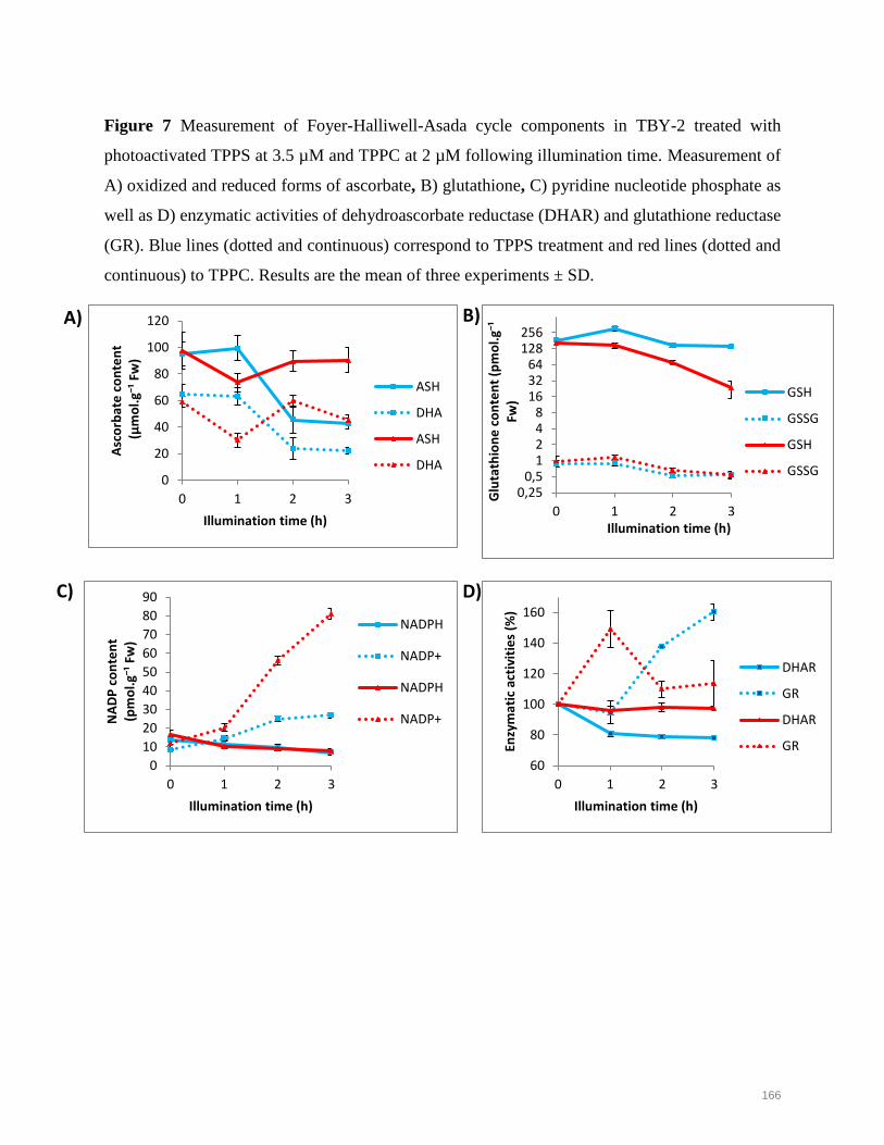
166
Figure 7 Measurement of Foyer-Halliwell-Asada cycle components in TBY-2 treated with
photoactivated TPPS at 3.5 µM and TPPC at 2 µM following illumination time. Measurement of
A) oxidized and reduced forms of ascorbate, B) glutathione, C) pyridine nucleotide phosphate as
well as D) enzymatic activities of dehydroascorbate reductase (DHAR) and glutathione reductase
(GR). Blue lines (dotted and continuous) correspond to TPPS treatment and red lines (dotted and
continuous) to TPPC. Results are the mean of three experiments ± SD.
0
20
40
60
80
100
120
0 1 2 3
Asc
orb
ate
co
nte
nt
(µm
ol.
g⁻¹ F
w)
Illumination time (h)
ASH
DHA
ASH
DHA
60
80
100
120
140
160
0 1 2 3
Enzy
mat
ic a
ctiv
itie
s (%
)
Illumination time (h)
DHAR
GR
DHAR
GR
0
10
20
30
40
50
60
70
80
90
0 1 2 3
NA
DP
co
nte
nt
(pm
ol.
g⁻¹ F
w)
Illumination time (h)
NADPH
NADP+
NADPH
NADP+
A)
D)
B)
C)
0,250,5
1248
163264
128256
0 1 2 3G
luta
thio
ne
co
nte
nt
(pm
ol.
g⁻¹
Fw)
Illumination time (h)
GSH
GSSG
GSH
GSSG

167
Figure 8 Apoptosis-like cell death induction in TBY-2 cells treated with photoactivated TPPS
and TPPC. Caspase-like protease activities A) morphological marker. B) CLK-1 and CLK-3
activities. C) Genomic DNA fragmentation. Lines 1 and 4: DNA from dark treated cells, lines 2
and 5: Fragmented DNA from light treated cells, Lines 3 and 6 DNA size ladder. Lines 1 and 2
and Lines 4 and 5 correspond to TPPS and TPPC, respectively Arrow indicated the typical 250
pb fragment scale.
0
5
10
15
20
25
1
CLK
-1 a
ctiv
ity
(nM
.µg⁻
¹ pro
tein
.h⁻¹
)
0
5
10
15
20
25
1
CLK
-3 a
ctiv
ity
(nM
.µg⁻
¹ pro
tein
.h⁻¹
)
*** *** ***
***
TPPC 2 µM Dark TPPC 2 µM 3 h Light
B)
50 µm
TPPS 3.5 µM Dark
50 µm
TPPS 3.5 µM 3 h Light
50 µm
50 µm
A)
C)
TPPC 2 µM TPPS 3.5 µM TPPC 2 µM TPPS 3.5 µM

168
Figure 9 Schematic representation of chemical structure and charge of anionic porphyrins and
their interaction and cross from cell wall to cytoplasm of TBY-2 cells in regard to crucial
importance of pH change from spent medium to cytoplasm. Carboxylate groups of TPPC,
sulfonate groups of TPPS and phosphonate groups of TPPP are shown in green, red and purple
respectively. At the pH of the spent medium (4.5), TPPC was fully neutral whereas 3 species of
TPPS coexisted as tetra-anionic, tri-anionic (with 2 positive charges at nitrogens of the core) and
di-anionic (with 2 positive charges at the N of the core). TPPP coexisted by means of two species
as tri-anionic and di-anionic as described for TPPS. In the apoplast, at weak acidic pH, TPPC can
be deprotonated to yield tetra-anionic species. While at neutral pH, TPPC should be completely
anionic in the cytoplasm. Phosphonate groups of TPPP have more complicated acido-basic
properties. Hence, TPPP could be more and more anionic in the apoplast as well as in the
cytoplasm (hydrogens represented in orange as -OH can loose protons). The three arrows (pink)
ascribed for TPPC indicate the fast cell wall crossing whereas one arrow suggest that the
diffusion of TPPS and TPPP was delayed. The tetra-anionic form of TPPC and TPPS and TPPP
got inside cytoplasm by endocytosis whereas the fully neutral TPPC diffuse through
phospholipids disruption upon photosensitization.
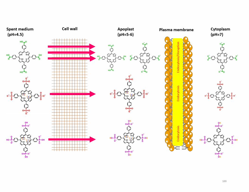
169

Results
Mohammad ISSAWI | Thèse de doctorat | Université de Limoges | 2018
Licence CC BY-NC-ND 3.0
170
Discussion and perspectives
Chapter II provided a dynamic study concerning plant photodynamic stress at cellular
level. For all the experiments, we isolated TBY-2 cells at the beginning of exponential growth
phase when cell spent medium reached a pH at 4.5 (Publication 4). As we discussed, such
medium is very complex and contained minerals, phytohormones, vitamins, carbohydrates,
proteins, organic acids. We also determined the chemical composition of TBY-2 outermost
layer known as the cell wall which is exposed to extracellular microenvironment. We showed
that cell wall from exponential growing cells was rich in pectin (around 55%) defined as
galacturonic acid-rich polymers assigning therefore cell wall negative charge (Table 2).
Table 2. Cell wall composition of TBY-2 cells.
The cell wall of young TBY-2 cells at the beginning of exponential phase (the growth phase at which we treat
them with porphyrins) was isolated and analysed. D3 and D4 cells were similar as they were rich in galacturonic
acid-rich pectin polymers and poor in cellulose. D3: 3 day-old cells, D4: 4 day-old cells.
In parallel, basing on the precedent data on cell wall, we monitored porphyrin
spectrum in the spent medium and performed titration experiment in methanol/water,
phosphate buffer and water in order to determine the precise structure of porphyrins at the
time when they just interact with the TBY-2 cells (Annex 4, Publication 5).

Results
Mohammad ISSAWI | Thèse de doctorat | Université de Limoges | 2018
Licence CC BY-NC-ND 3.0
171
All that insightful findings allowed us to decipher the interaction porphyrin-cell wall,
discussed in Publication 6. Strikingly, TPPC was fully neutralized at pH=4.5 and it seems that
it across the porous cell wall with a high diffusion rate interacting with surrounding polymers
via hydrogen bonds. Since sulfonic acid group is a very strong acid, TPPS remained anionic at
the peripheral side. However, its core showed protonation on the nitrogen atoms and its
absorption spectrum suggests that three species coexisted in the spent medium. We expected
that TPPS formed electrostatic interactions with negatively charged pectins without excluding
H bonds. In the other hand, TPPP showed the weakest interaction with cell wall. In addition,
TPPP-treated cells seen under confocal microscopy exhibited a very weak signal suggesting
that TPPP barely cross the cell wall. Indeed, TPPP have interesting binding properties as they
bear a strong H bond acceptor known for its biological role (P=O), in addition to an extra H
bond donor (P-OH) comparing to TPPS. Therefore, we think that TPPP interacted with
surrounding molecules in the spent medium before reaching the cell wall. (Kubat 2004;
Kralova 2014). Cell wall form a continuum with the apoplast, the compartment between cell
wall and plasma membrane where the pH fluctuates between 5 and 6 (Villiers and Kwak
2013). Facing the plasma membrane, TPPS was reported to get into the cytoplasm by
endocytosis whereas the aspect of TPPC is ambiguous at the pH range of the apoplasm. TPPC
side groups can be neutralized. Thus, two species must coexist bearing either neutral
carboxylic acid or anionic carboxylate groups. One hypothesis is that anionic species could be
internalized via endocytosis whereas neutral forms remained trapped at the plasma membrane
due to their symmetric, non-polar and hydrophobic features. However, they could reach the
cytoplasm upon photosensitization-mediated porous formation within plasma membrane
triggering phospholipids loss. That was supported by the increase in MDA level. Altogether,
we emphasized a dynamic trafficking of porphyrins within cellular barriers concluding that it
is highly linked to their chemical structures and behaviours.
Upon crossing cellular barriers, TPPS and TPPC were able to induce an oxidative
burst and overcome defense components as well as cause homeostasis disturbance by means
of membrane peroxidation, ROS generation, antioxidative enzyme inhibition and alteration of
buffering capacity. We finally discussed the ultimate outcome of photodynamic treatment and
determined the mode of cell death although plant cell death is clearly questionable (Gadjev et
al 2008, Reape and Mccabe, 2010, Van Doorn, 2011, Vartapetian et al 2011, Rantong and
Gunawardena 2015, Locato and De Gara 2018). Basing on morphological, biochemical and
molecular hallmarks, we revealed that photodynamic cell death in porphyrin-treated TBY-2 is

Results
Mohammad ISSAWI | Thèse de doctorat | Université de Limoges | 2018
Licence CC BY-NC-ND 3.0
172
in agreement with apoptosis-like plan cell death as we outlined the evidence of membrane
shrinkage, caspase-like activities and DNA fragmentation.
Taken together, novel information concerning structure-activity relationship,
interaction with plant cell wall, cellular photodynamic mechanisms were gathered. Our study
will be valuable for drug-cell interaction, rather it constitutes the first step for photosensitizers
application on plants or in the context of an agronomic APDT.
173
General conclusion
Mohammad ISSAWI | Thèse de doctorat | Université de Limoges | 2018
Licence CC BY-NC-ND 3.0
IV.General conclusion
In my PhD work, I conducted in parallel two studies to validate the possibility to use
APDT in agricultural domain. The first approach was performed on two unrelated plant
species: Arabidopsis and tomato to explore a possible agronomic application and the second
one on TBY-2 cells to explore the fundamental aspect of the photodynamic stress induced by
anionic porphryins. Our great question was: is it possible to imagine APDT strategy as a
valuable, eco-friendly and alternative approach in agriculture from both sides: plants and plant
pathogens? According to my work on the “green” side, the answer is yes, APDT could be a
reality. Nevertheless, further investigations on plant pathogens, adventices, soil wildlife and
water quality should be required to validate this approach. Some aspects of this tremendous
work will be monitored by Veronica Ambrosini (PhD student). Thus, our presented study on
plants is just the beginning of a promising story. Indeed, APDT applied to the field of
agriculture is may be still a dream but could be pretty soon a reality.

Perspectives and outlook
Mohammad ISSAWI | Thèse de doctorat | Université de Limoges | 2018
Licence CC BY-NC-ND 3.0
174
V. Perspectives and outlook
Some specific points must be considered to achieve this work on TBY-2 cell and PS
interactions and validation of APDT approach.
As molecular biologist, it is of importance now to decipher signaling process and/or
regulation of gene expression influenced by ROS generation underlying the differential
responses upon TPPC and TPPS photodynamic treatments in TBY-2 cells using molecular
tools such as transcriptomic profiling and/or proteomic approach. Indeed, as TBY-2 cells
share some common features with tumor cells and microorganisms such as high proliferation
rate and active metabolisms, such genetic studies will be meaningful for PDT applied to
cancerology.
According to our biological and chemical studies (Chapter II), porphyrin
characterization and TBY-2 cell wall interaction supported the involvement of structural
neutrality (carried by TPPC) in the interaction with plant cell barriers and the crossing
towards cytosolic compartment. To gain insight in neutrality hypothesis, we tested Tetrakis(4-
hydroxyphenyl) prophyrine (THPP) at very low concentration (0.1 µM) that led to a very
important cell death (47% cell death after 3h light exposure, data not shown). This neutral
porphyrin soluble in water is for the moment the strongest that we never tested, and it is in
favour to our hypothesis relative to the power of PS and its neutrality. Further experiments are
to be made to confirm this finding.
As cellular biologist working hand by hand with chemist, it will be amazing to follow
porphyrin traffic inside the cells. As we showed in planta or TBY-2 we can already follow
porphyrins by confocal microscopy under spectral acquisition. Nevertheless, porphyrin
fluorescence emission at around 640 nm remains very weak and quite difficult to detect. In
order to overcome such drawback, molecular dyads were synthesized before I started my
PhD. It consists in porphyrin conjugation with fluorophores as fluorescein resulting in a
stronger fluorescence when light excited (Rezazgui et al. 2016). However, these smart
constructs were not water-soluble and then not suitable for common use in biology (Figure
11).
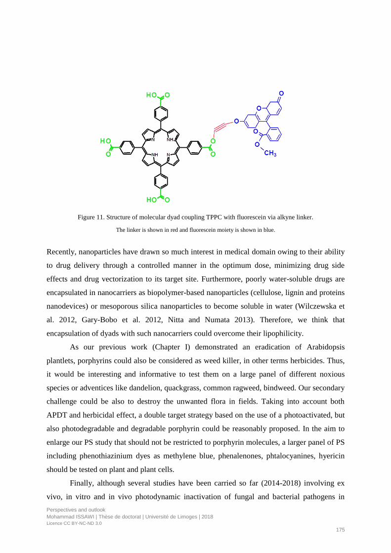
Perspectives and outlook
Mohammad ISSAWI | Thèse de doctorat | Université de Limoges | 2018
Licence CC BY-NC-ND 3.0
175
Figure 11. Structure of molecular dyad coupling TPPC with fluorescein via alkyne linker.
The linker is shown in red and fluorescein moiety is shown in blue.
Recently, nanoparticles have drawn so much interest in medical domain owing to their ability
to drug delivery through a controlled manner in the optimum dose, minimizing drug side
effects and drug vectorization to its target site. Furthermore, poorly water-soluble drugs are
encapsulated in nanocarriers as biopolymer-based nanoparticles (cellulose, lignin and proteins
nanodevices) or mesoporous silica nanoparticles to become soluble in water (Wilczewska et
al. 2012, Gary-Bobo et al. 2012, Nitta and Numata 2013). Therefore, we think that
encapsulation of dyads with such nanocarriers could overcome their lipophilicity.
As our previous work (Chapter I) demonstrated an eradication of Arabidopsis
plantlets, porphyrins could also be considered as weed killer, in other terms herbicides. Thus,
it would be interesting and informative to test them on a large panel of different noxious
species or adventices like dandelion, quackgrass, common ragweed, bindweed. Our secondary
challenge could be also to destroy the unwanted flora in fields. Taking into account both
APDT and herbicidal effect, a double target strategy based on the use of a photoactivated, but
also photodegradable and degradable porphyrin could be reasonably proposed. In the aim to
enlarge our PS study that should not be restricted to porphyrin molecules, a larger panel of PS
including phenothiazinium dyes as methylene blue, phenalenones, phtalocyanines, hyericin
should be tested on plant and plant cells.
Finally, although several studies have been carried so far (2014-2018) involving ex
vivo, in vitro and in vivo photodynamic inactivation of fungal and bacterial pathogens in

Perspectives and outlook
Mohammad ISSAWI | Thèse de doctorat | Université de Limoges | 2018
Licence CC BY-NC-ND 3.0
176
addition to PS spotting on their corresponding host leaves, host responses as well as
evaluation of PS administration methods are missing. Until now, these studies are restricted to
perennial plants that hindered PS activity due to their anatomical strength excepted for the
herbaceous low-growing strawberry that were damaged upon sunlight-activated coumarins
and furocoumarins (De Menezes et al. 2014a). Our group will focus its study on the grey
mold Botrytis cinerea and its corresponding hosts (grapevine and potato plantlets) (V.
Ambrosini, PhD).
References
Mohammad ISSAWI | Thèse de doctorat | Université de Limoges | 2018
Licence CC BY-NC-ND 3.0
177
References
Abrahamse, H., and Hamblin, M.R. 2016. New photosensitizers for photodynamic therapy.
Biochemical Journal 473: 347–364.
Agostinis, P., Berg, K., Cengel, K.A., Foster, T.H., Girotti, A.W., Gollnick, S.O., Hahn, S.M.,
Hamblin, M.R., Juzeniene, A., Kessel, D., et al. 2011. Photodynamic therapy of cancer: an
update. CA: A Cancer Journal for Clinicians 61: 250–281.
Almeida, A., Cunha, A., Faustino, M.A.F., Tomé, A.C., and Neves, M.G.P.M.S. 2011.
Porphyrins as Antimicrobial Photosensitizing Agents In: Hamblin, M.R., and G. Jori, Eds.,
Comprehensive Series in Photochemical & Photobiological Sciences. Cambridge: Royal
Society of Chemistry, pp. 83–160.
Alves, E., Faustino, M.A.F., Neves, M.G.P.M.S., Cunha, Â., Nadais, H., and Almeida, A.
2015. Potential applications of porphyrins in photodynamic inactivation beyond the medical
scope. Journal of Photochemistry and Photobiology C: Photochemistry Reviews 22: 34–57.
Arrojado, C., Pereira, C., Tomé, J.P.C., Faustino, M.A.F., Neves, M.G.P.M.S., Tomé, A.C.,
Cavaleiro, J.A.S., Cunha, Â., Calado, R., Gomes, N.C.M., et al. 2011. Applicability of
photodynamic antimicrobial chemotherapy as an alternative to inactivate fish pathogenic
bacteria in aquaculture systems. Photochemical & Photobiological Sciences 10: 1691-1701.
Azizullah, A., Rehman, Z.U., Ali, I., Murad, W., Muhammad, N., Ullah, W., and Häder, D.-P.
2014. Chlorophyll derivatives can be an efficient weapon in the fight against dengue.
Parasitology Research 113: 4321–4326.
Azzouzi, A.R., Barret, E., Moore, C.M., Villers, A., Allen, C., Scherz, A., Muir, G., Wildt, M.
de, Barber, N.J., Lebdai, S., et al. 2013. TOOKAD ® Soluble vascular-targeted photodynamic
(VTP) therapy: determination of optimal treatment conditions and assessment of effects in
patients with localised prostate cancer. BJU International 112: 766–774.
Babilas, P., Schreml, S., Landthaler, M., and Szeimies, R.-M. 2010. Photodynamic therapy in
dermatology: state-of-the-art. Photodermatology, Photoimmunology & Photomedicine 26:
118–132.
Babu, P.S.S., Manu, P.M., Dhanya, T.J., Tapas, P., Meera, R.N., Surendran, A., Aneesh,
K.A., Vadakkancheril, S.J., Ramaiah, D., Nair, S.A., et al. 2017. Bis(3,5-diiodo-2,4,6-
trihydroxyphenyl) squaraine photodynamic therapy disrupts redox homeostasis and induce
mitochondria-mediated apoptosis in human breast cancer cells. Scientific Reports 7: 42126-
42140.
Baptista, M.S., Cadet, J., Di Mascio, P., Ghogare, A.A., Greer, A., Hamblin, M.R., Lorente,
C., Nunez, S.C., Ribeiro, M.S., Thomas, A.H., et al. 2017. Type I and type ii photosensitized
oxidation reactions: guidelines and mechanistic pathways. Photochemistry and Photobiology
93: 912–919.
Barker, C.A., Zeng, X., Bettington, S., Batsanov, A.S., Bryce, M.R., and Beeby, A. 2007.
Porphyrin, phthalocyanine and porphyrazine derivatives with multifluorenyl substituents as
References
Mohammad ISSAWI | Thèse de doctorat | Université de Limoges | 2018
Licence CC BY-NC-ND 3.0
178
efficient deep-red emitters. Chemistry - A European Journal 13: 6710–6717.
Bartolomeu, M., Reis, S., Fontes, M., Neves, M., Faustino, M., and Almeida, A. 2017.
Photodynamic Action against Wastewater Microorganisms and Chemical Pollutants: An Effective
Approach with Low Environmental Impact. Water 9: 630-647.
Bassham, J.A. 1959. Photosynthesis. Journal of Chemical Education 36: 549-554.
Beale, S.I. 1990. Biosynthesis of the tetrapyrrole pigment precursor, δ-aminolevulinic acid,
from glutamate. Plant Physiology 93: 1273–1279.
Benov, L. 2015. Photodynamic Therapy: Current Status and Future Directions. Medical Principles
and Practice 24: 14–28.
Berg, K., Selbo, P.K., Weyergang, A., Dietze, A., Prasmickaite, L., Bonsted, A., Engesaeter,
B., Angell-petersen, E., Warloe, T., and Frandsen, N. 2005. Porphyrin-related photosensitizers
for cancer imaging and therapeutic applications. Journal of Microscopy 218: 133–147.
Brzezowski, P., Richter, A.S., and Grimm, B. 2015. Regulation and function of tetrapyrrole
biosynthesis in plants and algae. Biochimica et Biophysica Acta (BBA) - Bioenergetics 1847:
968–985.
Buchovec, I., Lukseviciute, V., Marsalka, A., Reklaitis, I., and Luksiene, Z. 2016. Effective
photosensitization-based inactivation of Gram (−) food pathogens and molds using the
chlorophyllin–chitosan complex: towards photoactive edible coatings to preserve
strawberries. Photochemical & Photobiological Sciences 15: 506–516.
Buck, S.T.G., Bettanin, F., Orestes, E., Homem-de-Mello, P., Imasato, H., Viana, R.B.,
Perussi, J.R., and Silva, A.B.F. da 2017. Photodynamic Efficiency of Xanthene Dyes and
Their Phototoxicity against a Carcinoma Cell Line: A Computational and Experimental
Study. Journal of Chemistry 2017: 1–9.
Carvalho, C.M., Tome, J.P., Faustino, M.A., Neves, M.G., Tome, A.C., Cavaleiro, J.A.,
Costa, L., Alves, E., Oliveira, A., and Cunha, A. 2009. Antimicrobial photodynamic activity
of porphyrin derivatives: potential application on medical and water disinfection. Journal of
Porphyrins and Phthalocyanines 13: 574–577.
Costa, L., Alves, E., Carvalho, C.M.B., Tomé, J.P.C., Faustino, M.A.F., Neves, M.G.P.M.S.,
Tomé, A.C., Cavaleiro, J.A.S., Cunha, Â., and Almeida, A. 2008. Sewage bacteriophage
photoinactivation by cationic porphyrins: a study of charge effect. Photochemical &
Photobiological Sciences 7: 415-423.
Dąbrowski, J.M., and Arnaut, L.G. 2015. Photodynamic therapy (PDT) of cancer: from local
to systemic treatment. Photochemical & Photobiological Sciences 14: 1765–1780.
Dąbrowski, J.M., Pucelik, B., Regiel-Futyra, A., Brindell, M., Mazuryk, O., Kyzioł, A.,
Stochel, G., Macyk, W., and Arnaut, L.G. 2016. Engineering of relevant photodynamic
processes through structural modifications of metallotetrapyrrolic photosensitizers.
Coordination Chemistry Reviews 325: 67–101.
Dagani, R. 1992. fullerenes in nature: C60 and C70 found in ancient Russian rock. Chemical
& Engineering News 70: p, 6.
References
Mohammad ISSAWI | Thèse de doctorat | Université de Limoges | 2018
Licence CC BY-NC-ND 3.0
179
Daub, M.E. 1982. Cercosporin, a photosensitizing toxin from Cercospora species.
Phytopathology 72: 370-374.
Daub, M.E., Herrero, S., and Chung, K.-R. 2005. Photoactivated perylenequinone toxins in
fungal pathogenesis of plants. FEMS Microbiology Letters 252: 197–206.
Davies, K.S., Linder, M.K., Kryman M.W., and Detty, M.R. 2016. Extended rhodamine
photosensitizers for photodynamic therapy of cancer cells. Bioorganic & Medicinal Chemistry
24: 3908–3917.
Delcanale, P., Montali, C., Rodríguez-Amigo, B., Abbruzzetti, S., Bruno, S., Bianchini, P.,
Diaspro, A., Agut, M., Nonell, S., and Viappiani, C. 2016. Zinc-substituted myoglobin is a
naturally occurring photo-antimicrobial agent with potential applications in food
decontamination. Journal of Agricultural and Food Chemistry 64: 8633–8639.
De Menezes, H.D., Pereira, A.C., Brancini, G.T.P., Leão, H.C., Massola Júnior, N.S.,
Bachmann,
L., et al. 2014a. Furocoumarins and coumarins photoinactivate Colletotrichum acutatum and
Aspergillus nidulans fungi under solar radiation. Journal of photochemistry and Photobiology
B: Biology 131: 74–83.
De Menezes, H.D., Rodrigues, G.B., Teixeira, S.P., Massola, N.S., Bachmann, L.,
Wainwright, M., et al. 2014b. In vitro photodynamic inactivation of plant-pathogenic fungi
Colletotrichum acutatum and Colletotrichum gloeosporioides with novel phenothiazinium
photosensitizers. Applied and Environmental Microbiology 80: 1623–1632.
De Menezes, H.D., Tonani, L., Bachmann, L., Wainwright, M., Braga, G.U.L., Marcia Regina
Kress, M.R.V.Z. 2016. Photodynamic treatment with phenothiazinium photosensitizers kills
both ungerminated and germinated microconidia of the pathogenic fungi Fusarium
oxysporum, Fusarium moniliforme and Fusarium solani. Journal of Photochemistry and
Photobiology B: Biology 164: 1-12.
DiCosmo, F., Towers G.H.N. and Lam, j. 1982. Photo-induced fungicidal activity elicited by
naturally occurring thiophene derivatives. Journal of Pest science 13: 589-594.
Ding, H., Yu, H., Dong, Y., Tian, R., Huang, G., Boothman, D.A., Sumer, B.D., and Gao, J.
2011. Photoactivation switch from type II to type I reactions by electron-rich micelles for
improved photodynamic therapy of cancer cells under hypoxia. Journal of Controlled Release
156: 276–280.
Drogat, N., Barrière, M., Granet, R., Sol, V., and Krausz, P. 2011. High yield preparation of
purpurin-18 from Spirulina maxima. Dyes and Pigments 88: 125–127.
Drogat, N., Gady, C., Granet, R., and Sol, V. 2013. Design and synthesis of water-soluble
polyaminated chlorins and bacteriochlorins with near-infrared absorption. Dyes and Pigments
98: 609-614.
Dumoulin, F., Durmuş, M., Ahsen, V., and Nyokong, T. 2010. Synthetic pathways to water-
soluble phthalocyanines and close analogs. Coordination Chemistry Reviews 254: 2792–2847.
Durantini, A.M., Heredia, D.A., Durantini, J.E., and Durantini, E.N. 2018. BODIPYs to the
References
Mohammad ISSAWI | Thèse de doctorat | Université de Limoges | 2018
Licence CC BY-NC-ND 3.0
180
rescue: Potential applications in photodynamic inactivation. European Journal of Medicinal
Chemistry 144: 651–661.
Ebermann, R., Alth, G., Kreitner, M., and Kubin, A. 1996. Natural products derived from
plants as potential drugs for the photodynamic destruction of tumor cells. Journal of
Photochemistry and Photobiology B: Biology 36: 95–97.
Evstigneev, V.B. 1965. On the mechanism of photosensitizing action of chlorophyll.
Photochemistry and Photobiology 4: 171–182.
Fayyaz, F., Rahimi, R., Rassa, M., and Yaghoobi, R.Z. 2015. Photodynamic Antimicrobial
Chemotherapy, A Pathway for Photo-Inactivation of Bacteria by Porphyrin Compounds In:
Proceedings of the 1st International Electronic Conference on Molecular Science, Sciforum
Electronic Conference Series.
Feese, E., Sadeghifar, H., Gracz, H.S., Argyropoulos, D.S., and Ghiladi, R.A. 2011.
Photobactericidal porphyrin-cellulose nanocrystals: synthesis, characterization, and
antimicrobial properties. Biomacromolecules 12: 3528–3539.
Fracarolli, L., Rodrigues, G.B., Pereira, A.C., Massola Júnior, N.S., Silva-Junior, G.J.,
Bachmann, L., et al. 2016. Inactivation of plant-pathogenic fungus Colletotrichum acutatum
with natural plant-produced photosensitizers under solar radiation. Journal of Photochemistry
and Photobiologt B: Biology 162: 402–411.
Gadjev, I., Stone, J.M., and Gechev, T.S. 2008. Programmed cell death in plants. In:
International Review of Cell and Molecular Biology. Elsevier, pp. 87–144.
Gary-Bobo, M., Hocine, O., Brevet, D., Maynadier, M., Raehm, L., Richeter, S., Charasson,
V., Loock, B., Morère, A., Maillard, P., et al. 2012. Cancer therapy improvement with
mesoporous silica nanoparticles combining targeting, drug delivery and PDT. International
Journal of Pharmaceutics 423: 509–515.
Glueck, M., Schamberger, B., Eckl, P., and Plaetzer, K. 2017. New horizons in
microbiological food safety: Photodynamic Decontamination based on a curcumin derivative.
Photochemical & Photobiological Sciences 16: 1784–1791.
Gonzales, J.C., Brancini, G.T.P., Rodrigues, G.B., Silva-Junior, G.J., Bachmann, L.,
Wainwright, M., et al. 2017. Photodynamic inactivation of conidia of the fungus
Colletotrichum abscissum on Citrus sinensis plants with methylene blue under solar radiation.
J. Photochemistry and Photobiology B: Biology. 176: 54-61.
Grimm, B. 1998. Novel insights in the control of tetrapyrrole metabolism of higher plants.
Current Opinion in Plant Biology 1: 245–250.
Guldi, D.M., Mody, T.D., Gerasimchuk, N.N., Magda, D., and Sessler, J.L. 2000. Influence of
Large Metal Cations on the Photophysical Properties of Texaphyrin, a Rigid Aromatic Chromophore.
Journal of the American Chemical Society 122: 8289–8298.
Guillaumot, D., Issawi, M., Da Silva, A., Leroy-Lhez, S., Sol, V., and Riou, C. 2016.
Synergistic enhancement of tolerance mechanisms in response to photoactivation of cationic
References
Mohammad ISSAWI | Thèse de doctorat | Université de Limoges | 2018
Licence CC BY-NC-ND 3.0
181
tetra (N-methylpyridyl) porphyrins in tomato plantlets. Journal of Photochemistry and
Photobiology B: Biology 156: 69–78.
Häder, D.-P., Schmidl, J., Hilbig, R., Oberle, M., Wedekind, H., and Richter, P. 2016.
Fighting fish parasites with photodynamically active chlorophyllin. Parasitology Research
115: 2277–2283.
Hager, B., Strauss, W.S.L., and Falk, H. 2009. Cationic Hypericin Derivatives as Novel Agents
with Photobactericidal Activity: Synthesis and Photodynamic Inactivation of Propionibacterium
acnes. Photochemistry and Photobiology 85: 1201–1206.
Hazen, M.J., Villaneuva, A., and Stockert, J.C. 1987. Induction of sister chromatide echanges
in Allium cepa meristematic cells exposed to meso-tetra (4-pyridyl) porphine and
hematoporphyrin photoradiation. Journal of Photochemistry and Photobiology B: Biology.
46: 463-467.
Hazen, M.J., and Gutierrez-Gonzalvez, M.G. (1988). UV-mediated toxic bioactivity of
harmine in the meristematic cells of Allium cepa. Mutagenesis 3: 333–335.
Henderson, B.W., Sumlin, A.B., Owczarczak, B.L., and Dougherty, T.J. 1991.
Bacteriochlorophyll-a as photosensitizer for photodynamic treatment of transplantable murine
tumors. Journal of Photochemistry and Photobiology B: Biology 10: 303–313.
Hill, R. 1937. Oxygen Evolved by Isolated Chloroplasts. Nature 139: 881–882.
Huang, H.C., and Hasan, T. 2014. The nano world in photodynamic therapy. Austin Journal
of Nanomedicine & Nanotechnology 2: 4-8.
Hong, E.J., Choi, D.G., and Shim, M.S. 2016. Targeted and effective photodynamic therapy
for cancer using functionalized nanomaterials. Acta Pharmaceutica Sinica B 6: 297–307.
Horne, T.K., and Cronjé, M.J. 2014. Novel Porphyrazine Derivatives show Promise for
Photodynamic Therapy despite Restrictions in Hydrophilicity. Photochemistry and Photobiology
90: 648–658.
Huang, L., Huang, Y.-Y., Mroz, P., Tegos, G.P., Zhiyentayev, T., Sharma, S.K., Lu, Z.,
Balasubramanian, T., Krayer, M., Ruzie, C., et al. 2010. Stable synthetic cationic
bacteriochlorins as selective antimicrobial photosensitizers. Antimicrobial Agents and
Chemotherapy 54: 3834–3841.
Hohmann-Marriott, M.F., and Blankenship, R.E. 2011. Evolution of photosynthesis. Annual
Review of Plant Biology 62: 515–548.
Hsu, Y.-C., Chiang, C.-P., Chen, J.W., Lee, J.-W., and How, M.-H. 2010. Topical
chlorophyll-pheophytin derivative-mediated photodynamic therapy for DMBA-induced
hamster buccal pouch premaligant lesions: an in vivo study In: Kessel, D.H., Ed., pp. 755118-
755127.
Issawi, M., Muhieddine, M., Girard, C., Sol, V. and Riou, C. 2017. Unexpected features of
exponentially growinh Tobacco Bright Yellow-2 cell suspension culture in relation to
excrected extracellular polysaccharides and cell wall composition. Glycoconjugate Journal
References
Mohammad ISSAWI | Thèse de doctorat | Université de Limoges | 2018
Licence CC BY-NC-ND 3.0
182
34: 585-590.
Issawi, M., Guillaumot, D., Sol, V., and Riou, C. 2018. Responses of an adventitious fast growing plant to photodynamic stress: comparative study of anionic and cationic porphyrin
effect on Arabidopsis thaliana. Physiologia Plantarum 162: 379-390.
Jesus, V., Martins, D., Branco, T., Valério, N., Neves, M.G.P.M.S., Faustino, M.A.F., et al.
2018. An insight into the photodynamic approach versus copper formulations in the control of
Pseudomonas syringae pv. actinidiae in kiwi plants. Photochemical and Photobiological
Sciences. 17: 180-191.
Josefsen, L.B., and Boyle, R.W. 2008. photodynamic therapy and the development of metal-
based photosensitisers. Metal-Based Drugs 2008: 267109–267132.
Joshi, P., and Saenz, C. 2013. Recent Advances in developing improved agents for
photodynamic therapy, in Handbook of Photomedicine, 2011, pp. 227-267.
Junqueira, J.C., Ribeiro, M.A., Rossoni, R.D., Barbosa, J.O., Querido, S.M.R., and Jorge,
A.O.C. 2010. Antimicrobial photodynamic therapy: photodynamic antimicrobial effects of
malachite green on Staphylococcus , Enterobacteriaceae, and Candida. Photomedicine and
Laser Surgery 28: 67–72.
Juzenienne, A. 2009. Chlorin e6-based photosensitizers for photodynamic
therapy and photodiagnosis. Photodiagnosis and Photodynamic Therapy 6: 94-96.
Kataoka, H., Nishie, H., Hayashi, N., Tanaka, M., Nomoto, A., Yano, S., and Joh, T. 2017.
New photodynamic therapy with next-generation photosensitizers. Annals of Translational
Medicine 5: 183–183.
Kessel, D., and Oleinick, N.L. 2018. Cell Death Pathways Associated with Photodynamic
Therapy: An Update. Photochemistry and Photobiology 94: 213–218.
Kharkwal, G.B., Sharma, S.K., Huang, Y.-Y., Dai, T., and Hamblin, M.R. 2011.
Photodynamic therapy for infections: Clinical applications: PHOTODYNAMIC THERAPY
FOR INFECTIONS. Lasers in Surgery and Medicine 43: 755–767.
Konopka, K., and Goslinski, T. 2007. Photodynamic Therapy in Dentistry. Journal of Dental
Research 86: 694–707.
Králová, J., Kejík, Z., Bříza, T., Kaplánek, R., Záruba, K., Martásek, P., and Král, V. (2014)
Design, Synthesis, Selective Recognition Properties and Targeted Drug Delivery Application
in Handbook of Porphyrin Science With Applications to Chemistry, Physics, Materials
Science, Engineering, Biology and Medicine. Volume 33: Applications — Part II (Kadish,
K.M., Smith, K.M., and Guilard, R. eds.) pp. 1-75.
Krasnovsky, A.A., and Brin, G.P. 1947. Photosensitizing agent of Mg-phtalocyanine and
chlorophyll in solution.
Krieger-Liszkay, A. 2004. Singlet oxygen production in photosynthesis. Journal of
Experimental Botany 56: 337–346.
Kubat, P., Lang, K., and Azenbacher Jr., P. 2004. Modulation of porphyrin binding to swrum

References
Mohammad ISSAWI | Thèse de doctorat | Université de Limoges | 2018
Licence CC BY-NC-ND 3.0
183
albumin by pH. Biochimica et Biophysica Acta 1670: 40-48.
Lhotáková, Y., Plíštil, L., Morávková, A., Kubát, P., Lang, K., Forstová, J., and Mosinger, J.
2012. Virucidal Nanofiber Textiles Based on Photosensitized Production of Singlet Oxygen.
PLoS ONE 7: 11-20.
Liu, Y.C., Tu, S.Y., Lin, H.Y. 2015. Evaluation of the practicality of melanin as a
photodynamic-inactivation agent photosensitizer by its nanonization. Journal of
Photopolymer Science and Technology 28: 739-746.
Locato, V., and De Gara, L. 2018. Programmed Cell Death in Plants: An Overview, in: De
Gara, L., and V. Locato, Eds., Plant Programmed Cell Death. New York, NY: Springer New
York, pp. 1–8.
Luksiene, Z., and Brovko, L. 2013. Antibacterial photosensitization-based treatment for food
safety. Food Engineering Reviews 5: 185–199.
Lukšienė, Ž., Danilčenko, H., Tarasevičienė, Ž., Anusevičius, Ž., Marozienė, A., and
Nivinskas, H. 2007. New approach to the fungal decontamination of wheat used for wheat
sprouts: Effects of aminolevulinic acid. International Journal of Food Microbiology 116:
153–158.
Maisch, T., Szeimies, R.-M., Lehn, N., and Abels, C. 2005. Antibacterial photodynamic
therapy. A new treatment for bacterial skin diseases? Der Hautarzt 56: 1048–1055.
Maisch, T., Eichner, A., Späth, A., Gollmer, A., König, B., Regensburger, J., and Bäumler,
W. 2014. Fast and effective photodynamic inactivation of multiresistant bacteria by cationic
riboflavin derivatives. PLoS ONE 9: 1-18.
Mandal, A.K., Sahin, T., Liu, M., Lindsey, J.S., Bocian, D.F., and Holten, D. 2016.
Photophysical comparisons of PEGylated porphyrins, chlorins and bacteriochlorins in water.
New Journal of Chemistry 40: 9648–9656.
Master, A., Livingston, M., and Sen Gupta, A. 2013. Photodynamic nanomedicine in the
treatment of solid tumors: Perspectives and challenges. Journal of Controlled Release 168: 8-
40.
Matsubara, T., Kusuzaki, K., Matsumine, A., Murata, H., Marunaka, Y., Hosogi, S., Uchida,
A., and Sudo, A. 2010. Photodynamic therapy with acridine orange in musculoskeletal
sarcomas. Bone & Joint Journal 92: 760–762.
McCoy, C.P., O’Neil, E.J., Cowley, J.F., Carson, L., De Baróid, Á.T., Gdowski, G.T.,
Gorman, S.P., and Jones, D.S. 2014. Photodynamic antimicrobial polymers for infection
control. PLoS ONE 9: 108500-108511.
McCullagh, C. and Robertson, P.K.J. 2006. Photosensitized Destruction of Chlorella vulgaris
by Methylene blue or nuclear fast red combined with hydrogen peroxide under visible light
irradiation. Environmental Science & Technology 40: 2421–2425.
Molero, M.L., and Hazen, M.J. 1988. Photodynamic effect of acridine orange, eosin Y and
orcein in a plant system in vivo measured by the sister chromatid exchanges test. Journal of
Plant Physiology 132: 636–637.
References
Mohammad ISSAWI | Thèse de doctorat | Université de Limoges | 2018
Licence CC BY-NC-ND 3.0
184
Molero, M.L., Hazen, M.J., and Stockert, J.C. 1985. Photodynamic effect of berberine sulfate
on the growth rate of Allium cepa roots. Jourmal of Plant Physiology. 120: 91–94.
Monteiro, J.S.C., Oliveira, S.C.P.S. de, Pires Santos, G.M., Pires Sampaio, F.J., Pinheiro
Soares, L.G., and Pinheiro, A.L.B. 2017. Effectiveness of antimicrobial photodynamic
therapy (AmPDT) on Staphylococcus aureus using phenothiazine compound with red laser.
Lasers in Medical Science 32: 29–34.
Montoya, S.C.N., Comini, L.R., Sarmiento, M., Becerra, C., Albesa, I., Argüello, G.A., and
Cabrera, J.L. 2005. Natural anthraquinones probed as Type I and Type II photosensitizers:
singlet oxygen and superoxide anion production. Journal of Photochemistry and Photobiology
B: Biology 78: 77–83.
Mroz, P., Yaroslavsky, A., Kharkwal, G.B., and Hamblin, M.R. 2011. Cell Death Pathways in
Photodynamic Therapy of Cancer. Cancers 3: 2516–2539.
Muehler, D., Sommer, K., Wennige, S., Hiller, K.-A., Cieplik, F., Maisch, T., and Späth, A.
2017. Light-activated phenalen-1-one bactericides: efficacy, toxicity and mechanism
compared with benzalkonium chloride. Future Microbiology 12: 1297–1310.
Nagata, T., Nemoto, Y., and Hasezawa, S. 1992. Tobacco BY-2 cell line as the “Hela” cell in
the cell biology of higher plants. International Review of Cytology 132: 1-31.
Nazir, M., El Maddah, F., Kehraus, S., Egereva, E., Piel, J., Brachmannb, A.O., and König,
G.M. 2015. Phenalenones: insight into the biosynthesis of polyketides from the marine alga-
derived fungus Coniothyrium cereale. Organic & Biomolecular Chemistry 13: 8071-8080.
Nitta, S., and Numata, K. 2013. Biopolymer-Based Nanoparticles for Drug/Gene Delivery and
Tissue Engineering. International Journal of Molecular Sciences 14: 1629–1654.
Ogawa, K., and Kobuke, Y. 2008. Recent advances in two-photon photodynamic therapy.
Anti-Cancer Agents in Medicinal Chemistry (Formerly Current Medicinal Chemistry-Anti-
Cancer Agents) 8: 269–279.
Orlob, G.B. 1967. Inactivation of purified plant viruses and their nucleic acids by
photosensitizing dyes. Virology 31: 402–413.
Ormond, A., and Freeman, H. 2013. Dye Sensitizers for Photodynamic Therapy. Materials 6:
817–840.
Plaetzer, K., Berneburg, M., Kiesslich, T., and Maisch, T. 2013. New applications of
photodynamic therapy in biomedicine and biotechnology. BioMed Research International 1–
3.
Pohl, J., Saltsman, I., Mahammed, A., Gross, Z. and Röder, B. 2014. Inhibition of green algae
growth by corrole-based photosensitizers. Journal of Applied Microbiology 118: 305–312.
Rantong, G., and Gunawardena, A.H.L.A.N. 2015. Programmed cell death: genes involved in
signaling, regulation, and execution in plants and animals. Botany 93: 193–210.
References
Mohammad ISSAWI | Thèse de doctorat | Université de Limoges | 2018
Licence CC BY-NC-ND 3.0
185
Reape, T.J., and McCabe, P.F. 2010. Apoptotic-like regulation of programmed cell death in
plants. Apoptosis 15: 249–256.
Rezazgui, O., Trouillas, P., Qiu S.H., Siegler, B., Gierschner, J., Leroy-Lhez, S. 2016.
Synthesis and conformation of a novel fluorescein-Zn-porphyrin dyad and intramolecular
energy transfer. New Journal of Chemistry 40: 3843-3856.
Richter, A.S., and Grimm, B. 2013. Thiol-based redox control of enzymes involved in the
tetrapyrrole biosynthesis pathway in plants. Frontiers in Plant Science 4: 1-11.
Ringot, C., Sol, V., Barrière, M., Saad, N., Bressollier, P., Granet, R., Couleaud, P., Frochot,
C., and Krausz, P. 2011. Triazinyl Porphyrin-Based Photoactive Cotton Fabrics: Preparation,
Characterization, and Antibacterial Activity. Biomacromolecules 12: 1716–1723.
Riou, C., Calliste, C.A., Da Silva, A., Guillaumot, D., Rezazgui, O., Sol, V., et al. 2014.
Anionic porphyrin as a new powerful cell death inducer of Tobacco Bright Yellow-2 cells.
Photochemical and Photobiological Sciences 13: 621-625.
Rishi, P., and Agarwal, V. 1972. Current Role of Photodynamic Therapy in Ophthalmic Practice.
Lancet 2: 1175–7.
Robertson, C.A., Evans, D.H., and Abrahamse, H. 2009. Photodynamic therapy (PDT): A
short review on cellular mechanisms and cancer research applications for PDT. Journal of
Photochemistry and Photobiology B: Biology 96: 1–8.
Rodionov, D.A., Vitreschak, A.G., Mironov, A.A., and Gelfand, M.S. 2003. Comparative
Genomics of the Vitamin B 12 Metabolism and Regulation in Prokaryotes. Journal of Biological
Chemistry 278: 41148–41159.
Salci, A., and Toprak, M. 2017. Spectroscopic investigations on the binding of Pyronin Y to
human serum albumin. Journal of Biomolecular Structure and Dynamics 35: 8–16.
Senge, M.O., MacGowan, S.A., and O’Brien, J.M. 2015. Conformational control of cofactors
in nature – the influence of protein-induced macrocycle distortion on the biological function
of tetrapyrroles. Chemical Communications 51: 17031–17063.
Senge, M., Ryan, A., Letchford, K., MacGowan, S., and Mielke, T. 2014. Chlorophylls,
Symmetry, Chirality, and Photosynthesis. Symmetry 6: 781–843.
Sessler, J.L., and Miller, R.A. 2000. Texaphyrins: new drugs with diverse clinical applications
in radiation and photodynamic therapy. Biochemical Pharmacology 59: 733–739
Shin, H., Yoon, J.-S., Koh, W., Kim, J.Y., Kim, C.-H., Han, K.M., Kim, E.J., and Kwon, O.
2016. Nonpigmented hair removal using photodynamic therapy in animal model:
nonpigmented hair removal using photodynamic therapy. Lasers in Surgery and Medicine 48:
748–762.
Sieber, F., Krueger, G.J., O’Brien, J.M., Schober, S.L., Sensenbrenner, L.L. and Sharkis S.J.
1989. Inactivation of Friend Erythroleukemia Virus and Friend Virus-Transformed Cells by
Merocyanine 540-Mediated Photosensitization Blood 73: 45-350.
Skwor, T.A., Klemm, S., Zhang, H., Schardt, B., Blaszczyk, S., and Bork, M.A. 2016.

References
Mohammad ISSAWI | Thèse de doctorat | Université de Limoges | 2018
Licence CC BY-NC-ND 3.0
186
Photodynamic inactivation of methicillin-resistant Staphylococcus aureus and Escherichia
coli: a metalloporphyrin comparison. Journal of Photochemistry and Photobiology B: Biology
165: 51–57.
Song, R., Feng, Y., Wang, D., Xu, Z., Li, Z., and Shao, X. 2017. Phytoalexin phenalenone
derivatives inactivate mosquitolarvae and root-knot nematode as type-ii photosensitizer.
Scientific Reports 7: 42058-42067.
Spaeth, A., Graeler, A., Maisch, T., and Plaetzer, K. 2017. CureCuma–cationic curcuminoids
with improved properties and enhanced antimicrobial photodynamic activity. European
Journal of Medicinal Chemistry.
Tanaka, R., and Tanaka, A. 2007. Tetrapyrrole Biosynthesis in Higher Plants. Annual Review of
Plant Biology 58: 321–346.
Tanaka, M., Kinoshita, M., Yoshihara, Y., Shinomiya, N., Seki, S., Nemoto, K., Hirayama,
T., Dai, T., Huang, L., Hamblin, M.R., et al. 2012. Optimal Photosensitizers for Photodynamic
Therapy of Infections Should Kill Bacteria but Spare Neutrophils. Photochemistry and
Photobiology 88: 227–232.
Tegos, G.P., Demidova, T.N., Arcila-Lopez, D., Lee, H., Wharton, T., Gali, H., and Hamblin,
M.R. 2005. Cationic fullerenes are effective and selective antimicrobial photosensitizers.
Chemistry & Biology 12: 1127–1135.
Thandu, M., Comuzzi, C., and Goi, D. 2015. Phototreatment of water by organic
photosensitizers and comparison with inorganic semiconductors. International Journal of
Photoenergy 1–22.
Thomas, A.P., Saneesh Babu, P.S., Asha Nair, S., Ramakrishnan, S., Ramaiah, D.,
Chandrashekar, T.K., Srinivasan, A., and Radhakrishna Pillai, M. 2012. meso -Tetrakis (p -
sulfonatophenyl) N-Confused Porphyrin Tetrasodium Salt: A Potential Sensitizer for Photodynamic
Therapy. Journal of Medicinal Chemistry 55: 5110–5120.
Tripathy, B.C., Sherameti, I., and Oelmüller, R. 2010. Siroheme: an essential component for
life on earth. Plant Signaling & Behavior 5: 14–20.
Uebelhoer, N.S., and Dover, J.S. 2005. Photodynamic therapy for cosmetic applications.
Dermatologic Therapy 18: 242–252.
Valicsek, Z., and Horváth, O. 2013. Application of the electronic spectra of porphyrins for
analytical purposes: The effects of metal ions and structural distortions. Microchemical
Journal 107: 47–62.
Van Doorn, W.G. 2011. Classes of programmed cell death in plants, compared to those in
animals. Journal of Experimental Botany 62: 4749–4761.
Van straten, D., Mashayekhi, V., Bruijn, H. de, Oliveira, S., and Robinson, D. 2017.
Oncologic Photodynamic Therapy: Basic Principles, Current Clinical Status and Future Directions.
Cancers 9: 19-73.
Villiers, F., and Kwak, J.M. 2013. Rapid apoplastic pH measurement in Arabidopsis leaves
using a fluorescent dye. Plant Signaling & Behavior 8: 22587-22590.
References
Mohammad ISSAWI | Thèse de doctorat | Université de Limoges | 2018
Licence CC BY-NC-ND 3.0
187
Wilczewska, A.Z., Niemirowicz, K., Markiewicz, K.H., and Car, H. 2012. Nanoparticles as
drug delivery systems. Pharmacological Reports 64: 1020–1037.
Wilson, B.C., and Patterson, M.S. 2008. The physics, biophysics and technology of
photodynamic therapy. Physics in Medicine and Biology 53: 61–109.
Yoshida, A., Sasaki, H., Toyama T., Araki M., Fujioka, J., Tsukiyama, K., Hamada, N., and
Yoshino, F. 2017. Antimicrobial effect of blue light using Porphyromonas gingivalis
pigment. Scientific reports 7: 5225-5234.
Yin, R., Agrawal, T., Khan, U., Gupta, G.K., Rai, V., Huang, Y.-Y., and Hamblin, M.R.
2015. Antimicrobial photodynamic inactivation in nanomedicine: small light strides against
bad bugs. Nanomedicine 10: 2379–2404.
Yoon, H.E., Oh, S.H., kim, S.A., Yoon, J.H., and Ahn, S.G. 2014. Pheophorbide a-mediated
photodynamic therapy induces autophagy and apoptosis via the activation of MAPKs in
human skin cancer cells. Oncology Reports 31: 137-144.
Zhao, J., Wu, W., Sun, J., and Guo, S. 2013. Triplet photosensitizers: from molecular design
to applications. Chemical Society Reviews 42: 5323.
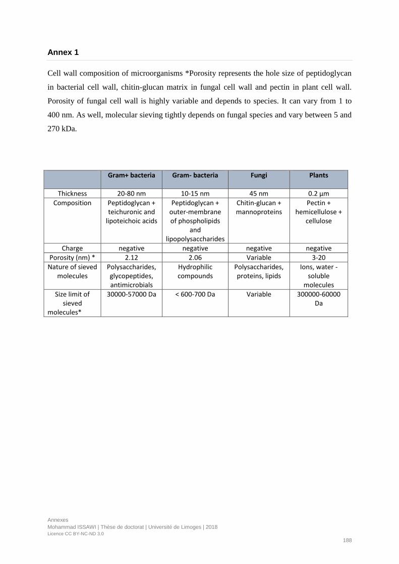
Annexes
Mohammad ISSAWI | Thèse de doctorat | Université de Limoges | 2018
Licence CC BY-NC-ND 3.0
188
Annex 1
Cell wall composition of microorganisms *Porosity represents the hole size of peptidoglycan
in bacterial cell wall, chitin-glucan matrix in fungal cell wall and pectin in plant cell wall.
Porosity of fungal cell wall is highly variable and depends to species. It can vary from 1 to
400 nm. As well, molecular sieving tightly depends on fungal species and vary between 5 and
270 kDa.
Gram+ bacteria Gram- bacteria Fungi Plants
Thickness 20-80 nm 10-15 nm 45 nm 0.2 µm
Composition Peptidoglycan + teichuronic and
lipoteichoic acids
Peptidoglycan + outer-membrane of phospholipids
and lipopolysaccharides
Chitin-glucan + mannoproteins
Pectin + hemicellulose +
cellulose
Charge negative negative negative negative
Porosity (nm) * 2.12 2.06 Variable 3-20
Nature of sieved molecules
Polysaccharides, glycopeptides, antimicrobials
Hydrophilic compounds
Polysaccharides, proteins, lipids
Ions, water -soluble
molecules
Size limit of sieved
molecules*
30000-57000 Da < 600-700 Da Variable 300000-60000 Da

Annexes
Mohammad ISSAWI | Thèse de doctorat | Université de Limoges | 2018
Licence CC BY-NC-ND 3.0
189
Annex 2
Cell wall structure of Gram+, Gram- bacteria, Fungi and plants.
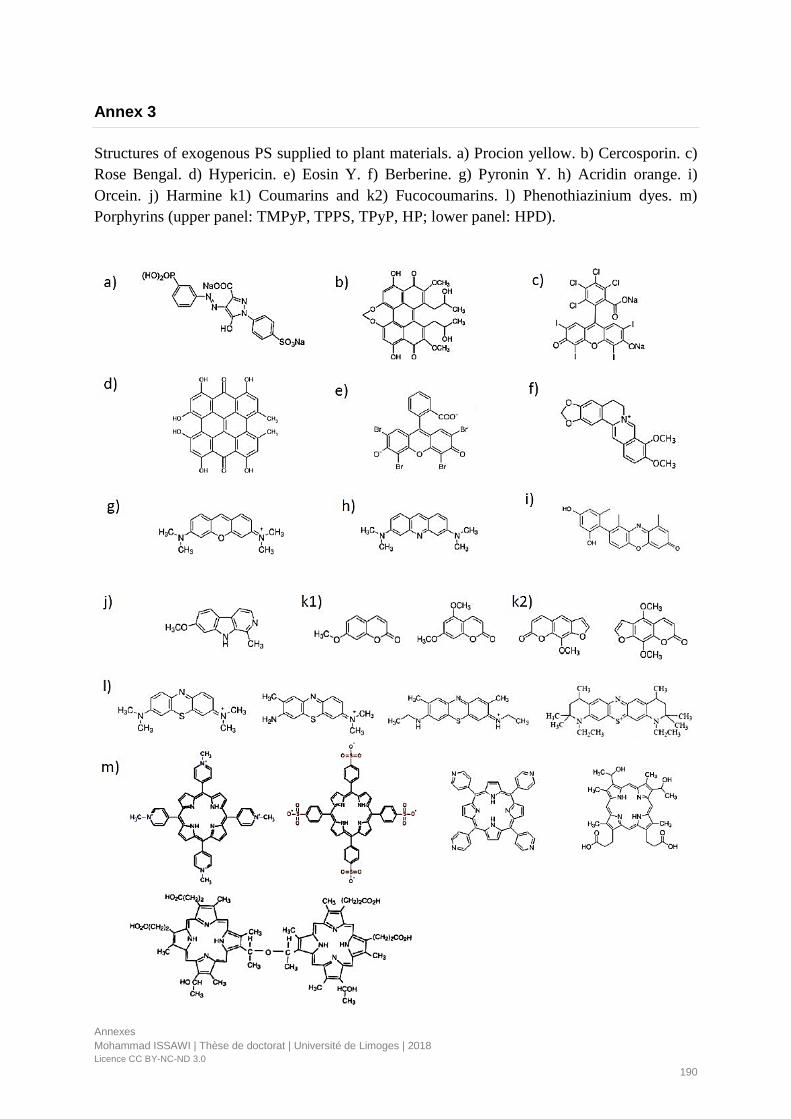
Annexes
Mohammad ISSAWI | Thèse de doctorat | Université de Limoges | 2018
Licence CC BY-NC-ND 3.0
190
Annex 3
Structures of exogenous PS supplied to plant materials. a) Procion yellow. b) Cercosporin. c)
Rose Bengal. d) Hypericin. e) Eosin Y. f) Berberine. g) Pyronin Y. h) Acridin orange. i)
Orcein. j) Harmine k1) Coumarins and k2) Fucocoumarins. l) Phenothiazinium dyes. m)
Porphyrins (upper panel: TMPyP, TPPS, TPyP, HP; lower panel: HPD).
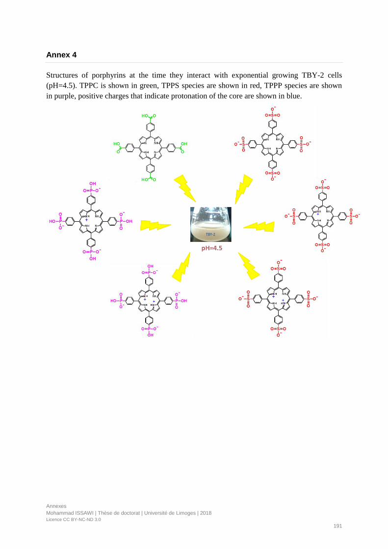
Annexes
Mohammad ISSAWI | Thèse de doctorat | Université de Limoges | 2018
Licence CC BY-NC-ND 3.0
191
Annex 4
Structures of porphyrins at the time they interact with exponential growing TBY-2 cells
(pH=4.5). TPPC is shown in green, TPPS species are shown in red, TPPP species are shown
in purple, positive charges that indicate protonation of the core are shown in blue.

Languages
Arabic
French
English
Informatics
Microsoft office, statistic tools
(SPSS, OiginPro, R
programming, PAST
software), bio-informatic tools,
Image processing (Image J,
Photoshop).
Technics
Molecular Biology, Cellular
Biology and Physiology,
Biochemistry, In Vitro culture.
Good teamwork integrations,
strategical thinking, problem
solving, and master’s student
supervision.
Hobbies
Football, literature, music,
travelling.
2009 High school diploma in Life science, with Distinction - Education and teaching high school, Lebanon.
Bachelor’s degree in general biology (Average 73.23/100) – Lebanese University, Lebanon
2012 DELF B2 “Diplôme en langue française” (70/100) – French institute of Beirut, Lebanon
2012/2013 Master 1 Plant biology and environment, with Distinction - Lebanese University, Lebanon
2013/2014 Master 2 (research) Applied plant biotechnology with highest honors - Lebanese University, Lebanon.
2014 M2 Internship entitled “Response of lignification to drought and salinity stresses through cinnamyl alcohol
dehydrogenase enzyme in Barley” - Shahid Beheshti University, Iran.
2015-2018 PhD in Plant physiology entitled « Plant photodynamic stress: study of molecular and cellular mechanisms
in plant and plant cells upon porphyrin treatment» - University of Limoges, France.
Career path
Publications
• Mohammad Issawi, Vincent Sol and Catherine Riou (2018) Plant photodynamic stress: what’s new?
Frontiers in Plant Science DOI: 10.3389/fpls.2018.00681 (IF 4.29).
• Mohammad Issawi, Damien Guillaumot, Vincent Sol and Catherine Riou (2018) Responses of an
adventitious fast-growing plant to photodynamic stress: comparative study of anionic and cationic
porphyrin effect on Arabidopsis thaliana. Physiologia Plantarum DOI: 10.1111/ppl.12666 (IF 3.33)
• Mohammad Issawi, Mohammad Muhieddine, Céline Girard, Vincent Sol and Catherine Riou (2017)
Unexpected features of exponentially growing Tobacco Bright Yellow-2 cell suspension culture in relation
to excreted extracellular polysaccharides and cell wall composition. Glycoconjugate Journal DOI:
10.1007/s10719-017-9782-7 (IF 2.2).
• Damien Guillaumot, Mohammad Issawi, Anne Blondeau, Stephanie Lhez, Vincent Sol and Catherine
Riou (2016) Synergistic enhancement of tolerance mechanisms in response to photoactivation of cationic
tetra (N-methylpyridyl) porphyrins in tomato plantlets. Journal of Photochemistry and Photobiology B
Biology DOI: 10.1016/j.jphotobiol.2016.01.015 (IF 2.96).
• Mohammad Issawi, Stéphanie Leroy-Lhez, Vincent Sol and Catherine Riou “Crossing the first threshold:
New insights in the Influence of chemical structure of anionic porphyrins from cell wall interactions to
photodynamic cell death induction in TBY-2 suspension culture”. (Ongoing)
• Stéphanie Leroy-Lhez, Olivier Rezazgui, Mohammad Issawi, Mourad Elhabiri, Claude Calliste and
Catherine Riou “Characterization of pH dependent charge states and physico-chemical properties of
anionic porphyrins” Submitted to Photochemical and Photobiological Sciences (IF 2.34).
• Mohammad Issawi, Veronica Ambrosini, Vincent Sol and Catherine Riou “Starchy surprise: new ability
of TBY-2 cells to synthesize an amylose-rich starch in presence of auxin.” (Ongoing)
Congress attended
• International photodynamic association - Coimbra, Portugal. Contribution: poster presentation entitled:
Photoactivation of cationic tetra (N-methylpyridyl) porphyrin dramatically altered Arabidopsis thaliana
plantlet development.
• Société française de biologie végétale - Angers, France. Contribution: oral presentation entitled:
Comparative study of three anionic porphyrins in Tobacco Bright Yellow-2 cells: from cell wall
interaction and localization to phototoxic damage and cell defense.
Communication and
leadership skills
2012
(IPA 2017)
Mohammad Issawi
13, Petit Faubourg Manigne 87000, Limoges-France 09/01/1991, Beirut-Lebanon Single [email protected]
+33624483373
Driving licence B Researchgate.net/profile/Mohammad_Issawi
(SFBV 2016)

Réponses des plantes et des cellules végétales au stress photodynamique induit par les porphyrines
Le traitement photodynamique antimicrobien (acronyme anglais APDT) est apparu comme une solution
alternative pour lutter contre les microorganismes multi-résistants. Cette méthode basée sur l'utilisation de
photosensibilisateurs dont les porphyrines, fonctionne aussi contre les agents pathogènes des plantes ce qui
nous a conduit à proposer une approche de type APDT dans le domaine de l’agriculture. Au cours de ce travail
de thèse, nous nous sommes intéressés au coté « vert » de la mise en place de cette approche en réalisant une
étude approfondie sur des plantules de deux espèces végétales: Arabidopsis thaliana et Lycopersicum
esculentum (tomate) et sur une suspension cellulaire de tabac (TBY-2). Des porphyrines anioniques et
cationiques hydrosolubles ont été testées. Nous avons montré qu’aucune de ces porphyrines testées à forte
concentration (⩾ 80 µM) n’était cytotoxique à l’obscurité sur les plantules ou la suspension. Par contre sous
photopériode (16h), les porphyrines cationiques testées à faible concentration (3,5 µM) se sont révélées létales
pour les plantules d’Arabidopsis alors qu’elles n’ont fait que ralentir la croissance des plantules de tomate.
Etonnamment, les porphyrines anioniques même testées à forte concentration n’ont pas (ou très peu) induit
d’altérations de croissance des plantules. Cette situation se trouve inversée dans les cellules TBY-2 qui sont
beaucoup plus sensibles aux porphyrines anioniques photoactivées qui induisent leur mort par apoptose. Ce
modèle cellulaire nous a permis de comprendre i) les mécanismes d'interaction porphyrines anioniques avec la
paroi cellulaire et ii) quels mécanismes étaient mis en place dans les cellules en réponse au stress
photodynamique. En conclusion, ces études préliminaires sur le végétal laissent sérieusement entrevoir la
possibilité de développer l’APDT en agriculture ciblée aux pathogènes de plantes et sans effet notable sur les
plantes d’intérêt agronomique et les microorganismes du sol.
Mots-clés : Porphyrines, Photosensibilisateurs, stress photodynamique des plantes, Arabidopsis thaliana,
Lycopersicum esculentum, cellules TBY-2, agriculture.
Responses of plants and plant cells to porphyrin-induced photodynamic stress
Antimicrobial photodynamic treatment (APDT) has emerged as an alternative modality to strive against multidrug-resistant microorganisms. This method, based on the use of photosensitizers including porphyrins, has also shown substantial efficiency to kill plant pathogens. This led us to propose an APDT approach in the field of agriculture. During this thesis work, we were interested in the "green" side of this approach. Thus, we performed an in-depth study on two plant species: Arabidopsis thaliana and Lycopersicum esculentum (tomato) and on Tobacco Bright Yellow-2 suspension culture (TBY-2 cells) using anionic and cationic water-soluble porphyrins. We have shown that none of these porphyrins tested at high concentration (⩾ 80 μM) was cytotoxic on plantlets or suspension cells under dark conditions. On the other hand, the cationic porphyrins tested at low concentration (3.5 μM) were lethal for Arabidopsis while they only slowed the growth of tomato plantlets under 16 h photoperiod. Surprisingly, the same anionic porphyrins tested at high concentrations did not induce growth alterations of both plantlets. This situation is reversed in TBY-2 cells, which are much more sensitive to photoactivated anionic porphyrins that induce apoptosis-like cell death. TBY-2 cells allowed to understand i) the interactions of anionic porphyrins with the cell wall and ii) the induced cellular and molecular mechanisms in response to photodynamic stress. In conclusion, these preliminary studies on the plants and plant cells suggest the possibility of developing APDT in agriculture. It will be targeted to plant pathogens without any side effects on plants of agronomic interest and soil microorganisms.
Keywords: Pophyrins, photosensitizers, plant photodynamic stress, Arabidopsis thaliana, Lycopersicum esculentum, TBY-2 cells, agriculture.