water-soluble phthalocyanines mediated photodynamic effect on mesothelioma cells
TRANSCRIPT
1st Reading
Journal of Porphyrins and PhthalocyaninesJ. Porphyrins Phthalocyanines 2009; 13: 681–690
Published at http://www.worldscinet.com/jpp/
Copyright © 2009 World Scientific Publishing Company
1st Reading
Copyright © 2009 World Scientific Publishing Company
INTRODUCTION
Phthalocyanines and metallophthalocyanines have been studied extensively for many years, mostly for their use as dyes and catalysts [1, 2]. Recently, they have also found applications in many fields in materials science [3, 4], especially in nonlinear optical (NLO) devices [5], liquid crystals [6, 7], Langmuir-Blodgett films [8], elec-trochromic devices [9], gas sensors [10, 11], and photo-sensitizers [12–15], among others.
Metallophthalocyanine complexes have proved to be highly promising as photosensitizers for photodynamic therapy (PDT), due to their intense absorption in the red
region of the electromagnetic radiation. High triplet state quantum yields and long triplet lifetimes are required for efficient sensitization. Phthalocyanines display cytotoxic effects when activated by light. Upon irradiation, these photosensitizers are promoted to their excited states and generate singlet oxygen. Surrounding biomolecules are damaged and this starts a series of biological responses leading to the tumor death. The photophysical properties of the phthalocyanine dyes are strongly influenced by the presence and nature of the central metal ion. Complexation of phthalocyanine with transition metals gives dyes with short triplet lifetimes (τT) of the photoexcited triplet state of metallophthalocyanine. Closed shell, diamagnetic ions, such as Zn2+, Al3+ and Si4+, give phthalocyanine complexes with both high triplet yields and long lifetimes [16].
PDT is an attractive new approach to treating super-ficially growing tumors and has been investigated under
Water-soluble phthalocyanines mediated photodynamic effect on mesothelioma cells
Nil Saydana, Mahmut Durmuşb, Meltem G. Dizgeb, Hanifi Yamanb, Ayşe G. Gürekb, Edith Antunesc, Tebello Nyokongc and Vefa Ahsen*b,d
a Gebze Institute of Technology, Faculty of Sciences, Department of Biology, PO Box 141, Gebze, 41400, Turkey b Gebze Institute of Technology, Faculty of Sciences, Department of Chemistry, PO Box 141, Gebze, 41400, Turkey c Department of Chemistry, Rhodes University, Grahamstown, 6140, South Africa d TUBITAK-Marmara Research Center, Materials Institute, PO Box 21, Gebze, 41470, Turkey
Received 7 August 2008Accepted 10 September 2008
ABSTRACT: The new peripherally 2-mercaptopyridine tetrasubstituted zinc phthalocyanine (2) and its quaternized derivative (3) have been synthesized and characterized by elemental analysis, IR, 1H NMR spectroscopy, electronic spectroscopy and mass spectra. The quaternized compound (3) shows excellent solubility in water, which makes it a potential photosensitizer for use in photodynamic therapy (PDT) of cancer. Fluorescence and singlet oxygen quantum yield measurements were conducted on 2-mercaptopyridine appended zinc phthalocyanines in dimethylsulphoxide (DMSO) for both the non-ionic (2) and quaternized (3) derivatives, and in aqueous media for the water-soluble complex 3. General trends are described for fluorescence and singlet oxygen quantum yields of these compounds. In this study, the cells were incubated with a novel water-soluble zinc phthalocyanine derivative (3) and thereafter the cells were illuminated using broad-band incoherent light source of various energy levels. Cytotoxicity of PDT on two pleural malign mesothelioma cell lines was determined by colorimetric proliferation assay. In addition, after PDT treatment, determination of activity matrix metalloproteinases (MMPs) were evaluated using gelatine zymography.
KEYWORDS: water-soluble zinc phthalocyanines, quaternization, fluorescence, cytotoxicity, meso-thelioma cell lines, matrix metalloproteinases.
SPP full member in good standing
*Correspondence to: Vefa Ahsen, email: [email protected], tel: +90 262-6053106, fax: +90 262-6538490
00086.indd 1 7/27/2009 1:38:03 Pm
1st Reading
Copyright © 2009 World Scientific Publishing Company J. Porphyrins Phthalocyanines 2009; 13: 682–690
682 N. SaydaN et al.
experimental and clinical conditions for the treatment of malignant mesothelioma (MM) [17]. The treatment involves the administration of a photosensitizer, followed by irradiation of the targeted lesion with visible light. This multi-step procedure initiates the photochemical genera-tion of cytotoxic reactive oxygen species such as singlet oxygen within the treatment field and leads to direct tumor destruction and microvascular disruption [18].
PDT induces oxidative stress, localized inflammation, and vascular injury within treatment fields, and each of these responses can lead to increased expression of angio-genic factors, cytokines and extracellular matrix (ECM) components such as matrix metalloproteinases (MMPs) [19]. Matrix metalloproteinases produced by tumor and stromal cells play a key role in tumor invasion and metas-tasis. Prevention of ECM degradation by MMPs inhibi-tion has shown to be a promising therapeutic approach to inhibition of cancer development [20]. MMPs are a family of structurally related zinc-dependent endopeptidases that are frequently elevated in the tumor microenvironment and capable of degrading essentially all components of the ECM [21]. According to their structure, MMPs can be divided into eight distinct groups, five of which are secreted and three of which are membrane-type MMPs. Interestingly, MMP-7 is one of the few MMPs overexpressed by carci-noma cells rather than stromal cells. MMP-7 is abundantly produced by many types of carcinomas such as pancre-atic, colon, breast and non-small-cell lung cancer, making it an attractive anticancer target. Using phthalocyanine in PDT might generate matrix protein cross-links and induce structural alteration of matrix binding sites [22].
The aim of our ongoing research is to synthesize water-soluble zinc phthalocyanines to be used as potential PDT agents. Photophysical properties of substituted zinc phthalocyanines are very useful in applications involving
PDT. Herein, we report on the synthesis and spectroscopic properties as well as fluorescence and singlet oxygen quantum yields and some preliminary in vitro cell stud-ies of zinc phthalocyanines tetrasubstituted at the periph-eral positions with 2-mercaptopyridine group (Scheme 1a and 1b). In the present study, the cells were incubated with a novel water-soluble zinc phthalocyanine derivative and thereafter the cells were illuminated using broad-band incoherent light source consisting of various energy levels. Cytotoxicity of PDT on two pleural malign mesothelioma cell lines was determined by colorimetric proliferation assay. After PDT treatment the determination of activity of MMPs were evaluated using gelatine zymography.
EXPERIMENTAL
Materials
Quinoline, dimethylsulphoxide (DMSO), methanol, n-hexane, chloroform (CHCl3), dichloromethane (DCM), tetrahydrofuran (THF), acetone, ethanol and dimeth-ylformamide (DMF) were dried as described by Perrin and Armarego [23] before use. Zinc(II) acetate, K2CO3, dimethylsulphate (DMS) were purchased from Aldrich. 2-mercaptopyridine was purchased from Fluka. Prepara-tive thin layer chromatography was performed on silica gel 60 PF254. 4-nitrophthalonitrile [24], 4-(2-mercapto-pyridine) phthalonitrile (1) [25] were synthesized and purified according to literature procedures. Zinc phthalo-cyanine was purchased from Aldrich.
Equipment
Absorption spectra in the UV-visible region were recorded with a Shimadzu 2001 UV Pc spectrophotometer
S
N
CN
CN
N
N
N
N
N
N
N
N
NZn
S
NS
NS
S
N
Zn(AcO)2 , DBU
n-hexanol, 160OC
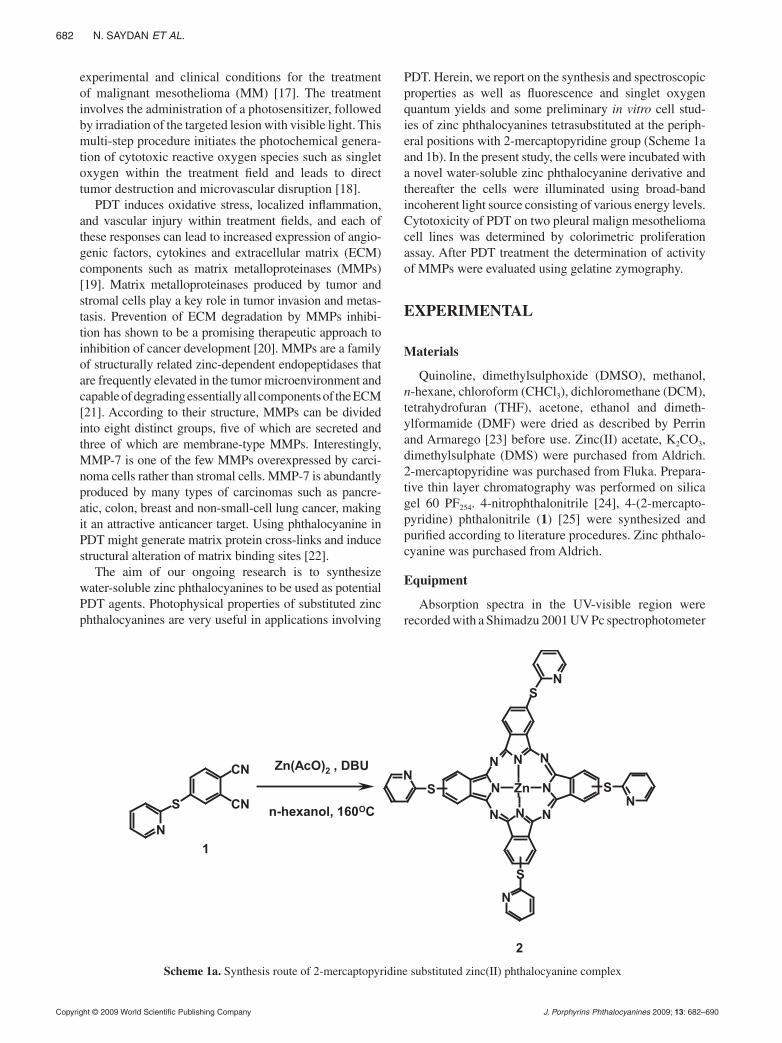
1
2
Scheme 1a. Synthesis route of 2-mercaptopyridine substituted zinc(II) phthalocyanine complex
00086.indd 2 7/27/2009 1:38:04 Pm
1st Reading
Copyright © 2009 World Scientific Publishing Company J. Porphyrins Phthalocyanines 2009; 13: 683–690
Water-Soluble PhthaloCyaNiNeS mediated PhotodyNamiC effeCt oN meSothelioma CellS 683
and Varian 500 UV-Vis/NIR spectrophotometer. Fluores-cence excitation and emission spectra were recorded on a Varian Eclipse spectrofluoremeter using 1 cm pathlength cuvettes at room temperature. IR spectra (KBr pellets) were recorded on a Bio-Rad FTS 175C FTIR spectrom-eter. The mass spectra were acquired on a Bruker Dalton-ics (Bremen, Germany) MicrOTOF mass spectrometer equipped with an electronspray ionization (ESI) source. The instrument was operated in positive-ion mode using a m/z range of 50–3000. The capillary voltage of the ion source was set at 6000 V and the capillary exit at 190 V. The nebulizer gas flow was 1 bar and drying gas flow 8 mL/min. The drying temperature was set at 200 °C for non-ionized complex (2) and positive-ion and linear-mode MALDI-MS spectrum of quaternized complex (3) was obtained in 3,5-dimethoxy-4-hydroxycinnamic acid MALDI matrix using nitrogen laser accumulat-ing 50 laser shots using Bruker Microflex LT MALDI-TOF mass spectrometer. Protonated molecular ion peak of quaternized complex (3) was obtained at 1059.8 Da for [M-2SO4-CH3]
+, representing the protonated ion of quaternized complex (3). 1H spectra were recorded in DMSO-d6 solutions on a Varian 500 MHz spectrometer. Elemental analyses were obtained with a Thermo Finni-gan Flash 1112 instrument.
Photo irradiations for singlet oxygen determination were performed using a General Electric Quartz line lamp (300 W). A 600 nm glass cut off filter (Schott) and water were used to filter off ultraviolet and infrared radia-tions, respectively. An interference filter (Intor, 700 nm with a bandwidth of 40 nm) was additionally placed in the light path before the sample.
Fluorescence quantum yields and lifetimes
Fluorescence quantum yields (ΦF) were determined by the comparative method (Equation 1) [26, 27],
Φ ΦF F
Std
Std Std
StdF A n
F A n= ( )
. .
. .
2
2 (1)
where F and FStd are the areas under the fluorescence emission curves of the samples (2 and 3) and the stan-dard, respectively. A and AStd are the respective absor-bances of the samples and standard at the excitation wavelengths, respectively. The refractive indices (n) of the solvents were employed in calculating fluorescence quantum yields in different solvents. Unsubstituted ZnPc (in DMSO) (ΦF = 0.20) [28] was employed as the stan-dard. Both the samples and standard were excited at the same wavelength. The absorbance of the solutions at the excitation wavelength ranged between 0.04 and 0.05. Radiative lifetimes were determined using Photochem-CAD software application, which uses the Strickler-Berg equation [29]. Lifetimes of fluorescence (τF) were then calculated from the radiative lifetime using fluorescence quantum yields (Equation 2).
ΦFF
= ττ
0 (2)
Singlet oxygen quantum yields
Equation 3 was employed for calculating singlet oxy-gen quantum yields:
Φ Φ∆ ∆= Std absStd
Stdabs
.RI
R I (3)
where Φ∆Std is the singlet oxygen quantum yield for the
standard (ZnPcSmix, Φ∆Std = 0.45 in aqueous solution) [30]
and ZnPc (Φ∆Std = 0.67 in DMSO [31]). R and RStd are the
ADMA or DPBF photobleaching rates in the presence of the respective MPcs under investigation and the stan-dard, respectively. Iabs and Iabs
Std are the rates of light absorp-tion by the MPcs and the standard, respectively. To avoid
Scheme 1b. Synthesis route of quaternized 2-mercaptopyridine substituted zinc(II) phthalocyanine complex
N
N
N
N
N
N
N
N
NZn
S
NS
NS
S
N
N
N
N
N
N
N
N
N
NZn
S
NS
NS
S
N
DMF, 120OC
+
+
+
+
2SO42-
2 3
Dimethyl sulphate
4+
00086.indd 3 7/27/2009 1:38:05 Pm
1st Reading
Copyright © 2009 World Scientific Publishing Company J. Porphyrins Phthalocyanines 2009; 13: 684–690
684 N. SaydaN et al.
chain reactions, the concentration of ADMA was kept at ~6 × 10⁻⁵ M while that of DPBF was kept at ~3 × 10⁻⁵ M. Solutions of the MPcs with an absorbance of 0.2 at the irradiation wavelength were prepared in the dark and irradiated at the Q band region, monitoring the ADMA and DPBF degradation at 380 and 417 nm, respectively. The error was ~10% from several values of Φ∆.
Synthesis
2,(3)-tetra-(2-mercaptopyridine)phthalo cya ni na-tozinc(II) (2) (Scheme 1a). A mixture of anhydrous zinc (II) acetate (0.77 g, 4.22 mmol), 4-(2-mercaptopyridine)phthalonitrile (1) (1.0 g, 4.22 mmol), DBU (0.97 mL, 0.65 mmol) and 10 mL n-hexanol was stirred at 160 °C for 12 h under nitrogen atmosphere. After cooling, the solution was dropped in the n-hexane. The green solid product was precipitated and collected by filtration and washed with n-hexane. The crude product was dissolved in chloroform and precipitated again by dropping in n-hexane. The green solid product was collected by filtra-tion and washed with n-hexane. The green crude product was purified by passing through a silica gel column with firstly ethyl acetate and then CH2Cl2/MeOH (10:1) elu-tion. Yield: 0.11 g (10%), mp > 240 °C. UV-vis (DMSO): λmax nm (log ε) 366 (5.00), 619 (4.80), 685 (5.52). IR (KBr): νmax, cm-1 3045 (Ar-CH), 1574 (C=C), 1451, 1416, 1304, 1096, 1042, 910, 760, 744. 1H NMR (DMSO-d6): δ, ppm 8.92 (4H, m, Pyridyl-H), 8.65 (4H, m, Pyridyl-H), 8.52 (4H, d, Pc-H), 8.24 (4H, m, Pyridyl-H), 7.75 (4H, m, Pyridyl-H), 7.50 (4H, m, Pc-H), 7.27 (4H, m, Pc-H). Calcd. for C52H28N12S4Zn: C, 61.56, H, 2.78, N, 16.57. Found: C, 61.45, H, 2.72 N, 16.52. ESI-MS: m/z calcd. 1013.9; found [M]+ 1014.1.
Quaternized 2,(3)-tetra-[(2-mercaptopyridine)ph -tha lo cyaninato]zinc(II) (3) (Scheme 1b). This complex was prepared according to the method previously reported by Smith et al. [32]. Compound 2 (180 mg, 0.177 mmol) was heated to 120 °C in freshly distilled DMF (5 ml) and dimethyl sulphate (0.168 ml) was added dropwise. The mixture was stirred at 120 °C for 12 h. It was then cooled to room temperature and the product was precipitated with hot acetone and collected by filtration. The green solid product was washed successively with hot ethyl acetate, chloroform, n-hexane, CCl4 and diethylether. The resulting hygroscopic product was dried over phos-phorous pentoxide. Yield: 0.16 g (73%), mp 220 °C. UV-vis (DMSO): λmax nm (log ε) 319 (4.49), 617 (4.20), 685 (4.91). IR (KBr): νmax, cm-1 3027 (Ar-CH), 2949 (CH), 1563 (C=C), 1485, 1435, 1188 (S=O), 1107 (S=O), 602 (S-O). 1H NMR (DMSO-d6): δ, ppm 7.22–9.86 (28H, m, Pc-H and Pyridyl-H), 4.40 (12H, s, CH3). Calcd. for C56H46N12O11S6Zn (+3H2O): C, 50.92, H, 3.51, N, 12.73. Found: C, 50.36, H, 3.58, N, 12.81. MALDI-TOF-MS: m/z calcd. for C56H40N12O8S6Zn, 1266; found [M-2SO4-CH3]
+ 1059.8, [M-2SO4-2CH3]+ 1044.8, [M-2SO4-3CH3]+
1029.8, [M-2SO4-4CH3]+ 1014.8.
Cells
SPC111 and SPC212 cell lines were derived from pleural effusions of untreated mesothelioma patients. Cells were grown in RPMI 1640 medium (Gibco, UK) supplemented with 10% fetal bovine serum, antibiotics (penicillin-streptomycin) and were incubated at 37 °C in a humidified atmosphere of 5% CO2 in air. The cells were regularly subcultured according to their growth rate.
Light source
The Lumacare Model LC-122 consists of two main parts: a Quartz halogen lamp light source (100 W) hous-ing with a control panel and power supply, and a fiber- optic probe (FOP) adaptors with filters designed to meet any optical protocol ranging from 380 to 750 nm. The fiber-optic probes of the LC-122 offers output power from 10 mW.cm-2 up to 1 W.cm-2 at their output tips, depending on filter transmission wavelength. For illumination proto-col we used FOP systems with activation wavelength of 670 ± 10 nm. The exposure area was 50 × 75 mm and the distance between FOP tip and the cell plate surface was 20 cm. The light power of FOP systems on the exposure area was measured with a power meter that has silicon detector (Ophir). The exposure energy is controlled from the control panel by a timer.
Cytotoxicity assay
To assess the cytotoxic effect of the phthalocyanines, two mesothelioma cell lines were seeded onto 96-well plates at 4000 cells per well. The cells were allowed to exponentially grow and attach for 24 h. For dark cyto-toxicity experiments, phthalocyanine was added to tripli-cate wells in a final concentration of phthalocyanine of 0, 0.2, 0.4, 0.6, 0.8, 1 µM and the cells were incubated with phthalocyanine for 24 h. The compounds were replaced with fresh medium and the cells were illuminated with Lumacare equipment. After illumination, cytotoxicity was measured using (MTS)-Cell Proliferation Assay (Promega) kit and the absorbance spectra were used to measure cell proliferation rate. To each well, 20 µL of MTS compound was added and the plate was incubated for 4 h before reading. MTS tetrazolium compound is metabolized by metabolically active cells into a col-ored formazan product that can be measured by read-ing the absorbance at 490 nm with a microplate reader. The average of the triplicate wells for each sample was calculated.
Gelatin zymography
SPC111 and SPC212 cell lines were seeded in 24-well plates using serum-free medium containing optimal con-centration of phthalocyanine (0.5 µM) and treated with three PDT light doses (1, 2, and 3 J.cm-2). After 24, 48 and 72 h incubation, medium was collected and cen-trifuged at 5000 rpm for 10 min. Zymographic assays
00086.indd 4 7/27/2009 1:38:06 Pm
1st Reading
Copyright © 2009 World Scientific Publishing Company J. Porphyrins Phthalocyanines 2009; 13: 685–690
Water-Soluble PhthaloCyaNiNeS mediated PhotodyNamiC effeCt oN meSothelioma CellS 685
for gelatinase were performed according to the method of Liota, LA and et al. [33, 34]. Briefly, equivalent vol-umes of supernatant from each sample were subjected to sodium dodecyl sulfate polyacrylamide gel electro-phoresis (SDS-PAGE) on gelatin-containing polyacryl-amide gel (11% acrylamide and 1 mg/mL gelatin) at a constant voltage (125 V) for approximately 1 h at room temperature under non-reducing conditions. The gel was re-natured with 2.5% Triton X-100 for 30 min at room temperature, stained with 0.5% (w/v) Coomassie brilliant blue R-250 and destained with 50% ethanol, 5% acetic acid and 45% H2O. MMPs exhibit clear zones against a blue background.
RESULTS AND DISCUSSION
Synthesis and characterization
Generally, substituted phthalocyanines are prepared by cyclotetramerization of substituted phthalonitriles or 1,3-diimino-1H-isoindoles. 2(3),9(10),16(17),23(24)-tetra substituted phthalocyanines can be synthesized from 4-substituted phthalonitriles [35]. In both cases a mix-ture of four possible structural isomers are obtained. The four probable isomers can be designed by their molecular symmetry as C4h, C2v, Cs and D2h. The 2(3)-substituted compounds always occur in the expected statistical mix-ture of 12.5% C4h-, 25%C2v-, 50%Cs- and 12.5%D2h- isomers [36]. In this study, synthesized phthalocyanine compounds are obtained as isomer mixtures as expected. No attempt was made to separate the isomers of 2 and 3.
The preparation of phthalocyanine derivatives from the aromatic nitriles occurs under different reaction con-ditions. The synthesis of zinc phthalocyanine complex (2) was achieved by treatment of phthalonitrile 1 with anhy-drous zinc(II) acetate in dried n-hexanol (Scheme 1a). Column chromatography with silica gel was employed to obtain the pure products from the reaction mixtures.
Quaternization of the zinc phthalocyanine compound was achieved by reaction with excess dimethylsulphate (DMS) as quaternization agent in DMF as solvent at 120 °C. Yield of the product was 73% for 3. After reac-tion with DMS, quaternized complex is very soluble in water.
Generally, phthalocyanine complexes are insoluble in most organic solvents; however, introduction of substitu-ents on the ring increases their solubility. Both complexes (2 and 3) exhibited excellent solubility in DMF and DMSO. Quaternized complex (3) is soluble in water as well. The new compounds were characterized by UV-vis, IR and NMR spectroscopies, MALDI-TOF mass spectra and elemental analysis. The analyses are consistent with the predicted structures, as shown in the experimental section. The sharp peak in the IR spectra for the C≡N vibrations of phthalonitrile 1 at ~ 2234 cm-1 disappeared after conversion into zinc phthalocyanine, indicative
of metallophthalocyanine formation. The characteristic vibrations corresponding to C=C groups at 1574 and 1563 cm-1, aromatic CH stretching at 3068–3015 cm-1 were observed for all complexes. S=O stretching at 1188 cm-1 and S–O stretching at 602 cm-1 for complex 3 are indica-tive of quaternization formation.
The 1H NMR spectra of tetrasubstituted phthalocya-nine derivatives (2 and 3) show complex patterns due to the mixed isomer character of these compounds. The complexes were found to be pure by 1H NMR, with all the substituents and ring protons observed in their respective regions. The resonances belonging to ring protons were observed at 8.52 ppm as doublet, 7.50 ppm as multiplet, 7.27 ppm as multiplet for 2 integrating for 4, 4, 4 pro-tons each, making a total of 12 protons expected for both peripheral ring protons. The mercaptopyridine protons were observed as multiplets at 8.92, 8.65, 8.24 and 7.75 ppm integrating for 4, 4, 4, 4 protons each, respectively, making a total of 16 protons as expected.
The NMR spectra of the quaternized phthalocyanine complex 3 showed more unresolved patterns compared to non-quaternized derivative. This complex showed the phthalocyanine ring protons and mercaptopyridine group protons as unresolved multiplets, integrating for a total of 28 protons. Phthalocyanine ring and mercaptopyri-dine protons were observed between 7.22 and 9.86 ppm as multiplets. The methyl protons which integrated for 12 protons were observed at 4.40 ppm for complex 3 as a singlet.
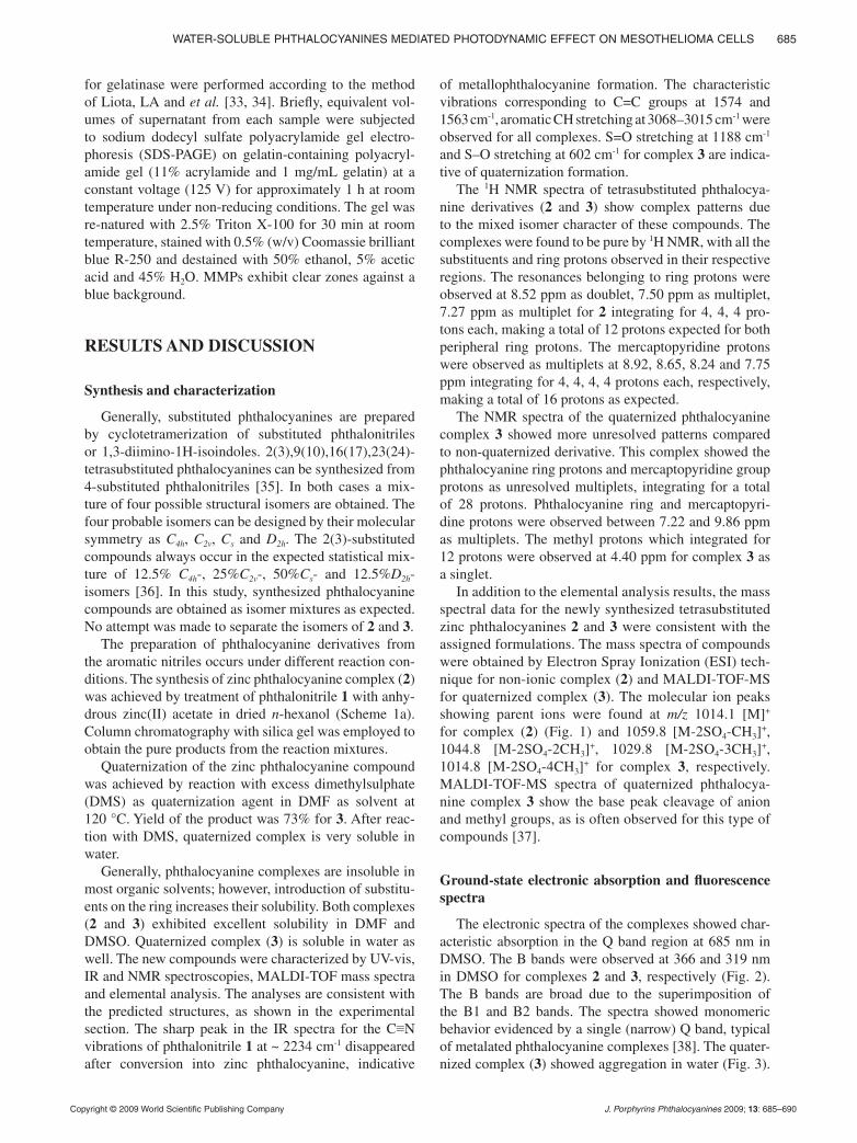
In addition to the elemental analysis results, the mass spectral data for the newly synthesized tetrasubstituted zinc phthalocyanines 2 and 3 were consistent with the assigned formulations. The mass spectra of compounds were obtained by Electron Spray Ionization (ESI) tech-nique for non-ionic complex (2) and MALDI-TOF-MS for quaternized complex (3). The molecular ion peaks showing parent ions were found at m/z 1014.1 [M]+ for complex (2) (Fig. 1) and 1059.8 [M-2SO4-CH3]
+, 1044.8 [M-2SO4-2CH3]
+, 1029.8 [M-2SO4-3CH3]+,
1014.8 [M-2SO4-4CH3]+ for complex 3, respectively.
MALDI-TOF-MS spectra of quaternized phthalocya-nine complex 3 show the base peak cleavage of anion and methyl groups, as is often observed for this type of compounds [37].
Ground-state electronic absorption and fluorescence spectra
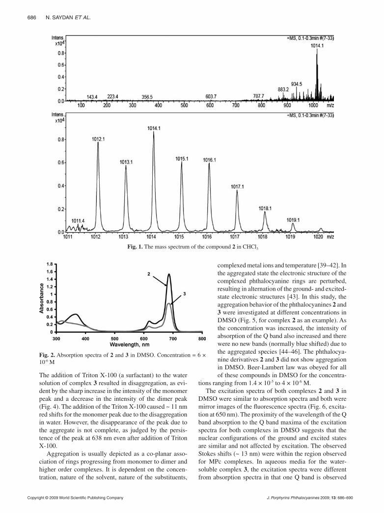
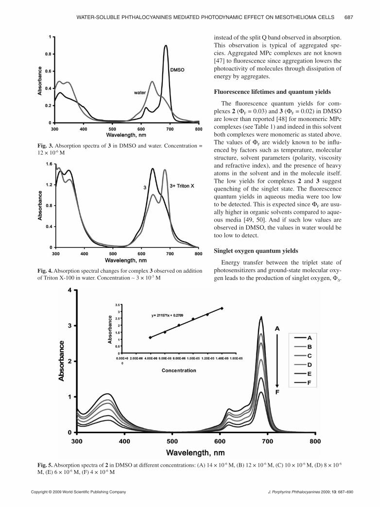
The electronic spectra of the complexes showed char-acteristic absorption in the Q band region at 685 nm in DMSO. The B bands were observed at 366 and 319 nm in DMSO for complexes 2 and 3, respectively (Fig. 2). The B bands are broad due to the superimposition of the B1 and B2 bands. The spectra showed monomeric behavior evidenced by a single (narrow) Q band, typical of metalated phthalocyanine complexes [38]. The quater-nized complex (3) showed aggregation in water (Fig. 3).
00086.indd 5 7/27/2009 1:38:06 Pm
1st Reading
Copyright © 2009 World Scientific Publishing Company J. Porphyrins Phthalocyanines 2009; 13: 686–690
686 N. SaydaN et al.
The addition of Triton X-100 (a surfactant) to the water solution of complex 3 resulted in disaggregation, as evi-dent by the sharp increase in the intensity of the monomer peak and a decrease in the intensity of the dimer peak (Fig. 4). The addition of the Triton X-100 caused ~ 11 nm red shifts for the monomer peak due to the disaggregation in water. However, the disappearance of the peak due to the aggregate is not complete, as judged by the persis-tence of the peak at 638 nm even after addition of Triton X-100.
Aggregation is usually depicted as a co-planar asso-ciation of rings progressing from monomer to dimer and higher order complexes. It is dependent on the concen-tration, nature of the solvent, nature of the substituents,
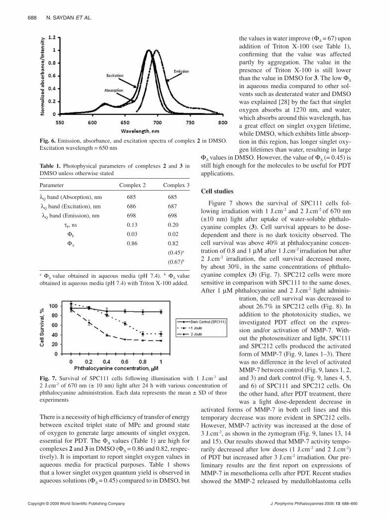
complexed metal ions and temperature [39–42]. In the aggregated state the electronic structure of the complexed phthalocyanine rings are perturbed, resulting in alternation of the ground- and excited-state electronic structures [43]. In this study, the aggregation behavior of the phthalocyanines 2 and 3 were investigated at different concentrations in DMSO (Fig. 5, for complex 2 as an example). As the concentration was increased, the intensity of absorption of the Q band also increased and there were no new bands (normally blue shifted) due to the aggregated species [44–46]. The phthalocya-nine derivatives 2 and 3 did not show aggregation in DMSO. Beer-Lambert law was obeyed for all of these compounds in DMSO for the concentra-
tions ranging from 1.4 × 10⁻⁵ to 4 × 10⁻⁶ M.The excitation spectra of both complexes 2 and 3 in
DMSO were similar to absorption spectra and both were mirror images of the fluorescence spectra (Fig. 6, excita-tion at 650 nm). The proximity of the wavelength of the Q band absorption to the Q band maxima of the excitation spectra for both complexes in DMSO suggests that the nuclear configurations of the ground and excited states are similar and not affected by excitation. The observed Stokes shifts (~ 13 nm) were within the region observed for MPc complexes. In aqueous media for the water-soluble complex 3, the excitation spectra were different from absorption spectra in that one Q band is observed
Fig. 1. The mass spectrum of the compound 2 in CHCl3
Fig. 2. Absorption spectra of 2 and 3 in DMSO. Concentration = 6 × 10⁻⁶ M
00086.indd 6 7/27/2009 1:38:07 Pm
1st Reading
Copyright © 2009 World Scientific Publishing Company J. Porphyrins Phthalocyanines 2009; 13: 687–690
Water-Soluble PhthaloCyaNiNeS mediated PhotodyNamiC effeCt oN meSothelioma CellS 687
instead of the split Q band observed in absorption. This observation is typical of aggregated spe-cies. Aggregated MPc complexes are not known [47] to fluorescence since aggregation lowers the photoactivity of molecules through dissipation of energy by aggregates.
Fluorescence lifetimes and quantum yields
The fluorescence quantum yields for com-plexes 2 (ΦF = 0.03) and 3 (ΦF = 0.02) in DMSO are lower than reported [48] for monomeric MPc complexes (see Table 1) and indeed in this solvent both complexes were monomeric as stated above. The values of ΦF are widely known to be influ-enced by factors such as temperature, molecular structure, solvent parameters (polarity, viscosity and refractive index), and the presence of heavy atoms in the solvent and in the molecule itself. The low yields for complexes 2 and 3 suggest quenching of the singlet state. The fluorescence quantum yields in aqueous media were too low to be detected. This is expected since ΦF are usu-ally higher in organic solvents compared to aque-ous media [49, 50]. And if such low values are observed in DMSO, the values in water would be too low to detect.
Singlet oxygen quantum yields
Energy transfer between the triplet state of photosensitizers and ground-state molecular oxy-gen leads to the production of singlet oxygen, Φ∆.
Fig. 3. Absorption spectra of 3 in DMSO and water. Concentration = 12 × 10-6 M
Fig. 4. Absorption spectral changes for complex 3 observed on addition of Triton X-100 in water. Concentration ∼ 3 × 10⁻⁵ M
Fig. 5. Absorption spectra of 2 in DMSO at different concentrations: (A) 14 × 10-6 M, (B) 12 × 10-6 M, (C) 10 × 10-6 M, (D) 8 × 10⁻⁶ M, (E) 6 × 10⁻⁶ M, (F) 4 × 10⁻⁶ M
00086.indd 7 7/27/2009 1:38:08 Pm
1st Reading
Copyright © 2009 World Scientific Publishing Company J. Porphyrins Phthalocyanines 2009; 13: 688–690
688 N. SaydaN et al.
There is a necessity of high efficiency of transfer of energy between excited triplet state of MPc and ground state of oxygen to generate large amounts of singlet oxygen, essential for PDT. The Φ∆ values (Table 1) are high for complexes 2 and 3 in DMSO (Φ∆ = 0.86 and 0.82, respec-tively). It is important to report singlet oxygen values in aqueous media for practical purposes. Table 1 shows that a lower singlet oxygen quantum yield is observed in aqueous solutions (Φ∆ = 0.45) compared to in DMSO, but
the values in water improve (Φ∆ = 67) upon addition of Triton X-100 (see Table 1), confirming that the value was affected partly by aggregation. The value in the presence of Triton X-100 is still lower than the value in DMSO for 3. The low Φ∆ in aqueous media compared to other sol-vents such as deuterated water and DMSO was explained [28] by the fact that singlet oxygen absorbs at 1270 nm, and water, which absorbs around this wavelength, has a great effect on singlet oxygen lifetime, while DMSO, which exhibits little absorp-tion in this region, has longer singlet oxy-gen lifetimes than water, resulting in large
Φ∆ values in DMSO. However, the value of Φ∆ (= 0.45) is still high enough for the molecules to be useful for PDT applications.
Cell studies
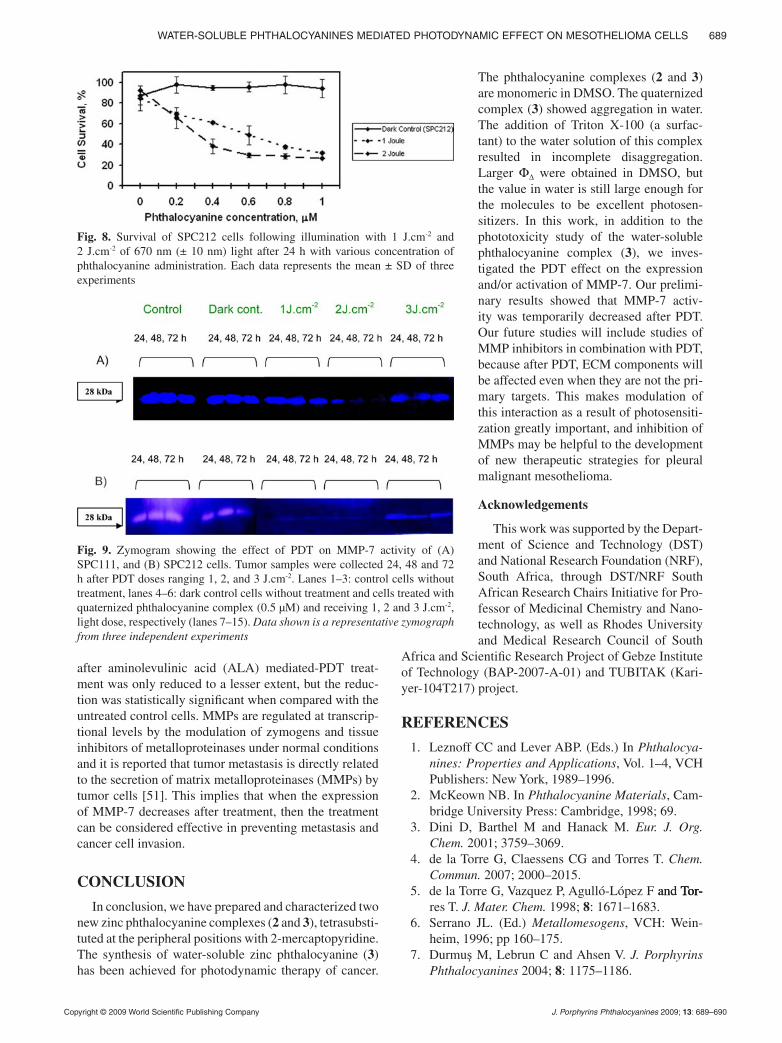
Figure 7 shows the survival of SPC111 cells fol-lowing irradiation with 1 J.cm-2 and 2 J.cm-2 of 670 nm (±10 nm) light after uptake of water-soluble phthalo-cyanine complex (3). Cell survival appears to be dose- dependent and there is no dark toxicity observed. The cell survival was above 40% at phthalocyanine concen-tration of 0.8 and 1 µM after 1 J.cm-2 irradiation but after 2 J.cm-2 irradiation, the cell survival decreased more, by about 30%, in the same concentrations of phthalo-cyanine complex (3) (Fig. 7). SPC212 cells were more sensitive in comparison with SPC111 to the same doses. After 1 µM phthalocyanine and 2 J.cm-2 light adminis-
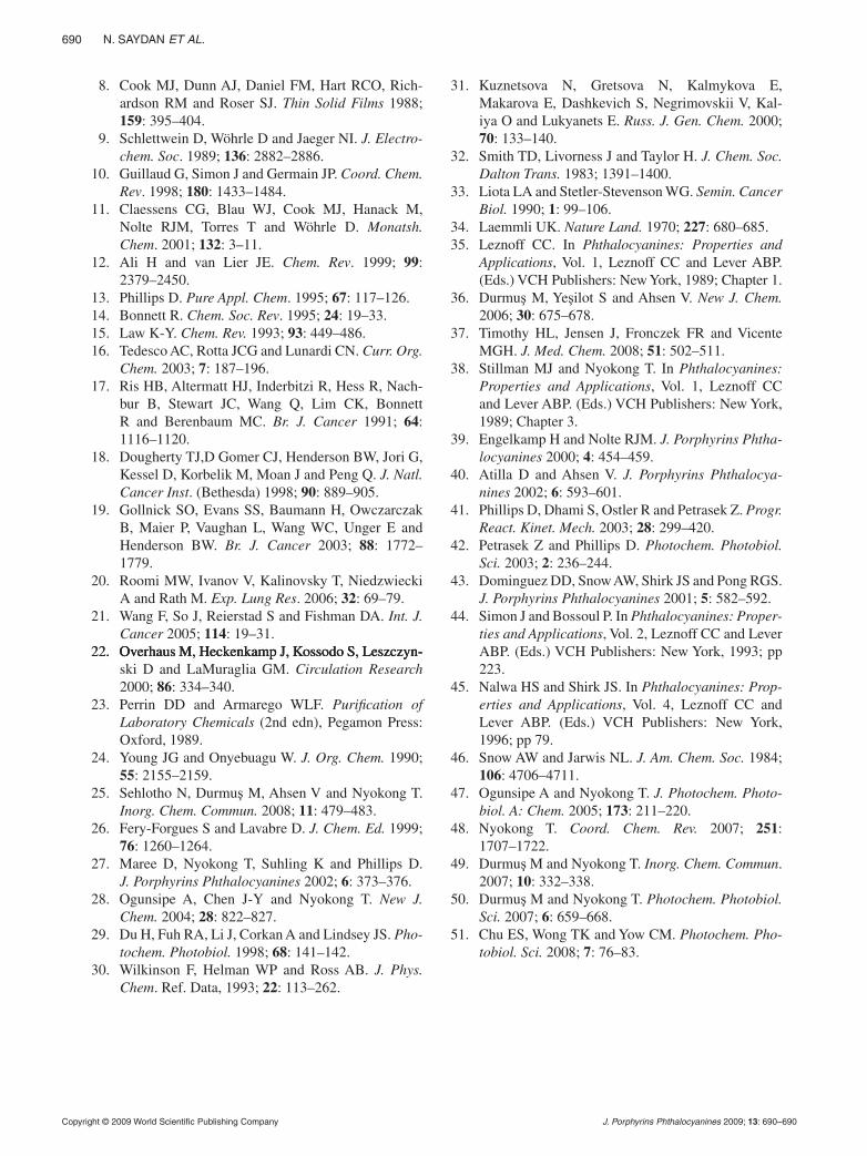
tration, the cell survival was decreased to about 26.7% in SPC212 cells (Fig. 8). In addition to the phototoxicity studies, we investigated PDT effect on the expres-sion and/or activation of MMP-7. With-out the photosensitizer and light, SPC111 and SPC212 cells produced the activated form of MMP-7 (Fig. 9, lanes 1–3). There was no difference in the level of activated MMP-7 between control (Fig. 9, lanes 1, 2, and 3) and dark control (Fig. 9, lanes 4, 5, and 6) of SPC111 and SPC212 cells. On the other hand, after PDT treatment, there was a light dose-dependent decrease in
activated forms of MMP-7 in both cell lines and this temporary decrease was more evident in SPC212 cells. However, MMP-7 activity was increased at the dose of 3 J.cm-2, as shown in the zymogram (Fig. 9, lanes 13, 14 and 15). Our results showed that MMP-7 activity tempo-rarily decreased after low doses (1 J.cm-2 and 2 J.cm-2) of PDT but increased after 3 J.cm-2 irradiation. Our pre-liminary results are the first report on expressions of MMP-7 in mesothelioma cells after PDT. Recent studies showed the MMP-2 released by medulloblastoma cells
Table 1. Photophysical parameters of complexes 2 and 3 in DMSO unless otherwise stated
Parameter Complex 2 Complex 3
λQ band (Absorption), nm 685 685
λQ band (Excitation), nm 686 687
λQ band (Emission), nm 698 698
τF, ns 0.13 0.20
ΦF 0.03 0.02
Φ∆ 0.86 0.82
(0.45)a
(0.67)b
a Φ∆ value obtained in aqueous media (pH 7.4). b Φ∆ value obtained in aqueous media (pH 7.4) with Triton X-100 added.
Fig. 6. Emission, absorbance, and excitation spectra of complex 2 in DMSO. Excitation wavelength = 650 nm
Fig. 7. Survival of SPC111 cells following illumination with 1 J.cm-2 and 2 J.cm-2 of 670 nm (± 10 nm) light after 24 h with various concentration of phthalocyanine administration. Each data represents the mean ± SD of three experiments
00086.indd 8 7/27/2009 1:38:10 Pm
1st Reading
Copyright © 2009 World Scientific Publishing Company J. Porphyrins Phthalocyanines 2009; 13: 689–690
Water-Soluble PhthaloCyaNiNeS mediated PhotodyNamiC effeCt oN meSothelioma CellS 689
after aminolevulinic acid (ALA) mediated-PDT treat-ment was only reduced to a lesser extent, but the reduc-tion was statistically significant when compared with the untreated control cells. MMPs are regulated at transcrip-tional levels by the modulation of zymogens and tissue inhibitors of metalloproteinases under normal conditions and it is reported that tumor metastasis is directly related to the secretion of matrix metalloproteinases (MMPs) by tumor cells [51]. This implies that when the expression of MMP-7 decreases after treatment, then the treatment can be considered effective in preventing metastasis and cancer cell invasion.
CONCLUSION
In conclusion, we have prepared and characterized two new zinc phthalocyanine complexes (2 and 3), tetrasubsti-tuted at the peripheral positions with 2-mercaptopyridine. The synthesis of water-soluble zinc phthalocyanine (3) has been achieved for photodynamic therapy of cancer.
The phthalocyanine complexes (2 and 3) are monomeric in DMSO. The quaternized complex (3) showed aggregation in water. The addition of Triton X-100 (a surfac-tant) to the water solution of this complex resulted in incomplete disaggregation. Larger Φ∆ were obtained in DMSO, but the value in water is still large enough for the molecules to be excellent photosen-sitizers. In this work, in addition to the phototoxicity study of the water-soluble phthalocyanine complex (3), we inves-tigated the PDT effect on the expression and/or activation of MMP-7. Our prelimi-nary results showed that MMP-7 activ-ity was temporarily decreased after PDT. Our future studies will include studies of MMP inhibitors in combination with PDT, because after PDT, ECM components will be affected even when they are not the pri-mary targets. This makes modulation of this interaction as a result of photosensiti-zation greatly important, and inhibition of MMPs may be helpful to the development of new therapeutic strategies for pleural malignant mesothelioma.
Acknowledgements
This work was supported by the Depart-ment of Science and Technology (DST) and National Research Foundation (NRF), South Africa, through DST/NRF South African Research Chairs Initiative for Pro-fessor of Medicinal Chemistry and Nano-technology, as well as Rhodes University and Medical Research Council of South
Africa and Scientific Research Project of Gebze Institute of Technology (BAP-2007-A-01) and TUBITAK (Kari-yer-104T217) project.
REFERENCES
1. Leznoff CC and Lever ABP. (Eds.) In Phthalocya-nines: Properties and Applications, Vol. 1–4, VCH Publishers: New York, 1989–1996.
2. McKeown NB. In Phthalocyanine Materials, Cam-bridge University Press: Cambridge, 1998; 69.
3. Dini D, Barthel M and Hanack M. Eur. J. Org. Chem. 2001; 3759–3069.
4. de la Torre G, Claessens CG and Torres T. Chem. Commun. 2007; 2000–2015.
5. de la Torre G, Vazquez P, Agulló-López F and Tor- and Tor- Tor-res T. J. Mater. Chem. 1998; 8: 1671–1683.
6. Serrano JL. (Ed.) Metallomesogens, VCH: Wein-heim, 1996; pp 160–175.
7. Durmuş M, Lebrun C and Ahsen V. J. Porphyrins Phthalocyanines 2004; 8: 1175–1186.
Fig. 8. Survival of SPC212 cells following illumination with 1 J.cm-2 and 2 J.cm-2 of 670 nm (± 10 nm) light after 24 h with various concentration of phthalocyanine administration. Each data represents the mean ± SD of three experiments
Fig. 9. Zymogram showing the effect of PDT on MMP-7 activity of (A) SPC111, and (B) SPC212 cells. Tumor samples were collected 24, 48 and 72 h after PDT doses ranging 1, 2, and 3 J.cm-2. Lanes 1–3: control cells without treatment, lanes 4–6: dark control cells without treatment and cells treated with quaternized phthalocyanine complex (0.5 µM) and receiving 1, 2 and 3 J.cm-2,
light dose, respectively (lanes 7–15). Data shown is a representative zymograph from three independent experiments
00086.indd 9 7/27/2009 1:38:13 Pm
1st Reading
Copyright © 2009 World Scientific Publishing Company J. Porphyrins Phthalocyanines 2009; 13: 690–690
690 N. SaydaN et al.
8. Cook MJ, Dunn AJ, Daniel FM, Hart RCO, Rich-ardson RM and Roser SJ. Thin Solid Films 1988; 159: 395–404.
9. Schlettwein D, Wöhrle D and Jaeger NI. J. Electro-chem. Soc. 1989; 136: 2882–2886.
10. Guillaud G, Simon J and Germain JP. Coord. Chem. Rev. 1998; 180: 1433–1484.
11. Claessens CG, Blau WJ, Cook MJ, Hanack M, Nolte RJM, Torres T and Wöhrle D. Monatsh. Chem. 2001; 132: 3–11.
12. Ali H and van Lier JE. Chem. Rev. 1999; 99: 2379–2450.
13. Phillips D. Pure Appl. Chem. 1995; 67: 117–126. 14. Bonnett R. Chem. Soc. Rev. 1995; 24: 19–33. 15. Law K-Y. Chem. Rev. 1993; 93: 449–486. 16. Tedesco AC, Rotta JCG and Lunardi CN. Curr. Org.
Chem. 2003; 7: 187–196. 17. Ris HB, Altermatt HJ, Inderbitzi R, Hess R, Nach-
bur B, Stewart JC, Wang Q, Lim CK, Bonnett R and Berenbaum MC. Br. J. Cancer 1991; 64: 1116–1120.
18. Dougherty TJ,D Gomer CJ, Henderson BW, Jori G, Kessel D, Korbelik M, Moan J and Peng Q. J. Natl. Cancer Inst. (Bethesda) 1998; 90: 889–905.
19. Gollnick SO, Evans SS, Baumann H, Owczarczak B, Maier P, Vaughan L, Wang WC, Unger E and Henderson BW. Br. J. Cancer 2003; 88: 1772– 1779.
20. Roomi MW, Ivanov V, Kalinovsky T, Niedzwiecki A and Rath M. Exp. Lung Res. 2006; 32: 69–79.
21. Wang F, So J, Reierstad S and Fishman DA. Int. J. Cancer 2005; 114: 19–31.
22. Overhaus M, Heckenkamp J, Kossodo S, Leszczyn-22. Overhaus M, Heckenkamp J, Kossodo S, Leszczyn-. Overhaus M, Heckenkamp J, Kossodo S, Leszczyn-ski D and LaMuraglia GM. Circulation Research 2000; 86: 334–340.
23. Perrin DD and Armarego WLF. Purification of Laboratory Chemicals (2nd edn), Pegamon Press: Oxford, 1989.
24. Young JG and Onyebuagu W. J. Org. Chem. 1990; 55: 2155–2159.
25. Sehlotho N, Durmuş M, Ahsen V and Nyokong T. Inorg. Chem. Commun. 2008; 11: 479–483.
26. Fery-Forgues S and Lavabre D. J. Chem. Ed. 1999; 76: 1260–1264.
27. Maree D, Nyokong T, Suhling K and Phillips D. J. Porphyrins Phthalocyanines 2002; 6: 373–376.
28. Ogunsipe A, Chen J-Y and Nyokong T. New J. Chem. 2004; 28: 822–827.
29. Du H, Fuh RA, Li J, Corkan A and Lindsey JS. Pho-tochem. Photobiol. 1998; 68: 141–142.
30. Wilkinson F, Helman WP and Ross AB. J. Phys. Chem. Ref. Data, 1993; 22: 113–262.
31. Kuznetsova N, Gretsova N, Kalmykova E, Makarova E, Dashkevich S, Negrimovskii V, Kal-iya O and Lukyanets E. Russ. J. Gen. Chem. 2000; 70: 133–140.
32. Smith TD, Livorness J and Taylor H. J. Chem. Soc. Dalton Trans. 1983; 1391–1400.
33. Liota LA and Stetler-Stevenson WG. Semin. Cancer Biol. 1990; 1: 99–106.
34. Laemmli UK. Nature Land. 1970; 227: 680–685. 35. Leznoff CC. In Phthalocyanines: Properties and
Applications, Vol. 1, Leznoff CC and Lever ABP. (Eds.) VCH Publishers: New York, 1989; Chapter 1.
36. Durmuş M, Yeşilot S and Ahsen V. New J. Chem. 2006; 30: 675–678.
37. Timothy HL, Jensen J, Fronczek FR and Vicente MGH. J. Med. Chem. 2008; 51: 502–511.
38. Stillman MJ and Nyokong T. In Phthalocyanines: Properties and Applications, Vol. 1, Leznoff CC and Lever ABP. (Eds.) VCH Publishers: New York, 1989; Chapter 3.
39. Engelkamp H and Nolte RJM. J. Porphyrins Phtha-locyanines 2000; 4: 454–459.
40. Atilla D and Ahsen V. J. Porphyrins Phthalocya-nines 2002; 6: 593–601.
41. Phillips D, Dhami S, Ostler R and Petrasek Z. Progr. React. Kinet. Mech. 2003; 28: 299–420.
42. Petrasek Z and Phillips D. Photochem. Photobiol. Sci. 2003; 2: 236–244.
43. Dominguez DD, Snow AW, Shirk JS and Pong RGS. J. Porphyrins Phthalocyanines 2001; 5: 582–592.
44. Simon J and Bossoul P. In Phthalocyanines: Proper-ties and Applications, Vol. 2, Leznoff CC and Lever ABP. (Eds.) VCH Publishers: New York, 1993; pp 223.
45. Nalwa HS and Shirk JS. In Phthalocyanines: Prop-erties and Applications, Vol. 4, Leznoff CC and Lever ABP. (Eds.) VCH Publishers: New York, 1996; pp 79.
46. Snow AW and Jarwis NL. J. Am. Chem. Soc. 1984; 106: 4706–4711.
47. Ogunsipe A and Nyokong T. J. Photochem. Photo-biol. A: Chem. 2005; 173: 211–220.
48. Nyokong T. Coord. Chem. Rev. 2007; 251: 1707–1722.
49. Durmuş M and Nyokong T. Inorg. Chem. Commun. 2007; 10: 332–338.
50. Durmuş M and Nyokong T. Photochem. Photobiol. Sci. 2007; 6: 659–668.
51. Chu ES, Wong TK and Yow CM. Photochem. Pho-tobiol. Sci. 2008; 7: 76–83.
00086.indd 10 7/27/2009 1:38:13 Pm

Copyright of the works in this Journal is vested with World Scientific Publishing. Thearticle is allowed for individual use only and may not be copied, further disseminated, orhosted on any other third party website or repository without the copyright holder’swritten permission.