protein kinase c beta in malignant pleural mesothelioma
TRANSCRIPT
Protein kinase C beta (PKCβ) in malignant pleural mesothelioma
Leonardo Faoroa, Sivakumar Loganathana, Maria Westerhoffb, Rahul Modia, Aliya N.Husainb, Maria Tretiakovab, Tanguy Seiwerta, Hedy L. Kindlera, Everett E. Vokesa, and RaviSalgiaa
aSection of Hematology/Oncology, Department of Medicine, University of Chicago Pritzker School ofMedicine, and University of Chicago Cancer Research Center, Chicago, IL 60637, USA
bDepartment of Pathology, University of Chicago Pritzker School of Medicine, and University of ChicagoCancer Research Center, Chicago, IL 60637, USA
AbstractPurpose—Malignant pleural mesothelioma (MPM) is a disease with few therapeutic options.Protein kinase C beta (PKCß) is involved in important cellular functions. Enzastaurin(LY317615.HCl) is a novel inhibitor of PKC in clinical development.
Experimental Design—MPM cell lines (7) and patient tumor tissues (24) were evaluated forexpression of PKCß by immunoblotting and immunohistochemistry, respectively. In-vitro cell-growth assays were performed with enzastaurin with or without cisplatin. Cell migration wasevaluated with the wound healing assay. Downstream signaling (survival and focal adhesionpathways) was studied by immunobloting for related molecules in the presence of phorbol-ester withor without enzastaurin.
Results—Expression for PKCß1 was seen in all cases, with a mean integrated optical density (IOD)of 152.5 (standard deviation = 95.47, n=24), whereas PKCß2 expression was less intense, with amean IOD of 11.45 (standard deviation = 16.27, n=21). There was a trend toward lower overallsurvival among patients expressing above-median PKCß1 (p=.064), but not PKCß2. Robustexpression of PKCß1 and low expression of PKCß2 was observed in MPM cell lines. Treatment ofMPM cell lines with enzastaurin revealed IC50 of 5 μM, and strong synergism was observed whencombined with cisplatin. Wound healing assay revealed that treatment of H2461 cells withenzastaurin reduced migration by 59.2 %. Enzastaurin treatment led to disruption of F-actinarchitecture. Downstream signaling showed reduced phosphorylation of: AKT, FAK, p130Cas, S6ribosomal protein and paxillin.
Conclusions—PKCß1 was expressed in the majority of MPM samples. Enzastaurin has pre-clinical activity against MPM, and exhibited synergism with cisplatin. PKCß inhibition in MPMmight be able to reduce the invasiveness of MPM by affecting cytoskeletal function.
Keywordsmalignant pleural mesothelioma; protein kinase C; receptor tyrosine kinase; therapy
IntroductionMalignant pleural mesothelioma (MPM) is a rare disease, with approximately one in 100,000people being diagnosed per year in the US. This disease can affect individuals that have been
Correspondence: Ravi Salgia, MD, PhD, Section of Hematology/Oncology, The University of Chicago Pritzker School of Medicine,5841 South Maryland Avenue, Room M-255A, MC2115, Chicago, IL 60637-1470, USA, E-mail: [email protected].
NIH Public AccessAuthor ManuscriptAnticancer Drugs. Author manuscript; available in PMC 2008 December 19.
Published in final edited form as:Anticancer Drugs. 2008 October ; 19(9): 841–848. doi:10.1097/CAD.0b013e32830ce506.
NIH
-PA Author Manuscript
NIH
-PA Author Manuscript
NIH
-PA Author Manuscript
potentially exposed to asbestos, and in some cases, infection with the simian virus 40 (SV40)has been implicated in the pathogenesis of MPM [1,2]. Median survival from the time ofdiagnosis is approximately 9 months. Treatment options include surgery and/or chemotherapy,and sometimes radiation therapy [3,4]. Only one chemotherapeutic agent (pemetrexed) hasbeen approved by the FDA in recent years for treatment of this disease, and new and moreefficacious therapeutic options are needed [5,6].
The protein kinase C (PKC) family of serine-threonine protein kinases has been implicated inseveral important cellular functions including proliferation, motility, invasion, and apoptosis[1]. Of the various PKC isoforms, PKCß expression has been demonstrated in several humancancers, most notably B cell lymphomas [7]. Its overexpression has been shown to be anadverse prognostic factor in diffuse large B cell lymphomas [7-9]. This was evaluated in a geneexpression study, where 6817 genes were evaluated in relation to refractoriness versuscurability in diffuse large B cell lymphomas; patients whose tumors had higher expression ofPKCß2 has a worse 5-year event-free survival (36 vs 49%, p=0.054) [7]. PKCß has been,implicated in angiogenesis, making it an attractive target for therapeutic inhibition in cancer[10]. Downstream, PKC can target PI3K/AKT pathway and other signal transduction pathways[11,12].
Enzastaurin (LY317615.HCl) is an oral small-molecule acyclic bisindolylmaleimide inhibitorof PKCß, currently undergoing phase I-III clinical trials, and able to inhibit PKCß in the lownanomolar range. At higher dosages, it is able to inhibit other PKC isoforms. It is being studiedin multiple myeloma [13], breast cancer [14], cutaneous T-cell lymphoma [15], thyroid cancer[16], colon cancer, glioblastoma [11] and non-small cell lung cancer [17]. In this NSCLC phaseII clinical trial where enzastaurin was used as second- or third-line, the overall survival was9.9 months at a 12-month rate of 46.3%. 35% had a stable disease with no objective responsesobserved. Most drug-related toxicities were mild, with grade 3 toxicities being uncommon(ataxia, fatigue, thrombo-embolism, anemia).
In this study, we evaluated the expression of PKCß in MPM and its relationship to prognosis.We also determined the effects of inhibition of PKCß with enzastaurin and combination withcisplatin in MPM. PKCß can affect the cytoskeleton. Inhibition of cell motility/migration andrelationship to the focal adhesion proteins was determined in MPM, and these wereconsiderably effected with enzastaurin treatment.
Materials and MethodsCell lines and cell culture
Malignant pleural mesothelioma (MPM) cell lines H513 (epithelioid), H2461 (epithelioid) andH2596 (sarcomatoid) were cultured as previously described [18,19]. H28 (epithelioid), H2052(sarcomatoid), H2452 (biphasic), MSTO-211H (biphasic), and the nonmalignant mesothelialcell line (MeT-5A) were obtained from the American Type Culture Collection (Rockville,MD). MPM cells were cultured as per our established protocols [20].
Reagents and antibodiesEnzastaurin was provided by Eli Lilly (Indianapolis, IN). Cisplatin was purchased from Sigma(St. Louis, MO). Phorbol ester (Phorbol-12-myristate-13-acetate, PMA) was obtained fromCalbiochem (San Diego, CA). Fetal bovine serum (FBS) was obtained from GeminiBioproducts (Woodland, CA). Cell culture media, penicillin, and streptomycin were obtainedfrom Cellgro (Boehringer Ingelheim, Heidelberg, Germany). Antibodies used included:PKCß1 and PKCß2 (Santa Cruz, Santa Cruz, CA); phospho-AKT (Ser473), phospho-p70ribosomal protein S6 (Ser240/244), phospho-GSK3ß (Ser9), GSK3ß, phospho-pCas130
Faoro et al. Page 2
Anticancer Drugs. Author manuscript; available in PMC 2008 December 19.
NIH
-PA Author Manuscript
NIH
-PA Author Manuscript
NIH
-PA Author Manuscript
(Tyr165), phospho-FAK (Tyr925) (Cell Signaling Technology, Beverly, MA); phospho-paxillin (Tyr31) was purchased from Invitrogen (Carlsbad, CA); ß-actin monoclonal antibodyand all other chemicals were purchased from Sigma (St. Louis, MO).
Immunohistochemistry and Tissue MicroarraysParaffin-embedded, formalin-fixed tumor tissues were processed into a tissue microarray(TMA) with clinical information, under an institutional review board approved protocol.Immunohistochemistry (IHC) was performed using biotin-free HRP-labeled polymer complexbound to secondary antibody (DAKO Cytomation, Carpinteria, CA), and performed accordingto previously published procedures [19]. Negative controls were performed by substituting theprimary antibody step with non-immune mouse immunoglobulins. Nuclear staining with PKC-beta 1 & 2 antibodies was quantified by using Automated Cellular Imaging System (ACIS)from Clarient (San Juan Capistrano, CA). This system consists of a bright field microscopewith several objectives, digital camera, an automated slide loading system, and a computer.The measurement of intensity of the staining is based on three related color parameters: thecolor defined by hue, the “darkness” defined as luminosity, and density of the color defined asthe saturation. ACIS software was programmed by experienced user-pathologist (MT), bysetting the color-specific thresholds, to determine the intensity of brown positivity of cellswithin the outlined areas of interest. For each TMA core we selected representative area oftumor containing comparable number of cells, approximately 50-100 cells. ACIS softwarecalculated the average intensity for each region as a measure of IOD (integrated optical density)in nuclear compartment. The IOD of each image (region) is given as the average of opticaldensities of each molecule (pixel) within the region. Computing of IOD is directly proportionalto the concentration of molecule recognized by the stain according to Beer-Lambert Law[21]. IOD is a proxy for antigen content and it’s calculated as intensity multiplied by brownarea in microns. For comparison purposes we normalized IOD value to the entire measuredarea by calculating IOD/10um2.
ImmunoblottingTo examine protein expression in mesothelioma and nonmalignant mesothelial cells underbasal conditions, subconfluent cells were cultured in medium supplemented with 10% FBS.To detect the activation and further inhibition of cell transduction pathways, cells grown on10 cm culture dishes for 24 hours were washed twice with PBS, and incubated at 37C withenzastaurin 2.5 μM (or DMSO) for 3 hours, followed by PMA 50 nM (or DMSO) for 30 min.Whole cell lysates were collected and immunoblotting was performed following routineprotocols [20]. The same membranes were subsequently stripped and reprobed in a similarfashion with different primary antibodies. ß-actin levels were used to control for equal loadingamounts. Quantification of bands was performed by utilizing the ImageJ software (NationalInstitutes of Health, Bethesda, MD). Ratios of the integrated intensity of the band of interestto the corresponding ß-actin band were used for comparisons.
Wound healing assayIn vitro wound healing assay studies were done using previously methods [20]. Briefly, cellswere plated in six-well tissue culture plates with complete culture media for 24 hours. Usinga pipette tip, a wound was created through the middle portion of the culture plates. Culturemedia containing enzastaurin (or DMSO) was used. Plates were photographed at baseline andafter 24h using an Olympus IX71 research microscope. The areas of re-epithelization weremeasured using ImageJ software (National Institutes of Health, Bethesda, MD). Three separatevisual fields were measured in each experiment.
Faoro et al. Page 3
Anticancer Drugs. Author manuscript; available in PMC 2008 December 19.
NIH
-PA Author Manuscript
NIH
-PA Author Manuscript
NIH
-PA Author Manuscript

Viability assays and synergism studiesCells were plated in 96-well plates at 5×10^3 per well, in serum-containing media, and grownfor 24 hours. Drugs (or drug carrier) were added in serum-free media, and cell were incubatedfor 72 hours. Cell growth was estimated utilizing fluorometric readings after the addition ofAlamar Blue, a non-radiactive, non-toxic dye that is reduced and fluorescence is proportionaldo the metabolic activity. A HT Synergy Plus microplate reader (Biotek, Winooski, VT) wasused to measure fluorescence. Drug synergism was estimated by the median-effect analysis[22], using the Calcusyn 3.0 software package (Biosoft, Camdridge, UK).
Immunofluorescence5×103 were plated on glass coverslips on 6 wells plates, and grown for 24 hours with 10%FBS-containing media. Treatment conditions were applied as described in individualexperiments, and immunofluorescence performed as previously published [19]. Visualizationwas achieved with an Olympus IX81 DSU spinning disk confocal microscope with DIC andback-thinned EMCCD camera. Images were processed with Slidebook 4.0 (Intelligent ImagingInnovations, Denver, CO) and ImageJ (National Institutes of Health, Bethesda MD).
Statistical analysisComparisons of frequencies among different categories, the Fischer’s exact test was used.Kaplan Meier curves were generated and differences between curves were compared by theLog-Rank test. The statistical software used was SPSS, version 15.0 (SPSS Inc, Chicago, IL).
ResultsPKCß expression in MPM tumor tissues and relationship to survival
Tissue microarrays from 24 patients with MPM were analyzed by immunohistochemistry.Expression for PKCß1 was seen in all cases, with a mean IOD of 152.5 (standard deviation =95.47, n=24), whereas for PKCß2 expression was less intense, with a mean IOD of 11.45(standard deviation = 16.27, n=21). Histograms depicting the distribution of the IODs observedfor PKCß1 and PKCß2 are shown in figures 1A and 1B, respectively. A survival analysis wasperformed, by grouping patients whose tumors expressed values above or below the medianIOD. For PKCß1, patients with IODs above the median tended to have poorer survival (mediansurvival of 894 versus 296 days, p=.064, n=22, fig. 1C), whereas no statistically significantsurvival differences were observed among patients with PKCß2 IOD levels above or belowthe median (523 versus 482 days,pP=.552, n=19, fig. 1D). Representative microscopic fieldsare shown in figures 1E-1F.
Expression of PKCß and phospho-PKCß in MPM cell linesIn order to evaluate whether available MPM cell lines expressed PKCβ, immunoblots wereperformed utilizing PKCβ1 and PKCβ2 antibodies. A total of 8 cell lines were analyzed (7MPM cell lines and one benign mesothelial cell line, MeT-5A). Expression was consistentlystronger for PKCβ1 as compared to PKCβ2, similar to what was observed in patient tumorsamples. Cell lines with strongest reactivity included H28, H2461, H2691 and Met5a. Thesame pattern could be observed for PKCβ2 (Fig 2).
Decreased cell growth of MPM cells with enzastaurin and synergism with cisplatinCell growth studies were conducted in order to evaluate the dose-effect of enzastaurin. In thetwo MPM cell lines tested, H28 and H2461, enzastaurin significantly decreased cell growth inthe micromolar range, with IC50s at approximately 5 μM after 72 hours of incubation withdrug (Fig 3A). Importantly, there was no effect of enzastaurin on Met-5A cells (data not shown,0-10 uM). Synergism of enzastaurin with cisplatin for MPM cells was also determined utilizing
Faoro et al. Page 4
Anticancer Drugs. Author manuscript; available in PMC 2008 December 19.
NIH
-PA Author Manuscript
NIH
-PA Author Manuscript
NIH
-PA Author Manuscript
the median-effect analysis [22], combination indexes at IC50, IC75 and IC90 were 0.46, 0.46and 0.70, respectively, demonstrating significant synergism (Fig 3B).
Wound healing assayTo determine the potential biological effects of PKCß inhibition, cell migration was studiedwith wound healing assays. The wound areas were quantitated 36 hours after the scratch wascreated and enzastaurin (4 uM) was added. Cells treated with enzastaurin has significantlydifferent areas of re-epithelization, revealing that the treated cells had larger wound areas 36hours after treatment, when compared to controls (0.84 mm2 and 0.551 mm2, respectively;p=.002), a relative reduction of 59.2% in area of re-epithelization (Fig 4A and 4B).
Inhibition of PKCß and downstream signalingWe further studied the downstream signaling inhibited by enzastaurin in H2461 cells, withPMA stimulation as positive control. The AKT pathway, an important mechanism for cellsurvival, was significantly inhibited by enzastaurin, as shown by decreased levels of AKT[Ser473], even after treatment with PMA (experiment performed twice, with consistentreproducible results); this effect could be observed in its downstream effectors as well(phospho-GSK3β and phospho-S6). Expression and phosphorylation of focal adhesionmolecules, important proteins for cellular migration, were evaluated as well. Enzastaurindecreased phosphorylation of AKT (Ser473) by 30.8%, FAK (Tyr925) by 66.3%, p130Cas(Tyr165) by 67.2%, S6 ribosomal protein (Ser240/244) by 57.8% and paxillin (Tyr31) by78.9% (all measurements compared to maximum stimulation with PMA) (Fig 5A).Immunofluorescence also revealed significant F-actin cytoskeletal changes, such asdisorganization of cytoskeletal structure and membranous blebs in more than 80% of visualizedcells, and decreased phosphorylation of p130Cas when cells were exposed to enzastaurin for24 hours (Fig 5B).
DiscussionMPM is a difficult disease to treat and with an overall poor prognosis. In order to impact onthis disease, new and novel targets have to be identified. In this study, we show that PKCß wasoverexpressed in MPM (as compared to normal tissue), and overexpression of PKCß1 mightbe associated with an overall poor prognosis. Inhibition of PKCß with specific small moleculeinhibitor enzastaurin led to dose-response growth inhibition of MPM cells and not normalmesothelial Met5A cells. Cisplatin is a cytotoxic chemotherapy that is routinely utilized inMPM, and in MPM cells there was synergism of inhibition with cisplatin and enzastaurin.Downstream, enzastaurin lead to inhibition of AKT/GSK3b pathways [11], as well as inhibitingthe focal adhesion protein phosphorylation. This also led to decreased cell migration withenzastaurin.
The identification of PKCß pathway as an important therapeutic target in MPM shouldultimately lead to clinical trials in this disease. There have been several novel therapeutic targetsthat have been evaluated in MPM that have not yielded promising results. The epidermalgrowth factor receptor (EGFR), a receptor tyrosine kinase overexpressed in 97% of patientswith MPM, had been evaluated in a phase II CALGB trial. Treatment with gefitinib, an anti-EGFR small-molecule inhibitor, failed to demonstrate significant activity as a single-agent,with responses seen in only 4% of patients, and stable disease observed in another 49%. Mediansurvival was 6.8 months in this trial [23]. Recently, the addition of bevacizumab, an anti-VEGFantibody, to chemotherapy (gemcitabine and cisplatin) in patients with MPM failed to showimprovement in overall survival (15.6 and 14.7 months, non-significant) or progression-freesurvival (6.9 and 6.0 months, non-significant) [24]. Our laboratory had shown that c-Met, areceptor tyrosine kinase involved in cellular scattering, invasion and metastasis, was
Faoro et al. Page 5
Anticancer Drugs. Author manuscript; available in PMC 2008 December 19.
NIH
-PA Author Manuscript
NIH
-PA Author Manuscript
NIH
-PA Author Manuscript
overexpressed in 82% of MPM tumors, and that targeting c-Met with a small molecule inhibitoris effective in vitro [19]. Clinical trials aimed at evaluating this molecule as a target in MPMare currently being planned. It would be interesting, pre-clinically and ultimately clinically, todetermine if there is any synergism between c-Met and PKCß inhibition in MPM for the future.
Protein Kinase C expression has been demonstrated in several tumors, including B celllymphomas [7], colon [25] and non-small cell lung cancers (71% of samples) [26]; in bladdercancers, PKCß2 expression has been shown to be dependent upon tumor grade and stage, withtumors with higher grade and stage having lower expression, whereas the opposite was truefor PKC zeta [27]. Also, elevated expression of PKCß2 in colonic epithelium induces cellularhyperproliferation [28]; transgenic mice expressing PKCß2 in the colonic epithelium displayedincreased propensity to development of colon tumor formation [25]. Interestingly, inmesothelial cells, inhibition of PKC-α prevented asbestos-induced c-fos and c-jun proto-oncogene expression, therefore proving that inhibition of PKC may have a role in disruptingasbestos-induced oncogenic signaling. Furthermore, PKCß2 localization to the cellularmembrane predicts for worse overall survival in diffuse large B-cell lymphoma (14 versus64%, p=0.005), confirmed in multivariate analysis as being the strongest adverse prognosticfactor [29]. It would now also be interesting to determine the other isoforms of PKC expressionin MPM.
Several strategies to inhibit PKC have been attempted. Agents with a broad spectrum againstPKC isoforms (in particular alfa and beta) have been evaluated, such as PKC412 in metastaticmelanoma in-vivo [30] and UCN-01 (7-hydroxystaurosporine) against breast cancer in-vitro[31]. UCN-01 also was shown to abrogate DNA damage-induced S and G(2) arrest, makingp53-mutant cells more susceptible to cytotoxicity [32]. Go6976, an inhibitor against isoformsalfa and beta of PKC, has been shown to have direct effects on checkpoint kinases Chk1 andChk2, and was able to enhance cytotoxicity of the topoisomerase inhibitor SN-38, a metaboliteof irinotecan [33]. An antisense oligodeoxynucleotide against PKC α, ISIS3521 (aprinocarsen,LY9000003), has been clinically evaluated in a phase III study in combination with carboplatinand paclitaxel in patients with advanced NSCLC. This study failed to reveal significantimprovement in survival (10.0 versus 9.7 months), time to progression (4.7 versus 4.5 months),and both arms had equal response rates as well as toxicity rates [34].
Enzastaurin (LY317615.HCl) is the newest specific PKCß small molecule inhibitor.Enzastaurin also leads to considerable angiogenesis inhibition, through modulating the PKCßin endothelial cells [35]. In addition, treatment with paclitaxel or carboplatin followed byenzastaurin produced 2- to 3-fold tumor growth delays of small cell lung cancer cells [36]. Oneimportant aspect of PKCβ it its involvement in resistance to chemotherapy; in an in-vitroexperiment, inhibition of PKCβ in combination with chemotherapy was able to reverse drugresistance; given that the function of P-glycoprotein 170 (Pgp-170), and important mediatorof multidrug resistance, is dependent upon PKC phosphorylation, it has been shown thatprimary resistance to paclitaxel could be reversed by concomitant treatment of ovarian cancercells with antisense nucleotides against PKCalfa and PKCß [37]. This makes enzastaurin aparticularly interesting agent to study in combination with established chemotherapy regimensin other tumor types, in particular MPM.
Focal adhesion proteins play an important role in cancer cell growth and invasion. FAK is asubstrate for src, an oncogene which confers anchorage-independent growth on chickenembryo fibroblasts [38]. These pathways play a major role in cancer cell migration [39,40],invasion [41] and metastasis [42]. Recently, Cheng and colleagues demonstrated that themechanism by which PKC may regulate the actin cytoskeleton involves the Src suppressed Ckinase substrate (SSeCKS) [43]. SSeCKS co-localized with FAK, and by suppressing SSeCKSwith siRNAs, the formation of actin stress fibers and focal adhesions were inhibited. While
Faoro et al. Page 6
Anticancer Drugs. Author manuscript; available in PMC 2008 December 19.
NIH
-PA Author Manuscript
NIH
-PA Author Manuscript
NIH
-PA Author Manuscript
enzastaurin does not directly inhibit FAK or its downstream targets, it has been shown thatstimulation of PKC with phorbol ester increases phosphorylation of FAK, while inhibiton ofPKC suppressed the formation of adherens junctions in epithelial cells [44]. Our results furthercorroborate that indeed inhibition of PKCß is sufficient to decrease phosphorylation of keymolecules in the focal adhesion, such as FAK, p130Cas and paxillin, supporting the notionthat PKCß blockage affects multiple cellular functions. Further studies need to be performedin order to verify in-vivo whether these changes translate into decreased tumor growth, invasionand metastasis.
In summary, we have shown that targeting PKCß is an effective strategy in an in-vitro MPMmodel. The cytoskeletal changes associated with enzastaurin treatment might indicate thattumor invasion might be minimized by this strategy. Our future goals will be to further thesestudies to evaluate the mechanisms by which enzastaurin synergizes with cisplatin and otherdrugs, including small molecule tyrosine kinase inhibitors.
AcknowledgementsFunding Support: These studies were supported in part by NIH/National Cancer Institute R01 grants, CA100750-04and CA125541-02, American Lung Association, V-Foundation (Guy Geleerd Memorial Foundation), Kate McMullenFoundation, Respiratory Health Association of Chicago, and Mesothelioma Applied Research Foundation (Jeffrey P.Hayes Memorial Grant) (R. Salgia).
References1. Carbone M, Kratzke RA, Testa JR. The pathogenesis of mesothelioma. Semin Oncol 2002;29:2–17.
[PubMed: 11836664]2. Pass HI, Bocchetta M, Carbone M. Evidence of an important role for SV40 in mesothelioma. Thorac
Surg Clin 2004;14:489–95. [PubMed: 15559055]3. Janne PA. Chemotherapy for malignant pleural mesothelioma. Clin Lung Cancer 2003;5:98–106.
[PubMed: 14596692]4. Sugarbaker DJ, Flores RM, Jaklitsch MT, Richards WG, Strauss GM, Corson JM, et al. Resection
margins, extrapleural nodal status, and cell type determine postoperative long-term survival intrimodality therapy of malignant pleural mesothelioma: results in 183 patients. J Thorac CardiovascSurg 1999;117:54–63. [PubMed: 9869758]discussion-5
5. Kindler HL. The emerging role of pemetrexed for the treatment of malignant mesothelioma. Oncology(Williston Park) 2004;18:49–53. [PubMed: 15339060]
6. Vogelzang NJ, Rusthoven JJ, Symanowski J, Denham C, Kaukel E, Ruffie P, et al. Phase III study ofpemetrexed in combination with cisplatin versus cisplatin alone in patients with malignant pleuralmesothelioma. J Clin Oncol 2003;21:2636–44. [PubMed: 12860938]
7. Shipp MA, Ross KN, Tamayo P, Weng AP, Kutok JL, Aguiar RC, et al. Diffuse large B-cell lymphomaoutcome prediction by gene-expression profiling and supervised machine learning. Nat Med2002;8:68–74. [PubMed: 11786909]
8. Li S, Phong M, Lahn M, Brail L, Sutton S, Lin BK, et al. Retrospective analysis of protein kinase C-beta (PKC-beta) expression in lymphoid malignancies and its association with survival in diffuse largeB-cell lymphomas. Biology direct 2007;2:8. [PubMed: 17313671]
9. Schaffel R, Morais JC, Biasoli I, Lima J, Scheliga A, Romano S, et al. PKC-beta II expression hasprognostic impact in nodal diffuse large B-cell lymphoma. Mod Pathol 2007;20:326–30. [PubMed:17235350]
10. Wang Y, Pampou S, Fujikawa K, Varticovski L. Opposing effect of angiopoietin-1 on VEGF-mediated disruption of endothelial cell-cell interactions requires activation of PKC beta. J CellPhysiol 2004;198:53–61. [PubMed: 14584044]
11. Graff JR, McNulty AM, Hanna KR, Konicek BW, Lynch RL, Bailey SN, et al. The protein kinaseCbeta-selective inhibitor, Enzastaurin (LY317615.HCl), suppresses signaling through the AKTpathway, induces apoptosis, and suppresses growth of human colon cancer and glioblastomaxenografts. Cancer Res 2005;65:7462–9. [PubMed: 16103100]
Faoro et al. Page 7
Anticancer Drugs. Author manuscript; available in PMC 2008 December 19.
NIH
-PA Author Manuscript
NIH
-PA Author Manuscript
NIH
-PA Author Manuscript

12. Rascoe PA, Cao X, Daniel JC, Miller SD, Smythe WR. Receptor tyrosine kinase andphosphoinositide-3 kinase signaling in malignant mesothelioma. J Thorac Cardiovasc Surg2005;130:393–400. [PubMed: 16077404]
13. Podar K, Raab MS, Zhang J, McMillin D, Breitkreutz I, Tai YT, et al. Targeting PKC in multiplemyeloma: in vitro and in vivo effects of the novel, orally available small-molecule inhibitorEnzastaurin (LY317615.HCl). Blood. 2006
14. Sledge GW Jr. Gokmen-Polar Y. Protein kinase C-beta as a therapeutic target in breast cancer. SeminOncol 2006;33:S15–8. [PubMed: 16797377]
15. Querfeld C, Rizvi MA, Kuzel TM, Guitart J, Rademaker A, Sabharwal SS, et al. The selective proteinkinase C beta inhibitor enzastaurin induces apoptosis in cutaneous T-cell lymphoma cell lines throughthe AKT pathway. J Invest Dermatol 2006;126:1641–7. [PubMed: 16645590]
16. Oberschmidt O, Eismann U, Schulz L, Struck S, Blatter J, Lahn MM, et al. Enzastaurin andpemetrexed exert synergistic antitumor activity in thyroid cancer cell lines in vitro. Int J ClinPharmacol Ther 2005;43:603–4. [PubMed: 16372535]
17. Bepler G, Oh Y, Burris H, Cleverly A, Lahn M, Herbst RS. A phase II study of enzastaurin as second-or third-line treatment of non-small cell lung cancer (NSCLC). Journal of Clinical Oncology2007;25:7543.
18. Hoang CD, Zhang X, Scott PD, Guillaume TJ, Maddaus MA, Yee D, et al. Selective activation ofinsulin receptor substrate-1 and -2 in pleural mesothelioma cells: association with distinct malignantphenotypes. Cancer Res 2004;64:7479–85. [PubMed: 15492273]
19. Jagadeeswaran R, Ma PC, Seiwert TY, Jagadeeswaran S, Zumba O, Nallasura V, et al. Functionalanalysis of c-Met/hepatocyte growth factor pathway in malignant pleural mesothelioma. Cancer Res2006;66:352–61. [PubMed: 16397249]
20. Ma PC, Maulik G, Christensen J, Salgia R. c-Met: structure, functions and potential for therapeuticinhibition. Cancer metastasis reviews 2003;22:309–25. [PubMed: 12884908]
21. Oberholzer M, Ostreicher M, Christen H, Bruhlmann M. Methods in quantitative image analysis.Histochemistry and cell biology 1996;105:333–55. [PubMed: 8781988]
22. Chou TC, Talalay P. Quantitative analysis of dose-effect relationships: the combined effects ofmultiple drugs or enzyme inhibitors. Advances in enzyme regulation 1984;22:27–55. [PubMed:6382953]
23. Govindan R, Kratzke RA, Herndon JE 2nd, Niehans GA, Vollmer R, Watson D, et al. Gefitinib inpatients with malignant mesothelioma: a phase II study by the Cancer and Leukemia Group B. ClinCancer Res 2005;11:2300–4. [PubMed: 15788680]
24. Karrison T, Kindler HL, Gandara DR, Lu C, Guterz TL, Nichols K, et al. Final analysis of a multi-center, double-blind, placebo-controlled, randomized phase II trial of gemcitabine/cisplatin (GC)plus bevacizumab (B) or placebo (P) in patients (pts) with malignant mesothelioma (MM). J ClinOncol [43rd Annu Meet Am Soc Clin Oncol (ASCO)(June 1-5, Chicago) 2007] 2007;25:18.
25. Gokmen-Polar Y, Murray NR, Velasco MA, Gatalica Z, Fields AP. Elevated protein kinase C betaIIis an early promotive event in colon carcinogenesis. Cancer Res 2001;61:1375–81. [PubMed:11245437]
26. Lahn M, McClelland P, Ballard D, Mintze K, Thornton D, Sandusky G. Immunohistochemicaldetection of protein kinase C-beta (PKC-beta) in tumour specimens of patients with non-small celllung cancer. Histopathology 2006;49:429–31. [PubMed: 16978209]
27. Langzam L, Koren R, Gal R, Kugel V, Paz A, Farkas A, et al. Patterns of protein kinase C isoenzymeexpression in transitional cell carcinoma of bladder. Relation to degree of malignancy. Americanjournal of clinical pathology 2001;116:377–85. [PubMed: 11554166]
28. Murray NR, Davidson LA, Chapkin RS, Clay Gustafson W, Schattenberg DG, Fields AP.Overexpression of protein kinase C betaII induces colonic hyperproliferation and increasedsensitivity to colon carcinogenesis. The Journal of cell biology 1999;145:699–711. [PubMed:10330400]
29. Espinosa I, Briones J, Bordes R, Brunet S, Martino R, Sureda A, et al. Membrane PKC-beta 2 proteinexpression predicts for poor response to chemotherapy and survival in patients with diffuse large B-cell lymphoma. Annals of hematology 2006;85:597–603. [PubMed: 16830142]
Faoro et al. Page 8
Anticancer Drugs. Author manuscript; available in PMC 2008 December 19.
NIH
-PA Author Manuscript
NIH
-PA Author Manuscript
NIH
-PA Author Manuscript
30. Nakamura K, Yoshikawa N, Yamaguchi Y, Kagota S, Shinozuka K, Kunitomo M. Effect of PKC412,an inhibitor of protein kinase C, on spontaneous metastatic model mice. Anticancer research2003;23:1395–9. [PubMed: 12820400]
31. Seynaeve CM, Stetler-Stevenson M, Sebers S, Kaur G, Sausville EA, Worland PJ. Cell cycle arrestand growth inhibition by the protein kinase antagonist UCN-01 in human breast carcinoma cells.Cancer Res 1993;53:2081–6. [PubMed: 7683251]
32. Wang Q, Fan S, Eastman A, Worland PJ, Sausville EA, O’Connor PM. UCN-01: a potent abrogatorof G2 checkpoint function in cancer cells with disrupted p53. Journal of the National Cancer Institute1996;88:956–65. [PubMed: 8667426]
33. Kohn EA, Yoo CJ, Eastman A. The protein kinase C inhibitor Go6976 is a potent inhibitor of DNAdamage-induced S and G2 cell cycle checkpoints. Cancer Res 2003;63:31–5. [PubMed: 12517773]
34. Lynch TJ, Raju R, Lind M, Riviere A, Gatzemeier U, Drorr A, et al. Randomized phase III trial ofchemotherapy and antisense oligonucleotide LY900003 (ISIS 3521) in patients with advancedNSCLC: Initial report. Proc Am Soc Clin Oncol Annual Meeting 2003:2504.
35. Xia P, Aiello LP, Ishii H, Jiang ZY, Park DJ, Robinson GS, et al. Characterization of vascularendothelial growth factor’s effect on the activation of protein kinase C, its isoforms, and endothelialcell growth. The Journal of clinical investigation 1996;98:2018–26. [PubMed: 8903320]
36. Teicher BA, Alvarez E, Menon K, Esterman MA, Considine E, Shih C, et al. Antiangiogenic effectsof a protein kinase Cbeta-selective small molecule. Cancer chemotherapy and pharmacology2002;49:69–77. [PubMed: 11855754]
37. Masanek U, Stammler G, Volm M. Modulation of multidrug resistance in human ovarian cancer celllines by inhibition of P-glycoprotein 170 and PKC isoenzymes with antisense oligonucleotides.Journal of experimental therapeutics & oncology 2002;2:37–41. [PubMed: 12415618]
38. Schaller MD, Borgman CA, Cobb BS, Vines RR, Reynolds AB, Parsons JT. pp125FAK a structurallydistinctive protein-tyrosine kinase associated with focal adhesions. Proceedings of the NationalAcademy of Sciences of the United States of America 1992;89:5192–6. [PubMed: 1594631]
39. Gilmore AP, Romer LH. Inhibition of focal adhesion kinase (FAK) signaling in focal adhesionsdecreases cell motility and proliferation. Molecular biology of the cell 1996;7:1209–24. [PubMed:8856665]
40. Salgia R, Li JL, Ewaniuk DS, Wang YB, Sattler M, Chen WC, et al. Expression of the focal adhesionprotein paxillin in lung cancer and its relation to cell motility. Oncogene 1999;18:67–77. [PubMed:9926921]
41. Ma PC, Tretiakova MS, Nallasura V, Jagadeeswaran R, Husain AN, Salgia R. Downstream signallingand specific inhibition of c-MET/HGF pathway in small cell lung cancer: implications for tumourinvasion. British journal of cancer 2007;97:368–77. [PubMed: 17667909]
42. Mitra SK, Lim ST, Chi A, Schlaepfer DD. Intrinsic focal adhesion kinase activity controls orthotopicbreast carcinoma metastasis via the regulation of urokinase plasminogen activator expression in asyngeneic tumor model. Oncogene 2006;25:4429–40. [PubMed: 16547501]
43. Cheng C, Liu H, Ge H, Qian J, Qin J, Sun L, et al. Essential role of Src suppressed C kinase substratesin endothelial cell adhesion and spreading. Biochemical and biophysical research communications2007;358:342–8. [PubMed: 17482576]
44. Ozaki M, Ogita H, Takai Y. Involvement of integrin-induced activation of protein kinase C in theformation of adherens junctions. Genes Cells 2007;12:651–62. [PubMed: 17535255]
Faoro et al. Page 9
Anticancer Drugs. Author manuscript; available in PMC 2008 December 19.
NIH
-PA Author Manuscript
NIH
-PA Author Manuscript
NIH
-PA Author Manuscript
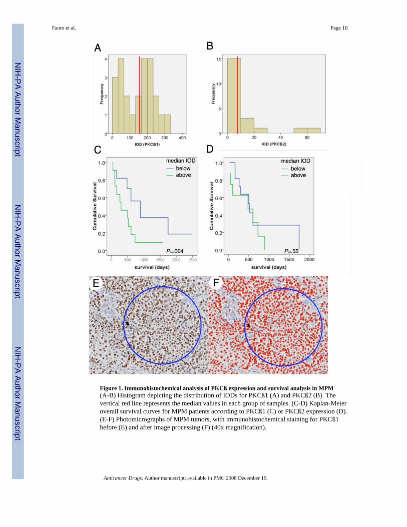
Figure 1. Immunohistochemical analysis of PKCß expression and survival analysis in MPM(A-B) Histogram depicting the distribution of IODs for PKCß1 (A) and PKCß2 (B). Thevertical red line represents the median values in each group of samples. (C-D) Kaplan-Meieroverall survival curves for MPM patients according to PKCß1 (C) or PKCß2 expression (D).(E-F) Photomicrographs of MPM tumors, with immunohistochemical staining for PKCß1before (E) and after image processing (F) (40x magnification).
Faoro et al. Page 10
Anticancer Drugs. Author manuscript; available in PMC 2008 December 19.
NIH
-PA Author Manuscript
NIH
-PA Author Manuscript
NIH
-PA Author Manuscript
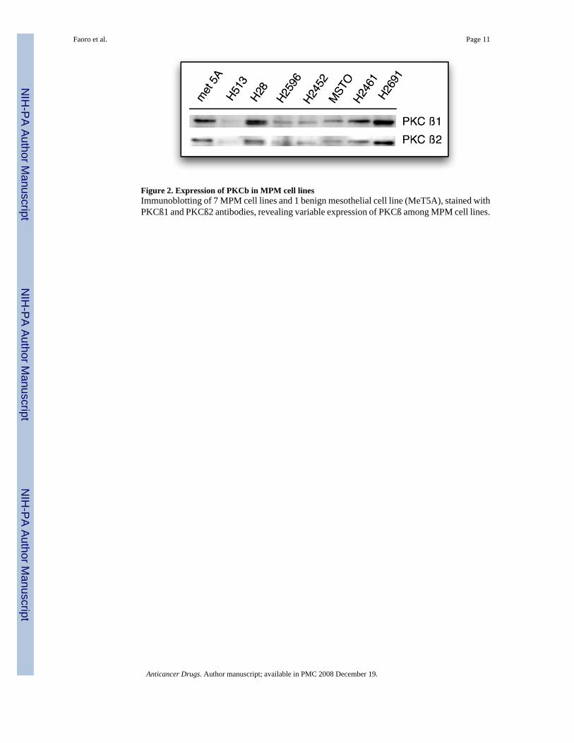
Figure 2. Expression of PKCb in MPM cell linesImmunoblotting of 7 MPM cell lines and 1 benign mesothelial cell line (MeT5A), stained withPKCß1 and PKCß2 antibodies, revealing variable expression of PKCß among MPM cell lines.
Faoro et al. Page 11
Anticancer Drugs. Author manuscript; available in PMC 2008 December 19.
NIH
-PA Author Manuscript
NIH
-PA Author Manuscript
NIH
-PA Author Manuscript

Figure 3. Enzastaurin’s effects on growth of MPM cell lines(A) curves of serum-starved MPM cell lines after treatment with enzastaurin at increasingconcentrations for 72 hours. HCT-116 was used as a positive control. IC50s observed wereapproximately 5 μM, with no significantly increased effect with increasing concentrations. (B)Dose-effect curves after treatment with enzastaurin, cisplatin or both for 72 hours, at a 1:1ratio. The ordinate axis indicates the dosage of each individual drug. The drug combinationdisplayed significant synergism, with combination indices at IC50, IC75 and IC90 below 1.
Faoro et al. Page 12
Anticancer Drugs. Author manuscript; available in PMC 2008 December 19.
NIH
-PA Author Manuscript
NIH
-PA Author Manuscript
NIH
-PA Author Manuscript
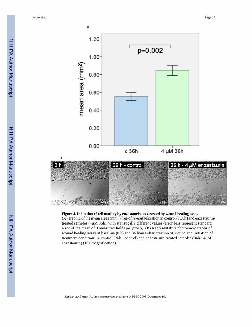
Figure 4. Inhibition of cell motility by enzastaurin, as assessed by wound healing assay(A) graphic of the mean areas (mm2) free of re-epithelization in control (c 36h) and enzastaurin-treated samples (4μM 36h), with statistically different values (error bars represent standarderror of the mean of 3 measured fields per group). (B) Representative photomicrographs ofwound healing assay at baseline (0 h) and 36 hours after creation of wound and initiation oftreatment conditions in control (36h - control) and enzastaurin-treated samples (36h - 4μMenzastaurin) (10x magnification).
Faoro et al. Page 13
Anticancer Drugs. Author manuscript; available in PMC 2008 December 19.
NIH
-PA Author Manuscript
NIH
-PA Author Manuscript
NIH
-PA Author Manuscript

Figure 5. Effects of enzastaurin on downstream signaling molecules in H2461 MPM cell line(A) Immunoblotting of samples after exposure to enzastaurin for 3h, followed by stimulationwith the PKC agonist PMA. Apropriate negative controls are shown. Enzastaurin was able toabrogate PMA-induced phosphorylation of key molecules involved in the AKT pathway andfocal adhesion complex. Beta-actin is shown as loading control. (B) Immunofluorescencemicroscopy showing cellular effects of enzastaurin on the cytoskeleton (phalloidin) andphosphorylation of p130Cas. Nuclei are counter-stained with Hoechst 33342 (60x).
Faoro et al. Page 14
Anticancer Drugs. Author manuscript; available in PMC 2008 December 19.
NIH
-PA Author Manuscript
NIH
-PA Author Manuscript
NIH
-PA Author Manuscript