a phase i clinical trial of single-dose intrapleural ifn- gene transfer for malignant pleural...
TRANSCRIPT
APhase IClinicalTrialof Single-Dose Intrapleural IFN-BGeneTransferforMalignant PleuralMesotheliomaandMetastatic PleuralEffusions:HighRate ofAntitumor ImmuneResponsesDaniel H. Sterman,1Adri Recio,1,2 Richard G. Carroll,2 Colin T. Gillespie,1AndrewHaas,1Anil Vachani,1
VeenaKapoor,1JingSun,1RichardHodinka,3 Jennifer L. Brown,5Michael J. Corbley,5Michael Parr,5
Mitchell Ho,6 Ira Pastan,6 Michael Machuzak,1William Benedict,7 Xin-qiao Zhang,7
ElainaM. Lord,2 Leslie A. Litzky,2Daniel F. Heitjan,2,4 Carl H. June,2 LarryR. Kaiser,1
Robert H.Vonderheide,2 andStevenM.Albelda1
Abstract Purpose: This phase 1dose escalation study evaluated the safety and feasibility of single-doseintrapleural IFN-h gene transfer using an adenoviral vector (Ad.IFN-h) in patients with malignantpleural mesothelioma (MPM) and metastatic pleural effusions (MPE).Experimental Design: Ad.IFN-h was administered through an indwelling pleural catheter indoses ranging from 9 � 1011to 3 � 1012 viral particles (vp) in two cohorts of patients with MPM(7 patients) and MPE (3 patients). Subjects were evaluated for (a) toxicity, (b) gene transfer, (c)humoral, cellular, and cytokine-mediated immune responses, and (d) tumor responses via18-fluorodeoxyglucose-positron emission tomography scans and chest computed tomographyscans.Results: Intrapleural Ad.IFN-h was generally well tolerated with transient lymphopenia as themost common side effect. The maximally tolerated dose achieved was 9 � 1011vp secondaryto idiosyncratic dose-limiting toxicities (hypoxia and liver function abnormalities) in two patientstreated at 3 � 1012 vp. The presence of the vector did not elicit a marked cellular infiltrate inthe pleural space. Intrapleural levels of cytokines were highly variable at baseline and afterresponse to gene transfer. Gene transfer was documented in 7 of the 10 patients by demonstra-tion of IFN-h message or protein. Antitumor immune responses were elicited in 7 of the10 patients and included the detection of cytotoxicTcells (1patient), activation of circulatingnatural killer cells (2 patients), and humoral responses to known (Simian virus 40 largeTantigenandmesothelin) and unknown tumor antigens (7 patients). Four of10 patients showedmeaning-ful clinical responses defined as disease stability and/or regression on 18-fluorodeoxyglucose-positron emission tomography and computed tomography scans at day 60 after vector infusion.Conclusions: Intrapleural instillation of Ad.IFN-h is a potentially useful approach for the gene-ration of antitumor immune responses in MPM and MPE patients and should be investigatedfurther for overall clinical efficacy.
Malignant pleural mesothelioma (MPM) is a refractoryneoplasm. Except for a few patients who benefit from aggressivemultimodality approaches, the majority of patients die fromthe disease within 8 to 14 months of diagnosis (1, 2). Meta-static pleural effusions (MPE) also portend a poor prognosisand are typically treated solely with palliative measures.Given this current lack of effective therapies, our group has
focused on the development of adenoviral vectors for the treat-ment of intrapleural malignancies. We hypothesized that MPMand MPE would be particularly attractive targets for genetransfer studies given preclinical data showing evidence ofeffective adenoviral gene therapy in peritoneal and pleuralmodels of tumor (3–5).Our initial clinical trials used intrapleural delivery of adeno-
viral vectors expressing the suicide gene, herpes simplex thy-midine kinase (Ad.HSVtk), into patients with MPM followedby 2 weeks of i.v. ganciclovir (6–8). Toxicity was minimal, nomaximally tolerated dose was reached, and post-gene transfer
Cancer Therapy: Clinical
Authors’Affiliations: 1Thoracic Oncology GeneTherapy Program and 2AbramsonFamily Cancer Research Institute, University of Pennsylvania Medical Center;3Virology Laboratory, Children’s Hospital of Philadelphia; 4Department ofBiostatistics and Epidemiology, University of Pennsylvania, Philadelphia,Pennsylvania; 5BiogenIdec, Cambridge, Massachusetts; 6Laboratory of MolecularBiology, Center for Cancer Research, National Cancer Institute, NIH, Bethesda,Maryland; and 7Department of Genitourinary Medical Oncology,The University ofTexasM. D. Anderson Cancer Center, Houston,TexasReceived 2/16/07; revised 4/22/07; accepted 5/10/07.The costs of publication of this article were defrayed in part by the payment of pagecharges.This article must therefore be hereby marked advertisement in accordancewith18 U.S.C. Section1734 solely to indicate this fact.Note: Supplementary data for this article are available at Clinical Cancer ResearchOnline (http://clincancerres.aacrjournals.org/).Requests for reprints: Daniel H. Sterman, Interventional Pulmonology Program,Pulmonary, Allergy and Critical Care Division, University of Pennsylvania MedicalCenter, 833 West Gates Building, 3400 Spruce Street, Philadelphia, PA 19104-4283. Phone: 215-614-0984; Fax : 215-662-3226; E-mail: [email protected].
F2007 American Association for Cancer Research.doi:10.1158/1078-0432.CCR-07-0403
www.aacrjournals.orgClin Cancer Res 2007;13(15) August1, 2007 4456
pleural biopsies revealed dose-related transgene expression atthe higher doses, but only in the superficial tumor layers.Interestingly, two patients with stage 1 MPM had significanttumor responses, as documented by serial 18-fluorodeoxyglu-cose-positron emission tomography (18FDG-PET) and chestcomputed tomography (CT) scans, associated with very longsurvivals (>8 years; ref. 9).Given the facts that the Ad.HSVtk gene transfer transduced
only a few tumor cells and that the observed clinical responseswere delayed and persistent, we reasoned that our successfulantitumor responses were not primarily due to massive tumorcell killing by the suicide gene, as we had first postulated, butrather due to ‘‘secondary’’ antitumor immune responses inducedby the Ad.HSV.tk/GCV treatment. This idea was supported bydata inmice showing that cell death induced by HSVtk generatesa strong Th-1 type antitumor immune response (10, 11). Inaddition, it is well established that adenovirus induces strongactivation of the innate and acquired immune system (12, 13).We thus decided to develop a new vector that would (a) continueto take advantage of adenoviral-induced inflammation, (b)directly induce cell death (like Ad.HSVtk/GCV), and (c) enhanceantitumor immune responses by secretion of an immunostimu-latory cytokine.Several published clinical trials have shown clinical responses
to intrapleural infusion of IFN-h, IFN-a, and IFN-g in patientswith MPM or MPE (14–18). Type 1 IFNs (such as IFN-a andIFN-h) are known to inhibit tumor cell growth and stimulatethe immune system (19). IFNs have immunoregulatory effectson antibody production, natural killer (NK) and T-cell acti-vation, macrophage function, delayed-type hypersensitivity,and MHC antigen expression, as well as antiproliferative effectsand antiangiogenic properties (20–24). Finally, IFN-h genetransfer in animal models of various malignancies (both xeno-grafts and autologous tumors) has shown impressive anti-tumor effects (25–29).We therefore conducted preclinical studies using an adeno-
viral vector expressing mouse IFN-h (Ad.muIFN-h). Our pre-clinical data showed that (a) Ad.muIFN-h had dramatictherapeutic efficacy in syngeneic animal models of MPM andlung cancer, (b) i.p. and i.t. injections of Ad.muIFN-h showedsignificant antitumor activity both in the injected tumor siteand in distant tumors, and (c) in these models, this effect wasdue, in large part, to the generation of CTLs directed againsttumor antigens and activation of NK cells (30–32).Based on these data, as well as the availability of clinical
grade Ad.humanIFN-h from BiogenIdec, we conducted a single-center dose escalation phase 1 clinical trial of single-doseintrapleural infusion of Ad.huIFN-h in patients with MPM/MPE. The goals of the trial were as follows: (a) determine safetyand toxicities of a single intrapleural infusion of Ad.huIFN-h;(b) establish the maximally tolerated dose of a single intra-pleural infusion of Ad.huIFN-h; (c) evaluate induced antivectorand antitumor immune responses; and (d) assess antitumoractivity of Ad.huIFN-h before and after vector instillation usingchest CT and whole-body 18FDG-PET scanning.
Materials andMethods
Preclinical and regulatory issues
After conducting a formal animal toxicology study with intrapleuraldelivery of Ad.muIFN-h (and Ad.huIFN-h) under Food and Drug
Administration Good Laboratory Practices guidelines, the study was
approved by all appropriate human study and biosafety committees at
Penn and the Recombinant DNA Advisory Committee of the NIH’s
Office of Biotechnology Activities. We obtained a physician-sponsored
investigational new drug from the Food and Drug Administration
(BB-IND 10603).
Vector
We used Ad.huIFN-h virus (BG00001), developed at BiogenIdec.
The vector is a good manufacturing practices grade, E1/E3-deleted
replication-incompetent adenovirus with an insertion of the human
IFN-b gene in the E1 region of the adenoviral genome. The transgene
was driven by a human cytomegalovirus promoter. Food and Drug
Administration approval for this vector was obtained by BiogenIdec and
cross-referenced in our investigational new drug proposal.
Patients
Patients were eligible for these studies based on the following: (a) a
pathologically confirmed diagnosis of MPM or MPE; (b) an Eastern
Cooperative Oncology Group performance status of 0, 1, or 2; and (c)
an accessible pleural space for instillation of vector. Exclusion criteria
included prior surgical resection, successful pleurodesis, recent chemo-
therapy or radiotherapy, inadequate pulmonary function, or the
presence of significant cardiac/hepatic/renal disease. Patients 102 and
103 signed the consent form but were not dosed because of interval
disease progression.
Protocol summary
Eligible patients underwent insertion of a tunneled intrapleural
catheter (Pleurx, Cardinal Health) under local anesthesia and had a
single-blood volume leukapheresis for harvesting peripheral blood
mononuclear cells to be used for immunoassessment. On study day 1,
patients were admitted to the Penn General Clinical Research Center,
hydrated i.v., and premedicated with acetaminophen. All obtain-
able pleural fluid was drained from the chest through the Pleurx
catheter. Subsequently, a single dose of Ad.huIFN-h (BG00001), diluted
in 50 cc of sterile normal saline, was instilled via the catheter into
the pleural space. The catheter was then flushed and capped to maxi-
mize vector delivery to intrapleural tumor. Patients were monitored
in the General Clinical Research Center and discharged to home after
72 h with the pleural catheter capped. Patients were followed closely
as out-patients for the next 6 months. Approximately 4 weeks after
vector instillation, the pleural catheter was removed, unless still needed
for control of symptomatic malignant pleural effusion. Chest CT
scans and dual-time point 18FDG-PET scans were done at baseline
and 2 months after vector instillation, as well as at 6 months, if
clinically indicated.
Sample collection and generation of cell lines from patient
samples
Peripheral blood mononuclear cells were purified by Ficoll densitygradient centrifugation and were viably cryopreserved as bulk peri-pheral blood mononuclear cells. Cells contained in malignant pleuraleffusions (or 20 mL pleural lavages, if no pleural fluid was present)were purified by density gradient centrifugation using either Ficoll orPercoll. The cells were generally frozen in bulk, but if needed, werefractionated into specific subsets using either magnetic beads or the Mo-Flo cell sorter (DakoCytomation).
Radiographic response assessment
We used the ‘‘modified Response Evaluation Criteria in SolidTumors (RECIST)’’ schema for the evaluation of tumor response inthe special case of mesothelioma (2, 33). These criteria apply equallywell to evaluation of MPE. In addition, we evaluated response using18FDG-PET, which is increasingly being used to assess mesotheliomatumor activity before and after therapy, with the potential for earlierand more accurate determinations of treatment efficacy (34).
Ad.IFN-b ImmunogeneTherapy forMesothelioma
www.aacrjournals.org Clin Cancer Res 2007;13(15) August1, 20074457
Measurement of viral shedding
Samples of whole blood, pleural fluid, and swabs from the pleu-ral catheter insertion site were obtained at baseline and varioustimes after gene instillation and sent to the Children’s Hospitalof Philadelphia Virology laboratory for analysis. Viral DNA wasdetected by PCR. Specimens were also cultured for wild-typeadenovirus on A549 cells and replication-deficient virus (vector)on 293 cells. Details of these procedures can be found inSupplementary Data.
Assessment of gene transfer
Transgene and endogenous IFN-b mRNA assays. PCR primers weredesigned to target an area of the transcript within the 3¶ untranslatedregion where the endogenous and vector-produced IFN-h mRNAdiffered. Glyceraldehyde-3-phosphate dehydrogenase served as acontrol for RNA isolation and stability. Studies with primer pairsdesigned as controls for DNA contamination of the RNA prep showedno contamination (data not shown). Control studies are described inSupplementary data.
Cells that had been isolated from the pleural effusions of patients104, 108, 110, 111, and 112 were thawed and 1 � 107 total cells wereused for RNA preparations. Patients 101, 106, and 107 had pleuraltumor, but no effusion; therefore, no cells were available for RNAanalysis. Samples from time points taken before dose, 24 h after vectortreatment, 48 h, and 7 days after vector treatment (when cells wereavailable) were analyzed for glyceraldehyde-3-phosphate dehydroge-nase, endogenous, and transgene RNA transcripts.
IFN-h intracellular staining
IFN-h intracellular staining was done from two patients’ pleural fluidcytospins (patients 108 and 110) using a primary anti-IFN-h antibody(Chemicon). See Supplementary Data for details.
Cytokine assays
Commercial cytokine ELISA assays were used to measure the levels of
IFN-h, IFN-a, IFN-g, interleukin (IL)-6, transforming growth factor-h(total and heat activated), and vascular endothelial growth factor in
serial dilutions of pleural fluid and serum at various time points.
Additional cytokines (IL-1h, IL-10, Macrophage Chemotactic Protein-1
[MCP-1/CCL2], IL-8, and Regulated Upon Activation, Normal T-cell
Expressed and Secreted [RANTES/CCL5]) were measured using a
cytokine bead assay from Luminex according to the manufacturer’s
instructions.
Cellular analysis of pleural fluids
Pleural fluid mononuclear cells were isolated from patient samples
by Ficoll centrifugation. Cells were labeled with fluorochrome-
conjugated monoclonal antibodies as described previously (35) and
four-color flow cytometric analysis was done using a Becton Dickinson
FACSCanto flow cytometer (BD Biosciences). All labeled antibodies
were from BD Biosciences.
Immunoblots
To detect humoral responses against tumor antigens, immunoblot-ting against purified proteins and extracts from mesothelioma, lungcancer, and ovarian cancer cell lines was done. Purified WT-1 proteinwas purchased from Santa Cruz Biotechnology. Purified Simian virus40 (SV40) large T-antigen (Tag) protein was purchased from Chimerx.Purified mesothelin was provided by Drs. M. Ho and I. Pastan(National Cancer Institute, Bethesda, MD). Cell lines derived frompatient samples were grown in culture. Extracts from cells or purifiedproteins were prepared and immunoblotted with patient serum asdescribed previously (9). See Supplementary Data for details. Patientserum samples (diluted at 1:1,500) from time points before treatmentand 6 weeks to 6 months after treatment were used and bound humanantibody was visualized.
Anti-mesothelin antibody ELISA
Quantitative estimates of levels of anti-mesothelin antibodies wereobtained using a modified ELISA on 1:100 dilutions of serum samplesat various time points before and after vector instillation, as describedby Ho et al. (36).
Cellular antitumor immune responses
Cellular immune responses were measured by transfecting totaltumor RNA or control RNA into autologous dendritic cells. AutologousT cells were stimulated in vitro and used for CTL activity againstchromium 53– labeled autologous tumor cells or HLA-matchedallogeneic tumor cell lines as described previously (37).
Adenovirus serotype 5 neutralizing antibody levels
Serum neutralizing antibodies specific for adenovirus were evaluatedas described previously (4, 9). See Supplementary Data for details.
Statistical analysis
We used a standard 3+3 design to determine the maximally tolerateddose, with an implicit 50% chance of further dose escalation after
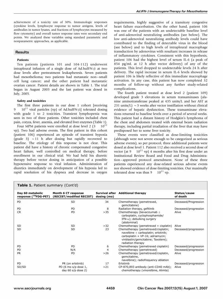
Table 1. Patient summary
ID G Age Primary (stage at entry) Dose level Dosing Day 60 CT response(RECIST/modified RECIST)
101 M 80 Mesothelioma (IA) 1 Tolerated PD104 F 57 Lung (IIIB) 1 Tolerated PD105 F 47 Ovarian (IV) 1 Tolerated SD106 M 72 Mesothelioma (IIA) 2 DLT: hypoxia SD107 M 64 Mesothelioma (IIB) 2 Tolerated PD
108 M 66 Mesothelioma (IV) 2 Tolerated PD109 F 40 Mesothelioma (IIA) 2 DLT: elevated
transaminasesPD
110 M 78 Mesothelioma (IIIA) 1 Tolerated SD111 M 76 Mesothelioma (IV) 1 Tolerated PD112* M 61 Mesothelioma (IIA) 1 Tolerated (�2) SD (status post dose 1)
Abbreviations: G, gender; SD, stable disease; PD, progressive disease; CR, complete response; PR, partial response; DLT, dose-limitingtoxicity; mAb, monoclonal antibody; VP16, etoposide.*Patient 112 received a second vector dosed at dose level 1 under an Institutional Review Board and Food and Drug Administration–approvedprotocol amendment 4 mo after initial dosing.
Cancer Therapy: Clinical
www.aacrjournals.orgClin Cancer Res 2007;13(15) August1, 2007 4458
achievement of a toxicity rate of 30%. Immunologic responses(cytokine levels, lymphocyte response to tumor antigens, levels ofantibodies in tumor lysates, and fractions of lymphocytes measured byflow cytometry) and overall tumor response rates were secondary endpoints. We analyzed these variables using standard parametric andnonparametric approaches, as applicable.
Results
Patients
Ten patients (patients 101 and 104-112) underwentintrapleural infusion of a single dose of Ad.huIFN-h at twodose levels after pretreatment leukapheresis. Seven patientshad mesothelioma; two patients had metastatic non–smallcell lung cancer; and the other patient had metastaticovarian cancer. Patient details are shown in Table 1. The trialbegan in August 2003 and the last patient was dosed inJuly, 2005.
Safety and toxicities
The first three patients in our dose 1 cohort [receiving9 � 1011 viral particles (vp) of Ad.huIFN-h] tolerated dosingwith grade 1 to 2 toxicities. Transient lymphopenia wasseen in two of three patients. Other toxicities included chestpain, coryza, fever, anemia, and elevated liver enzymes (Table 1).Four MPM patients were enrolled at dose level 2 (3 � 1012
vp). Two had adverse events. The first patient in this cohort(patient 106) experienced an episode of transient hypoxia(grade 3) f11 h after dosing but rapidly recovered tobaseline. The etiology of this response is not clear. Thispatient did have a history of chronic compensated congestiveheart failure, well controlled on medical therapy, beforeenrollment in our clinical trial. We had held his diuretictherapy before vector dosing in anticipation of a possiblehypotensive response to viral infusion. Administration ofdiuretics immediately on development of his hypoxia led torapid resolution of his dyspnea and decrease in oxygen
requirements, highly suggestive of a transitory congestiveheart failure exacerbation. On the other hand, patient 106was one of the patients with an undetectable baseline levelof anti-adenoviral neutralizing antibodies (see below). Thelow anti-adenoviral neutralizing antibody levels could havecontributed to the finding of detectable virus in the blood(see below) and to high levels of intrapleural macrophagetransduction by adenovirus with resultant increases in releaseof inflammatory cytokines. Consistent with this hypothesis,patient 106 had the highest level of serum IL-6 (a peak of854 pg/mL at 12 h after vector delivery) of any of thepatients. This level dropped to undetectable levels 24 h afterdelivery. The rapid increase in serum IL-6 levels showed bypatient 106 is likely reflective of this immediate macrophageactivation. In any case, this patient has now completed 32months of follow-up without any further study-relatedcomplications.The fourth patient treated at dose level 2 (patient 109)
developed grade 3 elevations in serum transaminases (ala-nine aminotransferase peaked at 435 units/L and her AST at231 units/L) f3 weeks after vector instillation without clinicalevidence of hepatic dysfunction. These transaminase eleva-tions returned to baseline levels over a period of several weeks.This patient had a distant history of Hodgkin’s lymphoma ofthe chest and abdomen treated with external beam radiationtherapy, including partial irradiation of the liver that may havepredisposed her to some liver toxicity.These events were classified as dose-limiting toxicities
(although were not severe enough to be categorized as seriousadverse events), so per protocol, three additional patients weredosed at dose level 1. Patient 112 also received a second dose ofvector (at 9 � 1011 vp) 4 months after his first dose under anInstitutional Review Board and Food and Drug Administra-tion–approved protocol amendment. None of these threepatients experienced any dose-related serious adverse eventsnor showed evidence of dose-limiting toxicities. Our maximallytolerated dose was thus 9 � 1011 vp.
Table 1. Patient summary (Cont’d)
Day 60 metabolicresponse (18FDG-PET)
Month 6 CT response(RECIST/modified RECIST)
Survival afterdosing (mo)
Additional therapy Status/causeof death
PD SD 34 Chemotherapy (pemetrexed,gemcitabine)
Deceased/Progression
PD PD 8 Radiation therapy, gefitinib Deceased/progressionCR PD >35 Chemotherapy (bevacizumab F
carboplatin, cyclophosphamide/IFN-a); debulking surgery(abdominal)
Alive
SD SD >32 Chemotherapy (pemetrexed + cisplatin) AliveSD SD 23 Chemotherapy (pemetrexed/cisplatin;
navelbine F carboplatin; erlotinib;carboplatin + VP-16; adriamycin;vinblastin/gemcitabine; Taxotere);radiation therapy
Deceased/progression
PD PD 6 Chemotherapy (pemetrexed cisplatin) Deceased/progressionPD N/A 4 Chemotherapy (pemetrexed) Deceased/progressionPR PD >26 Chemotherapy (pemetrexed/cisplatin,
gemcitabine,navelbine); radiofrequency ablation
Alive
PD PR (on erlotinib) 17 Erlotinib Deceased/progressionSD/SD PD (6 mo s/p dose 1,
day 60 s/p dose 2)>21 CP-870,893 antibody (anti-CD40 mAb);
chemotherapy (vinorelbine, Alimta)Alive
Ad.IFN-b ImmunogeneTherapy forMesothelioma
www.aacrjournals.org Clin Cancer Res 2007;13(15) August1, 20074459
Viral shedding
Chest wall swabs, pleural fluid, and serum were analyzed onday 0 (pretreatment) and days 2, 3, 4, 7, 14, 21, and 28 byaddition to cell monolayers with subsequent analysis for cyto-pathic effects. Samples were cultured on A549 cells to detectreplication-competent adenovirus and on 293 cells (whichexpress adenoviral E1 protein) to detect replication-defectivevector (Supplementary Table S1). No samples were positive forreplication-competent adenovirus. Pleural fluid cultures show-ing replication-defective vector were positive in five of thepatients (for up to 7 days in three patients) but were negative inall patients by 14 days. Only one patient (patient 106) had apositive culture for vector in serum (and only on day 1; Table 2).Specimens were also analyzed for vector-specific DNA
sequences by PCR. Four patients (patients 104, 106, 107, and110) had positive serum adenoviral PCRs for up to 4 days aftervector instillation. Pleural fluid samples (or flushes, if needed)were analyzed at days 2, 3, 4, 7, and 14 for all 10 patients.Thereafter, pleural fluid was analyzed in the patients wherePleurex catheter fluid was obtainable (see Table 2). We iden-tified adenoviral vector DNA for 10 days in pleural fluid in allof the patients. PCR was positive in 6 of 8 patients at day 28,in 3 of 5 patients at day 42, and in none of the 4 evaluablepatients by day 56.
Antiviral immune responses
Serum anti-adenoviral neutralizing antibody titers weremeasured before and after gene transfer. As shown in Table 3,baseline titers of anti-adenoviral neutralizing antibody rangedfrom <1:10 to 1:750. Only three patients had baseline titers>1:100. All patients increased their neutralizing antibodytiters after gene transfer. Four patients had weak antibodyresponses to vector instillation (defined as an increase of titerby <10-fold). Six patients had more than a 10-fold increase intiter (average of a 45-fold increase). Patient 112 received twodoses of vector. The first dose led to an increase from 1:50 to1:3,200. The second dose of vector led to a doubling ofneutralizing antibody titers from 1:3,200 to 1:6,400 (Table 3).
Gene transfer
Gene transfer was assessed using pleural fluid obtainedthrough the tunneled pleural catheter (Table 4). In patientswith no accessible pleural fluid (patients 106, 107, and 109),we collected pleural lavages.Pleural fluid samples were tested for the presence of
adenoviral DNA by PCR (Table 2). No patient had detectable
adenoviral DNA in pretreatment samples. All 10 patients haddetectable adenoviral DNA after gene transfer.We measured IFN-h protein levels in the before and after
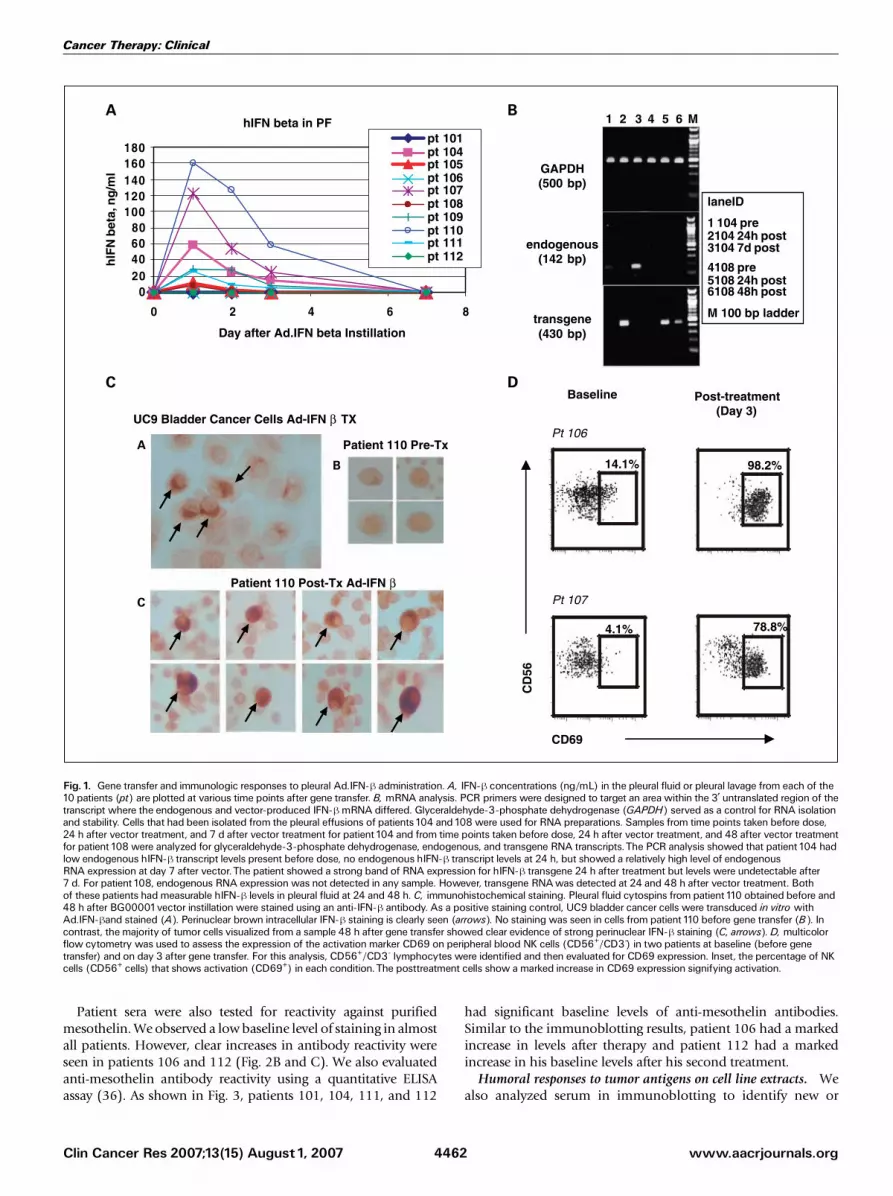
vector instillation pleural fluid and serum samples. Thesepleural measurements should be considered semiquantitativebecause the volume of pleural fluid clearly affected the finalconcentration, and, in three of the patients, saline lavages weredone to obtain samples. As summarized in Table 4, detectablelevels of IFN-h in pleural fluid (ranging from 200 pg/mL to160 ng/mL) were found in all but two patients. The time courseis shown in Fig. 1A. IFN-h was detectable for up to 3 days inmost patients. Serum levels of IFN-h were undetectable in mostpatients (<0.2 ng/mL) and quite low in those who haddetectable levels (Table 4).To further document gene transfer, we assessed endogenous
and vector-produced IFN-h mRNA using reverse transcription-PCR in the five samples in which enough cellular material wasavailable (Fig. 1B). Transgene-specific RNA was detected in allfive patients (patients 104, 108, 110, 111, and 112). Like theprotein measurements, vector mRNA expression was relativelytransient, usually detectable for only 48 h, although in patient111, mRNA was seen at 7 days. Only two patients haddetectable message for endogenous IFN-h; patient 104 hadendogenous message on day 7 and patient 111 at 24 h.Finally, to further confirm successful intrapleural IFN-h gene
transfer and to show the actual transduction of tumor cells, wedid IFN-h staining on pleural fluid cytospins obtained beforeand 48 h after BG00001 vector instillation in two patientswhere sufficient numbers of tumor cells were present. Positivelystaining cells were seen in both patients. Results are shown forpatient 110 (Fig. 1C). Whereas tumor cells obtained from pre-gene transfer pleural fluid showed no positive staining, themajority of tumor cells visualized from the sample 48 h aftergene transfer showed clear evidence of strong perinuclear IFN-hstaining.
Inflammatory and cytokine responses
The patterns of inflammatory pleural leukocyte cell responsewere quite variable among patients. The average ‘‘fold increase’’in total leukocytes (comparing preinstillation with the peakcount in the next 4 days) was 3.3-fold (F 0.7-fold SE) with anyincreases due to monocytes and lymphocytes (not neutrophils).We did not observe consistent alterations in the percentage orabsolute count of pleural T cells, regulatory T cells, NK cells, ordendritic cells. No consistent change in the activation of T cellsor dendritic cells was observed.
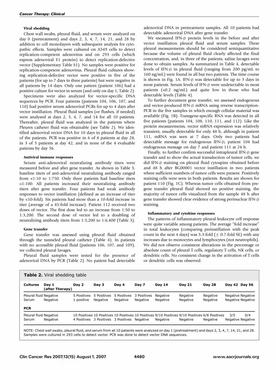
Table 2. Viral shedding table
Cultures Day 1(after Therapy)
Day 2 Day 3 Day 4 Day 7 Day 14 Day 21 Day 28 Day 42 Day 56
Pleural fluid Negative 5 Positives 5 Positives 5 Positives 3 Positives Negative Negative Negative Negative NegativeSerum Negative 1 positive Negative Negative Negative Negative Negative Negative Negative Negative
PCR
Pleural fluid Negative 10 Positives 10 Positives 10 Positives 10 Positives 9/10 Positives 9/10 Positives 6/8 Positives 3/5 0/4Serum Negative 4 Positives 3 Positives 3 Positives Negative Negative Negative Negative Negative Negative
NOTE: Chest wall swabs, pleural fluid, and serum from all 10 patients were analyzed on day 1 (pretreatment) and days 2, 3, 4, 7, 14, 21, and 28.Samples were cultured in 293 cells to detect vector. PCR was done to detect vector DNA sequences.
Cancer Therapy: Clinical
www.aacrjournals.orgClin Cancer Res 2007;13(15) August1, 2007 4460
Levels of pleural cytokines (including IL-6, IL-1h, IL-10,vascular endothelial growth factor, MCP-1, IL-8, RANTES,transforming growth factor-h1 and transforming growthfactor-h2, and IFN-g) were highly variable among the patients,both at baseline and after response to gene transfer, perhapsreflecting the dilutional issues mentioned above.Levels of serum cytokines were also monitored with special
focus on IL-6, which can be elevated in mesothelioma (38) andhas been reported as a marker of systemic inflammatoryresponse after adenoviral instillation (39, 40). Serum IL-6 levelswere detectable in four patients, and in these patients, the peakswere only f250 pg/mL. Only one other cytokine, MCP-1, wasconsistently detected in serum after gene transfer, whichaveraged 72 F 17 pg/mL at baseline with peaks averaging943 F 407 pg/mL, primarily day 1 after vector instillation.
Antitumor immunologic responses
Innate immune responses. Because IFN-h has the ability toactivate NK cells (20, 31), we examined the activation state ofcirculating NK cells at an early time point (3 days) after vectorinstillation. Using flow cytometry, we first identified NK cellsby the CD56+/CD3- cell surface phenotype among lympho-cytes. Five of the nine patients had <1% circulating NK cellsbefore and after vector administration and were not analyzedfurther. In the remaining four patients, the percentages of NK
cells among total peripheral blood lymphocytes were 3%(patient 107), 3.6% (patient 101), 10.2% (patient 106), and10.4% (patient 108). These percentages did not change aftergene transfer. We evaluated the activation state of NK cells inthese patients by determining the percentage of NK cellsexpressing the activation marker CD69 before and after genetransfer (41). In two of these patients, the percentage of CD69+
activated cells did not change. However, as shown in Fig. 1D,the percentage of CD69+ activated NK cells went up remarkablyin patient 106 (14.1-98.2%) and patient 107 (4.1-78.3%).Thus, in two of four evaluable patients, a single Ad.IFN-hintrapleural infusion led to the activation of circulating NK cells(see Supplementary Table S1).Humoral responses to known MPM tumor antigens. Humoral
responses to three defined mesothelioma-associated antigenswere evaluated by immunoblotting purified proteins usingpatient sera from before and after gene transfer. Most patientshad low-level baseline reactivity to purified Wilms’ tumorantigen-1 (WT-1 protein), but none showed clear increases aftertreatment. Most patients also had some baseline reactivityagainst purified SV40 large Tag (SV40 Tag protein) with noincreases after gene transfer. However, one patient (patient107) developed a significant increase in antibody responseto SV40 Tag after gene transfer (Fig. 2A; see SupplementaryTable S1).
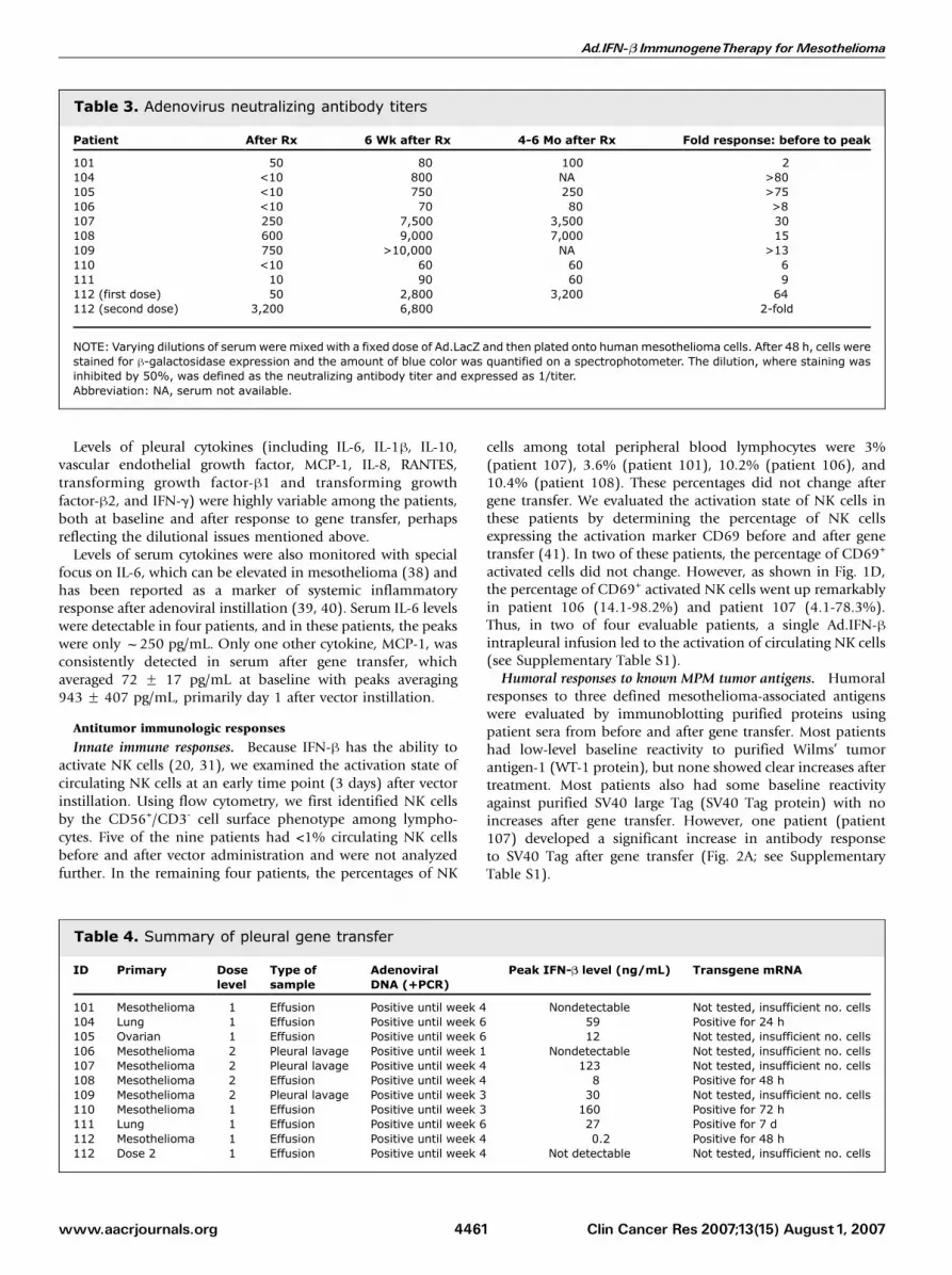
Table 3. Adenovirus neutralizing antibody titers
Patient After Rx 6 Wk after Rx 4-6 Mo after Rx Fold response: before to peak
101 50 80 100 2104 <10 800 NA >80105 <10 750 250 >75106 <10 70 80 >8107 250 7,500 3,500 30108 600 9,000 7,000 15109 750 >10,000 NA >13110 <10 60 60 6111 10 90 60 9112 (first dose) 50 2,800 3,200 64112 (second dose) 3,200 6,800 2-fold
NOTE: Varying dilutions of serum were mixed with a fixed dose of Ad.LacZ and then plated onto humanmesothelioma cells. After 48 h, cells werestained for h-galactosidase expression and the amount of blue color was quantified on a spectrophotometer. The dilution, where staining wasinhibited by 50%, was defined as the neutralizing antibody titer and expressed as 1/titer.Abbreviation: NA, serum not available.
Table 4. Summary of pleural gene transfer
ID Primary Doselevel
Type ofsample
AdenoviralDNA (+PCR)
Peak IFN-B level (ng/mL) Transgene mRNA
101 Mesothelioma 1 Effusion Positive until week 4 Nondetectable Not tested, insufficient no. cells104 Lung 1 Effusion Positive until week 6 59 Positive for 24 h105 Ovarian 1 Effusion Positive until week 6 12 Not tested, insufficient no. cells106 Mesothelioma 2 Pleural lavage Positive until week 1 Nondetectable Not tested, insufficient no. cells107 Mesothelioma 2 Pleural lavage Positive until week 4 123 Not tested, insufficient no. cells108 Mesothelioma 2 Effusion Positive until week 4 8 Positive for 48 h109 Mesothelioma 2 Pleural lavage Positive until week 3 30 Not tested, insufficient no. cells110 Mesothelioma 1 Effusion Positive until week 3 160 Positive for 72 h111 Lung 1 Effusion Positive until week 6 27 Positive for 7 d112 Mesothelioma 1 Effusion Positive until week 4 0.2 Positive for 48 h112 Dose 2 1 Effusion Positive until week 4 Not detectable Not tested, insufficient no. cells
Ad.IFN-b ImmunogeneTherapy forMesothelioma
www.aacrjournals.org Clin Cancer Res 2007;13(15) August1, 20074461
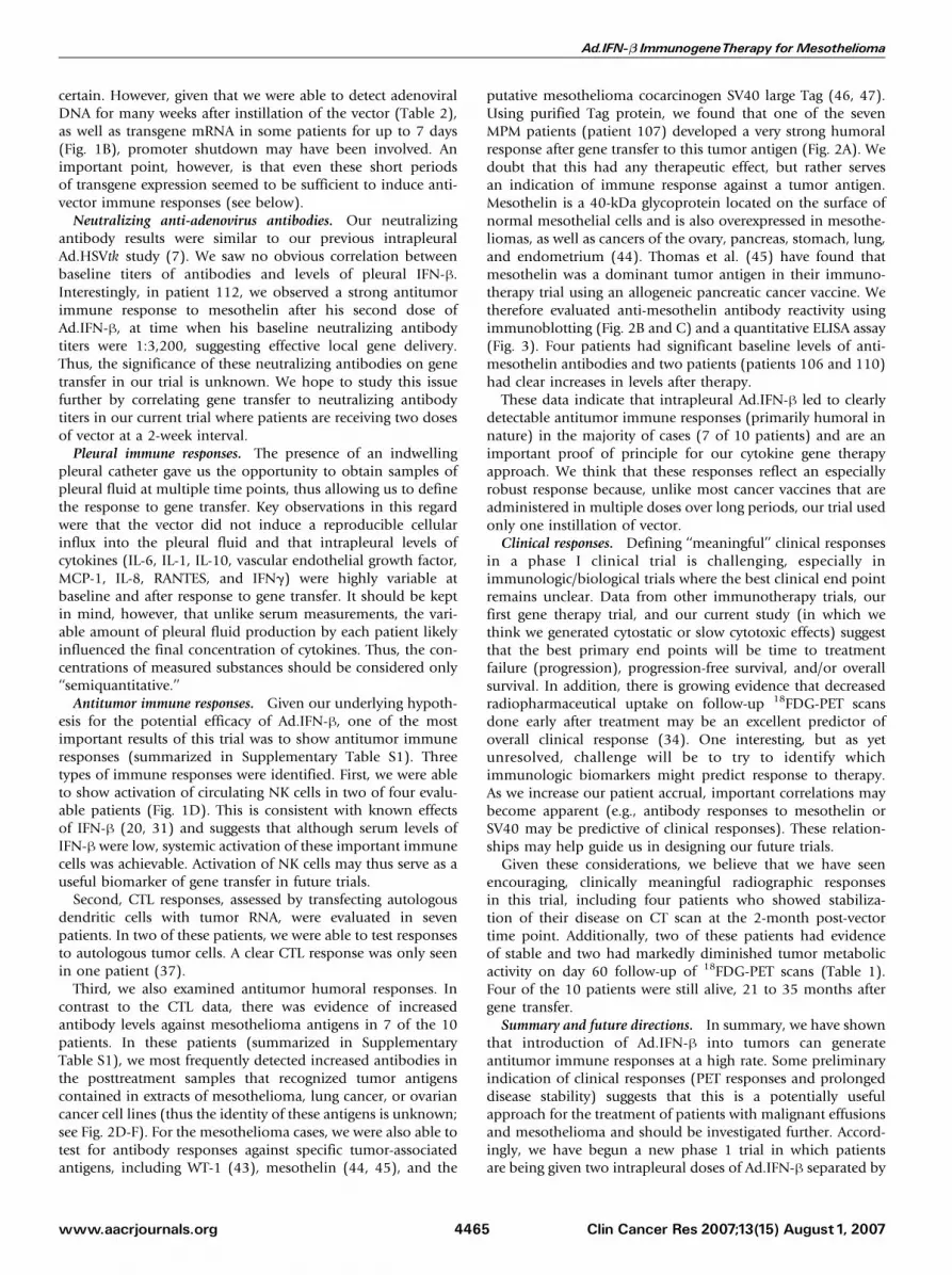
Patient sera were also tested for reactivity against purifiedmesothelin.We observed a lowbaseline level of staining in almostall patients. However, clear increases in antibody reactivity wereseen in patients 106 and 112 (Fig. 2B and C). We also evaluatedanti-mesothelin antibody reactivity using a quantitative ELISAassay (36). As shown in Fig. 3, patients 101, 104, 111, and 112
had significant baseline levels of anti-mesothelin antibodies.Similar to the immunoblotting results, patient 106 had a markedincrease in levels after therapy and patient 112 had a markedincrease in his baseline levels after his second treatment.Humoral responses to tumor antigens on cell line extracts. We
also analyzed serum in immunoblotting to identify new or
Fig. 1. Gene transfer and immunologic responses to pleural Ad.IFN-h administration. A, IFN-h concentrations (ng/mL) in the pleural fluid or pleural lavage from each of the10 patients (pt) are plotted at various time points after gene transfer. B, mRNA analysis. PCR primers were designed to target an area within the 3¶ untranslated region of thetranscript where the endogenous and vector-produced IFN-hmRNA differed. Glyceraldehyde-3-phosphate dehydrogenase (GAPDH) served as a control for RNA isolationand stability. Cells that had been isolated from the pleural effusions of patients104 and108 were used for RNA preparations. Samples from time points taken before dose,24 h after vector treatment, and 7 d after vector treatment for patient 104 and from time points taken before dose, 24 h after vector treatment, and 48 after vector treatmentfor patient 108 were analyzed for glyceraldehyde-3-phosphate dehydrogenase, endogenous, and transgene RNA transcripts.The PCR analysis showed that patient 104 hadlow endogenous hIFN-h transcript levels present before dose, no endogenous hIFN-h transcript levels at 24 h, but showed a relatively high level of endogenousRNA expression at day 7 after vector.The patient showed a strong band of RNA expression for hIFN-h transgene 24 h after treatment but levels were undetectable after7 d. For patient 108, endogenous RNA expression was not detected in any sample. However, transgene RNAwas detected at 24 and 48 h after vector treatment. Bothof these patients had measurable hIFN-h levels in pleural fluid at 24 and 48 h. C, immunohistochemical staining. Pleural fluid cytospins from patient110 obtained before and48 h after BG00001vector instillation were stained using an anti-IFN-h antibody. As a positive staining control, UC9 bladder cancer cells were transduced in vitro withAd.IFN-hand stained (A). Perinuclear brown intracellular IFN-h staining is clearly seen (arrows). No staining was seen in cells from patient 110 before gene transfer (B). Incontrast, the majority of tumor cells visualized from a sample 48 h after gene transfer showed clear evidence of strong perinuclear IFN-h staining (C, arrows). D, multicolorflow cytometry was used to assess the expression of the activation marker CD69 on peripheral blood NK cells (CD56+/CD3-) in two patients at baseline (before genetransfer) and on day 3 after gene transfer. For this analysis, CD56+/CD3- lymphocytes were identified and then evaluated for CD69 expression. Inset, the percentage of NKcells (CD56+ cells) that shows activation (CD69+) in each condition.The posttreatment cells show a marked increase in CD69 expression signifying activation.
Cancer Therapy: Clinical
www.aacrjournals.orgClin Cancer Res 2007;13(15) August1, 2007 4462
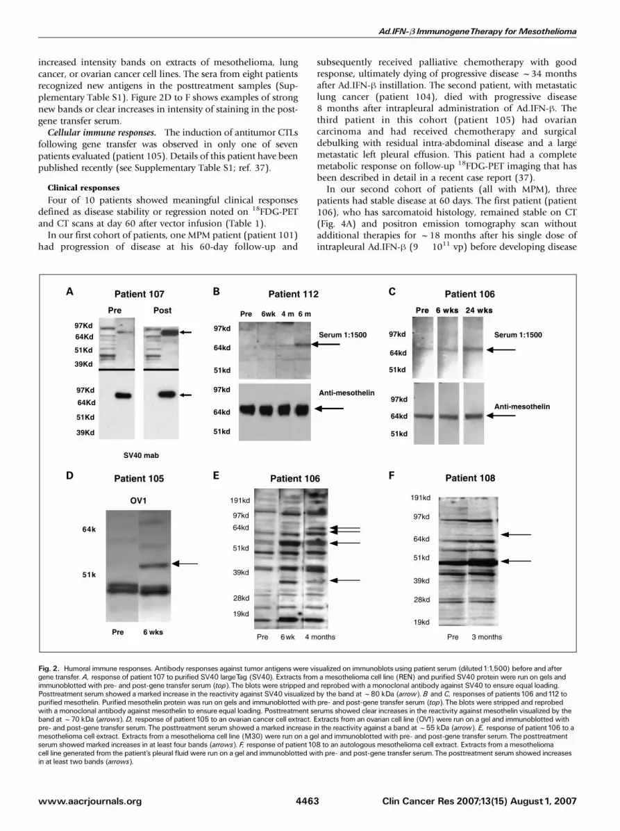
increased intensity bands on extracts of mesothelioma, lungcancer, or ovarian cancer cell lines. The sera from eight patientsrecognized new antigens in the posttreatment samples (Sup-plementary Table S1). Figure 2D to F shows examples of strongnew bands or clear increases in intensity of staining in the post-gene transfer serum.Cellular immune responses. The induction of antitumor CTLs
following gene transfer was observed in only one of sevenpatients evaluated (patient 105). Details of this patient have beenpublished recently (see Supplementary Table S1; ref. 37).
Clinical responses
Four of 10 patients showed meaningful clinical responsesdefined as disease stability or regression noted on 18FDG-PETand CT scans at day 60 after vector infusion (Table 1).In our first cohort of patients, one MPM patient (patient 101)
had progression of disease at his 60-day follow-up and
subsequently received palliative chemotherapy with goodresponse, ultimately dying of progressive disease f34 monthsafter Ad.IFN-h instillation. The second patient, with metastaticlung cancer (patient 104), died with progressive disease8 months after intrapleural administration of Ad.IFN-h. Thethird patient in this cohort (patient 105) had ovariancarcinoma and had received chemotherapy and surgicaldebulking with residual intra-abdominal disease and a largemetastatic left pleural effusion. This patient had a completemetabolic response on follow-up 18FDG-PET imaging that hasbeen described in detail in a recent case report (37).In our second cohort of patients (all with MPM), three
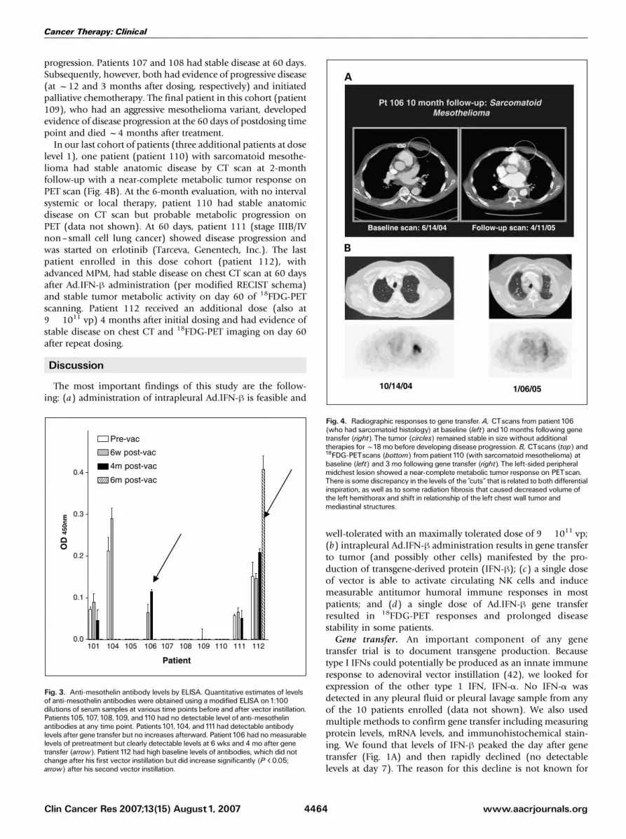
patients had stable disease at 60 days. The first patient (patient106), who has sarcomatoid histology, remained stable on CT(Fig. 4A) and positron emission tomography scan withoutadditional therapies for f18 months after his single dose ofintrapleural Ad.IFN-h (9 � 1011 vp) before developing disease
Fig. 2. Humoral immune responses. Antibody responses against tumor antigens were visualized on immunoblots using patient serum (diluted1:1,500) before and aftergene transfer. A, response of patient 107 to purified SV40 largeTag (SV40). Extracts from a mesothelioma cell line (REN) and purified SV40 protein were run on gels andimmunoblotted with pre- and post-gene transfer serum (top).The blots were stripped and reprobed with a monoclonal antibody against SV40 to ensure equal loading.Posttreatment serum showed a marked increase in the reactivity against SV40 visualized by the band atf80 kDa (arrow). B and C, responses of patients106 and112 topurified mesothelin. Purified mesothelin protein was run on gels and immunoblotted with pre- and post-gene transfer serum (top).The blots were stripped and reprobedwith a monoclonal antibody against mesothelin to ensure equal loading. Posttreatment serums showed clear increases in the reactivity against mesothelin visualized by theband atf70 kDa (arrows). D, response of patient 105 to an ovarian cancer cell extract. Extracts from an ovarian cell line (OV1) were run on a gel and immunoblotted withpre- and post-gene transfer serum.The posttreatment serum showed a marked increase in the reactivity against a band atf55 kDa (arrow). E, response of patient 106 to amesothelioma cell extract. Extracts from a mesothelioma cell line (M30) were run on a gel and immunoblotted with pre- and post-gene transfer serum.The posttreatmentserum showed marked increases in at least four bands (arrows). F, response of patient 108 to an autologous mesothelioma cell extract. Extracts from a mesotheliomacell line generated from the patient’s pleural fluid were run on a gel and immunoblotted with pre- and post-gene transfer serum.The posttreatment serum showed increasesin at least two bands (arrows).
Ad.IFN-b ImmunogeneTherapy forMesothelioma
www.aacrjournals.org Clin Cancer Res 2007;13(15) August1, 20074463
progression. Patients 107 and 108 had stable disease at 60 days.Subsequently, however, both had evidence of progressive disease(at f12 and 3 months after dosing, respectively) and initiatedpalliative chemotherapy. The final patient in this cohort (patient109), who had an aggressive mesothelioma variant, developedevidence of disease progression at the 60 days of postdosing timepoint and died f4 months after treatment.In our last cohort of patients (three additional patients at dose
level 1), one patient (patient 110) with sarcomatoid mesothe-lioma had stable anatomic disease by CT scan at 2-monthfollow-up with a near-complete metabolic tumor response onPET scan (Fig. 4B). At the 6-month evaluation, with no intervalsystemic or local therapy, patient 110 had stable anatomicdisease on CT scan but probable metabolic progression onPET (data not shown). At 60 days, patient 111 (stage IIIB/IVnon–small cell lung cancer) showed disease progression andwas started on erlotinib (Tarceva, Genentech, Inc.). The lastpatient enrolled in this dose cohort (patient 112), withadvanced MPM, had stable disease on chest CT scan at 60 daysafter Ad.IFN-h administration (per modified RECIST schema)and stable tumor metabolic activity on day 60 of 18FDG-PETscanning. Patient 112 received an additional dose (also at9 � 1011 vp) 4 months after initial dosing and had evidence ofstable disease on chest CT and 18FDG-PET imaging on day 60after repeat dosing.
Discussion
The most important findings of this study are the follow-ing: (a) administration of intrapleural Ad.IFN-h is feasible and
well-tolerated with an maximally tolerated dose of 9 � 1011 vp;(b) intrapleural Ad.IFN-h administration results in gene transferto tumor (and possibly other cells) manifested by the pro-duction of transgene-derived protein (IFN-h); (c) a single doseof vector is able to activate circulating NK cells and inducemeasurable antitumor humoral immune responses in mostpatients; and (d) a single dose of Ad.IFN-h gene transferresulted in 18FDG-PET responses and prolonged diseasestability in some patients.Gene transfer. An important component of any gene
transfer trial is to document transgene production. Becausetype I IFNs could potentially be produced as an innate immuneresponse to adenoviral vector instillation (42), we looked forexpression of the other type 1 IFN, IFN-a. No IFN-a wasdetected in any pleural fluid or pleural lavage sample from anyof the 10 patients enrolled (data not shown). We also usedmultiple methods to confirm gene transfer including measuringprotein levels, mRNA levels, and immunohistochemical stain-ing. We found that levels of IFN-h peaked the day after genetransfer (Fig. 1A) and then rapidly declined (no detectablelevels at day 7). The reason for this decline is not known for
Fig. 3. Anti-mesothelin antibody levels by ELISA. Quantitative estimates of levelsof anti-mesothelin antibodies were obtained using a modified ELISA on1:100dilutions of serum samples at various time points before and after vector instillation.Patients105, 107, 108,109, and110 had no detectable level of anti-mesothelinantibodies at any time point. Patients101,104, and111had detectable antibodylevels after gene transfer but no increases afterward. Patient106 had nomeasurablelevels of pretreatment but clearly detectable levels at 6 wks and 4 mo after genetransfer (arrow). Patient 112 had high baseline levels of antibodies, which did notchange after his first vector instillation but did increase significantly (P < 0.05;arrow) after his second vector instillation.
Fig. 4. Radiographic responses to gene transfer. A, CTscans from patient 106(who had sarcomatoid histology) at baseline (left) and10 months following genetransfer (right).The tumor (circles) remained stable in size without additionaltherapies forf18mo before developing disease progression.B, CTscans (top) and18FDG-PETscans (bottom) from patient110 (with sarcomatoid mesothelioma) atbaseline (left) and 3 mo following gene transfer (right).The left-sided peripheralmidchest lesion showed a near-complete metabolic tumor response on PETscan.There is some discrepancy in the levels of the ‘‘cuts’’ that is related to both differentialinspiration, as well as to some radiation fibrosis that caused decreased volume ofthe left hemithorax and shift in relationship of the left chest wall tumor andmediastinal structures.
Cancer Therapy: Clinical
www.aacrjournals.orgClin Cancer Res 2007;13(15) August1, 2007 4464
certain. However, given that we were able to detect adenoviralDNA for many weeks after instillation of the vector (Table 2),as well as transgene mRNA in some patients for up to 7 days(Fig. 1B), promoter shutdown may have been involved. Animportant point, however, is that even these short periodsof transgene expression seemed to be sufficient to induce anti-vector immune responses (see below).Neutralizing anti-adenovirus antibodies. Our neutralizing
antibody results were similar to our previous intrapleuralAd.HSVtk study (7). We saw no obvious correlation betweenbaseline titers of antibodies and levels of pleural IFN-h.Interestingly, in patient 112, we observed a strong antitumorimmune response to mesothelin after his second dose ofAd.IFN-h, at time when his baseline neutralizing antibodytiters were 1:3,200, suggesting effective local gene delivery.Thus, the significance of these neutralizing antibodies on genetransfer in our trial is unknown. We hope to study this issuefurther by correlating gene transfer to neutralizing antibodytiters in our current trial where patients are receiving two dosesof vector at a 2-week interval.Pleural immune responses. The presence of an indwelling
pleural catheter gave us the opportunity to obtain samples ofpleural fluid at multiple time points, thus allowing us to definethe response to gene transfer. Key observations in this regardwere that the vector did not induce a reproducible cellularinflux into the pleural fluid and that intrapleural levels ofcytokines (IL-6, IL-1, IL-10, vascular endothelial growth factor,MCP-1, IL-8, RANTES, and IFNg) were highly variable atbaseline and after response to gene transfer. It should be keptin mind, however, that unlike serum measurements, the vari-able amount of pleural fluid production by each patient likelyinfluenced the final concentration of cytokines. Thus, the con-centrations of measured substances should be considered only‘‘semiquantitative.’’Antitumor immune responses. Given our underlying hypoth-
esis for the potential efficacy of Ad.IFN-h, one of the mostimportant results of this trial was to show antitumor immuneresponses (summarized in Supplementary Table S1). Threetypes of immune responses were identified. First, we were ableto show activation of circulating NK cells in two of four evalu-able patients (Fig. 1D). This is consistent with known effectsof IFN-h (20, 31) and suggests that although serum levels ofIFN-h were low, systemic activation of these important immunecells was achievable. Activation of NK cells may thus serve as auseful biomarker of gene transfer in future trials.Second, CTL responses, assessed by transfecting autologous
dendritic cells with tumor RNA, were evaluated in sevenpatients. In two of these patients, we were able to test responsesto autologous tumor cells. A clear CTL response was only seenin one patient (37).Third, we also examined antitumor humoral responses. In
contrast to the CTL data, there was evidence of increasedantibody levels against mesothelioma antigens in 7 of the 10patients. In these patients (summarized in SupplementaryTable S1), we most frequently detected increased antibodies inthe posttreatment samples that recognized tumor antigenscontained in extracts of mesothelioma, lung cancer, or ovariancancer cell lines (thus the identity of these antigens is unknown;see Fig. 2D-F). For the mesothelioma cases, we were also able totest for antibody responses against specific tumor-associatedantigens, including WT-1 (43), mesothelin (44, 45), and the
putative mesothelioma cocarcinogen SV40 large Tag (46, 47).Using purified Tag protein, we found that one of the sevenMPM patients (patient 107) developed a very strong humoralresponse after gene transfer to this tumor antigen (Fig. 2A). Wedoubt that this had any therapeutic effect, but rather servesan indication of immune response against a tumor antigen.Mesothelin is a 40-kDa glycoprotein located on the surface ofnormal mesothelial cells and is also overexpressed in mesothe-liomas, as well as cancers of the ovary, pancreas, stomach, lung,and endometrium (44). Thomas et al. (45) have found thatmesothelin was a dominant tumor antigen in their immuno-therapy trial using an allogeneic pancreatic cancer vaccine. Wetherefore evaluated anti-mesothelin antibody reactivity usingimmunoblotting (Fig. 2B and C) and a quantitative ELISA assay(Fig. 3). Four patients had significant baseline levels of anti-mesothelin antibodies and two patients (patients 106 and 110)had clear increases in levels after therapy.These data indicate that intrapleural Ad.IFN-h led to clearly
detectable antitumor immune responses (primarily humoral innature) in the majority of cases (7 of 10 patients) and are animportant proof of principle for our cytokine gene therapyapproach. We think that these responses reflect an especiallyrobust response because, unlike most cancer vaccines that areadministered in multiple doses over long periods, our trial usedonly one instillation of vector.Clinical responses. Defining ‘‘meaningful’’ clinical responses
in a phase I clinical trial is challenging, especially inimmunologic/biological trials where the best clinical end pointremains unclear. Data from other immunotherapy trials, ourfirst gene therapy trial, and our current study (in which wethink we generated cytostatic or slow cytotoxic effects) suggestthat the best primary end points will be time to treatmentfailure (progression), progression-free survival, and/or overallsurvival. In addition, there is growing evidence that decreasedradiopharmaceutical uptake on follow-up 18FDG-PET scansdone early after treatment may be an excellent predictor ofoverall clinical response (34). One interesting, but as yetunresolved, challenge will be to try to identify whichimmunologic biomarkers might predict response to therapy.As we increase our patient accrual, important correlations maybecome apparent (e.g., antibody responses to mesothelin orSV40 may be predictive of clinical responses). These relation-ships may help guide us in designing our future trials.Given these considerations, we believe that we have seen
encouraging, clinically meaningful radiographic responsesin this trial, including four patients who showed stabiliza-tion of their disease on CT scan at the 2-month post-vectortime point. Additionally, two of these patients had evidenceof stable and two had markedly diminished tumor metabolicactivity on day 60 follow-up of 18FDG-PET scans (Table 1).Four of the 10 patients were still alive, 21 to 35 months aftergene transfer.Summary and future directions. In summary, we have shown
that introduction of Ad.IFN-h into tumors can generateantitumor immune responses at a high rate. Some preliminaryindication of clinical responses (PET responses and prolongeddisease stability) suggests that this is a potentially usefulapproach for the treatment of patients with malignant effusionsand mesothelioma and should be investigated further. Accord-ingly, we have begun a new phase 1 trial in which patientsare being given two intrapleural doses of Ad.IFN-h separated by
Ad.IFN-b ImmunogeneTherapy forMesothelioma
www.aacrjournals.org Clin Cancer Res 2007;13(15) August1, 20074465
a 2-week time interval. This is based on preclinical studiesshowing that multiple doses of Ad.IFN-h are more effectivethan one dose in reducing tumor size. We will carefully studythe relationship of gene transfer to anti-adenoviral neutralizingantibody titers. We are also following new tumor markers of
disease burden in mesothelioma, such as serum mesothelin(35) and osteopontin (48). When our current two-dose phase 1trial is completed, we hope to move to larger phase 2 trialswhere we will combine multidose Ad.IFN-h with debulkingsurgery and/or chemotherapy.
References1. Baldini EH, Recht A, Strauss GM, et al. Patterns offailure after trimodality therapy for malignant pleuralmesothelioma. AnnThorac Surg1997;63:334^8.2.Vogelzang NJ, Rusthoven JJ, Symanowski J, et al.Phase III study of pemetrexed in combination withcisplatin versus cisplatin alone in patients with malig-nant pleural mesothelioma. J Clin Oncol 2003;21:2636^44.3. SmytheWR, Kaiser LR, Hwang HC, et al. Successfuladenovirus-mediated gene transfer in an in vivo modelof human malignant mesothelioma. AnnThorac Surg1994;57:1395^401.4. KucharczukJC, Raper S, Elshami AA, et al. Safety ofintrapleurally administered recombinant adenoviruscarrying herpes simplex thymidine kinase DNA fol-lowed by ganciclovir therapy in nonhuman primates.Hum GeneTher 1996;7:2225^33.5. Elshami AA, Kucharczuk JC, Zhang HB, et al. Treat-ment of pleuralmesothelioma in an immunocompetentrat model utilizing adenoviral transfer of the herpessimplex virus thymidine kinase gene. Hum GeneTher1996;7:141^8.6. Sterman DH, Treat J, Litzky LA, et al. Adenovirus-mediated herpes simplex virus thymidine kinase/ganciclovir gene therapy in patients with localizedmalignancy: results of a phase I clinical trial in malig-nantmesothelioma. HumGeneTher1998;9:1083^92.7.Molnar-Kimber KL, Sterman DH, Chang M, et al. Im-pact of preexisting and induced humoral and cellularimmune responses in an adenovirus-based gene ther-apy phase I clinical trial for localized mesothelioma.Hum GeneTher 1998;9:2121^33.8. Sterman DH, Molnar-Kimber K, Iyengar T, et al. Apilot study of systemic corticosteroid administrationin conjunction with intrapleural adenoviral vectoradministration in patients with malignant pleuralmesothelioma. Cancer GeneTher 2000;7:1511^8.9. Sterman DH, Recio A, Vachani A, et al. Long-termfollow-up of patients with malignant pleural mesothe-lioma receiving high-dose adenovirus herpes simplexthymidine kinase/ganciclovir suicide gene therapy.Clin Cancer Res 2005;11:7444^53.10.Vile RG, Castleden S, Marshall J, Camplejohn R,Upton C, Chong H. Generation of an anti-tumourimmune response in a non-immunogenic tumour:HSVtk killing in vivo stimulates a mononuclear cell in-filtrate and aTh1-like profile of intratumoural cytokineexpression. Int JCancer 1997;71:267^74.11.MelcherA,Todryk S, Hardwick N, FordM, JacobsonM,Vile RG. Tumor immunogenicity is determined bythe mechanism of cell death via induction of heatshock protein expression. Nat Med1998;4:581^7.12.Muruve DA.The innate immune response to adeno-virus vectors. Hum GeneTher 2004;15:1157^66.13.Gallo P, Dharmapuri S, Cipriani B,Monaci P. Adeno-virus as vehicle for anticancer genetic immunotherapy.GeneTher 2005;12 Suppl 1:S84^91.14. Astoul P, Picat-Joossen D, Viallat JR, Boutin C.Intrapleural administration of interleukin-2 for thetreatment of patients with malignant pleural mesothe-lioma: a phase II study. Cancer 1998;83:2099^104.15. Boutin C,ViallatJR,Van Zandwijk N, et al. Activity ofintrapleural recombinant g-interferon in malignantmesothelioma. Cancer 1991;67:2033^7.16. Boutin C, Nussbaum E, Monnet I, et al. Intrapleural
treatment with recombinant g-interferon in early stagemalignant pleural mesothelioma. Cancer 1994;74:2460^7.17. ChristmasTI, Manning LS, Garlepp MJ, Musk AW,Robinson BW. Effect of interferon-a 2a on malignantmesothelioma. J Interferon Res1993;13:9^12.18. Ravandi F, Estrov Z, Kurzrock R, Breitmeyer JB,MaschekBJ,TalpazM. A phase I studyof recombinantinterferon-h in patients with advanced malignantdisease. Clin Cancer Res1999;5:3990^8.19. Sen GC, Lengyel P. The interferon system. A bird’seye view of its biochemistry. J Biol Chem 1992;267:5017^20.20. Biron CA. Role of early cytokines, including a and hinterferons (IFN-a/h), in innate and adaptive immuneresponses to viral infections. Semin Immunol1998;10:383^90.21. Qin XQ, Runkel L, Deck C, DeDios C, Barsoum J.Interferon-h induces S phase accumulation selectivelyin human transformed cells. J Interferon Cytokine Res1997;17:355^67.22. Pfeffer LM, Dinarello CA, Herberman RB, et al. Bio-logical properties of recombinant a-interferons: 40thanniversary of the discovery of interferons. CancerRes1998;58:2489^99.23. Le Bon A, Tough DF. Links between innate andadaptive immunity via type I interferon. Curr OpinImmunol 2002;14:432^6.24. Brem H, Gresser I, Grosfeld J, Folkman J. Thecombination of antiangiogenic agents to inhibit pri-mary tumor growth and metastasis. J Pediatr Surg1993;28:1253^7.25. Yagi K, Hayashi Y, Ishida N, et al. Interferon-hendogenously produced by intratumoral injectionof cationic liposome-encapsulated gene: cytocidaleffect on glioma transplanted into nude mouse brain.BiochemMol Biol Int 1994;32:167^71.26. Natsume A, Mizuno M, Ryuke Y, Yoshida J. Anti-tumor effect and cellular immunity activation bymurine interferon-h gene transfer against intracerebralglioma inmouse. GeneTher1999;6:1626^33.27. Natsume A, Tsujimura K, Mizuno M, Takahashi T,Yoshida J. IFN-h gene therapy induces systemicantitumor immunity against malignant glioma.JNeurooncol 2000;47:117^24.28. Qin XQ,Tao N, Dergay A, et al. Interferon-h genetherapy inhibits tumor formation and causes regres-sion of established tumors in immune-deficient mice.Proc Natl Acad Sci US A1998;95:14411^6.29. Lu W, Fidler IJ, Dong Z. Eradication of primarymurine fibrosarcomas and induction of systemic im-munity by adenovirus-mediated interferon h genetherapy. Cancer Res1999;59:5202^8.30. Odaka M, Sterman DH,Wiewrodt R, et al. Eradica-tion of intraperitoneal and distant tumor by adenovi-rus-mediated interferon-h gene therapy is attributableto induction of systemic immunity. Cancer Res 2001;61:6201^12.31. Odaka M,Wiewrodt R, DeLong P, et al. Analysis ofthe immunologic response generated by Ad.IFN-hduring successful intraperitoneal tumor gene therapy.MolTher 2002;6:210^8.32.Wilderman MJ, Sun J, JassarAS, et al. Intrapulmo-nary IFN-h gene therapy using an adenoviral vector ishighly effective in a murine orthotopic model of bron-
chogenic adenocarcinoma of the lung. Cancer Res2005;65:8379^87.33. Byrne MJ, Nowak AK. Modified RECISTcriteria forassessment of response inmalignant pleuralmesothe-lioma. Ann Oncol 2004;15:257^60.34. Ceresoli GL, Chiti A, Zucali PA, et al. Early responseevaluation in malignant pleural mesothelioma by posi-tron emission tomography with [18F]fluorodeoxyglu-cose. JClin Oncol 2006;24:4587^93.35. DeLong P, Carroll RG, HenryAC, et al. RegulatoryT cells and cytokines in malignant pleural effusionssecondary to mesothelioma and carcinoma. CancerBiolTher 2005;4:342^6.36. Ho M, Hassan R, Zhang J, et al. Humoral immuneresponse to mesothelin in mesothelioma and ovariancancer patients. Clin Cancer Res 2005;11:3814^20.37. Sterman DH, Gillespie CT, Carroll RG, et al. Inter-feron h adenoviral gene therapy in a patient with ovar-ian cancer. Nat Clin Pract Oncol 2006;3:633^9.38. NakanoT, Chahinian AP, Shinjo M, et al. Interleukin6 and its relationship to clinical parameters in patientswith malignant pleural mesothelioma. Br J Cancer1998;77:907^12.39. Ben-Gary H, McKinney RL, Rosengart T, LesserML, Crystal RG. Systemic interlukein-6 responses fol-lowing administrationof adenovirus gene transfer vec-tors to humans by different routes. Mol Ther 2002;6:287^97.40. Raper SE, Chirmule N, Lee FS, et al. Fatal systemicinflammatory response syndrome in a ornithine trans-carbamylase deficient patient following adenoviralgene transfer. Mol Genet Metab 2003;80:148^58.41. Ntrivalas EI, Kwak-Kim JY, Gilman-Sachs A, et al.Status of peripheral blood natural killer cells inwomen with recurrent spontaneous abortions andinfertility of unknown aetiology. Hum Reprod 2001;16:855^61.42. Huarte E, Larrea E, Hernandez-Alcoceba R, et al.Recombinant adenoviral vectors turn on the type Iinterferon system without inhibition of transgeneexpression and viral replication. Mol Ther 2006;14:129^38.43. Amin KM, Litzky LA, SmytheWR, et al. TheWilmstumor 1susceptibility (WT1) gene products are selec-tively expressed in malignant mesothelioma. Am JPathol 1995;146:344^56.44. Robinson BW, CreaneyJ, Lake R, et al. Mesothelin-family proteins and diagnosis ofmesothelioma. Lancet2003;362:1612^6.45.Thomas AM, Santarsiero LM, Lutz ER, et al. Meso-thelin-specific CD8(+) Tcell responses provide evi-dence of in vivo cross-priming by antigen-presentingcells in vaccinated pancreatic cancer patients. J ExpMed 2004;200:297^306.46. Bright RK, Kimchi ET, Shearer MH, Kennedy RC,Pass HI. SV40 Tag-specific cytotoxicT lymphocytesgenerated from the peripheral blood of malignantpleural mesothelioma patients. Cancer ImmunolImmunother 2002;50:682^90.47.Vilchez RA, Butel JS. Emergent human pathogensimian virus 40 and its role in cancer. Clin MicrobiolRev 2004;17:495^508, table of contents.48. Pass HI, Lott D, Lonardo F, et al. Asbestos expo-sure, pleural mesothelioma, and serum osteopontinlevels. NEnglJMed 2005;353:1564^73.
Cancer Therapy: Clinical
www.aacrjournals.orgClin Cancer Res 2007;13(15) August1, 2007 4466