distribution and persistence of pleural penetrations by multi-walled carbon nanotubes
TRANSCRIPT
RESEARCH Open Access
Distribution and persistence of pleuralpenetrations by multi-walled carbon nanotubesRobert R Mercer1,2*, Ann F Hubbs1, James F Scabilloni1, Liying Wang1, Lori A Battelli1, Diane Schwegler-Berry1,Vincent Castranova1, Dale W Porter1,2
Abstract
Background: Multi-walled carbon nanotubes (MWCNT) are new manufactured nanomaterials with a widespectrum of commercial applications. The durability and fiber-like dimensions (mean length 3.9 μm long × 49 nmdiameter) of MWCNT suggest that these fibers may migrate to and have toxicity within the pleural region. Toaddress whether the pleura received a significant and persistent exposure, C57BL/6J mice were exposed bypharyngeal aspiration to 10, 20, 40 and 80 μg MWCNT or vehicle and the distribution of MWCNT penetrationsdetermined at 1, 7, 28 and 56 days after exposure. Following lung fixation and sectioning, morphometric methodswere used to determine the distribution of MWCNT and the number of MWCNT fiber penetrations of threebarriers: alveolar epithelium (alveolar penetrations), the alveolar epithelium immediately adjacent to the pleura(subpleural tissue), and visceral pleural surface (intrapleural space).
Results: At 1 day 18%, 81.6% and 0.6% of the MWCNT lung burden was in the airway, the alveolar, and thesubpleural regions, respectively. There was an initial, high density of penetrations into the subpleural tissue and theintrapleural space one day following aspiration which appeared to decrease due to clearance by alveolarmacrophages and/or lymphatics by day 7. However, the density of penetrations increased to steady state levels inthe subpleural tissue and intrapleural from day 28 - 56. At day 56 approximately 1 in every 400 fiber penetrationswas in either the subpleural tissue or intrapleural space. Numerous penetrations into macrophages in the alveolarairspaces throughout the lungs were demonstrated at all times but are not included in the counts presented.
Conclusions: The results document that MWCNT penetrations of alveolar macrophages, the alveolar wall, andvisceral pleura are both frequent and sustained. In addition, the findings demonstrate the need to investigate thechronic toxicity of MWCNT at these sites.
BackgroundCarbon nanotubes (CNT) are nanometer diameter tubesof pure carbon which are being developed and producedin mass quantities for a variety of applications such asstrengthening of composite materials, ballistics fabrics,medical imaging, drug delivery and as a key component inlithium batteries. CNT production is estimated to reachinto the millions of tons [1]. Many variations on the form,end configuration and number of concentric shells ortubes of the carbon atoms are actively being developedwith single-walled carbon nanotubes (SWCNT) and
multi-walled carbon nanotubes (MWCNT) being the twoprincipal forms.Due to the physical and chemical durability and
fibrous shape of MWCNT, concern has been raised thatMWCNT may exhibit potentially significant healthhazards similar to asbestos [2]. A recent report demon-strates that intraperitoneal instillation of MWCNT inp53 +/- mice results in mesothelioma [3]. This studyhas been questioned due to the high dose of MWCNT(3 mg) injected into the abdomen [4]. However, a followup preliminary report from the same group demon-strated mesothelioma also occurred after intraperitonealinjection of as little as 50 μg/mouse [5]. Abdominal andthoracic mesothelioma was also reported after intrascro-tal injection of MWCNT into Fischer rats [6]. Polandet al [7] reported that intraperitoneal injection of 50 μg
* Correspondence: [email protected] and Physiology Research Branch, HELD, NIOSH, Morgantown, WV,USAFull list of author information is available at the end of the article
Mercer et al. Particle and Fibre Toxicology 2010, 7:28http://www.particleandfibretoxicology.com/content/7/1/28
© 2010 Mercer et al; licensee BioMed Central Ltd. This is an Open Access article distributed under the terms of the Creative CommonsAttribution License (http://creativecommons.org/licenses/by/2.0), which permits unrestricted use, distribution, and reproduction inany medium, provided the original work is properly cited.
of long, but not short MWCNT, in mice resulted ininflammation and granulomatous lesions on the abdom-inal side of the diaphragm at 1-2 weeks post-exposure.In contrast, Muller et al. [8] reported no mesothelioma2 years after intraperitoneal injection of MWCNT. How-ever, the MWCNT sample used in this study consistedof very short fibers (<1 μm), which would have beenpredicted by Poland and coworkers to exhibit low bioac-tivity in that assay. Although the data above indicatethat MWCNT can cause mesothelioma after intraperito-neal instillation, as does asbestos, data are required thatMWCNT actually come in contact with the mesothelialcells lining the lung after pulmonary exposure. A reviewby Donaldson et al. [9] argues that migration into theintrapleural space is common for particles deposited inthe distal lung. However, risk assessment requires evi-dence that MWCNT not only can reach the intrapleuralspace but quantification of the dose-dependence andtime course of such migration.The multiple concentric walls of carbon in MWCNT
are substantially more rigid than SWCNT and arefound to penetrate and/or pass through cells andmembranes in the exposed lungs. We have previouslyreported the extensive degree to which lungs respondto MWCNT, including the rapid development of pul-monary fibrosis by 7 days post-exposure and the
formation of granulomatous lesions [10,11]. In thatstudy, MWCNT were observed to reach the pleuraafter pulmonary exposure. This result is supported byRyman-Rasmussen et al. [12] who reported thatMWCNT can reach the subpleural tissue after inhala-tion. In order to conduct an evaluation of this poten-tial hazard, further study is necessary. Determinationof the proportion of the lung burden which is trans-ported to the pleura, the time-course of the transport,and finally the dose-response of tissues in the pleurawhich are exposed to the transported MWCNT, shouldbe determined. The present study was designed toextend the initial observations by measurement ofMWCNT migration to the subpleural tissue and intra-pleural space at different lung burdens and determina-tion of the time course of that transport.
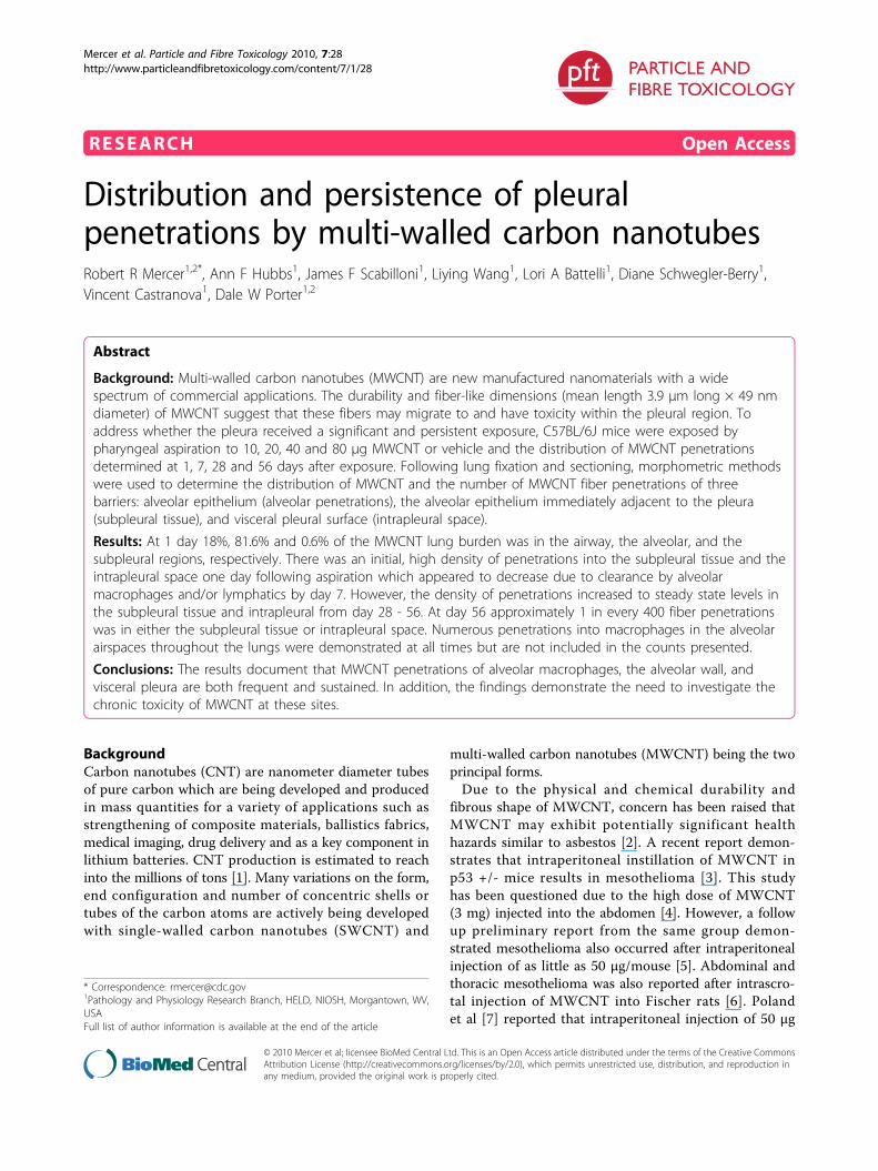
ResultsThe light microscopic section of Figure 1 shows arepresentative cross-section from terminal bronchioleout to the pleural surface of the lungs 1 day afteraspiration of a 80 μg dose. As illustrated in the micro-graph, MWCNT were deposited throughout the alveolarregion of the lungs with the highest concentrations visi-ble in the alveoli immediately proximal to the terminalbronchiole.
Figure 1 Light micrograph of MWCNT deposition in alveolar region of lungs. Sirius Red stained micrograph showing the generaldeposition pattern of MWCNT (arrows) one day after aspiration. A deposit of MWCNT on the epithelium of the terminal bronchiole (TB) near thetransition between the airways and the alveolar region is indicated by the asterisk. Smaller deposits near the subpleural tissue region areindicated by double arrows. (dose 80 μg).
Mercer et al. Particle and Fibre Toxicology 2010, 7:28http://www.particleandfibretoxicology.com/content/7/1/28
Page 2 of 11
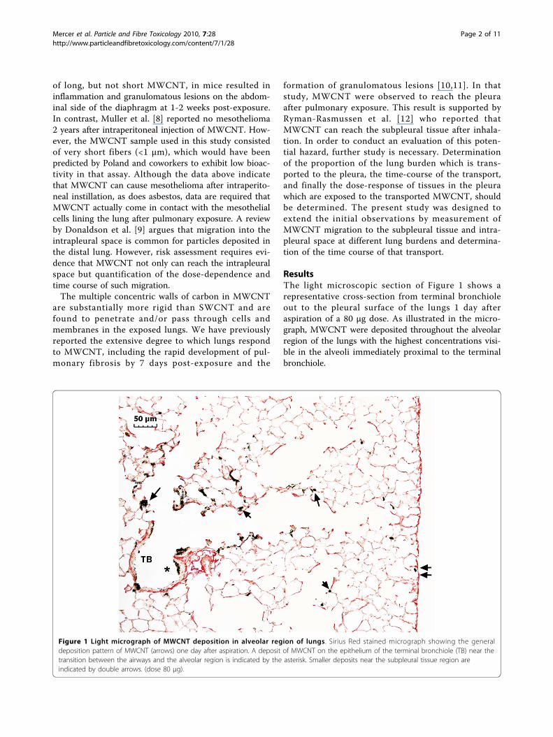
The lung burden distribution of MWCNT at 1 daypost-aspiration (80 μg) is shown in Figure 2. The resultsdemonstrate the prominent role of alveolar macro-phages. The majority of the lung burden was depositedin the alveolar region with alveolar macrophages receiv-ing 62% of the total dose. The airways and alveolarregions accounted for 18 and 81% of the lung burden,respectively. MWCNT in the visceral pleura region (sub-pleural tissue and intrapleural space) accounted forapproximately 0.6% of the total lung burden.A representative image of the granulomatous lesions
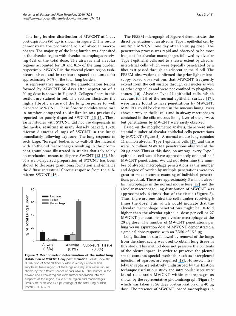
formed by MWCNT 56 days after aspiration of a20 μg dose is shown in Figure 3. Collagen fibers in thissection are stained in red. The section illustrates thehighly fibrotic nature of the lung response to welldispersed MWCNT. These fibrotic nodules were rarein number compared to similar lesions previouslyreported for poorly dispersed SWCNT [13-15]. Theseearlier studies with SWCNT did not use dispersants inthe media, resulting in many densely packed, 15-20micron diameter clumps of SWCNT in the lungsimmediately following exposure. The lung response tosuch large, “foreign” bodies is to wall-off the materialwith epitheloid macrophages resulting in the promi-nent granulomas illustrated in studies that rely solelyon mechanical means to disperse SWCNT [13-15]. Useof a well-dispersed preparation of SWCNT has beenshown to decrease granuloma formation and enhancethe diffuse interstitial fibrotic response from the sub-micron SWCNT [16].
The FESEM micrograph of Figure 4 demonstrates thedirect penetration of an alveolar Type I epithelial cell bymultiple MWCNT one day after an 80 μg dose. Thepenetration process was rapid and observed to be mostfrequent for alveolar macrophages followed by alveolarType I epithelial cells and to a lesser extent by alveolarinterstitial cells which were typically penetrated by afiber as it passed through an adjacent epithelial cell. TheFESEM observations confirmed the prior light micro-scopy based observations that MWCNT frequentlyextend from the cell surface through cell nuclei as wellas other organelles and were not confined to phagolyso-somes [10]. Alveolar Type II epithelial cells, whichaccount for 2% of the normal epithelial surface [17],were rarely found to have penetrations by MWCNT.MWCNT could be observed in the mucous lining layersabove airway epithelial cells and in airway macrophagescontained in the cilia-mucous lining layer of the airwaysbut penetrations by MWCNT were rarely observed.Based on the morphometric analysis, there were sub-
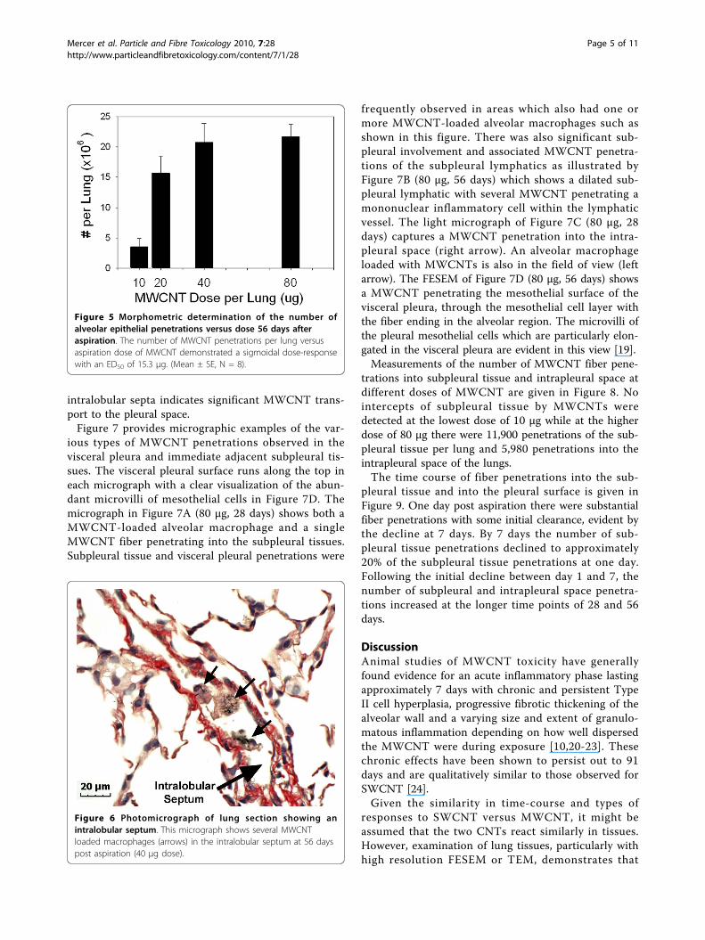
stantial number of alveolar epithelial cells penetrationsby MWCNT (Figure 5). A normal mouse lung contains11 million alveolar Type I epithelial cells [17] and therewere 15 million MWCNT penetrations observed at the20 μg dose. Thus at this dose, on average, every Type Iepithelial cell would have approximately one and halfMWCNT penetration. We did not determine the num-ber of alveolar macrophage penetrations as the numberand degree of overlap by multiple penetrations were toogreat to make accurate counting of individual penetra-tions practical. There are approximately 3 million alveo-lar macrophages in the normal mouse lung [17] and thealveolar macrophage lung distribution of MWCNT wasapproximately 6 times that of the tissue (Figure 2).Thus, there are one third the cell number receiving 6times the dose. This which would indicate that thealveolar macrophage penetrations might be 18-foldhigher than the alveolar epithelial dose per cell or 27MWCNT penetrations per alveolar macrophage at the20 μg dose. The number of MWCNT penetrations perlung versus aspiration dose of MWCNT demonstrated asigmoidal dose-response with an ED50 of 15.3 μg.Lung fixation in-situ followed by removal of the lungs
from the chest cavity was used to obtain lung tissue inthis study. This method does not preserve the contentsof the pleural space. In order to preserve the pleuralspace contents special methods, such as interpleuralinjection of agarose, are required [18]. However, intra-lobular septa are relatively undisturbed by the fixationtechnique used in our study and intralobular septa werefound to contain MWCNT within macrophages asshown by the representative photomicrograph (Figure 6)which was taken at 56 days post-aspiration of a 40 μgdose. The presence of MWCNT loaded macrophages in
Figure 2 Morphometric determination of the initial lungdistribution of MWCNT 1 day post aspiration. Results show thedistribution of MWCNT fiber burden in airways, alveolar andsubpleural tissue regions of the lungs one day after aspiration. Asshown by the different shades of bars, MWCNT fiber burden in theairways and alveolar regions were further subdivided into theairspaces of the region, tissue of the region and macrophages.Results are expressed as a percentage of the total lung burden.(Mean ± SE, N = 7).
Mercer et al. Particle and Fibre Toxicology 2010, 7:28http://www.particleandfibretoxicology.com/content/7/1/28
Page 3 of 11
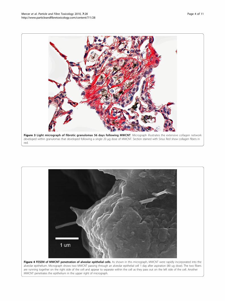
Figure 3 Light micrograph of fibrotic granulomas 56 days following MWCNT. Micrograph illustrates the extensive collagen networkdeveloped within granulomas that developed following a single 20 μg dose of MWCNT. Section stained with Sirius Red show collagen fibers inred.
Figure 4 FESEM of MWCNT penetration of alveolar epithelial cells. As shown in this micrograph, MWCNT were rapidly incorporated into thealveolar epithelium. Micrograph shows two MWCNT passing through an alveolar epithelial cell 1 day after aspiration (80 ug dose). The two fibersare running together on the right side of the cell and appear to separate within the cell as they pass out on the left side of the cell. AnotherMWCNT penetrates the epithelium in the upper right of micrograph.
Mercer et al. Particle and Fibre Toxicology 2010, 7:28http://www.particleandfibretoxicology.com/content/7/1/28
Page 4 of 11
intralobular septa indicates significant MWCNT trans-port to the pleural space.Figure 7 provides micrographic examples of the var-
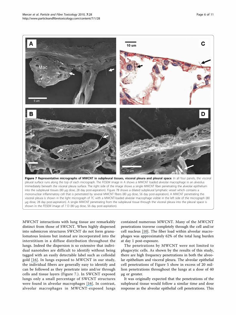
ious types of MWCNT penetrations observed in thevisceral pleura and immediate adjacent subpleural tis-sues. The visceral pleural surface runs along the top ineach micrograph with a clear visualization of the abun-dant microvilli of mesothelial cells in Figure 7D. Themicrograph in Figure 7A (80 μg, 28 days) shows both aMWCNT-loaded alveolar macrophage and a singleMWCNT fiber penetrating into the subpleural tissues.Subpleural tissue and visceral pleural penetrations were
frequently observed in areas which also had one ormore MWCNT-loaded alveolar macrophages such asshown in this figure. There was also significant sub-pleural involvement and associated MWCNT penetra-tions of the subpleural lymphatics as illustrated byFigure 7B (80 μg, 56 days) which shows a dilated sub-pleural lymphatic with several MWCNT penetrating amononuclear inflammatory cell within the lymphaticvessel. The light micrograph of Figure 7C (80 μg, 28days) captures a MWCNT penetration into the intra-pleural space (right arrow). An alveolar macrophageloaded with MWCNTs is also in the field of view (leftarrow). The FESEM of Figure 7D (80 μg, 56 days) showsa MWCNT penetrating the mesothelial surface of thevisceral pleura, through the mesothelial cell layer withthe fiber ending in the alveolar region. The microvilli ofthe pleural mesothelial cells which are particularly elon-gated in the visceral pleura are evident in this view [19].Measurements of the number of MWCNT fiber pene-
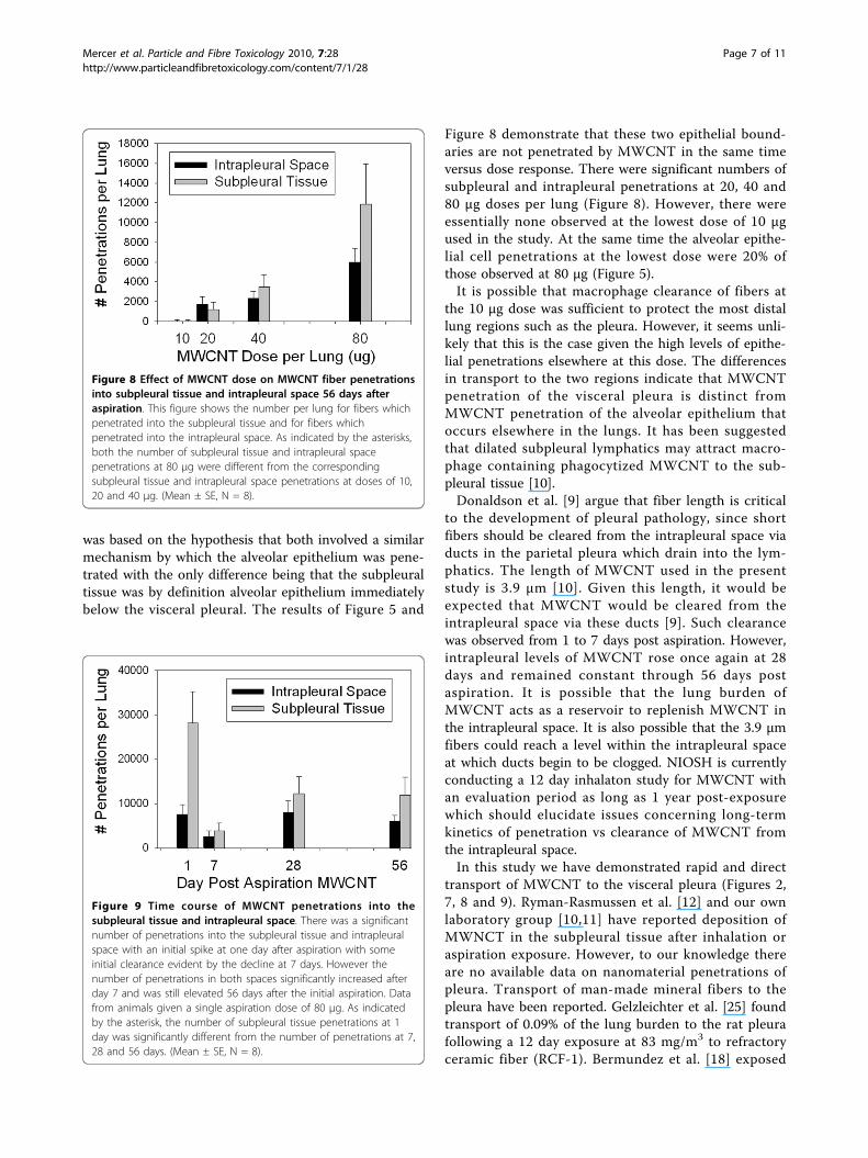
trations into subpleural tissue and intrapleural space atdifferent doses of MWCNT are given in Figure 8. Nointercepts of subpleural tissue by MWCNTs weredetected at the lowest dose of 10 μg while at the higherdose of 80 μg there were 11,900 penetrations of the sub-pleural tissue per lung and 5,980 penetrations into theintrapleural space of the lungs.The time course of fiber penetrations into the sub-
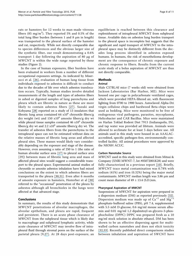
pleural tissue and into the pleural surface is given inFigure 9. One day post aspiration there were substantialfiber penetrations with some initial clearance, evident bythe decline at 7 days. By 7 days the number of sub-pleural tissue penetrations declined to approximately20% of the subpleural tissue penetrations at one day.Following the initial decline between day 1 and 7, thenumber of subpleural and intrapleural space penetra-tions increased at the longer time points of 28 and 56days.
DiscussionAnimal studies of MWCNT toxicity have generallyfound evidence for an acute inflammatory phase lastingapproximately 7 days with chronic and persistent TypeII cell hyperplasia, progressive fibrotic thickening of thealveolar wall and a varying size and extent of granulo-matous inflammation depending on how well dispersedthe MWCNT were during exposure [10,20-23]. Thesechronic effects have been shown to persist out to 91days and are qualitatively similar to those observed forSWCNT [24].Given the similarity in time-course and types of
responses to SWCNT versus MWCNT, it might beassumed that the two CNTs react similarly in tissues.However, examination of lung tissues, particularly withhigh resolution FESEM or TEM, demonstrates that
Figure 5 Morphometric determination of the number ofalveolar epithelial penetrations versus dose 56 days afteraspiration. The number of MWCNT penetrations per lung versusaspiration dose of MWCNT demonstrated a sigmoidal dose-responsewith an ED50 of 15.3 μg. (Mean ± SE, N = 8).
Figure 6 Photomicrograph of lung section showing anintralobular septum. This micrograph shows several MWCNTloaded macrophages (arrows) in the intralobular septum at 56 dayspost aspiration (40 μg dose).
Mercer et al. Particle and Fibre Toxicology 2010, 7:28http://www.particleandfibretoxicology.com/content/7/1/28
Page 5 of 11
MWCNT interactions with lung tissue are remarkablydistinct from those of SWCNT. When highly dispersedinto submicron structures SWCNT do not form granu-lomatous lesions but instead are incorporated into theinterstitium in a diffuse distribution throughout thelungs. Indeed the dispersion is so extensive that indivi-dual nanotubes are difficult to identify without beingtagged with an easily detectable label such as colloidalgold [16]. In lungs exposed to MWCNT in our study,the individual fibers are generally easy to identify andcan be followed as they penetrate into and/or throughcells and tissue layers (Figure 7.). In SWCNT exposedlungs only a small percentage of SWCNT structureswere found in alveolar macrophages [16]. In contrast,alveolar macrophages in MWCNT-exposed lungs
contained numerous MWCNT. Many of the MWCNTpenetrations traverse completely through the cell and/orcell nucleus [10]. The fiber load within alveolar macro-phages was approximately 62% of the total lung burdenat day 1 post-exposure.The penetrations by MWCNT were not limited to
phagocytic cells. As shown by the results of this study,there are high frequency penetrations in both the alveo-lar epithelium and visceral pleura. The alveolar epithelialcell penetrations of Figure 5 show in excess of 20 mil-lion penetrations throughout the lungs at a dose of 40μg or greater.It was originally expected that the penetrations of the
subpleural tissue would follow a similar time and doseresponse as the alveolar epithelial cell penetrations. This
Figure 7 Representative micrographs of MWCNT in subpleural tissues, visceral pleura and pleural space. In all four panels, the visceralpleural surface runs along the top of each micrograph. The FESEM image in A shows a MWCNT loaded alveolar macrophage in an alveolusimmediately beneath the visceral pleura surface. The right side of the image shows a single MWCNT fiber penetrating the alveolar epitheliuminto the subpleural tissues (80 μg dose, 28 day post-aspiration). Figure 7B shows a dilated subpleural lymphatic vessel which contains amononuclear inflammatory cell that is penetrated by several MWCNT fibers (80 μg dose, 56 day post-aspiration). A MWCNT penetrating thevisceral pleura is shown in the light micrograph of 7C with a MWCNT-loaded alveolar macrophage visible in the left side of the micrograph (80μg dose, 28 day post-aspiration). A single MWCNT penetrating from the subpleural tissue through the visceral pleura into the pleural space isshown in the FESEM image of 7 D (80 μg dose, 56 day post-aspiration).
Mercer et al. Particle and Fibre Toxicology 2010, 7:28http://www.particleandfibretoxicology.com/content/7/1/28
Page 6 of 11
was based on the hypothesis that both involved a similarmechanism by which the alveolar epithelium was pene-trated with the only difference being that the subpleuraltissue was by definition alveolar epithelium immediatelybelow the visceral pleural. The results of Figure 5 and
Figure 8 demonstrate that these two epithelial bound-aries are not penetrated by MWCNT in the same timeversus dose response. There were significant numbers ofsubpleural and intrapleural penetrations at 20, 40 and80 μg doses per lung (Figure 8). However, there wereessentially none observed at the lowest dose of 10 μgused in the study. At the same time the alveolar epithe-lial cell penetrations at the lowest dose were 20% ofthose observed at 80 μg (Figure 5).It is possible that macrophage clearance of fibers at
the 10 μg dose was sufficient to protect the most distallung regions such as the pleura. However, it seems unli-kely that this is the case given the high levels of epithe-lial penetrations elsewhere at this dose. The differencesin transport to the two regions indicate that MWCNTpenetration of the visceral pleura is distinct fromMWCNT penetration of the alveolar epithelium thatoccurs elsewhere in the lungs. It has been suggestedthat dilated subpleural lymphatics may attract macro-phage containing phagocytized MWCNT to the sub-pleural tissue [10].Donaldson et al. [9] argue that fiber length is critical
to the development of pleural pathology, since shortfibers should be cleared from the intrapleural space viaducts in the parietal pleura which drain into the lym-phatics. The length of MWCNT used in the presentstudy is 3.9 μm [10]. Given this length, it would beexpected that MWCNT would be cleared from theintrapleural space via these ducts [9]. Such clearancewas observed from 1 to 7 days post aspiration. However,intrapleural levels of MWCNT rose once again at 28days and remained constant through 56 days postaspiration. It is possible that the lung burden ofMWCNT acts as a reservoir to replenish MWCNT inthe intrapleural space. It is also possible that the 3.9 μmfibers could reach a level within the intrapleural spaceat which ducts begin to be clogged. NIOSH is currentlyconducting a 12 day inhalaton study for MWCNT withan evaluation period as long as 1 year post-exposurewhich should elucidate issues concerning long-termkinetics of penetration vs clearance of MWCNT fromthe intrapleural space.In this study we have demonstrated rapid and direct
transport of MWCNT to the visceral pleura (Figures 2,7, 8 and 9). Ryman-Rasmussen et al. [12] and our ownlaboratory group [10,11] have reported deposition ofMWNCT in the subpleural tissue after inhalation oraspiration exposure. However, to our knowledge thereare no available data on nanomaterial penetrations ofpleura. Transport of man-made mineral fibers to thepleura have been reported. Gelzleichter et al. [25] foundtransport of 0.09% of the lung burden to the rat pleurafollowing a 12 day exposure at 83 mg/m3 to refractoryceramic fiber (RCF-1). Bermundez et al. [18] exposed
Figure 8 Effect of MWCNT dose on MWCNT fiber penetrationsinto subpleural tissue and intrapleural space 56 days afteraspiration. This figure shows the number per lung for fibers whichpenetrated into the subpleural tissue and for fibers whichpenetrated into the intrapleural space. As indicated by the asterisks,both the number of subpleural tissue and intrapleural spacepenetrations at 80 μg were different from the correspondingsubpleural tissue and intrapleural space penetrations at doses of 10,20 and 40 μg. (Mean ± SE, N = 8).
Figure 9 Time course of MWCNT penetrations into thesubpleural tissue and intrapleural space. There was a significantnumber of penetrations into the subpleural tissue and intrapleuralspace with an initial spike at one day after aspiration with someinitial clearance evident by the decline at 7 days. However thenumber of penetrations in both spaces significantly increased afterday 7 and was still elevated 56 days after the initial aspiration. Datafrom animals given a single aspiration dose of 80 μg. As indicatedby the asterisk, the number of subpleural tissue penetrations at 1day was significantly different from the number of penetrations at 7,28 and 56 days. (Mean ± SE, N = 8).
Mercer et al. Particle and Fibre Toxicology 2010, 7:28http://www.particleandfibretoxicology.com/content/7/1/28
Page 7 of 11
rats or hamsters for 12 weeks to man-made vitreousfibers (85 mg/m3). They reported 5% and 0.5% of thetotal lung fiber burden (between 1 and 8 μm in length)was transported to the pleural surface of the hamsterand rat, respectively. While not directly comparable dueto species differences and the obvious larger size ofthe synthetic fiber, our result demonstrating a 0.6%transport 1 day following the aspiration of 80 μg ofMWCNT is within the wide range reported by thesestudies (Figure 2).In the case of human exposures, fiber burdens have
been evaluated in workers from a number of differentoccupational exposures settings. As indicated by Miser-occi et al. [26], evaluation of human lung tissue fromindividuals exposed to asbestos is difficult to conductdue to the decades of life over which asbestos transloca-tion occurs. Typically, human studies involve detailedmeasurements of the length and types of asbestos fibersin ashed or digested samples of lung parenchyma andpleura which are fibrotic in nature as these are morelikely to contain asbestos fibers [27]. Suzuki andKohyama [28] reported on a study of 13 insulators thatfibrotic lung areas contained 64 x106 chrysotile fibers/gdry weight (wt) and 150 x106 amosite fibers/g dry wtwhile pleural tissue samples contained 46 x106 chrysotilefibers/g dry wt and 2.2 x10⁶ amosite fibers/g dry wt. Thetransfer of asbestos fibers from the parenchyma to theintrapleural space can not be estimated without data onthe relative masses of fibrotic lung areas and affectedpleural sites. These masses would likely vary consider-ably depending on the exposure and stage of the disease.However, even assuming a ratio of 250 to 1 (the ratio ofhuman alveolar surface area [17] to pleural surface area[29]) between mass of fibrotic lung area and mass ofaffected pleural sites would suggest a considerable trans-port to the pleural space. Experimental animal studies ofchrysotile or amosite asbestos inhalation have had mixedconclusions on the extent to which asbestos fibers aretransported to the pleura [30,31]. Even after 6 monthsof amosite exposure in hamsters, Hesterber et al [30]referred to the “occasional” penetration of the pleura byasbestos although all bronchioles in the lungs wereaffected at that advanced stage.
ConclusionsIn summary, the results of this study demonstrate thatMWCNT penetrations of alveolar macrophages, thealveolar epithelium, and pleura are rapid, frequentand persistent. There is an acute phase clearance ofMWCNT from the subpleural tissue which is likely dueto macrophage and subpleural lymphatic activity, whileacute clearance of MWCNT may involve flow of intra-pleural fluid through stromal pores on the surface of theparietal pleura [9,27]. At 28 days post-exposure, an
equilibrium is reached between this clearance andreplenishment of intrapleural MWCNT from subpleuraltissue. Available data on asbestos lung burden transportto the pleural space is incomplete but suggests that thesignificant and rapid transport of MWCNT to the intra-pleural space may be distinctly different from the dec-ades long process identified in asbestos exposedhumans. In humans, the risk of mesothelioma develop-ment are the consequence of chronic exposure andchronic response to fibers. Results from the currentacute study of a bolus aspiration of MWCNT are thusnot directly comparable.
MethodsAnimalMale C57BL/6J mice (7 weeks old) were obtained fromJackson Laboratories (Bar Harbor, ME). Mice werehoused one per cage in polycarbonate ventilated cages,which were provided HEPA-filtered air, with fluorescentlighting from 0700 to 1900 hours. Autoclaved Alpha-Drivirgin cellulose chips and hardwood Beta-chips wereused as bedding. Mice were monitored to be free ofendogenous viral pathogens, parasites, mycoplasms,Helicobacter and CAR Bacillus. Mice were maintainedon Harlan Teklad Rodent Diet 7913 (Indianapolis, IN),and tap water was provided ad libitum. Animals wereallowed to acclimate for at least 5 days before use. Allanimals used in this study were housed in an AAALAC-accredited, specific pathogen-free, environmentally con-trolled facility. All animal procedures were approved bythe NIOSH ACUC.
Carbon Nanotube SourceMWCNT used in this study were obtained from Mitsui &Company (XNRI MWNT-7, lot #05072001K28) and werefully characterized in a previous report [10]. Briefly,MWCNT trace metal contamination was 0.78% withsodium (41%) and iron (0.32%) being the major metalcontaminants. MWCNT median length was 3.86 μm andcount mean diameter of 49 ± 13.4 (S.D.)nm.
Pharyngeal Aspiration of MWCNTSuspensions of MWCNT for aspiration were prepared ina dispersion medium (DM) as reported previously [32].Dispersion medium was made up of Ca+2 and Mg+2
phosphate-buffered saline (PBS), pH 7.4, supplementedwith 5.5 mM D-glucose, 0.6 mg/ml mouse serum albu-min and 0.01 mg/ml 1,2 dipalmitoyl-sn-glycero-3-phos-phocholine (DPPC) DPPC was prepared fresh as a 10mg/ml stock solution in absolute ethanol. DM has beenshown to be an effective dispersing agent for multi-walled carbon nanotubes and does not elicit toxicity[32,33]. Recently published direct comparisons studiesbetween inhalation and aspiration of SWCNT have
Mercer et al. Particle and Fibre Toxicology 2010, 7:28http://www.particleandfibretoxicology.com/content/7/1/28
Page 8 of 11
further demonstrated the applicability of the aspirationtechnique for such studies [24].Mice were anesthetized with isoflurane (Abbott
Laboratories, North Chicago, IL) for pharyngeal aspira-tion. When fully anesthetized, the mouse was positionedwith its back against a slant board and suspended by theincisor teeth using a rubber band. The mouth wasopened, and the tongue gently pulled aside from theoral cavity. A 50 μl aliquot of sample was pipetted atthe base of the tongue, and the tongue was restraineduntil at least 2 deep breaths were completed (but fornot longer than 15 seconds). Following release of thetongue, the mouse was gently lifted off the board, placedon its left side, and monitored for recovery fromanesthesia. In order to eliminate the possibility of inci-dental food aspiration, food was removed 4 hours priorto the procedure; water was removed 1 hour prior. Thisalternative to inhalation has been shown to produce asimilar distribution of lung burden to that followinginhalation and has been described in detail elsewhere[34]. Mice received either DM (vehicle control), 10, 20,40 or 80 μg MWCNT.At 1, 7, 28 and 56 days after aspiration, mice were
euthanized by an overdose of pentobarbital (>100 mg/kgbody weight, i.p.) followed by transection of the abdom-inal aorta to provide exsanguination. Seven to 8 animalswere studied at each time point. The lungs were fixedby intratracheal perfusion with 1 ml of 10% neutral buf-fered formalin after 30 minutes the lungs were removedfrom the chest cavity. Lungs were trimmed the sameday, processed overnight in a tissue processor, andembedded in paraffin.For morphometric studies, paraffin sections of the left
lung (5 μm thick) were cut. A new region of the disposa-ble knife blade was used to section each block in order toprevent potential cross-contamination that might resultfrom MWCNT passage on the knife between sections.The sections were then deparaffinized and rehydratedwith xylene-alcohol series to distilled water. To enhancethe contrast between tissue and MWCNT, lung sectionswere stained with Sirius Red [35]. Sirius Red stainingconsisted of immersion of the slides in 0.1% Picrosiriussolution (100 mg of Sirius Red F3BA in 100 ml of satu-rated aqueous picric acid, pH 2) for 1 - 2 hours followedby washing for 1 minute in 0.01 N HCl. Sections werethen briefly counterstained in freshly filtered Mayer’shematoxylin for 2 minutes, dehydrated, and mountedwith a coverslip. Additional sections were stained withhematoxylin and eosin for routine pathology assessmentas previously reported [10].
Field Emission Scanning Electron MicroscopyFor scanning electron microscopy, sections of the lungwere cut at 8 microns, placed on carbon planchets,
deparaffinized and sputter coated. After coating, the spe-cimens were examined with a Hitachi Model S-4800Field Emission Scanning electron microscope (FESEM)at 5 to 20 kV. Use of thin sections from paraffinembedded tissue was found to be preferable to large,unevenly cut blocks because it provided a uniformthickness of organic material on the carbon planchet.The 8 micron sections were thick enough to conveythree-dimensional information but were also less likelyto charge or undergo shifts when examined at the highmagnifications necessary to study nanomaterials.
Lung Distribution of MWCNTThe distribution of MWCNT in the lungs was deter-mined by counting the occurrence of MWCNT underan eyepiece point counting overlay using standard mor-phometric point counting methods [36] similar to thosepreviously described for study of the distribution ofSWCNT [16,36]. Point counting categories were subdi-vided into points over MWCNT in airway, points overMWCNT in the alveolar region, and points overMWCNT in the subpleural tissue region. Airway regionswere defined as those containing airway tissue (airwayepithelial cells-basement membrane and tissues of thebroncho-vascular cuff), airway lumen, and associatedblood vessels greater than 25 microns. Alveolar regionswere those containing alveolar tissue and alveolar airspace. The visceral pleura region included MWCNT inthe subpleural tissue and MWCNT in the pleural sur-face. The subpleural tissue regions included the immedi-ately subpleural alveolar interstitial-epithelium layer andsubpleural lymphatics but did not include any portion ofalveolar walls attaching to the pleura. Points in airwayand alveolar regions were further subdivided into pointsover MWCNT that were in the airspace, points overMWCNT that were in tissue of the region, and pointsover MWCNT that were in macrophages.To accomplish the counting, an eyepiece counting
overlay consisting of 11 by 11 lines (121 total points foreach throw of the overlay) was used with a 100× oilimmersion objective. A grid pattern for throws of thecounting overlay was used in order to insure a uniformsampling of the section which did not overweight inter-ior points. The counting overlay throws of the eyepiecewere positioned over the section at 12 uniformly spacedgrid points in both × and Y co-ordinates. These 12 gridpoints were determined using the stage micrometerscale to measure the × and Y bounds of the section.Using the bounding rectangle of these co-ordinates a 3by 4 grid was selected and the 12 intersections wereused as the center point for each of the eyepiece count-ing overlay throws.For each animal, three sections were counted and the
counts for the airways, alveolar and subpleural tissue
Mercer et al. Particle and Fibre Toxicology 2010, 7:28http://www.particleandfibretoxicology.com/content/7/1/28
Page 9 of 11
regions were summed. Each counting category wasdivided by this total and multiplied by 100 to expressthe results as a percentage of total lung burden.
Alveolar Epithelial PenetrationsPenetrations of the alveolar epithelium by MWCNTfibers were determined by counting the number of fiberpenetrations per unit area based on repeated throws ofan eyepiece counting box overlay. To tabulate thecounts for each section, 12 placements of the eyepiececounting box overlay were carried out using a grid pla-cement system similar to that described for determina-tion of the lung distribution of MWCNT. For eachplacement, a count was made of the MWCNT whichpenetrated the alveolar epithelium within the box. Totalpenetrations per unit area of lung section were dividedby the volume of the counting box (area times thick-ness) to obtain the number of penetrations per unitvolume which was then multiplied by the fixed lungvolume to express the results as number of alveolarepithelial penetrations per lung. Section thickness was5.14 μm. The section thickness was determined by tak-ing micrometer measurements of a tissue block beforeand after cutting 2000 sections. Two sections per animalwere analyzed and the results averaged. Eight animalswere analyzed per group.
MWCNT Penetrations of the Subpleural Tissue andIntrapleural SpaceFor each lung section, the total number of penetrationsof MWCNT into the subpleural tissue and into theintrapleural space was determined. Subpleural penetra-tions were defined as MWCNT fibers which penetratedfrom the alveolar air side of the pleura into the alveolarepithelium and interstitium/subpleural lymphaticsimmediately beneath the mesothelial cell layer of thevisceral pleura. In the rudimentary visceral pleura of thenormal mouse, this is essentially a 1 to 2 micron depthbelow the surface of the mesothelial cells forming thevisceral pleura. Intrapleural penetrations were defined asMWCNT fibers which penetrated the surface of thevisceral pleura. Occasionally fibers where sufficientlylong to penetrate completely from the alveolar sidethrough to the visceral pleural (see Figure 7 in Results).These cases were counted as both subpleural tissue andintrapleural penetrations.The counting of penetrations was done by sweeping
around the pleural perimeter of the section using a100× oil immersion objective with constant refocusingas the complete perimeter of the visceral pleura (includ-ing interlobular septa) was scanned. These counts weredivided by the area of the pleural surface in the crosssection (perimeter length times the section thickness) toobtain the number of penetrations (subpleural and
intrapleural) per unit of pleural surface area. The peri-meter length of the pleura was determined from lowmagnification images digitized for each section andmeasured using ImageJ. To express the results as num-ber of penetrations per lung, the penetrations per unitof pleural surface area were multiplied by the pleuralsurface area of the mouse lung (500 mm3 , [29]). Cor-rection for the potential over-estimation of counts dueto the size dependent projection of the length of thefibers, Holmes effect [37], was not necessary as thecounts were done of the intersections between the fiberand the surface. The area of intersection corresponds todiameter of the fiber which is ~51 nm. This is a con-stant factor between groups and is negligible relative tothe section thickness used for counting. Two sectionsper animal were analyzed and the results averaged. Eightanimals were analyzed per group.
Statistical AnalysesData were analyzed using analysis of variance (STAT-GRAF). Bartlett’s test was used to test for homogeneityof variances between groups. Statistical differences weredetermined using one-way analysis of variance with sig-nificance set at p ≤ 0.05. When significant F values wereobtained, individual means were compared to controlusing Duncan’s multiple range test [38] and P < 0.05was considered to be significant. Data are given as mean± SE.
DisclaimerThe findings and conclusions in this report are those ofthe authors and do not necessarily represent the viewsof the National Institute for Occupational Safety andHealth.
AbbreviationsCNT: carbon nanotubes; DM: dispersion medium; DPPC: 1,2 dipalmitoyl-sn-glycero-3-phosphocholine; ED50: effective dose for 50 percent response;FESEM: field emission scanning electron microscope; MWCNT: multiwalledcarbon nanotubes; PBS: phosphate-buffered saline; SWCNT: single-walledcarbon nanotubes.
AcknowledgementsWe appreciate Dean Newcomer and Sherri Friend for their excellenttechnical assistance with the pathology preparation and staining of theslides.
Author details1Pathology and Physiology Research Branch, HELD, NIOSH, Morgantown, WV,USA. 2Department of Physiology and Pharmacology, West Virginia University,Morgantown, WV, USA.
Authors’ contributionsRM conceived of the study, developed the morphometric methods,conducted the FESEM evaluation, analyzed the experimental results anddrafted the manuscript. AH made the original observation of pleuralpenetrations by MWCNT and was involved in the planning and writing ofthe manuscript. JS performed the morphometric counting of theparenchyma and pleural samples and assisted in analysis of results. LW
Mercer et al. Particle and Fibre Toxicology 2010, 7:28http://www.particleandfibretoxicology.com/content/7/1/28
Page 10 of 11
contributed to the experimental design and assisted in lung preparation. LBprovided important information on sampling of the lungs for pleural studyand conducted lung preparation for histopathology. DS-B assisted in thesampling design and operation of the FESEM studies. VC and DPcontributed to the experimental design, acquisition of funding and writingof the manuscript. All authors read and approved the final manuscript.
Competing interestsThe authors declare that they have no competing interests.
Received: 24 June 2010 Accepted: 4 October 2010Published: 4 October 2010
References1. Ball P: Roll up for the revolution. Nature 2001, 414:142-144.2. Donaldson K, Aitken R, Tran L, Stone V, Duffin R, Foreast G, Alexander A:
Carbon nanotubes: a review of their properties in relation to pulmonarytoxicology and workplace safety. Toxicol Sci 2006, 92:5-22.
3. Takagi A, Hirose A, Nishimura T, Fukumori N, Ogata A, Ohashi N: Inductionof mesothelioma in p53+/- mouse by intraperitoneal applicaition ofmulti-walled carbon nanotube. J Toxicol Sci 2008, 33:105-116.
4. Ichihara G, Castranova V, Tanioka A, Miyazawa K: Letter to the editor,Induction of mesothelioma in p53+/- mouse by intraperitonealapplication of multi-wall carbon nanotube. J Toxicol Sci 2008, 33:381-382.
5. Kanno J, Takagi A, Nishimura T, Hirose A: Mesothelioma induction bymicrometer-sized multi-walled carbon nanotube intraperitoneallyinjected to p53 heterozygous mice. The Toxicologist 2010, 114:A1397.
6. Sakamoto Y, Nakae D, Fukumori N, Tayama K, Maekawa A, Imai K, Hirose A,Nishimura T, Ohashi N, Ogata A: Induction of mesothelioma by a singleintrascrotal administration of multi-wall carbon nanotube in intact maleFischer 344 rats. J Toxicol Sci 2009, 34:65-76.
7. Poland CA, Duffin R, Kinloch I, AD M, Wallace WA, Seaton A: Carbonnanotubes introduced into the abdominal cavity of mice show asbestos-like pathogenicity in a pilot study. Nat Nanotechnol 2008, 3:423-428.
8. Muller J, Delos M, Panin N, Rabolli V, Huaux F, Lison D: Absence ofcarcinogenic response to multiwall carbon nanotubes in a 2-yearbioassy in the peritoneal cavity of the rat. Toxicol Sci 2009, 110:442-447.
9. Donaldson K, Murphy FA, Duffin R, Poland CA: Asbestos, carbonnanotubes and the pleural mesothelium. Partitcle Fibre Toxicol 2010,7:1-17.
10. Porter DW, Hubbs AF, Mercer RR, Wu N, Wolfarth MG, Sriram K, Leonard S,Battelli LA, Schwegler-Berry D, Friend S, et al: Mouse pulmonary dose- andtime course-responses induced by exposure to multi-walled carbonnanotubes. Toxicology 2010, 269:136-147.
11. Hubbs AF, Mercer RR, Coad JE, Batteli LA, Willard PA, Sriram K, Wolfarth MG,Castranova V, Porter DW: Persistent pulmonary inflammation, airwaymucous metaplasia and migration of multi-walled carbon nanotubesfrom the lung after subchronic exposure. The Toxicologist 2009, 108(S1):A2192.
12. Ryman-Rasmussen JP, Cesta MF, Brody AR, Shipley-Phillips JK, Everitt JI,Tewksbury EW, Moss OR, Wong BA, Dodd DE, Andersen ME, Bonner JC:Inhaled carbon nanotubes reach the subpleural tissue in mice. NatNanotechnol 2009, 4:747-751.
13. Warheit DB, Laurence BR, Reed KL, Roach DH, Reynolds GAM, Webb TR:Comparative pulmonary toxicity assessment of single-wall carbonnanotubes in rats. Toxicol Sci 2004, 77:117-125.
14. Shvedova AA, Kisin ER, Mercer RR, Murray AR, Johnson VJ, Potapovich AI,Tyurina YY, Gorelik O, Arepalli S, Schwegler-Berry D, et al: Unusualinflammatory and fibrogenic pulmonary responses to single-walledcarbon nanotubes in mice. Am J Physiol 2005, 289:L698-L708.
15. Lam CW, James JT, McCluskey R, Hunter RL: Pulmonary toxicity of single-wall carbon nanotubes in mice 7 and 90 days after intratrachealinstillation. Toxicol Sci 2004, 77:126-134.
16. Mercer RR, Scabilloni JF, Wang L, Kisin E, Murray AR, Schwegler-Berry D,AA S, Castranova V: Alteration of deposition pattern and pulmonaryresponse as a result of improved dispersion of aspirated single-walledcarbon nanotubes in a mouse model. Am J Physiol Lung Cell Mol Physiol2008, 294:L87-L97.
17. Stone KC, Mercer RR, Gehr P, Stockstill B, Crapo JD: Allometric relationshipsof cell numbers and size in the mammalian lung. Am J Respir Cell Mol Biol1992, 6:235-243.
18. Bermudez E, Mangum JB, Moss OR, Wong BA, Everitt JI: Pleural dosimetryand pathobiological responses in rats and hamsters exposedsubchronically to MMVF 10a fiberglass. Toxicol Sic 2003, 74:165-173.
19. Wang NS: Anatomy and physiology of the pleural space. Clin Chest Med1985, 6:3-13.
20. Muller J, Huaux F, Moreau N, Misson P, Heilier JF, Delos M, Arras M,Fonseca A, Nagy JB, Lison D: Respiratory toxicity of multi-wall carbonnanotubes. Toxicol Appl Pharma 2005, 207:221-231.
21. Mitchell LA, Gao J, Wall RV, Gigliotti A, Burchiel SW, McDonald JD:Pulmonary and systemic immune response to inhaled multiwalledcarbon nanotubes. Toxicol Sci 2007, 100:203-214.
22. Li JG, Li WX, Xu JY, Cai XQ, Liu RL, Li YJ, Zhao QF, Li QN: Comparativestudy of pathological lesions induced by multiwalled carbon nanotubesin lungs of mice by intratracheal instillation and inhalation. EnvironToxicol 2007, 22:415-421.
23. Aiso S, Yamazaki K, Umed Y, Asakura M, Takaya M, Toya T, Koda S,Nagano K, Arito H, Fukushima S: Pulmonary toxicity of intratracheallyinstilled multiwall carbon nanotubes in male Fischer 344 rats. Ind Health2010, Epub.
24. Shvedova AA, Kisin E, Murray AR, Johnson VJ, Gorelik O, Arepalli S,Hubbs AF, Mercer RR, Keohavong P, Sussman N, et al: Inhalation vs.aspiration of single-walled carbon nanotubes in C57BL/6 mice:inflammation, fibrosis, oxidative stress, and mutagenesis. Am J PhysiolCell Mol Physiol 2008, 295:L552-565.
25. Gelzleichter TR, Bermudex E, Mangum JB, Wong BA, Moss OR, Everitt JI:Pulmonary and pleural response in Fischer 344 rats following short-terminhalaltion of a synthetic viterous fibers. Fundam Appl Toxicol 1996,30:39-46.
26. Miserocchi G, Sancini G, Mantegazza F, Chiappino G: Translocationpathways for inhaled asbestos fibers. Environ Health 2008, 7:1-8.
27. Suzuki Y, Yuen SR, Ashley R: Short, thin asbestos fibers contribute to thedevelopment of human malignant mesothelioma: pathological evidence.Int J Hyg Environ Health 2005, 208:201-210.
28. Suzuki Y, Kohyama N: Translocation of inhaled asbestos fibers from thelung to other tissues. AM J Ind Med 1991, 19:701-704.
29. Staub NC, Wiener-Kronish JP, Albertine KH: Transport through the pleura.In The Pleura in Health and Disease. Edited by: Chretien J, Bigon J, Hirsch A.New York, NY: Marcel Dekker, Inc; 1985:30:169-194, [Lenfant C (SeriesEditor): Lung Biology in Health].
30. Hesterber TW, Axten C, McConnel EE, Oberdorster G, Everitt JI, Miller WC,Chevalier J, Chase GR, Thevenaz P: Chronic inhalation study of fiber glassand amosite asbestos in hamsters: twelve-month preliniary results.Environ Health Perspect 1997, 105(S5):1223-1229.
31. Coin PG, Roggli VL, Brody AR: Persistence of long, thin chrysotile asbestosfibers in the lungs of rats. Environ Health Perspect 1994, 102(S5):197-199.
32. Porter DW, Sriram K, Wolfarth MG, Jefferson A, Schwegler-Berry D,Andrew M, Castranova V: A biocompatible medium for nanoparticledispersion. Nanotoxicology 2008, 2:144-154.
33. Sager T, Porter D, Robinson V, Lindsley G, Schwegler-Berry D, Castranova V:Improved method to disperse nanoparticles for in vitro and in vivoinvestigation of toxicity. Nanotoxicology 2007, 2:1-6.
34. Rao GVS, Tinkle S, Weissman DN, Antonini JM, Kashon ML, Salmen R,Battelli LA, Willard PA, Hubbs AF: Efficacy of a technique for exposing themouse lung to particles aspirated from the pharynx. J Toxicol EnvironHealth A 2003, 66:1441-1452.
35. Junqueira LCU, Bignolas G, Brentani RR: Picrosirius staining pluspolarization microscopy, a specific method for collagen detection intissue sections. Histochem, J 1979, 11:447-455.
36. Underwood EE: Quantitative Stereology Reading, MA: Addison-Wesley 1970.37. Holmes AH: Petrographic Methods and Calculations London: Murby 1921.38. Duncan DB: Multiple range and multiple F tests. Biometrics 1955, 11:1-42.
doi:10.1186/1743-8977-7-28Cite this article as: Mercer et al.: Distribution and persistence of pleuralpenetrations by multi-walled carbon nanotubes. Particle and FibreToxicology 2010 7:28.
Mercer et al. Particle and Fibre Toxicology 2010, 7:28http://www.particleandfibretoxicology.com/content/7/1/28
Page 11 of 11