functionalized mesoporous silica nanoparticles for two-photon photodynamic therapy
TRANSCRIPT
Functionalized Mesoporous Silica Nanoparticles with Mucoadhesiveand Sustained Drug Release Properties for Potential Bladder CancerTherapyQuan Zhang,†,§ Koon Gee Neoh,*,† Liqun Xu,† Shengjie Lu,† En Tang Kang,† Ratha Mahendran,‡
and Edmund Chiong‡
†Department of Chemical and Biomolecular Engineering and ‡Department of Surgery, National University of Singapore, Kent Ridge,Singapore 119077§The Key Laboratory of Carbohydrate Chemistry and Biotechnology, Ministry of Education, School of Biotechnology, JiangnanUniversity, Wuxi 214122, China
*S Supporting Information
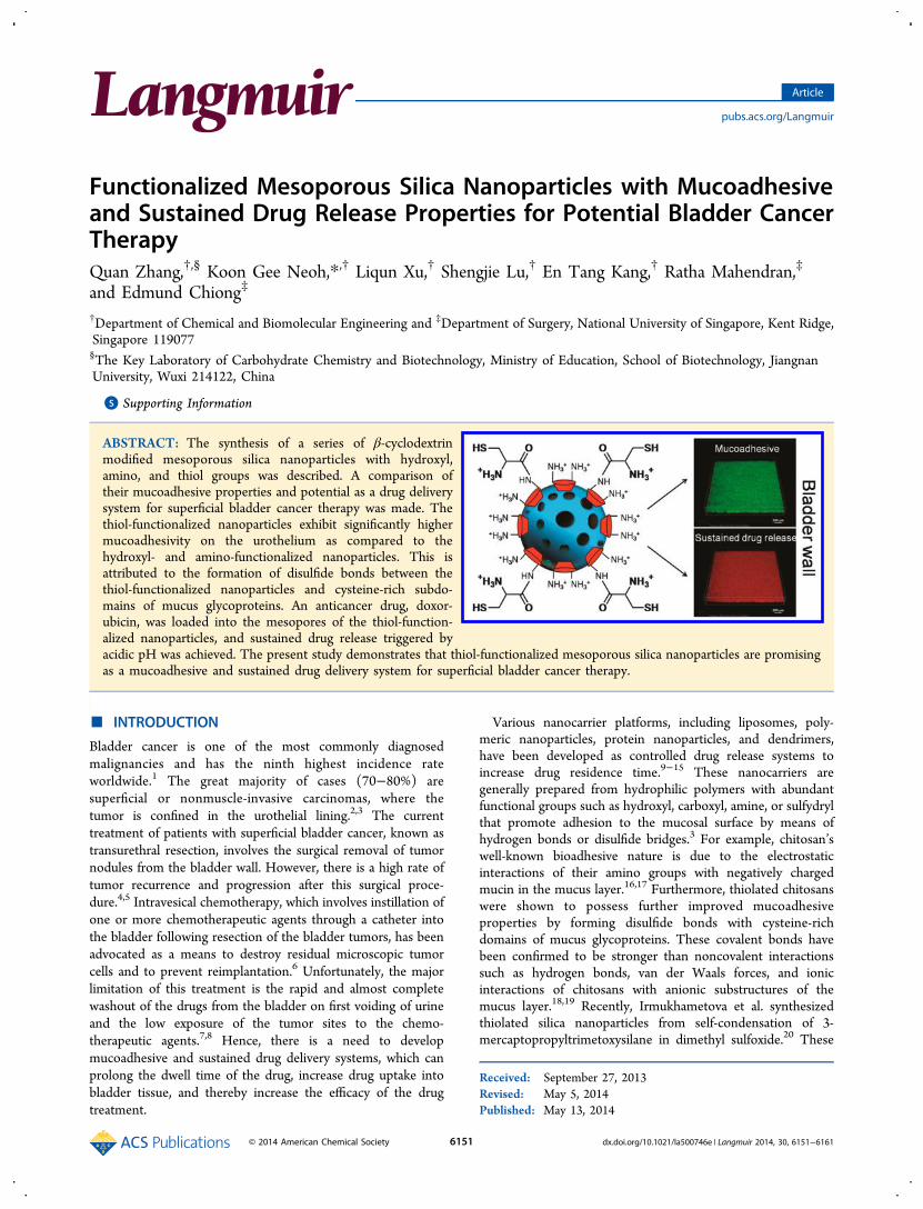
ABSTRACT: The synthesis of a series of β-cyclodextrinmodified mesoporous silica nanoparticles with hydroxyl,amino, and thiol groups was described. A comparison oftheir mucoadhesive properties and potential as a drug deliverysystem for superficial bladder cancer therapy was made. Thethiol-functionalized nanoparticles exhibit significantly highermucoadhesivity on the urothelium as compared to thehydroxyl- and amino-functionalized nanoparticles. This isattributed to the formation of disulfide bonds between thethiol-functionalized nanoparticles and cysteine-rich subdo-mains of mucus glycoproteins. An anticancer drug, doxor-ubicin, was loaded into the mesopores of the thiol-function-alized nanoparticles, and sustained drug release triggered byacidic pH was achieved. The present study demonstrates that thiol-functionalized mesoporous silica nanoparticles are promisingas a mucoadhesive and sustained drug delivery system for superficial bladder cancer therapy.
■ INTRODUCTION
Bladder cancer is one of the most commonly diagnosedmalignancies and has the ninth highest incidence rateworldwide.1 The great majority of cases (70−80%) aresuperficial or nonmuscle-invasive carcinomas, where thetumor is confined in the urothelial lining.2,3 The currenttreatment of patients with superficial bladder cancer, known astransurethral resection, involves the surgical removal of tumornodules from the bladder wall. However, there is a high rate oftumor recurrence and progression after this surgical proce-dure.4,5 Intravesical chemotherapy, which involves instillation ofone or more chemotherapeutic agents through a catheter intothe bladder following resection of the bladder tumors, has beenadvocated as a means to destroy residual microscopic tumorcells and to prevent reimplantation.6 Unfortunately, the majorlimitation of this treatment is the rapid and almost completewashout of the drugs from the bladder on first voiding of urineand the low exposure of the tumor sites to the chemo-therapeutic agents.7,8 Hence, there is a need to developmucoadhesive and sustained drug delivery systems, which canprolong the dwell time of the drug, increase drug uptake intobladder tissue, and thereby increase the efficacy of the drugtreatment.
Various nanocarrier platforms, including liposomes, poly-meric nanoparticles, protein nanoparticles, and dendrimers,have been developed as controlled drug release systems toincrease drug residence time.9−15 These nanocarriers aregenerally prepared from hydrophilic polymers with abundantfunctional groups such as hydroxyl, carboxyl, amine, or sulfydrylthat promote adhesion to the mucosal surface by means ofhydrogen bonds or disulfide bridges.3 For example, chitosan’swell-known bioadhesive nature is due to the electrostaticinteractions of their amino groups with negatively chargedmucin in the mucus layer.16,17 Furthermore, thiolated chitosanswere shown to possess further improved mucoadhesiveproperties by forming disulfide bonds with cysteine-richdomains of mucus glycoproteins. These covalent bonds havebeen confirmed to be stronger than noncovalent interactionssuch as hydrogen bonds, van der Waals forces, and ionicinteractions of chitosans with anionic substructures of themucus layer.18,19 Recently, Irmukhametova et al. synthesizedthiolated silica nanoparticles from self-condensation of 3-mercaptopropyltrimetoxysilane in dimethyl sulfoxide.20 These
Received: September 27, 2013Revised: May 5, 2014Published: May 13, 2014
Article
pubs.acs.org/Langmuir
© 2014 American Chemical Society 6151 dx.doi.org/10.1021/la500746e | Langmuir 2014, 30, 6151−6161
nanoparticles showed excellent mucoadhesivity and retentionon ocular surfaces. However, they were nonporous andnonswellable,15 and hence their potential as a drug carrierwill be limited.Mesoporous silica nanoparticles (MSNPs) have attracted
much attention for their potential use as drug deliverycarriers.21−26 The unique properties of MSNPs, such as tunablenanoparticle size, uniform mesopores, porous interior amend-able to drug loading, high surface area, and easily functionaliz-able surface, make them highly suitable as a therapeutic deliveryvehicle.27−29 MSNPs-based controlled-release systems havebeen synthesized using different kinds of capping agents such asinorganic nanoparticles,30−32 polymer,33,34 nucleotides,35,36
antibody,37 and supramolecular assemblies.38−40 Differentstimuli, such as pH,41−45 light,46,47 redox effect,48−50 temper-ature,51 and enzymatic action,52−55 have been applied as“triggers” for uncapping the pores and releasing the cargos fromMSNPs. Encouraged by these achievements, we postulate thatthe introduction of thiol groups onto MSNP-based controlled-release systems would promote the adhesion of the MSNPs tothe mucous membrane of the urothelium through theformation of disulfide bonds and provide sustained release ofthe loaded anticancer drugs during intravesical chemotherapy.To the best of our knowledge, no study has reported on the useof MSNPs as mucoadhesive and sustained drug delivery carriersfor bladder cancer therapy.Herein, we describe the design and synthesis of function-
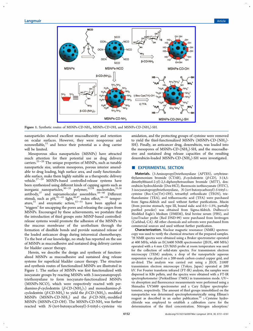
alized MSNPs as mucoadhesive and sustained drug releasesystems for superficial bladder cancer therapy. The structureand synthesis routes of functionalized MSNPs are illustrated inFigure 1. The surface of MSNPs was first functionalized withisocyanate groups by reacting MSNPs with 3-isocyanatopropyl-triethoxysilane to form isocyanate-functionalized MSNPs(MSNPs-NCO), which were respectively reacted with per-diamino-β-cyclodextrin (β-CD-(NH2)7) and monodiamino-β-cyclodextrin (β-CD-NH2) to yield the β-CD-(NH2)7 modifiedMSNPs (MSNPs-CD-NH2) and the β-CD-NH2-modifiedMSNPs (MSNPs-CD-OH). The MSNPs-CD-NH2 was furtherreacted with N-(tert-butoxycarbonyl)-S-trityl-L-cysteine via
amidation, and the protecting groups of cysteine were removedto yield the thiol-functionalized MSNPs (MSNPs-CD-(NH2)-SH). Finally, an anticancer drug, doxorubicin, was loaded intothe mesopores of MSNPs-CD-(NH2)-SH, and the mucoadhe-sive and sustained drug release capacities of the resultingdoxorubicin-loaded MSNPs-CD-(NH2)-SH were investigated.
■ EXPERIMENTAL SECTIONMaterials. (3-Aminopropyl)triethoxysilane (APTES), cetyltrime-
thylammonium bromide (CTAB), β-cyclodextrin (β-CD), 3-(4,5-dimethylthiazol-2-yl)-2,5-diphenyltetrazolium bromide (MTT), dox-orubicin hydrochloride (Dox·HCl), fluorescein isothiocyanate (FITC),3-isocyanatopropyltriethoxysilane, N-(tert-butoxycarbonyl)-S-trityl-L-cysteine (Boc-Cys(Trt)-OH), tetraethyl orthosilicate (TEOS), trie-thanolamine (TEA), and trifluoroacetic acid (TFA) were purchasedfrom Sigma-Aldrich and used without further purifications. Mucin(from porcine stomach, type III, bound sialic acid 0.5−1.5%, partiallypurified powder) was obtained from Sigma-Aldrich. Dulbecco’sModified Eagle’s Medium (DMEM), fetal bovine serum (FBS), andLysoTracker probe (Red DND-99) were purchased from Invitrogen(Carlsbad, CA). All other chemicals and solvents were purchased fromcommercial sources and used without further purification.
Characterization. Nuclear magnetic resonance (NMR) spectros-copy was used to verify the chemical structure of the prepared samples.1H NMR spectra were obtained using a Bruker spectrometer operatedat 400 MHz, while an ECA400 NMR spectrometer (JEOL, 400 MHz)operated with a 4 mm CD/MAS probe at room temperature was usedfor the collection of solid-state spectra. For transmission electronmicroscopy (TEM) analysis, a drop of the nanoparticle aqueoussuspension was placed on a 200-mesh carbon-coated copper grid, andair-dried. The analysis was carried out using a JEOL 2100Ftransmission electron microscope (Tokyo, Japan) operated at 200kV. For Fourier transform infrared (FT-IR) analysis, the samples weredispersed in KBr pellets, and the spectra were obtained with a FT-IRspectrophotometer (PerkinElmer 1760X) in transmission mode. UV−vis absorption and fluorescence measurements were performed using aShimadzu UV3600 spectrometer and a Cary Eclipse spectropho-tometer, respectively. The amount of thiol groups immobilized on thenanoparticles was determined spectrophotometrically using Ellman’sreagent as described in an earlier publication.20 L-Cysteine hydro-chloride was employed to establish a calibration curve for thedetermination of the thiol concentration. Nitrogen adsorption/
Figure 1. Synthetic routes of MSNPs-CD-NH2, MSNPs-CD-OH, and MSNPs-CD-(NH2)-SH.
Langmuir Article
dx.doi.org/10.1021/la500746e | Langmuir 2014, 30, 6151−61616152
desorption isotherms of the nanoparticles (after degassing at 100 °Cfor 12 h) were obtained as described in an earlier publication.45 Thespecific surface area and pore size of the nanoparticles were thendetermined using the Brunauer−Emmett−Teller (BET) model andthe Barrett−Joyner−Halenda (BJH) method, respectively. X-raydiffraction (XRD) measurements were performed using an ARLX’TRA powder diffractometer with Cu Kα radiation (λ = 1.540 562Å). The zeta potential and hydrodynamic size of the nanoparticles inaqueous medium were determined at 37 °C using a Malvern ZetasizerNanoZS instrument. The mean hydrodynamic size was calculatedfrom dynamic light scattering (DLS) using the Zetasizer software. Aminimum of three measurements were made at each condition, andthe results were reported as average ± standard derivation (SD).Synthesis of β-CD-NH2 and β-CD-(NH2)7. The synthesis
procedures are described in the Supporting Information.Synthesis of MSNPs and FITC-Labeled MSNPs. CTAB (0.57 g)
and TEA (50 mg) were added to distilled water (22 mL). The reactionmixture was heated to 95 °C under vigorous stirring. After thetemperature was stabilized for 1 h, TEOS (1.5 mL) was addeddropwise to the solution, and the mixture was stirred for another hourto form the MSNPs. For the preparation of FITC-labeled MSNPs(denoted as FMSNPs), FITC (2.7 mg) was dissolved in absoluteethanol (1 mL) containing APTES (6 μL), and the solution was gentlystirred for 2 h in the dark before adding into the above-mentionedCTAB−TEOS solution. For both MSNPs and FMSNPs, the solidproduct formed was centrifuged (14 000 rpm, 15 min) and washedextensively with ethanol. To remove the surfactant template (CTAB),the nanoparticles were sonicated three times in a mixture solution ofethanol (15 mL) and HCl (1 mL, 37%). The nanoparticles aftersurfactant removal were washed extensively with ethanol and driedunder reduced pressure.Synthesis of MSNPs-NCO, MSNPs-CD-NH2, and MSNPs-CD-
OH. MSNPs (100 mg) were suspended in anhydrous toluene (20mL), and 3-isocyanatopropyltriethoxysilane (1 mL) was added to thesolution. The mixture was stirred at room temperature under a N2atmosphere for 12 h. The nanoparticles were collected bycentrifugation and washed with toluene followed by methanol. Finally,the sample was dried under reduced pressure to yield the 3-isocyanatopropyl-functionalized MSNPs (MSNPs-NCO).To prepare the MSNPs-CD-NH2, MSNPs-NCO (50 mg) and β-
CD-(NH2)7 (100 mg) were added to anhydrous dimethylformamide(DMF, 5 mL). The mixture was stirred for 24 h at 60 °C. Thenanoparticles were then centrifuged and washed thoroughly with DMFand ethanol, respectively. MSNPs-CD-OH was prepared by simplyreacting MSNPs-NCO with β-CD-NH2 using the same steps asdescribed for the formation of MSNPs-CD-NH2.Synthesis of MSNPs-CD-(NH2)-SH. MSNPs-CD-NH2 (20 mg)
was dispersed in anhydrous DMF (5 mL). Boc-Cys(Trt)-OH (200mg, 0.43 mmol) was then added to the mixture in the presence of 2-(1H-benzotriazole-1-yl)-1,1,3,3-tetramethyluronium hexafluorophos-phate (HBTU)/N-hydroxybenzotriazole (HOBt). After the reactionmixture was stirred for 48 h, the resulting nanoparticles were collectedby centrifugation and washed extensively with DMF. To remove theBoc and Trt protecting groups, the nanoparticles were further treatedwith trifluoroacetic acid/thioanisole/ethanedithiol/anisole (90/5/3/2). Finally, the nanoparticles were washed extensively with DMF andethanol and denoted as MSNPs-CD-(NH2)-SH.Drug Loading and Release. MSNPs-CD-(NH2)-SH (5 mg) was
added to a phosphate buffered saline (PBS, pH 7.4) solution (1 mL)containing Dox·HCl (2.5 mg mL−1). The mixture was stirred andsonicated to maximize the dispersion of the nanoparticles. After themixture was stirred in the dark for 24 h, the Dox-loaded nanoparticleswere collected by centrifugation and washed extensively with PBSbuffer solution. This product is denoted as Dox@MSNPs-CD-(NH2)-SH. The amount of Dox loaded into MSNPs-CD-(NH2)-SH wasestimated by subtracting the amount of Dox in the collectedsupernatant from the initial amount of Dox in the loading solutionusing UV−vis absorption spectroscopy. The loading of Dox into theother types of nanoparticles was carried out in the same manner. Todetermine the kinetics of Dox release from these nanoparticles, the
Dox-loaded nanoparticles (5 mg) were dispersed in 5 mL of PBS in adialysis membrane bag (MWCO = 3500), and the bag was immersedin 50 mL of PBS (pH 7.4) or artificial urine (comprising 54 mM NaCl,30 mM KCl, 15 mM NH4Cl, 3 mM CaCl2, 2 mM MgSO4, 2 mMNaHCO3, 9 mM Na2SO4, 3.6 mM NaH2PO4, 0.4 mM Na2HPO4, and200 mM urea in DI water, pH 6.1)56 and shaken at 100 rpm at 37 °C.Fluorescence spectroscopy (λex = 485 nm, λem = 590 nm) was used tomonitor the amount of Dox released at different time intervals over aperiod of 48 h.
Evaluation of Mucoadhesivity Using the Mucin-ParticleMethod.57,58 Mucin powder was suspended in 10 mM acetate buffersolution (ABS, pH 4.5) at a concentration of 0.6% (w/v). The mucinsuspension was incubated at 37 °C overnight and sonicated at 40 kHz(Branson 2510 ultrasonic bath) for 30 min until the mucin particle sizewas smaller than 1 μm as indicated by DLS. The mucin suspensionwas then centrifuged at 4000 rpm for 30 min, and the supernatant wascollected. The mean hydrodynamic size of mucin particles in thesupernatant was less than 200 nm. Since very little of the mucin wasprecipitated during centrifugation, it can be assumed that all the mucinremained in the supernatant, which was then was diluted toconcentrations of 0.012%, 0.05%, 0.1%, 0.2%, and 0.4% (w/v) with10 mM ABS, pH 4.5, before use. For evaluation of the mucoadhesivityof the nanoparticles, MSNPs-CD-OH, MSNPs-CD-NH2, and MSNPs-CD-(NH2)-SH were dispersed in DI water (0.5 mg mL−1). Eachnanoparticle suspension was mixed with an equal volume of the mucinsuspension in ABS of different concentrations. After the mixtures wereincubated at 37 °C for 30 min, the particle size was determined usingthe Zetasizer instrument.
Bladder Tissue Preparation and Mucoadhesivity Studies.Urinary bladders, freshly excised from 6 to 10 month old pigs, wereobtained from a commercial abattoir and transported on ice inTyrode’s buffer to our laboratory within 1−1.5 h of sacrifice. Excessadipose tissue on the bladder was removed, and the bladder was cutlongitudinally into left and right lateral halves and subdivided intopieces of approximately 5 cm × 5 cm. The bladder pieces wereincubated with FMSNPs-CD-OH (0.5 mL, 5 mg mL−1), FMSNPs-CD-NH2 (0.5 mL, 5 mg mL−1), or FMSNPs-CD-(NH2)-SH (0.5 mL,5 mg mL−1) in artificial urine at 37 °C for 2 h. The pieces were thenrinsed three times with artificial urine, and the bladder wall wasobserved under a Nikon confocal laser-scanning A1 microscope (10×objective, 488 nm excitation).
The mucoadhesivity of Dox-loaded nanoparticles was tested usingbladder pieces prepared in a similar fashion as described above. Thebladder pieces were divided into two groups. For the first group, thebladder pieces were incubated with either Dox-loaded nanoparticles(0.5 mL, 5 mg mL−1) or free Dox (0.5 mL, 200 μg mL−1) in artificialurine at 37 °C for 2 h. The bladder pieces were then rinsed three timeswith artificial urine and subjected to confocal microscopy observation.For the second group, the bladder pieces were similarly incubated witheither free Dox or Dox-loaded nanoparticles for 2 h, and after rinsingthree times with artificial urine, the bladder pieces were allowed toincubate in artificial urine for another 3 h before the final washing withartificial urine and confocal microscopy observation as mentionedabove.
Cell Culture and Viability Studies. UMUC3 (human urothelialcarcinoma) cells, purchased from American Type Culture Collection(ATCC, Manassas, VA), were cultured in DMEM containing 10%FBS, penicillin (100 U mL−1), streptomycin (100 μg mL−1), and L-glutamine (2 mM) in a humidified atmosphere with 5% CO2 at 37 °C.
The cytotoxicity of the nanoparticles was evaluated using the MTTassay. UMUC3 cells were seeded into a 96-well plate at a density of 5× 10³ cells per well and maintained in growth medium (100 μL) for 24h. The culture medium was then replaced with complete DMEMmedium (100 μL) containing either free Dox or Dox-loadednanoparticles at different Dox dosages. After 5 h, the cells werewashed three times with PBS, and the medium in each well wasreplaced with 100 μL of fresh culture medium. After incubation for 72h, the culture medium from each well was replaced with 100 μL ofmedium containing MTT solution (0.5 mg mL−1). After another 4 hof incubation, culture supernatants were removed and DMSO (100
Langmuir Article
dx.doi.org/10.1021/la500746e | Langmuir 2014, 30, 6151−61616153
μL) was added into each well. The plate was gently shaken for 15 minto dissolve the formazan crystals and the absorbance at 570 nm wasmeasured using a microplate reader (Tecan GENios, Switzerland). Acontrol experiment was carried out in a similar manner but withoutDox or nanoparticles in the medium. The viability of the cells treatedwith Dox or nanoparticles (relative to the cells in the controlexperiment) was calculated from ([A]test − [A]0)/([A]control − [A]0),where [A]test, [A]control, and [A]0 are the average absorbance obtainedwith the test and control samples and culture medium without cells,respectively. GraphPad Prism software (version 5.01) was used tocalculate the IC50 values from three independent experiments.Cellular Uptake. UMUC3 bladder cancer cells were seeded in 35
mm plastic-bottomed Ibidi μ-dishes and allowed to grow for 24 h.After incubation with Dox@FMSNPs-CD-(NH2)-SH (25 μg mL−1,loaded Dox of about 5 μg mL−1) or free Dox (5 μg mL−1) for apredetermined period of time, the cells were washed three times withPBS (pH 7.4) and then fixed with 4.0% formaldehyde at roomtemperature for 15 min. After washing with PBS, the cells were stainedwith 4′,6-diamidino-2-phenylindole (DAPI, 1 μg mL−1) for 15 min.59
After washing with PBS, the cells were subjected to confocalmicroscopy observation (100× oil objective, 405/488 nm excitation).
■ RESULTS AND DISCUSSIONPreparation and Characterization of MSNPs-CD-
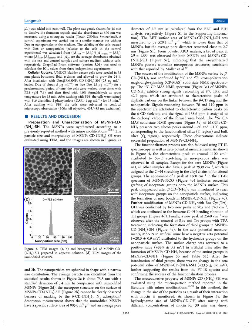
(NH2)-SH. The MSNPs were synthesized according to apreviously reported method with minor modifications.60,61 Theparticle size and morphology of MSNPs-CD-(NH2)-SH wereevaluated using TEM, and the images are shown in Figures 2a
and 2b. The nanoparticles are spherical in shape with a narrowsize distribution. The average particle size calculated from thestatistical results shown in Figure 2c is about 75.5 nm with astandard deviation of 3.4 nm. In comparison with unmodifiedMSNPs (Figure 2d), the mesopore structure on the surface ofMSNPs-CD-(NH2)-SH (Figure 2b) cannot be clearly observedbecause of masking by the β-CD-(NH2)7. N2 adsorption/desorption measurement shows that the unmodified MSNPshave a specific surface area of 805.0 m2 g−1 and an average pore
diameter of 2.7 nm as calculated from the BET and BJHanalysis, respectively (Figure S1 in the Supporting Informa-tion). The BET surface area of MSNPs-CD-(NH2)-SH wasmeasured to be 520.2 m2 g−1, which is lower than that ofMSNPs, but the average pore diameter remained close to 2.7nm (Figure S1). From powder XRD analysis, a broad peak at2θ = 1.55° was observed for both MSNPs and MSNPs-CD-(NH2)-SH (Figure S2), indicating that the as-synthesizedMSNPs possess wormlike mesoporous structures, consistentwith that reported by Moller et al.60
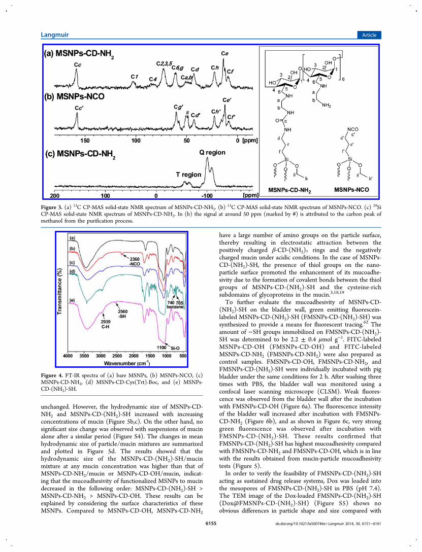
The success of the modification of the MSNPs surface by β-CD-(NH2)7 was confirmed by 13C and 29Si cross-polarizationmagic-angle-spinning (CP-MAS) solid-state NMR spectrosco-py. The 13C CP-MAS NMR spectrum (Figure 3a) of MSNPs-CD-NH2 exhibits strong signals resonating at 8.7, 13.8, and42.7 ppm, which are attributed to characteristic peaks ofaliphatic carbons on the linker between the β-CD ring and thenanoparticle. Signals resonating between 70 and 110 ppm inthe spectrum are attributed to characteristic carbon peaks onthe β-CD skeleton, and the signal at 158.6 ppm is assigned tothe carbonyl carbon of the formed urea bond. The 29Si CP-MAS solid-state NMR spectrum (Figure 3c) of MSNPs-CD-NH2 presents two silicon peaks around −60 and −100 ppm,corresponding to the functionalized silica (T region) and bulksilica (Q region), respectively. These observations indicatesuccessful preparation of MSNPs-CD-NH2.The functionalization process was also followed using FT-IR
spectroscopy as well as zeta-potential measurements. As shownin Figure 4, the characteristic peak at around 1100 cm−1
attributed to Si−O stretching in mesoporous silica wasobserved in all samples. Except for the bare MSNPs (Figure4a), all other samples also have a peak at 2939 cm−1, which isassigned to the C−H stretching in the alkyl chains of functionalgroups. The appearance of a peak at 2360 cm−1 in the FT-IRspectrum of MSNPs-NCO (Figure 4b) indicates successfulgrafting of isocyanate groups onto the MSNPs surface. Thispeak disappeared after β-CD-(NH2)7 was introduced to reactwith isocyanate groups on the nanoparticle surface, indicatingthe formation of urea bonds in MSNPs-CD-NH2 (Figure 4c).Further modification of MSNPs-CD-NH2 with Boc-Cys(Trt)-OH was confirmed by two new peaks at 705 and 740 cm−1,which are attributed to the benzene C−H bending vibration ofTrt groups (Figure 4d). Finally, a new peak at 2560 cm−1 wasobserved after the removal of Boc and Trt groups with TFAtreatment, indicating the formation of thiol groups in MSNPs-CD-(NH2)-SH (Figure 4e). In the zeta potential measure-ments, MSNPs in artificial urine have a negative zeta potential(−20.0 ± 0.9 mV) attributed to the hydroxide groups on thenanoparticle surface. The surface charge was reversed to apositive value (+33.9 ± 0.5 mV) in artificial urine after theformation of MSNPs-CD-NH2 because of the amino groups inMSNPs-CD-NH2 (Figure S3 and Table S1). After theintroduction of thiol groups, there was no change in the zetapotential value of MSNPs-CD-(NH2)-SH (+33.5 ± 0.6 mV),further supporting the results from the FT-IR spectra andconfirming the success of the functionalization process.The mucoadhesive property of MSNPs-CD-(NH2)-SH was
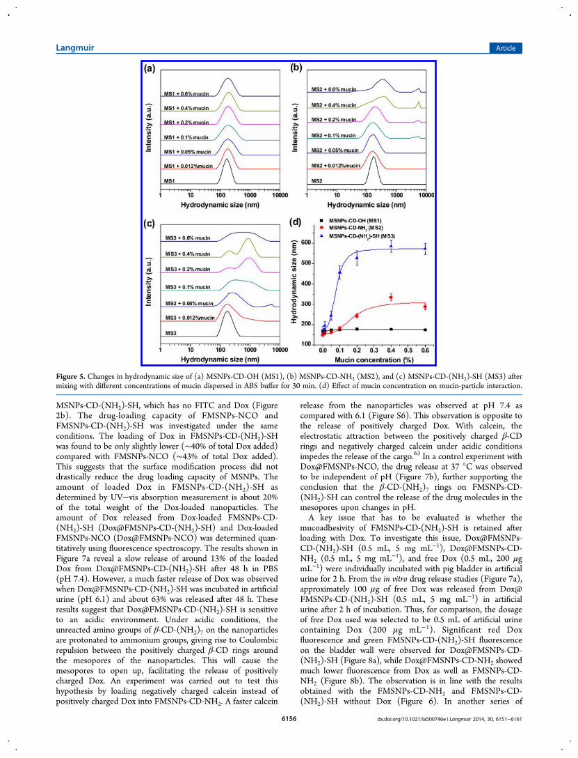
evaluated using the mucin-particle method reported in theliterature with minor modifications.57,58 In this method, thechange in the size of the particles as a result of their interactionwith mucin is monitored. As shown in Figure 5a, thehydrodynamic size of MSNPs-CD-OH after mixing withdifferent concentrations of mucin for 30 min was almost
Figure 2. TEM images (a, b) and histogram (c) of MSNPs-CD-(NH2)-SH prepared in aqueous solution. (d) TEM images of theunmodified MSNPs.
Langmuir Article
dx.doi.org/10.1021/la500746e | Langmuir 2014, 30, 6151−61616154
unchanged. However, the hydrodynamic size of MSNPs-CD-NH2 and MSNPs-CD-(NH2)-SH increased with increasingconcentrations of mucin (Figure 5b,c). On the other hand, nosignificant size change was observed with suspensions of mucinalone after a similar period (Figure S4). The changes in meanhydrodynamic size of particle/mucin mixtures are summarizedand plotted in Figure 5d. The results showed that thehydrodynamic size of the MSNPs-CD-(NH2)-SH/mucinmixture at any mucin concentration was higher than that ofMSNPs-CD-NH2/mucin or MSNPs-CD-OH/mucin, indicat-ing that the mucoadhesivity of functionalized MSNPs to mucindecreased in the following order: MSNPs-CD-(NH2)-SH >MSNPs-CD-NH2 > MSNPs-CD-OH. These results can beexplained by considering the surface characteristics of theseMSNPs. Compared to MSNPs-CD-OH, MSNPs-CD-NH2
have a large number of amino groups on the particle surface,thereby resulting in electrostatic attraction between thepositively charged β-CD-(NH2)7 rings and the negativelycharged mucin under acidic conditions. In the case of MSNPs-CD-(NH2)-SH, the presence of thiol groups on the nano-particle surface promoted the enhancement of its mucoadhe-sivity due to the formation of covalent bonds between the thiolgroups of MSNPs-CD-(NH2)-SH and the cysteine-richsubdomains of glycoproteins in the mucin.3,18,19
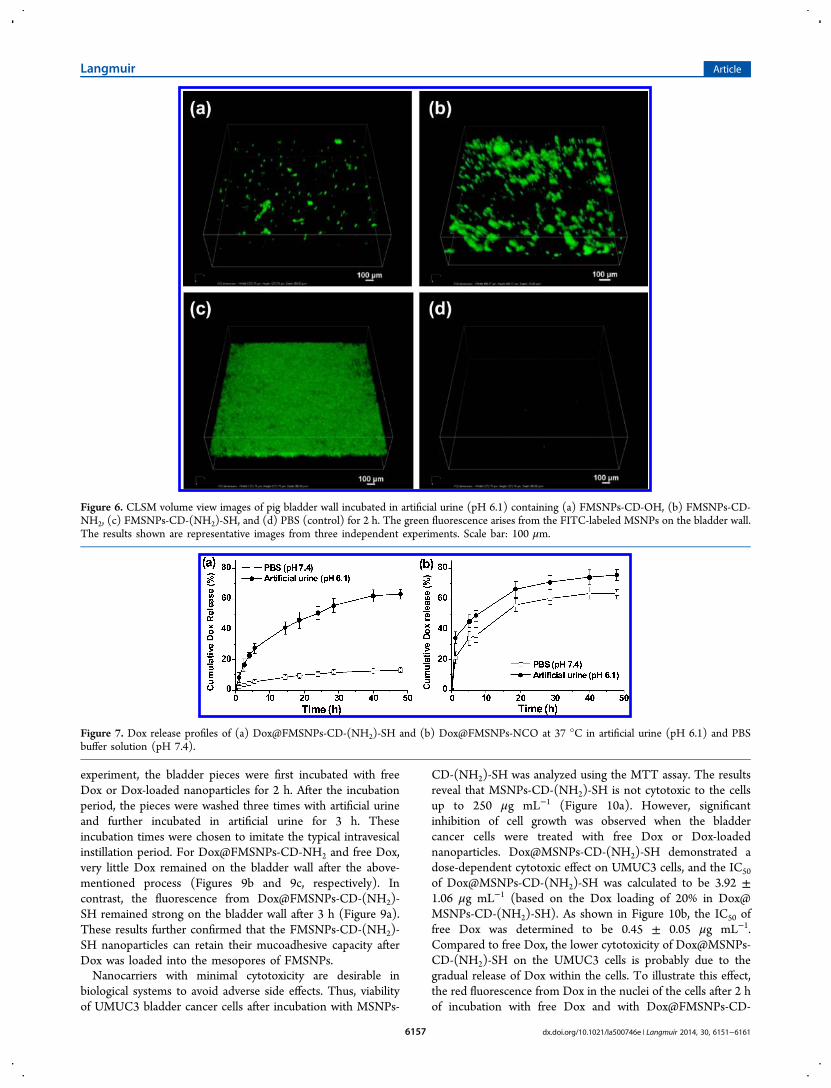
To further evaluate the mucoadhesivity of MSNPs-CD-(NH2)-SH on the bladder wall, green emitting fluorescein-labeled MSNPs-CD-(NH2)-SH (FMSNPs-CD-(NH2)-SH) wassynthesized to provide a means for fluorescent tracing.62 Theamount of −SH groups immobilized on FMSNPs-CD-(NH2)-SH was determined to be 2.2 ± 0.4 μmol g−1. FITC-labeledMSNPs-CD-OH (FMSNPs-CD-OH) and FITC-labeledMSNPs-CD-NH2 (FMSNPs-CD-NH2) were also prepared ascontrol samples. FMSNPs-CD-OH, FMSNPs-CD-NH2, andFMSNPs-CD-(NH2)-SH were individually incubated with pigbladder under the same conditions for 2 h. After washing threetimes with PBS, the bladder wall was monitored using aconfocal laser scanning microscope (CLSM). Weak fluores-cence was observed from the bladder wall after the incubationwith FMSNPs-CD-OH (Figure 6a). The fluorescence intensityof the bladder wall increased after incubation with FMSNPs-CD-NH2 (Figure 6b), and as shown in Figure 6c, very stronggreen fluorescence was observed after incubation withFMSNPs-CD-(NH2)-SH. These results confirmed thatFMSNPs-CD-(NH2)-SH has highest mucoadhesivity comparedwith FMSNPs-CD-NH2 and FMSNPs-CD-OH, which is in linewith the results obtained from mucin-particle mucoadhesivitytests (Figure 5).In order to verify the feasibility of FMSNPs-CD-(NH2)-SH
acting as sustained drug release systems, Dox was loaded intothe mesopores of FMSNPs-CD-(NH2)-SH in PBS (pH 7.4).The TEM image of the Dox-loaded FMSNPs-CD-(NH2)-SH(Dox@FMSNPs-CD-(NH2)-SH) (Figure S5) shows noobvious differences in particle shape and size compared with
Figure 3. (a) 13C CP-MAS solid-state NMR spectrum of MSNPs-CD-NH2. (b)13C CP-MAS solid-state NMR spectrum of MSNPs-NCO. (c) 29Si
CP-MAS solid-state NMR spectrum of MSNPs-CD-NH2. In (b) the signal at around 50 ppm (marked by #) is attributed to the carbon peak ofmethanol from the purification process.
Figure 4. FT-IR spectra of (a) bare MSNPs, (b) MSNPs-NCO, (c)MSNPs-CD-NH2, (d) MSNPs-CD-Cys(Trt)-Boc, and (e) MSNPs-CD-(NH2)-SH.
Langmuir Article
dx.doi.org/10.1021/la500746e | Langmuir 2014, 30, 6151−61616155
MSNPs-CD-(NH2)-SH, which has no FITC and Dox (Figure2b). The drug-loading capacity of FMSNPs-NCO andFMSNPs-CD-(NH2)-SH was investigated under the sameconditions. The loading of Dox in FMSNPs-CD-(NH2)-SHwas found to be only slightly lower (∼40% of total Dox added)compared with FMSNPs-NCO (∼43% of total Dox added).This suggests that the surface modification process did notdrastically reduce the drug loading capacity of MSNPs. Theamount of loaded Dox in FMSNPs-CD-(NH2)-SH asdetermined by UV−vis absorption measurement is about 20%of the total weight of the Dox-loaded nanoparticles. Theamount of Dox released from Dox-loaded FMSNPs-CD-(NH2)-SH (Dox@FMSNPs-CD-(NH2)-SH) and Dox-loadedFMSNPs-NCO (Dox@FMSNPs-NCO) was determined quan-titatively using fluorescence spectroscopy. The results shown inFigure 7a reveal a slow release of around 13% of the loadedDox from Dox@FMSNPs-CD-(NH2)-SH after 48 h in PBS(pH 7.4). However, a much faster release of Dox was observedwhen Dox@FMSNPs-CD-(NH2)-SH was incubated in artificialurine (pH 6.1) and about 63% was released after 48 h. Theseresults suggest that Dox@FMSNPs-CD-(NH2)-SH is sensitiveto an acidic environment. Under acidic conditions, theunreacted amino groups of β-CD-(NH2)7 on the nanoparticlesare protonated to ammonium groups, giving rise to Coulombicrepulsion between the positively charged β-CD rings aroundthe mesopores of the nanoparticles. This will cause themesopores to open up, facilitating the release of positivelycharged Dox. An experiment was carried out to test thishypothesis by loading negatively charged calcein instead ofpositively charged Dox into FMSNPs-CD-NH2. A faster calcein
release from the nanoparticles was observed at pH 7.4 ascompared with 6.1 (Figure S6). This observation is opposite tothe release of positively charged Dox. With calcein, theelectrostatic attraction between the positively charged β-CDrings and negatively charged calcein under acidic conditionsimpedes the release of the cargo.63 In a control experiment withDox@FMSNPs-NCO, the drug release at 37 °C was observedto be independent of pH (Figure 7b), further supporting theconclusion that the β-CD-(NH2)7 rings on FMSNPs-CD-(NH2)-SH can control the release of the drug molecules in themesopores upon changes in pH.A key issue that has to be evaluated is whether the
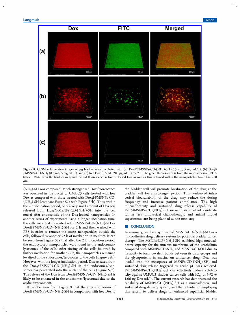
mucoadhesivity of FMSNPs-CD-(NH2)-SH is retained afterloading with Dox. To investigate this issue, Dox@FMSNPs-CD-(NH2)-SH (0.5 mL, 5 mg mL−1), Dox@FMSNPs-CD-NH2 (0.5 mL, 5 mg mL−1), and free Dox (0.5 mL, 200 μgmL−1) were individually incubated with pig bladder in artificialurine for 2 h. From the in vitro drug release studies (Figure 7a),approximately 100 μg of free Dox was released from Dox@FMSNPs-CD-(NH2)-SH (0.5 mL, 5 mg mL−1) in artificialurine after 2 h of incubation. Thus, for comparison, the dosageof free Dox used was selected to be 0.5 mL of artificial urinecontaining Dox (200 μg mL−1). Significant red Doxfluorescence and green FMSNPs-CD-(NH2)-SH fluorescenceon the bladder wall were observed for Dox@FMSNPs-CD-(NH2)-SH (Figure 8a), while Dox@FMSNPs-CD-NH2 showedmuch lower fluorescence from Dox as well as FMSNPs-CD-NH2 (Figure 8b). The observation is in line with the resultsobtained with the FMSNPs-CD-NH2 and FMSNPs-CD-(NH2)-SH without Dox (Figure 6). In another series of
Figure 5. Changes in hydrodynamic size of (a) MSNPs-CD-OH (MS1), (b) MSNPs-CD-NH2 (MS2), and (c) MSNPs-CD-(NH2)-SH (MS3) aftermixing with different concentrations of mucin dispersed in ABS buffer for 30 min. (d) Effect of mucin concentration on mucin-particle interaction.
Langmuir Article
dx.doi.org/10.1021/la500746e | Langmuir 2014, 30, 6151−61616156
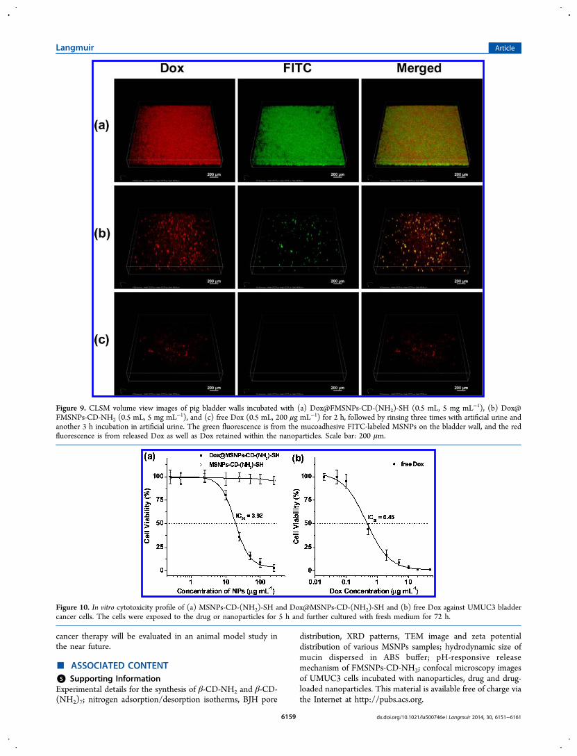
experiment, the bladder pieces were first incubated with freeDox or Dox-loaded nanoparticles for 2 h. After the incubationperiod, the pieces were washed three times with artificial urineand further incubated in artificial urine for 3 h. Theseincubation times were chosen to imitate the typical intravesicalinstillation period. For Dox@FMSNPs-CD-NH2 and free Dox,very little Dox remained on the bladder wall after the above-mentioned process (Figures 9b and 9c, respectively). Incontrast, the fluorescence from Dox@FMSNPs-CD-(NH2)-SH remained strong on the bladder wall after 3 h (Figure 9a).These results further confirmed that the FMSNPs-CD-(NH2)-SH nanoparticles can retain their mucoadhesive capacity afterDox was loaded into the mesopores of FMSNPs.Nanocarriers with minimal cytotoxicity are desirable in
biological systems to avoid adverse side effects. Thus, viabilityof UMUC3 bladder cancer cells after incubation with MSNPs-
CD-(NH2)-SH was analyzed using the MTT assay. The resultsreveal that MSNPs-CD-(NH2)-SH is not cytotoxic to the cellsup to 250 μg mL−1 (Figure 10a). However, significantinhibition of cell growth was observed when the bladdercancer cells were treated with free Dox or Dox-loadednanoparticles. Dox@MSNPs-CD-(NH2)-SH demonstrated adose-dependent cytotoxic effect on UMUC3 cells, and the IC50
of Dox@MSNPs-CD-(NH2)-SH was calculated to be 3.92 ±1.06 μg mL−1 (based on the Dox loading of 20% in Dox@MSNPs-CD-(NH2)-SH). As shown in Figure 10b, the IC50 offree Dox was determined to be 0.45 ± 0.05 μg mL−1.Compared to free Dox, the lower cytotoxicity of Dox@MSNPs-CD-(NH2)-SH on the UMUC3 cells is probably due to thegradual release of Dox within the cells. To illustrate this effect,the red fluorescence from Dox in the nuclei of the cells after 2 hof incubation with free Dox and with Dox@FMSNPs-CD-
Figure 6. CLSM volume view images of pig bladder wall incubated in artificial urine (pH 6.1) containing (a) FMSNPs-CD-OH, (b) FMSNPs-CD-NH2, (c) FMSNPs-CD-(NH2)-SH, and (d) PBS (control) for 2 h. The green fluorescence arises from the FITC-labeled MSNPs on the bladder wall.The results shown are representative images from three independent experiments. Scale bar: 100 μm.
Figure 7. Dox release profiles of (a) Dox@FMSNPs-CD-(NH2)-SH and (b) Dox@FMSNPs-NCO at 37 °C in artificial urine (pH 6.1) and PBSbuffer solution (pH 7.4).
Langmuir Article
dx.doi.org/10.1021/la500746e | Langmuir 2014, 30, 6151−61616157
(NH2)-SH was compared. Much stronger red Dox fluorescencewas observed in the nuclei of UMUC3 cells treated with freeDox as compared with those treated with Dox@FMSNPs-CD-(NH2)-SH (compare Figure S7a with Figure S7b). Thus, withinthe 2 h incubation period, only a very small amount of Dox wasreleased from Dox@FMSNPs-CD-(NH2)-SH into the cellnuclei after endocytosis of the Dox-loaded nanoparticles. Inanother series of experiments using a longer incubation time,the cells were first incubated with FMSNPs-CD-(NH2)-SH orDox@FMSNPs-CD-(NH2)-SH for 2 h and then washed withPBS in order to remove the excess nanoparticles outside thecells, followed by another 72 h of incubation in medium. It canbe seen from Figure S8a that after the 2 h incubation period,the endocytosed nanoparticles were found in the endosomes/lysosomes of the cells. After rinsing of the cells followed byfurther incubation for another 72 h, the nanoparticles remainedlocalized in the endosomes/lysosomes of the cells (Figure S8b).However, with the longer incubation period, Dox released fromthe Dox@FMSNPs-CD-(NH2)-SH in the endosomes/lyso-somes has penetrated into the nuclei of the cells (Figure S7c).The release of the Dox from Dox@FMSNPs-CD-(NH2)-SH islikely to be enhanced in the endosomes/lysosomes due to theacidic environment.It can be seen from Figure 9 that the strong adhesion of
Dox@FMSNPs-CD-(NH2)-SH in comparison with free Dox to
the bladder wall will promote localization of the drug at thebladder wall for a prolonged period. Thus, enhanced intra-vesical bioavailability of the drug may reduce the dosingfrequency and increase patient compliance. The highmucoadhesivity and sustained drug release capability ofDox@MSNPs-CD-(NH2)-SH make it an excellent candidatefor in vivo intravesical chemotherapy, and animal modelexperiments are being planned as the next step.
■ CONCLUSIONIn summary, we have synthesized MSNPs-CD-(NH2)-SH as amucoadhesive drug delivery system for potential bladder cancertherapy. The MSNPs-CD-(NH2)-SH exhibited high mucoad-hesive capacity for the mucous membrane of the urotheliumcompared with MSNPs-CD-NH2 and MSNPs-CD-OH due toits ability to form covalent bonds between its thiol groups andthe glycoproteins in mucin. An anticancer drug, Dox, wasloaded into the mesopores of MSNPs-CD-(NH2)-SH, andsustained drug release triggered by acidic pH was achieved.Dox@MSNPs-CD-(NH2)-SH can effectively induce cytotox-icity against UMUC3 bladder cancer cells with IC50 of 3.92 ±1.06 μg Dox mL−1. The current research has demonstrated thecapability of MSNPs-CD-(NH2)-SH as a mucoadhesive andsustained drug delivery system, and the potential of employingthis system to deliver drugs for enhanced superficial bladder
Figure 8. CLSM volume view images of pig bladder walls incubated with (a) Dox@FMSNPs-CD-(NH2)-SH (0.5 mL, 5 mg mL−1), (b) Dox@FMSNPs-CD-NH2 (0.5 mL, 5 mg mL
−1), and (c) free Dox (0.5 mL, 200 μg mL−1) for 2 h. The green fluorescence is from the mucoadhesive FITC-labeled MSNPs on the bladder wall, and the red fluorescence is from released Dox as well as Dox retained within the nanoparticles. Scale bar: 200μm.
Langmuir Article
dx.doi.org/10.1021/la500746e | Langmuir 2014, 30, 6151−61616158
cancer therapy will be evaluated in an animal model study inthe near future.
■ ASSOCIATED CONTENT*S Supporting InformationExperimental details for the synthesis of β-CD-NH2 and β-CD-(NH2)7; nitrogen adsorption/desorption isotherms, BJH pore
distribution, XRD patterns, TEM image and zeta potentialdistribution of various MSNPs samples; hydrodynamic size ofmucin dispersed in ABS buffer; pH-responsive releasemechanism of FMSNPs-CD-NH2; confocal microscopy imagesof UMUC3 cells incubated with nanoparticles, drug and drug-loaded nanoparticles. This material is available free of charge viathe Internet at http://pubs.acs.org.
Figure 9. CLSM volume view images of pig bladder walls incubated with (a) Dox@FMSNPs-CD-(NH2)-SH (0.5 mL, 5 mg mL−1), (b) Dox@FMSNPs-CD-NH2 (0.5 mL, 5 mg mL
−1), and (c) free Dox (0.5 mL, 200 μg mL−1) for 2 h, followed by rinsing three times with artificial urine andanother 3 h incubation in artificial urine. The green fluorescence is from the mucoadhesive FITC-labeled MSNPs on the bladder wall, and the redfluorescence is from released Dox as well as Dox retained within the nanoparticles. Scale bar: 200 μm.
Figure 10. In vitro cytotoxicity profile of (a) MSNPs-CD-(NH2)-SH and Dox@MSNPs-CD-(NH2)-SH and (b) free Dox against UMUC3 bladdercancer cells. The cells were exposed to the drug or nanoparticles for 5 h and further cultured with fresh medium for 72 h.
Langmuir Article
dx.doi.org/10.1021/la500746e | Langmuir 2014, 30, 6151−61616159

■ AUTHOR INFORMATION
Corresponding Author*Tel +65 65162176; Fax +65 67791936; e-mail [email protected] (K.G.N.).
NotesThe authors declare no competing financial interest.
■ ACKNOWLEDGMENTS
This research was supported by the National Medical ResearchCouncil of Singapore Grant IRG10nov116.
■ REFERENCES(1) Chiong, E.; Gaston, K. E.; Grossman, H. B. Urinary Markers inScreening Patients with Hematuria. World J. Urol. 2008, 26, 25−30.(2) Ploeg, M.; Aben, K. K. H.; Kiemeney, L. A. The Present andFuture Burden of Urinary Bladder Cancer in the World. World J. Urol.2009, 27, 289−293.(3) GuhaSarkar, S.; Banerjee, R. Intravesical Drug Delivery:Challenges, Current Status, Opportunities and Novel Strategies. J.Controlled Release 2010, 148, 147−159.(4) Dalbagni, G. The Management of Superficial Bladder Cancer.Nat. Clin. Pract. Urol. 2007, 4, 254−260.(5) van Rhijn, B. W.; Burger, M.; Lotan, Y.; Solsona, E.; Stief, C. G.;Sylvester, R. J.; Witjes, J. A.; Zlotta, A. R. Recurrence and Progressionof Disease in Non-Muscle-Invasive Bladder Cancer: from Epidemiol-ogy to Treatment Strategy. Eur. Urol. 2009, 56, 430−442.(6) Sexton, W. J.; Wiegand, L. R.; Correa, J. J.; Politis, C.; Dickinson,S. I.; Kang, L. C. Bladder Cancer: A Review of Non-Muscle InvasiveDisease. Cancer Control. 2010, 17, 256−268.(7) Tyagi, P.; Tyagi, S.; Kaufman, J.; Huang, L.; de Miguel, F. LocalDrug Delivery to Bladder Using Technology Innovations. Urol. Clin.North Am. 2006, 33, 519−530.(8) Huang, C.; Neoh, K. G.; Xu, L.; Kang, E. T.; Chiong, E.Polymeric Nanoparticles with Encapsulated Superparamagnetic IronOxide and Conjugated Cisplatin for Potential Bladder Cancer.Biomacromolecules 2012, 13, 2513−2520.(9) Smart, J. D. The Basics and Underlying Mechanisms ofMucoadhesion. Adv. Drug Delivery Rev. 2005, 57, 1556−1568.(10) Fraser, M. O.; Chuang, Y. C.; Tyagi, P.; Yokoyama, T.;Yoshimura, N.; Huang, L.; De Groat, W. C.; Chancellor, M. B.Intravesical Liposome Administration - a Novel Treatment forHyperactive Bladder in the Rat. Urology 2003, 61, 656−663.(11) Lu, Z.; Yeh, T. K.; Tsai, M.; Au, J. L. S.; Wientjes, M. G.Paclitaxel-Loaded Gelatin Nanoparticles for Intravesical BladderCancer Therapy. Clin. Cancer Res. 2004, 10, 7677−7684.(12) Mugabe, C.; Hadaschik, B. A.; Kainthan, R. K.; Brooks, D. E.;So, A. I.; Gleave, M. E.; Burt, H. M. Paclitaxel Incorporated inHydrophobically Derivatized Hyperbranched Polyglycerols for Intra-vesical Bladder Cancer Therapy. BJU Int. 2009, 103, 978−986.(13) Mugabe, C.; Raven, P. A.; Fazli, L.; Baker, J. H. E.; Jackson, J. K.;Liggins, R. T.; So, A. I.; Gleave, M. E.; Minchinton, A. I.; Brooks, D. E.;Burt, H. M. Tissue Uptake of Docetaxel Loaded HydrophobicallyDerivatized Hyperbranched Polyglycerols and Their Effects on theMorphology of the Bladder Urothelium. Biomaterials 2012, 33, 692−703.(14) Barthelmes, J.; Dunnhaupt, S.; Unterhofer, S.; Perera, G.;Schlocker, W.; Bernkop-Schnurch, A. Thiolated Particles as EffectiveIntravesical Drug Delivery Systems for Treatment of Bladder-RelatedDiseases. Nanomedicine 2013, 8, 65−75.(15) Storha, A.; Mun, E. A.; Khutoryanskiy, V. V. Synthesis ofThiolated and Acrylated Nanoparticles Using Thiol-Ene ClickChemistry: Towards Novel Mucoadhesive Materials for DrugDelivery. RSC Adv. 2013, 3, 12275−12279.(16) Sogias, I. A.; Williams, A. C.; Khutoryanskiy, V. V. Why isChitosan Mucoadhesive? Biomacromolecules 2008, 9, 1837−1842.
(17) Xu, J.; Soliman, G. M.; Barralet, J.; Cerruti, M. Mollusk GlueInspired Mucoadhesives for Biomedical Applications. Langmuir 2012,28, 14010−14017.(18) Bravo-Osuna, I.; Vauthier, C.; Farabollini, A.; Palmieri, G. F.;Ponchel, G. Mucoadhesion Mechanism of Chitosan and ThiolatedChitosan-Poly(isobutyl cyanoacrylate) Core-Shell Nanoparticles.Biomaterials 2007, 28, 2233−2243.(19) Yin, L.; Ding, J.; He, C.; Cui, L.; Tang, C.; Yin, C. DrugPermeability and Mucoadhesion Properties of Thiolated TrimethylChitosan Nanoparticles in Oral Insulin Delivery. Biomaterials 2009, 30,5691−5700.(20) Irmukhametova, G. S.; Mun, G. A.; Khutoryanskiy, V. V.Thiolated Mucoadhesive and PEGylated Nonmucoadhesive Organo-silica Nanoparticles from 3-Mercaptopropyltrimethoxysilane. Langmuir2011, 27, 9551−9556.(21) Vivero-Escoto, J. L.; Slowing, I. I.; Trewyn, B. G.; Lin, V. S. Y.Mesoporous Silica Nanoparticles for Intracellular Controlled DrugDelivery. Small 2010, 6, 1952−1967.(22) Rosenholm, J. M.; Sahlgren, C.; Linden, M. TowardsMultifunctional, Targeted Drug Delivery Systems Using MesoporousSilica Nanoparticles - Opportunities & Challenges. Nanoscale 2010, 2,1870−1883.(23) Ambrogio, M. W.; Thomas, C. R.; Zhao, Y. L.; Zink, J. I.;Stoddart, J. F. Mechanized Silica Nanoparticles: A New Frontier inTheranostic Nanomedicine. Acc. Chem. Res. 2011, 44, 903−913.(24) Tang, F.; Li, L.; Chen, D. Mesoporous Silica Nanoparticles:Synthesis, Biocompatibility and Drug Delivery. Adv. Mater. 2012, 24,1504−1534.(25) Li, Z.; Barnes, J. C.; Bosoy, A.; Stoddart, J. F.; Zink, J. I.Mesoporous Silica Nanoparticles in Biomedical Applications. Chem.Soc. Rev. 2012, 41, 2590−2605.(26) Chen, Y.; Chen, H.; Shi, J. In Vivo Bio-Safety Evaluations andDiagnostic/Therapeutic Applications of Chemically Designed Meso-porous Silica Nanoparticles. Adv. Mater. 2013, 25, 3144−3176.(27) Kecht, J.; Schlossbauer, A.; Bein, T. Selective Functionalizationof the Outer and Inner Surfaces in Mesoporous Silica Nanoparticles.Chem. Mater. 2008, 20, 7207−7214.(28) He, Q.; Shi, J.; Cui, X.; Zhao, J.; Chen, Y.; Zhou, J. RhodamineB-Co-Condensed Spherical SBA-15 Nanoparticles: Facile Co-Con-densation Synthesis and Excellent Fluorescence Features. J. Mater.Chem. 2009, 19, 3395−3403.(29) Wu, S. H.; Mou, C. Y.; Lin, H. P. Synthesis of Mesoporous SilicaNanoparticles. Chem. Soc. Rev. 2013, 42, 3862−3875.(30) Lai, C. Y.; Trewyn, B. G.; Jeftinija, D. M.; Jeftinija, K.; Xu, S.;Jeftinija, S.; Lin, V. S. Y. A Mesoporous Silica Nanosphere-BasedCarrier System with Chemically Removable CdS Nanoparticle Capsfor Stimuli-Responsive Controlled Release of Neurotransmitters andDrug Molecules. J. Am. Chem. Soc. 2003, 125, 4451−4459.(31) Giri, S.; Trewyn, B. G.; Stellmaker, M. P.; Lin, V. S. Y. Stimuli-Responsive Controlled-Release Delivery System Based on Mesopo-rous Silica Nanorods Capped with Magnetic Nanoparticles. Angew.Chem., Int. Ed. 2005, 44, 5038−5044.(32) Aznar, E.; Marcos, M. D.; Martínez-Manez, R.; Sancenon, F.;Soto, J.; Amoros, P.; Guillem, C. pH- and Photo-Switched Release ofGuest Molecules from Mesoporous Silica Supports. J. Am. Chem. Soc.2009, 131, 6833−6843.(33) Yuan, L.; Tang, Q.; Yang, D.; Zhang, J. Z.; Zhang, F.; Hu, J.Preparation of pH-Responsive Mesoporous Silica Nanoparticles andTheir Application in Controlled Drug Delivery. J. Phys. Chem. C 2011,115, 9926−9932.(34) Chang, B.; Chen, D.; Wang, Y.; Chen, Y.; Jiao, Y.; Sha, X.; Yang,W. Bioresponsive Controlled Drug Release Based on MesoporousSilica Nanoparticles Coated with Reductively Sheddable PolymerShell. Chem. Mater. 2013, 25, 574−585.(35) He, X.; Zhao, Y.; He, D.; Wang, K.; Xu, F.; Tang, J. ATP-Responsive Controlled Release System Using Aptamer-FunctionalizedMesoporous Silica Nanoparticles. Langmuir 2012, 28, 12909−12915.(36) Chen, C.; Pu, F.; Huang, Z.; Liu, Z.; Ren, J.; Qu, X. Stimuli-Responsive Controlled-Release System Using Quadruplex DNA-
Langmuir Article
dx.doi.org/10.1021/la500746e | Langmuir 2014, 30, 6151−61616160
Capped Silica Nanocontainers. Nucleic Acids Res. 2011, 39, 1638−1644.(37) Climent, E.; Bernardos, A.; Martínez-Manez, R.; Maquieira, A.;Marcos, M. D.; Pastor-Navarro, N.; Puchades, R.; Sancenon, F.; Soto,J.; Amoros, P. Controlled Delivery Systems Using Antibody-CappedMesoporous Nanocontainers. J. Am. Chem. Soc. 2009, 131, 14075−14080.(38) Nguyen, T. D.; Liu, Y.; Saha, S.; Leung, K. C. F.; Stoddart, J. F.;Zink, J. I. Design and Optimization of Molecular Nanovalves Based onRedox-Switchable Bistable Rotaxanes. J. Am. Chem. Soc. 2007, 129,626−634.(39) Liu, R.; Zhang, Y.; Feng, P. Multiresponsive SupramolecularNanogated Ensembles. J. Am. Chem. Soc. 2009, 131, 15128−15129.(40) Nguyen, T. D.; Leung, K. C. F.; Liong, M.; Liu, Y.; Stoddart, J.F.; Zink, J. I. Versatile Supramolecular Nanovalves Reconfigured forLight Activation. Adv. Funct. Mater. 2007, 17, 2101−2110.(41) Angelos, S.; Yang, Y. W.; Patel, K.; Stoddart, J. F.; Zink, J. I. pH-Responsive Supramolecular Nanovalves Based on Cucurbit[6]urilPseudorotaxanes. Angew. Chem., Int. Ed. 2008, 47, 2222−2226.(42) Park, C.; Oh, K.; Lee, S. C.; Kim, C. Controlled Release ofGuest Molecules from Mesoporous Silica Particles Based on a pH-Responsive Polypseudorotaxane Motif. Angew. Chem., Int. Ed. 2007,46, 1455−1457.(43) Zhao, Y. L.; Li, Z.; Kabehie, S.; Botros, Y. Y.; Stoddart, J. F.;Zink, J. I. pH-Operated Nanopistons on the Surfaces of MesoporousSilica Nanoparticles. J. Am. Chem. Soc. 2010, 132, 13016−13025.(44) Meng, H.; Xue, M.; Xia, T.; Zhao, Y. L.; Tamanoi, F.; Stoddart,J. F.; Zink, J. I.; Nel, A. E. Autonomous in Vitro Anticancer DrugRelease from Mesoporous Silica Nanoparticles by pH-SensitiveNanovalves. J. Am. Chem. Soc. 2010, 132, 12690−12697.(45) Zhang, Q.; Liu, F.; Nguyen, K. T.; Ma, X.; Wang, X.; Xing, B.;Zhao, Y. Multifunctional Mesoporous Silica Nanoparticles for Cancer-Targeted and Controlled Drug Delivery. Adv. Funct. Mater. 2012, 22,5144−5156.(46) Park, C.; Lee, K.; Kim, C. Photoresponsive Cyclodextrin-Covered Nanocontainers and Their Sol-Gel Transition Induced byMolecular Recognition. Angew. Chem., Int. Ed. 2009, 48, 1275−1278.(47) He, D.; He, X.; Wang, K.; Cao, J.; Zhao, Y. A Light-ResponsiveReversible Molecule-Gated System Using Thymine-Modified Meso-porous Silica Nanoparticles. Langmuir 2012, 28, 4003−4008.(48) Liu, R.; Zhao, X.; Wu, T.; Feng, P. Tunable Redox-ResponsiveHybrid Nanogated Ensembles. J. Am. Chem. Soc. 2008, 130, 14418−14419.(49) Wan, X.; Wang, D.; Liu, S. Fluorescent pH-Sensing Organic/Inorganic Hybrid Mesoporous Silica Nanoparticles with TunableRedox-Responsive Release Capability. Langmuir 2010, 26, 15574−15579.(50) Kim, H.; Kim, S.; Park, C.; Lee, H.; Park, H. J.; Kim, C.Glutathione-Induced Intracellular Release of Guests from MesoporousSilica Nanocontainers with Cyclodextrin Gatekeepers. Adv. Mater.2010, 22, 4280−4283.(51) Schlossbauer, A.; Warncke, S.; Gramlich, P. M. E.; Kecht, J.;Manetto, A.; Carell, T.; Bein, T. A Programmable DNA-BasedMolecular Valve for Colloidal Mesoporous Silica. Angew. Chem., Int.Ed. 2010, 49, 4734−4737.(52) Park, C.; Kim, H.; Kim, S.; Kim, C. Enzyme ResponsiveNanocontainers with Cyclodextrin Gatekeepers and Synergistic Effectsin Release of Guests. J. Am. Chem. Soc. 2009, 131, 16614−16615.(53) Schlossbauer, A.; Kecht, J.; Bein, T. Biotin-Avidin as a Protease-Responsive Cap System for Controlled Guest Release from ColloidalMesoporous Silica. Angew. Chem., Int. Ed. 2009, 48, 3092−3095.(54) Bernardos, A.; Aznar, E.; Marcos, M. D.; Martínez-Manez, R.;Sancenon, F.; Soto, J.; Barat, J. M.; Amoros, P. Enzyme-ResponsiveControlled Release Using Mesoporous Silica Supports Capped withLactose. Angew. Chem., Int. Ed. 2009, 48, 5884−5887.(55) Bernardos, A.; Mondragon, L.; Aznar, E.; Marcos, M. D.;Martínez-Manez, R.; Sancenon, F.; Soto, J.; Barat, J. M.; Perez-Paya,E.; Guillem, C.; Amoro s, P. Enzyme-Responsive Intracellular
Controlled Release Using Nanometric Silica Mesoporous SupportsCapped with “Saccharides”. ACS Nano 2010, 4, 6353−6368.(56) Chutipongtanate, S.; Thongboonkerd, V. Systematic Compar-isons of Artificial Urine Formulas for In Vitro Cellular Study. Anal.Biochem. 2010, 402, 110−112.(57) Carvalho, F. C.; Bruschi, M. L.; Evangelista, R. C.; Gremiao, M.P. D. Mucoadhesive Drug Delivery Systems. Braz. J. Pharm. Sci. 2010,46, 1−17.(58) Thongborisute, J.; Takeuchi, H. Evaluation of Mucoadhesive-ness of Polymers by BIACORE Method and Mucin-Particle Method.Int. J. Pharm. 2008, 354, 204−209.(59) Zhang, Q.; Luan, L.; Feng, S.; Yan, H.; Liu, K. Using aBifunctional Polymer for the Functionalization of Fe3O4 Nano-particles. React. Funct. Polym. 2012, 72, 198−205.(60) Moller, K.; Kobler, J.; Bein, T. Colloidal Suspensions ofNanometer-Sized Mesoporous Silica. Adv. Funct. Mater. 2007, 17,605−612.(61) Pan, L.; He, Q.; Liu, J.; Chen, Y.; Ma, M.; Zhang, L.; Shi, J.Nuclear-Targeted Drug Delivery of TAT Peptide-ConjugatedMonodisperse Mesoporous Silica Nanoparticles. J. Am. Chem. Soc.2012, 134, 5722−5725.(62) Lu, J.; Liong, M.; Zink, J. I.; Tamanoi, F. Mesoporous SilicaNanoparticles as a Delivery System for Hydrophobic AnticancerDrugs. Small 2007, 3, 1341−1346.(63) Casasus, R.; Marcos, M. D.; Martínez-Manez, R.; Ros-Lis, J. V.;Soto, J.; Villaescusa, L. A.; Amoros, P.; Beltran, D.; Guillem, C.;Latorre, J. Toward the Development of Ionically ControlledNanoscopic Molecular Gates. J. Am. Chem. Soc. 2004, 126, 8612−8613.
Langmuir Article
dx.doi.org/10.1021/la500746e | Langmuir 2014, 30, 6151−61616161