honey bee dopamine and octopamine receptors linked to intracellular calcium signaling have a close...
TRANSCRIPT

Honey Bee Dopamine and Octopamine Receptors Linkedto Intracellular Calcium Signaling Have a ClosePhylogenetic and Pharmacological RelationshipKyle T. Beggs1, Joel D. A. Tyndall2, Alison R. Mercer1*
1 Department of Zoology, University of Otago, Dunedin, New Zealand, 2 School of Pharmacy, University of Otago, Dunedin, New Zealand
Abstract
Background: Three dopamine receptor genes have been identified that are highly conserved among arthropod species.One of these genes, referred to in honey bees as Amdop2, shows a close phylogenetic relationship to the a-adrenergic-likeoctopamine receptor family. In this study we examined in parallel the functional and pharmacological properties ofAmDOP2 and the honey bee octopamine receptor, AmOA1. For comparison, pharmacological properties of the honey beedopamine receptors AmDOP1 and AmDOP3, and the tyramine receptor AmTYR1, were also examined.
Methodology/Principal Findings: Using HEK293 cells heterologously expressing honey bee biogenic amine receptors, wefound that activation of AmDOP2 receptors, like AmOA1 receptors, initiates a rapid increase in intracellular calcium levels.We found no evidence of calcium signaling via AmDOP1, AmDOP3 or AmTYR1 receptors. AmDOP2- and AmOA1-mediatedincreases in intracellular calcium were inhibited by 10 mM edelfosine indicating a requirement for phospholipase C-b activityin this signaling pathway. Edelfosine treatment had no effect on AmDOP2- or AmOA1-mediated increases in intracellularcAMP. The synthetic compounds mianserin and epinastine, like cis-(Z)-flupentixol and spiperone, were found to havesignificant antagonist activity on AmDOP2 receptors. All 4 compounds were effective antagonists also on AmOA1 receptors.Analysis of putative ligand binding sites offers a possible explanation for why epinastine acts as an antagonist at AmDOP2receptors, but fails to block responses mediated via AmDOP1.
Conclusions/Significance: Our results indicate that AmDOP2, like AmOA1, is coupled not only to cAMP, but also to calcium-signalling and moreover, that the two signalling pathways are independent upstream of phospholipase C-b activity. Thestriking similarity between the pharmacological properties of these 2 receptors suggests an underlying conservation ofstructural properties related to receptor function. Taken together, these results strongly support phylogenetic analysesindicating that the AmDOP2 and AmOA1 receptor genes are immediate paralogs.
Citation: Beggs KT, Tyndall JDA, Mercer AR (2011) Honey Bee Dopamine and Octopamine Receptors Linked to Intracellular Calcium Signaling Have a ClosePhylogenetic and Pharmacological Relationship. PLoS ONE 6(11): e26809. doi:10.1371/journal.pone.0026809
Editor: Immo A. Hansen, New Mexico State University, United States of America
Received May 16, 2011; Accepted October 4, 2011; Published November 11, 2011
Copyright: � 2011 Beggs et al. This is an open-access article distributed under the terms of the Creative Commons Attribution License, which permitsunrestricted use, distribution, and reproduction in any medium, provided the original author and source are credited.
Funding: This research was supported by a grant from the Royal Society of New Zealand Marsden Fund (UOO0615, http://marsden.rsnz.org). The funder had norole in study design, data collection and analysis, decision to publish, or preparation of the manuscript.
Competing Interests: The authors have declared that no competing interests exist.
* E-mail: [email protected]
Introduction
Some invertebrate and vertebrate dopamine (DA) receptor types
demonstrate a strong phylogenetic relationship that is reflected in an
apparent conservation of common functional properties [1,2,3]. For
example, D2-like DA receptors in arthropods exhibit significant
homology in primary amino acid sequence with vertebrate D2-like
dopamine receptors [4,5,6] and in arthropods, as in vertebrates,
activation of D2-like receptors generally inhibits adenylyl cyclase
activity leading to a reduction in intracellular levels of cAMP [1,3].
However, phylogenetic analyses indicate that at least one DA
receptor type may be specific to invertebrate species [1,7]. The
presence of this ‘invertebrate-type’ DA receptor (Figure 1) raises a
number of interesting questions about the origin, function and role
of this receptor protein in the invertebrate nervous system.
The first of these invertebrate-type DA receptors to be functionally
described was the Drosophila DAMB/Dop99B receptor [8,9].
Orthologues of this receptor have subsequently been described in
several arthropod species [10,11,12,13], including in honey bees
(AmDOP2, [7]). All of the invertebrate-type DA receptors examined
so far have been found to be positively coupled to adenylyl cyclase
and hence are often described as D1-like DA receptors [1].
Interestingly, stimulation of cells expressing the DAMB/Dop99B
receptor with DA also induces a rapid, transient increase in
intracellular calcium (Ca2+) levels [9,14,15]. However, this property
has yet to be demonstrated in other invertebrate-type DA receptors.
One intriguing feature of invertebrate-type DA receptors is that
their primary amino acid sequences, when compared to other
invertebrate biogenic amine receptors, show highest homology to
an octopamine (OA) receptor type [1,7,16,17,18] described as
being ‘a-adrenergic-like’ [19]. The first a-adrenergic-like OA
receptor was described in a pond snail [20] and orthologues that
have been identified subsequently include the Drosophila OAMB
receptor [14], honey bee AmOA1 receptor [21], cockroach Pa oa1
PLoS ONE | www.plosone.org 1 November 2011 | Volume 6 | Issue 11 | e26809

receptor [22] and silkworm BmOAR1 [23] receptor. Functional
studies of cells expressing a-adrenergic-like OA receptor ortholo-
gues have consistently found that activation by OA results in a
rapid, transient rise in intracellular Ca2+ concentrations, but also
an increase in intracellular levels of cAMP [14,16,20,21,22,23].
Interestingly, depletion of intracellular Ca2+ with BAPTA-AM was
found to have no significant effect on cAMP signaling mediated
either, via DAMB/Dop99B receptors [15] or Pa oa1-receptors
Figure 1. Phylogram of selected arthropod amine receptor sequences. A phylogenetic analysis showing known invertebrate-type DAreceptors and a-adrenergic-like OA receptors and their relationship to other DA, OA and TA receptors from Apis mellifera and Drosophilamelanogaster. The honey bee rhodopsin protein is used as an outlier. Conserved regions of receptor protein sequences were aligned using ClustalW2software (version 2.0.10) using the default settings (http://ww.ebi.ac.uk/Tools/clustalw2/). Phylograms were prepared as described in [30] by a 1000trial N-J bootstrap analysis, and using ClustalX software (version 2.0). Resulting bootstrap scores are displayed on selected nodes as percentagesvalues, if greater than 50%.doi:10.1371/journal.pone.0026809.g001
Honey Bee Dopamine and Octopamine Receptors
PLoS ONE | www.plosone.org 2 November 2011 | Volume 6 | Issue 11 | e26809

[22] indicating that both of these receptor types mediate their
effects on cAMP via a pathway independent of the Ca2+ signaling
pathway. However this concept of pathway independence has also
been challenged [16,19,21], with increases in cAMP levels
suggested to be the result of Ca2+-induced adenylyl cyclase activity.
Phylogenetic models have indicated that the invertebrate-type
DA receptors and the a-adrenergic-like OA receptors are
immediate paralogs [17,18]. However, as such models may
contain inherent weaknesses in their ability to discern distant
evolutionary relationships correctly [24], evidence at a functional
level is required also to confirm the existence of such relationships.
In this study we examined the functional and pharmacological
properties of the invertebrate-type DA receptor from honey bee,
AmDOP2, and the honey bee a-adrenergic-like octopamine
receptor, AmOA1, as representative examples of each of the two
receptor types (Figure 1). Analysis of these two receptors in parallel
enabled us to test predictions arising from phylogenetic modelling.
Our study provides evidence that agonist activation of either,
AmDOP2 or AmOA1 receptors results in stimulation of cAMP and
Ca2+ signaling pathways, and that these pathways are activated
independently upstream of PLCb activity. We also show that there
are marked similarities in the pharmacological properties of these
two receptor types, a finding supported further by comparisons
with other related receptor types. The homology we find in the
functional and pharmacological properties of AmDOP2 and
AmOA1 receptors supports the phylogenetic model of these
receptor genes as being immediate paralogs.
Results
AmDOP2 receptors couple to Ca2+ signaling pathwaysWe found that exposure of AmDOP2-expressing HEK293 cells to
1 mM DA initiated a rapid increase in intracellular Ca2+ levels, with
maximum response amplitude occurring 15 to 20 seconds post
injection (Figure 2A). Exposure of AmDOP2-expressing cells to
1 mM OA also initiated a Ca2+ response, but responses to this amine
were smaller in amplitude and slower to reach peak levels
(approximately 25 s) than responses to DA (Figure 2A). Exposure
to 1 mM tyramine (TA) had no observable effect on AmDOP2-
expressing cells (Figure 2A). Control cells expressing the b-Gal
reporter protein showed no Ca2+ response to DA, OA or TA (data
not shown). The Ca2+ response of AmDOP2-expressing HEK293
cells to DA was dose-dependent (Figure 2C).
For comparison we also examined AmOA1-expressing HEK293
cells. AmOA1-mediated Ca2+ signaling has been identified previously
in HEK293 cells using single cell monitoring [21] and we found also
that AmOA1-expressing HEK293 cells showed a rapid increase in
intracellular Ca2+ levels in response to 1 mM OA (Figure 2B).
Responses to 1 mM OA peaked approximately 15 to 20 seconds post
injection and then declined steadily. AmOA1-expressing HEK293
cells also showed a smaller amplitude response to 1 mM TA, but no
response to 1 mM DA (Figure 2B). The Ca2+ response of AmOA-
expressing cells to OA was dose-dependent (Figure 2D).
AmDOP2 receptors and AmOA1 receptors couple tocAMP signaling via a PLCb-independent pathway
To determine if AmDOP2 and AmOA1 receptors are coupled to
intracellular Ca2+ signaling via a PLCb-mediated pathway we
investigated the effect of the PLC inhibitor, edelfosine [25]. Using
HEK293 cells expressing either AmDOP2 receptors (Figure 3A) or
AmOA1 receptors (Figure 3B), we found that treatment with
10 mM edelfosine significantly reduced the amplitude of Ca2+
signals elicited by 1 mM DA and 1 mM OA, respectively. These
results indicate that PLCb activity is part of the signaling pathway
that leads to increases in intracellular Ca2+ levels resulting from
AmDOP2- or AmOA1-receptor activation.
To determine whether AmDOP2- and/or AmOA1-mediated
increases in intracellular cAMP levels require PLCb activity we
tested the effects of edelfosine on responses to DA and OA in
AmDOP2- and AmOA1-expressing HEK293 cells, respectively.
We found that in contrast to its effects on Ca2+ signaling,
treatment with 10 mM edelfosine had no significant effect on
agonist-induced changes in levels of cAMP mediated by AmDOP2
or by AmOA1 (Figures 4A,B). These results suggest that AmDOP2
receptors and AmOA1 receptors couple to cAMP signaling via a
PLCb-independent pathway.
AmDOP2 and AmOA1 receptors have similarpharmacological properties
To compare the pharmacological profiles of AmDOP2 and
AmOA1 receptors we tested four amine-receptor antagonists: cis-
(Z)-flupentixol, spiperone, mianserin and epinastine. Cis-(Z)-
flupentixol and spiperone have been used extensively in insects
as dopamine-receptor antagonists, whereas mianserin and epinas-
tine are most commonly used in insects as octopamine receptor
antagonists (see Discussion).
We began by investigating the effects of each of these
compounds on cAMP responses generated through the activation
of AmDOP2 receptors and AmOA1 receptors. Cells expressing
AmDOP2 or AmOA1 receptors were exposed to 1 mM DA or
1 mM OA respectively, together with one of the four putative
antagonists (Figure 5). We found that irrespective of whether
cAMP responses were mediated via AmDOP2 receptors or via
AmOA1 receptors they could be blocked in a dose-dependent way
by all 4 antagonists (Figure 5). Next we tested the effect of a 10 mM
concentration of each of these antagonists on AmDOP2- and
AmOA1-mediated Ca2+ signals. Again we found that all four
compounds acted as effective antagonists of responses mediated by
AmDOP2 receptors, as well as by AmOA1 receptors (Figure 6).
To assess further the specificity of the 4 antagonists, we
examined their actions on changes in intracellular cAMP mediated
via the honey bee dopamine receptors AmDOP1 [26] and
AmDOP3 [5], and the honey bee tyramine receptor, AmTYR1
[27]. Our results are shown in Figure 7 and summarized in
Table 1. We found that mianserin acted as a relatively weak
antagonist on the AmTYR1 receptor and that spiperone, also in
relatively high concentrations, blocked cAMP responses mediated
via AmDOP1. Responses mediated via AmDOP3 receptors were
not affected significantly by mianserin, epinastine, cis-(Z)-flupen-
tixol or spiperone.
Selectivity of epinastineAnalysis of putative ligand binding sites reveals only subtle
differences between AmDOP1, AmDOP2 and AmOA1 (Figure 8).
Three aromatic residues are completely conserved in TM6 (W285,
F288 and F289; AmDOP1 numbering). Why then does the
antihistamine, epinastine, bind both to AmDOP2 and AmOA1 but
not to AmDOP1? A structural analysis of homology models reveals
AmDOP2, AmOA1 and AmTYR1 possess a hydrophilic residue
(E201, N243 and R192, respectively, yellow Figure 8) prior to
TMV whereas AmDOP1 has a leucine residue (L188). The
corresponding position (D186) has been previously shown to be
important for histamine binding to H1 receptors (see Discussion).
Discussion
Phylogenetic analyses indicate that invertebrate-type DA
receptors are more closely related to a-adrenergic-like OA
Honey Bee Dopamine and Octopamine Receptors
PLoS ONE | www.plosone.org 3 November 2011 | Volume 6 | Issue 11 | e26809

Figure 2. Agonist-induced changes in intracellular Ca2+ levels in HEK293 cells expressing either AmDOP2 receptors or AmOA1receptors. Cells were preloaded with the [Ca2+]I reporter dye Fluo-4 and monitored for agonist-induced changes in fluorescence signal. The datashown are from a single trial and are representative of results obtained from 3 independent trials at each agonist concentration (with two internalreplicates per trial), and over 30 trials using 1 mM. Changes in fluorescence examined in the following: (A) AmDOP2-expressing HEK293 cells exposedto 1 mM DA, OA or TA; (B) AmOA1-expressing HEK293 cells exposed to 1 mM DA, OA or TA; (C) AmDOP2-expressing HEK293 cells exposed to DA at theconcentrations indicated to the right of Figure 1C; (D) AmOA1-expressing HEK293 cells exposed to OA at the concentrations indicated to the right ofFigure 1D.doi:10.1371/journal.pone.0026809.g002
Figure 3. PLCb activity is required for AmDOP2- and AmOA1-mediated intracellular Ca2+ signaling. Treatment with edelfosine(edlf) was found to inhibit AmDOP2 (P,0.0001) and AmOA1 (P = 0.0002)mediated Ca2+ signaling. HEK293 cells expressing either AmDOP2receptors (A) or AmOA1 receptors (B) were loaded with the intracellularCa2+ reporter dye Fluo-4, with or without the inclusion of 10 mMedelfosine (Edlf) in the loading buffer. Cells were subsequently exposedto a 1 mM concentration of agonist and the maximal DF/Fb in following50 s period determined. Data are normalized to the percentageresponse observed in cells not treated with edelfosine, and are theresult of three independent experiments, with two internal replicatesper experiment. Error bars represent the SEM. Statistical significancewas determined using Student’s two-tailed t tests.doi:10.1371/journal.pone.0026809.g003
Figure 4. PLCb activity is not required for AmDOP2- or AmOA1-mediated intracellular cAMP signaling. Treatment with edelfosine(edlf) had no significant effect on [cAMP]i signaling mediated viaAmDOP2 receptors (P = 0.9751), or via AmOA1 receptors (P = 0.2224).HEK293 cells were co-transfected with expression constructs for eitherthe AmDOP2 receptor (A) or AmOA1 receptor (B), and a CRE-luciferasereporter construct. Cells were treated with either 1 mM agonist (DA andOA respectively) or 1 mM agonist and 10 mM edelfosine (edlf). Data arenormalized to the response observed in cells not treated withedelfosine, and the mean of three independent experiments withinwhich, each treatment was tested twice. Error bars represent the SEM.Statistical significance was determined using Student’s two-tailed ttests.doi:10.1371/journal.pone.0026809.g004
Honey Bee Dopamine and Octopamine Receptors
PLoS ONE | www.plosone.org 4 November 2011 | Volume 6 | Issue 11 | e26809

receptors than to vertebrate D1-like receptors [17,18], see Figure 1.
However, while phylogenetic models have proven very useful for
the reliable identification of conserved GPCR receptor ortholog
families, limitations inherent in such models restrict their utility for
confident identification of evolutionary relationships between
GPCR receptors [24,28]. Direct comparison in this study of the
functional properties of the invertebrate-type DA receptor,
AmDOP2, and the a-adrenergic-like OA receptor, AmOA1,
enabled us to determine whether the functional properties of
these two receptor types are indeed conserved.
AmDOP2 and AmOA1 receptors exhibit similar functionalproperties
Previous studies have shown that activation of the honey bee
DA receptor, AmDOP2, leads to a rise in intracellular levels of
cAMP [7,29,30], and our results indicate that this invertebrate-
type DA receptor couples also to intracellular Ca2+ signaling
pathways (Figure 2). The Drosophila ortholog of the AmDOP2
receptor (the DAMB/DopR99B receptor [9,8]) has been found to
show similar properties [9,14,15]. For example, expressed in
HEK293 cells, DopR99B/DAMB receptors respond to DA not
only with an increase in intracellular cAMP, but also with a rapid,
transient rise in intracellular Ca2+ levels [14], and activation of
DAMB/DopR99B receptors expressed in Xenopus oocytes results
in transient activation of an endogenous Ca2+-dependent chloride
current [9,15].
The ability to interact with both cAMP and Ca2+ signaling
pathways has been reported also for several invertebrate a-
adrenergic-like OA receptor orthologues [22,23], including the
honey bee receptor, AmOA1 [21]. However, in marked contrast to
the invertebrate-type DA receptor, AmDOP2, and the adrenergic-
like OA receptor, AmOA1, we could find no evidence that the
honey bee DA receptors, AmDOP1 or AmDOP3, interact with
Figure 5. Effects of amine-receptor antagonists on cAMPresponses mediated via AmDOP2 and AmOA1 receptors.HEK293 cells were co-transfected with expression constructs for eitherthe AmDOP2 receptor (blue symbols) or AmOA1 receptor (red symbols)and a CRE-luciferase reporter construct. Cells were treated with 1 mMagonist (DA or OA respectively) or 1 mM agonist and either cis-(Z)-flupentixol (A), spiperone (B), mianserin (C) or epinastine (D), at a rangeof concentrations indicated in the figure. Due to evidence of significantcell toxicity, cis-(Z)-flupentixol was not tested at a concentration higherthan 10 mM. Data are normalized to the response observed in cellstreated with agonist alone (not shown), and are the result of twoindependent experiments within which, each treatment was testedtwice. Error bars (estimated SEM) are included to provide an indicationof consistency between experiments. Dose response curves weredetermined by non-linear regression using GraphPad Prism software forMacintosh version 5.0b.doi:10.1371/journal.pone.0026809.g005
Figure 6. Effect of antagonists on AmDOP2- and AmOA1-mediated intracellular Ca2+ signaling. HEK293 cells expressingeither AmDOP2 receptors (A) or AmOA1 receptors (B) were loaded withthe intracellular Ca2+ reporter dye Fluo-4 and exposed to 1 mM agonist(DA and OA respectively), or 1 mM agonist and either 10 mM cis-(Z)-flupentixol, spiperone, mianserin or epinastine and the maximal DF/Fb
over following 50 second period determined. Data are normalized tothe response observed in cells treated with agonist alone, and are theresult of three independent experiments with two internal replicatesper experiment. Error bars represent the SEM. Statistical significancewas determined using one-way ANOVA and Dunnett’s multiplecomparison test, with treatment with agonist alone used as the controlcolumn. F4,10 = 239.1, P,0.0001 for (A); F4,10 = 35.95, P,0.0001 for (B).doi:10.1371/journal.pone.0026809.g006
Honey Bee Dopamine and Octopamine Receptors
PLoS ONE | www.plosone.org 5 November 2011 | Volume 6 | Issue 11 | e26809

calcium-signaling pathways, as well as with cAMP. This was true also
for the honey bee tyramine receptor, AmTyr1, a result consistent with
the findings of Blenau et al. [27]. Thus, in terms of their coupling to
second-messenger systems, the functional properties of AmDOP2
receptors appear to be more similar to those of AmOA1 receptors
than to those of AmDOP1, AmDOP3 or AmTyr1 receptors.
AmDOP2 receptors show independent coupling to twosignaling pathways
Previous studies have shown that depleting intracellular Ca2+
levels with BAPTA-AM has no significant effect on changes in
intracellular cAMP mediated either via the Drosophila DA receptor,
DAMB/DopR99B [15], or the cockroach a-adrenergic-like OA
receptor, Pa oa1 [22]. These results suggest that these receptor
types mediate their effects on intracellular cAMP, independent of
Ca2+ signaling [15,22]. However, this conclusion has been
challenged with the alternative explanation that increases in
intracellular cAMP levels are a secondary effect of receptor
activation, resulting from receptor-mediated increases in intracel-
lular Ca2+ inducing adenylyl cyclase activity [16,21].
Studies of the Dop99B/DAMB receptor [9,15] have suggested
that this invertebrate-type DA receptor is most likely to be coupled
Figure 7. Pharmacological profile of AmDOP1, AmDOP3 and AmTYR1 receptors. HEK293 cells were co-transfected with expressionconstructs for one of the following amine receptors, the AmDOP1 receptor (A, D, G, J), AmDOP3 receptor (B, E, H, K), or AmTYR1 receptor (C, F, I, L) anda CRE-luciferase reporter construct. Cells were treated with 1 mM agonist, or with 1 mM agonist together with one of the following antagonists: cis-(Z)-flupentixol (A, B, C), spiperone (D, E, F), mianserin (G, H, I) or epinastine (J, K, L) at concentrations indicated in each figure. For assaying the activity ofAmDOP3 and AmTYR1 receptors, both of which reduce intracellular cAMP levels, basal cAMP levels in test cells were elevated by inclusion of anonsaturating concentration of the adenylyl cyclase stimulant, forskolin (100 nM). Data are normalized to the percentage response observed in cellstreated with agonist alone (not shown), and are the result of two independent experiments within which, each treatment was tested twice. Error barsrepresent the estimated SEM. Dose response curves were determined by non-linear regression using GraphPad Prism software for Macintosh version5.0b, and displayed when the resulting curves were unambiguous.doi:10.1371/journal.pone.0026809.g007
Honey Bee Dopamine and Octopamine Receptors
PLoS ONE | www.plosone.org 6 November 2011 | Volume 6 | Issue 11 | e26809
to phosphoinositide metabolism via the activity of PLCb, with
receptor activation giving rise to rapid increases in intracellular
Ca2+ levels. In an attempt to determine whether AmDOP2 and
AmOA1 receptors couple to two independent signaling pathways,
we tested the effect of the PLCb-specific inhibitor edelfosine on
responses mediated via AmDOP2 and AmOA1. Results showing
that edelfosine treatment significantly reduces AmDOP2- and
AmOA1-receptor-mediated Ca2+ signaling (Figure 3), indicate the
involvement of PLCb activity in the Ca2+ signals initiated by the
activation of these receptors. Interestingly, AmDOP2- and
AmOA1-receptor-mediated increases in intracellular levels of
cAMP were not affected by edelfosine (Figure 4). While this result
does not rule out the possibility that adenylyl cyclase activity is
affected by changes in intracellular Ca2+ levels, it suggests that
AmDOP2- and AmOA1-receptor-mediated cAMP signaling is not
dependent on receptor-mediated changes in intracellular Ca2+.
It will be of considerable interest in the future to clarify the
mechanism of G-protein coupling of AmDOP2 and AmOA1
receptors to cAMP- and Ca2+-mediated signaling, and the relative
importance of these two signaling pathways in vivo. The coupling of
the DAMB/Dop99B receptor to specific classes of heterotrimeric
G proteins has been investigated in Xenopus oocytes [15].
Unusually, it was found that cAMP signalling via this receptor
was inhibited by pertussis toxin, suggesting coupling to the Gi/o
class of G-proteins and signaling by Gbc subunits. In contrast
DAMB/Dop99B-mediated Ca2+ signaling was found not to be
sensitive to pertussis toxin treatment nor to involve signaling via
Gbc subunits. These results were suggested to indicate that this
receptor is able to couple to multiple G-protein effectors,
potentially mediating its effects on Ca2+ by coupling to the Gq
class of subunits [15]. Interestingly, a study of the orthologous
D1aPan receptor in the spiny lobster found evidence for coupling to
the Gs class of G-proteins, but neither the Gq or Gi/o classes [11].
To date there have been no reports on the G-protein coupling of
any member of the a-adrenergic-like OA receptors.
AmDOP2 and AmOA1 share similar pharmacologicalproperties
GPCR receptors with a close evolutionary relationship are
frequently found to display similarities in their pharmacological
properties [24] and in the present study we found this to be true of
AmDOP2 and AmOA1. Interestingly, cis-(Z)-flupentixol and
spiperone, compounds generally used as dopamine receptor
antagonists in insects [1,29], as well as mianserin and epinastine,
which are known to function as invertebrate OA receptor
antagonists [20,23,31], were all found to be effective blockers of
AmDOP2 receptors, as well as AmOA1 receptors (Figure 5).
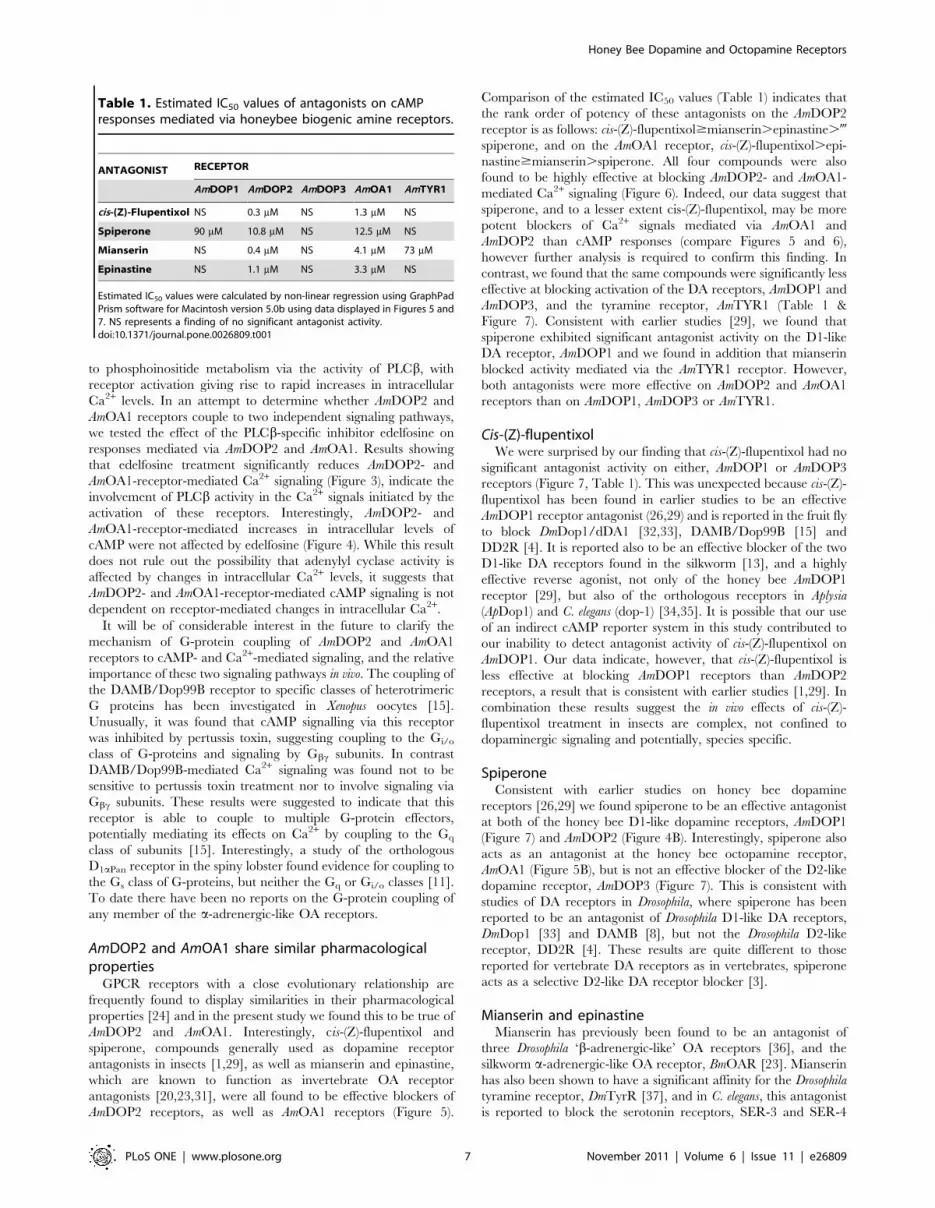
Comparison of the estimated IC50 values (Table 1) indicates that
the rank order of potency of these antagonists on the AmDOP2
receptor is as follows: cis-(Z)-flupentixol$mianserin.epinastine.-
spiperone, and on the AmOA1 receptor, cis-(Z)-flupentixol.epi-
nastine$mianserin.spiperone. All four compounds were also
found to be highly effective at blocking AmDOP2- and AmOA1-
mediated Ca2+ signaling (Figure 6). Indeed, our data suggest that
spiperone, and to a lesser extent cis-(Z)-flupentixol, may be more
potent blockers of Ca2+ signals mediated via AmOA1 and
AmDOP2 than cAMP responses (compare Figures 5 and 6),
however further analysis is required to confirm this finding. In
contrast, we found that the same compounds were significantly less
effective at blocking activation of the DA receptors, AmDOP1 and
AmDOP3, and the tyramine receptor, AmTYR1 (Table 1 &
Figure 7). Consistent with earlier studies [29], we found that
spiperone exhibited significant antagonist activity on the D1-like
DA receptor, AmDOP1 and we found in addition that mianserin
blocked activity mediated via the AmTYR1 receptor. However,
both antagonists were more effective on AmDOP2 and AmOA1
receptors than on AmDOP1, AmDOP3 or AmTYR1.
Cis-(Z)-flupentixolWe were surprised by our finding that cis-(Z)-flupentixol had no
significant antagonist activity on either, AmDOP1 or AmDOP3
receptors (Figure 7, Table 1). This was unexpected because cis-(Z)-
flupentixol has been found in earlier studies to be an effective
AmDOP1 receptor antagonist (26,29) and is reported in the fruit fly
to block DmDop1/dDA1 [32,33], DAMB/Dop99B [15] and
DD2R [4]. It is reported also to be an effective blocker of the two
D1-like DA receptors found in the silkworm [13], and a highly
effective reverse agonist, not only of the honey bee AmDOP1
receptor [29], but also of the orthologous receptors in Aplysia
(ApDop1) and C. elegans (dop-1) [34,35]. It is possible that our use
of an indirect cAMP reporter system in this study contributed to
our inability to detect antagonist activity of cis-(Z)-flupentixol on
AmDOP1. Our data indicate, however, that cis-(Z)-flupentixol is
less effective at blocking AmDOP1 receptors than AmDOP2
receptors, a result that is consistent with earlier studies [1,29]. In
combination these results suggest the in vivo effects of cis-(Z)-
flupentixol treatment in insects are complex, not confined to
dopaminergic signaling and potentially, species specific.
SpiperoneConsistent with earlier studies on honey bee dopamine
receptors [26,29] we found spiperone to be an effective antagonist
at both of the honey bee D1-like dopamine receptors, AmDOP1
(Figure 7) and AmDOP2 (Figure 4B). Interestingly, spiperone also
acts as an antagonist at the honey bee octopamine receptor,
AmOA1 (Figure 5B), but is not an effective blocker of the D2-like
dopamine receptor, AmDOP3 (Figure 7). This is consistent with
studies of DA receptors in Drosophila, where spiperone has been
reported to be an antagonist of Drosophila D1-like DA receptors,
DmDop1 [33] and DAMB [8], but not the Drosophila D2-like
receptor, DD2R [4]. These results are quite different to those
reported for vertebrate DA receptors as in vertebrates, spiperone
acts as a selective D2-like DA receptor blocker [3].
Mianserin and epinastineMianserin has previously been found to be an antagonist of
three Drosophila ‘b-adrenergic-like’ OA receptors [36], and the
silkworm a-adrenergic-like OA receptor, BmOAR [23]. Mianserin
has also been shown to have a significant affinity for the Drosophila
tyramine receptor, DmTyrR [37], and in C. elegans, this antagonist
is reported to block the serotonin receptors, SER-3 and SER-4
Table 1. Estimated IC50 values of antagonists on cAMPresponses mediated via honeybee biogenic amine receptors.
ANTAGONIST RECEPTOR
AmDOP1 AmDOP2 AmDOP3 AmOA1 AmTYR1
cis-(Z)-Flupentixol NS 0.3 mM NS 1.3 mM NS
Spiperone 90 mM 10.8 mM NS 12.5 mM NS
Mianserin NS 0.4 mM NS 4.1 mM 73 mM
Epinastine NS 1.1 mM NS 3.3 mM NS
Estimated IC50 values were calculated by non-linear regression using GraphPadPrism software for Macintosh version 5.0b using data displayed in Figures 5 and7. NS represents a finding of no significant antagonist activity.doi:10.1371/journal.pone.0026809.t001
Honey Bee Dopamine and Octopamine Receptors
PLoS ONE | www.plosone.org 7 November 2011 | Volume 6 | Issue 11 | e26809

[38]. In this study we found that mianserin blocked responses
mediated via AmOA1, AmDOP2 and AmTYR1 receptors, and that
epinastine was an effective antagonist not only of the honey bee
OA receptor, AmOA1, but also the DA receptor, AmDOP2.
Taken together, these results suggest that in vivo effects of treatment
with either mianserin or epinastine are unlikely to be confined to
octopaminergic targets, as has previously been suggested [31,39].
Nonetheless, epinastine may prove useful in future studies for
differentiating between responses mediated via AmDOP1 and
AmDOP2, as AmDOP2 receptors are blocked very effectively by
this antagonist, whereas AmDOP1 receptors are not. AmDOP2 and
AmOA1 share a hydrophilic residue prior to TMV (E201 and
N243 respectively, yellow alignment, Figure 8). In the correspond-
ing position (L188) AmDOP1 has a leucine residue. This difference
Figure 8. Sequence alignment of AmDOP1, AmDOP2, AmOA1, AmTYR1, human H1 histamine receptor (NP_001091683.1), humanb-adrenergic receptor (NP_000015.1). Residues highlighted in gray represent the transmembrane helices from the structure of the human b-adrenergic receptor (pdb2rh1), residues highlighted in cyan are those conserved in the GPCR family [43], the red aspartic acid is the highly conservedD107 (hHis numbering: Asp113 - HmB-Adr) on helix 3 (TMIII; residues not shown for I3).doi:10.1371/journal.pone.0026809.g008
Honey Bee Dopamine and Octopamine Receptors
PLoS ONE | www.plosone.org 8 November 2011 | Volume 6 | Issue 11 | e26809

may help to explain the activity of epinastine at AmDOP2 and
AmOA1 but not AmDOP1 as in human H1 receptors, the
corresponding position (D186) has been shown to be important
for histamine binding [40]. Interestingly however, responses
mediated via AmTYR1, which possesses a hydrophilic but
positively charged amino acid at the position (R192), were not
affected by epinastine.
Amdop2 and Amoa1: immediate paralogs?Our results show that of the five receptors examined, AmDOP2
and AmOA1 are the most similar in terms of their functional
properties and pharmacological profile. This evidence suggests to
us that the evolutionary relationship between these two receptor
types is most likely to be that of immediate paralogs, and that
despite divergence in their native ligand specificities, invertebrate-
type DA receptors and a-adrenergic-like OA receptors still display
significant conservation in their functional properties. The results
of this study highlight the need to identify antagonists that act
selectively on specific invertebrate receptor types. The identifica-
tion of such compounds would greatly assist studies exploring the
in vivo function(s) of biogenic amine receptors in invertebrates.
Materials and Methods
MaterialsDopamine hydrochloride, DL-octopamine hydrochloride, cis-
(Z)-flupenthixol dihydrochloride, spiperone, epinastine hydrochlo-
ride and mianserin hydrochloride were obtained from Sigma-
Aldrich. Edelfosine (2-O-methyl-PAF C-18) was obtained from
Cayman Chemical Company, Ann Arbor, U.S.A.
Heterologous expression of the honey bee receptorproteins
HEK293 cells (Invitrogen) were maintained as adherent
cultures at 37uC, 5% CO2 in phenol-red free DMEM/F12
medium containing 10% fetal calf serum (Invitrogen). For
expression of the receptor proteins in HEK293 cells, expression
plasmid constructs were transiently transfected into the cells using
FuGene-HD reagent (Roche) in accordance with the manufactur-
er’s instructions. Control cells transfected with pIB/V5-His-GW/
LacZ for expression of the beta-galactosidase reporter protein
indicated that transfection efficiency was .95%.
The creation of plasmid expression constructs for AmDOP1,
AmDOP2, AmDOP3 and AmOA1 has been described in detail
elsewhere [29]. The expression construct for the AmTYR1
receptor was created by PCR amplification of the coding
sequences of the Amtyr1 cDNA [27] and insertion into the HindIII
and XbaI site of pcDNA3.1(+) vector using the following primer
sequences; forward – GCACGAAGCTTGCCACCAT-
GAACTCGAGCGGGGAATCAG; reverse – GACTTCTA-
GATCAACGAATGCGCAACAACCGTCT.
Measurement of [Ca2+]i levels for assaying receptorfunction
Exponentially growing HEK293 cells (Invitrogen) were dis-
pensed at a density of 26104 cells per well in 96-well, black-walled,
clear-bottomed tissue culture plates (Greiner Bio-One) and
allowed to grow for 24 hours. The cells were transfected with
the desired honey bee receptor expression construct. Transfected
cells were maintained for a further 24 hours at 37uC prior to
assaying for receptor function. Intracellular Ca2+ levels were
assayed by preloading the cells with Fluo-4 NW reporter dye
dissolved in Hank’s buffer in accordance with the manufacturer’s
instructions (Invitrogen). The fluorescence signal (excitation
480 nm, emission 520 nM) of individual wells was detected using
a BMG Labtech Fluostar Omega microplate instrument. Amines
were prepared immediately prior to use in Hanks buffer, and a
2 ul volume was introduced into test wells using onboard injectors.
Treatment concentrations indicated in figure legends represent the
final concentration of amine in the test well. Wells were monitored
for fluorescent signal immediately prior to treatment to establish
the baseline fluorescence (Fb) and for 50 seconds post agonist
injection to record changes in fluorescence (DF) at 0.24 second
intervals. Amines added to test wells remained in the medium
throughout the post-injection recording period.
Indirect measurement of intracellular cAMP levels forassaying receptor function
Receptor coupling to intracellular cAMP signaling was assessed
using a CRE-luciferase reporter as detailed previously [29]. In
brief, HEK293 cells were co-transfected with the desired honey
bee receptor expression construct together with the pGL4.29
[luc2P/CRE/Hygro] reporter construct (Promega) and grown for
a further 24 hours. The amount of AmDOP1 expression construct
used for transfections was reduced to 1/10th of the concentration
used in earlier studies [29], because high-level expression of this
constitutively active receptor [28] was found to swamp the
capacity of the reporter system [41]. Cells were then incubated
for 3 hours in serum-free growth medium containing the test
treatments detailed in figure legends, and then immediately
assayed for luciferase enzyme activity. Duplicate measurements
were determined for each test treatment examined in the
performance of independent trials. All in vitro expression work
was conducted under approvals issued by the University of Otago
Institutional Biological Safety Committee.
Sequence alignment and homology modellingThe sequence alignment was initially carried out using T-coffee
and then manually adjusted in a similar fashion to that described
elsewhere [42]. Models were generated using Modeller9v7 [43]
using the human beta-adrenergic structure (pdb2rh1) as a
template. The models with the lowest objective function were
selected for further analysis. Docking experiments were carried out
using Gold 4.1 [44] to dock the epinastine into the binding site of
the human histamine receptor. Both isomers of epinastine were
used in the docking calculations and were downloaded from the
Cambridge Crystallographic Data Centre (ID: CALQUC: R,
CALRAJ; S).
Author Contributions
Conceived and designed the experiments: KTB ARM. Performed the
experiments: KTB. Analyzed the data: KTB JT. Contributed reagents/
materials/analysis tools: JT. Wrote the paper: KTB ARM.
References
1. Mustard JA, Beggs KT, Mercer AR (2005) Molecular biology of the invertebrate
dopamine receptors. Arch Insect Biochem Physiol 59: 103–117.
2. Mcdonald P, Jessen T, Field J, Blakely R (2006) Dopamine signaling architecture
in Caenorhabditis elegans. Cell Mol Neurobiol 26: 593–618.
3. Missale C, Nash SR, Robinson SW, Jaber M, Caron MG (1998) Dopamine
receptors: from structure to function. Physiol Rev 78: 189–225.
4. Hearn MG, Ren Y, McBride EW, Reveillaud I, Beinborn M, et al. (2002) A
Drosophila dopamine 2-like receptor: Molecular characterization and identifica-
Honey Bee Dopamine and Octopamine Receptors
PLoS ONE | www.plosone.org 9 November 2011 | Volume 6 | Issue 11 | e26809

tion of multiple alternatively spliced variants. Proc Natl Acad Sci USA 99:
14554–14559.5. Beggs KT, Hamilton IS, Kurshan PT, Mustard JA, Mercer AR (2005)
Characterization of a D2-like dopamine receptor (AmDOP3) in honey bee, Apis
mellifera. Insect Biochem Mol Biol 35: 873–882.6. Clark MC, Baro DJ (2007) Arthropod D2 receptors positively couple with cAMP
through the Gi/o protein family. Comp Biochem Physiol B Biochem Mol Biol146: 9–19.
7. Humphries MA, Mustard JA, Hunter SJ, Mercer A, Ward V, et al. (2003)
Invertebrate D2 type dopamine receptor exhibits age-based plasticity ofexpression in the mushroom bodies of the honeybee brain. J Neurobiol 55:
315–330.8. Han KA, Millar NS, Grotewiel MS, Davis RL (1996) DAMB, a novel dopamine
receptor expressed specifically in Drosophila mushroom bodies. Neuron 16:1127–1135.
9. Feng G, Hannan F, Reale V, Hon YY, Kousky CT, et al. (1996) Cloning and
functional characterization of a novel dopamine receptor from Drosophila
melanogaster. J Neurosci 16: 3925–3933.
10. Ono H, Yoshikawa H (2004) Identification of amine receptors from a swallowtailbutterfly, Papilio xuthus L.: cloning and mRNA localization in foreleg
chemosensory organ for recognition of host plants. Insect Biochem Mol Biol
34: 1247–1256.11. Clark MC, Baro DJ (2006) Molecular cloning and characterization of crustacean
type-one dopamine receptors: D1alphaPan and D1betaPan. Comp BiochemPhysiol B, Biochem Mol Biol 143: 294–301.
12. Gerber S, Krasky A, Rohwer A, Lindauer S, Closs E, et al. (2006) Identificationand characterisation of the dopamine receptor II from the cat flea Ctenocephalides
felis (CfDopRII). Insect Biochem Mol Biol 36: 749–758.
13. Ohta H, Tsuchihara K, Mitsumasu K, Yaginuma T (2009) Comparativepharmacology of two D1-like dopamine receptors cloned from the silkworm
Bombyx mori. Insect Biochem Mol Biol 39: 342–347.14. Han KA, Millar NS, Davis RL (1998) A novel octopamine receptor with
preferential expression in Drosophila mushroom bodies. J Neurosci 18:
3650–3658.15. Reale V, Hannan F, Hall LM, Evans PD (1997) Agonist-specific coupling of a
cloned Drosophila melanogaster D1-like dopamine receptor to multiple secondmessenger pathways by synthetic agonists. J Neurosci 17: 6545–6553.
16. Balfanz S, Strunker T, Frings S, Baumann A (2005) A family of octopaminereceptors that specifically induce cyclic AMP production or Ca2+ release in
Drosophila melanogaster. J Neurochem 93: 440–451.
17. Hauser F, Cazzamali G, Williamson M, Blenau W, Grimmelikhuijzen CJ (2006)A review of neurohormone GPCRs present in the fruitfly Drosophila melanogaster
and the honey bee Apis mellifera. Prog Neurobiol 80: 1–19.18. Hauser F, Cazzamali G, Williamson M, Park Y, Li B (2007) A genome-wide
inventory of neurohormone GPCRs in the red flour beetle Tribolium castaneum.
Front Neuroendocrinol 29: 142–165.19. Evans PD, Maqueira B (2005) Insect octopamine receptors: a new classification
scheme based on studies of cloned Drosophila G-protein coupled receptors. InvertNeurosci 5: 111–118.
20. Gerhardt CC, Bakker RA, Piek GJ, Planta RJ, Vreugdenhil E, et al. (1997)Molecular cloning and pharmacological characterization of a molluscan
octopamine receptor. Mol Pharmacol 51: 293–300.
21. Grohmann L, Blenau W, Erber J, Ebert PR, Strunker T, et al. (2003) Molecularand functional characterization of an octopamine receptor from honeybee (Apis
mellifera) brain. J Neurochem 86: 725–735.22. Bischof LJ, Enan EE (2004) Cloning, expression and functional analysis of an
octopamine receptor from Periplaneta americana. Insect Biochem Mol Biol 34:
511–521.
23. Ohtani A, Arai Y, Ozoe F, Ohta H, Narusuye K, et al. (2006) Molecular cloning
and heterologous expression of an alpha-adrenergic-like octopamine receptorfrom the silkworm Bombyx mori. Insect Mol Biol 15: 763–772.
24. Vernier P, Cardinaud B, Valdenaire O, Philippe H, Vincent JD (1995) An
evolutionary view of drug-receptor interaction: the bioamine receptor family.Trends Pharmacol Sci 16: 375–381.
25. Powis G, Seewald MJ, Gratas C, Melder D, Riebow J, et al. (1992) Selectiveinhibition of phosphatidylinositol phospholipase C by cytotoxic ether lipid
analogues. Cancer Research 52: 2835–2840.
26. Blenau W, Erber J, Baumann A (1998) Characterization of a dopamine D1receptor from Apis mellifera: cloning, functional expression, pharmacology, and
mRNA localization in the brain. J Neurochem 70: 15–23.27. Blenau W, Balfanz S, Baumann A (2000) Amtyr1: characterization of a gene
from honeybee (Apis mellifera) brain encoding a functional tyramine receptor.J Neurochem 74: 900–908.
28. Baldauf S (2003) Phylogeny for the faint of heart: a tutorial. Trends Genet 19:
345–351.29. Mustard JA, Blenau W, Hamilton IS, Ward VK, Ebert PR, et al. (2003) Analysis
of two D1-like dopamine receptors from the honey bee Apis mellifera revealsagonist-independent activity. Mol Brain Res 113: 67–77.
30. Beggs KT, Mercer AR (2009) Dopamine receptor activation by honey bee
queen pheromone. Curr Biol 19: 1206–1209.31. Roeder T, Degen J, Gewecke M (1998) Epinastine, a highly specific antagonist
of insect neuronal octopamine receptors. Eur J Pharmacol 349: 171–177.32. Gotzes F, Balfanz S, Baumann A (1994) Primary structure and functional
characterization of a Drosophila dopamine receptor with high homology tohuman D1/D5 receptors. Receptors Channels 2: 131–141.
33. Sugamori KS, Demchyshyn LL, McConkey F, Forte MA, Niznik HB (1995) A
primordial dopamine D1-like adenylyl cyclase-linked receptor from Drosophila
melanogaster displaying poor affinity for benzazepines. FEBS Lett 362: 131–138.
34. Barbas D, Zappulla JP, Angers S, Bouvier M, et al. (2006) An Aplysia dopamine-like receptor: molecular and functional characterization. J Neurochem 96:
414–427.
35. Sanyal S, Wintle RF, Kindt KS, Nuttley WM, et al. (2004) Dopamine modulatesthe plasticity of mechanosensory responses in Caenorhabditis elegans. EMBO J 23:
473–482.36. Maqueira B, Chatwin H, Evans PD (2005) Identification and characterization of
a novel family of Drosophila b-adrenergic-like octopamine G-protein coupledreceptors. J Neurochem 94: 547–560.
37. Enan EE (2005) Molecular response of Drosophila melanogaster tyramine receptor
cascade to plant essential oils. Insect Biochem Mol Biol 35: 309–321.38. Petrascheck M, Ye X, Buck LB (2007) An antidepressant that extends lifespan in
adult Caenorhabditis elegans. Nature 450: 553–556.39. Degen J, Gewecke M, Roeder T (2000) Octopamine receptors in the honey bee
and locust nervous system: pharmacological similarities between homologous
receptors of distantly related species. Br J Pharmacol 130: 587–594.40. Gantz I, DelValle J, Wang L, Tashiro T, Munzert G, et al. (1992) Molecular
basis for the interaction of histamine with the histamine H2 receptor. J BiolChem 267: 20840–20843.
41. Hill SJ, Baker JG, Rees S (2001) Reporter-gene systems for the study of G-protein-coupled receptors. Curr Opinion Pharmacol 1: 526–532.
42. Bissantz C, Bernard P, Hibert M, Rognan D (2003) Protein-based virtual
screening of chemical databases. II. Are homology models of G-Protein CoupledReceptors suitable targets? Proteins 50: 5–25.
43. Fiser A, Sali A In: Methods in Enzymology: Macromolecular CrystallographyPart D. Carter CWJ, Sweet RM, eds. Vol. 374. 461 p.
44. Jones G, Willett P, Glen RC, Leach AR, Taylor RJ (1997) Development and
validation of a genetic algorithm for flexible docking. Mol Biol 267: 727–748.
Honey Bee Dopamine and Octopamine Receptors
PLoS ONE | www.plosone.org 10 November 2011 | Volume 6 | Issue 11 | e26809