intracellular compartments and protein sorting
TRANSCRIPT
Intracellular Compartments and Protein Sorting
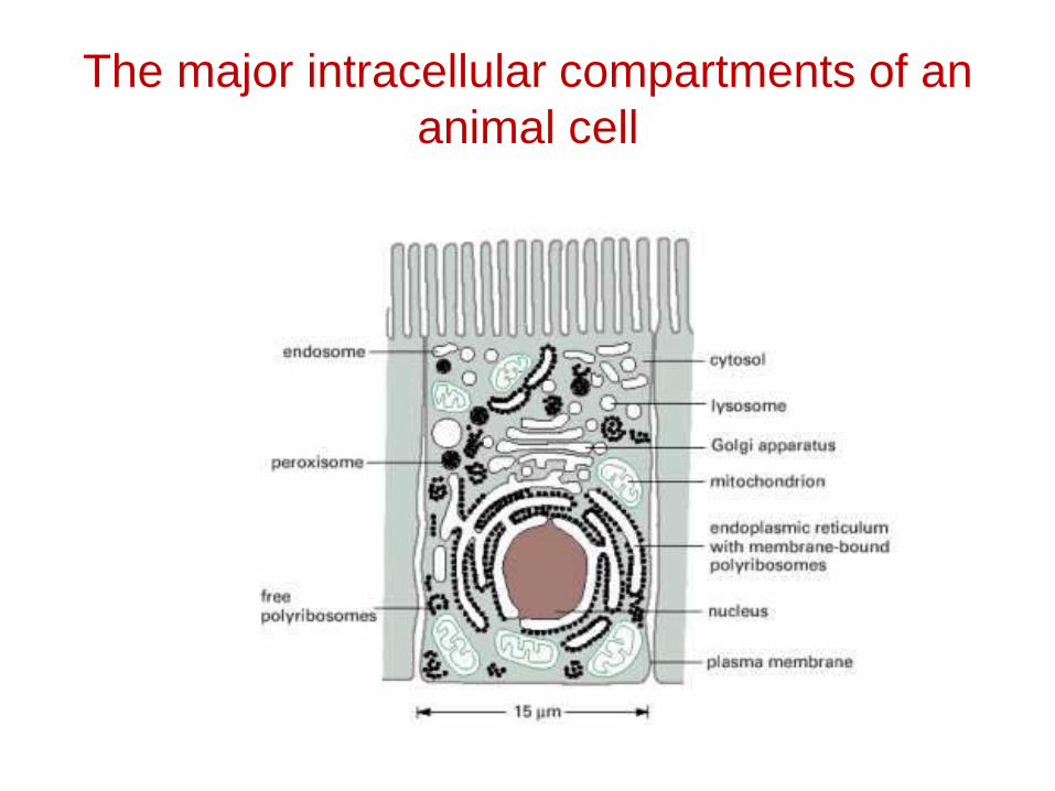
The major intracellular compartments of an animal cell
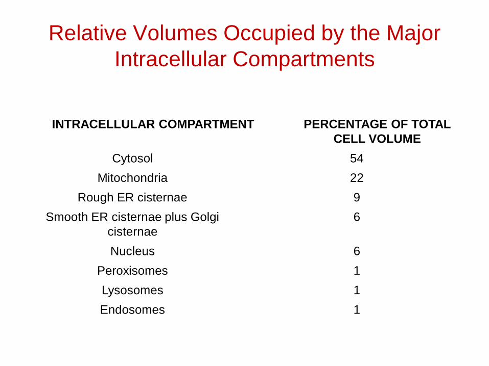
Relative Volumes Occupied by the Major Intracellular Compartments
INTRACELLULAR COMPARTMENT PERCENTAGE OF TOTAL CELL VOLUME
Cytosol 54
Mitochondria 22
Rough ER cisternae 9
Smooth ER cisternae plus Golgi cisternae
6
Nucleus 6
Peroxisomes 1
Lysosomes 1
Endosomes 1
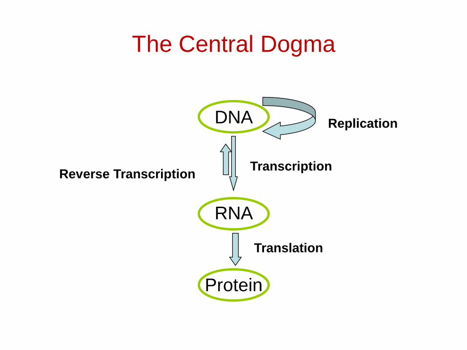
The Central Dogma
DNA
RNA
Protein
Replication
TranscriptionReverse Transcription
Translation
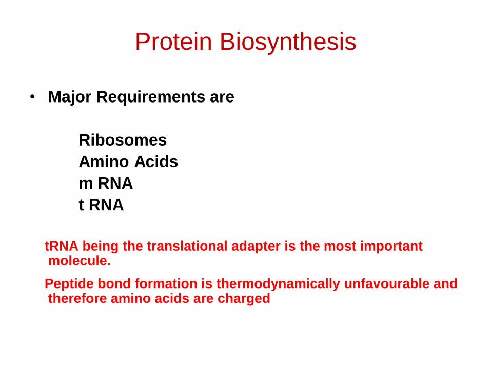
Protein Biosynthesis
• Major Requirements are
RibosomesAmino Acidsm RNAt RNA
tRNA being the translational adapter is the most important molecule.
Peptide bond formation is thermodynamically unfavourable and therefore amino acids are charged
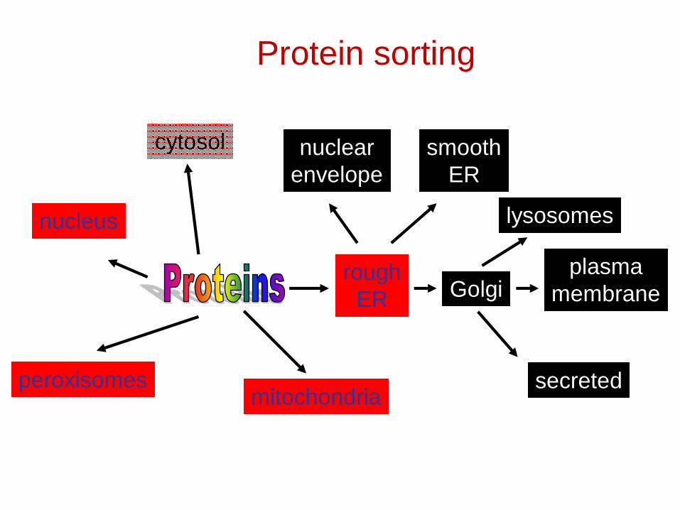
Protein sorting
mitochondria
roughER
peroxisomes
cytosol
nucleus
plasmamembraneGolgi
lysosomes
nuclearenvelope
smoothER
secreted
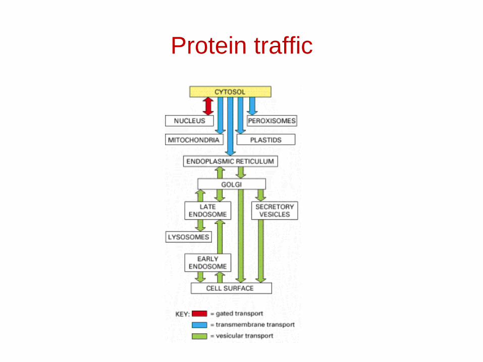
Protein traffic
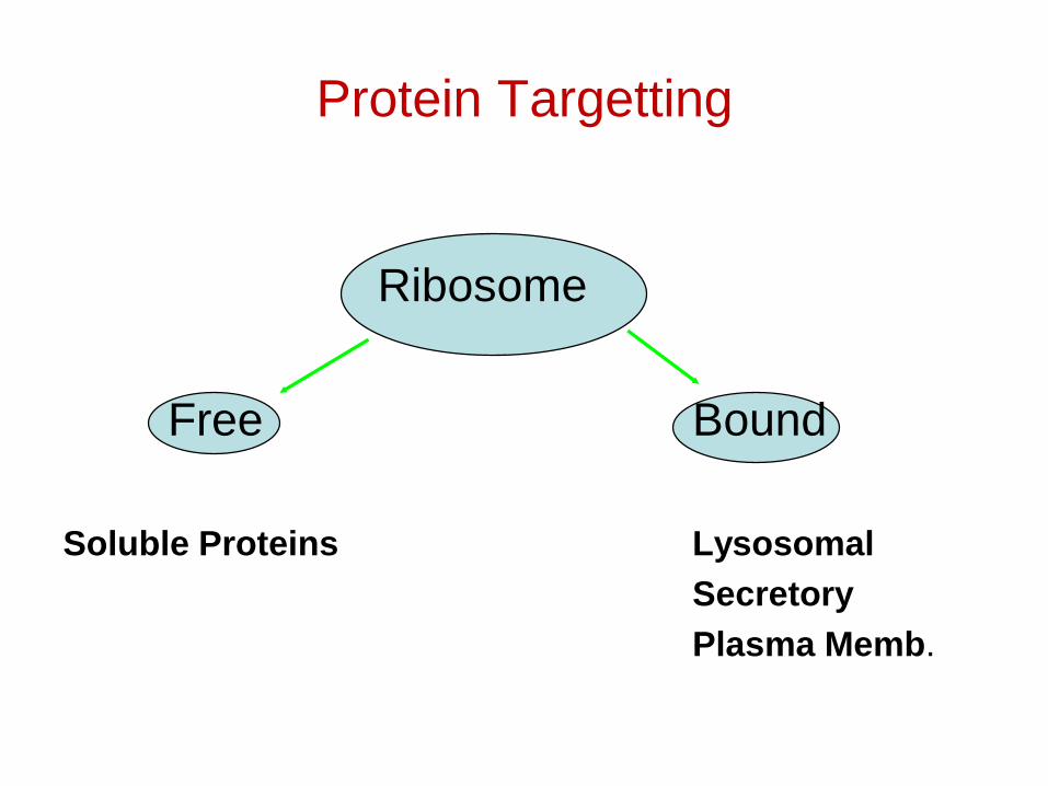
Protein Targetting
Ribosome
Free Bound
Soluble Proteins LysosomalSecretoryPlasma Memb.
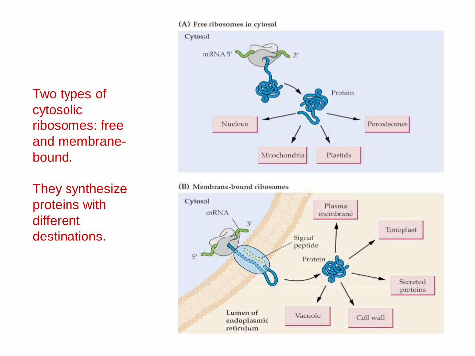
Two types of cytosolicribosomes: free and membrane-bound.
They synthesize proteins with different destinations.
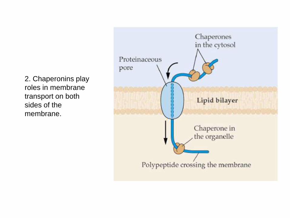
2. Chaperonins play roles in membrane transport on both sides of the membrane.
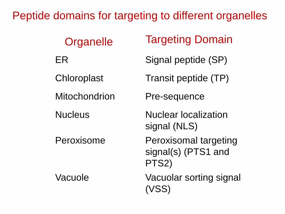
ER Signal peptide (SP)
Chloroplast Transit peptide (TP)
Mitochondrion Pre-sequence
Nucleus Nuclear localization signal (NLS)
Peroxisome Peroxisomal targeting signal(s) (PTS1 and PTS2)
Vacuole Vacuolar sorting signal (VSS)
Organelle Targeting Domain
Peptide domains for targeting to different organelles
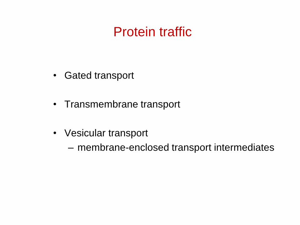
Protein traffic
• Gated transport
• Transmembrane transport
• Vesicular transport– membrane-enclosed transport intermediates
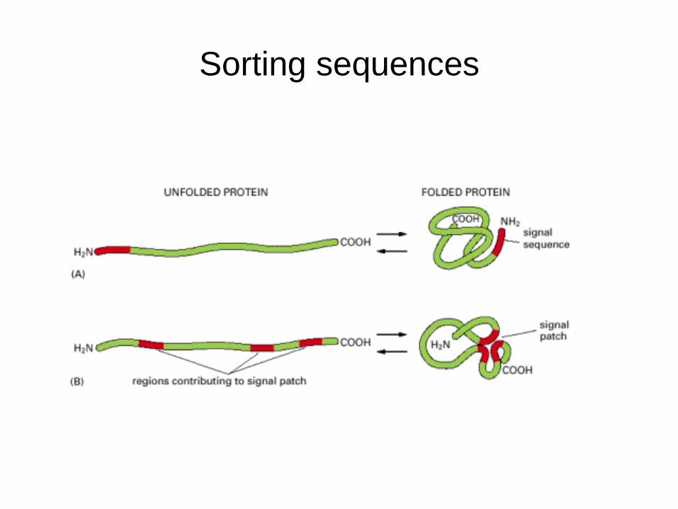
Sorting sequences
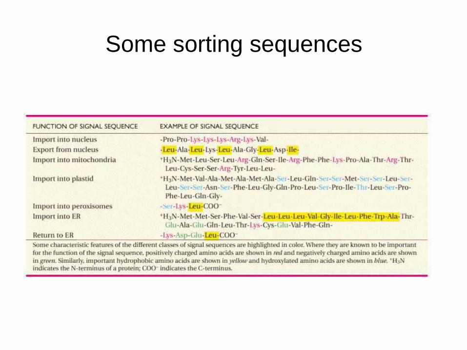
Some sorting sequences

The Signal Sequence
• 13-36 residues long
• The N terminus always contain a positivelycharged amino acid
• The central portion is a stretch of hydrophobicamino acids
• Some proteins have internal signal sequence
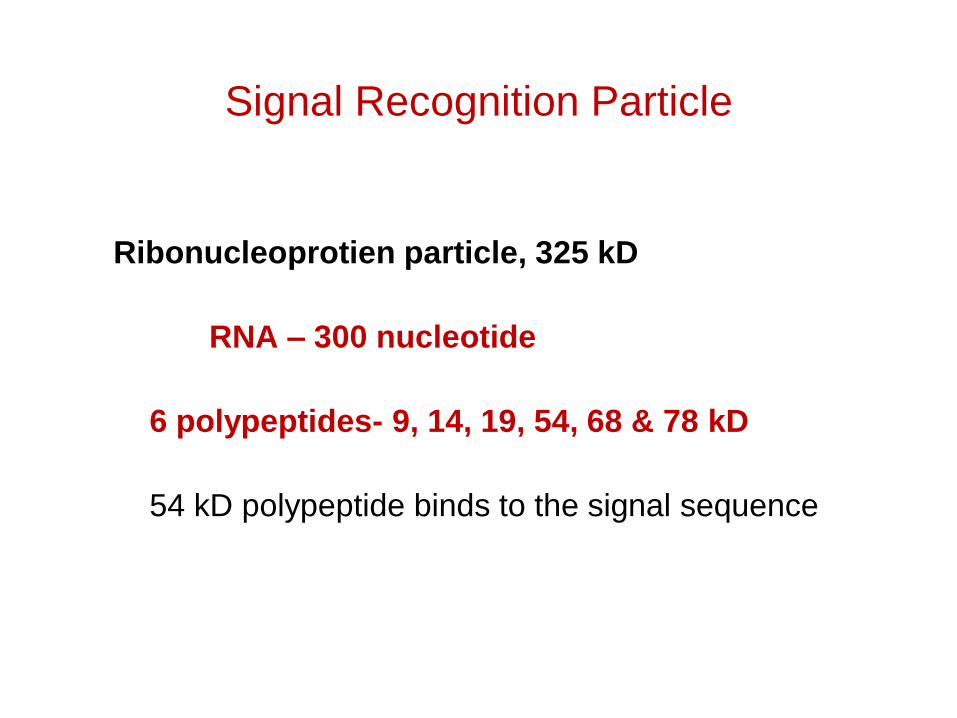
Signal Recognition Particle
Ribonucleoprotien particle, 325 kD
RNA – 300 nucleotide
6 polypeptides- 9, 14, 19, 54, 68 & 78 kD
54 kD polypeptide binds to the signal sequence
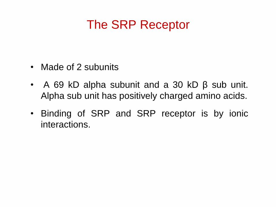
The SRP Receptor
• Made of 2 subunits
• A 69 kD alpha subunit and a 30 kD β sub unit.Alpha sub unit has positively charged amino acids.
• Binding of SRP and SRP receptor is by ionicinteractions.
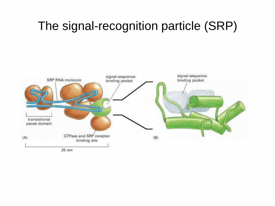
The signal-recognition particle (SRP)
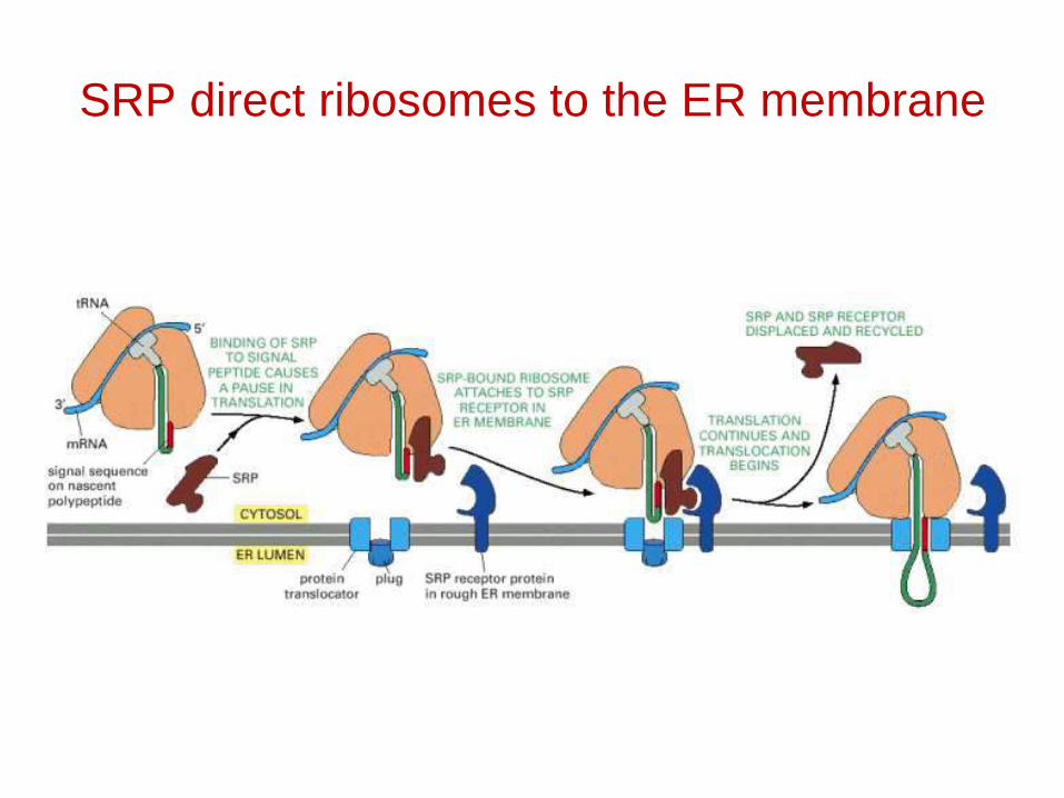
SRP direct ribosomes to the ER membrane
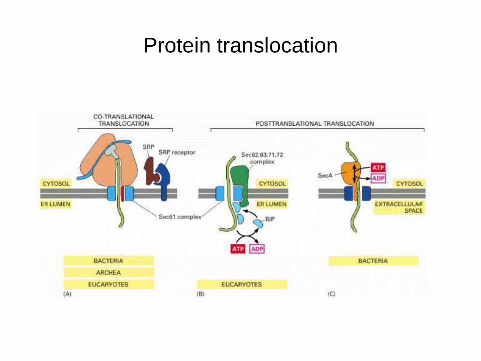
Protein translocation
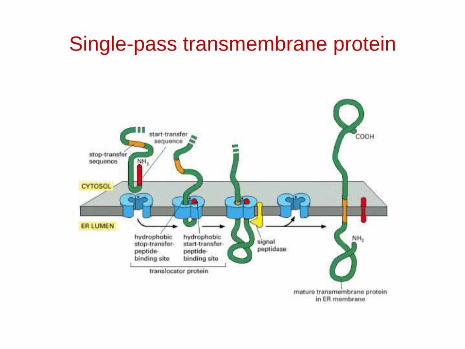
Single-pass transmembrane protein
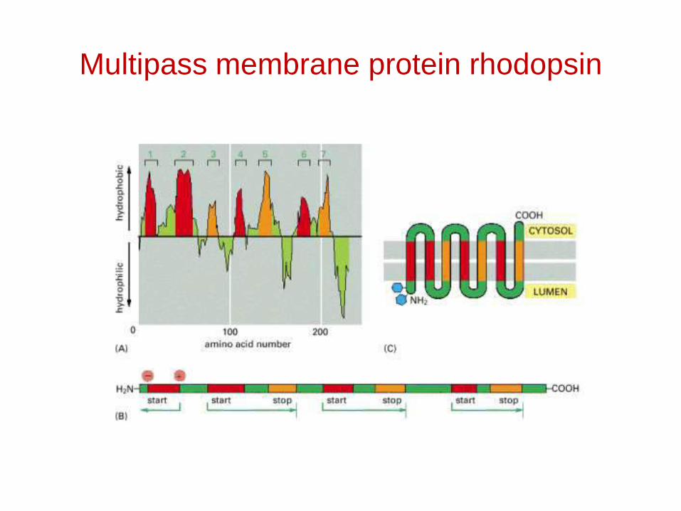
Multipass membrane protein rhodopsin
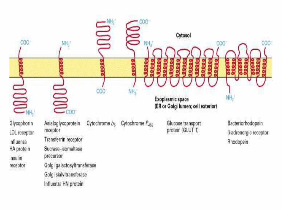

Topogenic sequences direct membrane proteins synthesized on the rough ER to assume their appropriate orientation in the ER membrane. This orientation is retained during transport of a membrane protein to its final destination.
Topogenic sequences include N-terminal cleaved signal sequences; stop-transfer membrane-anchor sequences; internal uncleaved signal-anchor sequences; and GPI-attachment sequences.
Many proteins have several membrane-spanning α helices. Each α-helical segment in such multipass proteins functions as an internal uncleaved signal anchor sequence or a stop-transfer membrane-anchor sequence depending on its location in the polypeptide chain.

The ER is an impressive factory
Lipid synthesis
Secretory protein synthesis
Integral membrane protein synthesis
Protein folding
Post-translational modification
Protein degradation
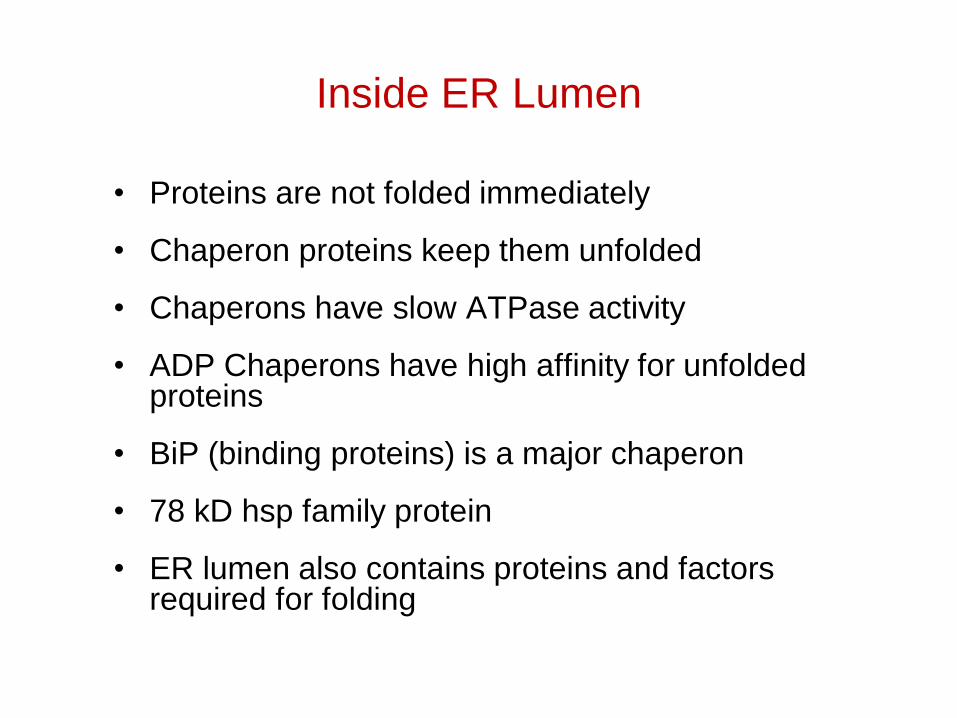
Inside ER Lumen
• Proteins are not folded immediately
• Chaperon proteins keep them unfolded
• Chaperons have slow ATPase activity
• ADP Chaperons have high affinity for unfolded proteins
• BiP (binding proteins) is a major chaperon
• 78 kD hsp family protein
• ER lumen also contains proteins and factors required for folding

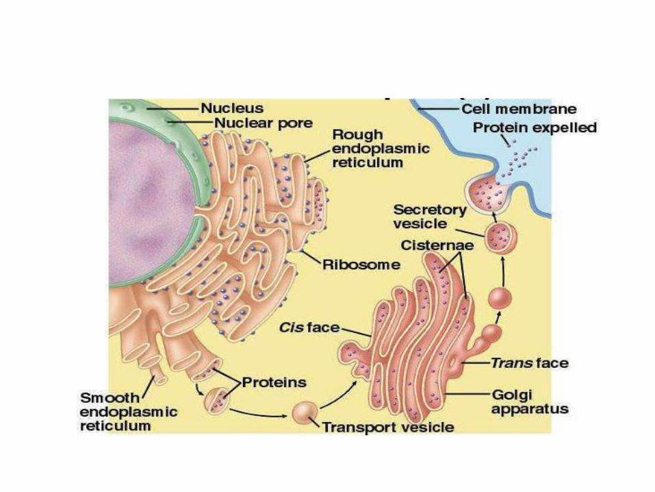
Golgi Apparatus
• Major sorting centre - GPO of cell
Made of 6 cisternaeCis (importing end)Medial Trans (exporting end)
• Transport vesicle mediate transfer b/w ER and golgi
• Small GTP binding proteins, coat proteins etc play a key role in vesicular transport
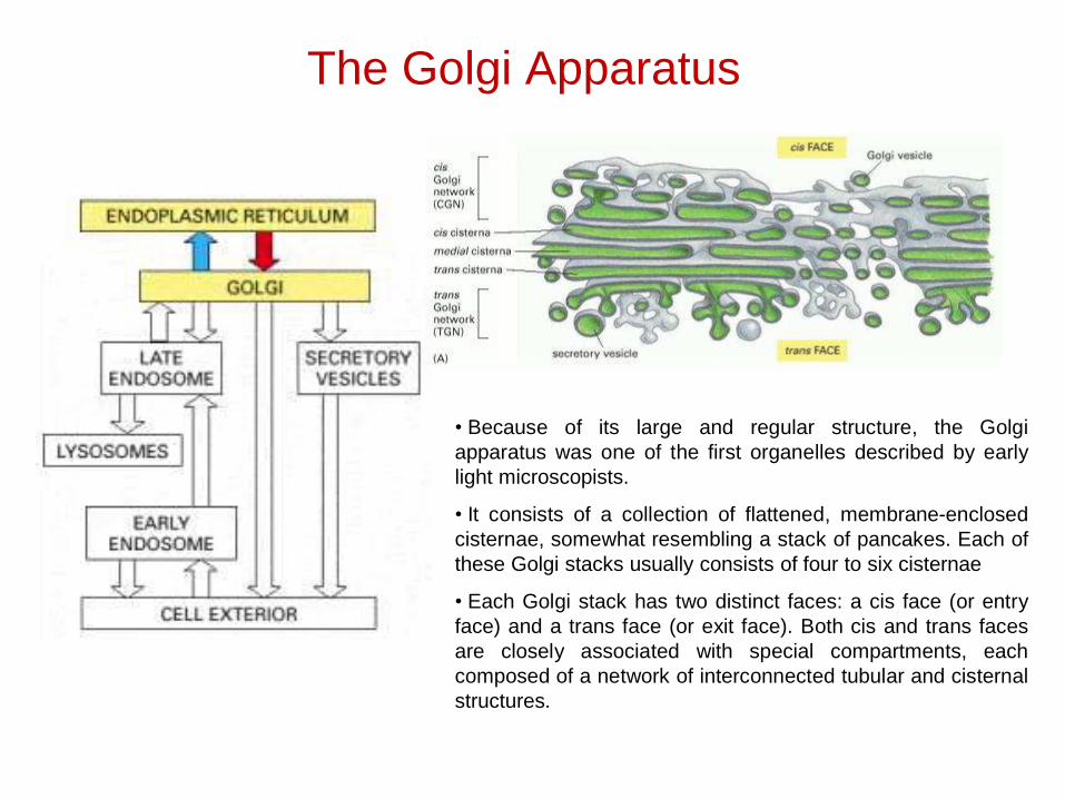
The Golgi Apparatus
• Because of its large and regular structure, the Golgiapparatus was one of the first organelles described by earlylight microscopists.
• It consists of a collection of flattened, membrane-enclosedcisternae, somewhat resembling a stack of pancakes. Each ofthese Golgi stacks usually consists of four to six cisternae
• Each Golgi stack has two distinct faces: a cis face (or entryface) and a trans face (or exit face). Both cis and trans facesare closely associated with special compartments, eachcomposed of a network of interconnected tubular and cisternalstructures.
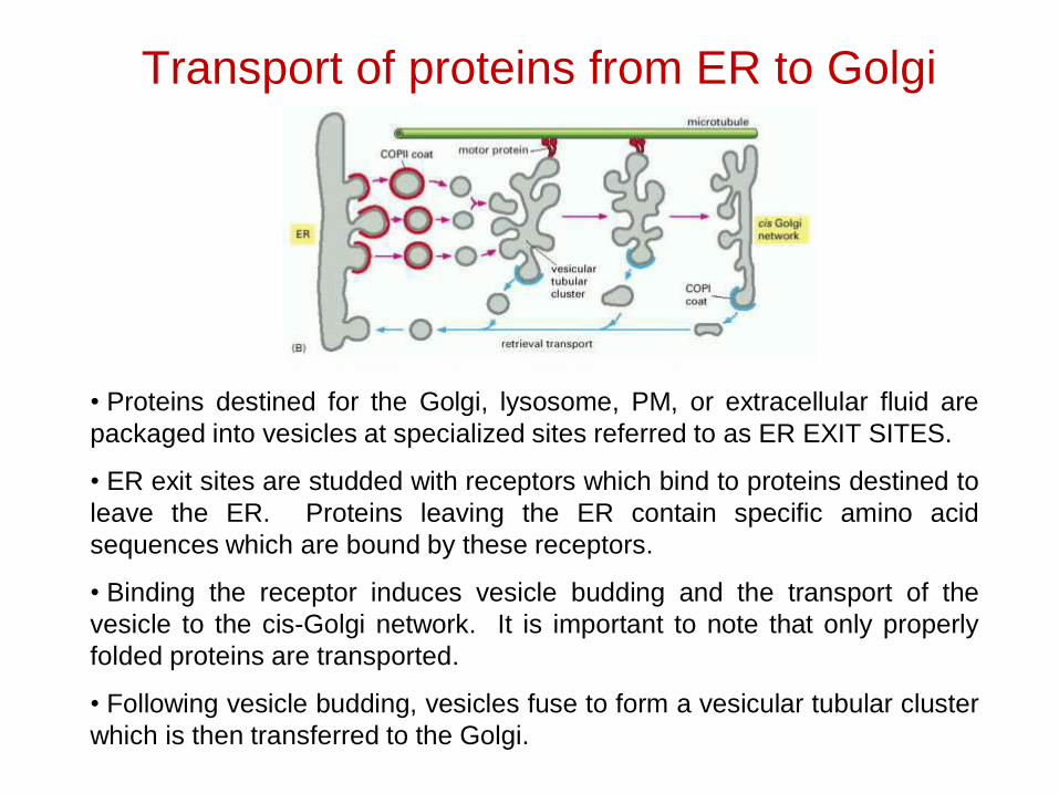
Transport of proteins from ER to Golgi
• Proteins destined for the Golgi, lysosome, PM, or extracellular fluid arepackaged into vesicles at specialized sites referred to as ER EXIT SITES.
• ER exit sites are studded with receptors which bind to proteins destined toleave the ER. Proteins leaving the ER contain specific amino acidsequences which are bound by these receptors.
• Binding the receptor induces vesicle budding and the transport of thevesicle to the cis-Golgi network. It is important to note that only properlyfolded proteins are transported.
• Following vesicle budding, vesicles fuse to form a vesicular tubular clusterwhich is then transferred to the Golgi.
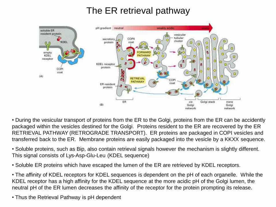
The ER retrieval pathway
• During the vesicular transport of proteins from the ER to the Golgi, proteins from the ER can be accidently packaged within the vesicles destined for the Golgi. Proteins resident to the ER are recovered by the ER RETRIEVAL PATHWAY (RETROGRADE TRANSPORT). ER proteins are packaged in COPI vesicles and transferred back to the ER. Membrane proteins are easily packaged into the vesicle by a KKXX sequence.
• Soluble proteins, such as Bip, also contain retrieval signals however the mechanism is slightly different. This signal consists of Lys-Asp-Glu-Leu (KDEL sequence)
• Soluble ER proteins which have escaped the lumen of the ER are retrieved by KDEL receptors.
• The affinity of KDEL receptors for KDEL sequences is dependent on the pH of each organelle. While the KDEL receptor has a high affinity for the KDEL sequence at the more acidic pH of the Golgi lumen, the neutral pH of the ER lumen decreases the affinity of the receptor for the protein prompting its release.
• Thus the Retrieval Pathway is pH dependent

Golgi are involved in the sorting and post-translational modification of proteins
• During the passage of proteins through the Golgi compartments, various covalent modifications take place in order to provide the specific structure and function to the protein
• Modification of existing glycosyl groups, O-glycosylation, sulfation (addition of sulfates to OH on tyrosine), and phosphorylation all take place within the various Golgi compartments.
• For simplicity, the primary focus of this lecture series will be the modification of proteins by glycosylation
• As mentioned previously, the ER N-glycosylates various proteins with oligosaccharides The Golgi then modifies these oligos providing either a COMPLEX OLIGOSACCHARIDE and HIGH MANNOSE CONTAINING OLIGOSACCHARIDE.
• The high mannose oligo is produced by removing glucose and N-acetylglucosamine moeities while the complex oligo is produced adding addition monosaccharides consecutively.
• Some proteins require additional oligosaccharides to provide a specific function. The Golgi also modifies proteins by O-glycosylation. Serines are used for this type of post-translational modification.
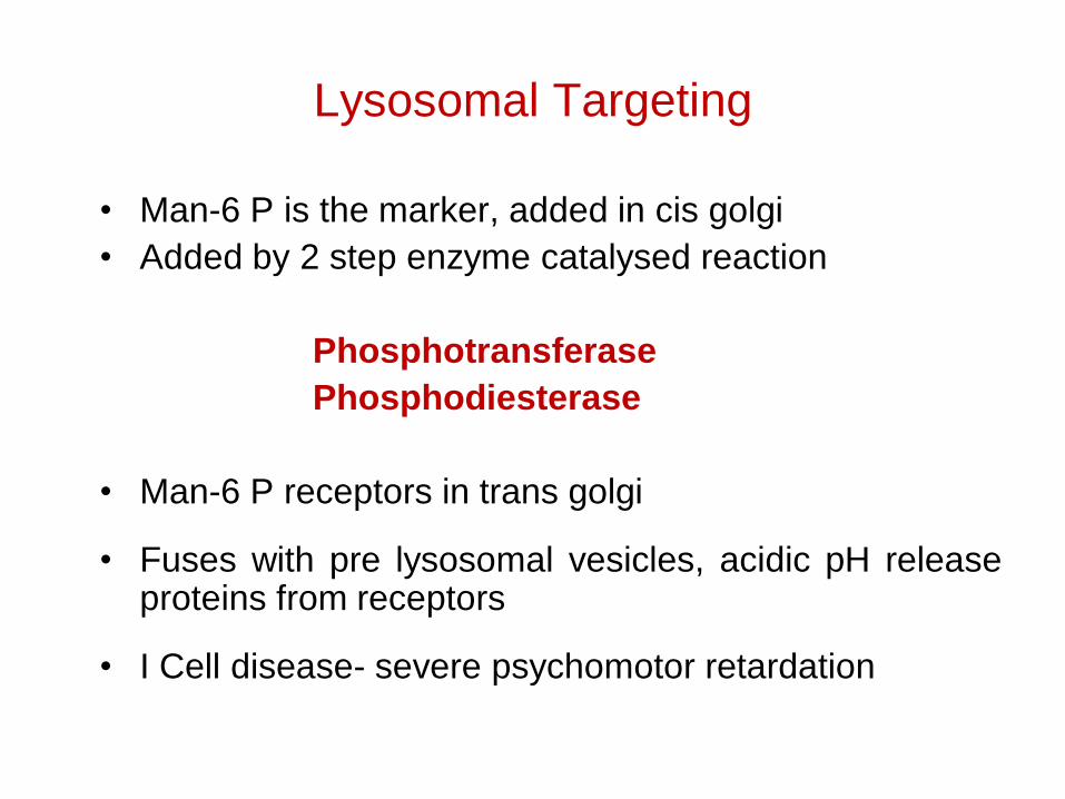
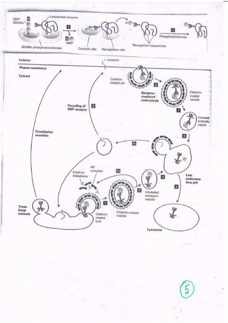
Lysosomal Targeting
• Man-6 P is the marker, added in cis golgi• Added by 2 step enzyme catalysed reaction
PhosphotransferasePhosphodiesterase
• Man-6 P receptors in trans golgi
• Fuses with pre lysosomal vesicles, acidic pH releaseproteins from receptors
• I Cell disease- severe psychomotor retardation

Nuclear pore complexes
Nuclear lamina
• Consists of "intermediate filaments", 30-100 nm thick.
• These intermediate filaments are polymers of lamin, ranging from 60-75 kD.
• A-type lamins are inside, next to nucleoplasm; B-type laminsare near the nuclear membrane (inner). They may bind to integral proteins inside that membrane.
• The lamins may be involved in the functional organization of the nucleus.

Nuclear localization signals (NLSs)
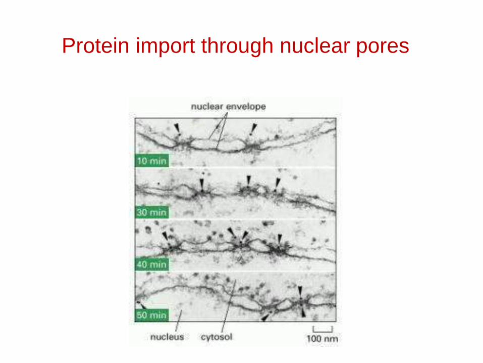
Protein import through nuclear pores
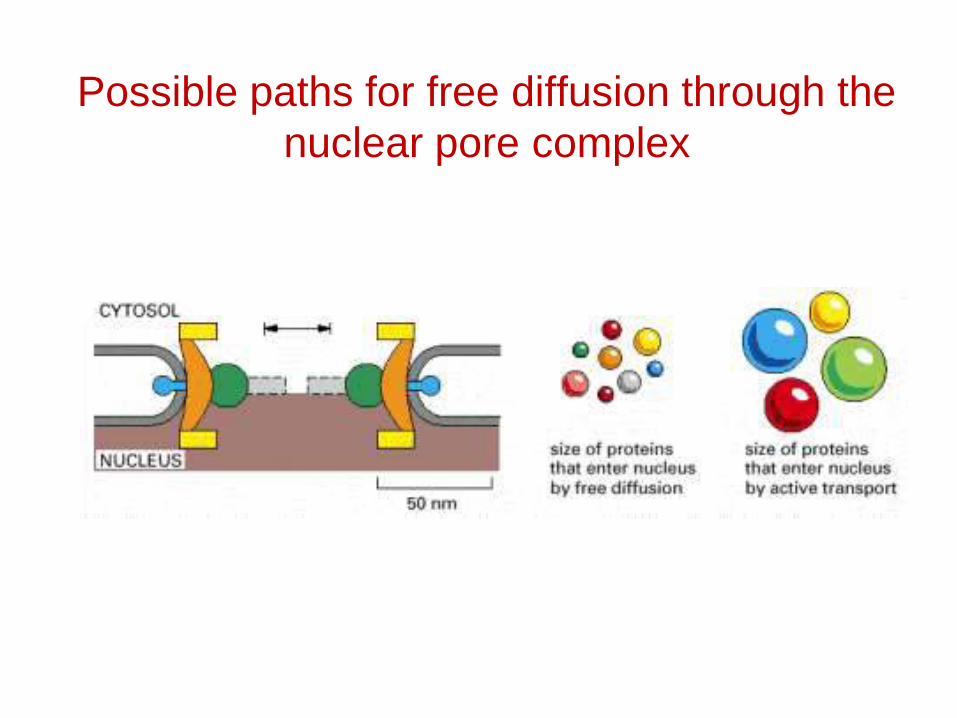
Possible paths for free diffusion through the nuclear pore complex
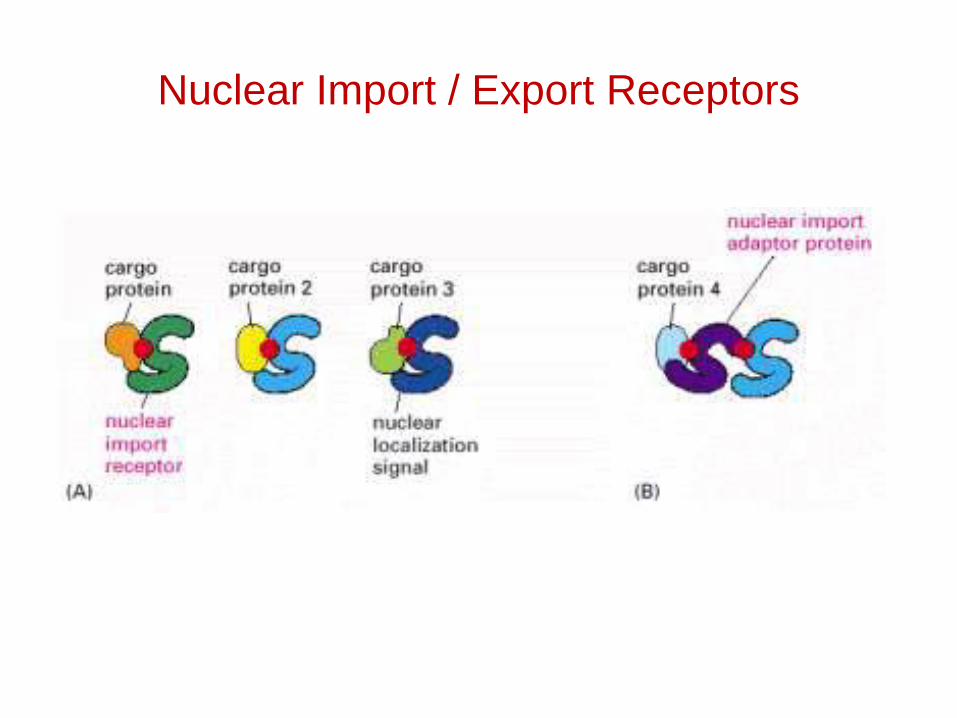
Nuclear Import / Export Receptors
Targeting and assembly of proteins destined for chloroplasts and mitochondria
• How are proteins targeted to chloroplasts and mitochondria from the cytoplasm?
• How do they get through the membranes?
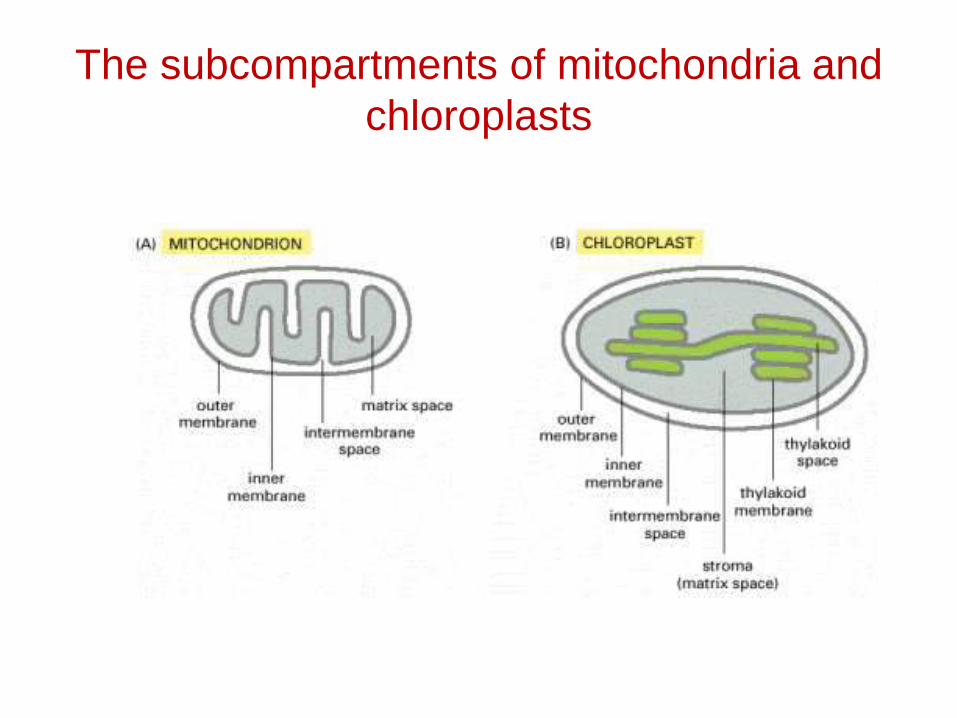
The subcompartments of mitochondria and chloroplasts

A signal sequence for mitochondrial protein import
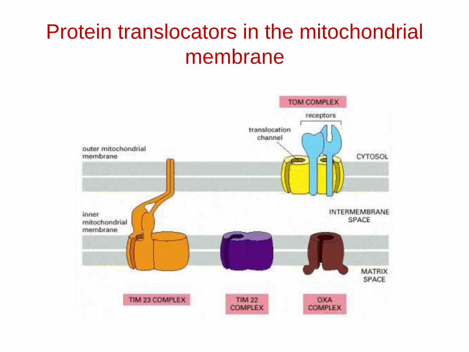
Protein translocators in the mitochondrial membrane

Protein import by mitochondria
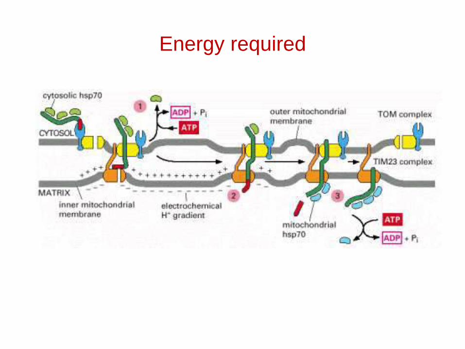
Energy required
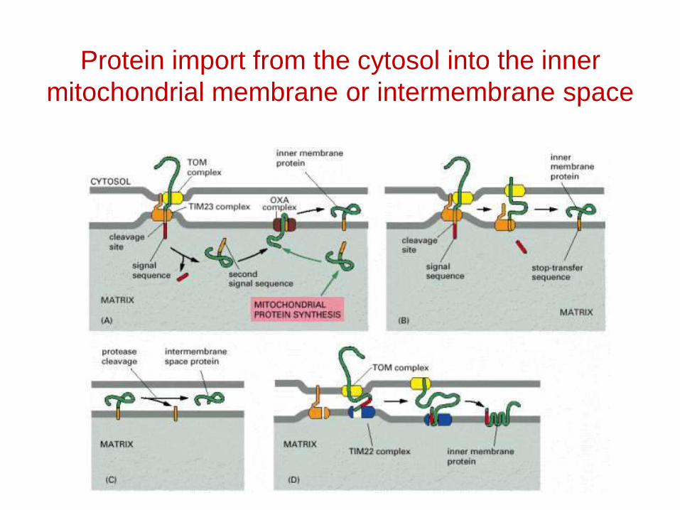
Protein import from the cytosol into the inner mitochondrial membrane or intermembrane space
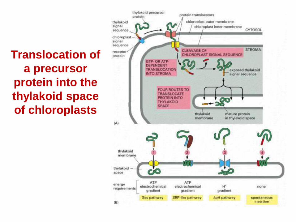
Translocation of a precursor
protein into the thylakoid space of chloroplasts
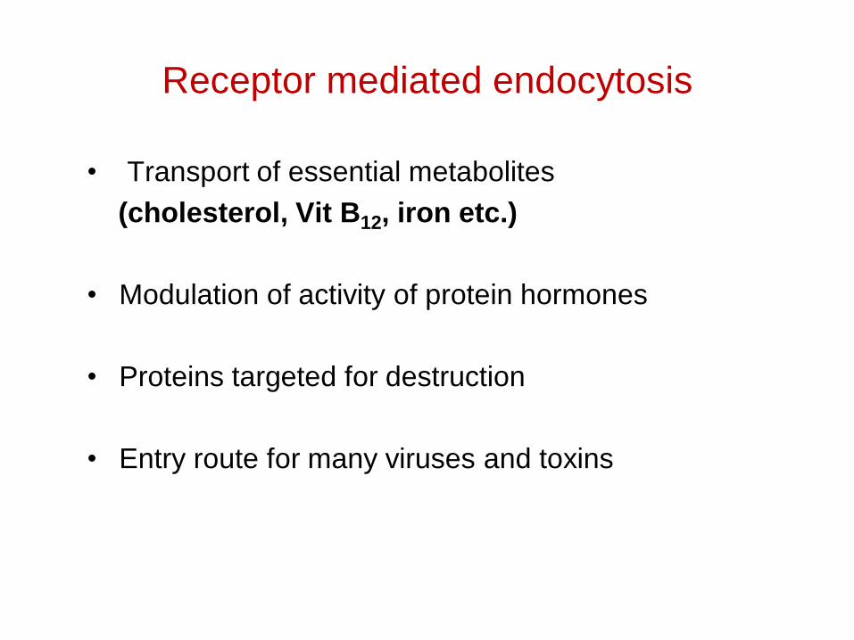
Receptor mediated endocytosis
• Transport of essential metabolites (cholesterol, Vit B12, iron etc.)
• Modulation of activity of protein hormones
• Proteins targeted for destruction
• Entry route for many viruses and toxins
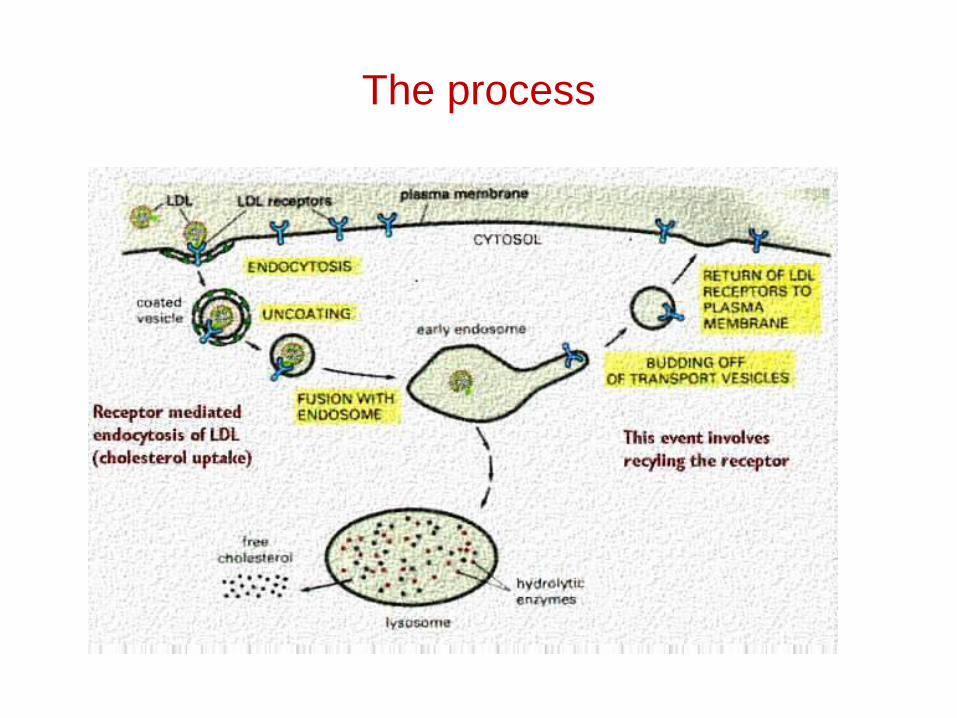
The process
The life of a protein
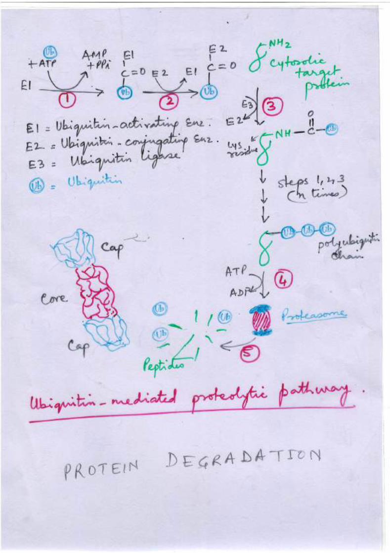
• Determined by N terminal amino acid.• Proteins with ala, met, gly, ser, val, thr etc at the
N terminus have more half life.• Proteins with glu, gln, asp and asn have less half
life.• The tagged proteins are turned over by a 26S
protease complex.• It leaves ubiquitin unaffected.
Protein Destruction
• Ubiquitin serves as a tag
• It is a small 8.5 KD protein
• Gets attached by its C terminal to lys of target protein
• Reaction catalysed by three enzymes, E1, E2 and E3.