multiplex analysis of intracellular signaling pathways in lymphoid cells by microbead suspension...
TRANSCRIPT

Multiplex Analysis of Intracellular SignalingPathways in Lymphoid Cells by MicrobeadSuspension Arrays*Imran H. Khan‡§¶, Sara Mendoza‡, Paul Rhyne�, Melanie Ziman‡, Joseph Tuscano**,Dominic Eisinger�, Hsing-Jien Kung‡‡, and Paul A. Luciw‡§
Phosphorylation analysis of signaling proteins is key forexamining intracellular signaling pathways. Conventionalbiochemical approaches, e.g. immunoprecipitation, West-ern blot, and ELISA, have played a major role in elucida-tion of individual signaling events. However, these meth-ods are laborious, time-consuming, and difficult to adaptfor high throughput analysis. A multiplex approach tomeasure phosphorylation state of multiple signaling pro-teins simultaneously would significantly enhance the effi-ciency and scope of signaling pathway analysis for mech-anistic studies and clinical application. This reportdescribes a novel multiplex microbead suspension arrayapproach to examine phosphoproteomic profiles inlymphoid cells. In the Jurkat T-cell leukemia line, the mul-tiplex assay enabled targeted investigation of phospho-rylation kinetics of signal transduction from receptorproximal events (tyrosine phosphoproteins CD3, Lck,Zap-70, and linker for T-cell activation) to cytosolic events(serine/threonine phosphoproteins Erk and Akt) to tran-scription factors (serine/threonine phosphorylated Rsk,cyclic AMP-response element-binding protein, andSTAT3). To broaden the application of the multiplex anal-ysis, signaling pathways were also studied in B-celllymphoid tumor lines that included chronic lymphocyticleukemia lines. In these cell lines, multiplex suspensionarray enabled phosphoproteomic analysis of signalingcascade mediated by Syk, a homolog of Zap-70. Resultsobtained by multiplex analysis were confirmed by immu-noprecipitation and Western blot methods. The examplesof T-cell and B-cell signaling pathway analyses in thisreport demonstrate the utility of the multiplex suspensionarrays to investigate phosphorylation dynamics and kinet-ics of several signaling proteins simultaneously in signaltransduction pathways. Molecular & Cellular Proteom-ics 5:758–768, 2006.
Regulated phosphorylation of signaling proteins in cellsplays a critical role in signal transduction from the surfacereceptor along a pathway that produces a physiological re-sponse in the cell, such as a novel pattern of gene expressionwith subsequent effects on cell function (1). Abnormally highactivation, or lack of activation of certain signaling proteins,may interfere with proper functioning of the signal transduc-tion apparatus in a cell. Such anomalies may result in disease(2, 3).
In cells of the immune system, tight regulation of cellularsignaling events orchestrates a finely tuned signaling systemthat leads to specific and measured responses to pathologicalchallenges (4). Additionally cell signaling plays a key role in thematuration of lymphoid cell populations during developmentalprogression (4–7). In the T-cell signaling pathway, a signalemanates from the T-cell receptor (TCR)1 on the cell surfaceand is transduced across the cell membrane to the cytosol viaa series of signaling proteins. TCR-proximal events are carriedout by regulated phosphorylation of several proteins, Lck,Zap-70, and LAT (8, 9). A key target for activated Zap-70kinase is LAT, a palmitoylated membrane-associated adapterprotein (10, 11), which functions as a platform for the assem-bly of other key signaling proteins, including SLP-76, Vav, andphospholipase C-� (10, 12–14). This multiprotein complexserves as a hub, coupling the signal received from the TCR tocytosolic components of the T-cell activation pathway. Down-stream of the LAT complex, the signal leads to rearrangementof the actin cytoskeleton via phosphorylation of Wiscott-Al-drich syndrome protein and to transcriptional activation viathe Erk pathway (15). Subsequent activation of specific tran-scription factors (including Rsk, CREB, and STAT3) results inexpression of several cytokines including interleukin-2, a hall-mark of T-cell activation (16, 17). In B-cells, signaling initiates
From the ‡Center for Comparative Medicine, §Department of Med-ical Pathology and Laboratory Medicine, **Division of Hematologyand Oncology, ‡‡Department of Biological Chemistry and UC DavisCancer Center, University of California, Davis, California 95616 and�Upstate USA, Charlottesville, Virginia 22903
Received, October 24, 2005, and in revised form, December 19,2005
Published, MCP Papers in Press, December 20, 2005, DOI10.1074/mcp.T500032-MCP200
1 The abbreviations used are: TCR, T-cell receptor; BCR, B-cellreceptor; B-CLL, B-cell chronic lymphocytic leukemia; CREB, cyclicAMP-response element-binding protein; Erk, extracellular signal-reg-ulated kinase; IgG, immunoglobulin G; IgM, immunoglobulin M; IP,immunoprecipitation; LAT, linker for T-cell activation; Lck, lympho-cyte-specific kinase; MFI, median fluorescence intensity; STAT3, sig-nal transducers and activators of transcription 3; Syk, Syk/Zap-70family of kinases; Zap-70, �-associated protein; WB, Western blot;MAP, mitogen-activated protein; MAPKAP, MAP kinase-activatedprotein.
Technology
© 2006 by The American Society for Biochemistry and Molecular Biology, Inc.758 Molecular & Cellular Proteomics 5.4This paper is available on line at http://www.mcponline.org

through the B-cell receptor (BCR) complex on the cell surface(18, 19), and the activation cascade utilizes the Syk tyrosinekinase, a homolog of Zap-70 tyrosine kinase. Subsequentsignaling events, including phosphorylation of Akt, Erk, andCREB, are shared not only between B- and T-cells but alsoother cell types (20).
For elucidating basic molecular mechanisms underlyingcellular signaling, conventional biochemical and cell biologicalmethodologies, such as immunoprecipitation, Western blot,and immunofluorescence, have been instrumental. However,investigation of complex signaling pathways, such as thoseregulating lymphoid cell activation, requires the developmentof more efficient approaches to better define the relationshipsof signal transduction pathways and multiple outcomes (21,22). Novel approaches that will enable comprehensive meas-urement of signaling kinetics, magnitude of signal, and path-way cross-talk will also lead to a better understanding ofcellular signaling in both normal cells and cells displayingabnormal functions, including tumor cells.
This report describes a multiplex microbead suspensionarray approach for analysis of phosphorylation of multiplesignaling proteins by simultaneous detection in a single sam-ple. We evaluated the multiplex microbead suspension arraytechnology developed by Luminex Corp. (Austin, TX) termedmultianalyte profiling (Lab-MAP). This technology utilizes mix-tures of unique fluorochrome-coded sets of polystyrene mi-crobead suspension (5.6 �m in diameter) to detect specifictarget analytes (23, 24). Individual microbead sets are coatedwith specific capture molecules, e.g. antibodies. Multiplexcapability involves mixing several populations of these fluo-rescent coded microbeads, each coated with a specific cap-ture antibody, into one reaction vessel at the start of the test.Subsequently sample (e.g. tumor cell lysate containing targetproteins) is incubated with this microbead mixture. Analytesare captured by the relevant antibodies on the microbeads.Detection of the analytes is performed by the addition ofsecondary reagent(s) attached to reporter fluorochrome (e.g.phycoerythrin). Finally analysis is performed in the Luminexflow cytometer where lasers and digital signal processingmethods identify the bead set and detect the reporter. Thisapproach is rapid and sensitive and lends itself to highthroughput in an economical fashion.
The key element of our novel multiplex microbead suspen-sion array immunoassay is the detection of tyrosine phospho-rylation of multiple proteins by the use of a single anti-phos-photyrosine antibody. In addition, for the detection of selectedserine/threonine phosphorylated signaling proteins, anti-phosphoprotein-specific antibodies were used. We studiedT-cell activation pathway in the Jurkat T-cell leukemia line asa model system (25, 26). This novel multiplex microbeadsuspension array system enabled analysis of the dynamicsand kinetics of Jurkat T-cell signaling by the detection ofphosphorylation of protein kinases (Lck, Zap-70, Syk, Akt,
and Erk), an adaptor (LAT), and transcription factors (Rsk,CREB, and STAT3) in a single cell lysate sample. In addition,this multiplex suspension array immunoassay was applied tothe analysis of signal transduction in B-cells, including theRamos line derived from an Epstein-Barr virus lymphoma andseveral cell lines from patients with B-cell chronic lymphocyticleukemia (B-CLL). Expression of Zap-70 in leukemic cells ofB-CLL patients is of prognostic value for aggressive diseaseand shorter patient life span (27, 28). Importantly our findingswith the multiplex microbead suspension arrays for thesephosphoproteins were confirmed by conventional methods ofimmunoprecipitation and Western blotting.
EXPERIMENTAL PROCEDURES
Cells—The human T-cell leukemia Jurkat (E6-1) cell line and Burkittlymphoma Ramos (RA 1) B-cell line were obtained from ATCC (Ma-nassas, VA). CLL cell lines Mec-1 and Mec-2 (29) were obtained fromDeutsche Sammlung von Mikroorganismen und Zellkulturen GbH(56). The CLL cell line MO1043 (30) was a gift from Dr. RicardoDela-Favera (Columbia University, New York, NY) These CLL lines areof B-cell origin derived from human chronic B-cell leukemia patientblood. By flow cytometry, using fluorescent labeled antibodies, weverified the above cell lines to be positive for B-cell markers CD19 andCD20. Jurkat, Ramos, and MO1043 cells were propagated in RPMI1640 medium containing 10% fetal bovine serum. Mec-1 and Mec-2cells were propagated in Iscove’s modified Dulbecco’s medium con-taining 10% fetal bovine serum.
Antibodies and Reagents—Monoclonal antibodies against signal-ing proteins Lck, Zap-70, LAT, and Syk and biotinylated anti-phos-photyrosine monoclonal antibody (4G10) were supplied as purifiedIgG by Upstate USA (Charlottesville, VA). The following BeadmatesTM
(capture beads and biotinylated reporter antibody pairs) were alsoobtained from Upstate USA: phospho-Akt/PKB� (Ser-473), phospho-Erk/MAP kinase 1/2 (Thr-185/Tyr-187), phospho-Rsk MAPKAP kinase1a (Ser-380), phospho-STAT3 (Ser-727), and phospho-CREB (Ser-133). In addition, capture and reporter antibodies from the aboveBeadmates (Upstate USA) were used for immunoprecipitation andWestern blot analysis. Antibodies for the detection of total proteins(Lck, Zap-70, Syk, LAT, Erk, Akt, Rsk, CREB, and STAT3) in Westernblots were also from Upstate USA. Anti-CD3 antibody UCHT1 and itsisotype control antibody were obtained from BD Pharmingen.Streptavidin-conjugated R-phycoerythrin was from CalTag (Burl-ingame, CA). Anti-IgM antibody was purchased from Jackson Immu-noresearch Laboratories, Inc. (West Grove, PA). Protease inhibitormixture and purified 10% Nonidet P-40 were purchased from RocheApplied Science. Protein G-conjugated Sepharose was from Sigma.
Treatment of Lymphoid Cells—Jurkat (E6-1) T-cells, Ramos B-cells, and the CLL cell lines (Mec-1, Mec-2, and MO1043) weretreated with sodium pervanadate to inhibit intracellular tyrosine phos-phatases, resulting in hyperphosphorylation of tyrosine kinases andtheir substrates including serine/threonine kinases. Sodium pervana-date was prepared by adding 330 �l of 30% hydrogen peroxide to 1ml of 20 mM sodium vanadate (pH 10.0). Cells were resuspended toa density of 1 � 108 cells/ml in plain RPMI 1640 medium and pre-warmed to 37 °C for 5 min. To each milliliter of cell suspension, 40 �lof sodium pervanadate was added. Cells were mixed and immedi-ately incubated at 37 °C for 5 min. Treated cells were lysed, and celllysates were processed as described below.
To examine intracellular signaling kinetics using anti-CD3 antibody,Jurkat cells were resuspended in RPMI 1640 medium at a density of1 � 108/ml and prewarmed to 37 °C for 5 min. Anti-CD3 antibody(UCHT1) was added to a final concentration of 5 �g/ml. Cells were
Multiplex Analysis of Lymphoid Cell Signaling
Molecular & Cellular Proteomics 5.4 759

mixed and incubated at 37 °C for various times ranging from 15 s to1 h. Treatment was terminated by the addition of cell lysis buffer asdescribed below.
Preparation of Cell Lysates—Cells were lysed by adding lysis buffer(2% Nonidet P-40, protease inhibitor mixture (both from Roche Ap-plied Science, in PBS), and 0.5 mM sodium orthovanadate). Lysatewas immediately vortexed and incubated on ice for 15 min. Celldebris were removed by low speed centrifugation. Total protein con-centration of lysates was determined by BCA reagent kit (Bio-Rad).Lysates were stored frozen at �80 °C until used.
Coating Microbeads with Antibodies—Monoclonal antibodiesagainst CD3, Lck, Zap-70, LAT, and Syk were coated as captureantibodies on individual sets of microbeads. In addition, one bead setwas coated with BSA as a control for nonspecific interactions. An-other bead set was coated with biotin-conjugated goat IgG (JacksonImmunoresearch Laboratories, Inc.), a positive control for the reportermolecule (streptavidin-conjugated phycoerythrin). Luminex beadswere coated with the above proteins as described previously (31).Briefly bead stock was resuspended by vortexing and sonication(15–30 s). An aliquot of 2.5 � 106 beads was removed and centrifugedat 12,000 � g for 2 min. Beads were resuspended in 80 �l ofactivation buffer (100 mM monobasic sodium phosphate, pH 6.3) byvortexing and sonication (15–30 s). To activate the beads for cross-linking to proteins, 10 �l of 50 mg/ml N-hydroxysulfosuccinimide(Pierce) was added, and beads were mixed by vortexing. Then 10 �lof 50 mg/ml 1-ethyl-3-[3-dimethylaminopropyl]carbodiimide (Pierce)was added, and beads were mixed again by vortexing. The beadmixture was shaken on a rotator at room temperature for 20 min andcentrifuged at 12,000 � g for 3 min. Beads were washed twice with 1ml of 50 mM MES (pH 6.0) buffer. To coat with antibody, pelletedbeads were resuspended in the relevant antibody solution (25–100�g/ml) in 50 mM MES (pH 6.0) buffer. The mixture of activated beadsand antibodies was incubated by shaking on a rocker for 2 h at roomtemperature for coupling. After coating, beads were washed twicewith wash buffer (0.1% Tween 20 in PBS, pH 7.40) and resuspendedin 1 ml of blocking buffer (1% BSA, 0.1% Tween 20 in PBS, pH 7.4,0.05% sodium azide). Blocking was performed by shaking on a rockerat room temperature for 30 min. After blocking, beads were washedtwice in 1 ml of blocking buffer. Finally antibody-coated beads wereresuspended in 1 ml of blocking buffer and stored at 4 °C for up to aweek. For long term storage, beads were kept frozen at �70 °C forseveral months.
Microbead Suspension Array Immunoassay of Signaling Proteins—Immunoreactions were set up in 96-well, filter-bottomed plates de-signed for high throughput separations (1.2-�m MultiScreen, MilliporeCorp.). Microbeads (2000 beads of each set) coated with a specificantibody were mixed. This multiplex, microbead mixture was addedto each well. To this, 25 �l of cell lysate (0.4 mg/ml total protein;corresponding to approximately 8 � 104 cells) was added. Perform-ance of antibody-coated microbeads on serial dilutions of cell lysateswas tested. The lysate concentration used in this study is within thelinear range of signal responses of the microbead sets (data notshown).2 The contents were mixed at 1400 rpm on a plate shaker(Labnet International Inc., Woodbridge, NJ) for 2 h at room tempera-ture. After incubation with the lysate, liquid was drained from thebottom of the plate under vacuum. The microbeads were washedtwice by adding 150 �l of wash buffer/well and draining out undervacuum successively. For detection of tyrosine phosphorylated sig-naling proteins bound to antibodies coated on microbeads, 25 �l ofbiotinylated anti-phosphotyrosine antibody 4G10 (0.5 �g/ml in washbuffer) was added as the detection reagent. Antibody 4G10 displayed
excellent sensitivity at this concentration for the detection of phos-photyrosine residues with no cross-reactivity to phosphoserines/phosphothreonines (21). Microbeads were mixed as before and incu-bated at room temperature for 30 min. Microbeads were washedtwice as before. To detect biotinylated 4G10, streptavidin-conjugatedR-phycoerythrin was added at a dilution of 1:1000 in wash buffer asthe reporter molecule and incubated for 15 min at room temperature.Microbeads were washed once with wash buffer, resuspended in 100�l of wash buffer/well, and analyzed in the Luminex-100TM instru-ment. Beadmate kits were used according to the manufacturer’sinstructions (Luminex Corp.).
Luminex-100 Operation and Data Analysis—The Luminex-100 in-strument was used at default settings, set by the manufacturer forroutine applications, as directed by the user’s manual. Data wereacquired by Luminex Data Collection Software (Version 1.7). Thissoftware package was used according to instructions in the user’smanual supplied by the manufacturer for routine operation of theinstrument, data acquisition, and data analysis. The instrument wascalibrated with Calibration Beads supplied by the manufacturer toadjust the settings for bead set identification or “Classification” andfor the detection of “Reporter” (phycoerythrin). Events were gated toexclude doublets and other aggregates. A hundred independent,gated events were acquired for each bead set. The median fluores-cence intensity (MFI) or “signal” of a hundred events (beads) wasused as a measure of the detection of protein phosphorylation. Afteracquisition by Luminex software, the data were further processed byMicrosoft Excel software.
Immunoprecipitaion and Western Blotting—Immunoprecipitationwas performed by mixing capture antibody (5 �g/ml of purified IgG)with 0.5 ml of cell lysates (1 mg/ml total protein) on a rotator for 1 hat 4 °C. Protein G-conjugated Sepharose was added (25 �l of pre-swollen gel/ml) and mixed on rotator for 30 min at 4 °C. Sepharosebeads were washed three times in wash buffer (PBS containing 1%Tween 20) and resuspended in 40 �l of SDS sample buffer (Novex 2�SDS, Tris-glycine sample buffer with 0.71 mM �-mercaptoethanol;Invitrogen). The samples were boiled for 5 min. The immunocom-plexes were resolved on SDS-polyacrylamide gels (Novex 8–16%precast gradient Tris-glycine gels). Resolved proteins were trans-ferred to PVDF membranes. To detect tyrosine phosphorylation, bi-otin-conjugated anti-phosphotyrosine antibody (4G10, 0.5 �g/ml)was used. For the detection of serine/threonine phosphorylation,biotinylated antibodies from the respective Beadmates (phospho-Akt/PKB� (Ser-473), phospho-Erk/MAP kinase 1/2 (Thr-185/Tyr-187),phospho-Rsk MAPKAP kinase 1a (Ser-380), phospho-STAT3 (Ser-727), and phospho-CREB (Ser-133) were used according to the man-ufacturer’s instructions (Upstate USA). Blots were developed withVectastain ABC detection reagent (Vector Laboratories, Burlingame,CA) and ECL Plus Western blotting detection system (AmershamBiosciences) and visualized on a Typhoon 9410 variable mode imager(Amersham Biosciences).
RESULTS
Phosphorylation of T-cell Signaling Proteins in JurkatCells—The multiplex microbead suspension array immunoas-say was performed to detect phosphorylation of different cellsignaling proteins in Jurkat T-cells treated with sodium per-vanadate. Treatment with pervanadate inhibits intracellulartyrosine phosphatases resulting in sustained phosphorylationof various tyrosine kinases and substrates reflecting the acti-vation state of the cells (32). Tyrosine kinases so activatedsubsequently phosphorylate their substrates including certaindownstream serine/threonine kinases resulting in their activa-
2 I. H. Khan, S. Mendoza, P. Rhyne, M. Ziman, J. Tuscano, D.Eisinger, H.-J. Kung, and P. A. Luciw, unpublished.
Multiplex Analysis of Lymphoid Cell Signaling
760 Molecular & Cellular Proteomics 5.4

tion (33). Ten sets of microbeads coated with monoclonalantibodies to signaling proteins (i.e. CD3, Lck, Zap-70, LAT,Erk, Akt, Rsk, CREB, STAT3, and Syk) were mixed and incu-bated with cell lysate. A microbead set coated with anti-Sykantibody was included as a specificity control because Sykprotein is not expressed in Jurkat (E6-1) T-cells (34). Tyrosinephosphorylation of Lck, Zap-70, LAT, CD3, and Syk wasdetected by the anti-phosphotyrosine antibody (4G10),whereas serine/threonine phosphorylation of Erk, Akt, Rsk,CREB, and STAT3 was detected by anti-phosphoreporterantibodies (Beadmates). Proteins captured by microbeadscoated with antibodies to Lck, Zap-70, LAT, and CD3 showedstrong reaction to 4G10 antibody in lysate from pervanadate-treated Jurkat cells (Fig. 1A). Similarly proteins captured bymicrobeads coated with antibodies to Erk, Akt, Rsk, CREB,and STAT3 displayed a reaction to the relevant anti-phos-phopeptide antibodies in lysates of the treated cells. Interas-say variability was less than 10% (Fig. 1A, bars representS.D.). This analysis showed that phosphorylated signalingproteins were readily detected by the multiplex microbead sus-pension array upon treatment of cells with sodium pervanadate.Interestingly constitutive phosphorylation of all the signalingproteins (i.e. in untreated cells) except Akt was relatively minimal(Fig. 1A). Constitutive phosphorylation of Akt in Jurkat T-cellshas been reported previously (35). As expected, Syk phospho-rylation was undetectable in Jurkat (E6-1) T-cells treated withsodium pervanadate (Fig. 1A). Data obtained by immunopre-cipitation (IP) and Western blot (WB) confirmed that Syk was notdetectable in these cells (Fig. 1B) as shown previously (34).
Antibodies used in coating fluorescent microbeads for cap-turing different signaling proteins in cell lysates were alsotested by IP. Detection of the phosphorylated forms as well asthe total amount of these proteins in lysates of Jurkat T-cellswas performed by WB analysis of the IP complexes. Resultsof IP/WB analysis are presented in Fig. 1B. Together themultiplex and IP/WB analyses of Jurkat cells treated withsodium pervanadate demonstrated the following features. (a)Phosphorylated bands of the relevant proteins were detectedonly in lysates from cells treated with sodium pervanadatewith the exception of Akt, which was phosphorylated in un-treated cells as well. (b) Phosphorylated species of signalingproteins predominated in their respective immunoprecipi-tates. (c) The total amount of each signaling protein in thelysates from non-treated and treated cells was similar. The datapresented here demonstrate the utility of multiplex microbeadsuspension array approach for analyzing phosphorylation ofmultiple signaling proteins simultaneously in a single sample.
Cell Signaling Kinetics in Jurkat T-cells Treated with Anti-CD3 Antibody—To study the kinetics of signaling pathway,Jurkat T-cells were treated with anti-CD3 antibody (UCHT1)for various times between 15 s and 1 h. Receptor-proximalsignaling events were monitored by detecting tyrosine phos-phorylation of Lck, Zap-70, Syk, and LAT. Results of thisstudy are shown in Fig. 2. The multiplex microbead immuno-assay data showed that Lck was phosphorylated within 15 safter stimulation followed by phosphorylation of Zap-70 andLAT between 30 s and 1 min after stimulation. Phosphoryla-tion of these three T-cell proteins reached a peak within 2 min
FIG. 1. A, phosphoproteomic analysis by multiplex microbead suspension array immunoassay of signaling proteins in Jurkat T-cells. Cellswere treated with sodium pervanadate, and cell lysates were prepared as described under “Experimental Procedures.” A mixture ofmicrobeads coated with antibodies to CD3, Lck, Zap-70, Syk, LAT, Erk, Akt, CREB, STAT3, and Rsk were incubated with the cell lysates.Detection of phosphorylation was performed using a mixture of 4G10 antibody and protein-specific anti-phospho antibodies to Erk, Akt, CREB,STAT3, and Rsk. The results are an average of two independent experiments performed on duplicate samples in each experiment. S.D. isrepresented by error bars. B, immunoprecipitation and Western blot analysis of phosphorylation and total amount of signaling proteins in Jurkatcells. The sequence of proteins is the same as shown for multiplex suspension array immunoassay (A). Antibodies used for immunoprecipi-tation were the same as those coated on the microbeads for multiplex analysis. Western blot analyses were performed for the detection ofprotein phosphorylation (4G10 antibody and protein-specific anti-phospho antibodies) and total proteins as indicated.
Multiplex Analysis of Lymphoid Cell Signaling
Molecular & Cellular Proteomics 5.4 761

and subsequently decayed to the preactivation levels in 30min. The kinetics of phosphorylation of Lck and Zap-70 pre-sented here correlated with those reported previously in afluorescent imaging study of the immunological synapse us-ing confocal microscopy (36). Syk is not expressed in Jurkat(E6-1) T-cells (34). Accordingly no increase in Syk phospho-rylation, above the basal level observed in untreated cells,was detected during the 1-h period after the treatment withanti-CD3 antibody (Fig. 2). Syk phosphorylation thus servedas an excellent internal control for the phosphoproteomicanalysis of T-cell signaling pathway kinetics.
Signal transduction beyond these receptor-proximal eventswas also examined by the multiplex suspension array immu-noassay, and the data are also shown in Fig. 2. Phosphoryl-ation of Erk and Rsk was detected at 1 and 2 min postacti-vation, respectively. The signal for both of these serine/threonine kinases also decayed to the prestimulation levels in1 h. An increase in phosphorylation of Akt over the basal levelwas detected after 2 min. However, in contrast to the othersignaling proteins, phosphorylation levels of Akt continued toremain high for the duration of the period studied here (1 h).The above described multiplex suspension array immunoas-say was also performed on lysates obtained from cells treatedwith an isotype control antibody for UCHT1 under the sametime course conditions. In this control experiment, back-ground reactivity of different microbead sets was similar tothose with lysate from untreated cells (data not shown).2 Inaddition, the control BSA-coated beads yielded low back-ground reactivity (MFI of 20–30 units) with all the lysatesregardless of activation status of cells.
Phosphorylation of B-cell Signaling Proteins in RamosCells—A multiplex immunoassay was evaluated for detectingphosphorylation of multiple signaling proteins in the well char-acterized Ramos B-cell line, which was derived from an indi-vidual with Burkitt lymphoma. The same antibody-coated mi-crobead sets described in Fig. 1 for the analysis of signaling inJurkat T-cells were used (Fig. 1). As shown in Fig. 3B, Syk wasmore strongly phosphorylated in Ramos cells than its homo-logue Zap-70 was in Jurkat cells (Fig. 1) after the treatmentwith sodium pervanadate. This result is consistent with Sykbeing a much more active kinase than Zap-70 (37). Specificityof the anti-Syk antibody coated on microbeads was demon-strated by detection of Syk in lysates from the B-cell lines byIP/WB analysis (Fig. 4). Importantly as mentioned above, thisantibody was not reactive in IP/WB analysis performed onJurkat (E6-1) T-cell lysates (Fig. 1B). In contrast to the multi-plex microbead immunoassay analysis of Jurkat T-cell acti-vation, phosphorylation of CD3 was undetectable, whereasphosphorylation of Zap-70 was marginal in Ramos B-cells(Fig. 3). IP/WB analysis was consistent with this observation,showing low level expression of Zap-70 protein and its phos-phorylation in Ramos B-cells (Fig. 4). Taken together, resultsof the multiplex analysis presented in Figs. 1A and 3 andIP/WB data presented in Figs. 1B and 4 demonstrate utility ofthe multiplex microbead analysis for T-cell and B-cell signal-ing pathways.
Expression of Signaling Proteins in B-CLL Cell Lines—Themultiplex analysis of B-cell signaling was extended to celllines derived from patients with CLL. First the conventionalmethods of IP/WB were used to assess total and phospho-
FIG. 2. Kinetics of signaling protein phosphorylation in Jurkat T-cells treated with anti-CD3 antibody analyzed by multiplexmicrobead suspension array immunoassay. Anti-CD3 treatment was terminated at various time points, and cell lysates were prepared. Amixture of microbeads coated with antibodies to Lck, Zap-70, Syk, LAT, Erk, Akt, and Rsk were incubated with the cell lysates. Detection ofphosphorylation in this kinetic experiment was performed using a mixture of recombinant 4G10 antibody (Upstate USA, 1:500 dilution) andprotein-specific anti-phospho antibodies to Erk, Akt, and Rsk. Multiplex microbead suspension array immunoassay was performed, and thedata are presented as described for Fig. 1A.
Multiplex Analysis of Lymphoid Cell Signaling
762 Molecular & Cellular Proteomics 5.4

rylated proteins (Syk, Zap-70, Lck, and LAT) in CLL B-celllines (Mec-1 and MO1043) to make comparisons with RamosB-cells. Both of the Mec-1 and MO1043 lines expressed Sykprotein (Fig. 4). The data also showed that Ramos and Mec-1cells had low levels of Zap-70. In contrast, the MO1043 cellsexpressed a high amount of Zap-70 (Fig. 4). Thus, theMO1043 cell line may be representative of B-CLL caseswhere patient leukemic cells have strong expression ofZap-70 protein (38, 39). Normal, mature B-cells, except cer-tain subpopulations, do not express Zap-70 (40). Two otherT-cell proteins, Lck and LAT, not usually expressed by matureB-cells (20, 41), were also examined. Ramos cells produced arelatively high amount of Lck, and Mec-1 and MO1043 cellsboth showed barely detectable amounts of Lck (Fig. 4). Inaddition, Ramos B-cells displayed low level expression ofLAT, whereas LAT was undetectable in Mec-1 and MO1043cells by IP/WB analysis (Fig. 4). Analysis of tyrosine phospho-rylation of Syk, Zap-70, Lck, and LAT with and without sodiumpervanadate treatment was also performed, and the data areshown in Fig. 4 (left panel for each protein). In general, whereprotein expression was detectable, phosphorylation of theprotein in treated cells was also detectable, i.e. the phospho-rylation level was proportional to the level of expression. Forexample, Lck and LAT were expressed and phosphorylated inRamos B-cells. Expression of these proteins was low to nonein Mec-1 and MO1043 cells; accordingly phosphorylation ofLAT in both of these cell lines was also not detectable (Fig. 4).
Surprisingly Zap-70 expression in MO1043 cells did not followthis pattern. Although Zap-70 protein was expressed in highamounts in MO1043 cells, the phosphorylation of Zap-70 inthis cell line after treatment with sodium pervanadate was notstrong (Fig. 4).
Multiplex Suspension Array Analysis of B-cell Signaling inCLL Cell Lines—In general, B-CLL cells not only have a lowsurface expression of BCR but also respond poorly to activa-tion by anti-IgM antibodies (42). Detection of surface expres-sion of BCR in the CLL cell lines, in comparison with theRamos B-cell line (Burkitt lymphoma), was performed by flowcytometric analysis using anti-IgM antibody. This analysisrevealed that Ramos cells expressed high levels of surfaceIgM (data not shown).2 In contrast, all three CLL cell linesexpressed low levels of surface IgM (data not shown)2 asoriginally reported (29, 30). Accordingly treatment with anti-IgM antibody resulted in strong phosphorylation of Syk inRamos cells, whereas no detection of Syk phosphorylationwas observed by IP/WB analysis in the three CLL cell lines(Mec-1, Mec-2, and MO1043) treated with anti-IgM antibody(data not shown).2 Therefore, for experimental consistency inthe phosphorylation of various signaling proteins in theRamos cells and these three CLL cell lines, the cells weretreated with sodium pervanadate.
For multiplex microbead suspension array analysis of CLLcell lines, lysates from sodium pervanadate-treated and un-treated Mec-1, Mec-2, and MO1043 cells were tested for
FIG. 4. Immunoprecipitation/West-ern blot analysis of B-cell lysates fromsodium pervanadate-treated cells. Im-munoprecipitations were performed withmonoclonal antibodies against Syk,Zap-70, Lck, and LAT. Western blottingwas performed for the detection of pro-tein phosphorylation by 4G10 antibody,and total protein detection was carriedout using protein-specific antibodies.Ab, antibody.
FIG. 3. Phosphoproteomic analysis by multiplex microbead suspension array immunoassay of signaling proteins in Ramos B-cells.Cells were treated with sodium pervanadate. Cell lysates were prepared, and assay was performed as described for Fig. 1A. Due to theextremely high phosphorylation level of Syk, the results are presented on two scales. A, all proteins except Syk. B, Syk protein only. Multiplexmicrobead suspension array immunoassay was performed, and data are presented as described for Fig. 1A.
Multiplex Analysis of Lymphoid Cell Signaling
Molecular & Cellular Proteomics 5.4 763

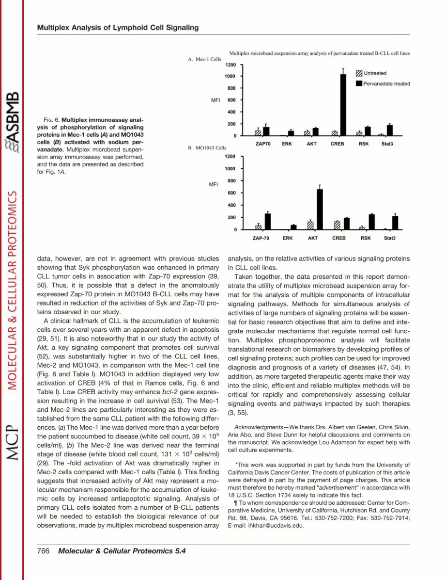
phosphorylation of several cell signaling proteins. Phospho-proteomic profiles of signaling proteins in two of the cell lines,Mec-1 and MO1043, are shown in Fig. 6. The same captureantibody microbead sets used in Figs. 1 and 3 were used forthis analysis. Table I shows the relative -fold activation ofZap-70, Syk, and other signaling proteins in the Mec-1 andMO1043 lines as well as the Mec-2 line. For the receptor-proximal signaling events, the -fold activation of Syk in theCLL lines was comparable or higher than that in RamosB-cells with the notable exception of MO1043 cells. Syk ac-tivation was dramatically reduced in MO1043 cells as com-pared with the other CLL cell lines (Table I). Similarly despitestrong expression of Zap-70 in MO1043 cells, the -fold acti-vation of Zap-70 in this cell line was much lower than that ofJurkat T-cells (81% lower, Table I). This lack of Zap-70 acti-vation could be due to the effects of Epstein-Barr virus trans-formation and/or mutation in the zap-70 gene. Mec-2 cellsexhibited significantly higher Syk activation compared withRamos and Mec-1 cells, both of which displayed comparableSyk activation (Table I). Both Mec-1 and Mec-2 cell lines wereestablished from the same patient’s blood, drawn within ap-proximately a 1-year interval (29). The Mec-2 cell line wasestablished near the terminal stage of CLL disease when thepatient’s white blood cell count was more than 3-fold highercompared with the time when the Mec-1 cell line was estab-lished (29). Thus, the significantly higher Syk activity in theMec-2 cell line may correlate with the advanced stage ofmalignancy.
To assess cytosolic signaling events in the CLL cell lines,the -fold increase in activation of Akt, Erk, and Rsk wasexamined. The data are shown in Fig. 6 and Table I. The mostdramatic difference was observed for Akt activation. As in thecase of Syk, -fold activation of Akt was dramatically increasedin Mec-2 cells as compared with Mec-1 and Ramos cells. Theactivity of transcription factors CREB and STAT3 was also
examined. In the MO1043 cell line, which expressed a highamount of Zap-70, activation of CREB was severely inhibited(Fig. 6 and Table I).
DISCUSSION
This report describes a novel multiplex suspension arrayapproach to study cell signaling pathways by simultaneousanalysis of phosphorylation of multiple signaling proteins ineach sample. As a model signaling pathway, we examinedlymphoid cell activation in the Jurkat (E6-1) T-cell tumor line(26). In addition, the multiplex phosphorylation immunoassaywas extended to investigate B-cell activation in a Burkittlymphoma cell line (Ramos) and three B-CLL cell lines. Wedemonstrate that the microbead suspension array immuno-assay is versatile and sensitive. Phosphoproteomic profilingof 10 signaling proteins (CD3, Lck, Zap-70, LAT, Erk, Akt, Rsk,CREB, STAT3, and Syk) in the Jurkat T-cell line needed �8 �
104 cells (Fig. 1A). In contrast, to obtain the same amount ofinformation by IP/WB and ELISA analyses of all 10 signalingproteins, approximately 500 times and 100 times more Jurkatcells (4 � 107 and 8 � 106), respectively, were required (Fig.1B; ELISA data not shown).2 Thus, the need for only a smallamount of sample for the phosphoproteomic profiling of sev-eral signaling proteins (theoretically up to 100) is a clearadvantage. Additionally this assay format readily enables theuse of internal controls. In comparison with fixed formats suchas peptide array systems (43), the microbead suspensionarray approach described here offers flexibility and ease ofadaptability where microbead sets coated with capture anti-bodies can be included or excluded from the mixture at will.Methods based on two-dimensional gel electrophoresis andMS have been applied recently for simultaneous analysis ofmultiple signaling proteins in cells (44–48). However, thesemethods require complex protocols as well as complicatedand very costly instrumentation for broad biological and clin-ical applications. Nevertheless these methodologies have anadvantage over immunoassays as they do not depend uponthe availability of highly specific and high affinity antibodies. Incontrast, development of the microbead suspension arrayimmunoassay described in this report, as for other morecommonly used immunoassays (e.g. immunoprecipitation,ELISA, and planar antibody array), required well characterizedantibodies with high specificity and affinity. This technologyhas a dynamic range of over a thousandfold; therefore groupsof analytes, e.g. cytokines/chemokines (17), that are found inconcentration ranges outside of the dynamic range can beefficiently measured in panels of several dozen analytes in themultiplex format. Also in the context of signaling pathways, forthe global proteomic applications, the two-dimensional gelelectrophoresis/MS methodologies may be more suitable,whereas the multiplex microbead suspension array immuno-assay enables more targeted analysis of specific componentsof signaling pathways.
For the multiplex microbead suspension array approach,
TABLE IRelative activation of signaling proteins in B-cell lines by
multiplex microbead suspension array analysis
Relative activity of each protein is presented. To obtain relativeactivities, -fold activation of each signaling protein was obtained as aratio of MFI (phosphorylation) of each protein in activated cells tononactivated cells. Data were normalized to obtain relative activitiesas follows. 1) -Fold activation of Zap-70 (in bold) in each B-cell linewas normalized to the -fold activation of Zap-70 in Jurkat T-cells. 2)-Fold activations of all other proteins were normalized to those inRamos B-cells.
ProteinB-cell B-CLL
Ramos Mec-1 Mec-2 MO1043
Syk 100 86.4 491 43.9Zap-70 14.7 10.7 13.7 18.6Akt 100 37.5 256 110Erk 100 67.5 106 54.0Rsk 100 48.7 51.5 92.3CREB 100 40.0 90 4.20STAT3 100 100 57 140
Multiplex Analysis of Lymphoid Cell Signaling
764 Molecular & Cellular Proteomics 5.4
monoclonal antibodies to different signaling proteins wereconjugated to individually identifiable microbead sets to serveas capture molecules. Results of the multiplex microbeadimmunoassay using these capture antibodies were confirmedby IP and WB analysis (Figs. 1, A and B, and 4). Importantly asa key feature of the multiplex immunoassay for detection oftyrosine phosphorylation of several signaling proteins (CD3,Lck, Zap-70, LAT, and Syk), the single detection reagent,anti-phosphotyrosine antibody (i.e. the 4G10 antibody), wasaccurate and sufficient. This procedure circumvents the needfor mixtures of phosphotyrosine protein-specific antibodies;such mixtures may require additional optimization of the mul-tiplex assay to minimize reagent cross-reactivity. However,detection of phosphorylation of serine/threonine residues inthe relevant signaling proteins was performed by using phos-phoprotein-specific antibodies. The data (Figs. 1, A and B,and 3–5) clearly show the similarity in the extent of phospho-rylation of each signaling protein in untreated and pervana-date-treated cells as detected by multiplex method and theIP/WB method. Importantly specificity of multiplex suspen-sion array format was well demonstrated by the phosphoryl-ation analysis of two kinases, Zap-70 and Syk. Zap-70 isexpressed in T-cells and usually not in mature B-cells,whereas Syk is expressed in mature B-cells. In this study,Zap-70 phosphorylation was detected by the multiplex phos-phorylation assay in Jurkat (E6-1) T-cells. A low level ofZap-70 phosphorylation was detected in Ramos B-cells bythe multiplex assay (Fig. 3). This result is consistent with thelow level of expression and phosphorylation of Zap-70 de-tected by IP/WB (Fig. 4). In contrast to Zap-70, strong phos-phorylation of Syk was detected in Ramos B-cells by both themultiplex assay (Fig. 3) and IP/WB (Fig. 4) and not in Jurkat(E6-1) cells (Fig. 1, A and B). However, as mentioned under“Results,” pervanadate treatment of cells leads to generalphosphorylation of most of the intracellular tyrosine kinasesand their substrates and is therefore not specific to a givenpathway (32, 33). Targeted stimulation of the T-cell activationpathway was achieved by anti-CD3 antibody as describedbelow. Nevertheless the above results clearly demonstratespecificity of the multiplex assay for the detection of Zap-70and Syk phosphorylation. Phosphoproteomic profiles of dif-ferentially expressed and activated Zap-70 and Syk in variouspopulations of T-cells and B-cells (9, 40, 49) can therefore beefficiently and accurately studied with the microbead suspen-sion array immunoassay described here.
The utility of this novel multiplex phosphorylation assaywas further demonstrated by the analysis of various signal-ing proteins in a time course activation of Jurkat (E6-1) cellstreated with anti-CD3 antibody, which activates the T-cellsignaling pathway (Fig. 2). This analysis highlighted thefollowing features of the multiplex assay. (a) Activation ki-netics of Lck and Zap-70 were similar to those observed inthe immunological synapse between T-cell and antigen-
presenting cell using immunofluorescence and confocal mi-croscopy (36). (b) The assay allowed investigation of tem-poral changes in phosphorylation of several proteinssimultaneously in a kinetic experiment involving multipletime points. (c) The assay was convenient for monitoring thepeak activation level as well as signal decay of multiplesignaling proteins simultaneously.
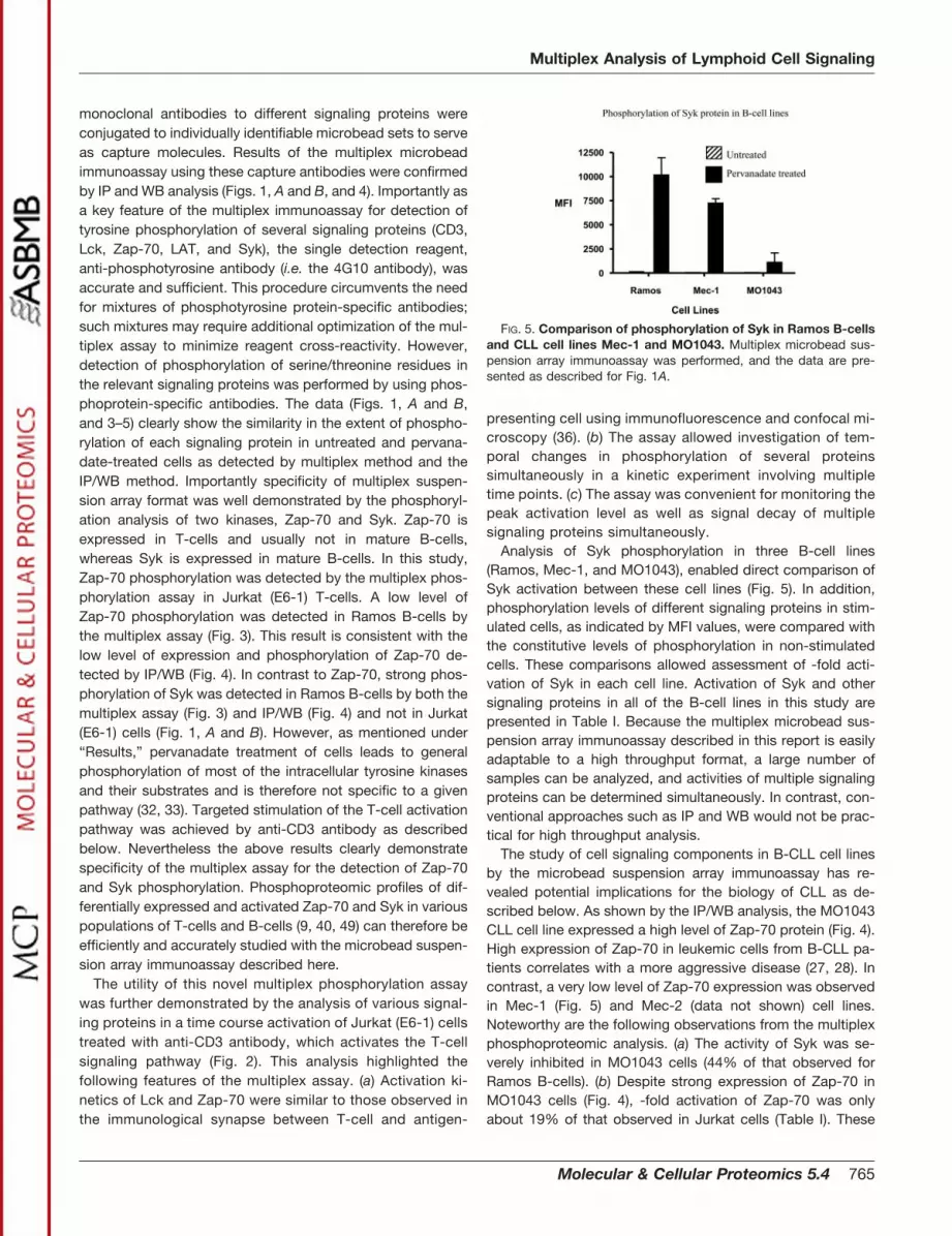
Analysis of Syk phosphorylation in three B-cell lines(Ramos, Mec-1, and MO1043), enabled direct comparison ofSyk activation between these cell lines (Fig. 5). In addition,phosphorylation levels of different signaling proteins in stim-ulated cells, as indicated by MFI values, were compared withthe constitutive levels of phosphorylation in non-stimulatedcells. These comparisons allowed assessment of -fold acti-vation of Syk in each cell line. Activation of Syk and othersignaling proteins in all of the B-cell lines in this study arepresented in Table I. Because the multiplex microbead sus-pension array immunoassay described in this report is easilyadaptable to a high throughput format, a large number ofsamples can be analyzed, and activities of multiple signalingproteins can be determined simultaneously. In contrast, con-ventional approaches such as IP and WB would not be prac-tical for high throughput analysis.
The study of cell signaling components in B-CLL cell linesby the microbead suspension array immunoassay has re-vealed potential implications for the biology of CLL as de-scribed below. As shown by the IP/WB analysis, the MO1043CLL cell line expressed a high level of Zap-70 protein (Fig. 4).High expression of Zap-70 in leukemic cells from B-CLL pa-tients correlates with a more aggressive disease (27, 28). Incontrast, a very low level of Zap-70 expression was observedin Mec-1 (Fig. 5) and Mec-2 (data not shown) cell lines.Noteworthy are the following observations from the multiplexphosphoproteomic analysis. (a) The activity of Syk was se-verely inhibited in MO1043 cells (44% of that observed forRamos B-cells). (b) Despite strong expression of Zap-70 inMO1043 cells (Fig. 4), -fold activation of Zap-70 was onlyabout 19% of that observed in Jurkat cells (Table I). These
FIG. 5. Comparison of phosphorylation of Syk in Ramos B-cellsand CLL cell lines Mec-1 and MO1043. Multiplex microbead sus-pension array immunoassay was performed, and the data are pre-sented as described for Fig. 1A.
Multiplex Analysis of Lymphoid Cell Signaling
Molecular & Cellular Proteomics 5.4 765
data, however, are not in agreement with previous studiesshowing that Syk phosphorylation was enhanced in primaryCLL tumor cells in association with Zap-70 expression (39,50). Thus, it is possible that a defect in the anomalouslyexpressed Zap-70 protein in MO1043 B-CLL cells may haveresulted in reduction of the activities of Syk and Zap-70 pro-teins observed in our study.
A clinical hallmark of CLL is the accumulation of leukemiccells over several years with an apparent defect in apoptosis(29, 51). It is also noteworthy that in our study the activity ofAkt, a key signaling component that promotes cell survival(52), was substantially higher in two of the CLL cell lines,Mec-2 and MO1043, in comparison with the Mec-1 cell line(Fig. 6 and Table I). MO1043 in addition displayed very lowactivation of CREB (4% of that in Ramos cells, Fig. 6 andTable I). Low CREB activity may enhance bcl-2 gene expres-sion resulting in the increase in cell survival (53). The Mec-1and Mec-2 lines are particularly interesting as they were es-tablished from the same CLL patient with the following differ-ences. (a) The Mec-1 line was derived more than a year beforethe patient succumbed to disease (white cell count, 39 � 103
cells/ml). (b) The Mec-2 line was derived near the terminalstage of disease (white blood cell count, 131 � 103 cells/ml)(29). The -fold activation of Akt was dramatically higher inMec-2 cells compared with Mec-1 cells (Table I). This findingsuggests that increased activity of Akt may represent a mo-lecular mechanism responsible for the accumulation of leuke-mic cells by increased antiapoptotic signaling. Analysis ofprimary CLL cells isolated from a number of B-CLL patientswill be needed to establish the biological relevance of ourobservations, made by multiplex microbead suspension array
analysis, on the relative activities of various signaling proteinsin CLL cell lines.
Taken together, the data presented in this report demon-strate the utility of multiplex microbead suspension array for-mat for the analysis of multiple components of intracellularsignaling pathways. Methods for simultaneous analysis ofactivities of large numbers of signaling proteins will be essen-tial for basic research objectives that aim to define and inte-grate molecular mechanisms that regulate normal cell func-tion. Multiplex phosphoproteomic analysis will facilitatetranslational research on biomarkers by developing profiles ofcell signaling proteins; such profiles can be used for improveddiagnosis and prognosis of a variety of diseases (47, 54). Inaddition, as more targeted therapeutic agents make their wayinto the clinic, efficient and reliable multiplex methods will becritical for rapidly and comprehensively assessing cellularsignaling events and pathways impacted by such therapies(3, 55).
Acknowledgments—We thank Drs. Albert van Geelen, Chris Silvin,Arie Abo, and Steve Dunn for helpful discussions and comments onthe manuscript. We acknowledge Lou Adamson for expert help withcell culture experiments.
*This work was supported in part by funds from the University ofCalifornia Davis Cancer Center. The costs of publication of this articlewere defrayed in part by the payment of page charges. This articlemust therefore be hereby marked “advertisement” in accordance with18 U.S.C. Section 1734 solely to indicate this fact.
¶ To whom correspondence should be addressed: Center for Com-parative Medicine, University of California, Hutchison Rd. and CountyRd. 98, Davis, CA 95616. Tel.: 530-752-7200; Fax: 530-752-7914;E-mail: [email protected].
FIG. 6. Multiplex immunoassay anal-ysis of phosphorylation of signalingproteins in Mec-1 cells (A) and MO1043cells (B) activated with sodium per-vanadate. Multiplex microbead suspen-sion array immunoassay was performed,and the data are presented as describedfor Fig. 1A.
Multiplex Analysis of Lymphoid Cell Signaling
766 Molecular & Cellular Proteomics 5.4

REFERENCES
1. Hunter, T. (2000) Signaling—2000 and beyond. Cell 100, 113–1272. Finkel, T., and Gutkind, J. S. (2003) Signal Transduction and Human Dis-
ease, John Wiley & Sons Publishers, Hoboken, NJ3. Levitzki, A. (2003) Protein kinase inhibitors as a therapeutic modality. Acc.
Chem. Res. 36, 462–4694. Palacios, E. H., and Weiss, A. (2004) Function of the src family kinases, Lck
and Fyn, in T-cell development and activation. Oncogene 23, 7990–80005. Dustin, M. L., and Chan, A. C. (2000) Signaling takes shape in the immune
system. Cell 103, 283–2946. Love, P. E., and Shore, E. W. (2000) ITAM multiplicity and thymocyte
selection: how low can you go? Immunity 12, 591–5977. Pracht, C., Gimborn, K., Reth, M., and Huber, M. (2002) BCR mutants
deficient in ligand-independent and more sensitive for ligand dependentsignaling. Eur. J. Immunol. 32, 1614–1620
8. van Oers, N. S., Lowin-Kropf, B., Finlay, D., Connolly, K., and Weiss, A.(1996) ��T cell development is abolished in mice lacking both Lck andFyn protein tyrosine kinases. Immunity 5, 429–436
9. Steinberg, M., Adjali, O., Swainson, L., Merida, P., Di Bartolo, V., Pelletier,L., Taylor, N., and Noraz, N. (2004) T-cell receptor-induced phosphoryl-ation of the � chain is efficiently promoted by Zap-70 but not Syk. Blood104, 760–767
10. Zhang, W., Trible, R. P., and Samelson, L. E. (1998) LAT palmitoylation: itsessential role in membrane microdomain targeting and tyrosine phos-phorylation during T-cell activation. Immunity 9, 239–246
11. Paz, P. E., Wang, S., Clarke, H., Lu, X., Stokoe, D., and Abo, A. (2001)Mapping the Zap-70 phosphorylation sites on LAT (linker for activation ofT-cells) required for recruitment and activation of signaling proteins in Tcells. Biochem. J. 356, 461–471
12. Zhang, W., Sommers, C. L., Burshtyn, D. N., Stebbins, C. C., DeJarnette,J. B., Trible, R. P., Griberg, A., Tsay, H. C., Jacobs, H. M., Kessler, C. M.,Long, E. O., Love, P. E., and Samelson, L. E. (1999) Essential role of LATin T cell development. Immunity 10, 323–332
13. Lin, J., and Weiss, A. (2001) Identification of the minimal tyrosine residuesrequired for linker for activation of T cell function. J. Biol. Chem. 276,29588–29595
14. Hartgroves, L. C., Lin, J., Langen, H., Zech, T., Weiss, A., and Harder, T.(2003) Synergistic assembly of linker for activation of T cells signalingprotein complexes in T cell plasma membrane domains. J. Biol. Chem.278, 20389–20394
15. Silvin, C., Belisle, B., and Abo, A. (2001) A role for Wiskott-Aldrich syn-drome protein in T-cell receptor-mediated transcriptional activation in-dependent of actin polymerization. J. Biol. Chem. 276, 21450–21457
16. Wange, R. L., and Samelson, L. E. (1996) Complex complexes: signaling atthe TCR. Immunity 5, 197–205
17. Mustelin, T., Abraham, T. R., Rudd, C. E., Alonso, A., and Merlo, J. J. (2002)Protein tyrosine phosphorylation in T cell signaling. Front. Biosci. 7,d918–d969
18. Gauld, S. B., and Cambier, J. C. (2004) Src-family kinases in B-cell devel-opment and signaling. Oncogene 23, 8001–8006
19. Dal Porto, J. M., Gauld, S. B., Merrell, K. T., Mills, D., Pugh-Bernard, A. E.,Cambier, J. (2004) B cell antigen receptor signaling 101. Mol. Immunol.41, 599–613
20. Dal Porto, J. M., Buke, K., and Cambier, J. C. (2004) Regulation of BCRsignal transduction in B-1 cells requires the expression of the Src familykinase Lck. Immunity 21, 443–453
21. Gembitsky, D. S., Lawlor, K., Jacovina, A., Yaneva, and Tempst, P. (2004)A prototype antibody microarray platform to monitor changes in proteintyrosine phosphorylation. Mol. Cell. Proteomics 3, 1102–1118
22. Papin, J. A., Hunter, T., Palsson, B. O., and Subramaniam, S. (2005)Reconstruction of cellular signaling networks and analysis of their prop-erties. Nat. Rev. Mol. Cell. Biol. 6, 99–1111
23. Kettman, J. R., Davies, T., Chandler, D., Oliver, K. G., and Fulton, R. J.(1998) Classification and properties of 64 multiplexed microsphere sets.Cytometry 33, 234–243
24. Prabhaker, U., Eirikis, E., and Davis, H. M. (2002) Simultaneous quantifica-tion of proinflammatory cytokines in human plasma using the LabMapassay. J. Immunol. Methods 1, 201–218
25. Abraham, T. (2000) Mutant T-cell lines as model systems for the dissectionof T cell antigen receptor signaling pathways. Immunol. Res. 22, 95–117
26. Abraham, T., and Weiss, A. (2004) Jurkat T cells and development of the
T-cell receptor signaling paradigm. Nat. Rev. Immunol. 4, 301–30827. Rassenti, L. Z., Huynh, L., Toy, T. L., Chen, L., Keating, M. J., Gribben, J. G.
Neuberg, D. S., Flinn, I. W., Rai, K. R., Byrd, J. C., Kay, N. E., Greaves,A., Weiss, A., and Kipps, T. J. (2004) ZAP-70 compared with immuno-globulin heavy-chain gene mutation status as a predictor of diseaseprogression in chronic lymphocytic leukemia. N. Engl. J. Med. 351,893–901
28. Chiorazzi, N., Rai, K. R., and Ferrarini, M. (2005) Chronic lymphocyticleukemia. N. Engl. J. Med. 352, 804–815
29. Stachini, A., Aragno, M., Vallario, A., Alfarano, A., Circosta, P., Gottardi, D.,Faldella, A., Rege-Cambrin, G., Thunberg, U., Nilsson, K., and Caligaris-Cappio, F. (1999) MEC1and MEC2: two new cell lines derived fromB-chronic lymphocytic leukemia in prolymphocytoid transformation.Leuk. Res. 23, 127–136
30. Kawata A., Han, Y., Dadey, B., Weier, H. G., Okazaki, M., Yokota, S.,Fukiage, T., Xiao, H., Block, A. W., Barcos, M., Hendersen, E., Yoshida,M., and Seon, B. K. (1993) Establishment and characterization of thetumors of chronic lymphocytic leukemia cell line in nude and SCID mice.Leuk. Res. 17, 883–894
31. Khan, I. H., Kendall, L. V., Ziman, M., Wong, S., Mendoza, S., Fahey, J.,Griffey, S. M., Barthold, S. W., and Luciw, P. A. (2005) Simultaneousserodetection of 10 highly prevalent mouse infectious pathogens in asingle reaction by multiplex analysis. Clin. Diagn. Lab. Immunol. 12,513–519
32. Tao, Q., Spring, S. C., and Terman, B. I. (2005) Comparison of the signalingmechanisms by which VEGF, H2O2, and phosphatase inhibitors activateendothelial cell ERK1/2 MAP-kinase. Microvasc. Res. 69, 36–44
33. Gerling, N., Culmsee, C., Klumpp, S., and Krieglstein, J. (2004) The tyrosineinhibitor orthovanadate mimics NGF-induced neuroprotective signalingin rat hippocampal neurons. Neurochem. Int. 44, 505–520
34. Fargnoli, J., Burkhardt, A. L., Laverty, M., Kut, S. A., van Oers, N. S., Weiss,A., and Bolen, J. B. (1995) Syk mutation in Jurkat E6-derived clonesresults in lack of p72syk expression. J. Biol. Chem. 270, 26533–26537
35. Shan, X., Czar, M. J., Bunnell, S. C., Liu, P., Liu, Y., Schwartzberg, P. L., andWange, R. L. (2000) Deficiency of PTEN in Jurkat T cells causes consti-tutive localization of Itk to the plasma membrane and hyperresponsive-ness to CD3 stimulation. Mol. Cell. Biol. 20, 6945–6957
36. Lee, K. H., Holdorf, A. D., Dustin, M. L., Chan, A. C., Allen, P. M., and Shaw,A. S. (2002) T-cell receptor signaling precedes immunological synapseformation. Science 295, 1539–1542
37. Latour, S., Chow, L., and Veillete, A. (1996) Differential intrinsic enzymaticactivity of Syk and Zap-70 protein-tyrosine kinases. J. Biol. Chem. 271,22782–22790
38. Rosenwald, A., Alizadeh, A. A., Widhopf, G., Simon, R., Davis, R. E., Yu, X.,Yang, L., Pickeral, O. K., Rassenti, L. Z., Powell, J., Botstein, D., Byrd,J. C., Grever, M. R., Cheson, B. D., Chiorazzi, N., Wilson, W. H., Kipps,T. J., Brown, P. O., and Staudt, L. M. (2001) Relation of gene expressionphenotype to immunoglobulin mutation genotype in B cell chronic lym-phocytic leukemia. J. Exp. Med. 194, 1639–1647
39. Chen, L., Widhopf, G., Huynh, L., Rassenti, L., Rai, K. R., Weiss, A., andKipps, T. J. (2002) Expression of ZAP-70 is associated with increasedB-cell receptor signaling in chronic lymphocytic leukemia. Blood 100,4609–4614
40. Nolz, J. C., Tschumper, R. C., Pittner, B. T., Darce, J. R., Kay, N. E., andJelinek, D. F. (2005) ZAP-70 is expressed by a subset of normal humanB-lymphocytes displaying an activated phenotype. Leukemia 19, 1–7
41. Tedoldi, S., Paterson, J. C., Hansmann, M. L., Natkunam, Y., Ruediger, T.,Angelisova, P., Du, M. Q., Roberton, H., Roncador, G., Sanchez, L.,Pozzobon, M., Masir, N., Barry, R., Pileri, S., Mason, D. Y., Marafioti, T.,and Horejsi, V. (2006) Transmembrane adaptor molecules: a new cate-gory of lymphoid-cell markers. Blood 107, 213–221
42. Alfarano, A., Indraccolo, S., Circosta, P., Minuzzo, S., Vallario, A., Zamarchi,R., Fregonese, A., Calderazzo, F., Faldella, A., Aragno, M., Camaschella,C., Amadori, A., and Caligaris-Cappio, F. (1999) An alternatively splicedform of CD79b gene may account for altered B-cell receptor expressionin B-chronic lymphocytic leukemia. Blood 93, 2327–2335
43. Reimer, U., Reineke, U., and Schneider-Mergener, J. (2002) Peptide arrays:from macro to micro. Curr. Opin. Biotechnol. 13, 315–320
44. Imam-Sghiouar, N., Laude-Lemaire, I., Labas, V., Pflieger, D., Le Caer,J. P., Caron, M., Nabias, D. K., and Joubert-Caron, R. (2002) Subpro-teomics analysis of phosphorylated proteins: application to the study of
Multiplex Analysis of Lymphoid Cell Signaling
Molecular & Cellular Proteomics 5.4 767

B-lymphoblasts from a patient with Scott syndrome. Proteomics 2,828–838
45. Steinberg, T. H., Agnew, B. J., Gee, K. R., Leung, W. Y., Goodman, T.,Schulenberg, B., Hendrickson, J., Beechem, J. M., Haugland, R. P., andPatton, W. F. (2003) Global quantitative phosphoprotein analysis usingmultiplexed proteomics technology. Proteomics 3,1128–44
46. Brill, L. M., Salomon, A. R., Ficarro, S. B., Mukherji, M., Stettler-Gill, M., andPeters, E. C. (2004) Robust phosphoproteomic profiling of tyrosine phos-phorylation sites from human T cells using immobilized metal affinitychromatography and tandem mass spectrometry. Anal. Chem. 76,2763–2772
47. Veenstra, T. D., Prieto, D. A., and Conrads, T. P. (2004) Proteomic patternsfor early cancer detection. Drug Discov. Today 9, 889–897
48. Ficarro, S. B., Salomon, A. R., Brill, L. M., Mason, D. E., Stettler-Gill, M.,Brock, A., and Peters, E. C. (2005) Automated immobilized metal affinitychromatography/nano-liquid chromatography/electrospray ionizationmass spectrometry platform for profiling protein phosphorylation sites.Rapid Commun. Mass Spectrom. 19, 57–71
49. Chan, A. C., van Oers, N. S., Tran, A., Turka, L., Law, C. L., Ryan, J. C.,Clark, E. A., and Weiss, A. (1994) Differential expression of ZAP-70 andSyk protein tyrosine kinases, and the role of this family of protein tyrosinekinases in TCR signaling. J. Immunol. 152, 4758–4766
50. Chen, L., Apgar, J., Huynh, L., Dicker, F., Giago-McGahan, T., Rassenti, L.,
Weiss, A., and Kipps, T. J. (2005) ZAP-70 directly enhances IgM signal-ing in chronic lymphocytic leukemia. Blood 105, 2036–2041
51. Keating, M. J., Chiorazzi, N., Messmer, B., Damle, R. N., Allen, S. L., Rai,K. R., Ferrarini, M., and Kipps, T. J. (2003) Biology and treatment ofchronic lymphocytic leukemia. Hematology 153–175
52. Vara, J. A. F., Casado, E., de Castro, J., Cejas, P., Belda-Iniesta, C., andGonzalez-Baron, M. (2004) PI3K/Akt signaling pathway and cancer. Can-cer Treat. Rev. 30, 193–204
53. Barlow, C. A., Shukla, A., Mossman, B. T., and Lounsbury, K. M. (2006)Oxidant-mediated CREB activation: calcium regulation and role in apo-ptosis of lung epithelial cells. Am. J. Respir. Cell Mol. Biol. 34, 7–14
54. Petricoin, E. F., III, Bichsel, V. E., Calvert, V. S., Espina, V., Young, L.,Belluco, C., Trock, B. J., Lippmsan, M., Fishman, D. A., Sgroi, D. C.,Munson, P. J., Esserman, L. J., and Liotta, L. A. (2005) Mapping molec-ular networks using proteomics: a vision for patient tailored combinationtherapy. J. Clin. Oncol. 20, 3614–3621
55. Klein, S., McCormick, F., and Levitzki, A. (2005) Killing time for cancer cells.Nat. Rev. Cancer 5, 573–580
56. Drexler, H. G., Dirks, W., MacLeod, R. A. F., Quentmeier, H., Steube, K. G.,and Uphoff, C. C. (eds) (2001) Deutsche Sammlung von Mikroorganis-men und Zellkulturen GbH (DSMZ) Catalogue of Human and Animal CellLines, 8th Ed., DSMZ, Braunschweig, Germany
Multiplex Analysis of Lymphoid Cell Signaling
768 Molecular & Cellular Proteomics 5.4