intracellular expression of irf9 stat fusion protein overcomes the defective jak-stat signaling and...
TRANSCRIPT

RESEARCH Open Access
Intracellular expression of IRF9 Stat fusion proteinovercomes the defective Jak-Stat signaling andinhibits HCV RNA replicationBret Poat1, Sidhartha Hazari1, Partha K Chandra1, Feyza Gunduz1,2, Xavier Alvarez4, Luis A Balart2, Robert F Garry3,Srikanta Dash1,2*
Abstract
Interferon alpha (IFN-a) binds to a cell surface receptor that activates the Jak-Stat signaling pathway. A critical com-ponent of this pathway is the translocation of interferon stimulated gene factor 3 (a complex of three proteinsStat1, Stat2 and IRF9) to the nucleus to activate antiviral genes. A stable sub-genomic replicon cell line resistant toIFN-a was developed in which the nuclear translocation of Stat1 and Stat2 proteins was prevented due to the lackof phosphorylation; whereas the nuclear translocation of IRF9 protein was not affected. In this study, we sought toovercome defective Jak-Stat signaling and to induce an antiviral state in the IFN-a resistant replicon cell line bydeveloping a chimera IRF9 protein fused with the trans activating domain (TAD) of either a Stat1 (IRF9-S1C) orStat2 (IRF9-S2C) protein. We show here that intracellular expression of fusion proteins using the plasmid constructsof either IRF9-S1C or IRF9-S2C, in the IFN-a resistant cells, resulted in an increase in Interferon Stimulated ResponseElement (ISRE) luciferase promoter activity and significantly induced HLA-1 surface expression. Moreover, we showthat transient transfection of IRF9-S1C or IRF9-S2C plasmid constructs into IFN-a resistant replicon cells containingsub-genomic HCV1b and HCV2a viruses resulted in an inhibition of viral replication and viral protein expressionindependent of IFN-a treatment. The results of this study indicate that the recombinant fusion proteins of IRF9-S1C,IRF9-S2C alone, or in combination, have potent antiviral properties against the HCV in an IFN-a resistant cell linewith a defective Jak-Stat signaling.
IntroductionHepatitis C virus (HCV) infection is a major publichealth problem with 170 million infected individualsworldwide [1,2]. HCV infection establishes a chronicinflammatory liver disease in over 70 percent of patients.In chronically infected individuals the disease slowlyprogresses over decades resulting in liver cirrhosis,hepatocellular carcinoma (HCC), and liver failure [3].The World Health Organization estimates that 3% ofthe world’s population is infected with HCV andapproximately three to four million new cases of HCVinfection occur globally per year [4]. Pegylated IFN-aplus ribavirin is the standard of care for the treatmentof chronic HCV infection [5,6]. Approximately one half
of treated patients clear the virus infection with thisregimen and as many as 20% of patients prematurelydiscontinue therapy due to side effects [7]. The molecu-lar details that determine response to treatment are notwell understood.IFN-a signal transduction is initiated by the binding of
the IFN-a molecule to its surface receptor. This bindingactivates the receptor associated tyrosine kinases, Januskinase 1 (Jak-1) and tyrosine kinase 2 (Tyk2), whichthen phosphorylate Stat1 and Stat2 proteins. Phosphory-lated Stat1 and Stat2 then disassociate from the receptorand form the hetero-trimeric IFN-stimulated gene factor3 (ISGF3) complex which then translocates into thenucleus and induces antiviral gene transcription. Thiscascade of biochemical reactions occurring in normalcells following IFN-a treatment is called the Jak-Statpathway [8].* Correspondence: [email protected]
1Department of Pathology and Laboratory Medicine, Tulane UniversityHealth Sciences Center, 1430 Tulane Avenue, New Orleans, LA-70112, USAFull list of author information is available at the end of the article
Poat et al. Virology Journal 2010, 7:265http://www.virologyj.com/content/7/1/265
© 2010 Poat et al; licensee BioMed Central Ltd. This is an Open Access article distributed under the terms of the Creative CommonsAttribution License (http://creativecommons.org/licenses/by/2.0), which permits unrestricted use, distribution, and reproduction inany medium, provided the original work is properly cited.

The mechanism of IFN-a resistance has beendescribed to be related to several viral and host relatedfactors [9-11]. For this purpose, we have developed mul-tiple IFN-a resistant cell lines containing sub-genomicHCV RNA as a model to study IFN resistance. Charac-terization of these cell lines have revealed that Jak-Statpathway defects give rise to the IFN-a resistant pheno-type [12,13]. A more recent analysis revealed that eachof our nine resistant cell lines express a truncated IFN-areceptor 1 (IFNAR1), resulting in the functional inacti-vation of the IFN-a receptor (unpublished data). Afusion product of IRF9 to the TAD of either Stat1 orStat2 was previously engineered [14]. We show herethat intracellular expression of an IRF9-Stat fusion pro-tein in an IFN-resistant replicon cell line bypasses cellu-lar defects and induces transcription of the genes underthe control of the interferon sensitive response element(ISRE) promoter. Furthermore, we show here that intra-cellular expression of these constructs in an IFN-a resis-tant cell line containing sub-genomic HCV RNAinhibited viral replication and viral protein expressionand induced HLA-1 surface expression without IFN-atreatment. These studies provide evidence that targetingthe Stat1 or Stat2 proteins can induce an intracellularantiviral state independent of the IFN treatment thusproviding an alternative strategy to overcome HCVresistance to standard IFN-a based therapy.
Materials and methodsIFN-a resistant HCV replicon cell linesIFN-a resistant cells containing sub-genomic HCV geno-type 1b RNA were generated in our laboratory by pro-longed treatment of the low inducer replicon cell lineCon-15-3 with IFN-a as described previously [12]. IFNsensitive S9-13 cells containing sub-genomic HCV geno-type 1b HCV RNA were kindly provided by Ralf Bar-tenschlager, University of Heidelberg, Heidelberg,Germany. Stable cell lines, S3-GFP (IFN sensitive) andR4-GFP (IFN resistant) containing HCV genotype 2asubgenomic RNA with a green fluorescence protein(GFP) fusion in frame with HCV NS5A were preparedin our laboratory as previously described [13]. All celllines were maintained in Dulbecco’s modified Eagle’smedium (DMEM) supplemented with 2 mM L-gluta-mine, nonessential amino acids, 100 U/ml of penicillin,100 μg/ml of streptomycin, 500 μg/ml of G418, and 5%fetal bovine serum.
Plasmid constructs and DNA transfectionThree different expression plasmid constructs (Fig. 1)were used in this study and assessed for their ability toovercome defective Jak-Stat signaling and to induce anantiviral state in R15-3 cells. The first plasmid,pcDNA3-IRF9 contains the open reading frame of the
human IRF9 gene under the control of a cytomegalo-virus (CMV) promoter. The second plasmid, IRF9-S1Ccontains the 38 C-terminal amino acids of Stat1 pro-tein fused in frame with the human IRF9. The thirdplasmid construct, IRF9-S2C contains the 104 C-term-inal amino acids of Stat2 fused in frame with thehuman IRF9 coding sequence. All three recombinantplasmid constructs were obtained as a gift from CurtHorvath, Feinberg School of Medicine, NorthwesternUniversity, Evanston, Illinois [14]. The ISRE-Luciferaseplasmid was obtained from Steve Goodbourn, St.George’s Hospital, University of London, London, UK[15]. The pRL-TK plasmid was obtained from PromegaCorporation, Madison, WI. The Stat1-GFP and IRF9-GFP constructs were obtained from Addgene Inc,Cambridge, MA [16]. The Stat2-GFP construct wasobtained from Dr. Mario Koster Braunschweig, Ger-many [17]. DNA transfections were performed usingFugene-6 transfection reagent following the manufac-turer’s recommendations (Roche Molecular Diagnos-tics, Indianapolis, IN).
Luciferase Reporter AssaysThe R15-3 cell line was transfected using either IRF9,IRF9-S1C, or IRF9-S1C plus IRF9-S2C plasmid, ISREfirefly-luciferase (FL) plasmid, pRL-TK renilla-luciferase(RL) plasmid and treated with or without IFN-a (Scher-ing, Kenilworth, NJ). At 24 hours post-transfection FLand RL activity was measured by integrating the totallight emission over ten seconds with a luminometer(Luman LB9507, EG&G Bethold, Berlin, Germany). Theresults were normalized by dividing the FL value withthe RL value for the same sample.
Nuclear Translocation AssayR15-3 and 9-13 cells were were plated in a two wellLab-Tek chamber slide (Electron Microcopy Sciences,Hatfield, PA) at a density of 5 × 10⁴ cells per ml.Twenty four hours later, cells were transfected with 1μg of the respective Stat1-GFP, Stat2-GFP or IRF9-GFPplasmid DNA using Fugene-6 transfection reagent. At48 hours post- transfection, To-Pro3 nuclear marker(Invitrogen, Molecular Probes, Oregon) was added tothe samples at 1 μg/ml, and incubated for five minutesin PBS. IFN-a (1000 IU/ml) was then added to theappropriate groups. Confocal microscopy was performedusing a Leica TCS SP2 confocal microscope equippedwith three lasers (Leica Microsystems, Exton, PA). Opti-cal slices were collected at 512 × 512 pixel resolution.NIH Image version 1.62 and Adobe Photoshop version7.0 were used to assign correct colors of channels col-lected, including the Green Fluorescent Protein (green),To-Pro3 633 (far red), and the differential interferencecontrast image (DIC) (gray scale).
Poat et al. Virology Journal 2010, 7:265http://www.virologyj.com/content/7/1/265
Page 2 of 12
HLA-1 Expression by Flow CytometryR15-3 and 9-13 cells were transfected with IRF9, IRF9-S1C, IRF9-S2C, or IRF9-S1C plus IRF9-S2C plasmid. At48 hours post-transfection the cultured cells were sus-pended in 100 μl of PBS and 20 μl of phycoerythrinconjugated mouse anti-human HLA-ABC antibody(Pharmingen, San Jose, CA) and incubated for 15 min-utes at 4°C. The cells were then resuspended in 500 μlof PBS and analyzed by a BD LSR-II flow cytometer(Becton-Dickinson, Franklin Lakes, New Jersey) usingBD FACS Diva software.
Ribonuclease Protection Assay (RPA) of the HCV NegativeSense Strand RNAThe antiviral properties of each IRF9 construct was eval-uated by RPA detection of the HCV RNA negative sensestrand utilizing a previously described method [15]. R15-3 cells were transfected with IRF9, IRF9-S1C, IRF9-S2C,or IRF9-S1C plus IRF9-S2C plasmid in the presence orabsence of IFN-a (1,000 IU/ml). At 72 hours total RNAwas isolated, hybridized, and analyzed by RPA.Real-time RT-PCRR15-3 cells were transfected with IRF9, IRF9-S1C, IRF9-S2C, or both IRF9-S1C, and IRF9-S2C plasmid. At 72
hours real time RT-PCR was used to quantify HCVRNA levels using a previously described method [13].S9-13 and R15-3 cells with and without IFN-a (1,000IU/ml) treatment were used as positive and negativecontrols respectively. Values represent the mean logcopies of HCV RNA per μg of cellular RNA from threeexperiments. Error bars represent standard deviation(SD). The student’s t-test was used to compare R15-3transfected cells to R15-3 cells plus IFN-a (1,000 IU/ml).Values < 0.05 were considered significant.
Immunocytochemical stainingR15-3 cells were transfected with IRF9, IRF9-S1C, IRF9-S2C or both IRF9-S1C, and IRF9-S2C, and were treatedwith or without IFN-a (1,000 IU/ml). At 48 hours thecells were mounted onto a glass slide and stained forHCV NS3 utilizing a previously described method [12].
Analysis of Stat1 Phosphorylation by Co-immunoprecipitationR15-3 and S9-13 cells were transfected with each of theplasmids listed in Fig. 1A. At 72 hours the cells weretreated with or without IFN-a (1000 IU/ml). Forty-fiveminutes after IFN-a addition the cells were lysed by the
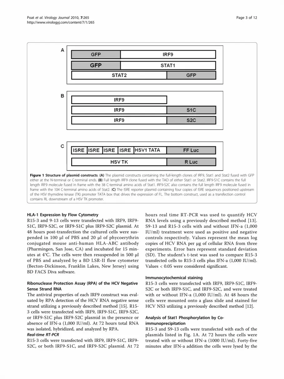
Figure 1 Structure of plasmid constructs. (A) The plasmid constructs containing the full-length clones of IRF9, Stat1 and Stat2 fused with GFPeither at the N-terminal or C-terminal ends. (B) Full length IRF9 clone fused with the TAD of either Stat1 or Stat2. IRF9-S1C contains the fulllength IRF9 molecule fused in frame with the 38 C-terminal amino acids of Stat1. IRF9-S2C also contains the full length IRF9 molecule fused inframe with the 104 C-terminal amino acids of Stat2. (C) The ISRE reporter plasmid containing four copies of ISRE sequences positioned upstreamof the HSV thymidine kinase (TK) promoter TATA box that drives the expression of FL. The bottom construct, used as a transfection controlcontains RL downstream of a HSV TK promoter.
Poat et al. Virology Journal 2010, 7:265http://www.virologyj.com/content/7/1/265
Page 3 of 12

addition of RIPA buffer with proteinase and phosphataseinhibitors (1 × PBS, 1% NP-40, 0.5% Deoxycholate, 0.1%SDS, 50 μg/ml PMSF, 5 μg/ml aprotinin, 5 μg/ml leu-peptin, 1 μg/ml pepstatin). The GFP primary antibody(Santa Cruz Biotechnology, Santa Cruz, CA) was addedto the lysate and rotated at 4°C overnight. The nextmorning protein A/G plus-agarose (Santa Cruz Biotech-nology, Santa Cruz, CA) was added to each sample androtated at 4°C for three hours. Western blotting wasthen performed using a Phospho-Stat1 Y701 (p-Stat1)primary antibody (Cell Signaling Technology, Danvers,MA) diluted (1:1,000) in blocking reagent, (0.1% tween20, Tris buffered saline, and 5% NFDM) as describedbelow.
Immunoblot Blot AnalysisCell lysates were prepared from IRF9, IRF9-S1C, IRF9-S2C, or both IRF9-S1C, and IRF9-S2C transfected R15-3cells at 72 hours post-transfection. Western blot analysiswas then performed using HCV NS5B (Abcam, Cam-bridge, MA), p-eIF-2a (Cell Signalling Technologies,Danvers, MA) and b-actin (Cell Signalling Technologies,Danvers, MA) primary antibodies.
MTT AssayThe viability of IRF9 fusion construct transfected R15-3cells was evaluated by the MTT assay. At 72 hourspost-transfection 100 μl of the MTT solution (Sigma, St.Louis, MO) was added to the media in each well. Afterthree hours of incubation at 37°C the media was aspi-rated and 1 ml of solubilization buffer (0.1 N HCL inabsolute isopropanol) was added to each well. Theabsorbance of each sample was then measured at 570nm utilizing a Beckman DU Spectrophotometer afterblanking. The viability was then calculated by the for-mula dividing the OD570 of each sample by the meanOD570 of the controls.
ResultsIFN-a independent nuclear localization of IRF9 in the R15-3 and S9-13 cellsThe GFP fused Stat1, Stat2 and IRF9 plasmid clonesillustrated in Fig. 1A were used to establish thedynamics of nuclear translocation in the S9-13 and R15-3 cells in the presence and absence of IFN-a. Using atransient transfection experiment, we demonstrate thatboth the Stat1-GFP and Stat2-GFP fusion proteins wereexpressed at high levels in the cytoplasm of both celllines. Both the Stat1-GFP and Stat2-GFP fusion proteintranslocated to the nucleus of S9-13 cells 30 minutesafter IFN-a treatment and returned to the cytoplasm by24 hours (Fig. 2). However, the Stat1-GFP and Stat2-GFP proteins did not localize to the nucleus of the R15-3 cell line following IFN-a treatment. These results
suggest that the nuclear translocation of the Stat1 andStat2 proteins were impaired in the R15-3 cells. TheIRF9-GFP fusion protein was expressed at intermediatelevels in the S9-13 and R15-3 cell lines. The distributionof IRF9-GFP chimera protein was localized in both thecytoplasm and nucleus at all time points in both theR15-3 and S9-13 cell lines. These results suggest thatthe IRF9 protein diffuses freely from cytoplasm to thenucleus of both cell lines independent of IFN-a treat-ment. To understand the reason for differences in thenuclear translocation of Stat1 and Stat2-GFP fusion pro-teins in R15-3 and S9-13 cells, their phosphorylationstatus was examined after transfection. The co-immuno-precipitation experiments show that the Stat1-GFP andStat2-GFP proteins were phosphorylated in the S9-13cells but not in the R15-3 cells (Fig. 3).
Intracellular expression of IRF9-Stat fusion proteinactivates ISRE transcriptionSince the IRF9-GFP construct efficiently localized to thenucleus of the R15-3 cells, we wanted to test whetherfusing the TAD of either the Stat1 or Stat2 proteincould induce IFN promoter activation. For this reason,expression plasmid constructs of IRF9 fused with theTAD of either Stat1 or Stat2 (Fig. 1B) were tested fortheir ability to activate the ISRE promoter in the R15-3cell line. Intracellular expression of IRF9 after plasmidDNA transfection did not activate the ISRE-luciferaseactivity in the R15-3 cell line even after IFN-a treatment(Fig. 4), whereas the IRF9-S1C and IRF9-S2C fusionconstructs significantly induced the ISRE-luciferase pro-moter (p < .025). The combination treatment with bothIRF9-S1C and IRF9-S2C also showed high ISRE-lucifer-ase activity in the R15-3 cells. There were no significantdifferences (p < .05 student’s t-test) in ISRE-luciferaseactivity between the IFN-a (+) and (-) groups suggestingthat the ISRE-luciferase induction was IFN independent.These results suggest that the fusion of the TAD ofeither Stat1 or Stat2 to IRF9 efficiently localized to thenucleus and activated the ISRE promoter in the R15-3cell line. The results of the ISRE promoter inductionexperiments were confirmed by examining the HLA-ABC (HLA-1) surface expression of IRF9-Stat fusiontransfected R15-3 cells by flow cytometry at 72 hourspost-transfection. HLA-1 is an important moleculeinduced by the ISRE promoter which is involved in theimmune recognition of virally infected cells [8]. Intracel-lular expression of either IRF9-S1C or IRF9-S2Cinduced HLA-1 surface expression in the R15-3 cells(Fig. 5A). The change in surface HLA-1 expression inR15-3 cells following plasmid transfection was quanti-fied and compared to the fold change in HLA-1 surfaceexpression of untransfected R15-3 cells after the addi-tion of IFN-a (Fig. 5B). Significant increases (p < .05) in
Poat et al. Virology Journal 2010, 7:265http://www.virologyj.com/content/7/1/265
Page 4 of 12

HLA-1 expression were observed for IRF9-S1C, IRF9-S2C and IRF9-S1C plus IRF9-S2C transfected R15-3cells. In the control S9-13 cells, the expression of HLA-1 was induced after IFN-a treatment.
Efficient clearance of HCV replication from R15-3 cell lineexpressing IRF9-Stat fusion proteinsThe ability of the different IRF9-Stat fusion plasmids toinhibit viral replication was examined using stable repli-con cell lines containing either HCV genotype 1b orHCV 2a sub-genomic RNA. RPA detection of the HCV
negative strand RNA replication intermediate was usedto assess viral replication in IRF9-Stat fusion transfectedR15-3 cells. The RPA results demonstrate that intracel-lular expression of IRF9 alone had no effect upon HCVnegative strand RNA levels, whereas all of the IRF9-Statfusion constructs showed antiviral activity in an IFN-aindependent manner (Fig. 6A). Transfection with IRF9-S1C reduced HCV negative sense RNA strand levels,and IRF9-S2C alone or in combination with IRF9-S1Cinhibited HCV replication below the limit of detectionof the RPA assay. The HCV negative strand RNA RPA
Figure 2 Nuclear translocation of STAT-GFP constructs. The S9-13 and R15-3 cell lines were transfected with IRF9, Stat1, or Stat2 GFP fusionconstructs (-) and (+) IFN-a (1,000 IU/ml) and their nuclear translocation was observed under a confocal microscope. The images are representedas the superimposition of Green Fluorescent Protein (green), To-Pro3 633 (far red), and the differential interference contrast images (DIC) (grayscale). Fluorescence green and red microscopic picture of the same area were taken and superimposed using Abode Photoshop. Left panelshows high resolution picture showing that the IRF9-GFP fusion protein translocates to the nucleus of both S9-13 and R15-3 cells in an IFN-aindependent manner. Middle panel shows stat1-GFP fusion protein efficiently localized to the nucleus of S9-13 cells within 30 minutes afterIFN-a treatment. Stat1-GFP was unable to localize to the nucleus in R15-3 cells. Right panel shows stat2-GFP efficiently localized to the nucleusof S9-13 cells within 30 minutes after IFN-a treatment. Stat2-GFP was unable to localize to the nucleus in R15-3 cells.
Figure 3 Phosphorylation of Stat1-GFP and Stat2-GFP fusion proteins in S9-13 and R15-3 cells determined by co-immunoprecipitation.S9-13 and R15-3 cells were transfected with either Stat1-GFP or Stat2-GFP. After 48 hours IFN-a (1,000 IU/ml) was added. Protein lysates wereprepared, subjected to immunoprecipitation with a GFP antibody, and western blot analysis was performed using a p-STAT1 antibody.
Poat et al. Virology Journal 2010, 7:265http://www.virologyj.com/content/7/1/265
Page 5 of 12

results were verified by quantifying HCV positive strandRNA by real-time RT-PCR. The results in Fig. 6Bdemonstrate that R15-3 cells transfected with IRF9-S1C,IRF9-S2C, or the combination experienced a significantreduction (p < .05) in log copies of HCV RNA per μg ofcellular RNA compared to R15-3 cells plus IFN-a (1,000IU/ml). IRF9 alone transfected R15-3 cells and showed amodest reduction of HCV RNA. S9-13 cells with andwithout IFN-a (1,000 IU/ml) were used as a positivecontrol.Viral protein expression was assessed by immunocyto-
chemical staining for HCV NS3 protein. The results pre-sented in Fig. 7A suggest that the transfection of R15-3cells using the individual constructs IRF9-S1C, IRF9-S2C or the combination reduced HCV NS3 proteinexpression in an IFN-a independent manner. The IRF9alone-transfected R15-3 cells showed no reduction inviral protein levels while the IRF9-S1C, IRF9-S2C andthe combination of both transfected R15-3 cells dis-played a reduction of HCV NS3 expression (Fig. 7A).The addition of IFN-a had no effect on viral proteinlevels in each experimental group. Western blot analysiswas then performed using an antibody against HCVNS5B to verify the results of the antiviral effect of theIRF9-Stat fusions. IRF9-S2C and IRF9-S1C plus IRF9-S2C reduced HCV NS5B protein levels in R15-3 cells(Fig. 7B). R15-3 cells transfected with IRF9 aloneshowed no reduction in HCV NS5B, while the IRF9-S1Ctransfected cells showed weak reduction in viral proteinexpression. All constructs exerted their antiviral activity
in an IFN independent manner. The antiviral propertiesof the IRF9-Stat fusion constructs appear more robustat the 72 hour time point as compared to the previousimmunocytochemical assessment of HCV NS3 at 48hours. This increase in antiviral efficacy between thesetime points correlates with our experience with the anti-viral activity of IFN-a in the S9-13 cell line. The antiviraleffect of IRF9-Stat fusion protein expression in the HCV2a-GFP resistant replicon cells (R4-GFP) was examineddirectly via fluorescence microscopy and then quantifiedby flow analysis (Fig. 8). It was determined that R4-GFPcells expressing IRF9 alone had no antiviral effectswhereas all the remaining constructs had potential anti-HCV activity as they all inhibited HCV-GFP expressionto various degrees. The strongest antiviral activity wasobserved in the IRF9-S2C transfected cells. The viabilityof the R15-3 cell line expressing different IRF9-Statfusion constructs was then examined to exclude thepossibility that the enhanced viral clearance was not dueto the toxic effect of intracellular expression of theIRF9-Stat fusion proteins. The results presented in Fig.9 suggest that no cellular toxicity was associated withthe expression of the Stat fusion proteins in the R15-3cells. Taken together, these results suggest that theintracellular expression of IRF9-S1C and IRF9-S2C leadsto effective clearance of HCV RNA replication in anIFN-a resistant replicon cell line of both HCV1b andHCV2a genotype viruses.Next, the mechanism of inhibition of viral RNA repli-
cation in cells expressing the IRF9-Stat fusions was
Figure 4 Analysis of ISRE-luciferase promoter activation due to intracellular expression of IRF9-Stat fusion proteins. R15-3 cells weretransfected with IRF9, IRF9-S1C or IRF9-S2C plasmid and cultured (-) and (+) IFN-a (1,000 IU/ml). At 48 hours FL and RL activity were measured.Values are expressed in RL normalized units and error bars represent the SEM from three experiments. The Student’s t-test was used to compareIRF9 with and without IFN to IRF9-S1C, IRF9-S2C and IRF9-S1C plus IRF9-S2C with and without IFN. Values <.05 were considered significant.
Poat et al. Virology Journal 2010, 7:265http://www.virologyj.com/content/7/1/265
Page 6 of 12

examined. A principal mechanism that contributes tothe antiviral state and which is activated upon ISRE pro-moter induction is the phosphorylation of eIF2a. Phos-phorylation of eIF2a leads to translational inhibitionwithin the cell thereby preventing viral replication.Therefore, we assessed the ability of each construct toinduce p-eIF2a both in the presence and absence ofIFN-a. The result shown in Fig. 10 suggests that IRF9alone was unable to induce p-eIF2a, while IRF9-S1Ccaused low-level induction of p-eIF2a. Transfection ofIRF9-S2C alone and IRF9-S1C plus IRF9-S2C causedhigh-level p-eIF2a induction. The addition of IFN-a didnot significantly (p < .025) alter the expression patternsof p-eIF2a.
DiscussionThe molecular mechanisms of IFN-a resistance areunclear, however a number of studies have provided evi-dence that both viral and cellular factors are involved
[18-23]. We provided evidence that replicon cellsdevelop IFN-a resistance due to defective Jak-Stat sig-naling. Using long-term IFN-a treatment of repliconcells we have established IFN-a resistant sub-genomicHCV based stable replicon cell lines for HCV 1b andHCV 2a genotype viruses. Replication of HCV RNA inthese stable cell lines remained resistant to IFN-a due todefects in Stat1 and Stat2 phosphorylation and the sub-sequent impairment of nuclear translocation of ISGF3.In this study, a potential therapeutic strategy againstchronic HCV infection using a chimeric protein of IRF9fused to the TAD of either the Stat1 or Stat2 moleculeof the Jak-Stat signaling pathway was examined.We showed that the IRF9 protein was expressed at
intermediate levels in the R15-3 cells after transfectionand efficiently localized to the nucleus, suggesting thatthe nuclear translocation of IRF9 protein in the R15-3cells is not dependent upon IFN-a induced Jak-Stat sig-naling. Nuclear translocation of the Stat1-GFP and
Figure 5 Intracellular expression of IRF9-S1 or IRF9-S2C induced HLA-1 surface expression in the R15-3 cells. S9-13 and R15-3 cells weretransfected and cultured (-) and (+) IFN-a (1,000 IU/ml). After 48 hours, HLA-1 surface expression was quantified by flow cytometry. (A) Showsthe HLA-1 surface expression in the IRF9-Stat fusion transfected R15-3 cells. The red cell population represents IFN-a naïve cells or untransfectedcells and the blue cell population represents IFN-a treated or transfected cells. IRF9-S1C, IRF9-S2C, and IRF9-S1C plus IRF9-S2C transfected R15-3cells demonstrated significant increases in HLA-1 surface expression. (B) Each value represents the mean fluorescence intensity from sixexperiments. The student’s t-test was used to compare the fold increase of transfected R15-3 cells to untransfected R15-3 cells plus IFN-a.Asterisk (*) indicates transfected R15-3 cells with significantly different HLA-1 surface expression relative to untransfected R15-3 cells. Significancewas considered at p-values < 0.05.
Poat et al. Virology Journal 2010, 7:265http://www.virologyj.com/content/7/1/265
Page 7 of 12

Stat2-GFP chimera proteins was efficient in the S9-13cell line; however the nuclear localization of Stat1-GFPand Stat2-GFP did not occur in the R15-3 cells that pos-sess defective Jak-Stat signaling. These results led us toexamine whether the development of a chimera proteinbetween the IRF9 and the TAD of either Stat1 or Stat2protein could facilitate nuclear translocation and induceIFN antiviral gene expression. We showed here thatintracellular expression of the IRF9-Stat1 and Stat2fusion proteins in the R15-3 cell line activated the ISRE-luciferase promoter in a concentration dependent man-ner. Expression of the IRF9 protein alone did not acti-vate the ISRE-luciferase promoter activity suggestingthat IRF9 alone does not control the antiviral gene tran-scription induced by IFN-a. The results of this studyalso validate the previous finding that the fusion of theTAD of either Stat1 or Stat2 to IRF9 protein inducedthe ISRE promoter [14]. The activity of IRF9-S2C wasstronger than IRF9-S1C. The recombinant IRF9-Stat
fusion constructs also induced HLA-1 surface expres-sion in R15-3 cells in an IFN-a independent manner.Based on these results we speculate that intracellularexpression of IRF9-S2C or IRF9-S1C in HCV infectedhepatocytes may induce HLA-1 surface expression andmay thus increase cytotoxic T lymphocyte (CTL)mediated viral clearance. Interestingly we observed thatthe intracellular expression of IRF9-S2C alone or thecombination with IRF9-S1C showed stronger activationof ISRE-luciferase promoter activation than IRF9-S1Calone, indicating that Stat2 expression contributes moretowards the potent antiviral activity against HCVinfected hepatocytes with defective Jak-Stat signaling.On the other hand, intracellular expression of eitherIRF9-S1C, IRF9-S2C or the combination of both showsimilar results in the mobilization of HLA-1 surfaceexpression in R15-3 cells. These results suggest thatintracellular expression Stat1 fusion could contribute
Figure 6 Ribonuclease protection assay and Real-time RT-PCR of sub-genomic HCV 1b RNA. (A) Intracellular expression of IRF9-S1C andIRF9-S2C or both in R15-3 cells inhibits HCV negative strand RNA detection by RPA. R15-3 cells were transfected treated (-) and (+) IFN-a (1,000IU/ml), and total RNA was isolated at 72 hours post-transfection. The upper panel shows the RPA results of HCV negative strand detection. Thebottom panel shows GAPDH mRNA levels by RPA. (B) Real-time RT-PCR was performed to quantify HCV RNA in R15-3 transfected cells. CellularRNA was isolated at 72 hours, retrotranscribed, and assayed by real time PCR. S9-13 and R15-3 cells (+) and (-) IFN-a were used as (+) and (-)controls, respectively. Error bars represent SD. Asterisk (*) indicates transfected R15-3 cells with significantly lower HCV RNA relative tountransfected R15-3 cells treated with IFN. Significance was considered at p-values < 0.05.
Poat et al. Virology Journal 2010, 7:265http://www.virologyj.com/content/7/1/265
Page 8 of 12

more towards the immunomodulatory activity in HCVinfected hepatocytes with defective Jak-Stat signaling.The antiviral properties of the IRF9-Stat fusion pro-
teins were also examined using stable IFN-resistant celllines of HCV 1b and 2a genotype viruses. Using theR15-3 cell line containing sub-genomic HCV genotype1b RNA, we showed that intracellular expression ofIRF9-S1C and IRF9-S2C efficiently inhibited negative
strand HCV RNA levels and viral NS3 protein expres-sion in a replicon cell line containing defective Jak-Statsignaling. This approach was also tested using IFN-aresistant HCV 2a sub-genomic GFP replicon. Weshowed that transient transfection of IRF9-S1C andIRF9-S2C or both inhibited GFP expression in an IFN-aindependent manner. These results suggest that thisantiviral strategy may be effective against different HCV
Figure 7 Immunostaining of HCV NS3 and Western blot of HCV NS5B proteins. (A) The antiviral activity of the IRF9-Stat fusion constructsin R15-3 cells containing HCV 1b sub-genomic RNA HCV. At 48 hours post-transfection the cells were mounted onto a glass slide, stained forHCV NS3 protein and counterstained with hematoxylin. Panel i and ii: Huh-7 cells (-) and (+) IFN-a (1,000 IU/ml). Panel iii and iv: R15-3 cells (-)and (+) IFN-a. Panel v: IRF9 transfected R15-3 cells Panel vi: IRF9-S1C transfected R15-3 cells Panel vii: IRF9-S2C transfected R15-3 cells Panel viii:IRF9-S1C plus IRF9-S2C transfected R15-3 cells. (B) Upper panel: HCV NS5B expression levels in R15-3 transfected cells at 72 hours post-transfection. Lower panel: Shows b-actin protein expression levels using equal amounts of protein lysate.
Poat et al. Virology Journal 2010, 7:265http://www.virologyj.com/content/7/1/265
Page 9 of 12

genotypes. To understand the mechanism of inhibitionof HCV replication in the R15-3 cell line, the proteinkinase R (PKR) mediated eIF-2a phosphorylation path-way was examined by western blot analysis. Our resultssuggest that the mechanism of inhibition of viral replica-tion by intracellular IRF9-Stat fusion expression is
similar to that described for IFN-a and involves PKRinduced eIF-2a phosphorylation [24].A critical determinant in the outcome of HCV infec-
tion is the host’s ability to mount an effective HCV spe-cific CD8+ T cell response. It was also previouslydemonstrated that a defective Jak-Stat system
Figure 8 The antiviral activity of the IRF9-Stat fusion constructs in an IFN resistant cell line containing HCV 2a sub-genomic RNA (R4-GFP). (A) GFP expression levels in R4-GFP cells transfected with IRF9, IRF9-S1C and IRF9-S2C at 72 hrs post-transfection. (B) Flow cytometricanalysis of mean fluorescence intensity of R4-GFP transfected cells at 72 hrs. S3-GFP and R4-GFP cells treated with and without IFN-a were usedas controls. The error bars represent the SEM from three experiments. The Student’s t-test was used to compare R4-GFP transfected cells to R4-GFP cells plus IFN-a. P-values < 0.05 were considered significant.
Poat et al. Virology Journal 2010, 7:265http://www.virologyj.com/content/7/1/265
Page 10 of 12

contributed to impaired HLA-1 surface expression [25].It is therefore conceivable that the ineffective CD8+responses of chronically infected HCV patients and IFNnon-responders may in part be linked to the inability ofinfected hepatocytes to surface display the HLA-1: anti-gen complex. This impaired surface presentation mayalso contribute to the immune evasion observed invirally infected hepatocellular carcinoma cells. A sub-stantial body of clinical evidence exists to support therole of dysregulated Jak-Stat signaling in IFN non-response in chronic HCV infection [26-29]. These clini-cal studies as well as our cell culture studies support theimportance of Jak-Stat signaling in infected hepatocytesin viral persistence and response to IFN-a treatment.Using HCV cell culture models we now provide
evidence that components of the Jak-Stat pathway canbe engineered to circumvent defective signaling therebyinducing an antiviral state resulting in HCV eradicationwithout IFN-a treatment. Based on these in vitro studieswe propose that combination treatment using IRF9-S1Cand IRF9-S2C recombinant proteins may provide arationale for future development of an antiviral strategyto overcome IFN-a resistance by circumventing Jak-Statcellular defects in chronically infected liver cells. Wepropose that liver targeted delivery of IRF9-Stat fusionprotein can be used as a second line treatment inchronically infected hepatitis C patients with defectiveJak-STAT signaling in an attempt to stimulate an anti-viral response as well as increase HLA-1 expression inhepatocytes in an IFN-a independent manner.
Figure 9 MTT Assay of IRF9-Stat fusion constructs in resistant cell line. R15-3 cells were transfected with IRF9 constructs and at 72 hourspost-transfection cell viability was determined by the MTT assay. Each value represents experiments performed in triplicate. Error bars representSEM.
Figure 10 Intracellular expression of IRF9-Stat fusion constructs induced eIF2a phosphorylation in the R15-3 cell line at 72 hourspost-transfection. Upper panel: Western blot of p-eIF2a in R15-3 cells transfected with different IRF9 plasmids in the absence and presence ofIFN-a (1,000 IU/ml). Bottom Panel: b-actin protein expression levels.
Poat et al. Virology Journal 2010, 7:265http://www.virologyj.com/content/7/1/265
Page 11 of 12

AcknowledgementsThis work was supported by funds received from the National CancerInstitute (CA127481, CA129776), Louisiana Board of Regents, LouisianaCancer Research Consortium and Tulane Cancer Center. The authorsacknowledge Dr. Curt Horvath, Feinberg School of Medicine, NorthwesternUniversity, Illinois, USA for the generous gift of the IRF9-STAT recombinantplasmids. The authors also acknowledge Dr. Mario Koster, Department ofGene Regulation and Differentiation, GBF-National Research Institute forBiotechnology, Braunschweig, Germany for kindly providing the STAT2-GFPconstruct. Ralf Bartenschlager, University of Heidelberg, Heidelberg, Germanyfor providing 9-13 cell line. The authors thank Dr. Astrid Engel, Dr. SrutiChandra, and Mallory Heath for critically evaluating this manuscript.
Author details1Department of Pathology and Laboratory Medicine, Tulane UniversityHealth Sciences Center, 1430 Tulane Avenue, New Orleans, LA-70112, USA.2Section of Gastroenterology and Hepatology, Tulane University HealthSciences Center, 1430 Tulane Avenue, New Orleans, LA-70112, USA.3Department of Microbiology and Immunology, Tulane University HealthSciences Center, 1430 Tulane Avenue, New Orleans, LA-70112, USA. 4Divisionof Comparative Pathology, Tulane National Primate Research Center, 18703Three Rivers Road, Covington, LA-70433, USA.
Authors’ contributionsBP performed major biochemical experiments, participated in the design ofthe study and wrote the initial draft of the manuscript. SH, PKC, FG and XAdid some biochemical experiments and participated in the design of thestudy. SD and RFG supervised, helped to design the study and finally wrotethe manuscript. LAB share ideas and helped for manuscript preparation. Allauthors read and approved the final manuscript.
Competing interestsThe authors declare that they have no competing interests.
Received: 17 July 2010 Accepted: 12 October 2010Published: 12 October 2010
References1. Alter MJ: Epidemiology of hepatitis C virus infection. World J Gastroenterol
2007, 13:2436-2441.2. Sy T, Jamal M: Epidemiology of Hepatitis C virus infection. Int J Med Sci
2006, 3:41-46.3. Post J, Ratnarajah S, Lloyd AR: Immunological determinants of the
outcomes from primary hepatitis C infection. Cell Mol Life Sci 2009,66:733-756.
4. Te HS, Jensen DM: Epidemiology of hepatitis B and C viruses: a globaloverview. Clin Liver Dis 2010, 14:1-21.
5. Fried MW, Shiffman ML, Reddy KR, Smith C, Marinos G, Gonçales FL Jr,Häussinger D, Diago M, Carosi G, Dhumeaux D, Craxi A, Lin A, Hoffman J,Yu J: Peginterferon alfa-2a plus ribavirin for chronic hepatitis C virusinfection. N Engl J Med 2002, 347:975-982.
6. Manns MP, McHutchison JG, Gordon SC: Peg-interferon alfa-2a plusribavirin compared with interferon alfa-2b plus ribavirin for initialtreatment of chronic hepatitis C: a randomised trial. Lancet 2001,358:958-965.
7. Manns M, Wedemeyer H, Cornberg M: Treating viral hepatitis C: efficacy,side effects, and complications. Gut 2006, 55:1350-1359.
8. Samuel CE: Antiviral actions of interferons. Clin Microbiol Rev 2001,14:778-809.
9. Trepo C: Genotype and viral load as prognostic indicators in thetreatment of hepatitis C. J Viral Hepatitis 2000, 7:250-257.
10. Taylor DR, Shi ST, Lai MM: Hepatitis C virus and interferon resistance.Microbes Infect 2000, 2:1743-1756.
11. He Y, Katze MG: To interfere and to anti-interfere: the interplay betweenhepatitis C virus and interferon. Viral Immunol 2002, 15:95-119.
12. Hazari S, Taylor L, Garry RF, Dash S: Reduced expression of Jak-1 and Tyk-2 proteins leads to interferon resistance in hepatitis C virus replicon.Virol J 2007, 4:89.
13. Hazari S, Chandra PK, Poat B, Datta S, Garry RF, Dash S: Impaired antiviralactivity of IFN-α in Huh-7 cells with defective Jak-Stat pathway. Virol J2010, 7:36.
14. Kraus TA, Lau JP, Parisien JP, Horvath CM: A hybrid IRF9-Stat2 proteinrecapitulates interferon stimulated gene expression and antiviralresponse. J Biol Chem 2003, 278:13033-13038.
15. Mirabel RS, Pai M, Prabhu R, Panebra A, Nangle S, Bastian F, Garry RF,Agrawal K, Goodbourn S, Dash S: Activation of Interferon StimulatedResponse Element in a Huh-7 cell line replicating hepatitis C virus sub-genomic RNA. Intervirology 2005, 48:301-311.
16. Timofeeva OA, Plisov S, Evseev AA, Peng S, Jose-Kampfner M, Lovvorn HN,Dome JS, Perantoni AO: Serine-phosphorylated STAT1 is a prosurvivalfactor in Wilms’ tumor pathogenesis. Oncogene 2006, 25:7555-7564.
17. Köster M, Hauser H: Dynamic redistribution of STAT1 protein in IFNsignaling visualized by GFP fusion proteins. Eur J Biochem 1999,260:137-144.
18. Zhu H, Nelson DR, Crawford JM, Liu C: Defective Jak-Stat activation inhepatoma cells is associated with hepatitis C viral IFN-α resistance. JInterferon Cytokine Res 2005, 25:528-539.
19. Pawlotsky JM: The nature of IFN-α resistance in hepatitis C virusinfection. Curr Opin Infect Dis 2003, 16:587-592.
20. Pawlotsky JM: Mechanisms of antiviral treatment efficacy and failure inchronic hepatitis C. Antiviral Res 2003, 59:1-11.
21. Gale MJ Jr, Korth MJ, Tang NM, Tan SL, Hopkins DA, Dever TE, Polyak SJ,Gretch DR, Katze MG: Evidence that hepatitis C virus resistance tointerferon is mediated through repression of the PKR protein kinase bythe nonstructural 5A protein. Virology 1997, 230:217-227.
22. Gao B, Hong F, Radaeva S: Host factors and failure of IFN-α treatment inHCV. Hepatology 2004, 39:880-890.
23. Su X, Yee LJ, Im K, Rhodes SL, Tang Y, Tong X, Howell C, Ramcharran D,Rosen HR, Taylor MW, Liang TJ, Yang H, Virahep-C Study Group:Association of single nucleotide polymorphisms in interferon signalingpathway genes and interferon-stimulated genes with the response tointerferon therapy for chronic hepatitis C. J Hepatol 2008, 49:184-191.
24. Rivas-Estillas AM, Svitkin Y, Lastra ML, Hatzoglou M, Sherker A, Koromilas AE:PKR-dependent mechanisms of gene expression from a subgenomichepatitis C virus clone. J Virol 2002, 76:10637-10653.
25. Rhodes SL, Erlich H, Im KA, Wang J, Li J, Bugawan T, Jeffers L, Tong X, Su X,Rosen HR, Yee LJ, Liang TJ, Yang H, Virahep-C study group: Associationbetween the human MHC and sustained virologic response in thetreatment of chronic hepatitis C virus infection. Genes Immun 2008,9:328-333.
26. Welzel TM, Morgan TR, Bonkovsky HL, Naishadham D, Pfeiffer RM,Wright EC, Hutchinson AA, Crenshaw AT, Bashirova A, Corrington M:Variants in IFN-α pathway genes and response to pegylated IFN-α2aplus ribavirin for treatment of chronic hepatitis C virus infection in thehepatitis C Antiviral Long-term treatment against Cirrhosis trial.Hepatology 2009, 49:1847-1858.
27. Aceti A, Zechini B, Griggi T, Marangi M, Pasquazzi C, Quaranta G, Sorice M:Undetectable phospho-STAT1 in peripheral blood mononuclear cellsfrom patients with chronic hepatitis C who do not respond to IFN-αtherapy. Liver Int 2005, 5:987-993.
28. Miyaaki H, Ichikawa T, Nakao K, Takeshita S, Shibata H, Ozawa E, Akiyama M,Miuma S, Eguchi K: Predictive value of the phosphorylation of signaltransducers and activators of transcription in the outcome of IFNtherapy for chronic hepatitis C. Intervirology 2008, 51:394-399.
29. Sarasin-Filipowicz M, Oakeley EJ, Duong FH, Christen V, Terracciano L,Filipowicz W, Heim MH: IFN signaling and treatment outcome in chronichepatitis C. Proc Natl Acad Sci USA 2008, 105:7034-9039.
doi:10.1186/1743-422X-7-265Cite this article as: Poat et al.: Intracellular expression of IRF9 Stat fusionprotein overcomes the defective Jak-Stat signaling and inhibits HCVRNA replication. Virology Journal 2010 7:265.
Poat et al. Virology Journal 2010, 7:265http://www.virologyj.com/content/7/1/265
Page 12 of 12