copyright © 2008 pearson allyn & bacon inc.1 chapter 6 vision this multimedia product and its...
TRANSCRIPT

Copyright © 2008 Pearson Allyn & Bacon Inc.1
Chapter 6
Vision
This multimedia product and its contents are protected under copyright law. The following are prohibited by law
•any public performance or display, including transmission of any image over a network
•preparation of any derivative work, including the extraction, in whole or in part, of any images
•any rental, lease or lending of the program.

Copyright © 2008 Pearson Allyn & Bacon Inc.2
• Chapter 6 Outline
• The Stimulus
• Anatomy of the Visual System
• Coding of Visual Information in the Retina
• Analysis of Visual Information:
Role of the Striate Cortex
• Analysis of Visual Information:
Role of the Visual Association Cortex

Copyright © 2008 Pearson Allyn & Bacon Inc.3
• Vision
• Sensory _______________• A specialized __________ that detects a particular
category of _________________ events.
• Sensory __________________• The process by which sensory stimuli are transduced
into slow, graded ____________ potentials.
• Receptor potential• A slow, graded electrical potential produced by a
receptor cell in response to a physical __________.

Copyright © 2008 Pearson Allyn & Bacon Inc.4
• The Stimulus• Perceived color of light is determined by:
• Hue• Determined by _______________
• Brightness• Determined by the ______________ of the
electromagnetic radiation
• Saturation• Determined by the ___________of the light wave• White light is a blend of all wavelengths, if white light
is added to a monochromatic light it becomes unsaturated - pastel

Copyright © 2008 Pearson Allyn & Bacon Inc.5
Electromagnetic spectrum

Copyright © 2008 Pearson Allyn & Bacon Inc.6

Copyright © 2008 Pearson Allyn & Bacon Inc.7
• Anatomy of the Visual System
• The eyes
• _____________• Bony pockets in the front of the skull.
• Sclera• The ____________ tissue of the eye.
• Conjunctiva• Mucous membranes that line the eyelid and protect
the eye. Prevents objects from __________________________________________________________________________________.

Copyright © 2008 Pearson Allyn & Bacon Inc.8
• Anatomy of the Visual System
• The eyes
• Cornea• ______________________________ of the eye that
admits light.
• Pupil• Adjustable opening in the ___________ that
regulates the amount of light that enters the eye.
• Iris• __________________________ situated behind the
cornea.

Copyright © 2008 Pearson Allyn & Bacon Inc.9
• Anatomy of the Visual System
• The eyes
• Lens• Consists of a series of transparent, onion-like
layers. Its shape can be changed by ________________________________________________________________________.
• ______________________• Changes in the ________________of the lens,
accomplished by the ciliary muscles, that focus images of near or distant objects on the retina.

Copyright © 2008 Pearson Allyn & Bacon Inc.10
• Anatomy of the Visual System
• The eyes
• ____________• The neural tissue and photoreceptive cells located
on the inner surface of the posterior portion of the eye.
• Photo______________• One of the receptor cells of the retina; transduces
photic energy into electrical potentials

Copyright © 2008 Pearson Allyn & Bacon Inc.11
• Anatomy of the Visual System
• The eyes
• __________ (120 million)• Photoreceptor cells of the retina, sensitive to the• light of low intensity.
• _______________ (6 million)• Photoreceptor cells of the retina; maximally
sensitive to one of three different wavelengths of light and hence encodes color vision.

Copyright © 2008 Pearson Allyn & Bacon Inc.12
• Anatomy of the Visual System
• The eyes
• ____________• Region of the retina that mediates the most acute
vision of birds and higher mammals. Color sensitive cones constitute the only type of photoreceptor found in the fovea.
• _______________• The location of the exit point from the retina of the
fibers of the ganglion cells that form the optic nerve; responsible for the ______________.

Copyright © 2008 Pearson Allyn & Bacon Inc.13

Copyright © 2008 Pearson Allyn & Bacon Inc.14

Copyright © 2008 Pearson Allyn & Bacon Inc.15
• Anatomy of the Visual System
• The eyes
• Bipolar cell• A bipolar neuron located in the middle layer of the
retina, conveying information from the ____________________________.
• Ganglion cell• A neuron that receives visual information from
bipolar cells; its ______________________________________________________________________________.

Copyright © 2008 Pearson Allyn & Bacon Inc.16

Copyright © 2008 Pearson Allyn & Bacon Inc.17
• Anatomy of the Visual System
• The eyes
• _________________cell• A neuron in the retina that interconnects adjacent
photoreceptors and the outer processes of the bipolar cells.
• _____________________cell• A neuron in the retina that interconnects adjacent
ganglion cells and the inner processes of the bipolar cells.

Copyright © 2008 Pearson Allyn & Bacon Inc.18

Copyright © 2008 Pearson Allyn & Bacon Inc.19
• Anatomy of the Visual System
• Photoreceptors
• Lamella• A layer of membrane containing ______________;
found in rods and cones.
• Photopigment• A protein dye bonded to retinal, a substance derived
from vitamin ___; responsible for the transduction of visual information.
• Opsin• A class of protein that, together with retinal,
constitutes the photopigments.

Copyright © 2008 Pearson Allyn & Bacon Inc.20
• Anatomy of the Visual System
• Photoreceptors
• Retinal• A chemical synthesized from vitamin A, joins with
an opsin to form a _________________.
• ________________• A particular opsin found in __________.• In the dark adapted state it appears _________.• In the bleached state the color changed to
_________.

• The action of light on some neurons in the visual system.
• Photoreceptors are ___________________by light.
• Hyperpolarization causes a reduction in the photoreceptor’s release of the neurotransmitter ______________.
• Because glutamate normally hyperpolarizes the bipolar cell, the reduction in glutamate causes the bipolar cell to ______________.
• The relative depolarization of the bipolar cell causes the ganglion cell to ___________________.
• Thus light ____________ the ganglion cell.Copyright © 2008 Pearson Allyn & Bacon Inc.21

Copyright © 2008 Pearson Allyn & Bacon Inc.22
• Anatomy of the Visual System
• Connections between eye and brain
• Dorsal -___________________________• A group of cell bodies within the lateral geniculate
body of the thalamus; receives inputs from the retina and projects to the primary visual cortex.
• Magnocellular layer• One of the two inner layers in the dorsal lateral
geniculate nucleus; transmits information necessary for the perception of form, ________________, depth, and small differences in brightness to the primary visual cortex.

Copyright © 2008 Pearson Allyn & Bacon Inc.23

Copyright © 2008 Pearson Allyn & Bacon Inc.24
• Anatomy of the Visual System
• Connections between eye and brain
• Parvocellular layer• One of the four outer layers of neurons in the
dorsal lateral geniculate nucleus; transmits information necessary for perception ________________________________________________________________________________
• Koniocellular sublayer• One of the sublayers of neurons in the dorsal
lateral geniculate nucleus found ventral to each of the magnocellular and parvocellular layers; transmits information from short-wavelength cones to the primary visual cortex.
• These two layers are responsible ________________________________________________________________________________

Copyright © 2008 Pearson Allyn & Bacon Inc.25
• Anatomy of the Visual System
• Connections between eye and brain
• Calcarine fissure• Horizontal fissure on the inner surface of the
posterior cerebral cortex; the location of the primary visual cortex.
• Striate cortex• The primary visual cortex.
• Optic chiasm• A connection between the optic nerves, located
below the base of the brain, just anterior to the pituitary gland.

Copyright © 2008 Pearson Allyn & Bacon Inc.26

Copyright © 2008 Pearson Allyn & Bacon Inc.27
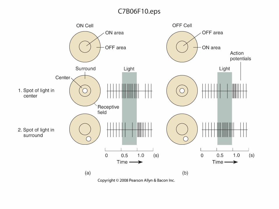
• Coding of Visual Information in the Retina
• Coding of Light and Dark
• Receptive Field• That portion of the visual field in which the presentation of
visual stimuli will produce an alteration in the firing rate of a particular neuron. If the neuron receives information from receptors located in the fovea, the receptive field will be at the point the eye is looking. If the input is from receptors in the peripheral retina, the receptive field will be off to the side, or above or below where the eye is looking.

Copyright © 2008 Pearson Allyn & Bacon Inc.28
Copyright © 2008 Pearson Allyn & Bacon Inc.29

Copyright © 2008 Pearson Allyn & Bacon Inc.30
• Coding of Visual Information in the Retina
• Photoreceptors: trichromatic coding
• _________________• An inherited form of defective color vision in which
red and green hues are confused; ___________________________________________________________________________________.
• They see the world in shades of yellow and blue; both red and green look _____________to them.
• Visual acuity is normal.

• Theories of color vision coding.
• Trichromatic – any color can be reproduced by mixing three colors selected along the spectrum.
• Opponent-process – ________________of color
• Red-green
• Blue-yellow
• Black-white
• Produces complimentary colors
• Color vision yields the ability to distinguish ________________________________________.
Copyright © 2008 Pearson Allyn & Bacon Inc.31
Copyright © 2008 Pearson Allyn & Bacon Inc.32
• Coding of Visual Information in the Retina
• Photoreceptors: trichromatic coding
• ________________• An inherited form of defective color vision in which red
and green hues are confused; __________________________________________________________________________________________.
• Visual acuity is normal.

Copyright © 2008 Pearson Allyn & Bacon Inc.33
• Coding of Visual Information in the Retina
• Photoreceptors: trichromatic coding
• _______________• An inherited form of defective color vision in which
hues with short wavelengths are confused; ______________________________________________________________________________.
• See the world in greens and reds.
• Blue looks green and yellow looks pink.
• Visual acuity is normal.

Copyright © 2008 Pearson Allyn & Bacon Inc.34

Copyright © 2008 Pearson Allyn & Bacon Inc.35

Copyright © 2008 Pearson Allyn & Bacon Inc.36
• Analysis of Visual Information: The Striate Cortex
• Anatomy of the striate cortex
• David Hubel and Torsten Wiesel
• 1960s at Harvard University
• Discovered that neurons in the visual cortex did not simply respond to light; they selectively responded to ____________________ of the visual world. The receptive field size exceeds that of a single ganglion cell.

Copyright © 2008 Pearson Allyn & Bacon Inc.37

Copyright © 2008 Pearson Allyn & Bacon Inc.38
• Analysis of Visual Information: The Striate Cortex
• Orientation and movement
• ______________ cell• An orientation-sensitive neuron in the striate
cortex whose receptive field is organized in an opponent fashion.

Copyright © 2008 Pearson Allyn & Bacon Inc.39
• Analysis of Visual Information: The Striate Cortex
• Orientation and movement
• ______________ cell• A neuron in the visual cortex that responds to the
presence of a line segment with a particular orientation located within its receptive field,especially when the line ____________ perpendicular to its orientation.

Copyright © 2008 Pearson Allyn & Bacon Inc.40
• Analysis of Visual Information: The Striate Cortex
• Orientation and movement
• ____________________ cell• A neuron in the visual cortex that responds to the
presence of a line segment with a particular orientation that ___________ at a particular point within a cell’s receptive field.

Copyright © 2008 Pearson Allyn & Bacon Inc.41

Copyright © 2008 Pearson Allyn & Bacon Inc.42

Copyright © 2008 Pearson Allyn & Bacon Inc.43
• Analysis of Visual Information: The Striate Cortex
• Spatial frequency
• _______________ grating• A series of straight parallel bands varying
continuously in the brightness according to a sine-wave function, along a line perpendicular to their lengths.
• Cortical cells tend to respond ___________ to sine wave gratings than bars

Copyright © 2008 Pearson Allyn & Bacon Inc.44
• Analysis of Visual Information: The Striate Cortex
• Spatial Frequency
• Spatial frequency• The relative width of the bands in a sine-wave grating,
measured in ____________________________________________________________________________________.

Copyright © 2008 Pearson Allyn & Bacon Inc.45

Copyright © 2008 Pearson Allyn & Bacon Inc.46

Copyright © 2008 Pearson Allyn & Bacon Inc.47
• Analysis of Visual Information: The Striate Cortex
• Retinal Disparity
• Retinal disparity• The fact that points on objects located at different
distances from the observer will fall on slightly different locations on the two retinas; provides the basis for _____________ or depth perception
• Some neurons respond __________to images that are produced by retinal disparity.

• Hypercolumns and retinotopic maps
• The _______ is very small (about 2 degrees of visual angle, and is about 1/1000 of the area of the retina.
• In the striate cortex about 25% of the cortex is devoted to the analysis of the fovea.
• ______________________________for foveal processing in the cortex.
Copyright © 2008 Pearson Allyn & Bacon Inc.48

Copyright © 2008 Pearson Allyn & Bacon Inc.49

Copyright © 2008 Pearson Allyn & Bacon Inc.50
• Analysis of Visual Information: The Striate Cortex
• Color
• Cytochrome oxidase (CO) __________• The central region of a module of the primary visual
cortex, revealed by a stain for cytochrome oxidase; contains ______________________; part of the parvocellular system.

Copyright © 2008 Pearson Allyn & Bacon Inc.51

Copyright © 2008 Pearson Allyn & Bacon Inc.52

Copyright © 2008 Pearson Allyn & Bacon Inc.53
• Analysis of Visual Information: The Visual Association Cortex
• _________________ cortex• A region of the visual association cortex; receives
fibers from the striate cortex and from the superior colliculi and projects to the inferior _________ cortex.
• Regions respond to particular _____________of visual information such as orientation, movement, spatial frequency, retinal disparity, or color.

Copyright © 2008 Pearson Allyn & Bacon Inc.54
• Analysis of Visual Information: The Visual Association Cortex
• _______________ stream• A system of interconnected regions of the visual
cortex involved in the perception of spatial location, beginning with the striate cortex and ending with the posterior ______________ cortex.
• _______________ stream• A system of interconnected regions of visual cortex
involved in the perception of form, beginning with the striate cortex and ending with the inferior ____________ cortex.

Copyright © 2008 Pearson Allyn & Bacon Inc.55

Copyright © 2008 Pearson Allyn & Bacon Inc.56

Copyright © 2008 Pearson Allyn & Bacon Inc.57
• Analysis of Visual Information: The Visual Association Cortex
• Color constancy• The relative constant ____________ of the colors of
objects viewed under varying __________conditions.
• This ability compensates for the perception of the ___________________ under different lighting conditions.

Copyright © 2008 Pearson Allyn & Bacon Inc.58
• Analysis of Visual Information: The Visual Association Cortex
• Studies with humans
• Cerebral _________________• Inability to discriminate among different hues; caused
by damage to the visual association cortex.• Damage to V4 disrupts ________________, but not
the ability to discriminate between colors.• Damage to TEO (V8) disrupts __________________
(and __________ for colors), but not the ability to distinguish between shades of gray.

Copyright © 2008 Pearson Allyn & Bacon Inc.59

Copyright © 2008 Pearson Allyn & Bacon Inc.60
• Analysis of Visual Information: The Visual Association Cortex
• Studies with humans
• _____________• Inability to perceive or identify a stimulus by means of
a particular sensory modality.
• Visual agnosia• Deficits in visual perception in the absence of
_______________; caused by brain damage.
• Apperceptive visual agnosia• Failure to ____________________ even though
visual acuity is relatively normal.
Copyright © 2008 Pearson Allyn & Bacon Inc.61
• Analysis of Visual Information: The Visual Association Cortex
• Analysis of form
• _________________• Failure to recognize particular people by the sight of
their ____________.
• Associative visual agnosia• Inability to ___________________ that are perceived
visually, even though the form of the perceived object can be drawn or matched with similar objects.

Copyright © 2008 Pearson Allyn & Bacon Inc.62
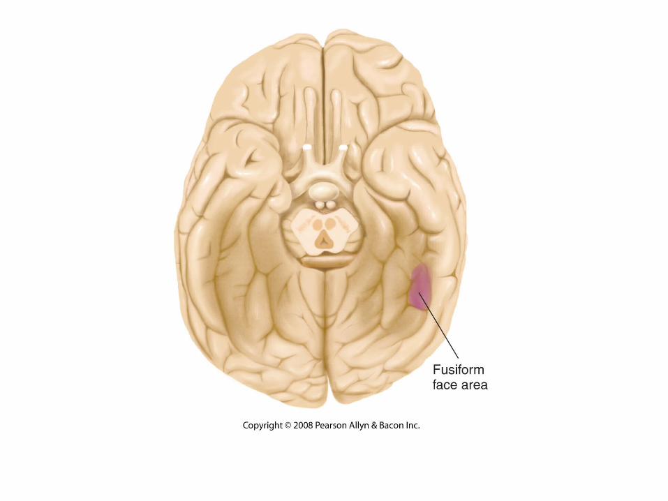
• Analysis of Visual Information: The Visual Association Cortex
• _________________ area• A region of the extrastriate cortex located at the base
of the brain; involved in perception of faces and other objects that require expertise to recognize.
Individuals with ______________ show a deficit in the ability to recognize faces, and the fusiform facial area is weakly activated when individuals look at faces.
• Akinetopsia• Inability to perceive___________, caused by damage to
area V5 of the visual association cortex.
Copyright © 2008 Pearson Allyn & Bacon Inc.63

Copyright © 2008 Pearson Allyn & Bacon Inc.64
Subjects with visual object agnosia may be able to recognize a face, but not the vegetables that compose it.

Copyright © 2008 Pearson Allyn & Bacon Inc.65

Copyright © 2008 Pearson Allyn & Bacon Inc.66
• Analysis of Visual Information: The Visual Association Cortex
• Perception of movement
• Extrastriate body area (EBA)• A region of the visual association cortex located in the
lateral occipitotemporal cortex; involved in perception of ____________________________________________.
• Parahippocampal place area (PPA)• A region of the medial temporal cortex; involved in
perception of particular places (_______________).

Copyright © 2008 Pearson Allyn & Bacon Inc.67
• Analysis of Visual Information: The Visual Association Cortex
• Perception of movement• ________________
• The complex motion of points in the visual field caused by relative movement between the observer and environment; provides information about the relative _______________of objects from the observer and of the relative ____________________.
• Akinetopsia• Inability to perceive movement, caused by damage to
area V5 of the visual association cortex. Unable to ___________________________________________________________________________________.

Copyright © 2008 Pearson Allyn & Bacon Inc.68
V5: damage produces akinetopsia

Copyright © 2008 Pearson Allyn & Bacon Inc.69
• Analysis of Visual Information: The Visual Association Cortex
• Perception of spatial location
• Intraparietal sulcus• The end of the dorsal stream of the visual association
coretex; involved in perception of __________, visual _______________, and control of eye and head movements..

• Dorsal and Ventral Pathways
• What is it? _____________ pathway.
• Where is it and how do I engage it? _______pathway.
• Involved in control of ____________________________________________________________________________________________________________________________________________________________________________________________________________.
Copyright © 2008 Pearson Allyn & Bacon Inc.70

Copyright © 2008 Pearson Allyn & Bacon Inc.71