title utilising synergism between the transverse abdominal
TRANSCRIPT
1
Title 1
Utilising synergism between the transverse abdominal and pelvic floor muscles at different 2
postures in nulliparous women: a case control study 3
Authors 4
Tímea Molnár1, Andrea Domján
1, Mónika Szűcs
2, Andrea Surányi
3a, József Bódis 4a 5
T Molnár: project development, data collection, manuscript writing 6
A Domján: Study designe, manuscript revision 7
M Szűcs: statistical analysis, manuscript revision 8
A Surányi: data analysis, manuscript edit and revision 9
J Bódis: study designe, manuscript revision 10
1 University of Szeged, Faculty of Health Sciences and Social Studies, Department of 11
Physiotherapy (during the study period) H-6726 Szeged Temesvári 31., Hungary, (present: 12
University of Szeged, Albert Szent-Györgyi Health Centre, Department of Orthopedics, 13
Physiotherapy Centre H-6725 Szeged Semmelweis u. 6.) email: 14
2 University of Szeged, Faculty of Medicine, Faculty of Science and Informatics, Department 16
of Medical Physics and Informatics, H-6720 Szeged Korányi 9., Hungary, email: 17
3 University of Szeged, Faculty of Medicine, Department of Obstetrics and Gynaecology, 19
Albert Szent-Györgyi Health Center, H-6725 Szeged Semmelweis 5., Hungary, email: gaspar-20
2
4 University of Pécs, Faculty of Health Sciences, Doctoral School of Health Sciences, H-7621 22
Pécs Vörösmarty 4., Hungary, email: [email protected] 23
a: shared last authorship 24
25
Corresponding author: 26
Andrea Suranyi, email: [email protected] -Tel.:+3662-545499 FAX: 27
+3662-545711 28
Conflict of interest 29
The authors have no conflict of interests. 30
Statement: 31
Each author’s participation in the manuscript 32
T Molnár: project development, study design, ultrasound investigation, data collection, 33
manuscript writing and editing 34
A Domján: project development, study design, manuscript revision 35
M Szűcs: data collection, statistical analysis, 36
A Surányi: study design, ultrasound investigation, manuscript writing, editing and revision 37
József Bódis: project development, manuscript revision 38
39
Word count: 3823 40
Table count:3 41
Figure count:3 42
43
Abstract 44
3
Background: To determine the effects of the pelvic floor muscle training (PFM-T) in 45
combination with transverse abdominal muscle (TRA) activation (cPFM-T) in female urinary 46
incontinence. 47
Methods: We enrolled nulliparous women in supine (SUG) (n = 22), sitting (SIG) (n = 19) 48
and control (COG) (n = 14) groups. We performed the 8-week cPFM-T programme. We 49
examined the effect of training on the parameters with the Kruskal–Wallis test, and the 50
pairwise comparisons with the Mann-Whitney U-test and the Wilcoxon-rank test with the 51
Bonferroni correction. 52
Results: Before training, 15 participants reported occasional urinary leakage. After cPFM-T 53
seven participants reported that urinary leakage had disappeared. Maximal isometric 54
contraction of the pelvic floor muscles (PFM) until fatigue improved significantly in the SUG 55
(p < 0.001) and SIG (p = 0.015) and not significantly in the COG (p = 0.499). Holding time 56
increased in the SUG (p = 0.972) and the SIG (p = 0.717), and decreased in the COG (p = 57
0.132). The dynamic endurance of the PFM improved significantly in the SUG (p < 0.001), 58
but not in the SIG (p = 0.798) and the COG (p = 0.153). The number of maximal fast 59
contractions within 1 minute increased in both the SUG (p < 0.001) and the SIG (p=0.813) 60
and decreased in the COG (p = 0.257). Relaxation improved significantly in the SIG (p = 61
0.011). TRA thickness increased in both training groups. 62
Conclusions: Slow-twitch fibres of the PFM can be trained effectively with PFM-T in both 63
the body positions. 64
Trial registration: This study was registered in the Hungarian National Healthcare Service 65
Center: 019234/2014/OTIG Registered 07 April 2014 66
Keywords: 67
4
pelvic floor muscle training, transverse abdominal muscle, ultrasound measurement, urinary 68
incontinence, vaginal surface electromyography 69
Brief summary 70
This physiotherapist-guided group training programme should be performed in both the 71
supine and the sitting positions; this results in better and more cost-effective patient 72
motivation. 73
74
Background 75
Worldwide, an estimated 20%–30% of young women have urinary incontinence (UI), making 76
the frequency of this condition a fundamental problem [1]. 77
According to Kegel, regular, specific strength training of the pelvic floor muscles (PFM) has a 78
beneficial effect on female UI and pelvic organ prolapse [2]. 79
The effectiveness of Kegel’s PFM-T exercises—classified by the International Consultation 80
on Incontinence as level A evidence in Evidence Based Medicine [3, 4] has been proven in 81
numerous randomised controlled trials. 82
Several research groups have reported a co-contraction between the deep abdominal muscles 83
and the PFM [5, 6, 7, 8]. 84
Together, these results suggest the necessity of involving a coordinated approach of the PFM 85
and the deep abdominal muscles in the effective treatment of UI. 86
Several studies have revealed significantly higher resting PFM activity in unsupported sitting 87
as compared to supported sitting and in a standing posture as compared to a supine position 88
[8, 9, 10, 11]. 89
5
Chmielewska [11] reported a significant a long-lasting contraction during unsupported sitting, 90
due to the superior recruitment by the sitting posture of the sensorimotor control system to 91
that by the supine position. They identified different coactivation patterns of the PFM and 92
abdominal muscles during sitting to those during standing and lying. 93
Sufficient evidence that regular PFM-T improves the symptoms of incontinence has been 94
reported [3, 12, 13]. However, only a few studies support the effect of the coordination of the 95
diaphragm, deep abdominal muscles and PFM on incontinence [14, 15]. 96
Current evidence on skeletal muscle training and other factors (UI, supervised PFM-T 97
programme, e.t.c.) are known to have an impact on women’s participation in and adherence to 98
PFM-T [3]. 99
Here we aimed to investigate whether—based on trunk muscle synergism—the condition and 100
functioning of the PFM would improve in the sitting and supine postures or in the control 101
group during PFM-T with forced exhalation. 102
103
Methods 104
Participants 105
We performed our study at the Faculty of Health Sciences and Social Studies, University of 106
Szeged, Hungary between October 1, 2016 and December 1, 2016. We enrolled 58 healthy, 107
young (mean±SD: 21.27±1.46 years), nulliparous women in this trial. We recruited them 108
through an online advertisement and then selected them for the training groups and the control 109
group (Additional file 3). The participants had no previous experience of PFM-T. We divided 110
them into two study groups depending on the strength of the PFM and treated them with 111
cPFM-T in both the supine position and the sitting position. The assessment comprised one 112
phase: 3 repetitions of sustained 5-second voluntary PFM contraction with 10-second 113
6
relaxation. We calculated the mean of 3 contractions and set the obtained values in ascending 114
order. The 22 participants with lower muscle strength (under 60 V) comprised the SUG and 115
the 22 participants with higher muscle strength (over 60 V) formed the SIG. Only 19 116
participants, however, completed the programme in the SIG (Additional file 3). We enrolled 117
these 19 participants with higher PFM tension (over 60 V) in the SIG because the PFM must 118
be stronger to resist gravity [16]. We created a control group (n = 14) (COG) comprising 119
seven persons with a PFM tension under 60 V and seven individuals with a PFM tension 120
over 60 V. The COG did not change their lifestyles and did not undergo PFM-T. We 121
included in the study groups participants willing to participate in the study and able to 122
contract the pelvic floor and TRA muscles correctly. Participants were required to maintain 123
their everyday activities (attending lessons, sport activities, and so on). The local ethics 124
committee (National Healthcare Service Center) approved the study (019234/2014/OTIG) and 125
we received written, informed consent from all participants. Exclusion criteria were known 126
neurological or rheumatological diseases and previous vaginal or abdominal surgery. 127
Subjective measurements 128
In the study, before and after the training programme, we used a self-administered 129
questionnaire (Additional file 7), based on three validated questionnaires (the King’s health 130
questionnaire, the Incontinence impact questionnaire and the Urogenital distress inventory) 131
[17]. We included in the questionnaire risk factors for UI (obstetric history, height and weight, 132
stress, physical activity level, sport, vaginal and abdominal surgery) and questions relating to 133
the urinary tract and anal canal (involuntary urinary leakage, cystitis, constipation) and sexual 134
activity (orgasm problems). 135
136
Objective measurements 137
7
Vaginal surface electromyography (vsEMG) 138
We measured changes in PFM activity with a vaginal surface electromyographic (vsEMG) 139
instrument (FemiScan™ MultiTrainer™, Mega Electronics, Finland), which measures the 140
electronic signals of PFM activity, using a sterile Periform™ intravaginal probe with vsEMG 141
electrodes [7]—a pear-shaped device 8 cm in length and 3.4 cm in medial–lateral diameter at 142
its peak width, but tapering at the introitus, with nickel detection surfaces on both sides and an 143
indicator to help patients to perform the tasks correctly. A correct voluntary contraction of the 144
PFM contributes to downward (posterior) movement of the indicator [18]. We positioned a 145
reference electrode on the patient’s left forearm. 146
Procedure 147
We tested the participants in a lying position with the hips and knees flexed, feet resting on a 148
plinth and knees supported to allow the hips and PFM to relax [7]. We instructed the 149
volunteers on the proper placement of a FemiScan™ vsEMG probe in situ within the vagina. 150
The same physiotherapist (T.M.) assessed all participants twice (at 0 weeks and at 8 weeks), 151
at which times PFM activities were measured in a supine position. Before each measurement, 152
the therapist instructed the participants how to perform PFM tasks. The participants were not 153
allowed to move the hips and lumbar spine. We displayed the vsEMG data as line graphs, 154
thus providing visual feedback for the participants, and recorded the values in microvolts. 155
The participants performed three tasks: 156
1. PFM relaxation state for 30 seconds; 157
2. maximal isometric contraction till fatigue: one maximum voluntary tonic contraction 158
of the PFM, held until fatigue, carried out once; 159
3. dynamic endurance: fast, sudden maximum voluntary phasic contractions of the PFM, 160
performed for one minute. 161
8
162
Transabdominal ultrasound measurement 163
We performed the TRA measurements at the same time as the vaginal measurements and 164
PFM tasks. We measured the degree of the change in the muscle thickness. We measured the 165
thickness of the TRA because Madill and McLean [7] found that the synergistic coactivation 166
between the TRA and the PFM was stronger than that between the rectus abdominis muscle 167
(RA), the external oblique muscle (EO) and the PFM. Thus we decided that it was sufficient 168
to measure the thickness of the TRA only, assessing it by ultrasound (US) visualisation 169
(Z.ONARE™ SP/Musculoskeletal, 8 MHz, 35-mm curved linear array transducer) on the 170
lateral abdominal wall. The same operator (A.S.), who is an expert in US evaluation, and was 171
blind to the grouping of the patients, performed all imaging procedures. The technique of 172
acquiring images of the TRA and the measurement techniques have been previously described 173
[19]. She placed the centre of the transducer in the transverse plane just superior to the iliac 174
crest in line with the mid-axillary line, performing the measurement of muscle thickness on 175
the left side of the participant at rest and during the isometric tasks before and after cPFM-T. 176
The participants were not allowed any movement of the hip and lumbar spine. We acquired 177
the following recordings: (1) the thickness of the TRA during PFM relaxation, and (2) the 178
TRA thickness during maximal isometric contraction till PFM fatigue. We measured the 179
thickness of the TRA at the point where the muscle was at its thickest. 180
181
Pelvic floor muscle training 182
A physical therapists (T.M.) supervised the training, and carried out the evaluation. We 183
divided the participants into two study groups depending on the strength of the PFM and 184
treated them with cPFM-T in both supine (n = 22) (SUG) and sitting (n = 19) (SIG) positions. 185
9
We also set up one control group (n = 14) (COG). The treatment for the SUG and the SIG 186
comprised 8 sessions, with a 1-hour cPFM-T session each week in a group and 15 minutes of 187
individual home training, six times a week for a total of 8 weeks of treatment. Before the 188
cPFM-T, we instructed all participants in the anatomy of the PFM and the lower urinary tract, 189
the mechanism of continence and unsupported sitting posture. We discouraged the slumped 190
sitting posture since unsupported sitting postures require greater PFM activity than supported 191
sitting postures [9]. All training sessions comprised warming-up, gradual muscle 192
strengthening and relaxation exercises. We found palpation and visualisation to be very 193
powerful tools for re-educating muscles and very helpful, especially in the training of 194
invisible muscles [20, 21]. During the training sessions, the physiotherapist taught awareness 195
through palpation and visualisation. Any woman can feel both TRA and PFM activity at the 196
medial anterior superior iliac spina (ASIS), because the initial intravaginal pressure is 197
predominated by PFM activity and the later increase in pressure (up to 70% maximum 198
pressure) is produced by the combined activation of the PFM, the RA, the internal oblique 199
muscle (IO) and the TRA [22]. It is possible for anybody to feel the different levels of PFM 200
contractions by TRA/PFM co-contraction at the medial ASIS (through the abdominal wall). 201
The RA muscle had to be relaxed. 202
I. In the first 4 weeks of cPFM-T all study groups (n = 41) did exercises in the supine 203
position with the hip and knees flexed, feet resting on the plinth, a hip width apart. 204
II. In the second 4 weeks the participants performed exercises, but while the SUG (n = 205
22) did exercises in the supine position with the hips and knees flexed, feet resting on 206
the plinth, the SIG (n = 19) did exercises while sitting upright without support, feet 207
resting on the plinth. 208
10
The Additional file 8 provides a detailed exercise regimen (based on the Sapsford’s method 209
[23]) and progression, including prerequisites, exercise position, instructions, feedback and a 210
home exercises set for each week. 211
Statistical analysis 212
We report our data as mean±SD or median [1st quartile–3rd quartile] and sample size for each 213
parameter and study group. We examined the effect of training on the parameters with the 214
Kruskal–Wallis test. We performed the pairwise comparisons with the Mann–Whitney U-test 215
and the Wilcoxon-rank test with the Bonferroni correction. We performed all statistical 216
analyses using R statistical programme (version 3.5.1, R Foundation for R statistical 217
computing). We considered values of p < 0.05 to be statistically significant. A statistician (M. 218
SZ) performed the evaluation. 219
Results 220
Questionnaire results 221
The participants were aged between 18 years and 25 years, with a body mass index (BMI) of 222
19.77 kg/m2–23.32 kg/m2. In Additional file 1(Table 1) we list the characteristics of 223
participants. 224
At the beginning of the training programme 15 participants (4 SUG, 8 SIG and 3 COG 225
participants) complained of urinary leakage during stress (coughing, sneezing, laughing, nose 226
blowing). After the training programme urinary leakage disappeared in 7 participants (3 SUG 227
and 4 SIG participants), while the symptoms of the COG remained unchanged. All 228
participants experienced the leakage of a few drops of urine, but rarely urinary loss (first 229
degree SI). 230
11
Women with a sexual partner (n = 31) reported positive changes after the training programme 231
in their sexual life. 232
20 participants (10 SUG, 8 SIG and 2 COG participants) complained of air flow to the vagina 233
in unusual positions. After the training programme only 5 participants (2 SUG, 1 SIG and 2 234
COG participants) reported unchanged conditions. 235
6 participants (3 SUG and 3 SIG participants) suffered from constipation, which was in all 236
cases resolved by the end of the training programme. 237
Electromyographic and ultrasound results 238
We present the data in Additional file 2. The maximal isometric contraction of the PFM till 239
fatigue improved significantly in both study groups; however, after cPFM-T, this increase was 240
higher in the SUG. The tonic contraction of the PFM improved in the COG, but the 241
improvement was not significant (p = 0.499) (Additional file 4, Fig. 2. A). Although in both 242
study groups, the holding time of the maximal isometric contraction of the PFM till fatigue 243
and the thickness of the TRA during the maximal isometric contraction of the PFM till fatigue 244
both increased, these changes were not significant (Additional file 4, Fig. 2. B). The latter 245
parameter decreased significantly in the COG (Additional file 4, Fig. 2. C) (Additional file 2, 246
Table 2). 247
On the other hand, while the strength of the maximal fast contractions of the PFM within one 248
minute increased significantly in the SUG, it decreased in the SIG and the COG; this 249
decrease, however, was not significant (Additional file 5, Fig. 3. A). In both study groups, the 250
number of maximal fast contractions within a minute increased, but the increase was 251
significant only in the SUG. This parameter decreased in the COG (Additional file 2, Table 2) 252
(Additional file 5, Fig. 3. B). 253
12
In both study groups, the values for relaxation of the PFM decreased but the decrease was 254
significant only in the SIG (p = 0.011). This value increased in the COG (Additional file 6, 255
Fig. 4. A). The thickness of the TRA during relaxation of the PFM decreased in the SUG (p = 256
0.422) and the COG (p = 0.209), but not in the SIG (p = 0.717). Neither of these changes were 257
significant (Additional file 6, Fig. 4. B) (Additional file 2, Table 2). 258
Discussion 259
Our study demonstrated that an eight-week period of cPFM-T with forced exhalation, 260
performed by young nulliparous women in both the supine posture and the sitting posture, 261
using trunk muscle synergies, is effective in improving cases of incontinence. 262
Furthermore, the holding period, rapid reaction and resting function of the PFM—as well as 263
the thickness of TRA—improved due to both the modification of body position and the 264
exhalation technique. Improvements in the holding function of the PFM were significant in 265
both the supine position and the sitting position, but improvements in the rapid reaction of the 266
PFM were significant only in the supine position, while those in the resting function of the 267
PFM were significant only in the sitting position. The thickness of the TRA improved in both 268
the supine position and the sitting position. 269
Assessments of the PFM can be used to determine which structural or functional features are 270
deficient, and to inform the design of subsequent training regimens to address these 271
dysfunctions. A diversity of exercises, possibly tailored to the abilities of each woman, may 272
be used and proposed training includes raising the number of repetitions of contractions 273
[24]—a recommendation endorsed by our results. 274
Sapsford [23] advocated a new approach to the rehabilitation of urinary incontinence—motor 275
relearning for diaphragmatic, deep abdominal muscles and the PFM rather than selective 276
muscle strengthening. 277
13
Thompson et al. [25] suggested careful monitoring of Sapsford’s complex rehabilitation 278
training, because abdominal muscles are more active than PFMs in symptomatic women. 279
These results suggest that a coordinated approach involving both deep abdominal muscles and 280
PFM is necessary. 281
The correct breathing technique is very important in PFM-T. The diaphragm is a respiratory 282
muscle participating in the stabilisation of the lumbar spine by enhancing abdominal pressure 283
[26], which in turn stabilises the lumbar spine. Hodges et al. [27] reported synergism between 284
the diaphragm and the TRA. We also utilised this synergism by forced exhalation in our study. 285
Neumann and Gill [6] suggested that the activation of deep abdominal muscles is essential for 286
an effective contraction of the PFM, because their continent participants were unable to 287
contract the PFM effectively while maintaining relaxation of the TRA and the IO. Similarly, 288
Madill and McLean [22] found that the patterns of abdominal muscle activity appear to occur 289
due to voluntary PFM contractions in healthy continent women. 290
As a representation of vaginal closure force, the isometric contraction was considered to be 291
greater in the supine than in the standing position. Subsequent studies, however, have 292
determined that women are able to perform equally strong PFM contractions in either body 293
position [7, 8, 11]. 294
The PFM is a striated muscle, with two thirds of its fibres are type I (slow-twitch fibres), 295
responsible for the resting tone of the levator muscle and one third of its fibres, type II (fast-296
twitch fibres), responsible for sudden, fast but powerful contractions. The activity of the slow 297
fibres is necessary for the resting potential and the retention of urine and stool, while the fast 298
fibres are responsible for resistance during sudden abdominal pressure [28]. We monitored the 299
activity of these two fibre types (retention and rapid function) in our study with vsEMG 300
measurements and trained both types in weaker and stronger PFM-T. 301
14
The fact that SUI happens most frequently in the upright position informed our study of the 302
functional (static and dynamic) parameters occurring in everyday life. The holding function of 303
the PFM is important for the patients—that is, they need to be able to get to the toilet in time 304
before their urine starts leaking. The quality of breathing plays an important role in PFM 305
training. It is necessary to teach participants the correct abdominal breathing technique which 306
activates the TRA. In our study, we activated the TRA and voluntary contraction of the PFM 307
with strong exhalation techniques during both measurements and training. Using biofeedback 308
(TRA ultrasound imaging and vsEMG curves of PFM) we visualised the co-contraction of the 309
two muscles with the participants. Our vsEMG findings indicate that the static isometric 310
contraction force of the PFM increased significantly in both study groups—and the holding 311
time of static isometric contraction of the PFM increased slightly—with forced exhalation. In 312
the COG, the static strength of the PFM increased slightly, but this increase was not 313
significant and was characterised by a short retention time. The thickness of TRA during 314
maximal isometric contraction of the PFM till fatigue improved in both training groups, but in 315
neither group was this improvement significant. The COG claimed that this parameter 316
decreased significantly because, during our study, they had spent much more time in a sitting 317
position and were therefore in an enforced inactive lifestyle. Another study obtained similar 318
results for musculus multifidus [29]. 319
Hung et al. [14] 2010 investigated the effects of combining voluntary PFM- and deep 320
abdominal muscle training in different body positions. The TRA activity was significantly 321
greater in the sitting and standing positions than in the supine position. Chmielewska et al. 322
[11] also reported that long-lasting contractions in the unsupported sitting position utilised the 323
sensorimotor control system significantly compared with those in the supine position. 324
However, in our study, cPFM-T in both the weaker PFM (in the SUG) and the stronger PFM 325
15
(in the SIG) led to significant development in long-lasting contractions regardless of the body 326
position of the measure. 327
Chmielewska et al. [11] measured the rapid activity of the PFM with vsEMG in the supine 328
position and in the sitting position, finding no significant differences between the PFM 329
average peak amplitudes in the investigated positions, while we found these values to be 330
significantly improved in the supine position. In our study, only in the supine position did the 331
strength and repetition of dynamic fast contractions increase\ significantly; in the sitting 332
position, changes in dynamic fast contractions were not significant. The fast activity of the 333
PFM is responsible for resistance during sudden abdominal pressure. In the COG, dynamic 334
strength and repetition decreased and did not manifest significant changes. 335
Capson et al. [8] and Chmielewska et al. [11] measured the relaxation tone of the PFM in the 336
supine position and in the sitting position. They deduced that gravity forces increase the 337
pressure on the PFM in the vertical position, increasing its tone and leading to a higher resting 338
activity of the PFM in the sitting position than in the supine position. During our measures, 339
the SIG participants found it easier to relax in the horizontal position. Based on our results, 340
practising relaxation exercises can be beneficial not only in the horizontal position but also in 341
the vertical position. Improvement in the relaxation ability of the PFM is also a beneficial 342
result, because especially during urination, for a healthy, normal urination mechanism it is 343
necessary to consciously relax the PFM. The relaxation state of the PFM improved 344
significantly due to gravity forces only in the SIG. 345
We observed that the conditioning capabilities of the PFM improved in the SUG during the 8 346
week period of cPFM-T. The ability to sustain isometric contractions improved significantly, 347
while holding time also improved, but this improvement was not significant. There was also a 348
significant improvement in dynamic endurance and repetitions. Therefore we recommend a 349
more intense strengthening of the TRA in the horizontal body position. 350
16
In the case of the SIG, the maximal isometric contraction of the PFM till fatigue also 351
improved significantly, together with the holding time, but the latter improvement was not 352
significant. The dynamic endurance decreased and the number of repetitions improved 353
slightly, but these changes were not significant. The relaxation state of the PFM improved 354
significantly due to gravity forces. The resting tone of the TRA remained unchanged after 8 355
weeks. Thus, even more intense TRA activation is required in the vertical position, as a 356
reliance on the enhanced gravitational forces caused by the lumbopelvic posture or on the 357
activated TRA induced by strong exhalation is insufficient to induce the necessary changes in 358
the functioning of the TRA. 359
In the case of the COG, all parameters decreased—particularly the thickness of TRA during 360
maximal isometric contraction of the PFM till fatigue, which decreased significantly—by 361
reason of sedentary lifestyle. 362
We also established that young nulliparous women might also be affected by urinary leakage 363
(27%) as confirmed by the results of Haslam et al. [1]. 364
In the case of the SUG, both training exercises and measurements were performed in the 365
supine position, whereas the SIG participants were measured in the lying position but 366
performed all the exercises in the sitting position. Since SUI takes place most frequently in 367
the vertical position, we should not measure and strengthen the static and dynamic functions 368
of the PFM only in the supine position. We recommend that during cPFM-T, isometric and 369
relaxation tasks should be performed both in the supine position and in the sitting position. 370
According to Sapsford et al. [5], the antigravity posture requires more intense PFM activity. 371
Furthermore, Chmielewska et al [11] reported a significant difference between the sustained 372
1-minute contraction of the PFM in the supine position and that in sitting position, while we 373
found that the maximal isometric sustained contraction of PFM till fatigue significantly 374
improved both in the supine position and in the sitting position. 375
17
Group training and individual training, according to recent research, are equally effective, and 376
group training is more cost-effective [30]. Furthermore, due to differences in knowledge and 377
behaviour, physiotherapist-guided training can help and motivate patients in persistent PFM-T 378
[31]. 379
380
Conclusions 381
TRA relaxion is easier in the horizontal position and strengthening is more effective in the 382
horizontal body position. Physiotherapist-guided group training is more efficient. 383
The authors suggest that during PFM training the isometric tasks should be performed both in 384
the supine position and in the sitting position, the dynamic endurance tasks of the PFM should 385
be performed at the beginning of the training only in the supine position and more intensive 386
strengthening of the TRA should be performed in the supine position to achieve maximal 387
PFM contraction. 388
389
Abbreviations 390
ANOVA analysis of variance
ASIS anterior superior iliac spine
cPFM-T combined pelvic floor muscle training
COG control group
EO external oblique muscle
IO internal oblique muscle
PFM pelvic floor muscles
PFM-T pelvic floor muscle training
RA rectus abdominis muscle
18
SD standard deviation
SIG sitting group
SUI stress urinary incontinence
SUG supine group
TRA transverse abdominal muscle
RCT randomised controlled trial
UI urinary incontinence
US Ultrasound
vsEMG vaginal surface electromyography
391
Declarations 392
Ethics approval and consent to participate 393
The protocol and consent forms were approved by ethics committee of the Hungarian 394
National Healthcare Service Center. The title of the ethics approval: Non-interventional study 395
"Effect of PFM on Urinary Incontinence and Sexual Quality of Life". The number of the 396
ethics approval: 019234/2014/OTIG Registered 07 April 2014. The name of the Ethics 397
Committe: Scientific and Research Ethics Committee of the Health Science Council. The 398
adress of the Ethics Committee: Hungary, 1051 Budapest, Zrínyi street 3. Phone number: 399
+36(1)8869329. E-mail: [email protected]. All participants provided written informed 400
consent. 401
Consent for publication 402
Not applicable. 403
Availability of data and materials 404
19
The datasets used and/or analysed during the current study are available from the 405
corresponding author on reasonable request. 406
Competing interests 407
All financial and non-financial competing interests must be declared in this section. 408
Funding 409
The project has been supported by the European Union, co-financed by the European Social 410
Fund. EFOP-3.6.1-16-2016-00008. The role of the funding body in the design of the study 411
and collection, analysis, and interpretation of data and in writing the manuscript should be 412
declared. 413
Authors’ contributions 414
Each author’s participation in the manuscript 415
T M: project development, study design, ultrasound investigation, data collection, manuscript 416
writing and editing 417
A D: project development, study design, manuscript revision 418
M Sz: data collection, statistical analysis, 419
A S: study design, ultrasound investigation, manuscript writing, editing and revision 420
J B: project development, manuscript revision 421
Acknowlegements 422
Not applicable. 423
Author informaiton 424
This may include details about the authors' qualifications, current positions they hold at 425
institutions or societies, or any other relevant background information. Please refer to authors 426
using their initials. 427
Legend to the tables 428
20
TABLE 1. Participant characteristics 429
TABLE 2. PFM activity while resting during tonic and phasic contractions in the COG, the 430
SIG and the SUG, and the thickness of TRA during isometric contraction of PFMs until 431
fatigue and in a relaxed state 432
433
Legend to the figures 434
FIGURE 1. Flow chart of study participants 435
FIGURE 2. (A) vsEMG values of the maximal isometric contraction of PFMs till fatigue; (B) 436
holding time of maximal isometric contraction of PFMs till fatigue; (C) the thickness of TRA 437
during maximal isometric contraction of PFMs till fatigue. 438
FIGURE 3. (A) EMG values of strength of maximal fast contractions within 1 min. (B) 439
Number of maximal fast contractions within 1 min. 440
FIGURE 4. (A) Relaxation values for PFMs. (B) Thickness of TRA during relaxation of 441
PFMs. 442
443
References 444
1. Haslam J The prevalence of stress urinary incontinence in women. Nurs Times. 445
2004;18;100(20):71–73. 446
2. Kegel AH Progressive resistance exercise in the functional restoration of the perineal 447
muscles. Am J Obstet Gynecol. 1948;56(2):238–248. 448
3. Dumoulin C, Hay-Smith J Pelvic floor muscle training versus no treatment or inactive 449
control treatments, for urinary incontinence in women. Cochrane Database Syst Rev. 450
2010;20;(1):CD005654. 451
21
4. Thüroff WJ, Abrams P, Andersson K-E, Artibani W, Chapple RC, Darke JM, Hampel 452
C, Neisuis A, Schröder A, Tubaro A EAU Guidelines on urinary incontinence. Eur 453
Urol. 2011;59:387–400. 454
5. Sapsford RR The pelvic floor. A clinical model for function and rehabilitation. 455
Physiotherapy. 2001;87:620–30. 456
6. Neumann P, Gill V Pelvic floor and abdominal muscle interaction: EMG activity and 457
intra-abdominal pressure. Int Urogynecol J Pelvic Floor Dysfunct. 2002; 13(2):125–458
132. 459
7. Madill SJ, McLean L Quantification of abdominal and pelvic floor muscle synergies 460
in response to voluntary pelvic floor muscle contractions. J Electromyogr Kinesiol. 461
2008;18(6):955–964. 462
8. Capson AC, Nashed J, Mclean L The role of lumbopelvic posture in pelvic floor 463
muscle activation in continent women. J Electromyogr Kinesiol. 2011;21(1)166–177. 464
9. Sapsford RR, Richardson CA, Stanton WR Sitting posture affects pelvic floor muscle 465
activity in parous women: an observational study. Aust J Physiother. 2006;52(3):219–466
222. 467
10. Sapsford RR, Richardson CA, Maher CF, Hodges PW Pelvic floor muscle activity in 468
different sitting postures in continent and incontinent women. Arch Phys Med Rehabil. 469
2008;89(9):1741–1747. 470
11. Chmielewska D, Stania M, Sobota G, Kwasna K, Blaszczak E, Taradaj J, Juras G 471
Impact of different body positions on bioelectrical activity of the pelvic floor muscles 472
in nulliparous continet women. BioMed Res Int. 2015; doi: 10.1155/2015/905897. 473
12. Bø K, Mørkved S, Frawley H, Sherburn M Evidence for benefit of transversus 474
abdominis training alone or in combination with pelvic floor muscle training to treat 475
22
female urinary incontinence: a systematic review. Neurourol Urodyn. 2009;28:368–476
373. 477
13. Nie XF, Ouyang YQ, Wang L, Redding SR A meta-analysis of pelvic floor muscle 478
training for the treatment of urinary incontinence. Int J Gynaecol Obstet. 479
2017;138(3):250-255. 480
14. Hung H-C, Hsiao S-M, Chih, S-Y, Lin H-H, Tsauo J-Y An alternative intervention for 481
urinary incontinence: Retraining diaphragmatic, deep abdominal and pelvic floor 482
muscle function coordinated function. Man Ther. 2010;15:273–279. 483
15. Sriboonreung T, Wongtra-ngan S, Eungpinichpong W, Laopaiboon M Effectiveness 484
of pelvic floor muscle training in incontinent women at Maharaj Nakorn Chiang Mai 485
Hospital: a randomized controlled trial. J Med Assoc Thai. 2011;94:1–7. 486
16. Bø K, Finckenhagen HB Is there any difference in measurement of pelvic floor muscle 487
strength in supine and standing position? Acta Obstet Gynecol Scand. 2003;82:1120–488
1124. 489
17. Da Roza T, de Araujo MP, Viana R, Viana S, Jorge RN, Bø K, Mascarenhas T Pelvic 490
floor muscle training to improve urinary incontinence in young, nulliparous sport 491
students: a pilot study. Int Urogynecol J. 2012;23(8):1069–1073. 492
18. Bø K, Kvarstein B, Hagen R, Larsen S Pelvic floor muscle exercise for the treatment 493
of female stress urinary incontinence: Validity of vaginal pressure measurements of 494
pelvic floor muscle strength and the necessity of supplementary methods for control of 495
correct contraction. Neurourol Urodyn. 1990;9:479–487. 496
19. Whittaker LJ, Warner BM, Stokes M Comparison of the sonographic features of the 497
abdominal wall muscles and connective tissues in individuals with and without 498
lumbopelvic pain. J Orthop Sports Phys Ther. 2013;43(1):11–19. 499
23
20. Carrière B Sensory awareness- feeling the pelvic floor. In: Carrière B editor. Fitness 500
for the pelvic floor. 1st ed. Stuttgart-New York: Georg Thieme Verlag; 2002. p 24–30. 501
21. Shamsi M, Mirzaei M, HameiRad M Comparison of muscle activation imbalance 502
following core stability or general exercises in nonspecific low back pain: a quasi-503
randomized controlled trial. BMC Sports Sci Med Rehabil. 2020;12:24 504
doi:10.1186/s13102-020-00173-0- eCollection 2020. 505
22. Madill SJ, McLean L Relationship between abdominal and pelvic floor muscle 506
activation and intravaginal pressure during pelvis floor muscle contraction in healthy 507
continent women. Neurol Urodyn. 2006;25:722–730. 508
23. Sapsford RR Rehabilitation of pelvic floor muscles utilizing trunk stabilization. Man 509
Ther. 2004;9:3–12. 510
24. Dumoulin C, Glazener C, Jenkinson D Determining the pelvic floor muscle training 511
regimen for women with stress urinary incontinence. Neurourol Urodyn. 512
2011;30:746–753. 513
25. Thompson JA, O’Sullivan PB, Briffa NK, Neumann P Difference in muscle activation 514
patterns during pelvic floor muscle contraction and valsalva manouevre. Neurourol 515
Urodyn. 2006;25:148–155. 516
26. Kapandji IA The lumbar spine In: Kapandji IA editor. The physiology of the joints 517
trunk and the vertebral column. London: Churchill Livingstone; 2008. p 84–141. 518
27. Hodges PW, Butler JE, Mckenzie DK, Gandevia SC Contraction of the human 519
diaphragm during rapid postural adjustments. J Physiol. 1997;505(Pt 2):539–548. 520
28. Gosling JA, Dixon JS, Critchley HOD, Thompson SA A comparative study of the 521
human external sphincter and periurethral levator ani muscle. Br J Urol. 1981;53:35-522
41. 523
24
29. Finta R, Nagy E, Bender T The effect of diaphragm training on lumbar stabilizer 524
muscles: a new concept for improving segmental stability in the case of low back pain. 525
Pain Research. 2018. 11:3031–3045. doi: 10.2147/JPR.S181610. 526
30. Dumoulin C, Morin M, Mayrand MH, Tousignant M, Abrahamowicz M Group 527
physiotherapy compared to individual physiotherapy to treat urinary incontinence in 528
aging women: study protocol for randomized controlled trial. Trials. 2017;18(1):544. 529
31. Hay-Smith EJ, McClurg D, Frawley H, Dean SG Exercise adherence: integrating 530
theory, evidence and behavior change techniques. Physiotherapy. 2016;102(1):7–9. 531
532
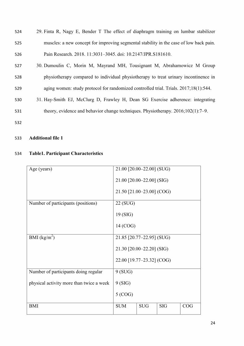
Additional file 1 533
Table1. Participant Characteristics 534
Age (years) 21.00 [20.00–22.00] (SUG)
21.00 [20.00–22.00] (SIG)
21.50 [21.00–23.00] (COG)
Number of participants (positions) 22 (SUG)
19 (SIG)
14 (COG)
BMI (kg/m2) 21.85 [20.77–22.95] (SUG)
21.30 [20.00–22.20] (SIG)
22.00 [19.77–23.32] (COG)
Number of participants doing regular
physical activity more than twice a week
9 (SUG)
9 (SIG)
5 (COG)
BMI SUM SUG SIG COG
25
(n = 55) (n = 22) (n = 19) (n = 14)
Normal BMI 18-24 45 18 16 11
Overweight BMI 25-29 9 3 3 3
Underweight BMI <18 1 1 0 0
535
Values are median [1st - 3rd] quartiles for age and BMI and numbers of participants. 536
BMI: body mass index; SUG: the supine study group; SIG: the sitting study group; COG: the 537
control group 538
Additional file 2 539
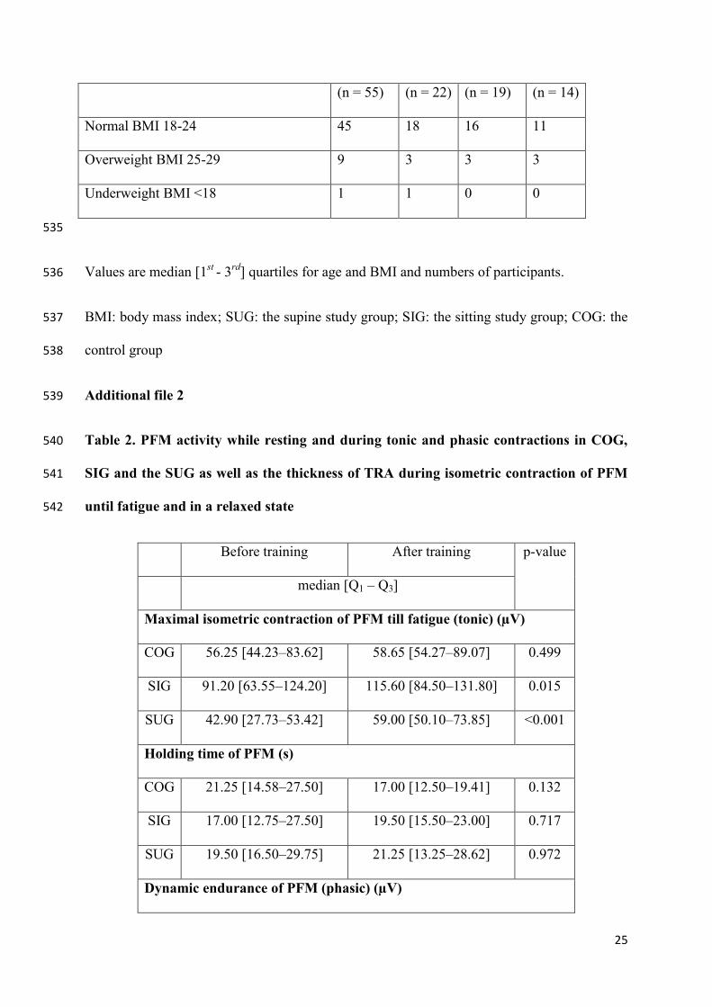
Table 2. PFM activity while resting and during tonic and phasic contractions in COG, 540
SIG and the SUG as well as the thickness of TRA during isometric contraction of PFM 541
until fatigue and in a relaxed state 542
Before training After training p-value
median [Q1 – Q3]
Maximal isometric contraction of PFM till fatigue (tonic) (µV)
COG 56.25 [44.23–83.62] 58.65 [54.27–89.07] 0.499
SIG 91.20 [63.55–124.20] 115.60 [84.50–131.80] 0.015
SUG 42.90 [27.73–53.42] 59.00 [50.10–73.85] <0.001
Holding time of PFM (s)
COG 21.25 [14.58–27.50] 17.00 [12.50–19.41] 0.132
SIG 17.00 [12.75–27.50] 19.50 [15.50–23.00] 0.717
SUG 19.50 [16.50–29.75] 21.25 [13.25–28.62] 0.972
Dynamic endurance of PFM (phasic) (µV)
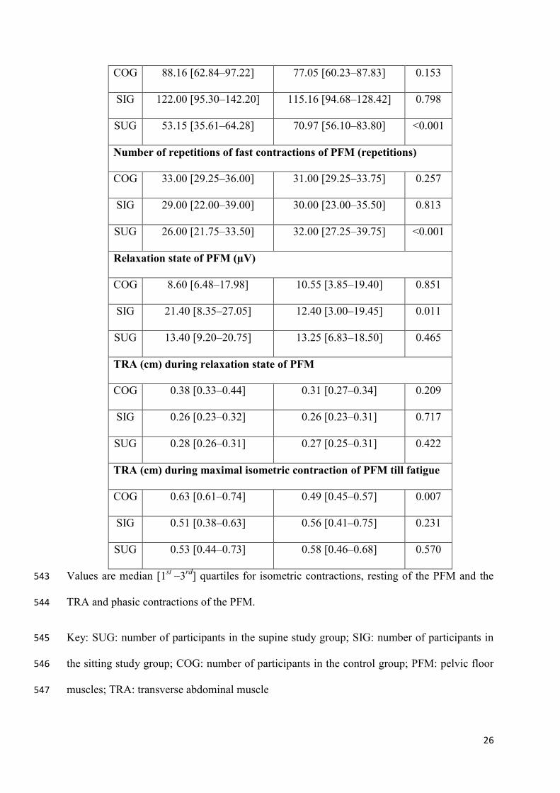
26
COG 88.16 [62.84–97.22] 77.05 [60.23–87.83] 0.153
SIG 122.00 [95.30–142.20] 115.16 [94.68–128.42] 0.798
SUG 53.15 [35.61–64.28] 70.97 [56.10–83.80] <0.001
Number of repetitions of fast contractions of PFM (repetitions)
COG 33.00 [29.25–36.00] 31.00 [29.25–33.75] 0.257
SIG 29.00 [22.00–39.00] 30.00 [23.00–35.50] 0.813
SUG 26.00 [21.75–33.50] 32.00 [27.25–39.75] <0.001
Relaxation state of PFM (µV)
COG 8.60 [6.48–17.98] 10.55 [3.85–19.40] 0.851
SIG 21.40 [8.35–27.05] 12.40 [3.00–19.45] 0.011
SUG 13.40 [9.20–20.75] 13.25 [6.83–18.50] 0.465
TRA (cm) during relaxation state of PFM
COG 0.38 [0.33–0.44] 0.31 [0.27–0.34] 0.209
SIG 0.26 [0.23–0.32] 0.26 [0.23–0.31] 0.717
SUG 0.28 [0.26–0.31] 0.27 [0.25–0.31] 0.422
TRA (cm) during maximal isometric contraction of PFM till fatigue
COG 0.63 [0.61–0.74] 0.49 [0.45–0.57] 0.007
SIG 0.51 [0.38–0.63] 0.56 [0.41–0.75] 0.231
SUG 0.53 [0.44–0.73] 0.58 [0.46–0.68] 0.570
Values are median [1st –3rd] quartiles for isometric contractions, resting of the PFM and the 543
TRA and phasic contractions of the PFM. 544
Key: SUG: number of participants in the supine study group; SIG: number of participants in 545
the sitting study group; COG: number of participants in the control group; PFM: pelvic floor 546
muscles; TRA: transverse abdominal muscle 547
27
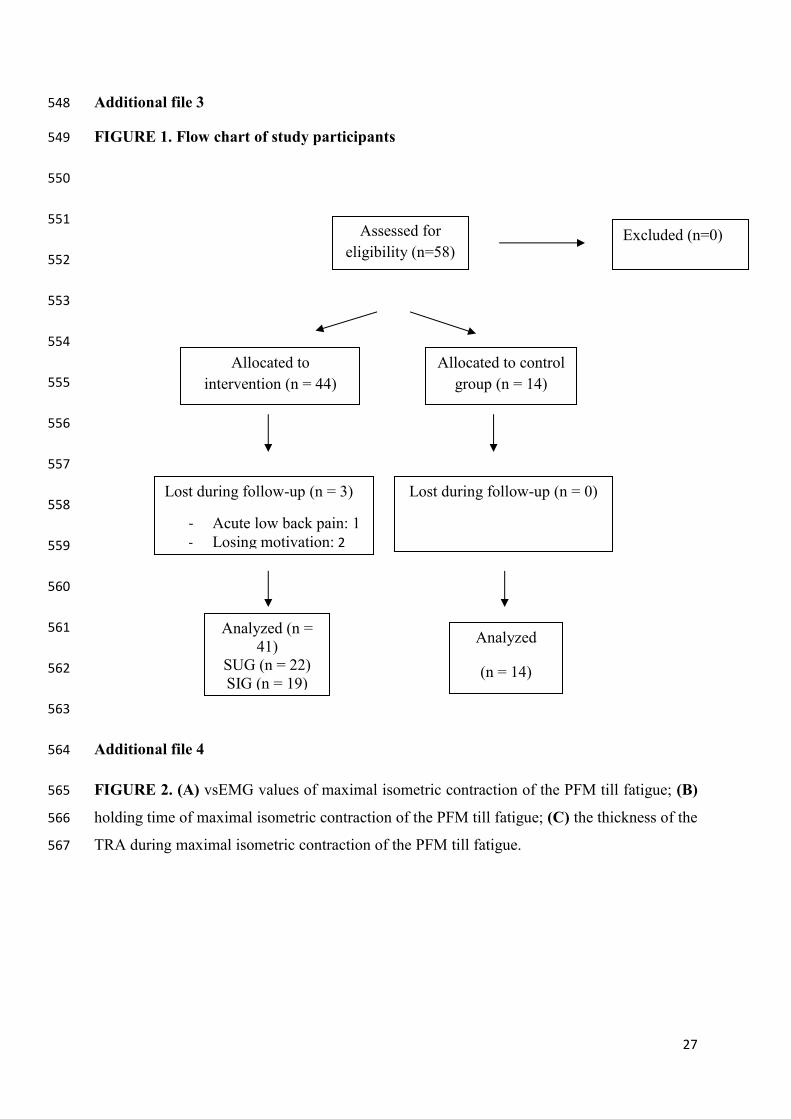
Additional file 3 548
FIGURE 1. Flow chart of study participants 549
550
551
552
553
554
555
556
557
558
559
560
561
562
563
Additional file 4 564
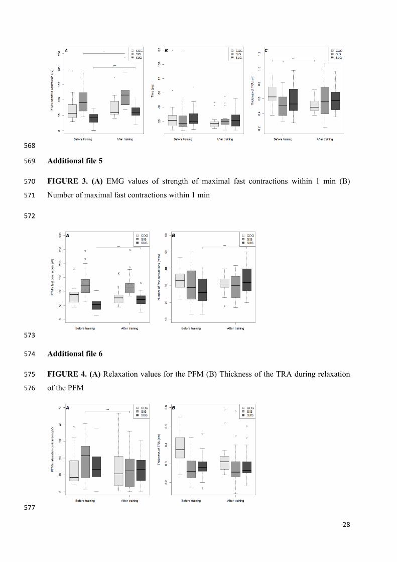
FIGURE 2. (A) vsEMG values of maximal isometric contraction of the PFM till fatigue; (B) 565
holding time of maximal isometric contraction of the PFM till fatigue; (C) the thickness of the 566
TRA during maximal isometric contraction of the PFM till fatigue. 567
Assessed for eligibility (n=58)
Excluded (n=0)
Allocated to intervention (n = 44)
Allocated to control group (n = 14)
Lost during follow-up (n = 3)
- Acute low back pain: 1 - Losing motivation: 2
Lost during follow-up (n = 0)
Analyzed (n = 41)
SUG (n = 22) SIG (n = 19)
Analyzed
(n = 14)
28
568
Additional file 5 569
FIGURE 3. (A) EMG values of strength of maximal fast contractions within 1 min (B) 570
Number of maximal fast contractions within 1 min 571
572
573
Additional file 6 574
FIGURE 4. (A) Relaxation values for the PFM (B) Thickness of the TRA during relaxation 575
of the PFM 576
577
29
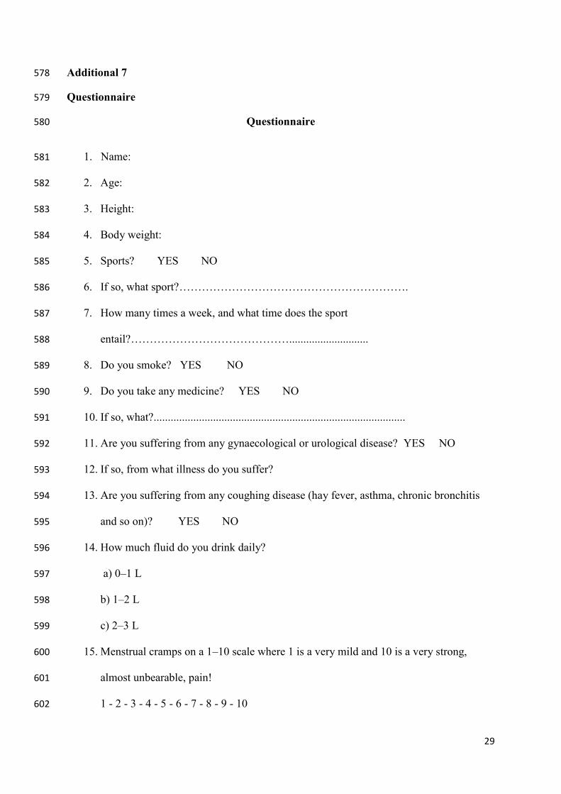
Additional 7 578
Questionnaire 579
Questionnaire 580
1. Name: 581
2. Age: 582
3. Height: 583
4. Body weight: 584
5. Sports? YES NO 585
6. If so, what sport?……………………………………………………. 586
7. How many times a week, and what time does the sport 587
entail?……………………………………............................ 588
8. Do you smoke? YES NO 589
9. Do you take any medicine? YES NO 590
10. If so, what?......................................................................................... 591
11. Are you suffering from any gynaecological or urological disease? YES NO 592
12. If so, from what illness do you suffer? 593
13. Are you suffering from any coughing disease (hay fever, asthma, chronic bronchitis 594
and so on)? YES NO 595
14. How much fluid do you drink daily? 596
a) 0–1 L 597
b) 1–2 L 598
c) 2–3 L 599
15. Menstrual cramps on a 1–10 scale where 1 is a very mild and 10 is a very strong, 600
almost unbearable, pain! 601
1 - 2 - 3 - 4 - 5 - 6 - 7 - 8 - 9 - 10 602
30
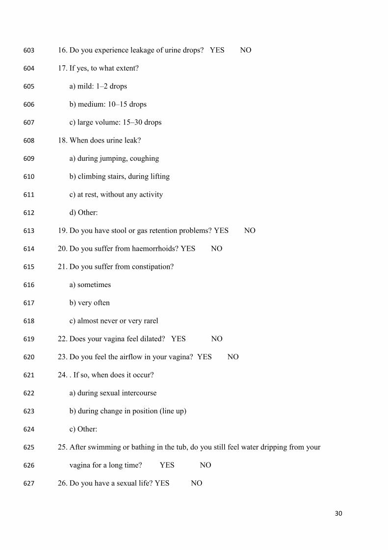
16. Do you experience leakage of urine drops? YES NO 603
17. If yes, to what extent? 604
a) mild: 1–2 drops 605
b) medium: 10–15 drops 606
c) large volume: 15–30 drops 607
18. When does urine leak? 608
a) during jumping, coughing 609
b) climbing stairs, during lifting 610
c) at rest, without any activity 611
d) Other: 612
19. Do you have stool or gas retention problems? YES NO 613
20. Do you suffer from haemorrhoids? YES NO 614
21. Do you suffer from constipation? 615
a) sometimes 616
b) very often 617
c) almost never or very rarel 618
22. Does your vagina feel dilated? YES NO 619
23. Do you feel the airflow in your vagina? YES NO 620
24. . If so, when does it occur? 621
a) during sexual intercourse 622
b) during change in position (line up) 623
c) Other: 624
25. After swimming or bathing in the tub, do you still feel water dripping from your 625
vagina for a long time? YES NO 626
26. Do you have a sexual life? YES NO 627
31
27. If so, how often? 628
a) 1 time per month 629
b) 1 time per week 630
c) 2–4 times per week 631
d) 5–7 times per week 632
e) occasionally 633
f) Other: …………………… 634
28. Do you feel pain when penetrating a tampon or sexual intercourse? YES NO 635
29. Sexual libido on a scale of 1 to 10, where 1 is low and weak and 10 is strong libido! 636
0 - 1 - 2 - 3 - 4 - 5 - 6 - 7 - 8 - 9 - 10 637
30. Frequency of orgasm on a 1–10 scale, where 1 means very rarely and 10 is almost 638
always during sexual intercourse! 639
0 - 1 - 2 - 3 - 4 - 5 - 6 - 7 - 8 - 9 - 10 640
641
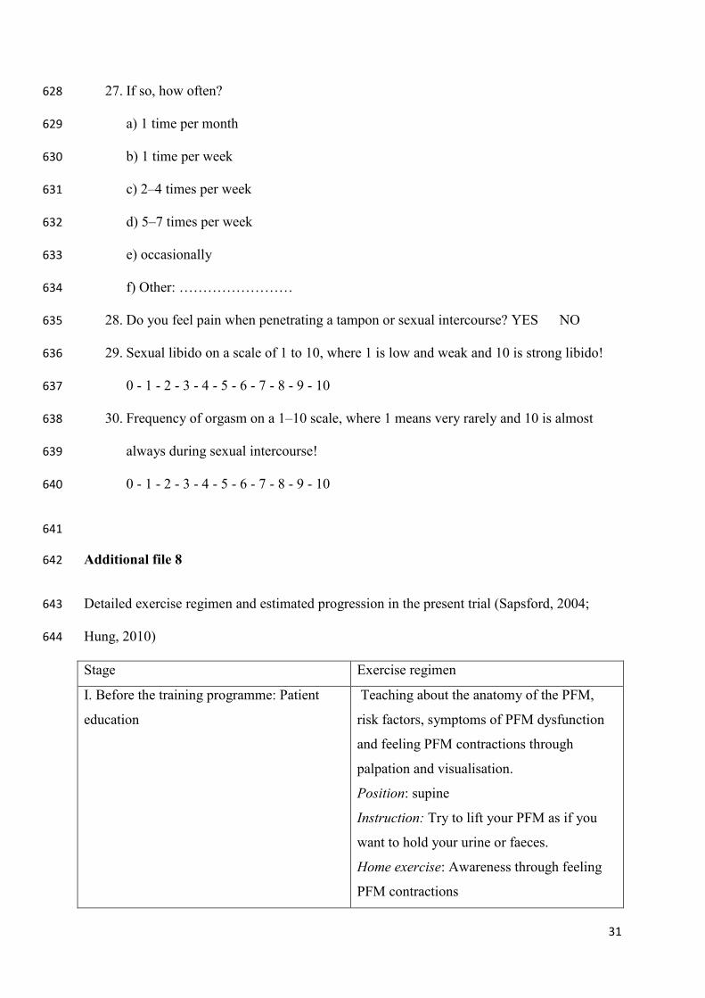
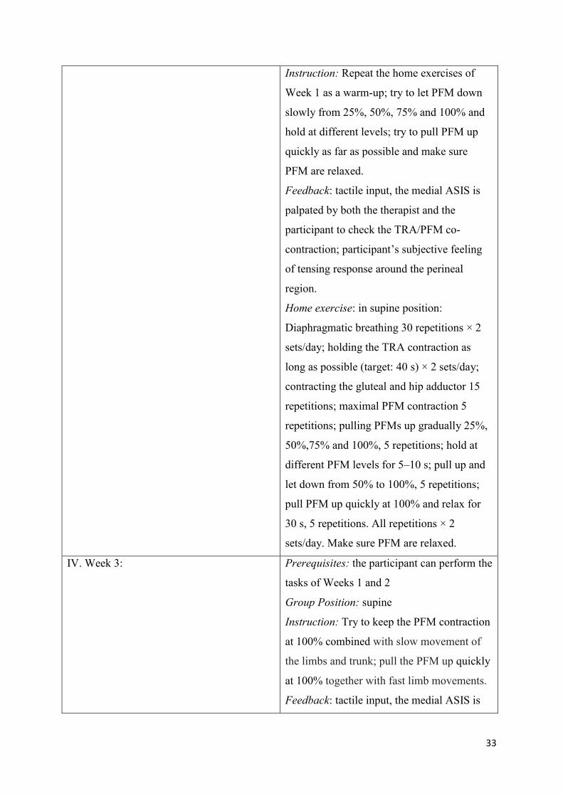
Additional file 8 642
Detailed exercise regimen and estimated progression in the present trial (Sapsford, 2004; 643
Hung, 2010) 644
Stage Exercise regimen
I. Before the training programme: Patient
education
Teaching about the anatomy of the PFM,
risk factors, symptoms of PFM dysfunction
and feeling PFM contractions through
palpation and visualisation.
Position: supine
Instruction: Try to lift your PFM as if you
want to hold your urine or faeces.
Home exercise: Awareness through feeling
PFM contractions
32
Feedback: palpation, mirror and during
urination
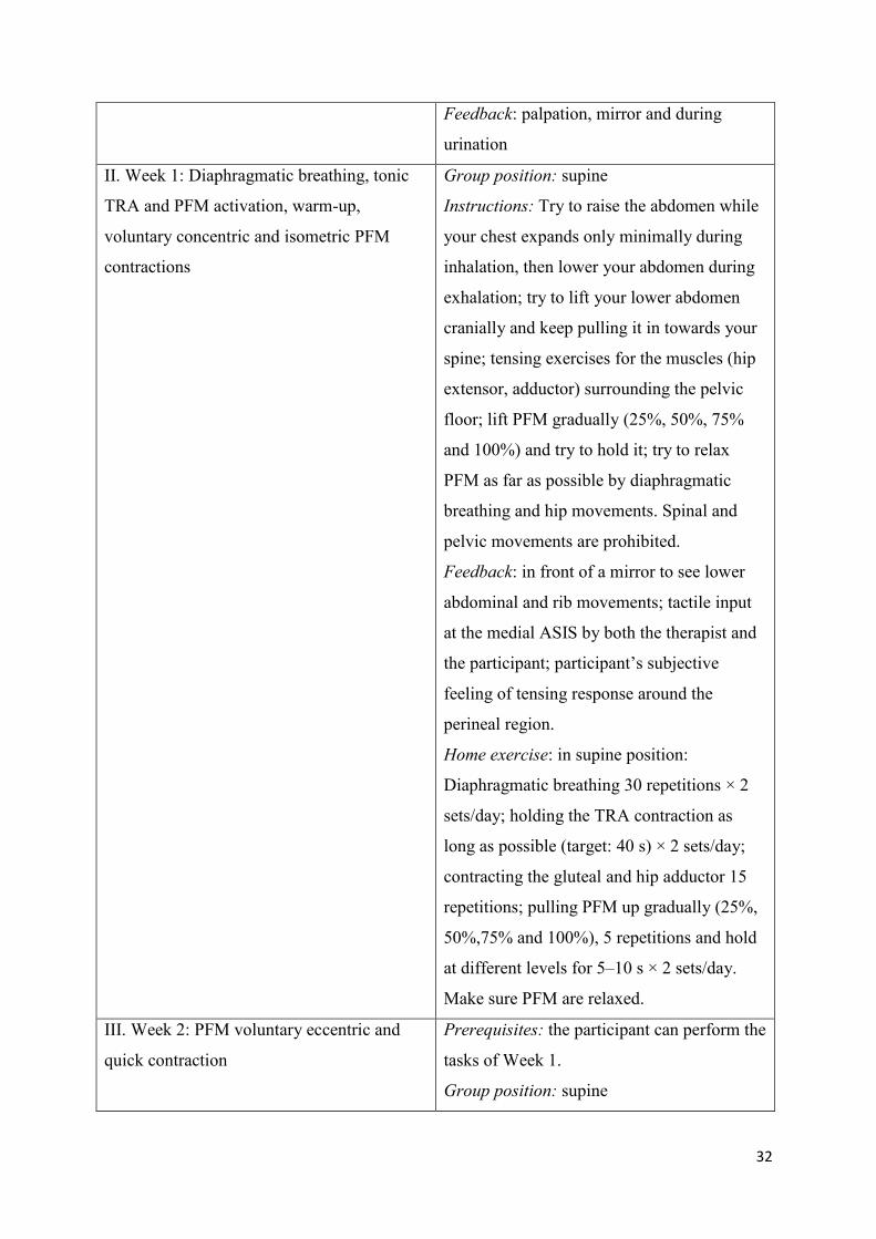
II. Week 1: Diaphragmatic breathing, tonic
TRA and PFM activation, warm-up,
voluntary concentric and isometric PFM
contractions
Group position: supine
Instructions: Try to raise the abdomen while
your chest expands only minimally during
inhalation, then lower your abdomen during
exhalation; try to lift your lower abdomen
cranially and keep pulling it in towards your
spine; tensing exercises for the muscles (hip
extensor, adductor) surrounding the pelvic
floor; lift PFM gradually (25%, 50%, 75%
and 100%) and try to hold it; try to relax
PFM as far as possible by diaphragmatic
breathing and hip movements. Spinal and
pelvic movements are prohibited.
Feedback: in front of a mirror to see lower
abdominal and rib movements; tactile input
at the medial ASIS by both the therapist and
the participant; participant’s subjective
feeling of tensing response around the
perineal region.
Home exercise: in supine position:
Diaphragmatic breathing 30 repetitions × 2
sets/day; holding the TRA contraction as
long as possible (target: 40 s) × 2 sets/day;
contracting the gluteal and hip adductor 15
repetitions; pulling PFM up gradually (25%,
50%,75% and 100%), 5 repetitions and hold
at different levels for 5–10 s × 2 sets/day.
Make sure PFM are relaxed.
III. Week 2: PFM voluntary eccentric and
quick contraction
Prerequisites: the participant can perform the
tasks of Week 1.
Group position: supine
33
Instruction: Repeat the home exercises of
Week 1 as a warm-up; try to let PFM down
slowly from 25%, 50%, 75% and 100% and
hold at different levels; try to pull PFM up
quickly as far as possible and make sure
PFM are relaxed.
Feedback: tactile input, the medial ASIS is
palpated by both the therapist and the
participant to check the TRA/PFM co-
contraction; participant’s subjective feeling
of tensing response around the perineal
region.
Home exercise: in supine position:
Diaphragmatic breathing 30 repetitions × 2
sets/day; holding the TRA contraction as
long as possible (target: 40 s) × 2 sets/day;
contracting the gluteal and hip adductor 15
repetitions; maximal PFM contraction 5
repetitions; pulling PFMs up gradually 25%,
50%,75% and 100%, 5 repetitions; hold at
different PFM levels for 5–10 s; pull up and
let down from 50% to 100%, 5 repetitions;
pull PFM up quickly at 100% and relax for
30 s, 5 repetitions. All repetitions × 2
sets/day. Make sure PFM are relaxed.
IV. Week 3: Prerequisites: the participant can perform the
tasks of Weeks 1 and 2
Group Position: supine
Instruction: Try to keep the PFM contraction
at 100% combined with slow movement of
the limbs and trunk; pull the PFM up quickly
at 100% together with fast limb movements.
Feedback: tactile input, the medial ASIS is
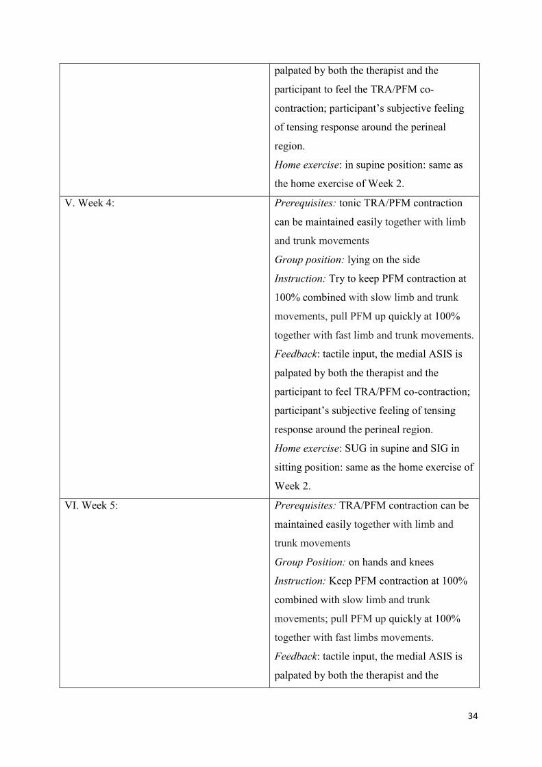
34
palpated by both the therapist and the
participant to feel the TRA/PFM co-
contraction; participant’s subjective feeling
of tensing response around the perineal
region.
Home exercise: in supine position: same as
the home exercise of Week 2.
V. Week 4: Prerequisites: tonic TRA/PFM contraction
can be maintained easily together with limb
and trunk movements
Group position: lying on the side
Instruction: Try to keep PFM contraction at
100% combined with slow limb and trunk
movements, pull PFM up quickly at 100%
together with fast limb and trunk movements.
Feedback: tactile input, the medial ASIS is
palpated by both the therapist and the
participant to feel TRA/PFM co-contraction;
participant’s subjective feeling of tensing
response around the perineal region.
Home exercise: SUG in supine and SIG in
sitting position: same as the home exercise of
Week 2.
VI. Week 5: Prerequisites: TRA/PFM contraction can be
maintained easily together with limb and
trunk movements
Group Position: on hands and knees
Instruction: Keep PFM contraction at 100%
combined with slow limb and trunk
movements; pull PFM up quickly at 100%
together with fast limbs movements.
Feedback: tactile input, the medial ASIS is
palpated by both the therapist and the
35
participant to feel TRA/PFM co-contraction;
participant’s subjective feeling of tensing
response around the perineal region.
Home exercise: SUG in supine and SIG in
sitting position: same as the home exercise of
Week 2.
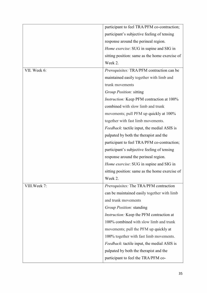
VII. Week 6: Prerequisites: TRA/PFM contraction can be
maintained easily together with limb and
trunk movements
Group Position: sitting
Instruction: Keep PFM contraction at 100%
combined with slow limb and trunk
movements; pull PFM up quickly at 100%
together with fast limb movements.
Feedback: tactile input, the medial ASIS is
palpated by both the therapist and the
participant to feel TRA/PFM co-contraction;
participant’s subjective feeling of tensing
response around the perineal region.
Home exercise: SUG in supine and SIG in
sitting position: same as the home exercise of
Week 2.
VIII.Week 7: Prerequisites: The TRA/PFM contraction
can be maintained easily together with limb
and trunk movements
Group Position: standing
Instruction: Keep the PFM contraction at
100% combined with slow limb and trunk
movements; pull the PFM up quickly at
100% together with fast limb movements.
Feedback: tactile input, the medial ASIS is
palpated by both the therapist and the
participant to feel the TRA/PFM co-
36
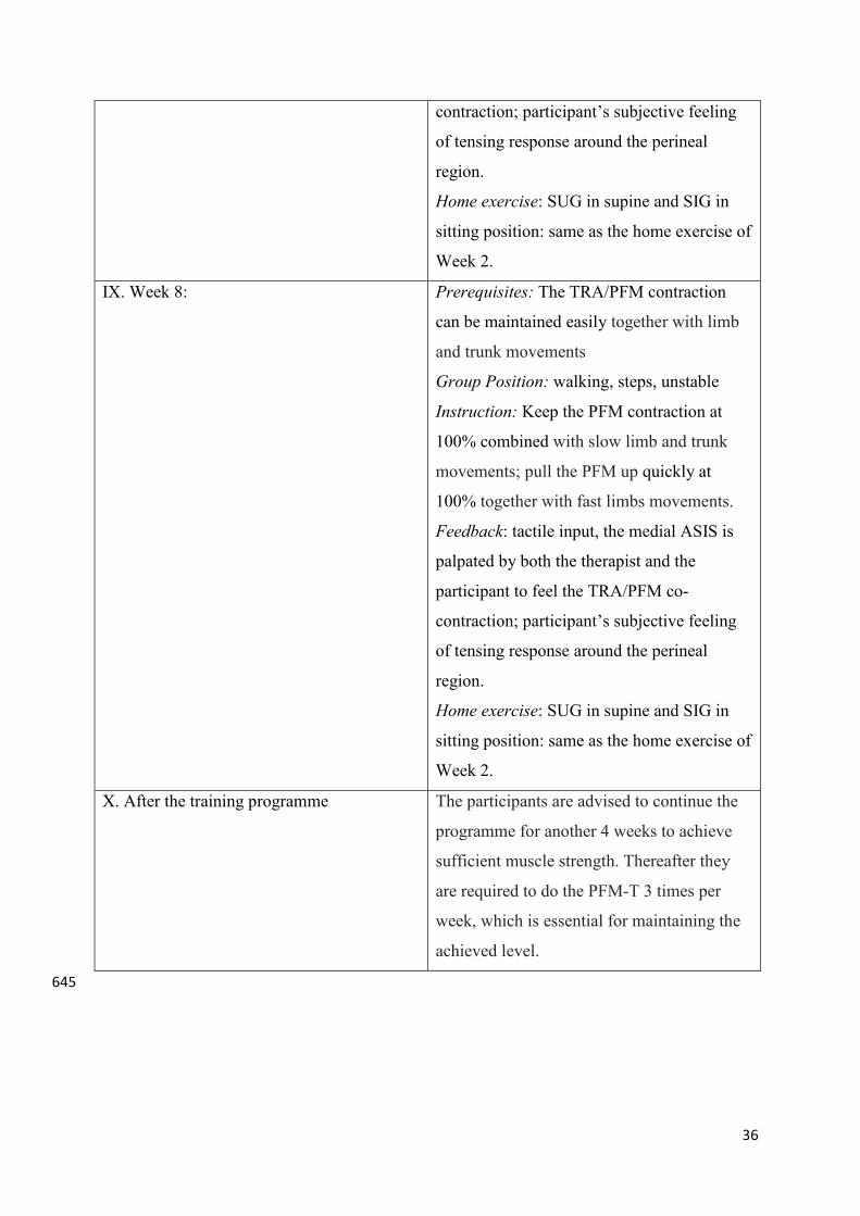
contraction; participant’s subjective feeling
of tensing response around the perineal
region.
Home exercise: SUG in supine and SIG in
sitting position: same as the home exercise of
Week 2.
IX. Week 8: Prerequisites: The TRA/PFM contraction
can be maintained easily together with limb
and trunk movements
Group Position: walking, steps, unstable
Instruction: Keep the PFM contraction at
100% combined with slow limb and trunk
movements; pull the PFM up quickly at
100% together with fast limbs movements.
Feedback: tactile input, the medial ASIS is
palpated by both the therapist and the
participant to feel the TRA/PFM co-
contraction; participant’s subjective feeling
of tensing response around the perineal
region.
Home exercise: SUG in supine and SIG in
sitting position: same as the home exercise of
Week 2.
X. After the training programme The participants are advised to continue the
programme for another 4 weeks to achieve
sufficient muscle strength. Thereafter they
are required to do the PFM-T 3 times per
week, which is essential for maintaining the
achieved level.
645