a comprehensive review of abdominal infections
TRANSCRIPT
REVIEW Open Access
A Comprehensive review of abdominal infectionsNicole Lopez1, Leslie Kobayashi2*, Raul Coimbra3
IntroductionIntra-abdominal infection (IAI) is an important cause ofmorbidity and mortality. It is the second most com-monly identified cause of severe sepsis in the intensivecare unit (ICU). Recent studies have associated severeintra-abdominal infection with a significant mortalityrate.Most IAI are a result of processes involving inflamma-
tion and perforations of the gastrointestinal tract, suchas appendicitis, peptic ulcer disease, and diverticulitis.Patients with diffuse peritonitis may be due to sponta-neous perforation, post-operative, post-interventional orpost-traumatic causes. The lower GI tract is most oftenthe location of perforation. Among patients with IAIwho develop peritonitis, many may progress to severesepsis, defined by The American College of Chest Physi-cians/Society of Critical Care Medicine as a severe sys-temic inflammatory response to infection that isassociated with acute organ dysfunction.Successful treatment of IAI is based on early and
appropriate source recognition, containment and antimi-crobial coverage. We will review clinical definitions,pathophysiology, and treatment strategies for IAI in aneffort to provide guidelines for clinical management.
DefinitionsIntra-abdominal infection (IAI) describes a diverse setof diseases. It is broadly defined as peritoneal inflamma-tion in response to microorganisms, resulting in puru-lence in the peritoneal cavity [1]. IAI are classified asuncomplicated or complicated based on the extent ofinfection [2].Uncomplicated abdominal infections involve intra-
mural inflammation of the gastrointestinal (GI) tractwithout anatomic disruption. They are often simple totreat; however, when treatment is delayed or inappropri-ate, or the infection involves a more virulent nosocomial
microbe, the risk of progression into a complicatedabdominal infection becomes significant [3,4].Complicated abdominal infections extend beyond
the source organ into the peritoneal space. They causeperitoneal inflammation, and are associated with loca-lized or diffuse peritonitis[5]. Localized peritonitis oftenmanifests as an abscess with tissue debris, bacteria, neu-trophils, macrophages, and exudative fluid contained ina fibrous capsule. Diffuse peritonitis is categorized asprimary, secondary or tertiary peritonitis.Primary peritonitis is also known as spontaneous
bacterial peritonitis. It is thought to be the result of bac-terial translocation across an intact gut wall [6]. Theseinfections are commonly monomicrobial, and the infect-ing organism is primarily determined by patient demo-graphics. For example, healthy young girls are mostoften infected by streptococcal organisms, cirrhotics bygram negative or enterococcal organisms, and peritonealdialysis patients by Staphylococcus aureus [7,8]. Diagno-sis requires peritoneal fluid aspiration. Characteristics ofinfection include white blood cell count (WBC) > 500cells/mm3, high lactate, and low glucose levels. Positiveperitoneal fluid cultures are definitive, and resolution ofinfection is marked by peritoneal fluid with < 250WBC/mm3[9].Secondary peritonitis is caused by microbial contam-
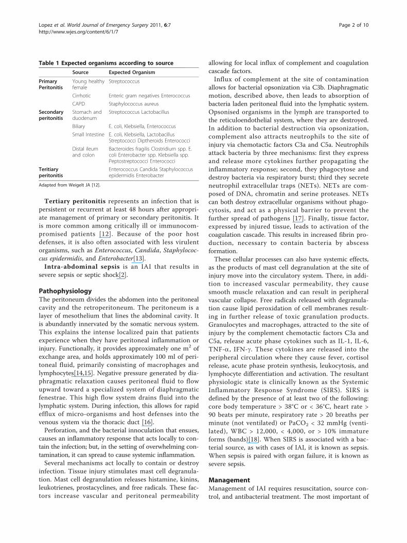
ination through a perforation, laceration, or necroticsegment of the GI tract[7]. Definitive diagnosis is basedon clinical examination and history, and specific diag-noses can be confirmed by radiographic imaging[10]. Ifa patient is stable enough for transport, computedtomography (CT) scan with intravenous and oralcontrast is the standard method of evaluating mostintra-abdominal pathologies, such as appendicitis, diver-ticulitis, and colitis [11]. Suspected biliary pathology isthe exception, and ultrasound is the preferred initialimaging modality for this spectrum of disease includingacute cholecystitis, emphysematous cholecystitis, andcholangitis. Infections associated with secondary perito-nitis are commonly polymicrobial and the infectingorganisms are those most commonly associated with thesource of contamination (see Table 1).
* Correspondence: [email protected] Professor of Surgery, University of California, San Diego, 200 W.Arbor Dr. #8896, San Diego, CA 92103-8896, USAFull list of author information is available at the end of the article
Lopez et al. World Journal of Emergency Surgery 2011, 6:7http://www.wjes.org/content/6/1/7 WORLD JOURNAL OF
EMERGENCY SURGERY
© 2011 Lopez et al; licensee BioMed Central Ltd. This is an Open Access article distributed under the terms of the Creative CommonsAttribution License (http://creativecommons.org/licenses/by/2.0), which permits unrestricted use, distribution, and reproduction inany medium, provided the original work is properly cited.
Tertiary peritonitis represents an infection that ispersistent or recurrent at least 48 hours after appropri-ate management of primary or secondary peritonitis. Itis more common among critically ill or immunocom-promised patients [12]. Because of the poor hostdefenses, it is also often associated with less virulentorganisms, such as Enterococcus, Candida, Staphylococ-cus epidermidis, and Enterobacter[13].Intra-abdominal sepsis is an IAI that results in
severe sepsis or septic shock[2].
PathophysiologyThe peritoneum divides the abdomen into the peritonealcavity and the retroperitoneum. The peritoneum is alayer of mesothelium that lines the abdominal cavity. Itis abundantly innervated by the somatic nervous system.This explains the intense localized pain that patientsexperience when they have peritoneal inflammation orinjury. Functionally, it provides approximately one m2 ofexchange area, and holds approximately 100 ml of peri-toneal fluid, primarily consisting of macrophages andlymphocytes[14,15]. Negative pressure generated by dia-phragmatic relaxation causes peritoneal fluid to flowupward toward a specialized system of diaphragmaticfenestrae. This high flow system drains fluid into thelymphatic system. During infection, this allows for rapidefflux of micro-organisms and host defenses into thevenous system via the thoracic duct [16].Perforation, and the bacterial innoculation that ensues,
causes an inflammatory response that acts locally to con-tain the infection; but, in the setting of overwhelming con-tamination, it can spread to cause systemic inflammation.Several mechanisms act locally to contain or destroy
infection. Tissue injury stimulates mast cell degranula-tion. Mast cell degranulation releases histamine, kinins,leukotrienes, prostacyclines, and free radicals. These fac-tors increase vascular and peritoneal permeability
allowing for local influx of complement and coagulationcascade factors.Influx of complement at the site of contamination
allows for bacterial opsonization via C3b. Diaphragmaticmotion, described above, then leads to absorption ofbacteria laden peritoneal fluid into the lymphatic system.Opsonised organisms in the lymph are transported tothe reticuloendothelial system, where they are destroyed.In addition to bacterial destruction via opsonization,complement also attracts neutrophils to the site ofinjury via chemotactic factors C3a and C5a. Neutrophilsattack bacteria by three mechanisms: first they expressand release more cytokines further propagating theinflammatory response; second, they phagocytose anddestroy bacteria via respiratory burst; third they secreteneutrophil extracellular traps (NETs). NETs are com-posed of DNA, chromatin and serine proteases. NETscan both destroy extracellular organisms without phago-cytosis, and act as a physical barrier to prevent thefurther spread of pathogens [17]. Finally, tissue factor,expressed by injured tissue, leads to activation of thecoagulation cascade. This results in increased fibrin pro-duction, necessary to contain bacteria by abscessformation.These cellular processes can also have systemic effects,
as the products of mast cell degranulation at the site ofinjury move into the circulatory system. There, in addi-tion to increased vascular permeability, they causesmooth muscle relaxation and can result in peripheralvascular collapse. Free radicals released with degranula-tion cause lipid peroxidation of cell membranes result-ing in further release of toxic granulation products.Granulocytes and macrophages, attracted to the site ofinjury by the complement chemotactic factors C3a andC5a, release acute phase cytokines such as IL-1, IL-6,TNF-a, IFN-g. These cytokines are released into theperipheral circulation where they cause fever, cortisolrelease, acute phase protein synthesis, leukocytosis, andlymphocyte differentiation and activation. The resultantphysiologic state is clinically known as the SystemicInflammatory Response Syndrome (SIRS). SIRS isdefined by the presence of at least two of the following:core body temperature > 38°C or < 36°C, heart rate >90 beats per minute, respiratory rate > 20 breaths perminute (not ventilated) or PaCO2 < 32 mmHg (venti-lated), WBC > 12,000, < 4,000, or > 10% immatureforms (bands)[18]. When SIRS is associated with a bac-terial source, as with cases of IAI, it is known as sepsis.When sepsis is paired with organ failure, it is known assevere sepsis.
ManagementManagement of IAI requires resuscitation, source con-trol, and antibacterial treatment. The most important of
Table 1 Expected organisms according to source
Source Expected Organism
PrimaryPeritonitis
Young healthyfemale
Streptococcus
Cirrhotic Enteric gram negatives Enterococcus
CAPD Staphylococcus aureus
Secondaryperitonitis
Stomach andduodenum
Streptococcus Lactobacillus
Biliary E. coli, Klebsiella, Enterococcus
Small Intestine E. coli, Klebsiella, LactobacillusStreptococci Diptheroids Enterococci
Distal ileumand colon
Bacteroides fragilis Clostridium spp. E.coli Enterobacter spp. Klebsiella spp.Peptostreptococci Enterococci
Teritiaryperitonitis
Enterococcus Candida Staphylococcusepidermidis Enterobacter
Adapted from Weigelt JA [12].
Lopez et al. World Journal of Emergency Surgery 2011, 6:7http://www.wjes.org/content/6/1/7
Page 2 of 10
these factors is source control, which, “encompasses allmeasures undertaken to eliminate the source of infec-tion and to control ongoing contamination”[19]. Thereare three key components of source control: drainage,debridement, and definitive management.
Resuscitation and Support of Organ SystemsIAI causes volume depletion by several mechanisms.Nausea, anorexia and ileus lead to a decrease in oralintake, while vomiting and diarrhea increase sensiblelosses. In addition, ileus with third space losses into thebowel wall and ascites, as well as fever both increaseinsensible losses. Elevated body temperature leads toboth an increase in dermal loss via sweating, and anincrease in respiratory loss by causing tachypnea. Dermalloss in a febrile patient can account for approximately600 ml of volume loss per day, while tachypnea causesapproximately 100 ml of volume loss per day [20,21].In uncomplicated IAI, replacing volume is essential; in
severe sepsis or septic shock, it becomes critical.Patients suspected of having severe sepsis or septicshock should be admitted to an ICU for careful moni-toring of vital signs and volume status. With regard tothe initial volume resuscitation, we recommend follow-ing the Surviving Sepsis Campaign recommendations.As soon as hypotension is recognized, or, ideally if it isanticipated, attention should be paid to early goal direc-ted volume resuscitation. Isotonic fluid, or in the casesof severe anemia or coagulopathy, blood products,should be administered with the intent to achieve amean arterial pressure (MAP) > 65 mmHg and a centralvenous pressure (CVP) of 12-15 mmHg within the first6 hours [22]. If a MAP > 65 mmHg cannot be obtainedby volume resuscitation alone then vasopressors shouldbe used, with a preference for norepinepherine or dopa-mine [22]. In cases where low cardiac output or elevatedfilling pressures indicate severe myocardial dysfunction,use of inotropic agents such as dobutamine may be effi-cacious in obtaining adequate MAP [22]. Care shouldalso be taken to monitor clinical indicators of end organperfusion, such as hourly urine output and mental sta-tus, to ensure adequate oxygen delivery.The goal of resuscitation is correction of cellular oxy-
gen debt. Various endpoints for resuscitation have beensuggested, including: mixed venous oxygen (SVO2), lac-tate and base deficit. While a normal or high SVO2 doesnot ensure adequate tissue oxygenation, a low SVO2
indicates a need to increase tissue oxygenation. Resusci-tation to maintain an SVO2 > 65% has been shown toimprove outcomes [23,24]. Lactate, a product of anaero-bic metabolism, has also been used as an indirect mea-sure of oxygen debt. More recently sepsis has beenrecognized as a hypermetabolic state that uses glycolysisin the absence of hypoxia, making it less reliable as a
marker of oxygen debt. Still, its early normalization maypredict improved outcomes [25-27]. Base deficit is yetanother indicator of oxygen debt. It describes the amountof base that would be required to bring the blood to anormal pH under normal physiologic conditions. Thedegree of base deficit has been shown to correlate withresuscitation requirements and mortality [28,29]. Whilenone of these measures are perfect, they can be helpful inguiding resuscitation when used in combination with theother clinical endpoints discussed above.
DrainageThe goal of drainage is to evacuate purulent, contami-nated fluid, or to control drainage of ongoing entericcontamination. This is accomplished by either percuta-neous or open surgical intervention. Percutaneous drai-nage can be performed with or without image guidance,and is most commonly performed using ultrasound orCT. In many circumstances it is as efficacious as surgi-cal drainage, and is often used as the initial treatment ofchoice because it is less invasive and more affordable[30,31]. Percutaneous drainage is also useful in patientswho are poor surgical candidates, and might not survivedefinitive surgical treatment. However, percutaneousdrainage is unlikely to result in adequate source controlin cases of frank bowel perforation with ongoing con-tamination, or if there is a significant amount of necro-tic tissue present. In these cases, surgery is thetreatment of choice.Open surgical drainage should be used in the case of
generalized peritonitis, ongoing gross contaminationfrom an uncontrolled enteric source, if bowel necrosisor ischemia is suspected, and in cases of failure of per-cutaneous drainage. Unstable patients, or those withcomplicated or difficult anatomy such as post-operativepatients or those with advanced malignancy pose a par-ticular challenge.In these situations, damage control techniques can be
employed with temporary abdominal closure. Damagecontrol procedures are typically used for patients whoare unstable and unable to tolerate definitive surgicaltreatment, have intra-abdominal hypertension (IAH), orhave loss of abdominal domain that prevents fascial clo-sure. The first stage in damage control surgery is eva-cuation of infected material and control of grosscontamination. This is followed by temporary abdominalclosure with a conventional dressing, negative pressuredressing, or skin closure. This first operative stage is fol-lowed by ongoing resuscitation, once normal physiologyis restored resuscitation can then be followed byplanned re-laparotomy for definitive source control andreconstruction. In cases of physiologic worsening afterfirst laparotomy, or in cases of concern for IAH, orintestinal ischemia, on demand repeat laparotomy can
Lopez et al. World Journal of Emergency Surgery 2011, 6:7http://www.wjes.org/content/6/1/7
Page 3 of 10
be performed. Once all surgical issues have beenaddressed, physiology has been restored and there areno longer concerns for ongoing ischemia, necrosis, orIAH the abdomen can be definitively closed.Intra-abdominal lavage is a subject of ongoing contro-
versy. Proponents of peritoneal lavage reason that con-tamination is both removed and diluted by lavagevolumes greater than 10 L, additionally, by adding anti-biotics bacterial pathogens can be specifically targeted.One group has suggested that lavage with volumes ofapproximately 20 L reduces infectious complications inblunt traumatic small bowel perforation [32]. However,its application with or without antibiotics in abdominalsepsis is largely unsubstantiated; at this time there isminimal evidence in the literature to support its use[33,34].
DebridementDebridement is essential for removal of foreign bodies,fecal matter, hematoma, and infected or necrotic tissue.The necessity to remove fibrin deposits is controversial.One early study showed improved postoperative courseswith fewer continued infections; however, more recentstudies have shown no benefit to this strategy [35,36].
Definitive managementDefinitive management involves restoration of anatomyand function. While staged procedures were once thestandard, single stage procedures with primary anasto-moses have become accepted as both safe and costeffective in the stable patient [37]. Still, establishingbowel continuity may need to be delayed in patientswho are unable to tolerate a lengthy procedure or haveinadequate capacity for tissue healing [38].Specific Surgical Pathologies
AppendicitisAcute appendicitis is the most common intra-abdominalsurgical emergency [19]. Lifetime risk is approximately7-9% [39]. Currently, imaging is recommended for allpatients suspected of having appendicitis except menunder 40 years of age [40]. Generally, CT scan is theaccepted imaging modality, however, ultrasound mayhave a role in women at risk for other pelvic patholo-gies, in pregnancy and in children [41]. The sensitivityand specificity of CT scan in the diagnosis of acuteappendicitis are 87-100% and 91-98%, respectively[42,43]. Ultrasound is very user dependent, and resultscan be affected by patient body habitus, however overallsensitivity is 76-96% and specificity is 91-100% [44].Ultrasound, with its decreased cost, lack of ionizingradiation and ability to assess ovarian pathology, hasbeen the preferred initial imaging modality in children[45-47]. However, CT should be used in children when
the initial ultrasound is negative or non-diagnostic andthere is a high clinical suspicion for appendicitis [45,48].Ultrasound is also the initial imaging procedure ofchoice in pregnant women, however, the appendix isvisualized only 13-50% of the time. Magnetic resonanceimaging (MRI) is an emerging imaging modality forcases of appendicitis in pregnancy with non-visualizationof the appendix on ultrasound. Its sensitivity and specifi-city are 100% and 93.6%, respectively [49].Though acute appendicitis is a very common entity, its
management still contains areas of controversy includingthe role of laparoscopy, and the emerging role of medi-cal management. These decisions can be complicated bythe presence of an abscess or phlegmon.Surgical management of acute appendicitis has been
the gold standard of treatment for decades. However,many groups have proposed that in select patients,acute uncomplicated appendicitis can be treated withantibiotics alone. Initial success rates for conservativemanagement of acute appendicitis range from 88-95%;however, recurrence is common, occurring in up to 35%of cases [50].Both laparoscopic and open appendectomy are safe
and effective. In large reviews, laparoscopic appendect-omy has been associated with fewer surgical site infec-tions, less pain, shorter hospital stays, and more rapidreturn to normal activity [51]. Common disadvantagesfound include increased cost and longer operative times[52,53]. Additionally, laparoscopy has been associatedwith increased risk of intra-abdominal abscess forma-tion, especially in the presence of perforation or gang-rene. In these cases, open surgery may be preferred [54].Ultimately, the differences in outcomes between laparo-scopic and open appendectomy are largely equivocaland the decision should be based on available technol-ogy and surgeon expertise, with increased considerationfor laparoscopy in young female or obese patients[51,55,56].Management of patients presenting with abscess or
phlegmon is conservative, with antibiotics and drainageinitially. Traditionally this has been followed by intervalappendectomy. However, recently the need for intervalappendectomy has been questioned. Controversy pri-marily surrounds the issues of recurrence and potentialfor malignancy. In a large review the recurrence ratewas 7.4% and the risk of malignancy 1.2% [57]. This isin accord with similar studies that conclude that inasymptomatic patients, interval appendectomy has noadvantages over a thorough work up for inflammatoryappendiceal masses [58,59].
Gastroduodenal perforationAfter bleeding, perforation is the second most commoncomplication requiring emergent operative intervention
Lopez et al. World Journal of Emergency Surgery 2011, 6:7http://www.wjes.org/content/6/1/7
Page 4 of 10
in peptic ulcer disease [60,61]. Helicobacter pylori infec-tion is the most common cause of gastric and duodenalulcers. Since the development of treatments forH. pylori, its prevalence in the United States hasdecreased. However, prevalence of gastric and duodenalulcers has remained the same [62].Previously, ulcer perforation was treated by excision
and vagotomy. However, with antimicrobial eradicationand anti-secretory pharmaceuticals, H. pylori positiveulcer recurrence has been significantly reduced [63]. As aresult, the current standard of care is simple ulcer exci-sion and primary repair of the bowel defect, or omentalpatch and subsequent H. pylori eradication, with little orno role for anti-secretory ulcer surgery [61,64].Both open and laparoscopic approaches are reasonable
options for treatment of perforated peptic ulcers.Laparoscopic surgery is associated with significantly lesspain, but downfalls include longer operative times, andpotentially inadequate repair of large perforations. Com-parisons of sutured versus non-sutured repair with fibringlue plug reveal that both are safe [65].Conservative management has also been proposed as a
safe option for management of contained or sealed gas-troduodenal perforations. One randomized studyshowed similar morbidity and mortality for operativeand conservative approaches; however, conservativetreatment was associated with longer hospital stays andincreased failure in patients over 70 years old [66]. Simi-larly, another author suggests that patients less than40 years old and not on NSAIDS are the most likely tobe infected with H. pylori and therefore, the most likelyto benefit from non-operative therapy [67]. Alterna-tively, one group suggests that non-operative therapycan be guided by documented self-sealing on gastroduo-denogram [68].
DiverticulitisDiverticular disease has increased since the turn of the20th century [69]. The prevalence of diverticular diseaseamong the general population is unknown, but atautopsy more than 50% of people over 80 years old areaffected [70]. The lifetime prevalence of diverticulitisamong patients with diverticulosis is 10-25% [69].The standard treatment for uncomplicated diverticuli-
tis is bowel rest and antibiotics. Most patients withuncomplicated diverticulitis respond to conservativemanagement. Two studies found that patients who didnot respond to antibiotics within 48 hours were morelikely to require prolonged hospital stays for IV antibio-tics and/or surgical intervention [71,72].Diverticulitis can be complicated by phlegmon, abscess,
or free perforation and is generally classified according tomodified Hinchey criteria [73]. Approximately 15-20% ofcases are associated with abscesses [74]. In cases of
uniloculated abscess, the initial treatment is usually per-cutaneous drainage; although, in small abscesses (< 4cm), antibiotics have been used as a primary treatmentwith success rates comparable to drainage [75,76]. Whenpercutaneous drainage is performed it has success ratesof up to 90% [77]. Of importance, the success of percuta-neous drainage also seems to be dependent upon loca-tion. Ambrosetti and colleagues found that compared tomesocolic abscesses, pelvic abscesses were more aggres-sive, needed earlier drainage, and were more likely torequire surgery [78].Traditionally, patients who present with an abscess or
phlegmon then undergo elective surgery to avoid the highrisk of recurrence and further complications [71,73].Recently though, some have begun to question the need foroperative therapy when initial management with percuta-neous drainage and antibiotics is successful [79]. Twoauthors have found that perforation, which is the mostcommon cause of mortality in complicated diverticulitis, ismore likely to be the initial presentation of disease, ratherthan a manifestation of recurrence [79,80]. They concludedthat abscesses in complicated diverticulitis might then beadequately managed with antibiotics and drainage alone.While conservative management may be appropriate in
uniloculated abscesses, timely initial operative manage-ment is required for cases in which abscesses are large,multiloculated, or inaccessible, as well as in cases of freeperforation, or diffuse peritonitis. Acute diverticulitis iscomplicated by free perforation in approximately 1.5% ofepisodes [81]. The standard procedure in cases of peritoni-tis is a Hartmann’s procedure. However, the Hartmann’sprocedure is associated with significant morbidity andmortality, and while it can be reversed in 3-6 months,30-70% of patients never undergo reversal [82-86].Recently, it has been suggested that primary resection andanastomosis should be preferred [83,86,87]. Finally, laparo-scopic resections for complicated diverticulitis have alsobeen shown to be safe; and, in spite of longer operativetimes, they are associated with fewer major complications,less pain, and shorter hospital stays [88].
Antibiotic TherapySurgery is the definitive treatment for complicated IAI,but systemic antibiotic therapy is a necessary adjunct.The role of antibiotics in this setting is prevention andtreatment of hematogenous spread of infection andreduction of late complications [89]. Treatment shouldbe initiated as soon as a diagnosis is suspected, andwithin an hour in the case of severe sepsis [22]. Antibio-tic choice should depend on the most likely source ofinfection, immune status of the patient, and the likeli-hood of opportunistic or resistant organisms.In general, the gastrointestinal tract is sterile in the
stomach and duodenum, with enteric gram negatives in
Lopez et al. World Journal of Emergency Surgery 2011, 6:7http://www.wjes.org/content/6/1/7
Page 5 of 10
the proximal small bowel, and anaerobes populating thedistal ileum and colon [7]. Table 1 lists the expectedorganisms according to source of contamination.In cases where the source is known, antimicrobial
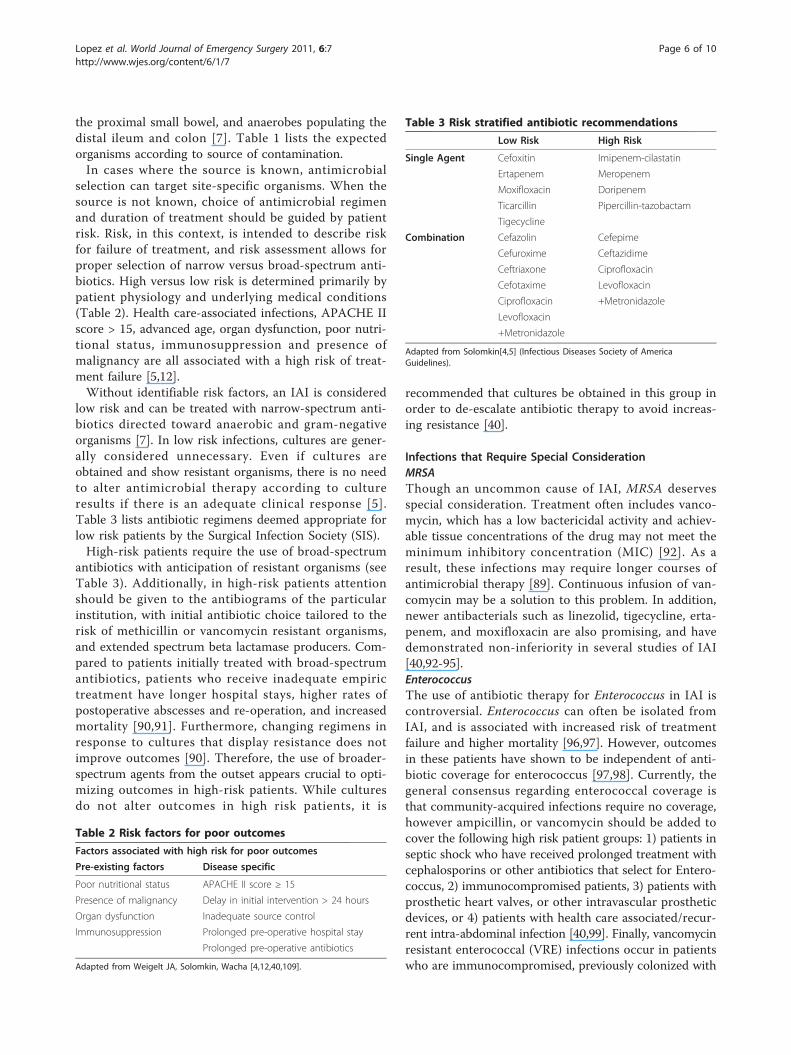
selection can target site-specific organisms. When thesource is not known, choice of antimicrobial regimenand duration of treatment should be guided by patientrisk. Risk, in this context, is intended to describe riskfor failure of treatment, and risk assessment allows forproper selection of narrow versus broad-spectrum anti-biotics. High versus low risk is determined primarily bypatient physiology and underlying medical conditions(Table 2). Health care-associated infections, APACHE IIscore > 15, advanced age, organ dysfunction, poor nutri-tional status, immunosuppression and presence ofmalignancy are all associated with a high risk of treat-ment failure [5,12].Without identifiable risk factors, an IAI is considered
low risk and can be treated with narrow-spectrum anti-biotics directed toward anaerobic and gram-negativeorganisms [7]. In low risk infections, cultures are gener-ally considered unnecessary. Even if cultures areobtained and show resistant organisms, there is no needto alter antimicrobial therapy according to cultureresults if there is an adequate clinical response [5].Table 3 lists antibiotic regimens deemed appropriate forlow risk patients by the Surgical Infection Society (SIS).High-risk patients require the use of broad-spectrum
antibiotics with anticipation of resistant organisms (seeTable 3). Additionally, in high-risk patients attentionshould be given to the antibiograms of the particularinstitution, with initial antibiotic choice tailored to therisk of methicillin or vancomycin resistant organisms,and extended spectrum beta lactamase producers. Com-pared to patients initially treated with broad-spectrumantibiotics, patients who receive inadequate empirictreatment have longer hospital stays, higher rates ofpostoperative abscesses and re-operation, and increasedmortality [90,91]. Furthermore, changing regimens inresponse to cultures that display resistance does notimprove outcomes [90]. Therefore, the use of broader-spectrum agents from the outset appears crucial to opti-mizing outcomes in high-risk patients. While culturesdo not alter outcomes in high risk patients, it is
recommended that cultures be obtained in this group inorder to de-escalate antibiotic therapy to avoid increas-ing resistance [40].
Infections that Require Special ConsiderationMRSAThough an uncommon cause of IAI, MRSA deservesspecial consideration. Treatment often includes vanco-mycin, which has a low bactericidal activity and achiev-able tissue concentrations of the drug may not meet theminimum inhibitory concentration (MIC) [92]. As aresult, these infections may require longer courses ofantimicrobial therapy [89]. Continuous infusion of van-comycin may be a solution to this problem. In addition,newer antibacterials such as linezolid, tigecycline, erta-penem, and moxifloxacin are also promising, and havedemonstrated non-inferiority in several studies of IAI[40,92-95].EnterococcusThe use of antibiotic therapy for Enterococcus in IAI iscontroversial. Enterococcus can often be isolated fromIAI, and is associated with increased risk of treatmentfailure and higher mortality [96,97]. However, outcomesin these patients have shown to be independent of anti-biotic coverage for enterococcus [97,98]. Currently, thegeneral consensus regarding enterococcal coverage isthat community-acquired infections require no coverage,however ampicillin, or vancomycin should be added tocover the following high risk patient groups: 1) patients inseptic shock who have received prolonged treatment withcephalosporins or other antibiotics that select for Entero-coccus, 2) immunocompromised patients, 3) patients withprosthetic heart valves, or other intravascular prostheticdevices, or 4) patients with health care associated/recur-rent intra-abdominal infection [40,99]. Finally, vancomycinresistant enterococcal (VRE) infections occur in patientswho are immunocompromised, previously colonized with
Table 2 Risk factors for poor outcomes
Factors associated with high risk for poor outcomes
Pre-existing factors Disease specific
Poor nutritional status APACHE II score ≥ 15
Presence of malignancy Delay in initial intervention > 24 hours
Organ dysfunction Inadequate source control
Immunosuppression Prolonged pre-operative hospital stay
Prolonged pre-operative antibiotics
Adapted from Weigelt JA, Solomkin, Wacha [4,12,40,109].
Table 3 Risk stratified antibiotic recommendations
Low Risk High Risk
Single Agent Cefoxitin Imipenem-cilastatin
Ertapenem Meropenem
Moxifloxacin Doripenem
Ticarcillin Pipercillin-tazobactam
Tigecycline
Combination Cefazolin Cefepime
Cefuroxime Ceftazidime
Ceftriaxone Ciprofloxacin
Cefotaxime Levofloxacin
Ciprofloxacin +Metronidazole
Levofloxacin
+Metronidazole
Adapted from Solomkin[4,5] (Infectious Diseases Society of AmericaGuidelines).
Lopez et al. World Journal of Emergency Surgery 2011, 6:7http://www.wjes.org/content/6/1/7
Page 6 of 10
VRE or treated with vancomycin [100]. In these circum-stances VRE should be suspected and treated with alterna-tives such as linezolid, tigecycline, or daptomycin. In theabsence of these risk factors, specific coverage for VRE isnot recommended [40].CandidaCandida is similar to Enterococcus, in that isolation ofCandida from intra-abdominal cultures is associatedwith increased mortality, but anti-fungal treatment hasnot been shown to alter this risk [101]. Therefore, fungalcoverage is unnecessary unless the patient is immuno-compromised, has a severe IAI with Candida grownfrom intra-abdominal cultures, or has perforation of agastric ulcer while on acid suppressive medications [102].Fluconazole is an appropriate initial choice for Candidaalbicans peritonitis. However, increasingly, non-albicansCandida spp., with resistance to commonly used anti-fungals are responsible for candidemia [103,104]. Studieshave shown that echinocandins are both safe and effec-tive in the treatment of invasive candidiasis. Therefore, incritically ill patients echinocandins, such as caspofunginor echinofungin, should be considered for primary treat-ment [102,104]. Required treatment duration for Can-dida peritonitis is 2-3 weeks [102].
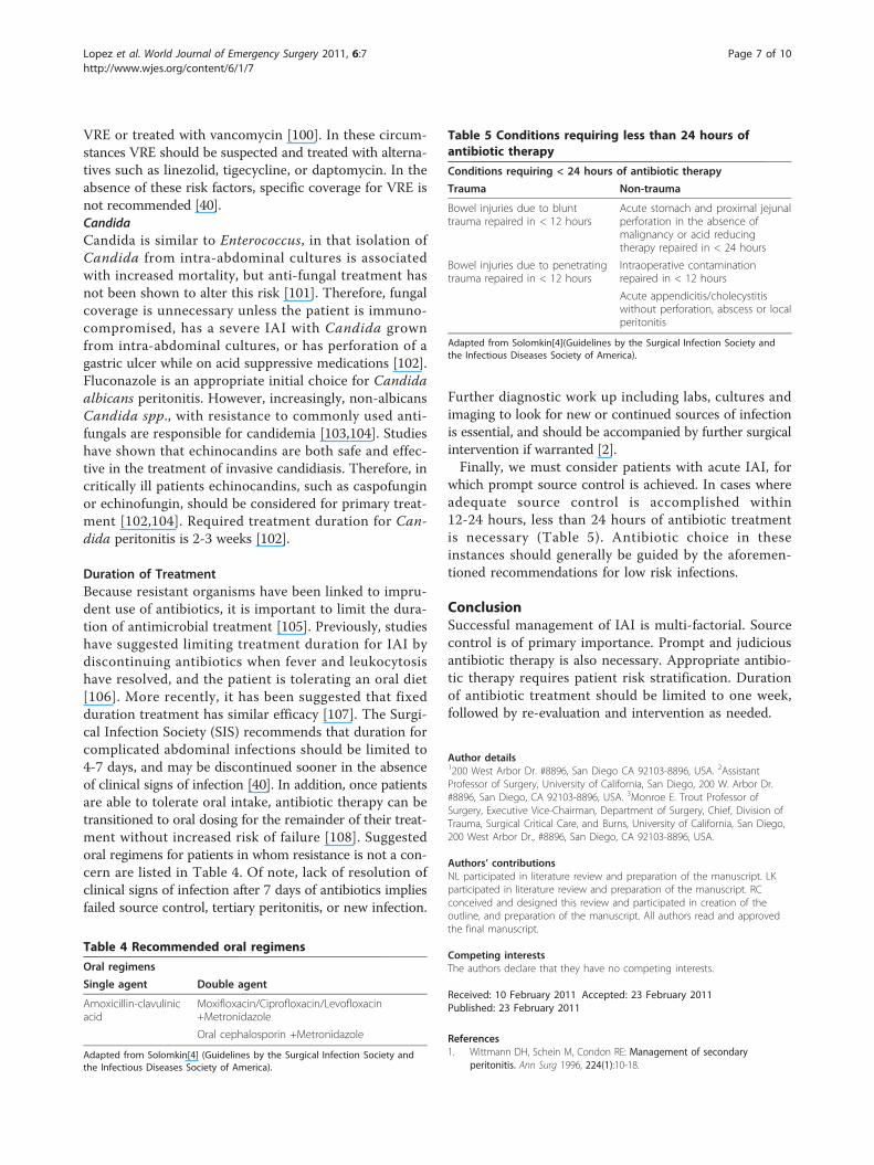
Duration of TreatmentBecause resistant organisms have been linked to impru-dent use of antibiotics, it is important to limit the dura-tion of antimicrobial treatment [105]. Previously, studieshave suggested limiting treatment duration for IAI bydiscontinuing antibiotics when fever and leukocytosishave resolved, and the patient is tolerating an oral diet[106]. More recently, it has been suggested that fixedduration treatment has similar efficacy [107]. The Surgi-cal Infection Society (SIS) recommends that duration forcomplicated abdominal infections should be limited to4-7 days, and may be discontinued sooner in the absenceof clinical signs of infection [40]. In addition, once patientsare able to tolerate oral intake, antibiotic therapy can betransitioned to oral dosing for the remainder of their treat-ment without increased risk of failure [108]. Suggestedoral regimens for patients in whom resistance is not a con-cern are listed in Table 4. Of note, lack of resolution ofclinical signs of infection after 7 days of antibiotics impliesfailed source control, tertiary peritonitis, or new infection.
Further diagnostic work up including labs, cultures andimaging to look for new or continued sources of infectionis essential, and should be accompanied by further surgicalintervention if warranted [2].Finally, we must consider patients with acute IAI, for
which prompt source control is achieved. In cases whereadequate source control is accomplished within12-24 hours, less than 24 hours of antibiotic treatmentis necessary (Table 5). Antibiotic choice in theseinstances should generally be guided by the aforemen-tioned recommendations for low risk infections.
ConclusionSuccessful management of IAI is multi-factorial. Sourcecontrol is of primary importance. Prompt and judiciousantibiotic therapy is also necessary. Appropriate antibio-tic therapy requires patient risk stratification. Durationof antibiotic treatment should be limited to one week,followed by re-evaluation and intervention as needed.
Author details1200 West Arbor Dr. #8896, San Diego CA 92103-8896, USA. 2AssistantProfessor of Surgery, University of California, San Diego, 200 W. Arbor Dr.#8896, San Diego, CA 92103-8896, USA. 3Monroe E. Trout Professor ofSurgery, Executive Vice-Chairman, Department of Surgery, Chief, Division ofTrauma, Surgical Critical Care, and Burns, University of California, San Diego,200 West Arbor Dr., #8896, San Diego, CA 92103-8896, USA.
Authors’ contributionsNL participated in literature review and preparation of the manuscript. LKparticipated in literature review and preparation of the manuscript. RCconceived and designed this review and participated in creation of theoutline, and preparation of the manuscript. All authors read and approvedthe final manuscript.
Competing interestsThe authors declare that they have no competing interests.
Received: 10 February 2011 Accepted: 23 February 2011Published: 23 February 2011
References1. Wittmann DH, Schein M, Condon RE: Management of secondary
peritonitis. Ann Surg 1996, 224(1):10-18.
Table 4 Recommended oral regimens
Oral regimens
Single agent Double agent
Amoxicillin-clavulinicacid
Moxifloxacin/Ciprofloxacin/Levofloxacin+Metronidazole
Oral cephalosporin +Metronidazole
Adapted from Solomkin[4] (Guidelines by the Surgical Infection Society andthe Infectious Diseases Society of America).
Table 5 Conditions requiring less than 24 hours ofantibiotic therapy
Conditions requiring < 24 hours of antibiotic therapy
Trauma Non-trauma
Bowel injuries due to blunttrauma repaired in < 12 hours
Acute stomach and proximal jejunalperforation in the absence ofmalignancy or acid reducingtherapy repaired in < 24 hours
Bowel injuries due to penetratingtrauma repaired in < 12 hours
Intraoperative contaminationrepaired in < 12 hours
Acute appendicitis/cholecystitiswithout perforation, abscess or localperitonitis
Adapted from Solomkin[4](Guidelines by the Surgical Infection Society andthe Infectious Diseases Society of America).
Lopez et al. World Journal of Emergency Surgery 2011, 6:7http://www.wjes.org/content/6/1/7
Page 7 of 10
2. Pieracci FM, Barie PS: Management of severe sepsis of abdominal origin.Scand J Surg 2007, 96(3):184-196.
3. Merlino JI, Yowler CJ, Malangoni MA: Nosocomial infections adverselyaffect the outcomes of patients with serious intraabdominal infections.Surg Infect (Larchmt) 2004, 5(1):21-27.
4. Solomkin JS, Mazuski JE, Bradley JS, Rodvold KA, Goldstein EJ, Baron EJ,O’Neill PJ, Chow AW, Dellinger EP, Eachempati SR, Gorbach S, Hilfiker M,May AK, Nathens AB, Sawyer RG, Bartlett JG: Diagnosis andmanagement of complicated intra-abdominal infection in adults andchildren: guidelines by the Surgical Infection Society and theInfectious Diseases Society of America. Surg Infect (Larchmt)11(1):79-109.
5. Solomkin JS, Mazuski JE, Baron EJ, Sawyer RG, Nathens AB, DiPiro JT,Buchman T, Dellinger EP, Jernigan J, Gorbach S, Chow AW, Bartlett J:Guidelines for the selection of anti-infective agents for complicatedintra-abdominal infections. Clin Infect Dis 2003, 37(8):997-1005.
6. Sola R, Soriano G: Why do bacteria reach ascitic fluid? Eur J GastroenterolHepatol 2002, 14(4):351-354.
7. Marshall JC, Innes M: Intensive care unit management of intra-abdominalinfection. Crit Care Med 2003, 31(8):2228-2237.
8. Williams JD, Coles GA: Gram-positive infections related to CAPD. JAntimicrob Chemother 1991, 27(Suppl):B31-35.
9. Ljubicic N, Spajic D, Vrkljan MM, Altabas V, Doko M, Zovak M, Gacina P,Mihatov S: The value of ascitic fluid polymorphonuclear cell countdetermination during therapy of spontaneous bacterial peritonitis inpatients with liver cirrhosis. Hepatogastroenterology 2000,47(35):1360-1363.
10. Adam EJ, Page JE: Intra-abdominal sepsis: the role of radiology. BaillieresClin Gastroenterol 1991, 5(3 Pt 1):587-609.
11. Crandall M, West MA: Evaluation of the abdomen in the critically illpatient: opening the black box. Curr Opin Crit Care 2006, 12(4):333-339.
12. Weigelt JA: Empiric treatment options in the management ofcomplicated intra-abdominal infections. Cleve Clin J Med 2007, 74(Suppl4):S29-37.
13. Nathens AB, Rotstein OD, Marshall JC: Tertiary peritonitis: clinical featuresof a complex nosocomial infection. World J Surg 1998, 22(2):158-163.
14. Henderson LW, Nolph KD: Altered permeability of the peritonealmembrane after using hypertonic peritoneal dialysis fluid. J Clin Invest1969, 48(6):992-1001.
15. Heemken R, Gandawidjaja L, Hau T: Peritonitis: pathophysiology and localdefense mechanisms. Hepatogastroenterology 1997, 44(16):927-936.
16. Hall JC, Heel KA, Papadimitriou JM, Platell C: The pathobiology ofperitonitis. Gastroenterology 1998, 114(1):185-196.
17. Brinkmann V, Reichard U, Goosmann C, Fauler B, Uhlemann Y, Weiss DS,Weinrauch Y, Zychlinsky A: Neutrophil extracellular traps kill bacteria.Science 2004, 303(5663):1532-1535.
18. Bone RC, Balk RA, Cerra FB, Dellinger RP, Fein AM, Knaus WA, Schein RM,Sibbald WJ: Definitions for sepsis and organ failure and guidelines forthe use of innovative therapies in sepsis. The ACCP/SCCM ConsensusConference Committee. American College of Chest Physicians/Society ofCritical Care Medicine. Chest 1992, 101(6):1644-1655.
19. Sartelli M: A focus on intra-abdominal infections. World J Emerg Surg 59.20. Lamke LO, Nilsson G, Reithner L: The influence of elevated body
temperature on skin perspiration. Acta Chir Scand 1980, 146(2):81-84.21. Reithner L: Insensible water loss from the respiratory tract in patients
with fever. Acta Chir Scand 1981, 147(3):163-167.22. Dellinger RP, Levy MM, Carlet JM, Bion J, Parker MM, Jaeschke R, Reinhart K,
Angus DC, Brun-Buisson C, Beale R, Calandra T, Dhainaut JF, Gerlach H,Harvey M, Marini JJ, Marshall J, Ranieri M, Ramsay G, Sevransky J,Thompson BT, Townsend S, Vender JS, Zimmerman JL, Vincent JL:Surviving Sepsis Campaign: international guidelines for management ofsevere sepsis and septic shock: 2008. Crit Care Med 2008, 36(1):296-327.
23. Vincent JL, Gerlach H: Fluid resuscitation in severe sepsis and septicshock: an evidence-based review. Crit Care Med 2004, 32(11 Suppl):S451-454.
24. Yu M, Burchell S, Hasaniya NW, Takanishi DM, Myers SA, Takiguchi SA:Relationship of mortality to increasing oxygen delivery in patients > or= 50 years of age: a prospective, randomized trial. Crit Care Med 1998,26(6):1011-1019.
25. Levy B: Lactate and shock state: the metabolic view. Curr Opin Crit Care2006, 12(4):315-321.
26. James JH, Luchette FA, McCarter FD, Fischer JE: Lactate is an unreliableindicator of tissue hypoxia in injury or sepsis. Lancet 1999,354(9177):505-508.
27. Mikulaschek A, Henry SM, Donovan R, Scalea TM: Serum lactate is notpredicted by anion gap or base excess after trauma resuscitation. JTrauma 1996, 40(2):218-222, discussion 222-214.
28. Rutherford EJ, Morris JA Jr, Reed GW, Hall KS: Base deficit stratifiesmortality and determines therapy. J Trauma 1992, 33(3):417-423.
29. Davis JW, Shackford SR, Mackersie RC, Hoyt DB: Base deficit as a guide tovolume resuscitation. J Trauma 1988, 28(10):1464-1467.
30. Hemming A, Davis NL, Robins RE: Surgical versus percutaneous drainageof intra-abdominal abscesses. Am J Surg 1991, 161(5):593-595.
31. Bufalari A, Giustozzi G, Moggi L: Postoperative intraabdominal abscesses:percutaneous versus surgical treatment. Acta Chir Belg 1996,96(5):197-200.
32. Sugimoto K, Hirata M, Kikuno T, Takishima T, Maekawa K, Ohwada T: Large-volume intraoperative peritoneal lavage with an assistant device fortreatment of peritonitis caused by blunt traumatic rupture of the smallbowel. J Trauma 1995, 39(4):689-692.
33. Whiteside OJ, Tytherleigh MG, Thrush S, Farouk R, Galland RB: Intra-operative peritoneal lavage–who does it and why? Ann R Coll Surg Engl2005, 87(4):255-258.
34. Schein M, Gecelter G, Freinkel W, Gerding H, Becker PJ: Peritoneal lavagein abdominal sepsis. A controlled clinical study. Arch Surg 1990,125(9):1132-1135.
35. Hudspeth AS: Radical surgical debridement in the treatment of advancedgeneralized bacterial peritonitis. Arch Surg 1975, 110(10):1233-1236.
36. Polk HC Jr, Fry DE: Radical peritoneal debridement for establishedperitonitis. The results of a prospective randomized clinical trial. AnnSurg 1980, 192(3):350-355.
37. Schilling MK, Maurer CA, Kollmar O, Buchler MW: Primary vs. secondaryanastomosis after sigmoid colon resection for perforated diverticulitis(Hinchey Stage III and IV) a prospective outcome and cost analysis. DisColon Rectum 2001, 44(5):699-703, discussion 703-695.
38. Solomkin JS, Mazuski JE, Bradley JS, Rodvold KA, Goldstein EJ, Baron EJ,O’Neill PJ, Chow AW, Dellinger EP, Eachempati SR, Gorbach S, Hilfiker M,May AK, Nathens AB, Sawyer RG, Bartlett JG: Diagnosis and managementof complicated intra-abdominal infection in adults and children:guidelines by the Surgical Infection Society and the Infectious DiseasesSociety of America. Clin Infect Dis 50(2):133-164.
39. Humes D, Speake WJ, Simpson J: Appendicitis. Clin Evid (Online) 2007, 2007.40. Solomkin JS, Mazuski J: Intra-abdominal sepsis: newer interventional and
antimicrobial therapies. Infect Dis Clin North Am 2009, 23(3):593-608.41. Lee SL, Walsh AJ, Ho HS: Computed tomography and ultrasonography do
not improve and may delay the diagnosis and treatment of acuteappendicitis. Arch Surg 2001, 136(5):556-562.
42. Lee SL, Ho HS: Ultrasonography and computed tomography insuspected acute appendicitis. Semin Ultrasound CT MR 2003, 24(2):69-73.
43. Brown MA: Imaging acute appendicitis. Semin Ultrasound CT MR 2008,29(5):293-307.
44. Lee JH: Sonography of acute appendicitis. Semin Ultrasound CT MR 2003,24(2):83-90.
45. Sivit CJ, Applegate KE: Imaging of acute appendicitis in children. SeminUltrasound CT MR 2003, 24(2):74-82.
46. Wan MJ, Krahn M, Ungar WJ, Caku E, Sung L, Medina LS, Doria AS: Acuteappendicitis in young children: cost-effectiveness of US versus CT indiagnosis–a Markov decision analytic model. Radiology 2009,250(2):378-386.
47. Kaneko K, Tsuda M: Ultrasound-based decision making in the treatmentof acute appendicitis in children. J Pediatr Surg 2004, 39(9):1316-1320.
48. Hagendorf BA, Clarke JR, Burd RS: The optimal initial management ofchildren with suspected appendicitis: a decision analysis. J Pediatr Surg2004, 39(6):880-885.
49. Pedrosa I, Levine D, Eyvazzadeh AD, Siewert B, Ngo L, Rofsky NM: MRimaging evaluation of acute appendicitis in pregnancy. Radiology 2006,238(3):891-899.
50. Mason RJ: Surgery for appendicitis: is it necessary? Surg Infect (Larchmt)2008, 9(4):481-488.
51. Sauerland S, Lefering R, Neugebauer EA: Laparoscopic versus opensurgery for suspected appendicitis. Cochrane Database Syst Rev 2002, , 1:CD001546.
Lopez et al. World Journal of Emergency Surgery 2011, 6:7http://www.wjes.org/content/6/1/7
Page 8 of 10
52. Katkhouda N, Mason RJ, Towfigh S, Gevorgyan A, Essani R: Laparoscopicversus open appendectomy: a prospective randomized double-blindstudy. Ann Surg 2005, 242(3):439-448, discussion 448-450.
53. Kehagias I, Karamanakos SN, Panagiotopoulos S, Panagopoulos K,Kalfarentzos F: Laparoscopic versus open appendectomy: which way togo? World J Gastroenterol 2008, 14(31):4909-4914.
54. Bennett J, Boddy A, Rhodes M: Choice of approach for appendicectomy.Surg Laparosc Endosc Percutan Techa meta-analysis of open versuslaparoscopic appendicectomy 2007, 17(4):245-255.
55. Eypasch E, Sauerland S, Lefering R, Neugebauer EA: Laparoscopic versusopen appendectomy: between evidence and common sense. Dig Surg2002, 19(6):518-522.
56. Kapischke M, Caliebe A, Tepel J, Schulz T, Hedderich J: Open versuslaparoscopic appendicectomy: a critical review. Surg Endosc 2006,20(7):1060-1068.
57. Andersson RE, Petzold MG: Nonsurgical treatment of appendiceal abscessor phlegmon: a systematic review and meta-analysis. Ann Surg 2007,246(5):741-748.
58. St Peter SD, Aguayo P, Fraser JD, Keckler SJ, Sharp SW, Leys CM, Murphy JP,Snyder CL, Sharp RJ, Andrews WS, Holcomb GW, Ostlie DJ: Initiallaparoscopic appendectomy versus initial nonoperative managementand interval appendectomy for perforated appendicitis with abscess: aprospective, randomized trial. J Pediatr Surg 45(1):236-240.
59. Deakin DE, Ahmed I: Interval appendicectomy after resolution of adultinflammatory appendix mass–is it necessary? Surgeon 2007, 5(1):45-50.
60. Manuel D, Cutler A, Goldstein J, Fennerty MB, Brown K: Decreasingprevalence combined with increasing eradication of Helicobacter pyloriinfection in the United States has not resulted in fewer hospitaladmissions for peptic ulcer disease-related complications. AlimentPharmacol Ther 2007, 25(12):1423-1427.
61. Wang YR, Richter JE, Dempsey DT: Trends and outcomes ofhospitalizations for peptic ulcer disease in the United States, 1993 to2006. Ann Surg 251(1):51-58.
62. Kleeff J, Friess H, Buchler MW: How Helicobacter Pylori changed the life ofsurgeons. Dig Surg 2003, 20(2):93-102.
63. Ford AC, Delaney BC, Forman D, Moayyedi P: Eradication therapy forpeptic ulcer disease in Helicobacter pylori positive patients. CochraneDatabase Syst Rev 2006, , 2: CD003840.
64. Svanes C: Trends in perforated peptic ulcer: incidence, etiology,treatment, and prognosis. World J Surg 2000, 24(3):277-283.
65. Lau WY, Leung KL, Kwong KH, Davey IC, Robertson C, Dawson JJ,Chung SC, Li AK: A randomized study comparing laparoscopic versusopen repair of perforated peptic ulcer using suture or suturelesstechnique. Ann Surg 1996, 224(2):131-138.
66. Crofts TJ, Park KG, Steele RJ, Chung SS, Li AK: A randomized trial ofnonoperative treatment for perforated peptic ulcer. N Engl J Med 1989,320(15):970-973.
67. Millat B, Fingerhut A, Borie F: Surgical treatment of complicated duodenalulcers: controlled trials. World J Surg 2000, 24(3):299-306.
68. Berne TV, Donovan AJ: Nonoperative treatment of perforated duodenalulcer. Arch Surg 1989, 124(7):830-832.
69. Schoetz DJ Jr: Diverticular disease of the colon: a century-old problem.Dis Colon Rectum 1999, 42(6):703-709.
70. Hughes LE: Postmortem survey of diverticular disease of the colon. II.The muscular abnormality of the sigmoid colon. Gut 1969, 10(5):344-351.
71. Evans J, Kozol R, Frederick W, Voytavich A, Pennoyer W, Lukianoff A,Lardner J: Does a 48-hour rule predict outcomes in patients with acutesigmoid diverticulitis? J Gastrointest Surg 2008, 12(3):577-582.
72. Sra HK, Shipman K, Virk HS: Does a 48-hour rule predict outcomes inpatients with acute sigmoid diverticulitis? J Gastrointest Surg 2009,13(10):1892.
73. Kaiser AM, Jiang JK, Lake JP, Ault G, Artinyan A, Gonzalez-Ruiz C, Essani R,Beart RW Jr: The management of complicated diverticulitis and the roleof computed tomography. Am J Gastroenterol 2005, 100(4):910-917.
74. Ambrosetti P, Becker C, Terrier F: Colonic diverticulitis: impact of imagingon surgical management – a prospective study of 542 patients. EurRadiol 2002, 12(5):1145-1149.
75. Brandt D, Gervaz P, Durmishi Y, Platon A, Morel P, Poletti PA: PercutaneousCT scan-guided drainage vs. antibiotherapy alone for Hinchey IIdiverticulitis: a case-control study. Dis Colon Rectum 2006,49(10):1533-1538.
76. Siewert B, Tye G, Kruskal J, Sosna J, Opelka F, Raptopoulos V, Goldberg SN:Impact of CT-guided drainage in the treatment of diverticular abscesses:size matters. AJR Am J Roentgenol 2006, 186(3):680-686.
77. Golfieri R, Cappelli A: Computed tomography-guided percutaneousabscess drainage in coloproctology: review of the literature. TechColoproctol 2007, 11(3):197-208.
78. Ambrosetti P, Chautems R, Soravia C, Peiris-Waser N, Terrier F: Long-termoutcome of mesocolic and pelvic diverticular abscesses of the left colon:a prospective study of 73 cases. Dis Colon Rectum 2005, 48(4):787-791.
79. Chapman J, Davies M, Wolff B, Dozois E, Tessier D, Harrington J, Larson D:Complicated diverticulitis: is it time to rethink the rules? Ann Surg 2005,242(4):576-581, discussion 581-573.
80. Salem TA, Molloy RG, O’Dwyer PJ: Prospective study on the managementof patients with complicated diverticular disease. Colorectal Dis 2006,8(3):173-176.
81. Ricciardi R, Baxter NN, Read TE, Marcello PW, Hall J, Roberts PL: Is the declinein the surgical treatment for diverticulitis associated with an increase incomplicated diverticulitis? Dis Colon Rectum 2009, 52(9):1558-1563.
82. Salem L, Anaya DA, Roberts KE, Flum DR: Hartmann’s colectomy andreversal in diverticulitis: a population-level assessment. Dis Colon Rectum2005, 48(5):988-995.
83. Salem L, Flum DR: Primary anastomosis or Hartmann’s procedure forpatients with diverticular peritonitis? A systematic review. Dis ColonRectum 2004, 47(11):1953-1964.
84. Chandra V, Nelson H, Larson DR, Harrington JR: Impact of primaryresection on the outcome of patients with perforated diverticulitis. ArchSurg 2004, 139(11):1221-1224.
85. Aydin HN, Remzi FH, Tekkis PP, Fazio VW: Hartmann’s reversal isassociated with high postoperative adverse events. Dis Colon Rectum2005, 48(11):2117-2126.
86. Richter S, Lindemann W, Kollmar O, Pistorius GA, Maurer CA, Schilling MK:One-stage sigmoid colon resection for perforated sigmoid diverticulitis(Hinchey stages III and IV). World J Surg 2006, 30(6):1027-032.
87. McCafferty MH, Roth L, Jorden J: Current management of diverticulitis.Am Surg 2008, 74(11):1041-1049.
88. Klarenbeek BR, Veenhof AA, Bergamaschi R, van der Peet DL, van denBroek WT, de Lange ES, Bemelman WA, Heres P, Lacy AM, Engel AF,Cuesta MA: Laparoscopic sigmoid resection for diverticulitis decreasesmajor morbidity rates: a randomized control trial: short-term results ofthe Sigma Trial. Ann Surg 2009, 249(1):39-44.
89. Blot S, De Waele JJ: Critical issues in the clinical management ofcomplicated intra-abdominal infections. Drugs 2005, 65(12):1611-1620.
90. Mosdell DM, Morris DM, Voltura A, Pitcher DE, Twiest MW, Milne RL,Miscall BG, Fry DE: Antibiotic treatment for surgical peritonitis. Ann Surg1991, 214(5):543-549.
91. Montravers P, Gauzit R, Muller C, Marmuse JP, Fichelle A, Desmonts JM:Emergence of antibiotic-resistant bacteria in cases of peritonitis afterintraabdominal surgery affects the efficacy of empirical antimicrobialtherapy. Clin Infect Dis 1996, 23(3):486-494.
92. Stass H, Rink AD, Delesen H, Kubitza D, Vestweber KH: Pharmacokineticsand peritoneal penetration of moxifloxacin in peritonitis. J AntimicrobChemother 2006, 58(3):693-696.
93. Vuagnat A, Stern R, Lotthe A, Schuhmacher H, Duong M, Hoffmeyer P,Bernard L: High dose vancomycin for osteomyelitis: continuous vs.intermittent infusion. J Clin Pharm Ther 2004, 29(4):351-357.
94. Babinchak T, Ellis-Grosse E, Dartois N, Rose GM, Loh E: The efficacy andsafety of tigecycline for the treatment of complicated intra-abdominalinfections: analysis of pooled clinical trial data. Clin Infect Dis 2005,41(Suppl 5):S354-367.
95. Solomkin JS, Yellin AE, Rotstein OD, Christou NV, Dellinger EP, Tellado JM,Malafaia O, Fernandez A, Choe KA, Carides A, Satishchandran V, Teppler H:Ertapenem versus piperacillin/tazobactam in the treatment ofcomplicated intraabdominal infections: results of a double-blind,randomized comparative phase III trial. Ann Surg 2003, 237(2):235-245.
96. Burnett RJ, Haverstock DC, Dellinger EP, Reinhart HH, Bohnen JM,Rotstein OD, Vogel SB, Solomkin JS: Definition of the role of enterococcusin intraabdominal infection: analysis of a prospective randomized trial.Surgery 1995, 118(4):716-721, discussion 721-713.
97. Sitges-Serra A, Lopez MJ, Girvent M, Almirall S, Sancho JJ: Postoperativeenterococcal infection after treatment of complicated intra-abdominalsepsis. Br J Surg 2002, 89(3):361-367.
Lopez et al. World Journal of Emergency Surgery 2011, 6:7http://www.wjes.org/content/6/1/7
Page 9 of 10
98. Teppler H, McCarroll K, Gesser RM, Woods GL: Surgical infections withenterococcus: outcome in patients treated with ertapenem versuspiperacillin-tazobactam. Surg Infect (Larchmt) 2002, 3(4):337-349.
99. Harbarth S, Uckay I: Are there patients with peritonitis who requireempiric therapy for enterococcus? Eur J Clin Microbiol Infect Dis 2004,23(2):73-77.
100. Mazuski JE: Vancomycin-resistant enterococcus: risk factors, surveillance,infections, and treatment. Surg Infect (Larchmt) 2008, 9(6):567-571.
101. Sandven P, Qvist H, Skovlund E, Giercksky KE: Significance of Candidarecovered from intraoperative specimens in patients with intra-abdominal perforations. Crit Care Med 2002, 30(3):541-547.
102. Pappas PG, Rex JH, Sobel JD, Filler SG, Dismukes WE, Walsh TJ, Edwards JE:Guidelines for treatment of candidiasis. Clin Infect Dis 2004, 38(2):161-189.
103. Krause DS, Reinhardt J, Vazquez JA, Reboli A, Goldstein BP, Wible M,Henkel T: Phase 2, randomized, dose-ranging study evaluating the safetyand efficacy of anidulafungin in invasive candidiasis and candidemia.Antimicrob Agents Chemother 2004, 48(6):2021-2024.
104. Pfaller MA, Messer SA, Hollis RJ, Boyken L, Tendolkar S, Kroeger J,Diekema DJ: Variation in susceptibility of bloodstream isolates ofCandida glabrata to fluconazole according to patient age andgeographic location in the United States in 2001 to 2007. J Clin Microbiol2009, 47(10):3185-3190.
105. Leaper D: Nosocomial infection. Br J Surg 2004, 91(5):526-527.106. Stone HH, Bourneuf AA, Stinson LD: Reliability of criteria for predicting
persistent or recurrent sepsis. Arch Surg 1985, 120(1):17-20.107. Hedrick TL, Evans HL, Smith RL, McElearney ST, Schulman AS, Chong TW,
Pruett TL, Sawyer RG: Can we define the ideal duration of antibiotictherapy? Surg Infect (Larchmt) 2006, 7(5):419-432.
108. Solomkin JS, Dellinger EP, Bohnen JM, Rostein OD: The role of oralantimicrobials for the management of intra-abdominal infections. NewHoriz 1998, 6(2 Suppl):S46-52.
109. Wacha H, Hau T, Dittmer R, Ohmann C: Risk factors associated withintraabdominal infections: a prospective multicenter study. PeritonitisStudy Group. Langenbecks Arch Surg 1999, 384(1):24-32.
doi:10.1186/1749-7922-6-7Cite this article as: Lopez et al.: A Comprehensive review of abdominalinfections. World Journal of Emergency Surgery 2011 6:7.
Submit your next manuscript to BioMed Centraland take full advantage of:
• Convenient online submission
• Thorough peer review
• No space constraints or color figure charges
• Immediate publication on acceptance
• Inclusion in PubMed, CAS, Scopus and Google Scholar
• Research which is freely available for redistribution
Submit your manuscript at www.biomedcentral.com/submit
Lopez et al. World Journal of Emergency Surgery 2011, 6:7http://www.wjes.org/content/6/1/7
Page 10 of 10