synergism of plant-derived polyphenols in adipogenesis: perspectives and implications
TRANSCRIPT
SP
MECa
b
c
d
e
a
KAHLMPS
I
dlAr
it6i1
MT
o
0d
Phytomedicine 19 (2012) 253– 261
Contents lists available at SciVerse ScienceDirect
Phytomedicine
j ourna l ho mepage: www.elsev ier .de /phymed
ynergism of plant-derived polyphenols in adipogenesis:erspectives and implications
aría Herranz-Lópeza,1, Salvador Fernández-Arroyob,1, Almudena Pérez-Sancheza,nrique Barrajón-Catalána, Raúl Beltrán-Debónc, Javier Abel Menéndezd,arlos Alonso-Villaverdee, Antonio Segura-Carreterob, Jorge Jovenc, Vicente Micola,∗
Instituto de Biología Molecular y Celular (IBMC), Universidad Miguel Hernández, Elche, Alicante, SpainDepartment of Analytical Chemistry, Faculty of Sciences, University of Granada, Av/Fuentenueva, 18071 Granada, SpainCentre de Recerca Biomèdica, Hospital Universitari de Sant Joan, IISPV, Universitat Rovira i Virgili, C/Sant Joan s/n, 43201 Reus, SpainCatalan Institute of Oncology (ICO), Girona, Catalonia, SpainServei de Medicina Interna, Hospital Son Llàtzer, Palma, Illes Balears, Spain
r t i c l e i n f o
eywords:dipogenesisibiscus sabdariffaeptinCP-1
olyphenolsynergism
a b s t r a c t
Dietary polyphenols may exert their pharmacological effect via synergistic interactions with multipletargets. Putative effects of polyphenols in the management of obesity should be primarily evaluatedin adipose tissue and consequently in well-documented cell model. We used Hibiscus sabdariffa (HS), awidely recognised medicinal plant, as a source of polyphenols with a number of salutary effects previ-ously reported. We present here the full characterisation of bioactive components of HS aqueous extractsand document their effects in a model of adipogenesis from 3T3-L1 cells and in hypertrophic and insulin-resistant adipocytes. Aqueous extracts were up to 100 times more efficient in inhibiting triglyceride
accumulation when devoid of fibre and polysaccharides. Significant differences were also observed inreactive oxygen species generation and adipokine secretion. We also found that, when polyphenols werefractionated and isolated, the benefits of the whole extract were greater than the sum of its parts, whichindicated a previously unnoticed synergism. In conclusion, polyphenols have interactive and complemen-tary effects, which suggest a possible application in the management of complex diseases and efforts toisolate individual components might be irrelevant for clinical medicine and/or human nutrition.© 2012 Elsevier GmbH. All rights reserved.
ntroduction
Obesity-associated metabolic, oxidative and inflammatoryisturbances are a growing epidemic and are associated with at
east six of the top ten causes of death (McGeer and McGeer 2004).dipocytes store excess energy, but when overloaded they becomeesistant to insulin, which compromises their ability to accumulate
Abbreviations: HS, Hibiscus sabdariffa; AHS, aqueous extract of H. sabdar-ffa; PEHS, phenolic extract of H. sabdariffa; FBS, foetal bovine serum; TNF-�,umour necrosis factor-alpha; IGF1, insulin-like growth factor-1; IL-6, interleukin-; VEGF, vascular endothelial growth factor; IL-1a, interleukin-1 alpha; IL-1b,
nterleukin-1 beta; MCP-1, monocyte chemoattractant protein-1; IBMX, 3-isobutyl--methylxanthine.∗ Corresponding author at: Instituto de Biología Molecular y Celular, Universidadiguel Hernández, Avda. de la Universidad S/N◦ , 03202 Elche, Alicante, Spain.
el.: +34 96 6658430; fax: +34 96 6658758.E-mail address: [email protected] (V. Micol).
1 These authors have equally contributed to this research and are listed in randomrder.
944-7113/$ – see front matter © 2012 Elsevier GmbH. All rights reserved.oi:10.1016/j.phymed.2011.12.001
lipids and facilitates alterations in structure and metabolism inremote tissues, such as the pancreas, liver and muscle (Yu andZhu 2004; Jernas et al. 2006; Rull et al. 2010). Excessive oxidationin adipose cells is common and triggers cellular stress (Furukawaet al. 2004; Yeop Han et al. 2010). The resulting sequence of eventsremains poorly understood in humans but tends to self-perpetuateif untreated. Initially, there is a complex process of cellular adapta-tion, monitored by tissue-resident macrophages. When failure andmalfunction become extreme, a chronic inflammatory response isunleashed (Rull et al. 2010).
If assumptions are accurate, it is conceivable that antioxidantand/or anti-inflammatory therapies that act on adipose tissue mayhave potential benefits in the amelioration of obesity-related dis-eases. However, current available drugs have not been assayedyet. The only validated therapeutic measure consists of prevent-ing hypertrophy in adipocytes via caloric restriction or increased
caloric expenditure, but changes in lifestyle are difficult to achieve.Plant-derived polyphenols may provide a similar effect with-out restricting caloric intake (Lamming et al. 2004; Howitz andSinclair 2008). Polyphenols are antioxidant and anti-inflammatory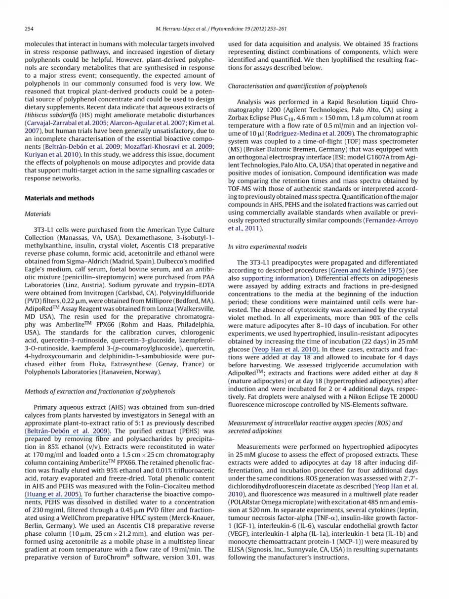
2 ytome
mipntprtdH(2anKttr
M
M
CmroEoLw(AMpUa34cP
M
ca(ptactai(noaBpfgp
1 (IGF-1), interleukin-6 (IL-6), vascular endothelial growth factor
54 M. Herranz-López et al. / Ph
olecules that interact in humans with molecular targets involvedn stress response pathways, and increased ingestion of dietaryolyphenols could be helpful. However, plant-derived polyphe-ols are secondary metabolites that are synthesised in responseo a major stress event; consequently, the expected amount ofolyphenols in our commonly consumed food is very low. Weeasoned that tropical plant-derived products could be a poten-ial source of polyphenol concentrate and could be used to designietary supplements. Recent data indicate that aqueous extracts ofibiscus sabdariffa (HS) might ameliorate metabolic disturbances
Carvajal-Zarrabal et al. 2005; Alarcon-Aguilar et al. 2007; Kim et al.007), but human trials have been generally unsatisfactory, due ton incomplete characterisation of the essential bioactive compo-ents (Beltrán-Debón et al. 2009; Mozaffari-Khosravi et al. 2009;uriyan et al. 2010). In this study, we address this issue, document
he effects of polyphenols on mouse adipocytes and provide datahat support multi-target action in the same signalling cascades oresponse networks.
aterials and methods
aterials
3T3-L1 cells were purchased from the American Type Cultureollection (Manassas, VA, USA). Dexamethasone, 3-isobutyl-1-ethylxanthine, insulin, crystal violet, Ascentis C18 preparative
everse phase column, formic acid, acetonitrile and ethanol werebtained from Sigma–Aldrich (Madrid, Spain). Dulbecco’s modifiedagle’s medium, calf serum, foetal bovine serum, and an antibi-tic mixture (penicillin–streptomycin) were purchased from PAAaboratories (Linz, Austria). Sodium pyruvate and trypsin–EDTAere obtained from Invitrogen (Carlsbad, CA). Polyvinyldifluoride
PVD) filters, 0.22 �m, were obtained from Millipore (Bedford, MA).dipoRedTM Assay Reagent was obtained from Lonza (Walkersville,D USA). The resin used for the preparative chromatogra-
hy was AmberliteTM FPX66 (Rohm and Haas, Philadelphia,SA). The standards for the calibration curves, chlorogeniccid, quercetin-3-rutinoside, quercetin-3-glucoside, kaempferol--O-rutinoside, kaempferol 3-(p-coumaroylglucoside), quercetin,-hydroxycoumarin and delphinidin-3-sambubioside were pur-hased either from Fluka, Extrasynthese (Genay, France) orolyphenols Laboratories (Hanaveien, Norway).
ethods of extraction and fractionation of polyphenols
Primary aqueous extract (AHS) was obtained from sun-driedalyces from plants harvested by investigators in Senegal with anpproximate plant-to-extract ratio of 5:1 as previously describedBeltrán-Debón et al. 2009). The purified extract (PEHS) wasrepared by removing fibre and polysaccharides by precipita-ion in 85% ethanol (v/v). Extracts were reconstituted in watert 170 mg/ml and loaded onto a 1.5 cm × 25 cm chromatographyolumn containing AmberliteTM FPX66. The retained phenolic frac-ion was finally eluted with 95% ethanol and 0.01% trifluoroaceticcid, rotary evaporated and freeze-dried. Total phenolic contentn AHS and PEHS was measured with the Folin–Ciocalteu methodHuang et al. 2005). To further characterise the bioactive compo-ents, PEHS was dissolved in distilled water to a concentrationf 230 mg/ml, filtered through a 0.45 �m PVD filter and fraction-ted using a WellChrom preparative HPLC system (Merck-Knauer,erlin, Germany). We used an Ascentis C18 preparative reverse
hase column (10 �m, 25 cm × 21.2 mm), and elution was per-ormed using acetonitrile as a mobile phase in a multistep linearradient at room temperature with a flow rate of 19 ml/min. Thereparative version of EuroChrom® software, version 3.01, wasdicine 19 (2012) 253– 261
used for data acquisition and analysis. We obtained 35 fractionsrepresenting distinct combinations of components, which wereidentified and quantified. We then lyophilised the resulting frac-tions for assays described below.
Characterisation and quantification of polyphenols
Analysis was performed in a Rapid Resolution Liquid Chro-matography 1200 (Agilent Technologies, Palo Alto, CA) using aZorbax Eclipse Plus C18, 4.6 mm × 150 mm, 1.8 �m column at roomtemperature with a flow rate of 0.5 ml/min and an injection vol-ume of 10 �l (Rodríguez-Medina et al. 2009). The chromatographicsystem was coupled to a time-of-flight (TOF) mass spectrometer(MS) (Bruker Daltonic Bremen, Germany) that was equipped withan orthogonal electrospray interface (ESI; model G1607A from Agi-lent Technologies, Palo Alto, CA, USA) that operated in negative andpositive modes of ionisation. Compound identification was madeby comparing the retention times and mass spectra obtained byTOF-MS with those of authentic standards or interpreted accord-ing to previously obtained mass spectra. Quantification of the majorcompounds in AHS, PEHS and the isolated fractions was carried outusing commercially available standards when available or previ-ously reported structurally similar compounds (Fernandez-Arroyoet al., 2011).
In vitro experimental models
The 3T3-L1 preadipocytes were propagated and differentiatedaccording to described procedures (Green and Kehinde 1975) (seealso supporting information). Differential effects on adipogenesiswere assayed by adding extracts and fractions in pre-designedconcentrations to the media at the beginning of the inductionperiod; these conditions were maintained until cells were har-vested. The absence of cytotoxicity was ascertained by the crystalviolet method. In all experiments, more than 90% of the cellswere mature adipocytes after 8–10 days of incubation. For otherexperiments, we used hypertrophied, insulin-resistant adipocytesobtained by increasing the time of incubation (22 days) in 25 mMglucose (Yeop Han et al. 2010). In these cases, extracts and frac-tions were added at day 18 and allowed to incubate for 4 daysbefore harvesting. We assessed triglyceride accumulation withAdipoRedTM; extracts and fractions were added either at day 8(mature adipocytes) or at day 18 (hypertrophied adipocytes) afterinduction and were incubated for 2 or 4 additional days, respec-tively. Fat droplets were analysed with a Nikon Eclipse TE 2000Ufluorescence microscope controlled by NIS-Elements software.
Measurement of intracellular reactive oxygen species (ROS) andsecreted adipokines
Measurements were performed on hypertrophied adipocytesin 25 mM glucose to assess the effect of proposed extracts. Theseextracts were added to adipocytes at day 18 after inducing dif-ferentiation, and incubation proceeded for four additional daysunder the same conditions. ROS generation was assessed with 2′,7′-dichlorodihydrofluorescein diacetate as described (Yeop Han et al.2010), and fluorescence was measured in a multiwell plate reader(POLARstar Omega microplate) with excitation at 485 nm and emis-sion at 520 nm. In separate experiments, several cytokines (leptin,tumour necrosis factor-alpha (TNF-�), insulin-like growth factor-
(VEGF), interleukin-1 alpha (IL-1a), interleukin-1 beta (IL-1b) andmonocyte chemoattractant protein-1 (MCP-1)) were measured byELISA (Signosis, Inc., Sunnyvale, CA, USA) in resulting supernatantsfollowing the manufacturer’s instructions.

ytome
S
fnwuTp1
R
R
ccsmfTwoia(nakd
FQa
M. Herranz-López et al. / Ph
tatistical analyses
Values were represented as means ± standard deviation. Dif-erences between two or more groups were compared usingon-parametric tests and were considered statistically significanthen p < 0.05. The means of quantitative variables were analysedsing one-way ANOVA, the Student t-test for unpaired samples andukey test for multiple comparisons. All statistical analyses wereerformed with the Statistical Package for Social Science version7.0 (SPSS, Chicago, IL, USA).
esults
elative composition of candidate bioactive components
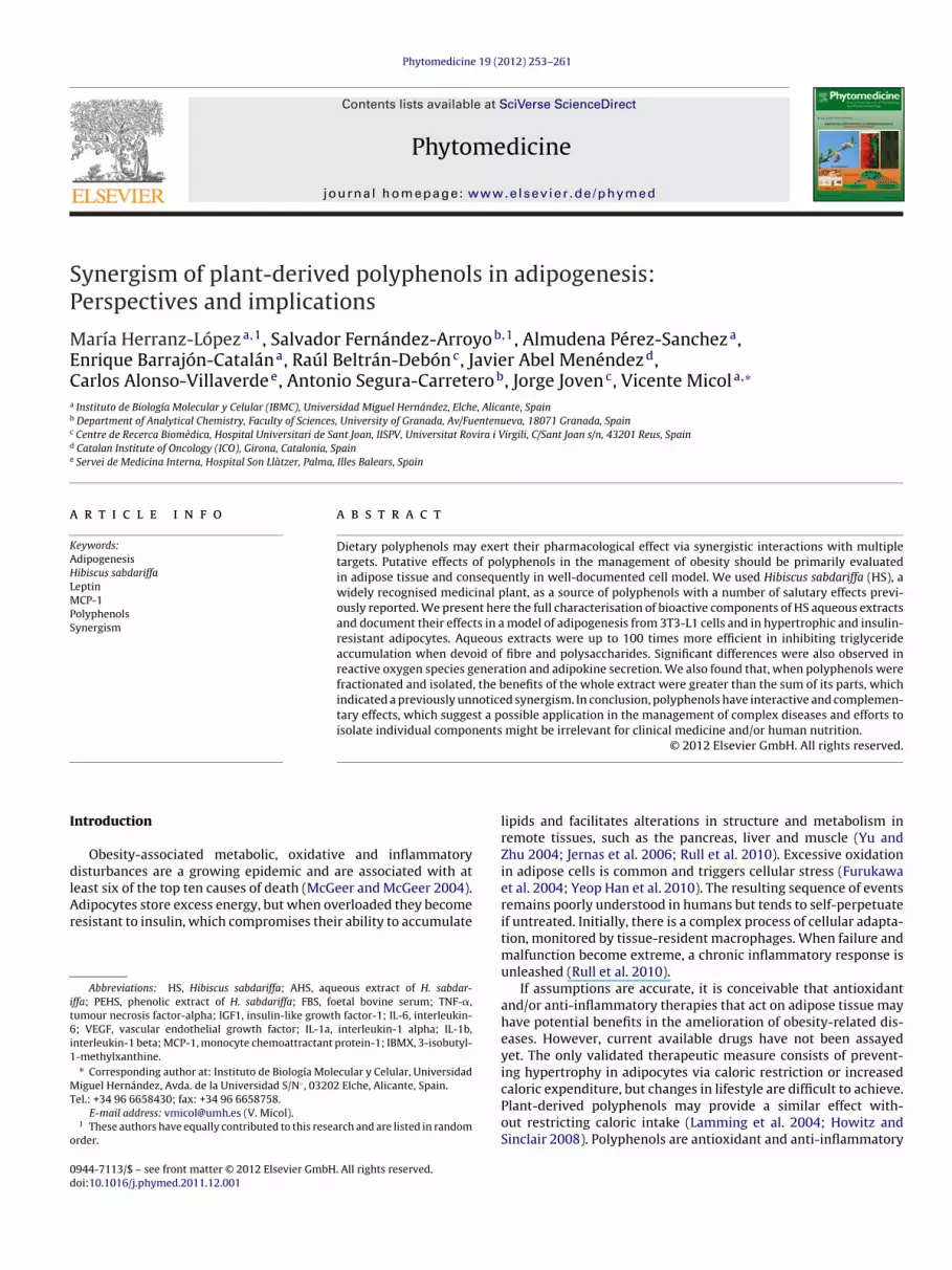
There were no major qualitative differences in polyphenolontent between AHS and PEHS; therefore, the precipitation pro-edures used to remove other soluble material did not result inelective losses in benefit (Fig. 1A and B and Table 1). However, suchanipulations resulted in immediately apparent quantitative dif-
erences and subsequent changes in relative contribution (Table 2).he phenolic content of AHS, as expressed in gallic acid equivalents,as significantly (p < 0.001) lower (2.26 ± 0.11 g/100 g) than that
f PEHS (28.42 ± 0.33 g/100 g). Relative differences were observedn organic acids and all families of phenolic compounds: organiccid derivatives (hydroxycitric and hibiscus acids), anthocyaninsdelphinidin-3-sambubioside and cyanidin-3-sambubioside), phe-
olic acid derivatives (chlorogenic acid and 5-O-caffeoylshikimiccid), and several flavonol derivatives (quercetin, myricetin andaempferol glycosides) (Table 2). PEHS was fractionated into 35ifferent fractions, and only fractions 6, 9 and 14 (Fig. 1C–Eig. 1. Representative base peak chromatograms obtained by HPLC–ESI-TOF-MS of Hibiscualitative differences between AHS and PEHS were deemed negligible, but quantitativenthocyanins and flavonols. Fractionation of PEHS, obtained through semipreparative liqui
dicine 19 (2012) 253– 261 255
and Table 2) significantly inhibited adipogenesis at concentra-tions ranging from 10 to 40 �g/ml. For clarity, negative resultsfor the remaining fractions are not shown. During the fractiona-tion procedure and the subsequent concentration, significant peakswere additionally detected and identified in PEHS (hibiscus aciddimethyl ester, coumaroylquinic acid, leucoside (kaempferol-3-O-sambubioside)) and several unidentified compounds. At leastsix new identified peaks (numbers 32–37) appeared only inisolated fractions. The composition of these fractions differednotably. The major component of fraction 6 was delphinidin-3-sambubioside, fraction 9 contained cyanidin-3-sambubioside,chlorogenic acid and tetra-O-methyljeediflavanone as major com-ponents, and fraction 14 was especially rich in glycosylatedflavonols, such as quercetin-3-sambubioside and myricetin-3-glucoside (Table 2).
Phenolic compounds inhibited adipogenic differentiation of3T3-L1 cells: fibre and/or polysaccharides were either inactive orinterfered in the model
We found that AHS significantly inhibited lipid accumulationand adipogenic differentiation of pre-adipocytes but only at con-centrations ≥500 �g/ml, which are not likely to be achieved in vivo.However, the relative activity of PEHS was much higher and evi-dent even at concentrations <10 �g/ml. Both extracts showed asignificant reduction in the number of differentiated cells whencompared with the control, as well as a dose-dependent response in
the reduction in the accumulation of triglycerides (Fig. 2A–E). Cellsdifferentiated in the presence of adipogenic agents plus 1000 �g/mlAHS or 40 �g/ml PEHS extracts accumulated 44.4% and 19.3% oftriglyceride, respectively, as compared to levels normally observedus sabdariffa AHS (A) and PEHS (B) extracts at 50 mg/ml and 5 mg/ml, respectively. differences were evident, especially with respect to the content of phenolic acids,d chromatography, showed that only a few fractions (C–E) were active in adipocytes.

256 M. Herranz-López et al. / Phytomedicine 19 (2012) 253– 261
Table 1Relevant analytical data for components isolated in AHS and PEHS (see also Fig. 1). Note that peaks 31–37 were only identified after a further process of purification.
Peak number Compound Retention time (min) Molecular formula [M−H]− UV–Vis (nm)
1 Hydroxycitric acid 4.35 C6H8O8 207.0140 –2 Hibiscus acid 4.72 C6H6O7 189.0035 –3 Delphinidin-3-sambubioside 5.50 C26H30O16 595.1446 280, 5204 Unknown 5.86 C8H12O8 235.0461 278, 3345 Cyanidin-3-sambubioside 6.11 C26H30O15 579.1493 280, 5206 Chlorogenic acid 6.52/8.41/8.92 C16H18O9 353.0891 297, 3247 Unknown 7.13 – 230.0127 272, 2988 Hibiscus acid dimethyl esther 7.91 C8H10O7 217.0354 2929 Methyl digallate 9.62 C15H12O9 335.0409 278
10 2-O-Trans-caffeoyl-hydroxicitric acid 10.60 C15H14O11 369.0463 285, 33011 Myricetin-3-arabinogalactoside 10.91 C26H28O17 611.1254 260, 35412 Coumaroylquinic acid 11.86 C16H18O8 337.0929 31013 Unknown 12.34 C11H11NO5 236.0564 27214 Quercetin-3-sambubioside 13.11 C26H28O16 595.1309 34515 Unknown 13.63 C16H20O10 371.0984 27816 Quercetin-3-rutinoside 14.50 C27H30O16 609.1462 255, 35317 5-O-Caffeoylshikimic acid 14.69 C16H16O8 335.0768 296, 32618 Leucoside (kaempferol-3-O-sambubioside) 15.44 C26H28O15 579.1355 278, 50519 Unknown 15.69 C17H22O10 385.1140 27020 Quercetin-3-glucoside 16.04 C21H20O12 463.0873 253, 35621 Kaempferol-3-O-rutinoside 16.71 C27H30O15 593.1512 265, 35022 Unknown 17.58 C18H22O9 381.1191 27823 Unknown 18.33 C21H30O11 457.1715 –24 Methyl-epigallocatechin 18.60 C16H16O7 319.0823 268, 33625 Unknown 18.93 – 503.1759 27826 Myricetin 22.19 C15H10O8 317.0298 37227 N-Feruloyltyramine 23.40/25.19 C18H20NO4 312.1234 286, 31628 Unknown 26.94 – 307.0726 288, 353, 41429 Quercetin 28.67 C15H10O7 301.0339 253, 37230 Unknown 29.89 C18H34O5 329.2333 –31 Prodelphinidin B3 24.82 C30H26O14 609.1250 31232 Tetra-O-methyljeediflavanone 4.30 C34H30O11 613.1715 34133 Unknown 6.11 C26H30O16 597.1461 33834 Caffeoylglucose 6.46 C15H18O9 341.0878 31435 Unknown 11.37 C26H44O16 611.2557 277
ibAtrttol
Ai
FftatcnfMttaaoef
36 Unknown 11.9337 Myricetin-3-glucoside 12.25
n control cells. The extract concentration that led to 50% of inhi-ition of triglyceride accumulation (IC50) was 799 ± 225 �g/ml forHS and 9.1 ± 2.8 �g/ml for PEHS, respectively. This result revealed
hat PEHS was approximately 90–100 times more effective ineducing triglyceride accumulation. This difference was around 10imes higher than that expected for actual polyphenol concen-rations, which indicated that the absence of polysaccharides andther soluble material improved the inhibition of triglyceride cel-ular uptake in this model.
nti-adipogenic activity of polyphenols was no longer conservedn most isolated fractions
Only 3 of 35 polyphenolic fractions (numbers 6, 9 and 14;ig. 1C–E) significantly inhibited adipogenesis. The efficacy of theseractions was dose-dependent; fraction 14 was the most activehroughout the dosing range (Fig. 2F). None of these fractionst 40 �g/ml achieved the effectiveness that was obtained withhe total mixture of polyphenols (PEHS). Changes in activity withombinations of these fractions at different concentrations wereegligible. None of the binary combinations (20 or 30 �g/ml of each
raction) achieved higher activities than the individual fractions.oreover, the strong inhibitory action of fraction 14 was main-
ained when using pairs 6/14 and 9/14. Finally, the combination ofhree of the fractions (20 �g/ml of each) also failed to improve thenti-adipogenic activity, as compared with both isolated fractions
nd combinations. Taking all results into account, the presencef all polyphenols was necessary in order to achieve maximumfficacy, and the relative proportion was potentially a relevantactor.C27H32O17 627.1567 278C21H20O13 479.0831 355
Phenolic compounds were active in mature as well ashypertrophied and insulin-resistant adipocytes
When extracts were assayed in mature adipocytes, the addi-tion of AHS did not significantly affect triglyceride content, even at1000 �g/ml, but 40 �g/ml of PEHS decreased triglyceride contentby 20–30% when assayed at 40 �g/ml (Supporting Information; Fig.2). These novel findings prompted the exploration of the effects ofPEHS in a cell model of adipocyte hypertrophy in the context ofinsulin resistance, similar to that observed in the adipose tissueof obese patients (Yu and Zhu 2004; Jernas et al. 2006; Takahashiet al. 2008; Yeop Han et al. 2010). Surprisingly, we found significanteffects with AHS and that both AHS and PEHS were more efficient atreducing triglyceride accumulation in insulin-resistant adipocytesthan in mature adipocytes (1000 �g/ml AHS: 19% mean valuesreduction; 40 �g/ml PEHS: 38% mean values reduction) (Fig. 3). Itappeared that the PEHS-mediated reduction in triglyceride accu-mulation was significantly higher during the adipogenesis processthan in mature or hypertrophic adipocytes but we found a differ-ential response with both extracts. The generation of endogenousROS was not affected by AHS (Fig. 4A and B), but the effect of PEHSwas significant and dose-dependent, achieving a 30% reduction,which indicated that the removed material might have deleteri-ous effects on either the diffusibility or the intrinsic antioxidantactivity of phenolic compounds. These deleterious effects werenot observed with their putative anti-inflammatory properties. We
observed that both extracts at the tested concentrations signifi-cantly decreased the amount of secreted adipokines with respect tocontrols (Fig. 4C). This result indicated that the effect of fibre and/orsaccharides could be specific. For most of the adipokines assayed,
M. Herranz-López et al. / Phytome
Tab
le
2Q
uan
tita
tive
dat
a
in
pp
m
(m/m
)
for
maj
or
com
pon
ents
fou
nd
in
extr
acts
and
frac
tion
s
wit
h
sign
ifica
nt
biol
ogic
al
acti
vity
.
Peak
nu
mbe
r
Com
pou
nd
Qu
anti
fica
tion
tech
niq
ue
AH
S
PEH
S
Frac
tion
6
Frac
tion
9
Frac
tion
14
1
Hyd
roxy
citr
ic
acid
MS-
TOF
(m/z
207)
8288
.0
±
397.
6
–
–
–
–2
Hib
iscu
s
acid
MS-
TOF
(m/z
189)
31,1
22.0
±
1128
.4
128,
134.
2
±
9486
.9
–
–
–3
Del
ph
inid
in-3
-sam
bubi
osid
e
DA
D-U
V
(520
nm
)
2701
.2
±
165.
6
207,
315.
5
±
1807
.6
415,
227.
9
±
18,5
58.3
–
–5
Cya
nid
in-3
-sam
bubi
osid
e
DA
D-U
V
(520
nm
)
1939
.2
±
39.3
87,1
43.1
±
393.
1
–
193,
354.
1
±
3119
.0
–6
Ch
loro
gen
ic
acid
DA
D-U
V
(325
nm
)
5720
.0
±
39.4
106,
469.
9
±
1182
.0
5381
.5
±
173.
9
137,
219.
1
±
1425
.8
–8
Hib
iscu
s
acid
dim
eth
yl
este
rM
S-TO
F
(m/z
217)
––
5300
.5±
325.
9
–
–9
Met
hyl
dig
alla
te
DA
D-U
V
(270
nm
)
–
2802
.3
±
46.6
–
–
–11
Myr
icet
in-3
-ara
bin
ogal
acto
se
DA
D-U
V
(350
nm
)
57.3
±
2.5
4755
.5
±
53.8
–
–
–12
Cou
mar
oylq
uin
ic
acid
DA
D-U
V
(310
nm
)
–
772.
9
±
10.4
–
–
–14
Qu
erce
tin
-3-s
ambu
bios
ide
DA
D-U
V
(350
nm
)
304.
0
±
5.9
7673
.8
±
34.6
–
– 11
3,13
0.2
±
2356
.416
Qu
erce
tin
-3-r
uti
nos
ide
DA
D-U
V
(350
nm
)49
5.7
±
4.3
4953
.2±
47.9
––
–17
5-O
-Caf
feoy
lsh
ikim
ic
acid
DA
D-U
V
(325
nm
)
171.
5
±
6.9
3526
.7
±
49.2
–
–
7312
.7
±
168.
918
Leu
cosi
de
DA
D-U
V
(350
nm
)
–
1123
.0
±
25.4
–
–
–20
Qu
erce
tin
-3-g
luco
sid
e
DA
D-U
V
(350
nm
)
143.
7
±
2.2
3071
.6
±
15.9
–
–
12,7
89.1
±
268.
621
Kae
mp
fero
l-3-
O-r
uti
nos
ide
DA
D-U
V
(350
nm
)
91.9
±
2.3
2185
.5
±
15.5
–
–
–24
Met
hyl
-ep
igal
loca
tech
inD
AD
-UV
(270
nm
)–
310.
9±
5.5
––
–26
Myr
icet
in
DA
D-U
V
(370
nm
)
–
4765
.9
±
49.1
–
–
–27
N-F
eru
loyl
tyra
min
eD
AD
-UV
(325
nm
)
99.0
±
1.8
867.
9
±
8.6
–
–
–29
Qu
erce
tin
DA
D-U
V
(370
nm
)
121.
2
±
2.0
5795
.2
±
61.7
–
–
–31
Prod
elp
hin
idin
B3
DA
D-U
V
(310
nm
)
1839
.2
±
25.3
327.
0
±
2.9
– –
–32
Tetr
a-O
-met
hyl
jeed
iflav
anon
e
DA
D-U
V
(350
nm
)
13,5
15.7
± 12
5.8
–
–34
Caf
feoy
lglu
cose
MS-
TOF
(m/z
341)
–
2902
.1
±
67.5
–37
Myr
icet
in-3
-glu
cosi
de
DA
D-U
V
(350
nm
)
–
–
29,3
83.6
±
1245
.1
dicine 19 (2012) 253– 261 257
both extracts showed the same quantitative efficacy. A differentialresponse, higher for PEHS than for AHS, was only observed with thesecretion of leptin and MCP-1.
Discussion
The efficacy of phytotherapy is currently under intense debate(Wagner 2011). To firmly establish beneficial effects that may yieldvaluable nutritional advice or dietary supplements, human stud-ies demand (Mozaffari-Khosravi et al. 2009; Kuriyan et al. 2010)full chemical characterisation of the source of polyphenols andthe effect of further manipulation in the relative composition. Ournovel findings suggested that the previous removal of fibre andpolysaccharides, which represent up to 60% of the total weightof soluble extracts (Müller and Franz 1992), might significantlyincrease the activity of a polyphenolic mixture. Adipogenesis wassubstantially inhibited by a standardised Hibiscus sabdariffa extract,and the effect of the full extract was higher than the sum of its parts,which provided further evidence that a combination of bioactivecomponents was superior to isolated constituents. In essence, theassayed extracts are a complex mixture of anthocyanins, organicacids, phenolic acids and flavonols (Rodríguez-Medina et al. 2009).Nevertheless, our results revealed that some compounds can havea higher contribution to the observed effects, which suggestedthe importance of relative composition and that different formu-lations might yield different outcomes. For instance, the putativehypolipidemic effects of polyphenolic mixtures have been mainlyassociated with the presence of organic acids (Carvajal-Zarrabalet al. 2005). In contrast, the described effects of PEHS in adipo-genesis were obtained in a scenario in which the proportion ofphenolic compounds was higher than that of organic acids. Fromdata obtained from isolated fractions, it could be concluded thatglycosylated flavonols were the most active compounds amongstindividual components, but this finding seemed irrelevant whencompared to the action of the full mixture. Although other isolatedphenolic compounds (apigenin, epigallocatechin gallate, resver-atrol and quercetin) have also shown to inhibit adipogenesis in3T3-L1 adipocytes in similar concentrations (10–50 �g/ml, Lin et al.2005; Bandyopadhyay et al. 2006; Yang et al. 2008), cytotoxicitywas readily observed. This point is extremely important becauseplant-derived polyphenols are a complex mixture that interactswith numerous endogenous molecular targets in humans but aresurprisingly safe even at high doses (Corson and Crews 2007; Goelet al. 2008; Efferth and Koch 2011).
Another novel finding was that PEHS also actively diminishedtriglyceride accumulation in mature and even insulin-resistanthypertrophied adipocytes, which suggested an induction in thelipolysis rate (Yu and Zhu 2004; Takahashi et al. 2008). This effectcould be important in the management of metabolic disturbancesbecause the uninhibited release of fatty acids from hypertrophiedadipocytes might lead to systemic lipotoxicity and insulin resis-tance (Unger 1995). It was also established that excess triglycerideaccumulation in adipocytes generated an excess of ROS thattriggered inflammation (Yeop Han et al. 2010). Although the dif-ferential abilities observed between AHS and PEHS deserve furtherconsideration, PEHS clearly possesses antioxidative and anti-inflammatory actions in mature and hypertrophied adipocytes.These properties might also have therapeutic implications becausethese are important processes in 3T3-L1 adipocytes that aredirectly related to the accumulation of fat and with potentialregulation via JNK/NF-�B pathways as described (Furukawa et al.
2004; Takahashi et al. 2008; Yeop Han et al. 2010). Once again wehighlight the importance of a particular formulation of phenoliccompounds because PEHS was particularly active in inhibiting thesecretion of leptin and MCP-1, which are important adipokines
258 M. Herranz-López et al. / Phytomedicine 19 (2012) 253– 261
Fig. 2. Polyphenols from Hibiscus sabdariffa significantly inhibited the accumulation of triglycerides in 3T3-L1 preadipocytes and programmed adipogenesis. Representativemicrophotographs of cells stained with AdipoRedTM (treatment groups: control, 1000 �g/ml AHS or 40 �g/ml PEHS; A–C), demonstrate that the number of mature adipocytesw of dio s show*
ta2wP
osabr
as substantially decreased in cells treated with extracts. The quantitative effectsf selected fractions in the accumulation of triglycerides in 3T3-L1 preadipocytes i**p < 0.001 versus control.
hat regulate the migration of non-resident macrophages to thedipose tissue and overall systemic metabolism (Furukawa et al.004). This anti-inflammatory effect has also been observedith resveratrol or alpha lipoic acid (Szkudelska et al. 2009;
rieto-Hontoria et al. 2011).The apparently additive, synergistic or even antagonistic action
f each polyphenol and the intrinsic complications of under-
tanding adipocyte metabolism without knowledge of geneticnd proteomic kinetics impede efforts to describe the possibleiological reactions and metabolic networks involved. Polyphenoleaction and diffusion rates also confound these efforts. Moreover,fferent doses on the final content of triglyceride are also shown (D–E). The effectn in (F). Cell viability was unaffected even at higher concentrations. *p < 0.05 and
the process of transformation and regeneration in these particularcells, and the effects that we described strongly suggested thepresence of repeat-pattern mechanisms (Gierer and Meinhardt1972), which is a self-organising, self-repairing, reaction–diffusionsystem (Turing 1990).
We speculate that this relationship can be applied to every com-bination of polyphenols with medicinal effects, but at least two
major points should be highlighted. Leptin production correlatespositively with insulin resistance, fat mass and adipocyte volume inresponse to metabolic stress (Frederich et al. 1995). The observedbenefit with PEHS was comparatively higher than that observed
M. Herranz-López et al. / Phytomedicine 19 (2012) 253– 261 259
Fig. 3. Polyphenols from Hibiscus sabdariffa significantly decreased the accumulation of triglycerides in 3T3-L1 hypertrophied/insulin-resistant adipocytes (A). Effects areshown for AHS (500–1000 �g/ml) (left panel) and PEHS (20–40 �g/ml) (right panel). (B)–(D) are representative microphotographs of cells stained with AdipoRedTM; treatmentg roplete
wee(SWlMba
dnmtOtlwiiifpamt
roups: control, 1000 �g/ml AHS or 40 �g/ml PEHS, respectively. The number of fat dven at higher concentrations. **p < 0.01 and ***p < 0.001 versus control.
ith triglyceride accumulation, which suggested that leptin genexpression was a possible pharmacologic target, although this isffect obviously modulated by actions on a variety of other targetsFrederich et al. 1995; Rupnick et al. 2002; Tilg and Moschen 2006;amuni et al. 2010). Results with MCP-1 excretion were expected.e have previously reported in humans that these polypheno-
ic extracts significantly reduce the concentration of circulatingCP-1, which is relevant because MCP-1 has been proposed as a
iomarker and therapeutic target in the management of obesitynd its related complications (Beltrán-Debón et al. 2009).
We should express caution, however, when interpreting in vitroata to actual actions of polyphenols in the body, especially ifo data have been collected regarding the action of physiologicaletabolites of tested polyphenols on the same cell systems. First,
he intestinal flora is likely to metabolise some of these compounds.nce the glucoside cleaved, the released aglycone is subjected to
he action of specific enzymes in the wall of the small intestineeading to glucuronide, sulphated, and methylated metabolites,
hich may reach their target tissues and organs. Obviously, ourn vitro assays do not take into account the in vivo bioavailabilityssue and can lead to false positive interpretations. To date, theres no data about human or animal studies on metabolites derivingrom HS polyphenols. Although some bioavailability studies on
henolic acids, anthocyanins and flavonols are available, thesere contained in very different food matrixes what may radicallyodify the interaction amongst these compounds in the gas-rointestinal tract and therefore their absorption. Whilst human
s per cell was decreased in the experiments with PEHS. Cell viability was unaffected
absorption and bioavailability studies have revealed that phenolicacids and anthocyanins can be retrieved in plasma and urine intheir intact form after food consumption (Paredes-Lopez et al.2010; Williamson et al. 2011), flavonols and flavanols seem tobe found in various forms, free or conjugated with glucuronide,sulphate or methyl groups (Day et al. 2001; Williamson et al. 2011).
Because in studies with green tea polyphenols, the metabolitesmostly had reduced biological activity, it might be tempting tosuggest that conversion of HS polyphenols into less-active metabo-lites would compromise the cellular effects denoted in the presentstudy. In some systems, however, polyphenols-derived metaboliteswere found to have the equivalent or even greater activity than theparental polyphenols (Lambert et al. 2007). In the present study,PEHS contains up to thirty identifiable phenolic compounds byHPLC–MS. Accepting that absorption of many of these compoundsis negligible; still several of them will be conjugated and will inter-act with multiple metabolic targets. Thus, to study the effects of allpossible metabolites becomes an enormous and fascinating target,which will be undoubtedly matter for future research.
In conclusion, we propose that this particular formulationof polyphenols should be assayed in clinical trials because ofits observed regulation of adipogenesis, its regulation of oxida-tive stress signalling pathways in mature and/or hypertrophied
adipocytes and its subsequent ability to alter the expression ofadipokines. In regards to the preparation of phytopharmaceuti-cals or dietary supplements, we propose that the separation ofsoluble material other than polyphenols prior to use will increase
260 M. Herranz-López et al. / Phytomedicine 19 (2012) 253– 261
Fig. 4. The phenolic fraction from Hibiscus sabdariffa inhibited ROS generation and modulated the concentration of secreted adipokines in hypertrophied 3T3–L1 adipocytes(A). Cells were incubated with different doses of AHS (400–1000 �g/ml) (left panel) or PEHS (10–40 �g/ml) (right panel) and ROS generation was measured by H2DCFDAl S did na ased
*
tbdr
abelling. Whereas PEHS affected ROS generation in a dose-dependent manner, AHre shown in (B). Although the secretion of assayed adipokines was efficiently decrep < 0.05 and ***p < 0.001 versus control.
he possibility of safely obtaining synergistic effects. Althoughioavailability and safety issues require further studies, we haveemonstrated the potential pharmacological and therapeutic supe-iority of a combination of polyphenols with respect to their
ot. Representative fluorescence microphotographs of cells stained with H2DCFDAwith both extracts, a differential response was obtained with leptin and MCP-1 (C).
individual components. The existence of synergistic efficacy ofbinary combinations of compounds has been evaluated and ver-ified by Berenbaum’s isobole method (Berenbaum 1989; Wagner2011). Unfortunately, this method cannot be applied in our case at

ytome
tsee“
C
A
aaptfRE
A
t
R
A
B
B
BC
C
D
E
F
F
F
G
G
G
M. Herranz-López et al. / Ph
his stage due to the complexity of the polyphenol mixture. It is pos-ible that the benefits of polyphenols are the result of mere additiveffects or simple combinatory actions, but the data suggest a syn-rgistic effect at least in the sense of that described by Mark Twain:a bonus achieved when things work together harmoniously”.
onflict of interest statement
The authors declare that there are no conflicts of interest.
cknowledgements
We thank MONTELOEDER, SL for providing raw plant materialnd for advice on the extraction and purification procedures. RBDnd MHL were the recipients of fellowships from the Comissionater a Universitats I Recerca del Departament d’Innovació, Universi-ats I Empresa de la Generalitat de Catalunya and Programa Vali+drom Generalitat Valenciana, respectively. We thank Andalusianegional Government Council of Innovation and Science for grantedxcellence Project
ppendix A. Supplementary data
Supplementary data associated with this article can be found, inhe online version, at doi:10.1016/j.phymed.2011.12.001.
eferences
larcon-Aguilar, F.J., Zamilpa, A., Perez-Garcia, M.D., Almanza-Perez, J.C., Romero-Nunez, E., Campos-Sepulveda, E.A., Vazquez-Carrillo, L.I., Roman-Ramos, R.,2007. Effect of Hibiscus sabdariffa on obesity in MSG mice. J. Ethnopharmacol.114, 66–71.
andyopadhyay, S., Lion, J.M., Mentaverri, R., Ricupero, D.A., Kamel, S., Romero,J.R., Chattopadhyay, N., 2006. Attenuation of osteoclastogenesis and osteoclastfunction by apigenin. Biochem. Pharmacol. 72, 184–197.
eltrán-Debón, R., Alonso-Villaverde, C., Aragonès, G., Rodríguez-Medina, I., Rull, A.,Micol, V., Segura-Carretero, A., Fernández-Gutiérrez, A., Camps, J., Joven, J., 2009.The aqueous extract of Hibiscus sabdariffa calices modulates the production ofmonocyte chemoattractant protein-1 in humans. Phytomedicine 17, 186–191.
erenbaum, M.C., 1989. What is synergy? Pharmacol. Rev. 41, 93–141.arvajal-Zarrabal, O., Waliszewski, S.M., Barradas-Dermitz, D.M., Orta-Flores, Z.,
Hayward-Jones, P.M., Nolasco-Hipolito, C., Angulo-Guerrero, O., Sanchez-Ricano, R., Infanzon, R.M., Trujillo, P.R., 2005. The consumption of Hibiscussabdariffa dried calyx ethanolic extract reduced lipid profile in rats. Plant FoodsHum. Nutr. 60, 153–159.
orson, T.W., Crews, C.M., 2007. Molecular understanding and modern applicationof traditional medicines: triumphs and trials. Cell 130, 769–774.
ay, A.J., Mellon, F., Barron, D., Sarrazin, G., Morgan, M.R., Williamson, G., 2001.Human metabolism of dietary flavonoids: identification of plasma metabolitesof quercetin. Free Radic. Res. 35, 941–952.
fferth, T., Koch, E., 2011. Complex interactions between phytochemicals. The multi-target therapeutic concept of phytotherapy. Curr. Drug Targets 12, 122–132.
ernandez-Arroyo, S., Rodriguez-Medina, I.C., Beltrán-Debón, R., Pasini, F., Joven,J., Micol, V., Segura-Carretero, A., Fernandez-Gutierrez, A., 2011. Quantificationof the polyphenolic fraction and in vitro antioxidant and in vivo anti-hyperlipemic activities of Hibiscus sabdariffa aqueous extract. Food Res. Int. 44,1490–1495.
rederich, R.C., Hamann, A., Anderson, S., Lollmann, B., Lowell, B.B., Flier, J.S., 1995.Leptin levels reflect body lipid content in mice: evidence for diet-induced resis-tance to leptin action. Nat. Med. 1, 1311–1314.
urukawa, S., Fujita, T., Shimabukuro, M., Iwaki, M., Yamada, Y., Nakajima, Y.,Nakayama, O., Makishima, M., Matsuda, M., Shimomura, I., 2004. Increasedoxidative stress in obesity and its impact on metabolic syndrome. J. Clin. Invest.114, 1752–1761.
ierer, A., Meinhardt, H., 1972. A theory of biological pattern formation. Kybernetik
12, 30–39.oel, A., Jhurani, S., Aggarwal, B.B., 2008. Multi-targeted therapy by curcumin: howspicy is it? Mol. Nutr. Food Res. 52, 1010–1030.
reen, H., Kehinde, O., 1975. An established preadipose cell line and its differentia-tion in culture. II. Factors affecting the adipose conversion. Cell 5, 19–27.
dicine 19 (2012) 253– 261 261
Howitz, K.T., Sinclair, D.A., 2008. Xenohormesis: sensing the chemical cues of otherspecies. Cell 133, 387–391.
Huang, D., Boxin, O.U., Prior, R.L., 2005. The chemistry behind antioxidant capacityassays. J. Agric. Food Chem. 53, 1841–1856.
Jernas, M., Palming, J., Sjoholm, K., Jennische, E., Svensson, P.-A., Gabrielsson, B.G.,Levin, M., Sjogren, A., Rudemo, M., Lystig, T.C., Carlsson, B., Carlsson, L.M.S., Lonn,M., 2006. Separation of human adipocytes by size: hypertrophic fat cells displaydistinct gene expression. FASEB J. 20, 1540–1542.
Kim, J.K., So, H., Youn, M.J., Kim, H.J., Kim, Y., Park, C., Kim, S.J., Ha, Y.A., Chai, K.Y., Kim,S.M., Kim, K.Y., Park, R., 2007. Hibiscus sabdariffa L. water extract inhibits theadipocyte differentiation through the PI3-K and MAPK pathway. J. Ethnophar-macol. 114, 260–267.
Kuriyan, R., Kumar, D.R., R, R., Kurpad, A.V., 2010. An evaluation of the hypolipidemiceffect of an extract of Hibiscus sabdariffa leaves in hyperlipidemic Indians: adouble blind, placebo controlled trial. BMC Complement. Altern. Med. 10, 27.
Lambert, J.D., Sang, S., Yang, C.S., 2007. Biotransformation of green tea polyphenolsand the biological activities of those metabolites. Mol. Pharm. 4, 819–825.
Lamming, D.W., Wood, J.G., Sinclair, D.A., 2004. Small molecules that regulate lifes-pan: evidence for xenohormesis. Mol. Microbiol. 53, 1003–1009.
Lin, J., Della-Fera, M.A., Baile, C.A., 2005. Green tea polyphenol epigallocatechin gal-late inhibits adipogenesis and induces apoptosis in 3T3-L1 adipocytes. Obes. Res.13, 982–990.
McGeer, P.L., McGeer, E.G., 2004. Inflammation and the degenerative diseases ofaging. Ann. N.Y. Acad. Sci. 1035, 104–116.
Mozaffari-Khosravi, H., Jalali-Khanabadi, B.A., Afkhami-Ardekani, M., Fatehi, F., 2009.Effects of sour tea (Hibiscus sabdariffa) on lipid profile and lipoproteins inpatients with type II diabetes. J. Altern. Complement. Med. 15, 899–903.
Müller, B.M., Franz, G., 1992. Chemical structure and biological activity of polysac-charides from Hibiscus sabdariffa. Planta Med. 58, 60–67.
Paredes-Lopez, O., Cervantes-Ceja, M.L., Vigna-Perez, M., Hernandez-Perez, T., 2010.Berries: improving human health and healthy aging, and promoting quality life– a review. Plant Foods Hum. Nutr. 65, 299–308.
Prieto-Hontoria, P.L., Perez-Matute, P., Fernandez-Galilea, M., Martinez, J.A.,Moreno-Aliaga, M.J., 2011. Lipoic acid inhibits leptin secretion and Sp1 activityin adipocytes. Mol. Nutr. Food Res. 55, 1–11.
Rodríguez-Medina, I.C., Beltrán-Debón, R., Micol, V., Alonso-Villaverde, C., Joven,J., Menéndez, J.A., Segura-Carretero, A., Fernández-Gutiérrez, A., 2009. Directcharacterization of aqueous extract of Hibiscus sabdariffa using HPLC with diodearray detection coupled to ESI and ion trap MS. J. Sep. Sci. 32, 3441–3448.
Rull, A., Camps, J., Alonso-Villaverde, C., Joven, J., 2010. Insulin resistance, inflam-mation, and obesity: role of monocyte chemoattractant protein-1 (or CCL2) inthe regulation of metabolism. Mediators Inflamm. 2010, 326580.
Rupnick, M.A., Panigrahy, D., Zhang, C.Y., Dallabrida, S.M., Lowell, B.B., Langer, R.,Folkman, M.J., 2002. Adipose tissue mass can be regulated through the vascula-ture. Proc. Natl. Acad. Sci. U.S.A. 99, 10730–10735.
Samuni, Y., Cook, J.A., Choudhuri, R., Degraff, W., Sowers, A.L., Krishna, M.C., Mitchell,J.B., 2010. Inhibition of adipogenesis by Tempol in 3T3-L1 cells. Free Radic. Biol.Med. 49, 667–673.
Szkudelska, K., Nogowski, L., Szkudelski, T., 2009. The inhibitory effect of resveratrolon leptin secretion from rat adipocytes. Eur. J. Clin. Invest. 39, 899–905.
Takahashi, K., Yamaguchi, S., Shimoyama, T., Seki, H., Miyokawa, K., Katsuta, H.,Tanaka, T., Yoshimoto, K., Ohno, H., Nagamatsu, S., Ishida, H., 2008. JNK- andI{kappa}B-dependent pathways regulate MCP-1 but not adiponectin releasefrom artificially hypertrophied 3T3-L1 adipocytes preloaded with palmitate invitro. Am. J. Physiol. Endocrinol. Metab. 294, E898–E909.
Tilg, H., Moschen, A.R., 2006. Adipocytokines: mediators linking adipose tissue,inflammation and immunity. Nat. Rev. Immunol. 6, 772–783.
Turing, A.M., 1990. The chemical basis of morphogenesis. 1953. Bull. Math. Biol. 52,153–197 (discussion 119–152).
Unger, R.H., 1995. Lipotoxicity in the pathogenesis of obesity-dependent NIDDM.Genetic and clinical implications. Diabetes 44, 863–870.
Wagner, H., 2011. Synergy research: approaching a new generation of phytophar-maceuticals. Fitoterapia 82, 34–37.
Williamson, G., Dionisi, F., Renouf, M., 2011. Flavanols from green tea and phenolicacids from coffee: critical quantitative evaluation of the pharmacokinetic datain humans after consumption of single doses of beverages. Mol. Nutr. Food Res.55, 864–873.
Yang, J.Y., Della-Fera, M.A., Rayalam, S., Ambati, S., Hartzell, D.L., Park, H.J., Baile,C.A., 2008. Enhanced inhibition of adipogenesis and induction of apoptosis in3T3-L1 adipocytes with combinations of resveratrol and quercetin. Life Sci. 82,1032–1039.
Yeop Han, C., Kargi, A.Y., Omer, M., Chan, C.K., Wabitsch, M., O’Brien, K.D., Wight,T.N., Chait, A., 2010. Differential effect of saturated and unsaturated free fattyacids on the generation of monocyte adhesion and chemotactic factors by
adipocytes: dissociation of adipocyte hypertrophy from inflammation. Diabetes59, 386–396.Yu, Y.H., Zhu, H., 2004. Chronological changes in metabolism and functions of cul-tured adipocytes: a hypothesis for cell aging in mature adipocytes. Am. J. Physiol.Endocrinol. Metab. 286, E402–E410.