effects of olive oil polyphenols on erythrocyte oxidative damage
TRANSCRIPT
1Mol. Nutr. Food Res. 2009, 53, 000 –000 DOI 10.1002/mnfr.200800276
Research Article
Effects of olive oil polyphenols on erythrocyteoxidative damage
F�tima Paiva-Martins1, 2, Jo¼o Fernandes2, Susana Rocha3, 4, Henrique Nascimento3, 4,Rui Vitorino5, Francisco Amado5, Fernanda Borges6, Lu�s Belo3, 4 andAlice Santos-Silva3, 4
1 Centro de Investiga�¼o em Qu�mica (CIQ), Faculdade de CiÞncias, Universidade do Porto, Porto, Portugal2 Departamento de Qu�mica, Faculdade de CiÞncias, Universidade do Porto, Porto, Portugal3 Servi�o de Bioqu�mica, Faculdade de Farm�cia, Universidade do Porto, Porto, Portugal4 Instituto de Biologia Molecular e Celular (IBMC), Universidade do Porto, Porto, Portugal5 Departamento de Qu�mica, Universidade de Aveiro, Aveiro, Portugal6 UQFM, Servi�o de Qu�mica Org�nica, Faculdade de Farm�cia, Universidade do Porto, Porto, Portugal
Many studies have investigated the protective effects of oleuropein and hydroxytyrosol against cellinjury, but few have investigated the protective effects of oleuropein aglycones 3,4-dihydroxyphenyle-thanol-elenolic acid (3,4-DHPEA-EA) and 3,4-dihydroxyphenylethanol-elenolic acid dialdehyde(3,4-DHPEA-EDA). The present work studied and compared the capacity of these four compounds,found at high concentrations in olive oil, to protect red blood cells (RBCs) from oxidative injury. Thein vitro oxidative stress of RBCs was induced by the water-soluble radical initiator 2,29-azo-bis(2-ami-dinopropane) dihydrochloride. RBC changes were evaluated either by optical microscopy or by theamount of hemolysis. All compounds were shown to significantly protect RBCs from oxidative dam-age in a dose-dependent manner. The order of activity at 20 lM was: 3,4-DHPEA-EDA A hydroxytyr-osol A oleuropein A 3,4-DHPEA-EA. Even at 3 lM, 3,4-DHPEA-EDA and hydroxytyrosol still hadan important protective activity. However, deleterious morphological RBC changes were much moreevident in the presence of hydroxytyrosol than with 3,4-DHPEA-EDA. For the first time it was dem-onstrated that 3,4-DHPEA-EDA, one of most important olive oil polyphenols, may play a noteworthyprotective role against ROS-induced oxidative injury in human cells since lower doses of this com-pound were needed to protect RBCs in vitro from oxidative mediated hemolysis.
Keywords: Erythrocytes / Hydroxytyrosol / Olea europaea / Olive oil / Polyphenols /
Received: July 3, 2008; revised: August 22, 2008; accepted: September 15, 2008
1 Introduction
A large body of epidemiological studies shows that the inci-dence of coronary heart disease and of certain cancers inthe Mediterranean countries is low, suggesting a crucialprotective role of the diet in this southern European areawhere virgin olive oil is the principal source of fat [1]. Thehypothesis of an antioxidant/atherosclerosis relationship
led to experimental studies on the potential protective roleof olive oil phenols against coronary heart disease. In vitrostudies and a few in vivo studies suggested that the highconcentration of phenolic compounds in extra virgin oliveoil may contribute to the healthy nature of this diet but con-troversial results have been obtained in several randomized,crossover, controlled studies [2–8].
In recent years, there has been much interest in antioxi-dants that retard oxidative modification of low-density lip-oproteins (LDL), which is believed to be a key step in thedevelopment of atherosclerosis. The stability of LDL iso-lated from animals and humans fed with virgin olive oil isincreased, and this increased stability is attributable to thephenolic compounds present in the oil [3, 7, 9–12]. Nowa-days, olive oil is marketed as being healthier than other veg-etable oils but a pertinent question is whether this claim isvalid for all virgin olive oils, or whether some have betternutritional value than others depending on their phenolic
Correspondence: Dr. F�tima Paiva-Martins, Departamento de Qu�mi-ca, Faculdade de CiÞncias, Universidade do Porto, Rua do Campo Ale-gre 687, 4169-007 Porto, PortugalE-mail: [email protected]: +351-22-6082959
Abbreviations: AAPH, 2,29-azo-bis(2-amidinopropane) dihydrochlor-ide; 3,4-DHPEA-EA, 3,4-dihydroxyphenylethanol-elenolic acid; 3,4-DHPEA-EDA, 3,4-dihydroxyphenylethanol-elenolic acid dialdehyde;MBH, membrane-bound hemoglobin; RBC, red blood cell
i 2009 WILEY-VCH Verlag GmbH & Co. KGaA, Weinheim www.mnf-journal.com
F. Paiva-Martins et al. Mol. Nutr. Food Res. 2009, 53, 000 –000
composition. Current research findings suggest that oliveoil consumption could reduce oxidative damage due to itsrichness in oleic acid and due to its minor components, par-ticularly the phenolic compounds. However, which compo-nents have a major role on this protection, is still unknown.
The red blood cell (RBC), anucleated and without cyto-plasmatic organelles, has poor repair and biosyntheticmechanisms, suffering and accumulating oxidative lesionswhenever oxidative stress develops. Moreover, RBCs areparticularly exposed to endogenous oxidative damagebecause of their specific role as oxygen carriers. As themost abundant blood cell, RBCs also play an important rolein the oxidative status of the whole blood constituents, inparticular of the lipoproteins. Nevertheless, RBCs areequipped with several antioxidants, i. e., antioxidantenzymes, glutathione, tocopherol and ascorbate. If reactiveoxygen species (ROS), i. e., H2O2 and O2
– , are overproducedoutside or within the erythrocyte, or if the endogenous anti-oxidant defenses are impaired, an “oxidative stress” condi-tion will develop, inducing oxidative damage to erythrocyteconstituents, i. e., those on membrane and hemoglobin,which may ultimately leads to hemolysis. A wide variety ofdrugs and xenobiotics that can undergo oxidation-reductionreactions have been found to cause RBC destruction. Inter-action between the xenobiotic and hemoglobin is veryimportant in the process, which is usually characterized byhemoglobin oxidation to methemoglobin and formation ofradical intermediates. When hemoglobin is denatured, itlinks to the membrane at the cytoplasmic domain of band 3protein, inducing its aggregation and the linkage of naturalanti-band 3 antibodies and complement fixation on theerythrocyte surface, marking the cell for removal by themacrophages of the reticuloendothelial system. HumanRBCs are, therefore, a metabolically simplified model sys-tem, useful in the evaluation of antioxidant properties ofseveral compounds, e.g., olive oil polyphenols.
Hydroperoxyl radicals (HOO.), from the aqueous phase,are important for initiating lipid peroxidation and proteindamage in membranes [13]. 2,29-Azo-bis(2-amidinopro-pane) dihydrochloride (AAPH) is a water-soluble azo com-pound extensively used as a free radical initiator for biolog-ical studies. It can generate radicals, at a constant rate, inthe aqueous phase through its thermo degradation at 378C[13].
Hydroxytyrosol, oleuropein and its aglycones 3,4-dihy-droxyphenylethanol-elenolic acid (3,4-DHPEA-EA) and
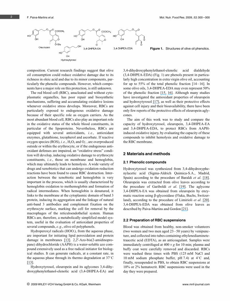
3,4-dihydroxyphenylethanol-elenolic acid dialdehyde(3,4-DHPEA-EDA) (Fig. 1) are phenols present in particu-larly high concentration in extra virgin olive oil, accountingfor up to 55% of the total phenolic fraction [14–16]. Insome olive oils, 3,4-DHPEA-EDA may even represent 50%of the phenolic fraction [15, 16]. Although many studieshave investigated the antioxidant properties of oleuropeinand hydroxytyrosol [17], as well as their protective effectsagainst cell injury and their bioavailability, there have beenonly few reports of the protective effects of oleuropein agly-cones.
The aim of this work was to study and compare thecapacity of hydroxytyrosol, oleuropein, 3,4-DHPEA-EAand 3,4-DHPEA-EDA, to protect RBCs from AAPH-induced oxidative injury, by evaluating the capacity of thesecompounds to inhibit hemolysis and oxidative damage tothe RBC membrane.
2 Materials and methods
2.1 Phenolic compounds
Hydroxytyrosol was synthesized from 3,4-dihydroxyphe-nylacetic acid (Sigma-Aldrich Quimica-S.A., Madrid,Spain) according to the procedure of Baraldi et al. [18].Oleuropein was extracted from olive leaves according tothe procedure of Gariboldi et al. [19]. The aglycone3,4-DHPEA-EA was obtained from oleuropein by enzy-matic reaction using b-glycosidase (Fluka, Buchs, Switzer-land), according to the procedure of Limirioli et al. [20].3,4-DHPEA-EDA was obtained from olive leaves asdescribed by Paiva-Martins and Gordon [21].
2.2 Preparation of RBC suspensions
Blood was obtained from healthy, non-smoker volunteers(two women and two men aged 23–50 years) by venipunc-ture, and collected into tubes containing ethylenediaminete-traacetic acid (EDTA), as an anticoagulant. Samples wereimmediately centrifuged at 4006g for 10 min; plasma andbuffy coat were carefully removed and discarded. RBCswere washed three times with PBS (125 mM NaCl and10 mM sodium phosphate buffer, pH 7.4) at 48C and,finally, resuspended in PBS, to obtain RBC suspensions at10% or 2% hematocrit. RBC suspensions were used in theday they were prepared.
2
i 2009 WILEY-VCH Verlag GmbH & Co. KGaA, Weinheim www.mnf-journal.com
Figure 1. Structures of olive oil phenolics.
Mol. Nutr. Food Res. 2009, 53, 000 –000
To evaluate the capacity of the olive oil phenolic com-pounds to protect RBCs from oxidative injury induced byAAPH several in vitro studies were performed: RBC lysis,morphology, membrane-bound hemoglobin (MBH) andmembrane protein profile.
2.3 AAPH-induced hemolysis and morphologicalchanges
RBC suspensions were prepared at 2% hematocrit, and theassays were performed using AAPH at final concentrationof 60 lM. In all sets of experiments (n = 4), a negative con-trol (RBCs in PBS) was used, as well as phenolic compoundcontrols (RBCs in PBS, with each phenolic compound).Controls and sample tests were run in duplicate. Incuba-tions of RBC suspensions were carried out at 378C for 4 h,under gentle shaking, in the presence of each individualcompound or in the presence of the phenolic compoundplus AAPH. Phenolic compounds were incubated for15 min with RBC before addition of AAPH and they weretested at concentrations of 20, 40 and 80 lM. Only hydrox-ytyrosol and 3,4-DHPEA-EDA were tested for lower con-centrations (3, 6 and 10 lM) as these compounds stillshowed a protective effect at 20 lM.
Hemolysis was determined spectrophotometrically,according to Ko et al. [22]. After the incubation period, analiquot of the RBC suspension was diluted with 20 volumesof saline and centrifuged (4000 rpm for 10 min). Theabsorption (A) of the supernatant was read at 540 nm. Theabsorption (B), corresponding to a complete hemolysis,was acquired after centrifugation of a RBC suspension thatwas previously treated with 20 volumes of ice-cold distilledwater. The percentage of hemolysis was then calculated(A/B6100).
To study the morphological changes of RBC suspensionsby optical microscopy, aliquots (50 lL) were taken fromsamples containing 40 lM phenolic compounds, with andwithout AAPH, and controls at the end of the incubations.The samples were mounted on a slide with a cover slip. Byusing the same volume of the RBC suspension, it was possi-ble to roughly compare the number of RBC per microscopicfield with the RBC lysis quantified previously by spectro-photometry.
2.4 AAPH-induced erythrocyte membranechanges
To study the effect of the phenolic compounds to protectRBCs from AAPH-induced oxidative injury, we chose themore suitable initiator concentration of this compound. Wethen evaluated the changes induced in MBH and in mem-brane protein profile, using increasing concentrations ofAAPH (3, 8, 16, 40, 60 and 120 lM). RBC suspensions(10% hematocrit) were incubated at 378C for 4 h, undergentle shaking. Afterwards, RBCs were washed in a saline
solution and immediately lysed according to the procedureof Dodge et al. [23]. Membranes were washed in Dodgebuffer; the protease inhibitor phenylmethylsulfonyl fluoridewas added to the first two washes (final concentration of0.1 mM). The protein concentration of the RBC membranesuspensions was determined by Bradford's method [24]. Itshould be noted that in these studies RBC suspensions at10% hematocrit were used to obtain a significant volume ofRBC membranes.
MBH was spectrophotometrically measured, after mem-brane protein dissociation with Triton X-100 (5% in Dodgebuffer), at 415 nm; the absorbance at this wavelength wascorrected by subtracting the absorbance of the backgroundat 700 nm; this value and membrane protein concentrationwere then used to calculate the %MBH.
Membranes of RBCs were treated with a solubilizationbuffer, heat denatured and submitted to electrophoresis(8 lg protein/lane). SDS-PAGE was carried out on a dis-continuous system, using a 5–15% linear acrylamide gra-dient gel and a 3.5–17% exponential acrylamide gradientgel, according to Laemmli and Fairbanks methods, respec-tively [25, 26]. The proteins were stained with Coomassiebrilliant blue, and scanned (Darkroom CN UV/wl, Bio-CaptMW version 99, Vilber Lourmat, France).
The concentration of 50 lM AAPH was found to be suit-able for the membrane protein assays, as important modifi-cations in RBC membrane proteins were produced. There-fore, the evaluation of the capacity of the phenolic com-pounds to inhibit oxidative changes was performed underthese experimental conditions.
2.5 Protective effect of phenolic compoundsagainst AAPH-induced RBC membranechanges
In all set of experiments (n = 4) controls (RBCs in PBS andRBCs in PBS plus AAPH) were run in duplicate. A fivefoldconcentration of RBC suspension (10% hematocrit) wasneeded to obtain RBC membranes to perform the assays.Phenolic compounds at 200 lM (final concentration) wereadded to controls and tests. The assay conditions were thoseas described above for the AAPH assays.
To clarify the nature of the hemoglobin linked to RBCmembranes and the concentration of oxy-hemoglobin inhemolysates [27], spectral scans (450–650 nm) were per-formed [28].
2.6 MS analysis of RBC membrane proteins
A protein band of high molecular mass (A220 kDa) and aprotein band of approximately 16–20 kDa observed inSDS-PAGE gels from the interaction of 3,4-DHPEA-EDAwith RBCs were excised from the gels stained with Coo-massie brilliant blue. The gel pieces were washed threetimes with 25 mM NH4HCO3/50% ACN, once with ACN
3
i 2009 WILEY-VCH Verlag GmbH & Co. KGaA, Weinheim www.mnf-journal.com
F. Paiva-Martins et al. Mol. Nutr. Food Res. 2009, 53, 000 –000
and dried in a SpeedVac (Thermo Savant); 25 lL 10 lg/mLsequence grade modified porcine trypsin (Promega) in25 mM NH4HCO3 was added to the dried gel pieces and thesamples were incubated overnight at 378C. Extraction oftryptic peptides was performed by addition of a 10% formicacid (FA)/50% ACN solution (three times); the extract wasthen lyophilized in a SpeedVac. Tryptic peptides wereresuspended in 10 lL 50% ACN/0.1% FA solution. Thesamples were mixed (1:1) with a matrix consisting of asaturated solution of a-cyano-4-hydroxycinnamic acid in50% ACN/0.1% FA. Aliquots of samples (0.5 lL) werespotted onto the MALDI sample target plate. Peptide massspectra were obtained using a MALDI-TOF/TOF massspectrometer (4800 Proteomics Analyzer, Applied Biosys-tems) in the positive ion reflector mode, in the mass range800–4500 Da with l1500 laser shots. For each samplespot, a data-dependent acquisition method was created toselect the most intense peaks, excluding those from thematrix, trypsin autolysis, or acrylamide peaks, for subse-quent MS/MS data acquisition. Trypsin autolysis peakswere used for internal calibration of the mass spectra,allowing a routine mass accuracy of better than 50 ppm.Spectra were processed and analyzed by the Global ProteinServer Workstation (Applied Biosystems), which usesinternal MASCOT (Matrix Science Ltd) software forsearching the peptide mass fingerprints and MS/MS data.Searches were performed against the NCBI non-redundantprotein database.
2.7 Statistical analysis
The results obtained for the four independent hemolysisexperiments (blood obtained each time from a differentdonor), performed in duplicate, are expressed as means lSE. Statistical differences between groups of experimentswith different antioxidant compounds were analyzed bytwo-way analysis of variance with post-hoc testing usingTukey's test. A p value lower than 0.05 was accepted asbeing statistically significant.
3 Results and discussion
In recent years, increasing evidence has supported thehypothesis that a number of nutrients or non-nutrient diet-ary components, designated “antioxidants”, might have abeneficial role regarding the course of chronic degenerativediseases. In particular, it has been claimed that olive oilpolyphenolic components may play a major role on the pro-tective effects against oxidative damage. However, littleresearch has been addressed to the study of the antioxidantprofile of the most significant phenolic compounds foundin olive oil, the oleuropein aglycones 3,4-DHPEA-EA and3,4-DHPEA-EDA, particulary in biological systems. Con-sequently, in this work the antioxidant properties of the
olive oil polyphenols oleuropein, hydroxytyrosol,3,4-DHPEA-EA and 3,4-DHPEA-EDAwere assessed usinghuman RBC under AAPH-induced oxidative stress. Thisbiological model has been extensively studied both as asource of free radicals and as a target for oxidative damage.To achieve this objective, the following parameters wereevaluated: RBC lysis, morphology, MBH and membraneprotein profile.
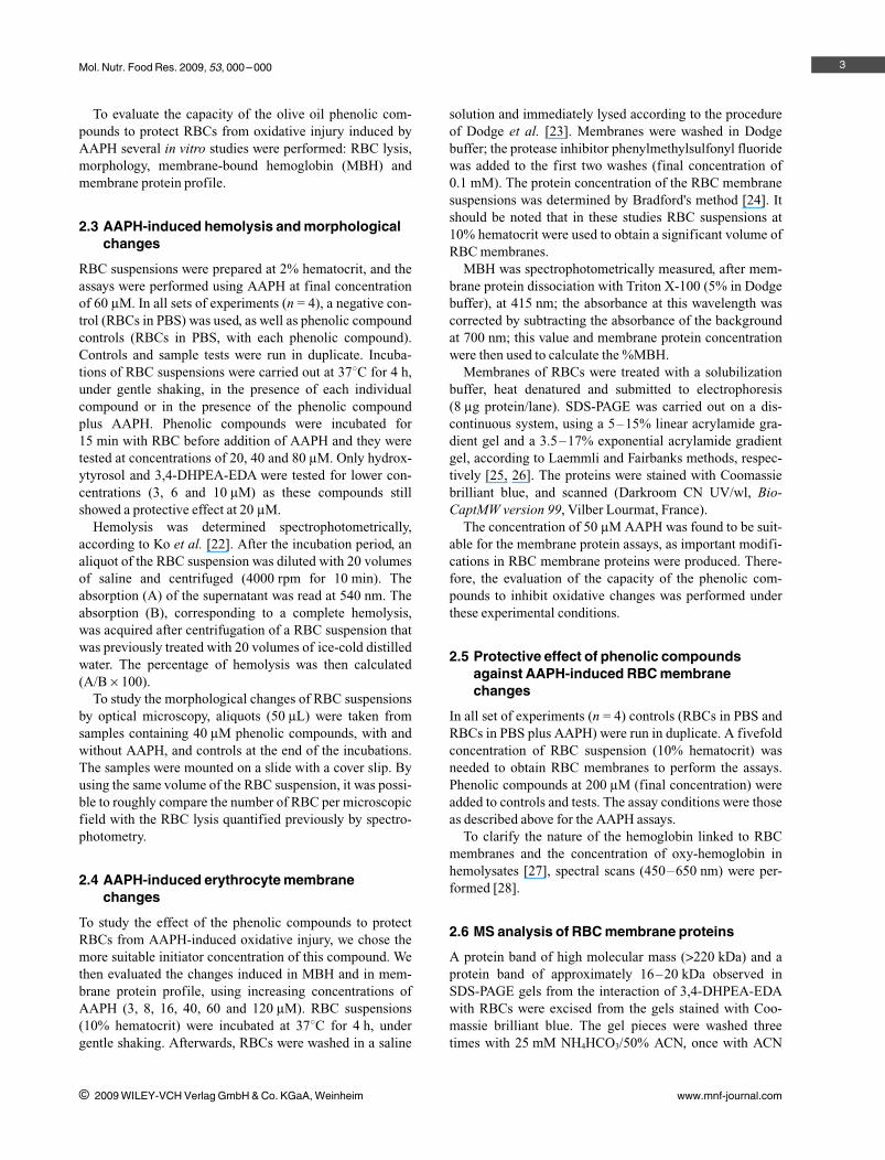
RBC lysis experiments showed that all compounds sig-nificantly protected RBCs from oxidative AAPH-inducedhemolysis at a concentration of 80 lM (Fig. 2). At lowerconcentrations (20–40 lM), oleuropein, hydroxytyrosoland 3,4-DHPEA-EDA still protected RBCs from oxidativehemolysis in a dose-dependent manner. Nevertheless,oleuropein protection effect was modest at 20 lM. Theranking activity order at 20 lM was: 3,4-DHPEA-EDA A
hydroxytyrosol A oleuropein. At this concentration,3,4-DHPEA-EA did not show any protection. At the lowestconcentration tested (3 lM), 3,4-DHPEA-EDA andhydroxytyrosol still had an important protective activity(Fig. 2). The data obtained for hydroxytyrosol agree withthose acquired in a similar system by Manna et al. [17]. Inthis study, hydroxytyrosol also protected RBC from oxida-tive injury in a dose-dependent manner.
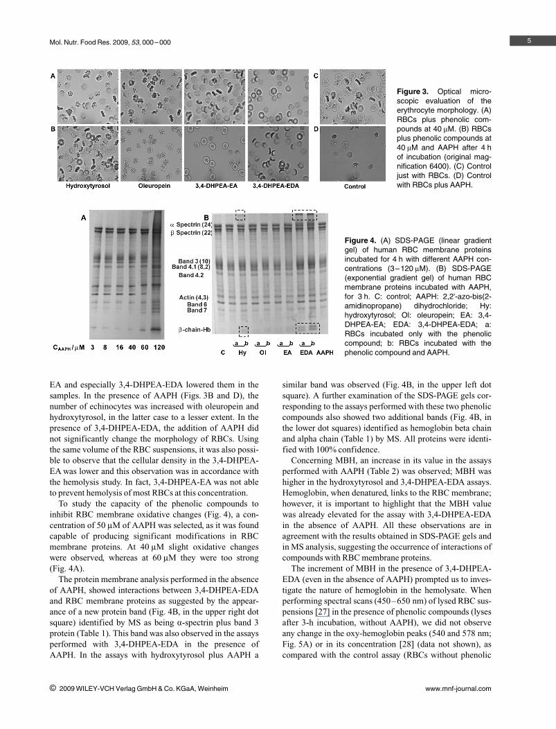
The RBCs morphology before and after exposure toAAPH in the absence and presence of 40 lM olive oil poly-phenols is illustrated in Fig. 3. At this concentration, olivephenolics, except 3,4-DHPEA-EA, were still able to protectRBC from hemolysis induced by AAPH. However, differ-ences in morphology could already be observed. Our datasuggest that the presence of polyphenols changed the RBCmorphology prior to the addition of AAPH (Figs. 3A andC). Hydroxytyrosol and oleuropein increased the number ofechinocytes produced by incubation, while 3,4-DHPEA-
4
i 2009 WILEY-VCH Verlag GmbH & Co. KGaA, Weinheim www.mnf-journal.com
Figure 2. Percentage of inhibition of lysis of RBCs at 2% hem-atocrit incubated for 4 h with 60 mM AAPH and phenolic com-pounds. Mean (error bars represent standard error) of fourdeterminations for each duplicate. Hy: hydroxytyrosol; Ol:oleuropein; EA: 3,4-DHPEA-EA; EDA: 3,4-DHPEA-EDA.Different letters within a concentration indicate samples thatwere significantly different (p a 0.05). * Sample not differentfrom control.
Mol. Nutr. Food Res. 2009, 53, 000 –000
EA and especially 3,4-DHPEA-EDA lowered them in thesamples. In the presence of AAPH (Figs. 3B and D), thenumber of echinocytes was increased with oleuropein andhydroxytyrosol, in the latter case to a lesser extent. In thepresence of 3,4-DHPEA-EDA, the addition of AAPH didnot significantly change the morphology of RBCs. Usingthe same volume of the RBC suspensions, it was also possi-ble to observe that the cellular density in the 3,4-DHPEA-EA was lower and this observation was in accordance withthe hemolysis study. In fact, 3,4-DHPEA-EA was not ableto prevent hemolysis of most RBCs at this concentration.
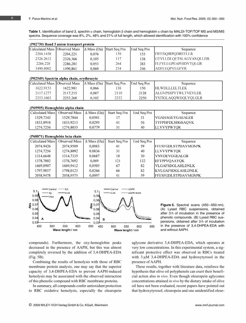
To study the capacity of the phenolic compounds toinhibit RBC membrane oxidative changes (Fig. 4), a con-centration of 50 lM of AAPH was selected, as it was foundcapable of producing significant modifications in RBCmembrane proteins. At 40 lM slight oxidative changeswere observed, whereas at 60 lM they were too strong(Fig. 4A).
The protein membrane analysis performed in the absenceof AAPH, showed interactions between 3,4-DHPEA-EDAand RBC membrane proteins as suggested by the appear-ance of a new protein band (Fig. 4B, in the upper right dotsquare) identified by MS as being a-spectrin plus band 3protein (Table 1). This band was also observed in the assaysperformed with 3,4-DHPEA-EDA in the presence ofAAPH. In the assays with hydroxytyrosol plus AAPH a
similar band was observed (Fig. 4B, in the upper left dotsquare). A further examination of the SDS-PAGE gels cor-responding to the assays performed with these two phenoliccompounds also showed two additional bands (Fig. 4B, inthe lower dot squares) identified as hemoglobin beta chainand alpha chain (Table 1) by MS. All proteins were identi-fied with 100% confidence.
Concerning MBH, an increase in its value in the assaysperformed with AAPH (Table 2) was observed; MBH washigher in the hydroxytyrosol and 3,4-DHPEA-EDA assays.Hemoglobin, when denatured, links to the RBC membrane;however, it is important to highlight that the MBH valuewas already elevated for the assay with 3,4-DHPEA-EDAin the absence of AAPH. All these observations are inagreement with the results obtained in SDS-PAGE gels andin MS analysis, suggesting the occurrence of interactions ofcompounds with RBC membrane proteins.
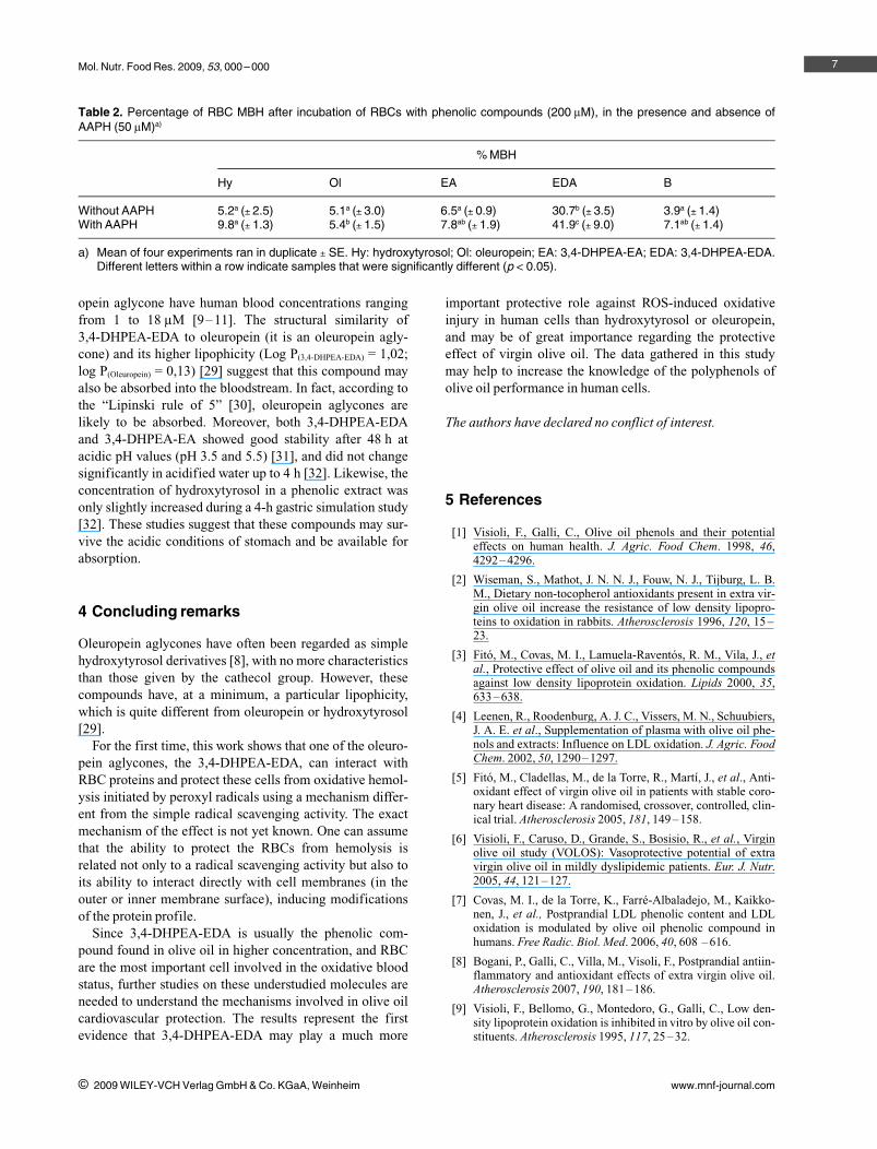
The increment of MBH in the presence of 3,4-DHPEA-EDA (even in the absence of AAPH) prompted us to inves-tigate the nature of hemoglobin in the hemolysate. Whenperforming spectral scans (450–650 nm) of lysed RBC sus-pensions [27] in the presence of phenolic compounds (lysesafter 3-h incubation, without AAPH), we did not observeany change in the oxy-hemoglobin peaks (540 and 578 nm;Fig. 5A) or in its concentration [28] (data not shown), ascompared with the control assay (RBCs without phenolic
5
i 2009 WILEY-VCH Verlag GmbH & Co. KGaA, Weinheim www.mnf-journal.com
Figure 3. Optical micro-scopic evaluation of theerythrocyte morphology. (A)RBCs plus phenolic com-pounds at 40 lM. (B) RBCsplus phenolic compounds at40 lM and AAPH after 4 hof incubation (original mag-nification 6400). (C) Controljust with RBCs. (D) Controlwith RBCs plus AAPH.
Figure 4. (A) SDS-PAGE (linear gradientgel) of human RBC membrane proteinsincubated for 4 h with different AAPH con-centrations (3–120 lM). (B) SDS-PAGE(exponential gradient gel) of human RBCmembrane proteins incubated with AAPH,for 3 h. C: control; AAPH: 2,2'-azo-bis(2-amidinopropane) dihydrochloride; Hy:hydroxytyrosol; Ol: oleuropein; EA: 3,4-DHPEA-EA; EDA: 3,4-DHPEA-EDA; a:RBCs incubated only with the phenoliccompound; b: RBCs incubated with thephenolic compound and AAPH.
F. Paiva-Martins et al. Mol. Nutr. Food Res. 2009, 53, 000 –000
compounds). Furthermore, the oxy-hemoglobin peaksdecreased in the presence of AAPH, but this was almostcompletely reversed by the addition of 3,4-DHPEA-EDA(Fig. 5B).
Combining the results of hemolysis with those of RBCmembrane protein analysis, one may say that the superiorcapacity of 3,4-DHPEA-EDA to prevent AAPH-inducedhemolysis may be associated with the observed interactionof this phenolic compound with RBC membrane proteins.
In summary, all compounds confer antioxidant protectionto RBC oxidative hemolysis, especially the oleuropein
aglycone derivative 3,4-DHPEA-EDA, which operates atvery low concentrations. In this experimental system, a sig-nificant protective effect was observed in RBCs treatedwith 3 lM 3,4-DHPEA-EDA and hydroxytyrosol in thepresence of AAPH.
These results, together with literature data, reinforce thehypothesis that olive oil polyphenols can exert their benefi-cial action also in vivo. Even though oleuropein aglyconesconcentrations attained in vivo by the dietary intake of oliveoil have not been evaluated, recent papers have pointed outthat hydroxytyrosol, oleuropein and one unidentified oleur-
6
i 2009 WILEY-VCH Verlag GmbH & Co. KGaA, Weinheim www.mnf-journal.com
Table 1. Identification of band 3, spectrin a chain, hemoglobin b chain and hemoglobin a chain by MALDI-TOF/TOF MS and MS/MSspectra. Sequence coverage was 8%, 2%, 48% and 21% of full length, which allowed identification with 100% confidence
Figure 5. Spectral scans (450–650 nm).(A) Lysed RBC suspensions, obtainedafter 3 h of incubation in the presence ofphenolic compounds. (B) Lysed RBC sus-pensions, obtained after 3 h of incubationin the presence of 3,4-DHPEA-EDA withand without AAPH.
Mol. Nutr. Food Res. 2009, 53, 000 –000
opein aglycone have human blood concentrations rangingfrom 1 to 18 lM [9–11]. The structural similarity of3,4-DHPEA-EDA to oleuropein (it is an oleuropein agly-cone) and its higher lipophicity (Log P(3,4-DHPEA-EDA) = 1,02;log P(Oleuropein) = 0,13) [29] suggest that this compound mayalso be absorbed into the bloodstream. In fact, according tothe “Lipinski rule of 5” [30], oleuropein aglycones arelikely to be absorbed. Moreover, both 3,4-DHPEA-EDAand 3,4-DHPEA-EA showed good stability after 48 h atacidic pH values (pH 3.5 and 5.5) [31], and did not changesignificantly in acidified water up to 4 h [32]. Likewise, theconcentration of hydroxytyrosol in a phenolic extract wasonly slightly increased during a 4-h gastric simulation study[32]. These studies suggest that these compounds may sur-vive the acidic conditions of stomach and be available forabsorption.
4 Concluding remarks
Oleuropein aglycones have often been regarded as simplehydroxytyrosol derivatives [8], with no more characteristicsthan those given by the cathecol group. However, thesecompounds have, at a minimum, a particular lipophicity,which is quite different from oleuropein or hydroxytyrosol[29].
For the first time, this work shows that one of the oleuro-pein aglycones, the 3,4-DHPEA-EDA, can interact withRBC proteins and protect these cells from oxidative hemol-ysis initiated by peroxyl radicals using a mechanism differ-ent from the simple radical scavenging activity. The exactmechanism of the effect is not yet known. One can assumethat the ability to protect the RBCs from hemolysis isrelated not only to a radical scavenging activity but also toits ability to interact directly with cell membranes (in theouter or inner membrane surface), inducing modificationsof the protein profile.
Since 3,4-DHPEA-EDA is usually the phenolic com-pound found in olive oil in higher concentration, and RBCare the most important cell involved in the oxidative bloodstatus, further studies on these understudied molecules areneeded to understand the mechanisms involved in olive oilcardiovascular protection. The results represent the firstevidence that 3,4-DHPEA-EDA may play a much more
important protective role against ROS-induced oxidativeinjury in human cells than hydroxytyrosol or oleuropein,and may be of great importance regarding the protectiveeffect of virgin olive oil. The data gathered in this studymay help to increase the knowledge of the polyphenols ofolive oil performance in human cells.
The authors have declared no conflict of interest.
5 References
[1] Visioli, F., Galli, C., Olive oil phenols and their potentialeffects on human health. J. Agric. Food Chem. 1998, 46,4292–4296.
[2] Wiseman, S., Mathot, J. N. N. J., Fouw, N. J., Tijburg, L. B.M., Dietary non-tocopherol antioxidants present in extra vir-gin olive oil increase the resistance of low density lipopro-teins to oxidation in rabbits. Atherosclerosis 1996, 120, 15 –23.
[3] Fit�, M., Covas, M. I., Lamuela-Ravent�s, R. M., Vila, J., etal., Protective effect of olive oil and its phenolic compoundsagainst low density lipoprotein oxidation. Lipids 2000, 35,633–638.
[4] Leenen, R., Roodenburg, A. J. C., Vissers, M. N., Schuubiers,J. A. E. et al., Supplementation of plasma with olive oil phe-nols and extracts: Influence on LDL oxidation. J. Agric. FoodChem. 2002, 50, 1290–1297.
[5] Fit�, M., Cladellas, M., de la Torre, R., Mart�, J., et al., Anti-oxidant effect of virgin olive oil in patients with stable coro-nary heart disease: A randomised, crossover, controlled, clin-ical trial. Atherosclerosis 2005, 181, 149 –158.
[6] Visioli, F., Caruso, D., Grande, S., Bosisio, R., et al., Virginolive oil study (VOLOS): Vasoprotective potential of extravirgin olive oil in mildly dyslipidemic patients. Eur. J. Nutr.2005, 44, 121–127.
[7] Covas, M. I., de la Torre, K., Farr�-Albaladejo, M., Kaikko-nen, J., et al., Postprandial LDL phenolic content and LDLoxidation is modulated by olive oil phenolic compound inhumans. Free Radic. Biol. Med. 2006, 40, 608 –616.
[8] Bogani, P., Galli, C., Villa, M., Visoli, F., Postprandial antiin-flammatory and antioxidant effects of extra virgin olive oil.Atherosclerosis 2007, 190, 181–186.
[9] Visioli, F., Bellomo, G., Montedoro, G., Galli, C., Low den-sity lipoprotein oxidation is inhibited in vitro by olive oil con-stituents. Atherosclerosis 1995, 117, 25 –32.
7
i 2009 WILEY-VCH Verlag GmbH & Co. KGaA, Weinheim www.mnf-journal.com
Table 2. Percentage of RBC MBH after incubation of RBCs with phenolic compounds (200 lM), in the presence and absence ofAAPH (50 lM)a)
% MBH
Hy Ol EA EDA B
Without AAPH 5.2a (l 2.5) 5.1a (l 3.0) 6.5a (l 0.9) 30.7b (l 3.5) 3.9a (l 1.4)With AAPH 9.8a (l 1.3) 5.4b (l 1.5) 7.8ab (l 1.9) 41.9c (l 9.0) 7.1ab (l 1.4)
a) Mean of four experiments ran in duplicate l SE. Hy: hydroxytyrosol; Ol: oleuropein; EA: 3,4-DHPEA-EA; EDA: 3,4-DHPEA-EDA.Different letters within a row indicate samples that were significantly different (p a 0.05).
F. Paiva-Martins et al. Mol. Nutr. Food Res. 2009, 53, 000 –000
[10] Coni, E., Benedetto, R., Pasquale, M., Masella, R., et al., Pro-tective effect of oleuropein, an olive oil biophenol, on lowdensity lipoprotein oxidizability in rabbits. Lipids 2000, 35,45–53.
[11] Covas, M. I., Fito, M., Lamuela-Raventos, R. M., Sebastia,N., et al., Virgin olive oil phenolic compounds: Binding tohuman low density lipoprotein (LDL) and effect on LDL oxi-dation. Int. J. Clin. Pharmacol. Res. 2000, 20, 49–54.
[12] Fito', M., De La Torre, R., Covas, M. I., Olive oil and oxida-tive stress. Mol. Nutr. Food Res. 2007, 51, 1215–1224.
[13] Terao, J., Piskula, M., Yao, Q., Protective effect of epicate-chin, epicatechin gallate, and quercetin on lipid peroxidationin phospholipid bilayers. Arch. Biochem. Biophys. 1994, 308,278–284.
[14] Garc�a, A., Brenes, M., Mart�nez, F., Alba, J., et al., High-per-formance liquid chromatography evaluation of phenols in vir-gin olive oil during extraction at laboratory and industrialscale. J. Am. Oil Chem. Soc. 2001, 78, 625–629.
[15] Tovar, M. J., Moltiva, M. J., Romero, M. P., Changes in thephenolic composition of virgin olive oil from young trees(Olea europaea L. cv. Arbequina) grown under linear irriga-tion strategies. J. Agric. Food Chem. 2001, 49, 5502 –5508.
[16] Brenes, M., Garc�a, A., Garc�a, P., Garrido, A., Acid hydroly-sis of secoiridoid aglycons during storage of virgin olive oil.J. Agric. Food Chem. 2001, 49, 5609–5614.
[17] Manna, C., Galletti, P., Cucciolla, V., Montedoro, G., Azppia,V., Olive oil hydroxytyrosol protects human erythrocytesagainst oxidative damages. J. Nutr. Biochem. 1999, 10, 159 –165.
[18] Baraldi, P. G., Simoni, D., Manfredini, S., Menziani, E., Prep-aration of 3,4-dihydroxy-1-benzeneethanol: A reinvestiga-tion. Liebigs Ann. Chem. 1983, 684–686.
[19] Gariboldi, P., Jommi, G., Verotta, L., Secoiridoids from Oleaeuropaea. Phytochemistry 1986, 25, 865 –869.
[20] Limiroli, R., Consonni, R., Ottolina, G., Marsilo, V., et al., 1HNMR and 13C NMR characterization of new oleuropein agly-cones. J. Chem. Soc. Perkin Trans. 1995, 1, 1519 –1523.
[21] Paiva-Martins, F., Gordon, M. H., Isolation and characteriza-tion of the antioxidant component 3,4-dihydroxyphenylethyl4-formyl-3-formylmethyl-4-hexenoate from olive (Oleaeuropaea). J. Agric. Food Chem. 2001, 49, 4214 –4219.
[22] Ko, F. N., Hsiao, G., Kuo, Y. H., Protection of oxidativehemolysis by demethyldiisoeugenol in normal and b-thalas-semic red blood cells. Free Radic. Biol. Med. 1997, 22, 215 –222.
[23] Dodge, J. T., Mitchell, C., Hanahan, D. J., The preparationand chemical characteristics of hemoglobin-free ghosts ofhuman erythrocytes. Arch. Biochem. Biophys. 1963, 100,119–130.
[24] Bradford, M. M., A rapid and sensitive method for the quanti-fication of microgram quantities of protein utilizing the prin-ciple of the protein dye binding. Anal. Biochem. 1976, 72,248–254.
[25] Laemmli, U. K., Cleavage of structural proteins during theassembly of the head of the bacteriophage T. Nature 1970,227, 680–685.
[26] Fairbanks, G., Steck, T. L., Wallach, D. F. H., Electrophoresisof the major polypeptides of the human erythocyte mem-brane. Biochemistry 1971, 10, 2606 –2616.
[27] Blakney, G. B., Dinwoodie, A. J., A spectrophotometricscanning technique for the rapid determination of plasmahemoglobin. Clin. Biochem. 1975, 8, 96–102.
[28] Lewis, S. M., Roper, D., in: Lewis, S. M., Bain, B. J., Bates, I.(Eds.), Dacie and Lewis Practical Haematology, ChurchillLivingstone, New York 2002, pp. 149–198.
[29] Paiva-Martins, F., Gordon, M. H., Gameiro, P., Activity andlocation of olive oil phenolic antioxidants in liposomes.Chem. Phys. Lipids 2003, 124, 26–34.
[30] Lipinski, C. A., Lombardo, F., Dominy, B. W., Feeney, P. J.,Experimental and computational approaches to estimate sol-ubility and permeability in drug discovery and developmentsettings. Adv. Drug Deliv. Rev. 1997, 23, 3–25.
[31] Paiva-Martins, F., Gordon, M. H., Interactions of ferric ionswith olive oil phenolic compounds. J. Agric. Food Chem2005, 53, 2704 –2709.
[32] Romero, C., Medina, E., Vargas, J., Brenes, M., De Castro,A., In vitro activity of olive oil polyphenols against Helico-bacter pylori. J. Agric. Food Chem. 2007, 55, 680 –686.
8
i 2009 WILEY-VCH Verlag GmbH & Co. KGaA, Weinheim www.mnf-journal.com